Role of follicle-stimulating hormone on biliary cyst growth in autosomal dominant polycystic kidney...
12
LIVER PATHOBIOLOGY Role of follicle-stimulating hormone on biliary cyst growth in autosomal dominant polycystic kidney disease Paolo Onori 1 , Romina Mancinelli 1 , Antonio Franchitto 1,2 , Guido Carpino 3 , Anastasia Renzi 1 , Stefania Brozzetti 4 , Julie Venter 5 , Heather Francis 5 , Shannon Glaser 5 , Douglas M. Jefferson 6 , Gianfranco Alpini 5 and Eugenio Gaudio 1 1 Department of Anatomical, Histological, Forensic Medicine and Orthopedic Sciences, University of Rome ‘Sapienza’, Rome, Italy 2 Eleonora Lorillard Spencer-Cenci Foundation, Rome, Italy 3 Department of Health Science, University of Rome ‘Foro Italico’, Rome, Italy 4 Department of Surgical Sciences, University of Rome ‘Sapienza’, Rome, Italy 5 Scott & White Digestive Disease Research Center, Central Texas Veterans Health Care System and Texas A&M Health Science Center, College of Medicine, Temple, TX, USA 6 Department of Physiology, Tufts University, Boston, MA, USA Keywords autosomal dominant polycystic kidney disease – biliary epithelium – follicle- stimulating hormone – immunohistochemistry Correspondence Professor Eugenio Gaudio, MD, Department of Anatomical, Histological, Forensic Medicine and Orthopedic Sciences, ‘Sapienza’ University of Rome, Via A. Borelli, 50-00161 Rome, Italy Tel: +39 06 4991 8060 Fax: +39 06 4991 8062 e-mail: [email protected] Received 5 April 2012 Accepted 11 March 2013 DOI:10.1111/liv.12177 Liver Int. 2013: 33: 914–925 Abstract Background: Autosomal dominant polycystic kidney disease (ADPKD) is a common genetic disorder characterized by the progressive development of renal and hepatic cysts. Follicle-stimulating hormone (FSH) has been dem- onstrated to be a trophic factor for biliary cells in normal rats and experi- mental cholestasis induced by bile duct ligation (BDL). Aims: To assess the effect of FSH on cholangiocyte proliferation during ADPKD using both in vivo and in vitro models. Methods: Evaluation of FSH receptor (FSHR), FSH, phospho-extracellular-regulated kinase (pERK) and c-myc expression in liver fragments from normal patients and patients with ADPKD. In vitro, we studied proliferating cell nuclear antigen (PCNA) and cAMP levels in a human immortalized, non-malignant cholangiocyte cell line (H69) and in an immortalized cell line obtained from the epithelium lining the hepatic cysts from the patients with ADPKD (LCDE) with or without transient silencing of the FSH gene. Results: Follicle-stimulating hormone is linked to the active proliferation of the cystic wall and to the localization of p-ERK and c-myc. This hormone sustains the biliary growth by activation of the cAMP/ERK sig- nalling pathway. Conclusion: These results showed that FSH has an impor- tant function in cystic growth acting on the cAMP pathway, demonstrating that it provides a target for medical therapy of hepatic cysts during ADPKD. Polycystic liver disease phenotypes arise from two dis- tinct inherited diseases, autosomal dominant polycystic kidney disease (ADPKD) and polycystic liver disease (PCLD). ADPKD, caused by mutations in PKD1 or PKD2 genes, is characterized by polycystic kidneys (1). In many patients with ADPKD, there is the develop- ment of a polycystic liver manifestation. On the other hand, PCLD is caused by mutations in PRKCSH or SEC63 genes and is characterized by the presence of an isolated polycystic liver without the kidney phenotype (2, 3). The diagnosis of polycystic liver is usually made during the third or fourth decade of life with hepatic capacity preserved in the great majority of patients (4, 5). This disease is usually asymptomatic, but the progressive growth of the liver cysts may cause dyspnoea, gastro- oesophageal reflux, nausea and mechanical low back pain arise because of the mass effect of the polycystic liver (6). Severe ADPKD primarily affects women and is characterized by the massive cystic liver disease. The number and size of hepatic cysts correlate with the occurrence of pregnancy, female gender, increased age and severity of the renal lesion (7). Treatment is initi- ated only in those with the symptoms and all interven- tional procedures are aimed to reduce liver volume (5). In the last few years, the number of studies to discover viable medical options has increased with indications that somatostatin analogues or mTOR inhibitors may slow cyst growth (8–10). Many experimental and clinical studies have demon- strated that cholangiocytes respond to hormones, growth factors, neuropeptides and cytokines increasing their proliferative capacity (11–13). In particular, oestrogens play a key role in sustaining cholangiocyte growth, cyst formation and progression in ADPKD patients. Oestro- gens act not only directly, but also by promoting the synthesis and release of other growth factors from the cystic epithelium (14). Additional sex hormones such as Liver International (2013) © 2013 John Wiley & Sons A/S. Published by John Wiley & Sons Ltd 914 Liver International ISSN 1478-3223
-
Upload
independent -
Category
Documents
-
view
0 -
download
0
Transcript of Role of follicle-stimulating hormone on biliary cyst growth in autosomal dominant polycystic kidney...

L IVER PATHOBIOLOGY
Role of follicle-stimulating hormone on biliary cyst growth in autosomal
dominant polycystic kidney disease
Paolo Onori 1, Romina Mancinelli1, Antonio Franchitto1,2, Guido Carpino3, Anastasia Renzi1, Stefania Brozzetti4,Julie Venter5, Heather Francis5, Shannon Glaser5, Douglas M. Jefferson6, Gianfranco Alpini5 and Eugenio Gaudio1
1 Department of Anatomical, Histological, Forensic Medicine and Orthopedic Sciences, University of Rome ‘Sapienza’, Rome, Italy
2 Eleonora Lorillard Spencer-Cenci Foundation, Rome, Italy
3 Department of Health Science, University of Rome ‘Foro Italico’, Rome, Italy
4 Department of Surgical Sciences, University of Rome ‘Sapienza’, Rome, Italy
5 Scott & White Digestive Disease Research Center, Central Texas Veterans Health Care System and Texas A&M Health Science Center, College of
Medicine, Temple, TX, USA
6 Department of Physiology, Tufts University, Boston, MA, USA
Keywords
autosomal dominant polycystic kidney
disease – biliary epithelium – follicle-
stimulating hormone –
immunohistochemistry
Correspondence
Professor Eugenio Gaudio, MD, Department
of Anatomical, Histological, Forensic
Medicine and Orthopedic Sciences,
‘Sapienza’ University of Rome, Via A. Borelli,
50-00161 Rome, Italy
Tel: +39 06 4991 8060
Fax: +39 06 4991 8062
e-mail: [email protected]
Received 5 April 2012
Accepted 11 March 2013
DOI:10.1111/liv.12177
Liver Int. 2013: 33: 914–925
Abstract
Background: Autosomal dominant polycystic kidney disease (ADPKD) is acommon genetic disorder characterized by the progressive development ofrenal and hepatic cysts. Follicle-stimulating hormone (FSH) has been dem-onstrated to be a trophic factor for biliary cells in normal rats and experi-mental cholestasis induced by bile duct ligation (BDL). Aims: To assess theeffect of FSH on cholangiocyte proliferation during ADPKD using bothin vivo and in vitro models. Methods: Evaluation of FSH receptor (FSHR),FSH, phospho-extracellular-regulated kinase (pERK) and c-myc expressionin liver fragments from normal patients and patients with ADPKD. In vitro,we studied proliferating cell nuclear antigen (PCNA) and cAMP levels in ahuman immortalized, non-malignant cholangiocyte cell line (H69) and in animmortalized cell line obtained from the epithelium lining the hepatic cystsfrom the patients with ADPKD (LCDE) with or without transient silencingof the FSH gene. Results: Follicle-stimulating hormone is linked to the activeproliferation of the cystic wall and to the localization of p-ERK and c-myc.This hormone sustains the biliary growth by activation of the cAMP/ERK sig-nalling pathway. Conclusion: These results showed that FSH has an impor-tant function in cystic growth acting on the cAMP pathway, demonstratingthat it provides a target for medical therapy of hepatic cysts during ADPKD.
Polycystic liver disease phenotypes arise from two dis-tinct inherited diseases, autosomal dominant polycystickidney disease (ADPKD) and polycystic liver disease(PCLD). ADPKD, caused by mutations in PKD1 orPKD2 genes, is characterized by polycystic kidneys (1).In many patients with ADPKD, there is the develop-ment of a polycystic liver manifestation. On the otherhand, PCLD is caused by mutations in PRKCSH orSEC63 genes and is characterized by the presence of anisolated polycystic liver without the kidney phenotype(2, 3).
The diagnosis of polycystic liver is usually madeduring the third or fourth decade of life with hepaticcapacity preserved in the great majority of patients (4, 5).This disease is usually asymptomatic, but the progressivegrowth of the liver cysts may cause dyspnoea, gastro-oesophageal reflux, nausea and mechanical low backpain arise because of the mass effect of the polycysticliver (6). Severe ADPKD primarily affects women and is
characterized by the massive cystic liver disease. Thenumber and size of hepatic cysts correlate with theoccurrence of pregnancy, female gender, increased ageand severity of the renal lesion (7). Treatment is initi-ated only in those with the symptoms and all interven-tional procedures are aimed to reduce liver volume (5).In the last few years, the number of studies to discoverviable medical options has increased with indicationsthat somatostatin analogues or mTOR inhibitors mayslow cyst growth (8–10).
Many experimental and clinical studies have demon-strated that cholangiocytes respond to hormones, growthfactors, neuropeptides and cytokines increasing theirproliferative capacity (11–13). In particular, oestrogensplay a key role in sustaining cholangiocyte growth, cystformation and progression in ADPKD patients. Oestro-gens act not only directly, but also by promoting thesynthesis and release of other growth factors from thecystic epithelium (14). Additional sex hormones such as
Liver International (2013)© 2013 John Wiley & Sons A/S. Published by John Wiley & Sons Ltd914
Liver International ISSN 1478-3223
prolactin (15), progesterone (16) and follicle-stimulatinghormone (FSH) (17) regulate biliary function.
Many events in the adult ovary are controlled by twohormones, FSH and luteinizing hormone (LH) secretedfrom the anterior pituitary gland under the control ofgonadotropin-releasing hormone (GnRH) from thehypothalamus. FSH is required for granulosa cell differ-entiation and facilitates the follicular growth (18). In theclassical cascade, occupancy of FSH receptor (FSHR)causes activation of the heterotrimeric GS protein,which stimulates the effector adenylyl cyclase with theconsequent increase in the synthesis of the second mes-senger cAMP (19, 20).
One of the most characterized components of theMAPK family is the extracellular-regulated kinase(ERK). The ERK pathway regulates cell proliferation,differentiation and cell survival (21). C-myc represents akey downstream target of this mechanism (22). Othershave demonstrated that c-myc participates in the pro-gression of the G1-cell cycle phase by enhancing cyclinexpression (23) and CDK/cyclin complex activities (24).Lastly, both c-myc and ERK, as a consequence of theirmarked capacity to promote proliferation, play a pivotalrole in the control of the differentiation programme inseveral cell types (25–27). We have previously shownthat the cAMP/ERK-dependent signalling mechanism isactivated in proliferating cholangiocytes (13, 28). Inparticular, in the hyperplastic BDL model, cholangiocyteproliferation is closely associated with increased cAMPlevels (29–32). It has been demonstrated that FSH playsan important role in stimulating rat cholangiocyte pro-liferation through an autocrine mechanism that is asso-ciated with increased cAMP-dependent phosphorylationof ERK1/2 and Elk-1 both in vivo and in vitro (17).However, no information exists regarding the role ofFSH and its receptors in the regulation of epithelial cellgrowth in the hepatic cysts. The aim of this study was toevaluate the hypothesis that FSH regulates hepatic cystsgrowth during the course of ADPKD.
Materials and methods
Materials
All reagents were purchased from Sigma Chemical (St.Louis, MO, USA) unless otherwise indicated. The FSHprimers for real-time PCR were purchased from SABio-sciences (Frederick, MD, USA). The RNeasy Mini Kit topurify total cholangiocyte RNA was purchased fromQiagen Inc. (Valencia, CA, USA). The RIA kits for themeasurement of intracellular cAMP [cAMP (125I) Biot-rak Assay System, RPA509] were purchased from GEHealthcare (Arlington Heights, IL, USA). All the anti-bodies used for immunohistochemistry, immunofluo-rescence and western blots were purchased from SantaCruz Biotechnology (Santa Cruz, CA, USA), Invitrogensrl (Milan, Italy), Dako Italia S.p.A. (Milan, Italy) orfrom Abcam (Cambridge, UK).
Human liver samples
We studied eight patients (six females and two males,61–78 years of age) with a diagnosis of ADPKD basedon the standard international criteria (33). Liver cystswere subdivided on the basis of their size in large(>3 cm maximum diameter) or small cysts (<3 cmmaximum diameter) as previously showed (14). As con-trols, we evaluated liver biopsies with a normal histologyfrom patients submitted to laparotomy (4 fragments, 2from female and 2 from male, 59–75 years of age). Thisstudy protocol was approved by the institutionalcommittee and abided by the ethical guidelines of the1975 Declaration of Helsinki.
Immunohistochemistry
Immunohistochemistry was performed in 3–4 lm sec-tions. Sections were deparaffinized and endogenous per-oxidase activity was blocked by a 30-min incubation inmethanolic hydrogen peroxide (2.5%). Later, theendogenous biotin was blocked by a biotin blocking sys-tem (code X0590; Dako, Copenhagen, Denmark)according to the instructions supplied by the vendor.Sections were then hydrated in graded alcohol andrinsed in 1x phosphate-buffered saline (PBS, pH 7.4)before applying the selected primary antibody. Sectionswere incubated overnight at 4°C with polyclonal anti-bodies for CK-19 (M0888; Dako Italia S.p.A.), FSHR(sc-7798; Santa Cruz), FSHb (sc-7797; Santa Cruz),pERK (sc-7383; Santa Cruz) or c-myc (ab39688;Abcam). The following day, samples were rinsed withPBS for 5 min, incubated for 20 min at room tempera-ture (RT) with secondary biotinylated antibody (LSABPlus system; Dako, Milan, Italy), then with DakoABC (LSAB Plus system), and finally developed with3,3′-diaminobenzidine. To confirm the specificity ofimmunoreaction, negative controls were performed forall immunoreactions.
Sections were examined with a Leica MicrosystemsDM 4500 B Microscopy (Weltzlar, Germany) equippedwith a Jenoptik Prog Res C10 Plus Videocam (Jena, Ger-many). Observations were processed with an ImageAnalysis System (IAS; Delta Sistemi, Rome, Italy) andwere independently performed by two researchers in ablinded fashion. The number of positive cells wascounted in six non-overlapping fields (magnification920) for each slide. The data are expressed as per centpositive cells (34).
Immunofluorescence
For double immunofluorescence, sections werehydrated in graded alcohol and rinsed in 1x PBS with0.1% Triton X (PBS-T) for 15 min and then incubatedwith 10% normal blocking serum in 1x PBS for30 min at RT. After washing, slides were incubatedovernight at 4°C with FSHR (sc-7798; goat polyclonal;
Liver International (2013)© 2013 John Wiley & Sons A/S. Published by John Wiley & Sons Ltd 915
Onori et al. Role of FSH on biliary cyst in ADPKD

Santa Cruz) primary antibodies and proliferating cellnuclear antigen (PCNA) (PC10, sc-7907; rabbit poly-clonal; Santa Cruz) or with the same FSHR and pERK(sc-7383; mouse monoclonal; Santa Cruz) diluted inPBS with 1.5% normal blocking serum. Samples wererinsed in PBS-T with three changes and incubated for45 min at RT with the specific secondary antibodiesconjugated with Alexa fluorochrome (488 or 594)diluted in 1x PBS with 1.5% normal blocking serum.Then the samples were washed in buffer and mountedwith Ultra-Cruz mounting medium (sc-24941; SantaCruz). Images were taken by DM4500B light micros-copy (Leica).
Regarding cellular staining, cholangiocytes from celllines were seeded on coverslip in a six-well-plate(500 000 per well) and allowed to adhere overnight.Immunofluorescence was performed by fixing cells in4% paraformaldehyde for 5 min and following washesand incubation in 4% bovine serum albumin (BSA), inPBS-T the cells were incubated with the selected pri-mary antibody (FSH, FSHR or pERK). After 1 h at RT,the cells were washed three times in PBS-T and thenplaced in the specific Alexa Fluor 594 secondary anti-body in a dark room for 45 min. Finally, cholangiocyteswere rinsed and the coverslip was put onto slide with adrop of DAPI. In the same manner of immunohisto-chemistry, to demonstrate the specificity of the immu-noreaction, negative controls were performed withoutthe incubation with primary antibody.
H69 and LCDE cell lines
The in vitro studies were performed utilizing a humanimmortalized non-malignant cholangiocyte cell line(H69) and an immortalized cell line obtained from theepithelium lining the hepatic cysts from patients withADPKD (LCDE). Cells were maintained in hormonallysupplemented medium consisting of Dulbecco’s modi-fied Eagle’s medium-Ham’s F-12 nutrient mixture (3:1)(Cambrex Bio Science, Walkersville, MD, USA) supple-mented with 1.8 9 104 mol/L adenine (LKT; SantaCruz Biotechnology, Santa Cruz, CA, USA), 5 g/mlinsulin, 5 g/ml transferrin (Calbiochem Biochemicals,Darmstadt, Germany), 2 9 109 mol/L triiodothyronine,1.1 9 106 mol/L hydrocortisone, 1.64 9 106 humanepidermal growth factor, 5.5 9 106 epinephrine, 10%foetal bovine serum (Gibco/BRL, Life Technologies,Italia srl., Milan, Italy), 100 U/ml of penicillin and100 g/ml of streptomycin in a 5% CO2 atmosphere at37°C. To evaluate the effect of FSH on proliferation,H69 and LCDE cells following culture in the appropri-ate medium containing 10% foetal bovine serum weredeprived of serum for 24 h. Cells were then maintainedin serum-deprived conditions for an additional 24 h forMTS proliferation assay (controls) or exposed to serum,FSH (1–100 lg/ml) with or without PD98059 (10 nM),a MEK/ERK inhibitor. In detail, cell medium wasreplaced with a fresh serum-free medium without
hormone supplementation, but added with the testedagent. We used a commercially available colorimetriccell proliferation assay (CellTiter 96 aqueous nonradio-active cell proliferation assay, MTS Kit; Promega,Madison, WI, USA), following the manufacturer’sinstructions. Proliferation index was calculated as theratio (multiplied 9100) between cell numbers in bothunstimulated and stimulated cultures.
In addition, we measured intracellular cAMP levels.After incubation for 1 h at 37°C, cholangiocytes(1 9 105 cells) were stimulated at RT for 5 min with0.2% BSA (basal), or FSH (100 lg/ml in 0.2% BSA) inthe absence or presence of PD98059 or an anti-FSHRantibody (150 pg/ml) (17). Intracellular cAMP levelswere measured with a commercially available kit [cAMP(125I) Biotrak Assay System, RPA509].
FSH silencing
To evaluate the effects of FSH on LCDE, we used anavailable silencer small interfering RNA (siRNA) toknock down the expression of FSH before evaluating:(i) cholangiocyte proliferation by PCNA and biliaryapoptosis by Bax protein expression using immunoblot-ting analysis; and (ii) intracellular cAMP levels. LCDEwere plated into six-well plates and allowed to adhereovernight. siRNA transfection (0.25–1 lg of FSH siRNAwas used) was carried out according to the instructionsprovided by Santa Cruz. The extent of FSH silencingwas evaluated by measuring the expression of total FSHin transfected vs. control LCDE cells by real-time PCRand western blots for FSH expression. Cellular growthwas investigated by western blots for PCNA, whereasbiliary apoptosis was evaluated by Bax protein expres-sion. PCNA and Bax expression was performed inprotein (10 lg) from whole cell lysates from LCDEcholangiocytes. Blots were normalized by b-actin immu-noblots. The intensity of the bands was determined byscanning video densitometry using the phospho-imager,Storm 860 (GE Healthcare, Piscataway, NJ, USA) andthe ImageQuant TL software version 2003.02 (GEHealthcare, Little Chalfont, Buckinghamshire, UK).Finally, spontaneous and secretin-stimulated intracellu-lar cAMP levels were determined. Transfected andcontrol cholangiocytes were incubated for 2 h at 37°Cto restore secretin receptor that may be damaged withthe treatment of proteolytic enzymes (35). Cells werestimulated with 10!7 M secretin in 1% BSA or 1% BSAalone for 5 min at 22°C (36). After extraction with etha-nol, cAMP levels were determined by a commerciallyavailable kit (cAMP [125I] Biotrak Assay System,RPA509) according to the instructions of the vendor.
Statistical analysis
Data are presented as arithmetic mean ± standarddeviation. The Student’s t-test or Mann–Whitney U-testwas used to determine differences between groups for
Liver International (2013)© 2013 John Wiley & Sons A/S. Published by John Wiley & Sons Ltd916
Role of FSH on biliary cyst in ADPKD Onori et al.

normally or not normally distributed data respectively.A P-value of <0.05 was considered statistically signifi-cant. Statistical analyses were performed using SPSS
statistical software (SPSS Inc., Chicago, IL, USA).
Results
FSHR and FSH cholangiocyte expression
Hepatic cysts are lined by epithelial cells and these cystsbud from interlobular or smaller biliary ducts with phe-notypical and functional characteristics of biliary epithe-lium as shown in Fig. 1 (immunohistochemistry forcytokeratin 19, a specific marker of cholangiocytes). Bil-iary epithelium also displays immunopositivity forFSHR and FSH hormone in liver sections from normalpatients and patients affected with ADPKD (Fig. 2). Theimmunohistochemistry for FSHR appears negative incholangiocytes lining interlobular bile ducts in normallivers (Fig. 2A), whereas FSH is faintly positive(Fig. 2D). In contrast, FSHR and FSH were more posi-tive in the epithelial cells lining the smallest hepatic cysts(Fig. 2B,E) and strongly expressed in the largest cysts(Fig. 2C,F). The expression of FSH and FSHR is relatedto the cyst size. We found that the percentage of FSHR-positive cholangiocytes is 47 ± 25.1% in small cysts(diameter <3 cm) vs. 72.3 ± 26.2% (P < 0.05) in largecysts (diameter >3 cm). Similarly, the expression of thehormone FSH is higher in cholangiocytes lining largecysts (73.8 ± 19.8%) in comparison with small cysts(39.6 ± 19.4%; P < 0.05) (Fig. 2).
Intracellular mechanisms of FSH regulation ofcholangiocyte growth
As we have previously shown (14), the cystic epitheliumshowed a marked proliferative index. Normal cholan-giocytes have a low expression of pERK and c-myc, twokey proteins of the intracellular cAMP mechanism
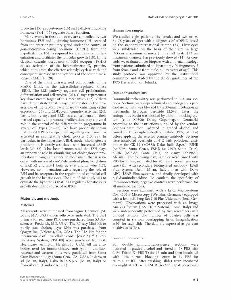
(Fig. 3A,D). In pathological cholangiocytes, the pres-ence of the two cAMP mediators increases in both smalland large cysts (Fig. 3B,C,E,F). The presence of pERK,the positivity for FSHR and the intense cholangiocyteproliferation in the course of ADPKD was confirmed byimmunofluorescence, where we initially co-localizedFSHR with PCNA (Fig. 4A) and then FSHR with pERK(Fig. 4B). In cystic cholangiocytes, FSHR presence maybe associated with a paracrine action, but in some cellsit can co-localize with PCNA thus sustaining an auto-crine mechanism (Fig. 4A). FSHR expression has alsobeen linked to the expression of pERK (Fig. 4B). Forthis reason, the phosphorylation of ERK is associatedwith the activation of the intracellular cAMP pathwayand many cells simultaneously express FSHR withPCNA and pERK with FSHR supporting the conceptthat FSH induces cholangiocyte proliferation via ERK(37).
Evaluation of the role of FSH in human cell lines
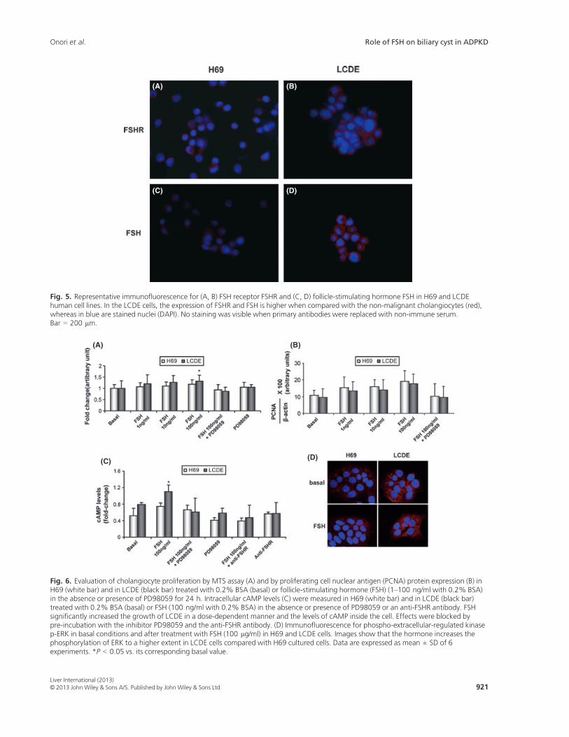
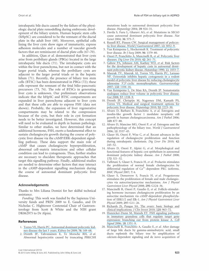
Both H69 and LCDE express FSHR and FSH (Fig. 5).These cells were starved without serum for 24 h andthen exposed to FSH with or without PD98059. Theaddition of FSH increased the cholangiocyte prolifera-tive index (tested by MTS proliferation assay andwestern blots for PCNA protein expression) whereaspre-incubation with PD98059 partially blocked thiseffect (Fig. 6A,B). To measure the intracellular levels ofcAMP, we treated normal and pathological cholangio-cytes with a basal solution of BSA or FSH in the absenceor presence of PD98059 or an anti-FHSR antibody. Sim-ilar to that shown for secretin (37), we found that FSHincreases cAMP levels, an increase that was prevented bypre-incubation with PD98059 or with the antibody anti-FSHR (Fig. 6C). Immunofluorescence for pERK in basalconditions and after treatment with the highest dose ofFSH (100 lg/ml) demonstrates that the hormoneincreases the phosphorylation of ERK to a higher extent
(A) (B) (C)
Fig. 1. Immunohistochemistry for CK-19, a specific marker of the biliary epithelium. (A) Normal liver demonstrates a characteristic portalspace, with some biliary ducts (stained in brown for the immunoreaction), a branch of the portal vein (the largest blood vessel) and a termi-nal ramification of hepatic artery (the smallest blood vessel). (B) A small cyst with a diameter <1 cm lined by a columnar biliary epithelium.(C) A large cyst with a diameter >3 cm is enclosed with cholangiocytes that are flatter in appearance compared with the non-cystic biliarycells, because of the enlargement of the cyst and the resulting epithelium strain. CK-19 cholangiocytes are stained brown. Original magnifi-cation, 409.
Liver International (2013)© 2013 John Wiley & Sons A/S. Published by John Wiley & Sons Ltd 917
Onori et al. Role of FSH on biliary cyst in ADPKD

in LCDE cells compared with H69 cultured cells sup-porting enhanced cell proliferation (Fig. 6D).
To confirm the evidence that FSH is a critical factorfor sustaining cholangiocyte growth, we specificallyknocked down the expression of FSH in LCDE cells bytransient transfection (siRNA) (Fig. 7A,B). Real-timePCR for FSH showed that the most efficient siRNA-FSHconcentration was 1 lg, which results in the largest reduc-tion in FSH message expression (Fig. 7A). Furthermore,the FSH siRNA cell line exhibited reduced PCNA proteinexpression compared with mock-transfected cells,indicating that decreasing FSH expression impairs theproliferative capacity of cholangiocytes (Fig. 8A). Thesecells manifest a higher apoptotic degree compared withmock-transfected cholangiocytes as demonstrated byincreased Bax protein expression (Fig. 8B). Lastly, wefound that in the knocked-down cells, the intracellularsecretin-stimulated cAMP levels as well as cholangiocyteproliferation decrease (Fig. 8C). This supports theconcept that FSH sustains biliary growth via acAMP-dependent signalling pathway. In general, themodifications of cAMP levels after stimulation withsecretin are considered to be a reliable test to evaluate
the effects of secretin on cholangiocyte proliferation asextensively demonstrated in the experimental models ofcholangiocyte proliferation (37–39).
Discussion
Our in vivo results show that: (i) the biliary epitheliumthat lines hepatic cysts stains positive for FSHR andFSH, whose expression is in relationship with the cystsize; (ii) FSH sustains cellular growth; and (iii) FSHRco-localizes with pERK in larger cysts. Regarding the invitro studies, we demonstrated that: (i) both H69 andLCDE cells express FSHR and FSH; (ii) FSH stimulationof cholangiocyte proliferation is associated withincreased cAMP levels; and (iii) knocking down FSHexpression by siRNA decreases cholangiocyte prolifera-tion and cAMP levels while increasing apoptosis. Cystfragments were obtained from patients with ADPKDwho underwent liver resection. ADPKD is caused bymutation in the PKD1 gene (85%) or PKD2 gene(10–15%) (40), which encodes the polycystin 1 (Pc-1)and polycystin 2 (Pc-2) proteins (41) respectively. ThePc-1/Pc-2 complex is located in the primary cilium at
(A) (B) (C)
(D) (E) (F)
Fig. 2. Representative immunohistochemistry for FSH receptor (FSHR) and follicle-stimulating hormone (FSH) in liver sections (3–4 lm thick)from normal patients or patients with autosomal dominant polycystic kidney disease (ADPKD). In particular, the staining showed a negativityfor FSHR in liver from normal patient (A) and higher expression in small (B) and in large (C) cysts. The hormone FSH presents a low positivityin normal liver (D) that increases in cysts of small (E) and large diameter (F). Original magnification, 409. The table contains the percentageof FSHR and FSH-positive cholangiocytes in normal biliary epithelium and in both small and large cysts. The positivity for the hormone and itsreceptor enhances with the cyst size, suggesting an important effect of FSH in the growth of the hepatic cysts.
Liver International (2013)© 2013 John Wiley & Sons A/S. Published by John Wiley & Sons Ltd918
Role of FSH on biliary cyst in ADPKD Onori et al.

the apical pole of cholangiocytes (42). Recently, the keyrole of hormones such as oestrogens in this pathologyhas been studied in detail. Indeed, 1 year of oestrogenuse in post-menopausal ADPKD patients selectivelyincreases total liver volume by 7%, whereas total kidneyvolume remains unaffected (43). In addition, oestrogenssustain the enhanced proliferative and secretory activi-ties of biliary epithelium, as experimentally shown inBDL rats, by acting either directly with growth factorsor potentiating their effects (11, 44–46). Studies haveshown that the epithelial surface of hepatic cysts ofADPKD patients displays a marked and diffuse immu-noreaction for oestrogen receptors (14).
According to these recent findings, we hypothesizedthat the hepatic cyst epithelium of ADPKD patientscould be considered as a hormone-responsive tissue.Hence, we have studied the role of FSH in thepathophysiology of hepatic cysts. FSH stimulates pre-ovulatory follicles of the ovaries and is related to steroi-dogenesis (47). FSH induces cell proliferation and DNAsynthesis by acting on its receptor (FSHR) (48). Thehuman FSHR belongs to the superfamily of G protein-coupled receptors (49). Agonist binding to the FSHRtriggers the rapid activation of multiple signallingcascades, mainly the cAMP–adenylyl cyclase–protein-kinase A cascade (50). We have already demonstratedthat the FSH induces cholangiocyte proliferation innormal rats by acting on the cAMP-dependent ERK1/2–Elk-1 signalling pathway (17). This increase was par-tially blocked by treatment with Antide (a GnRHantagonist) or by a neutralizing FSH antibody (17). In
general, FSH represents the major stimulator and regu-lator of oestrogen production. In particular, FSH deter-mines the aromatization of androgens into oestrogensvia the activation of the cAMP/protein kinase A (PKA)-dependent transcription factor, leading to the transcrip-tion of the aromatase enzyme (51, 52).
In this study, we found that normal human cholan-giocytes from interlobular bile ducts and those derivedfrom biliary epithelium of hepatic cysts express FSHRand FSH. The increasing presence of this hormone iscorrelated with a higher proliferation index, most likelybecause of the effect of FSH on the cAMP/ERK-depen-dent signalling pathway, which is one of the most criti-cal intracellular mechanisms regulating cholangiocyteproliferation and phosphorylation of ERK (20, 28,53–55). This role of FSH may also be because of theeffects of this hormone on the transcriptional activationof ER-responsive genes that are under the regulation ofthe cAMP/PKA/ERK-signalling pathway (56, 57). Pre-sumably, in this case FSH cooperates with oestrogensand other hormones to increase the proliferativeresponse of the biliary epithelium (58, 59). A direct linkwas not found in these preliminary studies, which willbe the aim of further studies. However, the combinationof FSH and the activation of other hormones may resultin a synergistic effect on cell proliferation that may beattenuated using anti-oestrogens. To support our in vivofindings, the in vitro studies were expanded in twoways: (i) ablation of the proliferative effect of FSHwith an inhibitor of the MEK/ERK pathway and (ii)silencing FSH by siRNA. FSH stimulates the increase in
(A) (B) (C)
(D) (E) (F)
Fig. 3. Immunohistochemistry for phospho-extracellular-regulated kinase (p-ERK) and c-myc in liver sections (3–4 lm thick) from normalpatients or with autosomal dominant polycystic kidney disease (ADPKD). In particular, the staining showed a positive reaction for pERK inliver from normal patient (A) and both in small (B) and in large (C) cysts. The same profile for c-myc in normal liver (D) and in cysts of small(E) and large diameter (F) was found. The staining demonstrated that the phosphorylation of ERK and the expression of c-myc increase incourse of polycystic disease. Original magnification, 409.
Liver International (2013)© 2013 John Wiley & Sons A/S. Published by John Wiley & Sons Ltd 919
Onori et al. Role of FSH on biliary cyst in ADPKD
(A)
(B)
Fig. 4. Immunofluorescences in liver sections from normal and autosomal dominant polycystic kidney disease (ADPKD) patients (3–4 lmthick) indicating the colocalization of two specific immunoreactions to co-staining: (A) proliferating cell nuclear antigen (PCNA) (a marker ofcellular proliferation) and FSH receptor (FSHR). Normal biliary epithelium does not show proliferative activity [nuclei are negative for prolifer-ating cell nuclear antigen (PCNA)] and it is negative for FSHR. In small cysts, cholangiocyte proliferation increases, as it is evident with morePCNA positive nuclei, and they start to express also FSHR. In large cysts, the number of PCNA positive nuclei enhances and FSHR is presentalmost in all cells. (B) Phospho-extracellular-regulated kinase (p-ERK) and FSHR expression. A normal bile duct shows negativity for both FSHRand pERK. In a small cyst, the biliary epithelium starts to express FSHR and pERK that co-localize in the same cells. In the end, in the largestcysts we find a higher presence of the receptor and the phosphorylated protein. Co-localization of PCNA and FSHR was associated withincreased cellular growth in cholangiocytes expressing FSHR. Simultaneously, FSHR expression is apparently linked to the phosphorylation ofERK. Original magnification, 409.
Liver International (2013)© 2013 John Wiley & Sons A/S. Published by John Wiley & Sons Ltd920
Role of FSH on biliary cyst in ADPKD Onori et al.
(A) (B)
(C) (D)
Fig. 5. Representative immunofluorescence for (A, B) FSH receptor FSHR and (C, D) follicle-stimulating hormone FSH in H69 and LCDEhuman cell lines. In the LCDE cells, the expression of FSHR and FSH is higher when compared with the non-malignant cholangiocytes (red),whereas in blue are stained nuclei (DAPI). No staining was visible when primary antibodies were replaced with non-immune serum.Bar = 200 lm.
(A) (B)
(C) (D)
Fig. 6. Evaluation of cholangiocyte proliferation by MTS assay (A) and by proliferating cell nuclear antigen (PCNA) protein expression (B) inH69 (white bar) and in LCDE (black bar) treated with 0.2% BSA (basal) or follicle-stimulating hormone (FSH) (1–100 ng/ml with 0.2% BSA)in the absence or presence of PD98059 for 24 h. Intracellular cAMP levels (C) were measured in H69 (white bar) and in LCDE (black bar)treated with 0.2% BSA (basal) or FSH (100 ng/ml with 0.2% BSA) in the absence or presence of PD98059 or an anti-FSHR antibody. FSHsignificantly increased the growth of LCDE in a dose-dependent manner and the levels of cAMP inside the cell. Effects were blocked bypre-incubation with the inhibitor PD98059 and the anti-FSHR antibody. (D) Immunofluorescence for phospho-extracellular-regulated kinasep-ERK in basal conditions and after treatment with FSH (100 lg/ml) in H69 and LCDE cells. Images show that the hormone increases thephosphorylation of ERK to a higher extent in LCDE cells compared with H69 cultured cells. Data are expressed as mean ± SD of 6experiments. *P < 0.05 vs. its corresponding basal value.
Liver International (2013)© 2013 John Wiley & Sons A/S. Published by John Wiley & Sons Ltd 921
Onori et al. Role of FSH on biliary cyst in ADPKD

cholangiocyte proliferation predominantly in LCDEcells, together with the enhanced cAMP levels, whichwere blocked by PD98059. As conclusive evidence thatFSH plays a key role in sustaining cyst growth acting onthe cAMP pathway, the knock down of FSH expressionin LCDE cells demonstrates that lack of this hormonedecreases the proliferative index of cholangiocyte andimpairs cellular levels of cAMP.
Parallel to our findings,others have shown that theeffects of FSH are mediated by the activation of acAMP-dependent mechanism, such as in granulosa cells,where FSH stimulates mTOR signalling through theERK- rather than the Akt-dependent pathway (60). ThemTOR signalling pathway regulates growth and prolif-eration of cells from yeast to mammals in response togrowth factors, hormones and nutrient availability (61).Inhibition of mTOR has been shown to cause G1 phasearrest of the cell cycle (62, 63). Hence the mTOR path-way may be involved in the mediation of the cyst pro-gression in an orthologous animal model of humanARPKD (64).
In addition, several studies investigated the role ofresident progenitor cells in liver pathophysiology (65,66); in polycystic liver disease, the implication of theliver regenerative compartment and its potential role in
generating liver cysts have not been elucidated. Interest-ingly, ADPKD and ARPLD are associated with acharacteristic cholangiopathy, which is considered to bea prototypic example of ductal plate malformation(DPM) (67). DPM are congenital diseases of the
(A)
(B)
Fig. 7. Evaluation of transient transfection with follicle-stimulatinghormone (FSH) siRNA in LCDE cells. FSH silencing was investigatedby real-time PCR to evaluate the message of the protein (A) and bywestern blot to assess the protein expression of FSH (B). Usingincreasing amounts of siRNA FSH (0.25–1 lg), there was a signifi-cant reduction in FSH mRNA and protein expression.
(A)
(B)
(C)
Fig. 8. The effects of follicle-stimulating hormone (FSH) silencing inproliferation of LCDE cells were evaluated by proliferating cellnuclear antigen (PCNA) protein expression (A), Bax protein expres-sion (B), and secretin-stimulated cAMP levels (C). Knockdown ofFSH blocked the stimulatory effects of FSH on (A) PCNA proteinexpression, and (C) secretin-stimulated cAMP levels, and increasedcholangiocyte apoptosis as evaluated by blots for Bax proteinexpression (B). Data are expressed as mean ± SD of 5 experiments.*P < 0.05 vs. the corresponding basal value.
Liver International (2013)© 2013 John Wiley & Sons A/S. Published by John Wiley & Sons Ltd922
Role of FSH on biliary cyst in ADPKD Onori et al.
intrahepatic bile ducts caused by the failure of the physi-ologic ductal plate remodelling during embryonic devel-opment of the biliary system. Human hepatic stem cells(hHpSC) are considered to be the remnant of the ductalplate in the adult liver (68). Moreover, epithelial cellslining the liver cysts show signs of immaturity, expressadhesion molecules and a number of vascular growthfactors that are reminiscent of ductal plate cells (67–70).
In addition, Qian et al. demonstrated that liver cystsarise from peribiliary glands (PBGs) located in the largeintrahepatic bile ducts (71). The intrahepatic cysts arewithin the liver parenchyma, but not in contact with thelarger portal triads, whereas the peribiliary cysts areadjacent to the larger portal triads or in the hepatichilum (71). Recently, the presence of biliary tree stemcells (BTSC) has been demonstrated in PBGs (72); thesecells represent the remnant of the fetal bilio-pancreaticprecursors (73, 74). The role of BTSCs in generatingliver cysts is unknown. Our preliminary observationsindicate that the hHpSC and BTSC compartments areexpanded in liver parenchyma adjacent to liver cystsand that these cells are able to express FSH (data notshown). Probably, the expansion of liver regenerativecompartments may be related to the compressionbecause of the cysts, but their role in cyst formationneeds to be better investigated. However, this conceptwill need to be evaluated in depth in human pathology.Similar to other studies, we have determined that anadditional hormone, FSH, exerts a fundamental effect tosustain cholangiocyte growth during the course of poly-cystic liver disease via the cAMP/ERK-dependent signal-ling pathway. These data support the main role ofcAMP that causes cholangiocyte hyperproliferation,abnormal cell–matrix interactions and other cellularcondition can lead to cystogenesis. Thus, further studiesare necessary to elucidate therapeutic approaches thattarget this signalling pathway. Finally, additional studiesare needed to determine other factors that may interactin the cAMP-dependent signalling mechanism duringthe course of autosomal dominant polycystic liverdisease.
Acknowledgements
Thanks to Mrs Liliana Domizi for her skilful technicalassistance.
Funding: This work was funded by the Sapienza Uni-versity funds and PRIN 2009 to E. Gaudio, and DrNicholas C. Hightower Centennial Chair of Gastroen-terology from Scott & White and the NIH grantDK062975 to Dr Alpini.
References
1. Torres VE, Harris PC. Autosomal dominant polycystic kid-ney disease: the last 3 years. Kidney Int 2009; 76: 149–68.
2. Drenth JP, Tahvanainen E, Te Morsche RH, et al.Abnormal hepatocystin caused by truncating PRKCSH
mutations leads to autosomal dominant polycystic liverdisease. Hepatology 2004; 39: 924–31.
3. Davila S, Furu L, Gharavi AG, et al. Mutations in SEC63cause autosomal dominant polycystic liver disease. NatGenet 2004; 36: 575–7.
4. Russell RT, Pinson CW. Surgical management of polycys-tic liver disease. World J Gastroenterol 2007; 13: 5052–9.
5. Van Keimpema L, Hockerstedt K. Treatment of polycysticliver disease. Br J Surg 2009; 96: 1379–80.
6. Onori P, Franchitto A, Mancinelli R, et al. Polycystic liverdiseases. Dig Liver Dis 2010; 42: 261–71.
7. Gabow PA, Johnson AM, Kaehny WD, et al. Risk factorsfor the development of hepatic cysts in autosomal domi-nant polycystic kidney disease. Hepatology 1990; 11: 1033–7.
8. Masyuk TV, Masyuk AI, Torres VE, Harris PC, LarussoNF. Octreotide inhibits hepatic cystogenesis in a rodentmodel of polycystic liver disease by reducing cholangiocyteadenosine 3′,5′-cyclic monophosphate. Gastroenterology2007; 132: 1104–16.
9. Van Keimpema L, De Man RA, Drenth JP. Somatostatinanalogues reduce liver volume in polycystic liver disease.Gut 2008; 57: 1338–9.
10. Drenth JP, Chrispijn M, Nagorney DM, Kamath PS,Torres VE. Medical and surgical treatment options forpolycystic liver disease. Hepatology 2010; 52: 2223–30.
11. Alvaro D, Barbaro B, Franchitto A, et al. Estrogens andinsulin-like growth factor 1 modulate neoplastic cellgrowth in human cholangiocarcinoma. Am J Pathol 2006;169: 877–88.
12. Alvaro D, Mancino MG, Onori P, et al. Estrogens and thepathophysiology of the biliary tree. World J Gastroenterol2006; 12: 3537–45.
13. Glaser SS, Onori P, Wise C, et al. Recent advances in theregulation of cholangiocyte proliferation and functionduring extrahepatic cholestasis. Dig Liver Dis 2010; 42:245–52.
14. Alvaro D, Onori P, Alpini G, et al. Morphological andfunctional features of hepatic cyst epithelium in autosomaldominant polycystic kidney disease. Am J Pathol 2008;172: 321–32.
15. Taffetani S, Glaser S, Francis H, et al. Prolactin stimulatesthe proliferation of normal female cholangiocytes bydifferential regulation of Ca2+-dependent PKC isoforms.BMC Physiol 2007; 7: 6.
16. Glaser S, Demorrow S, Francis H, et al. Progesteronestimulates the proliferation of female and male cholangio-cytes via autocrine/paracrine mechanisms. Am J PhysiolGastrointest Liver Physiol 2008; 295: G124–36.
17. Mancinelli R, Onori P, Gaudio E, et al. Follicle-stimulat-ing hormone increases cholangiocyte proliferation by anautocrine mechanism via cAMP-dependent phosphoryla-tion of ERK1/2 and Elk-1. Am J Physiol Gastrointest LiverPhysiol 2009; 297: G11–26.
18. Richards JS, Pangas SA. The ovary: basic biology andclinical implications. J Clin Invest 2010; 120: 963–72.
19. Hunzicker-Dunn M, Maizels ET. FSH signaling pathwaysin immature granulosa cells that regulate target geneexpression: branching out from protein kinase A. CellSignal 2006; 18: 1351–9.
20. Mancinelli R, Franchitto A, Gaudio E, et al. After damageof large bile ducts by gamma-aminobutyric acid, smallducts replenish the biliary tree by amplification ofcalcium-dependent signaling and de novo acquisition of
Liver International (2013)© 2013 John Wiley & Sons A/S. Published by John Wiley & Sons Ltd 923
Onori et al. Role of FSH on biliary cyst in ADPKD
large cholangiocyte phenotypes. Am J Pathol 2010; 176:1790–800.
21. Ryan KE, Casey SM, Canty MJ, et al. Akt and Erk signaltransduction pathways are early markers of differentiationin dominant and subordinate ovarian follicles in cattle.Reproduction 2007; 133: 617–26.
22. Sears R, Nuckolls F, Haura E, et al. Multiple Ras-depen-dent phosphorylation pathways regulate Myc protein sta-bility. Genes Dev 2000; 14: 2501–14.
23. Ussar S, Voss T. MEK1 and MEK2, different regulators ofthe G1/S transition. J Biol Chem 2004; 279: 43861–9.
24. Berns K, Hijmans EM, Bernards R. Repression of c-Mycresponsive genes in cycling cells causes G1 arrest throughreduction of cyclin E/CDK2 kinase activity. Oncogene1997; 15: 1347–56.
25. Oster SK, Ho CS, Soucie EL, Penn LZ. The myc oncogene:MarvelouslY Complex. Adv Cancer Res 2002; 84: 81–154.
26. Johnston LA, Prober DA, Edgar BA, Eisenman RN, GallantP. Drosophila myc regulates cellular growth during devel-opment. Cell 1999; 98: 779–90.
27. Kohno M, Pouyssegur J. Pharmacological inhibitors of theERK signaling pathway: application as anticancer drugs.Prog Cell Cycle Res 2003; 5: 219–24.
28. Glaser SS, Gaudio E, Miller T, Alvaro D, Alpini G. Cholan-giocyte proliferation and liver fibrosis. Expert Rev MolMed 2009; 11: e7.
29. Alpini G, Ulrich CD 2nd, Phillips JO, et al. Upregulationof secretin receptor gene expression in rat cholangiocytesafter bile duct ligation. Am J Physiol 1994; 266(5 Pt 1):G922–8.
30. Lesage G, Glaser SS, Gubba S, et al. Regrowth of the ratbiliary tree after 70% partial hepatectomy is coupled toincreased secretin-induced ductal secretion. Gastroenterol-ogy 1996; 111: 1633–44.
31. Lesage G, Glaser S, Alpini G. Regulation of cholangiocyteproliferation. Liver 2001; 21: 73–80.
32. Lesage G, Glaser S, Ueno Y, et al. Regression of cholangio-cyte proliferation after cessation of ANIT feeding is cou-pled with increased apoptosis. Am J Physiol GastrointestLiver Physiol 2001; 281: G182–90.
33. Ravine D, Gibson RN, Walker RG, et al. Evaluation ofultrasonographic diagnostic criteria for autosomal domi-nant polycystic kidney disease 1. Lancet 1994; 343: 824–7.
34. Carpino G, Cardinale V, Onori P, et al. Biliary tree stem/progenitor cells in glands of extrahepatic and intrahepticbile ducts: an anatomical in situ study yielding evidence ofmaturational lineages. J Anat 2012; 220: 186–99.
35. Kato A, Gores GJ, Larusso NF. Secretin stimulates exocy-tosis in isolated bile duct epithelial cells by a cyclic AMP-mediated mechanism. J Biol Chem 1992; 267: 15523–9.
36. Lenzen R, Alpini G, Tavoloni N. Secretin stimulates bileductular secretory activity through the cAMP system. AmJ Physiol 1992; 263(4 Pt 1): G527–32.
37. Francis H, Glaser S, Ueno Y, et al. cAMP stimulates thesecretory and proliferative capacity of the rat intrahepaticbiliary epithelium through changes in the PKA/Src/MEK/ERK1/2 pathway. J Hepatol 2004; 41: 528–37.
38. Lesage GD, Benedetti A, Glaser S, et al. Acute carbon tet-rachloride feeding selectively damages large, but not small,cholangiocytes from normal rat liver. Hepatology 1999; 29:307–19.
39. Alpini G, Ulrich C, Roberts S, et al. Molecular andfunctional heterogeneity of cholangiocytes from rat liver
after bile duct ligation. Am J Physiol 1997; 272(2 Pt 1):G289–97.
40. Mochizuki T, Wu G, Hayashi T, et al. PKD2, a gene forpolycystic kidney disease that encodes an integral mem-brane protein. Science 1996; 272: 1339–42.
41. Hanaoka K, Qian F, Boletta A, et al. Co-assembly of poly-cystin-1 and -2 produces unique cation-permeable cur-rents. Nature 2000; 408: 990–4.
42. Nauli SM, Alenghat FJ, Luo Y, et al. Polycystins 1 and 2mediate mechanosensation in the primary cilium of kid-ney cells. Nat Genet 2003; 33: 129–37.
43. Van Keimpema L, De Koning DB, Van Hoek B, et al.Patients with isolated polycystic liver disease referred toliver centres: clinical characterization of 137 cases. LiverInt 2011; 31: 92–8.
44. Alvaro D, Alpini G, Onori P, et al. Effect of ovariectomyon the proliferative capacity of intrahepatic rat cholangio-cytes. Gastroenterology 2002; 123: 336–44.
45. Alvaro D, Metalli VD, Alpini G, et al. The intrahepatic bil-iary epithelium is a target of the growth hormone/insulin-like growth factor 1 axis. J Hepatol 2005; 43: 875–83.
46. Alvaro D, Alpini G, Onori P, et al. Alfa and beta estrogenreceptors and the biliary tree. Mol Cell Endocrinol 2002;193: 105–8.
47. Park YH, Kim SJ, Jeong BH, et al. Follicular stimulatinghormone enhances Notch 1 expression in SK-OV-3 ovar-ian cancer cells. J Gynecol Oncol 2010; 21: 119–24.
48. Liu HY, Zeng WD, Cao AL, Zhang CQ. Follicle-stimulat-ing hormone promotes proliferation of cultured chickenovarian germ cells through protein kinases A and C activa-tion. J Zhejiang Univ Sci B 2010; 11: 952–7.
49. Dias JA, Cohen BD, Lindau-Shepard B, et al. Molecular,structural, and cellular biology of follitropin and follitro-pin receptor. Vitam Horm 2002; 64: 249–322.
50. Ulloa-Aguirre A, Zarinan T, Pasapera AM, Casas-GonzalezP, Dias JA. Multiple facets of follicle-stimulating hormonereceptor function. Endocrine 2007; 32: 251–63.
51. Chen H, Guo JH, Lu YC, et al. Impaired CFTR-dependentamplification of FSH-stimulated estrogen production incystic fibrosis and PCOS. J Clin Endocrinol Metab 2012;97: 923–32.
52. Stocco C. Aromatase expression in the ovary: hormonaland molecular regulation. Steroids 2008; 73: 473–87.
53. Francis H, Lesage G, Demorrow S, et al. The alpha2-adrenergic receptor agonist UK 14,304 inhibits secretin-stimulated ductal secretion by downregulation of thecAMP system in bile duct-ligated rats. Am J Physiol CellPhysiol 2007; 293: C1252–62.
54. Alpini G, Franchitto A, Demorrow S, et al. Activation ofalpha(1) -adrenergic receptors stimulate the growth ofsmall mouse cholangiocytes via calcium-dependent activa-tion of nuclear factor of activated T cells 2 and specificityprotein 1. Hepatology 2011; 53: 628–39.
55. Francis H, Glaser S, Demorrow S, et al. Small mouse cho-langiocytes proliferate in response to H1 histamine recep-tor stimulation by activation of the IP3/CaMK I/CREBpathway. Am J Physiol Cell Physiol 2008; 295: C499–513.
56. Onori P, Wise C, Gaudio E, et al. Secretin inhibits cholan-giocarcinoma growth via dysregulation of the cAMP-dependent signaling mechanisms of secretin receptor. Int JCancer 2010; 127: 43–54.
57. Pasapera AM, Jimenez-Aguilera Mdel P, Chauchereau A,et al. Effects of FSH and 17beta-estradiol on the
Liver International (2013)© 2013 John Wiley & Sons A/S. Published by John Wiley & Sons Ltd924
Role of FSH on biliary cyst in ADPKD Onori et al.

transactivation of estrogen-regulated promoters and cellproliferation in L cells. J Steroid Biochem Mol Biol 2005;94: 289–302.
58. Gaudio E, Barbaro B, Alvaro D, et al. Administration ofr-VEGF-A prevents hepatic artery ligation-induced bileduct damage in bile duct ligated rats. Am J PhysiolGastrointest Liver Physiol 2006; 291: G307–17.
59. Gaudio E, Barbaro B, Alvaro D, et al. Vascular endothelialgrowth factor stimulates rat cholangiocyte proliferationvia an autocrine mechanism. Gastroenterology 2006; 130:1270–82.
60. Kayampilly PP, Menon KM. Follicle-stimulating hormoneincreases tuberin phosphorylation and mammalian targetof rapamycin signaling through an extracellular signal-reg-ulated kinase-dependent pathway in rat granulosa cells.Endocrinology 2007; 148: 3950–7.
61. Harris TE, Lawrence JC Jr. TOR signaling. Sci STKE 2003;212: re15.
62. Crespo JL, Hall MN. Elucidating TOR signaling andrapamycin action: lessons from Saccharomyces cerevisi-ae. Microbiol Mol Biol Rev 2002; 66: 579–91, table ofcontents.
63. Oldham S, Hafen E. Insulin/IGF and target of rapamycinsignaling: a TOR de force in growth control. Trends CellBiol 2003; 13: 79–85.
64. Renken C, Fischer DC, Kundt G, Gretz N, Haffner D. Inhi-bition of mTOR with sirolimus does not attenuate pro-gression of liver and kidney disease in PCK rats. NephrolDial Transplant 2011; 26: 92–100.
65. Spee B, Carpino G, Schotanus BA, et al. Characterisationof the liver progenitor cell niche in liver diseases: potential
involvement of Wnt and Notch signalling. Gut 2010; 59:247–57.
66. Gaudio E, Carpino G, Cardinale V, et al. New insights intoliver stem cells. Dig Liver Dis 2009; 41: 455–62.
67. Raynaud P, Tate J, Callens C, et al. A classification of duc-tal plate malformations based on distinct pathogenicmechanisms of biliary dysmorphogenesis. Hepatology2011; 53: 1959–66.
68. Turner R, Lozoya O, Wang Y, et al. Human hepatic stemcell and maturational liver lineage biology. Hepatology2011; 53: 1035–45.
69. Kida T, Nakanuma Y, Terada T. Cystic dilatation of perib-iliary glands in livers with adult polycystic disease and liv-ers with solitary nonparasitic cysts: an autopsy study.Hepatology 1992; 16: 334–40.
70. Ramos A, Torres VE, Holley KE, et al. The liver in autoso-mal dominant polycystic kidney disease. Implications forpathogenesis. Arch Pathol Lab Med 1990; 114: 180–4.
71. Qian Q, Li A, King BF, et al. Clinical profile of autosomaldominant polycystic liver disease. Hepatology 2003; 37:164–71.
72. Cardinale V, Wang Y, Carpino G, et al. Multipotent stem/progenitor cells in human biliary tree give rise to hepato-cytes, cholangiocytes, and pancreatic islets. Hepatology2011; 54: 2159–72.
73. Cardinale V, Wang Y, Carpino G, et al. The biliary tree – areservoir of multipotent stem cells. Nat Rev GastroenterolHepatol 2012; 9: 231–40.
74. Semeraro R, Carpino G, Cardinale V, et al. Multipotentstem/progenitor cells in the human foetal biliary tree.J Hepatol 2012; 57: 987–94.
Liver International (2013)© 2013 John Wiley & Sons A/S. Published by John Wiley & Sons Ltd 925
Onori et al. Role of FSH on biliary cyst in ADPKD