Nasopalatine duct cyst – diagnosis, treatment and ... - SciELO
Upload
khangminh22Category
view
6download
0
CASE REPORT Open Access
Diagnosis and management of a giantovarian cyst in the gravid-puerperiumperiod: a case reportSibraogo Kiemtoré1,2* , Hyacinthe Zamané1,2, Yobi Alexis Sawadogo1, Rodrigue Sansan Sib3, Evelyne Komboigo2,Ali Ouédraogo1 and Blandine Bonané1,2
Abstract
Background: Giant ovarian cyst is very rare in gravid-puerperium period. It is a cause of a maternal-fetal morbidity.We report a case of a giant benign ovarian cyst in gravid-puerperium period which was diagnosed and managedin a hospital of a low-resource country.
Case presentation: Data were collected by historical review, clinical examination, laboratory investigations, imagingexamination, and by histopathological study of the excised surgical specimen. It is the case of a 25-year-old womanwho was third gravida and third para with unknown pathological history. After she had given birth through vagina,a giant ovarian cyst, unknown during pregnancy, was diagnosed. A left oophorectomy carrying the cyst wasperformed after laparotomy in Yalgado Ouedraogo University Hospital Center of Ouagadougou (Burkina Faso). Thecyst was 42 cm long and weighed 19.7 kg. The histology of the operative specimen revealed serous cystadenomaof the ovary. The postoperative course was uneventful.
Conclusion: This case reports that vaginal delivery is possible with a giant ovarian cyst associated with pregnancy.Surgical management of the cyst can be performed in the postpartum with satisfaction.
Keywords: Giant ovarian cyst, Gravid-puerperium period, Serous cystadenoma
IntroductionAdnexal masses during pregnancy are common. Indeed,0.2–2% of pregnancies are accompanied by ovarian cysts[1] which are often small and without symptoms. Thosecysts are generally discovered incidentally during an ultra-sound surveillance of pregnancy or when they becomesymptomatic. They are often functional and usually re-solve spontaneously before the 3rd trimester of pregnancy.Their persistence until the end of pregnancy is in favor ofthe organic nature of the cyst. Therefore, Adnexal massesmeasuring more than 5 cm, especially with a solid compo-nent in imaging, are most likely to be non-functional [2].They can rarely become large. A giant ovarian cyst isfluid-filled sac or pocket measuring more than 10 cm
developed at the expense of the female gonad [3, 4]. It isvery rare in the gravid-puerperium period and is oftenfunctional [5]. During pregnancy, this may not be discov-ered in the African context where ultrasound scans arerarely performed. The presence of an abdominopelvicmass in the postpartum may be a circumstance of discov-ery. In this work, we report the greatest ovarian cyst everdescribed in gravid-puerperium period.
Case presentationSociodemographic characteristics of the patientThe patient was a 25-year-old woman who dropped schoolearly and got married with a single farmer. She lived withher husband in Kongoussi, a rural commune about 100 kmfrom Ouagadougou, the capital city of Burkina Faso. Shedid not have any income-generating activity. She had herfirst menses at 16 years old. She was third gravida and thirdpara with three living children.
© The Author(s). 2019 Open Access This article is distributed under the terms of the Creative Commons Attribution 4.0International License (http://creativecommons.org/licenses/by/4.0/), which permits unrestricted use, distribution, andreproduction in any medium, provided you give appropriate credit to the original author(s) and the source, provide a link tothe Creative Commons license, and indicate if changes were made. The Creative Commons Public Domain Dedication waiver(http://creativecommons.org/publicdomain/zero/1.0/) applies to the data made available in this article, unless otherwise stated.
* Correspondence: [email protected] and Research Unit in Health Sciences, University Joseph Ki-Zerbo,7021, Ouagadougou 03, BP, Burkina Faso2Yalgado Ouedraogo teaching hospital, 7022, Ouagadougou 03, BP, BurkinaFasoFull list of author information is available at the end of the article
Kiemtoré et al. BMC Pregnancy and Childbirth (2019) 19:523 https://doi.org/10.1186/s12884-019-2678-8
Diagnostic approachFor her previous pregnancy, the date of the patient’s lastmenses was not known and no dating ultrasound wasperformed. She received prenatal care at the health cen-ter of her village, but no paraclinic investigation was car-ried out during her pregnancy.On November 18th, 2018, the patient gave birth to a baby
boy at health center of her village. At birth, the baby hadApgar’s score of 8/10 at the first minute, 10/10 at the 5th mi-nute, and weighed 2780 g. The patient reported that afterher last delivery her abdomen had remained large as com-pared to previous deliveries. She also said to have noticed agradual increase in the volume of her abdomen in the daysfollowing delivery. As she unsuccessfully went through trad-itional care made of decoction, 10 days after giving birth, shedecided to go back to the health center of her village for con-sultation. From this center, she was referred to the Depart-ment of Obstetrics Gynecology at Yalgado OuedraogoUniversity Hospital Center in Ouagadougou, 27 days aftergiving birth, i.e. on December 15, 2018. At admission, she
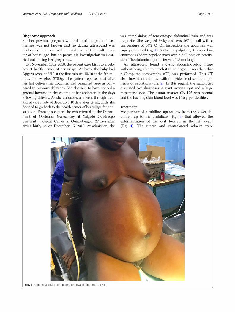
was complaining of tension-type abdominal pain and wasdyspnetic. She weighed 93 kg and was 167 cm tall with atemperature of 37°2 C. On inspection, the abdomen waslargely distended (Fig. 1). As for the palpation, it revealed anenormous abdominopelvic mass with a dull note on percus-sion. The abdominal perimeter was 126 cm long.An ultrasound found a cystic abdominopelvic image
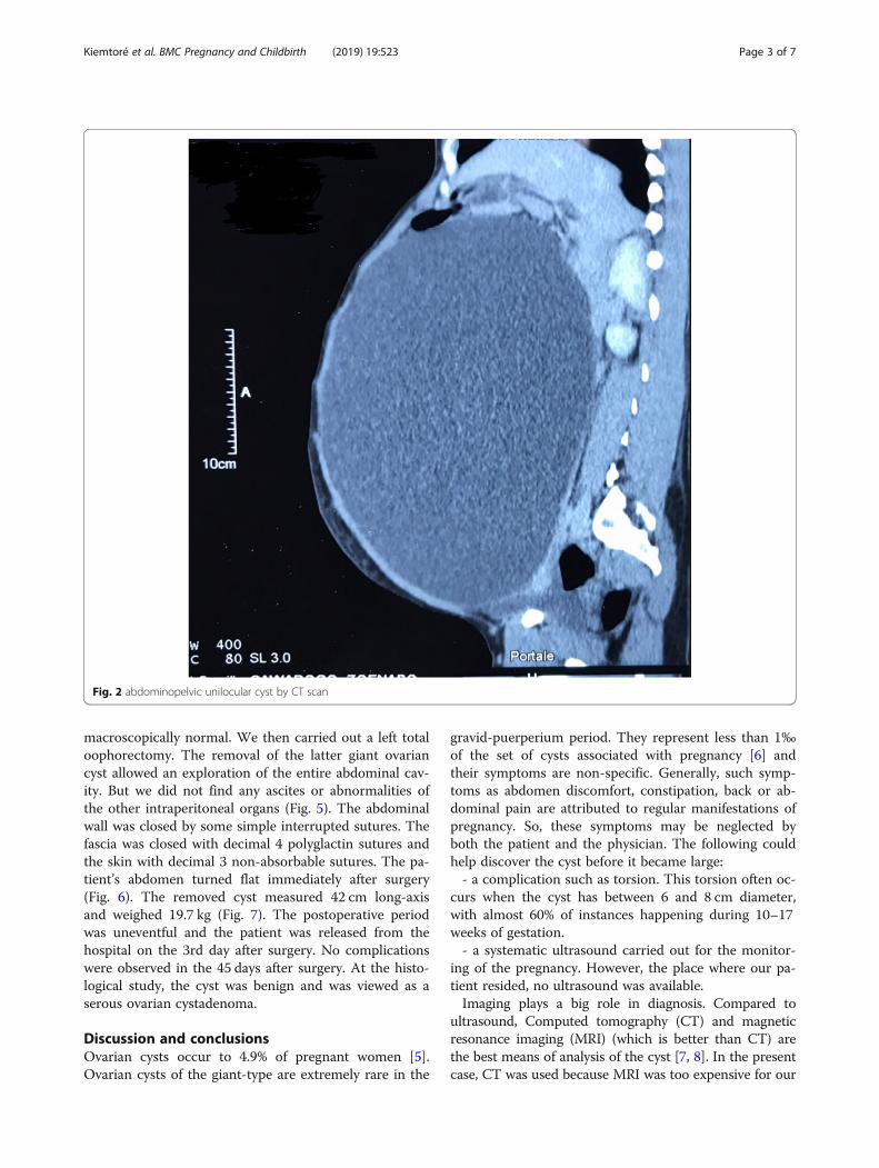
without being able to attach it to an organ. It was then thata Computed tomography (CT) was performed. This CTalso showed a fluid mass with no evidence of solid compo-nents or septations (Fig. 2). In this regard, the radiologistdiscussed two diagnoses: a giant ovarian cyst and a hugemesenteric cyst. The tumor marker CA-125 was normaland the haemoglobin blood level was 14.5 g per deciliter.
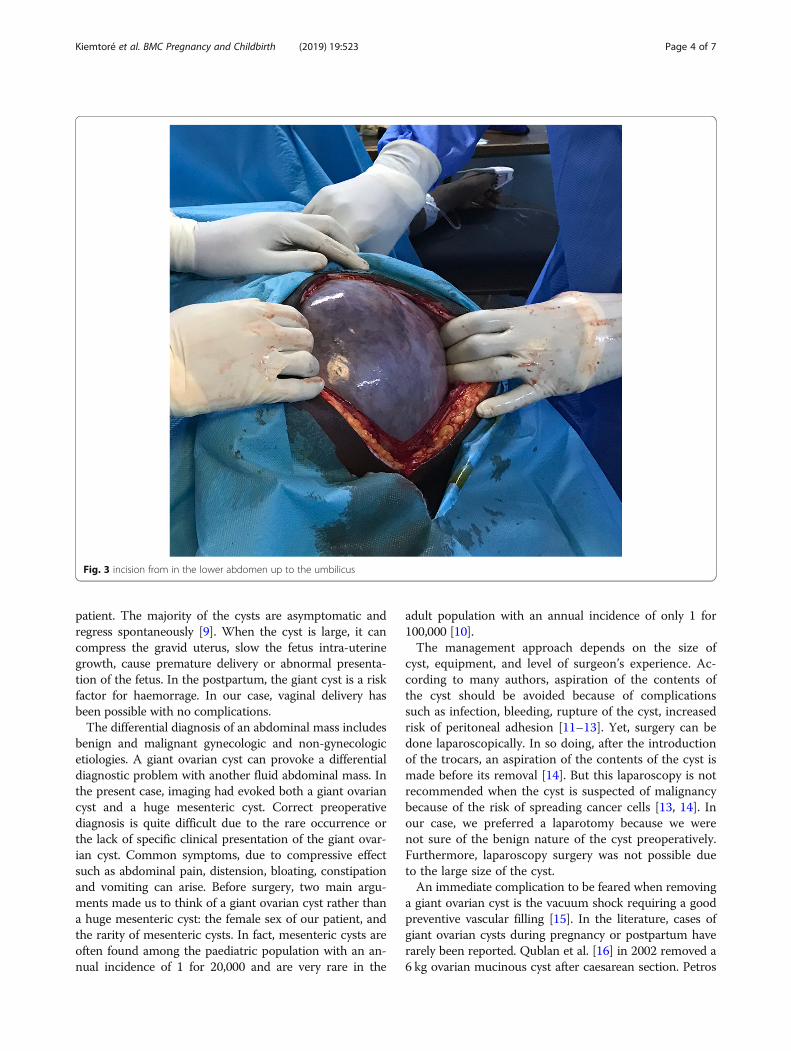
TreatmentWe performed a midline laparotomy from the lower ab-domen up to the umbilicus (Fig .3) that allowed theexternalization of the cyst located in the left ovary(Fig. 4). The uterus and contralateral adnexa were
Fig. 1 Abdominal distension before removal of abdominal cyst
Kiemtoré et al. BMC Pregnancy and Childbirth (2019) 19:523 Page 2 of 7
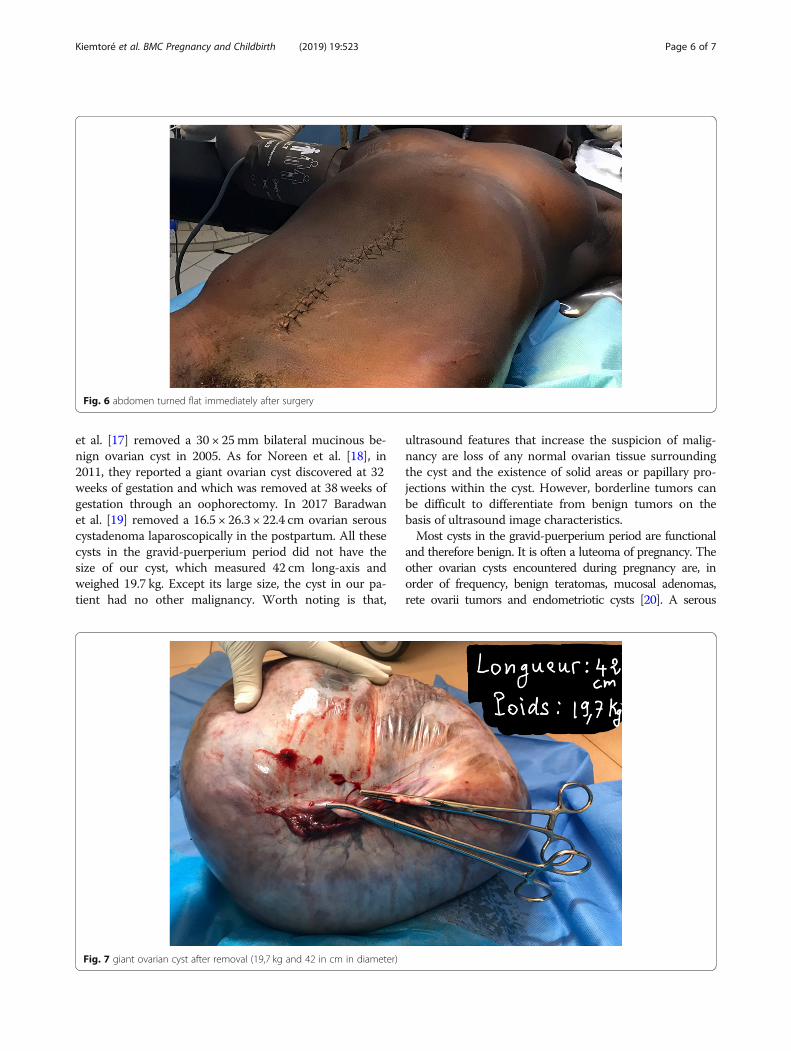
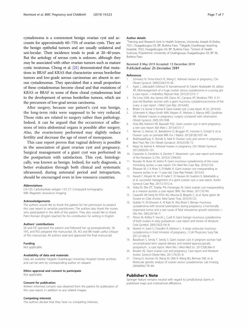
macroscopically normal. We then carried out a left totaloophorectomy. The removal of the latter giant ovariancyst allowed an exploration of the entire abdominal cav-ity. But we did not find any ascites or abnormalities ofthe other intraperitoneal organs (Fig. 5). The abdominalwall was closed by some simple interrupted sutures. Thefascia was closed with decimal 4 polyglactin sutures andthe skin with decimal 3 non-absorbable sutures. The pa-tient’s abdomen turned flat immediately after surgery(Fig. 6). The removed cyst measured 42 cm long-axisand weighed 19.7 kg (Fig. 7). The postoperative periodwas uneventful and the patient was released from thehospital on the 3rd day after surgery. No complicationswere observed in the 45 days after surgery. At the histo-logical study, the cyst was benign and was viewed as aserous ovarian cystadenoma.
Discussion and conclusionsOvarian cysts occur to 4.9% of pregnant women [5].Ovarian cysts of the giant-type are extremely rare in the
gravid-puerperium period. They represent less than 1‰of the set of cysts associated with pregnancy [6] andtheir symptoms are non-specific. Generally, such symp-toms as abdomen discomfort, constipation, back or ab-dominal pain are attributed to regular manifestations ofpregnancy. So, these symptoms may be neglected byboth the patient and the physician. The following couldhelp discover the cyst before it became large:- a complication such as torsion. This torsion often oc-
curs when the cyst has between 6 and 8 cm diameter,with almost 60% of instances happening during 10–17weeks of gestation.- a systematic ultrasound carried out for the monitor-
ing of the pregnancy. However, the place where our pa-tient resided, no ultrasound was available.Imaging plays a big role in diagnosis. Compared to
ultrasound, Computed tomography (CT) and magneticresonance imaging (MRI) (which is better than CT) arethe best means of analysis of the cyst [7, 8]. In the presentcase, CT was used because MRI was too expensive for our
Fig. 2 abdominopelvic unilocular cyst by CT scan
Kiemtoré et al. BMC Pregnancy and Childbirth (2019) 19:523 Page 3 of 7
patient. The majority of the cysts are asymptomatic andregress spontaneously [9]. When the cyst is large, it cancompress the gravid uterus, slow the fetus intra-uterinegrowth, cause premature delivery or abnormal presenta-tion of the fetus. In the postpartum, the giant cyst is a riskfactor for haemorrage. In our case, vaginal delivery hasbeen possible with no complications.The differential diagnosis of an abdominal mass includes
benign and malignant gynecologic and non-gynecologicetiologies. A giant ovarian cyst can provoke a differentialdiagnostic problem with another fluid abdominal mass. Inthe present case, imaging had evoked both a giant ovariancyst and a huge mesenteric cyst. Correct preoperativediagnosis is quite difficult due to the rare occurrence orthe lack of specific clinical presentation of the giant ovar-ian cyst. Common symptoms, due to compressive effectsuch as abdominal pain, distension, bloating, constipationand vomiting can arise. Before surgery, two main argu-ments made us to think of a giant ovarian cyst rather thana huge mesenteric cyst: the female sex of our patient, andthe rarity of mesenteric cysts. In fact, mesenteric cysts areoften found among the paediatric population with an an-nual incidence of 1 for 20,000 and are very rare in the
adult population with an annual incidence of only 1 for100,000 [10].The management approach depends on the size of
cyst, equipment, and level of surgeon’s experience. Ac-cording to many authors, aspiration of the contents ofthe cyst should be avoided because of complicationssuch as infection, bleeding, rupture of the cyst, increasedrisk of peritoneal adhesion [11–13]. Yet, surgery can bedone laparoscopically. In so doing, after the introductionof the trocars, an aspiration of the contents of the cyst ismade before its removal [14]. But this laparoscopy is notrecommended when the cyst is suspected of malignancybecause of the risk of spreading cancer cells [13, 14]. Inour case, we preferred a laparotomy because we werenot sure of the benign nature of the cyst preoperatively.Furthermore, laparoscopy surgery was not possible dueto the large size of the cyst.An immediate complication to be feared when removing
a giant ovarian cyst is the vacuum shock requiring a goodpreventive vascular filling [15]. In the literature, cases ofgiant ovarian cysts during pregnancy or postpartum haverarely been reported. Qublan et al. [16] in 2002 removed a6 kg ovarian mucinous cyst after caesarean section. Petros
Fig. 3 incision from in the lower abdomen up to the umbilicus
Kiemtoré et al. BMC Pregnancy and Childbirth (2019) 19:523 Page 4 of 7
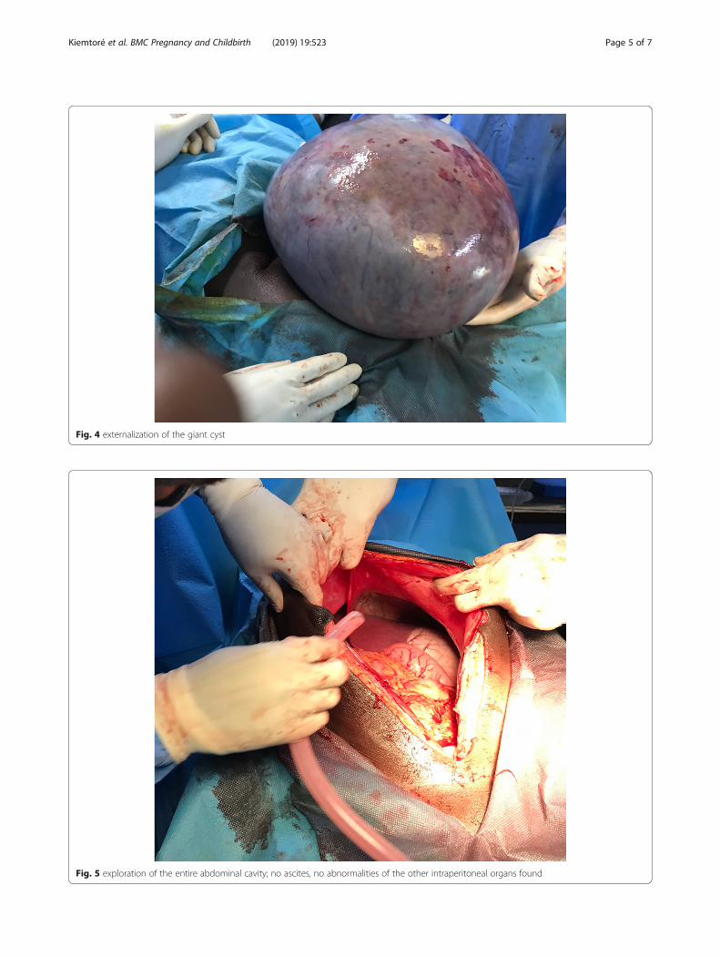
Fig. 5 exploration of the entire abdominal cavity; no ascites, no abnormalities of the other intraperitoneal organs found
Fig. 4 externalization of the giant cyst
Kiemtoré et al. BMC Pregnancy and Childbirth (2019) 19:523 Page 5 of 7
et al. [17] removed a 30 × 25mm bilateral mucinous be-nign ovarian cyst in 2005. As for Noreen et al. [18], in2011, they reported a giant ovarian cyst discovered at 32weeks of gestation and which was removed at 38 weeks ofgestation through an oophorectomy. In 2017 Baradwanet al. [19] removed a 16.5 × 26.3 × 22.4 cm ovarian serouscystadenoma laparoscopically in the postpartum. All thesecysts in the gravid-puerperium period did not have thesize of our cyst, which measured 42 cm long-axis andweighed 19.7 kg. Except its large size, the cyst in our pa-tient had no other malignancy. Worth noting is that,
ultrasound features that increase the suspicion of malig-nancy are loss of any normal ovarian tissue surroundingthe cyst and the existence of solid areas or papillary pro-jections within the cyst. However, borderline tumors canbe difficult to differentiate from benign tumors on thebasis of ultrasound image characteristics.Most cysts in the gravid-puerperium period are functional
and therefore benign. It is often a luteoma of pregnancy. Theother ovarian cysts encountered during pregnancy are, inorder of frequency, benign teratomas, mucosal adenomas,rete ovarii tumors and endometriotic cysts [20]. A serous
Fig. 7 giant ovarian cyst after removal (19,7 kg and 42 in cm in diameter)
Fig. 6 abdomen turned flat immediately after surgery
Kiemtoré et al. BMC Pregnancy and Childbirth (2019) 19:523 Page 6 of 7
cystadenoma is a commonest benign ovarian cyst and ac-counts for approximately 60–75% of ovarian cysts. They arethe benign epithelial tumors and are usually unilateral anduni-locular. Their incidence tends to peak at 20–40 years.But the aetiology of serous cysts is unknow, although theymay be associated with other ovarian tumors such as maturecystic teratomas. Cheng et al. [21] demonstrated that muta-tions in BRAF and KRAS that characterize serous borderlinetumors and low-grade serous carcinomas are absent in ser-ous cystadenomas. They speculated that a small proportionof these cystadenomas become clonal and that mutations ofKRAS or BRAF in some of these clonal cystadenomas leadto the development of serous borderline tumors, which arethe precursors of low-grad serous carcinoma.After surgery, because our patient’s cyst was benign,
the long-term risks were supposed to be very reduced.Those risks are related to surgery rather than pathology.Indeed, it can be argued that the occurrence of adhe-sions of intra-abdominal organs is possible after surgery.Also, the ovariectomy performed may slightly reducefertility and decrease the age of onset of menopause.This case report proves that vaginal delivery is possible
in the association of giant ovarian cyst and pregnancy.Surgical management of a giant cyst was performed inthe postpartum with satisfaction. This cyst, histologi-cally, was known as benign. Indeed, for early diagnosis, abetter evaluation through both clinical and systematicultrasound, during antenatal period and intrapartum,should be encouraged even in low-resource countries.
AbbreviationsCA-125: Carbohydrate antigen 125; CT: Computed tomography;MRI: Magnetic resonance imaging
AcknowledgementsThe authors would like to thank the patient for her permission to presentthis case report to sensitize practitioners. The authors also thank the nurseswho participated in the skills of the patient. They also would like to thankPalm Romain (English teacher) for his contribution for writing in English.
Authors’ contributionsSK and HZ operated the patient and followed her up postoperatively. SK,YAS, and RSS prepared the manuscript. EK, AO and BB made useful critiqueof this manuscript. All authors read and approved the final manuscript.
FundingNot applicable.
Availability of data and materialsData are available Yalgado Ouedraogo University Hospital Center archivesand can be sent by corresponding author on request.
Ethics approval and consent to participateNot applicable.
Consent for publicationWritten informed consent was obtained from the patient for publication ofthis case report, in addition to any related images.
Competing interestsThe authors declare that they have no competing interests.
Author details1Training and Research Unit in Health Sciences, University Joseph Ki-Zerbo,7021, Ouagadougou 03, BP, Burkina Faso. 2Yalgado Ouedraogo teachinghospital, 7022, Ouagadougou 03, BP, Burkina Faso. 3School of HealthSciences, Polytechnic University of Ouahigouya, Ouagadougou 03, BP 36,Burkina Faso.
Received: 8 May 2019 Accepted: 13 December 2019
References1. Schwartz N, Timor-tritsch IE, Wang E. Adnexal masses in pregnancy. Clin
Obstet Gynecol. 2009;52(4):570–85.2. Agah J, Jafarzadeh Esfehani R, Kamalimanesh B, Fattahi Abdizadeh M, Jalilian
AR. Mismanagement of a huge ovarian serous cystadenoma in a young girl;a case report. J midwifery Reprod Heal. 2015;3(1):315–7.
3. De Lima SHM, dos Santos VM, Daros AC, Campos VP, Modesto FRD. A 57-year-old Brazilian woman with a giant mucinous cystadenocarcinoma of theovary: a case report. J Med Case Rep. 2014;4:82.
4. Bhasin S k, Kumar V, Kumar R. Giant ovarian cyst: a case report. JK Sci. 2014;16:3.5. Schemeler K, Mayo-Smith WW, Reipert JF, Weitzen S, Manue IMD, Gordinier
ME. Adnexal masses in pregnancy: surgery compared with observation.Obstet Gynecol. 2005;105:1098.
6. Hota BM, Rukmini KR, Basavaih PGC. Giant ovarian cyst in term pregnancy -a rare case report. Bali Med J. 2015;4(1):5–7.
7. Nemec U, Nemec SF, Bettelheim D, Brugger PC, Horcher E, Schöpf V, et al.Ovarian cysts on prenatal MRI. Eur J Radiol. 2012;81(8):1937–44.
8. Mukhopadhyay A, Shinde A, Naik R. Ovarian cysts and cancer in pregnancy.Best Pract Res Clin Obstet Gynaecol. 2016;33:58–72.
9. Naqvi M, Kaimal A. Adnexal masses in pregnancy. Clin Obstet Gynecol.2015;58(3):93–101.
10. Casarotto A, Cerofolini A, Denitto F. Mesenteric cyst: case report and reviewof the literature. G Chir. 2010;31:239e242.
11. Rossato M, Burei M, Vettor R. Giant mucinous cystadenoma of the ovarymimicking ascites: a case report. Clin Med Rev Case Rep. 2016;3:103.
12. Elhassan M, S A Khan S, El-Makki A. Giant ovarian cyst masquerading asmassive ascites in an 11-year-old. Case Rep Pediatr. 2015;4:5.
13. Kassidi F, Moukit M. Ait El Fadel F, El Hassani M, Guelzim K, Babahabib a,et al. successful management of a giant ovarian cyst: a case report. AustinGynecol Case Rep. 2017;2:1012–6.
14. Yeika EV, Efie DT, Tolefac PN, Fomengia JN. Giant ovarian cyst masqueradingas a massive ascistes: a case report. BMC Res Notes. 2017;10:749.
15. Casanelli JM, Keita M, N’Dri AG, Moussa B, Blegole C, et al. Kyste géant del’ovaire en Côte d’ivoire. Med Sante Trop. 2010;70:122.
16. Qublan H, Al-Ghoweri A, Al-Kaisi N, Abu-Khait S. Benign mucinouscystadenoma with stromal luteinization during pregnancy: a hormonallyresponsive tumor and a rare cause of fetal intrauterine growth restriction. JObs Res. 2002;28:104–7.
17. Petros M, Koliba P, Sevcík L, Curík R. Giant benign mucinous cystadenomaof both ovaries in early puerperium: case report and review of literature.Cesk Gynekol. 2005;70(2):156–8.
18. Noreen H, Syed S, Chaudhri R, Kahloon L. A large unilocular mucinouscystadenoma in third trimester of pregnancy. J Coll Physicians Surg Pak.2011;21:426–8.
19. Baradwan S, Sendy F, Sendy S. Giant ovarian cyst in pregnant woman haduncomplicated term vaginal delivery and treated laparoscopicallypostpartum : a case report. Merit Res J Med Med Sci. 2017;5(8):366–9.
20. Rosales AE. Giant ovarian cyst and pregnancy. Case report and literaturereview. Ginecol Obstet Mex. 2011;79:235–8.
21. Cheng EJ, Kurman RJ, Wang M, Oldt R, Wang BG, Berman DM, et al.Molecular genetic analysis of ovarian serous cystadenomas. Lab Investig.2004;84(6):778–84.
Publisher’s NoteSpringer Nature remains neutral with regard to jurisdictional claims inpublished maps and institutional affiliations.
Kiemtoré et al. BMC Pregnancy and Childbirth (2019) 19:523 Page 7 of 7
Copyright © 2022 FDOKUMEN