Ovarian Macrophages And The Regulation Of Ovarian Function
217
Ovarian Macrophages and the Regulation of Ovarian Function A thesis submitted for the degree of Doctor of Philosophy Kylie H Van der Hoek (Bsc Hons) Research Centre for Reproductive Health and Department of Obstetrics and Gynaecology University of Adelaide Australia By December 2004
-
Upload
khangminh22 -
Category
Documents
-
view
1 -
download
0
Transcript of Ovarian Macrophages And The Regulation Of Ovarian Function
Ovarian Macrophages and the Regulation
of Ovarian Function
A thesis submitted for the degree of
Doctor of Philosophy
Kylie H Van der Hoek
(Bsc Hons)
Research Centre for Reproductive Health
and
Department of Obstetrics and Gynaecology
University of Adelaide
Australia
By
December 2004
T¡,nr-n ON CONTEI\TS
ABSTRACT V
DECLARATION VIIIACKNOWLEDGMENTS IX
ABBREVIATIONS XII1 LITERATURE REVIE\ry
1.1
T,2
1.2.11.2.21.2.3
1.3
1 .3.1L3,2L3.31.3.4
T,4
L4.1L4.21.4.3
1.5
I .5.11.5.2L5,31.5.41.5.5r,5.61.5,71.5.8
t.6L6.IL6.21.6.31,6.4
1.7
L7.1L7.21.7.3
1.8
L8.11.8,21.8.31,8.4
1
INtRooucrIoN,..........BASrc OveRnN FulqcrIoN .....
Follicle Growth......Ovulation,The Corpus luteum.......TuB Cvcuc PerrpnN op GoNRooTRoPHIN ¿,No SrBRoIo SncnnuoNLuteinising Hormone (LH) and Follicle Stimulating Hormone (FSH)
Progesterone......,...........estradiol and AndrogensStimulation of Ovulation in the Mouse via Exogenous GonadotrophinsLpurocvrn DtsrRIsurtoN rN THs Ov¡'nvLymphocyte,s...,,............NeutrophilsMacrophages ..............GsNpRAr CuenacrpRISTICS Rt to FuNcrIoNS oF M¡'cRopseGES .....'. ".Monocyte MigrationMacrophages in Infl ammation.......Macrophage Activation..Macrophage Interactions with T cells .....,..Macrophages in [(ound healing and Tissue Repair.'.....Macrophage Phagocytosis and Phagocytic Receptors "Macrophage Surface Markers and their Regulated Expression
Macrophages Respond to the Female Sex Steroid Hormones...
CgIRRcTpRISTICS OF TISSUE MRCNOPTNCES
TestisUterus........Human Placenta and Decidua...............'Brain Macrophage.r ..........,...Por¡urter M¡.cnopsecE FUNCTIoNS IN THE OvenvMacrophages and Follicular Growth and Atresia "."...'......Macrophages and the Stimulation of Ovulation...,,.,..Macrophages And the Corpus LuteumSutr¡lr¡er.v AND HYPorHnSIS .................Summary......Hypothesis....General Aim.Specific Aims
l4I515
171819
202224252729293034JI373839404l4I4245484850505I
2 TIJ.E DEPLETION OF MACROPHAGES IN THE MURINE OVARY BYINTRABURSAL INJECTION OF CLODRONATE (CL2MDP) LPOSOMES
2.t2.2
2,2,I2.2,22.2.32,2.42.2.52.2.6
2.32.3.1
2.3,2
2.3.32,3.4
2.4
3.1
3.23.2,13.2.23,2.33,2.43.2.53.2.63,2.73.2,83.2,93.2.10
3.2.113.2.123.2.133.2.14
J.J3.s.13.3.2
3.3,33.3.43,3.5
INTRoDUCTION .....
MarnnInrs ¡.Nn MersoosAnimals and Ovul ation Inductior? .......,............ "'..Intraburs al Inj ection TechniqueTreatment Groups and Oocyte Retrieval...Collection of Ovarian Tissue
Ovarian Morphol o gt and Immunohis to chemistryStatistics ...........RrsurrsEffect of Clodronate Liposome Treatment on Gonadotrophin StimulatedOvulation ..."".',,63Effect of Clodronate Liposome Treatment on The Ovarian MacrophagePopulation 65
Effect of Clodronate Liposome Treatment on Ovarian Tissue Morphology ,...72
Effect of Clodronate Liposome Treatment on Subsequent Natural Ovulation.74DrscussroN 77
S THE NUMBERS AND CHARACTERISTICS OF MURINE OVARIAN IA ANDF4l80 POSITIVE MACROPHAGES ISOLATED DURING THE GONADOTROPHIN.STIMULATED REPRODUCTIVE CYCLE 81
Dissociation of Ovaries ........85
Labelling of Ovarian macrophages...............' ""'."..,,86Antibody Panning ........."..'....86Viability..... .,,......87
Collection of Cells .""......,.,,,87Leukocyte Antigen Expression '..'."...'.'."'88Isolation of messenger RNA and generation of complementary DNA......""'."89Luteinising Hormone and Follicle Stimulating Hormone Receptor nRNAExpression 90
INrRooucrIoN .,........MerHoosAnimals And Gonadotrophin StimulationDissociation Conditions.....,,.,........
Macrophage Conditioned Media.....,........Progesterone AnalysisPhagocytic AssessmentPHA- Stimulat ed Sp lenocyte P roliferation
CycleViability and Purity of Isolated Macrophages
82
84
8484
9I91
9I9294
'..''''.,,,',,,..,96..................98
104106
RESULTS
Optimisation of Conditionsfor Ovariqn Macrophage Recovery.................,'...94
Ovarian Macrophage Cell Numbers Across the Gonadotrophin-Stimulated
Phagocytic Capability of Ovarian Macrophage.ç.,,.........'.. '........101Effect of Macrophage- Conditioned Media on P HA- Stimulated Spl eno cyte
Proliferation.DrscussroN..3.4
lll
4 THE EXPRESSION OF INFLAMMATORY MEDIATORS BY THEMACROPHAGE POPULATION IN THE OVARY OF THE GONADOTROPHIN-STIMULATED MOUSE II2
4.1
4.24.2.I4.2.24.2,34.2.44.2.5
4.34.3.14.3.24.3.34.3.4
4.4
5.1
5.25.3
5.45.55.6
INrRooucrtoNMnrsoos.......AnimalsMACROPHAGE MESSENGER RNA ISOLATION AND MEASUREMENT..
Reverse Transcription and Quantitative RT-PCRCulture of Isolated MacrophagesMeasurement of Secreted Ovulatory mediatorsRBsurrsIsolated Macrophage RNL........,..Primer Efficiencies and House-keeper ValidationExpression of Cytokine nRNA in Ovarian MacrophagesSoluble Mediators Secreted By Ovarian Macrophages....DrscussroN
113
116
116116117r20120r22122122I2s128135
FINAL DISCUSSION 146
SUIr¡Ii¡RRY OF FINDINGS....
REFERENCES 159
APPENDIXES 181
Evropr.rcp oF MACRoPHAGE HETERocENEITY IN THE MuRnqn Ovenv ....
IMpLrcATroNS FoR OvRRIaN MecRopuRGE PHENoTYPE AND FtrucrtoxSuvrueRvFuRrsBR SruorcsIuprrc¡,rIoNs.................
1V
Ansrrucr
The presence of macrophages within ovarian tissue has been acknowledged for many years.
There is substantial evidence showing typical macrophage products such as interleukin-l-beta
(IL-18) and tumour necrosis factor alpha (TNFo), can significantly influence ovarian
functions. To date the specific role these cells play in ovarian function has only been
postulated. It was the aim of the work presented in this thesis to determine firstly, if these
cells are critical to normal ovarian function, secondly to develop a method for isolating these
cells from other ovarian cells types and thirdly, to examine the cytokine profile of these cells
with the intention of determining which important inflammatory cytokines these cells are
producing.
Ovarian macrophages were demonstrated to be critical for normal ovarian function by
depleting this population using intrabursal injection of liposome-encapsulated clodronate
(CLÐ, liposome-encapsulated saline (SLi) or saline alone (S) in gonadotrophin-primed adult
mice, either 84 hrs (day -3) or 36 hrs (day -1) prior to ovulation. Injection of CLi on day -1
did not affect ovulation rates, while administration on day -3 caused a significant reduction in
ovulation rate from 9.13 t 0.9 down to 5.25 I 0.6 þ<0.05). Examination of macrophage
distribution within the theca and stroma of preovulatory ovaries with monoclonal antibodies
to the murine macrophage antigens macrosialin (FA/11), MHC class II (anti-Ia) and F4l80
revealed following CLi treatment on day -1 a reduction in macrosialin positive macrophages
in the theca at ovulation while CLi treatment on day -3 reduced the numbers of Ia positive and
macrosialin positive macrophages present in the theca, When the subsequent estrous cycle
was monitored by vaginal smearing the metestrus-2 /diestrus stage was found to be extended
in Cli-treated animals, from 3.4t0.4 days to 7.5 tl.3 days (p<0. 05). These results suggest
that thecal macrophages may be involved in the regulation of follicular growth or rupture, as
well as being important for the normal progression of the estrous cycle. A method for
isolating ovarian macrophages was then developed. Initial experiments optimised the tissue
digest conditions and demonstrated that ovarian dissociation at room temperature in
collagenase/DNase solution made up in alpha-minimum essential media (ctMEM) with added
calcium chloride was best for recovery of maximum live macrophages. Ovarian single cell
suspensions were then incubated with the specific rat monoclonal antibodies anti-Ia (MHC-IÐ
and F4l80, and then incubated on anti-rat coated antibody panning plates. Cells bound to the
plate were shown to be viable, 98% pure and did not produce detectable levels of
progesterone. The numbers of cells isolated per mg tissue increased significantly across the
gonadotrophin stimulated reproductive cycle with maximum numbers recovered 24 and 48 hrs
post ovulation. The isolated cells were more phagocytic after ovulation (75-80%) in
comparison to before owlation (55-60o/0, p<0.04) while conditioned media from cells both
before and after ovulation did not significantly influence PHA-stimulated proliferation of
adherence purified splenocytes. This isolation method was then used to examine the cytokine
profile of ovarian macrophages across the gonadotrophin-stimulated reproductive cycle.
Macrophages were isolated from ovaries of groups of 8-10 immature mice at differing stages
of the pregnant mare serum gonadotrophin (PMSG)lhuman chorionic gonadotrophin (hCG)-
stimulated cycle using the anti-Ia or F4l80 antibodies. Messenger RNA was isolated from
some groups for reverse transcription (RT) and quantitative real-time PCR (QRT-PCR)
analysis while others were cultured for 24 hrs and conditioned media collected for analysis of
protein content. Messenger RNA expression for the cytokines IL-l8, interleukin-l receptor
antagonist (Il-lra) and TNFo was transiently stimulated following the administration hCG
(increases of 5-7 fold, 4 fold and 5 fold respectively, p<0.05) while no significant changes in
the levels of mRNA for inducible nitric oxide synthase (iNOS), interleukin-lO (IL-10) or
transforming growth factor-beta I (TGF-p1) were detected. Protein levels in the conditioned
vl
media did not mirror the changing messenger RNA levels. Ovarian Ia positive cells were
found to produce limited amounts of IL-IB following hCG administration while although
nitric oxide (NO), L-10 and TNFcr, were detected with small fluctuations occurring post hCG
administration these changes were not found to be significant. The selection of transcripts and
proteins these cells produce show that they are not classical inflammatory cells and thus may
not be the primary source of inflammatory molecules with known roles in ovulation.
The work in this thesis has shown that ovarian macrophages are essential for normal
ovarian function and that these cells appear to be of an anti-inflammatory phenotype. The
major roles of these cells may therefore be in minimising tissue damage occurring following
the ovulatory event and tissue reorganisation in subsequent corpus luteum formation and
regression.
vil

AcTxOWLEDGMENTS
Firstly, I wish to sincerely thank my supervisor Prof R.J. Norman who has guided, supported and
inspired me through these studies, his knowledge and experience have been invaluable.
Thankyou. To Natalie Ryan, you are a gem. Your assistance was always forthcoming, your
friendship made working a joy, and you were many times my saving grace as well as my right
and left hand when required. Thankyou. Thanks also to Sarah Roberstson and Simon Maddocks
for their advice and expertise in setting up the macrophage depletion study. In addition, thanks
also to Carol Woodhouse who helped carry out the intrabursal injection experiments and
immunohistochemistry, it was a delight to share the laboratory with her. To Rebecca Robker,
your constructive comments and listening skills were invaluable during the final stages of
completing this thesis, thankyou, Thanks to all staff and students in the Department of Obstetrics
and Gynaecology whose work ethos and appraisal drives students to strive for better. More
particularly, thanks to those at TQEH campus where everyone made themselves available to help
whenever approached and more importantly kept up the supply of delicious cake on Thursday
momings. Thanks also to the University of Adelaide for supporting my studies by granting a
University of Adelaide Scholarship.
Thankyou to my beautiful sons Nathan and Aaron who have both slept like angels
through the night from a young age, without this I would not have been able to get far. Mummy
has finally finished the big book! Thankyou to my mother in-law and my parents for readily
taking on baby-sitting duties when required and your love and support, especially in the last few
months, you don't realise how much it has meant to me'
Finally, many thanks to my husband Mark who supported me through the tumultuous times of
both dismay and delight and who knows me well enough to know what I want, even when I
myself forget.
ix

Punr,rcATIoNS Anrsrxc
Van der Hoek KH, N.K.Ryan, R.J. Norman. Ovarian macrophages are type II activated anti-
infl ammatory macrophages. In preparation.
Van der Hoek KH, N.K.Ryan, R.J. Norman. The Isolation and Characteristics of ovarian
macrophages. In preparation.
Wu R, Van der Hoek KH, Ryan NK, Norman RJ, Robker RL. Macrophage contributions to
ov ar i an fu n c t i o n.Hum Reprod Update. 2 004 Mar-Ap r ;1 0 (2) : I I 9 - 3 3 . Review.
Van der Hoek KH, Maddocks S, Woodhouse CM, van Rooijen N, Robertson SA,
Norman RJ.Intrabursal injection of clodronate liposomes causes macrophage depletion and
inhibits ovulation in the mouse ovary. Biol Reprod' 2000 Apt;62(4):1059-66'
Abstracts ArisinsKH Van der Hoek, CM Woodhouse, N Van Rooijen, S Maddocks, and RJ Norman. The effect ofintrabursal injection of liposome encapsulated dichloromethylene diphosphonate on ovulation
in the mouse ovary. Australian Society of Reproductive Biology, Proceedings of the Twenty-eighth Annual Conference, Canberra, ACT, (1997) p973.
KH Van der Hoek, NK Ryan and RJ Norman The isolation of cells expressing the macrophage
markers Ia and F4/80from the murine ovary. Society of Reproductive Biology, Proceedings ofthe Thirty Second Annual Conference, Gold Coast, QLD, 2001 Abstract 57.
KH Van der Hoek, N.K. Ryan, S.A. Robertson and R.J. Norman. Cytokine nRNA expression in
ovarian macrophages during the murine gonadotrophin stimulated oestrous cycle. Australian
Society for Medical Research - South Australian Branch, Proceedings Annual Scientific
Meeting, 2003 Abstract O24.
KH Van der Hoek, N.K. Ryan, S.A. Robertson and R.J. Norman The expression of ovulatory
mediators by macrophages isolatedfrom the gonadotrophin-stimulated mouse ovary. Society forReproductive Biology, Proceedings of the Thirfy-fourth Annual conference Melbourne, Vic.2003 Abstract 32.
C. Haynes, KH Van der Hoek, N.K. Ryan, S.A. Robertson and R.J. Norman 'Ovarian
macrophage regulation of inflammatory responses at ovulation in murine ovaries.' Society forReproductive Biology, Proceedings of the Thirfy-fourth Annual conference Melboume, Vic.2003 Abstract 35.
Related PublicationsRyan NK, Van der Hoek KlI, Robertson SA, NormanRJ. Leptin and leptin receptor expression
in the rat ovary. Endocrinology.2003 Nov;144(11):5006-13' Epub 2003 Aug 14.
Duggal PS, Ryan NK, Van der Hoek KH, Ritter LJ, Armstrong DT, Magoffin DA, Norman RJ.
Effects of teptin qdministration and feed restriction on thecal leukocytes in the preovulatory ratovary and the effects of leptin on meiotic maturation, granulosa cell proliþration, steroid
x
hormone and PGE2 release in cultured rat ovarianfollicles. Reproduction.2002 Jun;123(6):891-
8.
Ryan NK, Woodhouse CM, Van der Hoek KH, Gilchrist RB, Armstrong DT, Norman RJ.
Expression of leptin and its receptor in the murine ovary: possible role in the
r e gul ation of o o cy te matur ation. Biol Reprod . 2002 May ; 66(5) : I 548-54'
Duggal PS, Van Der Hoek KH, Milner CR, Ryan NK, Armstrong DT, Magoffin DA, Norman
RJ. The in vivo and in vitro effects of exogenous leptin on ovulation in the rat. Endoctinology.
2000 Jun; 14 l(6):197 l-6.
Jasper MJ, Robertson SA, Van der Hoek KH, Bonello N, Brannstrom M, Norman RJ.
Characterization of ovarian function in granulocyte-macrophage colony-stimuløting factor-d efi c i ent m ic e. Biol Reprod. 2 000 Mar; 6 2(3) :7 0 4 - I 3'
Van der Hoek KH, Woodhouse CM, Brannstrom M, Norman P.J. Effects of interleukin (IL)-6 on
luteinizing hormone- and IL- I beta-inducedovulation and steroidogenesis in the rat ovary. Biol Reprod. 1998 May;58(5):1266-71'
Norman,RJ, Bonello,N, Jasper,MJ and Van der Hoek, KH (1998) 'Leukocytes: Essential cells in
ovarian function and ovulation', Reproductive Medicine Reviews, 6(2), 97 -I I
Bonello N, McKie K, Jasper M, Andrew L, Ross N, Braybon E, Brannstrom M, Norman RJ.
Inhibition of nitric oxide: effects on interleukin-l beta-enhanced ovulation rate, steroidhormones, and ovarian leukocyte distribution al ovuløtion in the rat. Biol Reprod. 1996
Feb;54(2):436-45.
xl

CL
CLi
CNOS
CT
DAB
DE
DNAse IEDTA
eNOS
FSH
HBSS
hCG
HI-FCS
HPRT
H-RPMI
IL-10
rL-lpIL-1ra
iNOS
LHLHRH
ME
oMEMMHC
mRNA
NMS
NO
PBS
PCR
PE
PHA
PMSG
RPE
SLi
rGFpTNFoVIA
AnnnnvIATIONS
corpus luteum
clodronate liposome
constituitive nitric oxide synthase
threshold cycle
diaminobenzidene
diestrus
deoxyribonuclease Iethylenediamine tetraacetic acid
endothelial nitric oxide synthase
follicle stimulating hormone
hanks buffered saline solution
human chorionic gonadotrophin
heat-inactivated foetal calf serum
hypoxanthine guanine phosphoribosyl transferase
hepes buffer RPMI
interleukin l0interleukin- 1-beta
interleukin- I - receptor antangonist
inducible nitric oxide synthase
luteinising hormone
luteinising hormone releaseing hormone
metestrus
minimum essential medium alpha
maj or histocompatability complex
messenger ribonucleic acid
normal mouse serum
nitric oxide
phosphate buffered saline
polymerase chain reaction
proestrus
phytohaemagglutnin
preganant mare serum gonadotrophin
R-phycoerythrin
saline liposomes
transforming growth factor beta
tumor necrosis factor alpha
video image analysis
xll
Chapter One
1 LTTNNATURE REVIEW
INTRODUCTION
1.1 lNrnooucrloN
The adult ovary is an active endocrine organ that contains and nurtures the female
germ cells or oocytes. It is responsible for the regulated release of mature oocytes from the
follicles of the ovary into the reproductive tract for fertilisation and the secretion of steroid
hormones that will ensure the oviduct and uterus are prepared to support, embryo
development and implantation, should fertilisation occur. If fertilisation or implantation is
unsuccessful, then the ovary must initiate a new cycle of growth and maturation of new
follicles and their oocytes. This results in ovarian cycles of follicular growth and atresia,
steroid secretion, and tissue remodelling. The mechanisms by which these important
processes are controlled in different mammalian species are complex. Interactions between
steroid hormones released from the ovary and gonadotrophins from the pituitary are clearly
the main regulatory mechanisms, but it is evident that complex interactions between the
reproductive, immune ll, 2] and metabolic systems exist [3, 4]. Furthermore, following
gonadotrophin stimulation the local signalling systems that operate within the normal ovary to
promote follicular growth, ovulation and corpus luteum growth and regression, are not clearly
defined.
Macrophages are cells of the immune system with roles in immunity and tissue
homeostasis. They are derived from blood borne monocytes that migrate into the peripheral
tissues and differentiate in response to the local microenvironmental signals to assume a
functional phenotype. Their roles may include the phagocytosis and degradation of foreign
organisms or apoptotie tissues, regulation of local immune and inflammatory responses, and
tissue remodelling and repair. The presence of macrophages within the ovary has been
recognized for many years, although their function remains undefined. .
This review describes basic ovarian and macrophage functions and then explores
current evidence implicating these cells in the regulation of ovarian function.
2LITERATURE REVIEW
OVARIAN FUNCTION
1.2 B¡src Ovnnr¿.N FuNcrroN
The ovary is composed primarily of growing and atretic follicles, developing and
regressing corpora lutea CL and stromal/interstitial tissue. All components are present
simultaneously in the adult ovary until menopause, with the proportion of each dependent on
the stage of the reproductive cycle and age.
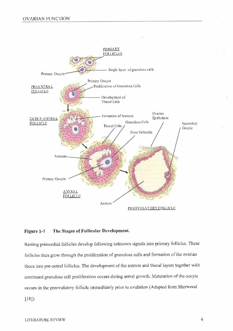
1.2.I FoLucrB Gnowrn
The follicles are the components of the ovary that protect and provide for the
developing oocytes. Follicles arise from resting primordial follicles consisting of an oocyte
arrested in prophase of the first meiosis surrounded by a single layer of flattened epithelial
cells and a basement membrane. They are located in the cortical region of the adult ovary and
if the appropriate stimulus is provided will grow in size and cell composition and number, to
large preovulatory or graafran follicles (Figure 1-1). In the rodent, growth to the preovulatory
stage takes 19 days [5, 6]; consequently growing follicles destined for ovulation in subsequent
cycles are present during a single estrous cycle. In the mouse follicular development can be
divided into 8 stages based on the number of granulosa cells and the stage of oocyte growth
[7]. Similar patterns and classification systems have been established in the development of
rat and human follicles tS-10]. For the purposes of this introduction follicle growth will be
described as preantral, antral and preovulatory growth (reviewed by Johnson [11] and
Greenwald [12]).
Primordial follicles become primary follicles following the transformation of the
flattened epithelial cells into layers of proliferating cuboidal granulosa cells and, following
synthesis of RNA, an increase in oocyte volume. The exact mechanism/s that initiate the
growth of quiescent primordial follicles are ill defined although several growth factors have
JLITERATURE REVIEW
OVARIAN FUNCTION
PRIMARYFOLLICLES
Single layer of granulosa cells
Formation of Antrum
Granulosa CellsThecal Cells
Zona Pellucida
t"
)i¡l
Primary Oocyte
PREANTRALFOLLICLE
EARLYANTRALFOLLICLE
Primary Oocyte
Primary Oocyte
Proliferation of Granulosa Cells
Development ofThecal Cells
OvarianEp itheliu m
SecondaryOocyte
t,,.
I
¡iitlA
I
,{t:
ANTRALFOLLICLE
AntrumPREOWLATORY FOLLICLE
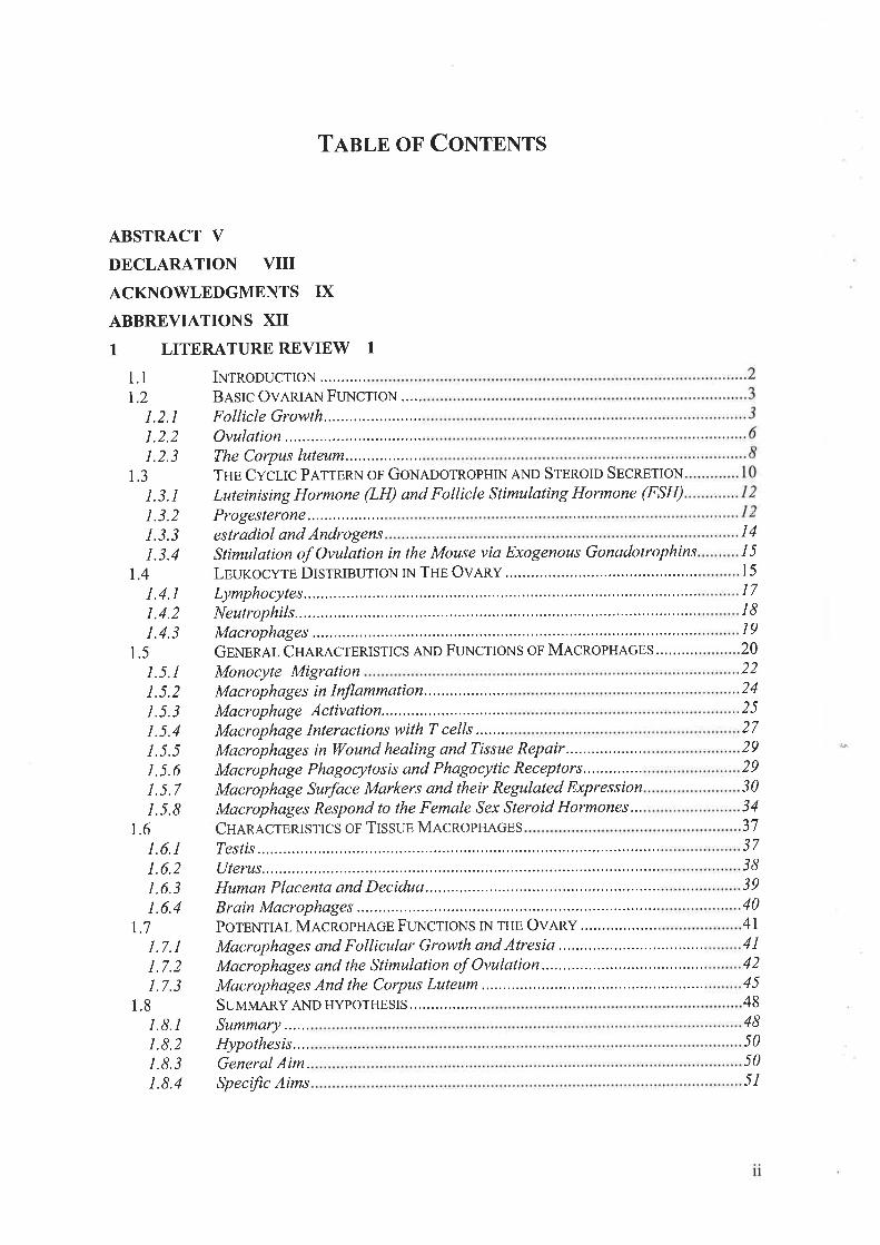
Figure 1-1 The Stages of Follicular Development.
Resting primordial follicles develop following unknown signals into primary follicles. These
follicles then grow through the proliferation of granulosa cells and formation of the ovarian
theca into pre-antral follicles. The development of the antrum and thecal layers together with
continued granulosa cell proliferation occurs during antral growth. Maturation of the oocyte
occurs in the preovulatory follicle immediately prior to ovulation (Adapted from Sherwood
t 1 8l).
4LITERATURE REVIEV/
OVARIAN FLINCTION
been implicated in this process (reviewed by Nilsson [13]), including insulin [14], androgens
[15], basic fibroblast growth factor, stem cell factor [16] and growth and differentiation
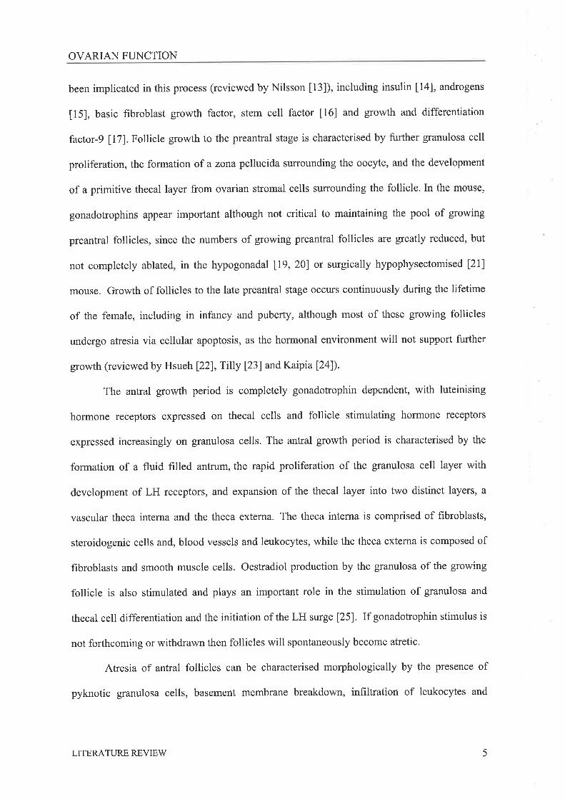
factor-9 [17]. Follicle growth to the preantral stage is characterised by further granulosa cell
proliferation, the formation of a zona pellucida surrounding the oocyte, and the development
of a primitive thecal layer from ovarian stromal cells surrounding the follicle. In the mouse,
gonadotrophins appear important although not critical to maintaining the pool of growing
preantral follicles, since the numbers of growing preantral follicles arc greatly reduced, but
not completely ablated, in the hypogonadal [19, 20] or surgically hypophysectomised [21]
mouse. Growth of follicles to the late preantral stage occurs continuously during the lifetime
of the female, including in infancy and puberty, although most of these growing follicles
undergo atresia via cellular apoptosis, as the hormonal environment will not support fuither
growth (reviewed by Hsueh l22l,Tilly l23l andKaipial24l).
The antral growth period is completely gonadotrophin dependent, with luteinising
hormone receptors expressed on thecal cells and follicle stimulating hormone receptors
expressed increasingly on granulosa cells. The antral growth period is characterised by the
formation of a fluid filled antrum, the rapid proliferation of the granulosa cell layer with
development of LH receptors, and expansion of the thecal layer into two distinct layers, a
vascular theca interna and the theca externa. The theca interna is comprised of fibroblasts,
steroidogenic cells and, blood vessels and leukocytes, while the theca externa is composed of
fibroblasts and smooth muscle cells. Oestradiol production by the granulosa of the growing
follicle is also stimulated and plays an important role in the stimulation of granulosa and
thecal cell differentiation and the initiation of the LH surge [25]. If gonadotrophin stimulus is
not forthcoming or withdrawn then follicles will spontaneously become atretic.
Atresia of antral follicles can be characterised morphologically by the presence of
pyknotic granulosa cells, basement membrane breakdown, infiltration of leukocytes and
5LITERATURE REVIEW
OVARIAN FI-INCTION
degeneration of the oocyte. More precise molecular techniques allow the detection of DNA
laddering indicative of apoptotic cells, or the expression of death inducing genes, has allowed
earlier detection of apoptosis than that achieved with observation of morphological changes
(reviewed by Hsueh l22l,Tilly l23l and Kaipia [24]). Therefore for a follicle to mature past
the preantral and antral stages of development towards ovulation it must be 'rescued' from the
follicular atresia pathway by gonadotrophin stimulation'
Preowlatory follicles consist of an oocyte surrounded by layers of cumulus cells,
called the corona radiata. This complex extends into the fluid filled antrum of the follicle, via
a 'stalk' of granulosa cells that secures the cumulus cell -oocyte complex to one side of the
follicular wall (reviewed by Lipner 126) and Brannstrom l27l). At this stage there are many
layers of mural granulosa cells lining the basement membrane of the follicle, which is
enclosed in several layers of elongated theca interna cells interspersed with blood vessels, and
the theca externa. The preowlatory follicle protrudes at one site from the surface of the ovary
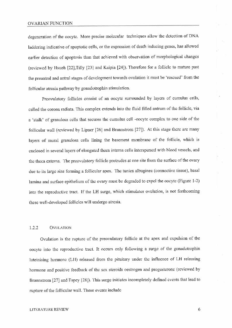
due to its large size forming a follicular apex. The tunica albuginea (connective tissue), basal
lamina and surface epithelium of the ovary must be degraded to expel the oocyte (Figure 1-2)
into the reproductive tract. If the LH surge, which stimulates ovulation, is not forthcoming
these well-developed follicles will undergo atresia.
1.2.2 OvurerIoN
Ovulation is the rupture of the preovulatory follicle at the apex and expulsion of the
oocyte into the reproductive tract. It occurs only following a surge of the gonadotrophin
luteinising hormone (LH) released from the pituitary under the influence of LH releasing
hormone and positive feedback of the sex steroids oestrogen and progesterone (reviewed by
Brannstrom l27l and Espey [28]). This surge initiates incompletely defined events that lead to
rupture of the follicular wall. These events include
6LITERATURE REVIEW
OVARIAN FUNCTION
Surface Epithelium
Basal l¿mina
Tunica Albuginea
Theca Eldema
Collagen Fibrils
Theca Intema
Capillaries
Basal l¿mina
Granulosa layer
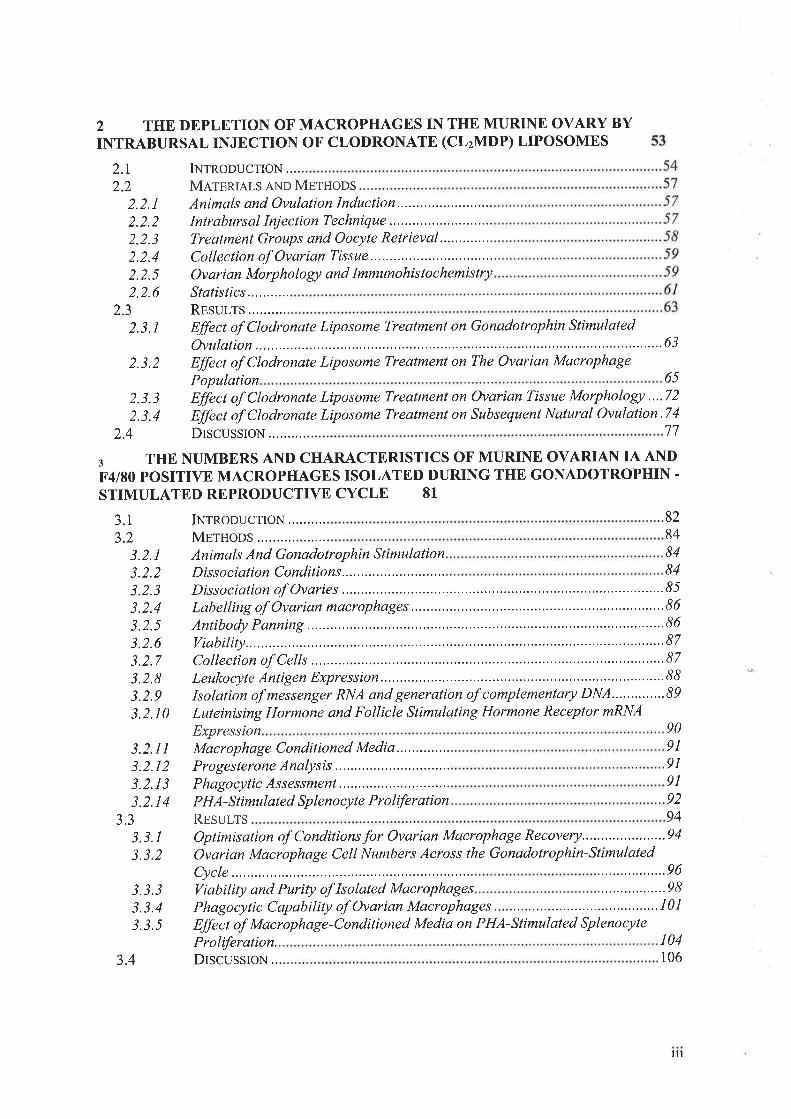
FÍgure 1-2 The Structure of the Follicular Wall
The many layers of the follicular wall that need to be degraded for ovulation to occur include
the basal lamina and tunica albuginea. (Adapted from Espey [28])
7LITERATURE REVIEW

OVARIAN FI.INCTION
vasodilation and an increase in blood supply to the ovary, leukocyte infiltration of the
theca of the follicle, increases in intrafollicular pressure, contractions in the theca externa,
increases in the activities of proteolytic enzymes, luteinisation of granulosa cells, changes in
steroid production, and the initiation of meiosis and extrusion of the first polar body in the
oocyte. Several characteristics of this process were recognised as similar to those of a
classical inflammatory reaction and a potential role for leukocytes in this process proposed in
early 1980's by Espey [29].
1.2.3 TUB CONPUS LUTEUM
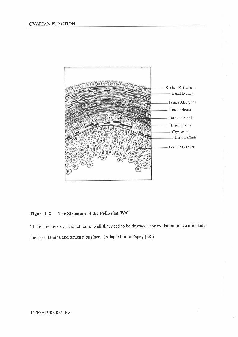
Following the LH surge and owlatory event the theca interna and granulosa cells of the
follicle transform into a predominantly progesterone secreting corpus luteum (CL) in a
process called luteinisation (Figure 1-3; reviewed by Murphy [30] and Niswender [31])'
Granulosa and thecal derived luteal cells within the CL can be distinguished as large (LLC's)
and small (SLC's) luteal cells respectively. The CL becomes a highly vascularized structure
also containing epithelial cells, fibroblasts and connective tissue, pericytes and blood
leukocytes. During CL development LLC's grow rapidly in size but not numbers while
SLC's, hbroblasts, and endothelial cells proliferate. Both large and small luteal cell types
contain PHSD and hence actively produce progesterone. The primary role of the CL is to
produce progesterone, which prepares the reproductive tract for embryo development and
implantation. If fertilisation occurs, the CL must respond to factors produced by the conceptus
with further production of progesterone, preventing the initiation of another follicular growth
cycle and thus maintaining pregnancy. If no signal to maintain progesterone secretion is
received, the CL deteriorates over several subsequent cycles into a corpus albicans or ovarian
scar tissue in a process known as CL regression or luteolysis, This process is stimulated by
luteolytic factors predominantly PGF2" derived from the uterus
8LITERATURE REVIEW
OVARIAN FUNCTION
Mature Oocyte
Zona Pellucida
Corona Radiata
OWLATING FOLLICLE
Rupture site
-'t iii:; ñ
'1, I
Iit
j
.t
I ..'\11' 'i/'
DEVELOPING CORPUS LUTETIM
ç
I
\JlFUNCTIONAL CORPUS LUTEUM CORPUS ALBICANS
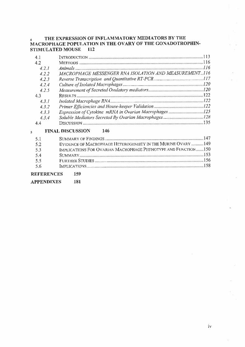
Figure L-3 Ovulation, and Development and Regression of the Corpus Luteum.
The oocyte is expelled from the preovulatory follicles which then transforms into the
progesterone producing CL. If no pregnancy occurs then the CL rapidly regresses into a
corpus albicans and subsequently to ovarian scar tissue. (Adapted from Sherwood [18])
9LITERATURE REVIEV/
OVARIAN FTINCTION
and is characterised by the loss of vascularization and progesterone synthesis,
followed by loss of the cells comprising the CL through apoptosis (reviewed by Murphy [30]
and Niswender [31]). In the human this luteal phase last 12-15 days with most other
mammalian species having a luteal phase of 15-19 days [11]. In the rodent and rabbit the
luteal phase of the cycle varies in length depending on whether or not they have mated. In the
rodent if mating does not occur then the CL begins to deteriorate within 2 days of owlation,
never establishing its full progesterone secreting potential. Therefore in a cycling non-
pregnant animal several CL's at differing stages of regression from the preceding cycles may
be present during any one reproductive cycle. If a non-fertile mating occurs the stimulation of
the cervix initiates the release of prolactin from the pituitary and the CL goes on to produce
progesterone for a full luteal phase of 11-12 days[6]. This is known as pseudopregnancy. In
the rabbit the act of mating stimulates the release of LH from the pituitary and hence
ovulation 10-12 hours post mating, However if a female is housed with an infertile male she
will exhibit a l4-day ovarian cycle, similar to that of the pig, consisting of a 2-day follicular
phase followed by a l2-day luteal phase [11].
1.3 TuB CycLIC PATTERN oF GoN¡,oorRoPHIN AND STEROID SECRETION
The female sex steroid hormones, produced by the granulosa and theca of the ovary,
'feedback' systemically to the pituitary, controlling the release of the gonadotrophins required
to stimulate follicular growth and ovulation (reviewed by Couzinet [32]). This results in
fluctuating systemic hormone levels that coordinate follicle growth and development, tissue
restructuring, andbehavioural changes to optimise the chances of conception (Figure 1-4)' In
the rodent this is called the estrous cycle based on the characteristic female behaviours
associated with approaching ovulation. In the human, behavioural estrus does not occur and
the reproductive cycle is recognized only by the event of menstruation. It is therefore termed
LITERATURE REVIEW 10

GONADOTROPHINS AND OVARIAN STEROIDS
\..."*'
^ -à¿
¡II
\.t\,
.-Éæir'!Ð.Òad
Progesterone (nglml)
Prolactin (ng/ml)
Estradiol (pg/ml)
LH (ng/ml)
FSH (ng/ml)
rl Çlt æ11 {Þ l! rt úEl
metestrus-l metestrus-2 diestrus prostrus metestrus-l
estrus
Figure 1-4 Fluctuating Hormone Levels During The Estrous Cycle Of The Rodent.
The systemic levels of the ovarian steroids (progesterone and oestradiol) and pituitary
hormones (LH/FSH/prolactin) that reguate follicle growth, ovulation and CL development
and demise. (Adapted from Freeman [6]) Levels in the mouse are comparable to those seen in
the rat [34]
LITERATURE REVIEV/ 11

GONADOTROPHINS AND OVARIAN STEROIDS
the menstrual cycle. Both cycles are regulated via complex hormonal interactions between the
hypothalamus, pituitary and ovary. The estrous cycle of the rodent is continuous throughout
the year and ovulation occurs spontaneously every 4-5 days (reviewed by Bronson [33]). This
cycle can be divided into stages by examining smears of the vaginal epithelium that is
sensitive to changes in systemic steroid levels (Table 1-1).
1.3.1 LuruNtsrNc HoRM9NE AND Folucre Srnr¡ur¿.u¡qc HoRMoNu (FSH)
LH and FSH are glycoproteins consisting of similar alpha subunits but differing beta
subunits. They are secreted from the anterior pituitary following gonadotrophin releasing
hormone secretion by the hypothalamus. (reviewed by Freeman [6]) They act on the ovary via
specific receptors expressed on the thecal (LH) and granulosa cells (FSH, and LH-during late
follicle development) of the follicle and stimulate proliferation, differentiation and
steroidogenesis in these cell types.
On the morning of metestrus-l, following ovulation, serum levels of LH and FSH are
low, remaining so until proestrus. On the afternoon of proestrus there is a simultaneous
increase in systemic levels of both FSH and LH that leads to follicular rupture. In the rat it has
been reported that during the estrous cycle of periods low levels of LH are characterised by a
pulsatile pattern of LH secretion that on the morning of proestrus increases in amplitude and
decreases in frequency to a single surge in some animals [35, 36]. More recently it has been
reported that LH pulse frequency does not change in the lead up to the LH surge [37]. On the
morning of metestrus-1 there is a small secondary rise in FSH levels.
1.3.2 PRocesrBnoNn
Progesterone is produced from pregnenolone by a A5-3p-hydroxysteroid
dehydrogenase-As-a-isomerase (3pHSD) complex, predominantly by cells in the corpus
LITERATURE REVIEW I2

GONADOTROPHINS AND OVARIAN STEROIDS
ESTROUSCYCLE STAGE
APPEARANCEOF VAGINALSMEAR
UTERINEMORPHOLOGY
OVARIANMORPHOLOGY
PROESTRUS
Mainly roundedepithelial cells withsome cornifiedepithelial cells,few leukocytesmay be present
Becomingdistended
Follicles growingrapidly
ESTRUS
Cornified epithelialcells with somerounded epithelialcells. Fewleukocytes present.
Maximumdistension reached
Ovulation occurs
METESTRUS - 1
Sheets or clumps of The uterusmany cornified becomes less
epithelial cells. No distended and
epithelial cells and leukocyte invasionfew leukocytes begins
Oocytes can be found inthe ampulla region ofthe oviduct and earlycorpora lutea form at
the site of the rupturedfollicle
Many leukocYtesand cornified
METESTRUS - 2 epithelium, maY be
some epithelialcells present
Walls collapsedwith degeneratingepithelium andmany leukocytespresent
Oocytes found in theoviduct and growingcorpora lutea in theovary.
DIESTRUSRounded epithelialcells andleukocytes
Walls collapsedwith healthyepithelium andmany leukocytes
Quiescence but folliclesbegin to grow towardsthe end of this stage
present
Table 1-1 Morphological Features Of The Rodent Estrous Cycle.
The relationship between the morphologies of the vaginal epithelium and, the ovarian and
uterine tissues during the estrous cycle of the mouse, (adapted from Bronson[33])
LITERATURE REVIEW l3

GONADOTROPHINS AND OVARIAN STEROIDS
luteum but also to a lesser extent by granulosa and thecal cells during follicle development, as
a substrate for the production ofandrogens and oestradiol [1 1, 38]. A surge in progesterone
secretion occurs at proestrus almost simultaneously with the increase in oestradiol and LH and
then falls again on the morning of metestrus-1[34]. A second increase derived from the
activity of the developing CL occurs in metestrus-2 and dies away again in diestrus.
Progesterone alone cannot reduce systemic LH levels, but it can enhance the inhibitory effect
of low levels of oestrogen on pituitary LH secretion and the magnitude of the oestrogen
induced LH surge 16,321.
1.3.3 OnsrnaotoI- AND ANoRocBNs
Androgens are produced from progesterone or pregnenolone by a cytochrome P450
oxidase enzyme complex that carries out two reactions: l7o-hydroxylation and cleavage of
theC 17,20-bond in the thecal cells [11,38]. It is an important substrate in the synthesis of
oestradiol but is also thought to have direct effects on the ovary itself. Oestradiol is produced
during follicular growth by a P450 oxidase (or 'aromatase') enzyme complex that performs
several reactions resulting in the formation of the aromatic oestrogens (reviewed by Simpson
et al [39]). In the rodent this occurs in the granulosa cells of the follicle, using androgens
provided by the thecal cells as the substrate. In some other species, such as the ovine and
porcine, the thecal cells are also capable of producing oestrogens. Oestradiol is the major
feedback mechanism from the ovary to the pituitary. It acts at low levels found in metestrus
through to diestrus, to reduce the amplitude or amount of LH and FSH released. In proestrus,
when due to developing follicles oestradiol levels become elevated, a positive feedback
mechanism is initiated and a surge in LH levels occurs resulting in the maturation of
preovulatory follicles and the initiation of the ovulatory cascade 16,32,381.
LITERATURE REVIE'W T4
GONADOTROPHINS AND OVARIAN STEROIDS
L3.4 Strrr¡ur.erroN oF Owl¡TroN rN ruE Mouse vln ExocnNous GoN¡,DOrROPHINS
In immature mice follicle growth can be stimulated from around 16-17 days of age up
to 28-30 days [40] using pregnant mare serum gonadotrophin (PMSG), also known as equine
chorionic gonadotrophin (eCG). This stimulates the growth of antral follicles to the
preovulatory stage while human chorionic gonadotrophin (hCG) or LH stimulates ovulation
of mature oocytes. These hormones stimulate follicle growth through the prevention of
follicular atresia in both rats [41] and mice [41]. Consequently, in animals stimulated with
PMSG/eCG and hCG few atretic follicles are present. The number of oocytes ovulated using
this protocol can also be influenced by mouse strain and body weight [40].
In adult animals, the time of ovulation may be manipulated using a LH releasing
hormone antagonist, which blocks release of LH from the pituitary and generates ovarian
quiescence, followed by PMSG/eCG and hCG to stimulate a new wave of follicle growth and
ovulation. These types of gonadotrophin priming protocols are conventional methods of
ovarian stimulation used in rodents when experimental design requires the synchronisation of
estrous cycles. Although the ovarian events following gonadotrophin stimulation closely
reflect those that occur in naturally cycling animals, it has been found that stimulation can
result in the development of embryos with a higher incidence of polyploidy þ2). Steroid
levels in stimulated animals have also been shown to differ from those seen in normally
maturing animals [43].
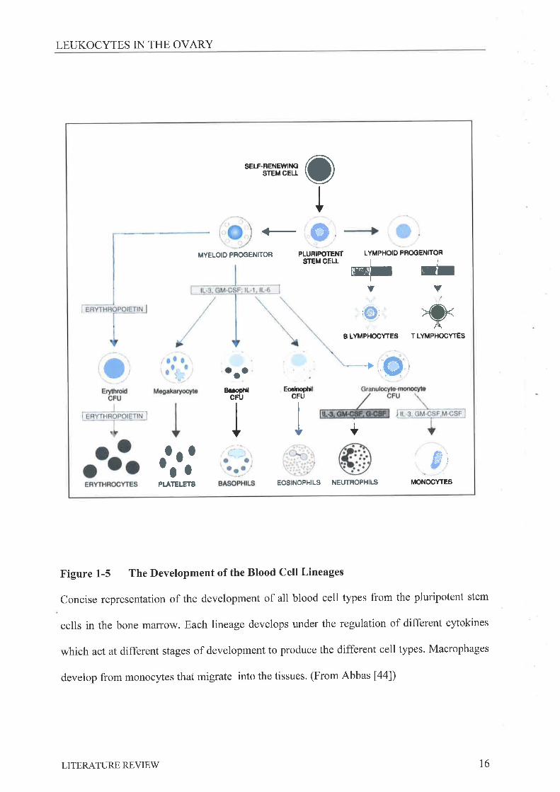
1.4 Lnuxocvrn DISTRTBUTIoN IN THE Ovlnv
Blood leukocyte types are all derived from a common stem cell precursor found in the
bone marrow (reviewed by Abbas [44], Figure 1-5). The main function of these mature cells
is to coordinate the surveillance and defence of the body against foreign organisms, as well as
LITERATURE REVIEW 15
LEUKOCYTES IN THE OVARY
sErf-RENElVll{CsrEt oEtl o
Io
MYELOIO PFOGENITOR PLURIPOTEI{TSTEM CELL
LYMPHOIO PBOOENITOR
-I -IJ+Ð _=.-.
XBLYI'PHOCYTES TLYMPHOCYTES
ao +'(}.)8úopt{
CFUEo.klopttl
CFU
I +
0 0I 0 $¡,aOPI.ATEI.ETS EOSINOPIIILS NEUTROPHILS MONOCYTES
Figure 1-5 The Development of the Blood Cell Lineages
Concise representation of the development of all blood cell types from the pluripotent stem
cells in the bone maffow. Each lineage develops under the regulation of different cytokines
which act at different stages of development to produce the different cell types. Macrophages
develop from monocytes that migrate into the tissues. (From Abbas [44])
LITERATURE REVIEW t6
LEUKOCYTES IN THE OVARY
mediating tissue inflammation and repair following injury or infection. These cells can
therefore readily migrate from the blood stream into the majority of tissues and organs of the
body. Since the events that occur in the mature ovary involve substantial tissue growth, atresia
and reorganization it follows that cells playing significant roles in these processes elsewhere
in the body would be active in the ovary, Early evidence that leukocytes play a role in the
regulation of the reproductive system demonstrated a possible link between these two systems
without clearly defîning which components of each system were involved. Splenectomy in
rats was found to delay ovulation [45] and could be reversed by the injection of splenocytes.
Neonatal thymectomy of mice prevents normal follicular development and ovulation in later
life [46], while treatment of rats with thymocyte antiserum results in reduced frequency of
ovulation, concluded to be due to persistent CLs [47]. More specifically, the supplementation
of the recirculating media of a perfused preovulatory rat ovary with blood leukocytes
increases the number of oocytes released following the administration of LH [48], suggesting
that these cells may have a role in the complex ovulatory cascade.
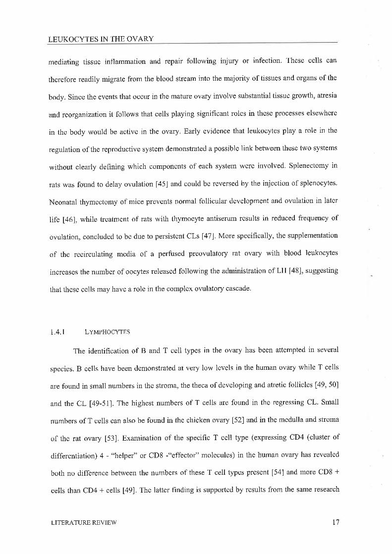
I.4.1 Lvtr¡pnocYtps
The identification of B and T cell types in the ovary has been attempted in several
species. B cells have been demonstrated at very low levels in the human ovary while T cells
are found in small numbers in the stroma, the theca of developing and atretic follicles [49, 50]
and the CL 149-511. The highest numbers of T cells are found in the regressing CL. Small
numbers of T cells can also be found in the chicken ovary [52] and in the medulla and stroma
of the rat ovary [53]. Examination of the specific T cell type (expressing CD4 (cluster of
differentiation) 4 - "helper" or CD8 -"effector" molecules) in the human ovary has revealed
both no difference between the numbers of these T cell types present [54] and more CD8 +
cells than CD4 + cells [49]. The latter finding is supported by results from the same research
LITERATURE REVIEW t7

LEUKOCYTES IN THE OVARY
group showing signiticantly more CD8+ than CD4+ cells in the rat ovary [53] as well as
results published by Suzuki [50], although in this study no statistically comparison of the
types of ovarian T cells was made. The role of T cells in the ovary has not been investigated
although reproductive function is significantly disrupted following surgical removal of the
thymus [46] or in animals with congenital athymia [55]. Elsewhere lymphocytes have been
found to play important roles in wound healing (reviewed by Schaffer [56]) with which events
in the CL are comparable. Hence it appears these cells must play a role in the regulation of
ovarian function, through either aiding in the control of the immune response following
ovulation, or alternatively through assisting in the regulation of apoptosis in cells of atretic
follicles or the degenerating CLs.
1.4.2 NpurRopstls
In the rat granulocyte numbers have been shown to vary, according to the stage of the
estrous cycle, in the medulla of the ovary and the thecal region of the follicle [53]. A large
increase in the numbers of neutrophils in the theca of the follicle was also shown to occur
immediately prior to ovulation. Neutrophils have also been demonstrated in the thecal layer
and CL of the human 149, 57) with numbers in the theca increasing as ovulation approaches,
Similar results were also obtained in the preovulatory follicles of rabbit ovaries using
histochemical analysis [58], and the preovulatory follicle and regressing CL of the pig ovary
[59]. In addition, experimental depletion of neutrophils in the rat ovary with either an
administered antibody [60] or by treatment with inhibitor of nitric oxide (NO) production [61]
results in reduced ovulation rates.
LITERATURE REVIEW 18
LEUKOCYTES IN THE OVARY
1.4.3 MecRopsacps
Studies carried out 40 years ago examining the distribution of the enzymes alkaline
phosphatase, esterase and beta-glucuronidase led to the conclusions that these cells are present
in the ovary [62]. Experiments carried out during the same period observing the uptake of
dianil blue (trypan blue) by cells in the ovary further suggested that these cells were
macrophages, however at this time no role for these cells in ovarian function was proposed. A
variation between species was noted with some of the enzymes studied more intense in some
species than others. With the advent of immunohistochemistry and the ability to produce
specif,rc antibodies to identi4r any desired cells type, the role of leukocytes in the ovary,
including macrophages, was more closely examined.
Macrophages have been found to be the predominant leukocyte type in the rat ovary,
with numbers found to vary in the medulla of the ovary and the thecal region of the follicle
[53], in relation to the stage of the estrous cycle. A large increase in thecal macrophage
numbers immediately prior to ovulation has been described. In the mouse ovary Cohen et al
[63] found no change in stromal macrophage distribution across the cycle but an increase in
thecal macrophage numbers as the follicle increases in size. Petrovska et al [64] also
demonstrated macrophages in the thecal layer of healtþ antral and preovulatory mouse
follicles and in the granulosa cell layer of atretic follicles. Macrophages have also been
demonstrated in the thecal layer of the follicles in the ovary of the chicken [65] and the human
l4g, 54, 571 with numbers similarly increasing as ovulation approaches. In addition,
macrophages are prevalent in the CLs of most species. They have been reported to be more
common in the layer of thecal derived luteal cells than the granulosa derived luteal cell layer
of the developing CL in the rat and human 149, 57 , 66, 671.In the mouse ovary, macrophage
density in these locations has found to be highest in proestrus and metestrus [64] with
numbers in the stroma and CL of luteinised mouse ovaries increasing as luteinisation
LITERATURE REVIEW t9
LEUKOCYTES IN THE OVARY
progresses [68]. Similarly, in the human the numbers of macrophages in early CL's increases
significantly with progressing CL regression [49, 54]. Signifrcant increases in macrophage
numbers in the newly formed and regressing CL of the pig have also been described [59, 69]'
1.5 GnNnnlr, Cn¿,n¡,crERrsrICS AND FuNcrIoNS oF MAcRoPHAGES
Macrophages develop from self-renewing stem cells found in the bone marrow from
which all haematopoietic cells develop (Figure 1-5). When these totipotent cells are
stimulated by the cytokines interleukin (IL) -1, IL-3 and IL-6 they develop into pluripotent
myeloid cells. Further exposure to IL-l and IL-3 commits these cells to becoming
granulocyte-macrophage colony forming units that will proliferate following exposure to
granulocyte-macrophage colony-stimulating factor (GM-CSF) [70]. The presence of
macrophage colony-stimulating factor (M-CSF) induces both proliferation and differentiation
of these cells into monocytic precursors or monoblasts that further divide into promonocytes
and then monocytes in the bone marrow. These monocytes then migrate into the blood stream
where the final maturation step for these cells occurs when they migrate via endothelial cell
adhesion molecule expression and chemotactic cytokines into tissues. Once within the tissue
the environment of that particular tissue can determine the specific characteristics of the
mature tissue macrophage, leading to widespread heterogeneous macrophage populations
[71]. Classically macrophages are considered cells of the immune system with a central role
in the regulation of immunity (Figure 1-6), Their most basic and critical role being that of a
scavenging phagocytic cell recognizing foreign molecules and apoptotic cells and disposing
of them by phagocytosis. Macrophages can also process ingested molecules intracellularly
into peptide fragments and present them, in association with a molecule called the major
histocompatibility complex or MHC, to the T cell repertoire with whom they interact via
LITERATURE REVIEV/ 20
MACROPHAGE CHARACTERISTICS AND FI-INCTION
Inflammation and fever
TNFIL-I
P rostaglm d ins
Conplement åctorsClotting Èctors
Tissue damage
MACROPHAGE
Lvmphocyte activation
Oxygen dependantHrOz
O:'-oH"
hypohal ite
Oxygen independentLysory me
Acid hydrolasa
Cationic proteins
Antigen presentation
Antigeu processing
lL-l produdion
Tissue rcorsanizationTissue damage
Elastæe, collagenase
Hy aluronidæeFibroblast growth åctors
Angiogenesis åctors
Elastæe, collagenase
Hy alu ron idæe
Fibroblast growth åctors
Angiogenesis åctors
Cytotoxic actionToxic fictors
HzO:, C3a
Proteæes
Arginase
Tu rrour necrosis åctor
r '-. '-.'i -i-.1: ; -'
Figure 1-6 The Central Role Of Macrophages In Immunity
Macrophages and their products play a central role in the induction and regulation of immune
responses as well as tissue reorganisation and repair. They also have effector functions in
defence of the body against microbial infection and tumour development. (Adapted from Roitt
tTsl)
LITERATURE REVIEW 2l

MACROPHAGE CHARACTERISTICS AND FUNCTION
cytokine messages to initiate an appropriate immune response. In addition to these roles in the
initiation of immune responses they also participate as effector cells migrating into
established wound or inflammatory sites and producing a large number of different factors
(Table 1-2) with numerous effects, including tumoricidal and microbicidal activities,
stimulation of tissue growth and repair, and the infiltration of other leukocytes'
1.5.1 MoNocvre Mtcn¡rtoN
All leukocytes, including monocytes, inf,tltrate inflammatory tissues via the processes
of tethering and rolling, activation, adhesion and transendothelial migration. Each of these
steps requires the expression of appropriate molecules by both the endothelium and the
leukocyte (reviewed by Ebnet 1721, Berton [73] and Weber [7a]). In addition, chemotactic
cytokines or chemokines expressed at the site of inflammation are thought to play a crucial
role in the regulation of endothelial cell and leukocyte activation, as well as manipulation of
the expression of the molecules required for secure leukocyte binding and transendothelial
migration (reviewed by Gale [80], Greaves [81] and Middleton [82]).
Leukocyte tethering and rolling is mediated by the expression of selectins and their
ligands. L- Selectin is expressed constitutively on the leukocyte surface, while E- and P-
selectin expression on the endothelium is induced [72]. Combined, these selectins initiate
tethering and rolling of leukocytes along endothelium expressing appropriate ligands' The
ligands that bind the selectins are numerous and fall into different classes of molecules
(reviewed by Varki [83]) with the different selectins binding with differing afhnities to some
of the same ligands.
Once rolling has been initiated cells become activated by selectin binding and the
presence of chemokines presented on the surface of the endothelium 172, 82]. Specif,rc
chemokine combinations are thought to attract specific leukocytes, monocytes respond to
LITERATURE REVIEW 22
MACROPHAGE CHARACTERISTICS AND FUNCTION
Growth Factors Extracellular matrix
Basic hbroblast growth factor(bFGF)Epidermal Growth Factor (EGF)Transforming Growth Factor -alpha/beta (TGF-c/Þ)Insulin-like growth factor I (IGF-I)Platelet derived growth factor(PDGF)Vascular Permeability Factor (VPF)Vascular Endothelial growth factor(vEGF)
Interleukin-l (IL-1)Interleukin-8Interleukin-12Interleukin-6 (IL-6)Interferon -alpha/gamma (IFN-o/y)Tumour Necrosis Factor -alpha(TNFo)Macrophage infl ammatory protein(MrP)Granulocyte Colony Stimulating Factor(G-csF)Granulocyte Macrophage ColonyStimulating Factor (GM-CSF)Macrophage Colony StimulatingFactor (M-CSF)
FibronectinProteoglycans
Reactive intermediates Bioactive Lipids Enrymes
SuperoxideHydrogen peroxideHydroxyl radicalNitrites/nitrates
Prostaglandins E2 and F2
LeukotreinesProstacyclin
Plasminogen Activator andinhibitors of ElastaseCollagenase and inhibitors
Table 1-2 Factors Known to be Produced by Macrophages in Different Environments
(Compiled from references [76-79])
LITERATURE REVIEV/ 23
MACROPHAGE CHARACTERISTICS AND FI.]NCTION
monocyte chemotactic peptide -1 (MCP-1), MIP-1o, -1P, RANTES, I -309, Fractalkine [80,
81]. Activation leads to stronger adhesion instigated by integrins expressed on the monocyte
and adhesion molecules expressed on the endothelium. The beta 2 (LFA-1, Mac-1) and beta 1
(VLA4) integrins appear most important for monocyte adhesion and bind ICAM-I, -2, -3 and
VCAM-I and fibronectin, respectively 173, 84]. Under the influence of chemokines
transendothelial migration is thought to occur by diapedesis at inter-endothelial cell junctions
through binding interactions between LFA-I on the leukocytes and junctional adhesion
molecule (JAM-1)t74]. Intergrins expressed by macrophages also bind extracellular matrix
allowing migration into the tissue and further activation specific to that tissue environment
[73]. The recruiting environment can, through the expression of integrins and chemokines,
dictate the characteristics recruited macrophages develop, generating regional populations that
differ widely in their functional characteristics.
I.5.2 MecnopHecEs INItlFrRN4N4aïoN
Inflammation occurs as a result of tissue injury or infection, and involves the
characteristic pathologies of swelling, redness and pain. These symptoms are a result of the
release of early mediators of inflammation, such as histamine, and products of the
complement and kinin enzyme systems, and are released by cells, including tissue
macrophages, located at the damaged site. They induce the infiltration and activation of
leukocytes (described in 1.5.1) and production of factors to promote vasodilation (reviewed
by Mutsaers [S5] and Greenhalgh [86]). Neutrophils are the first immune cells recruited to the
site releasing free radicals and proteases, which may cause some host tissue damage, to
eliminate foreign organisms and then dying via apoptotic mechanisms. Macrophages are the
second cell type recruited and are considered central regulators of inflammation and tissue
repair mechanisms. In inflammation these cells can promote vascular dilation, leukocyte
LITERATURE REVIEV/ 24
MACROPHAGE CHARACTERISTICS AND FUNCTION
infiltration and T and B cell proliferation and maturation through the production of classical
inflammatory mediators, such as prostaglandins, NO, IL-lB,tumour necrosis factor-alpha
(TNFcr) and platelet activating factor (PAF). This leads to the destruction of the invading
bacterial organism or virally infected cells. The resolution of this type of destructive
inflammatory response is regulated by the overriding expression of anti-inflammatory
cytokines such as IL-10, transforming growth factor beta 1 (TGFBI) and IL-1 receptor
antagonist (Il-lra), while healing of damaged tissues and clearance of apoptotic cells
occurring after the inflammatory phase is also a central role of the recruited macrophages.
1.5.3 M¡.cnopuecE AcTIVATIoN
The activation of macrophages has been a recognised developmental stage for many years,
characterised by changes in functions that increase the ability of these cells to combat
infections (Table 1-3)t87]. This type of 'classical' activation is bought about by the presence
of inflammatory mediators, bacterial products such as lipopolysaccharides (LPS), interferon
gamma (IFNy) released by T cells during the course of an infection to stimulate the
production of pro-inflammatory mediators such as IL-lP, TNFcr, IL-IZ, and reactive oxygen
species. This promotes the 'classical' inflammatory reaction with the production of
prostaglandins, leukotrienes and numerous cytokines chemotactic and stimulatory to T and B
cells. Anti-inflammatory agents such as IL-4 and IL-13 [88, 89] have the ability to
reduce the production of pro-inflammatory cytokines by these activated macrophages
(reviewed by Doherry [90] and Ma [91]), promoting deactivation of the macrophages and
resolution of the inflammatory response. These factors have also been found to promote what
is now termed the alternative route of macrophage activalion resulting in increased expression
of mannose receptor, increased endocytosis [92], lower pro-inflammatory cytokine levels
LITERATURE REVIEW 25

MACROPHAGE CHARACTERISTICS AND FTINCTION
Microbial activity (1)
Tumoricidal activity (1)
Chemotaxis (1)
Phagocytosis (1)
Pinocytosis (1)
Glucose transport and metabolism (1)
Respiratory burst (1)
Antigen presentation (1)
Prostaglandins, leukotrienes (1)
Apolipoprotein E and lipoprotein lipase (1)
Elastase (l)
Complementproteins (1)
Acid hydrolases (1)
Collagenase (1)
Plasminogen activator (1)
Cytolytic proteinase (1)
Arginase (1)
Fibronectin (t)Interleukin-l (1)
Tumour Necrosis Factor (1)
InterferonoandB(1)Angiogenesis factors (t)
Table 1-3 Major Macrophage Functions And Their Regulation By The
Process Of Activation. Adapted from Johnston [87].
LITERATURE REVIEW 26

MACROPHAGE CHARACTERISTICS AND FUNCTION
[93], and production of the anti-inflammatory cytokines lL-10 and TGFB [94]. These cells
are highly angiogenic in vitro and in vivo with the ability to actively inhibit mitogen induced
proliferation of PBL or CD4+ T cells in vitro [95, 96] while promoting the differentiation of
Th2 type cells involved in the stimulation of antibody responses l97l.It follows that these cell
types play an important role in the down regulation of inflammatory responses, and
subsequent wound healing.
1.5.4 MRcRopsecE INTERACTIoNS wITH T cELLS
Following the phagocytosis of a foreign body the macrophage is capable of
intracellular degradation of the particle and presentation of peptide fragments from the
particle on the surface of the cell in association with the class II MHC (MHC II)' These
MHC-II -antigen complexes can be recognised by helper T (Th) cells via the T cell receptor
(TcR) and the CD4 molecule. This results in the initiation of an immune response through the
activation of both the T cell and the antigen presenting macrophage. The type of immune
response mounted and the activation state of the macrophage that results has been found to be
dependant on the co-stimulatory molecules present on the cells and the cytokines in the
environment (reviewed by Reiner [98], Constant [99] and Goerdt [92]). In a cell-mediated
inflammatory response, binding of the TcR/CD4 on the helper T cell to the antigen/MHC II
molecule complex must be accompanied by interaction of co-stimulatory molecules. This
initiates the production of IL-2, IFN-y, and TNFB by the T cell, and IL-I2 by the
macrophage, which in turn stimulate proliferation of the T cells and activation of the
presenting macrophage (Figure 1-7).
Activation of the macrophage is characterised by the production of pro-inflammatory
cytokines, increased expression of MHC II molecules and the ability to produce reactive
oxygen intermediates. The proliferating T cells also produce pro-inflammatory cytokines
LITERATURE REVIEV/ 27

MACROPHAGE CHARACTERISTICS AND FUNCTION
Cytokineproduction
CD4O
{ MHC IITcR/CD3
cDso/87- l cD28
-4
cD86lB7-2T CELL
MACROPHAGE
Clokineproduction
Figure L-7 The Interactions Between Macrophages And T Cells During An Immune
Response. The MHC II, major histocompatibility complex II, on the macrophages presents
processed antigens to the T cell receptor (TcR). Co-stimulatory molecules must be present to
initiate an immune response. CD40 on the macrophages binds with the CD40 ligand on the T
cell and the 87 molecules (CD80/86) of the macrophage bind the CD28 or CTLA-4 which is
externalised in certain conditions on the surface of the T cell. Cytokines are produced by both
the macrophage and the T cell.
.ry<
LITERATURE REVIEV/ 28
MACROPHAGE CHARACTERISTICS AND FTINCTION
promoting antibody production by B cells, and assist in the initiation of a tissue destructive
cytotoxic response. This is termed a Thl or cell mediated response. If the appropriate co-
stimulatory molecules are not present and an apoptotic-inducing ligand is present on the
macrophage cell membrane, T cell deletion can occur [92]. In the presence of the cytokine IL-
4 , an alternative immune response can also be generated resulting in proliferation of Th2
cells, which produce lL-4 and IL-5 [8S]. This stimulates B cell proliferation and alternatively
activates macrophages to produce anti-inflammatory cytokines, such as IL-10 and
TGFB, promoting down regulation of the inflammatory response [100].
1.5.5 MNCROPUAGES IN WOUNO HEALING AND TISSUE REPEIN
The macrophages that pervade a wound site have the ability to release, proteases such
as collagenase or elastase which instigate tissue degradation, and cytokines that stimulate
fibroblastic proliferation and blood vessel growth, particularly in hypoxic conditions [101].
They also have the capacity to eliminate cell debris and other non-viable or apoptotic
material. The wound healing process itself can be divided into inflammatory, proliferative and
maturation phases [36]. The inflammatory, proliferative and maturation phases can co-exist in
a single wound site with the earlier phases existing in the central open areas of the wound and
later phases existing at the periphery of the wound. Members of the TGF-P family also have
critical roles in regulating wound healing [102]'
1.5.6 MACROPUAGE PHAGOCYTOSIS AND PHAGOCYTIC RBCSPTORS
Phagocytosis is a complex process mediated by binding of the molecule to the cell
surface which in turn initiates actin polymerisation and internalisation of the attached particle
into a phagosome or vacuole in the cell cytoplasm. Two models of internalisation have been
LITERATURE REVIEW 29
MACROPHAGE CHARACTERISTICS AND FTINCTION
proposed; a 'zipper' model, in which sequential interactions between the surface of the
molecule and receptors on the phagocyte cell surface are required as the molecule is engulfed,
and a 'triggering' model in which binding of the molecule to receptors on the surface of the
phagocyte initiates engulfment independent of further receptor interactions [103, 104].
Phagocytosis may be followed by fusion of the phagosome with enzyme containing
lysosomes, and degradation of the particle. Even though most cells have some phagocytic
capacity, macrophages are 'professional' phagocytes and can internalise particles much more
rapidly and efficiently than normal cells, due mainly to the expression of innate immune
receptors (Table 1-4). These receptors recognise various non-specif,tc entities which are
expressed on the surface of pathogens or whose production is triggered by the presence of a
pathogen. Particle uptake has also been shown to be dependent on particle size and surface
charge t105]. Although the phagocytosis of a particle generally leads to the activation of the
phagocytosing cell and release of pro-inflammatory cytokines this is not always the case. In
the human the uptake of apoptotic cells by blood monocyte derived macrophages via CDl4,
has been found to result in the inhibition of the release of pro-inflammatory cytokines, such as
IL-18, IL-8 TNFcr and GM-CSF, and stimulation of the release of anti-inflammatory factors
such as TGFB, PGEz and PAF [106, 107]. The anti-inflammatory response to apoptotic cell
uptake is hence thought to be dependent on the receptors used to internalise the altered 'self
cells [108], the cytokines released in response to uptake [106], and/or to the lack ofexpression
of macrophage co-stimulatory molecules [109].
I,5.7 M¡CROPHRGE SURFACE MARKERS AND THEIRR¡CUTATNO EXPRBSSTON
Monoclonal antibodies are the principal tool for the identification and classification of
macrophages, Several monoclonal antibodies raised against different proteins found on
macrophage cell membranes have been developed and are currently in common use
LITERATURE REVIEW 30
MACROPHAGE CHARACTERISTICS AND FI-INCTION
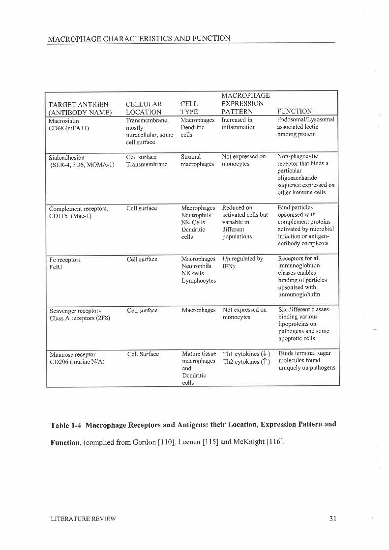
TARGET ANTIGEN(ANTIBODY NAME)
CELLULARLOCATION
CELLTYPE
MACROPHAGEEXPRESSIONPATTERN FUNCTION
MacrosialinCD68 (mFA11)
Transmembrane,mostlyintracellular, somecell surface
MacrophagesDendriticcells
Increased ininflammation
Endosomal/Lysosomalassociated lectinbinding protein
Sialoadhesion(sER-4, 3D6, MOMA-1)
Cell surfaceTransmembrane
Stromalmacrophages
Not expressed onmonocytes
Non-phagocyticreceptor that binds aparticularoligosaccharidesequence expressed onother immune cells
Complement receptors,CDllb (Mac-l)
Cell surface MacrophagesNeutrophilsNK CellsDendriticcells
Reduced onactivated cells butvariable indifferentpopulations
Bind particlesopsonised withcomplement proteinsactivated by microbialinfection or antigen-antibody complexes
Fc receptorsFcRI
Cell surface MacrophagesNeutrophilsNK cellsLymphocytes
Up regulated byIFNy
Receptors for allimmunoglobulinclasses enablesbinding of particlesopsonised withimmunoglobulin
Scavenger receptors
Class A receptors (2F8)Cell surface Macrophages Not expressed on
monocytesSix different classes-
binding variouslipoproteins onpathogens and someapoptotic cells
Mannose receptorCD206 (murine N/A)
Cell Surface Mature tissuemacrophagesandDendriticcells
Thl cytokines ( ) Binds terminal sugarmolecules founduniquely on pathogens
Th2 cytokines (1 )
Table 1-4 Macrophage Receptors and Antigens: their Location, Expression Pattern and
Function. (complied from Gordon [110], Leenen [115] and McKnight [116]'
LITERATURE REVIEW 31
MACROPHAGE CHARACTERISTICS AND FUNCTION
(Table 1-4). An understanding of how prevalent expression is in other cell types and the
circumstances in which they are expressed by macrophages is important when interpreting
results obtained using this technology. Since several excellent reviews of these and other
antigen markers in murine tissues are available [110-112], only the antigens used to isolate
and identiff cells in this thesis will be discussed here.
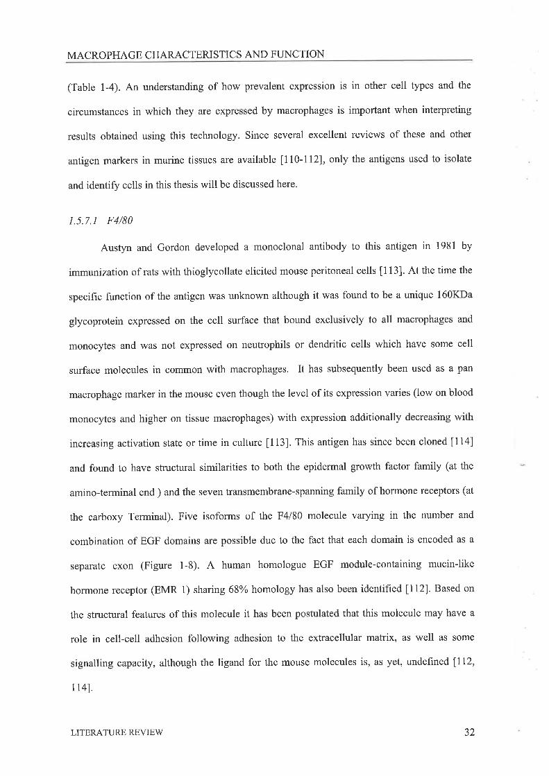
1.5.7.1 F4/80
Austyn and Gordon developed a monoclonal antibody to this antigen in 1981 by
immunization of rats with thioglycollate elicited mouse peritoneal cells [113]. At the time the
specific function of the antigen was unknown although it was found to be a unique 160KDa
glycoprotein expressed on the cell surface that bound exclusively to all macrophages and
monocytes and was not expressed on neutrophils or dendritic cells which have some cell
surface molecules in common with macrophages. It has subsequently been used as a pan
macrophage marker in the mouse even though the level of its expression varies (low on blood
monocytes and higher on tissue macrophages) with expression additionally decreasing with
increasing activation state or time in culture [113]. This antigen has since been cloned [114]
and found to have structural similarities to both the epidermal growth factor family (at the
amino-terminal end ) and the seven transmembrane-spanning family of hormone receptors (at
the carboxy Terminal). Five isoforms of the F4l80 molecule varying in the number and
combination of EGF domains are possible due to the fact that each domain is encoded as a
separate exon (Figure 1-8). A human homologue EGF module-containing mucin-like
hormone receptor (EMR 1) sharing 68% homology has also been identified lll2). Based on
the structural features of this molecule it has been postulated that this molecule may have a
role in cell-cell adhesion following adhesion to the extracellular matrix, as well as some
signalling capacity, although the ligand for the mouse molecules is, as yet, undeflrned [112,
1r41.
LITERATURE REVIEV/ 32
MACROPHAGE CHARACTERISTICS AND FI.INCTION
EGF domains(A) withN-linkedglycosolationsites (.)
Spacer regionwith O-linked(bars) sugarsand N-linkedglycosolationsites (.)
cooH cooH cooHcoöH
Membrane spanning regionwith short c)¡toplasmic tail.ss is a postulated disulfidebond.
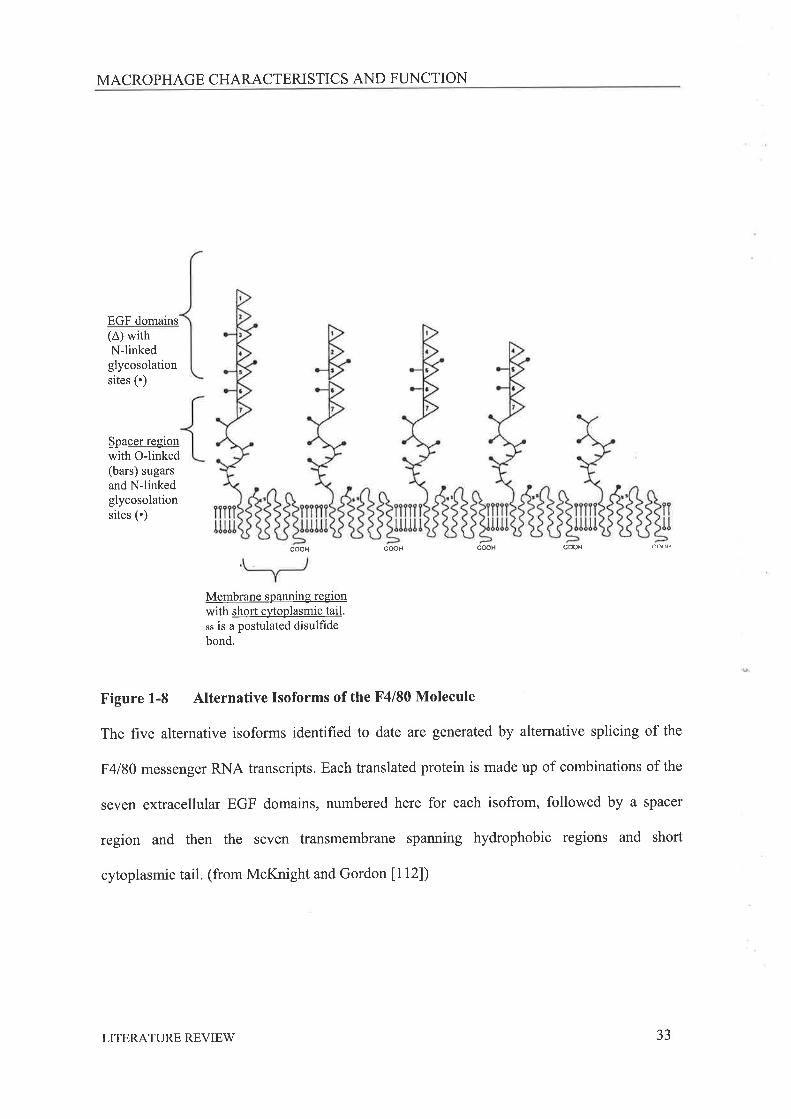
Figure 1-8 Alternative Isoforms of the F4l80 Molecule
The five alternative isoforms identified to date are generated by alternative splicing of the
F4l80 messenger RNA transcripts. Each translated protein is made up of combinations of the
seven extracellular EGF domains, numbered here for each isofrom, followed by a spacer
region and then the seven transmembrane spanning hydrophobic regions and short
cytoplasmic tail. (from McKnight and Gordon [112])
LITERATURE REVIEW 33
MACROPHAGE CHARACTERISTICS AND FLINCTION
L5.7.2 Class II Major Histocompatibility Complex (Ia)
The MHC is a region of polymorphic genes that are expressed on the surface of many
cell types. Individuals who express the same MHC antigens will accept tissue grafts from
each other while those who differ will rapidly reject grafts. The complex can be divided into
class I MHC molecules, which bind endogenously derived peptides and are present on nearly
all cells; class II MHC molecules, which bind exogenously derived peptides and are expressed
only on B cells and antigen presenting cells; and an S region encoding components of the
complement system. In the mouse the class I region can be divided into H-2K, H-zD andH2-
L, and the class II region can be divided in I-A (Ia) and I-E regions þa|In the human the
equivalent genes are called human leukocyte antigens (HLA); HLA -4, -8, or -C are class I
genes and HLA -DP, -DQ, or -DR are class II genes. In the mouse the glycoprotein products
of the I-A and I-E regions are called I region associated antigens and monoclonal antibodies
that detect these antigens are readily available. Class II MHC molecules are expressed at high
levels on all antigen presenting cells such as dendritic cells, macrophages, and some
fibroblasts and epithelial cells.
1.5.8 MACROPTNGES RESPOND TO THE FBIUETE SBX STNNOID HORMONES
Studies investigating the effect of progesterone on macrophages have been carried out
in other fields, while there is ample evidence indicating oestradiol has significant effects on
macrophage and monocyte function. Unforlunately, the results for both steroids are divergent,
probably due to the varying sources of macrophages used and their resulting activation states.
Thus it can be difficult to interpret these results in terms of the function these interactions may
play in regulating ovarian function.
LITERATURE REVIEW 34
MACROPHAGE CHARACTERISTICS AND FI-INCTION
1.5.8,1 Progesterone
Progesterone has been shown to inhibit superoxide release by unstimulated peritoneal
macrophages isolated during pseudopregnancy in the rat [117]. In contrast, progesterone has
also been reported to enhance the release of reactive oxygen intermediates and inhibit nitrite
production in l2-phorbol l3-myristate acetate (PMA) and opsonized zymosan (OZ)
stimulated mineral-oil-elicited peritoneal cells from male rats [118]. Progesterone treatment
of guinea pigs impairs Fc mediated clearance of erythrocytes in vivo throtgh a reduction in
the numbers of Fc receptors expressed by macrophages in the spleen [119], while in vitroFc
mediated phagocytosis by mineral oil elicited peritoneal cells from male rats was up regulated
by progesterone treatment [120]. These varying results may be explained by the proposition
that activated macrophages when exposed to progesterone are stimulated whereas resting or
inactive macrophages are inhibited by the presence of progesterone. In context of the ovary
the largest amounts of progesterone are found in the functional CL and perhaps during CL
development or pregnancy the macrophages present are 'resting' macrophages and their
activity is inhibited by the progesterone present. Following the initiation of luteolysis they
may become activated and the progesterone present further stimulates these cells leading to
luteolysis.
1.5.8.2 Oestradiol
Peritoneal cells from male rats pre-treated in vitro with oestradiol and then stimulated
with phorbol myristate acetate or opsonized zymoson exhibit increased release of reactive
oxygen intermediates and inhibited nitrite production [118]when compared to untreated cells.
Endogenous chemiluminescence, reflecting the production of reactive oxygen intermediates,
was also found to be stimulated by pre-exposure to oestradiol [120]. In addition, in these same
cells Fc receptor mediated phagocytosis of SRBC was also stimulated by oestradiol.
LITERATURE REVIEW 35
MACROPHAGE CHARACTERISTICS AND FLINCTION
Treatment of isolated human monocytes with oestradiol has also been found to stimulate the
release of NO ll2ll. High levels of oestradiol are found immediately prior to ovulation and
hence these effects of oestradiol on macrophages may play a role in the stimulation of the
production of reactive species involved in the owlatory event. It has also been demonstrated
that oestradiol can inhibit human monocyte chemotaxis in response to MCP-I in vitro ll22].
This effect was reversed by the addition of the oestrogen receptor antagonists, tamoxifen and
clomiphene, while blood monocytes and peritoneal macrophages from oestradiol treated
animals are more phagocytically active, but have reduced intracellular bactericidal activities
lr23l.
In addition to the effects of oestradiol presented above this steroid has also been
shown to modulate cytokine production by monocytes and macrophages. Adherent peritoneal
cells from oestradiol treated Listeria monocytogenes infected animals produce less IL-12,
TNFcr and IL-10 although it was suggested this may be mediated through the nonadherent cell
population (possibly T cells) which in oestradiol treated animals produced less IFN-gamma
and more IL-4, ILl0 and TGF-beta than untreated cells [123]. Elsewhere oestradiol has been
shown to reduce TNFo production by peritoneal macrophages isolated from rats subject to
myocardial ischaemia-reperfusion ll24l, and to significantly increase the level of platelet
derived growth factor-A messenger RNA (mRNA) in the monocyte cell line THP-1 following
stimulation with 12-O-tetradecanoylphorbol 13 acetate (TPA) ll25l. Oestradiol also inhibited
LPS stimulated MCP-1/JE mRNA expression by thioglycollate elicited peritoneal
macrophages and two murine macrophage cell lines, but had no effect on TNFcr expression.
The effect could be reversed by oestrogen antagonists and could not be replicated using
progesterone 1126]. It appears that oestradiol inhibits the production of inflammatory
cytokines by stimulated macrophages but enhances their phagocytic activity and production of
reactive oxygen.
LITERATURE REVIEW 36

CHARACTERISTICS OF TISSUE MACROPHAGES
1.6 Cnnn¡.crERrsrICS or Trssun M¡,cRopnAGES
Macrophages located in the tissues develop from blood borne monocytes that migrate
out of the blood stream via adhesion molecules and chemotactic factors. They differentiate in
response to the local microenvironmenal signals to assume a functional phenotypef127]. Their
roles may include the phagocytosis and degradation of foreign organisms or apoptotic tissues,
the regulation of local immune and inflammatory responses, and/or tissue remodelling. The
studies presented in this section demonstrate that macrophages have a specific tissue
phenotype, presumably generated by exposure to factors found in the local environment that
stimulate or inhibit selected activities of developing macrophages. This creates specialised
macrophage cells capable of performing the specific tasks required for normal function of that
particular tissue. The method of isolation and characteristics of macrophages recovered from
some complex tissues or organs deemed not to have a primary immune function are
examined.
1.6.1 Tpsns
Macrophages have been demonstrated in the interstitial tissue of the testis closely
associated with the Leydig cells of the testicular interstitium. Within the testis these
macrophages are thought to regulate steroid synthesis (reviewed by Hales U28]) and to
maintain the immunosuppressive environment of the testis (reviewed by Hedger ll29D.In the
rat the testicular macrophage population has been demonstrated to be heterogeneous by the
expression of specific macrophage markers [130] with their numbers closely correlated to the
numbers of Leydig cells. Testicular macrophages have been shown to significantly influence
normal testicular function. Macrophage conditioned media has been shown to significantly
influence Leydig cell steroidogenesis lI3I,I32l while cytotoxic depletion of the leydig cell
LITERATURE REVIEW 37

CHARACTERISTICS OF TISSUE MACROPHAGES
population or inhibition of Leydig cell function via testosterone implants both significantly
alter the numbers of testicular macrophages [130, 133]. Macrophages have been isolated from
this reproductive organ via adherence [134], collagenase digestion followed by cell elutriation
[135], and by immunoaffinity [136]. These cells have been reported to be poorly pro-
inflammatory, inhibiting the proliferation of lymphocytes both directly in co-culture
experiments through the secretion of prostaglandins[135], and to have a cytokine profile
significantly different to that of peritoneal macrophages. In addition when compared to
peritoneal cells they were found to respond in a unique manner to regular macrophage
stimulants t137]. Interestingly if inflammation is induced in the testis ir¿ vivothen testicular
function is compromised [138] and this is proposed to occur through the production of
inflammatory cytokines some of which may be derived from infiltrating monocytes [129].
t.6.2 UrBRus
Uterine macrophage distribution and numbers vary according to the stage estrous cycle. It is
evident that this occurs due to the production of chemotactic factors produced by the uterus in
response to fluctuating steroid levels. Chemokines such as colony stimulating factor -1 [139],
granulocyte-macrophage colony stimulating factor [140] and others [141] have been shown be
regulated by steroid levels, as well as by normal mating. The uterus displays an inflammatory
type reaction to mating, with a rapid but transient infiltration of macrophages that subsides on
day 3 of pregnancy ll4I,I42). Uterine macrophages have been shown to express GM-CSF
receptor following mating [143] indicating these cells respond to the surge of GM-CSF from
the uterine epithelium that occurs mating lI44]. Uterine macrophages are consequently
thought to play an important role in successful implantation and maintenance pregnancy
(reviewed by Hunt and Pollard [145] and Hunt [142]). Macrophages have been isolated from
the uteri of cycling mice by collagenase digestion followed by rosetting with sheep red blood
LITERATURE REVIEW 38

CHARACTERISTICS OF TISSUE MACROPHAGES
cells [146, 147] or immunomagnetic beads t148]. Uterine macrophages were shown to be
inhibitory in T cell mitogenesis assays in which elicited peritoneal cells were stimulatory
l¡47l.In contrast, uterine macrophages isolated from ovariectomised mice were found to be
stimulatory in the same assay. More recently it has been shown that macrophage/dendritic
cells located in the mouse uterus can be divided into three heterogeneous populations, defined
by their expression of cell surface markers U48]. These distinct uterine macrophage
populations have further been found to exhibit discrete immunostimulatory phenotypes U48]'
1.6.3 HutrlaN PraceNtR AND DECIDUA
Macrophages can be found in the human decidua but are less evident in the decidua of
the mouse and, rat t145]. In the human, macrophages comprise about 20o/o of the decidual
tissue [149] and are they thought to play an immunosuppressive role in this tissue U50]'
Incidentally these cells can stimulate lymphocyte proliferation although it is not comparable
to that stimulated by peripheral monocytes [151] and it has been proposed elsewhere that the
stimulated cells may be suppressor or regulatory T cells required to promote survival of the
conceptus t145]. Evidence suggests decidual macrophages may play arole in the regulation of
trophoblast invasion [149] as well as parturition ll52l. The macrophages in these studies have
been isolated from the decidua via numerous methods including collagenase or dispase
digestion followed by, density gradient centrifugation, immunomagnetic beads, adherence
andlor flow cytometry [151, 153, 154].
In the placenta, foetal macrophages or Hofbauer cells can be found in close proximity
to the trophoblast cells they comprise 25o/o of the human placental stromal cells [145]'
Evidence suggests these cells may have a significant role in the regulation of trophoblast
function [155] potentially through the secretion of cytokines. In the rat placenta macrophages
have been shown to express only low levels of MHC class II molecules [145] while in the
LITERATURE REVIEW 39
CHARACTERISTICS OF TISSUE MACROPHAGES
human comparing macrophages isolated from placenta early and late in gestation shows class
il MHC molecules were expressed at lower and higher levels respectively U56].
Macrophages from human placenta have been isolated by a variety of methods including
enzyme digestion, adherence, density gradient centrifugation and magnetic depletion [157-
lsel.
t.6.4 BRArN M¡,cRoPHRcns
Several cell types found in the central nervous system display characteristics of
macrophages. Whether or not these cells are related, representing different stages of
maturation is unclear, although evidence does exist to suggest this (reviewed by Thomas
t160]). The microglial cells are the main immune effector cells or macrophages of the brain.
These cells appear to be a resting cell type becoming activated by inflammatory signals or
injury in the brain, This activation has been graded into two steps (reviewed by Gehrmann
t161]) the first being characterised by proliferation, an increase in antigenic markers and the
released of common inflammatory mediators followed by the second step or development of
phagocytic activity if neuronal andlor terminal degeneration occurs. In addition, these cells
have been reported to present antigen to T cells and have cytotoxic activity. All antibodies
developed so far against microglial cells are cross- reactive with tissue macrophages while in
the human they have been reported to express the CD45ns (LCA) antigen 1162l. Microglial
cells have been isolated from the brain by mechanical dissociation and adherence, magnetic
selection [163] and enzymatic digestion [164].
LITERATURE REVIEW 40

SUMMARY AND HYPOTHESIS
l.t PornNrr¡,r, M.tcnoPHAGE FuNcrroNS IN THE Ovlnv
1.7 .I M¡cRops¡,cES AND Folucuren GRowrs eNo ArResla
The mechanisms initiating primary follicle growth are unknown although recent
evidence suggests the production of various growth factors by the oocyte and early granulosa
cells may lead to the initiation of follicle growth (reviewed by McNatty [165] and Nilsson
[13]), Macrophages have not been localised around primordial or primary follicles and are
unlikely to have any significant role in this initial stimulation.
Following the initiation of follicle growth, numerous factors potentially derived from
macrophages located in the thecal layer of growing follicles, are believed to play a role in
either stimulating the proliferation and growth of follicles or regulating follicular apoptosis.
Molecules such as hepatocyte growth factor [166], basic fibroblast growth factor (bFGF)
[167, 168], epidermal growth factor (EGF) 1167-1691, TGFP 11 [170], and NO ll7l, 172)
with roles in stimulating the proliferation and/or growth of follicles or regulating follicular
apoptosis have all been shown to be produced by macrophages in other systems 177-79,96,
173]. In agreement with this the co-culture of peritoneal macrophages with granulosa cells
results in stimulated proliferation of the granulosa cells above that of cells cultured alone
lt74l,
The presence of macrophages in follicles at advanced stages of atresia suggests these
cells participate in the removal of cell debris created during follicular atresia [175].
Macrophages have the ability to identiff molecules in the cell membranes of apoptotic cells
through the expression of scavenger receptors discussed earlier. Interestingly, macrophages
have also been found to stimulate apoptosis in some cell lines 1176, I77), while several
cytokines that macrophages produce such as TNFc¿ lI78,I79l and IL-6 [180-182] have been
found in vitro to induce apoptosis in ovarian and other cell types.
LITERATURE REVIEV/ 4t

SUMMARY AND HYPOTHESIS
1.7 .2 MecRopH¡cES AND ruB Sutr¡uI-ATIoN or OvulertoN
It was proposed in 1980 l29l that the ovulatory event was analogous to an
inflammatory response with vasodilation, infiltration of leukocytes and production of
inflammatory cytokines and prostaglandins triggered by the preovulatory LH surge. To date
studies examining these events have explored and reinforced this hypothesis showing
classical inflammatory mediators can influence many aspects of ovarian function.
Macrophages have a pivotal role in the inflammatory process producing or stimulating the
production of many of the inflammatory mediators required to regulate inflammation, tissue
growth and restructuring processes (Section 1.5). Macrophages have the capacity to release
numerous cytokines that have been demonstrated to be important in the owlatory process, as
well as some metallo-proteinases required for degrading the follicle wall and basement
membrane. Here several key products of classically activated macrophages with roles in
ovulation will be discussed.
1.7.2. I Interleukin-lBeta ( IL-1 P)
1¡-lP is a potent pro-inflammatory cytokine, released by classically activated
macrophages, with divergent effects on different cell types [183]. In the ovary it has been
implicated as a local regulator of follicle atresia [24], ovulation [184] and CL function [31].
Within the ovary the expression of the type I receptor for this cytokine has been demonstrated
in the granulosa, and to a lesser degree the theca of growing and preovulatory follicles of the
rat [185-187] and mouse t1881. The IL-IB protein can be found in human preovulatory
follicular fluid [139] and in samples taken from in vitro perfused preovulatory rat ovary [190],
with concentrations increasing as ovulation approaches. Levels of mRNA measured in the
whole rat ovary similarly increase leading up to owlation and have been localised in both the
theca and granulosa layersf185] of the follicle. In the mouse IL-1p protein has been localised
LITERATURE REVIEW 42
SUMMARY AND HYPOTHESIS
prior to ovulation only in the thecal layer of the mouse preovulatory follicle [188], although
following ovulation granulosa cells also express the protein. Culture of preovulatory follicles
with IL-18 inhibits the spontaneous apoptosis of isolated preovulatory follicles in vitro
through the production of NO [171], also known to be important for ovulation to proceed
[61], while in the same system the 'rescuing' effects of gonadotrophins on follicles can be
significantly reduced by the treatment with IL-lra [171]. Addition of IL-18 to the media of in
vitro perfased ovaries increases ovulation rates in the rat ll9I,l92l and rabbit [193], where it
was also reported to promote oocyte maturation. In the mouse, injection of the IL-Iru, a
naturally occurring receptor antagonist which has also been localised in the ovary [186, 194],
inhibits ovulation in vivo 1192, 1951. In the mare, intrafollicular injection of IL- 1B has also
been reported to promote oocyte maturation and owlation in vivo [196]. More recently this
cytokine has been shown to stimulate the expression of the vascular endothelial growth factor
(VEGF) gene, a factor important for vascularisation occurring during the pre- and post-
ovulatory stages, in whole ovarian dispersates and granulosa cell cultures ll97l.
1.7.2.2 Tumour Necrosis Factor alpha QNFa)
TNFcr is also an inflammatory cytokine produced by activated macrophages usually in
combination with IL-18. It is part of a TNF-like ligand and receptor superfamily, but
individually has only two specif,rc receptors, type I (p55/p60) and type 2 (p751p80), which
differ in their intracellular domain [198, 199]. It is believed that these receptors and their
associating factors (TRAFs) orchestrate the divergent cellular responses of TNFo binding'
Specific localisation of these receptors in the ovary has not been carried out although type 1
receptor 6RNA has been detected in whole rat ovarian dispersates [200], which have also
been shown to bind TNFcr protein. Similar binding of the TNFcr protein to pig [201] and
bovine 1202] granulosa cells has also been demonstrated. TNF protein has been measured in
LITERATURE REVIEW 43
SUMMARY AND HYPOTHESIS
human follicular fluid [203] and samples taken from the in vitro perfused preovulatory rat
ovary [190], while mRNA for this protein has been localized in the oocyte and macrophage-
like cells of preovulatory follicles [204]. Like IL-IB,TNFo can stimulate LH induced
ovulation in the perfused rat ovary [205] and the production of ovulatory mediators, such as
prostaglandin and progesterone, in cultured preovulatory rat follicles 1206l.In addition, it has
been proposed that in the sheep ovary, at the apex of the ovulatory follicle, the release of this
cytokine from thecal cells stimulates local cellular apoptosis and thus facilitates follicular
rupture 12071.
1,7.2.3 Nitric Oxide (NO)
Nitric oxide, a prominent product of activated macrophages [208], is a reactive oxygen
intermediate produced by the NO synthase enzyme, of which there is a constitutive (cNOS)
and an inducible form (iNOS). Two isoforms of the latter have been found one, expressed in
the endothelium (eNOS) and the other in the brain (nNOS). Both the inducible and
endothelial forms of this enzyme have been reported to be expressed in the ovary in a
regulated manner 1209, 210]. In the immature and preovulatory ovaries iNOS has been
localised to the theca and stroma l2lIl, with an increase in protein levels occurring following
LH administration l2l2l. Several studies have examined the effects of NO inhibitors on
ovulation, reporting that this molecule is critical in follicular rupture in the rat16l,2l2,2l3l
and rabbit 12141.Interestingly, it has also been hypothesised that IL-18 has its effects by
stimulating the production of this molecule 1215,2161. The NO molecule has been postulated
to have a role in regulating leukocyte adhesion and vascular permeability and dilation 1217,
218]. Research in the ovary suggests it may also have a role in the prevention of follicular and
luteal cell apoptosis 1171,2l9-22ll.It has also been shown to have significant stimulatory
effects on prostaglandin production, through activation of the cyclo-oxygenase enzymes in the
ovary 1222,2231and other tissues 1224,2251.
LITERATURE REVIEW 44
SUMMARY AND HYPOTHESIS
1.7.3 Mlcnopsecns ANo rus Conpus Lurpulr¡
Studies of the distribution of macrophages within the ovary have found the greatest
numbers in the CL, in particular the regressing CL. During the development and regression of
this endocrine organ substantial tissue restructuring, angiogenesis and apoptosis occurs, in a
manner similar to that seen in wound healing and resolution, events which macrophages are
known to regulate through the secretion of many different cytokines and growth factors.
1.7.3.1 Macrophages in Corpus Luteum Formation
Following ovulation, the granulosa cells of the CL luteinise and enlarge to become
LLC's while the SLC's derived from the theca rapidly proliferate. Both cell types become
much more steroidogenically active and begin to produce progesterone. This development of
the CL has been found to be dependent on angiogenesis, stimulated predominantly by VEGF
1226,2271with an intricate vascular network developing and enabling the CL to function as
an endocrine gland. Macrophages have elsewhere been shown to secrete many factors
capable of stimulating angiogenesis, including VEGF (reviewed by Sunderkotter [78, 79]),
hence they may play a role in the establishment of the vasculature of CL through the secretion
of factors such as EGF, bFGF (mentioned in 1.6.1) and VEGF, which can regulate vascular
dilation and permeability, and stimulate endothelial growth or angiogenesis in vivo' It has
been suggested elsewhere that macrophage inhibitory factor (MIF) is present in the early CL
to prevent macrophage migration from the ovary and promote these processes, although it was
also acknowledged that this factor is regulator of cellular differentiation [228]. Macrophages
may also play a role in stimulating progesterone production seen in the forming CL. The
addition of peritoneal macrophage conditioned media to granulosa cells results in increased
levels of progesterone produced by granulosa cells 12291, while several macrophage derived
LITERATURE REVIEW 45
SUMMARY AND HYPOTHESIS
cytokines such as IL-18 l230l,EGF [231] and TNFct l232lhave been shown to stimulate the
production of progesterone and proliferation of granulosa-luteal cells derived from
preovulatory follicles.
1,7.3.2 Macrophages and Luteal Regression
Luteolysis or regression of the CL canbe divided into; functional regression involving
reduced progesterone production by luteal cells; and structural regression involving decreased
blood flow and cellular apoptosis. The means by which these processes occur is not clearly
defined although the immune system is thought to be intricately involved [233]. In most
mammals regression is stimulated by PGF2cr derived from the non-pregnant uterus (reviewed
by Niswender [31]) and ultimately leads to apoptosis and regression of the CL, The initiation
of luteal regression stimulates the CL itself to produce endogenous PGF2cr, although
macrophages can themselves produce PGF2cr and may therefore contribute to endogenous
levels of this factor. Other work has shown that macrophage derived factors such as TNFü
and IL- 1 B can stimulate the production of endogenous PGF2a by the CL 1203 , 234-2361. The
inhibition of luteal cell progesterone production following PGF2cr, initiation of luteal
regression may also be enhanced by the production of local mediators potentially derived
from macrophages. The removal of leukocytes from luteal cell cultures has been shown to
increase progesterone production in purified luteal cells [237], whilst progesterone production
in these same cultures was stimulated by IL-18 only in the presence of leukocytes' The
presence of peritoneal macrophages has also been shown to inhibit granulosa progesterone
synthesis 1238,2391, while potentially macrophage secreted cytokines such as NO [240] and
IFN-y l24llhavebeen shown to inhibit progesterone secretion by luteinized cells.
Luteal regression is also characterized by the apoptotic death ofluteal cells and an active Fas-
Fas ligand system has been demonstrated in the CL 1242-2441. Classically this type of system
LITERATURE REVIEW 46
SUMMARY AND HYPOTHESIS
is involved in the deletion of auto reactive FasL expressing T cells [245, 246]. lt has been
implicated as a mechanism of CL regression. Macrophages possess receptors that recognize
apoptotic self cells and allow rapid clearance of these dying cells without the release of pro-
inflammatory cytokines. In addition to this basic function macrophages also have the capacity
to secrete factors that may stimulate apoptosis in the CL through this Fas system, such as
TNFcr and IFN1[2 47]. It has been also demonstrated that IFN-y mRNA 1248, 2491is present
in the bovine CL although the source of this cytokine within the tissue has not been clarified -
T cells or macrophages further infiltrating at the commencement of regression are both
potential sources. This cytokine has been found in vitro to stimulate apoptosis [250] and Fas
mRNA expression 12471inmouse and bovine luteal cells respectively, when added to cultures
in combination with TNFcr, yet it is also potent stimulator of macrophage activation. The
TNFcr protein itself has also been demonstrated in the CL of the mouse l25I), human [252]
and rat 12041 and has been equated with macrophage numbers in the CL1253,2541. This
protein has further been shown to stimulate of MHC antigens in luteal cells [235] expression
of which has in the bovine been shown to change at regression 12551. Finally the
demonstration that luteal cells can stimulate T cell proliferation provides convincing evidence
of a role for the immune system in CL regression 1256,2571.
LITERATURE REVIEW 47
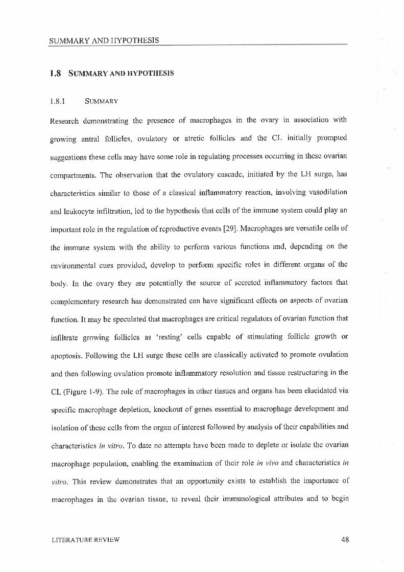
SUMMARY AND HYPOTHESIS
1.8 Suuvr¡Rv AND HYPoTHESIS
1.8.1 Sutr¡tr¿RRv
Research demonstrating the presence of macrophages in the ovary in association with
growing antral follicles, ovulatory or atretic follicles and the CL initially prompted
suggestions these cells may have some role in regulating processes occurring in these ovarian
compartments. The observation that the ovulatory cascade, initiated by the LH surge, has
characteristics similar to those of a classical inflammatory reaction, involving vasodilation
and leukocyte infiltration, led to the hypothesis that cells of the immune system could play an
important role in the regulation of reproductive events [29]. Macrophages are versatile cells of
the immune system with the ability to perform various functions and, depending on the
environmental cues provided, develop to perform specific roles in different organs of the
body. In the ovary they are potentially the source of secreted inflammatory factors that
complementary research has demonstrated can have significant effects on aspects of ovarian
function. It may be speculated that macrophages are critical regulators of ovarian function that
infiltrate growing follicles as 'resting' cells capable of stimulating follicle growth or
apoptosis, Following the LH surge these cells are classically activated to promote ovulation
and then following ovulation promote inflammatory resolution and tissue restructuring in the
CL (Figure 1-9). The role of macrophages in other tissues and organs has been elucidated via
speciflrc macrophage depletion, knockout of genes essential to macrophage development and
isolation of these cells from the organ of interest followed by analysis of their capabilities and
characteristics in vitro. To date no attempts have been made to deplete or isolate the ovarian
macrophage population, enabling the examination of their role in vivo and characteristics in
vitro. This review demonstrates that an opporlunity exists to establish the importance of
macrophages in the ovarian tissue, to reveal their immunological attributes and to begin
LITERATURE REVIEW 48
SUMMARY AND HYPOTHESIS
ln itiationLH
MACROPHAGE INFILTRATIONResting tissue macrophagesPossible role in stimulation ofcellular proliferation / folliclegrowth
MACROPHAGE INFILTRATIONAND CLASSICAL ACTIVATIONFollowing LH surge production ofclassical inflammatory cytokines topromote ovulation.
I
NFLAMMATI
oN
-Ooo
Resolution
MACROPHAGE INFILTRATIONINFLAMMATORY RESOLUTIONFactors to promote wound healing and
minimisation of the inflammatoryresponse
Figure L-9 The Potential Roles Of Macrophages In Ovarian Function. Macrophages
are located in the ovarian stroma and theca of antral and preovulatory follicles. Following the
LH surge they inhltrate the follicle in a manner similar to that seen in classical inflammatory
reactions. Further infiltration of macrophages into the CL occurs after follicular rupture for
inflammatory resolution
LITERATURE REVIEW 49
SUMMARY AND HYPOTHESIS
to examine the characteristics of these cells in the ovarian tissue. This research hopes to more
clearly define the role of macrophages in the murine ovary. The information obtained may
assist in the understanding of how the processes of follicular growth, ovulation and CL
formation and regression, in the mouse ovary are regulated. They may be extended to other
mammalian species. Through the manipulation of the ovarian cytokine environment and/or
ovarian macrophage function this work may have further implications in the understanding and
treatment of some cases of unexplained infertility and/or polycystic ovary syndrome (PCOS),
or in the regulation of human reproduction and the development of new contraceptions.
1.8.2 HvporH¡sIs
Ovarian macrophages, defined by the expression of the F4l80 and Ia antigens, play an
essential role in regulating normal ovarian function through the production of inflammatory
mediators across the different stages of the reproductive cycle.
1.8.3 GnNBner Anr¿
To determine if ovarian macrophages play a crucial role in regulating normal ovarian
function and examine their functional and secretory characteristics, in an attempt to clariff the
specific actions of these cells in the ovarian tissue.
LITERATURE REVIEW 50
SUMMARY AND HYPOTHESIS
1,8.4 Sppcmlc Atvts
L8.4.1 Aim I - To deplete the macrophage populationfrom the normal mouse ovary and
determine at which times in the cycle these cells are of critical importance.
Microscopic lipid spheres or 'liposomes' containing clodronate, which when released into the
cytoplasm induce macrophage apoptosis, have been used successfully to deplete the macrophage
population of various organs t25S]. Mature animals will have their ovarian cycles synchronised
and be primed to ovulate simultaneously with PMSG/eCG and hCG. Liposomes will then be
injected into the bursal cavity, which surrounds the murine ovary, on various days of the
stimulated ovarian cycle. This will result in the death of the ovarian macrophage population at
different times during the stages of follicular or CL development. The effect of the absence of an
ovarian macrophage population on follicular growth, owlation and cycle length will determine
the importance of these cells in these phases of the ovarian cycle.
1,8.4.2 Aim 2 - To isolate viable ovarian macrophages from the ovarian tissue and examine
bas ic immune functions.
Ovarian macrophages are present within the ovarian tissues and to extract them the tissue
structure must be dissociated. Various dissociation methods have been utilised for tissues of
different composition. The optimal conditions for ovarian dissociation and macrophage recovery
will be determined. The purity and viability of the cells will be determined, isolated cell numbers
correlated to previous studies, and the basic immune functions of phagocytosis and stimulation
of T cell proliferation examined in these cells.
LITERATURE REVIEW 51
SUMMARY AND HYPOTHESIS
1.8.4.3 Aim 3 - To show that ovarian macrophages produce cytokines involved in ovarian
function, and to show thqt thß profile is unique and varies across the stages of the
estrous cycle.
Various cytokines, which macrophages in the ovary have the ability to produce, have been found
in vitro to influence ovarian function. To determine which of these cytokines are produced by
macrophages in the ovary, immature female mice will be stimulated to ovulate with hCG and
PMSG/9CG, and cells expressing specific macrophage markers will be isolated from the ovarian
tissue and normal peritoneal cells at several time points in the ovarian cycle. From these isolated
cells mRNA will be extracted for the analysis of cytokine content by quantitative RT-PCR
techniques. This will allow comparison of the amount and type of cytokines produced by these
cells during late follicular growth, ovulation and early CL growth and regression'
LITERATURE REVIEW 52
Chapter Two
2 TTTB DEPLETION OF MACROPHAGES IN THE MURINE
ovARY BY INTRABURSAL IN¡NCUON OF'CLODRONATE
(ClzMDP) LnosoMES
INTRODUCTION
2.1 INrnooucrloN
The previous chapter introduced and developed the concept that at ovulation ovarian
macrophages may be a critical source of inflammatory cytokines that promote follicular
rupture. It was noted that macrophages residing in peripheral tissues are derived from blood
borne monocytes that differentiate in response to local cytokine and other microenvironmental
signals to assume a functional phenotype specific to the requirements of the host tissue. Their
roles in these tissues may include the phagocytosis and degradation of foreign organisms,
tissue remodelling and the regulation of local immune and inflammatory responses. Studies
examining the precise functions of these cells within the ovary have not been done'
The distribution of macrophages within the ovary has been reported to vary across the
reproductive cycle in parallel with fluctuations in gonadotrophic and ovarian steroid
hormones. In several species macrophages have been shown to be most abundant within the
theca of the follicle during follicular developmentl53,259,260],in atretic follicles 163,641
and in the CL 151,66,69]. In mice, immunohistochemical studies using macrophage-reactive
antibodies such as Mac-l, F4l80 and CD18 have shown the presence of macrophages in the
theca, stroma and CL as well as in association with atretic follicles [175]. These variations in
location and numbers during the different stages of the cycle suggest that macrophages may
participate in the tissue restructuring and inflammatory-like processes that occur in the normal
adult ovary. Many studies have provided substantial evidence that factors produced by
macrophages, such as IL-l8 and TNFcr can have stimulatory effects on ovulationll9l,26ll
and act to regulate steroid synthesis in cultured follicles 1206,262] or isolated granulosa and
theca cells 1229, 263, 2641.
Together, these studies provide indirect evidence that implicate the ovarian macrophage
population in the regulation of follicle development, ovulation and the formation and
regression of the CL. Although studies conclusively showing a direct role for these cells are
THE DEPLETION OF OVARIAN MACROPHAGES 54
INTRODUCTION
currently lacking the reproductive characteristics of the csfm"p/csfmop mouse, which has a null
mutation in the gene encoding colony stimulating factor-l (CSF-l), a cytokine that regulates
both the development and differentiation of the macrophage lineage, provide convincing
evidence to support this. These animals have few macrophages within the ovary, a
significantly impaired ovulation rate and an extended estrous cycle [63] suggesting that the
ovarian macrophage population is important in the normal functions of the adult cycling
ovary. However, since macrophages may not be the exclusive target of CSF-I in the ovary, it
cannot be concluded that local macrophage deficiency is the primary cause of ovarian
dysfunction in csfm'plcsfm'p mice. To clearly determine if macrophages play an essential role
in ovulation it is necessary to remove macrophages specifically from normal ovarian tissue
and study the resultant effects on ovulation and ovarian cyclicity. This may be achieved by
local injection of CLi to the ovary.
Liposomes are synthetic phospholipid spheres that have been used extensively to target
a variety of aqueous compounds to the macrophages present in different organs of the body'
Following in vivo administration, liposomes are phagocytosed by macrophages, where upon
the liposomal membranes are degraded by lysosomal phospholipases and the enclosed
compound is released into the cytoplasm of the cell. The accumulation of CLi in the
cytoplasm of target macrophages leads to cell death through the apoptosis pathway 1265).
Macrophages in the liver and spleen can be depleted within 24 hours following a single
intravenous injection of CLi and the population is not restored for 2 weeks thereafter Í2661.
Macrophages in the testis and peritoneal cavity have also been depleted following direct
injection of CLi into the testis 1267,2681 and peritoneum [269] respectively. It was therefore
the aim of the study presented in this chapter to show the presence of macrophages in the
ovary is essential for normal ovulation by the depletion of the macrophage population from
normal mouse ovaries using intrabursal injection of CLi and examination of the resultant
THE DEPLETION OF OVARIAN MACROPHAGES 55

INTRODUCTION
effects on ovarian function. It was anticipated this would establish whether or not ovarian
macrophages are important regulators of the ovulatory cascade.
THE DEPLETION OF OVARIAN MACROPHAGES 56
METHODS
2.2 M¿,rnnI¡,LS AND Mnrnoos
2.2.1 ANtlr¡,A,I-s ¡No Ovur,trtoN IxoucuoN
All animals were handled in accordance with The Australian Code of Practise for the
Care and Use of Animals for Scientific Purposes and experiments were approved by the ethics
committees of both The University of Adelaide and The Queen Elizabeth Hospital. Adult 8-
1l week old C57Bl6 black female mice (from The University of Adelaide Central Animal
House Facility) were maintained under controlled conditions (14 hour light : 10 hour dark
cycle) with free access to food and water. The estrous cycles of the mice were synchronised
with an intraperitoneal (ip) injection of 20 pg LHRH agonist (Des-Glyr0 [D-Ala6] LHRH
etþlamide from Sigma Aldrich, St Louis, MO) at 0900 hours on day -4. At 1200 hours on
day -2 animals were primed with a subcutaneous (sc) injection of 5IU of eCG (Folligon@
from Intervet, Boxmeer, Holland) and ovulation was stimulated 48 hours later (i200 hours on
day 0) with an ip injection of 5IU of hCG (Pregnyl@ from Organon, Oss, Holland) (Figure 2-
1). Ovulation occurred 12-15 hours after the hCG injection.
2.2.2 INrR¡eURsALINJECTIoNTECHNIQUE
Animals were anaesthetised using a mixture of fluorothane, nitrous oxide and oxygen
gases. Anaesthesia was then maintained throughout surgery with a continuous stream of the
same gas mixture regulated by a Midget anaesthetic machine (CIG, Aust'). A single midline
dorsal incision was made, followed by two small incisions into the peritoneum directly above
the fat pad of both left and right ovaries. Each ovary was externalised through the respective
incision and the intrabursal injection was performed under microscopic magnification by
inserting a 30G needle through the ovarian fat pad into the ovarian bursa. In each animal
THE DEPLETION OF OVARIAN MACROPHAGES 57
METHODS
approximately 10¡rl of either clodronate liposomes (CLi) (containing dichloromethylene
diphosphonate, CL2MDP, a gift from Boehringer Mannheim GmbH, Mannheim, Germany)
saline liposomes (SLi) (containing saline solution) or saline solution alone was delivered to
both ovaries. All liposomes were prepared and supplied by N. van Rooijen as previously
described 12701. Ovaries were returned to the peritoneal cavity and the wound was sealed with
a single Autoclip@ wound clip (from Becton Dickinson, Franklin lakes, NJ). Animals were
initially placed in separate cages under a radiant heat source until normal behavioural activity
was resumed and then housed separately under standard conditions until oocyte retrieval and
tissue collection. Further details in Appendix 1.
2.2.3 Tne¡,rlr¡BNT GRoUPS AND OocYTE RETRIEVAL
Intrabursal injections were carried out as described above on either day -3 or day -1
(Figure 2-1). In the first experiment, two groups of mice (n:8 in each) received either (a) CLi
in one ovary and saline solution in the other (CLilS group) or (b) CLi in one ovary and saline
solution in the other (SLi/S group). In a second experiment, mice received CLi in both
ovaries (CLilCLi group, n:7) or saline solution in both ovaries (S/S, n: 8 for day -1 and n: 6
for day -3). On the morning of day 1 following ovulation, animals were anaesthetised with
avertin (15¡rl/g), blood samples were taken via heart puncture and animals were killed by
cervical dislocation. Ovaries and oviducts were recovered and the numbers of ovulated
oocytes in the ampulla region were counted. In an additional experiment, animals treated as
described above were allowed to proceed to the subsequent 'natural' ovulation following the
induced owlation. The day of estrus in these animals was monitored by daily vaginal smear
tests [33] and on the morning when 100% cornified epithelial cells were detected, indicating
metestrus -1 (ME-l), blood samples were taken, animals were killed and the number of
ovulated oocytes in the ampulla region of the oviduct were counted. Further details in
THE DEPLETION OF OVARIAN MACROPHAGES 58
METHODS
Appendix 1. Progesterone levels were measured in serum samples using an automated
chemiluminescence system (Ciba-Corning, Medfield, MA) with assay parameters as
previously described 127 Il.
2.2.4 CorlncrtoN oF OvARIAN TISSUE
Recovered ovaries were dissected free of fat and connective tissue and snap frozen in
OCT (ornithine carbarnyl transferase, Tissue-Tek tissue freezing medium from Miles Inc.
Elkhart, IN) at either the preovulatory stage, t hour prior to expected ovulation, or on day 1
post ovulation, at the time of oocyte collection (Figure 2-1). To establish that liposomes are
able to penetrate the ovary, liposomes with the DiI fluorochrome (the lipophilic long-chain
carbocyanine Di-I (DiICrs (3), Molecular Probes, USA) incorporated into the lipid bilayer of
the liposome were injected into the bursa (n:2 per time point) and ovaries were recovered
from animals killed 2, 4, 6, 12,24 and 48 hours after surgery. Ten-micrometer sections from
these ovaries were then examined using an Olympus Vanox microscope equipped for
epifluoresence. Under conditions of green excitation (546nm) the Di-I in the liposomes
fluoresces red.
2.2.5 OvaRIRNMonruorocYANDIIr¡vrxosIsrocHEMISTRY
Six-micrometer serial sections were cut from both ovaries of each mouse resulting in
at least 140 consecutive sections from each ovary. Every 9th and 10th section was stained
with haematoxylin and eosin (H&E) for morphological examination by light microscopy.
Follicle size was measured in the H&E sections using video image analysis (VIA) software
(Leading Edge Pty Ltd, Marion, S.A.) and adjacent sections containing preovulatory follicles
THE DEPLETION OF OVARIAN MACROPHAGES 59

METHODS
Day -4Surgery
I-J
Surgery1
STIMULATEDOVULATION
0
i2??
NATURALOVULATION
a 1
LHRH eCG1200 hrs
hCG
0900 o
Daily Vaginal Smear
Figure 2-1 Schematic Diagram Of The Protocol For Ovulation Induction. The time of
injection of gonadotrophins (LHRH, eCG, hCG), intrabursal injection (surgery), tissue
collection (*) and, oocyte retrieval and blood collection ( t) ) are indicated.
i*
hrs
*200I
THE DEPLETION OF OVARIAN MACROPHAGES 60
METHODS
(>400pm) were stained with primary antibodies reactive with the anti-MHC II antigen
(Ia) TIB120 (reactive with activated macrophages and dendritic cells, from American Type
Culture Collection, Rockville MD), F4l80 (reactive with a surface glycoprotein specific to
macrophages [113] and FA/l1 (reactive with macrosialinl2T2l) both kindly supplied by S.
Gordon, University of Oxford. Sections were fixed in960/o alcohol (4"C for 10 minutes) and
then incubated with primary antibody from culture supernatant diluted in phosphate buffered
saline (pBS, Gibco, BRL, Life Technologies, Grand Island, NY) containing 10% normal
mouse serum (NMS) and lYo bovine serum albumin (BSA) (Boehringer Mannheim, Indpls.,
IN) (PBS-NMS) (Ia (1:200), F4l80 (1:10), FA/l 1 (1:600)) at 4"C for 3 hours, washed in PBS
and then incubated with biotinylated-rabbit-anti-rat secondary antibody (from DAKO,
Carpinteria, CA) diluted in PBS-NMS (1:300) at 4"C for 2 hours. Following another PBS
wash, sections were incubated with avidin-horseradish-peroxidase (from DAKO, Carpinteria,
CA) diluted in PBS-NMS (1:400) and enzyme rwas then visualised using Sigma-Fast DAB
tablets (from Sigma Aldrich, St Louis, MO). Uterus and spleen were used as positive control
tissues and negative controls included sections incubated without primary antibody or with
irrelevant monoclonal antibodies. The area of positive stain in each section was evaluated by
VIA and expressed as % positivity (area brown positive stain/area total stain x 100)' Three
thecal regions in each preovulatory follicle and six stromal regions in each ovary were
counted and the meanYopositivity value for each follicle and ovarian stroma was calculated.
Further details in Appendix L
2.2.6 Sreusucs
A paired Students t-test was used to evaluate differences in the numbers of ovulations
between the ovaries of animals within the CLilS treatment group as well as differences in the
numbers of preovulatory follicles present in the ovaries of these animals at the time of
THE DEPLETION OF OVARIAN MACROPHAGES 61
METHODS
ovulation and post owlation. A one-way ANOVA with Tukey-Kramer multiple comparisons
was used to examine the differences in ovulation rates between treatment groups and the
length of the different stages of the estrous cycle in animals from the CLilCLi and S/S groups.
An unpaired Students t-test was used to compare cycle length and serum progesterone levels
in animals from the CLilCLi and S/S groups'
THE DEPLETION OF OVARIAN MACROPHAGES 62
RESULTS
2.3 Rnsur,rs
2.3.I ErN¡Cr Or CIOPNONRTN LPOSOME TRERTMENT ON GONNOOTROPHIN STIIøIJI-ETEO
Ovul¡uoN
To determine the effect of administration of CLi on ovulation tate, mice were killed 9
hours after ovulation following treatment on either day -1 or day -3 prior to ovulation (Figure
2-2). No significant difference in ovulation rate between CLi treated and the contralateral
saline solution treated ovaries was observed in animals from the CLilS group when treatment
was administered on day -1 (number of oocytes ovulated, mean t SE : 7 + I.7 and 6'4 X 0.9
in between CLi treated and saline solution treated ovaries respectively). This ovulation rate
did not differ significantly from that of animals in the S/S group, (6.5 t 1,3 and 5.8 + 0'9 in
left and right saline solution treated ovaries respectively). However a signif,rcant reduction in
the ovulation rate in CLi treated ovaries was observed in animals from the CLilS group when
treatment was administered on day -3 û)<0.05, Figure 2-2a). No signif,rcant difference was
seen in the ovulation rate in saline liposome treated or saline solution treated ovaries in the
SLi/S group when treatment was administered on day -3. A large reduction in the ovulation
rate of both CLi treated ovaries was seen in the CLilCLi group, while the ovulation rate in the
S/S group was not diminished (Figure 2-2b). When these results \¡/ere pooled to give a single
mean value for all saline solution treated or CLi treated ovaries, a significant reduction in
ovulation rate was found in CLi treated ovaries (number of oocytes ovulated : 8'5* 1.2 and
3.7+ 0.8 in Sand CLi treated ovaries respectively, p< 0.05). Progesterone levels measured in
serum samples from animals in which treatment was administered on day -3 showed no
significant differences between any treatment groups, including those in which ovulation was
affected (S/S: 32.5 +5.7 nmol/L; SL/S : 39.6 +4.3nmollL; CLilCLi:34.7 +10.4nmol/L).
THE DEPLETION OF OVARIAN MACROPHAGES 63
RESULTS
{<
A)
CLi/S
(n:8)Treatment Crroup
B)
10Ë(.)E8goo
(r+HC)
_oE2z
0SLi/S
(n:8)
12()
Ë10
;8C)
3óo
(H
?4C)pÉa
z0
S/S CLICL|
(n:6) Treatment Group @:7)
Figure 2-2 TheEffect Of Clodronate Liposome Treatment On Ovulation Rate.
Data are mean (t SEM) number of oocytes ovulated after intrabursal administration on day -
3 of (a) clodronate liposome/saline (CLilS) and saline liposome/saline (SLi/S) groups and (b)
S/S and CLilCLi groups. When results were pooled to give a single mean value for all S-
treated or Cli-treated ovaries, a signif,rcant reduction in ovulation rate following CLi-
treatment was detected (p< 0.05) (S treated ovaries : 8.5* 1.2; CLi-treated ovaries :3.7+ 0.8).
In both graphs E : S-treated ovaries, [ : Sli-treated ovaries and I : Cli-treated
ovaries. * indicates significantly different to saline solution treated ovaries (p<0.05)'
THE DEPLETION OF OVARIAN MACROPHAGES 64

RESULTS
2.3.2 EprEcr or CroonoNnrE Lposovn TREATMENT ot{ THs Ov¡.nlRN M¡,cRopsecs
PopurRuoN
To establish that liposomes were able to penetrate the ovarian epithelium from beneath
the bursa and enter the ovarian tissue, liposomes containing the fluorescent marker DiI were
injected into the bursal cavity and animals were killed at several time points after injection' In
sections from ovaries collected two hours after injection, bright fluorescence could be seen in
the stroma and thecal regions of the follicles in the ovary but not the granulosa or antrum
(Figure 2-3). Four hours after fluorescent liposome injection the fluorescence was less bright
and six hours later the fluorescence appeared significantly diminished. No significant
difference in fluorescence intensity could be seen between untreated and fluorescent liposome
treated tissue at later time points.
To determine whether CLi treatment was effective in depleting macrophages from the
ovary and to determine whether the observed reduction in ovulation rate was correlated with
any decrease in thecal macrophage numbers, ovarian sections were incubated with antibodies
specific for the macrophage antigens Ia,F4l80 and macrosialin. Macrophages reactive with all
of these antibodies were present in all ovarian sections (Figure 2-4,2-5). A comparison of the
abundance of macrophages between the two ovaries from mice in the CLilS groups treated on
either day -1 or day -3 indicate that CLi treatment had no apparent effect on the mean
stromal positivity of any of the macrophage antigens (Figure 2-6C,D). In the theca of
preovulatory follicles a reduction in macrophage numbers was seen for all antigens and this
was statistically significant for the macrosialin* macrophages (p< 0.05, Figure 2-68) in
animals administered CLi on day -l and both the macrosialin* and Ia* macrophages in
animals administered CLi on day -3. (p<0'03, Figure 2-64).
THE DEPLETION OF OVARIAN MACROPHAGES 6s
RESULTS
2 hrs
6 hrs
12 hrs
4 hrs
8 hrs
48 hrs
Figure 2-3 Ovarian Distribution Of Fluorescent DiI Liposomes Following
Intrabursal Injection. Tissue sections were examited 2, 4, 6, 12, 24 and 48 hours
following intrabursal injection of liposomes containing the fluorescent marker DiI.
(Bars in each image represent 200pm).
THE DEPLETION OF OVARIAN MACROPHAGES 66
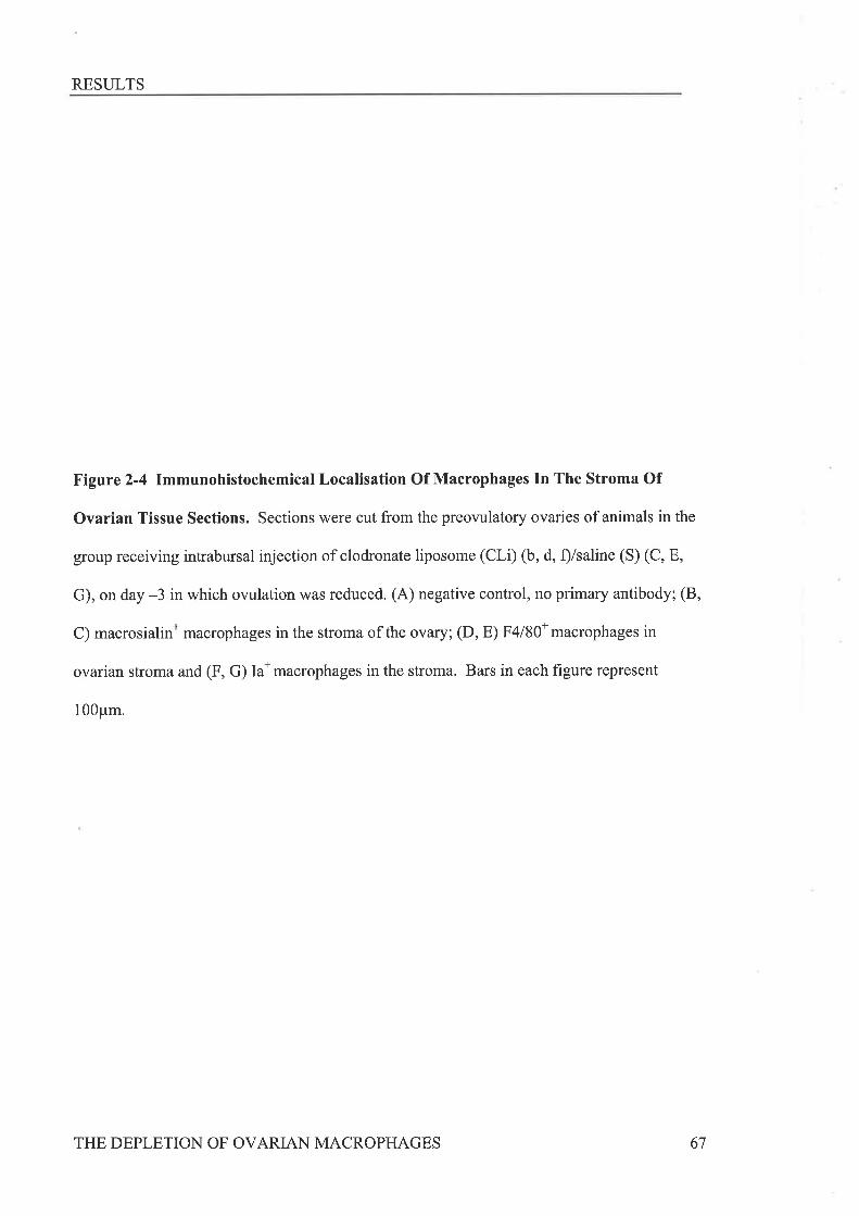
RESULTS
Figure 2-4 Immunohistochemical Localisation Of Macrophages In The Stroma Of
Ovarian Tissue Sections. Sections were cut from the preovulatory ovaries of animals in the
group receiving intrabursal injection of clodronate liposome (CLÐ (b, d, f)/saline (S) (C, E,
G), on day -3 in which ovulation was reduced. (A) negative control, no primary antibody; (B,
C) macrosialinn macrophages in the stroma of the ovary; (D, E) F4l80*macrophages in
ovarian stroma and (F, G) Ia* macrophages in the stroma. Bars in each figure represent
100pm.
THE DEPLETION OF OVARIAN MACROPHAGES 67
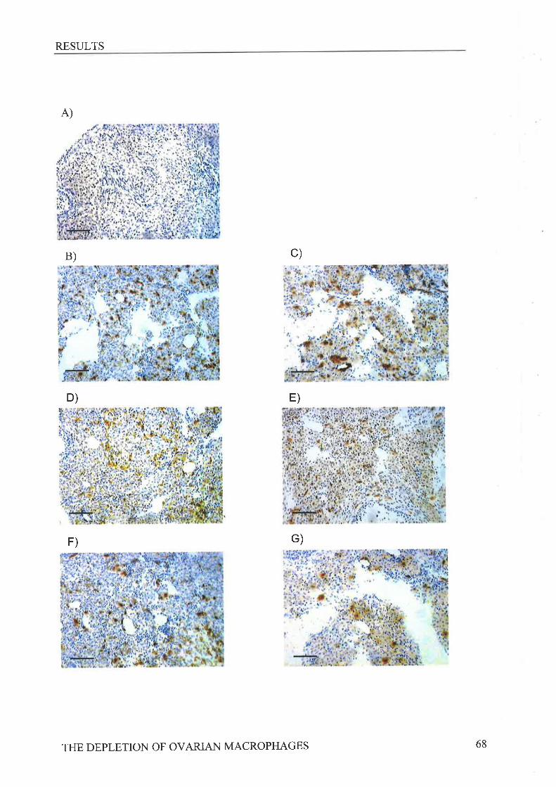
RESULTS
A)
B) c)
D) E)
F) G)
THE DEPLETION OF OVARIAN MACROPHAGES 68
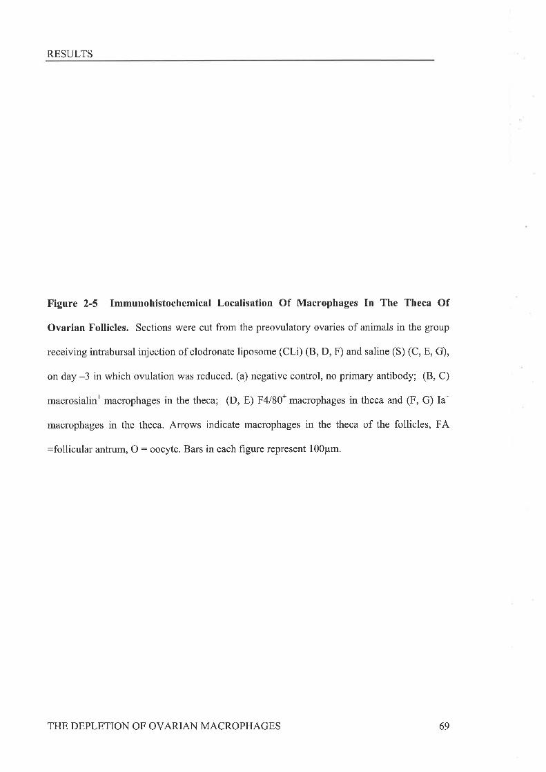
RESULTS
Figure 2-5 Immunohistochemical Localisation Of Macrophages In The Theca Of
Ovarian Follicles. Sections were cut from the preovulatory ovaries of animals in the group
receiving intrabursal injection of clodronate liposome (CLD (8, D, F) and saline (S) (C, E, G),
on day -3 in which ovulation was reduced. (a) negative control, no primary antibody; (8, C)
macrosialin* macrophages in the theca; (D, E) F4l80* macrophages in theca and (F, G) Ia*
macrophages in the theca. Arrows indicate macrophages in the theca of the follicles, FA
:follicular antrum, O: oocyte. Bars in each figure represent 100pm.
THE DEPLETION OF OVARIAN MACROPHAGES 69
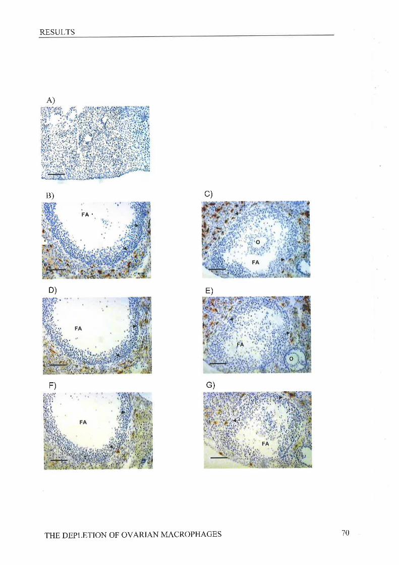
RESULTS
A)
B)
D)
FA.
c)
E)
F) G)
THE DEPLETION OF OVARIAN MACROPHAGES 70
RESULTS
A)
c)
B)
(n
U)o
Ê.
20
15
l0
Dav -3: Theca
Dav -3: Strons
F4l80
Macrophage Arfügen
Day-1:Tlæca
Day-1:Stroma
F4l80
MacroplngoArtþn
s.âo
ô\
20
l5
10
55
00
IaIa Fr'/1 1 FÁ/l1
D)
cC
(âo
Õ\
(n(t)o
o\
25
20
15
10
5
0
20
15
10
5
0 l
F4l80
Macroptnge Arúigen
FA/l 1Ia F4l80 F.A/11
Macrophage Artþn
Ia
Figure 2-6 The Effect Of Ctodronâte Liposome Treatment On The Number Of
Macrophages In The Ovarian Theca And Stromal Compartments. Data are mean (*
SEM) percentage of positive stain (macrophages) for the antigens la,F4180 and macrosialin
(FA/11). Tissue is from mice in the clodronate liposome/saline (CLilS) treatment group and
graphs show mean positivity in the theca (B) and stroma (D) of ovaries following intrabursal
injection of either S (E ,n:79 follicles, in4animals) orCLi (l,n:9 follicles, inother
ovary of same 4 animals) on day -1 or in the stroma (C) and theca (A) of ovaries following
intrabursal injection of either S (E , n:21follicles, in 4 animals) or CLi (I , n:16 follicles,
in other ovary of same 4 animals) on day -3. * indicates signihcantly different to saline
solution-treated ovaries (P<0.05).
THE DEPLETION OF OVARIAN MACROPHAGES 7l

RESULTS
2.3.3 Errecr op CrooRoNRr¡ Lposolr¿B TRBaIMENT oN OvRrueN Ttssup
MoRpgolocv
To determine whether the reduction in ovulation rate seen following CLi treatment on
day -3 was due to the prevention of follicular rupture or a reduction in the number of
preovulatory follicles that develop, the total number of follicles of preovulatory size (diameter
>400 um) and corpora lutea were counted in H&E sections of both the ovaries from 4 animals
in the CLilS group. When ovaries were collected following ovulation there appeared to be
fewer corpora lutea and more preovulatory sized, unruptured follicles in the CLi treated
ovaries than the S treated ovaries, although these differences were not significant (Table 2-1)'
In ovaries collected immediately prior to ovulation there appeared to be fewer preovulatory
sized follicles and more corpora lutea in the CLi treated ovaries than the S treated ovaties,
although no significant differences were detected.
THE DEPLETION OF OVARIAN MACROPHAGES 72
RESULTS
Ovarian Treatment Ovulationand collection time rate
No. of No. of CorporaFollicles lutea>400pm
Post Ovulation
S-treated
CLi-treated
Prior to Ovulation
S-treated
CLi-treated
12.5 !2.67.8 + 1.8
2+0.74r 1.6
6.5 + 1.8
5.3 + 1.9
13 + 1.8
9.3 + 1.4
6.8 r 1.8
7.8 + 1.6
Tabte 2-1 The Effect Of Clodronate Liposome Treatment On The Number Of
Preovulatory Follicles And Corpora Lutea. Data are mean (+SEM) number of
preovulatory follicles and corpora lutea counted in each ovary of animals in the CLi/S
group treated on day -3.n:4 in each group. S-treated: saline solution treated ovaries,
Cli-treated : clodronate liposome treated ovaries'
THE DEPLETION OF OVARIAN MACROPHAGES 73

RESULTS
2.3.4 EFFBCT OF GTOOROT.¡RTB LpOSOrr¿B TReerlr¡ENT ON SUeSrqUnNr N¡.ruR¡L
OvurRuoN
Since CLi treatment appeared to affect the numbers of follicles developing to the
preovulatory size, we sought to determine the consequences of this for the next ovulatory
event. In all animals allowed to progress to the next natural ovulation after treatment,
ovulation rates were reduced in comparison to those of the stimulated cycle. No significant
differences were detected regardless of the treatment group (CLilSLi, SLilSLi, CLi/CLi) or
the time point of treatment (day-l or day-3) (Table 2-2). However it was noted that some
animals receiving CLi treatment appeared to take longer to reach the subsequent natural
owlation. In animals treated on day -1 no significant differences in cycle length were found
between any of the treatment groups regardless of treatment, although the cycle length was
extended beyond the expected 4-5 days in all of these animals (Table 2-2).In animals treated
on day -3 in the CLilS and CLi/CLi groups, cycle length was significantly extended over that
in the SLi/SLi group (Table 2-2). Further breakdown of the cycle in this affected group
revealed a significant delay in the metestrus-2 / diestrus (ME-2/DE) stage (Figure 2-7). The
progesterone levels in serum obtained following ovulation were not signiflrcantly different
between the treatment groups displaying delayed }r4E-2 / DE stage (CLilCLi : 7.1 I
1.8nmol/L; S/S:4.9 + 1.3nmol/L).
THE DEPLETION OF OVARIAN MACROPHAGES 74
RESULTS
Treatment GroupS/S SLilSLi SLilCLi CLLICL|
Subsequent Ovulation Rate
IB injection on Day -1
IB injection on Day -3
Cvcle Lensth (davs)
IB injection on Day -1
IB injection on Day -3
(n:6)3 +0.8
2.8 +LI
(n:10)3.4 +0.6
3.2 +0.4
6.6 +1.4
5.2!0.6
n:6)4.2 +0.7
4.0 +r.2
(n:5)3.8 +1.1
4.2 +r.6
(n:8)2.8 +0.7
2.6 +0.8
(n:7)2.9 +0.6
2.0 +0.9
(n:6)3.3 +1.0
4.5 +t.3
7 .2 +1.3
3.4 !0.47.4 +2.6
6.2+0.7* 7.5 +I.3*
Table 2-2 The Effect Of Clodronate Liposome Treatment On Subsequent
Natural Ovulation And Cycle Length. Data are the mean (tSEM) ovulation rate
from each ovary and mean cycle length of animals in the different treatment groups.
IB: intrabursal injection. Mice in the S/S group received saline solution in both
ovaries, mice in the SLi/SLi group received saline liposomes in both ovaries, mice in
the SLi/CLi group received saline liposomes in one ovary and clodronate liposomes
in the other, mice in the CLi/CLi group received clodronate liposomes in both
ovaries. * indicates significantly different to SLi/SLi (p<0'01).
THE DEPLETION OF OVARIAN MACROPHAGES 75
RESULTS
ac
b
ab
Nß-2/DE
Stage ofthe oestrous cycle
PE
Figure 2-7 The Effect Of Clodronate Liposome Treatment On Progression
Through The Subsequent Estrous Cycle. Data are mean (t SEM) number of days
spent in each stage of the estrous cycle for animals treated on day -3. n : animals in
the s/S group (n:5), m : animals in the sl.ilsl,i group (n:5), I : animals in the
CLilSLi group (n:8) and I : animals from the CLilCLi group (n:6). Stages of the
cycle are represented as follows; ME-l denotes metestrus-l , }/.E-21DE denotes the
combined metestrus-l/diestrus stages and PE denotes proestrus. Bars with the same
letter are signif,rcantly different to each other (P<0.05).
c
+¡Òo
_Eea¡ ,{c)\_/>'O
7
6
5
4
J
2
1
0
ME1
THE DEPLETION OF OVARIAN MACROPHAGES 76
DISCUSSION
2,4 Drscussrox
Liposomes containing clodronate have been used extensively to target macrophage cells
in various organs within the body. In the current study, we have employed clodronate-filled
liposomes to achieve a moderate but significant reduction in macrosialin and MHC-Class II
(Ia) * macrophages in the ovarian theca. This depletion was found to be associated with
impaired ovarian function, as measured by the number of oocytes ovulated, and the duration of
the subsequent estrous cycle. The strongest results were obtained when CLi were injected on
day -3,84 hours prior to owlation. The reduction in ovulation rate seen following CLi
treatment at this time, during the early stages of the follicular cycle, can be fully attributed to
the clodronate contained within the liposomes since no reduction in ovulation rate was seen in
the SLi/S group. The corresponding immunohistochemistry data show that administering
treatment at this time point does not significantly affect the number of macrophages in the
ovarian stroma, while within the theca of preovulatory follicles there were significant
reductions in both Ia* and macrosialin* macrophages. This suggests that the detrimental effect
on ovulation rate might be the consequence of a perturbation in the thecal macrophage
population.
Examination of follicle numbers in the ovaries of this treatment group immediately prior
to and following ovulation suggests that the growth of follicles to the preovulatory size, as well
as follicular rupture, may be compromised by CLi treatment. This finding concurs with studies
in the CSF-I deficient csfmop/ csfmop mice where severe reduction in macrophage numbers in
the ovary is associated with significantly fewer antral and mature follicles present at the
proestrus stage 1273). The difference in the extent of the effect in the two experimental systems
might be related to the difference in the relative severity of the macrophage depletion achieved.
Mice treated with CLi 3 days prior to ovulation were also found to have delayed progression
through the ME2/DE stage of the subsequent cycle. This stage is characterised by high
THE DEPLETION OF OVARIAN MACROPHAGES 77
DISCUSSION
numbers of macrophages within the ovary when compared to other stages of the cycle [64]'
Interestingly this stage of the ovarian cycle appears to be particularly susceptible to
perturbations in macrophage population, as both GM-CSF null mice (which have a reduced
number of stromal Ia* cells in the ovarian stroma) and csfm"p/ csfmop mice, exhibit delays in
the DE stage of the estrous cycle [63]'
CLi treatment at the stage of late follicular development (day -1) did not affect the
numbers of oocytes ovulated. This suggests that macrophages may be less critical for the
events immediately preceding ovulation or that the extent of depletion achieved was not
sufficient to have a detectable effect on the process. In support of the latter the effect of
liposome treatment on macrophages is dependant on both tissue penetration by the liposomes
and the rate of macrophage phagocytosis. It has been suggested that splenic macrophages
expressing different markers ate depleted at different rates 12651 and the
immunohistochemistry results presented here also support this concept in the ovary. Therefore
it may take more than24 hours for the full extent of clodronate liposome treatment to occur in
the macrophages of the murine ovary.
Although numerous authors have shown significant effects of macrophages on
progesterone production in vitro 1174, 238, 2391 12291, the levels of peripheral blood
progesterone measured in this study were not altered by thecal macrophage depletion' This
implies that the cells we have targeted are not essential for the maintenance of systemic steroid
levels. This is in agreement with the finding that the reduced number of macrophage cells in
csfmop/ csfmop mice does not correlate with any change in the levels of progesterone produced
during the luteal phase [63].
Although our results show that CLi treatment is clearly influencing ovarian function and
altering macrophage numbers, complete depletion of the ovarian macrophage population, as
has been seen elsewhere with CLi treatment, vras not achieved. This may be due to either
THE DEPLETION OF OVARIAN MACROPHAGES 78
DISCUSSION
insufficient or uneven distribution of CLi through the ovarian tissue and might have been
improved if it was possible to administer a larger volume of CLi under the ovarian bursa.
Alternatively, it is plausible that a high turnover of macrophages in the ovary may result in
rapid replacement of those cells that are killed by CLi treatment. Clearly the treatment regime
employed in the study had some non-specific effect on ovarian function since all animals
treated on day -1 had extended cycles regardless of treatment type.This may to be due to the
proximity of anaesthesia and sugery to the ovulatory event, and interference of the drugs used
with hypothalamic function or the tissue remodelling that occurs during this process. We have
found in other experiments that these procedures have significant effects on ovulation when
carried out close to the initiation of this event (unpublished observations). We have been
unable to determine the duration of the effect of macrophage depletion on the numbers of
oocytes ovulated, as the ovulation rate in the subsequent natural ovulation was much reduced
in comparison to the ovulation rate of the first cycle regardless of the treatment type' This is
probably due to either the hyper-stimulation regime employed to induce the first ovulatory
event exhausting the pool of growing follicles, or the physiological stress related to the surgery
carried out.
It is interesting that macrophages in the vicinity of the thecal layer surrounding
developing follicles appear to be preferentially targeted by CLi treatment. The reasons for this
are not clear. The physical proximity of the developing follicle to the ovarian surface (and
hence the bursal cavity where the clodronate was administered) might be a contributing factor
and it may also be speculated that the composition of the extracellular matrix in this region is
more conducive to diffusion of liposomes than the more dense collagenous structures of the
ovarian stromal matrix. The results also indicate that even within the thecal compartment CLi
treatment may deplete macrosialin* and to a lesser extent Ia* macrophages in preference to
F4l80* macrophages. This suggests that there is heterogeneity amongst the ovarian macrophage
THE DEPLETION OF OVARIAN MACROPHAGES 79
DISCUSSION
population and implies that some subsets might be more phagocytically active or otherwise
more susceptible to the effect of CLi treatment. In view of the association of macrosialin with
the phagocytic process it might be speculated that strong macrosialin expression is indicative
of high phagocytic activity and perhaps a greater capacity to take up liposomes from the
extracellular milieu.
In conclusion, we have shown that local CLi treatment can be used to achieve a reduction
in ovarian macrophage numbers. The technique has been successfully employed in the current
study to generate datathat suggests thecal macrophages have a role in stimulating the growth
of follicles to the preovulatory stage of development and in achieving follicular rupture.
Further studies to define the phenotype and cytokine profile of the various macrophage
populations residing in the ovary will help to elucidate the precise mechanisms through which
they participate in regulating the different ovarian processes.
THE DEPLETION OF OVARIAN MACROPHAGES 80
3
Chapter Three
Tnrc NUVTnnRS AND Cu¿.nACTERISTICS OF MURINE
Ov¡.NrIN IA AND F4l80 POSTTTVE MACROPHAGES
ISOf,.lrED DURING THE GOX¡.UOTROPHIN -STIMULATED
RnpnoDucrrvn CYcr.n
INTRODUCTION
3.1 InrRooucrloN
The previous chapter presented direct experimental evidence that ovarian macrophages
are important for normal follicular development ovulation, however exactly what key
functions these cells perform in the ovary is not yet understood. Ovarian macrophages have
the potential to produce a large number of different cytokines including IL-18 and TNFcr,
which are known to significantly affect steroidogenesis [202, 2371, celbilar proliferation [203,
274,2751, vascularisation [197, 276] and apoptosis [171, 178] occurring in the ovarian tissue
(reviewed by Terranova et al 12771). To allow more in depth studies into the roles of these
cells, they must be isolated with high purity from the other ovarian cell types. Thus far
studies examining macrophages in the ovary have, utilising immunohistochemical techniques,
simply recognized and quantified these cells through their expression of surface molecules
specific to the monocyte/macrophage lineage 164,278-280]. Some studies have gone further
investigating the effects of the depletion of ovarian macrophages, by adherence to plastic
dishes, on the activities of ovarian dispersates Í2751. Still more specifically, selected mRNA
transcripts produced by CDllb* cells of the adherent population from ovarian dispersates
[281] have also been studied. To date, no attempt to purify murine ovarian macrophages
directly, using speciflrc macrophage antibodies, from an ovarian digest has been reported'
The isolation of mononuclear cell types from murine tissues other than the ovary has
been successful and is routinely performed in immunological studies. Blood monocytes,
peritoneal and alveolar macrophages can all be isolated with relative ease using density
gradient centrifugation and peritoneal or bronchi alveolar lavage respectively. However, the
isolation of cells from solid tissues, such as the ovary, requires dissociation of the tissue mass
into a single cell suspension before purification of the desired cell type can be performed'
Collagenase is most commonly used for tissue digestion and has been used to isolate cells
from many other murine organs including gut 1282],liver [283], spleen 12841, testis [134,
THE ISOLATION OF OVARIAN MACROPHAGES 82
INTRODUCTION
137] and uterus 1143, 1471. It has also been used in combination with trypsin or dispase to
isolate cells from heart and kidney, [285] or intestine [286] respectively. Ovarian
dissociations have been performed in previous studies with the purpose of examining
activities of thecal-interstitial cells [287], luteal cells [288] or of the ovarian dispersate as a
whole 1289,2901. The methods employed vary, with ovarian tissue dissociation lengths of
both 45 and 90 minutes reported 1281,289-2911. The specific isolation of CD1lb*cells from
luteinised mouse ovaries more recently reported by Tadros et al [281] gives no indication of
either the numbers of animals required or cells recovered'
It was the aim of this study to develop and describe a technique to isolate murine
ovarian macrophages. This will allow further studies of the characteristics of these
macrophages and lead to a better understanding of the contribution these cells make to
ovarian function.
THE ISOLATION OF OVARIAN MACROPHAGES 83
METHODS
3.2 Mprnoos
3.2.1 Attn¿ll-s ANo GoN¡DoTRoPHIN SrluurertoN
SV129/Ola mice were maintained in a I4L:12D light: dark cycle with food and water
available ad libitum. All experiments were approved by animal ethics committees of both
The Queen Elizabeth Hospital and The University of Adelaide and animals were used in
accordance with The Australian Code of Practise for the Care and Use of Animals for
Scientific Purposes. Unless otherwise stated, reagents were purchased from Sigma Aldrich
Chemical Co., St Louis, MO, USA. Adult animals were used at 7-9 wks of age. The first
ovulatory cycle was stimulated in immature animals aged 25-29 days with 5 IU of eCG
(Intervet, Boxmeer, Holland) in 0.1m1 PBS (Gibco BRL, Life Technologies, Grand island,
Ny, USA) with 0.1% BSA (Fraction V) at 1200 hours on day -2 followed by 5 IU of hCG
(Pregnyl@, Organon, Oss, Holland) 48 hours later on day 0 to stimulate ovulation
approximately 12 hours later. Animals were killed by cervical dislocation at 0900h on day -1,
day 0 am, day 1 and day 2 as well as 6 hours post hCG administration (day 0pm). Following
the collection of peritoneal macrophages by peritoneal lavage with 2.5mls Hanks Balanced
saltsolution(HBSS)with0'35g/LsodiumbicarbonateQ'laco3),5mMEthylenediamine
tetra-acetic acid and 0.0I% Sodium Azide (HBSS/EDTAIAz) ovaries were collected from
groups of 8-10 animals for digestion and subsequent isolation of macrophages.
3.2.2 DIssocnuoNCoNDITIoNS
Peritoneal cells collected from four adult animals in HBSS/EDTA/Az \¡/ere
centrifuged (300G) and 1 x 10⁶ re-suspended in 2mls of a lmg/ml Collagenase, 25Ulml
Deoxyribonuclease I (DNase I) solution made up in RPMI medium 1640 (Gibco, BRL, Life
Technologies, Grand Island, NY, USA) with 2 g/L sodium bicarbonate (NaHCO¡) and l0%o
THE ISOLATION OF OVARIAN MACROPHAGES 84
METHODS
heat-inactivated fetal calf serum (FCS) (Trace, Victoria, Australia) and cultured at either room
temperature (RT) or 37oC for 90 minutes in siliconised (Coatasil Glass Treatment Solution,
Labchem, ApS Ajax Fine Chem, NSW, Australia) glass or plastic beakers. Cells were then
washed in 1gmls of HBSS/EDTNAz and centrifuged (200G) at 4"C for 10 minutes. The cell
pellet was re-suspended in lml of HBSS/IO% FCS and the numbers of recovered live cells
counted using trypan blue exclusion. This was repeated four times and the percentage of cells
that were recovered live for each condition compared statistically using a One-Way Anova.
3.2.3 DtssocnrtoN oF Ov¡nrcs
To optimize the ovarian dissociation conditions, ovaries from three adult mice agedT-
9 weeks were collected, dissected free of the surrounding bursa and fat, weighed and minced
finely with vannas spring scissors under a dissecting microscope. The resulting tissue pieces
were cultured for either 45 or 90 minutes in lmg/ml collagenase (Sigma C9891) and
0.025mglml DNase made in each of the following media bases;
1) RPMI with 2 g/L Na HCO3 and 10%FCS (RPMI/FCS);
2) Hepes buffered (Hepes sodium salt 3,25 glL and Hepes acid salt 2.98 glL) alpha
Minimum Essential Medium (crMEM, Life Technologies, Grand Island NY, USA)) with 2'2
g/L NaHCO36MEMI) (This media lacks glutathione that can inhibit enzyme activity).
3) Hepes buffered crMEM with2.2 g/L NaHCO¡ and 5 mM Calcium Chloride (Caclz)'
(H-MEM) (The latter ensures maximal activation of the collagenase enzyme).
For every 20mg of ovarian tissue, lml of eîzyme solution was added. All digests were done
at RT with gentle shaking in siliconized glass beakers. The resulting cell suspension was
filtered through a 70 ¡rm cell strainer (Falcon@, Becton Dickinson Labware, NJ, USA)' The
filtered cells were washed twice in HBSS/EDT AIAZ and centrifuged (200G) at 4oC and then
THE ISOLATION OF OVAzuAN MACROPHAGES 85

METHODS
re-suspended in 1 ml HBSS/I0% FCS for counting of live and dead cells. The ovarian tissue
remnants remaining in the cell strainer were washed back into the beaker with 3mls of ice
cold HBSS IEDTNAZ and incubated for a further 20 minutes at 4"C. The resulting cell
suspension was filtered, washed once in HBSS/EDTAIAZ and centrifuged (200G) at 4"C
before being re-suspended in 500p1 of HBSS/FCS for counting of live and dead cells'
Ovarian digestion under each condition was repeated four times and the total number of cells
per milligram and percentage of live cells isolated was compared statistically using a One-
Way Anova. Other macrophage isolation methods attempted are described in Appendix 2.
3.2.4 LABELLING oF OvARIAN MACRoPHAGES
Digested cells were re-suspended in I ml HBSS/10%FCS and 500¡rl of cells were
incubated with 500¡rl of either anti-MHC class II antigen Qa) (TIB120 from American type
culture collection, MD, USA) or F4l80 (supplied by S. Gordon, University of Oxford)
hybridoma supernatant, in glass silicon coated tubes (Vacutainer@, Becton Dickinson, NJ,
USA) for I hour at 4C with gentle rocking. Unbound antibody was removed by washing
with HBSS IEDTNAz followed by centrifugation (200G) at 4"C. The pellet was re-
suspended in 3ml of HBSS/EDTAIAz over-laid onto 3ml of FCS and centrifuged (300G) at
4"C. Cells were re-suspended in lml of HBSS/IO%FCS and added to prepared panning
plates.
3.2.5 ANrlsooY PnNNrNc
Bacterial grade 36mm petri dishes (Sarstedt, NC, USA) were incubated for 15 hours at
4"C with 1gmg/ml anti-rat IgG (Calbiochem@, Merck KgaA, Darmstadt, Germany) in PBS,
washed with 5 ml PBS then cultured with PBS-1O%FCS for 20 minutes at 4"C. Plates were
THE ISOLATION OF OVARIAN MACROPHAGES 86
METHODS
washed again with 5 ml of PBS and the labeled cell suspension then added. The plates were
then incubated for 2 hours with gentle rocking at 4"C. Unbound negative cells were then
washed from the surface of the plate with 1Oml HBSS/I0%FCS. The numbers of cells bound
in 10 randomly selected fields were counted using an inverted phase contrast microscope and
the total numbers of cells bound per plate calculated for all the time points listed in 3.2.1. Cell
numbers were compared statistically using a One-Way Anova'
3.2.6 VlaslrtrY
Bound cells isolated before (day Oam) and after ovulation (day 1) were stained for 10
minutes with Hoechst (10 pl/ml) and propidium iodide (PI) (15 ¡rl/ml) in HBSS/I0oloFCS, wet
mounted and examined under an Olympus Vanox (AHBT3) fluorescent microscope. The
numbers of live (blue) and dead (red) cells were counted in randomly selected fields at each
time point and expressed as the percentage of total cells.
3.2.7 CoLl¡cuoNoFCELLS
3.2.7.1 Adherent Ovarian Cells
Animals were primed as in 3.2.1 and ovaries collected 24 hours post hCG. Ovaries
were dissociated in digest solution made with a H-MEM media base. Cells collected were
washed, added to 36mm petri dishes (Sarstedt, NC, USA) and culturedfot 2 hours at 37'C in
RPMI/FCS. Non-adherent cells were washed off with HBSS/FCS and plates placed at 4oC for
10 minutes with cold 0.25% trypsin in HBSS/EDTNAz, Cells were then sheared off the
plates with forcefi.rl pipetting and washed in HBSS/EDTA/FCS before leukocyte antigen
staining. This was only performed once.
THE ISOLATION OF OVARIAN MACROPHAGES 87
METHODS
3.2.7.2 Granulosa Cells
2I-26 day old mice were administered 5IU eCG, 48 hours later animals were killed by
cervical dislocation. Ovaries were removed and dissected free of fat and the bursa. In a petri
dish preovulatory follicles were then ruptured using fine forceps and 30G needle (Becton
Dickinson, NJ, USA). Granulosa cells released into the plate were collected in separate petri
dish in hepes buffered M199 with polyvinyl alcohol 3mg/10m1. Cells were washed with
HBSS/EDT AlAz and then re-suspended in 500p1 HBSS/EDTAlAz. Aliquots in triplicate of
50,000 and 100,000 cells were made. To lyse the cells 400u1 tri Tri-reagentrM was added and
left for 5 minutes before continuing with mRNA isolation'
3.2.7.3 Isolated MacroPhages
Ovarian Ia* and F4l80+ macrophages were collected as described(3.2.3-3.25) on day 0
am and day 1 of the gonadotrophin stimulated cycle.
3.2.8 LnurocYrn ANrtcnN ExPRESSIoN
populations obtained were fixed in 4% para-formaldehyde (PFA) in saline for 10
minutes and then washed in HBSS/EDTNAz. Positive cells bound to plates were then
incubated 20 minutes with 10% normal rat serum in HBSS/EDTNAz while negative cells or
cells in suspension were centrifuged at (200G) 4'C. NSE staining was also attempted, but
unsuccessful (Appendix 4).
3.2.8.1 LCA
All cells were then washed again in HBSS/EDT NAz and incubated at for 60 minutes
at RT with R-phycoerythrin (R-PE) conjugated rat anti-mouse CD45 (leukocyte common
antigen, LCA) monoclonal antibody (Becton Dickinson, NJ, USA) at I:25 dilution.
THE ISOLATION OF OVARIAN MACROPHAGES 88
METHODS
3.2.8.2 F4/80:Ia Mix
Cells were resuspended in 200p1 of PBS-10%FCS and then incubated for 30 minutes
at RT with 100¡rl of F4l80:Ia mix, made by combining 200 pl F4l80+ hybridoma supernatant
with 200 ¡rl Ia* hybridoma supernatant. Unbound antibody was washed off with PBS-
10%FCS and cells centrifuged at (200G) 4oC. Resuspended cells were then incubated with
FITC conjugated anti-rat IgG (1:100) for 30 minutes at4C. Unbound antibody was washed
off with HBSS/EDTNAz and cells were wet mounted in saline with Hoechst (1:25000) and
examined under Olympus Vanox (AHBT3) fluorescent microscope. The numbers of positive
(RPE-red cells or FITC-green cells) and negative cells in each population were counted and
expressed as the percentage of total cells (Hoechst-blue cells).
3.2.9 IsoIeuoN oF MESSENGER RNA AND GENERATION OF COMPLEMENTARY DNA
Total RNA was isolated from the cell lysate as recommended by the Tri reagentrM
manufacturers (mRNA isolation using Qiuagen@ RNeasy kits did not yield sufficient mRNA
to work with (Appendix 3)). Briefly, RNA was extracted with chloroform and precipitated
overnight with isopropanol. The pellet was washed in 70o/o ethanol dissolved in RNase free
water and DNase treated with RQl RNase-Free DNase (0.05u/pl) (Promega, Madison, USA),
and RNasin Ribonuclease inhibitor (1 U/pl) (Promega, Madison, USA) at 37"C for 90
minutes. RNA was then re-extracted with 1:1 phenol-chloroform: isoamylalcohol and
precipitated overnight with 2 M sodium acetate: 100% ethanol. The pellet was washed in
100% ethanol and re-suspended in 20¡rl RNase free water. Reverse transcription was
performed as described in Invitrogen Superscript rMII RNase H reverse transcriptase
(Invitrogen, CA, USA) product information. The mRNA from isolated ovarian macrophages
was measured prior to reverse transcription using a Ribogreen RNA Quantitation Kit
THE ISOLATION OF OVARIAN MACROPHAGES 89
METHODS
(Molecular Probes, Eugene, OR, USA). Messenger RNA standard curves were generated as
per the manufacturers instructions and concentration in samples calculated from this curve
using Fluorostar software (Appendix 1). This allowed for each ovarian macrophage sample
the reverse transcription of a volume containing the same amount of total RNA (40ng) while
for granulosa cells 10pl of each sample of RNA was reverse transcribed. All reverse
transcription was carried out in a total volume of 20¡rl using 250ng of random primers in the
presence of 40U of RNase OUT Recombinant Ribonuclease Inhibitor (Invitrogen, CA,
UsA)(Appendix 1). The generated cDNA samples were then diluted 1:4 for analysis using
quantitative RT-PCR.
3.2.10 LurBrNTsn'IG HoRMoNE AND ForuCLB SrNr¿UI-RrNqG HORMONE RECEPTOR MRNA
EXPRESSION
3.2.10.1 Primers
Primers for LH receptor (LHr) and FSH receptor (FSHr) were designed using NCBI
sequences and Primer Express (Applied Biosystems, Australia). Products generated by the
designed primers were isolated and sequenced (Appendix 1), before use in an ABI Prism 5700
sequence detection system (Applied Biosystems, Australia). It was established that both
primers had the same efficiency by plotting the log of the dilution against the CT value to
create a line and the slopes of each line were compared statistically (LHr, FSHr)'
3. 2. 1 0. 2 Quantitative RT-PCR
Quantitative analysis of mRNA levels was carried out using an ABI Prism 5700
sequence detection system (Applied Biosystems, Australia). 3 pl of sample was added to 17
pl of SYBR green master mix containing 0.5pM of each primer. Positive and negative
controls were included on every plate. Analysis of mRNA levels in each sample was done in
THE ISOLATION OF OVARIAN MACROPHAGES 90
METHODS
triplicate and thus for each gene of interest three separate real-time RT-PCR plates were
analysed and threshold levels (CT value) adjusted according to identical granulosa mRNA (G)
positive controls included on every plate. Samples with cycle threshold values of 37 or
greater were regarded as negative.
3.2.1I M¡.cRopu¡cE CoNDITIoNED MEDIA
Ovarian macrophages were isolated as described in 3.2.3-3.2.5 48 hours post eCG
(day gam) and, 24 hours post hCG (day 1) on three separate occasions. Isolated bound cells
were cultured at 37oC for 24 hours in 800¡rl of RPMI with IO%HI-FCS, L- glutamine (150
pglml)(ICN Biochemiclas, USA) and Penicillin (100 U/ml) Streptomycin (100 pglml)
solution (CSL Biosciences, Australia) (RPMl-medium) at So/oCOzin air, Media was collected,
centrifuged to remove debris and stored at -80oC for future assay'
3.2.12 PRocBsrBRoNE ANALYSIS
progesterone levels were measured in macrophage conditioned media using a
radioimmunoassay kit (Diagnostic Systems Laboratories, Webster, TX) as described
previously l2g2l. This kit has a lower detection limit of lnmol/L. This assay was introduced
as measurements can be done on much smaller sample volumes than the assay used in
Chapter 2 (2.2.3)
3.2.13 PTNCOCYTIC ASSESSMENT
Isolated ovarian and peritoneal macrophages obtained by antibody panning were
cultured in RPMI/IQ% FCS for 90 minutes to allow adherence of cells to the petri dish'
THE ISOLATION OF OVARIAN MACROPHAGES 9l
METHODS
Fluorescent beads (Fluoresbrite@ Polysciences Inc PA, USA) were then added (1:100
dilution) and cells cultured for a further 2 hours at37"C. Un-ingested beads were washed off
with 10 ml warm RPMI/FCSl0/o and the number of macrophages that had internalized 8 or
more beads (in an attempt to differentiate between uptake and attachment) was assessed and
expressed as a percentage of total cells (more than 400 cells were counted per replicate). An
additional group isolated prior to ovulation and cultured in the presence of hCG (0'05 IU/ml)
was also included. This was repeated four times at each time point, percentages were arcsin
transformed to satisff ANOVA criteria and then compared using a One -Way ANOVA.
3,2.14 PHA-SINr¿ULATED SPTNNOCYTE PROLIFERATION
Spleens were removed from three adult female mice (>Tweeks of age) and placed in
cold Hepes buffered RPMI (H-RPMI), minced with vannas spring scissors, homogenized in a
sterile glass homogeniser, filtered through a cell strainer and washed in H-RPMI. Red blood
cells were removed by flash lysis with 900¡rl of milli Q water followed immediately by 100p1
of l0X PBS. Adherent cells were removed by incubation in 20mls RPMV1O%FCS at 37"C in
a 60mm petri dish (Falcon@, Becton Dickinson, NJ, USA) for t hour. Non-adherent cells, or
splenic lymphocytes, were collected and washed in H-RPMI and then resuspended in RPMI
medium. These cells were then added at a concentration of 105 cells per well to 130p1
macrophage conditioned media from either ovarian or peritoneal macrophages and cultured
with phytohaemagglutnin (PHA)(10ug/ml) in 96 well plates in duplicate, for 48 hours'
Tritiated thymidine (H3-thymidine, 1 ¡rCi/ml) (ICN Biochemicals, USA) was then added in
RpMl-medium and cells cultured a further 24 hours. Cells were then harvested using a
Tomtec Harvester 96 and incorported tt3-ttrymidine was quantified using a'Wallac microbeta
THE ISOLATION OF OVARIAN MACROPHAGES 92
METHODS
scintillation counter. Results are expressed as percentage of the mean counts of PHA
stimulated proliferation and were compared statistically using a One-Way ANOVA.
THE ISOLATION OF OVARIAN MACROPHAGES 93
RESULTS
3.3 Rnsur,rs
3.3.1 OprwtIsRuoN OF CONotTIoNS rOn OventaN MICROPHAGE RECoVenv
As the macrophage population in the ovary comprises less than 10% of the total tissue
(evident in immunohistochemistry studies [68, 293]) small cell losses would result in severely
depleted numbers available for purification. Therefore we have carried out experiments to
optimize conditions of the tissue dissociation and obtain the maximum numbers of ovarian
macrophages.
3.1.1.1 Effect Of Siliconised Glassware
No significant differences were found in the percentage of live peritoneal
macrophages retrieved from plastic beakers regardless of incubation temperature (Table 3-1).
The use of siliconised glass beakers resulted in significantly more live cells recovered as the
temperature decreased and at 4"C a significant difference in cells recovered was found
between the use of plastic or siliconised glass beakers.
3.1.1.2 Effect Of Temperature
The collagenase enzyme is optimally active at 37"C, however digestion at this
temperature regardless of the digest vessel used results in substantial cell losses due to
adherence, evident as no significant differences were seen in the numbers of dead cells
present (data not shown). Reducing the dissociation temperature will inhibit the physical
ability of macrophages to adhere to surfaces as will the use of a siliconised glass vessel but
will limit activity of the collagenase enzyme, which is optimally active at 37oC, and thus
prevent tissue dissociation. Given that no significant difference was found between the
percentage of live cells recovered from siliconised glass beakers at 4oC ot 24"C (Table 3-1)
subsequent digests were carried out at room temperature in siliconised glass beakers as a
THE ISOLATION OF OVARIAN MACROPHAGES 94
RESULTS
Incubation temperature
Digest Vessel 4"C 24"C 37"C
Plastic 34a + 5.7 37.3 + 14.6 17 + Il.4
Siliconised Glass 66.5 u'+ 8.8 54b + r2.5 18b" + 10'2
Table 3-1 The Mean Percentage Of Live Peritoneal Cells
Recovered From Each Digest Vessel. Values are mean *
SEM. A total of 1x10⁶ live peritoneal cells were added to each
vessel in lml RPMI/lmg collagenase/25U Dnasel/lO%FCS and
incubated at the temperatures indicated for 90 minutes. (For all
experiments n:4). Values with the same letter are significantly
different (u p<0.01, o'p.0.02).
THE ISOLATION OF OVARIAN MACROPHAGES 95

RESULTS
compromise between optimal enzyme activity and the loss of macrophages due to adhesion'
3.1.L3 Effect Of Digest Length And Media Base
Ovaries from adult mice were used to determine the optimal dissociation length and
the most favourable media base to use. Utilizing a shorter digest period of 45 minutes as
opposed to 90 minutes did not significantly alter the total number of live cells recovered from
ovarian tissues in each media base. In fact, performing the digest for 90 minutes increased the
variation in the numbers of live cells recovered (Table 3-2). The use of cr,MEM with added
CaClz 5 mM (crMEM/CaClz) to optimally activate the collagenase enzyme increased the
numbers of live cells recovered per milligram of tissue in the 90 minute digest compared to
gMEM and RPMI/FCS. A significant increase in live cell numbers was seen in tissue
digested in gMEM/CaCl2 compared with RPMI/FCS for the 45 minute digest period. For all
subsequent experiments the digests were performed for 45 minutes at room temperature in
c¿MEM/CaClz.
3.3.2 OVERIEN MRCROPHRGE CELL NUIr¡SBRS ACNOSS THE GONADOTROPHIN-STIMULATED
CvcrB
Following the attainment of a viable, single cell population, attempts were made to
specifically isolate the macrophage population at various time points during the
gonadotrophin-stimulated cycle for fuither analysis of their characteristics. All ovarian
digests were performed with collagenase/DNase in crMEM/CaClz at room temperature. In
addition, following the post digest washing step the cells were resuspended in 3 ml of
HBSS/EDTAlAz,layered over 3 ml of FCS and centrifuged (300G) at4"C, to aid in the
removal of dead cells. Many peritoneal cells were found to bind to the panning plates at all
time points and with each experimental replication.
THE ISOLATION OF OVAzuAN MACROPHAGES 96
RESULTS
Media base of digest
RPMI/I0%FCS cr,MEM ctMEM+CaClz
Length of Digest
45 min cells/mg (xl03)
Vo live
23.1+3.7 24.6+3.2u
41.8+5.9 54.6+6.6b
9.7+ 4.2u
26.7+6.5b
90 min cells/mg (x103) 23.5 + 13.8 48.2+11.4 51.4t18.4
o/o live 33.4+5.9 29+2.1 38.1+4.3
Table 3-2 The Number And Percentage Of Live Cells Recovered Per Mg
Of Ovarian Tissue Dissociated. Values are mean + SEM. Each media base
was tested for digest of ovarian tissues for either 45 or 90 minutes at 24"C. (n
: 4 for all groups). The same letter indicates significant differences (P <
0.02).
THE ISOLATION OF OVAzuAN MACROPHAGES 97

RESULTS
Negative controls, where plates were not coated with anti-rat IgG, displayed no non-specific
binding of cells. At the time points examined prior to ovulation, low numbers of cells per
mg of tissue digested bound to the panning plates (Figure 3-1). After ovulation had occurred
there was a significant increase in the numbers of Ia* (day 1) cells isolated per mg of tissue
digested at which point there significantly more Ia* than F4l80* cells were isolated. On day
2 post owlation there was a significant increase in the number of F4l80+ cells isolated and a
further increase in the numbers of Ia* cells isolated with no significant difference between
the numbers of Ia* and F4l80+ cells isolated.
3.3.3 VIRSIUTY AND PURITY oF ISoLATED MACRoPHAGES
Viability was confirmed using two DNA binding dyes, Hoechst's and propidium
iodide. Cells were found tobe>90o/o viable using H/PI staining both before (day 0am) and
after (day 1) ovulation. In the populations isolated using antibody panning the expression of
the LCA antigen was then used to determine purity. This antigen is expressed uniquely by
cells of the haematopoietic lineage 12941. Cells bound to the panning plate were found to be
95-98%LCA positive while the negative fraction, washed from the panning plate, was orrly 2-
6% positive. This indicates minimal non-specific binding of non-leukocyte ovarian cells
types. Both LHr and FSHr mRNAs were expressed in all the isolated ovarian macrophage
samples, but not the peritoneal macrophage samples. This implies either ovarian macrophages
express low levels of mRNA for both LHr and FSHr or some contamination of the isolated
cells with granulosa cells. To determine if the levels of message for the gonadotrophin
receptors measured correspond to that which would be seen in granulosa cells contaminating
at the levels suggested by LCA staining (2-5%), granulosa cells were collected and a curve of
known numbers of granulosa cells against quantitative RT-PCR threshold cycle number (CT)
was constructed (Figure 3-2).
THE ISOLATION OF OVARIAN MACROPHAGES 98
RESULTS
a
3500
3000
1500
1000 de
500
0
ab
2500
0020
C)'Jatv)
b0É
(t)
(.)O
ov)C)
-o.'1
z
hCGOvulation
bc
bcd
cde
decde
de
e
day-l dayOamdayOPm daYl daY2
Figure 3-1 The Numbers Of Ovarian Ia (l ) And F4l80 (l ) Positive Cells
Isolated Per Milligram Of Tissue Digested. Values are mean (+ SEM). n :
3-4 ror each time point. Day -1 :24hpost eCG, day Oam:48h post eCG, day
Opm : 6h post hCG, day I :24hpost hCG, day 2: 48h post hCG)' Bars with
the same letter are not significantly different (for all differences p < 0'02).
THE ISOLATION OF OVARIAN MACROPHAGES 99
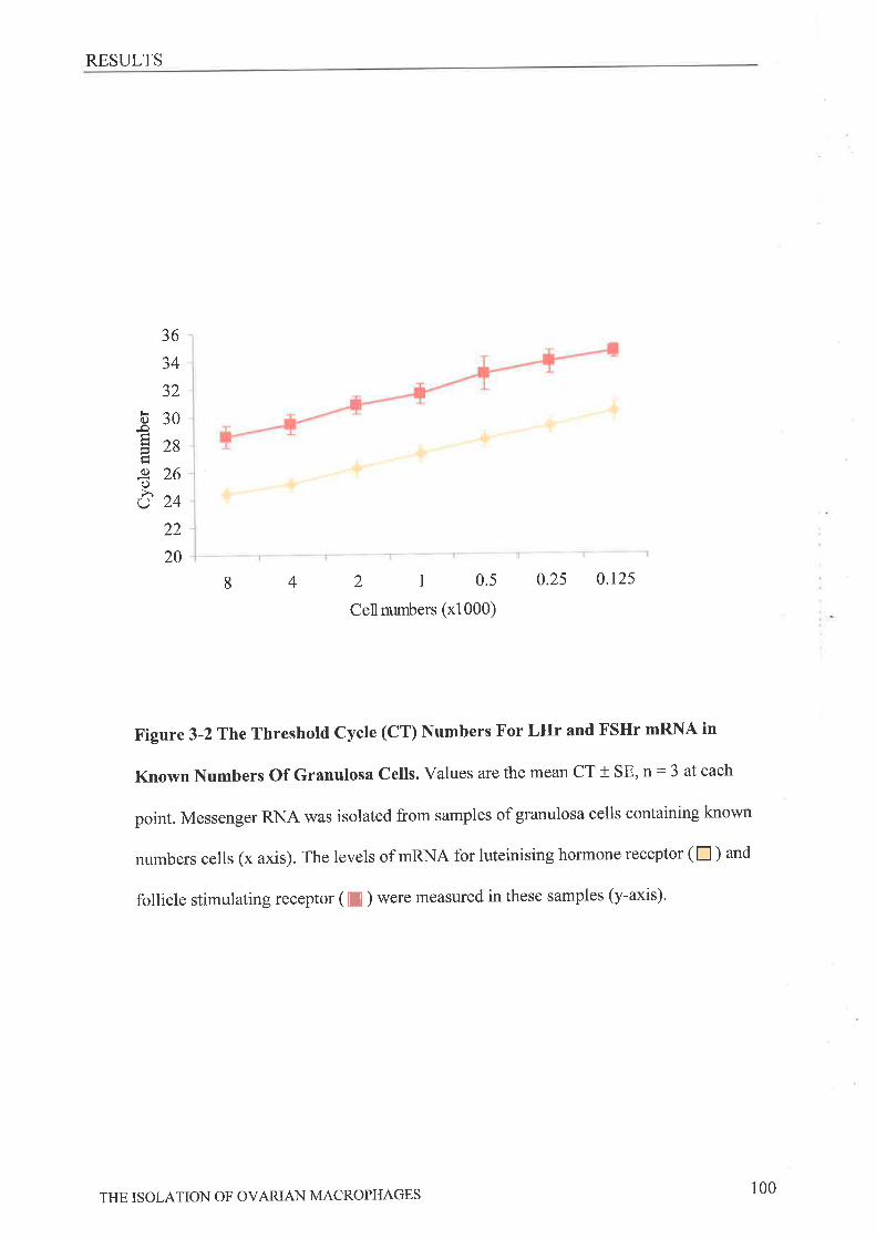
RESULTS
36
34
32
b30-oE2sã-e 26oôz+
22
20
8 4 2r0.5Cellnumbers (xl000)
0.25 0.125
Figure 3-2 The Threshold Cycle (CT) Numbers For LIIr and FSHr mRNA in
Known Numbers Of Granulosa Cells. Values are the mean CT + SE, n: 3 at each
point. Messenger RNA was isolated from samples of granulosa cells containing known
numbers cells (x axis). The levels of mRNA for luteinising hormone receptor (n ) and
follicle stimulating receptor ([ ) were measured in these samples (y-axis)'
THE ISOLATION OF OVARIAN MACROPHAGES 100
RESULTS
This was then used to equate the CT values of both FSHr and LHr measured in the ovarian
macrophage samples to the possible numbers of contamination granulosa cells. Minimal
numbers of contaminating cells were found (Table 3-3). Moreover, to conf,trm minimal
contamination we found that levels of progesterone in media conditioned fot 24 hours by
peritoneal or ovarian macrophages collected both before and after ovulation were not
detectable (data not shown, <lnmol/L) further indicating negligible contamination with
granulosa cells.
In addition, an analysis of LCA expression performed on the adherent and non-
adherant cells from an ovarian digest, an accepted method of macrophage isolation and used
extensively by others to obtain ovarian macrophages for further studies, show that the purity
of the antibody panning population obtained in this study far exceeds that of the ovarian
adherent cells. Adherent ovarian cells were found to be only 33 % LCA and 30Yo
F4l80*:Ia*while the non-adherent population were less than 3% positive for either LCA or
F4l80+:Ia. The antibody panning positive cells are 95%LCA.
3.3.4 Psecocvuc CAPABILITY oF Ov¡nl¡'r,t MecRops¿'cBs
To measure the phagocytic capabilities of ovarian macrophages, cells bound to the panning
plates were cultured with fluorescent beads. All isolated ovarian and peritoneal macrophage
populations were found to exhibit phagocytic ability. The percentage of cells able to
phagocytose beads increased significantly following ovulation (Figure 3-3), There was no
significant difference between the phagocytic capacity of isolated F4l80+ and Ia* cells'
Isolated peritoneal cells were approximately 50%o phagocytic regardless of the collection time
point (data not shown). Since fewer cells were phagocytic prior to ovulation than after,
isolated macrophages were cultured with hCG to determine if this hormone directly
stimulated phagocytosis, but this did not result in any significant change in the percentage of
THE ISOLATION OF OVAzuAN MACROPHAGES 101
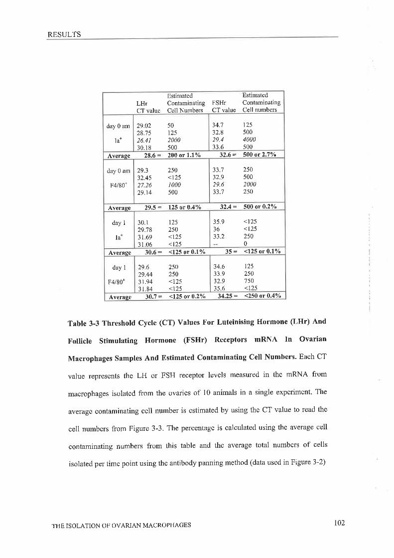
RESULTS
LHrCT value
EstimatedContaminatingCell Numbers
FSHrCT value
EstimatedContaminatingCell numbers
day 0 am
Ia*
29.0228.7526.4130.1 8
501252000500
34.',|
32.829.433.6
1255004000500
Averaqe 28.6 = 200 or l.lo/o 32,6 = 500 or 2.7Vo
day 0 am
F4l80+
29.332.4527.2629.r4
250<1251000500
33.732.929.65J. I
2505002000250
Averase 29.5 = 125 or 0.4Vo 32.4 = 500 or 0.2%o
day 1
Ia*
30.129.7831.693 1.06
125250<125<125
35.93633.2
<t25<1252500
Average 30.6: <125 or 0.lo/o 35 = <125 or 0.lo/o
day I
F4l80+
29.629.443r.9431.84
250250<125<125
34.633.932.935.6
12s2507s0<t25
Average 30.7 = <125 or 0.2o/o 34.25 = <250 or 0.4o/o
Table 3-3 Threshold Cycle (CT) Values For Luteinising Hormone (LHr) And
Follicle Stimulating Hormone (FSHr) Receptors mftNA In Ovarian
Macrophages Samples And Estimated Contaminating Cetl Numbers. Each CT
value represents the LH or FSH receptor levels measured in the mRNA from
macrophages isolated from the ovaries of 10 animals in a single experiment. The
average contaminating cell number is estimated by using the CT value to read the
cell numbers from Figure 3-3. The percentage is calculated using the average cell
contaminating numbers from this table and the average total numbers of cells
isolated per time point using the antibody panning method (data used in Figure 3-2)
THE ISOLATION OF OVAzuAN MACROPHAGES t02
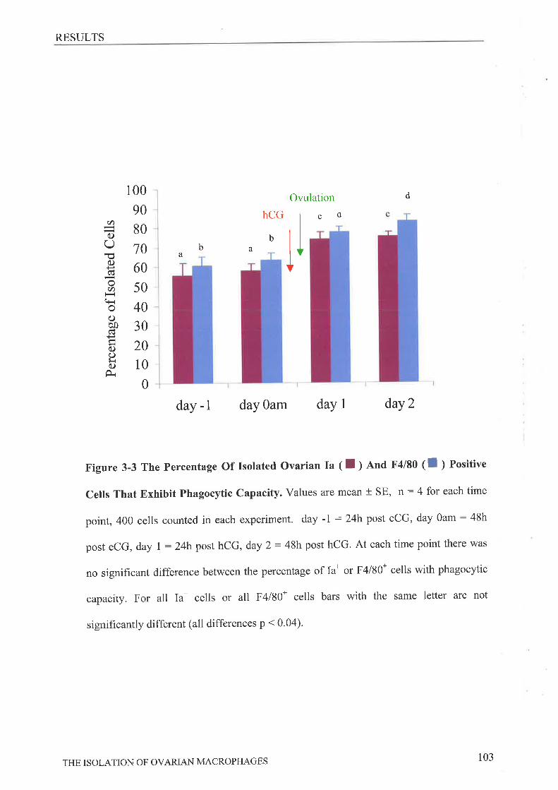
RESULTS
100
90
80
l060
50
40
30
20
10
0
day -l day Oam daY 1 daY 2
Figure 3-3 The percentage Of Isolated Ovarian Ia ( I ¡ nna F4l80 ( I ) Positive
Cells That Exhibit Phagocytic Capacity. Values are mean + SE, n: 4 for each time
point, 400 cells counted in each experiment. day -l : 24h post eCG, day Oam : 48h
post eCG, day 1 : 24h post hCG, day 2: 48h post hCG. At each time point there was
no significant difference between the percentage of Ia* or F4l80* cells with phagocytic
capacity. For all Ia* cells or all F4l80* cells bars with the same letter are not
significantly different (all differences p < 0.04).
Ovulation d
llaba
hCG
b
sd(t)
C)
OËc.)+¡cü
o(t)q-roC)o0ctcl(.)O¡.ic)
THE ISOLATION OF OVARIAN MACROPHAGES 103
RESULTS
phagocytic cells (Day 0Ia*; -hcG: 57.8 + 4yo,+ hcc :54.7 + 4.5Yo;Day 0 F4l80+; -hcc
: 63.4 + 3.8yo, + hcc : 69.5 t 1.9%)
3.3.5 ET.¡BcT oF MACROPHAGE-CONDITIONED MBPIE ON PHA-STIMULATED SPI-BNOCYTN
PROLIFERATION
To determine if ovarian macrophages participate in the regulation of T cell
proliferation, macrophage-conditioned media was added to PHA-stimulated, adherence-
purified splenocytes. Limited proliferation of splenocytes was seen in the absence of PHA,
whilst the addition of PHA significantly stimulated T cell proliferation to a level designated to
be 100% (Figure 3-4). The proliferation stimulated by PHA alone was not significantly
different to proliferation produced by conditioned media from any of the ovarian macrophage
populations. In addition, no significant differences were seen between proliferation induced
by media from Ia* or F4l80+ cells, either prior to or following owlation. Conditioned media
from Ia* and F4l80+ peritoneal cells were found to significantly inhibit PHA-stimulated
proliferation.
THE ISOLATION OF OVARIAN MACROPHAGES 104
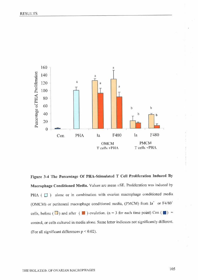
RESULTS
160
140
t20
100
80
60
40
20
0
Con PHA Ia F480
Iñfi.()!+-r
ofrt¿r
0i+ro(.)èoÐC)o¡r(.)
a
a
OMCMT cells +PHA
Ia F480
PMCMT cells +PHA
a
b b
bb
Figure 3-4 The Percentage Of PHA-stimulated T Cell Proliferation Induced By
Macrophage Conditioned Media. Values are mean +SE. Proliferation was induced by
pHA ( n ) alone or in combination with ovarian macrophage conditioned media
(OMCM) or peritoneal macrophage conditioned media, (PMCM) from Ia* or F4l80n
cells, before (tr) and after ( I ) ovulation. (n:3 for each time point) Con (l) :
control, or cells cultured in media alone. Same letter indicates not significantly different.
(For all signihcant differences p < 0'02).
THE ISOLATION OF OVARIAN MACROPHAGES 105
DISCUSSION
3,4 Drscussrox
This study has described a method for the isolation of the ovarian macrophage population to
purity levels not previously achieved for this tissue type. We have also described some basic
characteristics of these isolated cells.
We initially set out to ensure the ovarian dissociation minimised the loss of adherent
cell types, in this first critical step of the isolation procedure. This was achieved by carrying
out the dissociation at24"C, in combination with changing the media base from RPMI, which
contains small amounts of reduced glutathione that inhibits collagenase activity, to o-MEM,
which has no reduced glutathione, with chloride ions added to optimally activate the enzyme.
We also found, that using a shorter digest period (45 minutes and 90 minutes are both reported
in the literature) did not significantly alter the numbers of total live cells isolated per milligram
of tissue.
The dissociation conditions described ensure that the highest possible numbers of live
cells for further purification procedures are obtained. It is interesting to note that the eîzyme
Dispase can significantly alter the surface phenotype of lymphocytes [295] while the
collagenase enzyme itself has been reported to inhibit macrophage adherence through
disruption of surface antigen expression t157]. Conversely, it has more recently been shown
that tissue digestion with the enzyme collagenase type IA, as used in this study, does not have
such signifîcant detrimental effects on leukocyte surface antigen expression 12961.
We then proceeded to examine the isolated ovarian macrophage population, and have
shown it to be a viable and defined population suitable for further analysis. The data
demonstrating high prevalence of LCA antigen and minimal LHr or FSHr expression
combined with the absence of progesterone production by these cells show firstly, that the Ia
and F4l80 antibodies are indeed binding to and isolating the anticipated macrophage cell types
THE ISOLATION OF OVARIAN MACROPHAGES 106
DISCUSSION
and secondly, that contaminating cells are minimal. This is further supported by our previous
work in ovarian tissue sections showing that the F4l80 and anti-Ia antibodies used in the
isolation do not bind to thecal and granulosa cell layers 168,2931. Also, work in other fields
shows the markers are expressed exclusively on macrophage cells lll3, 2791and not other
leukocyte types. Unfortunately, attempts to enzymatically or mechanically remove the cells
from the panning plates for further verification of purity FACS analysis, was not successful or
resulted in the death of the majority of the population respectively, and consequently this type
ofanalysis could not be carried out here'
Following these experiments we went on to isolate the ovarian macrophage population
at different time points during gonadotrophin-stimulated immature mice. The isolated
macrophages are derived from whole dissociated ovaries and thus we cannot distinguish
between macrophages isolated from the stroma, theca, atretic follicles or CL, of the ovary. This
problem is limited by the use of gonadotrophin-primed immature animals rather than adult
mice where CL's from previous ovulations would contribute to the macrophage pool. The
administration of eCG establishes a cohort of follicles at the same stage of development,
limiting the presence of atretic follicles, so that cells isolated at the various time points are
predominantly of thecal or corpora luteal origin. Changes in the total numbers of macrophages
isolated per milligram of tissue substantiate immunohistochemical observations in various
species, showing increases in numbers of these cells in, the theca as ovulation approaches, and
during formation of the CL l4g, 53, 54, 59, 64,2971. Following ovulation (Day 1) the total
number of Ia* cells increased significantly above the numbers of F4l80+ cells. The F4l80
antigen is expressed on newly recruited monocytes/macrophages and its expression can be
down regulated on some activated macrophages [113] or on macrophages exposed to high
concentrations of cytokines t298]. This implies that following ovulation there are significantly
more activated macrophages than newly recruited macrophages present in the ovary. This is
THE ISOLATION OF OVARIAN MACROPHAGES 107
DISCUSSION
supported by previous work suggesting greater numbers of Ia* cells than F4l80 * cells are
present in the ovarian stroma and theca of follicles approaching ovulation [68]. Interestingly, it
was also found that GM-CSF deficiency reduced the numbers of Ia* or activated cells present
in the stroma of ovaries examined and the estrous cycles of these animals was extended' A
similar dysfunction of the estrous cycle was seen in macrophage depleted mice (Chapter 2
l¡g3l). Together these studies, suggest that an influx of Ia* cells may be essential in the
regulation of luteal function or regression. On day 2 after ovulation the numbers of Ia* and
F4l80+ cells are not significantly different, with both populations increasing significantly to
numbers greater than that seen on day 1. This corresponds \ /ith further invasion of the CL with
newly recruited macrophages and their subsequent activation, It has been suggested elsewhere
that macrophages have both luteotrophic and luteolytic affects 12991and there is evidence that
ovarian macrophages in the CL can be luteotrophic, with the ability to stimulate progesterone
production l22g,30Ol or luteolytic, with roles in the regulation of luteal regression [301, 302]'
These conflicting actions of macrophages could perhaps be attributed to the activation status of
these cells at differing times of the estrous cycle. In agreement with this, studies in other fields
show that the effects of steroids on macrophage functions [117, 118] and conversely, the
effects of macrophages on granulosa cells [174,238] may be vary depending on the source or
state (elicited or resident) of the macrophages. It would therefore be of interest to examine
additional markers of macrophage activation in this population to give an insight into any
changes in the activation status of the ovarian macrophage population across the
gonadotrophin-stimulated cycle.
The low phagocytic capacity of the isolated ovarian macrophages was unexpected since
it is considered a feature of the macrophage lineage. It has however, been shown elsewhere that
ovarian macrophages have a reduced phagocytic capacily when compared to testicular
macrophages [303], while in other tissues such as the brain, microglial cells express markers of
THE ISOLATION OF OVARIAN MACROPHAGES 108
DISCUSSION
the macrophages lineage, but do not exhibit phagocytic activity in early stages of activation
t161]. It should be noted that microglial cells isolated from the brain have been shown to be
only 50% phagocytic when cultured with non-apoptotic cells but this rates increases to 100% if
apoptotic cells are used [304]. Since ovarian macrophages also reside in a complex tissue mass,
they may also fall into a phagocytic category similar to these cells. The increase in phagocytic
activity post ovulation corresponds to the time at which profuse tissue reorganisation and
remodeling is occurring, and thus it seems reasonable that these cells would become activated
and involved in the clearance of tissue debris and apoptotic cells created by these processes.
Vy'e have also shown here that the addition of hCG to ovarian macrophages in vitro does not
stimulate a change in the percentage of phagocytic cells. This change must therefore be
initiated by other means, such as changes in ovarian steroid levels. Both progesterone and
oestradiol have been shown to influence macrophage and monocyte phagocytosis [119, 120,
123] Macrophage phagocytic capacity can also be manipulated by changes in cytokine levels
or simply by the presence of apoptotic cell debris and/or extracellular matrix fragments
generated following ovulation. The phagocytic capacity of the peritoneal population was lower
than expected even considering that resident peritoneal macrophages, as used in this study,
have a lower phagocytic ability than elicited/activated macrophages. It has also been reported
that phagocytic activity of cells can be reduced by exposure of the cells or animals to periods
of low temperatures [305, 306] and the peritoneal cells in this study were held at four degrees
for a period longer than that for the ovarian cells'
Another main function of macrophages is their interaction with T cells and subsequent
stimulation of an immune response. We found that ovarian macrophage conditioned medium
does not significantly alter PHA-stimulated splenocyte proliferation, Our results indicate that
the isolated Ia* and F4l80* cells do not produce significant levels of immunomodulatory
molecules. Hence at the time points that we have studied, the regulation of T cell proliferation
THE ISOLATION OF OVARIAN MACROPHAGES 109
DISCUSSION
within the ovary may not be an important function of these cells. In the cycling ovary, few T
cells are been locali zed in the ovarian tissue until luteinisation, when increases have been
reported [51,53]. Interestingly, it has been reported that at luteolysis bovine luteal cells
themselves express MHC II1255,307] and can stimulate T cell proliferation1256l, although,
macrophages were not depleted from these luteal cell populations. This suggests that during
CL regression the macrophages present are actively invoking an immune response- presenting
antigens and activating T cells, while our results suggest that prior to ovulation and in the early
CL macrophages are not actively regulating T cell proliferation. Alternatively, the apparent
lack of response in our study could be due to low concentration of immunomodulatory factors
as a results of low numbers of isolated cells, or the lack of direct T cell/macrophage contact.
Macrophages isolated from the uterus ll47l and testis [135] have both been shown to inhibit
PHA-stimulated splenocyte proliferation in assays in which the macrophages were in direct
contact with the splenocytes, rather than utilizing macrophage supernatant only as in this study.
The mitogen stimulated proliferation assay used in this study will determine only if the
supernatant contains any soluble components capable of stimulating or inhibiting splenocyte
proliferation. If it were possible to isolate higher numbers of ovarian macrophages to perform
cell co-culture experiments a different result may be observed. In contrast to our ovarian
macrophage results, the resident peritoneal macrophage population was found to significantly
inhibit PHA stimulated T cell proliferation suggesting that these cells are producing significant
amounts of inhibitory molecules. It has been reported elsewhere that peritoneal cells will
inhibit mitogen stimulated T cell proliferation [308] and there is ample evidence that this can
occur through the production NO and/or prostaglandins by activated cells [308-310].
In summary, we have for the first time described a method for isolating ovarian
macrophages. We found that these cells are more phagocytic post-ovulation than prior to
ovulation and conditioned culture supernatant from these cells has no influence on PHA-
THE ISOLATION OF OVAzuAN MACROPHAGES 110
DISCUSSION
induced splenocyte proliferation. The utilization of this method to isolate ovarian macrophages
will allow further examination of the cellular and molecular characteristics of these cells during
folliculogenesis, ovulation and luteinisation.
THE ISOLATION OF OVARIAN MACROPHAGES 111

Chapter Four
4 Tnn ExpnnSSION OF INFLAMMATORY MEDIATORS BY
THE M¡.CNOPHAGE POPUT,NTION IN THE OV¿,NY OF THE
Gox¿.n oTRoPHIN-STTMULATED Mousn

INTRODUCTION
4.1 lNrnonucrroN
The previous chapters of this thesis have demonstrated that ovarian macrophages are
necessary for normal ovarian functions and identified a method suitable for the isolation of
ovarian macrophages from the other prevailing ovarian cell types. Our results have implicated
these cells in follicular growth, ovulation and corpus luteum regression, and suggest these
isolated cells display irregular immune functions. No research examining the direct
contributions of these cells to the ovarian cytokine environment has previously been
performed even though various inflammatory cytokines, that are commonly produced by
macrophages elsewhere in the body, have been shown to have significant effects on follicle
growth/atresia [311-314] ovulation 16l, 191,2051 and isolated granulosa and thecal cell types
1202, 232,241, 3151.
In this study we have selected several cytokines of interest to measure in the isolated ovarian
macrophage population, in particular the classical inflammatory macrophage products
interleukin-l beta (IL-l8), nitric oxide (NO) and tumour necrosis factor alpha (TNFcr) as well
as selected anti-inflammatory molecules.
Nitric oxide (NO), an established product of classical inflammatory macrophages [208] has
been shown by our group [60, 61] and others l2l2-2141to be a critical component in the
ovulatory cascade. It is produced by the enzyme nitric oxide synthase (NOS) of which there is
two reported varieties - inducible (iNOS) and constitutive (oNOS). Jablonka-Shariff et al
demonstrated expression of iNOS in the theca and stroma of immature and preovulatory
ovaries while after ovulation iNOS staining was restricted to the outer layers of the CL and
became more widespread in the degenerating CL Í2111. The NO molecule itself has been
postulated to have a role in vascular permeability and dilation 1217,2181, the regulation of
follicular and luteal cell apoptosis [171, 219-22I], and the inhibition of leukocyte adhesion to
endothelium [316]. It has also been shown to have significant effects on both prostaglandin
INFLAMMATORY MEDIATORS EXPRESSED BY OVARIAN MACROPHAGES 113
INTRODUCTION
production, through activation of the COX enzyrnes in the ovary 1222,2231and other tissues
lZZ4, Z25l, and progesterone production by luteinized granulosa cells 1223,236,3171'
There are numerous studies supporting roles for TNFo in the regulation of many different
aspects of ovarian function including the ovulatory event 1207, 2611, apoptosis,
steroidogenesis and proliferation within both ovarian follicles and corpus luteum (reviewed in
t3lS]). ln the ovary, expression of TNFo has been demonstrated in'macrophage like' cells of
the theca and corpus luteum 1251, 252].It has been suggested by others that macrophages
may be a source of this cytokine in the ovary l3l9-321)'
IL-18 has been reported to stimulate ovulation in the rat in vitro 1191,193] while iz
vivo administration of interleukin-l receptor antagonist (Il-lra) inhibits ovulation 1322].It
has been postulated that IL-l8 directly on nitrite production [61, l7l, 215, 216]
steroidogenesis [206] or collagenase activity 1323,3241and vascularisation 11971' Il-lra is
expressed in the granulosa and thecal layers of human preovulatory follicles and the
regressing corpora lutea [186] while cultured granulosa cells and theca cells have been
reported not to express Il-lra mRNA 13251. In the rat ovary ll--lra expression has more
recently been reported to be confined to the granulosa cell compartment with some staining in
the oocytes [194]. lLira is a protein that significantly inhibits or down regulates the
inflammatory actions of IL-18 and in a classical inflammatory response the pattern of
expression of this protein is delayed but comparable to the expression of IL-1B'
TGFBT, a member of the TGF superfamily, is a pluripotent growth factor with established
roles in processes important in ovarian function such as the regulation of cell apoptosis [326],
proliferation, steroidogenesis, wound healing [102] and immune responses 13271. The TGFpI
protein is expressed in the theca of the follicle in the pig, mouse and rat 1173, 312,3281.
Increases in thecal staining following human chorionic gonadotrophin (hCG) administration
have been reported although the granulosa of the follicle also expresses TGFpr protein 13291'
INFLAMMATORY MEDIATORS EXPRESSED BY OVARIAN MACROPHAGES tr4
INTRODUCTION
In addition intrabursal injection of TGFpI into the mouse ovary has been shown to
significantly inhibit follicular rupture [330] and although naturally cycling TGFBr knockouts
have reduced ovulations and an extended estrous cycle [331], ovulation is normal following
gonadotrophin stimulation.
IL-10, produced predominantly by macrophages and Thz cells, is a potent anti-inflammatory
cytokine through its inhibitory effects on the proliferation and cytokine expression of T cells'
It also has significant effects on macrophages and dendritic cells, inhibiting differentiation
and antigen presentation as well as cytokine expression (reviewed in 1332,333]. Studies
examining the expression of this cytokine in the ovary are limited. The protein has been
detected in human follicular fluid [334, 335] and is present in human mononuclear/luteal cell
co cultures t336]. IL-10 mRNA has also been demonstrated in day 10-20 rabbit corpus
luteum 13371 andbeen found in cancerous ovarian cell types [338, 339]'
Using the method described in Chapter 3 to isolate ovarian macrophages the purpose of this
study was to determine if ovarian macrophages are producing cytokines that regulate ovarian
function, by analysing the expression of mRNA and protein for molecules with established
roles in ovarian function and immune regulation'
INFLAMMATORY MEDIATORS EXPRESSED BY OVARIAN MACROPHAGES 115
METHODS
4.2 Mnrnoos
4.2.1 ANItr¿Rrs
SVl29 animals were obtained from the University of Adelaide Animal services and
maintained in the eueen Elizabeth Hospital. Unless otherwise stated reagents were purchased
from Sigma Aldrich Chemical Co., St Louis, MO, USA. The first ovulatory cycle was
stimulated in immature animals of 25-27 days of age with 5IU eCG (Intervet, Boxmeer,
Holland) in 0.lml PBS (Gibco BRL, Life Technologies, Grand island, NY, USA) with 0.1%
BSA (Fraction V) on day -2, followed 48 hours later on day 0 by 5IU hCG (Pregnyl@,
Organon, Oss, Holland) to stimulate ovulation. Groups of 8-12 gonadotrophin primed animals
were killed by cervical dislocation at 0900 hours on day Oam, day I and day 2 with an
additional group killed 6 hours post hCG administration (day Opm). Following the collection
of peritoneal macrophages by peritoneal lavage with 2.5mls Hanks Balanced Salt Solution
(HBSS) (with sodium bicarbonate (0.35g1L) 5mM Ethylenediamine tetra acetic acid (EDTA),
g.Ot% Sodium Azide (Az)) ovaries were removed for digestion and subsequent isolation of
macrophages.
4.2,2 MACROPHAGE MESSENGER RNA ISOLATION AND MEASUREMENT
Ovarian macrophages were isolated on prepared antibody panning plates as devised in
Chapter 3, Section 3.3.1 on three separate occasions from groups of 8-12 animals. Ovaries
were digested in a collagenase/DNase solution, single cells were then incubated with either
the anti-Ia or F4l80 antibody supernatants and then added to anti-rat IgG-coated plates'
Immediately following washing of the unbound cells from the panning plate with HBSS/I0%
FCS, 400p1Tri reagentrM was added to the plate and cells lysed for 5 minutes. Any remaining
cells were then scraped from the plate into the lysis solution with cell scraper. Samples were
INFLAMMATORY MEDIATORS EXPRESSED BY OVAzuAN MACROPHAGES 116
METHODS
stored at -80 oC until commencement of purification of RNA. Total RNA was isolated from
the thawed cell lysate as recommended by the Tri reagentrM manufacturers and described in
Chapter 3, section 3.2.9. 2ul of each resultant RNA sample was put aside for RNA
measurement. Total RNA was measured in all samples prior to reverse transcription using a
Ribogreen RNA Quantitation Kit (Molecular Probes, Eugene, OR, UsA)(Appendix 1).
Standard curves were generated as per the manufacturers instructions. A high standard curve
(detecting 6.25-800ng RNA) was used to measure diluted (1:100) peritoneal macrophage
samples and a low standard curve (detecting 1.6-100ng/ml RNA) was used to measure neat
ovarian macrophage mRNA samples. All samples were measured in singleton on one plate in
a BMG microplate reader and values were calculated from standard curves generated by
Fluorostar software. The measurement of these concentrations allows us to use the same
amount of RNA for each reverse transcription reaction by adding the appropriate volume of
each sample.
4.2.3 R¡vBRsn TR¡.NscRlprIoN AND QuaNrtreuvE RT-PCR
A total volume of 20pl was used for each RT reaction which was performed as described in
Invitrogen Superscript IMII RNase H reverse transcriptase (Invitrogen, CA, USA) product
information (Appendix 1). For each sample, a volume containing 40ng of total RNA was
reverse transcribed using 250ng of random primers in the presence of 40U of RNase OUT
Recombinant Ribonuclease Inhibitor (Invitrogen, CA, USA). Both no RNA template (no
RNA) and no superscript enzyme (no SS) controls were also included. The generated oDNA
samples were then diluted 1:4 to give a total of 80¡rl of sample for analysis using quantitative
pCR. 1pg of RNA from each of spleen (SP), lymph node (LN), and granulosa cells was also
reverse transcribed for use as positive controls and to minimize plate-to-plate variability'
INFLAMMATORY MEDIATORS EXPRESSED BY OVARIAN MACROPHAGES tt7
METHODS
Specific primers for murine cytokines of interest and potential housekeeping genes were
designed using NCBI sequences and Primer Express (Applied Biosystems) or obtained as
indicated in Table 4-1. Primer efficiencies between the target genes and housekeeping gene
were determined to be the same by serial dilution of spleen or granulosa cell cDNA and
quantitative RT-PCR analysis. The log of the dilution was plotted against the CT value to
create a line and the slopes of each line were compared statistically. The products generated
were also purified and the sequence confirmed (Flinders University DNA Sequencing Core
Facility, SA) before being used for analysis of mRNA levels in isolated macrophage RNA
samples (Appendix 1). Quantitative analysis of mRNA levels was carried out using an ABI
prism 5700 sequence detection system (Applied Biosystems, Australia)' 3pl of sample was
added to 17pl of SYBR green master mix containing 0.5¡rM of each primer. Positive ( LN, SP
and GC) and negative (no SS, no RNA) controls were included on every plate' Analysis of
cytokine mRNA levels in each sample was done in triplicate and thus for each cytokine of
interest three separate real-time PCR plates were analysed and threshold levels (CT value)
adjusted according to identical splenic RNA (SP) and lymph node RNA (LN) positive
controls included on every plate. Samples with cycle threshold values of 37 or greater were
regarded as negative. The house-keeper genes 18S and HPRT were assessed for suitability by
confirming consistent expression in all samples across the murine stimulated cycle, between
cell source (ovary or peritoneal) and isolated cell type (Ia* or F4l80*) in relation to the amount
of total RNA reverse transcribed. Quantitative RT-PCR results for each of the tatgel genes
were norïnalized to the levels of the house-keeper gene and then expressed as the fold change
in mRNA levels from the control group, nominated for each gene as the average for the
ovarian Ia+ day gam group and gene. All results were log transformed to normalize the data
and then analysed by One-Way Anova across both Ia* or F4l80* cell types and ovary or
peritoneal cell source (Appendix 1).
INFLAMMATORY MEDIATORS EXPRESSED BY OVARIAN MACROPHAGES 118
SIZE GENBANKACCESSION NO.TARGET GENE
Interleukin-1-beta(IL-lp)Interleukin-10(r-10)Interleukin-1-recePtorantagonist (IL-lra)Tumour Necrosis Factor-alpha (TNF-ct)Transforming GrowthFactor-beta-1 (TGF-Þ r)iNOS(Supplied by Dr W Ingman)Hypoxanthine guaninephosphoribosyl transferase(HPRr)18S
FORWARD 5'.3'Start 69TGAAGTTGACGGACCCCfuq-A.
s1É'rt446
GGCGCTGTCATCGATTTCTC
Start 183
CCTTCAGAATCTGGGATACTAACCA
Start 236CCAGGCGGTGCCTATGTCT
Start 1162CCCGAAGCGGACTACTATGCT
StartCATCAGGTCGGCCATCACT
Start 43CTTCCTCCTCAGACCGCTTTT
Start 56AG AAACGGCTACCACATCCAA
REVERSE 5'-3'Start 169
TGATGTGCTGCTGCGAGATT
Start 532CTTGGTCTTGGAGCTTATT A'rq.rqr\TCA
Start 295CACCATGTCTATCTTTTCTTCTAGTTTGA
60 NM 008361
87 NM 010548
58 NM 031167
Start 358TACTCTTCAAGGGTTTACCGG
Start 1238GTTTTCTCATAGATGGCGTTGTTG
StartCGTACCGGATGAGCTGTGAA
Start 136
AACCTGGTTCATCATCGCTAATC
Start I 19
CCTGTATTGTTATTTTTCGTCACTACCT
82 NM 013693
77 NM 011577
86 NM 010927
94 NM 013556
91 4F176811
DrA
Table 4-1 Details Of The primers used In This study. For each gene of interest the quantitative reverse transcription polymerase chain
reaction primer sequences, product sizes in base pairs (bps), and Genbank Accession numbers for the gene sequences used in primer design, are
specified.
METHODS
4.2.4 CulruRn o¡ IsolRrpo M¡'cRopuacns
Cells were isolated using the antibody panning technique on three separate occasions from
groups of 8-12 animals at the same four time points across the stimulated reproductive cycle
as those used for the RNA collection. Following the removal of the unbound cells the
numbers of cells remaining were counted in ten 20x fields of view. Cells were then cultured in
800¡.rl gMEM with L- glutamine (150¡rg/ml)(ICN Biochemicals, USA) and Penicillin
(100U/ml)/Streptomycin (1OO¡rg/ml) solution (CSL Biosciences, Australia) at 5 o/oCOz fot 24
hours. Further details in Appendix 5. Media was collected, ultra-spun to remove any debris,
and then stored in 180p1 aliquots at -80oC until assay. For each time point conditioned media
was collected from three separate experiments.
4.2,5 MN¡.SURBIT¡ENT OF SBCR¡TBO OVUT¡'TORY MEDIATORS
euantikine@ kits for the measurement of the ovulatory mediators IL-IP, TNFo, NO and IL-
10 were purchased from R&D Systems Inc. (Minneapolis, MN, USA)' Assays were
performed as recommended by the manufacturer, except in the case of IL-IP and IL-10 in
which the initial incubation of samples on the plate was carried out overnight (15 hours) at
4"C (Appendix 1). The specihcations of these assays are provided in Table 4-2. Results were
expressed per 1000 cells cultured. Data was log transformed and a One-way ANOVA was
used to detect any significant changes
INFLAMMATORY MEDIATORS EXPRESSED BY OVARIAN MACROPHAGES 120
Detection limit 3 pglml
TUMOUR NECROSIS
INTERLEUKIN.I -BETA FACTOR ALPHA
5pg/ml
NITRIC OXIDE INTERLEUKIN-I0
1¡-rMoVL 4pglml
Intra-assay precision
7o CV's
20.2pglml
249 pglml
4.4
.51
5.6 pglml - 9
23 pglml :4.3
5.4pgml :9.2
32.5 pglml : 6.1
Murine TNF-cr, (r and n) NA
78o/o r rat TNFcr
20.9 ¡tmoVL :5.3
68.5 pmoVl :1.22.5 pglml
29 pglml
:9.3
:5
Inter-assay precision
7o CV's
19.6 pglml :6.1
258pglml :2.818.7 ¡rmoVl : 7
68.2 ¡tmoVL :3.3
1.7 pglml
3lpg/ml
:6.5
: 5.3
Cross reactivities
Table 4-2 Specifications Of The Assays Used To Detect Inflammatory Mediators In Ovarian Macrophage Conditioned Media'
All information is extracted from the product information booklets provided with the Quantikine@ assay kits by R&D systems (MN,
USA). r: recombinant, n: natural.
Murine IL-1p (r andn)
1olo r human IL-1P
4%or rat IL-l8
Murine IL -10 (r and n)
0.4o/o r rat IL-10
RESULTS
4.3 Rnsur,rs
4.3.I ISOTRTBO MRCROPHRCB RNA
The amount of total RNA isolated at each time point varied enormously. For ovarian cells
isolated with the anti-F480 hybridoma supematant the mean total RNA per experimental
group (cells from the ovaries of 8-12 animals) was day Oam : 109.9ng+ 22.4, day Opm :
202.7ng + 13.4, day 1 : 207ng+ 93.8, day 2 : 282n1t 90.9. For cells isolated with the anti-
Ia antibody supernatant day Oam : 62nE t 42.5, day Opm : 2\l.lng + 40, day 1 : l93ng t
74.6, day 2 : 422ng + 64.2. These differences reflect the variations in the numbers of
macrophages presênt in and therefore isolated from the ovary. At each time point many
peritoneal cells were isolated resulting in large amounts of peritoneal cell RNA varying from
2.¡t"gto 11.6pg.
4.3,2 PnruBn EprtcIeNcIES AND House-rnBPER VALIDATION
To enable the valid use of the AACT method for evaluation gene expression it must first be
established that the expression of the nominated house-keeper gene is not influenced by the
experimental treatments. Secondly, the primer sets used must be shown to prime with the
same efficiency within the PCR reaction. The potential house-keeper genes HPRT and 18S
were both found to be consistently expressed in cells isolated across the stimulated cycle with
no significant difference in the level of mRNA expression across the treatment groups (Table
4-3). HPRT was chosen as the house-keeper for this work since it was less variable and the
CT values were closer to those expected for the target genes. All primer sets were found to
amplify at the same effrciency as the HPRT house-keeper (Table 4-4).
INFLAMMATORY MEDIATORS EXPRESSED BY OVARIAN MACROPHAGES r22
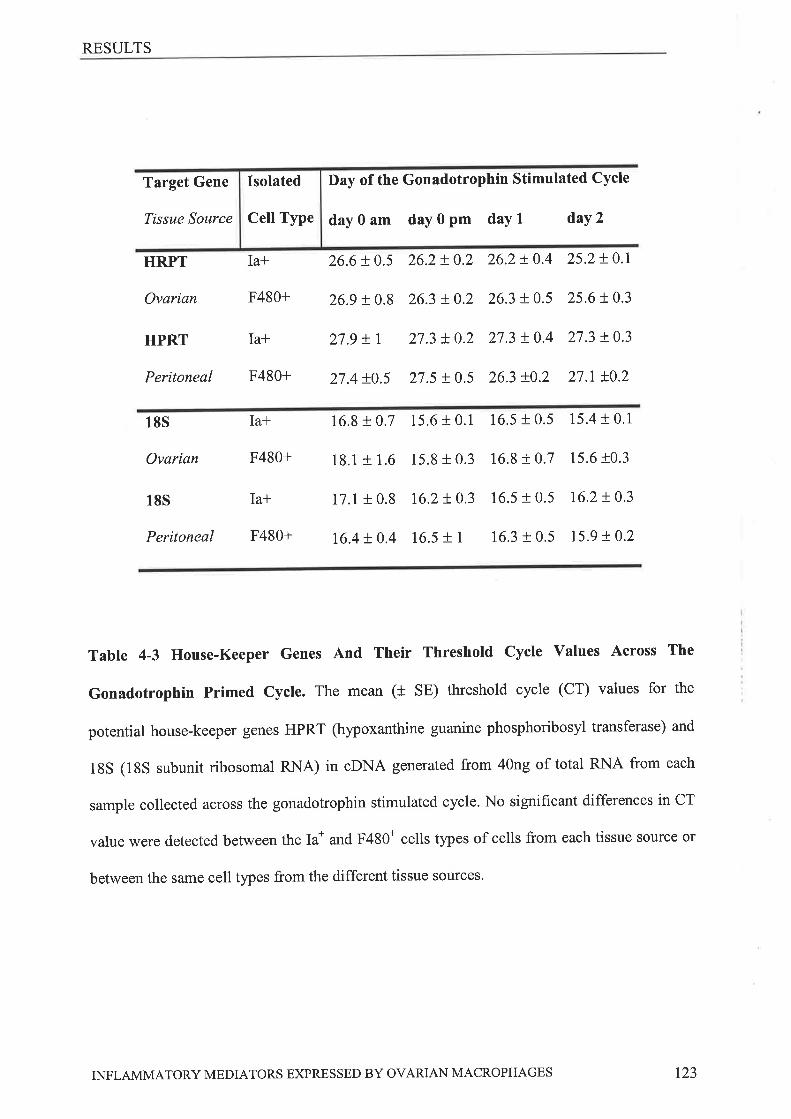
RESULTS
Cell Type
IsolatedTarget Gene
Tissue Source
Day of the Gonadotrophin Stimulated Cycle
day0am day0pm daYl daY2
HRPT Ia* 26.6 + 0.5 26.2 ! 0.2 26.2 ! 0.4 25.2 ! 0.1
Ovarian F480+ 269 t0,8 26.3 + 0.2 26.3 !0.5 25.6 r.0.3
HPRT Ia+ 27.9tl 27.3t0.2 27.3!0.4 27.3!0.3
Peritoneal F480+ 27.4 1.0.5 21.5 !0.5 263 .0.2 27.1t0.2
185 Ia* 16.8 I 0,7 15.6 t 0.1 16.5 t 0.5 15.4 f 0.1
Ovarian F480+ 13.1 + 1.6 15.8 t 0.3 16.8 r 0.7 15.6 r0.3
185 Ia* 17.1 r 0.8 16.2!0.3 16.5 + 0.5 16.2r0.3
Peritoneal F480+ 16.4t0.4 16.5 t 1 16.3 t 0.5 15.9 !0.2
Table 4-3 House-Keeper Genes And Their Threshotd Cycle Values Across The
Gonadotrophin Primed Cycle. The mean (f SE) threshold cycle (CT) values for the
potential house-keeper genes HPRT (hypoxanthine guanine phosphoribosyl transferase) and
18S (18S subunit ribosomal RNA) in oDNA generated from 40ng of total RNA from each
sample collected across the gonadotrophin stimulated cycle. No significant differences in CT
value were detected between the Ia* and F480* cells types of cells from each tissue source or
between the same cell types from the different tissue sources.
INFLAMMATORY MEDIATORS EXPRESSED BY OVARIAN MACROPHAGES r23
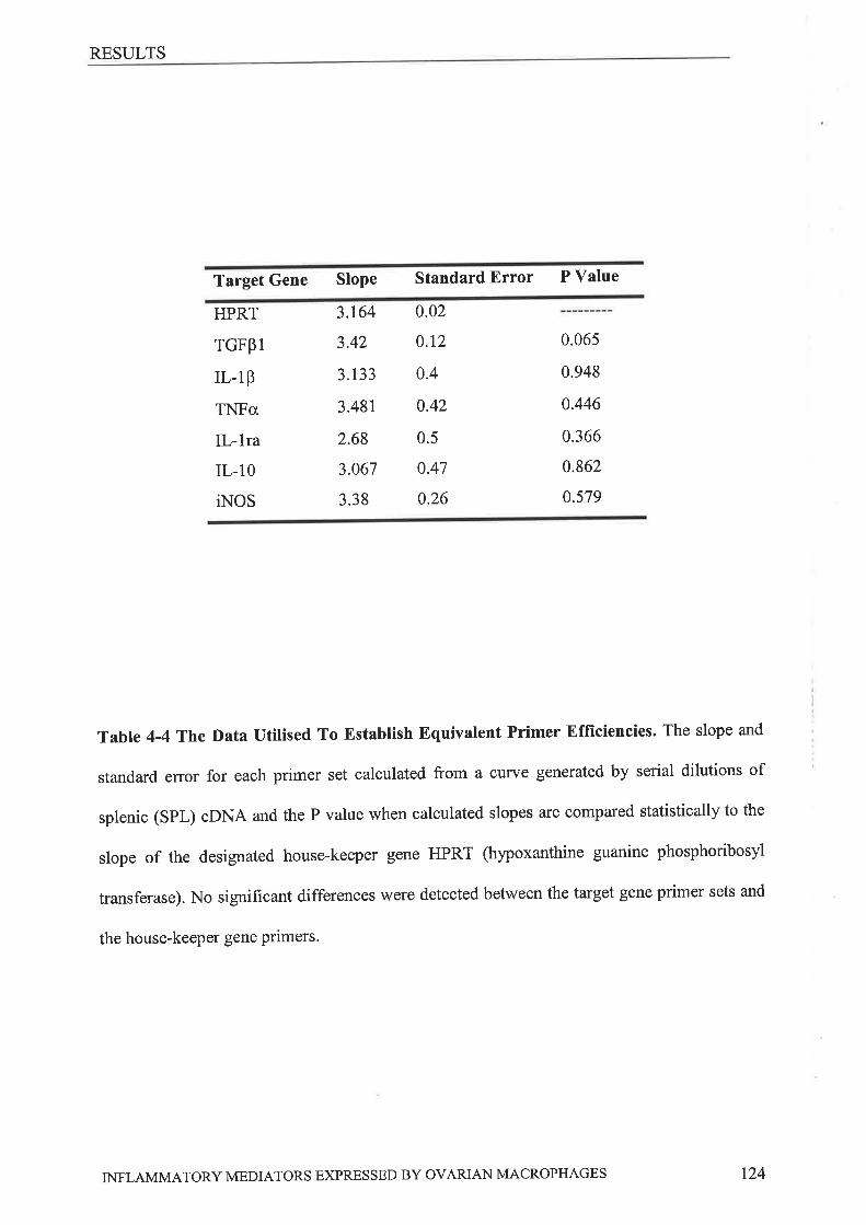
RESULTS
Target Gene Slope Standard Error P Value
HPRT
rGFpl
rl.-l8
TNFcr
IL-lra
IL-10
iNOS
3.164
3.42
3.133
3.481
2.68
3.067
3.38
0.02
0.r2
0.4
0.42
0.5
0.47
0.26
0.065
0.948
0.446
0.366
0.862
0.579
Table 4-4 The Data utilised To Establish Equivalent Primer Efficiencies. The slope and
standard error for each primer set calculated from a curve generated by serial dilutions of
splenic (SpL) gDNA and the P value when calculated slopes are compared statistically to the
slope of the designated house-keeper gene HPRT (hypoxanthine guanine phosphoribosyl
transferase). No significant differences were detected between the target gene primer sets and
the house-keeper gene Primers.
INFLAMMATORY MEDIATORS EXPRESSED BY OVARIAN MACROPHAGES t24
RESULTS
4.3.3 ExpRBssIoN o¡ CyrorrNe IURNA rN OvnzunN M¡'cRopHAGES
'We have examined the changes in expression of mRNA encoding the inflammatory
molecules IL-lp, TNFoc, iNOS, and the anti-inflammatory molecules IL-1ra, IL-10 and
TGFp1, in ovarian and peritoneal macrophages across the gonadotrophin-stimulated
reproductive cycle. The expression of mRNA for the classical inflammatory molecules IL-
lB, iNOS and TNFa was detected in all samples regardless of cell type isolated or tissue
source.
1¡-lp mRNA levels increased significantly following hCG administration in both
ovarian and peritoneal cell tlpes returning to pre-hCG levels on day 1 (24 hours post hCG,
Figure 4-lA, B). Isolated ovarian Ia* cells contained significantly more IL-lp mRNA than
isolated Ia* peritoneal cells at all time points while isolated ovarian F480* cells contained
significantly more IL-18 mRNA than isolated F480+ peritoneal cells on day Oam and day 2
only. No significant changes in ovarian expression of iNOS mRNA lvere seen. iNOS mRNA
was only detected in peritoneal cells following hCG administration and these levels were
significantly higher than those in ovarian F480+ cells at the same time-point (p<0.006) (Figure
-:E,F). TNFü mRNA increased following hCG administration in ovarian and peritoneal cell
types and this increase was significant in all cases except the for the ovarian Ia* cells. TNFa
mRNA levels returned to pre-hCG levels on day 2 (24 hours post hCG). Isolated ovarian
F4g0+ cells contained significantly more TNFcr mRNA than peritoneal F480* cells on day 1
only. Isolated Ovarian Ia* cells contain significantly TNFcr mRNA more than peritoneal Ia*
cells on day Oam and daY 1.
The expression of mRNA for the anti-inflammatory molecules IL-ba, IL-10 and
TGFpI was detected in all ovarian samples but only some the peritoneal samples' lL-ha
pRNA levels increased following hCG administration in ovarian and peritoneal cell types
INFLAMMATORY MEDIATORS EXPRESSED BY OVARIAN MACROPHAGES 125
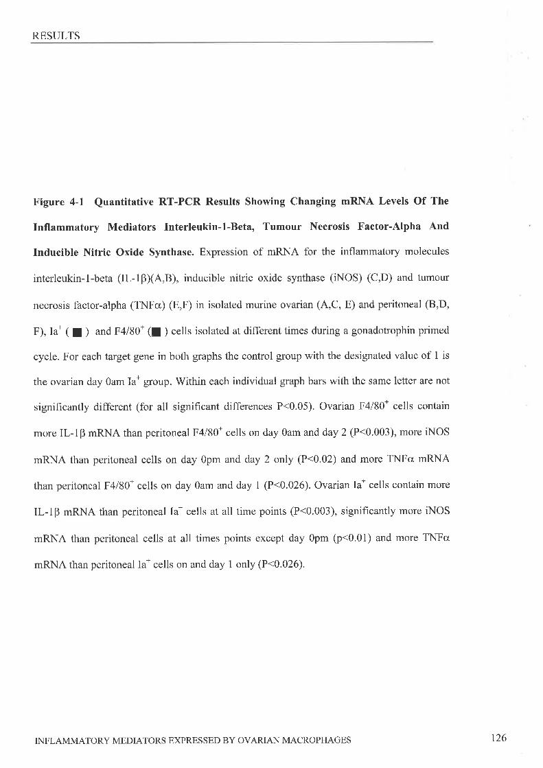
RESULTS
Figure 4-1 Quantitative RT-PCR Results Showing Changing mRNA Levels Of The
Inflammatory Mediators Interleukin-1-Beta, Tumour Necrosis Factor-Alpha And
Inducible Nitric Oxide Synthase. Expression of mRNA for the inflammatory molecules
interleukin-l-beta (IL-lBXA,B), inducible nitric oxide synthase (iNOS) (C,D) and tumour
necrosis factor-alpha (TNFo) (E,F) in isolated murine ovarian (4,C, E) and peritoneal (8,D,
F), Ia* ( | ) and F4l80* (I ) cells isolated at different times during a gonadotrophin primed
cycle. For each target gene in both graphs the control group with the designated value of 1 is
the ovarian day Oam Ia* group. Within each individual graph bars with the same letter are not
significantly different (for all significant differences P<0.05). Ovarian F4l80* cells contain
more IL-l8 mRNA than peritoneal F4l80* cells on day Oam and day 2 (P<0.003), more iNOS
mRNA than peritoneal cells on day Opm and day 2 only (P<0.02) and more TNFc¿ mRNA
than peritoneal F4l80* cells on day Oam and day 1 (P<0.026). Ovarian Ia* cells contain more
IL-1P mRNA than peritoneal Ia* cells at all time points (P<0.003), signifîcantly more iNOS
mRNA than peritoneal cells at all times points except day Opm (p<0.01) and more TNFc
mRNA than peritoneal Ian cells on and day 1 only (P<0.026).
INFLAMMATORY MEDIATORS EXPRESSED BY OVARIAN MACROPHAGES 126

RESULTS
Ia
a
I
10
9
8
7Qzè00Êcd-.É5
CJ€4ot¡r 3
2
I
0
t
cd
10
9
8
7C)
106d-q5O€¿.f!. J
2
I
0
a)
c)
hcG
Ovarian Macrophaqes Peritoneal Macrophaqes
hCG f)vulation
à
cd cd
B)
D)
1.5
ab
Ovulation
I
bc
cd
IL-lp
iNOS
TNFcr
d ¡bt"d
bb
bd cd
22
dayOam dayOpm daY 1 daYL
hCG Ovulation
day0am dayOpm daY I daY2
hCG a Ovttlation
+
bbb
day0am dayOpm dayl daY2
a
It1.5(.)o¡)
(ü
(-)ÉoIt
C)Ò0
ñsOEo
trr
ahcG Ovr"rlation
It1
.500.5
bb
0
E)
3.50
3.00
2.50
2.00
l.00
0.50
0.00
4
F)
3.50
3.00
2.50
2.00
0
1.00
0.50
0.00
dayOam dayOpm daY I daY2
hCG Ovulation
a
btr
II
a1.50
C)Þ0
Ë
QEtri
abab
ab1.50
(.)à0
¿d
O€ot\
t
b6
dayOam dayOpm day 1 daY2 dayOam dayOpm day 1 daY2
INFLAMMATORY MEDIATORS EXPRESSED BY OVARIAN MACROPHAGES t27
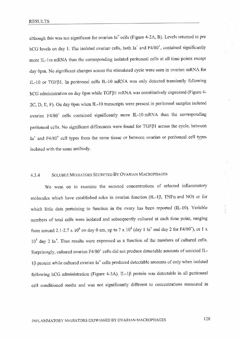
RESULTS
although this was not significant for ovarian Ia* cells (Figure 4-2A, B). Levels returned to pre
hCG levels on day 1. The isolated ovarian cells, both Ia* and F4l80*, contained significantly
more IL-lra mRNA than the corresponding isolated peritoneal cells at all time points except
day gpm. No significant changes across the stimulated cycle were seen in ovarian mRNA for
[-10 or TGFBI. In peritoneal cells IL-10 mRNA was only detected transiently following
hCG administration on day Opm while TGFBl mRNA was constitutively expressed (Figure 4-
¡C,D,E, F). On day Opm when IL-10 transcripts were present in peritoneal samples isolated
ovarian F4l80+ cells contained signif,rcantly more IL-10 mRNA than the corresponding
peritoneal cells. No significant differences were found for TGFBl across the cycle, between
Ia* and F4l80+ cell types from the same tissue or between ovarian or peritoneal cell types
isolated with the same antibodY'
4.3.4 SOLUETB MEONTORS SECN¡TBO BY OVRNIEN MACROPHAGES
We went on to examine the secreted concentrations of selected inflammatory
molecules which have established roles in ovarian function (IL-lP, TNFcr and NO) or for
which little data pertaining to function in the ovary has been reported (IL-10)' Variable
numbers of total cells were isolated and subsequently cultured at each time point, ranging
fromaround2.t-2.7x10aonday0am,upto7x10a(daylIa*andday2forF4/80*),or1x
10s day 2Ia*. Thus results \ilere expressed as a function of the numbers of cultured cells.
Surprisingly, cultured ovarian F4l80* cells did not produce detectable amounts of secreted IL-
1B protein while cultured ovarian Ia* cells produced detectable amounts of only when isolated
following hCG administration (Figure 4-34). IL-l8 protein was detectable in all peritoneal
cell conditioned medi a and was not significantly different to concentrations measured in
INFLAMMATORY MEDIATORS EXPRESSED BY OVARIAN MACROPHAGES 128
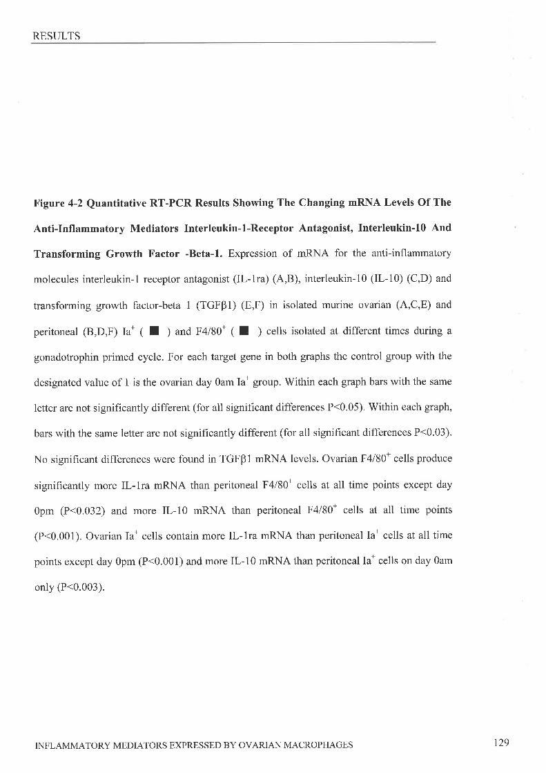
RESULTS
Figure 4-2 Quantitative RT-PCR Results Showing The Changing mRNA Levels Of The
Anti-Inflammatory Mediators Interleukin-l-Receptor Antagonist, Interleukin-l0 And
Transforming Growth Factor -Beta-l. Expression of mRNA for the anti-inflammatory
molecules interleukin-1 receptor antagonist (IL-1ra) (A,B), interleukin-10 (IL-10) (C,D) and
transforming growth factor-beta 1 (TGFPI) (E,F) in isolated murine ovarian (A,C,E) and
peritoneal (B,D,F) ta* ( I ) and F4l80* ( I ) cells isolated at different times during a
gonadotrophin primed cycle. For each target gene in both graphs the control group with the
designated value of 1 is the ovarian day Oam Ia* group. Within each graph bars with the same
letter are not signifîcantly different (for all significant differences P<0.05). Within each graph,
bars with the same letter are not significantly different (for all significant differences P<0.03).
No significant differences were found in TGFBl mRNA levels. Ovarian F4l80* cells produce
signif,rcantly more IL-lra mRNA than peritoneal F4l80* cells at all time points except day
Opm (P<0.032) and more IL-10 mRNA than peritoneal F4l80* cells at all time points
(P<0.001). Ovarian Ia* cells contain more Il,-lra mRNA than peritoneal Ia* cells at all time
points except day Opm (P<0.001) and more IL-l0 mRNA than peritoneal Ia* cells on day Oam
only (P<0.003).
INFLAMMATORY MEDIATORS EXPRESSED BY OVARIAN MACROPHAGES 129

RESULTS
A)
t2
10
Ovarian Macrophages
hCGOvulation
Peritoneal Macrophages
hCGOvulation
+
B)
12
10
aa
C)bo(Ë
O!otr
ka go
OËolr.
IL- 8
6
4
2
0
tab ab
8
6
4
2
0
acc bcc4cc cc bc bb
D)
2
c)2
tI1.5
dayOam dayOpm day I daY2
hCGOvulatiort
dayOam day0pm day 1 daY2
E)
2.5 hCGOvr.rlation
2
dayOam dayOpm day I daY2
hcc Ovulation
Iab
1.5
IL-10
Ia
a
1
0.5
()òo
(Ë
Q€oFr
0)Þo
(d
(-)€o
tJr0.5
ababab
0 _É_0
2.5
0
F)
dayOam dayOpm day 1 daY2
hCGOvulation
dayOam dayOpm dayl daY2
IiiI 2
.5(.)òoÊcd
OËotr.
1pTGF C)òo(Ú
O€o
trr
.5
II
550. 0
0
day0am dayOpm dayl daY2
INFLAMMATORY MEDIATORS EXPRES SED BY OVARIAN MACROPI{AGES 130
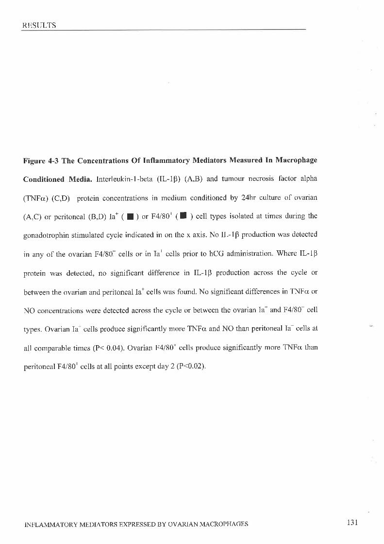
RESULTS
Figure 4-3 The Concentrations Of Inflammatory Mediators Measured In Macrophage
Conditioned Media. Interleukin-l-beta (IL-lP) (A,B) and tumour necrosis factor alpha
(TNFcr) (C,D) protein concentrations in medium conditioned by 24fu culture of ovarian
(A,C) or peritoneal (B,D) Ia* ( I ) or F4l80* ( tr ) cell types isolated at times during the
gonadotrophin stimulated cycle indicated in on the x axis. No IL-18 production was detected
in any of the ovarian F4l80* cells or in Ia* cells priorto hCG administration. Where IL-IP
protein was detected, no significant difference in IL-IB production across the cycle or
between the ovarian and peritoneal Ia* cells was found. No significant differences in TNFcr or
NO concentrations were detected across the cycle or between the ovarian Ia* and F4l80* cell
types. Ovarian Ia* cells produce significantly more TNFo and NO than peritoneal Ia* cells at
all comparable times (P< 0.04). Ovarian F4l80+ cells produce significantly more TNFa than
peritoneal F4l80+ cells at all points except day 2 (P<0.02).
INFLAMMATORY MEDIATORS EXPRESSED BY OVARIAN MACROPHAGES 131
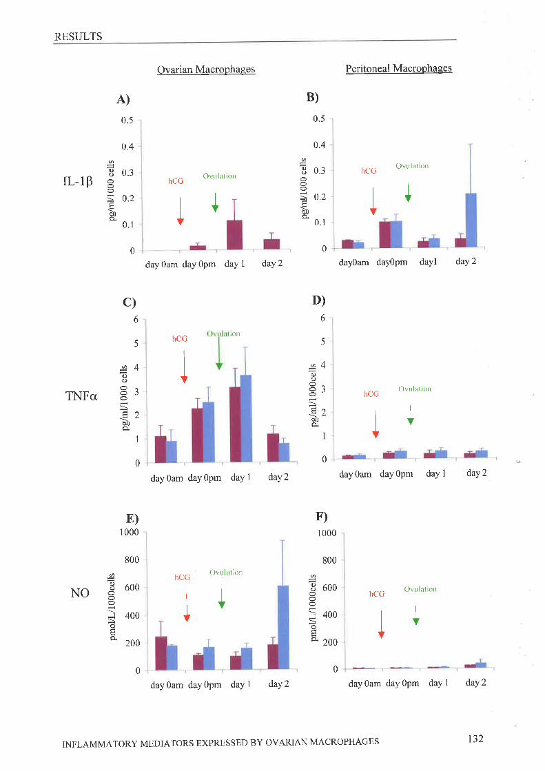
RESULTS
A)
0.5
Ovarian Macrophases
hCGOvu lation
I
day0am dayOpm daYl daY2
Peritoneal Macrophaees
hCGOvulation
dal0am dayOpm dayl daY2
hCGOvLllation
B)
0
0.
Ø(.)
rL-18 åIÊtrÒ0È
II
Ø(.)o
o
Ëa
0.4
0.3
.2
1
0.5
0.4
0.3
0.2
0.1
0 0
0
I I
c) D)
6
5
4
J
2
1
ôC)C)
OO
ÞoÈ
Ø
0.)o
ö0a
E)1000
800
600
400
200
0
Øq.)ooO
Jotso
hCGton
6
5
4
3
2
800
Ø
I 600oì 400
=oÀ 2oo
TNFcx,
NO
dayOam dayOpm dayl daY2 day0am dayOpm day I daY2
F)1000
hCGOvu lation
hCGOvulation
I0
dayOam dayOpm day I daY2 dayOam dayOpm dayl daY2
0
IJI
INFLAMMATORY MEDIATORS EXPRESSED BY OVARIAN MACROPHAGES t32

RESULTS
ovadan macrophages. Ovarian and peritoneal cells were both found to secrete detectable
concentrations of TNFcx, (Figure 4-3C, D) however, no significant differences were detected
across the stimulated cycle, even though it appeared concentrations were increased on day
gpm and day 1. Ovarian cells were found to produce significantly more TNFcx, than isolated
peritoneal cells at all times except on day 2 where conditioned media from ovarian F4l80*
cells contained protein concentrations not significantly different to that in conditioned media
from peritoneal F4l80* cells. NO (Figure 4-38, F) and IL-10 (Figure 4-4A, B) could be
detected in all samples from both ovarian and peritoneal conditioned media. Ovarian cells
were found to produce significantly more IL-10 protein and NO than peritoneal cells. Despite
a small increase in IL-10 concentration in ovarian macrophage conditioned media on day 1
post ovulation, no significant changes in IL-10 or NO concentrations were detected across the
cycle.
INFLAMMATORY MEDIATORS EXPRESSED BY OVAzuAN MACROPHAGES 133
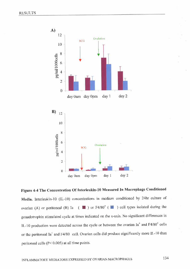
RESULTS
A)12
Ovulation
10
day0am dayOpm dayl daY2
B)n
Ovulation
day Oam day Opm daY 1 daY 2
Figure 4-4 T1¡e Concentration Of Interleukin-I0 Measured In Macrophage Conditioned
Media. Interleukin-l0 (IL-10) concentrations in medium conditioned by 24ht culture of
ovarian (A) or peritoneal (B) Ia* ( f ) or F4l80* ( I ) cell types isolated during the
gonadotrophin stimulated cycle at times indicated on the x-axis. No signihcant differences in
1¡-10 production were detected across the cycle or between the ovarian Ia* and F4l80* cells
or the peritoneal Ia* and F4l80* cell. Ovarian cells did produce significantly more IL-10 than
peritoneal cells (P< 0.005) at all time points.
rCG
(^
(.)O
ô09.
8
6
4
2
0
hCG
(tC)o
èÀ
10
8
6
4
2
0
INFLAMMATORY MEDIATORS EXPRESSED BY OVARIAN MACROPHAGES 134
DISCUSSION
4.4 DrscussloN
This study has examined for the first time the changing levels of mRNA and protein
expression of both inflammatory and anti-inflammatory molecules in isolated ovarian
macrophages. At the times points studied, isolated ovarian macrophages produced mRNA for
the inflammatory molecules, IL-IP, the enzyme iNOS, which generates NO, and TNFct.
Corresponding active proteins were detected in conditioned culture media although the
concentraions measured did not always reflect those anticipated as a result of the mRNA
measurements. A cytokine mRNA and protein profile for murine ovarian macrophages,
different to that seen in peritoneal macrophages, has been constructed in an attempt to further
clarif, the potential roles of these cells within the ovary'
A significant 3 fold increases in IL-IB mRNA leading up to owlation has also been
reported in whole rat ovary [185], using RNase protection assays, followed by analysis with
Image Quant Software. The fold changes we have measured are much more dramatic than
these, with a 5-7 fold increase in IL-IB mRNA. This may be a reflection of either the
increased sensitivity of measurement using the quantitative RT-PCR method, or the increased
purity of the IL-l8 mRNA expressing population. This implies that, post hCG, macrophages
may be a major contributor to the levels of ovarian IL-lP. Analysis of the conditioned culture
media for mature IL-IP protein using the commercially available Quantikine kits, revealed
that the amount of active secreted protein was virtually undetectable. This demonstrates that
at the time points studied, the substantial increases in the levels of transcript could not be
shown to be translated into mature protein. The regulation of the production of mature IL-l8
is a complex well regulated process that has been reported to require two stimulating steps, a
priming step and a processing step (reviewed by Bruns et al, [340]). The priming step initiates
transcription and translation of the inactive proform of IL-1B that then remains intracellular,
INFLAMMATORY MEDIATORS EXPRESSED BY OVARIAN MACROPHAGES 13s

DISCUSSION
while the secondary signal promotes processing of intracellular proll--18 and secretion of the
mature IL-lp protein The inactive proll,-lB is not accurately detected by the ELISA kit [341]
used in this study and it could be hypothesized that ovarian macrophages are producing this
form of IL-l8. If secreted this precursor protein could be converted extracellularly to active
IL-lp, by enzymes such as caspase-l, or IL-lP converting enzyme, and matrix
metalloproteinases (MMP's ) -2,-3 and -9 l342l,located within the ovarian tissues 1343,3441'
However this does seem unlikely since the proIL-lB is found predominantly intracellularly,
and has reported to be the principal secreted IL-18 form only in activated peritoneal cells
under certain stimulatory conditions t345]. It should also be noted that it has been suggested
that extracellular proll--l8 is derived predominantly from dying cells [346]' The results
presented in this study suggest that at ovulation ovarian macrophages are stimulated to
produce IL- I 6RNA and may contain intracellular pro-Il.- 1 B but these cells do not appear to
be a significant source of secreted mature IL-18 protein. Such a phenomenon has been
observed in other systems (3a7\ reviewed by Dinarello [3a8]). Furthermore, Simon, et al
[188] concluded that although macrophage cells stained positive for IL-1B protein, luteal cells
were thought to be the major source of this protein. Interestingly, the presence of
macrophages has been shown to increase the amount of IL-lp mRNA expressed by granulosa
cells [349], and therefore although ovarian macrophages may be required for maximal IL-lP
production, they are probably not a major source. IL-18 concentrations measured produced
ovarian macrophages are lower than those measured in resting blood monocytes and
monocyte derived macrophages [350,351] but comparable to those produced by isolated
resting microglial cells [352]. The levels measured by bioassays in testicular macrophage
cultures are reportedly low [137] and no differences between basal levels from these
macrophages and cultured peritoneal cells were found.
INFLAMMATORY MEDIATORS EXPRESSED BY OVARIAN MACROPHAGES r36
DISCUSSION
The current study also shows, ovarian macrophages express iNOS mRNA and produce
the NO molecule at consistent levels across the gonadotrophin-stimulated cycle. Elsewhere
iNOS mRNA expression in the ovary has been reported to fluctuate. Levels in theca have
been shown to decrease significantly 10 hours after hCG administration [353] while in
contrast Zack¡isson et al have reported an increase in iNOS 6 hours post hCG, with lower
levels measured in the theca, stroma and CL, at other times of the gonadotrophin stimulated
cycle [209]. Although the levels of iNOS mRNA and NO we have detected in ovarian
macrophages do not change over the stimulated cycle, the numbers of macrophage themselves
do vary and hence these cells may contribute to fluctuating iNOS mRNA levels and NO
concentrations measured by others in the rodent ovary. In comparison to other macrophages
those from the testis do not produce detectable concentrations of NO in culture [136] while
resting microglial cells in culture produce comparable concentrations [354]'
The levels of TNFcr mRNA and protein in ovarian macrophages were found to
increase following hCG administration, this increase was signihcant only for the mRNA
levels measured, not the secreted protein concentrations. In the ovary mRNA and protein for
this cytokine have both previously been demonstrated in regions of macrophage accumulation
125I,252], but studies examining levels of TNF mRNA expression across the reproductive
cycle have not been done. The data presented in this study show significant variations in
mRNA levels, and fluctuating protein production during the stimulated reproductive cycle'
This, in combination with previous studies, suggests that ovarian macrophages may be a
significant source of this cytokine in the murine ovary, stimulating ovulation, and CL
steroidogenesis. The TNFcr, levels measured in isolated ovarian macrophages are comparable
to those reported in microglial cells [354] while levels reported in resting blood monocytes
cultures are either equivalent [350] or marginally lower [351]. Resting testicular macrophage
production has been described as not signif,rcantly different to that seen in resting peritoneal
INFLAMMATORY MEDIATORS EXPRESSED BY OVAzuAN MACROPHAGES r37
DISCUSSION
macrophages [137] implying they are lower than those seen in ovarian macrophages, while
levels of this cytokine reported following LPS stimulation of testicular macrophages are
comparable to those produced by resting ovarian macrophages [355].
Thus, as macrophages producing NO and TNFct infiltrate the pre-ovulatory follicle
and CL in growing numbers detectable levels of mRNA and the gene products would
similarly increase, and may play roles in the stimulation of steroidogenesis and ovulation'
Ovarian macrophages may therefore be a source of NO and TNFcr, activities during the
preovulatory period and in the CL. We suggest that ovarian macrophages may not be the
predominant source of the inflammatory cytokine IL-18 prior to owlatior¡ although further
analysis of the IL-18 isoforms produced intra- and extra-cellularly by these cells is required to
confirm this. Ovarian macrophages may directly contribute to IL-1B concentrations in the CL,
as IL-IB can be detected in Ia* cells after ovulation, or may stimulate production of this
cytokine by the granulosa/luteal cells within the ovary'
This chapter further demonstrates that ovarian macrophages also produce the anti-
inflammatory cytokines ll.-lra, IL-10 and TGFp1. Ovulation has been likened to an
inflammatory event, displaying the typical features of vasodilation, leukocyte infiltration and
cytokine production [29] As such, it was anticipated that anti-inflammatory molecules would
be expressed predominantly post hCG administration or ovulation, where they would be key
molecules in containing the inflammatory ovulatory event.
As expected, the levels of IL-lra mRNA mirrored those of IL-l8, but initial levels of mRNA
for this anti-inflammatory factor (day 0am) were significantly higher in ovarian cells than
peritoneal cells, and since Il-lra is not expressed in resting monocytes, [356] this indicates
that, surprisingly, these isolated ovarian cells are activated prior to hCG administration. It
should be noted that there are two reported forms of IL-1ra, a soluble isoform (which contains
a hydrophobic leader sequence) and an intracellular isoform (without this leader sequence)
INFLAMMATORY MEDIATORS EXPRESSED BY OVARIAN MACROPHAGES 138
DISCUSSION
each is regulated by a different promoter [357]. The primers used in this study do not
distinguish between these mRNA forms, although it is probably not of importance for two
reasons. Firstly, using RNase protection assays it has been reported that in the rat ovary the
sIL-lra is more predominant than icIL-lra t194]. Secondly, it is possible the icIL-1ra isoform
is also secreted, since it is structurally very similar IL-IP and Il-lct, both of which are
secreted by a non-classical mechanism. Secreted Il--lra protein levels were not measured in
this study.
Expression of TGFB1 mRNA was found to be constitutive with no difference observed across
the gonadotrophin-stimulated cycle. This study does not rule out the possibility that TGFBI
concentrations may be manipulated by posttranscriptional regulatory pathways. Significant
post-transcriptional regulation of this factor has been reported at the translational [358]'
protein activation (reviewed in [359, 360]) and receptor expression 1361, 3621levels. The
levels of TGFBl mRNA previously reported in the theca of the follicle may coffespond to that
of macrophages within the thecal region with increases in mRNA as the ovulatory event
approaches corresponding to increased cell numbers.
The presence of IL-10 pRNA and protein prior to ovulation was not anticipated' The
concentrations produced by ovarian macrophages are higher than those reported in resting
blood monocyte t351] and microglial cell [352] cultures, being more comparable to
concentrations observed in these cultures following LPS stimulation. Due to its anti-
inflammatory properties, it was anticipated that this cytokine would be produced
predominantly following the ovulatory event, or at the earliest following hCG administration.
In direct contradiction with these expectations, we have shown production of both the mRNA
and protein of this cytokine, in ovarian macrophages both before and after the ovulatory
event. This, combined with evidence of a lack of active IL-1P production, discussed earlier,
has significant implications in determining the purpose and activation state of macrophages in
INFLAMMATORY MEDIATORS EXPRESSED BY OVARIAN MACROPHAGES t39
DISCUSSION
the ovarian tissue. Ovarian macrophages are the most likely source of this cytokine in the
ovary, since T cells, the other significant IL-10 producing cell type, arerare in ovarian tissues'
Little research examining the presence or role of IL-10 in the ovary has been done, although
there have been some reports that IL-10 may be involved in CL function 1336,337)'
Fluctuations in the production of inflammatory molecule transcripts seen in isolated
peritoneal cells were unexpected, and it was intended that these cells would be a resting
resident macrophage population with which to compare the isolated ovarian macrophages.
Further examination of this phenomenon led to us to speculate that this was caused by the
presence of BSA in the hCG dose, which was included to stabilise the hormone in storage.
Subsequent experiments performed in our lab have revealed that endotoxin levels (Limulus
Amebocyte Lysate QCL-1000 kit (Cat #50-648U), Bio Whittaker, Walkersville MD 21793-
0lZ7) in this preparation are greater than lEU/ml and if the BSA is excluded from the
hormone preparation the elevation in the peritoneal transcripts no longer occurs, while the
elevated levels remain in the ovarian cells. Therefore, these changes are as a result of a
classical inflammatory response of the peritoneal cells to the injected hormone solution,
which contains some endotoxin. Thus, at day 0 pm these cells may instead be considered a
positive control for the characteristics of classical inflammatory macrophages. Ovarian cells
were found to have a cytokine mRNA and protein profile significantly different to that of the
isolated peritoneal cells, the most notable being the relatively large amounts of IL-10
produced by the ovarian cells'
By combining the results presented in this chapter, a cytokine profile of the ovarian
macrophages present at the different stages of the gonadotrophin-stimulated cycle can be
constructed (Figure 4-5). On day Oam, prior to hCG administration and ovulation, ovarian
macrophages are activated, expressing all mRNA transcripts measured, except TGFBI, at
higher levels than isolated resting peritoneal cells. These macrophages are also actively
INFLAMMATORY MEDIATORS EXPRESSED BY OVAzuAN MACROPHAGES t40
DISCUSSION
producing TNFcr, NO and IL-10 molecules at concentrations significantly higher than the
peritoneal cells. Following hCG administration transcripts for the cytokines IL-1B, TNFcr, and
Il-lra increase significantly in ovarian macrophages although these changes are not as
dramatic as those increases seen in the classically activated peritoneal macrophages. This may
be due to the already activated state of the ovarian macrophages, a reduced response in more
activated cells has been reported elsewhere in other experimental systems, or to the type of
response, with that of the peritoneal cells being a classical endotoxin response and that of the
ovarian macrophages a response to factors induced in the ovary by the LH surge' Immediately
post ovulation, the stimulated cytokine transcripts in the isolated ovarian macrophages return
to preovulatory levels but production of the TNFcx,, NO and IL-10 molecules continues. TNFc¿
and IL-10 concentrations both appear to be at their peak at this time point returning to
preovulatory levels on day 2. These two cytokines have significant roles in the inhibition of
macrophage activation and consequently T cell functions that may both be important during
the early formation of the CL post ovulation.
INFLAMMATORY MEDIATORS EXPRESSED BY OVARIAN MACROPHAGES r4t
DISCUSSION
Figure 4-5 Overview Of The mRl[A And Protein Concentrations Measured In
Isolated Ovarian Macrophages. No differences in the amounts of mRNA present in Ian and
F4l80+ cells were detected at any time point, while the only significant difference in measured
protein concentrations was in IL-lB, that was not detected in conditioned media from F4l80+
cells. Those mediators in purple are inflammatory and those in blue are anti-inflammatory.
Day Oam is 4Shours post PMSG/eCG but prior to hCG, Day Opm is 6 hours post hCG
injection, day 1 is 24 hours post ovulation and day 2 is 48hours post ovulation.
INFLAMMATORY MEDIATORS EXPRESSED BY OVARIAN MACROPHAGES r42
DISCUSSION
MACROPHAGE
MRNA EXPRESSION
Ia* and F4l80* Cells
MACROPHAGE
PROTEIN EXPRESSION
Ia* Cells F4l80* Cells
lL-lB +
TNFc¿ +
iNOS +
rL-18 -TNFa +NO+
day Oam
day Opm
day 1
IL-10 +IL-lra +TGFB1 +
IL-10 ++
lL-l B +++TNFcr ++iNOS +
IL-1ra ++TGFPI +IL-10 ++
IL-l Þ +
TNFcr +
iNOS +
IL-lra +TGFBI +IL-10 +
hCG
lL-lþ t/2+
TNFa +NO+
IL-10 +
rL-l pTNFa +
NO+
IL-10 +
oaao
lL- I B+TNFa ++
NO+
IL-10 ++
IL-lþ 1/2 +
TNFa ++NO+
rL-l flTNFa ++
NO+
IL-10 ++
rL- 1p -TNFa ++
NO+
IL-lB +
TNFc¿ +
iNOS +
day 2IL-1ra +TGFBI +IL-l0 +
IL-10 ++
INFLAMMATORY MEDIATORS EXPRESSED BY OVARIAN MACROPHAGES
IL-10 ++
r43
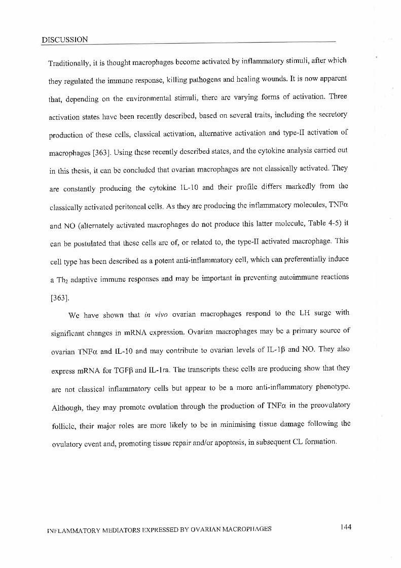
DISCUSSION
Traditionally, it is thought macrophages become activated by inflammatory stimuli, after which
they regulated the immune response, killing pathogens and healing wounds. It is now apparent
that, depending on the environmental stimuli, there are varying forms of activation. Three
activation states have been recently described, based on several traits, including the secretory
production of these cells, classical activation, alternative activation and type-Il activation of
macrophages [363]. Using these recently described states, and the cytokine analysis carried out
in this thesis, it can be concluded that ovarian macrophages are not classically activated' They
are constantly producing the cytokine IL-10 and their profile differs markedly from the
classically activated peritoneal cells. As they are producing the inflammatory molecules, TNFcr
and NO (alternately activated macrophages do not produce this latter molecule, Table 4-5) it
can be postulated that these cells are of, or related to, the type-Il activated macrophage. This
cell type has been described as a potent anti-inflammatory cell, which can preferentially induce
a Th2 adaptive immune responses and may be important in preventing autoimmune reactions
[363].
We have shown that in vivo ovaian macrophages respond to the LH surge with
significant changes in mRNA expression. Ovarian macrophages may be a primary source of
ovarian TNFg and IL-10 and may contribute to ovarian levels of IL-IB and NO. They also
express pRNA for TGFB and Il,-lra. The transcripts these cells are producing show that they
are not classical inflammatory cells but appear to be a more anti-inflammatory phenotype'
Although, they may promote ovulation through the production of TNFcx, in the preowlatory
follicle, their major roles are more likely to be in minimising tissue damage following the
ovulatory event and, promoting tissue repair and/or apoptosis, in subsequent CL formation'
INFLAMMATORY MEDIATORS EXPRESSED BY OVARIAN MACROPHAGES 144
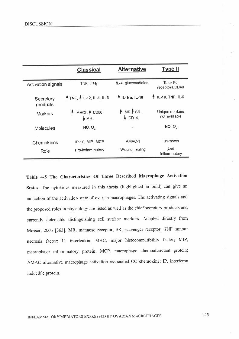
DISCUSSION
Classical Alternative TvPe ll
Activation signals TNF' IFNY lL-4, glucocortioids
Secretoryproducts
Markers
Molecules
Chemokines
Role
I rrup, ltt-tz, tL-1, tL-6
I H¡ncrt,f coao
lvnNO, 02
IP,.1O, MIP, MCP
Pro-inflammatory
I lt-tra, ll-to
I rr¡n,l sn,
I cot+,
AMAC-1
Wound healing
TL or Fcreceptors,CD40
I t-ro, TNF, rL-6
Unique markersnot awilable
NO, 02
unknown
Anti-inflammatory
Table 4-5 The Characteristics Of Three Described Macrophage Activation
States. The cytokines measured in this thesis (highlighted in bold) can give an
indication of the activation state of ovarian macrophages. The activating signals and
the proposed roles in physiology are listed as well as the chief secretory products and
currently detectable distinguishing cell surface markers, Adapted directly from
Mosser, 2003 [363]. MR, mannose receptor; sR, scavenger receptor; TNF tumour
necrosis factor; IL interleukin; MHC, major histocompatibility factor; MIP,
macrophage inflammatory protein; MCP, macrophage chemoattractant protein;
AMAC alternative macrophage activation associated CC chemokine; IP, interferon
inducible protein.
INFLAMMATORY MEDIATORS EXPRESSED BY OVARIAN MACROPHAGES r45

Chapter Five
5 Frxar. DrscussloN
SUMMARY OF FINDINGS
5.1 SurvrvrnnY or FINnrNcs
At the coÍìmencement of these studies it was established that macrophages are present in
ovarian tissues, with an influx of these cells at ovulation and during CL formation and
regression. In addition, much indirect experimental evidence implicated these cells in the
regulation of owlation and CL function. It was hypothesised that ovarian macrophages are
critical to normal ovarian function through the regulated secretion of various inflammatory
mediators. It was speculated that blood monocytes infiltrate the ovary becoming resting
resident cells, following the LH surge, these macrophages become activated producing
classical inflammatory cytokines that promote follicular rupture. Following ovulation and in
the developing CL, macrophages then produce factors important in tissue
reorganisation/wound healing and finally, in the regressing CL they are required for the
clearance of apoPtotic cells'
The work presented in this thesis provides evidence that ovarian macrophages, in
particular the Ia+ population, are critical for normal ovulation and estrus cyclicity. Conditions
for the isolation of maximum numbers of live macrophages from the murine ovary tissue were
determined and these isolated cells shown to be viable, 98% pure and have varying
phagocytic characteristics along with an inability to promote T cell proliferation. These cells
were further shown to produce a unique anay of both pro- and anti-inflammatory mediators
the absence of which may account for the experimental affects observed by clodronate
liposome treatment presented in Chapter 2. Combining the studies presented in this thesis a
preliminary description of the characteristics of ovarian macrophages during the
gonadotrophin primed cycle can for the first time be constructed.
prior to ovulation, 50Yo of isolated ovarian macrophages were shown to be phagocytic
(on day gam). Many of these are probably found in the sffoma of the ovary, with some located
around the growing follicles. These thecal macrophages may be important for optimal
FINAL DISCUSSION t47

SUMMARY OF FINDINGS
stimulation of follicle growth, as depleting these cells appears to reduce the numbers of
follicles reaching the preovulatory size. These ovarian macrophages are producing TNF0, NO
and IL-l0 and although conditioned culture media from these cells appears to stimulate PHA-
induced T cell proliferation, this was not statistically significant.
Following in vivo administration of hCG (on day 0pm), phagocytic activity of the isolated
macrophages remained the same but the expression of mRNA for TNFo, IL-lP and II--lra,
were augmented. It was found that these mRNA levels were not always consistent with the
levels of secreted mature protein these cells produce. TNFo was a signihcant product of these
cells while the Ia+ population produced IL-lp at very low concentrations. TGFBI, IL-10 and
iNOS 6RNA and protein concentrations were detectable but remained static' In the theca of
preovulatory follicles these ovarian macrophages, in particular those expressing the Ia
antigen, may be key regulators of normal follicular rupture'
Following the owlatory event, the percentage of phagocytic Ia* and F4l80+ ovarian
macrophages increased to 80% (on day 1). Many of these are probably found in the eatly CL,
and although these cells have less stimulatory influence on PHA stimulated T cells this effect
was again not statistically significant. These cells appear to be important in progression
through the postovulatory stages of the normal estrous cycle to the next ovulatory event. The
mRNA levels for those cytokines that had responded to hCG administration fell to levels not
significantly different to those prior to hCG administration. It appeared that IL-10 mRNA
levels may be less but this was not significant. At this time point, cells were found to produce
1¡-10 and TNFg proteins at slightly but not significantly higher concentrations, than those
prior to follicular rupture. NO concentrtions did not change, but Ia+ cells began to produce
detectable amounts of IL-1B'
It can be concluded from these results along with comparisons of the cytokine profile of
the isolated ovarian macrophages with the peritoneal macrophages, that the former are not
FINAL DISCUSSION 148
SUMMARY OF FINDINGS
classical inflammatory cells as anticipated, but display a cytokine profile more comparable to
that of anti-inflammatory macrophages, as described by Mosser [363]. Based on these results,
in particular the IL-10, the role of macrophages within the ovary is more complicated than
hypothesised and it is necessary to produce a new model in which the primary role of these
cells may be an anti-inflammatory one'
5.2 EVNNUCN OF MACROPHAGE HNTBROCENEITY IN THE MUNT¡¡N OV¿'NY
It was suggested in Chapter 2 that the ovarian macrophage population may be
heterogeneic, since macrosialin* (FA/l1*) and Ia* cells were significantly depleted by CLi
treatment but F4lg0+ cells, although reduced in numbers, were not significantly depleted' This
suggested a difference in the phagocytic activity of the different cell types, however such a
difference between these isolated cell types was not observed in the analysis of phagocytosis
employed in Chapter 3. In addition in Chapter 4, the cytokine analysis carried out did not
reveal any significant differences in the expression any of the cytokine mRNA by the Ia* and
F4l80+ cells types at any time point examined. These results combined suggest that the Ia+
and F4lg0+ cells are not significantly different to each other. Indeed, the only significant
difference observed was the very low levels of IL-18 detected following hCG administration
in conditioned media from the Ia* cells only. It does however seem important to note that in
Chapter 2 it was a significant reduction in this cell type that resulted in significantly reduced
ovulation rates. It may be that the small amount of mature IL-lB being secreted by these cells
is of critical importance for this event. This suggests the local ovarian factors, such as steroids
lllg, lZ0, I23) or cytokines 13641, dictate the activity and phagocytic capacity of the
FINAL DISCUSSION t49
MACRO PHAGE HETEROGENEITY IN THE OVARY
recruited macrophages as a whole. Even though no differences were observed between
the isolated Ia* and F4l80* cell types, whether differences in the characteristics of cells
located in the different ovarian compartments exist cannot be concluded from these studies,
although the differences in the depletion of ovarian macrophages populations in Chaptet 2
sugest they may exist. It could be proposed that the markers used in this thesis to isolate
ovarian macrophages are widespread in the ovarian macrophage populations and do not
distinguish between the heterogeneous populations. This would mean that the populations
examined in this thesis are of mixed characteristics. In light of this, the non-phagocytic
macrophage population isolated at each time point, may be mature resident macrophage cell
types located in the ovarian stroma, which was not significantly affected by CLi injection in
Chapter 2, while the phagocytic cells could be located chiefly in the theca of the follicles and
CL where significant reductions in macrophage numbers were seen following CLi treatment.
In support of this, the increase in phagocytic cells following ovulation could be explained by
the increasing numbers of new active macrophages recruited into the CL.
It follows that if thecal and luteal macrophage populations could be studied
independently, changes in cytokine production across the reproductive cycle may be even
more striking, although this would be very time consuming and required the sacrifice of many
laboratory mice. It would be more practical to simply examine the distribution of additional
distinguishing macrophage activation markers and cytokines in ovarian tissue sections.
5.3 Iupr,rCnuONS FoR Ov.lnr.lN M¡,CROpHAGE PHENOTYPE AND FUNCTION
The protein profile of ovarian macrophages seen in the hnal chapter of this thesis
suggests that ovarian macrophages are not a classically stimulated inflammatory cells but are
more likely to be an alternatively activated cell type and the foremost role of these cells in the
vary may be in minimising the inflammatory ovulatory event and regulating CL formation.
FINAL DISCUSSION 150

MACROPHA GE HETEROGENEITY IN THE OVARY
The later is supported by animals models in which regulators of macrophage functions
have been disrupted resulting in extended estrous cycles [68,331], as well as the work
presented in Paper I, showing extended cycles potentially due to lack of activated
macrophages to stimulate CL regression, while an important role for ovarian macrophages in
the regulation of follicular growth and ovulation is also inferred. It is clear that the phagocytic
Ia* macrophages located in the theca of the follicle, which were significantly depleted by CLi
treatment in Chapter 2, may play an important role in the stimulating either follicular growth
and/or the ovulatory event. The results in Chapter 4 (showing Ia* macrophages produce
significant amounts of TNFcr and NO prior to and following hCG administration, as well as
minimal amounts of IL-lB following hCG administration), suggest this could occur through
the production of either TNFcr,, IL-18 and/or possibly NO, which have all been shown
elsewhere to stimulate follicular growth 1220,318] or follicular rupture 1191,213,261]. The
examination of ovarian morphology carried out in Chapter 2, although not conclusive, does
suggest that this reduced ovulation rate is due to an inhibition of follicular rupture' fuither
suggesting that thecal macrophage production of TNFcr andlor IL-1Ê is critical in stimulating
follicular rupture.
It is of particular interest that we have found the alternative activation state is present
prior to the LH surge, suggesting the ovarian environment induces this state when
macrophages initially infiltrate the ovarian tissue. There are diverse reported regulators of
infiltrating macrophage function including the cytokines present in the tissue lI27l, the
adhesion molecules drawing the cells into the tissue t365] and the structure of the
extracellular matrix within the tissue 1366,36710r even the presence or absence of apoptotic
cells[36g]. It is primarily the production of the cytokine IL-10 and the minimal production of
mature IL-IB by the isolated ovarian macrophages that compels this view of ovarian
macrophage activation. IL-10 is an autoregulatory cytokine with numerous anti-inflammatory
FINAL DISCUSSION151

MACROPHAG E HETEROGENEITY IN THE OVARY
roles and interestingly, it is thought to be an important mediator in maintaining peripheral
tolerance t3691. It prevents the activation and migration of antigen presenting cells' inhibits T
cell activation and cytokine production and is considered a promoter of local resolution of
inflammation [370, 371]. More specihcally it can inhibit the expression of co-stimulatory
molecules CDs0/CDg6 on the surface of macrophages leading to immune tolerance through
the inactivation or skewing of T cells l37ll.In the ovary this cytokine may act to prevent the
migration and maturation of macrophages, and destructive cytotoxic T cells, following the
inflammatory ovulatory event. It follows that ovarian macrophages may be present to prevent
,immune surveillance' of the ovulated follicle. CL regression involving immune cells has
been proposed to occul via one two mechanisms, immune mediated regression, or
programmed cell death 12331. Both of these processes would require significant regulation by
ovarian macrophages by divergent mechanisms. Firstly, for immune mediated CL regression
macrophages would need to become classically activated expressing co-stimulatory molecules
and subsequently promoting cytotoxic T cell activation leading to cell mediated regression of
the CL. In support of this the production of the potent macrophage activator,IFNy also occurs
at CL regression 1249,372]. Secondly, for CL regression via programmed cell death, the anti-
inflammatory ovarian macrophages may promote, local resolution of the tissue damage
following ovulation, and the generation of regulatory T cells, restricting any inflammatory
reaction and promoting apoptosis. Only further research into the cytokines and co-stimulatory
molecules expressed by ovarian macrophages during the formation and regression of the CL,
including during pseudopregnancy, will assist in determining which of these roles ovarian
macrophages may have in CL function.
FINAL DISCUSSIONt52

SUMMARY
5.4 Suvrm¡.nY
Macrophages are critical to normal ovarian function. It is suggested that the local ovarian
environment influences resident and infiltrating macrophages to develop into cells of the type
II activated or antlinflammatory state. These macrophages, present in the ovary prior to the
LH surge and ovulation, maintain an immunosuppressive environment through the production
of IL-10, preventing macrophage maturation, migration and interactions that generate
destructive T cells. These cells may produce NO and TNFcr, to promote, follicle survival and
growth (Figure 5.1). Following the LH surge, the cells infiltrating the theca of the follicle' in
particular the Ia+ population, are critical to follicular rupture probably through the production
of TNFcr and/or IL-10 which stimulate follicular rupture. In the early CL although these cells
are more phagocytically active, they continue to produce IL-10 and TNFc¿' It is further
speculated that macrophages may have one of two roles in CL regression - maturation and
then stimulation of T cells cytotoxic towards the luteal cells, or maintenance of immune
restriction and stimulation of programmed luteal cell apoptosis.
On the basis of work presented in this thesis it may be proposed that the major role of
macrophages in the ovary is in the minimisation of the inflammatory-like reactions stimulated
at ovulation, with contributions to follicular rupture through the production of TNFcr and IL-
18, and potentially in subsequent survival and demise, of the cL.
FINAL DISCUSSION153
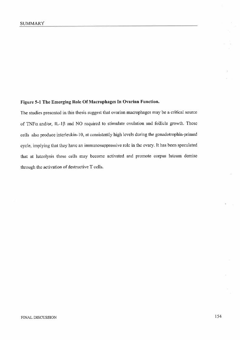
SUMMARY
Figure 5-1 The Emerging Role Of Macrophages In Ovarian Function.
The studies presented in this thesis suggest that ovarian macrophages may be a critical source
of TNFcr and/or,Il-lp and NO required to stimulate owlation and follicle growth. These
cells also produce interleukin-l0, at consistently high levels during the gonadotrophin-primed
cycle, implying that they have an immunosuppressive role in the ovary. It has been speculated
that at luteolysis these cells may become activated and promote corpus luteum demise
through the activation of destructive T cells.
!
FINAL DISCUSSION t54
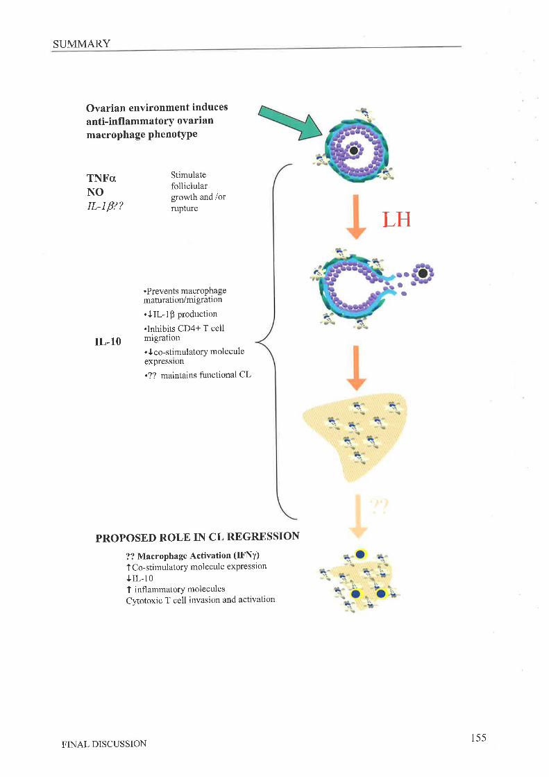
SUMMARY
Ovarian environment inducesanti-infl ammatory ovarianmacrophage phenotYPe
TNFcrNOrL-Lfr?
Stimulatefolliclulargrowth and /orrupture
LH
O
IL,1O
.Prevents macrophagematuration/migration
.tIL-1Ê production
.Inhibits CD4+ T cellmigration. t co-stimulatory moleculeexpresslon
.?? maintains functional CL
PROPOSED ROLE IN CL REGRESSION
?? MacroPhage Activation (IFNY)
I Co-stimulatory molecule expression
tIL-101 inflammatorY moleculesCytotoxic T cell invasion and activation
oa
FINAL DISCUSSION155
FURTHER STUDIES
5.5 FuRrunR Srunrps
The results of the studies presented in this thesis raise interesting questions regarding the
phenotypes of the macrophages located in the murine ovary. Examining these questions will
further clarify the roles these cells have in ovarian function'
Are The Macrophages In The ovarian compartments Heterogeneic?
o Studying the dual expression of the Ia* and F4l80+ and other distinctive macrophage
markers in digested ovarian tissues using flow cytometry, will determine the
proportions of the populations that overlap and thus more clearly establish the
presence or absence of heterogenic populations'
o Histological examination of macrophage activation markers, co-stimulatory molecules
and cytokine mRNAs in ovarian tissue sections will further elucidate the activation
status and heterogeneity of the localised ovarian macrophage populations across the
reproductive cycle, including during pseudo-pregnancy wele macrophages will
probably play aprimary role in CL survival and regression,'
o Isolation of macrophages from ovarian dissected follicles and corpora lutea of
different stages of development would allow specific determination of the
characteristics of these cell types but may be impractical.
Do Ovarian Macrophages Have Irregular Immune Functions?
o Further examination of the phagocytic activity of these cells before and after ovulation
as this may be a critical role of these cells in the ovarian tissue. It would be of
particular interest to analyse this activity in response to apoptotic cells, since these are
relatively common in some follicles and during CL regression.
o Examination of the ability of cells isolated before and after ovulation to directly
stimulate T cell proliferation including analysis of the cytokines and T cell phenotype
FINAL DISCUSSION156

FURTHER STUDIES
stimulated would enable further conclusions on this aspect of macrophage biology and
its role in ovarian biologY.
o How do these cells response to traditional stimulators of classical inflammatory
responses. Macrophages in others tissue have been shown to exhibit altered responses
to factors such as LPS. It would be of interest to determine if ovarian macrophages
have a response similar to that seen in any other macrophage cell type'
What Is It That Induces The Ovarian Macrophage Phenotype And Do Alterations Of This
Phenotype Lead To Ovarian Dysfunction ?
o Since obtaining ovarian macrophages is a difficult and 'animal expensive' process it
would be useful if this phenotype could be generated by other means, such as
culturing of peritoneal macrophages with factors from the ovarian environment or
even generation of an ovarian macrophage cell line. The effects of various factors on
ovarian macrophages and possibly ovarian function could then be studied'
Are Other Macrophage Derived Factors Involved In ovulation?
o Finally to confirm the role of macrophages in stimulation of the owlatory
event via analysis of mRNA from macrophages at several time points more immediate
to the ovulatory event using micro-array techniques. This will help determine if other
genes, not considered in this study, are activated in these cells following the LH surge
and therefore of interest for further studies. Results obtained must be interpreted
carefully since it was found in thesis that ovarian macrophage mRNA levels do not
always reflect active protein levels'
FINAL DISCUSSIONr57
IMPLICATIONS
5.6 lvtpr-rc¡,rloNs
A greater understanding of the activation status and secretory profile of the ovarian
macrophage will lead to better understanding of the mechanisms effecting normal ovarian
function. The specialised secretion by ovarian macrophages of factors that stimulate specific
functions at precise stages of the reproductive cycle could be a key component of reproductive
function. Alterations or defects in ovarian macrophage phenotype or secretory profile may be
found to have consequences that lead to ovarian dysfunction. Interactions between the
immune, reproductive and metabolic systems are becoming increasingly evident with
perturbations in one significantly influencing normal functions of the others' Clinically an
understanding of ovarian macrophage biology may assist in the understanding and/or treatment
of ovarian disorders such as the polycystic ovary syndrome (PCOS), ovarian autoimmune
disease or cases of unexplained anovulation. Further, these cells may also become a potential
target for manipulation in contraceptive development'
FINAL DISCUSSION158
REFERENCES
2.
4.
5.
6.
RnTNNENCES
Norman RJ, Bonello N, Jasper MJ, Van der Hoek KH 1997 Leukocytes:
essential cells in ovarian function and ovulation' Reprod Med Rev 6:97-lllBukulmez o, AricÍ A 2000 Leukocytes in ovarian function. Hum Reprod update
6:1-15Norman RJ, Clark AM 1998 Obesity and reproductive disorders: a review,
Reprod Fertil Dev 10:55-63Pasquali R, Pelusi C, Genghini S, Cacciari M, Gambineri A 2003 Obesity and
reprôductive disorders in women. Hum Reprod Update 9:359-72pôdersen T 1970 Determination of follicle growth rate in the ovary of the
immature mouse. J Reprod Fertil 21:81-93'
Freeman y1E, The Neuroendocrine Control of the Ovarian Cycle of the Rat, in The
Physiology of Reproduction,E Knobil and JD Neill, Editors. 1994, Raven Press:
N.Y.p. 613-658.pedeisen T, peters H 1968 Proposal for a classification of oocytes and follicles tn
the mouse ovary. J Reprod Fertil 17:555-7.
Osman p 1985 Rate and course of atresia during follicular development in the
adult cyclic rat. J Reprod Fertil 73:26I-70.Gaytañ F, Bellido ô, Morales C, Aguil tr E, Sanchez-Criado JF' 1997 Follicular
g.Ñtn puit"* in cyclic rats from late pro-oestrus to early oestrus' J Reprod Fertil
1 10:1 53-9.Gougeon A 1986 Dynamics of follicular growth in the human: a model from
preliminary results, Hum Reprod 1 :8 1-7.
iohnson \i, Everitt B 1988 Essential Reproduction. Third ed, London: Blackwell
S cientif,rc Publications.Greenwald GS, Roy SK, Follicular Development and its Control, in The
Physiologt of Reproduction, E Knobil and JD Neill, Editors. 1994, Raven Press,
NY.p. 629-724.Nilsson E, Skinner MK 2001 Cellular interactions that control primordial follicle
development and folliculogenesis. J Soc Gynecol Investig 8:S17-20'
Kezele pR, Nilsson EE, Skinner MK 2002 Insulin but not insulin-like growth
factor-1 promotes the primordial to primary follicle transition. Mol Cell Endocrinol
192:37-43vendola K, zhou J, Wang J, Famuyiwa oA, Bievre M, Bondy cA 1999
Androgens promote oocyte insulin-like growth factor I expression and initiation offollicle development in the primate ovary. Biol Reprod 6l:353-7
Nilsson EE, Skinner MK 2004 Kit ligand and basic fibroblast growth factor
interactions in the induction of ovarian primordial to primary follicle transition.
Mol Cell Endocrinol 214:19-25Braw-Tal R 2002 The initiation of follicle growth: the oocyte or the somatic cells?
Mol Cell Endocrinol 187:11-8
Sherwood L Ig93 Human Physiology : from cells to systems: West Publishing Co,
MN, USA.Halpin DM, Jones A, Fink G, Charlton HM 1986 Postnatal ovarian follicle
devålopment in hypogonadal (hpg) and normal mice and associated changes in the
hypothalamic-pituitary ovarian axis. J Reprod F efül 7 7 :287 -96'
7
8
9
10.
11
t2
13
l4
15.
t6
I7
18.
t9
REFERENCES 159
REFERENCES
20. Halpin DM, Charlton HM, Faddy MJ 1986 Effects of gonadotrophin deficiency
on follicular development in hypogonadal (hpg) mice. J Reprod Fertil T8:ll9-25'
21. wang XN, Greenwald GS 1993 Hypophysectomy of the cyclic mouse. I. Effects
on folliculogenesis, oocyte growth, and follicle-stimulating hormone and human
chorionic gonadotropin receptors' Biol Reprod 48:585-94'
22. Hsueh AJ, Billig H, Tsalriri ^
Ig94 Ovarian follicle atresia: a hormonally
controlled apoptotic process. Endocr Ptev I5:707-24'
23. Tilly JL 19Ò6 Apoptosis and ovarian function. Rev Reprod l:162-72
24. Kaþia A, Hsueñ Ãl 9gl Regulation of ovarian follicle atresia. Annu Rev Physiol
59349-6325. Richards JS, Jahnsen T, Hedin L, Lifka J, Ratoosh S, Durica JM, Goldring
NB 1987 Ovarian follicular development: from physiology to molecular biology.
Recent Prog Horm Res 43:231-76
26. Lipner H, Mechanisms of mammalian ovulation., in The Physiology ofRåproduction.,EKnobil andJ Neill, Editors. 1988, Raven Press Ltd: New York. p'
447-488.27. Brannstrom M, Janson PO, The Biochemistry of Ovulation, in Ovarian
Endo crinolo gy,'SG Hillier, Editor. 1 99 1, Blackwellscientific: Oxford' p' 132-166.
28. Espey LL, Lipner H, Ovulation, in The Physiologt of Reproduction,E Knobil and
JDNeill, Editors. 1994, Raven Press: N'Y'p' 725-780'
29. Espey LL 1980 Ovulation as an inflammatory reaction - a hypothesis. Biol Reprod
22:73-10630. Murphy BD 2000 Models of luteinization. Biol Reprod 63:2-lI.31. Niswender GD, Juengel JL, Silva PJ' Rollyson MK, Mclntush EW 2000
Mechanisms controllinf tne function and life span of the corpus luteum. Physiol
Rev 80:1-29.32. Couzinet B, Schaison G 1993 The control of gonadotrophin secretion by ovarian
steroids. Hum Reprod 8 Supp 2:97-l0l33. Bronson FH, Dagg CP, Snell GD, Reproduction, in Biologt of the Laboratory
Mouse,EL GreenlEditor. 1966, McGraw-Hill Book co': New York' p' 187-204'
34. Michael SD 1976 Plasma prolactin and progesterone during the estrous cycle in the
mouse. Proc Soc Exp Biol ili4ed I53:254-l '
35. Gallo RV l98l Pulsatile LH release during periods of low level LH secretion in the
rat estrous cycle. Biol Reprod24:77I-736. Gallo RV iô81 Pulsatile LH release during the ovulatory LH surge on proestrus in
the rat. Biol ReProd24:100-437. Fox SR, Smith MS 1985 Changes in the pulsatile pattern of luteinizing hormone
secretion during the rat estrous cycle. Endocrinology 116:1485-92
3g. Gore-Langton i.8,, Armstrong DT, Follicular Steroidogenesis and its Control, in
The physlotogy of Reproduction, E Knobil and JD Neill, Editors' 1994, Raven
Press: N.Y. P. 57I-627.39. Simpson Eh., Mahendroo MS, Means GD, Kilgore MW, Hinshelwood MM'
Graham-Lorence s, Amarneh B, Ito Y, Fisher cR, Michael MD, et al. 1994
Aromatase cytochrome P450, the enzyme responsible for estrogen biosynthesis'
Endocr Rev 15:342-5540. Gates AH, Bozarth JL 1978 Ovulation in the PMSG-treated immature mouse:
effect of dose, age, weight, puberty, season and strain (BALB/o, 129 and Cl2gFlhybrid). Biol ReProd 18:497-505
41. Braw RH, Tsafriri A 1980 Effect of PMSG on follicular atresia in the immature
rat ovary. J ReProd Fefül59:267-72
REFERENCES 160
REFERENCES
42. Ma S, Kalousek DK, Yuen BH, Moon YS 1997 Investigation of effects ofpregnant mare sentm gonadotropin (PMSG) on the chromosomal complement of
CD-l mouse embryos' J Assist Reprod Genet 14:162-9
43. Aguado LI, Ojedâ SR 1985 Physiological peripubertal activation of the ovary is
no*t reproduced by pregnant mare serum gonadotropin (PMSG) administration. Proc
Soc Exp Biol Med 178:l2l-544. Abbas AK, Lichtman AH, Pober JS 1991 Cellular and Molecular Immunology'
Philadelphia: W B Saunders Co., pg. 24'
45. Matsuyama s, ohta M, Takahashi M 1987 The critical period in which
splenectomy causes functional disorder of the ovary in adult rats' Endocrinol Jpn
34:849-55.46. Nishizuka Y, Sakaku ra T 1969 Thymus and reproduction: sex-linked dysgenesia
of the gonad after neonatal tþmectomy in mice. Science 166:753-5.
47. Bukoviky A, presl J, Krabec Z, BednarikT 1977 Ovarian function in adult rats
treated with antithymocyte serum. Experientia 33:280-1'
48. Hellberg P, Thomsen P, Janson PO, Brannstrom M I99l Leukocyte
supplemèntation increases the luteinizing hormone-induced ovulation rate in the in
vitro-perfus ed rat ovary. Biol Repro d 44:7 9I-749. Brannstrom M, Pascoe V, Ñorman RJ, McClure N 1994 Localization of
leukocyte subseis in the follicle wall and in the co{pus luteum throughout the
human menstrual cycle' Fertil Steril 61:488-95
50. suzuki T, sasano H, Takaya R, Fukaya T, Yajima A, I)ate F, Nagura H 1998
Leukocytes in normal,cycling human ovaries: immunohistochemical distribution
and, char acterization' Hum Reprod I 3 : 2 1 8 6-9 I'51. Wang LJ, pascoe V, Petrucco OM, Norman RJ 1992 Distribution of leukocyte
subpopulations in the human corpus luteum. Hum ReprodT:197-202
52. Withanage GS, Baba E, Sasai K, Fukata T, Kuwamura M, Miyamoto T,
Arakawa ^
IggT Localization and enumeration of T and B lymphocytes in the
reproductive tract of laying hens. Poult Sci76:671-6'
53. Brannstrom M, Mayrhoffer G, Robertson SA 1993 Localization of leukocyte
subsets in the rat ovary during the periovulatory period. Biol Repro d 48:277 -86
54. Best CL, Pudney ¡, Wetctr WR, Burger N, Hitl JA 1996 Localization and
characterization oiwhite blood cell populations within the human ovary throughout
the menstrual cycle and menopause. Hum Reprod Il:790-1'55. Lintern-Moo.é S 1977 Effect of atþmia on the initiation of follicular growth in
the rat ovary. Biol Reprod 17:155-61.
56. Schaffer l\i, Barbul A 1998 Lymphocyte function in wound healing and following
injury. Br J Surg 85:444-6057. Duncan WC, Rodger FE, Illingworth PJ 1998 The human corpus luteum:
reduction in macrophages during simulated maternal recognition of pregnancy.
Hum ReProd 13:2435-42.
5g, Gerdes U, Gafvels M, Bergh A, Cajander S 1992 Localized increases in ovarian
vascular permeability and leucocyte accumulation after induced ovulation in
rabbits. J ReProd Fertil95:539-5059. Standaert iE, Zu ora CS, Chew BP 1991 Quantitative and qualitative changes
in blood leukocytes in the porcine ovary. Am J Reprod Immunol25:163-8'
60. Brannstrom iM, Bonello N, No.tnan RJ, Robertson SA 1995 Reduction ofovulation rate in the rat by administration of a neutrophil-depleting monoclonal
antibody. J Reprod Immunol 29:265-70
REFERENCES 161
REFERENCES
61.
62.
63.
64.
65.
66.
67.
68.
69.
70.
71.
1alL.
73.
74.
75.
Bonello N, McKie K, Jasper M, Andrew L, Ross N' Braybon E, Brannstrom
M, Norman RJ 1996 Inhibition of Nitric oxide: Effects on interleukin-lB-
enûanced ovulation rate, steroidogenesis and ovarian leukocytes distribution at
ovulation in the rat. Biol Reprod 54:436-45
Bulmer D 1964 The Histochemistry of Ovarian Macrophages in the Rat. J Anat
Lond 98:313-319Cohen PE, Zhu L, Pollard J\ry 1997 Absence of colony stimulating factor-l in
osteopetrotic (csfmop/csfmop) mice disrupts estrous cycles and ovulation. Biol
Reprod 56:110-8Peirovska M, Dimitrov DG, Michael SD 1996 Quantitative changes
'nmacrophage distribution in normal mouse ovary over the course of the estrous cycle
examined with an image analysis system. Am J Reprod Immunol 36:175-83
Barua A, Yoshimu.ã Y, Tamura T 1998 Localization of macrophages in the
ovarian follicles during the follicular growth and postovulatory regression in
chicken, Gallus domesticus. Poult Sc\ 77 :1417 -21.
Brannstrom M, Giesecke L, Moore IC, Van den Heuvel cJ, RObertson sA
1994 Leukocyte subpopulations in the rat corpus luteum during pregnancy and
pseudopregnancy. Biol Reprod 50:1 16l-7katabuctri H, Suenaga Y, Fut<umatsu Y, Okamura H 1997 Distribution and fine
structure of macrophages in the human ovary during the menstrual cycle, pregnancy
and menopause. Endocr J 44:785-95.Jasper NiJ, Rob.rtson SA, Van der Hoek KH, Bonello N, Brannstrom M,
Noiman RJ 2000 Characterization of ovarian function in granulocyte-macrophage
colony- stimulating factor-deficient mice. Biol Reprod 62:704-t3
Hehnîe KE, Christenson LK, Ford SP, Taylor M 1994 Macrophage infiltration
into the porcine corpus luteum during prostaglandin F2 alpha-induced luteolysis.
Biol Reprod 50:10-5Valledor AF, Borras FE, Cullellyoung M, Celada A 1998 Transcription Factors
That Regulate Monocyte/Macrophage Differentiation. J Leuko Biol 63:405-417
Naito M 1993 Macrophage heterogeneity in development and differentiation' Arch
Histol Cytol56:331-51Ebnet i<, Vestweber D 1999 Molecular mechanisms that control leukocyte
extravasation: the selectins and the chemokines. Histochem Cell Biol II2:l-23Berton G, Lowell CA 1999 Integrin signalling in neutrophils and macrophages'
Cell Signal ll:621-35Webei C Z0O3 Novel mechanistic concepts for the control of leukocyte
transmigration: specialization of integrins, chemokines, and junctional molecules. J
Mol Med 81:4-19Roitt I, Brostoff J, Male D 1993 Immunology' Third Edition ed, ed' L Gamlin:
Mosby-Year Book Europe Ltd, Lond, UK.,pg9'7 '
Nathan CF 1987 Secretãry products of macrophages. J Clin Invest 79:319-26'
Rappolee DA, Mark D, Banda MJ, Werb z 1988 Wound macrophages express
fGï-alpha and other growth factors in vivo: analysis by mRNA phenotyping'
Science 241:708-12'sunderkotter c, Goebeler M, Schulze-osthoff K, Bhardwaj R, sorg c 1991
Macrophage-derived angiogenesis factors. Pharmacol Ther 5 1 : 19 5 -21 6
SunAeitoiter C, Steinbrink K, Goebeler M, Bhardwaj R, Sorg C 1994
Macrophages and angiogenesis' J Leukoc Biol55:410-22'Gale LM-, IvrcColi SR lggg Chemokines: extracellular messengers for all
occasions? BioessaYs 2l :17 -28
7677
78
79
80
REFERENCES t62
REFERENCES
g1. Greaves DR, Schall TJ 2000 Chemokines and myeloid cell recruitment. Microbes
Infect 2:331-682. Middleton J, Patterson AM, Gardner L, Schmutz c, Ashton BA 2002
Leukocyte extravasation: chemokine transport and presentation by the endothelium.
Blood 100:3853-6083. Varki
^ l9g4 Selectin ligands. Proc Natl Acad Sci U S A 9l:7390-7
84. Rhee JS, Santoso S, Herrmann M, Bierhaus A, Kanse SM, May AE, Nawrothpp, Colman RW, Preissner KT, Chavakis T 2003 New aspects of integrin-
mediated leukocyte adhesion in inflammation: regulation by haemostatic factors
and bacterial products' Curr Mol li4.ed3:387-92g5. Mutsaers SÉ, Bistrop JE, McGrouther G, Laurent GJ 1997 Mechanisms of
tissue repair: from wound healing to fibrosis. Int J Biochem Cell Biol29:5-17.g6. Greenhalgh DG 1998 The role of apoptosis in wound healing. Int J Biochem Cell
Biol30:1019-3087. Johnston RB, Jr. 1988 Current concepts: immunology. Monocytes and
macrophages. N Engl J Med 318:747-52.gg. Stein M, Keshav S, Harris N, Gordon S lgg2Interleukin 4 potently enhances
murine macrophage mannose receptor activity: a marker of alternative
immunologic macrophage activation' J Exp }l4ed 176:287-92
89. Hart PH, Bonder ÒS, nalogh J, Dickensheets HL, Donnelly RP, Finlay-Jones
JJ lggg Differential responses of human monocytes and macrophages to IL-4 and
IL-13. J Leukoc Biol 66:575-8
90. Doherty TM 1995 T-Cell Regulation of Macrophage Function. Cun Opin
Immunol 7:400-404gl. Ma J, Chen T, Mandelin J, ceponis A, Mitler NE, Hukkanen M, Ma GF'
Konttinen YT 2003 Regulation of macrophage activation, Cell Mol Life Sci
60:2334-4692. Goerdt S, Politz O, Schledzewski K, Birk R, Gratchev A, Guillot P, Hakiy N,
Klemke CD, Dippel E, Kodelja V, Orfanos CE 1999 Alternative versus classical
activation of macrophages. Pathobiology 67 :222-6
93. Hart PH, Vitti GF, Bu.g.ss DR, WhittY GA, Piccoli DS, Hamilton JA 1989
potential antiinflammatory effects of interleukin 4: suppression of human monocyte
tumor necrosis factor alpha, interleukin 1, and prostaglandinE2. Proc Natl Acad
SciUSA86:3803-794. Goerdt S, Orfanos CF., 1 9gg Other functions, other genes: alternative activation of
antigen-presenting cells. Immunity 1 0 : 137 -42
95. Schebesch C, rõoet¡a V, Muller C, Hakij N, Bisson S, Orfanos CE, Goerdt S
lggT Alternatively activated macrophages actively inhibit proliferation ofperipheral blood lymphocytes and CD4+ T cells in vitro. Immunology 92:478-86
96. kodetja V, Muller C, Tenorio S, Schebesch C, orfanos CE, Goerdt S 1997
Differences in angiogenic potential of classically vs alternatively activated
macrophages. Immunobiology 197 :47 8-93.
97. Andeison CF, Mosser DM 2OO2 A novel phenotype for an activated macrophage:
the type 2 activatedmacrophage. J Leukoc Biol72:101-69g. Reiner SL, Seder RA 1995 T helper cell differentiation in immune response. Curr
Opin Immunol T:360-6.99. Cõnstant SL, Bottomly K 1997 Induction of Thl and Th2 CD4+ T cell responses:
the alternative approaches. Annu Rev Immunol 15:297-322
REFERENCES r63
REFERENCES
100, Raes G, De Baetselier P, Noel W, Beschin A, Brombacher F, Hassanzadeh Gh
G 2002 Differential expression of FIZZT and Yml in alternatively versus
classically activated macrophages. J Leukoc Biol 7l:597 -602
101. Lewis JS, Lee JA, Underwood JC, Harris AL, Lewis CE 1999 Macrophage
responses to hypoxia: relevance to disease mechanisms. J Leukoc Biol66:889-900
I02. O'kane S, Färguson MW 1997 Transforming growth factor beta s and wound
healing. Int J Biochem Cell B\o129:63-78
103. Allenl4, Ad.re- , 1996 Mechanisms of phagocytosis. Curr Opin Immunol
8:36-40.104. Swanson JA, Baer SC 1995 Phagocytosis by zippers and triggers' Trends Cell
Biol 5:85-87105. Thiele L, Rothen-Rutishauser B, Jilek S, Wunderli-Allenspach H, Merkle HP'
Walter E 2001 Evaluation of particle uptake in human blood monocyte-derived
cells in vitro. Does phagocytosis activify of dendritic cells measure up with
macrophages? J Control Release 76:59-71
106. Fadok VA, Bratton DL, Konowal A, Freed PW, Westcott JY, Henson PM 1998
Macrophages that have ingested apoptotic cells in vitro inhibit proinflammatory
cytokine pioduction through autocrine/paracrine mechanisms involving TGF-beta,
PGE2, and PAF. J Clin Invest 101:890-8
107. Devitt A, Moffatt OD, Raykundalia C, Capra JD, Simmons DL, Gregory CI)
1998 Human CD14 mediates recognition and phagocytosis of apoptotic cells fsee
comments]. Nature 392:505-910g. platt N, da Silva RP, Gordon S 1998 Recognizing death: the phagocytosis of
apoptotic cells. Trends Cell BiolS:365-72109. Barker RN, Erwig L, Pearce WP, Devine A, Rees AJ 1999 Differential effects of
necrotic or apopiotic cell uptake on antigen presentation by macrophages'
PathobiologY 67:302-5110. Gordon s, Lawson L, Rabinowitz s, crocker P , Morris L, Perry VH 1992
Antigen markers of macrophage differentiation in murine tissues' Curr Top
Microbiol Immunol 1 81 : 1-37
111. Martinez-Pomares L, Platt N, McKnight AJ, da Silva RP, Gordon S 1996
Macrophage membrane molecules: markers of tissue differentiation and
heterogeneity. Immunobiology 195 :407 -16.
ll2. McKnight ÁJ, Gordon S 1998 The Egf-Tm7 Family - Unusual Structures At the
Leukocyte Surface' Jof Leuko Biol63:271-280113. AustynJM, Gordon S 1981 F4l80, a monoclonal antibody directed specifically
against the mouse macrophage. Eur J Immunol 1 1:805-i5
lI4. McKnight AJ, Macfariane AJ, Dri P, Turley L, Willis AC, Gordon S 1996
Molecular cloning of F4l80, a murine macrophage-restricted cell surface
glycoprotein with homology to the G-protein-link¡d transmembrune 7 hormone
receptor family. J Biol Chem 271:486-9'
115. Leenen PJ, ãe Bruijn MF, Voerman JS, Campbell PA, van Ewijk W 1994
Markers of mouse mácrophage development detected by monoclonal antibodies' J
Immunol Methods 17 4:5-19116. McKnight AJ, Gordon S 1998 Membrane molecules as differentiation antigens of
murine macrophages. Adv Immunol 68:271-314
II7. Sugino N, Shimamura K, Tamura H, Ono M, Nakamura Y, Ogino K, KatO H
1996 Progesterone inhibits superoxide radical production by mononuclear
phagocytes in pseudopregnant rats. Endocrinology 137 :7 49 -54
REFERENCES r64
REFERENCES
1 18.
1 19.
t20.
I2T.
r22.
t23.
r24.
t25.
t26.
r27.
t28.
t29.
130.
131,
132.
133.
chao TC, Van Alten PJ, Walter RJ 1994 Steroid sex hormones and macrophage
function: modulation of reactive oxygen intermediates and nitrite release. Am J
Reprod Immunol 32:43 -52
Gomez F, Ruiz P, Briceno F, Lopez R, Michan A 1998 Treatment with
progesterone analogues decreases macrophage Fcgamma receptors expression' Clin
Immunol ImmunoPathol 89 :23 1 -9'
Chao TC, Phuangsab A, Van Alten PJ, Walter RJ 1996 Steroid sex hormones
and macrophage frrnction: regulation of chemiluminescence and phagocytosis' Am
J Reprod Immunol 35:106-1 13
stefano GB, Prevot v, Beauvillain JC, Fimiani c, welters I, Cadet P, Breton
C, Pestel J, Salzet M, Bilfinger TV 1999 Estradiol coupling to human monocyte
nitric oxide release is dependent on intracellular calcium transients: evidence for an
estrogen surface receptor. J Immunol 163:3758-63'yamãda K, Hayashi T, Kuzuya M, Naito M, Asai K, Iguchi A 1996
physiological concentration of 17 beta-estradiol inhibits chemotaxis of human
mónocytes in response to monocyte chemotactic protein I ' Artery 22:24-35
satem ML, Matsuzaki G, Madkour GA, Nomoto K 1999 Beta-estradiol-induced
decrease in IL-I2 and TNF-alpha expression suppresses macrophage functions in
the course of Listeria monocytogenes infection in mice. Int J Immunopharmacol
2l:481-97.squadrito F, Altavilla D, squadrito G, campo GM, Arlotta M, Arcoraci v,Minutoli L, serrano M, Saitta A, Caputi
^P 1997 l7Beta-oestradiol reduces
cardiac leukocyte accumulation in myocardial ischaemia reperfusion injury in rat'
Eur J Pharmacol 335:185-92.Shanker G, Sorci-Thomas M, Adams MR 1995 Estrogen modulates the inducible
expression of platelet-derived growth factor mRNA by monocyte/macrophages'
Life Sci 56:499-507Frazier-Jessen MR, Kovacs EJ 1995 Estrogen modulation of JE/monocyte
chemoattractant protein-l mRNA expression in murine macrophages. J Immunol
I 54:1 838-45.Erwig LP, Kluth DC, Walsh GM, Rees AJ 1998 Initial cytokine exposure
determines function of macrophages and renders them unresponsive to other
cytokines. J Immunol 161:1983-8
Iiales DB 2112Testicular macrophage modulation of Leydig cell steroidogenesis. J
Reprod Immunol5T:3-18ffedger Mp 2002 Macrophages and the immune responsiveness of the testis' J
Reprod Immunol 57 :19-34wang J, Wreford NG, Lan HY, Atkins R, Hedger MP 1994 Leukocyte
pop,riátion, of the adult rat testis following removal of the Leydig cells by
ireãtment with ethane dimethane sulfonate and subcutaneous testosterone implants.
Biol Reprod 51:551-61yee JB, Hutson JC 1985 Effects of testicular macrophage-conditioned medium on
Leydig cells in culture' Endocrinology 116:2682-4
Sulscun MO, Calandra RS, Lustig L 2000 Effect of testicular macrophage
conditioned media from rats with autoimmune orchitis on Leydig cell function. Am
J Reprod Immunol 43:116-23Meinhardt A, Bacher M, Metz C, Bucala R, wreford N, Lan H, Atkins R,
Hedger M 1998 Local regulation of macrophage subsets in the adult rat testis:
examination of the roles of the seminiferous tubules, testosterone, and macrophage-
migration inhibitory factor. Biol Reprod 59:371-8
REFERENCES 165
REFERENCES
134. Sun XR, Hedger MP, Risbridger GP 1993 The effect of testicular macrophages
and interleukin-l on testosterone production by purif,red adult rat Leydig cells
cultured under in vitro maintenance conditions. Endocrinology 132:186-92
135, Kern S, Maddocks S 1995 Indomethacin blocks the immunosuppressive activity
of rat testicular macrophages cultured in vitro. J Reprod Immunol 28:189-20I.
136. Gerdprasert O, O'Bryan MK, Muir JA, Caldwetl AM, Schlatt S, de Kretser
DM, Hedger MP ZOOifheresponse of testicular leukocytes to lipopolysaccharide-
induced inflammation: further evidence for heterogeneity of the testicular
macrophage population. Cell Tissue Res 308:277-85
137. Kern S, Robertson SA, Mau VJ, Maddocks S 1995 Cytokine secretion by
macrophages in the rat testis. Biol Reprod 53:1407-16
138. O'Bryan MK, Schlatt S, Phitlips DJ, de Kretser DM, Hedger MP 2000
B acteiial lipopolysaccharide-induced infl ammation compromises testicular function
at multiple levels in vivo. Endocrinology 141:238-46
I3g. Wood GW, De M, Sanford T, Choudhuri R 1992 Macrophage colony
stimulating factor controls macrophage recruitment to the cycling mouse uterus.
Dev Biol 152:336-43140. Robertson SA, Mayrhofer G, Seamark RF 1996 Ovarian steroid hormones
regulate granulocyte-macrophage colony-stimulating factor synthesis by uterine
epithelial cells in the mouse. Biol Reprod 54:183-96
I4l. Wood GW, Hausmann E, Choudhuri R 1997 Relative role of CSF-1, MCP-I/JE,
and RANTES in macrophage recruitment during successful pregnancy. Mol Reprod
Dev 46:62-9; discussion 69 -7 0.
l4Z. Hunt JS, Robertson SA 1996 Uterine macrophages and environmental
programming for pregnancy success. J Reprod Immunol 32:l-25.
143. Robertson 54 ô'Cìnnell AC, Hudson SN, Seamark RF 2000 Granulocyte-
macrophage coiony-stimulating factor (GM-CSF) targets myeloid leukocytes in the
uterus during the post-mating inflammatory response in mice. J Reprod Immunol
46:13l-54.144. Tremellen KP, Seamark RF, Robertson SA 1998 Seminal transforming growth
factor betal stimulates granulocyte-macrophage colony-stimulating factor
production and inflammatory cell recruitment in the murine uterus. Biol Reprod
58:1217-25145. Hunt JS, pollard JW lgg2 Macrophages in the uterus and placenta' Curr Top
Microbiol Immunol I 8 1 :39-63
146. Hunt JS, Manning LS, Mitchell D, Selanders JR, Wood GW 1985 Localization
and characterization of macrophages in murine uterus. J Leukoc Biol 38:255-65
147. Hudson SN, Seamark RF, Robertson SA 1999 The effect of restricted nutrition
on uterine macrophage populations in mice. J Reprod Immunol 45:31-48'
14g. Keenihan SN, Robõrtson SA 2004 Diversity in phenotype and steroid hormone
dependence in dendritic cells and macrophages in the mouse uterus. Biol Reprod
70:1562-72l4g. Marzusch K, Buchholz F, Ruck P, Handgretinger R, Geiselhart A, Engelmann
L, Dieg J l9g7 Interleukin- 12- and interleukin-2-stimulated release of interferon-
gamma by uterine CD56++ large granular lymphocytes is amplified by decidual
macrophages. Hum ReProd l2:92t-4150. Lidstrom C, Matthieien L, Berg G, Sharma S, Ernerudh J, Ekerfelt C 2003
Cytokine secretion patterns of NK cells and macrophages in early human
pregnancy decidua and blood: implications for suppressor macrophages in decidua.
Am J Reprod Immunol 50:444-52
REFERENCES t66
REFERENCES
151, Mizuno M, Aoki K, Kimbara T 1994 Functions of macrophages in human
decidual tissue in early pfegnancy. Am J Reprod Immunol 31 :180-8
152. Narahara H, Kawano Y, Nusu K, Yoshimatsu J, Johnston JM, Miyakawa I2003 platelei-activating factor inhibits the secretion of platelet-activating factor
acetylhydrolase by human decidual macrophages. J Clin Endocrinol Metab
88:6029-33153. Vince GS, Starkey PM, Jackson MC, Sargent IL, Redman cw 1990 Flow
cytometric charactérisation of cell populations in hgman pregnancy decidua and
isolation of decidual macrophages. J Immunol Methods 132:181-9
154. Narahara H, Nishioka Y, Johnston JM 1993 Secretion of platelet-activating
factor acetylhydrolase by human decidual macrophages. J Clin Endocrinol Metab
77:1258-62.155. Cervar M, Blaschitz A, Dohr G, Desoye G 1999 Paracrine regulation of distinct
trophoblast functions in vitro by placental macrophages. Cell Tissue Ptes 295:297-
305156. Goldstein J, Braverman M, Salafia C, Buckley P 1988 The phenotype of human
placental macrophages and its variation with gestational age. Am J Pathol 133:648-
59157. Wilson CB, Haas JE, Weaver WM 1983 Isolation, purification and characteristics
of mononuclear phagocytes from human placentas. J Immunol Methods 56:305-17.
l5g. Sutton LN, Masoñ UV, Redman CW 1989 Isolation and characterization of
human fetal macrophages from placenta. clin Exp Immunol7S:437-43
159. Wetzka B, Clark DE, Charnock-Jones DS, Zahradnik HP, Smith SK 1997
Isolation of macrophages (Hofbauer cells) from human term placenta and their
prostagland in E2 andthromboxane production. Hum Reprod 12:847 -52
160. Thomas WE 1999 Brain macrophages: on the role of pericytes and perivascular
cells. Brain Res Brain Res Rev 3I:42-57 '
16l. Gehrmann J, Matsumoto Y, Kreutzberg GW 1995 Microglia: intrinsic
immuneffector cell of the brain. Brain Res Brain Res Rev 20:269-87 '
162. Akiyama H, Ikeda K, Katoh M, McGeer EG, McGeer PL 1994 Expression of
VRÞt+, 27p10, interferon-alpha and leukocyte common antigen by reactive
microglia in postmortem human brain tissue. J Neuroimmunol 50:195-201
163. TharnCS, iin FF, Rao TS, Yu N, Webb M 2003 Microglial activation state and
lysophospholipid acid receptor expression. Int J Dev Neurosci 2l:431-43
164. Saura J, Petegnief V, WuX, Liang Y, Paul SM 2003 Microglial apolipoprotein E
and astrogliaï apolipoprotein J expression in vitro: opposite effects of
lipopolysaccharide. J Neurochem 85 : 1455-67
165, i\icNatty KP, Heath DA, Lundy T, Fidler AE, Quirke L, o'Connell A, Smithp, Groome N, Tisdall DJ 1999 Control of early ovarian follicular development. J
Reprod Fertil SuPPI 54:3 -16
166. parrott JA, Vigne JL, Chu BZ, Skinner MK 1994 Mesenchymal-epithelial
interactions-in the ovarian follicle involve keratinocyte and hepatocyte growth
factor production by thecal cells and their action on granulosa cells. Endocrinology
135:569-75.167. Tilly JL, Billig H, Kowalski KI, Hsueh AJ 1992 Epidermal growth factor and
basic fibroblasi growth factor suppress the spontaneous onset of apoptosis in
cultured rat ovarian granulosa cells and follicles by a tyrosine kinase-dependent
mechanism. Mol Endocrinol 6:1942-50'16g, Roy SK, Greenwald GS 1991 In vitro effects of epidermal growth factor, insulin-
like growth factor- I, fibroblast growth factor, and follicle-stimulating hormone on
REFERENCES r67
REFERENCES
hamster follicular deoxyribonucleic acid synthesis and steroidogenesis. Biol Reprod
44:889-96.169. Boland NI, Gosden RG 1994 Effects of epidermal growth factor on the growth
and differentiation of cultured mouse ovarian follicles. J Reprod Fertil 101:369-74.
170. Roy SK 1993 Transforming growth factor-beta potentiation of follicle-stimulating
hormone-induced deoxyribõnucleic acid synthesis in hamster preantral follicles is
mediated by a latent induction of epidermal growth factor. Biol Reprod 48:558-63.
l7l. chun sY, Eisenhauer KM, Kubo M, Hsueh AJ 1995 Interleukin-l beta
suppresses apoptosis in rat ovarian follicles by increasing nitric oxide production'
EndocrinologY 1 36:3 120-7 .
172. Matsumi u,-ro¡i T, Yano T, Yano N, Tsutsumi o, Momoeda M, osuga Y,
Taketani y 199"8 Evidence for an inverse relationship between apoptosis and
inducible nitric oxide synthase expression in rat granulosa cells: a possible role of
nitric oxide in ovarian follicle atresia. Endocr J 45:745-51
173. May JV, Stephenson LA, Turzcynski cJ, Fong HW, Mau YH, Davis JS 1996
Transforming growth factor beta expression in the porcine ovary: evidence that
theca cells ui. t¡" major secretory source during antral follicle development' Biol
Reprod 54:485-96.174. Fukumatsu Y, Katabuchi H, Naito M, Takeya M, Takahashi K, okamura H
1992 Effect of macrophages on proliferation of granulosa cells in the ovary in rats.
J Reprod Fertll96:241-9175. Gayìan F, Morales C, Bellido C, Aguilar E, Sanchez Criado JE 1998 Ovarian
follicle macrophages: is follicular atresia in the immature rat a macrophage-
mediated event? Biol ReProd 58:52-9
t76. Duffield JS, Erwig t p, We¡ X, Liew FY, Rees AJ, Savill JS 2000 Activated
macrophag"é dir.riupoptosis and suppress mitosis of mesangial cells. J Immunol
164:2110-9I77. Diez-Roux G, Lang RA 1997 Macrophages induce apoptosis in normal cells in
vivo. DeveloPment 124:3633 -8.
17g. Kaipia A, C^hun SY, Eisenhauer K, Hsueh AJ 1996 Tumor necrosis factor-alpha
and its second messenger, ceramide, stimulate apoptosis in cultured ovarian
follicles. Endocrinol ogy 137 :4864-7 0.
I79. Cui S, Reichner JS, I\{ateo RB, Albina JE 1994 Activated murine macrophages
induce apoptosis in tumor cells through nitric oxide-dependent or -independent
mechanisms. Cancer Ptes 54:2462-7 '
180, Oritani K, Tomiyama Y, Kincade PW, Aoyama K, Yokota T, Matsumura I,
Kanakura y, Nalajima K, Hirano T, MatsuzawaY 1999 Both Stat3-activation
and Stat3-ináependãnt BCL} downregulation are important for interleukin-6-
induced apoptosis of 149-M cells. Blood 93:1346-54'
1gl. Oritani K,^ Kaisho T, Nakajima K, Hirano T 1992 Retinoic acid inhibits
interleukin-6-induced macrophage differentiation and apoptosis in a murine
hematopoietic cell line, Y6. Blood 80:2298-305'
182. Afford SC, Pongracz J, Stockley RA, Crocker J, BurnettD 1992 The induction
by human interlzukin-6 of apoptosis in the promonocytic cell line U937 and human
neutrophils. J Biol Chem 267:21612-6.1 g3. Dinarólo C^ I9g4 The biological properties of interleukin- 1 . Eur Cytokine Netw
5:517-3 L1g4. Adashi EY 1998 The potential role of interleukin-l in the ovulatory process: an
evolving hypothesis. Mol Cell Endocrinol 140:77-81'
REFERENCES 168
REFERENCES
185. Kol S, Ruutiainen-Altman K, Scherzer WJ, Ben-Shlomo I, Ando M, Rohan
RM, Adashi EY 1999 The rat intraovarian interleukin (IL)-l system: cellular
localization, cyclic variation and hormonal regulation of IL-lbeta and of the type I
and type II IL-1 receptors' Mol Cell Endocrinol 149:115-28
186. Wu"g LJ, Brannrlro- M, Cui KH, Simula AP, Hart RP, Maddocks S,
Norman RJ 1997 Localisation of mRNA for interleukin-1 receptor and interleukin-
1 receptor antagonist in the rat ovary' J Endocrinol152:ll-7187. Scherier WJ, Ruutiainen-Altman KS, Putowski LT, Kol S, Adashi EY, Rohan
RM 1996 Detection and in vivo hormonal regulation of rat ovarian type I and type
II interleukin-I receptor mRNAs: increased expression during the periowlatory
period. J Soc Gynecol Investig 3:131-9.
188. -Si-o¡ c, Frances A, Piquette 3, Polan ML 1994 Immunohistochemical
localization of the interleukin-1 system in the mouse ovary during follicular growth,
ovulation, and luteinization' Biol Reprod 50:449-57 '
1g9. Wang LJ, Norman RJ 1992 Concentrations of immunoreactive interleukin-1 and
interleukin-2 in human preovulatory follicular fluid. Hum Reprod 7:147-50
190. Brannstrom M, Norman RJ, Seamark RF, Robertson SA 1994 Rat ovary
produces cytokines during ovulation. Biol of Reprod 50:88-94
191. Brannstrom M, Wang i, No.-utt RJ 1993 Ovulatory effect of interleukin-l beta
on the perfused rat ovary. Endocrinol 132:399-404
lg¿. petersôn CM, Hales HA, Hatasaka HH, Mitchelt MD, Rittenhouse L, Jones
Kp 1993 Interieukin-1 beta (IL-l beta) modulates prostaglandin production and the
natural IL-1 receptor antagonist inhibits ovulation in the optimally stimulated rat
ovarian perfusion model' Endocrinology 133:2301-6'
lg3. Takehara Y, Dharmarajan AM, Kaufman G, Watlach EE 1994 Effect of
Interleukin-lbeta on ovulation in the in vitro perfused rabbit ovary. Endocrinology
134:1788-93lg4. Kol S, Donesky BW, Ruutiainen-Altman K, Ben-Shlomo I, Irahara M, Ando
M, Rohan nVf, AOastri EY 1999 Ovarian interleukin-1 receptor antagonist in rats:
gene expression, cellular localization, cyclic variation, and hormonal regulation of a
potential determinant of interleukin-1 action. Biol Reprod 6l:274-82'
195. -Si-on c, Tsafriri A, chun sY, Piquette GN, Dang W, Polan ML 1994
Interleukin-1 receptor antagonist suppresses human chorionic gonadotropin-
induced ovulation in the rat. Biol Reprod 5I:662-7 '
196. Martoriati A, I)uchamp G, Gerard N 2002 In Vivo Effect of Epidermal Growth
Factor, Interleukin- 1 {beia}, and Interleukin- 1 RA on Equine Preovulatory Follicles.
Biol Reprodlg7. Levital E, Chamoun D, Udoff LC, Ando M, Resnick CE, Adashi EY 2000
periovulatory and interleukin-l beta-dependent up-regulation of intraovarian
vascular endãthelial growth factor (VEGF) in the rat: potential role for VEGF in the
promotion of periovulatory angiogenesis and vascular permeability. J Soc Gynecol
Investig 7:5I-60.198. Darnay BG, Aggarwal BB 1997 Early events in TNF signaling: a story of
associations and dissociations' J Leukoc Biol 6l:559-66'lgg. MacEwan DJ 2002 TNF ligands and receptors--a matter of life and death. Br J
Pharmacol 135:855-75200. Balchak SK, Marcinkiewicz JL 1999 Evidence for the presence of tumor necrosis
factor alpha receptors during ovarian development in the rat. Biol Reprod 61:1506-
t2.
REFERENCES t69
REFERENCES
20r.
202.
203.
204.
205.
206.
207.
208.
209.
2r0.
2rl.
2t2.
2t3.
2t4.
2t5.
2t6.
217.
Veldhuis JD, Garmey JC, Urban RJ, Demers LM, Aggarwal BB 1991 Ovarian
actions of tumor necrosis factor-alpha (TNF alpha): pleiotropic effects of TNF
alpha on differentiated functions of untransformed swine granulosa cells'
Endocrinol ogy 129 :641 -8.
Spicer LJ lé98 Tumor necrosis factor-alpha (TNF-alpha) inhibits steroidogenesis
oi bovine ovarian granulosa and thecal cells in vitro. Involvement of TNF- alpha
receptors. Endocrine 8 : 109-1 5.
Wang LJ, Brannstrom M, Robertson SA, Norman RJ 1992 Tumor necrosis
factor" alpha in the human ovary: presence in follicular fluid and effects on cell
proliferaiion and prostaglandin production. Fertil steril 5 8 : 934-40'lVtarcinkiewicz
iL,Krishna A, Cheung CM, Terranova PF 1994 Oocytic tumor
necrosis factor alpha: localization in the neonatal ovary and throughout follicular
development in the adult rat. Biol Reprod 50:1251-60'
Brannstroffi M, Bonello N, Wang LJ, Norman RJ 1995 Effects of tumour
necrosis factor alpha (TNF alpha) on ovulation in the rat ovary. Reprod Fertil Dev
7:67 -73Brannstrom M, Wang LJ, Norman RJ 1993 Effects of cytokines on
prostaglandin production and steroidogenesis of incubated preovulatory follicles of
the rat. Biol of ReProd 48:165-71
Murdoch WJ, Colgin DC, Ellis JA 1997 Role of tumor necrosis factor-alpha in
the ovulatory mechanism of ewes' J Anim Sci 75:1601-5'
Macmicking.l, Xi. QW, Nathan C 1997 Nitric Oxide and Macrophage Function.
Ann Rev Immunoll 5 :323 -350
Zackrisson u, Mikuni M, wallin A, Delbro D, Hedin L, Brannstrom M 1996
Cell-specifi c localization of nitric oxide synthases (NOS) in the rat ovary during
follicular development, ovulation and luteal formation. Hum Reprod lI:2667-73. _
Van Voorhis Bi, Moore K, Strijbos PJ, Nelson S, Baylis SA, Grzybicki D'
Weiner Cp 1995 Expression and localization of inducible and endothelial nitric
oxide synthase in the rat ovary. Effects of gonadotropin stimulation in vivo. J Clin
Invest 96:2719-26Jabtonka-shariff A, Olson LM 1997 Hormonal regulation of nitric oxide
synthases and their cell-specific expression during follicular development in the rat
ovary. EndocrinologY I 38:460-8
Mitsube K, Mikuñi M, Matousek M, Brannstrom M 1999 Effects of a nitric
oxide donor and nitric oxide synthase inhibitors on luteinizing hormone-induced
ovulation in the ex-vivo perfused rat ovary. Hum Reprod 14:2537-43
Shukovski L, Tsafriri -A
I9g4 The involvement of nitric oxide in the ovulatory
process in the rat. Endocrinology 135:2287-90.
itesla JS, Pretthipan S, Maguire MP, Chang TSK, Waltach EE, Dharmarajan
AM 1996 Nitric oxide modulates human chorionic gonadotropin-induced owlation
in the rabbit, Fertil Steril 67:548-52Ellman C, Corbett JA, Misko TP, McDaniel M, P. BK 1993 Nitric oxide
mediates interleukin-l-induced cellular cytotoxicity in the rat ovary: A potential
role for nitric oxide in the ovulatory process. J Clin Invest 92:3053-6
Ben-shlomo I, Kokia E, Jackson MJ, Adashi EY, Payne DW 1994 Interleukin-
i B stimulates nitrite production in the rat ovary: Evidence for heterologous cell-cell
interaction and for insulin-mediated regulation of the inducible isoform of nitric
oxide synthase. Biol Reprod 51:310-8Kubes p, Granger nÑ tggZ Nitric oxide modulates microvascular permeability.
Am J Physiol262:H6ll-5
REFERENCES 170
REFERENCES
218. Ialenti A, Ianaro A, Moncada S, Rosa MD 1992 Modulation of acute
inflammation by endogenous nitric oxide. Eur J Pharmacol 2Il:171-822lg. yang H, Bhai GK, Wadley R, Wright KL, Chung BM, Whittaker JA'
Dhaimarajan AM, Sridaran R 2003 Gonadotropin-releasing hormone-agonist
inhibits synthesis of nitric oxide and steroidogenesis by luteal cells in the pregnant
rat. Biol Reprod 68:2222-3I220. yoon SJ, Choi KH, Lee KA2002 Nitric oxide-mediated inhibition of follicular
apoptosis is associated with HSP70 induction and Bax suppression. Mol Reprod
Dev 61:504-10221. Vega M, Urrutia L, Iniguez G, Gabler F, Devoto L, Johnson MC 2000 Nitric
oxide induces apoptosis in the human corpus luteum in vitro. Mol Hum Reprod
6:681-7222. Salvemini D, Misko TP, Masferrer JL, Seibert K, Currie MG, Needleman P
1993 Nitric oxide activates cyclooxygenase enzymes. Proc Natl Acad Sci U S A
90:1240-4223. Hurwitz A, Finci-Yeheskel Z, Milwidsky A, Mayer M 2002 Regulation of
cyclooxygenase activity and progesterone production in the rat corpus luteum by
inducible nitric oxide synthase. Reproduction 123:663-9
224. Chun KS, Cha HH, Shin JW, Na HK, Park KK, Chung wY, surh YJ 2004
Nitric oxide induces expression of cyclooxygenase-2 in mouse skin through
activation of NF-kappaB. Carcinogenesis 25:445-54
ZZ5. Vassalle C, lomeniõi C, Lubrano V, L'Abbate A 2003 Interaction between nitric
oxide and cyclooxygenase pathways in endothelial cells. J Vasc Res 40:491-9
226. Ferrara N, Chen H, Davis-Smyth T, Gerber HP, Nguyen TN, Peers D,
Chisholm V, Hillan KJ, Schwall RH 1998 Vascular endothelial growth factor is
essential for corpus luteum angiogenesis. Nat Med 4:336-40.
227. plendl J 2000 Angiogenesis and vascular regression in the ovary. Anat Histol
Embryol 29:257-66'228. Bove SE, Petroff MG, Nishibori M, Pate JL 2000 Macrophage migration
inhibitory factor in the bovine corpus luteum: characteÅzation of steady-state
messenger ribonucleic acid and immunohistochemical localization. Biol Reprod
62:879-85ZZg. Chen TT, Lane TA, Doody MC, Caudle MR 1992 The effect of peritoneal
macrophage-derived factor(s) on ovarian progesterone secretion and LH receptors:
the role of calcium. Am J Reprod Immunol 28:43-50
230. Chen HF, Shew JY, Chao KH, ChanELJ, Ho HN, Yang YS 2000 Luteinizing
hormone up-regulates the expression of interleukin-l beta mRNA in human
granulosa-luteal cells' Am J Reprod Immunol 43:125-33'
23L Serta RT, Seibel MM lg93 The influence of epidermal growth factor on
progesterone production by human granulosa--luteal cells in culture. Hum Reprod
8:1005-10.232. YanZ, Hunter V, Weed J, Hutchison S, Lyles R, Terranova P 1993 Tumor
necrosis factor-alpha alters steroidogenesis and stimulates proliferation of human
ovarian granulosal cells in vitro' Fertil Steril 59:332-8'
233. pate JL, Landis Keyes P 2001 Immune cells in the corpus luteum: friends or foes?
Reproducti on 122:66 5 -7 6
234. Nothnick WB, Pate JL 1990 Interleukin-l beta is a potent stimulator ofprostaglandin synthesis in bovine luteal cells. Biol Reprod 43:898-903
235. -n"tryo
DF, Pate JL lggT Tumor necrosis factor-alpha alters bovine luteal cell
synthetic capacity and viability. Endocrinology 1 30 : 854-60'
REFERENCES r71
REFERENCES
236. Estevez A, Tognetti T, Rearte B, Sander V, Motta ^B
2002Interleukin-lbeta in
the functional ãnd structural luteolysis. Relationship with the nitric oxide system'
Prostaglandins Leukot Essent Fatty Acids 67:4Il-7237. Kohen P, Castro A, Caballero-campo P, castro o, vega M, Makrigiannakis
A, Simon C, Carvallo P, Devoto L 1999 Interleukin-lbeta (Il-lbeta) is a
modulator of human luteal cell steroidogenesis: localization of the IL type I system
in the corpus luteum. J Clin Endocrinol Metab 84:4239-45'
23g. Whitehead S, Lacey M 1996 Inhibitory effect of peritoneal macrophages on
progesterone releasè from cocultured rat granulosa cells is reversed by
ãexámethasone: evidence for an action independent of nitric oxide and distal to
cyclic adenosine 3',5'-monophosphate generation. Biol Reprod 54:1317-25 .
23g. SÎakil T, Whitehead SA 1994 Inhibitory action of peritoneal macrophages on
progesterone secretion from co-cultured rat granulosa cells. Biol Reprod 50:1 183-9
240. bave S, Farrance DP, Whitehead SA 1997 Ev\dence that nitric oxide inhibits
steroidogenesis in cultured rat granulosa cells. Clin Sci Colch 92:277 -84
241. Fukuoka M, Yasuda K, Fujiwara H, Kanzaki H, Mori T 1992 Interactions
between interferon gamma, tumour necrosis factor alpha, and interleukin-l in
modulating progesterone and oestradiol production by human luteinized granulosa
cells in culture. Hum ReprodT:136I-4'242. Hakuno N, Koji T, Yano T, Kobayashi N, Tsutsumi o, Taketani Y, Nakane
pK 1996 Fas/APO-l lCD95 system as a mediator of granulosa cell apoptosis in
ovarian follicle atresia. Endocrinol o gy I37 :1 93 8-4 8'
243. euirk SM, Porter DA, Huber SC, Cowan RG 1998 Potentiation of Fas-Mediated
Àpoptosis of Murine Granulosa Cells by Interferon-Gamma, Tumor Necrosis
Factòr-Alph a, andCycloheximide. Endocrinology 139 :4860-4869
244. Quirk SM, Harman RM, Huber SC, Cowan RG 2000 Responsiveness of mouse
.àrporu luteal cells to Fas antigen (CD95)- mediated apoptosis. Biol Reprod 63:49-
56.
245. Nagata S, Golstein P 1995 The FAS Death Factor fReview]. Science 267:1449-
1456246. Van parijs L, Abbas AK 1996 Role of Fas-mediated cell death in the regulation of
immune responses' Curr Opin Immunol 8:355-61
247. Taniguchi H, Yoko-izoY, Okuda K2002 Fas-Fas ligand system mediates luteal
cell death in bovine corpus luteum' Biol Reprod 66:754-9
248. Penny LA, Armsttong D, Bramley TA, Webb R, Collins RA, Watson ED 1999
Immune cells and cytokine production in the bovine corpus luteum throughout the
oestrous cycle and after induced luteolysis. J Reprod Fertil 115:87-96.
24g. petroff MG, Petroff BK, Pate JL 1999 Expression of cytokine messenger
ribonucleic acids in the bovine corpus luteum. Endocrinology 140:1018-21.
250. Jo T, Tomiyama T, Ohashi K, saji F, Tanizawa o, ozaki M, Yamamoto R,
yamamoto T, Ni.hiruwa Y, Terada N 1995 Apoptosis of cultured mouse luteal
cells induced by tumor necrosis factor-alpha and interferon-gamma. Anat Rec
241:70-6.251. Chen HL, Marcinkiewicz JL, Sancho-Tello M, Hunt JS, Terranova PF 1993
Tumor necrosis factor-alpha gene expression in mouse oocytes and follicular cells,
Biol Reprod 48:707-14.252. Kondo H, Maruo T, Mochizuki M 1995 Immunohistochemical evidence for the
presence of tumor necrosis factor- alpha in the infant and adult human ovary.
Endocr J 42:77I-80'
REFERENCES t72
REFERENCES
253. Bagavandoss P, Kunkel SL, Wiggins RC, Keyes PL 1988 Tumor necrosis factor-
a liNn-a; production and localization of macrophages and T lymphocytes in the
rabbit corpus luteum. Endocrinol ogy 122:1 1 8 5-7'
254. Bagavandoss P, Wiggins RC, Kunkel SL, Remick DG, Keyes PL 1990 Tumor
neciosis factor production and accumulation of inflammatory cells in the corpus
luteum of pseudopregnancy and pregnancy in rabbits. Biol Reprod 42:367-76'
255. Benyo DF, Hai-bel GK, Laufman HB, Pate JL I99I Expression of major
histðcompatibility complex antigens on the bovine corpus luteum during the estrous
cycle, luteolysis, and early pregnancy. Biol Reprod45:229-34
256. petroff M, Coggeshall KM, Jones LS, Pate JL 1997 Bovine luteal cells elicit
major histocompatibility complex class Il-dependent T-cell proliferation' Biol
Reprod 57:887-93257. Cannon MJ, Petroff MG, Pate JL 2003 Effects of prostaglandin F2alpha and
progesterone on the ability of bovine luteal cells to stimulate T lymphocyte
proliferation. Biol Reprod 69 :695 -7 00
25g. Þirrto AJ, Stewart D, van Rooijen N, Morahan PS 1991 Selective depletion ofliver and splenic macrophages using liposomes encapsulating the drug
dichloromethylene diphosphonate: effects on antimicrobial resistance. J Leukoc
Biol49:579-8625g. Nishimura K, Tanaka N, ohshige A, Fukumatsu Y, Matsuura K, Okamura H
ßg5 Effects of marcophge colony-stimulating factor on folliculogenesis in
gonadotrophin-primed immature rats. J Reprod Fertil 104:325-30
260. katabuchi H, Fukumatsu Y, Araki M, Suenaga Y, Ohtake H, Okamura H
1996 Role of macrophages in ovarian follicular development. Horm Res 46 Suppl
1:45-51261. Brannstrom M, Bonello N, WanE LJ, Norman RJ 1995 Effects of tumour
necrosis factor cr on ovulation in the rat ovary. Reprod Fertil Dev 7:67-73
262. Bonello NP, Norman RJ, Brannstrom M 1995 Interleukin-1B inhibits luteinizing
hormone-induced plasminogen activator activity in rat preovulatory follicles ln
vitro. Endocrine 3 :49 -5 4
263. Adashi EY 1989 Cytokine-mediated regulation of ovarian function: encounters of a
third kind. Endocrinol ogy 124:2043-5
264. Best CL, Pudney J, Anderson DJ, Hill JA 1994 Modulation of human granulosa
cell steroid production in vitro by tumor necrosis factor alpha: implications of white
blood cells in culture. Obstet Gynecol 84:l2l-7265. Naito M, Nagai H, Kawano s, umezu I¡IrZ|fiu H, Moriyama H, Yamamoto T,
Takatsuka H, Takei Y 1996 Liposome-encapsulated dichloromethylene
diphosphonate induces macrophage apoptosis in vivo and in vitro. J Leukoc Biol
60:337-44266. van Rooijen N, Kors N, Kraal G 1989 Macrophage subset repopulation in the
spleen: differential kinetics after liposome-mediated elimination. J Leukoc Biol
45:97-104267. Gaytan F, Bellido C, Morales C, Garcia M, van Rooijen N, Aguilar E 1996 In
vivã manipulation (depletion versus activation) of testicular macrophages: central
and local effects. J Endocrinol 150:57-65
26g. Bergh A, Damber JE, van Rooijen N 1993 Liposome-mediated macrophage
depletion: an experimental approach to study the role of testicular macrophages in
the rat. J Endocrinol 136:407 -13
269. Biewenga J, van der Ende MB, Krist LF, Borst A, Ghufron M, van Rooijen N
1995 Macrophage depletion in the rat after intraperitoneal adrninistration of
REFERENCES t73
REFERENCES
270
271.
272
273.
274.
275
276
277.
278
279.
281
283
liposome-encapsulated clodronate: depletion kinetics and accelerated repopulation
oiperitoneal and omental macrophages by administration of Freund's adjuvant. Cell
Tissue Res 280:189-96Van Rooijen N, Sanders A lg94 Liposome mediated depletion of macrophages:
mechanism of action, preparation of liposomes and applications' J Immunol
Methods 174:83-93Van der Hoek KH, Woodhouse CM, Brannstrom M, Norman RJ 1998 Effects
of interleukin (IL)-6 on luteinizing hormone- and Il-lbeta-induced ovulation and
steroidogenesis in the rat ovary. Biol Reprod 58:1266-11
Rabinowitz SS, Gordon S 1991 Macrosialin, a macrophage-restricted membrane
sialoprotein differentially glycosylated in response to inflammatory stimuli. J Exp
lrled 174:827-836Araki M, Fukumatsu Y, Katabuchi H, shultz LD, Takahashi K, okamura H
1996 Follicular development and ovulation in macrophage colony-stimulating
factor-deficient mice homozygous for the osteopetrosis (op) mutation. Biol Reprod
54:478-84Basini G, Baratta M, Bussolati S, Tamanini C 1998 Interleukin-l beta fragment
(163-171) modulates bovine granulosa cell proliferation in vitro: dependence on
size of follicle. J Reprod Immunol 37:139-53
Spaczynski RZ, Aiici A, Duleba AJ 1999 Tumor necrosis factor-alpha stimulates
piolifeiation of rat ovarian theca-interstitial cells. Biol Reprod 61:993-8'-Nlachelon
V, Nome F 1999 Cellular distribution and relative amounts of vascular
endothelium growth factor mRNA in granulosa cells from human preovulatory
follicles. Eur Cytokine Netw t0:393-402Terranova PF, Rice VM 1997 Review: cytokine involvement in ovarian
processes, Am J Reprod Immunol 37:50-63i<irsch TM, Friedman AC, Vogel RL, Flickinger GL 1981 Macrophages in
corpora lutea of mice: characterizationand effects on steroid secretion' Biol Reprod
25:629-38.Hume DA, Halpin D, Charlton H, Gordon S 1984 The mononuclear phagocyte
system of the mouse defined by immunohistochemical localization of antigen
f+¡gO: macrophages of endocrine organs. Proc Natl Acad Sci U S A8I:4I74-7 '
Takaya R, Èukãya T, Sasano H, Suzuki T, Tamura M, Yajima A 1997
Macrðphages in normal cycling human ovaries; immunohistochemical localization
and characte::rization. Hum Reprod 12:1508-12.
Tadros H, Chretien M, Mbikay M 2001 The testicular germ-cell protease PC4 is
also expressed in macrophage- like cells of the ovary. J Reprod Immunol 49:133-
152.Nagi AM, Babiuk LA 1987 Bovine gut-associated lymphoid tissue--morphologic
anJ functional studies. I. Isolation and characteÅzation of leukocytes from the
epithelium and lamina propria of bovine small intestine. J Immunol Methods
105:23-37.woo J, Lu L, Rao AS, Li Y, Subbotin v, starzl TE, Thomson AW 1994
Isolation, phenotype, and allostimulatory activity of mouse liver dendritic cells.
Transplantation 5 8 :484-9 1 .
Buckiey pJ, Beelen RH, Burns J, Beard CM, Dickson SA, Walker WS 1984
Isolation of human splenic macrophages and lymphocytes by countercurrent
centrifugal elutriation. J Immunol Methods 66:201-17.
280
282
284
REFERENCES t74
REFERENCES
285. Austyn JM, Hankins DF, Larsen CP, Morris PJ, Rao AS, Roake JA 1994
Isolation and characterization of dendritic cells from mouse heart and kidney' J
Immunol 152:2401-10'2g6. pavli p, Woodhams CE, Doe WF, Hume DA 1990 Isolation and characterization
of antigen-presenting dendritic cells from the mouse intestinal lamina propria'
Immunology 70:40-7 '
2g7. Magoffin liA, Gan.edo B, Erickson GF 1989 Transforming growth factor-beta
proÃotes differentiation of ovarian thecal-interstitial cells but inhibits androgen
production. Endocrinol ogy I25 :1 95 1 -8.
2gg. Brannian JD IggT Expiession and function of a scavenger lipoprotein pathway in
porcine luteal cells' Biol Reprod 56:221-8
2Bg. Carnovale DE, Fukuda A, Underhill DC, Laffan JJ, Breuel KF 2001 Aspirin
dose dependently inhibits the interleukin-1 beta-stimulated increase in inducible
nitric oxide synthase, nitric oxide, and prostaglandin E(2) production in rat ovarian
dispersates cultured in vitro' Fertil Steril 75:778-84
290. Usñigoe K, Irahara M, Fukumochi M, Kamada M, Aono T 2000 Production
and rãgulation of cytokine-induced neutrophil chemoattractant in rat ovulation' Biol
Reprod 63:12I-62gl. Andreu c, Parborell F, vanzulli S, chemes H, Tesone M 1998 Regulation of
follicular luteinization by a gonadotropin-releasing hormone agonist: relationship
between steroidogenesis and apoptosis. Mol Reprod Dev 5l:287-94
292. Gilchrist RB, Morrissey MP, Ritter LJ, Armstrong DT 2003 Comparison ofoocyte factors and transforming growth factor-beta in the regulation of DNA
synihesis in bovine granulosa cells. Mol Cell Endocrinol20l:87-95
2g3. Van der Hoek KII, Maddocks S, Woodhouse CM, van Rooijen N, Robertson
SA, Norman RJ 2000 Intrabursal injection of clodronate liposomes causes
macrophage depletion and inhibits ovulation in the mouse ovary' Biol Reprod
62:1059-66.294. Trowbridge IS, Ostergaard HL, Johnson P 1991 CD45: a leukocyte-specific
member of the protein tyrosine phosphatase family. Biochim Biophys Acta
1095:46-562g5. Ford AL, Foulcher E, Goodsall AL, Sedgwick JD 1996 Tissue digestion with
dispase substantially reduces lymphocyte and macrophage cell-surface antigen
expression. J Immunol Methods 194:7I-5
296. Abuzakouk M, Feighery C, O'Farrelly C 1996 Collagenase and Dispase enzymes
disrupt lymphocyte surface molecules. J Immunol Methods 194:2ll-6
2g7. 'îozlwa H, Brannstrom M, Petrucco O, Walker S, Chambers H, Pascoe V,
Norman RJ 1995 Distribution of leukocyte subtypes in the sheep ovary after laser
drilling. Hum ReProd 10:544-50
2gg. Ezekowitz RA, bordon S 1982 Down-regulation of mannosyl receptor-mediated
endocytosis and antigen F4l80 in bacillus Calmette-Guerin-activated mouse
macrophages. Role of T lymphocytes and lymphokines. J Exp Med 155:1623-37
2gg. Brannstrom M, Norman RJ 1993 Involvment of leukocytes and cytokines in the
ovulatory pfocess and corpus luteum function. Hum ReprodS:1762-75
300. Lukyanenko Yo, carpenter AM, Boone MM, Baker cR, McGunegle DE,
Ilutson JC 2000 Specificity of a new lipid mediator produced by testicular and
peritoneal macrophages on steroidogenesis. Int J Androl23:258-65
301. b,r..u. WC 20b0 th" h.,ttrun corpus luteum: remodelling during luteolysis and
maternal recognition of pregnancy' Rev Reprod 5:12-7 '
l¡
REFERENCES r75
REFERENCES
302.
303.
304,
305.
306.
307.
308.
309.
3 10.
31L
312.
3 13.
314.
315.
3t6.
3t7.
3 18.
olson KK, Anderson LE, Wiltbank MC, Townson DH 2001 Actions ofprostaglandin F-2 alpha and prolactin on intercellular adhesion molecule-1
è*pt"rlrion and monocyte/macrophage accumulation in the rat corpus luteum. Biol
of Reprod 64:890-897Itoh M, Yano A, Li X, Miyamoto K, Takeuchi Y 1999 Limited uptake of foreign
materiais by resident macrophages in murine ovarian tissues. J Reprod Immunol
43:55-66.Magnus T, Korn T, Jung S 2004 Chronically stimulated microglial cells do no
long"er altei their immune functions in response to the phagocytosis of apoptotic
cells. J Neuroimmun ol 1 55 :64-7 2
salman H, Bergman M, Bessler H, Alexandrova s, Djaldetti M 2000
Ultrastructure and phagocytic activity of rat peritoneal macrophages exposed to low
temperatures in vitro. Cryobiology 4l:66-71Salman H, Bergman M, Bessler H, Alexandrova S, Beilin B, Djaldetti M 2000
Hypothermia afiects the phagocytic activity of rat peritoneal macrophages. Acta
Physiol Scand 168:431-6Fa-irchild DL, Pate JL 1989 Interferon-gamma induction of major
histocompatibility complex antigens on cultured bovine luteal cells. Biol Reprod
40:453-7Dohlsten M, Linden O, Hedlund G, Sjogren HO, Diamantstein T, Carlsson R
1986 Synergistic action of gamma interferon and catalase to reverse the suppressive
effect of peritoneal macrophages on concanavalin A-induced lymphocyte
proliferation. Scand J Immunol 24:49-58
iminu JE, Abate JA, Henry WL, Jr. 1991 Nitric oxide production is required for
murine resident peritoneal macrophages to suppress mitogen-stimulated T cell
proliferation. Role of IFN-gamma in the induction of the nitric oxide-synthesizing
pathway. J Immunol 147:144-8-Schteifôr KW, Mansfield JM 1993 Suppressor macrophages in African
trypanosomiasis inhibit T cell proliferative responses by nitric oxide and
prostaglandins. J Immunol 1 5 l :5492-503
Assetin E, Xiao CW, Wang YF, Tsang BK 2000 Mammalian follicular
development and atresia: role of apoptosis. Biol signals Recept 9:87-95.
Christàpher B 2000 Immunolocalization of transforming growth factor-betal
during fòllicular development and atresia in the mouse ovary. Endocr J 47:475-80.
Prange-Kiel J, Kreutzkamm C, Wehrenberg U, Rune GM 2001 Role of tumor
,r..rori. factor in preovulatory follicles of swine. Biol of Reprod 65:928-935
Chun Sy, Hsueh AJ 1998 Paracrine mechanisms of ovarian follicle apoptosis. J
Reprod Immunol 39:63-7 5.
Goìtschall pE, Katsuura G, Arimura A 1989 Interleukin-l beta is more potent
than interleukin-1 alpha in suppressing follicle-stimulating hormone-induced
clifferentiation of ovarian granulosa cells. Biochem Biophys Res Commun 163:764-
70Kubes p, Suzuki M, Granger DN 1991 Nitric oxide: An endogenous modulator ofleukocyte adhesion. Proc Natl Acad Sci U S A 88:4651-5
Boiti b, Guelfi G, Zampini D, Brecchia G, Gobbetti A' Zerani M 2003
Regulation of nitric oxide synthase isoforms and role of nitric oxide during
prostaglandin F2alpha-induced luteolysis in rabbits. Reproduction 125:807-16
î.r.uttonu PF 1997 Potential roles of tumor necrosis factor-alpha in follicular
development, ovulation, and the life span of the corpus luteum. Domest Anim
Endocrinol 14:1-15.
REFERENCES r76
REFERENCES
319.
320.
32r.
322.
323.
324.
325.
326.
327.
328,
329.
330.
331.
332.
JJJ.
Adashi EY, Resnick CE, Packman JN, Hurwitz A, Payne DW 1990 Cytokine-
mediated regulation of ovarian function: Tumor necrosis factor alpha inhibits
gonadotropin-supported progerterone accumulation by differentiating and
l-uteinized murine granulosa cells. Am J obstet Gynaecol 162:889-99
Andreani cL, Payne DW, Packman J r{, Resnick cE, Hurwitz A', Adashi EY
l99l Cytokine-mediated regulation of ovarian function. Tumor necrosis factor
alpha in'hibits gonadotropin-supported ovarian androgen biosynthesis. J Biol Chem
266:6761-6zhao Y, Burbach JA, Roby KF, Terranova PF, Brannian JD 1998
Macrophages are the major source of tumor necrosis factor alpha in the porcine
corpus luteum. Biol Reprod 59:1385-91.
simon c, Tsafriri A, chun s, Piquette GN, Dang W, Polan ML 1994
Interleukin-1 receptor antagonist suplesses human chorionic gonadotrophin-
induced ovulation in the rat' Biol Reprod 5l:662-7Hurwitz A, Dushnik M, solomon H, Ben-Chetrit A, Finci-Yeheskel 2,,
Milwidsky A, Mayer M, Adashi EY, Yagel S 1993 Cytokine-mediated regulation
of rat ovaiian function: interleukin-1 stimulates the accumulation of a 92-kilodalton
gelatinase. Endocrinol ogy 132:27 09 -14
Ñothnick WB, Curry TE, Jr. 1996 Divergent effects of interleukin-l beta on
steroidogenesis and matrix metalloproteinase inhibitor expression and activity in
cultured rat granulosa cells' Endocrinology I37:37 84-90
Hurwitz A,Loukides J, Ricciarelli E, Botero LrKatz E, Mc4llister JM, Garcia
JE, Rohan R, Adashi EY, Hernandez ER 1992 Human intraovarian interleukin-l
(L-1) system: Highly compartmentalized and hormonally dependant regulation of
ìh" g.når encoding ÌL-1, it's receptor and it's receptor antagonist. J Clin Invest
89 1746-54Foghi A, Teerds KJ, van der Donk H, Moore NC, Dorrington J 1998 Induction
of ãpoptosis in thecal/interstitial cells: action of transforming growth factor (TGF)
atpnä plus TGF beta on bcl-2 and interleukin-l beta-converting enzyme' J
Endocrinol 157:489-94Letterio JJ, Roberts AB 1998 Regulation of immune fesponses by TGF-beta.
Annu Rev Immunol 16:137-61
Gangrade BK, May JV 1990 The production of transforming growth factor-beta
in thã porcine ovary and its secretion in vitro. Endocrinology I27:2372-80
Ghigúeri C, Khatchadourian C, Tabone E, Hendrick JC, Benahmed M,
Menezo y 1995 Immunolocalization of transforming growth factor-beta 1 and
transforming growth factor-beta 2 in the mouse ovary during gonadotrophin-
incluced folliculat maturation. Hum Reprod 10:2115-9
Juneja SC, Chegini N, \ililliams RS, Ksander GA 1996 Ovarian intrabursal
administration of transforming growth factor beta 1 inhibits follicle rupture in
gonadotropin-primed mice. Biol Reprod 55 :1444-51'
írrg-ur, \üV 2001 Impaired ovarian function and preimplantation embryo
development in TGFB null mutant mice. Proceedings of the Thirly Second Annual
Conference for the Society for Reproductive Biology:Abstract 69
Moore KW, de Waal Malefyt R, Coffman RL, O'Garra A 2001 Interleukin-l0
and the interleukin-10 receptor. Annu Rev Immunol 19:683-765
Pestka s, Krause cD, sarkar D, walter MR, Shi Y, Fisher PB 2004
Interleukin-lg and Related Cytokines and Receptors. Annu Rev Immunol22:929-
979
REFERENCES t77
REFERENCES
334.
33s.
336.
J) t.
338.
339.
340.
341.
342.
343.
344.
345.
346.
347.
348.
Geva E, Lessing JB, Lerner-Geva L, Azem F, YOVeI I, Ben-YOsef D, Barkai u,Amit A 1997 Interleukin-lQ in preovulatory follicular fluid of patients undergoing
in-vitro fertilization and embryo transfer. Am J Reprod Immunol 37:187-90
calogero AE, Nicoletti F, Palumbo MA, Burrello N, Di Mauro M, Lunetta M,
Bendtzen K, Cianci A 1998 Macrophage-derived cytokines in the follicular fluids
of women with infertility due to immunological causes. Elevated levels ofinterleukin 6 and low levels of granulocyte-macrophage colony-stimulating factor.
Cytokine l0:814-8Hashii K, Fujiwara H, Yoshioka s, Kataoka N, Yamada s, Hirano T, Mori T,
Fujii S, Maeda M 1998 Peripheral blood mononuclear cells stimulate progesterone
prodrr.íiott by luteal cells derived from pregnant and non-pregnant women: possible^involvement
of interleukin-4 and interleukin-lO in corpus luteum function and
differentiation. Hum Reprod 13 :27 38-44
Krusche CA, Vloet Tb, Herrler A, Black S, Beier HM 2002 Functional and
structural regression of the rabbit corpus luteum is associated with altered luteal
immune celf phenotypes and cytokine expression patterns. Histochem Cell Biol
I18:479-89odukoya oA, Ajjan R, Lim K, Watson PF, Weetman AP, Cooke ID 1997 The
pattern - of cytokine mRNA expression in ovarian endometriomata. Mol Hum
Reprod 3:393-7risa r, Halapi B, Pisa EK, Gerdin E, Hising c, Bucht A, Gerdin B, Kiessling R
1992 Selective expression of interleukin 10, interferon gamma, and granulocyte-
macrophage colony-stimulating factor in ovarian cancer biopsies' Proc Natl Acad
Sci U S A89:7708-12Burns K, Martinon F, Tschopp J 2003 New insights into the mechanism of IL-
lbeta maturation. Curr Opin Immunol 15:26-30
Herzyk DJ, Berger nn, a.Uen JN, Wewers MD 1992 Sandwich ELISA formats
designed to deteci l7 kDaIL-1 beta significantly underestimate 35 kDa IL-1 beta. J
Immunol Methods 148:243-54
Schonbeck U, Mach F, Libby P 1998 Generation of biologically active IL-l beta
by matrix metalloproteinases: a novel caspase-1-independent pathway of IL-1 beta
processing. J Immunol 16l:3340-6.irahara M, Arrdo M, Kol S, Adashi EY 1999 Expression and hormonal regulation
of rat ovarian interleukin-lbeta converting eîzymq a putative apoptotic marker:
endocrine- and paracrine-dependence. J Reprod Immunol 45:67-79.
Curry TE, Song LF, Whõeler SE 2001 Cellular localization of gelatinases and
tissue inhibitors õf metalloproteinases during follicular growth, ovulation, and early
luteal formation in the rat' Biol of Reprod 65:855-865
Beuscher HU, Gunther C, Rollinghoff M 1990 IL-1 beta is secreted by activated
rnurine macrophages as biologically inactive plecufsor. J Immunol 144:2179-83
Brough D, Lé Feuvre RA, Wheeler RI), Solovyova N, Hitfiker s, Rothwell NJ'
Verk[ratsky A 2003 Ca2l stores and Ca2* entry differentially contribute to the
release of IL-l beta and IL-l alpha from murine macrophages. J Immunol
170:3029-36Depino AM, Earl c, Kaczmarczyk E, Ferrari c, Besedovsky H, del Rey A'pitãssi FJ, Oertel WH 2003 Microglial activation with atypical proinflammatory
cytokine expression inaratmodel of Parkinson's disease. Eur J Neurosci 18:2731-
42Dinarello C^ l9g7 Interleukin-1. Cytokine Growth Factor Rev 8:253-65.
REFERENCES 178
REFERENCES
349 Machelon V, Nome F, Durand-Gasselin I, Emilie D 1995 Macrophage and
granulosa interleukin-l beta mRNA in human ovulatory follicles' Hum Reprod
l0:2198-203peracoli MT, Kurokawa cs, calvi sA, Mendes RP, Pereira PC, Marques sA,
Soares AM 2003 Production of pro- and anti-inflammatory cytokines by
monocytes from patients with paracoccidioidomycosis. Microbes Infect 5:413-8
Bzowsla M, Guzik K, Barcryk K, Ernst M, Flad HD, Pryjma J 2112lncreased
[-10 production during spontaneous apoptosis of monocytes' Eur J Immunol
32:2011-20Ledeboer A, Breve JJ, Wierinckx A, van der Jagt S, Bristow AF, Leysen JE,
Tilders FJ, Van Dam AM 2002 Expression and regulation of interleukin-lO and
interleukin-l0 receptor in rat astroglial and microglial cells' Eur J Neurosci
16:1175-85Yamagata Y, Nakamura Y, Sugino N, Harada A, Takayama H, Kashida S'
Kato H 2002 Alterations in nitrate/nitrite and nitric oxide synthase in preovulatory
follicles in gonadotropin-primed immature rat. Endocr J 49:219-26
Kowalski J, lubur.t K, H.r-un ZS 2003 Flupentixol and trifluperidol reduce
secretion of tumor necrosis factor-alpha and nitric oxide by rat microglial cells'
Neurochem Int 43:173-8Xiong Y, Hales DB 1993 Expression, regulation, and production of tumor necrosis
factor--alpha in mouse testicular interstitial macrophages in vitro. Endocrinology
133:2568-73Lennard AC 1995 Interleukin-1 receptor antagonist. Crit Rev Immunol 15:77-105
Butcher C, Steinkasserer A, Tejura S, Lennard ^C
1994 Comparison of two
promoters controlling expression of secreted or intracellular IL-1 receptor
antagonist. J Immunol 153:701-11
Allison RS, Mumy ML, Wakefield LM 1998 Translational control elements in the
major human transforming growth factor-beta I mRNA. Growth Factors 16:89-100
Khalit N 1999 TGF-beta: from latent to active, Microbes Infect I:1255-63
Koli K, Saharinen J, Hyytiainen M, Penttinen C, Keski-Oja J 2001 Latency,
activation, and binding proteins of TGF-beta. Microsc Res Tech 52:354-62
Kleeff J, Korc M 19% Up-regulation of transforming growth factor (TGF)-beta
receptors by TGF-betal in colo-357 cells. J Biol chem 273:7495-500
frañt S, Madlener M, Werner S 1996 Transforming growth factors betal, beta2,
and beta3 and their receptors are differentially regulated during normal and
impaired wound healing. J Biol Chem 271:10188-93
Môsser DM 2003 The many faces of macrophage activation, J Leukoc Biol
73:209-12Miils cD, Kincaid K, Alt JM, Heilman MJ, Hilt AM 2000 M-I1M-Z
macrophages and the Thl/Th2 paradigm. J Immunol 164:6166-73
Coccia EM, Del Russo N, Stellacci E, Testa U, Marziali G, Battistini A 1999
STAT1 activation during monocyte to macrophage maturation: role of adhesion
molecules. Int Immunol 11:1075-83
Gudewicz PW, Frewin MB, Heinel LA, Minnear FL 1994 Priming of human
monocyte superoxide production and arachidonic acid metabolism by adherence to
collagen- urrd bur.-"nt membrane-coated surfaces. J Leukoc Biol55:423-9'
Newman SL, Tucci MA i990 Regulation of human monocyte/macrophage
function by extracellular matrix. Adherence of monocytes to collagen matrices
enhances pLagocytosis of opsonized bacteria by activation of complement receptors
and enhancement of Fc receptor function. J clin Invest 86:703-14.
350
351.
352
353,
354.
355
3s6.357.
359.360.
358
361
362
364
363
365.
366
367
REFERENCES r79
REFERENCES
368
369
370.
37r
372.
Magnus T, Chan A, Grauer O, Toyka KV, Gold R 2001 Microglial phagocytosis
of Jpoptotic inflammatory T cells leads to down-regulation of microglial immune
activation. J Immunol 167:5004-10
Seewaldt S, Alferink J, Forster | 2002Interleukin-lO is crucial for maintenance
but not for developmental induction of peripheral T cell tolerance. Eur J Immunol
32:3607-16Mocellin S, Panelli MC, Wang E, Nagorsen D, Marincola FM 2003 The dual
role of IL-10. Trends Immunol 24:36-43Moore KW, O'Garra A, de waal Malefyt R, vieira P, Mosmann TR 1993
Interleukin-l0. Annu Rev Immunol 11:165-190
Komatsu K, Manabe N, Kiso M, Shimabe M, Miyamoto H 2003 Changes in
localization of immune cells and cytokines in corpora lutea during luteolysis in
murine ovaries. J Exp ZoologPart A Comp Exp Biol 296:152-9
REFERENCES 180
APPENDIXES
APPENDTX I - GENERAL M8THODS............ """""182
ovARtAN INTRABURSAL INJECTION TECHNIQUE'.....
VAGINAL SMEARING, BLOOD SAMPLE COLLECTION , OOCYTE COUNTING
AND TISSUE FREEZING, 182
t82
TISSUE STAINING AND VIDEO IMAGE ANALYSIS (VIA)
PRODUCTION OF ANTI-IA AND F4l80 MONOCLONAL ANTIBODIES FROM
HYBRIDOMA CELL LINES
..183
185
RNA..186
187189
189191
.191
193
195
195
195
T CELL PROLIFERATION ASSAY METHODRNA QUANTIFICATION WITH A MOLECULAR PROBES RIBOGREEN@
QUANTITATION KIT...........REVERSE TRANSCRIPTION METHOD . " "...METHOD OF EXTRACTION OF CDNA FOR SEQUENCING."..""
QUANTITATTVE REAL TIME PCR CALCULATIONS .."
METHODS FOR THE IMMUNOASSAY KITS (R&D SYSTEMS).
METHODFORTHEMEASUREMENTTOTALNITRICOXIDE
APPENDIX 2 . OVARIAN MACROPHAGE ISOL4TION..............
MINIMACS.....DYNA BEADS
APPENDIX 3 - MESSENGER RNA EXTRACTION METHODS ..............197
QTAGEN RNEASY@ MINI KIT RNA ISOLATION METHOD """""""""'r97TRTREAGENT@ (TR) RNA ISOLATION METHOD.'....'......'. "'198
APPENDIIt 4 _NON-SPECIFIC ESTERASE STAINING METHODS...............""""'201
METHOD FROM YAM ETAL (19).......... ...............201..'...'....,,,,202SIGMA NSE STAINING KIT .
APPENDIX 5- VIABILITY OF CULTURED MACROPHAGES ................204
181
APPENDIX 1
APPENDIX I _ GENERAL METHODS
OVARIAN INTRABURSAL INJECTION TECHNIQUE
Animals were anaesthetised using a mix of fluorothane, nitrous oxide and oxygen
gases in a box. This was then maintained via regulated continuos delivery of the same gases
with a Midget anaesthetic machine (CIG, Aust.). Mice were placed on their ventral side and
swabbed generously with chlorhexidine. A small incision was made in the skin of the mid
dorsal region above the ovarian fat pad on both left and right sides. Skin was resected from
the wall of the peritoneum and then an incision made in the peritoneal wall above the ovarian
fat pad. The fat pad and attached ovary was then carefully externalized through the incision
and using a microscope a needle laden with liposome treatment or saline was inserted through
the fatpad into the intrabursal space. 10pl of fluidwas injected oruntil the bursawas visibly
bloated. The needle was then slowly withdrawn and any leakage of fluid or bleeding noted.
The ovary and fat pad were then gently returned to the peritoneum. Anaesthetic was applied
to the wounds and the skin drawn together with a wound clip. Animals were placed in
individual cages under a heat lamp until normal activity resumed, usually observed after 30
minutes.
VAGINAL SMEARING, BLOOD SAMPLE COLLECTION, OOCYTE COLINTING ANT)
TISSUE FREEZING.
Vaginal smears were performed every day following surgery at 0900-1000 hours.
Mice were held firmly to the bench with the left hand and the tail raised with the thumb and
forefinger. The vagina was gently flushed with 10¡-rl of sterile saline with a finepipette. Saline
could be seen to become clouded with cells. Smears were placed on a microscope slide and
observed on an inverted phase contrast microscope for stage of the cycle classified as
described in Chapter 1, Table 1-1.
If owlation had occurred, mice were anaesthetised with avertin and blood samples
takcn via heart puncture. This was performed by inserting a 26G gauge needle between the
ribs on the animals left, through the lungs and into the ventricles of the heart. As much blood
as possible was collected and the animal then killed by cervical dislocation. Ovaries with the
APPENDIXES r82
APPENDIX 1
fat pad intact were removed from the animal and trimmed further under a dissecting
microscope. The ampulla of the oviduct was examined for'swelling' and oocytes flushed out
from this region by piercing the bursa and squeezing the fluid, cumulus and oocytes from the
oviductal lumen. Collected cells and oocytes were placed in hyaluronidase to loosen cumulus
and enable accurate counting of oocytes. Ovaries were then placed into foil boxes f,rlled with
OCT and then placed in freezing isopentane immersed in liquid nitrogen. Blocks were stored
at -80'C till use.
TISSUE STAINING AND VIDEO IMAGE ANALYSIS (VIA)
Tissue blocks were removed from the -80"C freezer and placed in the -20"C cryostat
for 2 hours prior to cutting. Blocks were trimmed and 6pm sections cut through each entire
ovary with all intact sections collected, two to a slide. This resulted in from 80 to 180
consecutive slides for each ovary. These were also stored at -80'C in sealed boxes with silica
gel. Slides were allowed to cool to room temperature over night or at for least 5 hours.
Ilaematoxvlin/Eosin (H/E) stainins
To determine the number of preovulatory follicles every 5th section was fixed in alcohol
and then stained with H/E, dehydrated and mounted in DPX mounting media (BDH) as
described below:
. Haematoxylin
. Milli u
. Acid wash
. lvlilliU
. Tap water
longer)
. Milli U
' 95Yo alcohol
wash
5 minutes
5 minutes
30 seconds
5 minutes
5 minutes
5 minutes
. 100% alcohol 1
. Eosin-Y (Sigma)
. l00Yo alcohol 2
I xylene substitute 1
' xylene substitute 2
60 seconds
wash
Very quick 5 second diP
wash
2 minutes (check for blue stain under microscope if not dark re-stain,
APPENDIXES 183
APPENDIX 1
Each slide was examined under a Vanox microscope the CLs counted and the diameter of
each follicle in each section measured using a Video Image Analysis (VIA, Leading Edge, Pty
Ltd, S.A) enabling the identification of every follicle of preovulatory size in each ovary'
Specific MacroPhase Stainins \
For each preowlatory follicle identified in H/E stained sections, neighbouring slides were
selected, stained with macrophages specific antibodies and visualized with DAB. In each
follicle three regions of the theca and stroma were analysed using the VIA software. For each
follicle the average positivity was determined and for each animal the average of the all
follicles was determined. The average positivity of each experimental group of animals (CLi
or S treated) based on the results of 4 animals was then graphed.
Slides were labelled and primary antibody aliquot thawed. (Allow = 50pl per slide to
calculate amounts of diluted antibody etc needed).
Prepare 1L of PBS (Gibco)
50mls of PBS/ 1%BSA (0'5g BSA in 50ml PBS).
1O%NMS/ 1%BSA/PBS (400¡rl NMS in 3,600pr1 of 1%BSA/ PBS).
Sections were f,rxed with cold 96Yo ethanol, 10 minutes and then circled closely with
grease pen.
Slides were washed for 5 minutes in PBS, 3 times and then dipped in PBS/I%
BSA/10%NMS.
Sections were incubated with 200p1primary antibody, as hybridoma supernatant,
diluted in 20pl NMS + 20pl PBS/ 1%BSA (rat-anti-mouse F411, F4l80 or Ia), for 3
liours at 4uC in humidifred container.
Slides were washed for 5 mimttes in PBS, 3 times and then dipped in PBS/ 1%
BSA/IO%NMS.
Sections were incubated for 2 hours in an humidified container at4'C with 4.8p1
Biotin conjugated rabbit-anti-rat antibody (Dako, Carpentaria, CA) in I496¡i PBS/1%
BSA/1O%NMS (1:300).
Slides were washed for 5 minutes in PBS, 3 times and then dipped in PBS/ 1% BSA/
1O%NMS.
I
APPENDIXES 184
APPENDIX 1
Sections were incubated for 40 minutes in an humidifïed container at 4"C with 3.3pI
avidin-horseradish peroxidase eîzyme reagent (Dako, Carpentaria, CA) in 1497p'l
PBS/1% BSA/1 0%NMS (1 :400)'
Slides were washed for 5 minutes in PBS, 3 times and then incubated with DAB (1
tablet Sigma-Fast DAB + tablet Urea Hydrogen Peroxide * 5mls HzO. Vortex until
dissolved.)
Filter DAB solution through a 0.45pm filter. Best results are obtained if used within
one hour.
Slides were incubated with DAB solution for 10 minutes on a tray lined with foil and
absorbent paper.
DAB was tapped off onto hypochlorite solution (100mls NaHzOz + 100mls HzO)
soaked towel and slides rinsed in post-DAB PBS wash container.
Slides were counterstained as described in 7.1.3.1 except the 30 second Eosin-Y step
was omitted.
PRODUCTION OF ANTI-IA AND F4l80 MONOCLONAL ANTIBODIES FROM
HYBRIDOMA CELL LINES
Tharving
. place the cell aliquot in a37oC water bath until just thawed. Rinse the ampoule with
alcohol and transfer to a 10ml falcon tube.
. Add lml of RPMI-IO%FCS drop wise with swirling then stand for 10 minutes.
. Add another 2mls in a similar fashion and allow to stand for 5 minutes.
. Centrifu ge at200G for 5 minutes, remove supernatant and then resuspend in rnl
RPI\4I-10% FCS.
. Make up to 10mls with RPMI-I0%FCS and repeat spin'
. Resuspend pellet in lml RPMI-IO%FCS and then add to 30mls RPMI-IO%FCS in a
culture flask. Incubate at37"C,5% COzwith a loose lid'
Growing
. After 4 days cells should be confluent. These hybridoma cells are free floating and do
not adhere to the surface of the flask'
. Split the cells by adding 30mls RPMI-10% FCS and dislodge them with a few 'gentle'
taps of the side of the flask'
I
!
APPEI.\ÍDIXES 185
APPENDIX 1
. Remove 30mls containing dislodged cells and seed a new flask' Culture a further 4
days to confluence.
Collecting Ab and Freezins Cells
Make l2mls freezing mix.
60%-7.2mlsRPMI-10%20% - 2.4mls FCS
20% - 2.4mls DMSO.
Dislodge cells from the flask by a few 'gentle' taps of the side of the flask and collect
all the media within the flask'
Spin collected media at 200G.
Collect andfteeze (-20'C) the supernatant that contains the antibody,
Resuspend the cell pellet in 6mls of RPMI-I0%FCS and make up to l2mls with
freezingmedia.
place I ml aliquot's in cryotubes, place in -70"C freezer overnight and then transfer to
liquid nitrogen straws.
T CELL PROLIFERATION ASSAY METHOD
Cell Collection
. Place four spleens in 8-10m1of HBSS
' Tease the spleens apart with forceps/scissors, homogenise and pass through a 70¡rm
cell strainer.
' Spin suspension at200G for 10 minutes'
. Resuspend and lyse RBC by flash lysis. To cells add 0.9m1MQ, swirl and follow
quickly with 100F1 of 10x saline.
. Incul¡ate at RT with Ia and F4l80 antigens for 30 minutes'
. Wash and then culture at37oC for I hour in RPMI with (10% FCS) complement
enriched guinea Pig serum'
. Non-adherent cells are collected, washed in RPMI/FCS and then passed through cell
strainer. Cells passing through are collected and washed for PHA assay' Count and
then resuspend to lx106/ml.
T Cell Proliferation Assav
. Add 10a spleen cells (in 10¡rl) per well in 130¡rl conditioned media or plain media'
APPENDIXES 186
APPENDIX 1
. Culture with 1Opg/ml PHA (total 1.5¡rg) per well added in 10p1. Therefore make
150pg/ml and add 10Pl Per well'
. Culture for 48 hours at 37oC, 5%o COz.
o Pulse with lpCi/ml and culture lor 24 hours'
Flarvest.
RNA QUANTIFICATION WITH A MOLECULAR PROBES RIBOGREEN@ RNA
QUANTI'TATION KIT
Measurements of ovarian samples were carried out using a modified low-range assay
able to detect detecting concentrations from 0-100ng/m1. For peritoneal and ovarian samples
beyond the range of the low curve a modified high curve was used. Peritoneal samples were
prepared by dilution of 1:100 in TE assay buffer and ovarian samples \vere assayed without
dilution. For each sample prepared 2¡rl was diluted in 98pl TE water and then 100¡rl of
Ribogreen reagent added immediately before measurement. Fluorescence was measured using
a BMG microplate reader with Fluorostar software to generate curves and calculate sample
values. The stock ribosomal RNA standard provided is 100¡rg/ml and the Ribogreen
quantitation reagent is diluted for use in the TE assay buffer provided at 1:2000 for the low
cnn,e and 1:200 for the higher curve. For the lower curve rRNA stock was diluted l:2 to
lOpgiml and then 20pl diluted rRNA was added to 980¡rl TE water and the curve prepared as
in Table A1-1. For the higher curve 14.4¡tl rRNA stock added to 885.6p1 TE water and the
curve prepared as in Table Al-2.
APPENDIXES t87
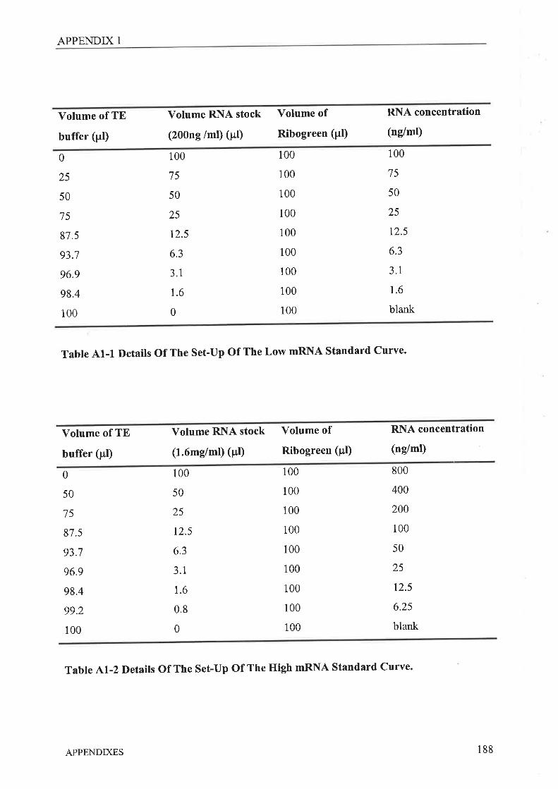
APPENDIX 1
Volume of TE
buffer (pl)
Volume RNA stock
(200ng /ml) (pl)
Yolume of
Ribogreen (pl)
RNA concentration
(ng/ml)
0
25
50
75
87.5
93.7
96.9
98.4
100
100
75
50
25
12.5
6.3
3.1
r.6
0
100
100
100
100
100
100
100
100
100
100
75
50
25
t2.5
6.3
3.1
1.6
blank
Table A1-1 Details of The set-up of The Low mRNA Standard curve.
Volume of TE
buffer (pl)
Volume RNA stock
(1.6mg/ml) (¡rt)
Volume of
Ribogreen (pl)
RNA concentration
(ng/ml)
0
50
75
87.5
93.7
96.9
98.4
99.2
100
100
50
25
12.5
6.3
3.1
1.6
0.8
0
100
100
100
100
100
100
100
100
100
800
400
200
100
50
25
12.5
6.25
blank
Table A1-2 Details Of The Set-Up Of The High mRNA Standard Curve
APPENDIXES 188
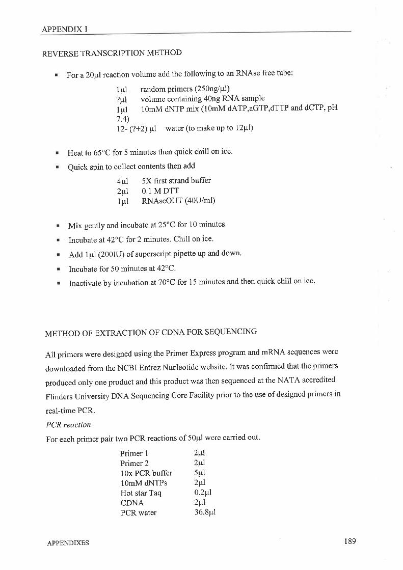
APPENDIX 1
REVERSE TRANSCRIPTION METHOD
For a 20¡rl reaction volume add the following to an RNAse free tube
1pl?pllpl7.4)12- (?+2) ¡l water (to make up to 12p1)
Heat to 65'C for 5 minutes then quick chill on ice.
Quick spin to collect contents then add
4fú 5X first strand buffer
4i 0.1 M DTTlpl RNAseOUT (a0U/m1)
Mix gently and incubate at25"C for 10 minutes.
Incubate at 42"C for 2 minutes. Chill on ice.
Add 1pl(200ru) of superscript pipette up and down.
Incubate for 50 minutes at 42oC.
Inactivate by incubation at 70'C for 15 minutes and then quick chill on ice'
METHOD OF EXTRACTION OF CDNA FOR SEQUENCING
All primers were designed using the Primer Express program and mRNA sequences were
downloaded from the NCBI EnfiezNucleotide website. It was confirmed that the primers
produced only one product and this product was then sequenced at the NATA accredited
Flinders University DNA Sequencing Core Facility prior to the use of designed primers in
real-time PCR.
PCR reaction
For each primer pair two PCR reactions of 50¡rl were carried out.
2p+l
2p+l
5pl2pú
0.2¡l2vl36.8pl
random primers (25 }ngl P'I)volume containing 40ng RNA samPle
10mM dNTP mix (10mM dATP,aGTP,dTTP and dCTP, pH
Primer 1
Primer 2
10x PCR buffer10mM dNTPsHot star TaqCDNAPCR water
APPENDIXES 189
APPENDIX 1
Samples were placed in a minicycler and subject to
15 minutes 95"C1 minute 95"C1 minute 60oC1 minute 72oC6 minutes 72"C
Cycled 39 times
pCR products from each reaction were combined, mixed with loading buffer (20p1) and run
on a 1 .5Yo agarose gel (1.3 mg agarose, 75ml 0.5 TBE, 3¡rl Ethidium bromide - Large wells
for the 100¡rl samples were created by taping together adjacent lane prongs) immersed in
0.5X Tris Borate EDTA at 80mV for 40 minutes.
cDNA extraction
A Qiagen QlAquick Gel Extraction Kit (using a microcentrifuge) was used to extract the
cDNA from the agarose gel as described in the handbook.
. Cut the Band from the agarose gel with a clean scalpel. Trim off excess gel.
. Place slice into a pre-weighed tube and add three volumes of buffer QG to 1 volume
of gel.
. Incubate at 50"C for 10 minutes, with regular mixing, until the gel has dissolved.
. Add one gel volume of isopropanol and mix. Add sample to a Qiagen spin column
over a 2 ml collection tube. Centrifuge for 1 minute.
. Add 0.5m1of buffer QG to column, stand for 5 minutes and then centrifuge for 1
minute.
. Place column over a 1.5m1microcentrifuge tube.
. Add 30¡.rl of elution buffer directly to the membrane, allow to stand for 1 minute and
then spin for 1 minute.
The sequencing facility requires 0.05 pmoV¡rl for the sequencing reaction and thus it is
necessary to determine the total amount of cDNA product. This was done by running 1pl and
2¡rl samples of purified cDNA on a 2Yo agarose gel with a low DNA mass ladder (Life
Technologies) (80mV, 45 minutes) to get an estimate for ng of DNA. Based on the known
product length (bp) the correct concentration was calculated:
¡ To calculate thengl pl required
0.05 pmole/¡rl : KB x 32.45 (KB is length in kilobases)
APPENDIXES 190
APPENDIX 1
To work out dilution of oDNA product
(nglDNAproduct) + (KB x32.45)
QUANTITATIVE REAL TIME PCR CALCULATIONS
Unknown 6RNA levels were calculated using the real-time PCR CT values as follows:
Each sample was normalised by subtracting the CT value for the house-keeper gene (HPRT)
for that sample from the CT value of the gene of interest (^CÐ. All the values from the
reference group, designated the arbitrary value of I (day Oam Ia), vrere averaged (value a).
This value was then subtracted from the normalised unknown samples (AACÐ and since the
pCR reaction is an exponential, levels calculated as z\^c. These resulting values were
averaged, with those for the reference group (a/) being very close to 1. The value a is altered
until the value al is equal to 1. The rest of the results are then averaged and the resultant
values represent a of fold change in mRNA levels relative to the mRNA levels present in the
group designated as l.
METHODS FOR THE IMMI.]NOASSAY KITS (R&D SYSTEMS)
¡ Reagents, standards and controls for each assay were prepared as indicated in Table
A1-3.
. Microplates were removed from the foil pouches.
. 50Frl of the appropriate assay diluent was added to each well.
' 50prl of sample, control or standard, was added in duplicate to the appropriate wells.
r The plate was mixed by gentle tapping, and sealed with the adhesive strip provided.
. Samples were incubated in the plates for the following times:
IL-1P and IL-10, 15 hours at 4"C,
TNFa, 2 hours at RT.
. Wells were aspirated and washed with wash buffer using a squirt bottle.
. The wash was repeated three times, ensuring complete removal of wash buffer from
the wells after each wash.
. Plates were blotted on clean, absorbent paper towels.
' 100Frl of the appropriate conjugate was added to each well.
APPENDIXES 191
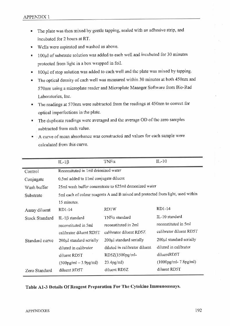
APPENDIX 1
. The plate was then mixed by gentle tapping, sealed with an adhesive strip, and
incubated for 2 hours at RT.
. Wells were aspirated and washed as above'
. 100p1 of substrate solution was added to each well and incubated for 30 minutes
protected from light in a box wrapped in foil.
. 100p1 of stop solution was added to each well and the plate was mixed by tapping.
. The optical density of each well was measured within 30 minutes at both 450nm and
570nm using a microplate reader and Microplate Manager Software from Bio-Rad
Laboratories, Inc.
. The readings at 570nm were subtracted from the readings at 450nm to correct for
optical imperfections in the plate'
. The duplicate readings were averaged and the average OD of the zero samples
subtracted from each value.
r . { curve of mean absorbance was constructed and values for each sample were
calculated from this curye.
IL-1p TNFa IL-10
Control
Conjugate
Wash buffer
Substrate
Assay diluent
Stock Standard
Standard curye
Zero Standard
Reconsituted in 1ml deionized water
0.5m1 added to 1lml conjugate diluent
25ml wash buffer concentrate to 625m1demonized water
5ml each of colour reagents A and B mixed and protected from light, used within
15 minutes.
RDl-14 RD1W RDl-14
IL-1P standard TNFcr standard IL-10 standard
reconstituted in 5ml reconstituted in 2ml reconstituted in 5ml
calibrator diluent RD5T calibrator diluent RD5Z calibrator diluent RD5T
200p1 standard serially 200p1 standard serially 200p1 standard serially
diluted in calibrator diluted in calibrator diluent diluted in calibrator
diluent RD5T RD5Z(1500pg/ml- diluentRD5T
(500pg/ml - 3.9pglml) 23.4pglml) (1000pg/ml- 7'8pg/m1)
diluent RD5T diluent RD5Z diluent RD5T
Table A1-3 Details Of Reagent Preparation For The Cytokine Immunoassays.
APPENDIXES t92

APPENDIX 1
METHOD FOR THE MEASUREMENT TOTAL NITRIC OXIDE
(R&D Systems)
Reaction buffer was made by diluting 30ml of the reaction buffer concentrate with 270m1of
deionized water.
NADH reagent was made by reconstituting one vial provided with lml deionized water' Mix
gently for 3 minutes, keep on ice. Immediately prior to use 900¡rl NADH reagent was diluted
with 1.8m1 of deionized water and kept on ice.
Nitrate reductase rwas reconstituted with lml of the provided nitrate reductase storage buffer,
briefly vortexed and then left on the bench for 15 minutes. The vortex was repeated and
reductase left again on the bench for 15 rninutes. This was kept on ice for the duration of the
assay, Immediately before use, 360¡rl nitrate reductase was mixed with 1440¡rl of reaction
buffer, placed on ice and used within l5 minutes'
Samples were filtered through a 10,000 MW Amicon MicroconÒ centrifuge filter unit (YM-
10, Millipore Australia, Sydney, Australia) and analysed as recommended by the
manufachrrer.
The highest standard was nrade from the 1000pmol/L standard; 100¡rl into 900p1 reaction
buffer. Subsequent standards were made by serial dilution in reaction buffer (500p1: 500p1) to
give a cuwe ranging from 1OO¡rmol/L to3.I2¡tmollL.
. 200þrl of reaction buffer was added to the blank wells.
' 50pl of reaction buffer was added to the zero standard wells. Standards or samples
rvere addecl to the remaining wells in duplicate.
. 25þlof NADH was added to all standards and samples.
. 25úof nitrate reductase was added to all standards and samples.
. 'I'he plate was mixed by tapping, covered with the adhesive strip provided and
incubated for 30 minutes ú37"C.
APPENDIXES r93
APPENDIX 1
. 50pl of Griess reagent I was added to all wells except the blanks, followed by 50¡rl of
Griess reagent II to all wells except the blanks.
. The plate was mixed by tapping and then incubated at RT for 10 minutes.
. The optical density of each well wàs measured at 540nm'
. Duplicate results were averaged, the blank average subtracted from each well and the
mean absorbance plotted against the total nitrite concentration.
. Values for each sample were calculated from this curve.
APPENDIXES 194
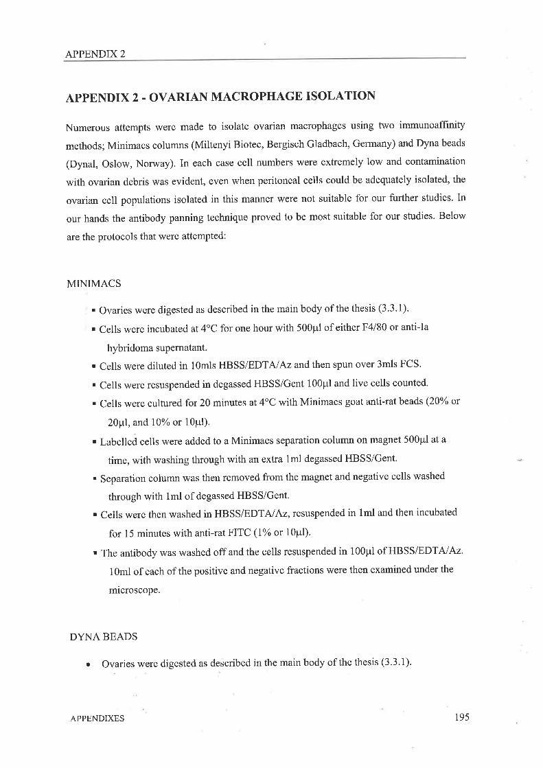
APPENDIX 2
APPENDIX2 - OVARIAN MACROPHAGE ISOLATION
Numerous attempts were made to isolate ovarian macrophages using two immunoaffinity
methods; Minimacs columns (Miltenyi Biotec, Bergisch Gladbach, Germany) and Dyna beads
(Dynal, Oslow, Norway). In each case cell numbers were extremely low and contamination
with ovarian debris was evident, even when peritoneal cells could be adequately isolated, the
ovarian cell populations isolated in this manner were not suitable for our further studies' In
our hands the antibody panning technique proved to be most suitable for our studies. Below
are the protocols that were attempted:
MINIMACS
. Ovaries were digested as described in the main body of the thesis (3.3.1)'
' Cells were incubated at 4oC for one hour with 500p1 of either F4l80 or anti-Ia
hybridoma supernatant.
. Cells were diluted in 10mls HBSS/EDTAlAzandthen spun over 3mls FCS'
. Cells were resuspended in degassed HBSS/Gent 100p1 and live cells counted.
. Cells were cultured for 20 minutes at 4"C with Minimacs goat anti-tat beads (20% or
20¡tl, and l\Yo or 10Pl).
' Labelled cells were added to a Minimacs separation column on magnet 500¡rl at a
time, with washing through with an extra lml degassed HBSS/Gent.
. Separation column was then removed from the magnet and negative cells washed
through with 1ml of degasserl HBSS/Gent'
' Ceils were then washed in HBSS/EDTNAz, resuspended in lml and then incubated
for 15 minutes with anti-rat FITC (Io/o or 10pl).
. The antibody was washed off and the cells resuspended in 100p1of HBSS/EDTNAz
10ml of each of the positive and negative fractions were then examined under the
rnicroscope.
DYNA BEADS
Ovaries were digested as described in the main body of the thesis (3.3.1).O
APPENDIXES 195
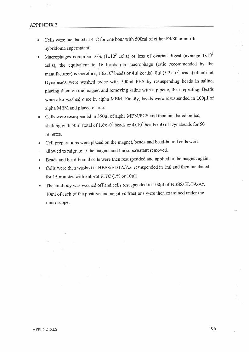
APPENDIX 2
Cells were incubated at 4"C for one hour with 500m1 of either F4l80 or anti-Ia
hybridoma supernatant.
Macrophages comprise l}Vo (1x10s cells) or less of ovarian digest (average 1x10⁶
cells), the equivalent to -16
beads per macrophage (ratio recommended by the
manufacturer) is therefore, 1.6x10⁶ beads or 4pl beads). 8pl (3.2x10⁶ beads) of anti-rat
Dynabeads were washed twice with 500m1 PBS by resuspending beads in saline,
placing them on the magnet and removing saline with a pipette, then repeating. Beads
were also washed once in alpha MEM. Finally, beads were resuspended in 100¡rl of
alpha MEM and placed on ice.
Cells were resuspended in 350p1 of alpha MEM/FCS and then incubated on ice,
shaking with 50¡rl (total of 1.6x10⁶ beads or 4x10⁶ beads/ml) of Dynabeads for 50
minutes.
Cell preparations were placed on the magnet, beads and bead-bound cells were
allowed to migrate to the magnet and the supernatant removed.
Beads and bead-bound cells were then resuspended and applied to the magnet again.
Cells were then washed in HBSS/EDTNAz,resuspended in lml and then incubated
for 15 minutes with anti-rat FITC (lYo or 1Opl).
The antibody was washed off and cells resuspended in 100p1of HBSS/EDTAIAz
1gml of each of the positive and negative fractions were then examined under the
microscope.
a
a
a
o
I
APPENDIXES r96
APPENDIX 3
APPENDIX 3 _ MESSENGER RNA EXTRACTION METHODS
A complete set of ovarian and peritoneal macrophage samples, consisting of 53 samples -three repeats of isolation of Ia* and F4l80+ cells from groups of 8 animals at 4 time points
across the stimulated reproductive cycle, was collected and the RNA isolated using the
eiagen RNeasy Mini Kit method recommended for isolation of RNA from very small
amounts of starting material.
QIAGEN RNEASY@ MINI KIT RNA ISOLATION METHOD
' Briefly lysate buffer from the kit was added directly to the isolated cells on the
panning plate.and then collected and frozen at -80oC for later RNA isolation.
. Thawed lysate was mixed with70% ethanol and added to a spin column'
. The spin column was washed with 700¡rl RWI buffer and spun for 15 seconds at
10,000RPM.
. The spin column was treated with 80pl DNase solution (10¡rl DNAse stock: 70pl RDD
buffer) for 15 minutes and washed with 350p1RWl buffer and spun for 15 seconds at
10,000 rpm.
. The spin column was further washed with 500p1RPE buffer and spun for 15 seconds
at 10,000 rpm, this wash was then repeated.
. The spin column was incubated for 20 minutes with RNAse free water and then
centrifuged 1 minute 10,000 to collect RNA.
Levels of RNA were measured in these samples using a Ribogreen Quantitation Reagent Kit.
Levels were found to be extremely low and variable (Table A2-I) and it was decided to
increase the numbers of animals in each group to 10, abandon the day-l group, and try the
TriReagent@ method of isolation instead. Some direct comparisons between the Qiagen and
TriReagent@ isolation methods were done. Peritoneal cells were collected washed and
counted, RNA was isolated using either the Qiagen kit method or the TriReagent@ method
alone or TriReagent@ spiked with glycogen from cells designated as high (80 000 cells) or
low (20 000cells) numbers .
APPENDIXES t97
APPENDIX 3
MRNA OBTAINED FROM ISOLATED OVARIAN MACROPHAGE
CELL TYPES
STAGE OF
STIMULATED
CYCLE
Ia positive Cells F4l80 positive Cells
Day-1
Day Oam
Day Opm
Dayl
Day2
Mean (mg/ml)
0.57
0.r7
1.28
2.34
10.4
SE
0.3
0.1
0.6
0.4
3.2
Mean (mg/ml)
0.2
0.6
0.s6
1.7
9.8
SE
0.2
0.5
0.3
0.7
4
Table A2-1 The Amount Of RNA Measured In Ovarian Macrophages Samples Following
RNA Isolation Using The Qiagen Rneasy@ Mini Kit RNA Isolation Method.
TRIREAGENT@ (TR) RNA ISOLATION METHOD
RNA Extraction
. Samples were thawed on ice and spun at 14,000 rpm, 4oC for 10 minutes.
. Supernatant was transferred to a fresh tube and the sample left on ice for 5 minutes'
. 250p1 of chloroform/5OOpl of TR (fume hood) was added, vortexed briefly and left on
ice for 15 minutes.
. Samples were spun at 14,000 rpm, 4oC for 10 minutes and the uppff (aqueous) layer
transferred to a fresh tube.
. l pl of glycogen & 25¡il isopropanol (per 500p1 TR) was added, mixed and the samples
were left on ice for 5 minutes.
. Samples were spun at 14,000 rpm, 4oC for 10 minutes and the supernatant transferred
to a fresh 0.6m1tube with 225p'l isopropanol (/500p1 TR)'
. RNA was precipitated overnight at -80'C'
APPENDIXES 198
APPENDIX 3
DNase Treatment
. RNA was pelleted by centrifuging for 30 minutes at 14,000rpm, 4oC.
' The supernatant was discarded and the pellet washed with 500p1 of ice coldT5Yo
ethanol.
. Samples were spun at 14,000 rpm,4oC for 10 minutes and the supernatant discarded
. The RNA pellet was dried by inverting the tube on the bench. (-15-20 minutes)
. After drying the pellet was resuspended in 79.5¡t"l of PCR grade water, vortexed
briefly and then allowed to dissolve at room temperature (-20 minutes).
DNase solution (make up on ice) per tube
10x DNase Buffer 10pl
RNAsin (aOu/pl) 2'5¡tlDNase I (lu/pl) 5Pl
. 17.5p1of DNase solution was added to RNA sample on ice, follwed by a vortexing'
' Samples were incubated at37"C for 90 minutes'
' 100p1of room temperature phenol (water saturated) & 100p1of chloroform: isoamyl
alcohol (24:I) was added and samples vortexed'
. Samples were spun at 14,000 rpm, 4oC for 10 minutes'
' Supernatant was transferred to a fresh tube
. RNA was re-extracted by adding 100p1 of chloroform: isoamyl alcohol, vortexing and
then spinning at 14,000 rpm,4oC for 10 minutes'
. Supernatant was transfened to a fresh tube RNA precipitated OA{ at -80'C with 1Opl
2M NaOAc (0. lX vol of aqueous phase) & 250¡tl of ice cold 99.7 -100% ethanol (2.5x
vol ofaqueous Phase).
R]\lA Recoverv
. The RNA was pelleted by centrifuging for 30 minutes at 14,000 rpm, 40c'
. The supernatant was discarded and the pellet washed with 500p1 of ice cold 75%
ethanol.
. Samples were spun at 14,000 rpm, 4oC for 10 minutes and the supernatant discarded'
. The RNA pellet was then dried by inverting tube on bench. (-15-20 minutes)
. Resuspend pellet in 20¡rl of PCR grade water and leave for 30 minutes to dissolve'
The RNA levels were measured in all samples using the Ribogreen assay. For both the high
and low cell numbers cases the spiked Trireagent@ method gave the greatest yield of mRNA
(Table A2.2). The Qiagen kit was better than Trireagent@ alone at high cell numbers but both
APPENDIXES 199
APPENDIX 3
were equivalent at low cell numbers. Samples in Chapter 4 were collected from across the
cycle for a second time with increased numbers of animals in each group and using the spiked
Trireagent@ method.
RNA ISOLATION METHOD
CELL NUMBERS
2 x10⁴ (low)
5 x10⁴ (high)
Qiagen
2.9 +0.4
14.9 + Ll
Tri Reagent Tri Reagent * glYcogen
2.5 + 0.3 6.2 + 0.7
10.1 + 1.1 19.1 È 0.9
Table ^2-2TheAmount
Of RNA Isolated From Known Numbers Of Cells Using
Various Methods. Values are mglml t SE'
APPENDIXES 200
APPENDIX 4
APPENDIX 4 _ NON.SPECIFIC ESTERASE STAINING METHODS
To conf,rrm the purity of the isolated macrophage population experiments to stain
macrophages bound to the panning plates for non-specific esterase were carried out' Staining
was attempted by two different methods. Unfortunately neither method was successful on
either the peritoneal or ovarian cells present on the panning plates.
METHOD FROM YAM ETAL (19)
Fixine Cells
. Cells can be stored unfixed at room temp for at least two weeks without affecting the
enzyme activity.
. Fix cells in the following solution, adjusted to pH 6.6'
20mg NazHPO¿100mg KHzPO¿30ml HzO45ml acetone25ml Formalin(37%)
. Fix for 30 seconds only at 4oC, wash in three changes of water and air-dry for 10-30
minutes before staining.
Make up the following solutions:
MI5 phosphate buffer (stock solution)I 4.73gNazHPO+ in 500mls RO water.
2 4.54gKHzPO+ in 500mls RO water.Add 21.7mls of 2 to 78.3mls of 1. Adjust to pH 7 '4with
phosphates. NazHPO+ will increase pH, KH2PO+ will decrease pH. Filter
and store at 4"C.
a-N ap hthy I ac et at e (stock solution)Mix 20mg of cr-naphtþlacetate per lml ethylene glycol
monomethyl ether
Hexazo itzed p araro s anilin
APPENDIXES 201
APPENDIX 4
Mix equal volumes of pararosanilin-HCl solution and 4%o sodium nitrite (0.4gl1Omls)
solution for I minute immediately before use.
Staining Mixture and Procedure
. Mix 1.8mls of hexatozoitzedpararosaniline with 1.5m1 of o-naphthyl acetate and
26.7mls M/l5 buffer.
. pH to 6.1 with NaOH and filter before use.
. Incubate slides/cells with this mixture at room temperature for 45 minutes.
. Wash with RO water.
. Counter stain with 1% metþl green for 1-2 minutes'
. Wash and allow to dry.
. Mount in permount and examine. Activity is seen as dark green granules in the
cytoplasm.
SIGMA NSE STAINING KIT
Ensure glassware is cleaned of all detergent residues by washing coplin jars and other
glassware with dilute bleach and then wash thoroughly with deionised water as any
residue can affect enzyme activities. Ensure all reagents are \ilarm as weak reactions
may result if the correct temperature is not achieved.
. Fix slides for I minute in freshly made Citrate-acetone-methanol fixative at room
temperature.
C itr ate - A c eton e- Methano I F ixat iv e
18mls of citrate dilute solution (1 part citrate concentrate with 9 parts water.
pH to 5,4.)
27mls ACS grade acetone
5ml methanol.
Discard after 8 hours.
. Wash in deionised water and air dry for at least 20 minutes'
. To 50ml of warm (37'C) Trizmal dilute buffer solution add (with constant stining) 1
capsule of Fast blue RR salt.
APPENDIXES 202
APPENDIX 4
I
¡
Trizmal Buffer
1 part TrizmalT.6ml concentrate to 9 parts deionised water pH to 7.6
When the capsule is completely dissolved add2ml of fresh o-naphthyl acetate
solution.
u- N aphthyl Ac et at e S o lut ion
Dissolve one capsule of cr-naphtþl acetate in 2mls of ethylene glycol
monomethyl ether. Use immediately'
Stir this yellow turbid solution for 30 seconds but do not filter.
place specimens in staining solution and incubate at37"C for 30 minutes and protect
from exposure to light with foil.
Remove slide from stain and rinse in deionzed water for 3 minutes.
Counter stain with Mayers Haematoxylin for 5-10 minutes and rinse in tap water'
Air dry and examine for positive stain. Use only aqueous mounting media if a cover
slip is required. Sites of activity appear as black granules.
APPENDIXES 203
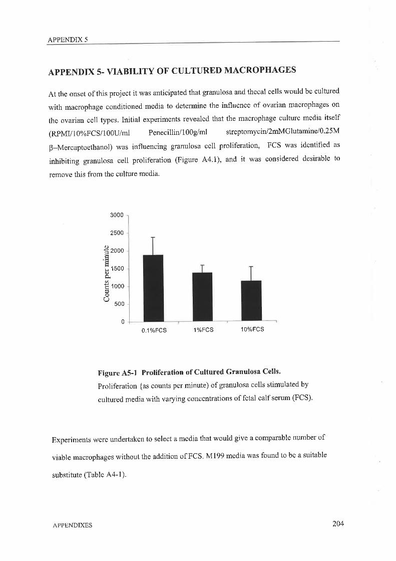
APPENDIX 5
APPENDIX 5. VIABILITY OF CULTURED MACROPHAGES
At the onset of this project it was anticipated that granulosa and thecal cells would be cultured
with macrophage conditioned media to determine the influence of ovarian macrophages on
the ovarian cell types. Initial experiments revealed that the macrophage culture media itself
(RpMV1g%FCS/100U/ml PenecillirVl00g/ml streptomycin/2mMGlutamine/0'25M
B-Mercaptoethanol) was influencing granulosa cell proliferation, FCS was identified as
inhibiting granulosa cell proliferation (Figure 44.1), and it was considered desirable to
remove this from the culture media.
3000
2500
0.1%FCS 1%FCS 10%FCS
Figure A5-1 Proliferation of Cultured Granulosa Cells.
Proliferation {as counts per minute) of granulosa cells stimulated by
cultured media with varying concentrations of fetal calf serum (FCS)
Experiments were undertaken to select a media that would give a comparable number of
viable macrophages without the addition of FCS. M199 media was found to be a suitable
substitute (Table A4-1).
I zoooÉ'=
X tsoo0.)È.t)
Ë looo)oo ooo
0
APPENDIXES 204
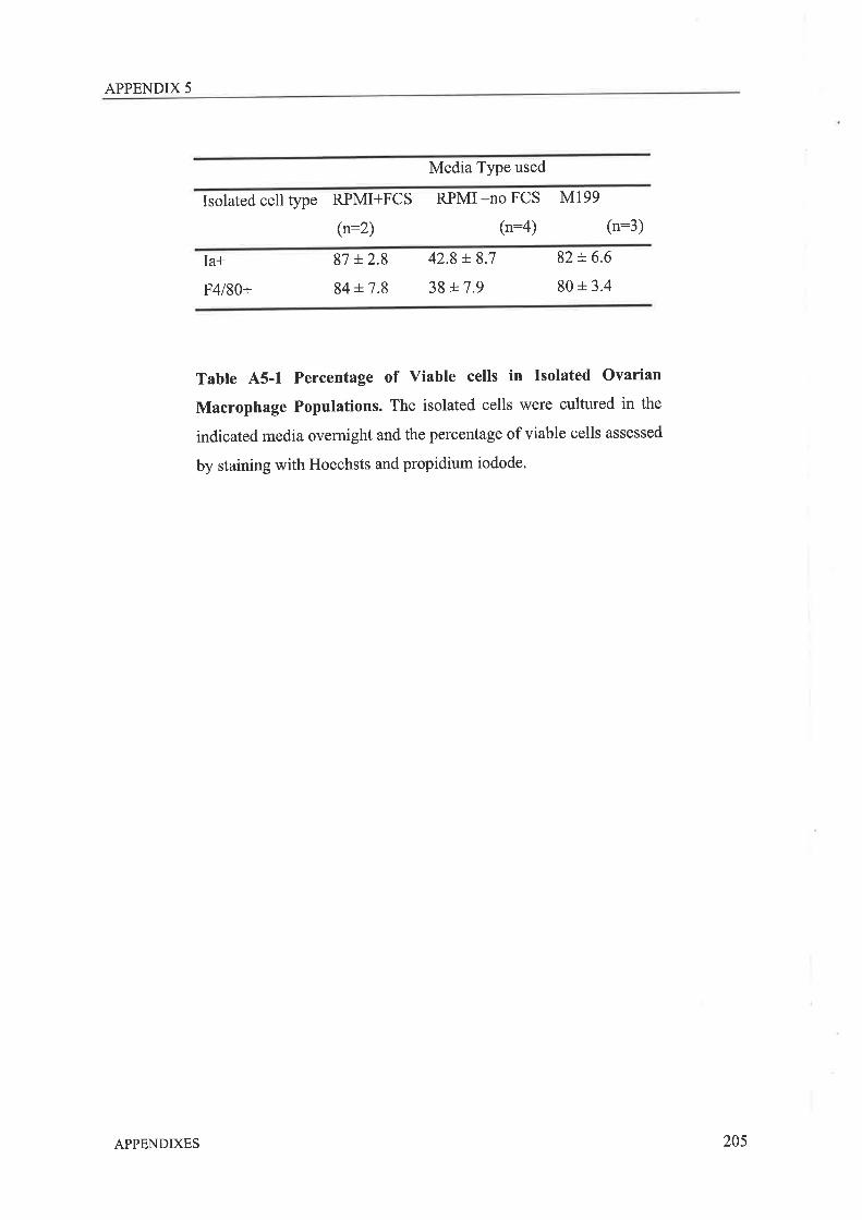
APPENDIX 5
Media Type used
Isolated cell type RPMI+FCS
(n:2)
RPMI -no FCS M199
(n:4) (n:3)
Ia*
F4l80+
87 L2.8
84 + 7.8
42.8 +8.7
38 t7.982 + 6.6
80 + 3.4
Tabte A5-1 Percentage of viable cells in Isolated ovarian
Macrophage Populations. The isolated cells were cultured in the
indicated media overnight and the percentage of viable cells assessed
by staining with Hoechsts and propidium iodode.
APPENDIXES 205