Imbalanced gp130-dependent signaling in macrophages alters macrophage colony-stimulating factor...
11
MOLECULAR AND CELLULAR BIOLOGY, Feb. 2004, p. 1453–1463 Vol. 24, No. 4 0270-7306/04/$08.000 DOI: 10.1128/MCB.24.4.1453–1463.2004 Copyright © 2004, American Society for Microbiology. All Rights Reserved. Imbalanced gp130-Dependent Signaling in Macrophages Alters Macrophage Colony-Stimulating Factor Responsiveness via Regulation of c-fms Expression Brendan J. Jenkins, 1 * Dianne Grail, 1 Melissa Inglese, 1 Cathy Quilici, 1 Steven Bozinovski, 2 Peter Wong, 3 and Matthias Ernst 1 Ludwig Institute for Cancer Research, Colon Molecular and Cell Biology Laboratory, 1 The Walter and Eliza Hall Institute for Medical Research, 3 and Pharmacology Department, University of Melbourne, 2 Parkville, Victoria, Australia Received 21 May 2003/Returned for modification 30 July 2003/Accepted 7 November 2003 The mechanisms by which interleukin-6 (IL-6) family cytokines, which utilize the common receptor signaling subunit gp130, influence monocyte/macrophage development remain unclear. Here we have utilized macro- phages devoid of either gp130-dependent STAT1/3 (gp130 STAT/STAT ) or extracellular signal-regulated ki- nases 1 and 2 (ERK1/2) mitogen-activated protein (MAP) kinase (gp130 Y757F/Y757F ) activation to assess the individual contribution of each pathway to macrophage formation. While the inhibition by IL-6 of macrophage colony-stimulating factor (M-CSF)-induced colony formation observed in gp130 wt/wt mice was abolished in gp130 STAT/STAT mice, inhibition of macrophage colony formation was enhanced in gp130 Y757F/Y757F mice. In gp130 STAT/STAT bone marrow-derived macrophages (BMMs), both IL-6- and M-CSF-induced ERK1/2 ty- rosine phosphorylation was enhanced. By contrast, tyrosine phosphorylation of ERK1/2 in response to M-CSF was reduced in gp130 Y757F/Y757F BMMs, and the pattern of ERK1/2 activation in gp130 mutant BMMs correlated with their opposing responsiveness to M-CSF-induced proliferation. When compared to the level of expression in gp130 wt/wt BMMs, c-fms expression was elevated in gp130 STAT/STAT BMMs but reduced in gp130 Y757F/Y757F BMMs. Finally, an ERK1/2 inhibitor suppressed M-CSF-induced BMM proliferation, and this result corresponded to a reduction in c-fms expression. Collectively, these results provide a functional and causal correlation between gp130-dependent ERK MAP kinase signaling and c-fms gene activation, a finding that provides a potential mechanism underlying the inhibition of M-CSF-dependent macrophage development by IL-6 family cytokines in mice. The formation of macrophages involves the proliferation, differentiation, and functional maturation of multipotential he- matopoietic progenitor cells from the bone marrow into mono- cytes and, ultimately, macrophages, a process that is primarily regulated by macrophage colony-stimulating factor (M-CSF, also known as CSF-1) (36). The central role that M-CSF plays in monocyte/macrophage development has been formally dem- onstrated by the severe depletion of macrophages in M-CSF- deficient osteopetrotic (op/op) mice (40) and, more recently, in mice subjected to targeted inactivation of the M-CSF receptor gene, c-fms (5). During mouse development, expression of c-fms is an early and robust marker of cells belonging to the macrophage lineage, and studies on the transcriptional regu- lation of c-fms have identified promoter elements underlying its lineage-specific activity (14, 15). Members of the interleukin-6 (IL-6) cytokine family, in par- ticular, IL-6, IL-11, and leukemia inhibitory factor (LIF), are multifunctional cytokines which also regulate the production of myelomonocytic cells (4, 27). In vitro studies with the M1 murine monocytic leukemia cell line revealed that IL-6 and LIF induce the terminal differentiation of these cells into mac- rophages, suggesting that these factors promote macrophage development (25). However, the IL-6 family cytokines appear to act as both positive and negative regulators of the prolifer- ation and differentiation of primary human and mouse macro- phages (4, 17, 27), with recent evidence demonstrating that this may occur by regulating the expression of c-fms (3). IL-6 family cytokines share gp130 as a signaling receptor (R) subunit (19), and receptor binding of IL-6 or IL-11 leads to gp130 homodimerization (13, 29), whereas receptor binding of all other family members induces heterodimerization of gp130 with one of the highly related R subunits, LIF-R or oncosta- tin M-R (10, 28). In all cases, subunit dimerization triggers activation of two major signaling cascades, namely the SHP2/ extracellular signal-regulated kinase (ERK) mitogen-activated protein (MAP) kinase (9) and the STAT1/3 (11) pathways. We have recently reported the generation of two strains of mice homozygous for knock-in mutations in gp130 which ren- der gp130 incapable of activating either the STAT1/3 (in gp130 STAT/STAT mice) (6) or the SHP2/ERK MAP kinase (in gp130 Y757F/Y757F mice) (38) pathway. These mice display a wide variety of penetrant phenotypes, which in part mimic phenotypic pathologies observed in knockout mice lacking in- dividual cytokines of the IL-6 family or their receptors, thereby genetically identifying the signaling pathway responsible for transducing a specific IL-6 family cytokine-mediated bio- logical response. Surprisingly, the gp130 STAT/STAT and gp130 Y757F/Y757F mice also display novel phenotypes, which * Corresponding author. Mailing address: Ludwig Institute for Can- cer Research, Colon Molecular and Cell Biology Laboratory, P. O. Box 2008, Royal Melbourne Hospital, Victoria 3050, Australia. Phone: 61 3 9341 3155. Fax: 61 3 9341 3104. E-mail: Brendan.Jenkins@ludwig .edu.au. 1453 on June 5, 2016 by guest http://mcb.asm.org/ Downloaded from
-
Upload
independent -
Category
Documents
-
view
0 -
download
0
Transcript of Imbalanced gp130-dependent signaling in macrophages alters macrophage colony-stimulating factor...

MOLECULAR AND CELLULAR BIOLOGY, Feb. 2004, p. 1453–1463 Vol. 24, No. 40270-7306/04/$08.00�0 DOI: 10.1128/MCB.24.4.1453–1463.2004Copyright © 2004, American Society for Microbiology. All Rights Reserved.
Imbalanced gp130-Dependent Signaling in Macrophages AltersMacrophage Colony-Stimulating Factor Responsiveness
via Regulation of c-fms ExpressionBrendan J. Jenkins,1* Dianne Grail,1 Melissa Inglese,1 Cathy Quilici,1
Steven Bozinovski,2 Peter Wong,3 and Matthias Ernst1
Ludwig Institute for Cancer Research, Colon Molecular and Cell Biology Laboratory,1 The Walter andEliza Hall Institute for Medical Research,3 and Pharmacology Department, University of
Melbourne,2 Parkville, Victoria, Australia
Received 21 May 2003/Returned for modification 30 July 2003/Accepted 7 November 2003
The mechanisms by which interleukin-6 (IL-6) family cytokines, which utilize the common receptor signalingsubunit gp130, influence monocyte/macrophage development remain unclear. Here we have utilized macro-phages devoid of either gp130-dependent STAT1/3 (gp130�STAT/�STAT) or extracellular signal-regulated ki-nases 1 and 2 (ERK1/2) mitogen-activated protein (MAP) kinase (gp130Y757F/Y757F) activation to assess theindividual contribution of each pathway to macrophage formation. While the inhibition by IL-6 of macrophagecolony-stimulating factor (M-CSF)-induced colony formation observed in gp130wt/wt mice was abolished ingp130�STAT/�STAT mice, inhibition of macrophage colony formation was enhanced in gp130Y757F/Y757F mice. Ingp130�STAT/�STAT bone marrow-derived macrophages (BMMs), both IL-6- and M-CSF-induced ERK1/2 ty-rosine phosphorylation was enhanced. By contrast, tyrosine phosphorylation of ERK1/2 in response to M-CSFwas reduced in gp130Y757F/Y757F BMMs, and the pattern of ERK1/2 activation in gp130 mutant BMMscorrelated with their opposing responsiveness to M-CSF-induced proliferation. When compared to the level ofexpression in gp130wt/wt BMMs, c-fms expression was elevated in gp130�STAT/�STAT BMMs but reduced ingp130Y757F/Y757F BMMs. Finally, an ERK1/2 inhibitor suppressed M-CSF-induced BMM proliferation, andthis result corresponded to a reduction in c-fms expression. Collectively, these results provide a functional andcausal correlation between gp130-dependent ERK MAP kinase signaling and c-fms gene activation, a findingthat provides a potential mechanism underlying the inhibition of M-CSF-dependent macrophage developmentby IL-6 family cytokines in mice.
The formation of macrophages involves the proliferation,differentiation, and functional maturation of multipotential he-matopoietic progenitor cells from the bone marrow into mono-cytes and, ultimately, macrophages, a process that is primarilyregulated by macrophage colony-stimulating factor (M-CSF,also known as CSF-1) (36). The central role that M-CSF playsin monocyte/macrophage development has been formally dem-onstrated by the severe depletion of macrophages in M-CSF-deficient osteopetrotic (op/op) mice (40) and, more recently, inmice subjected to targeted inactivation of the M-CSF receptorgene, c-fms (5). During mouse development, expression ofc-fms is an early and robust marker of cells belonging to themacrophage lineage, and studies on the transcriptional regu-lation of c-fms have identified promoter elements underlyingits lineage-specific activity (14, 15).
Members of the interleukin-6 (IL-6) cytokine family, in par-ticular, IL-6, IL-11, and leukemia inhibitory factor (LIF), aremultifunctional cytokines which also regulate the productionof myelomonocytic cells (4, 27). In vitro studies with the M1murine monocytic leukemia cell line revealed that IL-6 andLIF induce the terminal differentiation of these cells into mac-
rophages, suggesting that these factors promote macrophagedevelopment (25). However, the IL-6 family cytokines appearto act as both positive and negative regulators of the prolifer-ation and differentiation of primary human and mouse macro-phages (4, 17, 27), with recent evidence demonstrating that thismay occur by regulating the expression of c-fms (3). IL-6 familycytokines share gp130 as a signaling receptor � (R�) subunit(19), and receptor binding of IL-6 or IL-11 leads to gp130homodimerization (13, 29), whereas receptor binding of allother family members induces heterodimerization of gp130with one of the highly related R� subunits, LIF-R� or oncosta-tin M-R� (10, 28). In all cases, � subunit dimerization triggersactivation of two major signaling cascades, namely the SHP2/extracellular signal-regulated kinase (ERK) mitogen-activatedprotein (MAP) kinase (9) and the STAT1/3 (11) pathways. Wehave recently reported the generation of two strains of micehomozygous for knock-in mutations in gp130 which ren-der gp130 incapable of activating either the STAT1/3 (ingp130�STAT/�STAT mice) (6) or the SHP2/ERK MAP kinase(in gp130Y757F/Y757F mice) (38) pathway. These mice display awide variety of penetrant phenotypes, which in part mimicphenotypic pathologies observed in knockout mice lacking in-dividual cytokines of the IL-6 family or their receptors, therebygenetically identifying the signaling pathway responsible fortransducing a specific IL-6 family cytokine-mediated bio-logical response. Surprisingly, the gp130�STAT/�STAT andgp130Y757F/Y757F mice also display novel phenotypes, which
* Corresponding author. Mailing address: Ludwig Institute for Can-cer Research, Colon Molecular and Cell Biology Laboratory, P. O. Box2008, Royal Melbourne Hospital, Victoria 3050, Australia. Phone: 613 9341 3155. Fax: 61 3 9341 3104. E-mail: [email protected].
1453
on June 5, 2016 by guesthttp://m
cb.asm.org/
Dow
nloaded from

most likely are a consequence of disturbing the otherwise nor-mal balanced activation of these two signaling pathways (6, 38).
To dissect the contribution of each of the two gp130-depen-dent signaling cascades to the regulation of macrophage de-velopment by IL-6 family cytokines, we have studied macro-phage populations derived from gp130�STAT/�STAT andgp130Y757F/Y757F mice. We report here that the absence ofgp130-dependent SHP2/ERK MAP kinase activation in mac-rophage colony-forming cells (M-CFCs) from gp130Y757F/Y757F
mice enhanced the inhibition of M-CSF-induced macrophagecolony formation by IL-6. In contrast, IL-6 does not inhibitM-CSF-induced macrophage colony formation by M-CFCsfrom gp130�STAT/�STAT mice, where the absence of gp130-mediated STAT activation amplified signaling through theSHP2/ERK MAP kinase pathway. Similarly, bone marrow-derived macrophages (BMMs) from gp130�STAT/�STAT andgp130Y757F/Y757F mice also display opposing responsiveness toM-CSF-dependent proliferation and differentiation. More-over, we provide evidence that the level of c-fms expression isdirectly related to the net output of ERK MAP kinase signal-ing emanating from gp130, thereby providing a molecularmechanism by which the IL-6 family cytokines modulate M-CSF-dependent macrophage development.
MATERIALS AND METHODS
Mice. The generation of mice homozygous for the gp130�STAT mutation(gp130�STAT/�STAT mice) (6) and the gp130Y757F mutation (gp130Y757F/Y757F
mice) (38) and of the compound gp130Y757F/Y757F:IL-6�/� mice (38) has beendescribed previously. Experiments were conducted on adult mice 8 to 16 weeksof age and included wild-type littermate controls. Mice were housed underspecific-pathogen-free conditions.
Cytokines and reagents. Recombinant murine M-CSF was a kind gift from NicNicola (Walter and Eliza Hall Institute, Melbourne, Australia). Recombinanthuman IL-6 and human IL-11 were generously provided by Glenn Begley (Am-gen, Thousand Oaks, Calif.) and Lorraine Robb (Walter and Eliza Hall Insti-tute), respectively. Commercially available antibodies were purchased as follows:the phospho-specific ERK1/2 antibody was purchased from Cell Signaling Tech-nology (Beverly, Mass.), the ERK1 antibody was from Santa Cruz Biotechnology(Santa Cruz, Calif.), and the fluorescein isothiocyanate (FITC)-conjugatedCD11b (Mac-1) antibody and streptavidin-phycoerythrin (PE) were purchasedfrom Pharmingen (San Diego, Calif.). The Fms antibody was generously pro-vided by Nicholas Wilson (University of Melbourne, Melbourne, Australia), andthe biotinylated F4/80 antibody was a kind gift from Ken Harder (Ludwig Insti-tute for Cancer Research, Melbourne, Australia). The MEK1 inhibitor U0216and the p38 MAP kinase inhibitor SB203580 were obtained from Calbiochem(San Diego, Calif.).
Hematopoietic progenitor cell assays. Clonal cultures of hematopoietic pro-genitor cells were assayed in semisolid agar containing 0.3% agar, Dulbecco’smodified Eagle’s medium (DMEM), and 20% fetal calf serum (FCS). Bonemarrow cells were flushed from femurs in phosphate-buffered saline (PBS)containing 2% FCS, and cell counts revealed that the levels of cellularity (perfemur) were comparable among the genotypes used in this study (data notshown). Briefly, triplicate cultures of either whole blood (1 �l) or unfractionatedadult bone marrow cells (2.5 � 104 to 5 � 104) were stimulated with cytokineseither individually or in multiple combinations at the indicated concentrations(see figure legends). For M-CSF stimulation of cultures containing either pe-ripheral blood or bone marrow cells, maximal colony stimulation was achieved ata concentration of M-CSF of �5 ng/ml. Cultures were incubated for 7 days (bonemarrow) or 14 days (blood) at 37°C in a humidified atmosphere of 10% CO2 inair, and colonies containing 50 or more cells were enumerated by using a dis-section microscope. Agar cultures were then fixed with 2.5% gluteraldehyde andsequentially stained with Luxol Fast Blue and hematoxylin.
Reconstitution experiments. One day prior to bone marrow transfer,gp130wt/wt C57BL/6 Ly5.1-recipient mice were injected intraperitoneally with 300�g of the CD4-depleting monoclonal antibody GK1.5 (kindly provided by Y.Zhang, Walter and Eliza Hall Institute). Flow cytometric analysis of peripheralblood from GK1.5-treated mice has previously demonstrated 98% depletion of
peripheral CD4� T lymphocytes (22). Unfractionated bone marrow cells (5 �106) from Ly5.2 gp130wt/wt, gp130�STAT/�STAT, and gp130Y757F/Y757F mice werethen injected intravenously into lethally �-irradiated (twice at 5.5 Gy) adult Ly5.1recipients (4 or 5 per donor). Irradiated recipients were maintained on oralantibiotics (170 mg of enrofloxacin [Baytril] per liter; Bayer, Leverkusen, Ger-many). Flow cytometric analysis indicated that 89% � 2.1% (mean � standarddeviation [SD]) of peripheral blood leukocytes obtained from chimeras ex-pressed Ly5.2 donor cells 3 months posttransfer.
BMMs. Bone marrow cells were flushed from femurs into DMEM supple-mented with 15% FCS, 20% L-cell conditioned (LC) media (as a source ofM-CSF), 2 mM glutamax, and 0.075% sodium bicarbonate (Life Technologies,Rockville, Md.) and were cultured in flasks at 37°C with 10% CO2. The followingday, nonadherent cells were transferred into new flasks, and BMMs were usedafter 7 to 10 days of culture. Prior to experiments for cytokine stimulation, cellswere washed in PBS and then starved in DMEM plus 1% FCS for 6 h beforebeing stimulated.
Western blot analysis. Prior to lysis, BMMs were either grown continuously inLC media or were washed and starved of cytokine before being stimulated. Allstimulations were performed at 37°C with M-CSF at a concentration of 20 ng/mland IL-6 at a concentration of 100 ng/ml at specified times (see figure legends).Cells were lysed in ice-cold lysis buffer (50 mM NaCl, 50 mM Tris-HCl [pH 7.5],1% glycerol, 1% Triton X-100, 50 mM NaF, 0.1% sodium dodecyl sulfate, 2 mMEDTA, 2 mM orthovanadate, and complete protease inhibitors [Roche Diag-nostics Corporation, Indianapolis, Ind.]), and cell lysates were cleared of debrisby centrifugation. Cell lysates were separated on sodium dodecyl sulfate–10%polyacrylamide gels and then transferred to nitrocellulose membranes. Mem-branes were blocked in TBST (300 mM NaCl, 10 mM Tris-HCl [pH 7.5], 0.5%Tween 20) containing 1% bovine serum albumin and incubated with primaryantibodies according to the manufacturer’s recommendations; subsequently theywere incubated with the appropriate secondary antibodies (anti-mouse or -rab-bit) coupled to horseradish peroxidase (Bio-Rad, Hercules, Calif). Proteins werevisualized by enhanced chemiluminescence (Amersham Pharmacia Biotech, Pis-cataway, N.J.).
Northern blot analysis. Total RNA was extracted from BMMs and unfrac-tionated bone marrow cells with an RNeasy kit (QIAGEN, Hilden, Germany).RNA (10 to 20 �g) was run on 1% agarose gels and transferred onto nitrocel-lulose, following which membranes were hybridized with a radiolabeled full-length c-fms cDNA probe (kindly provided by Nicholas Wilson, University ofMelbourne). Membranes were stripped and reprobed with a radiolabeled glyc-eraldehyde 3-phosphate dehydrogenase (GAPDH) probe (kindly provided byMegan Baldwin, Ludwig Institute for Cancer Research) to confirm equal loadingand transfer of RNA samples.
Proliferation and cell viability assays. BMMs were washed, starved of LCmedia, and then harvested by scraping. Cells were then added to flat-bottom96-well tissue culture plates at 2 � 104 cells per well in triplicate containing 100�l of DMEM supplemented with 15% FCS, M-CSF (20 ng/ml), and/or IL-6 (100ng/ml) as indicated. In experiments that used MAP kinase inhibitors, SB203580was used at a final concentration of 30 �M, and U0216 was used at a finalconcentration of 10 �M. To measure proliferation, after 2 days of culture at37°C, 1 �Ci of [3H]thymidine (ICN Biomedicals Australasia, Seven Hills, Aus-tralia) was pulsed for the last 3 h of culture, and [3H]thymidine incorporation wasmeasured in a TopCountNXT microplate scintillation counter (Packard, Meri-den, Conn.). To measure cell viability, after 4 days of culture at 37°C, 10 �l ofMTT (dimethyl thiazole diphenyl tetrazolium bromide; Sigma, St. Louis, Mo.) ata concentration of 5 mg/ml was added to the wells and incubated for a further 4 hat 37°C. A total of 200 �l of isopropanol–0.04 N HCl was then added to eachwell, and absorbance was measured at 550 nm. Absorbance readings were nor-malized against those at day 0 for 2 � 104 cells per well in triplicate.
Flow cytometry. BMMs grown continuously in LC medium were harvested andresuspended in ice-cold PBS plus 2% FCS (staining solution). For staining withthe biotinylated F4/80 and FITC-conjugated CD11b (Mac-1) antibodies, cellswere initially incubated in Fc-block (1:100 dilution) on ice for 30 min, followingwhich they were incubated with antibodies (1:200 dilution) on ice for 30 min.Cells were then washed twice in staining solution and incubated with streptavi-din-PE (1:500 dilution) for a further 30 min on ice. Stained cells were thenwashed and immediately analyzed on a FACScan flow cytometer (Becton Dick-inson, Mountain View, Calif.).
Transfections and reporter assays. RAW cells were maintained in DMEMcontaining 10% FCS. Cells were transiently cotransfected with 10 �g of thepGL�3.5fms firefly luciferase reporter construct (14), 0.5 �g of the pRL-TKRenilla luciferase control reporter vector (Promega, Madison, Wis.), and 20 �gof either a hemagglutinin (HA)-tagged wild-type ERK1 construct (kindly pro-vided by Ken Harder, Ludwig Institute for Cancer Research) or the granulocyte-
1454 JENKINS ET AL. MOL. CELL. BIOL.
on June 5, 2016 by guesthttp://m
cb.asm.org/
Dow
nloaded from

CSF receptor (G-CSFR/gp130wt) or G-CSFR/gp130�STAT chimeric receptorconstructs (7) by electroporation as previously described (14). For experimentswith the receptor constructs, transfected cell cultures were incubated with G-CSF(100 ng/ml) in the absence or presence of U0216 (10 �M) for 48 h prior to theirlysis. Cell lysates were assayed for firefly luciferase activity and normalizedagainst Renilla luciferase activity by the Dual-Luciferase Reporter assay system(Promega).
RESULTS
IL-6-induced suppression of macrophage colony formationis enhanced in the absence of gp130-dependent SHP2/ERKactivation. Previous studies have indicated that activation ofgp130 by IL-6 family cytokines inhibited M-CSF-dependentdevelopment of murine macrophage populations in vitro (4).In order to identify the signaling pathway responsible for trans-ducing this activity, we initially examined the capacity of IL-6to modulate M-CSF-dependent in vitro colony formation frombone marrow and peripheral blood M-CFCs derived from micelacking either gp130-dependent STAT1/3 (gp130�STAT/�STAT)or SHP2/ERK (gp130Y757F/Y757F) activation. The frequenciesof M-CSF-responsive M-CFCs in gp130�STAT/�STAT andgp130Y757F/Y757F bone marrow (94 � 8 and 87 � 13 M-CFCs/5�104 marrow cells, respectively) were similar to that in wild-type (gp130wt/wt) bone marrow (92 � 9 M-CFCs/5 �104 mar-row cells). In peripheral blood, there was a significant increasein the circulating concentration of M-CFCs ingp130�STAT/�STAT mice (341 � 70 M-CFC/�l) compared tothat in gp130wt/wt mice (217 � 19 M-CFC/�l, P 0.05) andgp130Y757F/Y757F mice (196 � 37 M-CFC/�l, P 0.05). Thepresence of IL-6 (100 ng/ml) inhibited M-CSF-stimulated mac-
rophage colony formation by gp130wt/wt M-CFCs, and periph-eral blood M-CFCs were more sensitive to this inhibition thanbone marrow M-CFCs (90 and 55% reduction in colony num-bers, respectively) (Fig. 1A and B). However, IL-6 (100 ng/ml)did not suppress the proliferation of gp130�STAT/�STAT bonemarrow and peripheral blood M-CFCs. Rather, IL-6 recruitedadditional M-CFCs to form colonies from gp130�STAT/�STAT
bone marrow and peripheral blood (Fig. 1A and B). In con-trast, macrophage colony formation by gp130Y757F/Y757F M-CFCs was completely abolished by IL-6 at a concentration of100 ng/ml (Fig. 1A and B). Lower concentrations of IL-6 alsoled to a greater inhibition of macrophage colony formation bygp130Y757F/Y757F M-CFCs compared to that from gp130wt/wt
M-CFCs. We also examined the role of IL-11 in regulatingM-CSF-dependent macrophage colony formation since IL-11,like IL-6, signals exclusively via a gp130 homodimer. AlthoughIL-11 reduced gp130wt/wt and gp130Y757F/Y757F macrophagecolony formation (by 20 and 40% in bone marrow and by 55and 76% in peripheral blood, respectively) (Fig. 1C and D),this inhibition was not as great as that seen with IL-6. Further-more, IL-11 (100 ng/ml) had little or no effect on M-CSF-induced macrophage colony formation by gp130�STAT/�STAT
bone marrow and peripheral blood M-CFCs.Considering that these observations indicate a major role for
IL-6 in suppressing macrophage colony formation in vitro, wehypothesized that the absence of IL-6 in gp130wt/wt andgp130Y757F/Y757F mice might result in an increase in M-CFClevels in vivo. Therefore, we enumerated the concentrationof M-CFCs in peripheral blood of gp130wt/wt and IL-6-defi-
FIG. 1. Effects of IL-6 family cytokines on macrophage progenitor formation in gp130�STAT/�STAT mice (black bars), gp130Y757F/Y757F mice(grey bars) and wild-type littermates (white bars). In vitro macrophage colony formation of bone marrow cells (A and C) and peripheral blood (Band D) in semisolid agar containing M-CSF (10 ng/ml) and various concentrations of IL-6 (1 to 100 ng/ml) (A and B) or IL-11 (100 ng/ml) (C andD) is shown. In the presence of IL-6 or IL-11 alone, no colony formation was observed in cultures containing bone marrow or peripheral bloodprogenitors from mice of any genotype (data not shown). Data are presented as a percentage (� SD) of maximal stimulation achieved with 10 ngof M-CSF/ml alone and are representative of the results of 1 of at least 3 separate experiments. For each genotype, n was 3. *, P 0.05 versusdata from wild-type littermates.
VOL. 24, 2004 gp130-DEPENDENT SIGNALING REGULATES c-fms EXPRESSION 1455
on June 5, 2016 by guesthttp://m
cb.asm.org/
Dow
nloaded from

cient (IL-6�/�) mice and of gp130Y757F/Y757F and compoundgp130Y757F/Y757F:IL-6�/� mice. As shown in Fig. 2A, we ob-served significant increases in the concentration of peripheralblood M-CFCs from IL-6�/� mice compared to the level ingp130wt/wt mice and from gp130Y757F/Y757F:IL-6�/� mice com-pared to the level in gp130Y757F/Y757F mice. Furthermore, thesize of M-CSF-responsive colonies was greater in peripheralblood cultures from IL-6�/� and gp130Y757F/Y757F:IL-6�/�
mice compared to the size of the colonies in cultures fromgp130wt/wt and gp130Y757F/Y757F mice, respectively (Fig. 2B).Collectively, these observations suggest that the inhibitory ac-tions of IL-6 and, to a lesser extent, IL-11 on macrophagecolony formation by M-CFCs were reciprocally affected by theabsence of either one of the two major intracellular gp130signaling cascades.
Inhibition of macrophage colony formation by IL-6 is in-trinsic to the hematopoietic progenitors and not conditionedby the hematopoietic microenvironment. We next examinedwhether the inhibitory action of IL-6 on macrophage progen-itor formation was an intrinsic property of macrophage pro-genitors or was indirectly mediated by a conditioning effect ofthe stromal microenvironment. Accordingly, bone marrowcells from gp130wt/wt, gp130�STAT/�STAT, and gp130Y757F/Y757F
mice were injected into lethally irradiated gp130wt/wt recipientmice. Three months after transplantation, flow cytometricanalysis of peripheral blood leukocytes in recipient miceindicated that mice had been reconstituted with between77.9 and 97.3% donor cells (data not shown). Semisolid agarassays of peripheral blood M-CFCs from mice reconstitutedwith gp130wt/wt and gp130Y757F/Y757F bone marrow revealedthat in the presence of IL-6, M-CSF-stimulated macrophagecolony formation was inhibited by 88 and 100%, respectively(Fig. 3). In contrast, no inhibition by IL-6 on macrophagecolony formation was observed for peripheral blood M-CFCs
from mice reconstituted with gp130�STAT/�STAT bone marrow.These results are consistent with the relative sensitivities ofgp130�STAT/�STAT and gp130Y757F/Y757F peripheral blood mac-rophage progenitors to IL-6 (Fig. 1B) and therefore supportthe notion that the inhibitory action of IL-6 is directly on themacrophage progenitors.
Altered response of gp130 mutant M-CFCs to M-CSF. Theincrease in macrophage colony size in cultures of M-CFCs
FIG. 2. Analysis of macrophage progenitors in the peripheral blood of wild-type (wt/wt) and gp130Y757F/Y757F (F/F) mice on an IL-6-deficientbackground (IL-6�/�). (A) In vitro macrophage colony formation of peripheral blood in semisolid agar containing M-CSF (10 ng/ml). Data arepresented as the mean � SD from the results of 1 of at least 3 representative experiments. For each genotype, n was 4. *, P 0.05 for IL-6�/�
and gp130Y757F/Y757F:IL-6�/� mice versus data from wild-type and gp130Y757F/Y757F mice, respectively. (B) Photomicrograph of peripheral bloodmacrophage colonies from wild-type, IL-6�/�, gp130Y757F/Y757F, and gp130Y757F/Y757F:IL-6�/� mice. Size bars, 100 �m.
FIG. 3. Effects of IL-6 on macrophage progenitors in mice recon-stituted with gp130�STAT/�STAT and gp130Y757F/Y757F bone marrow.Wild-type recipient mice were reconstituted with bone marrow cellsfrom gp130�STAT/�STAT (�/�) mice, gp130Y757F/Y757F (F/F) mice, andwild-type (wt/wt) littermates as described in Materials and Methods. Invitro macrophage colony formation in the presence of M-CSF (10ng/ml) with or without IL-6 (100 ng/ml) was assessed on peripheralblood from recipient mice 3 months posttransplantation. For eachgroup of recipient mice, n was 4. Data are presented as a percentage(� SD) of maximal stimulation achieved with 10 ng of M-CSF/mlalone. *, P 0.05 versus data from stimulation with M-CSF alonewithin the same recipient group; †, P 0.05 versus data from recipientmice reconstituted with wild-type bone marrow cells and stimulatedwith M-CSF and IL-6.
1456 JENKINS ET AL. MOL. CELL. BIOL.
on June 5, 2016 by guesthttp://m
cb.asm.org/
Dow
nloaded from

from mice lacking IL-6 (Fig. 2) suggested that the in vitroproliferative potential (in response to M-CSF) of M-CFCs wassuppressed, at least in part, by basal levels of IL-6-driven gp130signaling. Therefore, we next assessed the sensitivity of gp130-mutant M-CFCs to M-CSF in vitro, in which maximal colonyformation was induced at 5 ng of M-CSF/ml. At lower con-centrations of M-CSF (�1 ng/ml), the size and number ofmacrophage colonies formed from gp130Y757F/Y757F bonemarrow and peripheral blood M-CFCs were significantlyreduced compared to those of colonies formed fromgp130wt/wt and gp130�STAT/�STAT bone marrow and periph-eral blood M-CFCs (Fig. 4 and data not shown). Further-more, in the presence of M-CSF alone, the proliferation ofgp130Y757F/Y757F bone marrow macrophages was reducedcompared to proliferation of gp130wt/wt macrophages, as mea-sured by [3H]thymidine incorporation (Fig. 5A). In contrast,the proliferation of gp130�STAT/�STAT macrophages wasgreater. Consistent with the responsiveness of bone marrowM-CFCs to IL-6 (Fig. 1), the presence of IL-6 in M-CSF-containing cultures further inhibited the proliferation ofgp130Y757F/Y757F macrophages but augmented the prolifera-tion of gp130�STAT/�STAT macrophages (Fig. 5A). Notably, cellviability, as measured by MTT assays (Fig. 5B), in M-CSF-containing cultures with or without IL-6 was comparable to theproliferation of the corresponding BMM cultures (Fig. 5A).Since viable cell number is the sum of proliferation andsurvival, we therefore reasoned that the opposing viability of
gp130�STAT/�STAT (increased) and gp130Y757F/Y757F (re-duced) BMMs, with respect to gp130wt/wt BMMs, was due todifferences in cell proliferation and not survival. Indeed, thephosphorylation and kinase activity levels of the serine andthreonine kinase Akt, which plays a critical role in the survivalof macrophages (24), among gp130wt/wt and gp130 mutantBMMs stimulated with either IL-6 or M-CSF were comparable(data not shown), suggesting that Akt-mediated survival sig-naling in macrophages was unaffected by the gp130Y757F andgp130�STAT mutations.
The M-CSF-induced macrophage differentiation program ischaracterized by a progressive decrease in proliferative ca-pacity accompanied by morphological changes. Thus, wereasoned that the altered growth rates of gp130�STAT/�STAT
and gp130Y757F/Y757F BMMs in M-CSF might reflect differentstages of macrophage differentiation compared to growth ingp130wt/wt BMMs. We therefore performed flow cytometriccharacterization of the BMM populations in the presence ofM-CSF to assess their morphology by cell size (forwardscatter) and granularity (side scatter), both of which in-crease during macrophage differentiation. Macrophages fromgp130Y757F/Y757F cultures were smaller and less granular thanthose from gp130wt/wt and gp130�STAT/�STAT cultures (Fig.6A). The frequency of gp130Y757F/Y757F macrophages ex-pressing the mature macrophage surface marker F4/80 wassignificantly lower than that of gp130wt/wt andgp130�STAT/�STAT macrophages. In contrast, levels of expres-
FIG. 4. Dose response of macrophage progenitor formation ingp130�STAT/�STAT and gp130Y757F/Y757F mice. Cells from bone marrow(A) and peripheral blood (B) of gp130�STAT/�STAT (�/�) mice,gp130Y757F/Y757F (F/F) mice, and wild-type (wt/wt) littermates wereplated in semisolid agar containing various concentrations of M-CSF(0.1 to 5 ng/ml). Data are presented as a percentage (� SD) ofmaximal stimulation achieved with 5 ng of M-CSF/ml alone. *, P 0.05 versus data from wild-type littermates at the corresponding con-centration of M-CSF.
FIG. 5. Altered proliferation of gp130 mutant macrophages. BMMsfrom gp130�STAT/�STAT (�/�) mice, gp130Y757F/Y757F (F/F) mice, andwild-type (wt/wt) littermates were cultured in media containing eitherIL-6 (100 ng/ml), M-CSF (10 ng/ml), or both for 2 to 4 days, followingwhich proliferation was measured by [3H]thymidine incorporation as-says (A), and cell viability was measured by MTT assays (B). Data areexpressed as the mean � SD for each triplicate from the results of 1 ofat least 3 representative experiments. *, P 0.05 versus data fromwild-type BMMs grown under the same conditions.
VOL. 24, 2004 gp130-DEPENDENT SIGNALING REGULATES c-fms EXPRESSION 1457
on June 5, 2016 by guesthttp://m
cb.asm.org/
Dow
nloaded from

sion of Mac-1, a myeloid cell-specific integrin, were similaramong the three genotypes (Fig. 6B). Collectively, the aboveobservations suggest that the absence of gp130-dependentSHP2/ERK MAP kinase activation in gp130Y757F/Y757F BMMsleads to a reduction or delay in their M-CSF-induced prolif-eration and differentiation, whereas the absence of gp130-dependent STAT signaling in gp130�STAT/�STAT BMMs ap-pears to augment their responsiveness to M-CSF.
Reciprocal negative regulation between gp130-dependentSTAT3 and ERK MAP kinase signaling pathways correlateswith the M-CSF-dependent growth responsiveness of gp130mutant BMMs. One of the unexpected findings reported herewas the failure of IL-6 to inhibit gp130�STAT/�STAT M-CSF-dependent macrophage colony formation and BMM prolifer-ation, which was in contrast to the exaggerated IL-6-mediatedinhibition observed on gp130Y757F/Y757F macrophage colonyformation and BMM proliferation (Fig. 1 and 5). These ob-servations are reminiscent of the opposing capacities of gp130mutant mice unable to elicit either gp130-dependent STAT3 orSHP2/ERK MAP kinase activation to mount an intestinalwound-healing response (38) or to balance the Th1 versus Th2immune response (31). In both reports, the absence of eitherSTAT3 binding or SHP2 binding to gp130 resulted in theenhanced IL-6-induced activation of the ERK MAP kinaseand STAT3 pathways, respectively. To determine whether asimilar reciprocal negative regulatory mechanism also oc-curred between the STAT3 and ERK MAP kinase pathways ingp130 mutant macrophages, we compared the levels of ty-rosine phosphorylated ERK1/2 MAP kinase ingp130�STAT/�STAT BMMs and STAT3 in gp130Y757F/Y757F
BMMs to that in gp130wt/wt cells. Indeed, as shown in Fig. 7,ERK1/2 tyrosine phosphorylation in gp130�STAT/�STAT BMMswas enhanced upon IL-6 stimulation compared to the level ingp130wt/wt BMMs. Conversely, IL-6-induced STAT3 tyrosinephosphorylation was enhanced in gp130Y757F/Y757F BMMs
compared to the level in gp130wt/wt BMMs. As expected, noIL-6-induced STAT3 tyrosine phosphorylation and ERK1/2tyrosine phosphorylation was detected in gp130�STAT/�STAT
and gp130Y757F/Y757F BMMs, respectively. Therefore, the re-ciprocal M-CSF responsiveness of gp130 mutant BMMs rela-tive to gp130wt/wt BMMs (i.e., reduced in gp130Y757F/Y757F
cells and enhanced in gp130�STAT/�STAT cells) correlates di-rectly with the extent of ERK MAP kinase signaling and in-versely with the degree of STAT3 activation. Together withprevious observations in the liver and intestine of gp130 mu-tant mice (38), these data suggest that the reciprocal negativeregulation between these two pathways may be of general
FIG. 6. Phenotypic analysis of gp130 mutant macrophages. (A and B) BMMs from gp130�STAT/�STAT (�/�) mice, gp130Y757F/Y757F (F/F) mice,and wild-type (wt/wt) littermates were cultured in 20% LC medium, and cells were subjected to dual-color flow cytometric analysis. In panel A,the x axis represents forward scatter (cellular size) and the y axis represents side scatter (granularity). Macrophage populations are presented inthe upper right quadrant. In panel B, the x axis represents FITC intensity (Mac-1), and the y axis represents PE intensity (F4/80).
FIG. 7. Reciprocal negative regulation between gp130-dependentSTAT3 and ERK MAP kinase activation in gp130 mutant macro-phages. BMMs from gp130�STAT/�STAT (�/�) mice, gp130Y757F/Y757F
(F/F) mice, and wild-type (wt/wt) littermates were starved (�) andthen stimulated with IL-6 for 5 min. Cell lysates were run out on 10%polyacrylamide gels, transferred to nitrocellulose membranes, andthen immunoblotted with either anti-phospho-ERK1/2 (A) and anti-phospho-STAT3 (B) antibodies (upper panels) or anti-ERK1/2(A) and anti-STAT3 (B) antibodies (lower panels), as indicated.
1458 JENKINS ET AL. MOL. CELL. BIOL.
on June 5, 2016 by guesthttp://m
cb.asm.org/
Dow
nloaded from
importance for the biological response of cells to gp130-de-pendent intracellular signals.
Altered M-CSF-dependent responsiveness of gp130 mutantBMMs correlates with differential regulation of c-fms expres-sion. Activation of the Ras/MAP kinase pathway has beensuggested to play an important role in M-CSF-induced mito-genesis (1, 23). Since the proliferation of gp130Y757F/Y757F andgp130�STAT/�STAT BMMs in response to M-CSF was reducedand elevated, respectively, compared to that of gp130wt/wt
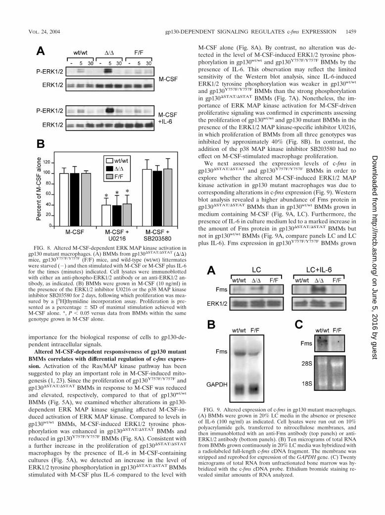
BMMs (Fig. 5A), we examined whether alterations in gp130-dependent ERK MAP kinase signaling affected M-CSF-in-duced activation of ERK MAP kinase. Compared to levels ingp130wt/wt BMMs, M-CSF-induced ERK1/2 tyrosine phos-phorylation was enhanced in gp130�STAT/�STAT BMMs andreduced in gp130Y757F/Y757F BMMs (Fig. 8A). Consistent witha further increase in the proliferation of gp130�STAT/�STAT
macrophages by the presence of IL-6 in M-CSF-containingcultures (Fig. 5A), we detected an increase in the level ofERK1/2 tyrosine phosphorylation in gp130�STAT/�STAT BMMsstimulated with M-CSF plus IL-6 compared to the level with
M-CSF alone (Fig. 8A). By contrast, no alteration was de-tected in the level of M-CSF-induced ERK1/2 tyrosine phos-phorylation in gp130wt/wt and gp130Y757F/Y757F BMMs by thepresence of IL-6. This observation may reflect the limitedsensitivity of the Western blot analysis, since IL-6-inducedERK1/2 tyrosine phosphorylation was weaker in gp130wt/wt
and gp130Y757F/Y757F BMMs than the strong phosphorylationin gp130�STAT/�STAT BMMs (Fig. 7A). Nonetheless, the im-portance of ERK MAP kinase activation for M-CSF-drivenproliferative signaling was confirmed in experiments assessingthe proliferation of gp130wt/wt and gp130 mutant BMMs in thepresence of the ERK1/2 MAP kinase-specific inhibitor U0216,in which proliferation of BMMs from all three genotypes wasinhibited by approximately 40% (Fig. 8B). In contrast, theaddition of the p38 MAP kinase inhibitor SB203580 had noeffect on M-CSF-stimulated macrophage proliferation.
We next assessed the expression levels of c-fms ingp130�STAT/�STAT and gp130Y757F/Y757F BMMs in order toexplore whether the altered M-CSF-induced ERK1/2 MAPkinase activation in gp130 mutant macrophages was due tocorresponding alterations in c-fms expression (Fig. 9). Westernblot analysis revealed a higher abundance of Fms protein ingp130�STAT/�STAT BMMs than in gp130wt/wt BMMs grown inmedium containing M-CSF (Fig. 9A, LC). Furthermore, thepresence of IL-6 in culture medium led to a marked increase inthe amount of Fms protein in gp130�STAT/�STAT BMMs butnot in gp130wt/wt BMMs (Fig. 9A, compare panels LC and LCplus IL-6). Fms expression in gp130Y757F/Y757F BMMs grown
FIG. 8. Altered M-CSF-dependent ERK MAP kinase activation ingp130 mutant macrophages. (A) BMMs from gp130�STAT/�STAT (�/�)mice, gp130Y757F/Y757F (F/F) mice, and wild-type (wt/wt) littermateswere starved (�) and then stimulated with M-CSF or M-CSF plus IL-6for the times (minutes) indicated. Cell lysates were immunoblottedwith either an anti-phospho-ERK1/2 antibody or an anti-ERK1/2 an-tibody, as indicated. (B) BMMs were grown in M-CSF (10 ng/ml) inthe presence of the ERK1/2 inhibitor U0216 or the p38 MAP kinaseinhibitor SB203580 for 2 days, following which proliferation was mea-sured by a [3H]thymidine incorporation assay. Proliferation is pre-sented as a percentage � SD of maximal stimulation achieved withM-CSF alone. *, P 0.05 versus data from BMMs within the samegenotype grown in M-CSF alone.
FIG. 9. Altered expression of c-fms in gp130 mutant macrophages.(A) BMMs were grown in 20% LC media in the absence or presenceof IL-6 (100 ng/ml) as indicated. Cell lysates were run out on 10%polyacrylamide gels, transferred to nitrocellulose membranes, andthen immunoblotted with an anti-Fms antibody (top panels) or anti-ERK1/2 antibody (bottom panels). (B) Ten micrograms of total RNAfrom BMMs grown continuously in 20% LC media was hybridized witha radiolabeled full-length c-fms cDNA fragment. The membrane wasstripped and reprobed for expression of the GAPDH gene. (C) Twentymicrograms of total RNA from unfractionated bone marrow was hy-bridized with the c-fms cDNA probe. Ethidium bromide staining re-vealed similar amounts of RNA analyzed.
VOL. 24, 2004 gp130-DEPENDENT SIGNALING REGULATES c-fms EXPRESSION 1459
on June 5, 2016 by guesthttp://m
cb.asm.org/
Dow
nloaded from

in medium containing M-CSF with or without IL-6 was belowthe level of detection by Western blot analysis (Fig. 9A). How-ever, Northern blot analysis indicated that c-fms was expressed,albeit at a lower level, in gp130Y757F/Y757F BMMs comparedto gp130wt/wt BMMs (Fig. 9B). Similarly, the amount ofc-fms RNA transcript in the bone marrow compartment ofgp130Y757F/Y757F mice was also severalfold lower than in bonemarrow from gp130wt/wt mice (Fig. 9C), confirming that thereduced expression of c-fms in cultured gp130Y757F/Y757F
BMM populations was not the result of their ex vivo expansion.Thus, the data presented in Fig. 8 and 9 correlate with alter-ations in the M-CSF-dependent proliferative response of gp130mutant macrophages with the level of ERK MAP kinase acti-vation, which in turn corresponds with the differential expres-sion of c-fms. In addition, our observations further support thenotion that ERK1/2 are the primary MAP kinases promotingM-CSF-dependent mitogenesis.
The c-fms gene is a direct target of the gp130-dependentSHP2/ERK MAP kinase pathway. The reciprocal negative reg-ulation among the two pathways emanating from gp130 pro-vides a unifying explanation for the opposing biological effectsof IL-6 on gp130�STAT/�STAT and gp130Y757F/Y757F BMMs.Our observation in Fig. 9A that the presence of IL-6 ingp130�STAT/�STAT BMM cultures increased Fms expressionsuggested that the altered capacity of IL-6 to modulate the
M-CSF responsiveness of gp130 mutant macrophages couldcorrelate directly with the extent of gp130-dependent ERKMAP kinase activation, since gp130-dependent STAT activa-tion is abolished in gp130�STAT/�STAT mice. In support of thisnotion, addition of the ERK MAP kinase inhibitor U0216 tocultured gp130�STAT/�STAT BMMs led to a severalfold reduc-tion in the level of Fms protein (Fig. 10A), which suggests thatthe relative overexpression of Fms in gp130�STAT/�STAT
BMMs compared to gp130wt/wt BMMs was due to enhancedactivation of the ERK MAP kinase pathway.
In order to test directly whether gp130-dependent ERK1/2activation enhanced c-fms promoter activity, we assayed thetranscriptional activation of a c-fms promoter–luciferase re-porter construct (14) following its transient transfection inRAW macrophage cells, which also expressed either a chimericG-CSFR/gp130wt or G-CSFR/gp130�STAT receptor construct(7). In unstimulated RAW cells, the 3.5-kb proximal c-fmspromoter showed a high level of basal activity (Fig. 10B), whichwas consistent with previous findings (14). However, c-fms–luciferase activity was reduced by 40% upon ligand-inducedactivation of the G-CSFR/gp130wt receptor. In contrast, c-fmsreporter activity was elevated by 55% upon activation of theG-CSFR/gp130�STAT receptor. Moreover, the presence of theERK1/2 MAP kinase inhibitor U0216 resulted in a threefoldreduction in promoter activity upon G-CSFR/gp130�STAT re-
FIG. 10. Regulation of the c-fms promoter by gp130-dependent ERK1/2 MAP kinase activation. (A) BMMs were grown in 20% LC media inthe absence or presence of the ERK1/2 inhibitor U0216 for 1 h, and cell lysates were subjected to Western blot analysis as described in the legendof Fig. 9A. (B and C) Transcriptional activation of the c-fms–luciferase reporter construct upon transient cotransfection with the G-CSFR/gp130wt
(GR/wt) or G-CSFR/gp130�STAT (GR/�) chimeric receptor constructs (B) and HA-tagged wild-type ERK1 construct (C) in RAW cells. In panelB, transfected cells were incubated with G-CSF (100 ng/ml) in the absence or presence of the ERK1/2 MAP kinase inhibitor U0216 for 48 h andassayed for luciferase activity. The data presented were corrected for transfection efficiency by reference to the activity of Renilla luciferase drivenby the thymidine kinase promoter in a cotransfected plasmid. *, P 0.05 versus basal c-fms–luciferase activity in cells transfected with thec-fms–luciferase reporter construct alone (Fms). (D) Cell lysates from transfected RAW cells were run out on 10% polyacrylamide gels, transferredto nitrocellulose membranes, and then immunoblotted with an anti-ERK1/2 antibody; overexpressed, HA-tagged ERK1 (HA-ERK1; upper band)runs above the endogenous ERK1/2 (middle and lower bands).
1460 JENKINS ET AL. MOL. CELL. BIOL.
on June 5, 2016 by guesthttp://m
cb.asm.org/
Dow
nloaded from

ceptor activation. A role for ERK MAP kinase in the tran-scriptional activation of the c-fms promoter was further dem-onstrated upon transient cotransfection of cells with constructsfor the c-fms promoter–luciferase reporter and wild-typeERK1, overexpression of which has been shown to enhancegene expression (32). As shown in Fig. 10C and 10D, overex-pression of wild-type ERK1 increased c-fms reporter activity by2.5-fold relative to that in cells transfected with the c-fmspromoter–luciferase reporter alone. Therefore, we proposethat the increased M-CSF responsiveness of BMMs followingactivation of the gp130�STAT receptor is attributable to en-hanced ERK MAP kinase activation which is the direct con-sequence of the incapacity of the gp130�STAT receptor to ac-tivate the STAT pathway. Moreover, our data presented hereidentify a unique mechanism whereby the differential regula-tion of the c-fms gene is dependent upon the level of gp130-dependent ERK1/2 MAP kinase activation.
DISCUSSION
We report here the use of mice deficient in gp130-dependentactivation of either STAT1/3 (gp130�STAT/�STAT) or SHP2/ERK MAP kinase (gp130Y757F/Y757F) to demonstrate that thecapacity of IL-6 family cytokines to inhibit M-CSF-dependentproliferation and differentiation of hematopoietic cells is in-versely related to their ability to activate gp130-dependentERK MAP kinase signaling. Hence, IL-6 and, to a lesser ex-tent, IL-11 exhibited greater inhibition of macrophage colonyformation (in response to M-CSF) from peripheral blood andbone marrow M-CFCs from gp130Y757F/Y757F mice than fromgp130wt/wt mice. By contrast, in gp130�STAT/�STAT mice, wheregp130-dependent ERK MAP kinase activation was enhanced,IL-6 and IL-11 failed to inhibit macrophage colony forma-tion by M-CFCs but, rather, augmented colony formation.Similarly, the presence of IL-6 in liquid cultures enhancedthe proliferation of gp130�STAT/�STAT BMMs in responseto M-CSF, whereas M-CSF-stimulated proliferation ofgp130Y757F/Y757F BMMs was reduced. In all cases, the inhibi-tion by IL-11 was less pronounced than inhibition by IL-6, anobservation which is consistent with that made by Clutterbucket al. (4), and may reflect reduced surface expression of IL-11receptor (IL-11R) compared to expression of IL-6R onmacrophage progenitor cells. We also note that peripheralblood M-CFCs from gp130wt/wt and gp130Y757F/Y757F micewere more sensitive than bone marrow M-CFCs to IL-6-in-duced inhibition of macrophage colony formation. The inhib-itory effect of IL-6 has been previously shown to be inverselyproportional to the proliferative capacity of the macrophageprogenitor, in which case peripheral blood progenitors pro-duce smaller colonies (i.e., have lower proliferative potential)than bone marrow progenitors and are more sensitive to IL-6inhibition (4). Although the molecular explanation for thisdifferential IL-6 sensitivity is unknown, it most likely reflects areprogramming of the transcriptional machinery within a pro-genitor cell during differentiation or maturation, which is as-sociated with a reduced proliferative capacity, to a growthinhibitory response by IL-6.
In gp130�STAT/�STAT and gp130Y757F/Y757F BMMs, wefound that functional elimination of one signaling pathwayresulted in enhanced activation of the remaining pathway in
response to IL-6. This observation is consistent with theemerging theme that the gp130-dependent SHP2/ERK MAPkinase and STAT1/3 pathways negatively regulate each other(31, 38). We have previously shown that the absence of gp130-dependent STAT1/3 signaling causes up-regulation of IL-6-induced SHP2 and ERK1/2 tyrosine phosphorylation due toimpaired STAT-mediated induction of SOCS1 and SOCS3,two key negative regulators of gp130 signaling (6). Conversely,the gp130Y757F substitution mutation removes the phosphoty-rosine residue that simultaneously provides a binding site forSHP2 and SOCS3, and therefore disrupts the capacity ofSOCS3 to negatively regulate gp130-dependent signaling (35)and of SHP2 to dephosphorylate critical tyrosine residues inthe gp130-JAK signaling complex (18). Therefore, the collec-tive loss of these negative feedback mechanisms may augmentJAK activity and subsequent STAT3 activation ingp130Y757F/Y757F mice. Importantly, this reciprocal negativeregulation between the STAT and ERK MAP kinase pathwayshighlights how the overall balance of positive and negativesignaling cascades can mediate specific biological responses toIL-6 family cytokines. Indeed, recent examples include thefindings that mucosal wound healing (38) and the Th1 versusTh2 immune response (31) are influenced by the balance ofgp130-dependent SHP2/ERK MAP kinase and STAT3 activa-tion.
The other major finding uncovered by our study presentedhere is the regulation of c-fms gene expression by IL-6 familycytokines via the gp130-dependent ERK MAP kinase pathway.The c-fms promoter contains several conserved Ets/AP-1growth factor response elements (15), and the synergistic ac-tions of the Ras pathway and Ets family transcription factors toinduce expression of M-CSF-responsive genes have been dem-onstrated (12, 41). Furthermore, activation of the Ras/ERKMAP kinase pathway is necessary for both Ets-2 gene expres-sion and protein activation (8), which in turn positively regu-lates Ras-dependent proliferation in response to M-CSF (20,21). The Ras pathway has been shown to be a signaling com-ponent downstream of Fms that is necessary for M-CSF-drivenmitogenesis (2, 21), which is also supported by our observationhere that the ERK1/2 MAP kinase-specific inhibitor U0216reduced M-CSF-dependent proliferation of BMMs. However,here we provide compelling evidence for a situation where theRas-MAP kinase pathway (activated via gp130) can also befunctionally upstream of Fms, thereby providing a link bywhich IL-6 family cytokines suppress the responsiveness ofmacrophages to M-CSF. Thus, we speculate that a reduction ingp130-dependent ERK1/2 MAP kinase signaling, as is the casefor gp130Y757F/Y757F macrophages, might suppress the activityof Ets family members, and therefore the level of c-fms expres-sion, which in turn leads to a reduced biological response toM-CSF-induced signaling. Detailed analysis of the ability ofgp130-dependent ERK MAP kinase signals to induce the ex-pression of downstream effectors of M-CSF-stimulated prolif-erative signaling, such as Ets-2 and AP-1, and induce c-fmspromoter activity in the absence of specific (i.e., Ets/AP-1)binding sites, will ultimately test the validity of this model.
Several recent studies have highlighted the importance ofSTAT3 signaling during maturation and activation of macro-phages. A study by Takeda et al. (37) revealed that IL-6 notonly failed to inhibit the M-CSF-driven proliferation of
VOL. 24, 2004 gp130-DEPENDENT SIGNALING REGULATES c-fms EXPRESSION 1461
on June 5, 2016 by guesthttp://m
cb.asm.org/
Dow
nloaded from

STAT3-deficient BMMs but, rather, enhanced their prolifera-tion. Notably, this observation correlates with our data pre-sented here for gp130�STAT/�STAT macrophages and is there-fore consistent with our interpretation that exaggerated gp130-dependent ERK MAP kinase activation, as a consequence ofan absence in STAT3 signaling, enhances these macrophageactivities. In contrast, it has also been reported that constitu-tive activation of STAT3 in macrophages inhibited their pro-liferation (30). While these reports provide compelling evi-dence for a STAT3-specific role in mediating antiproliferativeeffects on macrophages, they provide little evidence as to howaltering the activation status of STAT3 elicits these actions. Inlight of our observations here, we propose that altering thelevel of STAT3 activation, for example by gene-specific inhi-bition or expression of a constitutively active STAT3, mayaffect target gene regulation indirectly by unbalancing the netoutput of complex signaling networks. Specifically, we identifyhere that, at least in the context of gp130 activation, the sig-naling output from the ERK MAP kinase cascade is inverselyrelated to the extent of STAT3 activation.
The inhibition of M-CSF-stimulated macrophage formationby IL-6 family cytokines contrasts with the synergistic actionsof this cytokine family with other hematopoietic growth fac-tors, such as stem cell factor and IL-3, on promoting the pro-liferation and differentiation of multipotential (immature) he-matopoietic progenitors (16, 39). Furthermore, IL-6 promotesthe formation of macrophages in cultures containing granulo-cyte-M-CSF (17, 27) and has also been shown to enhance theexpansion of primitive macrophage-committed (pre-CFU-M)progenitors (26). These observations suggest differences in thetranscriptional programming of immature and committed (i.e.,CFU-M) hematopoietic progenitors to respond to gp130 sig-naling, with the latter primed for inhibition by IL-6 familycytokines. Indeed, such a notion is supported by recent obser-vations by Clutterbuck et al. (4) on the preferential inhibitoryaction of IL-6 on macrophage progenitors displaying a reducedproliferative capacity, which itself is a hallmark of committedcells undergoing differentiation and maturation. Although amolecular mechanism underlying these opposing effects re-mains to be elucidated, in both situations it is likely that theeffect of IL-6 is via the regulation of c-fms expression (3). Inthis regard, it is therefore not surprising that the human andmouse c-fms promoter regions contain binding sites for tran-scription factors that mediate opposing effects on the transcrip-tional regulation of c-fms (33). With the recent generation ofmice expressing a c-fms-enhanced green fluorescent proteinreporter gene within the monocyte/macrophage lineagethroughout development (34), the tools are now in place forfurther in vivo analyses of the transcriptional machinery con-trolling both negative and positive regulation of c-fms duringmacrophage development.
ACKNOWLEDGMENTS
We are grateful to Graham Lieschke and Ken Harder for valuablediscussions and critical reading of the manuscript, and we thank Nich-olas Wilson and Tony Burgess for critical reading of the manuscript.We also thank Elsbeth Richardson and Maureen Nerrie for technicalassistance.
This work was supported in part by a research grant from theNational Health and Medical Research Council of Australia.
REFERENCES
1. Bortner, D. M., M. Ulivi, M. F. Roussel, and M. C. Ostrowski. 1991. Thecarboxy-terminal catalytic domain of the GTPase-activating protein inhibitsnuclear signal transduction and morphological transformation mediated bythe CSF-1 receptor. Genes Dev. 5:1777–1785.
2. Buscher, D., R. A. Hipskind, S. Krautwald, T. Reimann, and M. Baccarini.1995. Ras-dependent and -independent pathways target the mitogen-acti-vated protein kinase network in macrophages. Mol. Cell. Biol. 15:466–475.
3. Chomarat, P., J. Banchereau, J. Davoust, and A. K. Palucka. 2000. IL-6switches the differentiation of monocytes from dendritic cells to macro-phages. Nat. Immunol. 1:510–514.
4. Clutterbuck, R., R. Powles, J. Millar, and D. Catovsky. 2000. Interleukin-6and other gp130-dependent cytokines selectively inhibit proliferation of mac-rophage-lineage hemopoietic progenitor cells. Exp. Hematol. 28:1120–1128.
5. Dai, X. M., G. R. Ryan, A. J. Hapel, M. G. Dominguez, R. G. Russell, S.Kapp, V. Sylvestre, and E. R. Stanley. 2002. Targeted disruption of themouse colony-stimulating factor 1 receptor gene results in osteopetrosis,mononuclear phagocyte deficiency, increased primitive progenitor cell fre-quencies, and reproductive defects. Blood 99:111–120.
6. Ernst, M., M. Inglese, P. Waring, I. K. Campbell, S. Bao, F. J. Clay, W. S.Alexander, I. P. Wicks, D. M. Tarlinton, U. Novak, J. K. Heath, and A. R.Dunn. 2001. Defective gp130-mediated signal transducer and activator oftranscription (STAT) signaling results in degenerative joint disease, gastro-intestinal ulceration, and failure of uterine implantation. J. Exp. Med. 194:189–203.
7. Ernst, M., U. Novak, S. E. Nicholson, J. E. Layton, and A. R. Dunn. 1999.The carboxyl-terminal domains of gp130-related cytokine receptors are nec-essary for suppressing embryonic stem cell differentiation: involvement ofSTAT3. J. Biol. Chem. 274:9729–9737.
8. Fowles, L. F., M. L. Martin, L. Nelsen, K. J. Stacey, D. Redd, Y. M. Clark,Y. Nagamine, M. McMahon, D. A. Hume, and M. C. Ostrowski. 1998.Persistent activation of mitogen-activated protein kinases p42 and p44 andets-2 phosphorylation in response to colony-stimulating factor 1/c-fms sig-naling. Mol. Cell. Biol. 18:5148–5156.
9. Fukada, T., M. Hibi, Y. Yamanaka, M. Takahashi-Tezuka, Y. Fujitani, T.Yamaguchi, K. Nakajima, and T. Hirano. 1996. Two signals are necessary forcell proliferation induced by a cytokine receptor gp130: involvement ofSTAT3 in anti-apoptosis. Immunity 5:449–460.
10. Gearing, D. P., C. J. Thut, T. VandeBos, S. D. Gimpel, P. B. Delaney, J. King,V. Price, D. Cosman, and M. P. Beckmann. 1991. Leukemia inhibitory factorreceptor is structurally related to the IL-6 signal transducer, gp130. EMBOJ. 10:2839–2848.
11. Gerhartz, C., B. Heesel, J. Sasse, U. Hemmann, C. Landgraf, J. Schneider-Mergener, F. Horn, P. C. Heinrich, and L. Graeve. 1996. Differential acti-vation of acute phase response factor/STAT3 and STAT1 via the cytoplasmicdomain of the interleukin 6 signal transducer gp130. I. Definition of a novelphosphotyrosine motif mediating STAT1 activation. J. Biol. Chem. 271:12991–12998.
12. Guidez, F., A. C. Li, A. Horvai, J. S. Welch, and C. K. Glass. 1998. Differ-ential utilization of Ras signaling pathways by macrophage colony-stimulat-ing factor (CSF) and granulocyte-macrophage CSF receptors during macro-phage differentiation. Mol. Cell. Biol. 18:3851–3861.
13. Hilton, D. J., A. A. Hilton, A. Raicevic, S. Rakar, M. Harrison-Smith, N. M.Gough, C. G. Begley, D. Metcalf, N. A. Nicola, and T. A. Willson. 1994.Cloning of a murine IL-11 receptor alpha-chain: requirement for gp130 forhigh affinity binding and signal transduction. EMBO J. 13:4765–4775.
14. Himes, S. R., H. Tagoh, N. Goonetilleke, T. Sasmono, D. Oceandy, R. Clark,C. Bonifer, and D. A. Hume. 2001. A highly conserved c-fms gene intronicelement controls macrophage-specific and regulated expression. J. Leukoc.Biol. 70:812–820.
15. Hume, D. A., X. Yue, I. L. Ross, P. Favot, A. Lichanska, and M. C. Ostrowski.1997. Regulation of CSF-1 receptor expression. Mol. Reprod. Dev. 46:46–52.
16. Ikebuchi, K., G. G. Wong, S. C. Clark, J. N. Ihle, Y. Hirai, and M. Ogawa.1987. Interleukin 6 enhancement of interleukin 3-dependent proliferation ofmultipotential hemopoietic progenitors. Proc. Natl. Acad. Sci. USA 84:9035–9039.
17. Jansen, J. H., J. C. Kluin-Nelemans, J. Van Damme, G. J. Wientjens, R.Willemze, and W. E. Fibbe. 1992. Interleukin 6 is a permissive factor formonocytic colony formation by human hematopoietic progenitor cells. J.Exp. Med. 175:1151–1154.
18. Kim, H., T. S. Hawley, R. G. Hawley, and H. Baumann. 1998. Proteintyrosine phosphatase 2 (SHP-2) moderates signaling by gp130 but is notrequired for the induction of acute-phase plasma protein genes in hepaticcells. Mol. Cell. Biol. 18:1525–1533.
19. Kishimoto, T., S. Akira, M. Narazaki, and T. Taga. 1995. Interleukin-6family of cytokines and gp130. Blood 86:1243–1254.
20. Klappacher, G. W., V. V. Lunyak, D. B. Sykes, D. Sawka-Verhelle, J. Sage, G.Brard, S. D. Ngo, D. Gangadharan, T. Jacks, M. P. Kamps, D. W. Rose,M. G. Rosenfeld, and C. K. Glass. 2002. An induced Ets repressor complexregulates growth arrest during terminal macrophage differentiation. Cell109:169–180.
1462 JENKINS ET AL. MOL. CELL. BIOL.
on June 5, 2016 by guesthttp://m
cb.asm.org/
Dow
nloaded from
21. Langer, S. J., D. M. Bortner, M. F. Roussel, C. J. Sherr, and M. C. Os-trowski. 1992. Mitogenic signaling by colony-stimulating factor 1 and ras issuppressed by the ets-2 DNA-binding domain and restored by myc overex-pression. Mol. Cell. Biol. 12:5355–5362.
22. Lawlor, K. E., I. K. Campbell, K. O’Donnell, L. Wu, and I. P. Wicks. 2001.Molecular and cellular mediators of interleukin-1-dependent acute inflam-matory arthritis. Arthritis Rheum. 44:442–450.
23. Lioubin, M. N., G. M. Myles, K. Carlberg, D. Bowtell, and L. R. Rohrschnei-der. 1994. Shc, Grb2, Sos1, and a 150-kilodalton tyrosine-phosphorylatedprotein form complexes with Fms in hematopoietic cells. Mol. Cell. Biol.14:5682–5691.
24. Liu, H., H. Perlman, L. J. Pagliari, and R. M. Pope. 2001. Constitutivelyactivated Akt-1 is vital for the survival of human monocyte-differentiatedmacrophages: role of Mcl-1, independent of nuclear factor (NF)-�B, Bad, orcaspase activation. J. Exp. Med. 194:113–125.
25. Lord, K. A., A. Abdollahi, S. M. Thomas, M. DeMarco, J. S. Brugge, B.Hoffman-Liebermann, and D. A. Liebermann. 1991. Leukemia inhibitoryfactor and interleukin-6 trigger the same immediate early response, includ-ing tyrosine phosphorylation, upon induction of myeloid leukemia differen-tiation. Mol. Cell. Biol. 11:4371–4379.
26. Metcalf, D. 1997. Murine hematopoietic stem cells committed to macro-phage/dendritic cell formation: stimulation by Flk2-ligand with enhancementby regulators using the gp130 receptor chain. Proc. Natl. Acad. Sci. USA94:11552–11556.
27. Mitani, H., N. Katayama, H. Araki, K. Ohishi, K. Kobayashi, H. Suzuki, K.Nishii, M. Masuya, K. Yasukawa, N. Minami, and H. Shiku. 2000. Activityof interleukin 6 in the differentiation of monocytes to macrophages anddendritic cells. Br. J. Haematol. 109:288–295.
28. Mosley, B., C. De Imus, D. Friend, N. Boiani, B. Thoma, L. S. Park, and D.Cosman. 1996. Dual oncostatin M (OSM) receptors: cloning and character-ization of an alternative signaling subunit conferring OSM-specific receptoractivation. J. Biol. Chem. 271:32635–32643.
29. Murakami, M., M. Hibi, N. Nakagawa, T. Nakagawa, K. Yasukawa, K.Yamanishi, T. Taga, and T. Kishimoto. 1993. IL-6-induced homodimeriza-tion of gp130 and associated activation of a tyrosine kinase. Science 260:1808–1810.
30. O’Farrell, A. M., Y. Liu, K. W. Moore, and A. L. Mui. 1998. IL-10 inhibitsmacrophage activation and proliferation by distinct signaling mechanisms:evidence for Stat3-dependent and -independent pathways. EMBO J. 17:1006–1018.
31. Ohtani, T., K. Ishihara, T. Atsumi, K. Nishida, Y. Kaneko, T. Miyata, S.Itoh, M. Narimatsu, H. Maeda, T. Fukada, M. Itoh, H. Okano, M. Hibi, andT. Hirano. 2000. Dissection of signaling cascades through gp130 in vivo:
reciprocal roles for STAT3- and SHP2-mediated signals in immune re-sponses. Immunity 12:95–105.
32. Park, J. H., and L. Levitt. 1993. Overexpression of mitogen-activated proteinkinase (ERK1) enhances T-cell cytokine gene expression: role of AP1, NF-AT, and NF-KB. Blood 82:2470–2477.
33. Reddy, M. A., B. S. Yang, X. Yue, C. J. Barnett, I. L. Ross, M. J. Sweet, D. A.Hume, and M. C. Ostrowski. 1994. Opposing actions of c-ets/PU. 1 andc-myb protooncogene products in regulating the macrophage-specific pro-moters of the human and mouse colony-stimulating factor-1 receptor (c-fms)genes. J. Exp. Med. 180:2309–2319.
34. Sasmono, R. T., D. Oceandy, J. W. Pollard, W. Tong, P. Pavli, B. J. Wain-wright, M. C. Ostrowski, S. R. Himes, and D. A. Hume. 2003. A macrophagecolony-stimulating factor receptor-green fluorescent protein transgene isexpressed throughout the mononuclear phagocyte system of the mouse.Blood 101:1155–1163.
35. Schmitz, J., M. Weissenbach, S. Haan, P. C. Heinrich, and F. Schaper. 2000.SOCS3 exerts its inhibitory function on interleukin-6 signal transductionthrough the SHP2 recruitment site of gp130. J. Biol. Chem. 275:12848–12856.
36. Stanley, E. R., K. L. Berg, D. B. Einstein, P. S. Lee, F. J. Pixley, Y. Wang, andY. G. Yeung. 1997. Biology and action of colony-stimulating factor-1. Mol.Reprod. Dev. 46:4–10.
37. Takeda, K., B. E. Clausen, T. Kaisho, T. Tsujimura, N. Terada, I. Forster,and S. Akira. 1999. Enhanced Th1 activity and development of chronicenterocolitis in mice devoid of Stat3 in macrophages and neutrophils. Im-munity 10:39–49.
38. Tebbutt, N. C., A. S. Giraud, M. Inglese, B. Jenkins, P. Waring, F. J. Clay,S. Malki, B. M. Alderman, D. Grail, F. Hollande, J. K. Heath, and M. Ernst.2002. Reciprocal regulation of gastrointestinal homeostasis by SHP2 andSTAT-mediated trefoil gene activation in gp130 mutant mice. Nat. Med.8:1089–1097.
39. Tsuji, K., S. D. Lyman, T. Sudo, S. C. Clark, and M. Ogawa. 1992. Enhance-ment of murine hematopoiesis by synergistic interactions between steel fac-tor (ligand for c-kit), interleukin-11, and other early acting factors in culture.Blood 79:2855–2860.
40. Wiktor-Jedrzejczak, W., A. Bartocci, A. W. Ferrante, Jr., A. Ahmed-Ansari,K. W. Sell, J. W. Pollard, and E. R. Stanley. 1990. Total absence of colony-stimulating factor 1 in the macrophage-deficient osteopetrotic (op/op)mouse. Proc. Natl. Acad. Sci. USA 87:4828–4832.
41. Yang, B. S., C. A. Hauser, G. Henkel, M. S. Colman, C. Van Beveren, K. J.Stacey, D. A. Hume, R. A. Maki, and M. C. Ostrowski. 1996. Ras-mediatedphosphorylation of a conserved threonine residue enhances the transactiva-tion activities of c-Ets1 and c-Ets2. Mol. Cell. Biol. 16:538–547.
VOL. 24, 2004 gp130-DEPENDENT SIGNALING REGULATES c-fms EXPRESSION 1463
on June 5, 2016 by guesthttp://m
cb.asm.org/
Dow
nloaded from