Animal models of ovarian cancer
11
BioMed Central Page 1 of 11 (page number not for citation purposes) Reproductive Biology and Endocrinology Open Access Review Animal models of ovarian cancer Barbara C Vanderhyden* 1,2,3 , Tanya J Shaw 1,3 and Jean-François Ethier 1,3 Address: 1 Department of Cellular and Molecular Medicine, University of Ottawa, 451 Smyth Road, Ottawa, Ontario, Canada K1H 8M5, 2 Department of Obstetrics and Gynecology, University of Ottawa, 501 Smyth Road, Ottawa, Ontario, Canada K1H 8L6 and 3 Ottawa Regional Cancer Centre, 503 Smyth Road, Ottawa, Ontario, Canada K1H 1C4 Email: Barbara C Vanderhyden* - [email protected]; Tanya J Shaw - [email protected]; Jean- François Ethier - [email protected] * Corresponding author Abstract Ovarian cancer is the most lethal of all of the gynecological cancers and can arise from any cell type of the ovary, including germ cells, granulosa or stromal cells. However, the majority of ovarian cancers arise from the surface epithelium, a single layer of cells that covers the surface of the ovary. The lack of a reliable and specific method for the early detection of epithelial ovarian cancer results in diagnosis occurring most commonly at late clinical stages, when treatment is less effective. In part, the deficiency in diagnostic tools is due to the lack of markers for the detection of preneoplastic or early neoplastic changes in the epithelial cells, which reflects our rather poor understanding of this process. Animal models which accurately represent the cellular and molecular changes associated with the initiation and progression of human ovarian cancer have significant potential to facilitate the development of better methods for the early detection and treatment of ovarian cancer. This review describes some of the experimental animal models of ovarian tumorigenesis that have been reported, including those involving specific reproductive factors and environmental toxins. Consideration has also been given to the recent progress in modeling ovarian cancer using genetically engineered mice. Introduction Despite improved knowledge of the etiology of ovarian cancer, aggressive cytoreductive surgery, and modern combination chemotherapy, there has been little change in the mortality statistics over the last 30 years, and approximately 60% of the women who develop ovarian cancer will die from their disease. Lack of an adequate screening test for early disease detection and the rapid progression to chemoresistance have prevented apprecia- ble improvement in the five year survival rate of patients with ovarian cancer. Experimental models for human diseases are of crucial importance not only to understand the biological and genetic factors that influence the phenotypic characteris- tics of the disease but to utilize as a basis for developing rational intervention strategies. Ovarian cancer cell lines derived from ascites or primary ovarian tumors have been used extensively and can be very effective for studying the processes controlling growth regulation and chemosensi- tivity. Our limited knowledge of the initiating events of ovarian cancer has restricted the development of models in which the early pathogenic events for ovarian cancer can be studied. However, there are a few animal models that develop ovarian tumors spontaneously, and others where the manipulation of various reproductive factors or exposure to environmental toxins have been shown to promote ovarian tumorigenesis. Finally, the recent Published: 07 October 2003 Reproductive Biology and Endocrinology 2003, 1:67 Received: 28 June 2003 Accepted: 07 October 2003 This article is available from: http://www.RBEj.com/content/1/1/67 © 2003 Vanderhyden et al; licensee BioMed Central Ltd. This is an Open Access article: verbatim copying and redistribution of this article are permitted in all media for any purpose, provided this notice is preserved along with the article's original URL.
-
Upload
independent -
Category
Documents
-
view
2 -
download
0
Transcript of Animal models of ovarian cancer

BioMed Central
Reproductive Biology and Endocrinology
ss
Open AcceReviewAnimal models of ovarian cancerBarbara C Vanderhyden*1,2,3, Tanya J Shaw1,3 and Jean-François Ethier1,3Address: 1Department of Cellular and Molecular Medicine, University of Ottawa, 451 Smyth Road, Ottawa, Ontario, Canada K1H 8M5, 2Department of Obstetrics and Gynecology, University of Ottawa, 501 Smyth Road, Ottawa, Ontario, Canada K1H 8L6 and 3Ottawa Regional Cancer Centre, 503 Smyth Road, Ottawa, Ontario, Canada K1H 1C4
Email: Barbara C Vanderhyden* - [email protected]; Tanya J Shaw - [email protected]; Jean-François Ethier - [email protected]
* Corresponding author
AbstractOvarian cancer is the most lethal of all of the gynecological cancers and can arise from any cell typeof the ovary, including germ cells, granulosa or stromal cells. However, the majority of ovariancancers arise from the surface epithelium, a single layer of cells that covers the surface of the ovary.The lack of a reliable and specific method for the early detection of epithelial ovarian cancer resultsin diagnosis occurring most commonly at late clinical stages, when treatment is less effective. Inpart, the deficiency in diagnostic tools is due to the lack of markers for the detection ofpreneoplastic or early neoplastic changes in the epithelial cells, which reflects our rather poorunderstanding of this process. Animal models which accurately represent the cellular and molecularchanges associated with the initiation and progression of human ovarian cancer have significantpotential to facilitate the development of better methods for the early detection and treatment ofovarian cancer. This review describes some of the experimental animal models of ovariantumorigenesis that have been reported, including those involving specific reproductive factors andenvironmental toxins. Consideration has also been given to the recent progress in modelingovarian cancer using genetically engineered mice.
IntroductionDespite improved knowledge of the etiology of ovariancancer, aggressive cytoreductive surgery, and moderncombination chemotherapy, there has been little changein the mortality statistics over the last 30 years, andapproximately 60% of the women who develop ovariancancer will die from their disease. Lack of an adequatescreening test for early disease detection and the rapidprogression to chemoresistance have prevented apprecia-ble improvement in the five year survival rate of patientswith ovarian cancer.
Experimental models for human diseases are of crucialimportance not only to understand the biological and
genetic factors that influence the phenotypic characteris-tics of the disease but to utilize as a basis for developingrational intervention strategies. Ovarian cancer cell linesderived from ascites or primary ovarian tumors have beenused extensively and can be very effective for studying theprocesses controlling growth regulation and chemosensi-tivity. Our limited knowledge of the initiating events ofovarian cancer has restricted the development of modelsin which the early pathogenic events for ovarian cancercan be studied. However, there are a few animal modelsthat develop ovarian tumors spontaneously, and otherswhere the manipulation of various reproductive factors orexposure to environmental toxins have been shown topromote ovarian tumorigenesis. Finally, the recent
Published: 07 October 2003
Reproductive Biology and Endocrinology 2003, 1:67
Received: 28 June 2003Accepted: 07 October 2003
This article is available from: http://www.RBEj.com/content/1/1/67
© 2003 Vanderhyden et al; licensee BioMed Central Ltd. This is an Open Access article: verbatim copying and redistribution of this article are permitted in all media for any purpose, provided this notice is preserved along with the article's original URL.
Page 1 of 11(page number not for citation purposes)

Reproductive Biology and Endocrinology 2003, 1 http://www.RBEj.com/content/1/1/67
identification of promoters that can drive gene expressionin the ovarian surface epithelium is providing new oppor-tunities for the generation of genetically engineeredmouse models of ovarian cancer. Here we describe someof the models that have been developed to investigateovarian cell transformation.
Spontaneous and Non-epithelial Ovarian TumorigenesisThere are few animal models that develop ovarian tumorsspontaneously. Hens maintained under intensive egg-lay-ing conditions develop ovarian adenocarcinomas; how-ever such tumors are uncommon in hens less than 2 yearsof age [1]. Ovarian tumors will also arise spontaneouslywith age in some strains of mice [2], and in Wistar andSprague-Dawley rats [3,4]. These tumors show a wide vari-ety of histologic sub-types, including tubular adenoma,adenocarcinoma, papillary cystadenoma, mesothelioma,granulosa cell tumor, and polycystic sex cord/stromaltumor. However, the low incidence and/or the length oftime required for the appearance of tumors in all of thesemodels render them poorly feasible for experimentalstudies of ovarian carcinogenesis.
Some strains of mice, including C3HeB/Fe and C3HeB/De, show a high incidence of spontaneously occurringgranulosa cell tumors and tubular adenomas [5]. StrainHAN:NMRI develop spontaneous Sertoli cell-like tumorsand (DBA × Ce)F1 hybrids have a high incidence granu-losa cell tumors [5]. Granulosa cell tumors also appearspontaneously at 4–6 weeks of age in SWR/J and in SWR/Bm inbred strain mice, with a maximum incidencereached by 10 weeks [6]. In some SWXJ strains, granulosacell tumors occur spontaneously, and in others granulosatumors can only be induced by treatment with dehydroe-piandrosterone [7].
Spontaneous germ cell tumors are less common, but havebeen reported in LT/Sv and related strains of mice. Thesemice have a high frequency of spontaneous ovarian ter-atomas arising from follicular oocytes that undergo par-thenogenetic activation. In some strains, this defectappears to be associated with an arrest of the oocytes atmetaphase of meiosis I [8]. Teratomas arising from par-thenogenetic activation of oocytes also occur in c-mos-deficient oocytes, which fail to maintain meiotic arrestafter oocyte maturation [9,10].
Mice generated to be deficient in the tumor suppressorgene Lats1 exhibit a lack of mammary gland development,infertility and growth retardation. Accompanying thesedefects are hyperplastic changes in the pituitary anddecreased serum hormone levels. The reproductive hor-mone defects of Lats1-/- mice are reminiscent of isolatedLH-hypogonadotropic hypogonadism and corpus luteum
insufficiency in humans. Lats1-/- mice develop soft-tissuesarcomas and ovarian stromal cell tumors [11].
The Ovarian Surface EpitheliumAlthough ovarian cancer in humans can arise from any ofthe cell types found in the ovary, almost 90% are derivedfrom the ovarian surface epithelium (OSE) [12]. The OSEcovers the entire ovarian surface, and varies morphologi-cally from simple squamous to cuboidal to low pseudos-tratified columnar [13,14]. Embryologically derived fromthe mesodermal epithelium of the gonadal ridges, OSEcells are continuous with the flattened mesothelium ofthe peritoneum [15] and are separated from the underly-ing stromal compartment of the ovary by a basementmembrane. Immunohistochemical staining has shownthat OSE cells express cytokeratin, desmoplakin, trans-forming growth factor-α (TGF-α) and receptors for estro-gen, progesterone and epidermal growth factor (EGF)[16–20]. Despite their rather unremarkable appearance invivo, it is believed that OSE cells actively participate in theovulatory process. Studies in rabbits and sheep haveshown that OSE release proteolytic enzymes that degradethe basement membrane and the underlying apical follic-ular wall, weakening the ovarian surface to the point ofrupture [21]. The OSE cells directly over the point of rup-ture undergo apoptotic cell death before ovulation [22]and the wound created at the ovulatory site surface isrepaired by rapid proliferation of OSE cells from theperimeter of the ruptured follicle [23]. The biology, endo-crinology and pathology of the ovarian surface epitheliumhave recently been reviewed in detail [24].
Although the ovarian surface is generally smooth in earlyreproductive life, with aging the ovary becomes more con-voluted. Invaginations of the epithelium result in cryptsor gland-like structures that can become pinched off toform epithelial inclusion cysts within the underlying stro-mal compartment [25]. This may occur following the pos-tovulatory proliferation of OSE, follicular attrition, and/or from inflammation caused by carcinogens or chemicalirritants like talcum powder [26]. The incidence of inclu-sion cysts increases with advancing age and are commonin postmenopausal women. Although generally benign innature, these epithelial rearrangements are widelythought to be the potential origin of many epithelial can-cers. The more frequent appearance of epithelial invagina-tions and inclusion cysts in women with hereditary risk ofovarian cancer has strengthened this hypothesis [27]. Inaddition, some microscopic borderline and malignanttumors have been observed to arise directly within thesesites, and they are often associated with dysplasia in simi-lar sites elsewhere in the same or contralateral ovary[28,29].
Page 2 of 11(page number not for citation purposes)

Reproductive Biology and Endocrinology 2003, 1 http://www.RBEj.com/content/1/1/67
Xenografts of OSE Cells Transformed in vitroOSE cells have been implicated as the cell of origin for themajority of ovarian cancers based primarily on histologi-cal and immunohistochemical analyses of patient sam-ples, but several recent experimental modelsmanipulating these cells in vitro have provided additionalsupport for this concept. Primary culture of human OSEwas first reported by Auersperg et al. in 1984 [30], and hergroup has since developed several in vitro models of ovar-ian epithelial carcinogenesis. Introduction of Kirstenmurine sarcoma virus into rat OSE cells results inendometrioid tumors following subcutaneous or intra-peritoneal injection into immunosuppressed rats [31].Transfection of SV40 T antigen early genes inducesimmortalization of human OSE cells that delays, but doesnot prevent, the senescence that normally occurs after afew passages [32]. Introduction of E-cadherin into these Tantigen-immortalized cells induces epithelial differentia-tion [33] and the cells formed transplantable, invasiveadenocarcinomas when injected into SCID mice [34]. Incontrast to T antigen-immortalized cells, introduction ofthe human papilloma virus E6 and E7 genes into humanOSE cells results in the spontaneous progression from abenign to invasive phenotype [35].
Unlike human OSE, rat and mouse OSE do not senesce.Rat OSE cells that have spontaneously immortalized butare not tumorigenic (eg. ROSE 199 cells; [36]) have beenused in a variety of experiments, including some to char-acterize the cellular features when SV40 T antigen or H-rasis introduced into immortalized cells and following theformation of tumors when these cells are xenografted intonude mice [37]. Repeated subculture of rat and mouseOSE cells to maintain continued proliferation results inspontaneous malignant transformation, as characterizedby loss of contact inhibition, substrate-independentgrowth and the ability to form tumors in nude mice[38,39]. In a variation of the above in vitro transformationapproaches, Orsulic and colleagues used the RCAS retro-viral vector to introduce oncogenes into OSE cells fromtransgenic mice bearing the RCAS receptor TVA and thecells were evaluated for tumorigenicity by injection intoimmune-deficient or syngeneic animals [40]. The investi-gators found that p53 deficiency in combination with twooncogenes from among C-MYC, K-RAS, or AKT wererequired to achieve transformation.
While these models allow an evaluation of oncogeneswhose activation may contribute to the development ofepithelial ovarian cancer, this approach does not allowthe investigation of the early events in ovarian tumorigen-esis inherent in mice when the tumors arise in situ. How-ever, the establishment of in vitro models of normal andtransformed OSE cells has provided the opportunity touse molecular approaches such as microarray or suppres-
sion subtractive hybridization to identify differential geneexpression patterns that can distinguish normal OSE andovarian cancer cells [41,42]. These data will be useful forthe elucidation of molecular events associated with OSEcell transformation.
Xenografts of Cancer CellsXenograft models, where ovarian cancer cells have beeninjected either subcutaneously or into the peritoneal cav-ity have been used extensively for the testing of novel ther-apeutics or modified regimens for administration ofstandard chemotherapeutic drugs [43–45]. Some mousemodels take advantage of the presence of a bursa, a sac-like structure that envelops rodent ovaries. For decades,researchers have used the intra-bursal space for trans-plants of xenografted ovaries, or to facilitate direct expo-sure of the ovary to various factors. For the generation ofmouse models of ovarian cancer, the injection of ovariancancer cells into the intra-bursal space results in tumorformation that can perhaps be viewed as more physiolog-ical (Figure 1), as the cancer cells are placed directly in theenvironment where ovarian tumors normally arise [46].
Reproductive Factors and Ovarian TumorigenesisUnlike most other cancers, the series of events involved inthe initiation, progression and metastasis of ovarian can-cer is not yet established. It is not clear if malignanciesarise from benign or borderline tumors or if they developde novo from the surface epithelium or inclusion cysts, asthere is evidence for both [47]. The incidence of ovariancancer climbs dramatically in women around the age atwhich they reach menopause. The reason for this is notclear, but two of the major changes associated with men-opause form the foundation for hypotheses regarding theorigin of ovarian tumors: 1) the depletion of oocytes orgerm cells, which is the underlying cause of menopause,and 2) a significant increase in the pituitary's productionof the gonadotropic hormones, follicle-stimulating hor-mone (FSH) and luteinizing hormone (LH), that arises asa consequence of the reduced follicular estrogen levels. Inaddition to the loss of germ cells and the associated alter-ations in hormone levels which normally occur at meno-pause, there are a number of non-menopausal factors thathave been shown to have physiological relevance in epi-thelial ovarian tumorigenesis, including ovulation. Eachof these will be discussed in the context of the animalmodels that have resulted from the experimental manipu-lations of these factors.
OvulationThe "incessant ovulation hypothesis" proposes that con-tinuous ovulation, with its successive rounds of surfacerupture and OSE cell mitosis to repair the wound, rendersthe cells susceptible to malignant transformation [48].
Page 3 of 11(page number not for citation purposes)
Reproductive Biology and Endocrinology 2003, 1 http://www.RBEj.com/content/1/1/67
Anecdotal support for this hypothesis comes from theobservation that intensive egg-laying domestic hens fre-quently develop peritoneal carcinomata that is presuma-bly of ovarian origin [1]. Epidemiological studies indicatethat circumstances that decrease the number of ovula-tions, i.e., pregnancy, oral contraceptive usage, durationof lactation and early menopause, all substantially reducethe risk of ovarian cancer [49,50].
Inherent in the incessant ovulation hypothesis for ovariancancer risk is the premise that repetitive damage of theOSE at ovulation and/or the subsequent mitotic repairfollowing ovulation increases the risk of developing ovar-ian cancer. Experimental evidence to support the suscepti-bility of OSE cells to mutagenic events during mitosis isprovided by studies showing that primary cultures of nor-mal rat and mouse OSE cells which have been repeatedlysubcultured to maintain continued proliferation acquirefeatures associated with malignant transformation,including loss of contact inhibition, substrate-independ-ent growth and the ability to form tumors in nude mice[38,39].
The risk generated by incessant ovulation may also beassociated with the formation of epithelial cell-lined
inclusion cysts that are frequently found in the ovarianstroma of perimenopausal women. As noted above, theseinclusion cysts may form as a result of the process of ovu-lation and the pinching off of deep clefts [47]. In mice, thelifetime total number of ovulations is associated with amarked increase in OSE invagination and stratification[51], although the incidence of inclusion cysts was morerelated to age than to number of ovulations. Therefore,unlike in humans, an association between number of ovu-lations and ovarian cancer risk has not been demonstratedin rodents.
GonadotropinsAn alternative, but not mutually exclusive, hypothesis forthe mechanism of ovarian carcinogenesis proposes thatthe development of ovarian tumors is related to excessivegonadotropin production associated with the onset ofmenopause or premature ovarian failure [52]. The medianage for epithelial ovarian cancer is 60–65 years, with only10–15% of the tumors appearing in premenopausalwomen [53]. Serum FSH and LH levels reach their peakduring perimenopausal and postmenopausal years andremain elevated thereafter [54]. High circulating levels ofpituitary gonadotropins may increase the risk of ovariancancer by stimulating the growth of ovarian epithelial
Development of ovarian tumors following injection of ES-2 ovarian cancer cells under the bursal membrane of nude mouse ovariesFigure 1Development of ovarian tumors following injection of ES-2 ovarian cancer cells under the bursal membrane of nude mouse ovaries. Left figure- Proliferating cancer cells invade the normal tissue and increase the ovarian mass to diameters > 10-fold in size (indicated by arrows). Right figure- A single follicle containing a growing oocyte, indicated by an arrow, is clearly visible in the mass of tumor tissue.
Page 4 of 11(page number not for citation purposes)

Reproductive Biology and Endocrinology 2003, 1 http://www.RBEj.com/content/1/1/67
cells, since normal human OSE cells and epithelial inclu-sions have been found to express receptors for FSH [55]and LH/hCG [56]. Enhanced cell proliferation in responseto FSH and/or LH/hCG has been reported for primary cul-tures of rabbit [57], mouse [58] and human [56] OSEcells. Schiffenbauer and colleagues [59] found thathuman epithelial ovarian cancers progressed faster in ova-riectomized mice due to elevated FSH and LH levels,which promoted increased vascular endothelial growthfactor expression and tumor neovascularization.
The gonadotropin theory of ovarian tumorigenesis sug-gests that elevated gonadotropin concentrations contrib-ute to the development of ovarian tumors. This theory isbased on the initial observation of Biskind and Biskind in1944 [60] who reported that transplantation of ovariesinto the splenic pulp of adult rats led to the developmentof ovarian tumors. The tumorigenesis was attributed toinactivation of estrogen in the liver, and the consequentelevation of gonadotropin levels due to the lack of steroidfeedback on the pituitary. Several transgenic or knockoutanimal models in which gonadotropin levels are elevatedalso result in ovarian tumorigenesis. For example, wheninhibin, the ovarian protein that inhibits the productionof FSH, is made deficient in mice, gonadal stromal tumorsarise [61]. Transgenic mice generated to have chronic LHhypersecretion develop granulosa cell tumors or luteo-mas, depending on the background strain [62,63]. Micewith disruption of the FSH receptor are acyclic and sterile,with very small, underdeveloped ovaries; they exhibithypergonadotropic-hypogonadism with high levels of cir-culating FSH and LH similar to the postmenopausal statein women. By 12 months, more than 92% of these ani-mals developed various kinds of ovarian pathology,including neoplasms of sex cord-stromal type as well ascysts, suggesting that FSH receptor insensitivity in the faceof prolonged elevated levels of gonadotropins may becontributing to the development of ovarian granulosa orstromal tumors [64]. None of the animal models with tar-geted manipulation of gonadotropin secretion or actionappear to promote ovarian epithelial tumorigenesis.
Steroid hormonesIn the developing fetal ovary, marked OSE cell prolifera-tion occurs at 16 to 20 weeks of gestation, coincident withthe appearance of steroid-producing cells in the ovariancortex [65]. Adult human OSE cells express receptors forestrogen, progesterone and androgens [66,67], andhuman OSE cell proliferation can be stimulated by andro-gens [68]. In contrast, human OSE cells in culture arereportedly unaffected by estradiol or progesterone [66],which would suggest that these steroid hormones do nothave a significant role in ovarian tumorigenesis. However,a recent study has found that menopausal women whohave taken hormone replacement therapy using estrogen
only are at an increased risk of ovarian cancer [69]. In ani-mals, continuous exposure to estradiol stimulates sheepOSE cell proliferation [70], while in guinea pigs and rab-bits, it results in the formation of a papillary ovarian sur-face resembling human serous neoplasms of lowmalignant potential [71,72]. The mechanisms by whichestrogen may contribute to ovarian cancer risk isunknown, but could be direct action on the OSE cells, ormay be indirect, as estrogen reduces GnRH receptorexpression in both OSE and ovarian cancer cells, therebysuppressing the growth inhibitory effects of GnRH [73].Estrogen also modulates levels of hepatocyte growth fac-tor which stimulates OSE cell growth [74].
A number of studies, largely epidemiological, providesupport for the hypothesis that androgens are involved inovarian carcinogenesis. Over 80% of tumors express AR[75] and an increased risk of ovarian cancer was found inwomen with elevated circulating levels of androgens [76].Testosterone-stimulated growth of OSE cells in guineapigs caused the formation of benign cysts, small adeno-mas in the ovarian parenchyma, and papillomas on theovarian surface [77]. Androgens may promote ovariantumorigenesis in part by decreasing TGF-β receptor levels,thereby allowing ovarian cancer cells to escape TGF-βgrowth inhibition [78].
Germ cell deficiency/depletionAging and hereditary risk are associated with a more fre-quent incidence of epithelial invaginations and inclusioncysts, putative preneoplastic precursor lesions, but theunderlying mechanisms for these epithelial-stromal rear-rangements are unknown. OSE cell hyperplasia with stro-mal invasion has been reported in a diverse array ofexperimental situations, all of them involving loss of germcells and consequent failure of follicle development. Forexample, mutations at the W (Kit) or Sl (Kitl) loci result insterility by preventing the normal proliferation and migra-tion of germ cells during fetal development [79]. Germcell deficiency in vivo, as is found in Wx/Wv mice, results inbilateral ovarian tubular adenomas in more than 95% ofthe animals by 5 months of age [80,81]. The tumors arisefrom interstitial cell hyperplasia, with proliferation andinvasion of the ovarian surface epithelium into the stro-mal compartment of the ovary. Invasive epithelial tubulesare also found in Sl/Slt germ cell deficient mice by 7months of age [82], and mice heterozygous for the Sld
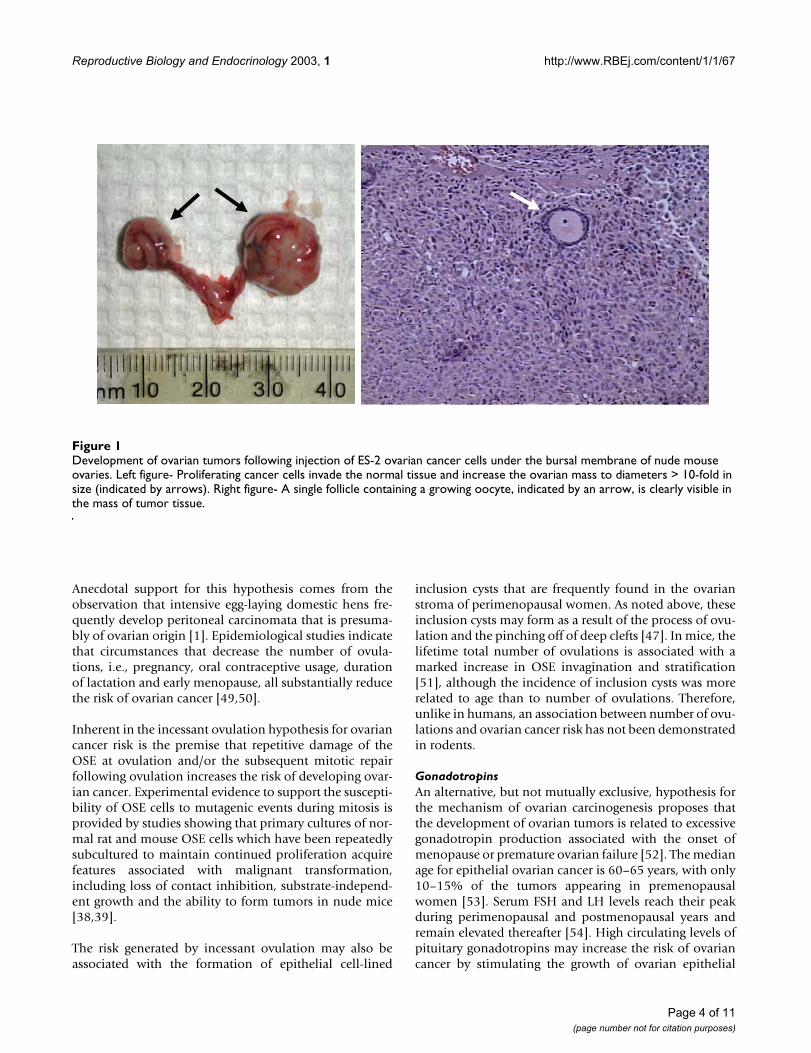
mutation, which carries a splicing defect, develop papil-lary structures and epithelial invaginations (Figure 2),similar to that seen in women [26]. Likewise, female micehomozygous for the germ cell deficient (gcd) mutationenter reproductive senescence prematurely due to a dearthof germ cells. By one year of age, 56% of homozygoteshave developed ovarian tubulostromal adenomas whilewild-type littermates are phenotypically normal [83].
Page 5 of 11(page number not for citation purposes)
Reproductive Biology and Endocrinology 2003, 1 http://www.RBEj.com/content/1/1/67
Morphology of the ovarian surface epithelium in wild-type (A; 12 months), Sld heterozygous (B, C; 12 months) and homozygous (D; 6 months) miceFigure 2Morphology of the ovarian surface epithelium in wild-type (A; 12 months), Sld heterozygous (B, C; 12 months) and homozygous (D; 6 months) mice. Ovaries from wild-type mice contain developing follicles and a covering layer of columnar OSE. In 12-month-old Sld heterozygous mice, there is a depletion of follicles, and the ovarian surface has become very convoluted (B), with this papillary surface sometimes leading to deep invaginations, as indicated by the arrow (C). By 6 months of age, the ovaries of homozygous Sld mice are completely abnormal, with no recognizable ovarian structures, and are composed primarily of inva-sive epithelial tubules. (E) Human ovarian papillomatosis, for comparison.
A
D
C
B
C
E
Page 6 of 11(page number not for citation purposes)

Reproductive Biology and Endocrinology 2003, 1 http://www.RBEj.com/content/1/1/67
Therefore, it appears that oocyte depletion is associatedwith formation of epithelial structures that resemble thepreneoplastic lesions in human ovaries.
Experimental ovarian tumorigenesis has been investigatedin inbred and hybrid strains of mice and induced by adiversity of mechanisms including X-irradiation, oocyto-toxic xenobiotic chemicals, ovarian grafting to ectopic ororthotopic sites, neonatal thymectomy, genetic defectsreducing germ cell populations, and aging [reviewed in[84]]. While germ cell deficiency seems to be a requiredelement for the development of epithelium-derived ade-nomas, the mechanisms by which germ cell loss contrib-utes to tumorigenesis in these models remain unclear.Ovarian follicles do not develop in the absence of oocytes,indicating that the oocyte directs the development of fol-licles. Pathogenetic factors that prematurely destroy ordiminish the numbers of germ cells lead to failure in fol-licle development and a resulting decrease in sex steroidhormone secretion (notably estradiol) leading to a com-pensatory over-production of pituitary gonadotropins,which places the ovary at an increased risk to developtumors. Therefore oocyte depletion, similar to that whichoccurs naturally by the time of menopause, may be a con-tributing factor to the oncogenic behavior of the surfaceepithelial cells.
The intense proliferation of OSE and stromal (interstitial)cells with the development of unique tubular adenomasin response to sterility seems to require both the lack ofgerm cells/follicles and the increased production of gona-dotropins. Elevated gonadotropins alone resulted in gran-ulosa cell tumors or luteomas [62,63]. Oocyte destructionby gamma irradiation in hypogonadal mice deficient ingonadotropins did not result in the development of tubu-lar adenomas [85]. Similarly, the experimental suppres-sion of gonadotropin levels in Wx/Wv mice was sufficientto prevent the development of ovarian tubular adenomasfrom the surface epithelium [86], suggesting that bothoocyte loss/destruction and elevated gonadotropins arenecessary for epithelial tumorigenesis.
Environmental CarcinogensAlthough the more established hypotheses that have beenproposed to explain increased risk of developing ovariancancer are related to the number of ovulations or toincreased hormone levels, there are additional risk factorsthat have been identified, including a number of environ-mental carcinogens. While these factors have beenreported to have effects on the ovarian surface epithelium,they are usually also associated with follicular destructionand/or ovotoxicity, so indirect actions due to alteredgonadotropin levels cannot be eliminated. Use of perinealtalc has been identified as a risk factor, possibly due to itsability to ascend the genital tract and affect the ovarian
surface [87]. Indeed, direct exposure of rat ovaries to talcresults in focal areas of papillary change in the ovariansurface epithelium, as well as ovarian cysts [88]. Exposureof rhesus and cynomolgus monkeys to the environmentalpollutant, hexachlorobenzene results in both reproduc-tive failure and notable alterations in the size, shape anddegree of stratification of the OSE cell layer [89]. Morerecent studies have shown that the insecticide methoxy-chlor increases both the height of the OSE cell layer andthe percentage of atretic follicles in exposed mice [90]. Inrodent studies, ovarian toxicity and/or carcinogenicity hasbeen documented for at least eight chemicals that result infollicular necrosis, tubular hyperplasia, granulosa celltumors and benign mixed tumors [91,92]. N-ethyl-N-nitrosourea administered to rats intraperitoneally ortransplacentally increases the incidence of ovarian tubularadenomas [93]. The mechanisms by which these environ-mental carcinogens enhance the risk of ovarian tumorsremain unexplored.
Transgenics and Targeted Approaches to Transform the Ovarian EpitheliumThe ideal model to investigate the pathogenic events asso-ciated with early ovarian tumorigenesis would be a mousemodel in which the tumor arises directly from the OSEcells. This model would differ from current xenograftmodels in that transgenic mice with defined geneticlesions could be studied at various stages as they inevita-bly develop ovarian cancer in situ. In addition, the devel-opment of a genetic model would permit the direct testingof oncogenes and tumor suppressors for theircontribution to the initiation and progression of overtmalignancies in the mouse ovary. Finally, a number of dif-ferent factors could be altered such as the genetic back-ground of the mouse strain, the frequency of ovulationand the levels of various hormones to determine theirimpact on the development of tumors in the susceptibletransgenic mouse line.
One approach to alter gene expression directly in the OSEcells would be to take advantage of the fact that these cellsreadily take up and express genes delivered by intra-bursalinjection of adenoviruses [94,95]. This method has thepotential advantage of mimicking somatic mutations thatcontribute to early ovarian tumorigenesis. One recentreport used intra-bursal adenovirus delivery and Cre-loxPmediated gene inactivation to render OSE cells deficientin two key tumor suppressor genes: p53 and Rb [95]. Thep53 tumor suppressor gene is the most frequentlymutated gene in human neoplasms. Mutations and/orover-expression of p53 have been described in 26–62% ofovarian cancers, particularly serous ovarian carcinomas[reviewed in [96]]. Aberrations in the Rb pathway havebeen reported [97]; however, direct evidence for their con-tribution to ovarian epithelial tumorigenesis is lacking. In
Page 7 of 11(page number not for citation purposes)

Reproductive Biology and Endocrinology 2003, 1 http://www.RBEj.com/content/1/1/67
this model, recombinant adenovirus expressing Cre wasinjected under the ovarian bursal membrane of doubletransgenic mice bearing floxed copies of p53 and Rb. Con-current inactivation of p53 and Rb was sufficient forreproducible induction of ovarian epithelial carcinogene-sis in mice homozygous for the conditional alleles. Whileless than 15% of mice with inactivation of either Rb or p53developed tumors, 33 of 34 mice with deficiencies in bothgenes succumbed to their ovarian cancers at a median of227 days, with 24% having abdominal ascites.
The major impediment to the development of transgenicmodels of ovarian cancer is the lack of specific promotersable to direct gene expression to OSE cells. Previous mod-els of ovarian cancer have resulted in granulosa celltumors using promoters, such as inhibin-alpha subunitpromoter, that are active in this cell type to drive theexpression of the large T antigen of SV40 [98,99]. Recentstudies have identified two other promoters that mayprove to be useful for the generation of transgenic modelsof ovarian cancer. The Ovarian Specific Promoter (OSP-1)was developed from a retrovirus-like element specificallyexpressed in the rat ovary. The promoter drives geneexpression specifically in normal and neoplastic ovarianepithelial cells [100] and expression of lacZ driven byOSP-1 in transgenic mice was restricted to the ovary asdetermined by X-gal staining of multiple organs [101].Immunohistochemical detection of β-galactosidaseshowed lacZ expression mainly in the granulosa cells andovarian surface epithelial cells. However, transgenic micein which OSP-1 drives the expression of the early regionof SV40 virus developed tumors in a variety of tissues,including unilateral granulosa cell tumors in two of threefemale founder mice. Thus, although transcription fromthe OSP-1 promoter occurs predominantly in the ovary,this promoter is sufficiently "leaky" in cells in other tis-sues to permit their tumorigenic conversion by SV40 TAg.
The first transgenic model of epithelial ovarian cancer wasrecently reported and used the upstream region of theMullerian inhibitory substance type II receptor (MISIIR)gene to drive tissue-specific expression [102]. MISIIR is asingle transmembrane serine/threonine kinase that shareshomology with the TGFβ-receptor [103,104]. Expressionof MISIIR has been reported to be restricted to mesenchy-mal cells surrounding the Mullerian duct during embryo-genesis, tubular and follicular structures of fetal gonads,Sertoli and Leydig cells of adult testis, and granulosa cellsof adult ovary [103,105,106]. More recently, expression ofMISIIR in established human ovarian cancer cell lines aswell as cell lines derived from the ascites of patients withovarian carcinomas has been demonstrated [107]. Trans-genic mice in which the 5' upstream regulatory sequencesof the mouse MISIIR gene were used to target expressionof the SV40 TAg specifically to the epithelium of the
female mouse reproductive tract, including the OSE,developed ovarian carcinomas with metastatic spread toperitoneal organs by 3 months of age. Female transgenicmice developed bilateral ovarian tumors in ~50% percentof cases. Histologically, these tumors were poorly differ-entiated carcinomas with occasional cysts and papillarystructures present at the surface of the ovary. These tumorsdisseminated intraperitoneally, invaded the omentumand formed ascites in a manner that resembles humanovarian carcinomas. The demonstration that the MISIIRpromoter can be used successfully to drive gynecologicaltissue-specific transgene expression in mice and that thisoften results in the formation of ovarian carcinoma offersvery promising opportunities for testing the efficacy ofchemotherapeutic and chemopreventive agents in a herit-able model of epithelial ovarian cancer.
ConclusionsThe two most pressing problems in the management ofovarian cancer are the lack of adequate diagnostic orscreening strategies, and the recurrence of disease that isoften chemoresistant. In part, the deficiency in diagnostictools is due to the lack of markers for the detection of pre-neoplastic or early neoplastic changes in the OSE cells.The generation of animal models in which OSE cellsundergo neoplastic transformation in vivo will providemuch-needed opportunities to investigate the cellular andmolecular changes associated with the initiation of OSEcell transformation, as well as to provide models in whichprevention, diagnostic, screening and therapeutic strate-gies can be developed.
AcknowledgementsThe authors wish to thank Dr. Ken Garson for critical review of this man-uscript and Dr. Thomas Hamilton, Fox Chase Cancer Center, for providing the photograph showing human papillomatosis. Research was supported by grants from the National Cancer Institute of Canada (BCV) and the National Institutes of Health (BCV; Dr. Thomas Hamilton, Principal Inves-tigator), a scholarship from the Canadian Institutes of Health Research (TJS) and a fellowship from a partnership of the Mitchell Family Fund, the National Ovarian Cancer Association and Cancer Care Ontario (JFE).
References1. Fredrickson TN: Ovarian tumors of the hen. Environ Health
Perspect 1987, 73:35-51.2. Tillmann T, Kamino K and Mohr U: Incidence and spectrum of
spontaneous neoplasms in male and female CBA/J mice. ExpToxicol Pathol 2000, 52:221-225.
3. Walsh KM and Poteracki J: Spontaneous neoplasms in controlWistar rats. Fundam Appl Toxicol 1994, 22:65-72.
4. Gregson RL, Lewis DJ and Abbott DP: Spontaneous ovarian neo-plasms of the laboratory rat. Vet Pathol 1984, 21:292-299.
5. Liebelt AG, Sass B and Lombard LS: Mouse ovarian tumors – areview including classification and induction of neoplasticlesions and description of several previously unreportedtypes. J Exp Pathol 1987, 3:115-145.
6. Beamer WG, Hoppe PC and Whitten WK: Spontaneous malig-nant granulosa cell tumors in ovaries of young SWR mice.Cancer Res 1985, 45:5575-5581.
Page 8 of 11(page number not for citation purposes)
http://www.ncbi.nlm.nih.gov/entrez/query.fcgi?cmd=Retrieve&db=PubMed&dopt=Abstract&list_uids=3665870
http://www.ncbi.nlm.nih.gov/entrez/query.fcgi?cmd=Retrieve&db=PubMed&dopt=Abstract&list_uids=8125215
http://www.ncbi.nlm.nih.gov/entrez/query.fcgi?cmd=Retrieve&db=PubMed&dopt=Abstract&list_uids=8125215
http://www.ncbi.nlm.nih.gov/entrez/query.fcgi?cmd=Retrieve&db=PubMed&dopt=Abstract&list_uids=6730218
http://www.ncbi.nlm.nih.gov/entrez/query.fcgi?cmd=Retrieve&db=PubMed&dopt=Abstract&list_uids=6730218
http://www.ncbi.nlm.nih.gov/entrez/query.fcgi?cmd=Retrieve&db=PubMed&dopt=Abstract&list_uids=2826730
http://www.ncbi.nlm.nih.gov/entrez/query.fcgi?cmd=Retrieve&db=PubMed&dopt=Abstract&list_uids=2826730
http://www.ncbi.nlm.nih.gov/entrez/query.fcgi?cmd=Retrieve&db=PubMed&dopt=Abstract&list_uids=2826730

Reproductive Biology and Endocrinology 2003, 1 http://www.RBEj.com/content/1/1/67
7. Tennent BJ, Shultz KL and Beamer WG: Genetic susceptibility forC19 androgen induction of ovarian granulosa cell tumorigen-esis in SWXJ strains of mice. Cancer Res 1993, 53:1059-1063.
8. Eppig JJ, Wigglesworth K, Varnum DS and Nadeau JH: Genetic reg-ulation of traits essential for spontaneous ovarian teratocar-cinogenesis in strain LT/Sv mice: aberrant meiotic cell cycle,oocyte activation, and parthenogenetic development. CancerRes 1996, 56:5047-5054.
9. Colledge WH, Carlton MB, Udy GB and Evans MJ: Disruption of c-mos causes parthenogenetic development of unfertilizedmouse eggs. Nature 1994, 370:65-68.
10. Hashimoto N, Watanabe N, Furuta Y, Tamemoto H, Sagata N,Yokoyama M, Okazaki K, Nagayoshi M, Takeda N and Ikawa Y et al.:Parthenogenetic activation of oocytes in c-mos-deficientmice. Nature 1994, 370:68-71.
11. St John MA, Tao W, Fei X, Fukumoto R, Carcangiu ML, BrownsteinDG, Parlow AF, McGrath J and Xu T: Mice deficient of Lats1develop soft-tissue sarcomas, ovarian tumours and pituitarydysfunction. Nat Genet 1999, 21:182-186.
12. Weiss NS, Homonchuk T and Young JLJ: Incidence of the histo-logic types of ovarian cancer: the US Third National CancerSurvey, 1969–1971. Gynecol Oncol 1977, 5:161-167.
13. Papadaki L and Beilby JO: The fine structure of the surface epi-thelium of the human ovary. J Cell Sci 1971, 8:445-465.
14. Blaustein A and Lee H: Surface cells of the ovary and pelvic per-itoneum: a histochemical and ultrastructure comparison.Gynecol Oncol 1979, 8:34-43.
15. Moore KL: The pelvis and perineum. In: Clinically oriented anatomyEdited by: Satterfield TS, Napora L, Lumpkin K. Baltimore, Williams &Williams; 1992:281-289.
16. Benjamin E, Law S and Bobrow LG: Intermediate filaments,cytokeratin and vimentin in ovarian sex cord-stromaltumours with correlative studies in adult and fetal ovaries. JPathol 1987, 152:253-263.
17. Isola J, Kallioniemi OP, Korte JM, Wahlstrom T, Aine R, Helle M andHelin H: Steroid receptors and Ki-67 reactivity in ovarian can-cer and in normal ovary: correlation with DNA flow cytom-etry, biochemical receptor assay, and patient survival. J Pathol1990, 162:295-301.
18. Czernobilsky B, Moll R, Levy R and Franke WW: Co-expression ofcytokeratin and vimentin filaments in mesothelial, granulosaand rete ovarii cells of the human ovary. Eur J Cell Biol 1985,37:175-190.
19. Jindal SK, Ishii E, Letarte M, Vera S, Teerds K and Dorrington JH: Reg-ulation of transforming growth factor-α gene expression inan ovarian surface epithelial cell line derived from a humancarcinoma. Biol Reprod 1995, 52:1027-1037.
20. Rodriguez GC, Berchuck A, Whitaker RS, Schlossman D, Clarke-Pearson DL and Bast RCJ: Epidermal growth factor receptorexpression in normal ovarian epithelium and ovarian cancer.II. Relationship between receptor expression and responseto epidermal growth factor. Am J Obstet Gynecol 1991,164:745-750.
21. Bjersing L and Cajander S: Ovulation and the mechanism of fol-licle rupture. V. Ultrastructure of tunica albuginea and thecaexterna of rabbit graafian follicles prior to induced ovulation.Cell Tissue Res 1974, 153:15-30.
22. Ackerman RC and Murdoch WJ: Prostaglandin-induced apopto-sis of ovarian surface epithelial cells. Prostaglandins 1993,45:475-485.
23. Osterholzer HO, Johnson JH and Nicosia SV: An autoradiographicstudy of rabbit ovarian surface epithelium before and afterovulation. Biol Reprod 1985, 33:729-738.
24. Auersperg N, Wong AS, Choi KC, Kang SK and Leung PC: Ovariansurface epithelium: biology, endocrinology, and pathology.Endocr Rev 2001, 22:255-288.
25. Nicosia SV: The aging ovary. Med Clin North Am 1987, 71:1-9.26. Hamilton TC: Ovarian cancer, Part I: Biology. Curr Probl Cancer
1992, 16:1-57.27. Salazar H, Godwin AK, Daly MB, Laub PB, Hogan WM, Rosenblum N,
Boente MP, Lynch HT and Hamilton TC: Microscopic benign andinvasive malignant neoplasms and a cancer-prone pheno-type in prophylactic oophorectomies. J Natl Cancer Inst 1996,88:1810-1820.
28. Deligdisch L and Gil J: Characterization of ovarian dysplasia byinteractive morphometry. Cancer 1989, 63:748-755.
29. Scully RE: Early de novo ovarian cancer and cancer developingin benign ovarian lesions. Int J Gynaecol Obstet 1995,49(Suppl):S9-15.
30. Auersperg N, Siemens CH and Myrdal SE: Human ovarian surfaceepithelium in primary culture. In Vitro 1984, 20:743-755.
31. Adams AT and Auersperg N: Transformation of cultured ratovarian surface epithelial cells by Kirsten murine sarcomavirus. Cancer Res 1981, 41:2063-2072.
32. Leung EH, Leung PC and Auersperg N: Differentiation andgrowth potential of human ovarian surface epithelial cellsexpressing temperature-sensitive SV40 T antigen. In Vitro CellDev Biol Anim 2001, 37:515-521.
33. Auersperg N, Pan J, Grove BD, Peterson T, Fisher J, Maines-BandieraS, Somasiri A and Roskelley CD: E-cadherin induces mesenchy-mal-to-epithelial transition in human ovarian surfaceepithelium. Proc Natl Acad Sci USA 1999, 96:6249-6254.
34. Ong A, Maines-Bandiera SL, Roskelley CD and Auersperg N: Anovarian adenocarcinoma line derived from SV40/E-cadherin-transfected normal human ovarian surface epithelium. Int JCancer 2000, 85:430-437.
35. Gregoire L, Rabah R, Schmelz EM, Munkarah A, Roberts PC and Lan-caster WD: Spontaneous malignant transformation of humanovarian surface epithelial cells in vitro. Clin Cancer Res 2001,7:4280-4287.
36. Adams AT and Auersperg N: A cell line, ROSE 199, derived fromnormal rat ovarian surface epithelium. Exp Cell Biol 1985,53:181-188.
37. Hoffman AG, Burghardt RC, Tilley R and Auersperg N: An in vitromodel of ovarian epithelial carcinogenesis: changes in cell-cell communication and adhesion occuring during neoplasticprogression. Int J Cancer 1993, 54:828-838.
38. Godwin AK, Testa JR, Handel LM, Liu Z, Vanderveer LA, Tracey PAand Hamilton TC: Spontaneous transformation of rat ovariansurface epithelial cells: association with cytogenetic changesand implications of repeated ovulation in the etiology ofovarian cancer. J Natl Cancer Inst 1992, 84:592-601.
39. Roby KF, Taylor CC, Sweetwood JP, Cheng Y, Pace JL, Tawfik O, Per-sons DL, Smith PG and Terranova PF: Development of a syn-geneic mouse model for events related to ovarian cancer.Carcinogenesis 2000, 21:585-591.
40. Orsulic S, Li Y, Soslow RA, Vitale-Cross LA, Gutkind JS and VarmusHE: Induction of ovarian cancer by defined multiple geneticchanges in a mouse model system. Cancer Cell 2002, 1:53-62.
41. Tonin PN, Hudson TJ, Rodier F, Bossolasco M, Lee PD, Novak J, Man-derson EN, Provencher D and Mes-Masson AM: Microarray analy-sis of gene expression mirrors the biology of an ovariancancer model. Oncogene 2001, 20:6617-6626.
42. Roberts D, Williams SJ, Cvetkovic D, Weinstein JK, Godwin AK,Johnson SW and Hamilton TC: Decreased expression of retinol-binding proteins is associated with malignant transformationof the ovarian surface epithelium. DNA Cell Biol 2002, 21:11-19.
43. Ward BG and Wallace K: Localization of the monoclonal anti-body HMFG2 after intravenous and intraperitoneal injectioninto nude mice bearing subcutaneous and intraperitonealhuman ovarian cancer xenografts. Cancer Res 1987,47:4714-4718.
44. Hamilton TC, Young RC, Louie KG, Behrens BC, McKoy WM, Grotz-inger KR and Ozols RF: Characterization of a xenograft modelof human ovarian carcinoma which produces ascites andintraabdominal carcinomatosis in mice. Cancer Res 1984,44:5286-5290.
45. Massazza G, Tomasoni A, Lucchini V, Allavena P, Erba E, Colombo N,Mantovani A, D'Incalci M, Mangioni C and Giavazzi R: Intraperito-neal and subcutaneous xenografts of human ovarian carci-noma in nude mice and their potential in experimentaltherapy. Int J Cancer 1989, 44:494-500.
46. Fu X and Hoffman RM: Human ovarian carcinoma metastaticmodels constructed in nude mice by orthotopic transplanta-tion of histologically-intact patient specimens. Anticancer Res1993, 13:283-286.
47. Scully RE, Young RH and Clement PB: Tumors of the Ovary, Maldevel-oped Gonads, Fallopian Tube, and Broad Ligament Washington DC, ArmedForces Institute of Pathology; 1996.
48. Fathalla MF: Incessant ovulation – a factor in ovarianneoplasia? Lancet 1971, 2:163.
Page 9 of 11(page number not for citation purposes)
http://www.ncbi.nlm.nih.gov/entrez/query.fcgi?cmd=Retrieve&db=PubMed&dopt=Abstract&list_uids=8439952
http://www.ncbi.nlm.nih.gov/entrez/query.fcgi?cmd=Retrieve&db=PubMed&dopt=Abstract&list_uids=8439952
http://www.ncbi.nlm.nih.gov/entrez/query.fcgi?cmd=Retrieve&db=PubMed&dopt=Abstract&list_uids=8439952
http://www.ncbi.nlm.nih.gov/entrez/query.fcgi?cmd=Retrieve&db=PubMed&dopt=Abstract&list_uids=8895763
http://www.ncbi.nlm.nih.gov/entrez/query.fcgi?cmd=Retrieve&db=PubMed&dopt=Abstract&list_uids=8895763
http://www.ncbi.nlm.nih.gov/entrez/query.fcgi?cmd=Retrieve&db=PubMed&dopt=Abstract&list_uids=8895763
http://www.ncbi.nlm.nih.gov/entrez/query.fcgi?cmd=Retrieve&db=PubMed&dopt=Abstract&list_uids=8015609
http://www.ncbi.nlm.nih.gov/entrez/query.fcgi?cmd=Retrieve&db=PubMed&dopt=Abstract&list_uids=8015609
http://www.ncbi.nlm.nih.gov/entrez/query.fcgi?cmd=Retrieve&db=PubMed&dopt=Abstract&list_uids=8015609
http://www.ncbi.nlm.nih.gov/entrez/query.fcgi?cmd=Retrieve&db=PubMed&dopt=Abstract&list_uids=8015610
http://www.ncbi.nlm.nih.gov/entrez/query.fcgi?cmd=Retrieve&db=PubMed&dopt=Abstract&list_uids=8015610
http://www.ncbi.nlm.nih.gov/entrez/query.fcgi?cmd=Retrieve&db=PubMed&dopt=Abstract&list_uids=8015610
http://www.ncbi.nlm.nih.gov/entrez/query.fcgi?cmd=Retrieve&db=PubMed&dopt=Abstract&list_uids=9988269
http://www.ncbi.nlm.nih.gov/entrez/query.fcgi?cmd=Retrieve&db=PubMed&dopt=Abstract&list_uids=9988269
http://www.ncbi.nlm.nih.gov/entrez/query.fcgi?cmd=Retrieve&db=PubMed&dopt=Abstract&list_uids=9988269
http://www.ncbi.nlm.nih.gov/entrez/query.fcgi?cmd=Retrieve&db=PubMed&dopt=Abstract&list_uids=5576081
http://www.ncbi.nlm.nih.gov/entrez/query.fcgi?cmd=Retrieve&db=PubMed&dopt=Abstract&list_uids=5576081
http://www.ncbi.nlm.nih.gov/entrez/query.fcgi?cmd=Retrieve&db=PubMed&dopt=Abstract&list_uids=2444685
http://www.ncbi.nlm.nih.gov/entrez/query.fcgi?cmd=Retrieve&db=PubMed&dopt=Abstract&list_uids=2444685
http://www.ncbi.nlm.nih.gov/entrez/query.fcgi?cmd=Retrieve&db=PubMed&dopt=Abstract&list_uids=2444685
http://www.ncbi.nlm.nih.gov/entrez/query.fcgi?cmd=Retrieve&db=PubMed&dopt=Abstract&list_uids=2290114
http://www.ncbi.nlm.nih.gov/entrez/query.fcgi?cmd=Retrieve&db=PubMed&dopt=Abstract&list_uids=2290114
http://www.ncbi.nlm.nih.gov/entrez/query.fcgi?cmd=Retrieve&db=PubMed&dopt=Abstract&list_uids=2290114
http://www.ncbi.nlm.nih.gov/entrez/query.fcgi?cmd=Retrieve&db=PubMed&dopt=Abstract&list_uids=3896804
http://www.ncbi.nlm.nih.gov/entrez/query.fcgi?cmd=Retrieve&db=PubMed&dopt=Abstract&list_uids=3896804
http://www.ncbi.nlm.nih.gov/entrez/query.fcgi?cmd=Retrieve&db=PubMed&dopt=Abstract&list_uids=3896804
http://www.ncbi.nlm.nih.gov/entrez/query.fcgi?cmd=Retrieve&db=PubMed&dopt=Abstract&list_uids=7626702
http://www.ncbi.nlm.nih.gov/entrez/query.fcgi?cmd=Retrieve&db=PubMed&dopt=Abstract&list_uids=7626702
http://www.ncbi.nlm.nih.gov/entrez/query.fcgi?cmd=Retrieve&db=PubMed&dopt=Abstract&list_uids=7626702
http://www.ncbi.nlm.nih.gov/entrez/query.fcgi?cmd=Retrieve&db=PubMed&dopt=Abstract&list_uids=2003535
http://www.ncbi.nlm.nih.gov/entrez/query.fcgi?cmd=Retrieve&db=PubMed&dopt=Abstract&list_uids=2003535
http://www.ncbi.nlm.nih.gov/entrez/query.fcgi?cmd=Retrieve&db=PubMed&dopt=Abstract&list_uids=2003535
http://www.ncbi.nlm.nih.gov/entrez/query.fcgi?cmd=Retrieve&db=PubMed&dopt=Abstract&list_uids=4442079
http://www.ncbi.nlm.nih.gov/entrez/query.fcgi?cmd=Retrieve&db=PubMed&dopt=Abstract&list_uids=4442079
http://www.ncbi.nlm.nih.gov/entrez/query.fcgi?cmd=Retrieve&db=PubMed&dopt=Abstract&list_uids=8321916
http://www.ncbi.nlm.nih.gov/entrez/query.fcgi?cmd=Retrieve&db=PubMed&dopt=Abstract&list_uids=8321916
http://www.ncbi.nlm.nih.gov/entrez/query.fcgi?cmd=Retrieve&db=PubMed&dopt=Abstract&list_uids=4052531
http://www.ncbi.nlm.nih.gov/entrez/query.fcgi?cmd=Retrieve&db=PubMed&dopt=Abstract&list_uids=4052531
http://www.ncbi.nlm.nih.gov/entrez/query.fcgi?cmd=Retrieve&db=PubMed&dopt=Abstract&list_uids=4052531
http://www.ncbi.nlm.nih.gov/entrez/query.fcgi?cmd=Retrieve&db=PubMed&dopt=Abstract&list_uids=3543537
http://www.ncbi.nlm.nih.gov/entrez/query.fcgi?cmd=Retrieve&db=PubMed&dopt=Abstract&list_uids=1547635
http://www.ncbi.nlm.nih.gov/entrez/query.fcgi?cmd=Retrieve&db=PubMed&dopt=Abstract&list_uids=8961970
http://www.ncbi.nlm.nih.gov/entrez/query.fcgi?cmd=Retrieve&db=PubMed&dopt=Abstract&list_uids=8961970
http://www.ncbi.nlm.nih.gov/entrez/query.fcgi?cmd=Retrieve&db=PubMed&dopt=Abstract&list_uids=8961970
http://www.ncbi.nlm.nih.gov/entrez/query.fcgi?cmd=Retrieve&db=PubMed&dopt=Abstract&list_uids=2914280
http://www.ncbi.nlm.nih.gov/entrez/query.fcgi?cmd=Retrieve&db=PubMed&dopt=Abstract&list_uids=2914280
http://www.ncbi.nlm.nih.gov/entrez/query.fcgi?cmd=Retrieve&db=PubMed&dopt=Abstract&list_uids=7589745
http://www.ncbi.nlm.nih.gov/entrez/query.fcgi?cmd=Retrieve&db=PubMed&dopt=Abstract&list_uids=7589745
http://www.ncbi.nlm.nih.gov/entrez/query.fcgi?cmd=Retrieve&db=PubMed&dopt=Abstract&list_uids=6083974
http://www.ncbi.nlm.nih.gov/entrez/query.fcgi?cmd=Retrieve&db=PubMed&dopt=Abstract&list_uids=6083974
http://www.ncbi.nlm.nih.gov/entrez/query.fcgi?cmd=Retrieve&db=PubMed&dopt=Abstract&list_uids=6263459
http://www.ncbi.nlm.nih.gov/entrez/query.fcgi?cmd=Retrieve&db=PubMed&dopt=Abstract&list_uids=6263459
http://www.ncbi.nlm.nih.gov/entrez/query.fcgi?cmd=Retrieve&db=PubMed&dopt=Abstract&list_uids=6263459
http://www.ncbi.nlm.nih.gov/entrez/query.fcgi?cmd=Retrieve&db=PubMed&dopt=Abstract&list_uids=4029476
http://www.ncbi.nlm.nih.gov/entrez/query.fcgi?cmd=Retrieve&db=PubMed&dopt=Abstract&list_uids=4029476
http://www.ncbi.nlm.nih.gov/entrez/query.fcgi?cmd=Retrieve&db=PubMed&dopt=Abstract&list_uids=8325708
http://www.ncbi.nlm.nih.gov/entrez/query.fcgi?cmd=Retrieve&db=PubMed&dopt=Abstract&list_uids=8325708
http://www.ncbi.nlm.nih.gov/entrez/query.fcgi?cmd=Retrieve&db=PubMed&dopt=Abstract&list_uids=8325708
http://www.ncbi.nlm.nih.gov/entrez/query.fcgi?cmd=Retrieve&db=PubMed&dopt=Abstract&list_uids=1556770
http://www.ncbi.nlm.nih.gov/entrez/query.fcgi?cmd=Retrieve&db=PubMed&dopt=Abstract&list_uids=1556770
http://www.ncbi.nlm.nih.gov/entrez/query.fcgi?cmd=Retrieve&db=PubMed&dopt=Abstract&list_uids=1556770
http://www.ncbi.nlm.nih.gov/entrez/query.fcgi?cmd=Retrieve&db=PubMed&dopt=Abstract&list_uids=3621167
http://www.ncbi.nlm.nih.gov/entrez/query.fcgi?cmd=Retrieve&db=PubMed&dopt=Abstract&list_uids=3621167
http://www.ncbi.nlm.nih.gov/entrez/query.fcgi?cmd=Retrieve&db=PubMed&dopt=Abstract&list_uids=3621167
http://www.ncbi.nlm.nih.gov/entrez/query.fcgi?cmd=Retrieve&db=PubMed&dopt=Abstract&list_uids=6333272
http://www.ncbi.nlm.nih.gov/entrez/query.fcgi?cmd=Retrieve&db=PubMed&dopt=Abstract&list_uids=6333272
http://www.ncbi.nlm.nih.gov/entrez/query.fcgi?cmd=Retrieve&db=PubMed&dopt=Abstract&list_uids=6333272
http://www.ncbi.nlm.nih.gov/entrez/query.fcgi?cmd=Retrieve&db=PubMed&dopt=Abstract&list_uids=2777413
http://www.ncbi.nlm.nih.gov/entrez/query.fcgi?cmd=Retrieve&db=PubMed&dopt=Abstract&list_uids=2777413
http://www.ncbi.nlm.nih.gov/entrez/query.fcgi?cmd=Retrieve&db=PubMed&dopt=Abstract&list_uids=2777413
http://www.ncbi.nlm.nih.gov/entrez/query.fcgi?cmd=Retrieve&db=PubMed&dopt=Abstract&list_uids=8517640
http://www.ncbi.nlm.nih.gov/entrez/query.fcgi?cmd=Retrieve&db=PubMed&dopt=Abstract&list_uids=8517640
http://www.ncbi.nlm.nih.gov/entrez/query.fcgi?cmd=Retrieve&db=PubMed&dopt=Abstract&list_uids=8517640

Reproductive Biology and Endocrinology 2003, 1 http://www.RBEj.com/content/1/1/67
49. Whittemore AS, Harris R and Itnyre J: Characteristics relating toovarian cancer risk: collaborative analysis of 12 US case-con-trol studies. IV. The pathogenesis of epithelial ovarian can-cer Collaborative Ovarian Cancer Group. Am J Epidemiol 1992,136:1212-1220.
50. La Vecchia C and Franceschi S: Oral contraceptives and ovariancancer. Eur J Cancer Prev 1999, 8:297-304.
51. Clow OL, Hurst PR and Fleming JS: Changes in the mouse ovar-ian surface epithelium with age and ovulation number. MolCell Endocrinol 2002, 191:105-111.
52. Cramer DW and Welch WR: Determinants of ovarian cancerrisk. II. Inferences regarding pathogenesis. J Natl Cancer Inst1983, 71:717-721.
53. Sell A, Bertelsen K, Andersen JE, Stroyer I and Panduro J: Rand-omized study of whole-abdomen irradiation versus pelvicirradiation plus cyclophosphamide in treatment of earlyovarian cancer. Gynecol Oncol 1990, 37:367-373.
54. Chakravarti S, Collins WP, Forecast JD, Newton JR, Oram DH andStudd JW: Hormonal profiles after the menopause. Br Med J1976, 2:784-787.
55. Zheng WX, Magid MS, Kramer EE and Chen YT: Follicle-stimulat-ing hormone receptor is expressed in human ovarian surfaceepithelium and fallopian tube. Am J Pathol 1996, 148:47-53.
56. Konishi I, Kuroda H and Mandai M: Review: gonadotropins anddevelopment of ovarian cancer. Oncology 1999, 57(Suppl2):45-48.
57. Osterholzer HO, Streibel EJ and Nicosia SV: Growth effects ofprotein hormones on cultured rabbit ovarian surface epithe-lial cells. Biol Reprod 1985, 33:247-258.
58. Davies BR, Finnigan DS, Smith SK and Ponder BA: Administrationof gonadotropins stimulates proliferation of normal mouseovarian surface epithelium. Gynecol Endocrinol 1999, 13:75-81.
59. Schiffenbauer YS, Abramovitch R, Meir G, Nevo N, Holzinger M, ItinA, Keshet E and Neeman M: Loss of ovarian function promotesangiogenesis in human ovarian carcinoma. Proc Natl Acad Sci US A 1997, 94:13203-13208.
60. Biskind MS and Biskind GS: Development of tumors in the ratovary after transplantation into the spleen. Proc Soc Exp BiolMed 1944, 55:176-179.
61. Matzuk MM, Finegold MJ, Su JG, Hsueh AJ and Bradley A: Alpha-inhibin is a tumor-suppressor gene with gonadal specificity inmice. Nature 1992, 360:313-319.
62. Nilson JH, Abbud RA, Keri RA and Quirk CC: Chronic hypersecre-tion of luteinizing hormone in transgenic mice disrupts bothovarian and pituitary function, with some effects modified bythe genetic background. Recent Prog Horm Res 2000, 55:69-89.
63. Keri RA, Lozada KL, Abdul-Karim FW, Nadeau JH and Nilson JH:Luteinizing hormone induction of ovarian tumors: oligo-genic differences between mouse strains dictates tumordisposition. Proc Natl Acad Sci USA 2000, 97:383-387.
64. Danilovich N, Roy I and Sairam MR: Ovarian pathology and highincidence of sex cord tumors in follitropin receptor knock-out (FORKO) mice. Endocrinology 2001, 142:3673-3684.
65. Gondos B: Surface epithelium of the developing ovary. Possi-ble correlation with ovarian neoplasia. Am J Pathol 1975,81:303-321.
66. Karlan BY, Jones J, Greenwald M and Lagasse LD: Steroid hormoneeffects on the proliferation of human ovarian surface epithe-lium in vitro. Am J Obstet Gynecol 1995, 173:97-104.
67. Lau KM, Mok SC and Ho SM: Expression of human estrogenreceptor-alpha and -beta, progesterone receptor, andandrogen receptor mRNA in normal and malignant ovarianepithelial cells. Proc Natl Acad Sci USA 1999, 96:5722-5727.
68. Hamilton TC, Davies P and Criffiths K: Steroid hormone receptorstatus of the normal and neoplastic ovarian surface germinalepithelium. In Factors regulating ovarian function Edited by: GreenwaldGS, Terranova PF. New York, Raven Press; 1983:81-85.
69. Lacey JV Jr, Mink PJ, Lubin JH, Sherman ME, Troisi R, Hartge P, Schatz-kin A and Schairer C: Menopausal hormone replacement ther-apy and risk of ovarian cancer. JAMA 2002, 288:334-341.
70. Murdoch WJ and Van Kirk EA: Steroid hormonal regulation ofproliferative, p53 tumor suppressor, and apoptoticresponses of sheep ovarian surface epithelial cells. Mol CellEndocrinol 2002, 186:61-67.
71. Silva EG, Tornos C, Deavers M, Kaisman K, Gray K and GershensonD: Induction of epithelial neoplasms in the ovaries of guineapigs by estrogenic stimulation. Gynecol Oncol 1998, 71:240-246.
72. Bai W, Oliveros-Saunders B, Wang Q, Acevedo-Duncan ME andNicosia SV: Estrogen stimulation of ovarian surface epithelialcell proliferation. In Vitro Cell Dev Biol Anim 2000, 36:657-666.
73. Kang SK, Choi KC, Tai CJ, Auersperg N and Leung PC: Estradiolregulates gonadotropin-releasing hormone (GnRH) and itsreceptor gene expression and antagonizes the growth inhib-itory effects of GnRH in human ovarian surface epithelial andovarian cancer cells. Endocrinology 2001, 142:580-588.
74. Liu Y, Lin L and Zarnegar R: Modulation of hepatocyte growthfactor gene expression by estrogen in mouse ovary. Mol CellEndocrinol 1994, 104:173-181.
75. Ilekis JV, Connor JP, Prins GS, Ferrer K, Niederberger C and ScocciaB: Expression of epidermal growth factor and androgenreceptors in ovarian cancer. Gynecol Oncol 1997, 66:250-254.
76. Helzlsouer KJ, Alberg AJ, Gordon GB, Longcope C, Bush TL, HoffmanSC and Comstock GW: Serum gonadotropins and steroid hor-mones and the development of ovarian cancer. JAMA 1995,274:1926-1930.
77. Silva EG, Tornos C, Fritsche HAJ, el-Naggar A, Gray K, Ordonez NG,Luna M and Gershenson D: The induction of benign epithelialneoplasms of the ovaries of guinea pigs by testosterone stim-ulation: a potential animal model. Mod Pathol 1997, 10:879-883.
78. Evangelou A, Jindal SK, Brown TJ and Letarte M: Down-regulationof transforming growth factor beta receptors by androgen inovarian cancer cells. Cancer Res 2000, 60:929-935.
79. Mintz B and Russell ES: Gene-induced embryological modifica-tions of primordial germ cells in the mouse. J Exp Zool 1957,134:207-237.
80. Murphy ED: Hyperplastic and early neoplastic changes in theovaries of mice after genic deletion of germ cells. J Natl CancerInst 1972, 48:1283-1295.
81. Murphy ED and Beamer WG: Plasma gonadotropin levels duringearly stages of ovarian tumorigenesis in mice of the Wx/Wvgenotype. Cancer Res 1973, 33:721-723.
82. Ishimura K, Matsuda H, Tatsumi H, Fujita H, Terada N and KitamuraY: Ultrastructural changes in the ovaries of Sl/Slt mutantmice, showing developmental deficiency of follicles andtubular adenomas. Arch Histol Jpn 1986, 49:379-389.
83. Duncan MK and Chada KK: Incidence of tubulostromal ade-noma of the ovary in aged germ cell-deficient mice. J CompPathol 1993, 109:13-19.
84. Capen CC, Beamer WG, Tennent BJ and Stitzel KA: Mechanisms ofhormone-mediated carcinogenesis of the ovary in mice.Mutat Res 1995, 333:143-151.
85. Tennent BJ and Beamer WG: Ovarian tumors not induced byirradiation and gonadotropins in hypogonadal (hpg) mice.Biol Reprod 1986, 34:751-760.
86. Blaakaer J, Baeksted M, Micic S, Albrectsen P, Rygaard J and Bock J:Gonadotropin-releasing hormone agonist suppression ofovarian tumorigenesis in mice of the Wx/Wv genotype. BiolReprod 1995, 53:775-779.
87. Gertig DM, Hunter DJ, Cramer DW, Colditz GA, Speizer FE, WillettWC and Hankinson SE: Prospective study of talc use and ovar-ian cancer. J Natl Cancer Inst 2000, 92:249-252.
88. Hamilton TC, Fox H, Buckley CH, Henderson WJ and Griffiths K:Effects of talc on the rat ovary. Br J Exp Pathol 1984, 65:101-106.
89. Sims DE, Singh A, Donald A, Jarrell J and Villeneuve DC: Alterationof primate ovary surface epithelium by exposure to hex-achlorobenzene: a quantitative study. Histol Histopathol 1991,6:525-529.
90. Borgeest C, Symonds D, Mayer LP, Hoyer PB and Flaws JA: Methox-ychlor may cause ovarian follicular atresia and proliferationof the ovarian epithelium in the mouse. Toxicol Sci 2002,68:473-478.
91. Maronpot RR: Ovarian toxicity and carcinogenicity in eightrecent National Toxicology Program studies. Environ HealthPerspect 1987, 73:125-130.
92. Collins JJ, Montali RJ and Manus AG: Toxicological evaluation of4-vinylcyclohexene. II. Induction of ovarian tumors in femaleB6C3F1 mice by chronic oral administration of 4-vinylcy-clohexene. J Toxicol Environ Health 1987, 21:507-524.
Page 10 of 11(page number not for citation purposes)
http://www.ncbi.nlm.nih.gov/entrez/query.fcgi?cmd=Retrieve&db=PubMed&dopt=Abstract&list_uids=1476143
http://www.ncbi.nlm.nih.gov/entrez/query.fcgi?cmd=Retrieve&db=PubMed&dopt=Abstract&list_uids=1476143
http://www.ncbi.nlm.nih.gov/entrez/query.fcgi?cmd=Retrieve&db=PubMed&dopt=Abstract&list_uids=1476143
http://www.ncbi.nlm.nih.gov/entrez/query.fcgi?cmd=Retrieve&db=PubMed&dopt=Abstract&list_uids=6578367
http://www.ncbi.nlm.nih.gov/entrez/query.fcgi?cmd=Retrieve&db=PubMed&dopt=Abstract&list_uids=6578367
http://www.ncbi.nlm.nih.gov/entrez/query.fcgi?cmd=Retrieve&db=PubMed&dopt=Abstract&list_uids=2351321
http://www.ncbi.nlm.nih.gov/entrez/query.fcgi?cmd=Retrieve&db=PubMed&dopt=Abstract&list_uids=2351321
http://www.ncbi.nlm.nih.gov/entrez/query.fcgi?cmd=Retrieve&db=PubMed&dopt=Abstract&list_uids=2351321
http://www.ncbi.nlm.nih.gov/entrez/query.fcgi?cmd=Retrieve&db=PubMed&dopt=Abstract&list_uids=8546225
http://www.ncbi.nlm.nih.gov/entrez/query.fcgi?cmd=Retrieve&db=PubMed&dopt=Abstract&list_uids=8546225
http://www.ncbi.nlm.nih.gov/entrez/query.fcgi?cmd=Retrieve&db=PubMed&dopt=Abstract&list_uids=8546225
http://www.ncbi.nlm.nih.gov/entrez/query.fcgi?cmd=Retrieve&db=PubMed&dopt=Abstract&list_uids=3933584
http://www.ncbi.nlm.nih.gov/entrez/query.fcgi?cmd=Retrieve&db=PubMed&dopt=Abstract&list_uids=3933584
http://www.ncbi.nlm.nih.gov/entrez/query.fcgi?cmd=Retrieve&db=PubMed&dopt=Abstract&list_uids=3933584
http://www.ncbi.nlm.nih.gov/entrez/query.fcgi?cmd=Retrieve&db=PubMed&dopt=Abstract&list_uids=9371824
http://www.ncbi.nlm.nih.gov/entrez/query.fcgi?cmd=Retrieve&db=PubMed&dopt=Abstract&list_uids=1448148
http://www.ncbi.nlm.nih.gov/entrez/query.fcgi?cmd=Retrieve&db=PubMed&dopt=Abstract&list_uids=1448148
http://www.ncbi.nlm.nih.gov/entrez/query.fcgi?cmd=Retrieve&db=PubMed&dopt=Abstract&list_uids=1448148
http://www.ncbi.nlm.nih.gov/entrez/query.fcgi?cmd=Retrieve&db=PubMed&dopt=Abstract&list_uids=1190291
http://www.ncbi.nlm.nih.gov/entrez/query.fcgi?cmd=Retrieve&db=PubMed&dopt=Abstract&list_uids=1190291
http://www.ncbi.nlm.nih.gov/entrez/query.fcgi?cmd=Retrieve&db=PubMed&dopt=Abstract&list_uids=7631734
http://www.ncbi.nlm.nih.gov/entrez/query.fcgi?cmd=Retrieve&db=PubMed&dopt=Abstract&list_uids=7631734
http://www.ncbi.nlm.nih.gov/entrez/query.fcgi?cmd=Retrieve&db=PubMed&dopt=Abstract&list_uids=7631734
http://www.ncbi.nlm.nih.gov/entrez/query.fcgi?cmd=Retrieve&db=PubMed&dopt=Abstract&list_uids=9826466
http://www.ncbi.nlm.nih.gov/entrez/query.fcgi?cmd=Retrieve&db=PubMed&dopt=Abstract&list_uids=9826466
http://www.ncbi.nlm.nih.gov/entrez/query.fcgi?cmd=Retrieve&db=PubMed&dopt=Abstract&list_uids=7988745
http://www.ncbi.nlm.nih.gov/entrez/query.fcgi?cmd=Retrieve&db=PubMed&dopt=Abstract&list_uids=7988745
http://www.ncbi.nlm.nih.gov/entrez/query.fcgi?cmd=Retrieve&db=PubMed&dopt=Abstract&list_uids=9264571
http://www.ncbi.nlm.nih.gov/entrez/query.fcgi?cmd=Retrieve&db=PubMed&dopt=Abstract&list_uids=9264571
http://www.ncbi.nlm.nih.gov/entrez/query.fcgi?cmd=Retrieve&db=PubMed&dopt=Abstract&list_uids=8568986
http://www.ncbi.nlm.nih.gov/entrez/query.fcgi?cmd=Retrieve&db=PubMed&dopt=Abstract&list_uids=8568986
http://www.ncbi.nlm.nih.gov/entrez/query.fcgi?cmd=Retrieve&db=PubMed&dopt=Abstract&list_uids=9310950
http://www.ncbi.nlm.nih.gov/entrez/query.fcgi?cmd=Retrieve&db=PubMed&dopt=Abstract&list_uids=9310950
http://www.ncbi.nlm.nih.gov/entrez/query.fcgi?cmd=Retrieve&db=PubMed&dopt=Abstract&list_uids=9310950
http://www.ncbi.nlm.nih.gov/entrez/query.fcgi?cmd=Retrieve&db=PubMed&dopt=Abstract&list_uids=4337905
http://www.ncbi.nlm.nih.gov/entrez/query.fcgi?cmd=Retrieve&db=PubMed&dopt=Abstract&list_uids=4337905
http://www.ncbi.nlm.nih.gov/entrez/query.fcgi?cmd=Retrieve&db=PubMed&dopt=Abstract&list_uids=4696472
http://www.ncbi.nlm.nih.gov/entrez/query.fcgi?cmd=Retrieve&db=PubMed&dopt=Abstract&list_uids=4696472
http://www.ncbi.nlm.nih.gov/entrez/query.fcgi?cmd=Retrieve&db=PubMed&dopt=Abstract&list_uids=4696472
http://www.ncbi.nlm.nih.gov/entrez/query.fcgi?cmd=Retrieve&db=PubMed&dopt=Abstract&list_uids=3800599
http://www.ncbi.nlm.nih.gov/entrez/query.fcgi?cmd=Retrieve&db=PubMed&dopt=Abstract&list_uids=3800599
http://www.ncbi.nlm.nih.gov/entrez/query.fcgi?cmd=Retrieve&db=PubMed&dopt=Abstract&list_uids=3800599
http://www.ncbi.nlm.nih.gov/entrez/query.fcgi?cmd=Retrieve&db=PubMed&dopt=Abstract&list_uids=8408777
http://www.ncbi.nlm.nih.gov/entrez/query.fcgi?cmd=Retrieve&db=PubMed&dopt=Abstract&list_uids=8408777
http://www.ncbi.nlm.nih.gov/entrez/query.fcgi?cmd=Retrieve&db=PubMed&dopt=Abstract&list_uids=8538621
http://www.ncbi.nlm.nih.gov/entrez/query.fcgi?cmd=Retrieve&db=PubMed&dopt=Abstract&list_uids=8538621
http://www.ncbi.nlm.nih.gov/entrez/query.fcgi?cmd=Retrieve&db=PubMed&dopt=Abstract&list_uids=3708055
http://www.ncbi.nlm.nih.gov/entrez/query.fcgi?cmd=Retrieve&db=PubMed&dopt=Abstract&list_uids=3708055
http://www.ncbi.nlm.nih.gov/entrez/query.fcgi?cmd=Retrieve&db=PubMed&dopt=Abstract&list_uids=8547469
http://www.ncbi.nlm.nih.gov/entrez/query.fcgi?cmd=Retrieve&db=PubMed&dopt=Abstract&list_uids=8547469
http://www.ncbi.nlm.nih.gov/entrez/query.fcgi?cmd=Retrieve&db=PubMed&dopt=Abstract&list_uids=8547469
http://www.ncbi.nlm.nih.gov/entrez/query.fcgi?cmd=Retrieve&db=PubMed&dopt=Abstract&list_uids=6696826
http://www.ncbi.nlm.nih.gov/entrez/query.fcgi?cmd=Retrieve&db=PubMed&dopt=Abstract&list_uids=6696826
http://www.ncbi.nlm.nih.gov/entrez/query.fcgi?cmd=Retrieve&db=PubMed&dopt=Abstract&list_uids=1804430
http://www.ncbi.nlm.nih.gov/entrez/query.fcgi?cmd=Retrieve&db=PubMed&dopt=Abstract&list_uids=1804430
http://www.ncbi.nlm.nih.gov/entrez/query.fcgi?cmd=Retrieve&db=PubMed&dopt=Abstract&list_uids=1804430
http://www.ncbi.nlm.nih.gov/entrez/query.fcgi?cmd=Retrieve&db=PubMed&dopt=Abstract&list_uids=3665857
http://www.ncbi.nlm.nih.gov/entrez/query.fcgi?cmd=Retrieve&db=PubMed&dopt=Abstract&list_uids=3665857
http://www.ncbi.nlm.nih.gov/entrez/query.fcgi?cmd=Retrieve&db=PubMed&dopt=Abstract&list_uids=3599093

Reproductive Biology and Endocrinology 2003, 1 http://www.RBEj.com/content/1/1/67
Publish with BioMed Central and every scientist can read your work free of charge
"BioMed Central will be the most significant development for disseminating the results of biomedical research in our lifetime."
Sir Paul Nurse, Cancer Research UK
Your research papers will be:
available free of charge to the entire biomedical community
peer reviewed and published immediately upon acceptance
cited in PubMed and archived on PubMed Central
yours — you keep the copyright
Submit your manuscript here:http://www.biomedcentral.com/info/publishing_adv.asp
BioMedcentral
93. Stoica G, Koestner A and Capen CC: Testicular (Sertoli's cell)-like tumors of the ovary induced by N-ethyl-N-nitrosourea(ENU) in rats. Vet Pathol 1985, 22:483-491.
94. Vanderhyden BC, Shaw TJ, Garson K and Tonary AM: Ovarian Car-cinogenesis. In: The Ovary Edited by: Leung PCK, Adashi EY. San Diego,Elsevier Science; 2003 in press.
95. Flesken-Nikitin A, Choi K-C, Eng JP, Shmidt EN and Nikitin AY:Induction of carcinogenesis by concurrent inactivation ofp53 and Rb1 in the mouse ovarian surface epithelium. CancerRes 2003 in press.
96. Aunoble B, Sanches R, Didier E and Bignon Y-J: Major oncogenesand tumor suppressor genes involved in epithelial ovariancancer. Int J Oncol 2000, 16:567-576.
97. Gras E, Pons C, Machin P, Matias-Guiu X and Prat J: Loss of heter-ozygosity at the RB-1 locus and pRB immunostaining in epi-thelial ovarian tumors: a molecular, immunohistochemical,and clinicopathologic study. Int J Gynecol Pathol 2001, 20:335-340.
98. Kananen K, Markkula M, Rainio E, Su JG, Hsueh AJ and Huhtaniemi IT:Gonadal tumorigenesis in transgenic mice bearing themouse inhibin alpha-subunit promoter/simian virus T-anti-gen fusion gene: characterization of ovarian tumors andestablishment of gonadotropin-responsive granulosa celllines. Mol Endocrinol 1995, 9:616-627.
99. Dutertre M, Gouedard L, Xavier F, Long WQ, di Clemente N, PicardJY and Rey R: Ovarian granulosa cell tumors express a func-tional membrane receptor for anti-Mullerian hormone intransgenic mice. Endocrinology 2001, 142:4040-4046.
100. Selvakumaran M, Bao R, Crijns AP, Connolly DC, Weinstein JK andHamilton TC: Ovarian epithelial cell lineage-specific geneexpression using the promoter of a retrovirus-like element.Cancer Res 2001, 61:1291-1295.
101. Garson K, Macdonald E, Dube M, Bao R, Hamilton TC and Vander-hyden BC: Generation of tumors in transgenic mice express-ing the SV40 T antigen under the control of ovarian-specificpromoter 1. J Soc Gynecol Investig 2003, 10:244-250.
102. Connolly DC, Bao R, Nikitin AY, Stephens KC, Poole TW, Hua X,Harris SS, Vanderhyden BC and Hamilton TC: Female mice chi-meric for expression of the simian virus 40 TAg under con-trol of the MISIIR promoter develop epithelial ovariancancer. Cancer Res 2003, 63:1389-1397.
103. di Clemente N, Wilson C, Faure E, Boussin L, Carmillo P, Tizard R,Picard JY, Vigier B, Josso N and Cate R: Cloning, expression, andalternative splicing of the receptor for anti-Mullerianhormone. Mol Endocrinol 1994, 8:1006-1020.
104. Baarends WM, Uilenbroek JT, Kramer P, Hoogerbrugge JW, vanLeeuwen EC, Themmen AP and Grootegoed JA: Anti-mullerianhormone and anti-mullerian hormone type II receptor mes-senger ribonucleic acid expression in rat ovaries during post-natal development, the estrous cycle, and gonadotropin-induced follicle growth. Endocrinology 1995, 136:4951-4962.
105. Baarends WM, van Helmond MJ, Post M, van der Schoot PJ, Hooger-brugge JW, de Winter JP, Uilenbroek JT, Karels B, Wilming LG andMeijers JH et al.: A novel member of the transmembrane ser-ine/threonine kinase receptor family is specifically expressedin the gonads and in mesenchymal cells adjacent to the mul-lerian duct. Development 1994, 120:189-197.
106. Teixeira J, He WW, Shah PC, Morikawa N, Lee MM, Catlin EA, Hud-son PL, Wing J, Maclaughlin DT and Donahoe PK: Developmentalexpression of a candidate mullerian inhibiting substancetype II receptor. Endocrinology 1996, 137:160-165.
107. Masiakos PT, MacLaughlin DT, Maheswaran S, Teixeira J, Fuller AF Jr,Shah PC, Kehas DJ, Kenneally MK, Dombkowski DM, Ha TU, PrefferFI and Donahoe PK: Human ovarian cancer, cell lines, and pri-mary ascites cells express the human Mullerian inhibitingsubstance (MIS) type II receptor, bind, and are responsive toMIS. Clin Cancer Res 1999, 5:3488-3499.
Page 11 of 11(page number not for citation purposes)
http://www.ncbi.nlm.nih.gov/entrez/query.fcgi?cmd=Retrieve&db=PubMed&dopt=Abstract&list_uids=4049675
http://www.ncbi.nlm.nih.gov/entrez/query.fcgi?cmd=Retrieve&db=PubMed&dopt=Abstract&list_uids=4049675
http://www.ncbi.nlm.nih.gov/entrez/query.fcgi?cmd=Retrieve&db=PubMed&dopt=Abstract&list_uids=4049675
http://www.ncbi.nlm.nih.gov/entrez/query.fcgi?cmd=Retrieve&db=PubMed&dopt=Abstract&list_uids=7565808
http://www.ncbi.nlm.nih.gov/entrez/query.fcgi?cmd=Retrieve&db=PubMed&dopt=Abstract&list_uids=7565808
http://www.ncbi.nlm.nih.gov/entrez/query.fcgi?cmd=Retrieve&db=PubMed&dopt=Abstract&list_uids=7565808
http://www.ncbi.nlm.nih.gov/entrez/query.fcgi?cmd=Retrieve&db=PubMed&dopt=Abstract&list_uids=7997230
http://www.ncbi.nlm.nih.gov/entrez/query.fcgi?cmd=Retrieve&db=PubMed&dopt=Abstract&list_uids=7997230
http://www.ncbi.nlm.nih.gov/entrez/query.fcgi?cmd=Retrieve&db=PubMed&dopt=Abstract&list_uids=7997230
http://www.ncbi.nlm.nih.gov/entrez/query.fcgi?cmd=Retrieve&db=PubMed&dopt=Abstract&list_uids=7588229
http://www.ncbi.nlm.nih.gov/entrez/query.fcgi?cmd=Retrieve&db=PubMed&dopt=Abstract&list_uids=7588229
http://www.ncbi.nlm.nih.gov/entrez/query.fcgi?cmd=Retrieve&db=PubMed&dopt=Abstract&list_uids=7588229
http://www.ncbi.nlm.nih.gov/entrez/query.fcgi?cmd=Retrieve&db=PubMed&dopt=Abstract&list_uids=8119126
http://www.ncbi.nlm.nih.gov/entrez/query.fcgi?cmd=Retrieve&db=PubMed&dopt=Abstract&list_uids=8119126
http://www.ncbi.nlm.nih.gov/entrez/query.fcgi?cmd=Retrieve&db=PubMed&dopt=Abstract&list_uids=8119126
http://www.ncbi.nlm.nih.gov/entrez/query.fcgi?cmd=Retrieve&db=PubMed&dopt=Abstract&list_uids=8536608
http://www.ncbi.nlm.nih.gov/entrez/query.fcgi?cmd=Retrieve&db=PubMed&dopt=Abstract&list_uids=8536608