Dendritic Cells Sensitize TCRs through Self-MHC-Mediated Src Family Kinase Activation1
Upload
independentCategory
view
1download
0
Cranberry Proanthocyanidins are Cytotoxic to Human CancerCells and Sensitize Platinum-Resistant Ovarian Cancer Cells toParaplatin
Ajay P. Singh1,2,ψ, Rakesh K. Singhψ,3, Kyu Kwang Kim3, K. S. Satyan4, RogerNussbaum1, Monica Torres1, Laurent Brard3,*, and Nicholi Vorsa1,2,*1Department of Plant Biology and Plant Pathology, Rutgers University, New Brunswick, NJ, USA2Philip E. Marucci Center for Blueberry and Cranberry Research and Extension, Rutgers University,New Brunswick, NJ, USA3Molecular Therapeutics Laboratory, Program in Women's Oncology, Department of Obstetrics andGynecology, Women and Infants' Hospital, Warren Alpert Medical School of Brown University,Providence, RI 02905, USA4Department of Pediatrics, Women and Infants' Hospital, Warren Alpert Medical School of BrownUniversity, Providence, RI 02905, USA
AbstractPolyphenolic extracts of the principal flavonoid classes present in cranberry were screened in vitrofor cytotoxicity against solid tumor cells lines, identifying two fractions composed principally ofproanthocyanidins (PACs) with potential anticancer activity. Matrix-Assisted Laser Desorption/Ionization Time-Of-Flight Mass Spectrometry (MALDI-TOF-MS) analysis of the proanthocyanidins(PACs) fractions indicated the presence of A-type PACs with 1–4 linkages containing between 2–8epicatechin units with a maximum of 1 epigallocatechin unit. PACs exhibited in vitro cytotoxicityagainst platinum-resistant human ovarian, neuroblastoma and prostate cancer cell lines (IC50 = 79–479 μg/mL) but were non-cytotoxic to lung fibroblast cells (IC50 > 1000 μg/ml). SKOV-3 ovariancancer cells treated with PACs exhibited classic apoptotic changes. PACs acted synergistically withparaplatin in SKOV-3 cells. Pretreatment of SKOV-3 cells with PACs (106 μg/ ml) resulted in asignificant reduction of the paraplatin IC50 value. Similarly, in a BrdU incorporation assay, co-treatment of SKOV-3 cells with PACs and paraplatin revealed reduced cell proliferation at lowerconcentrations than with either individually. In SKOV-3 cell cultures co-treated with PAC-1 andparaplatin, an HPLC analysis indicated differential quantitative presence of various PAC oligomerssuch as DP-8, -9, -11 and -14 indicating either selective binding or uptake. Cranberryproanthocyanidins exhibit cell-line specific cytotoxicity, induce apoptotic markers and augmentcytotoxicity of paraplatin in platinum-resistant SKOV-3 ovarian cancer cells.
Keywordsproanthocyanidins; anticancer; paraplatin; cranberry; MALDI-TOF
* Correspondence to: Nicholi Vorsa, PhD, Department of Plant Biology and Plant Pathology and PE Marucci Center, Rutgers University,Chatsworth, NJ 08019, USA., [email protected], And/or Laurent Brard, MD, PhD, Assistant Professor of Obstetrics andGynecology, Warren Alpert Medical School of Brown University, Director, Molecular Therapeutics Laboratory, Division of GynecologicOncology, Department of Obstetrics and Gynecology, Women and Infants Hospital of RI, 101 Dudley Street, Providence, RI 02905.,[email protected]; [email protected].ψAuthors contributed equally to this manuscript.
NIH Public AccessAuthor ManuscriptPhytother Res. Author manuscript; available in PMC 2010 August 1.
Published in final edited form as:Phytother Res. 2009 August ; 23(8): 1066–1074. doi:10.1002/ptr.2667.
NIH
-PA Author Manuscript
NIH
-PA Author Manuscript
NIH
-PA Author Manuscript
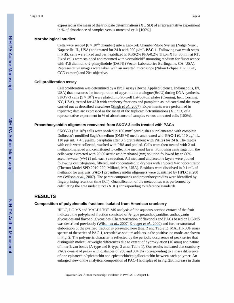
IntroductionThe North American cranberry (Vaccinium macrocarpon Ait Ericaceae) is receiving attentionas a ‘prophylactic’ food and source of ‘orthomolecular medicines’ (Neto et al., 2006).Cranberry fruit is rich in polyphenolic constituents with flavonols, anthocyanins, andproanthocyanidins (PACs) being the major classes of phytochemicals. In contrast to otherspecies, for example, cocoa, grape, pomegranate, noted for relatively high proanthocyanidincontent, cranberries contain proanthocyanidins with A-type linkages between units (Fig. 1).PACs with the A-type linkage are associated with antibacterial adhesion properties (Howelland Vorsa, 1998; Foo et al., 2000), low-density lipoprotein (LDL)-oxidation protectiveproperties (Kruger et al., 2000; Porter et al., 2001; Yan et al., 2002), and decreased oxidativedamage to rat neurons in a stroke model (Neto et al., 2005).
Potential anticancer and chemopreventive properties of cranberry extracts have been thesubject of many recent investigations. Cranberry extracts inhibited the proliferation of MCF-7and MDA-MB-435 breast cancer cells (Guthrie, 2000; Ferguson et al., 2004). Water-solubleextracts of commercial cranberry powder inhibited the proliferation of several human tumorcell lines (Sreeram et al., 2004). Cranberry phenolic-rich esters of pentacyclic triterpene ursolicacid selectively inhibited the growth of several types of tumor cells in vitro, particularly MCF-7(Murphy et al., 2003). Cranberry flavonoids may also play a role in chemo-prevention.Flavonoid-rich fractions from Vaccinium species, including cranberry, inhibited ornithinedecar-boxylase (ODC) expression in epithelial cells (Kandil et al., 2002) and induced thexenobiotic detoxification enzyme quinone reductase (QR) in vitro (Bomser et al., 1996).
Cranberry proanthocyanidins known to inhibit the growth of various tumor cell lines have notbeen well characterized, and the effects of cranberry extracts on platinum-resistant ovariancancer and neuroblastoma cell lines have not been described. Furthermore, the effects ofcranberry compounds as adjuvants to paraplatin, the major drug therapy for ovarian cancer,have not been examined. In this study, proanthocyanidin-rich fractions were isolated bychromatographic methods after sub-fractionating a polyphenolic-rich whole-cranberry extract.The fractions were evaluated for their ability to inhibit proliferation in vitro in three solid tumorcell lines, ovarian (SKOV-3), neuroblastoma (SMS-KCNR), and prostate (PC-3), and a normalhuman lung fibroblast cell line.
The primary objectives of this study were to isolate, characterize, and evaluate the cytotoxicityof cranberry polyphenols in ovarian, neuroblastoma and prostate cancer cell lines. The activityof cranberry constituents was investigated by (i) screening multiple purified and characterizedfractions to identify the most active; (ii) screening the most active fractions against severaltumor cell lines, and a normal human lung fibroblast cell line; (iii) observing morphologicalchanges associated with apoptosis; (iv) evaluating synergism of PACs when combined withparaplatin treatment in SKOV-3 ovarian cancer cells by measuring inhibition of cell viabilityand cell proliferation; and (v) measuring the differential uptake of PACs in SKOV-3 cells withand without paraplatin co-treatment using high performance liquid chromatography (HPLC).
Materials and MethodsPlant material
Fresh mature cranberries of the cultivars ‘Stevens’ and ‘Early Black’ were collected fromcommercial plantings in Burlington Co., New Jersey and maintained frozen at −20 °C untiluse.
Singh et al. Page 2
Phytother Res. Author manuscript; available in PMC 2010 August 1.
NIH
-PA Author Manuscript
NIH
-PA Author Manuscript
NIH
-PA Author Manuscript
Reagents and instrumentationSolvents were purchased from Fisher Scientific (Pittsburg, PA, USA) and were HPLC grade.Precoated aluminum plates with silica gel 60 with F254 fluorescent indicator were purchasedfrom Merck, Gibbstown, NJ, USA. Sephadex LH-20 for column chromatography was obtainedfrom Amersham Pharmacia Biotech AB (Uppsala, Sweden). Established standards were usedfor identification of individual flavonol glycosides (Gregoire et al., 2007) andproanthocyanidins (Duarte et al., 2006; Wilson et al., 2007). Previously described HPLCmethods for the identification of proanthocyanidins and flavonols (Wilson et al., 2007,Vvedenskaya et al., 2004) were followed with slight modifications. The calibration matrix 2,5-dihydroxybenzoic acid, α-cyano-4-hydroxycinnamic acid, trans-3-indoleacrylic acid, 2,5-dihydroxybenzoic acid were purchased from Sigma-Aldrich (St Louis, MO, USA). 1H NMRand 13C NMR spectra were recorded with a Bruker AM 500 instrument at room temperature(RT) using 3 mm tubes. Samples (5 mg) were dissolved in MeOH-d4. Chemical shifts wereexpressed in parts per million (ppm) relative to tetramethyl silane (TMS) as an internalstandard.
Extraction and isolation of polyphenolic constituentsFlavonol, anthocyanin and proanthocyanidin-rich fractions were isolated from frozencranberry fruit with aqueous acetone (80:20 acetone/water, v/v) extraction followed by an ethylacetate extraction and LH-20 column chromatography (Duarte et al., 2006; Gregoire et al.,2007). PACs containing fractions (1.3 g) obtained from methanol elution were reloaded on aSephadex column (100 mm × 45 mm) and eluted with increased gradients of methanol andwater (10, 20, 30, 40, 60, 80% methanol/water v/v) mixture to remove the flavonols.Oligomeric PACs were eluted with 80% acetone/water (v/v) to isolate PACs consisting of adegree of polymerization (DP) from DP-2 to DP-11, referred to as PAC-1. The PACs obtainedfrom 80% acetone/water (v/v) elution were again chromato-graphed using Sephadex LH-20and successively eluted with 50%, 80% methanol/water (v/v) and finally eluted with 80%acetone/water (v/v) to obtain polymeric PACs of molecular weight with degrees ofpolymerization ranging from DP-3 to DP-12, referred to as PAC-2. The fraction obtained fromcolumn chromatography containing both dimers (DP-2) and trimers (DP-3) was further purifiedby preparative HPLC. All fractions were characterized by LC-MS and/or MALDI-TOF.
Cell culture and conditionsSKOV-3 (ovarian epithelial adenocarcinoma), PC-3 (prostate adenocarcinoma), LF (lungfibroblasts) were purchased from ATCC (Manassas, VA, USA). SMS-KCNR (resistantneuroblastoma cell line) was a gift from Dr Giselle Sholler (University of Vermont, VermontCancer Center, Burlington, VT, USA). Cells were grown to 80% confluence in T75 cell cultureflasks (Corning, New York, NY, USA) in complete medium (Gibco, Rockville, MD, USA)according to the suppliers recommendation. For all assays, cells were allowed to attachovernight and then treated in complete medium.
Cell viability assayCell viability was determined using the 96® Aqueous-One-Solution Assay (Promega, Madison,WI, USA) following the manufacturer's recommendations. This colorimetric assay is based onthe ability of mitochondria to reduce a substrate (MTS) into a soluble formazan product withan absorbance at 490 nm (ELISA plate reader; Thermo Labsystems, Waltham, MA, USA) thatis directly proportional to the number of living cells (Malich et al., 1997). Cells (5 × 10³) wereplated into 96-well flat bottom plates (Corning, Inc., Corning, NY, USA) and treated withcranberry fractions at concentrations of 0, 31, 62, 125, 250 and 500 μg/mL or co-treated withparaplatin (4.5 μg/ mL). Following incubation for 46 h MTS reagent was added for an additional2–6 h and absorbance measured at 490 nm. Experiments were performed in triplicate; data are
Singh et al. Page 3
Phytother Res. Author manuscript; available in PMC 2010 August 1.
NIH
-PA Author Manuscript
NIH
-PA Author Manuscript
NIH
-PA Author Manuscript
expressed as the mean of the triplicate determinations (X ± SD) of a representative experimentin % of absorbance of samples versus untreated cells [100%].
Morphological studiesCells were seeded (6 × 10⁴/ chamber) into a Lab-Tek Chamber-Slide System (Nalge Nunc.,Naperville, IL, USA) and treated for 24 h with 200 μ/mL PAC-1. Following two wash-stepsin PBS, cells were fixed and permeabilized in PBS/2% PFA/0.2% Triton X for 30 min at RT.Fixed cells were stainded and mounted with vectoshield® mounting medium for fluorescencewith 4′,6 diamidino-2-phenylindole (DAPI) (Vector Laboratories Burlingame, CA, USA).Representative images were taken with an inverted microscope (Nikon Eclipse TE2000-E,CCD camera) and 20× objective.
Cell proliferation assayCell proliferation was determined by a BrdU assay (Roche Applied Science, Indianapolis, IN,USA) that measures the incorporation of a pyrimidine analogue (BrdU) during DNA synthesis.SKOV-3 cells (5 × 10³) were plated into 96-well flat-bottom plates (Corning, Inc., Corning,NY, USA), treated for 42 h with cranberry fractions and paraplatin as indicated and the assaycarried out as described elsewhere (Singh et al., 2007). Experiments were performed intriplicate; data are expressed as the mean of the triplicate determinations (X ± SD) of arepresentative experiment in % of absorbance of samples versus untreated cells [100%].
Proanthocyanidin oligomers recovered from SKOV-3 cells treated with PACsSKOV-3 (2 × 10⁶) cells were seeded in 100 mm2 petri dishes supplemented with completeDulbecco's modified Eagle's medium (DMEM) media and treated with PAC-1 (0, 110 μg/mL,110 μg/ mL + 4.5 μg/mL paraplatin after 3 h pretreatment with PACs) for 24 h. The mediawith cells were collected, washed with PBS and pooled. Cells were then treated with 2 mLmethanol, scraped and centrifuged to collect the methanol layer. Following centrifugation, thecells were extracted with 20:80 acetic acid/methanol (v/v) solution followed by an 80%acetone/water (v/v) (1 mL each) extraction. All methanol and acetone layers were pooledfollowing centrifugation, filtered, and concentrated to dryness with a Speed Vac concentrator(Thermo Model SPD 2010-220; Milford, MA, USA). Residues were dissolved in 0.1 mL ofmethanol for analysis. PAC-1 proanthocyanidin oligomers were quantified by HPLC at 280nm (Wilson et al., 2007). The parent compounds and proanthocyanidins were identified byfingerprinting retention time (RT). Quantification of the metabolites was performed bycalculating the area under curve (AUC) corresponding to reference standards.
RESULTSComposition of polyphenolic fractions isolated from American cranberry
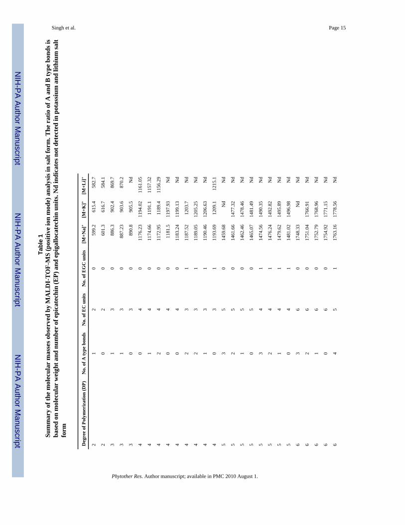
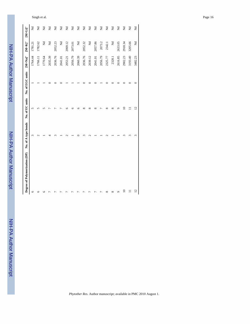
HPLC, LC-MS and MALDI-TOF-MS analysis of the aqueous acetone extract of the fruitindicated the polyphenol fraction consisted of A-type proanthocyanidins, anthocyaninglycosides and flavonol glycosides. Characterization of flavonols and PACs based on LC-MSwas described previously (Wilson et al., 2007; Krueger et al., 2000) and further structuralelaboration of the purified fraction is presented here (Fig. 2 and Table 1). MALDI-TOF massspectra of the series of PAC-1, recorded as sodium adducts in the positive ion mode, are shownin Fig. 2. The polymeric character is reflected by the periodic occurrence of peak series thatdistinguish molecular weight differences due to extent of hydroxylation (16 amu) and natureof interflavan bonds (A-type and B-type, 2 amu; Table 1). Our results indicated that cranberryPACs consist of peaks with distances of 288 and 304 Da corresponding to a mass differenceof one epicatechin/epicatechin and epicatechin/epigallocatechin between each polymer. Anenlarged view of the analytical composition of PAC-1 is displayed in Fig. 2B. Increase in chain
Singh et al. Page 4
Phytother Res. Author manuscript; available in PMC 2010 August 1.
NIH
-PA Author Manuscript
NIH
-PA Author Manuscript
NIH
-PA Author Manuscript
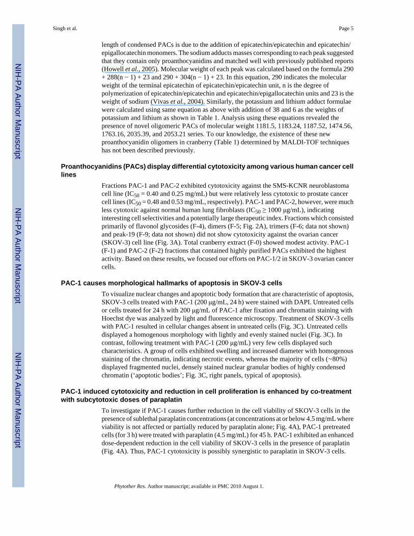
length of condensed PACs is due to the addition of epicatechin/epicatechin and epicatechin/epigallocatechin monomers. The sodium adducts masses corresponding to each peak suggestedthat they contain only proanthocyanidins and matched well with previously published reports(Howell et al., 2005). Molecular weight of each peak was calculated based on the formula 290+ 288(n − 1) + 23 and 290 + 304(n − 1) + 23. In this equation, 290 indicates the molecularweight of the terminal epicatechin of epicatechin/epicatechin unit, n is the degree ofpolymerization of epicatechin/epicatechin and epicatechin/epigallocatechin units and 23 is theweight of sodium (Vivas et al., 2004). Similarly, the potassium and lithium adduct formulaewere calculated using same equation as above with addition of 38 and 6 as the weights ofpotassium and lithium as shown in Table 1. Analysis using these equations revealed thepresence of novel oligomeric PACs of molecular weight 1181.5, 1183.24, 1187.52, 1474.56,1763.16, 2035.39, and 2053.21 series. To our knowledge, the existence of these newproanthocyanidin oligomers in cranberry (Table 1) determined by MALDI-TOF techniqueshas not been described previously.
Proanthocyanidins (PACs) display differential cytotoxicity among various human cancer celllines
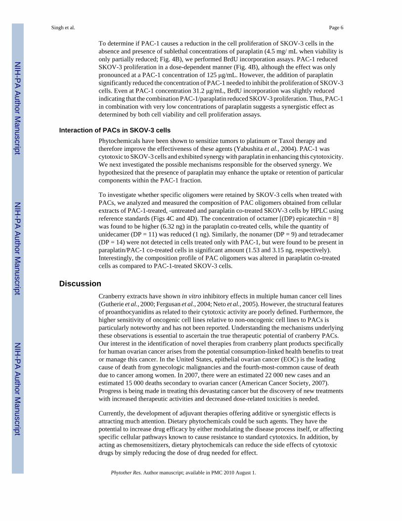
Fractions PAC-1 and PAC-2 exhibited cytotoxicity against the SMS-KCNR neuroblastomacell line (IC50 = 0.40 and 0.25 mg/mL) but were relatively less cytotoxic to prostate cancercell lines (IC50 = 0.48 and 0.53 mg/mL, respectively). PAC-1 and PAC-2, however, were muchless cytotoxic against normal human lung fibroblasts (IC50 ≥ 1000 μg/mL), indicatinginteresting cell selectivities and a potentially large therapeutic index. Fractions which consistedprimarily of flavonol glycosides (F-4), dimers (F-5; Fig. 2A), trimers (F-6; data not shown)and peak-19 (F-9; data not shown) did not show cytotoxicity against the ovarian cancer(SKOV-3) cell line (Fig. 3A). Total cranberry extract (F-0) showed modest activity. PAC-1(F-1) and PAC-2 (F-2) fractions that contained highly purified PACs exhibited the highestactivity. Based on these results, we focused our efforts on PAC-1/2 in SKOV-3 ovarian cancercells.
PAC-1 causes morphological hallmarks of apoptosis in SKOV-3 cellsTo visualize nuclear changes and apoptotic body formation that are characteristic of apoptosis,SKOV-3 cells treated with PAC-1 (200 μg/mL, 24 h) were stained with DAPI. Untreated cellsor cells treated for 24 h with 200 μg/mL of PAC-1 after fixation and chromatin staining withHoechst dye was analyzed by light and fluorescence microscopy. Treatment of SKOV-3 cellswith PAC-1 resulted in cellular changes absent in untreated cells (Fig. 3C). Untreated cellsdisplayed a homogenous morphology with lightly and evenly stained nuclei (Fig. 3C). Incontrast, following treatment with PAC-1 (200 μg/mL) very few cells displayed suchcharacteristics. A group of cells exhibited swelling and increased diameter with homogenousstaining of the chromatin, indicating necrotic events, whereas the majority of cells (∼80%)displayed fragmented nuclei, densely stained nuclear granular bodies of highly condensedchromatin (‘apoptotic bodies’; Fig. 3C, right panels, typical of apoptosis).
PAC-1 induced cytotoxicity and reduction in cell proliferation is enhanced by co-treatmentwith subcytotoxic doses of paraplatin
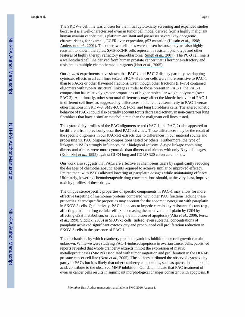
To investigate if PAC-1 causes further reduction in the cell viability of SKOV-3 cells in thepresence of sublethal paraplatin concentrations (at concentrations at or below 4.5 mg/mL whereviability is not affected or partially reduced by paraplatin alone; Fig. 4A), PAC-1 pretreatedcells (for 3 h) were treated with paraplatin (4.5 mg/mL) for 45 h. PAC-1 exhibited an enhanceddose-dependent reduction in the cell viability of SKOV-3 cells in the presence of paraplatin(Fig. 4A). Thus, PAC-1 cytotoxicity is possibly synergistic to paraplatin in SKOV-3 cells.
Singh et al. Page 5
Phytother Res. Author manuscript; available in PMC 2010 August 1.
NIH
-PA Author Manuscript
NIH
-PA Author Manuscript
NIH
-PA Author Manuscript
To determine if PAC-1 causes a reduction in the cell proliferation of SKOV-3 cells in theabsence and presence of sublethal concentrations of paraplatin (4.5 mg/ mL when viability isonly partially reduced; Fig. 4B), we performed BrdU incorporation assays. PAC-1 reducedSKOV-3 proliferation in a dose-dependent manner (Fig. 4B), although the effect was onlypronounced at a PAC-1 concentration of 125 μg/mL. However, the addition of paraplatinsignificantly reduced the concentration of PAC-1 needed to inhibit the proliferation of SKOV-3cells. Even at PAC-1 concentration 31.2 μg/mL, BrdU incorporation was slightly reducedindicating that the combination PAC-1/paraplatin reduced SKOV-3 proliferation. Thus, PAC-1in combination with very low concentrations of paraplatin suggests a synergistic effect asdetermined by both cell viability and cell proliferation assays.
Interaction of PACs in SKOV-3 cellsPhytochemicals have been shown to sensitize tumors to platinum or Taxol therapy andtherefore improve the effectiveness of these agents (Yabushita et al., 2004). PAC-1 wascytotoxic to SKOV-3 cells and exhibited synergy with paraplatin in enhancing this cytotoxicity.We next investigated the possible mechanisms responsible for the observed synergy. Wehypothesized that the presence of paraplatin may enhance the uptake or retention of particularcomponents within the PAC-1 fraction.
To investigate whether specific oligomers were retained by SKOV-3 cells when treated withPACs, we analyzed and measured the composition of PAC oligomers obtained from cellularextracts of PAC-1-treated, -untreated and paraplatin co-treated SKOV-3 cells by HPLC usingreference standards (Figs 4C and 4D). The concentration of octamer [(DP) epicatechin = 8]was found to be higher (6.32 ng) in the paraplatin co-treated cells, while the quantity ofunidecamer (DP = 11) was reduced (1 ng). Similarly, the nonamer (DP = 9) and tetradecamer(DP = 14) were not detected in cells treated only with PAC-1, but were found to be present inparaplatin/PAC-1 co-treated cells in significant amount (1.53 and 3.15 ng, respectively).Interestingly, the composition profile of PAC oligomers was altered in paraplatin co-treatedcells as compared to PAC-1-treated SKOV-3 cells.
DiscussionCranberry extracts have shown in vitro inhibitory effects in multiple human cancer cell lines(Gutherie et al., 2000; Fergusan et al., 2004; Neto et al., 2005). However, the structural featuresof proanthocyanidins as related to their cytotoxic activity are poorly defined. Furthermore, thehigher sensitivity of oncogenic cell lines relative to non-oncogenic cell lines to PACs isparticularly noteworthy and has not been reported. Understanding the mechanisms underlyingthese observations is essential to ascertain the true therapeutic potential of cranberry PACs.Our interest in the identification of novel therapies from cranberry plant products specificallyfor human ovarian cancer arises from the potential consumption-linked health benefits to treator manage this cancer. In the United States, epithelial ovarian cancer (EOC) is the leadingcause of death from gynecologic malignancies and the fourth-most-common cause of deathdue to cancer among women. In 2007, there were an estimated 22 000 new cases and anestimated 15 000 deaths secondary to ovarian cancer (American Cancer Society, 2007).Progress is being made in treating this devastating cancer but the discovery of new treatmentswith increased therapeutic activities and decreased dose-related toxicities is needed.
Currently, the development of adjuvant therapies offering additive or synergistic effects isattracting much attention. Dietary phytochemicals could be such agents. They have thepotential to increase drug efficacy by either modulating the disease process itself, or affectingspecific cellular pathways known to cause resistance to standard cytotoxics. In addition, byacting as chemosensitizers, dietary phytochemicals can reduce the side effects of cytotoxicdrugs by simply reducing the dose of drug needed for effect.
Singh et al. Page 6
Phytother Res. Author manuscript; available in PMC 2010 August 1.
NIH
-PA Author Manuscript
NIH
-PA Author Manuscript
NIH
-PA Author Manuscript
The SKOV-3 cell line was chosen for the initial cytotoxicity screening and expanded studiesbecause it is a well-characterized ovarian tumor cell model derived from a highly malignanthuman ovarian cancer that is platinum-resistant and possesses several key oncogeniccharacteristics, for example, EGFR over-expression, p53 mutation (Husain et al., 1998;Anderson et al., 2001). The other two cell lines were chosen because they are also highlyresistant to known therapies. SMS-KCNR cells represent a resistant phenotype and otherfeatures of highly therapy refractory neuroblastoma (Singh et al., 2007). The PC-3 cell line isa well-studied cell line derived from human prostate cancer that is hormone-refractory andresistant to multiple chemotherapeutic agents (Hart et al., 2005).
Our in vitro experiments have shown that PAC-1 and PAC-2 display partially overlappingcytotoxic effects in all cell lines tested. SKOV-3 cancer cells were more sensitive to PAC-1than to PAC-2 or other flavonoid fractions. Even though other fractions (F1–F5) containedoligomers with type-A structural linkages similar to those present in PAC-1, the PAC-1composition has relatively greater proportions of higher molecular weight polymers (overPAC-2). Additionally, other structural differences may affect the kinetic behavior of PAC-1in different cell lines, as suggested by differences in the relative sensitivity to PAC-1 versusother fractions in SKOV-3, SMS-KCNR, PC-3, and lung fibroblasts cells. The altered kineticbehavior of PAC-1 could also partially account for its decreased activity in non-cancerous lungfibroblasts that have a similar metabolic rate than the malignant cell lines tested.
The cytotoxicity profiles of the PAC oligomers tested (PAC-1 and PAC-2) also appeared tobe different from previously described PAC activities. These differences may be the result ofthe specific oligomers in our PAC-1/2 extracts due to differences in our material source andprocessing vs. PAC oligomeric compositions tested by others. Furthermore, the type oflinkages in PACs strongly influences their biological activity. A-type linkage containingdimers and trimers were more cytotoxic than dimers and trimers with only B-type linkages(Kolodziej et al., 1995) against GLC4 lung and COLO 320 colon carcinomas.
Our work also suggests that PACs are effective as chemosensitizers by significantly reducingthe dosages of chemotherapeutic agents required to achieve similar or improved efficacy.Pretreatment with PACs allowed lowering of paraplatin dosages while maintaining efficacy.Ultimately, lowering chemotherapeutic drug concentrations should, at the very least, improvetoxicity profiles of these drugs.
The unique stereospecific properties of specific components in PAC-1 may allow for moreeffective targeting of membrane proteins compared with other PAC fractions lacking theseproperties. Stereospecific properties may account for the apparent synergism with paraplatinin SKOV-3 cells. Qualitatively, PAC-1 appears to impede certain key resistance factors (e.g.,affecting platinum drug cellular efflux, decreasing the inactivation of platin by GSH byaffecting GSH metabolism, or reversing the inhibition of apoptosis) (Alia et al., 2006; Perezet al., 1998; Siddick, 2003) in SKOV-3 cells. Indeed, even sublethal concentrations ofparaplatin achieved significant cytotoxicity and pronounced cell proliferation reduction inSKOV-3 cells in the presence of PAC-1.
The mechanisms by which cranberry proanthocyanidins inhibit tumor cell growth remainunknown. While we were studying PAC-1-induced apoptosis in ovarian cancer cells, publishedreports revealed that whole cranberry extracts inhibit the expression of matrixmetalloproteinases (MMPs) associated with tumor migration and proliferation in the DU-145prostate cancer cell line (Neto et al., 2005). The authors attributed the observed cytotoxicitypartly to PACs but it is likely that other cranberry components, such as quercetin and ursolicacid, contribute to the observed MMP inhibition. Our data indicate that PAC treatment ofovarian cancer cells results in significant morphological changes consistent with apoptosis. It
Singh et al. Page 7
Phytother Res. Author manuscript; available in PMC 2010 August 1.
NIH
-PA Author Manuscript
NIH
-PA Author Manuscript
NIH
-PA Author Manuscript
is possible that MMP inhibition may be involved in the mechanism(s) of action of PAC-1/2in SKOV-3 cells.
Finally, our experiments also demonstrated that subcytotoxic concentrations of paraplatin mayalter the absorption, adherence or intracellular metabolism of PAC-1 in SKOV-3 cells. Wenoted a significant increase in DP-8, and very significant decrease in DP-11 (Figs 4C and 4D),suggesting that DP-11 may be rapidly hydrolyzed, possibly to DP-8 and other PACs but in anon-stoichiometric fashion vs. non-paraplatin-treated cells. Moreover, DP-9 and DP-14 weredetected only in cells co-treated with paraplatin/PAC-1, indicating altered oligomer specificityof absorption, which is induced only by combination treatment (Figs 4C and 4D). Thus it wouldappear that sublethal concentrations of paraplatin have a significant impact on the interactionof the cell membrane with PAC oligomers. The recovery of specific oligomers in SKOV-3cells following co-treatment suggests that the mechanism(s) of action involve specific drug–drug interactions.
As of now it is unknown to us if the consumption of DP-11 and/or the increased production ofDP-8 and the apparent differential formation of DP-9 and DP-14 are essential for cytotoxicity.To answer these questions would require the isolation and/or the chemical synthesis of DP-8,DP-9, and DP-14 PACs and assessing their biological activities. This is beyond the scope ofthis descriptive study but is the interest of future investigations that will specifically examinethe structural differences or key requirements and exact mechanism(s) of action of PACs.
ConclusionsPurified and MALDI-TOF characterized proanthocyanidins (PACs) found in cranberries arepolymeric high molecular weight proanthocyanidins with A-type linkages among epicatechinsconsisting of a mixture of polymeric proanthocyanidins (DP-2 to DP-14). These PACs areselectively cytotoxic to platinum-resistant ovarian cancer cells, hormone-refractory prostatecancer cells, and neuro-blastoma cells but are minimally cytotoxic to lung fibro-blasts. PACssensitized platinum-resistant ovarian cancer cells to paraplatin and displayed synergism withthis chemotherapeutic agent. Finally, paraplatin appeared to affect PAC metabolism in theseovarian cancer cells. These interesting findings warrant the further preclinical/ clinicalevaluation of PACs in the treatment of human solid tumors.
AcknowledgmentsThis work was supported by NIH NCCAM 5R01AT002058 to Dr Vorsa, and an Ovarian Cancer Research FundIndividual Investigator Grant and a NICHD, K12 HD043447 BIRCWH Scholar Grant to Dr Brard.
ReferencesAlia BH, Moundhri MSA. Agents ameliorating or augmenting the nephrotoxicity of cisplatin and other
platinum compounds: A review of some recent research. Food and Chem Toxicol 2006;44:1173–1183.[PubMed: 16530908]
American Cancer Society. Cancer Statistics. 2007. www.cancer.orgAnderson NG, Ahmad T, Chan K, Dobson R, Bundred NJ. ZD1839 (Iressa), a novel epidermal growth
factor receptor (EGFR) tyrosine kinase inhibitor, potently inhibits the growth of EGFR-positive cancercell lines with or without erbB2 overexpression. Int J Cancer 2001;94:774–782. [PubMed: 11745477]
Bomser J, Madhavi DL, Singletary K, Smith MAL. In vitro anticancer activity of fruit extracts fromVaccinium species. Planta Med 1996;62:212–216. [PubMed: 8693031]
Duarte S, Gregoire S, Singh AP, Vorsa N, Schaich K, Bowen WH, Koo H. Inhibitory effects of cranberrypolyphenols on formation and acidogenicity of Streptococcus mutans biofilms. FEMS Microbiol Lett2006;257:50–56. [PubMed: 16553831]
Singh et al. Page 8
Phytother Res. Author manuscript; available in PMC 2010 August 1.
NIH
-PA Author Manuscript
NIH
-PA Author Manuscript
NIH
-PA Author Manuscript
Ferguson P, Kurowska E, Freeman DJ, Chambers AF, Koropatnick DJ. A flavonoid fraction fromcranberry extract inhibits proliferation of human tumor cell lines. J Nutr 2004;134:1529–1535.[PubMed: 15173424]
Foo LY, Lu Y, Howell AB, Vorsa N. A-type Proanthocyanidin trimers from cranberry that inhibitadherence of uropathogenic P-fimbriated Escherischia coli. J Nat Prod 2000;63:1225–1228. [PubMed:11000024]
Gregoire S, Singh AP, Vorsa N, Koo H. Influence of cranberry phenolics on glucan synthesis byglucosyltransferases and Streptococcus mutans acidogenicity. J Appl Microbiol 2007;103:1960–1968.[PubMed: 17953606]
Guthrie N. Effect of cranberry juice and products on human breast cancer cell growth. FASEB J2000;14:A771.
Hart CA, Brown M, Bagley S, Sharrard M, Clarke NW. Invasive characteristics of human prostaticepithelial cells: understanding the metastatic process. Br J Cancer 2005;92:503–512. [PubMed:15668715]
Howell AB, Reed JD, Krueger CG, Winterbottom R, Cunningham DG, Leahy M. A-type cranberryproanthocyanidins and uropathogenic bacterial anti-adhesion activity. Phytochemistry2005;66:2281–2291. [PubMed: 16055161]
Howell A, Vorsa N. Inhibition of adherence of P-fimbricated Escherischia coli to uroepithelial cellsurfaces by proanthocyanidin extracts from cranberries. N Engl J Med 1998;339:1085–1086.[PubMed: 9767006]
Husain A, Rosales N, Schwartz GK, Spriggs DR. Lisofylline sensitizes p53 mutant human ovariancarcinoma cells to the cytotoxic effects of cis-diamminedichloroplatinum (II). Gynecol Oncol1998;70:17–22. [PubMed: 9698467]
Kandil FE, Smith MAL, Rogers RB, Pepin MF, Song LL, Pezzuto JM, Seigler DS. Composition of achemopreventive proanthocyanidin-rich fraction from cranberry fruits responsible for the inhibitionof TPA-induced ODC activity. J Agric Food Chem 2002;50:1063–1069. [PubMed: 11853481]
Kolodziej H, Haberland C, Woerdenbag HJ, Konings AWT. Moderate cytotoxicity of proanthocyanidinsto human tumour cell lines. Phytother Res 1995;9:410–415.
Krueger CG, Dopke N, Treichel PM, Folts J, Reed JD. Matrix-assisted laser desorption/ionization time-of-flight mass spectrometry of polygalloyl polyflavan-3-ols in grape seed extract. J Agric Food Chem2000;48:1663–1667. [PubMed: 10820075]
Murphy BT, MacKinnon SL, Yan X, Hammond GB, Vaisberg AJ, Neto CC. Identification of triterpenehydroxycinnamates with in vitro anti-tumor activity from whole cranberry fruit (Vacciniummacrocarpon). J Agric Food Chem 2003;51:3541–3545. [PubMed: 12769521]
Neto CC, Krueger CG, Lamoureaux TL, Kondo M, Vaisberg AJ. MALDI-TOF MS characterization ofproanthocyanidins from cranberry fruit (Vaccinium macrocarpon) that inhibit tumor cell growth andmatrix metalloproteinase expression in vitro. J Sci Food Agric 2006;86:18–25.
Neto, CC.; Sweeney-Nixon, MI.; Lamoureaux, TL.; Solomon, F.; Kondo, M.; MacKinnon, SL.Symposium Series on Phenolics in Foods and Natural Health Products. ACS Books; Washington,DC: 2005. Cranberry phenolics: effects on oxidative processes, neuron cell death and tumor cellgrowth; p. 271-282.
Perez RP. Cellular and molecular determinants of cisplatin resistance. Eur J Cancer 1998;34:1535–1542.[PubMed: 9893624]
Porter ML, Krueger CG, Wiebe DA, Cunningham DG, Reed JD. Cranberry proanthocyanidins associatewith low density lipoprotein and inhibit in vitro Cu2+ induced oxidation. J Sci Food Agric2001;81:1306–1313.
Sreeam NP, Adams LS, Hardy ML, Heber D. Total cranberry extract versus its phytochemicalconstituents: anti-proliferative and synergistic effects against human tumor cell lines. J Agric FoodChem 2004;52:2512–2517. [PubMed: 15113149]
Siddick ZH. Cisplatin: mode of cytotoxic action and molecular basis of resistance. Oncogene2003;22:7265–7279. [PubMed: 14576837]
Singh RK, Lange TS, Kim K, Zou Y, Lieb C, Sholler GL, Brard L. Effect of Indole Ethyl Isothiocyanateson Proliferation, Apoptosis and MAPK Signaling in Neuroblastoma Cell Lines. Bioorg Med ChemLett 2007;17:5846–5852. [PubMed: 17855093]
Singh et al. Page 9
Phytother Res. Author manuscript; available in PMC 2010 August 1.
NIH
-PA Author Manuscript
NIH
-PA Author Manuscript
NIH
-PA Author Manuscript

Vvedenskaya IO, Rosen RT, Guido JE, Russell DJ, Mills KA, Vorsa N. Characterization of Flavonolsin Cranberry (Vaccinium macrocarpon) Powder. J Agric Food Chem 2004;52:188–195. [PubMed:14733493]
Vivas N, Nonier MF, Vivas de Gaulejac N, Absalon C, Bertrand A, Mirabel M. Differentiation ofproanthocyanidin tannins from seeds, skins and stems of grapes (Vitis vinifera) and heartwood ofQuebracho (Schinopsis balansae) by matrix-assisted laser desorption/ionization time-of-flight massspectrometry and thioacidolysis/liquid chromatography/electrospray ionization mass spectrometry.Anal Chim Acta 2004;513:247–256.
Wilson T, Singh AP, Vorsa N, Goett CD, Kittleson KM, Roe CM, Kastello GM, Ragsdale FR. HumanGlycemic Response and Phenolic Content of Unsweetened Cranberry Juice. J Med Food.200710.1089/jmf.2007.0531.
Yabushita H, Kishida T, Noguchi M, Masuda T, Tsukada H, Sawaguchi K, Noguchi Y, Noguchi M.Weekly administration of paclitaxel and carboplatin in patients with recurrent ovarian cancer. J AppResearch 2004;4:591–599.
Yan X, Murphy BT, Hammond GB, Vinson JA, Neto CC. Antioxidant activities and anti-tumor screeningof extracts from cranberry fruit (Vaccinium macrocarpon). J Agric Food Chem 2002;20:5844–5849.[PubMed: 12358448]
Singh et al. Page 10
Phytother Res. Author manuscript; available in PMC 2010 August 1.
NIH
-PA Author Manuscript
NIH
-PA Author Manuscript
NIH
-PA Author Manuscript
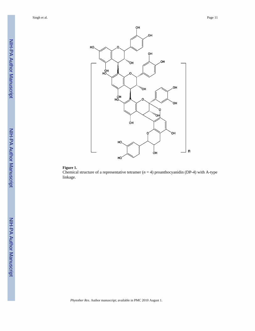
Figure 1.Chemical structure of a representative tetramer (n = 4) proanthocyanidin (DP-4) with A-typelinkage.
Singh et al. Page 11
Phytother Res. Author manuscript; available in PMC 2010 August 1.
NIH
-PA Author Manuscript
NIH
-PA Author Manuscript
NIH
-PA Author Manuscript
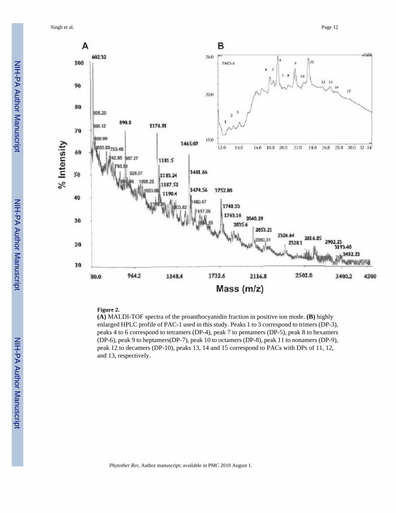
Figure 2.(A) MALDI-TOF spectra of the proanthocyanidin fraction in positive ion mode. (B) highlyenlarged HPLC profile of PAC-1 used in this study. Peaks 1 to 3 correspond to trimers (DP-3),peaks 4 to 6 correspond to tetramers (DP-4), peak 7 to pentamers (DP-5), peak 8 to hexamers(DP-6), peak 9 to heptamers(DP-7), peak 10 to octamers (DP-8), peak 11 to nonamers (DP-9),peak 12 to decamers (DP-10), peaks 13, 14 and 15 correspond to PACs with DPs of 11, 12,and 13, respectively.
Singh et al. Page 12
Phytother Res. Author manuscript; available in PMC 2010 August 1.
NIH
-PA Author Manuscript
NIH
-PA Author Manuscript
NIH
-PA Author Manuscript
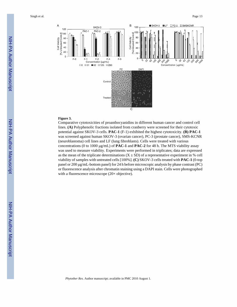
Figure 3.Comparative cytotoxicities of proanthocyanidins in different human cancer and control celllines. (A) Polyphenolic fractions isolated from cranberry were screened for their cytotoxicpotential against SKOV-3 cells. PAC-1 (F-1) exhibited the highest cytotoxicity. (B) PAC-1was screened against human SKOV-3 (ovarian cancer), PC-3 (prostate cancer), SMS-KCNR(neuroblastoma) cell lines and LF (lung fibroblasts). Cells were treated with variousconcentrations (0 to 1000 μg/mL) of PAC-1 and PAC-2 for 48 h. The MTS viability assaywas used to measure viability. Experiments were performed in triplicates; data are expressedas the mean of the triplicate determinations (X ± SD) of a representative experiment in % cellviability of samples with untreated cells [100%]. (C) SKOV-3 cells treated with PAC-1 (0-toppanel or 200 μg/mL-bottom panel) for 24 h before microscopic analysis by phase contrast (PC)or fluorescence analysis after chromatin staining using a DAPI stain. Cells were photographedwith a fluorescence microscope (20× objective).
Singh et al. Page 13
Phytother Res. Author manuscript; available in PMC 2010 August 1.
NIH
-PA Author Manuscript
NIH
-PA Author Manuscript
NIH
-PA Author Manuscript
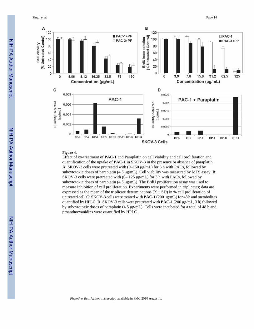
Figure 4.Effect of co-treatment of PAC-1 and Paraplatin on cell viability and cell proliferation andquantification of the uptake of PAC-1 in SKOV-3 in the presence or absence of paraplatin.A: SKOV-3 cells were pretreated with (0–150 μg/mL) for 3 h with PACs, followed bysubcytotoxic doses of paraplatin (4.5 μg/mL). Cell viability was measured by MTS assay. B:SKOV-3 cells were pretreated with (0– 125 μg/mL) for 3 h with PACs, followed bysubcytotoxic doses of paraplatin (4.5 μg/mL). The BrdU proliferation assay was used tomeasure inhibition of cell proliferation. Experiments were performed in triplicates; data areexpressed as the mean of the triplicate determinations (X ± SD) in % cell proliferation ofuntreated cell. C: SKOV-3 cells were treated with PAC-1 (200 μg/mL) for 48 h and metabolitesquantified by HPLC. D: SKOV-3 cells were pretreated with PAC-1 (200 μg/mL, 3 h) followedby subcytotoxic doses of paraplatin (4.5 μg/mL). Cells were incubated for a total of 48 h andproanthocyanidins were quantified by HPLC.
Singh et al. Page 14
Phytother Res. Author manuscript; available in PMC 2010 August 1.
NIH
-PA Author Manuscript
NIH
-PA Author Manuscript
NIH
-PA Author Manuscript
NIH
-PA Author Manuscript
NIH
-PA Author Manuscript
NIH
-PA Author Manuscript
Singh et al. Page 15
Tabl
e 1
Sum
mar
y of
the
mol
ecul
ar m
asse
s obs
erve
d by
MA
LD
I-T
OF-
MS
(pos
itive
ion
mod
e) a
naly
sis i
n sa
lt fo
rm. T
he r
atio
of A
and
B ty
pe b
onds
isba
sed
on m
olec
ular
wei
ght a
nd n
umbe
r of
epi
cate
chin
(EP)
and
epi
gallo
cate
chin
uni
ts. N
d in
dica
tes n
ot d
etec
ted
in p
otas
sium
and
lith
ium
salt
form
Deg
ree
of P
olym
eriz
atio
n (D
P)N
o. o
f A ty
pe b
onds
No.
of E
C u
nits
No.
of E
GC
uni
ts[M
+Na]
+[M
+K]+
[M+L
i]+
21
20
599.
261
5.4
582.
7
20
20
601.
361
6.7
584.
1
31
30
886.
390
2.4
869.
7
31
30
887.
2390
3.6
870.
2
30
30
890.
890
5.5
Nd
40
40
1176
.23
1194
.02
1161
.05
41
40
1174
.66
1191
.111
57.3
2
42
40
1172
.95
1189
.411
56.2
9
40
40
1181
.511
97.9
3N
d
40
40
1183
.24
1199
.13
Nd
42
31
1187
.52
1203
.7N
d
42
31
1189
.05
1205
.25
Nd
41
31
1190
.46
1206
.63
Nd
40
31
1193
.69
1209
.112
15.1
53
50
1459
.68
Nd
Nd
52
50
1461
.66
1477
.32
Nd
51
50
1462
.46
1478
.46
Nd
50
50
1465
.07
1481
.49
Nd
53
41
1474
.56
1490
.35
Nd
52
41
1476
.24
1492
.82
Nd
51
41
1479
.62
1495
.89
Nd
50
41
1481
.02
1496
.98
Nd
63
60
1748
.33
Nd
Nd
62
60
1751
.04
1766
.91
Nd
61
60
1752
.79
1768
.96
Nd
60
60
1754
.92
1771
.15
Nd
64
51
1763
.16
1778
.56
Nd
Phytother Res. Author manuscript; available in PMC 2010 August 1.
NIH
-PA Author Manuscript
NIH
-PA Author Manuscript
NIH
-PA Author Manuscript
Singh et al. Page 16
Deg
ree
of P
olym
eriz
atio
n (D
P)N
o. o
f A ty
pe b
onds
No.
of E
C u
nits
No.
of E
GC
uni
ts[M
+Na]
+[M
+K]+
[M+L
i]+
63
51
1764
.44
1781
.21
Nd
62
51
1766
.11
1782
.62
Nd
61
51
1770
.64
Nd
Nd
74
70
2035
.39
Nd
Nd
73
70
2036
.76
2052
.23
Nd
71
70
2041
.01
Nd
Nd
72
61
2053
.21
2069
.12
Nd
71
61
2056
.79
2073
.01
Nd
70
61
2060
.39
Nd
Nd
73
80
2036
.76
2052
.32
Nd
72
80
2039
.12
Nd
Nd
71
80
2041
.01
2057
.86
Nd
71
71
2056
.79
2073
.2N
d
82
80
2325
.77
2342
.1N
d
81
80
2328
.1N
dN
d
91
90
2616
.85
2633
.05
Nd
103
100
2902
.23
2918
.34
Nd
113
110
3193
.40
3205
.66
Nd
123
120
3482
.23
Nd
Nd
Phytother Res. Author manuscript; available in PMC 2010 August 1.
Copyright © 2022 FDOKUMEN