Histone deacetylase inhibitors enhance expression of NKG2D ligands in Ewing sarcoma and sensitize...
13
RESEARCH Open Access Histone deacetylase inhibitors enhance expression of NKG2D ligands in Ewing sarcoma and sensitize for natural killer cell-mediated cytolysis Dagmar Berghuis 1,2 , Marco W Schilham 2 , Hanneke I Vos 2 , Susy J Santos 2 , Stephan Kloess 3 , Emilie P Buddingh’ 2 , R Maarten Egeler 2 , Pancras CW Hogendoorn 1 and Arjan C Lankester 2* Abstract Background: Ewing sarcoma patients have a poor prognosis despite multimodal therapy. Integration of combination immunotherapeutic strategies into first-/second-line regimens represents promising treatment options, particularly for patients with intrinsic or acquired resistance to conventional therapies. We evaluated the susceptibility of Ewing sarcoma to natural killer cell-based combination immunotherapy, by assessing the capacity of histone deacetylase inhibitors to improve immune recognition and sensitize for natural killer cell cytotoxicity. Methods: Using flow cytometry, ELISA and immunohistochemistry, expression of natural killer cell receptor ligands was assessed in chemotherapy-sensitive/-resistant Ewing sarcoma cell lines, plasma and tumours. Natural killer cell cytotoxicity was evaluated in Chromium release assays. Using ATM/ATR inhibitor caffeine, the contribution of the DNA damage response pathway to histone deacetylase inhibitor-induced ligand expression was assessed. Results: Despite comparable expression of natural killer cell receptor ligands, chemotherapy-resistant Ewing sarcoma exhibited reduced susceptibility to resting natural killer cells. Interleukin-15-activation of natural killer cells overcame this reduced sensitivity. Histone deacetylase inhibitor-pretreatment induced NKG2D-ligand expression in an ATM/ATR-dependent manner and sensitized for NKG2D-dependent cytotoxicity (2/4 cell lines). NKG2D-ligands were expressed in vivo, regardless of chemotherapy-response and disease stage. Soluble NKG2D-ligand plasma concentrations did not differ between patients and controls. Conclusion: Our data provide a rationale for combination immunotherapy involving immune effector and target cell manipulation in first-/second-line treatment regimens for Ewing sarcoma. Keywords: Ewing sarcoma, natural killer cells, histone deacetylase inhibitor, combination immunotherapy, che- motherapy-resistance, tumour immunology Introduction Ewing sarcoma is an aggressive round cell sarcoma characterized by specific gene fusions most commonly involving TET gene family products, though rarely other activating transcription factors [1-3]. It usually affects bone or soft tissue in children and young adults. Despite multimodal therapy consisting of high-dose chemotherapy, surgery and radiotherapy, survival of patients with Ewing sarcoma has not improved signifi- cantly during the past decade. Patients with therapy- resistant or metastatic Ewing sarcoma have the most unfavorable prognosis, with a 5-year overall survival of less than 30% [4-6], which has recently been demon- strated to be independent of Ewing sarcoma-ETS fusion type [4,5]. Natural killer (NK) cells are the main cytotoxic effec- tor cells of the innate immune system, contributing to host anti-microbial and anti-tumour immune responses. * Correspondence: [email protected] 2 Department of Pediatrics, Leiden University Medical Center, Leiden, The Netherlands Full list of author information is available at the end of the article Berghuis et al. Clinical Sarcoma Research 2012, 2:8 http://www.clinicalsarcomaresearch.com/content/2/1/8 CLINICAL SARCOMA RESEARCH © 2012 Berghuis et al; licensee BioMed Central Ltd. This is an Open Access article distributed under the terms of the Creative Commons Attribution License (http://creativecommons.org/licenses/by/2.0), which permits unrestricted use, distribution, and reproduction in any medium, provided the original work is properly cited.
Transcript of Histone deacetylase inhibitors enhance expression of NKG2D ligands in Ewing sarcoma and sensitize...

RESEARCH Open Access
Histone deacetylase inhibitors enhanceexpression of NKG2D ligands in Ewing sarcomaand sensitize for natural killer cell-mediatedcytolysisDagmar Berghuis1,2, Marco W Schilham2, Hanneke I Vos2, Susy J Santos2, Stephan Kloess3, Emilie P Buddingh’2,R Maarten Egeler2, Pancras CW Hogendoorn1 and Arjan C Lankester2*
Abstract
Background: Ewing sarcoma patients have a poor prognosis despite multimodal therapy. Integration ofcombination immunotherapeutic strategies into first-/second-line regimens represents promising treatment options,particularly for patients with intrinsic or acquired resistance to conventional therapies. We evaluated thesusceptibility of Ewing sarcoma to natural killer cell-based combination immunotherapy, by assessing the capacityof histone deacetylase inhibitors to improve immune recognition and sensitize for natural killer cell cytotoxicity.
Methods: Using flow cytometry, ELISA and immunohistochemistry, expression of natural killer cell receptor ligandswas assessed in chemotherapy-sensitive/-resistant Ewing sarcoma cell lines, plasma and tumours. Natural killer cellcytotoxicity was evaluated in Chromium release assays. Using ATM/ATR inhibitor caffeine, the contribution of theDNA damage response pathway to histone deacetylase inhibitor-induced ligand expression was assessed.
Results: Despite comparable expression of natural killer cell receptor ligands, chemotherapy-resistant Ewingsarcoma exhibited reduced susceptibility to resting natural killer cells. Interleukin-15-activation of natural killer cellsovercame this reduced sensitivity. Histone deacetylase inhibitor-pretreatment induced NKG2D-ligand expression inan ATM/ATR-dependent manner and sensitized for NKG2D-dependent cytotoxicity (2/4 cell lines). NKG2D-ligandswere expressed in vivo, regardless of chemotherapy-response and disease stage. Soluble NKG2D-ligand plasmaconcentrations did not differ between patients and controls.
Conclusion: Our data provide a rationale for combination immunotherapy involving immune effector and targetcell manipulation in first-/second-line treatment regimens for Ewing sarcoma.
Keywords: Ewing sarcoma, natural killer cells, histone deacetylase inhibitor, combination immunotherapy, che-motherapy-resistance, tumour immunology
IntroductionEwing sarcoma is an aggressive round cell sarcomacharacterized by specific gene fusions most commonlyinvolving TET gene family products, though rarely otheractivating transcription factors [1-3]. It usually affectsbone or soft tissue in children and young adults. Despitemultimodal therapy consisting of high-dose
chemotherapy, surgery and radiotherapy, survival ofpatients with Ewing sarcoma has not improved signifi-cantly during the past decade. Patients with therapy-resistant or metastatic Ewing sarcoma have the mostunfavorable prognosis, with a 5-year overall survival ofless than 30% [4-6], which has recently been demon-strated to be independent of Ewing sarcoma-ETS fusiontype [4,5].Natural killer (NK) cells are the main cytotoxic effec-
tor cells of the innate immune system, contributing tohost anti-microbial and anti-tumour immune responses.
* Correspondence: [email protected] of Pediatrics, Leiden University Medical Center, Leiden, TheNetherlandsFull list of author information is available at the end of the article
Berghuis et al. Clinical Sarcoma Research 2012, 2:8http://www.clinicalsarcomaresearch.com/content/2/1/8 CLINICAL SARCOMA RESEARCH
© 2012 Berghuis et al; licensee BioMed Central Ltd. This is an Open Access article distributed under the terms of the CreativeCommons Attribution License (http://creativecommons.org/licenses/by/2.0), which permits unrestricted use, distribution, andreproduction in any medium, provided the original work is properly cited.
In contrast to T-lymphocytes, these cells lack antigen-specific receptors. Instead, recognition of target cellsand subsequent triggering of effector functions is regu-lated by integration of signals delivered from inhibitory(e.g. killer cell immunoglobulin receptors, CD94/NKG2A) and activating (e.g. natural killer group 2D(NKG2D), DNAX accessory molecule 1 (DNAM1), nat-ural cytotoxicity receptor (NCR)) cell surface receptors[7,8]. Natural killer cells respond to several cytokines,including several interleukins and type I interferons,resulting in increased cytolytic, secretory and prolifera-tive capacity [9]. Several studies have addressed thetherapeutic potential and safety of immunotherapeutic/natural killer cell-based approaches for various cancertypes, including sarcomas [10-15]. Sensitivity of tumoursto natural killer cells critically depends on expression ofligands for activating receptors. Likewise, low expressionof inhibitory human leukocyte antigen (HLA) class Imolecules is an important prerequisite for successfulnatural killer cell triggering. Manipulation of either thebalance between activating and inhibitory signals (by,for instance, ex vivo activation of immune cells) and/orsensitization of target cells for immune-mediated killingby combination immunotherapy may improve immu-notherapeutic efficacy. For example, pre-conditioning ofvarious cancer cell types by agents that activate theDNA damage response pathway may sensitize for nat-ural killer cell cytotoxicity via induction of activatingnatural killer cell receptor ligands and/or death receptorexpression. Comparable results have been observed forhistone deacetylase inhibitors (HDI), which are currentlyemerging as potent anti-tumour agents [16-19].Pre-clinical studies show that Ewing sarcoma can be
targeted by (cytokine-activated) natural killer cells in aNKG2D-, DNAM1- and, as recently demonstrated,NCR-dependent manner [20-24]. Moreover, a potentialrole for natural killer cell alloreactivity in Ewing sarcomadisease control has recently been suggested [25]. Inte-gration of natural killer cell-based (combination) immu-notherapy into first-line treatment regimens or as asecond-line approach represents a promising treatmentoption for Ewing sarcoma, in particular for patients witheither intrinsic or acquired resistance to conventionaltherapies [26]. However, chemotherapy-resistance ofEwing sarcoma correlates with expression of genesinvolved in, among others, apoptosis signaling pathways[27]. Depending on the (apoptotic) pathways involved,resistance to apoptosis might render cells cross-resistantto immune cell cytotoxicity [28]. As yet, data about thesusceptibility of therapy-resistant Ewing sarcoma to nat-ural killer cell cytotoxicity are lacking. To obtain insightinto the potential of natural killer cell-based combina-tion immunotherapy for Ewing sarcoma, natural killercell receptor ligand expression and susceptibility to
natural killer cell cytotoxicity were evaluated in che-motherapy-sensitive and -resistant Ewing sarcomatumour samples and cell lines respectively. Moreover,since HDI have the ability to exhibit direct cytotoxicityagainst Ewing sarcoma in vitro as well as in vivo and tosensitize for both conventional and more experimentaltreatment modalities [29-32], the potential of theseagents to sensitize Ewing sarcoma for natural killer cellcytotoxicity was investigated.
MethodsEwing sarcoma patients and samplesPeripheral blood samples from newly diagnosed Ewingsarcoma patients (2004-2009) were collected prior tostart of chemotherapy (n = 27) and, in case of completeremission, six months after completion of therapy (n =7) (treatment according to the EURO-EWING 99 trial[4]). Samples were centrifuged immediately and plasmawas stored at -80°C until assayed. Mean age at diagnosiswas 20.8 years (range 4-61 years). Plasma obtained fromage-comparable healthy controls (n = 27; mean age 21.8,range 5-65 years) was used as a reference. Formalin-fixed paraffin-embedded, sequential pre-treatment, post-chemotherapy and recurrent metastatic tumour samples(n = 33) from eight Ewing sarcoma patients wereobtained from the Department of Pathology, LeidenUniversity Medical Center. In all cases, diagnosis wasestablished according to WHO criteria including stan-dard confirmatory immunohistochemistry and fusiontranscript type. Follow-up provided information con-cerning initial tumour stage, (histological) chemotherapyresponse [33], recurrence rate and performance state.All patient material was coded, such that correlationwith clinical data was only possible for physiciansinvolved in treatment of the patients. Subsequentresearch was conducted following the ethical guidelinesof the Dutch organization of scientific societies(FEDERA).
Ewing sarcoma cell linesEwing sarcoma cell lines SK-ES-1, SK-N-MC, CADO-ESand STA-ET2.1 [34] were cultured in RPMI-1640 sup-plemented with streptomycin/penicillin (Invitrogen,Paisley, United Kingdom) and 10% fetal bovine serum((FBS); Greiner Bio-One, Alphen a/d Rijn, The Nether-lands). TC71 [34] and IOR/BER (kindly provided by dr.K. Scotlandi, Instituto Orthopedico Rizzoli, Bologna,Italy) were cultured in Iscove’s Modified Dulbecco’sMedium supplemented with streptomycin/penicillin and10% FBS. Previously reported [27,35] ‘natural’ sensitivityof these cell lines to chemotherapeutic drugs currentlyused for treatment of Ewing sarcoma [4] was used todiscriminate chemotherapy-sensitive (TC71, SK-ES-1,SK-N-MC) from chemotherapy-resistant (STA-ET2.1,
Berghuis et al. Clinical Sarcoma Research 2012, 2:8http://www.clinicalsarcomaresearch.com/content/2/1/8
Page 2 of 13

CADO-ES, IOR/BER) cell lines. Molecular HLA typingof the cell lines, performed at Leiden University MedicalCenter (LUMC), was converted to serological equiva-lents (no serological equivalents exist for HLA-Cw*16and -Cw*12): SK-ES-1 (A2/A11/B7/B44/Cw5/Cw7), SK-N-MC (A1/A25/B8/Cw7), CADO-ES (A11/A24/B15/B40/Cw4/Cw7), STA-ET-2.1 (A11/A24/B18/B40/Cw2/Cw5), TC71 (A2/A68/B15/B44/Cw3/Cw5), IOR/BER(A2/A11/B18/B51/Cw7/Cw15). The EBV B-LCL 107 cellline was generated from a healthy blood bank donorexpressing HLA alleles which are ligands to all inhibi-tory killer cell immunoglobulin receptors [20]. Thehuman erythroleukemia cell line K562 was obtainedfrom American Type Culture Collection (Manassas,VA). Cell lines were routinely screened for mycoplasmacontamination. Periodical authentication was performedby Short Tandem Repeat profiling and molecular HLAtyping.
Antibodies and reagentsThe antibodies used for staining of antigens by flowcytometry and immunohistochemistry as well as forblocking of specific natural killer cell receptors anddetection of soluble protein by ELISA are described inadditional file 1, table S1. HDI [N-(2-amino-phenyl)-4-[N-(pyridine-3-ylmethoxycarbonyl)aminomethyl]benza-mide] (MS-275) and suberoylanilide hydroxamic acid(SAHA) were purchased from Enzo Life Sciences(Raamsdonksveer, The Netherlands). Sodium butyrate(NaB) and the pharmacological ATM/ATR inhibitor caf-feine were obtained from Sigma-Aldrich (Zwijndrecht,The Netherlands).
Flow cytometric analysis of antigen surface expressionFlow cytometric analysis was performed on a FACScali-bur (Beckton Dickinson, Franklin Lakes, NJ) and resultswere analyzed using Cellquest software, as previouslydescribed [36]. Ligand expression was represented asfold increase in Mean Fluorescence Intensity (MFI) overisotype control staining (MFI-ratio).
Immunohistochemistry for detection of NKG2D ligandexpression in Ewing sarcoma tumour samples4-μm sections containing representative tumour weredeparaffinized and citrate antigen retrieval was per-formed. Subsequent immunohistochemical stainingswere performed and (semi-quantitatively) scored as pre-viously described [36].
Quantification of soluble MICA in Ewing sarcoma patientplasma samplesConcentrations of soluble MICA in plasma sampleswere determined by sandwich-ELISA using the MICA(human) detection set, according to the manufacturer’s
protocol (BAMOMAB/Axxora, Lörrach, Germany). Inshort, plates were coated overnight at 4°C with 5 μg/mlof the anti-MICA antibody (AMO1). After blocking with15% BSA/PBS for 15 minutes at 37°C, plates werewashed with 0.05% Tween-20/PBS. Samples or recombi-nant MICA*04, serving as a standard, were added in7.5% BSA/PBS. After incubation for two hours at 37°Cand washing, detection antibody (anti-MICA/B(BAMO3)) was added at a concentration of 1 μg/ml(two hours, 37°C). Plates were washed and incubatedwith HRP-conjugated anti-mouse IgG2a antibody (1080-05; SouthernBiotech, Birmingham, AL) for one hour at37°C. After extensive washing, TMB peroxidase sub-strate (KPL, Gaithersburg, MD) was added and plateswere incubated for 35 minutes at room temperature inthe dark. HRP activity was stopped by addition of 1 Mphosphoric acid and absorbance was measured at 450nm wavelength.
Isolation of natural killer cells from healthy donor-derivedperipheral blood mononuclear cellsPeripheral blood mononuclear cells were obtainedfrom healthy blood bank donors after informed con-sent and were isolated using Ficoll density gradientseparation. Isolation of natural killer cells was per-formed using the MACS NK enrichment kit and LScolumns, according to the manufacturer ’s protocol(Miltenyi Biotec, Bergisch Gladbach, Germany) andisolated cells were plated for overnight recovery at 1-2× 10⁶ cells/ml in RPMI-1640 supplemented with strep-tomycin/penicillin and 10% FBS. As determined byflow cytometric analysis, natural killer cell purityalways exceeded 88% and T-cell contamination > 0.2%was never detected. For cytokine activation, naturalkiller cells were cultured in AIM-V medium (Invitro-gen), supplemented with 10% pooled human AB-serum(Sanquin, Rotterdam, The Netherlands) and streptomy-cin/penicillin and stimulated with 10 ng/ml recombi-nant human interleukin-15 (IL-15) (Bender MedicalSystems, Vienna, Austria). Activated natural killer cellswere used for Chromium (51Cr) release assays withintwo-four weeks of culture.
Evaluation of natural killer cell cytotoxicity by Chromiumrelease assaysCytotoxicity was determined in standard 4-hour 51Crrelease assays as previously described [20]. For specificnatural killer cell receptor (ligand) blocking, naturalkiller cells or target cells were pre-incubated withblocking antibodies (20 μg/ml; additional file 1, tableS1) at room temperature for 20 minutes prior to the51Cr release assay. We previously demonstrated thatpre-incubation of natural killer cells with control iso-type (IgG1)-matched antibody (anti-CD56) did not
Berghuis et al. Clinical Sarcoma Research 2012, 2:8http://www.clinicalsarcomaresearch.com/content/2/1/8
Page 3 of 13

affect cytotoxicity [20]. Moreover, pre-incubation ofEwing sarcoma cells with control isotype (IgG1)-matched antibody (anti-CD99; clone B-N24; Diaclone/Sanquin Reagents, Amsterdam, Netherlands) had noeffect on natural killer cell-mediated cytolysis (data notshown).
Pre-treatment of Ewing sarcoma cell lines with histonedeacetylase inhibitorsEwing sarcoma cell lines SK-ES-1, CADO-ES, STA-ET2.1 and TC71 were seeded in 96 well plates at celldensities ranging from 3-15 × 103 cells/well. Followingovernight attachment, cells were incubated for 24hours with increasing concentrations of specific HDI.Cell viability was measured by 3-(4,5-dimethyl-thiazol-2-yl)-5-(3-carboxymethoxy-phenyl)-2-(4-sulfophenyl)-2H-tetrazolium (MTS) cell viability assay (PromegaBenelux, Leiden, The Netherlands). The cytotoxiceffect of HDI was quantified by determining IC50
values, as defined by the concentration of drug atwhich 50% of the cells were still metabolically active(table 1). For analysis of phenotypical and functionalconsequences of HDI treatment, cell lines were pre-treated with defined concentrations of HDI, as indi-cated in the results section. After 24 hours, cells wereharvested, washed and included in flow cytometric orcytotoxicity analyses. In some experiments, cells werepre-incubated for two hours with ATM/ATR inhibitorcaffeine (optimal dose based on dose response curve(data not shown)).
Statistical analyses and artworkStatistical analyses were performed with SPSS version16.0 software package. (Paired) t-tests or one-wayANOVA tests were used for comparison of meanswithin or between samples or groups of samples. P <0.05 was considered statistically significant. Artwork wascreated using Graphpad Prism 5.0 (La Jolla, CA).
ResultsChemotherapy-resistant Ewing sarcoma exhibit reducedsusceptibility to lysis by resting natural killer cellsTo assess possible cross-resistance of chemotherapy-resistant Ewing sarcoma to natural killer cell cytotoxi-city, a panel of both chemotherapy-sensitive (n = 3;TC71, SK-ES-1, SK-N-MC) and -resistant (n = 3;STA-ET2.1, CADO-ES, IOR/BER) cell lines was evalu-ated for susceptibility to lysis by resting natural killercells obtained from 4-8 healthy donors. As illustratedin Figure 1A-B, 51Cr release assays revealed signifi-cantly reduced sensitivity of chemotherapy-resistantcell lines to resting natural killer cell-mediated cytoly-sis, at effector-to-target ratio ’s ≥ 2.5:1 (t-test, p <0.05).
Comparable expression of ligands for activating andinhibitory natural killer cell receptors in chemotherapy-sensitive and -resistant Ewing sarcomaNatural killer cell cytotoxicity to Ewing sarcoma cellscritically depends on combined NKG2D and DNAM1signaling [20,23]. Therefore, constitutive surface expres-sion of ligands for these activating natural killer cellreceptors was analyzed in our panel of Ewing sarcomacell lines by flow cytometry. As represented in Figures2A-B and additional file 2, figure S1A, flow cytometricanalysis revealed no significant differences between che-motherapy-sensitive and -resistant cell lines with regardto expression of ligands for activating natural killer cellreceptors NKG2D (MICA, MICB, ULBP1-3) andDNAM1 (CD112, CD155) or killer cell immunoglobulinreceptors (HLA class I) (additional file 2, figure S1A, t-test, p > 0.05). Considerable inter-cell line variation,however, was observed for HLA class I expression: che-motherapy-resistant CADO-ES cells, demonstrating sub-stantial resistance to natural killer cells, exhibited 50-fold higher HLA class I expression as compared to che-motherapy-/natural killer cell-sensitive SK-ES-1 cells(Figure 1A and 2A). However, and consistent with pre-vious observations [20], blocking of HLA class I expres-sion could not restore the difference in natural killercell susceptibility between chemotherapy-sensitive and-resistant cell lines (additional file 2, figure S1B). Inaddition, and consistent with the observed comparableligand expression, blocking of NKG2D and DNAM1 onresting natural killer cells in 51Cr release assays reducednatural killer cell-mediated cytolysis of both chemother-apy-sensitive and -resistant cell lines to a similar degree(data not shown).Immunohistochemical staining for expression of
NKG2D-ligands MICA and ULBP1 in sequential pri-mary Ewing sarcoma tumour samples obtained frompatients with different histological responses to
Table 1 In vitro cytotoxicity (IC50 values) of histonedeacetylase inhibitors to chemo-sensitive and -resistantEWS cell linesa
cell line NaB (mM) SAHA (μM) MS-275 (μM)
chemo-sensitive
SK-ES-1 1.43 2.85 3.72
TC71 7.85 2.96 30.71
chemo-resistant
CADO-ES 38.99 3.92 49.97
STA-ET2.1 0.61 1.58 10.06a ’natural’ sensitivity of specific cell lines to chemotherapeutic drugs, aspreviously described [27,35].
Berghuis et al. Clinical Sarcoma Research 2012, 2:8http://www.clinicalsarcomaresearch.com/content/2/1/8
Page 4 of 13

chemotherapy [33] demonstrated abundant expressionof (at least one of) these activating ligands throughoutall stages of disease and regardless of the histologicalresponse to chemotherapy (table 2; Figure 2C-D). Insummary, both in vitro as well as in vivo, natural killercell receptor ligands are expressed regardless of che-motherapy-sensitivity.
MICA plasma levels are not elevated in Ewing sarcomapatientsShedding of NKG2D ligands, in particular MICA, fromthe cell surface represents a mechanism by whichtumours escape NKG2D immune surveillance [37-39].Therefore, and based on the in situ expression of MICAin Ewing sarcoma tumours, soluble MICA expression
Figure 1 Chemotherapy-resistant Ewing sarcoma display significantly reduced susceptibility to lysis by resting natural killer cells. A-B.Cytotoxic activity of resting natural killer cells was evaluated in 51Cr release assays using chemotherapy-sensitive (grey) and -resistant (black)Ewing sarcoma cell lines TC71 (grey dots), SK-ES-1 (grey squares) and SK-N-MC (grey triangles) respectively STA-ET2.1 (black squares), CADO-ES(black dots) and IOR/BER (black triangles) as target cells. Classical natural killer cell target K562 and EBV B-LCL cell line 107 were used as positiveand negative control respectively (A). Results are expressed as the mean ± SEM percentage of specific lysis obtained in at least four independentexperiments using different healthy donors. Statistical analysis (t-test) was performed on mean percentages of specific lysis of chemotherapy-sensitive versus -resistant cell lines, revealing significantly reduced sensitivity of chemotherapy-resistant cell lines to resting natural killer cell-mediated cytolysis at all effector-to-target ratio’s ≥ 2.5:1 (p < 0.05) (B).
Berghuis et al. Clinical Sarcoma Research 2012, 2:8http://www.clinicalsarcomaresearch.com/content/2/1/8
Page 5 of 13

levels were measured in plasma samples from Ewing sar-coma patients. Results were compared to those obtainedin plasma samples from age-comparable healthy controls.The majority of plasma samples from healthy controlscontained levels of soluble MICA close to the lower detec-tion limit (10 pg/ml) of the assay (median 10 pg/ml; mean39.63 pg/ml). Plasma concentrations of soluble MICA inpatients prior to treatment (median 10 pg/ml; mean 17.67pg/ml) or after completion of therapy (median 10 pg/ml;mean 25.14 pg/ml) did not significantly differ from thoseobserved in healthy controls (one-way ANOVA, p > 0.05)(Figure 2E).
Chemotherapy-sensitive and -resistant Ewing sarcomademonstrate comparable susceptibility to IL-15-activatednatural killer cellsPre-activation of natural killer cells by either recombi-nant human IL-15 or co-culture with genetically modi-fied IL-15/4-1BBL expressing K562 feeder cells resultsin more efficient recognition and lysis of Ewing sarcomacells [20,23]. Therefore, we evaluated whether thereduced sensitivity of chemotherapy-resistant Ewing sar-coma cell lines to resting natural killer cells could berestored by using IL-15-activated natural killer cells(including cells obtained from donors providing resting
ULBP-2
MHC class I
MICB
SK-ES-1CADO-ES
IOR/BERSTA-ET2.1
CADO-ESIOR/BER
B
A DC
E
controls pre-treatment post-treatment0
100
200
300
400 p > 0.05
solu
ble
MIC
A (p
g/m
l)
Figure 2 Non-differential expression of natural killer cell receptor ligands among chemotherapy-sensitive and -resistant Ewingsarcoma in vitro or in vivo. A. Constitutive surface expression of inhibitory (HLA class I) or activating (MICA/B, ULBP1-3, CD112, CD155) naturalkiller cell receptor ligands in chemotherapy-sensitive (grey; TC71, SK-ES-1 and SK-N-MC) and -resistant (black; CADO-ES, STA-ET2.1 and IOR/BER)Ewing sarcoma cell lines, as assessed by flow cytometry. Results are expressed as the mean ± SD MFI-ratio, obtained in at least two independentexperiments. B. Representative examples of flow cytometry plots for HLA class I, MICB and ULBP-2 for several Ewing sarcoma cell lines; isotypematched control staining is shown in grey. C-D. Light micrographs (20 × magnification) of immunohistochemical stainings for MICA (c; strongexpression) and ULBP-1 (d; moderate expression) in sequential primary Ewing sarcoma tumours. E. ELISA for detection of soluble MICA in plasmasamples obtained from Ewing sarcoma patients (either prior to start of chemotherapy (’pre-treatment’) or after completion of therapy (’post-treatment’)) and age-comparable controls (’controls’). Statistical analysis (one-way ANOVA) revealed no significant differences in mean solubleMICA levels among ‘controls’ (39.63 pg/ml), ‘pre-treatment’ (17.67 pg/ml) or ‘post-treatment’ (25.14 pg/ml) patients (p > 0.05).
Berghuis et al. Clinical Sarcoma Research 2012, 2:8http://www.clinicalsarcomaresearch.com/content/2/1/8
Page 6 of 13

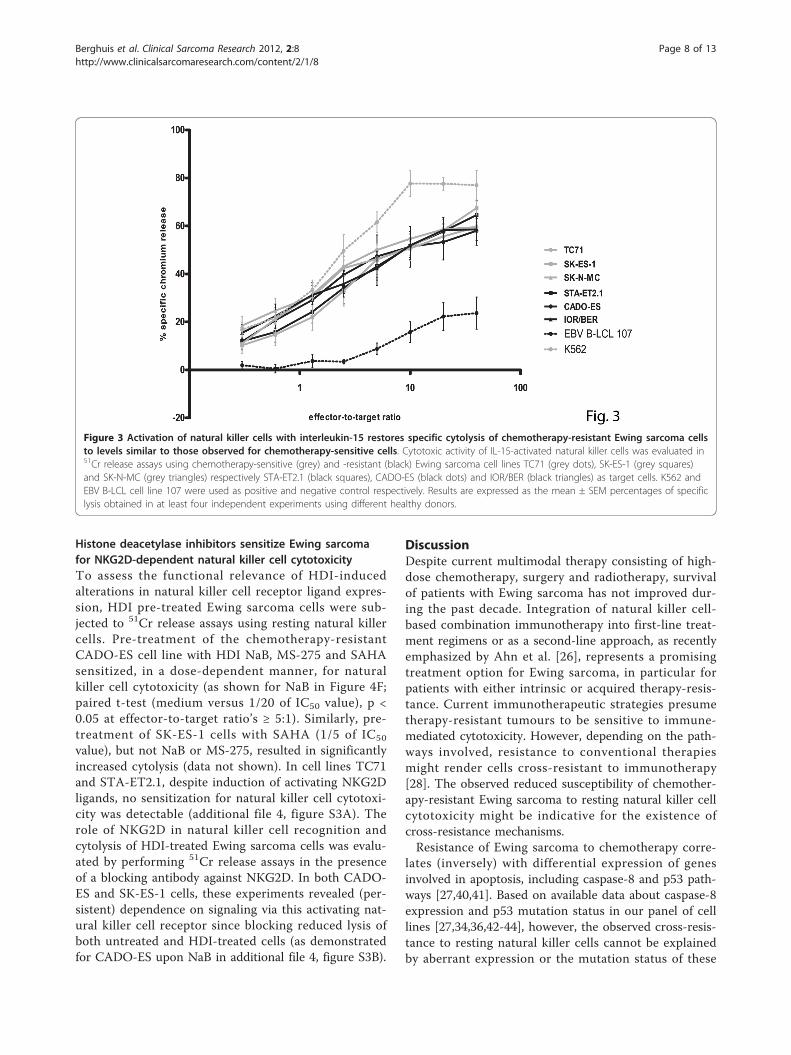
natural killer cells). As illustrated in Figure 3, activationof natural killer cells with IL-15 increased specific cyto-lysis of chemotherapy-resistant Ewing sarcoma to levelssimilar to those observed for chemotherapy-sensitivecells. Blocking studies using antibodies against NKG2Dand DNAM1 revealed comparable contributions of sig-nals provided by these activating natural killer cellreceptors to lysis of both chemotherapy-sensitive and
-resistant cells (as exemplified for TC71, SK-ES-1, STA-ET2.1 and CADO-ES in additional file 3, figure S2).Together, these data indicate that pre-activation of nat-ural killer cells with IL-15 can overcome resistance ofchemotherapy-resistant Ewing sarcoma to natural killercell-mediated cytolysis.
Histone deacetylase inhibitors induce NKG2D ligandexpression in Ewing sarcoma in an ATM/ATR-dependentmannerTo assess the capacity of HDI to sensitize Ewing sar-coma for immune-mediated cytotoxicity, cell lines wereexposed for 24 hours to HDI belonging to three differ-ent structural classes: hydroxamic acid SAHA, short-chain fatty acid NaB and benzamide MS-275. Analysisof the direct cytotoxic effects of these drugs by cell via-bility assays revealed variation in the sensitivity of thedifferent cell lines to these agents, as represented by var-iances in drug-specific IC50 values (table 1).Flow cytometric evaluation of natural killer cell recep-
tor ligand expression upon 24-hour pre-treatment of thecell lines with defined (IC50-related) concentrations ofHDI revealed heterogeneous but consistent induction ofseveral activating NKG2D ligands, in particular MICB,in all cell lines evaluated. The most pronounced effectswere observed upon pre-treatment with NaB and MS-275, resulting in up to five-fold induction of MICB (Fig-ure 4A-D). Expression of activating DNAM1 ligands(CD112 and CD155) remained largely unchanged.Induction of HLA class I expression was detectable incell lines STA-ET2.1 and TC71, whereas in CADO-ES,with relatively high levels of constitutive HLA class Iexpression (Figure 2A), no induction was observed.Induction of HLA class I expression was demonstratedin the SK-ES-1 cell line as well. However, since constitu-tive HLA class I expression was hardly detectable in thiscell line (Figure 2A), the observed less than two-foldinduction by histone deacetylase inhibitors still resultedin marginal HLA class I expression (Figure 4A-C).In addition to a direct epigenetic effect resulting in
increased expression of target genes, HDI may induceactivation of the ATM/ATR-mediated DNA damageresponse which, in turn, induces NKG2D ligand expres-sion [16]. To assess whether the ATM/ATR responsepathway contributed to HDI-induced expression ofNKG2D ligands in Ewing sarcoma cells, cell lines weretreated with ATM/ATR inhibitor caffeine (5 mM) for 2hours prior to incubation with these agents. Indeed, caf-feine pre-treatment largely prevented HDI-mediatedinduction of NKG2D ligands, but not HLA class I, in allcell lines (as exemplified for MICB/HLA class I expres-sion upon MS-275 in Figure 4E).
Table 2 In vivo NKG2D ligand expression in sequentialEwing sarcoma tumour samples
UPNa sample type (years after diagnosis) MICA ULBP1
1 post-chemotherapy resection-GRb n.e. +
lung metastasis (2) ++ +
mediastinal metastasis (3) ++ +
2 pre-treatment biopsy ++ +
post-chemotherapy resection-GR + ++
lung metastasis (5) + +/-
lymph node metastasis (9) ++ n.e.
3 pre-treatment biopsy + +
post-chemotherapy resection-GR ++ +
lung metastasis (3) + ++
lung metastasis (4) + n.e.
4 pre-treatment biopsy + +
post-chemotherapy resection-PRc ++ +
bone metastasis (1) ++ -
5 pre-treatment biopsy ++ +/-
post-chemotherapy resection-PR ++ +
lung metastasis (1) ++ +/-
6 pre-treatment biopsy ++ -
post-chemotherapy resection-PR ++ +/-
lung metastasis (0.5) ++ -
lung metastasis (2.5) ++ +/-
lung metastasis (3) ++ +/-
bone metastasis (3) ++ -
7 post-chemotherapy resection-PR ++ +/-
lung metastasis (1.5) ++ +
bone metastasis (2.5) ++ +/-
lung metastasis (4) ++ +
lung metastasis (4) ++ +/-
lung metastasis (5) ++ ++
paravertebral metastasis (5) ++ +
8 pre-treatment biopsy ++ +
post-chemotherapy resection-GR + n.e.
lung metastasis (8) ++ +aUPN = unique patient number. bGR = good chemotherapy response. cPR =poor chemotherapy
response. n.e. = not evaluable.-= absent, +/- = weak, + = moderate, ++ =strong expression.
Berghuis et al. Clinical Sarcoma Research 2012, 2:8http://www.clinicalsarcomaresearch.com/content/2/1/8
Page 7 of 13
Histone deacetylase inhibitors sensitize Ewing sarcomafor NKG2D-dependent natural killer cell cytotoxicityTo assess the functional relevance of HDI-inducedalterations in natural killer cell receptor ligand expres-sion, HDI pre-treated Ewing sarcoma cells were sub-jected to 51Cr release assays using resting natural killercells. Pre-treatment of the chemotherapy-resistantCADO-ES cell line with HDI NaB, MS-275 and SAHAsensitized, in a dose-dependent manner, for naturalkiller cell cytotoxicity (as shown for NaB in Figure 4F;paired t-test (medium versus 1/20 of IC50 value), p <0.05 at effector-to-target ratio’s ≥ 5:1). Similarly, pre-treatment of SK-ES-1 cells with SAHA (1/5 of IC50
value), but not NaB or MS-275, resulted in significantlyincreased cytolysis (data not shown). In cell lines TC71and STA-ET2.1, despite induction of activating NKG2Dligands, no sensitization for natural killer cell cytotoxi-city was detectable (additional file 4, figure S3A). Therole of NKG2D in natural killer cell recognition andcytolysis of HDI-treated Ewing sarcoma cells was evalu-ated by performing 51Cr release assays in the presenceof a blocking antibody against NKG2D. In both CADO-ES and SK-ES-1 cells, these experiments revealed (per-sistent) dependence on signaling via this activating nat-ural killer cell receptor since blocking reduced lysis ofboth untreated and HDI-treated cells (as demonstratedfor CADO-ES upon NaB in additional file 4, figure S3B).
DiscussionDespite current multimodal therapy consisting of high-dose chemotherapy, surgery and radiotherapy, survivalof patients with Ewing sarcoma has not improved dur-ing the past decade. Integration of natural killer cell-based combination immunotherapy into first-line treat-ment regimens or as a second-line approach, as recentlyemphasized by Ahn et al. [26], represents a promisingtreatment option for Ewing sarcoma, in particular forpatients with either intrinsic or acquired therapy-resis-tance. Current immunotherapeutic strategies presumetherapy-resistant tumours to be sensitive to immune-mediated cytotoxicity. However, depending on the path-ways involved, resistance to conventional therapiesmight render cells cross-resistant to immunotherapy[28]. The observed reduced susceptibility of chemother-apy-resistant Ewing sarcoma to resting natural killer cellcytotoxicity might be indicative for the existence ofcross-resistance mechanisms.Resistance of Ewing sarcoma to chemotherapy corre-
lates (inversely) with differential expression of genesinvolved in apoptosis, including caspase-8 and p53 path-ways [27,40,41]. Based on available data about caspase-8expression and p53 mutation status in our panel of celllines [27,34,36,42-44], however, the observed cross-resis-tance to resting natural killer cells cannot be explainedby aberrant expression or the mutation status of these
Figure 3 Activation of natural killer cells with interleukin-15 restores specific cytolysis of chemotherapy-resistant Ewing sarcoma cellsto levels similar to those observed for chemotherapy-sensitive cells. Cytotoxic activity of IL-15-activated natural killer cells was evaluated in51Cr release assays using chemotherapy-sensitive (grey) and -resistant (black) Ewing sarcoma cell lines TC71 (grey dots), SK-ES-1 (grey squares)and SK-N-MC (grey triangles) respectively STA-ET2.1 (black squares), CADO-ES (black dots) and IOR/BER (black triangles) as target cells. K562 andEBV B-LCL cell line 107 were used as positive and negative control respectively. Results are expressed as the mean ± SEM percentages of specificlysis obtained in at least four independent experiments using different healthy donors.
Berghuis et al. Clinical Sarcoma Research 2012, 2:8http://www.clinicalsarcomaresearch.com/content/2/1/8
Page 8 of 13

proteins alone. P-glycoprotein is a drug-efflux pumpthat confers multidrug resistance to a wide range of che-motherapeutic drugs, including key chemotherapeuticagents used for Ewing sarcoma treatment [4,45]. Inaddition to its ability to efflux drugs, P-glycoprotein reg-ulates apoptosis by inhibition of caspase activation [46].Theoretically, P-glycoprotein expression in our panel ofcell lines might contribute to the observed (relative)
cross-resistance to natural killer cells [47,48]. However,whereas caspase-dependent apoptosis is prevented, P-glycoprotein expressing cells remain sensitive to per-forin/granzyme B-induced cell death [46]. We previouslydemonstrated natural killer cell-mediated apoptosisinduction in Ewing sarcoma cells to be largely depen-dent on the perforin/granzyme B-mediated granule exo-cytosis pathway rather than on caspase-dependent death
CADO-ESSK-ES-1
MICBMHC cl
ass I
MICA
MICB
ULBP1
ULBP2
ULBP3
CD112
CD155
0
1
22
6
MediumCADO-ES
SK-ES-1STA-ET2.1
TC71
NaBA
NK cell receptor ligands
MFI
ratio
(com
pare
d to
med
ium
)
MHC clas
s IMIC
AMIC
B
ULBP1
ULBP2
ULBP3
CD112
CD155
0
1
22
6
MediumCADO-ES
SK-ES-1STA-ET2.1
TC71
SAHAC
NK cell receptor ligands
MFI
ratio
(com
pare
d to
med
ium
)
MHC clas
s IMICA
MICB
ULBP1
ULBP2
ULBP3
CD112
CD155
0
1
22
6
MediumCADO-ES
SK-ES-1STA-ET2.1
TC71
MS-275B
NK cell receptor ligands
MFI
ratio
(com
pare
d to
med
ium
)
A D
B
CF
E
Figure 4 Histone deacetylase inhibitor-induced, ATM/ATR-dependent NKG2D ligand expression sensitizes Ewing sarcoma for naturalkiller cell cytotoxicity. A-C. Heterogeneous induction of activating NKG2D ligands and/or inhibitory HLA class I molecules in chemotherapy-sensitive (grey) and -resistant (black) cell lines upon pre-treatment with HDI NaB (A), MS-275 (B) and SAHA (C), as assessed by flow cytometry.Results are expressed as mean ± SEM fold increase in MFI-ratio over medium control, obtained in at least three independent experiments. [HDI]= 1/5 IC50 value, except for CADO-ES (NaB 1/20 IC50 value; MS-275 and SAHA 1/10 IC50 value). D. Representative examples of flow cytometryplots for MICB for SK-ES-1 and CADO-ES upon pre-treatment with HDI MS-275 (IC50 values); untreated cells in grey. E. Pre-treatment with caffeine(5 mM) for 2 hours prior to incubation with HDI MS-275 (1/5 of IC50 value) largely abolished HDI-mediated MICB expression (similar results wereobserved for MICA and ULBP2-3), as assessed by flow cytometry. No such effects were observed for HLA class I expression. Results are expressedas the mean ± SEM fold increase in MFI-ratio over medium control, and are representative of at least two independent experiments. Similarresults were obtained for NaB and SAHA (not shown). F. Cytotoxicity of resting natural killer cells was evaluated in 51Cr release assays. Pre-treatment of CADO-ES cells with NaB (1/20 and 1/10 IC50 value) sensitized, in a dose-dependent manner, for natural killer cell cytotoxicity.Statistical analysis (paired t-test) revealed significantly increased sensitivity of NaB pre-treated cells at effector-to-target ratio’s > 5:1 (p < 0.05).Similar results were observed for CADO-ES with MS-275 and SAHA, and for SK-ES-1 with SAHA (not shown).
Berghuis et al. Clinical Sarcoma Research 2012, 2:8http://www.clinicalsarcomaresearch.com/content/2/1/8
Page 9 of 13

receptor pathways [36]. Therefore, we assume P-glyco-protein expression a non-crucial factor in cross-resis-tance of Ewing sarcoma to immune-mediatedcytotoxicity. Since susceptibility of Ewing sarcoma tonatural killer cell-mediated lysis depends on expressionof ligands for activating and inhibitory natural killer cellreceptors [20,23,24], differential expression of theseligands among chemotherapy-sensitive and -resistantcells could be an alternative explanation for theobserved chemo-/immunotherapy cross-resistance ofEwing sarcoma. Considerable inter-cell line variationwas observed for HLA class I expression, with a 50-foldincreased expression in chemotherapy- and natural killercell-resistant CADO-ES cells as compared to che-motherapy-/natural killer cell-sensitive SK-ES-1 cells. Ina recent report, Holmes et al. [24] point to the existenceof natural killer cell activation/inhibition thresholds thatallow small changes in HLA class I cell surface expres-sion to dramatically alter susceptibility of Ewing sar-coma to natural killer cells. However, no significanteffects of HLA class I blocking were observed (currentand previous studies [20]), excluding a major contribu-tion of dominant inhibitory signals provided by (differ-ential) HLA class I expression. Moreover, and inaddition to the observed comparable constitutive expres-sion of activating NKG2D and DNAM1-ligands, block-ing of NKG2D and DNAM1 on resting natural killercells reduced natural killer cell-mediated cytolysis ofboth chemotherapy-sensitive and -resistant cells to asimilar degree. Together, our results point to the exis-tence of true cross-resistance to immune-mediated cyto-lysis rather than to dissimilar expression of activating/inhibitory natural killer cell receptor ligands or differen-tial dependency on signaling via activating receptorsNKG2D and DNAM1. As yet, since ligands for activat-ing NCR are largely unknown [7,24,49], a possible con-tribution of such ligand can not be excluded. Similary,other possible causes for cross-resistance to conven-tional and immunotherapeutic approaches in Ewing sar-coma, that have not been addressed (e.g. (over)expression of (other) pro- or anti-apoptotic factors [50]or dysregulation of specific signaling pathways [51]),may need further investigation.Importantly, the relative resistance of chemotherapy-
resistant Ewing sarcoma cells to lysis by resting naturalkiller cells could be overcome by pre-activation of nat-ural killer cells with IL-15. Although the exact mechan-isms underlying cross-resistance to chemotherapeuticdrugs and immune-mediated cytotoxicity have not beenfully elucidated, utilization of complementary (apoptotic)pathways or stronger pro-apoptotic signals by cytokine-activated natural killer cells [52] may contribute to theobserved increased cytotoxic potential of these cellstoward (chemotherapy-resistant) Ewing sarcoma.
Irrespective of the exact mechanism, however, theobservation that chemotherapy-resistant Ewing sarcomado not exhibit cross-resistance to IL-15-activated naturalkiller cell-mediated immunotherapy suggests that cyto-kine-activated natural killer cell therapies may representpromising immunotherapeutic options for patients withEwing sarcoma.Due to the nature of this (bone) tumour, attempts to
establish primary tumour cell cultures from therapy-naive biopsies for evaluation of natural killer cell cyto-toxicity towards these targets have so far been unsuc-cessful. Support for potential in vivo immunerecognition of Ewing sarcoma tumours, however, wasprovided by immunohistochemical analysis of sequentialEwing sarcoma tumour samples demonstrating expres-sion of (at least one of the) activating natural killer cellreceptor ligands MICA and ULBP1 throughout all stagesof disease and regardless of the histological response tochemotherapy. Moreover, and similar to previous obser-vations in leukemia [10], a potential role for naturalkiller cell alloreactivity in Ewing sarcoma disease controlhas been suggested [25]. In addition to recognition bynatural killer cells, expression of NKG2D ligands mightimprove anti-tumour immune responses by specific T-lymphocyte subsets [53]. We recently reported on the(prognostically beneficial) impact of tumour-infiltratingT-lymphocytes on tumour progression in therapy-naiveEwing sarcoma [54]. Despite in situ expression ofNKG2D ligands, tumour cells may escape from NKG2Dimmunosurveillance by (enhanced) shedding of theseligands from their cell surfaces. Subsequently, circulatingtumour-derived soluble ligands may cause downregula-tion of NKG2D and, in turn, severely impair cytotoxiceffector functions of both T- and natural killer cells[37-39]. Based on our current results demonstratingnon-elevated soluble MICA levels in plasma of Ewingsarcoma patients (as compared to age-comparablehealthy controls) as well as our previous observation ofintact NKG2D expression on natural killer cells of thesepatients [20], we do not consider NKG2D ligand shed-ding a relevant immune escape mechanism in patientswith this tumour. We previously reported on reducedlevels of HLA class I expression in advanced-stageEwing sarcoma as a potential immune escape mechan-ism hampering recognition by tumour-reactive T-lym-phocytes and feasibility of T cell-basedimmunotherapeutic approaches [55]. Our current obser-vations of 1) in situ expression of ligands for activatingnatural killer cell receptors throughout all stages of dis-ease, including (chemotherapy-resistant) metastatic dis-ease, 2) the absence of (enhanced) shedding of theseactivating natural killer cell receptor ligands, 3) theabove mentioned reduced expression of inhibitory HLAclass I molecules in advanced-stage Ewing sarcoma
Berghuis et al. Clinical Sarcoma Research 2012, 2:8http://www.clinicalsarcomaresearch.com/content/2/1/8
Page 10 of 13

cases and 4) the observed susceptibility of (chemother-apy-resistant) Ewing sarcoma to IL-15-activated naturalkiller cells further support the potential of Ewing sar-coma as an attractive target for natural killer cell-basedimmunotherapy.Several phase I-III clinical trials now prove HDI-treat-
ment to be safe and effective in both hematological andsolid tumours (as reviewed by [56]) and recently, differ-ent HDI were approved by the US Food and DrugAdministration for use in patients [57]. With regard toEwing sarcoma, a role for HDI in reversal of oncogenictranscriptional repression has been proposed [58]. Inaddition, HDI-mediated cytotoxicity has been demon-strated both in vitro and in vivo and sensitization forboth conventional and more experimental treatmentmodalities has been suggested [29,31,32]. Here, wedemonstrate the ability of three structurally differentHDI to, at doses 5-20 times lower than the establishedIC50 values, improve potential immune recognition (bynatural killer cells and/or specific T-lymphocyte subsets)of both chemotherapy-sensitive and -resistant Ewingsarcoma. HDI-pretreatment resulted in ATM/ATR-dependent induction of NKG2D ligands, in particularMICA and MICB, in all cell lines. Moreover, dose-dependent sensitization for natural killer cell cytotoxicitywas observed in 2/4 cell lines, including the chemother-apy-resistant CADO-ES cell line. The doses used invitro were below or within the range of the maximumtolerated dose as determined in recent clinical trials (inchildren) [59,60]. Natural killer cell cytotoxicitydepended on NKG2D-NKG2D ligand interactions, sinceblocking of NKG2D abrogated cytolysis. No sensitizationwas observed for TC71 and STA-ET2.1 cells, despiteinduction of NKG2D ligands. As yet, an adequate expla-nation for this observation is lacking. Although HDI-pretreatment induced HLA class I in these cell lines, acontribution of dominant inhibitory signals provided bythis increased expression seems unlikely since HLAclass I blocking did not significantly affect natural killercell-mediated cytolysis.Collectively, our data provide a rationale for combina-
tion immunotherapy involving immune effector cell (IL-15-activated natural killer cells) and target cell (HDI)manipulation in first- and/or second-line treatment regi-mens for Ewing sarcoma.
ConclusionsPatients with Ewing sarcoma (EWS) have a poor prog-nosis, despite current multimodal therapy. Integration ofimmunotherapeutic strategies, including natural killer(NK) cell-based therapies, into first-line treatment regi-mens or introduction of these approaches as second-linetherapy may represent promising treatment options. Exvivo immune cell activation and/or (simultaneous)
sensitization of target cells for immune-mediated killingby combination immunotherapy may overcome intrin-sic/acquired resistance to conventional therapies andimprove (immuno)therapeutic efficacy. Here, we providethe first evidence that combination immunotherapyusing histone deacetylase inhibitors and (interleukin-15-activated) NK cells improves immune recognition ofboth therapy-sensitive and -resistant EWS and sensitizesfor NK cell cytotoxicity. In vivo expression of activatingNK cell receptor ligands throughout all disease-stages,regardless of chemotherapy-response, supports theirpotential in vivo role in immune recognition of EWS.Our data provide a rationale for combination immu-notherapy involving simultaneous immune cell (interleu-kin-15-activated NK cells) and target cell (histonedeacetylase inhibitors) manipulation in first-/second-linetreatment regimens for EWS.
Additional material
Additional file 1: Antibodies used for flow cytometry, NK cellreceptor (ligand) blocking, immunohistochemistry and ELISA.
Additional file 2: A. Constitutive surface expression of inhibitory(HLA class I) or activating (MICA/B, ULBP1-3, CD112, CD155) naturalkiller cell receptor ligands in chemotherapy-sensitive (grey) and-resistant (black) Ewing sarcoma cell lines, as assessed by flowcytometry. Results are expressed as the mean ± SD MFI-ratio, obtainedin at least two independent experiments. Statistical analysis (t-test) wasperformed on mean MFI-ratio’s (for each ligand) of chemotherapy-sensitive versus -resistant cell lines, revealing no significant differences inexpression levels of these ligands (p > 0.05). B. Cytotoxic activity ofresting natural killer cells was evaluated in 51Cr release assays usingchemotherapy-sensitive (TC71 (blue), SK-ES-1 (purple), SK-N-MC (pink))and -resistant (STA-ET-2.1 (black), CADO-ES (dark grey), IOR/BER (lightgrey)) Ewing sarcoma cell lines as target cells. Ewing sarcoma cells wereeither left untreated (solid bars) or pre-incubated with HLA class Iblocking antibody DX17 (checked bars). Results are expressed as themean ± SD percentage of specific lysis obtained in at least twoindependent experiments using different healthy donors.
Additional file 3: Cytotoxic activity of IL-15-activated natural killercells was evaluated in 51Cr release assays using chemotherapy-sensitive (TC71 (blue), SK-ES-1 (purple)) and -resistant (STA-ET-2.1(black), CADO-ES (grey) Ewing sarcoma cell lines as target cells.Ewing sarcoma cells were either left untreated (solid bars) or pre-incubated with NKG2D and DNAM-1 blocking antibodies (checked bars).Results are expressed as the mean ± SD percentage of specific lysisobtained in at least two independent experiments using differenthealthy donors.
Additional file 4: A. Cytotoxicity of resting natural killer cells wasevaluated in 51Cr release assays using MS-275-pretreated TC71 andSTA-ET2.1 cells. Despite induction of activating NKG2D ligands, nosensitization for natural killer cell cytotoxicity was detectable (at doses upto 1/5 of IC50 value). Similar results were observed for both cell linesupon pre-treatment with NaB and SAHA (not shown). Results areexpressed as the mean ± SEM percentages of specific lysis obtained in atleast two independent experiments using different healthy donors. B.Upon HDI-pretreatment, persistent dependency of resting natural killercell-mediated cytotoxicity on signaling via activating receptor NKG2Dwas demonstrated when 51Cr release assays were performed in thepresence of a blocking antibody against NKG2D. Blocking reducedresting natural killer cell-mediated lysis of both untreated and HDI pre-treated cells to similar levels, as demonstrated for CADO-ES upon pre-treatment with NaB. Similar results were obtained for CADO-ES with MS-
Berghuis et al. Clinical Sarcoma Research 2012, 2:8http://www.clinicalsarcomaresearch.com/content/2/1/8
Page 11 of 13

275 and SAHA, as well as for SK-ES-1 with SAHA (not shown). K562 andEBV B-LCL cell line 107 were used as positive and negative controlrespectively (not shown). Results are expressed as the mean ± SEMpercentages of specific lysis obtained in at least two independentexperiments using different healthy donors.
AcknowledgementsThe authors thank Jurre van Kesteren, Monique M. van Ostaijen-ten Dam, S.Eriaty N. Ruslan (Department of Pediatrics, Leiden University Medical Center(LUMC), The Netherlands) and M.J. Ronkes for technical assistance; KatiaScotlandi (Instituto Orthopedico Rizzoli, Bologna, Italy) for provision of Ewingsarcoma cell line IOR/BER; Maarten J.D. van Tol (Department of Pediatrics,LUMC), H. Gelderblom (Department of Clinical Oncology, LUMC) and A.H.M.Taminiau (Department of Orthopedic Surgery, LUMC) for provision of plasmaand tumour samples respectively. Grant support: European Commission(EuroBoNeT, grant no. 018814 (P.C.W.H.)); Foundation ‘The Quality of LifeGala 2007’ (A.C.L.); Dutch Cancer Society (grant RUL 2003-2800 (A.C.L. and P.C.W.H.)).
Author details1Department of Pathology, Leiden University Medical Center, Leiden, TheNetherlands. 2Department of Pediatrics, Leiden University Medical Center,Leiden, The Netherlands. 3Laboratory for Stem Cell Transplantation andImmunotherapy, Hospital of Johann Wolfgang Goethe-University, Frankfurtam Main, Germany.
Authors’ contributionsAll authors contributed to conception and/or design of the study. DB, HIV,SJS and SK conducted experiments and performed data analyses. DB, MWS,EPB, PCWH and ACL were involved in interpretation of data. All authorswere involved in drafting and/or critical revision of the manuscript andapproved the final submitted version.
Competing interestsThe authors declare that they have no competing interests.
Received: 3 January 2012 Accepted: 8 February 2012Published: 8 February 2012
References1. Riggi N, Cironi L, Suva ML, Stamenkovic I: Sarcomas: genetics, signalling,
and cellular origins. Part 1: The fellowship of TET. J Pathol 2007, 213:4-20.2. Szuhai K, Ijszenga M, de Jong D, Karseladze A, Tanke HJ, Hogendoorn PCW:
The NFATc2 gene is involved in a novel cloned translocation in a Ewingsarcoma variant that couples its function in immunology to oncology.Clin Cancer Res 2009, 15:2259-2268.
3. Ushigome S, Machinami R, Sorensen PH: Ewing Sarcoma/PrimitiveNeuroectodermal Tumour. In World Health Organization Classification ofTumours. Pathology and Genetics of Tumours of Soft tissue and Bone. Editedby: Fletcher CDM, Unni KK, Mertens F. Lyon: IARC Press; 2002:298-300.
4. Juergens C, Weston C, Lewis I, Whelan J, Paulussen M, Oberlin O, Michon J,Zoubek A, Juergens H, Craft A: Safety assessment of intensive inductionwith vincristine, ifosfamide, doxorubicin, and etoposide (VIDE) in thetreatment of Ewing tumors in the EURO-E.W.I.N.G. 99 clinical trial. PediatrBlood Cancer 2006, 47:22-29.
5. Le Deley MC, Delattre O, Schaefer KL, Burchill SA, Koehler G,Hogendoorn PCW, Lion T, Poremba C, Marandet J, Ballet S, Pierron G,Brownhill SC, Nesslbock M, Ranft A, Dirksen U, Oberlin O, Lewis IJ, Craft AW,Jurgens H, Kovar H: Impact of EWS-ETS fusion type on diseaseprogression in Ewing’s sarcoma/peripheral primitive neuroectodermaltumor: prospective results from the cooperative Euro-E.W.I.N.G. 99 trial. JClin Oncol 2010, 28:1982-1988.
6. van Doorninck JA, Ji L, Schaub B, Shimada H, Wing MR, Krailo MD,Lessnick SL, Marina N, Triche TJ, Sposto R, Womer RB, Lawlor ER: Currenttreatment protocols have eliminated the prognostic advantage of type1 fusions in Ewing sarcoma: a report from the Children’s OncologyGroup. J Clin Oncol 2010, 28:1989-1994.
7. Moretta L, Moretta A: Unravelling natural killer cell function: triggeringand inhibitory human NK receptors. EMBO J 2004, 23:255-259.
8. Cerwenka A, Lanier LL: Natural killer cells, viruses and cancer. Nat RevImmunol 2001, 1:41-49.
9. Biron CA, Nguyen KB, Pien GC, Cousens LP, Salazar-Mather TP: Natural killercells in antiviral defense: function and regulation by innate cytokines.Annu Rev Immunol 1999, 17:189-220.
10. Ruggeri L, Capanni M, Urbani E, Perruccio K, Shlomchik WD, Tosti A,Posati S, Rogaia D, Frassoni F, Aversa F, Martelli MF, Velardi A: Effectivenessof donor natural killer cell alloreactivity in mismatched hematopoietictransplants. Science 2002, 295:2097-2100.
11. Miller JS, Soignier Y, Panoskaltsis-Mortari A, McNearney SA, Yun GH,Fautsch SK, McKenna D, Le C, Defor TE, Burns LJ, Orchard PJ, Blazar BR,Wagner JE, Slungaard A, Weisdorf DJ, Okazaki IJ, McGlave PB: Successfuladoptive transfer and in vivo expansion of human haploidentical NKcells in patients with cancer. Blood 2005, 105:3051-3057.
12. Klingemann HG: Natural killer cell-based immunotherapeutic strategies.Cytotherapy 2005, 7:16-22.
13. Ljunggren HG, Malmberg KJ: Prospects for the use of NK cells inimmunotherapy of human cancer. Nat Rev Immunol 2007, 7:329-339.
14. Meyers PA, Schwartz CL, Krailo MD, Healey JH, Bernstein ML, Betcher D,Ferguson WS, Gebhardt MC, Goorin AM, Harris M, Kleinerman E, Link MP,Nadel H, Nieder M, Siegal GP, Weiner MA, Wells RJ, Womer RB, Grier HE:Osteosarcoma: the addition of muramyl tripeptide to chemotherapyimproves overall survival–a report from the Children’s Oncology Group.J Clin Oncol 2008, 26:633-638.
15. Whelan J, Patterson D, Perisoglou M, Bielack S, Marina N, Smeland S,Bernstein M: The role of interferons in the treatment of osteosarcoma.Pediatr Blood Cancer 2010, 54:350-354.
16. Gasser S, Orsulic S, Brown EJ, Raulet DH: The DNA damage pathwayregulates innate immune system ligands of the NKG2D receptor. Nature2005, 436:1186-1190.
17. Skov S, Pedersen MT, Andresen L, Straten PT, Woetmann A, Odum N:Cancer cells become susceptible to natural killer cell killing afterexposure to histone deacetylase inhibitors due to glycogen synthasekinase-3-dependent expression of MHC class I-related chain A and B.Cancer Res 2005, 65:11136-11145.
18. Schmudde M, Braun A, Pende D, Sonnemann J, Klier U, Beck JF, Moretta L,Broker BM: Histone deacetylase inhibitors sensitize tumour cells forcytotoxic effects of natural killer cells. Cancer Lett 2008, 272:110-121.
19. Soriani A, Zingoni A, Cerboni C, Iannitto ML, Ricciardi MR, Di G, Cippitelli M,Fionda C, Petrucci MT, Guarini A, Foa R, Santoni A: ATM-ATR-dependentup-regulation of DNAM-1 and NKG2D ligands on multiple myeloma cellsby therapeutic agents results in enhanced NK-cell susceptibility and isassociated with a senescent phenotype. Blood 2009, 113:3503-3511.
20. Verhoeven DH, de Hooge AS, Mooiman EC, Santos SJ, ten Dam MM,Gelderblom H, Melief CJ, Hogendoorn PCW, Egeler RM, van Tol MJ,Schilham MW, Lankester AC: NK cells recognize and lyse Ewing sarcomacells through NKG2D and DNAM-1 receptor dependent pathways. MolImmunol 2008, 45:3917-3925.
21. Staege MS, Hansen G, Baersch G, Burdach S: Functional and molecularcharacterization of interleukin-2 transgenic Ewing tumor cells for in vivoimmunotherapy. Pediatr Blood Cancer 2004, 43:23-34.
22. Atzpodien J, Gulati SC, Shimazaki C, Buhrer C, Oz S, Kwon JH, Kolitz JE,Clarkson BD: Ewing’s sarcoma: ex vivo sensitivity towards natural andlymphokine-activated killing. Oncology 1988, 45:437-443.
23. Cho D, Shook DR, Shimasaki N, Chang YH, Fujisaki H, Campana D:Cytotoxicity of activated natural killer cells against pediatric solidtumors. Clin Cancer Res 2010, 16:3901-3909.
24. Holmes TD, El-Sherbiny YM, Davison A, Clough SL, Blair GE, Cook GP: Ahuman NK cell activation/inhibition threshold allows small changes inthe target cell surface phenotype to dramatically alter susceptibility toNK cells. J Immunol 2011, 186:1538-1545.
25. Perez-Martinez A, Leung W, Munoz E, Iyengar R, Ramirez M, Vicario JL,Lassaletta A, Sevilla J, Gonzalez-Vicent M, Madero L, Diaz-Perez MA: KIR-HLAreceptor-ligand mismatch associated with a graft-versus-tumor effect inhaploidentical stem cell transplantation for pediatric metastatic solidtumors. Pediatr Blood Cancer 2009, 53:120-124.
26. Ahn YO, Weigel B, Verneris MR: Killing the killer: natural killer cells to treatEwing’s sarcoma. Clin Cancer Res 2010, 16:3819-3821.
Berghuis et al. Clinical Sarcoma Research 2012, 2:8http://www.clinicalsarcomaresearch.com/content/2/1/8
Page 12 of 13

27. Schaefer KL, Eisenacher M, Braun Y, Brachwitz K, Wai DH, Dirksen U,Lanvers-Kaminsky C, Juergens H, Herrero D, Stegmaier S, Koscielniak E,Eggert A, Nathrath M, Gosheger G, Schneider DT, Bury C, Diallo-Danebrock R, Ottaviano L, Gabbert HE, Poremba C: Microarray analysis ofEwing’s sarcoma family of tumours reveals characteristic geneexpression signatures associated with metastasis and resistance tochemotherapy. Eur J Cancer 2008, 44:699-709.
28. Ng CP, Bonavida B: A new challenge for successful immunotherapy bytumors that are resistant to apoptosis: two complementary signals toovercome cross-resistance. Adv Cancer Res 2002, 85:145-174.
29. Jaboin J, Wild J, Hamidi H, Khanna C, Kim CJ, Robey R, Bates SE, Thiele CJ:MS-27-275, an inhibitor of histone deacetylase, has marked in vitro andin vivo antitumor activity against pediatric solid tumors. Cancer Res 2002,62:6108-6115.
30. Nakatani F, Tanaka K, Sakimura R, Matsumoto Y, Matsunobu T, Li X,Hanada M, Okada T, Iwamoto Y: Identification of p21WAF1/CIP1 as adirect target of EWS-Fli1 oncogenic fusion protein. J Biol Chem 2003,278:15105-15115.
31. Sakimura R, Tanaka K, Nakatani F, Matsunobu T, Li X, Hanada M, Okada T,Nakamura T, Matsumoto Y, Iwamoto Y: Antitumor effects of histonedeacetylase inhibitor on Ewing’s family tumors. Int J Cancer 2005,116:784-792.
32. Sonnemann J, Dreyer L, Hartwig M, Palani CD, Hong lT, Klier U, Broker B,Volker U, Beck JF: Histone deacetylase inhibitors induce cell death andenhance the apoptosis-inducing activity of TRAIL in Ewing’s sarcomacells. J Cancer Res Clin Oncol 2007, 133:847-858.
33. van der Woude HJ, Bloem JL, Taminiau AH, Nooy MA, Hogendoorn PCW:Classification of histopathologic changes following chemotherapy inEwing’s sarcoma of bone. Skeletal Radiol 1994, 23:501-507.
34. Ottaviano L, Schaefer KL, Gajewski M, Huckenbeck W, Baldus S, Rogel U,Mackintosh C, de Alava E, Myklebost O, Kresse SH, Meza-Zepeda LA,Serra M, Cleton-Jansen AM, Hogendoorn PCW, Buerger H, Aigner T,Gabbert HE, Poremba C: Molecular characterization of commonly usedcell lines for bone tumor research: a trans-European EuroBoNet effort.Genes Chromosomes Cancer 2010, 49:40-51.
35. Scotlandi K, Perdichizzi S, Manara MC, Serra M, Benini S, Cerisano V,Strammiello R, Mercuri M, Reverter-Branch , Faircloth G, D’Incalci M, Picci P:Effectiveness of Ecteinascidin-743 against drug-sensitive and -resistantbone tumor cells. Clin Cancer Res 2002, 8:3893-3903.
36. de Hooge ASK, Berghuis D, Santos SJ, Mooiman E, Romeo S, Kummer JA,Egeler RM, van Tol MJ, Melief CJ, Hogendoorn PCW, Lankester AC:Expression of cellular FLICE inhibitory protein, caspase-8, and proteaseinhibitor-9 in Ewing sarcoma and implications for susceptibility tocytotoxic pathways. Clin Cancer Res 2007, 13:206-214.
37. Groh V, Wu J, Yee C, Spies T: Tumour-derived soluble MIC ligands impairexpression of NKG2D and T-cell activation. Nature 2002, 419:734-738.
38. Raffaghello L, Prigione I, Airoldi I, Camoriano M, Levreri I, Gambini C,Pende D, Steinle A, Ferrone S, Pistoia V: Downregulation and/or release ofNKG2D ligands as immune evasion strategy of human neuroblastoma.Neoplasia 2004, 6:558-568.
39. Salih HR, Holdenrieder S, Steinle A: Soluble NKG2D ligands: prevalence,release, and functional impact. Front Biosci 2008, 13:3448-3456.
40. Huang HY, Illei PB, Zhao Z, Mazumdar M, Huvos AG, Healey JH, Wexler LH,Gorlick R, Meyers P, Ladanyi M: Ewing sarcomas with p53 mutation orp16/p14ARF homozygous deletion: a highly lethal subset associatedwith poor chemoresponse. J Clin Oncol 2005, 23:548-558.
41. Abudu A, Mangham DC, Reynolds GM, Pynsent PB, Tillman RM, Carter SR,Grimer RJ: Overexpression of p53 protein in primary Ewing’s sarcoma ofbone: relationship to tumour stage, response and prognosis. Br J Cancer1999, 79:1185-1189.
42. Fulda S, Kufer MU, Meyer E, van Valen F, Dockhorn-Dworniczak B,Debatin KM: Sensitization for death receptor- or drug-induced apoptosisby re-expression of caspase-8 through demethylation or gene transfer.Oncogene 2001, 20:5865-5877.
43. Harada K, Toyooka S, Shivapurkar N, Maitra A, Reddy JL, Matta H,Miyajima K, Timmons CF, Tomlinson GE, Mastrangelo D, Hay RJ,Chaudhary PM, Gazdar AF: Deregulation of caspase 8 and 10 expressionin pediatric tumors and cell lines. Cancer Res 2002, 62:5897-5901.
44. Hotfilder M, Sondermann P, Senss A, van Valen F, Jurgens H, Vormoor J:PI3K/AKT is involved in mediating survival signals that rescue Ewing
tumour cells from fibroblast growth factor 2-induced cell death. Br JCancer 2005, 92:705-710.
45. Ambudkar SV, Kimchi-Sarfaty C, Sauna ZE, Gottesman MM: P-glycoprotein:from genomics to mechanism. Oncogene 2003, 22:7468-7485.
46. Johnstone RW, Cretney E, Smyth MJ: P-glycoprotein protects leukemiacells against caspase-dependent, but not caspase-independent, celldeath. Blood 1999, 93:1075-1085.
47. Roessner A, Ueda Y, Bockhorn-Dworniczak B, Blasius S, Peters A, Wuisman P,Ritter J, Paulussen M, Jurgens H, Bocker W: Prognostic implication ofimmunodetection of P glycoprotein in Ewing’s sarcoma. J Cancer Res ClinOncol 1993, 119:185-189.
48. Hijazi YM, Axiotis CA, Navarro S, Steinberg SM, Horowitz ME, Tsokos M:Immunohistochemical detection of P-glycoprotein in Ewing’s sarcomaand peripheral primitive neuroectodermal tumors before and afterchemotherapy. Am J Clin Pathol 1994, 102:61-67.
49. Brandt CS, Baratin M, Yi EC, Kennedy J, Gao Z, Fox B, Haldeman B,Ostrander CD, Kaifu T, Chabannon C, Moretta A, West R, Xu W, Vivier E,Levin SD: The B7 family member B7-H6 is a tumor cell ligand for theactivating natural killer cell receptor NKp30 in humans. J Exp Med 2009,206:1495-1503.
50. Tirado OM, MacCarthy CM, Fatima N, Villar J, Mateo-Lozano S, Notario V:Caveolin-1 promotes resistance to chemotherapy-induced apoptosis inEwing’s sarcoma cells by modulating PKCalpha phosphorylation. Int JCancer 2010, 126:426-436.
51. Saudemont A, Hamrouni A, Marchetti P, Liu J, Jouy N, Hetuin D, Colucci F,Quesnel B: Dormant tumor cells develop cross-resistance to apoptosisinduced by CTLs or imatinib mesylate via methylation of suppressor ofcytokine signaling. Cancer Res 2007, 67:4491-4498.
52. Zhang C, Zhang J, Niu J, Zhang J, Tian Z: Interleukin-15 improvescytotoxicity of natural killer cells via up-regulating NKG2D and cytotoxiceffector molecule expression as well as STAT1 and ERK1/2phosphorylation. Cytokine 2008, 42:128-136.
53. Champsaur M, Lanier LL: Effect of NKG2D ligand expression on hostimmune responses. Immunol Rev 2010, 235:267-285.
54. Berghuis D, Santos SJ, Baelde HJ, Taminiau AH, Egeler RM, Schilham MW,Hogendoorn PCW, Lankester AC: Pro-inflammatory chemokine-chemokinereceptor interactions within the Ewing sarcoma microenvironmentdetermine CD8(+) T-lymphocyte infiltration and affect tumourprogression. J Pathol 2011, 223:347-357.
55. Berghuis D, de Hooge AS, Santos SJ, Horst D, Wiertz EJ, VanEggermond MC, van den Elsen PJ, Taminiau AH, Ottaviano L, Schaefer KL,Dirksen U, Hooijberg E, Mulder A, Melief CJ, Egeler RM, Schilham MW,Jordanova ES, Hogendoorn PCW, Lankester AC: Reduced human leukocyteantigen expression in advanced-stage Ewing sarcoma: implications forimmune recognition. J Pathol 2009, 218:222-231.
56. Tan J, Cang S, Ma Y, Petrillo RL, Liu D: Novel histone deacetylaseinhibitors in clinical trials as anti-cancer agents. J Hematol Oncol 2010,3:5.
57. StatBite: FDA oncology drug product approvals in 2009. J Natl CancerInst 2010, 102:219.
58. Owen LA, Kowalewski AA, Lessnick SL: EWS/FLI mediates transcriptionalrepression via NKX2.2 during oncogenic transformation in Ewing’ssarcoma. PLoS One 2008, 3:e1965.
59. Fouladi M, Park JR, Stewart CF, Gilbertson RJ, Schaiquevich P, Sun J,Reid JM, Ames MM, Speights R, Ingle AM, Zwiebel J, Blaney SM,Adamson PC: Pediatric phase I trial and pharmacokinetic study ofvorinostat: a Children’s Oncology Group phase I consortium report. J ClinOncol 2010, 28:3623-3629.
60. Tan J, Cang S, Ma Y, Petrillo RL, Liu D: Novel histone deacetylaseinhibitors in clinical trials as anti-cancer agents. J Hematol Oncol 2010,3:5.
doi:10.1186/2045-3329-2-8Cite this article as: Berghuis et al.: Histone deacetylase inhibitorsenhance expression of NKG2D ligands in Ewing sarcoma and sensitizefor natural killer cell-mediated cytolysis. Clinical Sarcoma Research 20122:8.
Berghuis et al. Clinical Sarcoma Research 2012, 2:8http://www.clinicalsarcomaresearch.com/content/2/1/8
Page 13 of 13