Polyglutamic acid–PEG nanocapsules as long circulating carriers for the delivery of docetaxel

Research Paper
Efficacy of siRNA Nanocapsules Targeted Against the EWS–Fli1Oncogene in Ewing Sarcoma
Nedjma Toub,1,2 Jean-Remi Bertrand,2 Ali Tamaddon,2 Hind Elhamess,2 Herve Hillaireau,1
Andrei Maksimenko,2 Jean Maccario,3 Claude Malvy,2,4 Elias Fattal,1 and Patrick Couvreur1
Received August 29, 2005; accepted January 6, 2006
Abstract. The EWSYFli1 fusion gene encodes for a chimeric oncogenic transcription factor considered to
be the cause of the Ewing sarcoma. The efficiency of small interfering RNAs (siRNAs) targeted toward
the EWSYFli1 transcript (at the junction point type 1) was studied, free or encapsulated into recently
developed polyisobutylcyanoacrylate aqueous core nanocapsules. Because this mRNA sequence is only
present in cancer cells, it therefore constituted a relevant target. Studies of the intracellular penetration
by confocal microscopy in NIH/3T3 EWSYFli1 cells showed that nanocapsules improved the intracellular
penetration of siRNA with mainly a cytoplasmic localization. These biodegradable siRNA-loaded
nanocapsules were then tested in vivo on a mice xenografted EWSYFli1-expressing tumor; they were
found to trigger a dose-dependant inhibition of tumor growth after intratumoral injection. A specific
inhibition of EWSYFli1 was observed, too. These findings now open new prospects for the treatment of
experimental cancers with junction oncogenes.
KEY WORDS: Ewing sarcoma; EWSYFli1; isobutylcyanoacrylate; nanocapsules; siRNA.
INTRODUCTION
Chromosomal translocations are found in special typesof hematopoietic malignancies and sarcomas (1,2). Ewingsarcoma is a metastatic bone cancer of children and youngadults. At the cellular level, poorly differentiated round cellscharacterize it; a genetic abnormality is associated with thismalignancy, which consists in a rearrangement of chromo-somes 22 and 11. The EWSYFli1 fusion gene, a product of thetranslocation t(11;22) (q24;q12), is detected in 90% of Ewingsarcomas and primitive neuroectodermal tumors (PNETs)(3). The EWSYFli1 results in the fusion of the carboxyl-terminal region of Fli1 with the amino-terminal region of aputative RNA-binding protein, EWS (4,5). The chimericEWSYFli1 protein is believed to function as a transcriptionalactivator (4 Y8). The crucial role of this fusion protein incellular proliferation has been demonstrated previously inEwing sarcoma and PNET cells by using an antisenseoligodeoxyribonucleotide as well as a small interferingRNA (siRNA), both targeted toward the EWSYFli1 (9,10).
Thus, siRNA with relevant sequences may be considered forthe treatment of cancers characterized by a fusion oncogene.
RNA interference (RNAi) is a process of sequence-specific, posttranscriptional gene silencing found in a varietyof eukaryotes ranging from fungi to mammals (11Y15). Themechanism of the target inhibition has been proposed to takeplace via 21Y23 nucleotide siRNAs. Then, the siRNAsassociate with a complex of proteins termed the RNA-induced silencing complex (RISC), which directs the siRNAto the complementary target sequence, resulting in thecleavage of the target RNA (15). The most potent siRNAduplexes are 19Y21 nucleotides long, with two-nucleotide, 30-overhanging termini (14). One strategy for the introductionof siRNAs into cells is their direct expression after intracel-lular expression of a small hairpin RNA (shRNA). An RNApolymerase III (pol III) transcription system produces anshRNA into the cell, which is then transformed in an activesiRNA (16). Another method is to introduce the syntheticrelevant siRNA sequence directly into the cells. However, inthat case, the biological efficacy is hampered by the poorstability of siRNA in biological fluids and by their lowintracellular penetration (17,18). Thus, nanocarrier systems,mainly cationic lipids, represent an option to improve the in
vivo intracellular delivery of these nucleic acid-based drugsby protecting them from the harsh environment of theextracellular fluids (19Y21). However, an important issue isthat the cationic nature of these lipids leads to aggregationwhen administered systemically in vivo. This is the reasonwhy, in this study, we have chosen to use noncationicpolyisobutylcyanoacrylate nanocapsules with an aqueous core(22) for the delivery of siRNA targeted toward the EWSYFli1
0724-8741/06/0500-0892/0 # 2006 Springer Science + Business Media, Inc. 892
Pharmaceutical Research, Vol. 23, No. 5, May 2006 (# 2006)DOI: 10.1007/s11095-006-9901-9
1 Laboratoire de Physicochimie, Pharmacotechnie et Biopharmacie,
Faculte de Pharmacie, UMR CNRS 8612, 92286 Chatenay-Malabry,
France.2 Laboratoire de Vecterologie et Transfert des Genes, Institut
Gustave Roussy, UMR CNRS 8121, 39, rue Camille Desmoulins,
94805 Villejuif Cedex, France.3 Centre d’ Etudes Pharmaceutiques, 12 roe J.B. Clement, 92296
Chatenay-Malabry Cedex, France.4 To whom correspondence should be addressed. (e-mail: [email protected])

fusion oncogene. This formulation has been shown (a) invitro to penetrate intracellularly and (b) in vivo to inhibittumor growth in an experimental Ewing sarcoma. It is, to ourknowledge, the first time that an siRNA against a fusion on-cogene is efficiently delivered by means of nanotechnologies.
MATERIALS AND METHODS
Nucleic Acids
The oligonucleotides were purchased from Eurogentec(Seraing, Belgium). The siRNA target sequence is designedat the junction between EWS and Fli1 in the chimeric mRNA(nucleotides 822Y842). This sequence is specific of the tumorcells because the chimeric RNA is not expressed in normalcells. siRNAs with the following sequences were used:
& siRNA antisense (siRNA-AS). Sense strand: 5 0-r(GCUACGGGCAGCAGAACCC)d(TT)-30; antisensestrand: 50-r(GGGUUCUGCUGCCCGUAGC)d(TG)-30
& Control siRNA (siRNA-Ct). Sense strand: 50-r(GCUGCGGACAGCAGAAGCC)d(TT)-30; antisense strand: 50-r(GGCUUGUGCUGUCCGCAGC)d(TG)-30
The two 30-overhanging bases are synthesized with aDNA chemistry to enhance the siRNA nuclease resistance asdescribed by Tuschl (23). The sense and antisense strandswere hybridized in 30 mM HEPESYKOH (pH 7.4), 2 mMMg-acetate, 100 mM K-acetate for 1 min at 95-C (finalvolume 100 ml), and then for 1 h at 37-C to obtain a finalstock concentration of 50 mM. siRNAs have been purified byhigh-performance liquid chromatography (HPLC) and thencontrolled by mass spectrometry (Eurogentec).
siRNA Radiolabeling
The 50-end of one strand of the duplex was labeled by T4polynucleotide kinase (New England Biolabs, Frankfurt amMain, Germany) with [g-32P]ATP (111 TBq/mmol; MPBiomedicals, Illkirch, France) under the conditions indicatedby the manufacturers. The purified strands were recovered bygel filtration using a Bio-Spin 6 column (BioRad, Richmond,CA, USA) and centrifuged at 2500�g for 4 min. The puritywas then controlled by radioactivity analysis using anautomatic-TLC-linear analyzer (EG&G Berthold,Elancourt, France) as described by Aynie et al. (24).
Nanocapsule Preparation and siRNA Encapsulation
The preparation of an aqueous suspension of polyisobu-tylcyanoacrylate nanocapsules containing siRNA in theiraqueous core was performed by modification of the methodof Lambert et al. (25) and Watnasirichaikul et al. (26) asfollows. We first prepared a nanoemulsion at 4-C by mixing0.1 mL of demineralized water containing 0.5 mM (equiva-lent to 700 mg) siRNA duplex to an organic phase containing1.125 g of Montane 80 (sorbitan monooleate, Seppic, Paris,France) and 1 g of Miglyol 812 (medium chain triglyceride,Sasol, Witten, Germany) using an Ultraturrax for 1 min at24,000 rpm (4-C). Then, 10 mg of isobutylcyanoacrylate(IBCA) monomer (Loctite, Dublin, Ireland) was added
rapidly to the nanoemulsion under mechanical stirring (500rpm, room temperature). The system was left for at least4 h for polymerization to take place. Nanocapsules werecollected by ultracentrifugation at 30,000 rpm for 30 min at4-C (Beckman L8-70M Ultracentrifuge, 50 Ti rotor). Afterliquid-phase removal, the pellet was transferred to a newtube and resuspended under vortex agitation (1 min) insterile demineralized water (or physiological serum foranimal experiments) at the desired siRNA concentration.The nanocapsule suspension was then dispersed by fastsonication (8 s) and centrifuged at 4000�g for 10 min (4-C)to remove residual surfactant. This process was repeatedtwice to insure complete nanocapsule purification.
Nanocapsule Characterization
The particle size and distribution of the nanocapsuleswas measured after dilution (1:30) in demineralized waterusing dynamic laser light scattering (Nanosizer ND4, Coul-tronics, Margency, France). The zeta potential of nano-capsules was determined as follows: 200 ml of the sampleswas diluted in 2 mL of a 0.1 mM KCl solution adjusted to pH7.4 and analyzed with a Zetasizer (Malvern Instruments,Malvern, UK).
siRNA nanocapsules were incubated for 1 month inphosphate-buffered saline (PBS). Then, after ultracentrifu-gation (60,000�g for 30 min), supernatants were analyzed bypolyacrylamide gel electrophoresis (PAGE; 20%) for siRNAintegrity.
Freeze Fracture Electron Microscopy
A small drop of the suspension containing 30% ofglycerol as cryoprotectant was deposited on a thin copperplate and rapidly frozen in liquid propane. Fracturing andreplication (using Pt carbon) were performed with a BalzersBAF 301 freeze etch. The replicas were washed andexamined under a Philips 410 electron microscope.
Determination of siRNA Encapsulation Yield
After [g-32P]ATP labeling, siRNAs were introduced intonanocapsules as described above. After the first ultracen-trifugation, the radioactivity of supernatant and pellet wasdetermined by the Cherenkov effect using a liquid scintilla-tion counter (1900TR Packard). The final encapsulation yieldwas calculated as the ratio of the pellet radioactivity to thetotal used radioactivity (pellet + supernatant).
Cell Penetration Studies
NIH/3T3 cells stably transfected with the humanEWSYFli1 gene were a generous gift from Dr. J. Ghysdael(Institut Curie, Orsay, France). We followed the intracellulartrafficking of the siRNA by confocal microscopy using arhodamine-labeled siRNA. Briefly, 105 NIH/3T3 EWSYFli1cells were seeded into six-well plates containing a cover glassin 1 mL of Dulbecco’s modified Eagle’s medium (DMEM;Gibco, Karlsruhe, Germany) supplemented by 10% heat-inactivated newborn calf serum (Gibco), penicillin, andstreptomycin. The cells were then incubated overnight at
893Efficacy of siRNA Nanocapsules Against EWS–Fli1

37-C with 5% CO2 in a moist atmosphere. The medium wasdiscarded, and a fresh one containing 25 nM (equivalent to350 ng) of rhodamine-labeled siRNA free or encapsulatedinto nanocapsules, was added for 1, 2, and 4 h of incubation.The cells were then washed with PBS and fixed in PBScontaining 2% paraformaldehyde for 20 min at roomtemperature. The slides were then observed with a confocalmicroscope (Zeiss LSM 510/Axiovert 200M with a 63�/1.4oil immersion objective) after Mowiol (Calbiochem, BadSoden, Germany) mounting as an antifading agent.
In Vitro Cell Treatment with Vectorized siRNA
NIH/3T3 or NIH/3T3 EWSYFli1 cells were seeded ontosix-well plate at 2 � 105 cells/well 1 day before the treatmentin 1 mL of DMEM (Invitrogen, Carlsbad, CA, USA)containing 10% heat-inactivated newborn calf serum(Invitrogen) and penicillinYstreptomycin (Invitrogen). Then,the medium was replaced by 900 ml of fresh medium and 100ml of 100 mM NaCl, 10 mM HEPES, pH 7.2, containing 0.7mg cytofectin (Gene Therapy Systems, San Diego, CA, USA)either free or complexed with 50 nM siRNA. Incubation wasperformed for 24 h at 37-C, 5% CO2 in a moist atmosphere.
Cell survival was determined by the MTT test performedas follows. Cells were incubated for 2 h after adding 100 ml ofa 5 mg/mL MTT (Sigma, Saint Quentin Fallavier, France)solution in PBS. Then, the cells were lysed by 1 mL of a 10%sodium dodecyl sulfate (SDS), 10 mM HCl solution over-night. The produced formazan was solubilized and quantifiedby an OD determination at 570 nm. Results are expressed asa percentage of untreated cells.
For the determination of the EWSYFli1 expression,treated cells were lysed with 400 ml of 4 M guanidiumthiocyanate, 25 mM Na-citrate (pH 7), 0.5% sarcosyl, and 0.1M b-mercaptoethanol. Total RNA was then extracted afteraddition of 40 ml of 2 M Na-acetate (pH 4), 400 ml of water-saturated phenol, and 120 ml of chloroform/isoamyl alcohol(49:1). Three hundred microliters of the aqueous phase wasprecipitated with 300 ml of isopropanol. After centrifugation at13,000 rpm for 15 min, the pellet was washed with 180 ml of 70%ethanol, air-dried, and dissolved in 10 ml of water containing 5 URNasin (Promega, Madison, WI, USA). The RNA concentra-tion was determined by spectrophotometry at 260 nm.
siRNA Administration to the Nude Mice
Murine fibroblasts NIH/3T3 stably transfected with thehuman EWSYFli1 fusion gene were harvested from 60%confluent monolayer cultures and resuspended in sterile 9 g/l NaCl at 2 � 107 cells/mL, and 100 ml of the suspension wassubcutaneously inoculated into irradiated athymic nude mice.The tumor appeared in around 2 weeks, and the mice werethen treated by intratumoral injection (in the tumor,horizontally, using a Microlance 30-gauge needle, micebeing anesthetized by gaseous isoflurane) of 100 ml ofsiRNA free or encapsulated at a concentration of 1.6 mM(equivalent to 88 mg/kg in 9 g/l NaCl). The saline solutionalone was used for control mice. Two distinct protocols wereused (protocols are summarized in Table I).
In the first protocol, the animals received a dose of 40 mgof polymer in each injection. The complete treatment (nine
injections) corresponded to a cumulative dose of 0.8 mg/kg(1.44 nmol) of siRNA and 360 mg of polymer for siRNA-AS,siRNA-Ct, siRNA-AS nanocapsules (NC siRNA-AS), andsiRNA-Ct nanocapsules (NC siRNA-Ct). The nine injectionswere made at days 1, 2, 3, 6, 8, 10, 13, 15, and 17.
In the second protocol, the mice received a dose of 40 mgof polymer in each injection. The complete treatment (fiveinjections) corresponded to a cumulative dose of 1.1 mg/kg(2 nmol) of siRNA and 360 mg of polymer for NC siRNA-AS, NC siRNA-Ct, and empty nanocapsules. Five injectionswere made at days 1, 3, 6, 9, and 12.
The tumor volume was determined during the experi-ments by two perpendicular measurements of the length (a)and width (b) of the tumor and was calculated as ab2/2. Thetumor growth indicated during the treatment was evaluatedrelatively to the initial day of the treatment.
The experiments were carried out on five to six animals perpoint, and the error bars correspond to the standard deviation.
Animals were housed and handled according to therecommended guidelines (27). They were humanely sacri-ficed by CO2 inhalation.
Statistical Analysis
The p values, obtained by analysis of covariance usingthe two-way ANOVA test (GraphPad Prism Version 4.01),are those of the comparisons of tumor size to saline-treatedanimals at all days (1, 3, 6, 9, 12, and 15), using size at day 1as covariable and treatments as factors. Tests were consid-ered significant when the p values were less than 0.05.
RNA Extraction from Tumors
RNA extraction from the tumor was performed with aTRIzol
\solution (Invitrogen) according to the manufactur-
er’s instructions. Briefly, 100 mg of frozen tissue was added toa tube containing 1 mL TRIzol
\, homogenized with a FastPrep
system (Qbiogene, Illkirch, France) for 30 s and then cen-trifuged at 14,000 rpm for 5 min at 4-C. A 200-ml chloroformsolution was added to 800 ml supernatant, vortexed, and
Table I. Different Treatment Conditions for siRNA Administration
to Xenografted EWSYFli1 Mice (Two Protocols: Cumulative Doses
of 0.8 and 1.1 mg/kg)
Single injection Cumulative dose
siRNA
(mg)
Polymer
(mg)
siRNA
(mg)
Polymer
(mg)
First treatment protocol
NC siRNA-AS 88 40 0.8 360
NC siRNA-Ct 88 40 0.8 360
siRNA-AS 88 0 0.8 0
siRNA-Ct 88 0 0.8 0
Control 0 0 0 0
Second treatment protocol
NC siRNA-AS 220 40 1.11 200
NC siRNA-Ct 220 40 1.11 200
siRNA-AS 220 0 1.11 0
siRNA-Ct 220 0 1.11 0
Control 0 0 0 0
894 Toub et al.

centrifuged at 14,000 rpm for 15 min. The total RNA presentin the aqueous phase (300 ml) was precipitated by 300ml isopropanol for 1 h at j20-C and centrifuged at 14,000rpm for 15 min at 4-C. The pellet was washed with 70%ethanol, dried, and reconstituted in 10 ml of RNasin 1 U/ml (Promega). RNA concentration and purity were deter-mined by spectrophotometry (28).
EWSYYFli1 and EWS Detection by ReverseTranscription-Polymerase Chain Reaction
The RNA quality was evaluated by 1% agarose gelelectrophoresis followed by ethidium bromide staining. Then,1 mg of total RNA was reverse-transcribed with randomhexamers using M-MLV first-strand kit (Promega) accordingto the manufacturer’s instructions. PCR amplification wasperformed in a final volume of 50 ml, containing 5 ml PCRbuffer 10�, 200 mM dNTP, 35 pmol of each primer, and 2.5 UTaq DNA Polymerase (New England Biolabs). Two sets ofprimers were used to study both EWS (forward 50-AGC AGTTAC TCT CAG CAG AAC ACC-30, reverse 50-TCC ACCAGG CTT ATT GAA GCC ACC-30) and EWSYFli1 (forward50-AGC AGT TAC TCT CAG CAG AAC ACC-30, reverse50-CCA GGA TCT GAT ACG GAT CTG GCT G-30) geneexpressions. The amplification profile was as follows: dena-turation at 94-C for 30 s, primer annealing at 61-C forEWSYFli1 and 63-C for EWS for 30 s, and extension at
74-C for 1 min, for a total of 35 cycles in a PCR thermalcycler (MJ Research, Watertown, MA, USA). To confirmthat an equal amount of RNA was reverse-transcribed tocDNA, PCR amplification was run for the housekeepinggene GAPDH (forward 50-GAC AAC TCA CTC AAG ATTGTC AG-30, reverse 50-CAT TGT CAT ACC AGG AAATG-30). Five-microliter PCR reaction of all three genes wasloaded onto 1% agarose gel in TrisYacetateYEDTA (TAE)buffer, and amplified segments were detected by ethidiumbromide staining. Data were analyzed using UN-SCAN-ITsoftware.
RESULTS
Nanocapsule Characterization
After siRNA encapsulation, physicochemical character-istics of the produced particles and encapsulation yield weredetermined. The mean diameter of the nanocapsules, asmeasured by laser light scattering through unimodal analysis,was 325 nm, with a polydispersity index of 0.1 [number ofmeasurements (n) = 10]. These results were confirmed byelectronic microscopy showing circular regular particles with asize of 300 nm, as illustrated in Fig. 1. The thin polymermembrane forming the shell of the capsule is clearly observ-able on these pictures.
Fig. 1. Visualization by electron microscopy after freeze fracture of siRNA-loaded nanocapsules. The
scale bar was 300 nm. The technique of freeze fracture electron microscopy requires a high dilution of
the nanocapsules; thus, only few are seen on each grid and far apart from each other.
895Efficacy of siRNA Nanocapsules Against EWS–Fli1
The yield of siRNA encapsulation was determined as97.8 T 0.5% through the use of 32P-labeled siRNA (seeBMaterials and Methods^). After being released and testedby PAGE, the siRNA was found to comigrate with the initialsiRNA before encapsulation and did not display any bandbroadening (data not shown).
The nanocapsules were found to be negatively charged,with a mean zeta potential value of j28 mV. This valuewas not modified after siRNA encapsulation, which isconsistent with the fact that these molecules were efficient-ly encapsulated and not adsorbed at the nanocapsule’ssurface.
Intracellular Localization and Activity of siRNA
To determine whether nanocapsules were able to deliversiRNA into tumor cells, the intracellular distribution of therhodamine-labeled siRNA was investigated by laser confocalmicroscopy after incubation of siRNA (free or encapsulated)with NIH/3T3 cells expressing EWSYFli1 (Fig. 2).
Confocal microscopic analysis showed that the intracel-lular fate of encapsulated and free siRNA was dramaticallydifferent. Whereas free siRNA added to the culture mediaproduced a low fluorescence localized on the extracellularmatrix (Fig. 2B), bright punctuated rhodamine fluorescencewas observed with the cells treated with siRNA nano-capsules (Fig. 2CYE). This red color was located intracellu-larly into the cell cytoplasm and as punctuate vesicles,suggesting a likely endosomal localization of the siRNAnanocapsules. This distribution and the time-dependentenhancement of the intracellular signal of fluorescencesuggested an endocytic pathway for the nanocapsule uptakefor 1Y4 h of incubation.
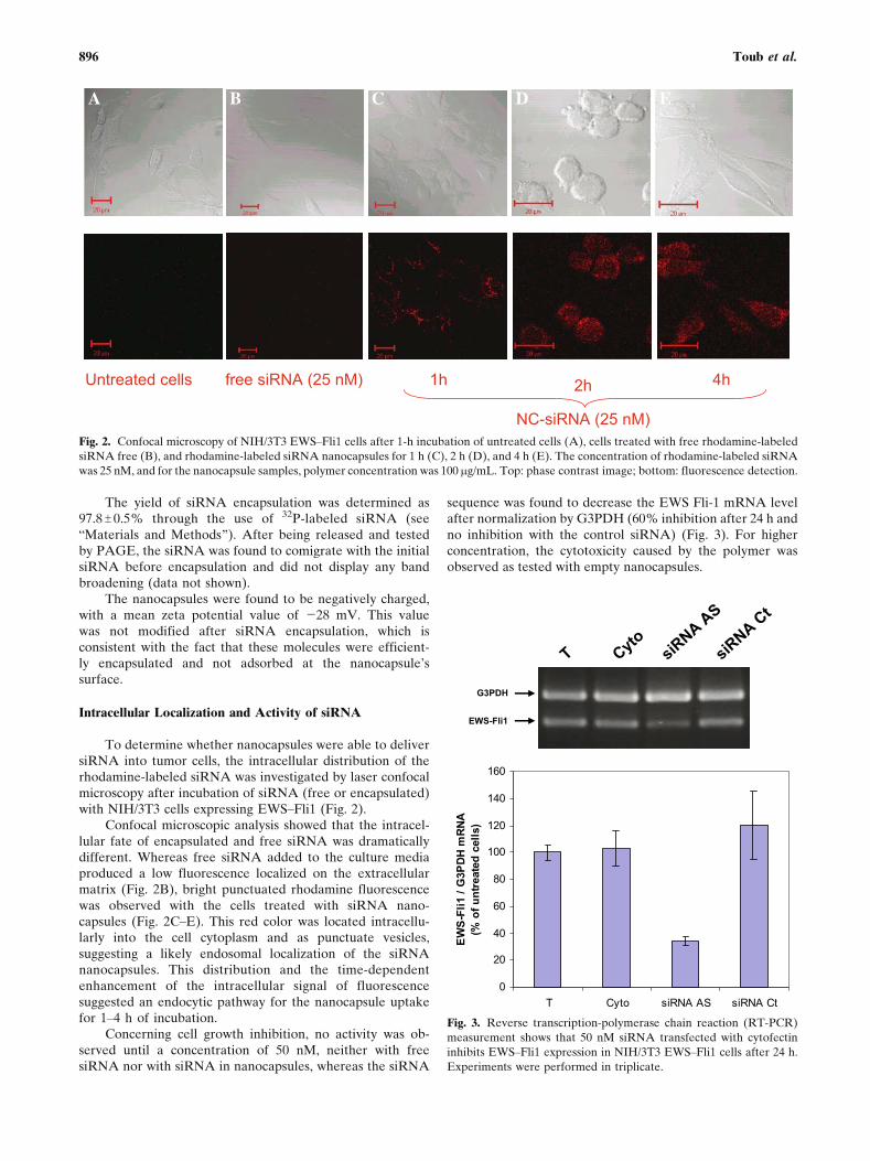
Concerning cell growth inhibition, no activity was ob-served until a concentration of 50 nM, neither with freesiRNA nor with siRNA in nanocapsules, whereas the siRNA
sequence was found to decrease the EWS Fli-1 mRNA levelafter normalization by G3PDH (60% inhibition after 24 h andno inhibition with the control siRNA) (Fig. 3). For higherconcentration, the cytotoxicity caused by the polymer wasobserved as tested with empty nanocapsules.
Fig. 2. Confocal microscopy of NIH/3T3 EWSYFli1 cells after 1-h incubation of untreated cells (A), cells treated with free rhodamine-labeled
siRNA free (B), and rhodamine-labeled siRNA nanocapsules for 1 h (C), 2 h (D), and 4 h (E). The concentration of rhodamine-labeled siRNA
was 25 nM, and for the nanocapsule samples, polymer concentration was 100 mg/mL. Top: phase contrast image; bottom: fluorescence detection.
Fig. 3. Reverse transcription-polymerase chain reaction (RT-PCR)
measurement shows that 50 nM siRNA transfected with cytofectin
inhibits EWSYFli1 expression in NIH/3T3 EWSYFli1 cells after 24 h.
Experiments were performed in triplicate.
896 Toub et al.

Administration to Mice
As explained in BMaterials and methods,^ two differentprotocols were used for the intratumoral administration ofsiRNA to the EWSYFli1 grafted mice. In the first therapeuticscheme and as shown in Fig. 4, only intratumoral injections ofNC siRNA-AS led to a significant inhibition in the tumorgrowth at a cumulative dose of 0.8 mg/kg (1.44 nmol). This
effect corresponded to a 3-fold decrease of tumor growthwhen compared with the control mice (0.9% NaCl). Underthe same conditions, no effect could be detected afterinjection of the free siRNA.
In the second therapeutic scheme (1.1 mg/kg of cumu-lative dose, five injections), a still greater inhibition (80%) oftumor growth was obtained. However, unexpectedly, astimulation of the tumor growth was observed with control
Fig. 4. Inhibition of EWSYFli1-expressing tumor growth in nude mice by siRNA
nanocapsules at a dose of 0.8 mg/kg. The treatment was performed by intratumoral
administration for 20 days. Arrows correspond to the days of treatment. The effect
of antisense siRNA in nanocapsules on tumor growth in nude mice is expressed
relatively to tumor volume on day 1 according to the following formula: tumor
volume at day x/tumor volume at day 1. Nine injections were performed at days 1,
2, 3, 6, 8, 10, 13, 15, and 17. All siRNA, free or encapsulated (NC), were used at a
dose of 88 mg for each injection. The cumulative dose of siRNA was 0.8 mg/kg
(1.44 nmol). ), NC siRNA-AS; r, NC siRNA-Ct;Í, siRNA-AS; 0, siRNA-Ct;&,
saline. Bars indicate the standard deviation of the mean for six mice.
Fig. 5. Inhibition of EWSYFli1-expressing tumor growth in nude mice by siRNA nanocapsules at a dose
of 1.11 mg/kg. The treatment was performed by intratumoral administration until day 15. Arrows
correspond to the days of treatment. The effect of antisense siRNA in nanocapsules on tumor growth in
nude mice is expressed relatively to tumor volume on day 1 according to the following formula: tumor
volume at day x/tumor volume at day 1. Five injections were performed at days 1, 3, 6, 9, and 12. All
siRNA, free or encapsulated (NC), were used at a dose of 220 mg for each injection. The cumulative dose
of siRNA was 1.11 mg/kg (2 nmol). ), NC siRNA-AS; r, NC siRNA-Ct; Í, siRNA-AS; 0, siRNA-Ct;
&, saline. Bars correspond to the standard deviation for six mice.
897Efficacy of siRNA Nanocapsules Against EWS–Fli1

siRNA administered either free or encapsulated (Fig. 5),whereas the same was observed with empty nanocapsules(data not shown).
The statistical analysis of covariance, with the tumorvolume at day 1 as a covariable, showed that the NC siRNA-AS gave a significant inhibition when compared with saline-treated animals ( p < 0.0003). The NC siRNA-Ct gave noinhibition ( p = 0.3544). The free siRNA-AS and siRNA-Ctproduced a nonsignificant stimulation of tumor growth. p
values are 0.73 and 0.25, respectively (six animals in eachgroup).
Tumor EWS�Fli1 and EWS Expressions
For this purpose, we have developed a method tomeasure simultaneously the expressions of EWSYFli1 andEWS. Because in mice the Fli1 gene was not expressed, it wasimportant to measure the EWS status at the same time thanthe EWSYFli1 fusion gene to have a better appreciationconcerning the specificity of the siRNA used. Thus, asexplained in BMaterials and methods,^ after total RNAextraction from the tumor, the expressions of EWSYFli1(322 bp), EWS (396 bp), and the housekeeping geneGAPDH (531 bp) were monitored by reverse transcription-polymerase chain reaction (RT-PCR). As observed in Fig. 6,very little variation of GAPDH expression was observed inall the experimental points. This makes it possible tocompare the expression of the target gene in the variousslots after GAPDH normalization. Intratumoral injection ofnaked antisense and control siRNA resulted in a stimulationof both EWSYFli1 and EWS gene expressions. Application ofnanocapsulated antisense siRNA decreased both EWSYFli1and EWS gene expressions to 43 and 61%, respectively, ascompared with the level measured in the nontreatedtumors. There remained a stimulation of both gene expres-sions after application of control siRNA encapsulated intonanocapsules.
DISCUSSION
Inhibition of gene expression by short double-strandedRNA (siRNA) is a promising strategy to develop therapeuticapplications. In animals, one of the limiting steps is a rapidRNA degradation and poor intracellular delivery. To over-come these limitations, particulate drug carriers may be used(29,30). In this study, we have employed the previouslydescribed polyisobutylcyanoacrylate nanocapsules (25) todeliver siRNA in vivo because these nanocapsules have beenshown to be capable of protecting the encapsulated nucleicacid (i.e., oligonucleotide) toward nuclease degradation (25).It is noteworthy that, for this application, we have used asmaller preparation volume [2 mL instead of 4.5 mL in (22)],which has resulted in an extremely high encapsulation rate ofsiRNA of 97%, as compared with 81% of encapsulation yieldwith oligonucleotides (25). It is possible that the efficacy ofstirring with an appropriate stem has led to a betterdispersion of the water droplets in the oily nanoemulsion.
To determine the ability of such complexes to deliverefficiently the siRNA intracellularly, we have measured the invitro cell penetration of rhodamine-labeled siRNA nano-capsules. By confocal microscopy, we observed (Fig. 2) thatfluorescent siRNA was time-dependently captured by thecells with cytoplasmic and endosomal localizations (Fig. 2CYE).This effect was not observed after the cells were incubatedwith free siRNA (Fig. 2B). This significant fluorescentenhancement with nanocapsules revealed a high level ofsiRNA cell uptake and a clear punctuated perinuclearlocalization, which fits with a capture of the nanocapsulesthrough an endocytotic pathway. However, using the MTTassay, no cell growth inhibition until an siRNA concentrationof 50 nM was observed, which was therefore not caused by theinability of the siRNA nanocapsules to cross the cell mem-brane. It was neither caused by the inability of siRNA toinhibit its target mRNA because it was shown that using thesame siRNA sequence, inhibition of EWSYFli1 mRNA wasefficiently obtained. This demonstrates again that cell cultureexperiments are not at all predictive of the in vivo data (see
Fig. 6. RT-PCR measurement shows that siRNA nanocapsules inhibit EWSYFli1 and EWS expressions
into mice grafted tumors.
898 Toub et al.

below), even when they were performed on exactly the sametumoral cell line. We have already observed such a phenom-enon with chitosan-coated nanospheres (35). The reasons ofsuch a discrepancy are numerous and have already beendiscussed previously by others (36).
To evaluate the therapeutic efficacy of siRNA nano-capsules in vivo, we have tested the ability of this formulationto inhibit tumor growth in an Ewing sarcoma model. Theanimals were treated by intratumoral injection of either thesiRNA free or encapsulated in two protocols: (a) one injectionevery 2 days, nine injections (0.8 mg/kg cumulative dose; 1.44nmol); (b) one injection every 3 days, five injections (1.1 mg/kgcumulative dose; 2 nmol). The second protocol was proposedbecause it was expected that the encapsulated siRNA wouldbe stable enough in cells after delivery, allowing to prolong thetime between two injections. Additionally, such treatment wassupposed to be better tolerated by the animals. For bothtreatments, a dramatic inhibition of tumor growth wasobserved but with a still greater effect in the second case (43and 80%, respectively) (Figs. 4 and 5). For both protocols, thesiRNAs alone had no inhibitory effect and even had atendency to stimulate tumor growth. A similar stimulation ofEWSYFli1 has already been observed previously in cellculture with free oligonucleotides (V. Pollard, personalcommunication). We suggest that this unspecific stimulationof NIH/3T3 EWSYFli1 cells could be as a result of anonspecific polyanionic effect of negatively charged mole-cules such as nucleic acids or nanocapsules. The hypothesis isthat receptors for polyanions could be present on the outsideof the cell membrane. When protected by the encapsulation,the siRNA could not induce such an unspecific effect, but thenanocapsules which are negatively charged (zeta potential ofthe surface was j28 mV) might play the same role (result notshown).
These data of tumor growth inhibition by siRNA nano-capsules resulted from a specific silencing inhibition of geneexpression. By real-time RT-PCR on RNA extracted fromtreated tumor, a decrease of the EWSYFli1 RNA expressionwas indeed observed. According to the known mechanism ofaction of siRNAs, this should be due mainly to the RISC-dependent degradation of the targeted mRNA. Alongsidethis inhibition, an EWS inhibition was also observed, which isprobably caused by the 17 bases of the siRNA also targetingthe EWS gene. Owing to the literature, it is unlikely that thetumor inhibition is caused by EWS inhibition. It is, however,conceivable that some synergistic effect could take placewhen inhibiting both genes. It has indeed been shown thatEWS and EWSYFli1 proteins form heterodimers (31).
It is now planned to test other siRNAs that would notinhibit EWS to closely investigate the role of EWS in thetumor inhibition that we have observed.
These results show the interest of nanocapsules forsiRNA delivery in vivo. Many publications have describedthe potency of siRNAs for cellular gene inhibition [for areview, see (29,30,32)]. More recently, several reports haveshown that protection of siRNAs by particulate vectors is avaluable approach for in vivo gene inhibition (33,34).
siRNA or antisense oligonucleotides will not be asubstitute for cancer chemotherapy but an additive to it andto other therapies. This can take place either when there is asynergy between antisense compounds and other anticancer
drugs or to maintain the residual disease at the backgroundlevel with treatments which should be far less toxic than theusual cancer chemotherapy. Many recurrences happen afterclassical treatments because of micrometastases, which haveescaped this treatment.
We demonstrate here that a potent activity can be ob-tained, as a result of nanoencapsulation, with an siRNA, whichis targeted to a fusion gene considered to be the cause of thetumor.
A promising outcome of this research would be thepossibility of treating Ewing sarcoma and PNET with varioustypes of specific nucleic acids protected by polymericencapsulation.
CONCLUSION
The present study reports for the first time the use ofaqueous core nanocapsules for the delivery of siRNA totumors with a junction oncogene as a specific target.Owing to the efficient intracellular penetration and to thein vivo anticancer activity, this type of formulation ispromising for the treatment of cancers such as Ewingsarcoma and PNET.
ACKNOWLEDGMENTS
We thank Mr. Jalil Abdelali and Mrs. Monique Stanciu(G. Roussy Institute) for excellent technical assistance andMrs. Ghislaine Frebourg (UMR CNRS 7138) for electronicmicroscopy experiments. N. Toub is supported by a fellow-ship from La Ligue Nationale Contre le Cancer. This workwas supported by the Association de Recherche sur le Cancer(grant no. 4310), Fondation de l’Avenir, and the Europeangrant PROTHETS (contract no. LSHC-CT-2004 5033036).
REFERENCES
1. T. H. Rabbitts. Chromosomal translocations in human cancer.Nature (Lond.) 372:143Y149 (1994).
2. C. Sreekantaiah, M. Ladanyi, E. Rodriguez, and R. S. K.Chaganti. Chromosomal aberration in soft tissue tumours. Am.J. Pathol. 144:1121Y1134 (1994).
3. O. Delattre, J. Zucman, B. Plougastel, C. Desmaze, T. Melot, M.Peter, H. Kovar, I. Joubert, P. D. Jong, G. Rouleau, A. Aurias,and G. Thomas. Gene fusion with an ETS DNA-binding domaincaused by chromosome translocation in human tumors. Nature(Lond.) 359:162Y165 (1992).
4. T. Ohno, V. N. Rao, and E. S. Reddy. EWS/Fli1 chimeric proteinis a transcriptional activator. Cancer Res. 53:5859Y5863 (1993).
5. R. A. Bailly, R. Bosselut, J. Zucman, F. Cornier, O. Delattre, M.Roussel, G. Thomas, and J. Ghysdael. DNA-binding andtranscriptional activation of the EWS-FLI-1 fusion proteinresulting from the t(11;22) translocation in Ewing sarcoma.Mol. Cell. Biol. 14:3230Y3241 (1994).
6. W. A. May, S. L. Lessnick, B. S. Braun, M. Klemsz, B. C. Lewis,L. B. Lunsford, R. Hromas, and C. T. Denny. The Ewingsarcoma EWS/FLI-1 fusion gene encodes a more potenttranscriptional activator and is a more powerful transforminggene than FLI-1. Mol. Cell. Biol. 13:7393Y7398 (1993).
7. X. Mao, S. Miesfeldt, H. Yang, J. M. Leiden, and C. B.Thompson. The FLI-1 and chimeric EWS-FLI-1 oncoproteins
899Efficacy of siRNA Nanocapsules Against EWS–Fli1

display similar DNA binding specificities. J. Biol. Chem.269:18216Y18222 (1994).
8. W. A. May, M. L. Gishizky, S. L. Lessnick, L. B. Lunsford, B. C.Lewis, O. Delattre, J. Zucman, G. Thomas, and C. T. Denny.Ewing sarcoma 11;22 translocation produces a chimeric tran-scription factor that require the DNA-binding domain encodedby FLI1 for transformation. Proc. Natl. Acad. Sci. USA90:5752Y5756 (1993).
9. K. Tanaka, T. Iwakuma, K. Harimaya, H. Sato, and Y. Iwamoto.EWSYFli1 antisense oligodeoxynucleotide inhibits proliferationof human Ewing’s sarcoma and primitive neuroectodermaltumor cells. J. Clin. Invest. 99:239Y247 (1997).
10. K. Hede. RNA-based nanoparticle treatment shows prom-ise in Ewing’s sarcoma model. J. Natl. Cancer Inst. 97:627(2005).
11. G. J. Hannon. RNA interference. Nature 418:244Y251 (2002).12. A. Fire, S. Xu, M. K. Montgomery, S. A. Kostas, S. E. Driver,
and C. C. Mello. Potent and specific genetic interferenceby double-stand RNA in Caenorhabditis elegans. Nature 391:806Y811 (1998).
13. M. Pal-Bhadra, U. Bhadra, and J. A. Birchler. RNAi relatedmechanisms affect both transcriptional and posttranscription-al transgene silencing in Drosophila. Mol. Cell 9:315Y327(2002).
14. S. M. Elbashir, J. Martinez, A. Patkaniowska, W. Lendeckel,and T. Tuschl. Functional anatomy of siRNAs for mediatingefficient RNAi in Drosophila melanogaster embryo lysate.EMBO J. 20:6877Y6888 (2001).
15. E. Bernstei, A. A. Caudy, S. M. Hammond, and G. J. Hannon.Role for a bidentate ribonuclease in the initiation step of RNAinterference. Nature 409:363Y366 (2001).
16. T. R. Brummelkamp, R. Bernards, and R. Agami. A system forstable expression of short interfering RNAs in mammalian cells.Science 296:550Y553 (2002).
17. T. R. Brummelkamp, R. Bernards, and R. Agami. A system forstable expression of short interfering RNAs in mammalian cells.Science 296:550Y553 (2002).
18. W. M. Bertling, M. Gareis, V. Paspaleeva, A. Zimmer, J.Kreuter, E. Nurnberg, and P. Harrer. Use of liposomes, viralcapsids, and nanoparticles as DNA carriers. Biotechnol. Appl.Biochem. 13:390Y405 (1991).
19. Z. Hassani, G. F. Lemkine, P. Erbacher, K. Palmier, G. Alfama,and C. Giovannageli, et al. Lipid-mediated siRNA deliverydowregulates exogenous gene expression in the mouse brain atpicomolar levels. J. Gene Med. 7:198Y207 (2004).
20. Y. Zhang, Y. F. Zhang, J. Bryant, A. Charles, R. J. Boado, andW. M. Pardridge. Intravenous RNA interference gene therapytargeting the human epidermal growth factor receptor prolongssurvival in intracranial brain cancer. Clin. Cancer Res.10:3667Y3677 (2004).
21. K. M. Ray and P. A. Whittaker. RNA interference: from genesilencing to gene-specific therapeutics. Pharmacol. Ther.107:222Y239 (2005).
22. G. Lambert, E. Fattal, H. Pinto-Alphandary, A. Gulik, and P.Couvreur. Polyisobutylcyanoacrylate nanocapsules containing
an aqueous core as a novel colloidal carrier for the delivery ofoligonucleotides. Pharm. Res. 17:707Y714 (2000).
23. T. Tuschl. Expanding small RNA interference. Nat. Biotechnol.20:446Y448 (2002).
24. I. Aynie, C. Vauthier, M. Foulquier, C. Malvy, E. Fattal, and P.Couvreur. Development of a quantitative polyacrylamide gelelectrophoresis analysis using a multichannel radioactivitycounter for the evaluation of oligonucleotide-bound drugcarrier. Anal. Biochem. 240:202Y209 (1996).
25. G. Lambert, J. R. Bertrand, E. Fattal, S. Subra, H. Pinto-Alphandary, C. Malvy, C. Auclair, and P. Couvreur. EWS Fli-1antisense nanocapsules inhibits Ewing sarcoma-related tumor inmice. Biochem. Biophys. Res. Commun. 279:401Y406 (2000).
26. S. Watnasirichaikul, N. M. Davies, T. Rades, and I. G. Tucker.Preparation of biodegradable insulin nanocapsules from bio-compatible microemulsions. Pharm. Res. 17:684Y689 (2000).
27. P. Workman, et al. United Kingdom Co-ordinating Committeeon Cancer Research (UKCCCR) Guidelines for the Welfare ofanimals in Experimental Neoplasia (Second Edition). Br. J.Cancer 77:1Y10 (1998).
28. T. Maniatis E. F. Fritsch and J. Sambrook. Molecular Cloning. ALaboratory Manual, Cold Spring Harbor Laboratory, ColdSpring Harbor, NY, 1982.
29. M. R. Schiffelers, A. Ansari, J. Xu, Q. Zhou, Q. Tang, G. Storm,G. Molema, P. Y. Lu, P. V. Scaria, and M. C. Woodle. CancersiRNA therapy by tumor selective delivery with ligand-targetedsterically stabilized nanoparticle. Nucleic Acids Res. 32:e149(2004).
30. W. Zhang, H. Yang, X. Kong, S. Mohapatra, H. San Juan-Vergara, G. Hellermann, S. Behera, R. Singam, R. F. Lockey,and S. S. Mohapatra. Inhibition of respiratory syncytial virusinfection with intranasal siRNA nanoparticles targeting the viralNS1 gene. Nat. Med. 11:56Y62 (2005).
31. L. Spahn, C. Siligan, R. Bachmaier, J. A. Schmid, D. N. Aryee,and H. Kovar. Homotypic and heterotypic interactions of EWS,Fli1 and their oncogenic fusion protein. Oncogene 9(22):6819Y6829 (2003).
32. Y. L. Chiu, A. Ali, C. Y. Chu, H. Cao T. M. Rana. Visualizing acorrelation between siRNA localization, cellular uptake, andRNAi in living cells. Chem. Biol. 11:1165Y1175 (2004).
33. Q. Leng and A. J. Mixson. Small interfering RNA targeting Raf-1 inhibits tumor growth in vitro and in vivo. Cancer Gene Ther.12(8): 682Y690 (2005).
34. H. Guan, Z. Zhou, H. Wang, W. L. Jia, and W. L. Kleinerman.A small interfering RNA targeting vascular endothelial growthfactor inhibits Ewing’s sarcoma growth in a xenograft mousemodel. Clin. Cancer Res. 11:2662Y2669 (2005).
35. A. Maksimenko, V. Polard, M. Villemeur, H. Elhamess, P.Couvreur, J. R. Bertrand, M. Aboubakar, M. Gottikh, and C.Malvy. In vivo potentialities of EWS-FLI-1 targeted antisenseoligonucleotidesYnanospheres complexes. Ann. N.Y. Acad. Sci.1058:52Y62 (2005).
36. H. Farhood, N. Serbina, and L. Huang. The role of dioleoylphosphatidylethanolamine in cationic liposome mediated genetransfer. Biochim. Biophys. Acta 1235:289Y295 (1995).
900 Toub et al.
Copyright © 2022 FDOKUMEN