Core/Shell Structured Hollow Mesoporous Nanocapsules
13
Core/Shell Structured Hollow Mesoporous Nanocapsules: A Potential Platform for Simultaneous Cell Imaging and Anticancer Drug Delivery Yu Chen, † Hangrong Chen, †, * Deping Zeng, ‡ Yunbo Tian, ‡ Feng Chen, † Jingwei Feng, † and Jianlin Shi* † State Laboratory of High Performance Ceramic and Superfine Microstructure, Shanghai Institute of Ceramics, Chinese Academy of Sciences, Shanghai 200050, China, and ‡ Department of Biomedical Engineering, Chongqing Medical University, Chongqing 400016, China C apsules, a common morphology with ultrathin shells and ultralarge cavities, have been commercially used as effective carriers for drug delivery, especially for oral drug administration. However, the extremely large particulate sizes (in milli- or centimeters) of commer- cial capsules severely limited their manipu- lation for intravenous drug administration, which has been generally considered as the most effective chemotherapeutic mode for serious diseases. The key that has fascinated biomedical researchers is how to design and control the sizes of capsules into the micro- and nanoscale, which might be solved by seeking help from chemistry. The emergence and development of nanotech- nology have created a chance for reducing the sizes of the capsules into the micro/ nano- scale range, making the intravenous drug administration of capsules possible. A large number of micro/nano- capsules, in- cluding organic, 13 inorganic, 4,5 and inor- ganic/organic hybrid ones, 6,7 have been de- veloped as drug delivery systems (DDSs) for transporting therapeutic agents to le- sion sites in the past decade. The biocom- patible and biodegradable nature of poly- mer based capsules, generally fabricated by the layer-by-layer self-assembly technique, 712 showed great potential ap- plication in chemotherapy of serious dis- eases such as cancer and HIV. 1317 The in- trinsic instable nature of organic systems, unfortunately, still cast a shadow on their further clinical use. Alternatively, inorganic capsules with biocompatible and biode- gradable features, especially porous ones, such as mesoporous silica, 4 mesoporous carbon, 18 carbon nanotube, 19 Fe 3 O 4 aggregates 20,21 and CaCO 3 , 22 exhibited vari- ous advantages compared to organic ones in their high chemical/thermal stability, high drug loading capability, sustained drug release from the supports and rich sur- face chemical functionalities for possible molecular recognition and targeted deliv- ery, etc. On the other hand, the multifunctional- ization of capsules may enable the develop- ment of multifunctional nanomedical plat- forms for simultaneous imaging diagnosis and therapy. 23,24 The general methodology to achieve multifunctionalities on capsules *Address correspondence to [email protected], [email protected]. Received for review July 4, 2010 and accepted August 31, 2010. Published online September 3, 2010. 10.1021/nn1015117 © 2010 American Chemical Society ABSTRACT A potential platform for simultaneous anticancer drug delivery and MRI cell imaging has been demonstrated by uniform hollow inorganic core/shell structured multifunctional mesoporous nanocapsules, which are composed of functional inorganic (Fe 3 O 4 , Au, etc.) nanocrystals as cores, a thin mesoporous silica shell, and a huge cavity in between. The synthetic strategy for the creation of huge cavities between functional core and mesoporous silica shell is based on a structural difference based selective etching method, by which solid silica middle layer of Fe 2 O 3 @SiO 2 @mSiO 2 (or Au@SiO 2 @mSiO 2 ) composite nanostructures was selectively etched away while the mesoporous silica shell could be kept relatively intact. The excellent biocompatibility of obtained multifunctional nanocapsules (Fe 3 O 4 @mSiO 2 ) was demonstrated by very low cytotoxicity against various cell lines, low hemolyticity against human blood red cells and no significant coagulation effect against blood plasma. The cancer cell uptake and intracellular location of the nanocapsules were observed by confocal laser scanning microscopy and bio-TEM. Importantly, the prepared multifunctional inorganic mesoporous nanocapsules show both high loading capacity (20%) and efficiency (up to 100%) for doxorubicin simultaneously because of the formation of the cavity, enhanced surface area/pore volume and the electrostatic interaction between DOX molecules and mesoporous silica surface. Besides, the capability of Fe 3 O 4 @mSiO 2 nanocapsules as contrast agents of MRI was demonstrated both in vitro and in vivo, indicating the simultaneous imaging and therapeutic multifunctionalities of the composite nanocapsules. Moreover, the concept of multifunctional inorganic nanocapsules was extended to design and prepare GdSiDTPA grafted Au@mSiO 2 nanocapsules for nanomedical applications, further demonstrating the generality of this strategy for the preparation of various multifunctional mesoporous nanocapsules. KEYWORDS: mesoporous silica · nanocapsules · drug delivery · MRI · Fe 3 O 4 · Au ARTICLE www.acsnano.org VOL. 4 ▪ NO. 10 ▪ 6001–6013 ▪ 2010 6001
-
Upload
khangminh22 -
Category
Documents
-
view
0 -
download
0
Transcript of Core/Shell Structured Hollow Mesoporous Nanocapsules
Core/Shell Structured HollowMesoporous Nanocapsules: A PotentialPlatform for Simultaneous Cell Imagingand Anticancer Drug DeliveryYu Chen,† Hangrong Chen,†,* Deping Zeng,‡ Yunbo Tian,‡ Feng Chen,† Jingwei Feng,† and Jianlin Shi*†State Laboratory of High Performance Ceramic and Superfine Microstructure, Shanghai Institute of Ceramics, Chinese Academy of Sciences, Shanghai 200050, China, and‡Department of Biomedical Engineering, Chongqing Medical University, Chongqing 400016, China
Capsules, a common morphologywith ultrathin shells and ultralargecavities, have been commercially
used as effective carriers for drug delivery,
especially for oral drug administration.
However, the extremely large particulate
sizes (in milli- or centimeters) of commer-
cial capsules severely limited their manipu-
lation for intravenous drug administration,
which has been generally considered as the
most effective chemotherapeutic mode for
serious diseases. The key that has fascinated
biomedical researchers is how to design
and control the sizes of capsules into the
micro- and nanoscale, which might be
solved by seeking help from chemistry. The
emergence and development of nanotech-
nology have created a chance for reducing
the sizes of the capsules into the micro/
nano- scale range, making the intravenous
drug administration of capsules possible. A
large number of micro/nano- capsules, in-
cluding organic,1�3 inorganic,4,5 and inor-
ganic/organic hybrid ones,6,7 have been de-
veloped as drug delivery systems (DDSs)
for transporting therapeutic agents to le-
sion sites in the past decade. The biocom-
patible and biodegradable nature of poly-
mer based capsules, generally fabricated by
the layer-by-layer self-assembly
technique,7�12 showed great potential ap-
plication in chemotherapy of serious dis-
eases such as cancer and HIV.13�17 The in-
trinsic instable nature of organic systems,
unfortunately, still cast a shadow on their
further clinical use. Alternatively, inorganic
capsules with biocompatible and biode-
gradable features, especially porous ones,
such as mesoporous silica,4 mesoporous
carbon,18 carbon nanotube,19 Fe3O4
aggregates20,21 and CaCO3,22 exhibited vari-ous advantages compared to organic onesin their high chemical/thermal stability,high drug loading capability, sustaineddrug release from the supports and rich sur-face chemical functionalities for possiblemolecular recognition and targeted deliv-ery, etc.
On the other hand, the multifunctional-ization of capsules may enable the develop-ment of multifunctional nanomedical plat-forms for simultaneous imaging diagnosisand therapy.23,24 The general methodologyto achieve multifunctionalities on capsules
*Address correspondence [email protected],[email protected].
Received for review July 4, 2010and accepted August 31, 2010.
Published online September 3, 2010.10.1021/nn1015117
© 2010 American Chemical Society
ABSTRACT A potential platform for simultaneous anticancer drug delivery and MRI cell imaging has been
demonstrated by uniform hollow inorganic core/shell structured multifunctional mesoporous nanocapsules, which
are composed of functional inorganic (Fe3O4, Au, etc.) nanocrystals as cores, a thin mesoporous silica shell, and a
huge cavity in between. The synthetic strategy for the creation of huge cavities between functional core and
mesoporous silica shell is based on a structural difference based selective etching method, by which solid silica
middle layer of Fe2O3@SiO2@mSiO2 (or Au@SiO2@mSiO2) composite nanostructures was selectively etched away
while the mesoporous silica shell could be kept relatively intact. The excellent biocompatibility of obtained
multifunctional nanocapsules (Fe3O4@mSiO2) was demonstrated by very low cytotoxicity against various cell
lines, low hemolyticity against human blood red cells and no significant coagulation effect against blood plasma.
The cancer cell uptake and intracellular location of the nanocapsules were observed by confocal laser scanning
microscopy and bio-TEM. Importantly, the prepared multifunctional inorganic mesoporous nanocapsules show
both high loading capacity (20%) and efficiency (up to 100%) for doxorubicin simultaneously because of the
formation of the cavity, enhanced surface area/pore volume and the electrostatic interaction between DOX
molecules and mesoporous silica surface. Besides, the capability of Fe3O4@mSiO2 nanocapsules as contrast agents
of MRI was demonstrated both in vitro and in vivo, indicating the simultaneous imaging and therapeutic
multifunctionalities of the composite nanocapsules. Moreover, the concept of multifunctional inorganic
nanocapsules was extended to design and prepare Gd�Si�DTPA grafted Au@mSiO2 nanocapsules for
nanomedical applications, further demonstrating the generality of this strategy for the preparation of various
multifunctional mesoporous nanocapsules.
KEYWORDS: mesoporous silica · nanocapsules · drug delivery · MRI · Fe3O4 · Au
ARTIC
LE
www.acsnano.org VOL. 4 ▪ NO. 10 ▪ 6001–6013 ▪ 2010 6001
is to add diverse functional inorganic nanocrystals intothe cavity part of the capsules or to chemically graftfunctional groups onto the shells based on the rational/elaborate design in nanoscale. These inorganic nano-crystals can be Au,25�32 Ag,33 quantum dots (QDs),34,35
Fe3O4 magnetic nanoparticles,36�38 and the functionalgroups can be fluorescent molecules39 or targeting mol-ecules such as folic acid, etc., endowing capsules withunique optical, acoustic and magnetic properties forclinical multi-imaging purposes (CT, MRI, US, PET, etc.)or targeting features for drug delivery. So far, however,the combined functionalities between physical imag-ing, such as MRI for cancer diagnosis, and an enhancedanticancer drug delivery property (e.g., high drug load-ing amount and efficiency) have been rarelyreported.23,36,37 Such a combination, as we believe, isgreatly important because it allows simultaneous imag-ing diagnosis and drug delivery in one.
Herein, multifunctional hollow core/shell structurednanocapsules with magnetic Fe3O4 or Au nanocrystalsas the cores, thin mesoporous silica layer as the shelland controllable cavities between the core and shellhave been fabricated and applied as both MRI contrastagent and carriers for anticancer DDSs. We employedthe magnetite core-based nanocapsules of ellipsoidalmorphology to demonstrate our concept of multifunc-tional core/shell nanocapsules as DDSs because meso-porous nanomaterials with larger aspect ratios could betaken up by cells in larger amounts and have higher in-ternalization rates,40,41 which is of great significance forthe enhancement of chemotherapy effectiveness. Be-sides, the reason for adopting magnetic Fe3O4 nano-crystals to functionalize the nanocapsules is that mag-netic nanocrystals could be used for magneticallytargeted drug delivery,4,42 magnetic hyperthermia,43
and as MRI contrast agents as well.44,45 This is the first re-port, as far as we know, to prepare multifunctional core/shell structured hollow mesoporous silica nanocap-sules with ellipsoidal morphology as both MRI imagingagents and DDSs. Moreover, the concept of multifunc-tional inorganic nanocapsules was extended to designand prepare Gd�Si�DTPA grafted Au@mSiO2 nano-capsules for T1-weighted MRI and dark-field light scat-tering live cell imaging, further demonstrating the gen-
erality of this strategy for the preparation ofmultifunctional mesoporous nanocapsules.
RESULTS AND DISCUSSIONSynthesis of Core/Shell Structured Multifunctional Hollow
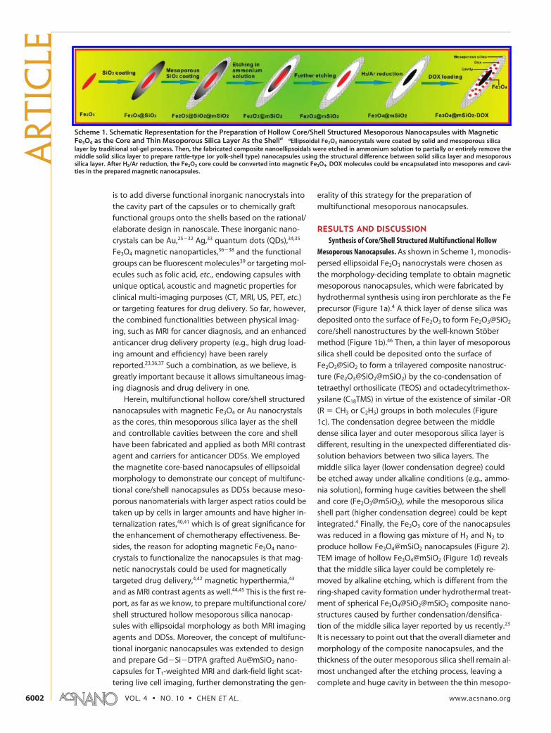
Mesoporous Nanocapsules. As shown in Scheme 1, monodis-persed ellipsoidal Fe2O3 nanocrystals were chosen asthe morphology-deciding template to obtain magneticmesoporous nanocapsules, which were fabricated byhydrothermal synthesis using iron perchlorate as the Feprecursor (Figure 1a).4 A thick layer of dense silica wasdeposited onto the surface of Fe2O3 to form Fe2O3@SiO2
core/shell nanostructures by the well-known Stobermethod (Figure 1b).46 Then, a thin layer of mesoporoussilica shell could be deposited onto the surface ofFe2O3@SiO2 to form a trilayered composite nanostruc-ture (Fe2O3@SiO2@mSiO2) by the co-condensation oftetraethyl orthosilicate (TEOS) and octadecyltrimethox-ysilane (C18TMS) in virtue of the existence of similar -OR(R � CH3 or C2H5) groups in both molecules (Figure1c). The condensation degree between the middledense silica layer and outer mesoporous silica layer isdifferent, resulting in the unexpected differentiated dis-solution behaviors between two silica layers. Themiddle silica layer (lower condensation degree) couldbe etched away under alkaline conditions (e.g., ammo-nia solution), forming huge cavities between the shelland core (Fe2O3@mSiO2), while the mesoporous silicashell part (higher condensation degree) could be keptintegrated.4 Finally, the Fe2O3 core of the nanocapsuleswas reduced in a flowing gas mixture of H2 and N2 toproduce hollow Fe3O4@mSiO2 nanocapsules (Figure 2).TEM image of hollow Fe3O4@mSiO2 (Figure 1d) revealsthat the middle silica layer could be completely re-moved by alkaline etching, which is different from thering-shaped cavity formation under hydrothermal treat-ment of spherical Fe3O4@SiO2@mSiO2 composite nano-structures caused by further condensation/densifica-tion of the middle silica layer reported by us recently.23
It is necessary to point out that the overall diameter andmorphology of the composite nanocapsules, and thethickness of the outer mesoporous silica shell remain al-most unchanged after the etching process, leaving acomplete and huge cavity in between the thin mesopo-
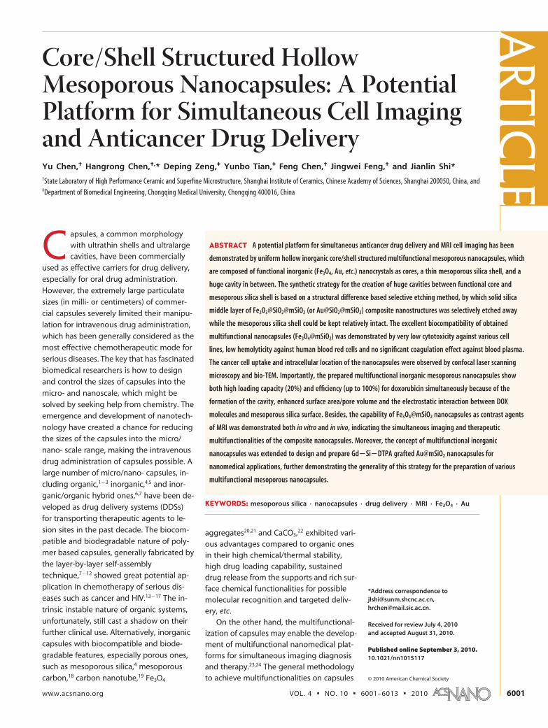
Scheme 1. Schematic Representation for the Preparation of Hollow Core/Shell Structured Mesoporous Nanocapsules with MagneticFe3O4 as the Core and Thin Mesoporous Silica Layer As the Shella aEllipsoidal Fe2O3 nanocrystals were coated by solid and mesoporous silicalayer by traditional sol-gel process. Then, the fabricated composite nanoellipsoidals were etched in ammonium solution to partially or entirely remove themiddle solid silica layer to prepare rattle-type (or yolk-shell type) nanocapsules using the structural difference between solid silica layer and mesoporoussilica layer. After H2/Ar reduction, the Fe2O3 core could be converted into magnetic Fe3O4. DOX molecules could be encapsulated into mesopores and cavi-ties in the prepared magnetic nanocapsules.
ART
ICLE
VOL. 4 ▪ NO. 10 ▪ CHEN ET AL. www.acsnano.org6002
rous shell and magnetic Fe3O4 core. In addition, the di-
mension of the cavity in Fe3O4@mSiO2 nanocapsules
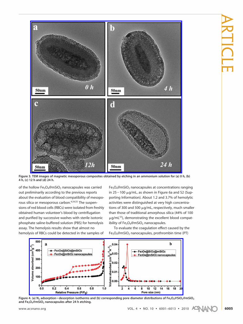
could be simply tuned by selecting adequate etching
time, supported by TEM observations of different etch-
ing time intervals (Figure 3; 0 h, 4 h, 12 and 24 h) of
Fe2O3@SiO2@mSiO2 composite nanostructures. A sec-
ondary electron SEM image of the Fe2O3 nanocrystals
and Fe3O4@mSiO2 show that the particles are uniformly
ellipsoidal in shape (Figure 1e and 1f), while a composi-
tional backscattered SEM image (Figure 1f) reveals the
contrast difference between the central heavy core and
the cavity/silica shell of the hollow structure, which
could be further confirmed by a purposely selected bro-
ken nanocapsule (inset of Figure 1f).
N2 adsorption�desorption technique was adopted
to characterize the pore structural evolution of the
magnetic nanocapsules after alkaline etching in ammo-
nia solution. Surprisingly, the isotherms of
Fe3O4@SiO2@mSiO2 core/shell nanostructures and
Fe3O4@mSiO2 nanocapsules show apparently different
styles, where rattle-type core�shell nanocapsules ex-
hibit a much larger hysteresis loop than those without
Figure 1. TEM images of ellipsoidal (a) Fe2O3, (b) Fe2O3@SiO2, (c) Fe2O3@SiO2@mSiO2 and (d) Fe3O4@mSiO2; Secondary electronSEM image of ellipsoidal Fe2O3 (e, inset: SEM image at high magnification) and backscattered electron SEM (f) image of ellipsoi-dal Fe3O4@mSiO2 nanocapsules (inset: purposely selected backscattered electron image of broken nanocapsules to reveal thehollow nanostructure).
ARTIC
LE
www.acsnano.org VOL. 4 ▪ NO. 10 ▪ 6001–6013 ▪ 2010 6003
cavities between the core and shell (Figure 4a), which
represents the typical ink-bottle-type pores in which
larger cavities are connected by narrow windows.47 The
surface area increased from 261 m2/g to 318 m2/g while
the pore volume from 0.44 cm3/g to 0.78 cm3/g after al-
kaline etching. The average pore size of Fe3O4@mSiO2
also increased from 2.5 to 3.6 nm (Figure 4b). As shown
in Scheme 2, the mesoporous silica shell of
Fe2O3@SiO2@mSiO2 is still relatively unstable under
alkaline condition because of the existence of abun-
dant Si�O�Si bonds in the shell.4,48 Partial Si�O�Si
bonds in the shell can be broken during the etch-
ing process, resulting in the enlargement of the ini-
tial pores templated by the surfactants (C18TMS) and
finally causing the increase of surface area and pore
volume. According to above analysis, we believe that
this etching process provides an alternative but ver-
satile route to tune the structural parameter of me-
soporous nanocapsules, such as surface area, pore
volume and pore size. Selective etching of silica to
form nanocapsules, such as Au@SiO2, CNTs@SiO249
and SiO2@SiO2,50 has been reported recently. How-
ever, there were no well-defined mesoporous struc-
tures on those silica shells due to their intrinsic ab-
sence, which may limit their practical applications
due to the mass transfer blockage by the solid silica
shells. Our prepared Fe3O4@mSiO2 hollow core/shell
nanocapsules with well-defined mesopores in the
silica shell and controllable cavity sizes show the ap-
parent advantage superior to nonporous inorganic
nanocapsules in drug deliv-ery and/or magnetic-recycledcatalysis applications.
Cell Uptake, IntracellularLocation, Cytotoxicity, Hemolysis,Coagulation of MultifunctionalMesoporous Fe3O4@mSiO2
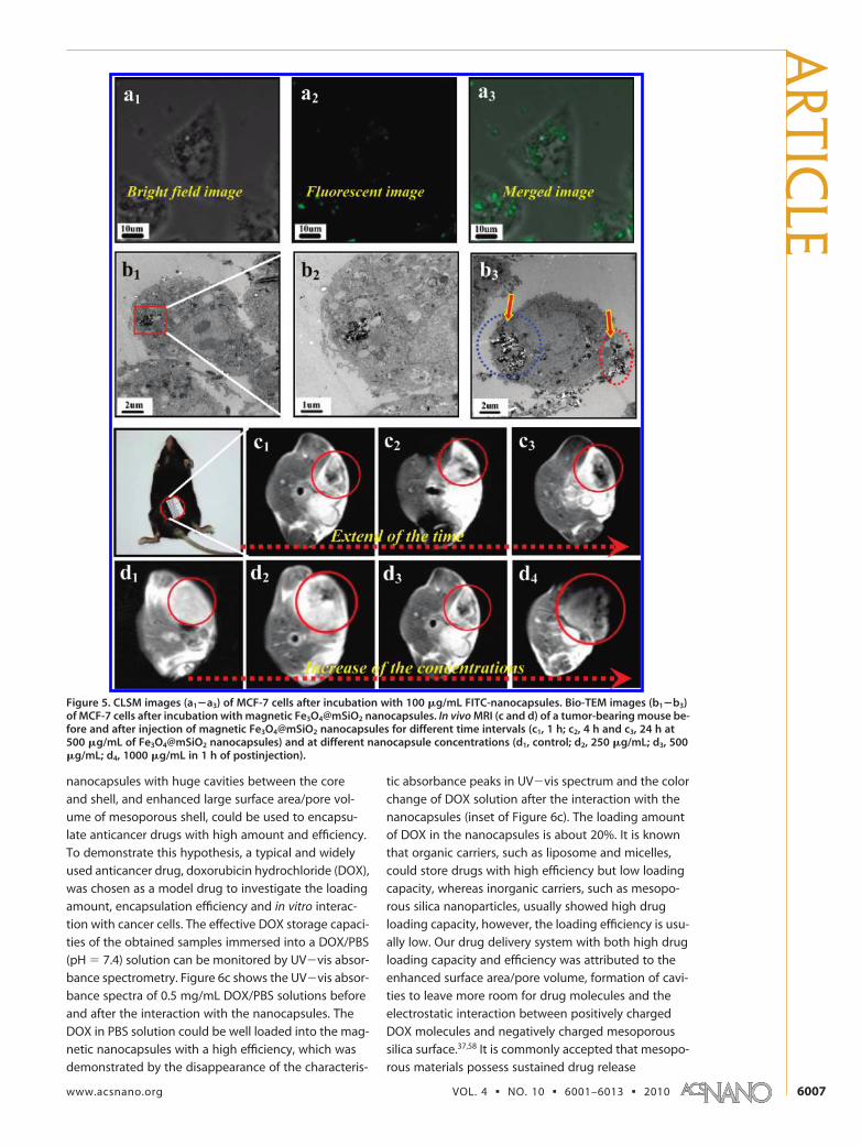
Nanocapsules. Excellent cell up-take of the prepared inor-ganic nanocapsules guaran-tees the high deliveryefficiency of encapsulateddrugs because intracellular re-lease of drugs from the carri-ers could cause higher cyto-toxicity than extracellulardrugs. To facilitate the obser-vations of cell uptake of thenanocapsules by confocal la-ser scanning microscopy(CLSM), the preparedFe3O4@mSiO2 nanocapsuleswere grafted by fluoresceinisothiocyanate (FITC-capsules). Figure 5a1�a3 showbright-field, fluorescent andmerged images of breast can-
cer MCF-7 cells incubated with FITC-capsules (100 �g/mL) for 3 h. The CLSM images show that the nanocap-sules could be taken up by MCF-7 cells within a shortperiod as manifested by the appearance of green fluo-rescence in cells. The overlay of the bright field and fluo-rescent images further demonstrate that the lumines-cence is strongly correlated with the intracellularlocation (Figure 5a and S1, Supporting Information),suggesting the feasibility and efficiency of the nanocap-sules for fluorescent cell imaging and anticancer drugsdelivery into cancer cells. To further reveal the uptakemechanism of the magnetic nanocapsules into MCF-7cells and the intracellular location, bio-TEM analysis wascarried out after ultrathin section of MCF-7 cells afterthe uptake of the nanocapsules. The distribution ofmagnetic nanocapsules could be clearly observed inTEM images, and a large number of them were foundin the cytoplasm while no nanocarriers were found inthe nucleus (Figure 5b1 and b2). In addition, part of thenanocapsules could be found near the cell membraneforming vesicles (Figure 5b3). After the uptake by can-cer cells via endocytosis, the nanocapsules will be pro-cessed in endosomes and lysosomes, and are eventu-ally released into the cytoplasm.51�53
It is important to investigate the biocompatibility ofthe hollow Fe3O4@mSiO2 nanocapsules with blood cellsto guarantee the successful intravenous administrationof drug-loaded nanocapsules. In fact, the blood com-patibility of the inorganic hollow core/shell nanocap-sules has been rarely reported. Hemolysis experiment
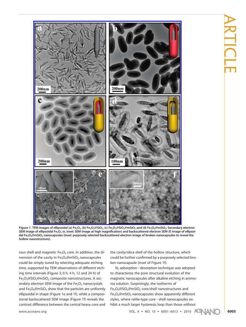
Figure 2. (a) Small-angle and (b) wide-angle X-ray diffraction patterns of Fe2O3@mSiO2 andFe3O4@mSiO2 nanocapsules. (c) Digital pictures of (A) Fe2O3@mSiO2 and (B) Fe3O4@mSiO2 dispersedin PBS solution, and (C) Fe3O4@mSiO2 nanocapsules in PBS solution under magnetic field. The appear-ance of characteristic diffraction peaks in wide-angle XRD pattern of Fe3O4 in Fe3O4@mSiO2 and dis-appearance of characteristic diffraction peaks of Fe2O3 in Fe2O3@mSiO2 demonstrated the successfulconversion of Fe2O3@mSiO2 into Fe3O4@mSiO2. Besides, small-angle XRD patterns indicated that themesopores in the nanocapsules are disordered, in consistent with TEM observations.
ART
ICLE
VOL. 4 ▪ NO. 10 ▪ CHEN ET AL. www.acsnano.org6004
of the hollow Fe3O4@mSiO2 nanocapsules was carried
out preliminarily according to the previous reports
about the evaluation of blood compatibility of mesopo-
rous silica or mesoporous carbon.4,54,55 The suspen-
sions of red blood cells (RBCs) were isolated from freshly
obtained human volunteer’s blood by centrifugation
and purified by successive washes with sterile isotonic
phosphate saline-buffered solution (PBS) for hemolysis
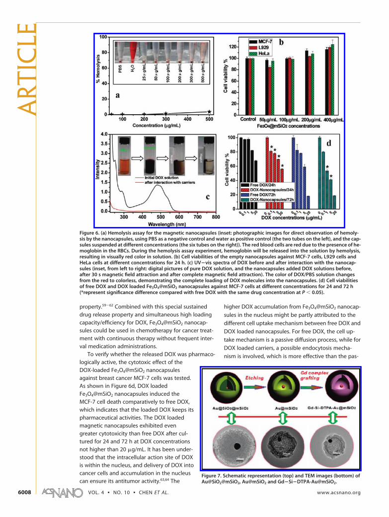
assay. The hemolysis results show that almost no
hemolysis of RBCs could be detected in the samples of
Fe3O4@mSiO2 nanocapsules at concentrations ranging
in 25�100 �g/mL, as shown in Figure 6a and S2 (Sup-
porting Information). About 1.2 and 3.7% of hemolytic
activities were distinguished at very high concentra-
tions of 300 and 500 �g/mL, respectively, much smaller
than those of traditional amorphous silica (44% of 100
�g/mL55), demonstrating the excellent blood compat-
ibility of Fe3O4@mSiO2 nanocapsules.
To evaluate the coagulation effect caused by the
Fe3O4@mSiO2 nanocapsules, prothrombin time (PT)
Figure 3. TEM images of magnetic mesoporous composites obtained by etching in an ammonium solution for (a) 0 h, (b)4 h, (c) 12 h and (d) 24 h.
Figure 4. (a) N2 adsorption�desorption isotherms and (b) corresponding pore diameter distributions of Fe3O4@SiO2@mSiO2
and Fe3O4@mSiO2 nanocapsules after 24 h etching.
ARTIC
LE
www.acsnano.org VOL. 4 ▪ NO. 10 ▪ 6001–6013 ▪ 2010 6005
and activated partial thromboplastin time (APTT) val-
ues were tested by mixing fresh blood plasma with
nanocapsules at different concentrations (25�500 �g/
mL). As shown in Figure S3 (Supporting Information),
the PT and APTT values of the plasma exposure to
Fe3O4@mSiO2 nanocapsules of different concentrations
do not show significant difference compared to those
of the blank control, demonstrating that no significant
coagulation/anticoagulation effect of prepared nano-
capsules is present. It is known the coagulation effect
caused by the nanomaterials as DDSs in blood vessel
must avoided, so the coagulation test results of
Fe3O4@mSiO2 nanocapsules guarantee the biosafety
for the intravenous administration of the prepared
nanocapsules.
To evaluate the cytotoxicity of the empty capsules,
in vitro cytotoxicity tests were conducted against differ-
ent cell lines, such as MCF-7 cells, HeLa cells and L929
cells by conducting MTT (3-[4,5-dimethylthiazol-2-yl]-
2,5-diphenyltetrazolium bromide) assays. Figure 6b and
S4 (Supporting Information) show the effect of concen-
tration of Fe3O4@mSiO2 nanocapsules on the cell viabil-
ity for different incubation time periods. The difference
in cell viabilities after 24 and 48 h of incubation is neg-
ligibly small, and there is no apparent cytotoxicity after
incubation at a very high concentration up to 400
�g/mL for 48 h. The excellent biocompatibility (blood-
compatibility and low cytotoxicity) of the Fe3O4@mSiO2
nanocapsules guarantees the practical applications for
human beings as carriers in DDSs.
Multifunctional Mesoporous Nanocapsules as Contrast Agentsof MRI Both In Vitro and In Vivo. The functional inorganicnanocrystals in the core part of the nanocapsules couldendow the capsules with simultaneous imaging anddrug delivery functionalities. For instance, the magneticFe3O4 core of the prepared core/shell nanocapsulescould be used as the MRI contrast agent during the che-motherapeutic process, playing a role of monitoringthe therapeutic efficiency and providing high-qualityimages for operators. As an initial step in exploring po-tential imaging functionality of the nanocapsules dur-ing chemotherapy both in vitro and in vivo, the visibil-ity of the nanocapsules was tested in water, in cells andin mice by MRI. T2 phantom images of the nanocap-sules dispersed in water changed significantly in signalintensity with the increase of Fe concentration (FigureS5a, Supporting Information), indicating that the mag-netic nanocapsules have generated magnetic reso-nance contrast on transverse (T2) proton relaxations-times weighted sequence due to the dipolar interactionof magnetic moments between the nanocapsules andproton in the water.56,57 An r2 value of 137.8 mM�1s�1
was measured for the magnetic nanocapsules (FigureS5b, Supporting Information), revealing high potentialof the nanocapsules as T2 contrast agents for cancer di-agnosis. Under T2-weighted imaging mode, cells ex-posed to the magnetic nanocapsules of a concentra-tion of 50 �g/mL for 6 h could be easily detected(Figure S5c, Supporting Information).
Furthermore, the nanocapsules were injected intomice subcutaneously and noninvasively detected withMRI at diverse concentrations and different time inter-vals (Figure 5c and d). The darkened images from the in-jection site confirmed the imaging capability of thenanocapsules (Figure 5c, red circled area). The dark-ened area extended through the tumor part with time(from 1 to 24 h), indicating the diffusion of magneticnanocapsules within tumor. Besides, the darkened areawas also larger at increased concentrations, demon-strating the concentration-dependent MRI feature ofmagnetic nanocapsules (Figure 5d).
Multifunctional Mesoporous Nanocapsules as DDSs forDoxorubicin. To achieve the goal of complete eradicationof tumors, therapeutic agents have to be administratedsystematically in high dose to ensure the sufficient andsustained therapy efficiency. However, most of antican-cer drugs effective in eradicating cancer cells couldcause severe side-effect when the high doses are ad-ministrated, which is caused by the nonspecific uptakeof anticancer drugs by healthy tissues/organs such askidney, liver, bone marrow and heart. An effective wayto reduce the side-effect is to encapsulate the antican-cer drugs in DDSs with high loading amount and effi-ciency, which could largely protect the healthy organsfrom the toxic drugs and prevent the decomposition/denaturing of the drugs prior to reaching the targetedcells. We believe that the obtained multifunctional
Scheme 2. Schematic Representation of the Microstructure EvolutionBefore, During and After the Alkaline Etchinga aSi�O�Si bonds in themesoporous silica shell part could be partially broken by OH�1 in ammonia solu-tion under hydrothermal treatment (150°C; 24 h), causing the expansion of sizesof initial pores templated by C18TMS.
ART
ICLE
VOL. 4 ▪ NO. 10 ▪ CHEN ET AL. www.acsnano.org6006
nanocapsules with huge cavities between the core
and shell, and enhanced large surface area/pore vol-
ume of mesoporous shell, could be used to encapsu-
late anticancer drugs with high amount and efficiency.
To demonstrate this hypothesis, a typical and widely
used anticancer drug, doxorubicin hydrochloride (DOX),
was chosen as a model drug to investigate the loading
amount, encapsulation efficiency and in vitro interac-
tion with cancer cells. The effective DOX storage capaci-
ties of the obtained samples immersed into a DOX/PBS
(pH � 7.4) solution can be monitored by UV�vis absor-
bance spectrometry. Figure 6c shows the UV�vis absor-
bance spectra of 0.5 mg/mL DOX/PBS solutions before
and after the interaction with the nanocapsules. The
DOX in PBS solution could be well loaded into the mag-
netic nanocapsules with a high efficiency, which was
demonstrated by the disappearance of the characteris-
tic absorbance peaks in UV�vis spectrum and the color
change of DOX solution after the interaction with the
nanocapsules (inset of Figure 6c). The loading amount
of DOX in the nanocapsules is about 20%. It is known
that organic carriers, such as liposome and micelles,
could store drugs with high efficiency but low loading
capacity, whereas inorganic carriers, such as mesopo-
rous silica nanoparticles, usually showed high drug
loading capacity, however, the loading efficiency is usu-
ally low. Our drug delivery system with both high drug
loading capacity and efficiency was attributed to the
enhanced surface area/pore volume, formation of cavi-
ties to leave more room for drug molecules and the
electrostatic interaction between positively charged
DOX molecules and negatively charged mesoporous
silica surface.37,58 It is commonly accepted that mesopo-
rous materials possess sustained drug release
Figure 5. CLSM images (a1�a3) of MCF-7 cells after incubation with 100 �g/mL FITC-nanocapsules. Bio-TEM images (b1�b3)of MCF-7 cells after incubation with magnetic Fe3O4@mSiO2 nanocapsules. In vivo MRI (c and d) of a tumor-bearing mouse be-fore and after injection of magnetic Fe3O4@mSiO2 nanocapsules for different time intervals (c1, 1 h; c2, 4 h and c3, 24 h at500 �g/mL of Fe3O4@mSiO2 nanocapsules) and at different nanocapsule concentrations (d1, control; d2, 250 �g/mL; d3, 500�g/mL; d4, 1000 �g/mL in 1 h of postinjection).
ARTIC
LE
www.acsnano.org VOL. 4 ▪ NO. 10 ▪ 6001–6013 ▪ 2010 6007
property.59�62 Combined with this special sustaineddrug release property and simultaneous high loadingcapacity/efficiency for DOX, Fe3O4@mSiO2 nanocap-sules could be used in chemotherapy for cancer treat-ment with continuous therapy without frequent inter-val medication administrations.
To verify whether the released DOX was pharmaco-logically active, the cytotoxic effect of theDOX-loaded Fe3O4@mSiO2 nanocapsulesagainst breast cancer MCF-7 cells was tested.As shown in Figure 6d, DOX loadedFe3O4@mSiO2 nanocapsules induced theMCF-7 cell death comparatively to free DOX,which indicates that the loaded DOX keeps itspharmaceutical activities. The DOX loadedmagnetic nanocapsules exhibited evengreater cytotoxicity than free DOX after cul-tured for 24 and 72 h at DOX concentrationsnot higher than 20 �g/mL. It has been under-stood that the intracellular action site of DOXis within the nucleus, and delivery of DOX intocancer cells and accumulation in the nucleuscan ensure its antitumor activity.63,64 The
higher DOX accumulation from Fe3O4@mSiO2 nanocap-
sules in the nucleus might be partly attributed to the
different cell uptake mechanism between free DOX and
DOX loaded nanocapsules. For free DOX, the cell up-
take mechanism is a passive diffusion process, while for
DOX loaded carriers, a possible endocytosis mecha-
nism is involved, which is more effective than the pas-
Figure 6. (a) Hemolysis assay for the magnetic nanocapsules (inset: photographic images for direct observation of hemoly-sis by the nanocapsules, using PBS as a negative control and water as positive control (the two tubes on the left), and the cap-sules suspended at different concentrations (the six tubes on the right)). The red blood cells are red due to the presence of he-moglobin in the RBCs. During the hemolysis assay experiment, hemoglobin will be released into the solution by hemolysis,resulting in visually red color in solution. (b) Cell viabilities of the empty nanocapsules against MCF-7 cells, L929 cells andHeLa cells at different concentrations for 24 h. (c) UV�vis spectra of DOX before and after interaction with the nanocap-sules (inset, from left to right: digital pictures of pure DOX solution, and the nanocapsules added DOX solutions before,after 30 s magnetic field attraction and after complete magnetic field attraction). The color of DOX/PBS solution changesfrom the red to colorless, demonstrating the complete loading of DOX molecules into the nanocapsules. (d) Cell viabilitiesof free DOX and DOX loaded Fe3O4@mSiO2 nanocapsules against MCF-7 cells at different concentrations for 24 and 72 h(*represent significance difference compared with free DOX with the same drug concentration at P � 0.05).
Figure 7. Schematic representation (top) and TEM images (bottom) ofAu@SiO2@mSiO2, Au@mSiO2 and Gd�Si�DTPA-Au@mSiO2.
ART
ICLE
VOL. 4 ▪ NO. 10 ▪ CHEN ET AL. www.acsnano.org6008
sive diffusion. After incubated with MCF-7 cells, the ma-
jority of DOX-containing nanocapsules are endocytosed
by the cancer cells, whereas, only limited amount of
DOX diffused into the cells for free DOX. Thus, the bet-
ter uptake of DOX-containing nanocapsules by the
MCF-7 cells should be responsible for the higher con-
centration of DOX in the nucleus, leading to the en-
hanced DOX cytotoxicity and cell death. On the other
hand, when the cancer cells are treated with free DOX,
the development of multidrug resistance (MRD) of can-
cer cells, which results from the expression of
p-glycoprotein pump in the cell membrane, hampers
the drug action by pumping out drug molecules from
cytosol to extracellular area.65 However, the uptake of
the drug loaded nanocarriers within cancer cells might,
to some extent, circumvent the MRD effect.65
Preparation of Au@mSiO2 Nanocapsules and Their BiologicalBimodal Imaging. Many functional inorganic nanocrystals
could be coated by silica layers. The subsequent meso-porous silica layer coating depends on the surfacechemistry of coated dense silica layer rather than thenanocrystals, so this nanocapsule formation strategycould be extended to synthesize various multifunc-tional hollow core/shell structured nanocapsules bysimply changing the functional core, depending on thepractical requirements and elaborate design in nano/micrometer scale.
We further substituted ellipsoidal Fe3O4 with spheri-cal Au nanoparticles to demonstrate this hypothesis, invirtue of the unique optical properties of Au nanoparti-cles such as surface plasma resonance enhanced lightscattering and absorption, and the ability to efficientlyconvert the strongly absorbed light into localized heat,which could be exploited for in vivo imaging and the se-lective laser photothermal therapy of cancer.66 Further-more, we grafted Gd�Si�DTPA into the pores ofAu@mSiO2 nanocapsules using the abundant surfacechemistry of mesoporous silica layer (e.g., Si�OH),which could be used for biological bimodal imaging in-cluding dark-field light scattering cell imaging and T1-weighted MRI, and anticancer drug encapsulation anddelivery. Figure 7 and S6 show the structural evolutionduring the preparation process, including coating non-
porous silica and mesoporous silica shells on Au sur-
face, etching away the middle silica layer and further
grafting Gd�Si�DTPA (T1 MRI contrast agent) for mul-
tifunctionalization. The obtained Gd complex grafted
Au@mSiO2 nanocapsules exhibit strong surface plasma
enhanced absorption (Figure 8), which makes them
useful for biomedical dark-field light scattering imag-
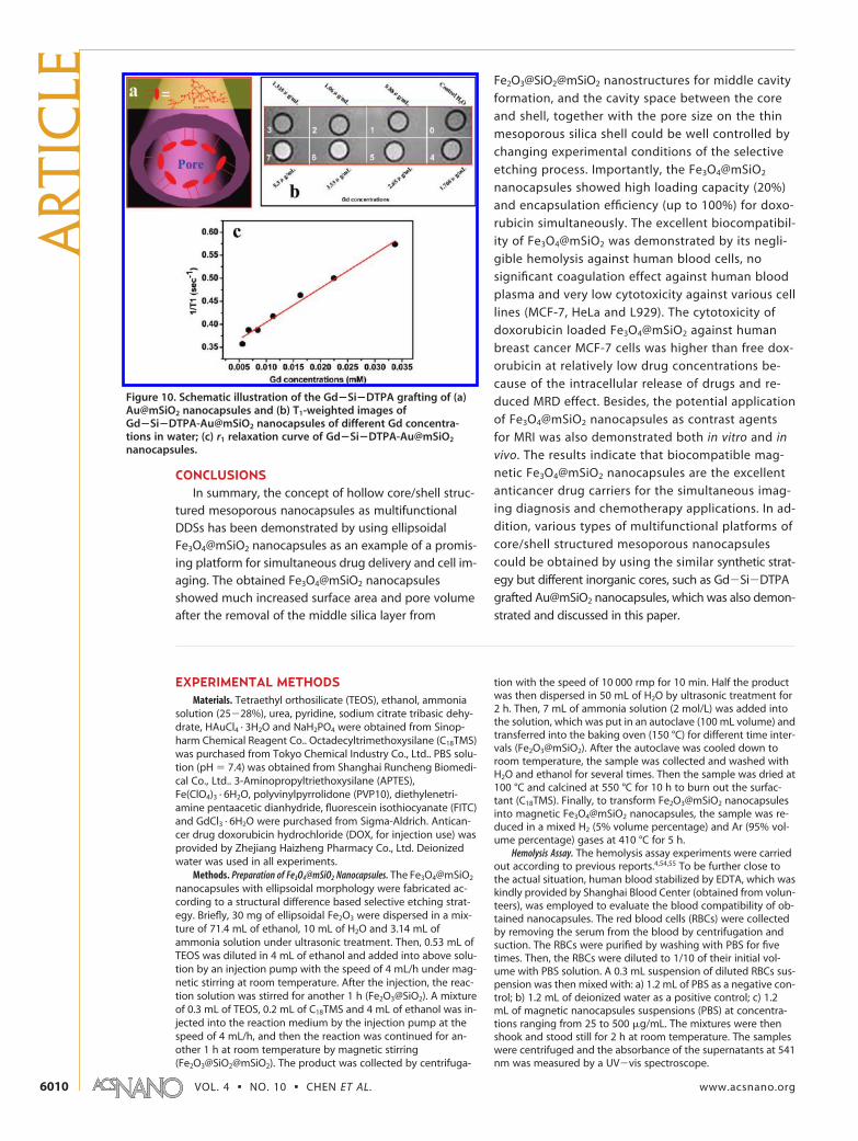
ing and detection of live cells (Figure 9).67 Besides, the
obtained Gd�Si�DTPA grafted Au@mSiO2 nanocap-
sules show T1-weighted signal enhancement with a r1
value of 7.43 mM�1s�1, demonstrating the potential ap-
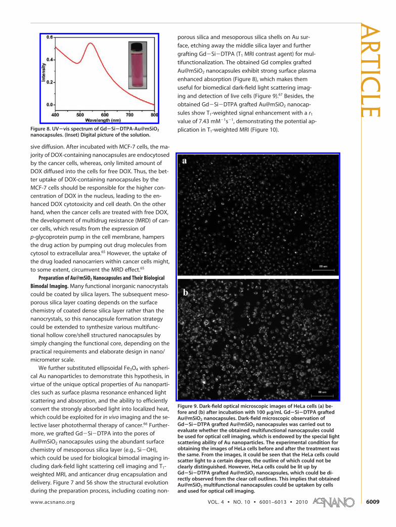
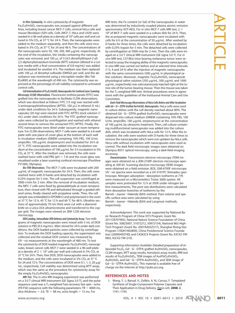
plication in T1-weighted MRI (Figure 10).Figure 8. UV�vis spectrum of Gd�Si�DTPA-Au@mSiO2
nanocapsules. (Inset) Digital picture of the solution.
Figure 9. Dark-field optical microscopic images of HeLa cells (a) be-fore and (b) after incubation with 100 �g/mL Gd�Si�DTPA graftedAu@mSiO2 nanocapsules. Dark-field microscopic observation ofGd�Si�DTPA grafted Au@mSiO2 nanocapsules was carried out toevaluate whether the obtained multifunctional nanocapsules couldbe used for optical cell imaging, which is endowed by the special lightscattering ability of Au nanoparticles. The experimental condition forobtaining the images of HeLa cells before and after the treatment wasthe same. From the images, it could be seen that the HeLa cells couldscatter light to a certain degree, the outline of which could not beclearly distinguished. However, HeLa cells could be lit up byGd�Si�DTPA grafted Au@mSiO2 nanocapsules, which could be di-rectly observed from the clear cell outlines. This implies that obtainedAu@mSiO2 multifunctional nanocapsules could be uptaken by cellsand used for optical cell imaging.
ARTIC
LE
www.acsnano.org VOL. 4 ▪ NO. 10 ▪ 6001–6013 ▪ 2010 6009
CONCLUSIONSIn summary, the concept of hollow core/shell struc-
tured mesoporous nanocapsules as multifunctionalDDSs has been demonstrated by using ellipsoidalFe3O4@mSiO2 nanocapsules as an example of a promis-ing platform for simultaneous drug delivery and cell im-aging. The obtained Fe3O4@mSiO2 nanocapsulesshowed much increased surface area and pore volumeafter the removal of the middle silica layer from
Fe2O3@SiO2@mSiO2 nanostructures for middle cavity
formation, and the cavity space between the core
and shell, together with the pore size on the thin
mesoporous silica shell could be well controlled by
changing experimental conditions of the selective
etching process. Importantly, the Fe3O4@mSiO2
nanocapsules showed high loading capacity (20%)
and encapsulation efficiency (up to 100%) for doxo-
rubicin simultaneously. The excellent biocompatibil-
ity of Fe3O4@mSiO2 was demonstrated by its negli-
gible hemolysis against human blood cells, no
significant coagulation effect against human blood
plasma and very low cytotoxicity against various cell
lines (MCF-7, HeLa and L929). The cytotoxicity of
doxorubicin loaded Fe3O4@mSiO2 against human
breast cancer MCF-7 cells was higher than free dox-
orubicin at relatively low drug concentrations be-
cause of the intracellular release of drugs and re-
duced MRD effect. Besides, the potential application
of Fe3O4@mSiO2 nanocapsules as contrast agents
for MRI was also demonstrated both in vitro and in
vivo. The results indicate that biocompatible mag-
netic Fe3O4@mSiO2 nanocapsules are the excellent
anticancer drug carriers for the simultaneous imag-
ing diagnosis and chemotherapy applications. In ad-
dition, various types of multifunctional platforms of
core/shell structured mesoporous nanocapsules
could be obtained by using the similar synthetic strat-
egy but different inorganic cores, such as Gd�Si�DTPA
grafted Au@mSiO2 nanocapsules, which was also demon-
strated and discussed in this paper.
EXPERIMENTAL METHODSMaterials. Tetraethyl orthosilicate (TEOS), ethanol, ammonia
solution (25�28%), urea, pyridine, sodium citrate tribasic dehy-drate, HAuCl4 · 3H2O and NaH2PO4 were obtained from Sinop-harm Chemical Reagent Co.. Octadecyltrimethoxysilane (C18TMS)was purchased from Tokyo Chemical Industry Co., Ltd.. PBS solu-tion (pH � 7.4) was obtained from Shanghai Runcheng Biomedi-cal Co., Ltd.. 3-Aminopropyltriethoxysilane (APTES),Fe(ClO4)3 · 6H2O, polyvinylpyrrolidone (PVP10), diethylenetri-amine pentaacetic dianhydride, fluorescein isothiocyanate (FITC)and GdCl3 · 6H2O were purchased from Sigma-Aldrich. Antican-cer drug doxorubicin hydrochloride (DOX, for injection use) wasprovided by Zhejiang Haizheng Pharmacy Co., Ltd. Deionizedwater was used in all experiments.
Methods. Preparation of Fe3O4@mSiO2 Nanocapsules. The Fe3O4@mSiO2
nanocapsules with ellipsoidal morphology were fabricated ac-cording to a structural difference based selective etching strat-egy. Briefly, 30 mg of ellipsoidal Fe2O3 were dispersed in a mix-ture of 71.4 mL of ethanol, 10 mL of H2O and 3.14 mL ofammonia solution under ultrasonic treatment. Then, 0.53 mL ofTEOS was diluted in 4 mL of ethanol and added into above solu-tion by an injection pump with the speed of 4 mL/h under mag-netic stirring at room temperature. After the injection, the reac-tion solution was stirred for another 1 h (Fe2O3@SiO2). A mixtureof 0.3 mL of TEOS, 0.2 mL of C18TMS and 4 mL of ethanol was in-jected into the reaction medium by the injection pump at thespeed of 4 mL/h, and then the reaction was continued for an-other 1 h at room temperature by magnetic stirring(Fe2O3@SiO2@mSiO2). The product was collected by centrifuga-
tion with the speed of 10 000 rmp for 10 min. Half the productwas then dispersed in 50 mL of H2O by ultrasonic treatment for2 h. Then, 7 mL of ammonia solution (2 mol/L) was added intothe solution, which was put in an autoclave (100 mL volume) andtransferred into the baking oven (150 °C) for different time inter-vals (Fe2O3@mSiO2). After the autoclave was cooled down toroom temperature, the sample was collected and washed withH2O and ethanol for several times. Then the sample was dried at100 °C and calcined at 550 °C for 10 h to burn out the surfac-tant (C18TMS). Finally, to transform Fe2O3@mSiO2 nanocapsulesinto magnetic Fe3O4@mSiO2 nanocapsules, the sample was re-duced in a mixed H2 (5% volume percentage) and Ar (95% vol-ume percentage) gases at 410 °C for 5 h.
Hemolysis Assay. The hemolysis assay experiments were carriedout according to previous reports.4,54,55 To be further close tothe actual situation, human blood stabilized by EDTA, which waskindly provided by Shanghai Blood Center (obtained from volun-teers), was employed to evaluate the blood compatibility of ob-tained nanocapsules. The red blood cells (RBCs) were collectedby removing the serum from the blood by centrifugation andsuction. The RBCs were purified by washing with PBS for fivetimes. Then, the RBCs were diluted to 1/10 of their initial vol-ume with PBS solution. A 0.3 mL suspension of diluted RBCs sus-pension was then mixed with: a) 1.2 mL of PBS as a negative con-trol; b) 1.2 mL of deionized water as a positive control; c) 1.2mL of magnetic nanocapsules suspensions (PBS) at concentra-tions ranging from 25 to 500 �g/mL. The mixtures were thenshook and stood still for 2 h at room temperature. The sampleswere centrifuged and the absorbance of the supernatants at 541nm was measured by a UV�vis spectroscope.
Figure 10. Schematic illustration of the Gd�Si�DTPA grafting of (a)Au@mSiO2 nanocapsules and (b) T1-weighted images ofGd�Si�DTPA-Au@mSiO2 nanocapsules of different Gd concentra-tions in water; (c) r1 relaxation curve of Gd�Si�DTPA-Au@mSiO2
nanocapsules.
ART
ICLE
VOL. 4 ▪ NO. 10 ▪ CHEN ET AL. www.acsnano.org6010
In Vitro Cytotoxicity. In vitro cytotoxicity of magneticFe3O4@mSiO2 nanocapsules was assayed against different celllines, including breast cancer MCF-7 cells, cervical HeLa cells andmouse fibroblast L929 cells. Cells (MCF-7, HeLa and L929) wereseeded in a 96-well plate at a density of 104 cells per well and cul-tured in 5% CO2 at 37 °C for 24 h. Then, the nanocapsules wereadded to the medium separately, and then the cells were incu-bated in 5% CO2 at 37 °C for 24 and 48 h. The concentrations ofthe nanocapsules were 50, 100, 200, 400 �g/mL respectively. Atthe end of the incubation, the media containing the nanocap-sules was removed, and 100 �L of 3-[4,5-dimethylthiazol-2-yl]-2,5-diphenyltetrazolium bromide (MTT) solution (diluted in a cul-ture media with a final concentration of 0.8 mg/mL) was addedand incubated for another 4 h. The medium was then replacedwith 100 �L of dimethyl sulfoxide (DMSO) per well, and the ab-sorbance was monitored using a microplate reader (Bio-TekELx800) at the wavelength of 490 nm. The cytotoxicity was ex-pressed as the percentage of cell viability compared to untreatedcontrol cells.
Cell Internalization of Fe3O4@mSiO2 Nanocapsules for Confocal Laser ScanningMicroscopy (CLSM) Observations. Fluorescein isothiocyanate (FITC) wasgrafted into the nanocapsules to facilitate CLSM observations,which was described as follows: FITC (15 mg) was reacted with3-aminopropyltriethoxysilane (APTES, 100 �L) in ethanol (5 mL)under dark conditions for 24 h. Subsequently, magnetic nano-capsules (20 mg) were reacted with FITC-APTES stock solution (1mL) under dark conditions for 24 h. The FITC grafted nanocap-sules were collected by centrifugation and washed with ethanolseveral times to remove the unreacted FITC-APTES. Finally, theFITC-nanocapsules were dried under vacuum at room tempera-ture. For CLSM observations, MCF-7 cells were seeded in a 6-wellplate with one piece of cover glass at the bottom of each wellin incubation medium (DMEM containing 10% FBS, 100 U/mLampicillin, 100 �g/mL streptomycin) and incubated for 24 h at37 °C. FITC-nanocapsules were added into the incubation me-dium at the concentration of 100 �g/mL for 3 h incubation in 5%CO2 at 37 °C. After the medium was removed, the cells werewashed twice with cold PBS (pH � 7.4) and the cover glass wasvisualized under a laser scanning confocal microscope (FluoViewFV1000, Olympus).
Bio-TEM Observations. The MCF-7 cells were incubated with 200�g/mL of magnetic nanocapsules for 24 h. Then, the cells werewashed twice with D-hanks and detached by incubation with0.25% trypsin for 5 min. The cell suspension was centrifuged at5000 r/min for 2 min. After the removal of incubation medium,the MFC-7 cells were fixed by glutaraldehyde at room tempera-ture, then rinsed with PB and dehydrated through a graded eth-anol series, finally cleared with propylene oxide. Then, the cellsample was embedded in EPOM812 and polymerized in the ovenat 37 °C for 12 h, 45 °C for 12 h and 60 °C for 48 h. Ultrathin sec-tions of approximately 70 nm thick were cut with a diamondknife on a Leica UC6 ultramicrotome and transferred to the cop-per grid. The images were viewed on JEM-1230 electronmicroscopy.
DOX Loading, Intracellular DOX Delivery and Cytotoxicity Assay. Ten milli-grams of magnetic nanocapsules were mixed with 5 mL of DOXsolution in PBS (0.5 mg/mL). After stirred for 24 h under dark con-ditions, the DOX-loaded particles were collected by centrifuga-tion. To evaluate the DOX loading capacity, the supernatant wascollected and the residual DOX content was measured byUV�vis measurements at the wavelength of 480 nm. To testthe cytotoxicity of DOX loaded magnetic Fe3O4@mSiO2 nanocap-sules, breast cancer cells MCF-7 were seeded in a 96-well plateat a density of 2 � 103 cells per well and cultured in 5% CO2 at37 °C for 24 h. Then, free DOX, DOX-nanocapsules were added tothe medium, and the cells were incubated in 5% CO2 at 37 °Cfor 24 and 72 h. The concentrations of DOX were 0.1, 1, 5, 20 �g/mL, respectively. Cell viability was determined using MTT assay,which was the same as the procedure for cytotoxicity assay forthe empty Fe2O3@mSiO2 nanocapsules.
MRI Test. The in vitro MR imaging experiment was performedon a 3.0 T clinical MRI instrument (GE Signa 3.0 T), and the pulsesequence used was a T2-weighted Fast-recovery fast spin�echo(FR-FSE) sequence with the following parameters: TR � 4000 ms,slice thickness � 3.0, TE � 98 ms, echo length � 15 ms. For
MRI tests, the Fe content (or Gd) of the nanocapsules in waterwas determined by inductively coupled plasma atomic emissionspectrometry (ICP-AES). For in vitro MCF-7 cells MRI test, 2 �106 of MCF-7 cells were seeded in a culture dish for 24 h. Then,the as-prepared magnetic nanocapsules were incubated withcells for 6 h at the concentration of 50 �g/mL. After washed byD-hanks for three times, the cells were detached by incubationwith 0.25% trypsin for 5 min. The detached cells were collectedby centrifugation at 5000 rmp for 2 min. Then the cells were im-aged on a 3.0 T clinical MRI instrument (GE signa 3.0 T). For invivo MRI test, C57/BL6 mice bearing melanoma tumor were se-lected to assay the imaging ability of the magnetic nanocapsules.In vivo MRI was carried out before and at selected time intervals(1 h, 4 and 24 h) after the injection of magnetic nanocapsuleswith the same concentrations (500 �g/mL in physiological sa-line solution). Moreover, magnetic Fe3O4@mSiO2 nanocapsulephysiological saline solution (250 �g/mL, 500 �g/mL and 1000�g/mL, respectively) was subcutaneously injected right at the tu-mor site of the tumor-bearing mouse. Then the mouse was takenfor the T2-weighted MRI test. Animal procedures were in agree-ment with the guidelines of the Instituonal Animal Care and UseCommittee.
Dark-Field Microscopy Observations of HeLa Cells Before and After Incubationwith Gd�Si�DTPA Grafted Au@mSiO2 Nanocapsules. HeLa cells were seedin culture dishes until the cell density reached about 80%. Theobtained Gd�Si�DTPA grafted Au@mSiO2 nanocapsules weredispersed into culture medium (DMEM containing 10% FBS, 100U/mL ampicillin, 100 �g/mL streptomycin) at the concentrationof 100 �g/mL by ultrasonic treatment. Then, cell culture contain-ing multifunctional nanocapsules was added into the culturedish, which was incubated with HeLa cells for 12 h. After the in-cubation, the cells were washed with D-hanks for three times toremove the nanocapsules which were not uptaken by HeLa cells.HeLa cells without incubation with nanocapsules were used ascontrol. The dark field microscopic images were obtained onOlympus BX51 optical microscopy, using a dark-field imagingaccessory.
Characterization. Transmission electron microscopy (TEM) im-ages were obtained on a JEM-2100F electron microscope oper-ating at 200 kV. Scanning electron microscopy (SEM) imageswere obtained on a field emission JEOL JSM-6700F microscope.UV�vis spectra were recorded on a UV-3101PC Shimadzu spec-troscope. Nitrogen adsorption�desorption isotherms at 77Kwere measured on a Micrometitics Tristar 3000 system. Allsamples were pretreated for 12 h at 393K under nitrogen be-fore measurements, The pore size distributions were calculatedfrom desorption branches of isotherms by theBarrett�Joyner�Halenda (BJH) method. Pore volume and spe-cific surface area were calculated by usingBarrett�Joyner�Halenda (BJH) and Langmuir methods,respectively.
Acknowledgment. This work was supported by National Ba-sic Research Program of China (973 Program, Grant No.2011CB707905), National Nature Science Foundation of China(Grant No. 50823007, 51072212, 50972154), National 863 High-Tech Program (Grant No. 2007AA03Z317), Shanghai Rising-StarProgram (10QH1402800), China Postdoctoral Science Founda-tion (20090450740) and CASKJCX Projects (Grant No. KJCX2-YW-M02, KJCX2-YW-210).
Supporting Information Available: Detailed preparation of el-lipsoidal Fe2O3, Gd�Si�DTPA grafted Au@mSiO2 nanocapsules,CLSM images, MTT assay results, hemolysis assay results, MRI testresults of Fe3O4@mSiO2, TEM images of Au@SiO2@mSiO2,Au@mSiO2 and Gd�Si�DTPA-Au@mSiO2, and SEM image ofGd�Si�DTPA-Au@mSiO2. This material is available free ofcharge via the Internet at http://pubs.acs.org.
REFERENCES AND NOTES1. Wang, Y. J.; Bansal, V.; Zelikin, A. N.; Caruso, F. Templated
Synthesis of Single-Component Polymer Capsules andTheir Application in Drug Delivery. Nano Lett. 2008, 8,1741–1745.
ARTIC
LE
www.acsnano.org VOL. 4 ▪ NO. 10 ▪ 6001–6013 ▪ 2010 6011
2. Meier, W. Polymer Nanocapsules. Chem. Soc. Rev. 2000, 29,295–303.
3. Qiu, X. P.; Leporatti, S.; Donath, E.; Mohwald, H. Studies onthe Drug Release Properties of Polysaccharide MultilayersEncapsulated Ibuprofen Microparticles. Langmuir 2001, 17,5375–5380.
4. Chen, Y.; Chen, H. R.; Guo, L. M.; He, Q. J.; Chen, F.; Zhou, J.;Feng, J. W.; Shi, J. L. Hollow/Rattle-Type MesoporousNanostructures by a Structural Difference-Based SelectiveEtching Strategy. ACS Nano 2010, 4, 529–539.
5. Zhang, T. R.; Ge, J. P.; Hu, Y. X.; Zhang, Q.; Aloni, S.; Yin,Y. D. Formation of Hollow Silica Colloids Through aSpontaneous Dissolution-Regrowth Process. Angew. Chem.Int. Et. 2008, 47, 5806–5811.
6. Zhu, Y. F.; Shi, J. L.; Shen, W. H.; Dong, X. P.; Feng, J. W.;Ruan, M. L.; Li, Y. S. Stimuli-Responsive Controlled DrugRelease from a Hollow Mesoporous SilicaSphere/Polyelectrolyte Multilayer Core-Shell Structure.Angew. Chem.Int. Ed. 2005, 44, 5083–5087.
7. Caruso, F.; Caruso, R. A.; Mohwald, H. Nanoengineering ofInorganic and Hybrid Hollow Spheres by ColloidalTemplating. Science 1998, 282, 1111–1114.
8. Donath, E.; Sukhorukov, G. B.; Caruso, F.; Davis, S. A.;Mohwald, H. Novel Hollow Polymer Shells by Colloid-Templated Assembly of Polyelectrolytes. Angew. Chem.,Int. Ed. 1998, 37, 2202–2205.
9. Johnston, A. P. R.; Cortez, C.; Angelatos, A. S.; Caruso, F.Layer-by-Layer Engineered Capsules and TheirApplications. Curr. Opin. Colloid Interface Sci. 2006, 11,203–209.
10. Schneider, G.; Decher, G. From Functional Core/ShellNanoparticles Prepared via Layer-by-Layer Deposition toEmpty Nanospheres. Nano Lett. 2004, 4, 1833–1839.
11. Wang, Y.; Angelatos, A. S.; Caruso, F. Template Synthesis ofNanostructured Materials via Layer-by-Layer Assembly.Chem. Mater. 2008, 20, 848–858.
12. Pastoriza-Santos, I.; Scholer, B.; Caruso, F. Core-ShellColloids and Hollow Polyelectrolyte Capsules Based onDiazoresins. Adv. Funct. Mater. 2001, 11, 122–128.
13. Yu, J.; Javier, D.; Yaseen, M. A.; Nitin, N.; Richards-Kortum,R.; Anvari, B.; Wong, M. S. Self-Assembly Synthesis, TumorCell Targeting, and Photothermal Capabilities of Antibody-Coated Indocyanine Green Nanocapsules. J. Am. Chem.Soc. 2010, 132, 1929–1938.
14. Sexton, A.; Whitney, P. G.; Chong, S. F.; Zelikin, A. N.;Johnston, A. P. R.; De Rose, R.; Brooks, A. G.; Caruso, F.;Kent, S. J. A Protective Vaccine Delivery System for In VivoT Cell Stimulation Using Nanoengineered PolymerHydrogel Capsules. ACS Nano 2009, 3, 3391–3400.
15. Sexton, A.; Whitney, P. G.; De Rose, R.; Zelikin, A. N.; Chong,S.; Johnston, A. P.; Caruso, F.; Kent, S. J. NanoengineeredLayer-by-Layer Capsules as a Novel Delivery System forHIV Vaccines. Retrovirology 2009, 6, 2.
16. Chiappetta, D. A.; Hocht, C.; Taira, C.; Sosnik, A. Efavirenz-Loaded Polymeric Micelles for Pediatric Anti-HIVPharmacotherapy with Significantly Higher OralBioavailaibility. Nanomedicine 2010, 5, 11–23.
17. Tong, W. J.; Gao, C. Y. Multilayer Microcapsules withTailored Structures for Bio-Related Applications. J. Mater.Chem. 2008, 18, 3799–3812.
18. Kim, T. W.; Chung, P. W.; Slowing, I. I.; Tsunoda, M.; Yeung,E. S.; Lin, V. S. Y. Structurally Ordered Mesoporous CarbonNanoparticles as Transmembrane Delivery Vehicle inHuman Cancer Cells. Nano Lett. 2008, 8, 3724–3727.
19. Liu, Z.; Sun, X. M.; Nakayama-Ratchford, N.; Dai, H. J.Supramolecular Chemistry on Water-Soluble CarbonNanotubes for Drug Loading and Delivery. ACS Nano2007, 1, 50–56.
20. Cao, S. W.; Zhu, Y. J.; Ma, M. Y.; Li, L.; Zhang, L.Hierarchically Nanostructured Magnetic Hollow Spheres ofFe3O4 and Gamma-Fe2O3: Preparation and PotentialApplication in Drug Delivery. J. Phys. Chem. C 2008, 112,1851–1856.
21. Piao, Y.; Kim, J.; Bin Na, H.; Kim, D.; Baek, J. S.; Ko, M. K.; Lee,
J. H.; Shokouhimehr, M.; Hyeon, T. Wrap-Bake-Peel processfor Nanostructural Transformation from Beta-FeOOHNanorods to Biocompatible Iron Oxide Nanocapsules. Nat.Mater. 2008, 7, 242–247.
22. Wei, W.; Ma, G. H.; Hu, G.; Yu, D.; McLeish, T.; Su, Z. G.;Shen, Z. Y. Preparation of Hierarchical Hollow CaCO3
Particles and the Application as Anticancer Drug Carrier.J. Am. Chem. Soc. 2008, 130, 15808–15810.
23. Zhao, W. R.; Chen, H. R.; Li, Y. S.; Li, L.; Lang, M. D.; Shi, J. L.Uniform Rattle-type Hollow Magnetic MesoporousSpheres as Drug Delivery Carriers and their Sustained-Release Property. Adv. Funct. Mater. 2008, 18, 2780–2788.
24. Landfester, K.; Musyanovych, A.; Mailander, V. FromPolymeric Particles to Multifunctional Nanocapsules forBiomedical Applications Using the Miniemulsion Process.J. Polym. Sci., Polym. Chem. 2010, 48, 493–515.
25. Roca, M.; Haes, A. J. Silica-Void-Gold Nanoparticles:Temporally Stable Surf ace-Enhanced Raman ScatteringSubstrates. J. Am. Chem. Soc. 2008, 130, 14273–14279.
26. Liang, Z. J.; Susha, A.; Caruso, F. Gold Nanoparticle-BasedCore-Shell and Hollow Spheres and Ordered AssembliesThereof. Chem. Mater. 2003, 15, 3176–3183.
27. Hah, H. J.; Um, J. I.; Han, S. H.; Koo, S. M. New SyntheticRoute for Preparing Rattle-Type Silica Particles with MetalCores. Chem. Commun. 2004, 1012–1013.
28. Kim, J. Y.; Yoon, S. B.; Yu, J. S. Fabrication of Nanocapsuleswith Au Particles Trapped inside Carbon and SilicaNanoporous Shells. Chem. Commun. 2003, 790–791.
29. Kim, M.; Sohn, K.; Bin Na, H.; Hyeon, T. Synthesis ofNanorattles Composed of Gold NanoparticlesEncapsulated in Mesoporous Carbon and Polymer Shells.Nano Lett. 2002, 2, 1383–1387.
30. Kamata, K.; Lu, Y.; Xia, Y. N. Synthesis and Characterizationof Monodispersed Core-Shell Spherical Colloids withMovable Cores. J. Am. Chem. Soc. 2003, 125, 2384–2385.
31. Lee, J.; Park, J. C.; Bang, J. U.; Song, H. Precise Tuning ofPorosity and Surface Functionality in Au@SiO2
Nanoreactors for High Catalytic Efficiency. Chem. Mater.2008, 20, 5839–5844.
32. Lee, J.; Park, J. C.; Song, H. A Nanoreactor Framework of aAu@SiO2 Yolk/Shell Structure for Catalytic Reduction of p-nitrophenol. Adv. Mater. 2008, 20, 1523–1528.
33. Skirtach, A. G.; Antipov, A. A.; Shchukin, D. G.; Sukhorukov,G. B. Remote Activation of Capsules Containing AgNanoparticles and IR Dye by Laser Light. Langmuir 2004,20, 6988–6992.
34. Kim, J.; Lee, J. E.; Lee, J.; Yu, J. H.; Kim, B. C.; An, K.; Hwang,Y.; Shin, C. H.; Park, J. G.; Hyeon, T. Magnetic FluorescentDelivery Vehicle using Uniform Mesoporous Silica SpheresEmbedded with Monodisperse Magnetic andSemiconductor Nanocrystals. J. Am. Chem. Soc. 2006, 128,688–689.
35. Gaponik, N.; Radtchenko, I. L.; Gerstenberger, M. R.;Fedutik, Y. A.; Sukhorukov, G. B.; Rogach, A. L. Labeling ofBiocompatible Polymer Microcapsules with Near-InfraredEmitting Nanocrystals. Nano Lett. 2003, 3, 369–372.
36. Zhu, Y. F.; Kockrick, E.; Ikoma, T.; Hanagata, N.; Kaskel, S. AnEfficient Route to Rattle-Type Fe3O4@SiO2 HollowMesoporous Spheres Using Colloidal Carbon SpheresTemplates. Chem. Mater. 2009, 21, 2547–2553.
37. Zhu, Y. F.; Ikoma, T.; Hanagata, N.; Kaskel, S. Rattle-TypeFe3O4@SiO2 Hollow Mesoporous Spheres as Carriers forDrug Delivery. Small , 6, 471–478.
38. Zhang, L.; Qiao, S. Z.; Jin, Y. G.; Chen, Z. G.; Gu, H. C.; Lu,G. Q. Magnetic Hollow Spheres of Periodic MesoporousOrganosilica and Fe3O4 Nanocrystals: Fabrication andStructure Control. Adv. Mater. 2008, 20, 805–809.
39. Angelatos, A. S.; Radt, B.; Caruso, F. Light-ResponsivePolyelectrolyte/Gold Nanoparticle Microcapsules. J. Phys.Chem. B 2005, 109, 3071–3076.
40. Huang, X. L.; Teng, X.; Chen, D.; Tang, F. Q.; He, J. Q. TheEffect of The Shape of Mesoporous Silica Nanoparticles onCellular Uptake and Cell Function. Biomaterials 2010, 31,438–448.
ART
ICLE
VOL. 4 ▪ NO. 10 ▪ CHEN ET AL. www.acsnano.org6012
41. Mitragotri, S.; Lahann, J. Physical Approaches toBiomaterial Design. Nat. Mater. 2009, 8, 15–23.
42. Zhao, W. R.; Gu, J. L.; Zhang, L. X.; Chen, H. R.; Shi, J. L.Fabrication of Uniform Magnetic Nanocomposite Sphereswith a Magnetic Core/Mesoporous Silica Shell Structure.J. Am. Chem. Soc. 2005, 127, 8916–8917.
43. Shi, D. L.; Cho, H. S.; Chen, Y.; Xu, H.; Gu, H. C.; Lian, J.;Wang, W.; Liu, G. K.; Huth, C.; Wang, L. M.; et al.Fluorescent Polystyrene-Fe3O4 Composite NanospheresforIn Vivo Imaging and Hyperthermia. Adv. Mater. 2009,21, 2170–2173.
44. Lu, C. W.; Hung, Y.; Hsiao, J. K.; Yao, M.; Chung, T. H.; Lin,Y. S.; Wu, S. H.; Hsu, S. C.; Liu, H. M.; Mou, C. Y.; et al.Bifunctional Magnetic Silica Nanoparticles for HighlyEfficient Human Stem Cell Labeling. Nano Lett. 2007, 7,149–154.
45. Lee, J. E.; Lee, N.; Kim, H.; Kim, J.; Choi, S. H.; Kim, J. H.; Kim,T.; Song, I. C.; Park, S. P.; Moon, W. K.; et al. UniformMesoporous Dye-Doped Silica Nanoparticles Decoratedwith Multiple Magnetite Nanocrystals for SimultaneousEnhanced Magnetic Resonance Imaging, FluorescenceImaging, and Drug Delivery. J. Am. Chem. Soc. 2010, 132,552–557.
46. Stober, W.; Fink, A.; Bohn, E. Controlled Growth ofMonodisperse Silica Spheres in Micron Size Range. J.Colloid Interface Sci. 1968, 26, 62–69.
47. Morishige, K.; Tateishi, N. Adsorption Hysteresis in Ink-Bottle Pore. J. Chem. Phys. 2003, 119, 2301–2306.
48. Park, S. J.; Kim, Y. J. Size-Dependent Shape Evolution ofSilica Nanoparticles into Hollow Structures. Langmuir2008, 24, 12134–12137.
49. Grzelczak, M.; Correa-Duarte, M. A.; Liz-Marzan, L. M.Carbon Nanotubes Encapsulated in Wormlike HollowSilica Shells. Small 2006, 2, 1174–1177.
50. Chen, D.; Li, L. L.; Tang, F. Q.; Qi, S. O. Facile and ScalableSynthesis of Tailored Silica “Nanorattle” Structures. Adv.Mater. 2009, 21, 3804–3807.
51. Tao, Z. M.; Toms, B.; Goodisman, J.; Asefa, T. MesoporousSilica Microparticles Enhance the Cytotoxicity ofAnticancer Platinum Drugs. ACS Nano 2010, 4, 789–794.
52. Conner, S. D.; Schmid, S. L. Regulated Portals of Entry intoThe Cell. Nature 2003, 422, 37–44.
53. Marsh, M.; McMahon, H. T. Cell Biology-The Structural Eraof Endocytosis. Science 1999, 285, 215–220.
54. Guo, L. M.; Zhang, L. X.; Zhang, J. M.; Zhou, J.; He, Q. J.;Zeng, S. Z.; Cui, X. Z.; Shi, J. L. Hollow Mesoporous CarbonSpheres-an Excellent Bilirubin Adsorbent. Chem. Commun.2009, 6071–6073.
55. Slowing, I. I.; Wu, C. W.; Vivero-Escoto, J. L.; Lin, V. S. Y.Mesoporous Silica Nanoparticles for Reducing HemolyticActivity Towards Mammalian Red Blood Cells. Small 2009,5, 57–62.
56. Sun, C.; Fang, C.; Stephen, Z.; Veiseh, O.; Hansen, S.; Lee, D.;Ellenbogen, R. G.; Olson, J.; Zhang, M. Q. Tumor-TargetedDrug Delivery and MRI Contrast Enhancement byChlorotoxin-Conjugated Iron Oxide Nanoparticles.Nanomedicine 2008, 3, 495–505.
57. Na, H. B.; Song, I. C.; Hyeon, T. Inorganic Nanoparticles forMRI Contrast Agents. Adv. Mater. 2009, 21, 2133–2148.
58. Lebold, T.; Jung, C.; Michaelis, J.; Brauchle, C.Nanostructured Silica Materials As Drug-Delivery Systemsfor Doxorubicin: Single Molecule and Cellular Studies.Nano Lett. 2009, 9, 2877–2883.
59. Zhu, Y. F.; Shi, J. L.; Li, Y. S.; Chen, H. R.; Shen, W. H.; Dong,X. P. Storage and Release of Ibuprofen Drug Molecules inHollow Mesoporous Silica Spheres with Modified PoreSurface. Microporous Mesoporous Mater. 2005, 85, 75–81.
60. Vallet-Regi, M; Balas, F.; Arcos, D. Mesoporous Materials forDrug Delivery. Angew. Chem. Int. Ed. 2007, 46, 7548–7558.
61. Vallet-Regi, M.; Ramila, A.; del Real, R. P. Perez-Pariente, ANew Property of MCM-41: Drug Delivery System. Chem.Mater. 2001, 13, 308–311.
62. Slowing, I. I.; Vivero-Escoto, J. L.; Wu, C. W.; Lin, V. S. Y.
Mesoporous Silica Nanoparticles as Controlled ReleaseDrug Delivery and Gene Transfection Carriers. Adv. DrugDelivery Rev. 2008, 60, 1278–1288.
63. Yoshida, M.; Shiojima, I.; Ikeda, H.; Komuro, I. ChronicDoxorubicin Cardiotoxicity is Mediated by Oxidative DNADamage-ATM-p53-Apoptosis Pathway and Attenuated byPitavastatin Through The Inhibition of Rac1 Activity. J. Mol.Cell. Cardiol. 2009, 47, 698–705.
64. dos Santos, R. A.; Jordao, A. A.; Vannucchi, H.; Takahashi,C. S. Protection of Doxorubicin-Induced DNA Damage bySodium Selenite and Selenomethionine in Wistar Rats.Nutr. Res. (N.Y.) 2007, 27, 343–348.
65. Kim, J.; Lee, J. E.; Lee, S. H.; Yu, J. H.; Lee, J. H.; Park, T. G.;Hyeon, T. Designed Fabrication of a MultifunctionalPolymer Nanomedical Platform for Simultaneous Cancer-Targeted Imaging and Magnetically Guided Drug Delivery.Adv. Mater. 2008, 20, 478–483.
66. Jain, P. K.; El-Sayed, I. H.; El-Sayed, M. A. Au NanoparticlesTarget Cancer. Nano Today 2007, 2, 18–29.
67. Hu, Y.; Chen, Q.; Ding, Y.; Li, R. T.; Jiang, X. Q.; Liu, B. R.Entering and Lighting Up Nuclei Using Hollow Chitosan-Gold Hybrid Nanospheres. Adv. Mater. 2009, 21,3639–3643.
ARTIC
LE
www.acsnano.org VOL. 4 ▪ NO. 10 ▪ 6001–6013 ▪ 2010 6013