Ibuprofen loading into Mesoporous Silica Nanoparticles using ...
41
HAL Id: hal-02378347 https://hal.archives-ouvertes.fr/hal-02378347 Submitted on 25 Nov 2019 HAL is a multi-disciplinary open access archive for the deposit and dissemination of sci- entific research documents, whether they are pub- lished or not. The documents may come from teaching and research institutions in France or abroad, or from public or private research centers. L’archive ouverte pluridisciplinaire HAL, est destinée au dépôt et à la diffusion de documents scientifiques de niveau recherche, publiés ou non, émanant des établissements d’enseignement et de recherche français ou étrangers, des laboratoires publics ou privés. Ibuprofen loading into Mesoporous Silica Nanoparticles using Co- Spray Drying : a Multi-Scale Study Lucas Ruffel, Jeremy Soulié, Yannick Coppel, Pierre Roblin, Fabien Brouillet, Christine Frances, Mallorie Tourbin To cite this version: Lucas Ruffel, Jeremy Soulié, Yannick Coppel, Pierre Roblin, Fabien Brouillet, et al.. Ibuprofen loading into Mesoporous Silica Nanoparticles using Co- Spray Drying : a Multi-Scale Study. Microporous and Mesoporous Materials, Elsevier, 2020, 29, 10.1016/j.micromeso.2019.109689. hal-02378347
-
Upload
khangminh22 -
Category
Documents
-
view
1 -
download
0
Transcript of Ibuprofen loading into Mesoporous Silica Nanoparticles using ...
HAL Id: hal-02378347https://hal.archives-ouvertes.fr/hal-02378347
Submitted on 25 Nov 2019
HAL is a multi-disciplinary open accessarchive for the deposit and dissemination of sci-entific research documents, whether they are pub-lished or not. The documents may come fromteaching and research institutions in France orabroad, or from public or private research centers.
L’archive ouverte pluridisciplinaire HAL, estdestinée au dépôt et à la diffusion de documentsscientifiques de niveau recherche, publiés ou non,émanant des établissements d’enseignement et derecherche français ou étrangers, des laboratoirespublics ou privés.
Ibuprofen loading into Mesoporous Silica Nanoparticlesusing Co- Spray Drying : a Multi-Scale Study
Lucas Ruffel, Jeremy Soulié, Yannick Coppel, Pierre Roblin, Fabien Brouillet,Christine Frances, Mallorie Tourbin
To cite this version:Lucas Ruffel, Jeremy Soulié, Yannick Coppel, Pierre Roblin, Fabien Brouillet, et al.. Ibuprofen loadinginto Mesoporous Silica Nanoparticles using Co- Spray Drying : a Multi-Scale Study. Microporous andMesoporous Materials, Elsevier, 2020, 29, �10.1016/j.micromeso.2019.109689�. �hal-02378347�
1
The content of this manuscript is published in the article in press under the reference:
L. Ruffel, J. Soulié, Y. Coppel, P. Roblin, F. Brouillet, C. Frances, M. Tourbin, Ibuprofen loading into Mesoporous Silica Nanoparticles using Co-Spray Drying : a Multi-Scale Study, Microporous and Mesoporous Materials, https://doi.org/10.1016/j.micromeso.2019.109689
Ibuprofen loading into Mesoporous Silica Nanoparticles using Co-
Spray Drying : a Multi-Scale Study
Lucas Ruffel1, Jérémy Soulié2*, Yannick Coppel3, Pierre Roblin1, Fabien Brouillet4,
Christine Frances1, Mallorie Tourbin1*
1 Laboratoire de Génie Chimique, Université de Toulouse, CNRS, INPT, UPS, Toulouse, France.
2 CIRIMAT, Université de Toulouse, CNRS, INP- ENSIACET, 4 allée Emile Monso, Toulouse, France.
3 Laboratoire de Chimie de Coordination, CNRS UPR8241, Université de Toulouse, Toulouse, France.
4 CIRIMAT, Université de Toulouse, CNRS, Université Toulouse 3 – Paul Sabatier, 35 Chemin des
Maraîchers, Toulouse, France.
*corresponding authors : [email protected], [email protected]
ABSTRACT
Mesoporous Silica Nanoparticles (MSN) are used in an increasing number of applications in
nanomedicine. Their synthesis and external/internal functionalization have been extensively
studied as well as their biological properties. Nevertheless, the conventional drug loading
processes of MSN (such as impregnation), do not enable sufficient efficiency and are difficult to
consider on an industrial scale. To overcome these limitations, we implemented an innovative co-
spray-drying process, using a nano spray-dryer, to load MSN with ibuprofen molecules. In this
contribution, complementary techniques were used to perform a multi-scale characterization of
the loaded particles. Spray-dried powders have been analysed from aggregates size and
morphology to pore loading and ibuprofen conformation. This study demonstrates that
ibuprofen/silica weight ratio in the initial suspension strongly affects the location (into
2
mesopores or external) and the conformation (crystallized, amorphous or liquid-like) of
ibuprofen. The quantification of each phase has allowed calculating precise loading rates and
demonstrate tunable pore filling.
KEYWORDS : Mesoporous silica nanoparticles, drug loading, spray-drying.
1. INTRODUCTION
During the last decade, Mesoporous Silica Nanoparticles (MSN) have been increasingly studied for
various therapeutic and diagnostic applications, and in particular for cancer treatments
[1,2,3,4].As other nanosystems like liposomes or dendrimers, MSN act as nanocarriers, delivering
therapeutic (bio)molecules to a targeted location (cancer cell, tumor), preventing or restricting
severe side effects. MSN are of huge interest because their numerous and complementary ways of
elaboration allow finely tuning their properties. Among them, four properties strongly impact
their efficiency[3,5] :
- Nanoparticles size and morphology/aspect ratio as they influence localization and
internalization of MSN in the body [1,6],
- Pore diameter, morphology (hollow sphere, interconnection) and organization (hexagonal,
radial, worm-like) that are predominant for drug loading and release (diffusion, dissolution)
[7,8],
- External and internal functionalization [9] The external (bio)functionalization could have
two main purposes: (a) Selective targeting for localized therapy using functions able to
selectively interact with specific overexpressed receptors, (b) smart control of drug delivery
thanks to gatekeepers that are (bio)organic stimuli-responsive entities blocking or opening
pores [3,10,11]. The internal functionalization enables a better interaction between the drug
molecules and the nanocarriers and could have an influence on the drug release[12,13,14],
- MSN loading processes / mechanisms (especially with hydrophobic drugs) and consequently
the optimization of loading rate considering the available specific surface area and/or pore
volume.
3
While the three first properties have been extensively described in the literature, the last one have
been much less studied. Several processes may be used for drug loading. The easiest technique is
commonly called impregnation. It consists of putting in extended contact MSN and a saturated
drug solution. The solvent (usually organic) is then removed by evaporation (heating),
centrifugation or filtration but the powder recovery yield is not optimized [15]. This method is
driven by the thermodynamic equilibrium between solvated ibuprofen molecules and those
physisorbed into mesoporous silica and the associated diffusion[16,17] Melt quenching is based
on the mix of MSN and drug when heated in order to melt the latter, which helps the active
substance to be loaded in the material; then, a quench is realized with liquid nitrogen. The main
limiting parameter is the loading efficiency and its localization due to the viscosity of the molten
drug [18]. A third alternative is the manual mixing where two different solids (silica matrix and
drug) are put in a mortar and mixed manually with a pestle [19] However, these different
techniques (manual mixing, melt quenching) are hard to optimize [15,19] and result in poor
loading rates. Moreover these processes cannot be easily transferred on an industrial scale.
Complete studies, considering several parameters (mesoporous structure, functionalization)[20]
and including fine characterizations (NMR, SAXS, TEM) [19,21,22,23,24,26] have been carried out
on mesoporous silica loaded thanks to previous processes. These articles were mostly focused on
mesoporous silica under powder form composed of micron-scale grains with larger diffusion
pathway for molecules into the mesoporous network, comparing to nanoparticles.
Compared to the above mentioned drug loading processes, the co-spray drying process seems to
have a high potential. Currently used in the industry to produce a dry powder from a solution or
a suspension, the spray-drying allows to load an active substance in a matrix, with a final product
in a stabilized form [27,28] It has some benefits like the process duration and the recovery yield
of the powder [19]. The greatest advantage of the spray-drier is its ability, in one step, to load the
drug and separate the loaded product from the solvent by drying it. Nano spray-dryers allow
highly efficient nanoparticles recovery thanks to electrostatic collection system [29,30,31,32].
One article [19] has been published on the use of a mini spray dryer to load micronic mesoporous
4
silica, but to the best of our knowledge, there is no description related to mesoporous silica
nanoparticles combined with the use of a nano spray-dryer and especially with full and multi-tool
characterization.
In this context, the aim of the present work consists in developing the co-spray-drying process
with nano spray-dryer for the effective loading of a model molecule into MSN. Ibuprofen has been
chosen due to its physico-chemical properties (slightly water-soluble) and its molecular size.
Moreover, a good knowledge of its properties is made possible thanks to an abundant literature
[33,34,35,36] The impact of MSN/ibuprofen ratio in the initial suspension (before spray-drying)
on morphological and structural properties of resulting powders will be especially studied thanks
to complementary techniques such as MEB, SAXS, solid state NMR, N2 adsorption, MET.
Consequently, this paper aspires to: i) highlight the mechanisms leading to the multi-scale
organization of MSN loaded with ibuprofen (from micronic agglomerates size to the molecular
conformation), ii) determine the correlation between the different ibuprofen states and their
location (internal/external) and quantify the proportion of each state to calculate precise pore
loading rates.
2. EXPERIMENTAL
2.1 Materials: Tetraethyl orthosilicate solution (TEOS, ≥99%), Hexadecyltrimethylammonium
bromide powder (CTAB, ≥98%) and sodium hydroxide pellets (NaOH, ≥98%) were purchased
from Sigma Aldrich. Ibuprofen 50 powder was purchased from BASF (particle size around 50 µm).
Technical ethanol (96%), used for filtrations, was purchased from Acros Organics and absolute
ethanol (≥99.8%), used for loading experiments, was purchased from Fisher Chemical. Deionized
water (18.2 MΩ.cm-1) was used for the particle synthesis and the filtrations.
2.2 Nanoparticle synthesis: Mesoporous Silica Nanoparticles (MSN) have been synthesized by
the sol-gel technique in a basic media, with TEOS as silica precursor and CTAB as cationic
surfactant [16,37,38]. Briefly, 1.0 g of CTAB and 7 mL of NaOH (2 mol.L-1), were added in 960 mL
of deionized water inside a double-jacketed reactor (1 L, four waffles) at 80 °C. The solution was
5
mixed by mechanical stirring at 175 rpm for 30 min with an A310 axial flow impeller (Lightin).
The stirring rate was next increased to 550 rpm and 10 mL of TEOS were added in the reactor
with a peristaltic pump (Masterflex L/S) at 120 mL.h-1. After 2 hours, the solution was filtered
(Sartorius 391 filter, particle retention=2-3 µmm) with a Büchner and the resulting filter cake was
washed with 3x500 mL of deionized water and 3x100 mL of technical ethanol 96%. After freezing
(12 h, -20 °C) and lyophilisation (24 h, -55 °C, 1 mbar) with a Alpha 1-2 LyoDisplay freeze-dryer
(Christ), the resulting powder has been calcined overnight in a tubular furnace at 600 °C under air
flow in order to remove CTAB. Several batches of MSN were synthesized in the same conditions.
The resulting powders were controlled thanks to DLS, SAXS and N2 adsorption. Then they were
mixed in order to have a sufficient amount of powder with uniform properties to be used during
the spray-drying experiments.
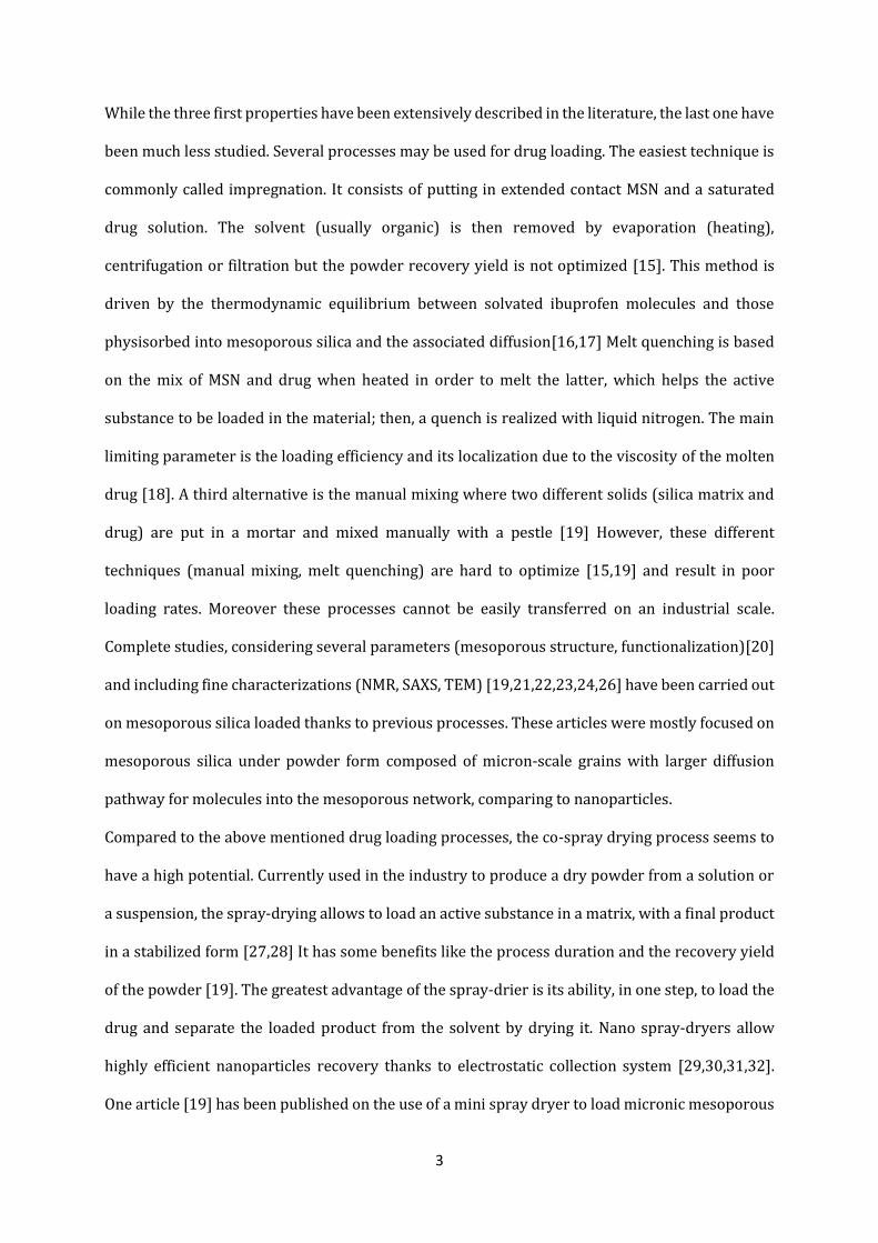
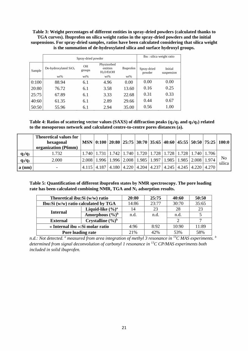
2.3 Drug loading by co-spray-drying: The co-spray-drying process was used to load the
ibuprofen into silica mesopores with the Nano Spray-Dryer B-90 from Büchi Labortechnik AG. The
Figure 1 illustrates the flowsheet of this apparatus [39].
Several experiments were performed varying the Ibuprofen/Silica mass ratio in the stock
suspension (Figure 1-①) in order to analyse the effect of this key parameter on the MSN loading.
The silica concentration stayed constant (5 g.L-1). The volume of ethanol was calculated depending
on the amount of silica, whereas the ibuprofen concentration was the variable parameter (from 1
to 20 g.L-1, see Table 1 and Figure 2-a,b,c). This initial suspension was sonicated before the co-
spray drying experiment during 5 min at 350 W (FB705 Fisherbrand Ultrasonic Processor), and
stirred during all the process in order to facilitate the dissolution of ibuprofen and the dispersion
of MSN. After the pumping of the suspension, droplets are generated with a piezoelectrically
driven vibrating mesh (Figure 1-③); these droplets are formed with a narrow size distribution
controlled by the membrane vibration (spray mesh size of 7 µm) [31,32]. A flow of hot nitrogen
gas (70 °C, 35 mbar, around 95 L.min-1) dries the droplets inside the drying chamber while
evaporating the solvent (Figure 1-② and ④) and generates dried powder composed of particles
6
agglomerates. Then, the powder is collected with an electrostatic collector (Figure 1-⑤) instead
of a cyclone technology as in conventional spray-dryers. A stainless steel cylinder allows to collect
the particles because of a high voltage application between this electrode and a star-shaped
counter electrode (cathode); spray dried powder is then charged and electrostatically deposited
on the inner wall of the cylinder electrode. This mechanism is independent of particle mass (unlike
for the cyclone technology) and is based on particles electrostatic charging.
The short configuration (height: 110 cm) of the drying chamber has been used. Due to the use of
a fully organic solvent (ethanol), the spray dryer was combined with different Büchi accessories
allowing to inert the system (Inert Loop B-295), avoid the presence of water (Dehumidifier B-
296) and recycle the drying gas used with the Büchi Aspirator. A sonication of the initial
suspension was made before the co-spray drying experiment during 5 min at 350 W (FB705
Fisherbrand Ultrasonic Processor), facilitating the dissolution of ibuprofen and the dispersion of
MSN. After the spray drying, the powder was removed from the cylinder electrode through a
particle scrapper.
2.4 Characterization: An important part of this work has been focused on the characterization
of powders generated by spray-drying in order to have a better understanding of the influence of
the mass ratio of ibuprofen over silica (noted R=ibu:Si thereafter) on the properties of the
particles.
Dynamic Light Scattering (DLS): Hydrodynamic diameters of the MSN in suspension were
obtained with a ZetaSizer Nano-ZS (Malvern Instruments Ltd). This equipment uses a laser (He-
Ne at λ=633 nm, under voltage of 3 mV) and the detector is located at 173 ° to analyse the scattered
intensity fluctuations. 10 mg of MSN were dispersed inside 20 mL of water with the ultrasonic
processor [40] (5 min, 350 W) prior to the measurement performed at a temperature of 25 °C.
N2 Adsorption: Nitrogen adsorption/desorption isotherms were performed to characterize the
textural properties of the MSN and the spray-dried samples with a Tristar II (Micromeritics). The
samples were vacuum outgassed at ambient temperature for 24 hours to remove physically
adsorbed water molecules from the pores. Pore size distributions of the MSN were determined

7
from the desorption isotherm with the Barret-Joyner-Hallenda (BJH) method [41] The specific
surface areas and pore volumes were determined from the linear portion of the Brunauer-Emmet-
Teller (BET) plots [42].
Small Angle X-Rays Scattering (SAXS): The SAXS analyses were performed on XEUSS 2.0 (Xenocs
Company) composed of X-ray microsource delivering at 8 keV a spot sized beam equal to 0.5 mm
with an intensity close to 30*106 photons.s-1. The samples were placed on sample loader dedicated
to powders with 387.5 mm of distance from the detector, providing a range of scattering vector
starting from 0.02 Å-1 to 1.6 Å-1. The samples were exposed during 300 s under vacuum and the
scattered beam was collected on the 1M Pilatus detector (1 million counts/pixel). Data integration
and reduction were performed with the software FOXTROT.
Transmission Electron Microscopy (TEM): TEM images were taken by a JEM-1400 electron
microscope (JEOL). The conditions were as follows: W filament, voltage of 120 kV, 3.8 Å of
resolution. Scanning Transmission Electron Microscopy (STEM) tests were performed using a
JEOL cold-FEG JEM-ARM200F operated at 200 kV equipped with a probe Cs corrector reaching a
spatial resolution of 0.078 nm. The detector used is the High-Angle Annular Dark Field (HAADF).
Scanning Electron Microscopy (SEM): SEM has been used to study the morphology of agglomerated
particles. Electron micrographs were taken using the secondary electron mode using a FEI 450
Scanning Electron Microscope (Quanta SEM), with a voltage of 12.5 kV.
X-Ray Diffraction (XRD): Powder X-Ray Diffraction was performed using a Symphonix 1000
(INEL). The X-ray source was a Co radiation (λ=1.7889 Å) and measurement conditions were as
follows: voltage of 30 kV, current of 30 mA, room temperature, step size of 0.01 °. Samples were
manually ground before measurements to randomize the orientation.
Thermogravimetric Analysis/Differential Thermal Analysis (TGA/DTA): TGA/DTA measurements
were performed on a TGA-DTA SETSYS Evolution (SETARAM Instrumentation) under air flow.
The samples were stabilized to 25 °C, then heated to 800 °C with a heating rate of 5 °C/min. The
acquisition system used was SETSYS Ev 1750. TGA has not been carried out for 80:20 sample due
to the lack of spray-dried collected powder.
8
Solid-state Nuclear Magnetic Resonance (NMR): Solid-state NMR experiments were recorded on
an Avance III HD 400 spectrometer (Bruker). Samples were packed into 4 mm zirconia rotors
which were spun at 8 to 10 kHz at 298 K. 1H, 13C and 29Si MAS single pulse experiments were
performed with recycle delays of 3 s, 5 s and 60 s, respectively. 13C-CP/MAS and 29Si-CP/MAS
spectra were recorded with a recycle delay of 2 s and contact times of 2 ms and 3 ms respectively.
13C MAS with Insensitive Nuclei Enhanced by Polarization Transfer (INEPT) were recorded with
a recycle delay of 3s and interpulses delays synchronized with the spinning rate. A mixing time of
4 ms was used for 1H-1H NOESY experiment. All of the chemical shifts are relative to
Tetramethylsilane.
3. RESULTS AND DISCUSSION
3.1 As-synthesized silica nanoparticles
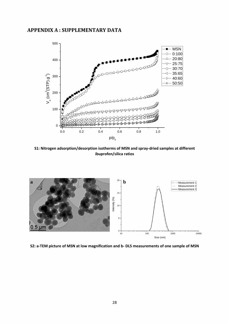
As-synthesized mesoporous silica nanoparticles (MSN) have been analysed by means of several
techniques. N2 adsorption/desorption isotherm (Appendix A Figure S1) is type IV according to
IUPAC43 and nanoparticles exhibit a high BET [42] specific surface area of 806 m².g-1. Pore size
distribution has been calculated with the BJH theory [41] and pores diameter is centered around
2.9 nm (Table 2). These values are similar to those obtained in the literature for this type of system
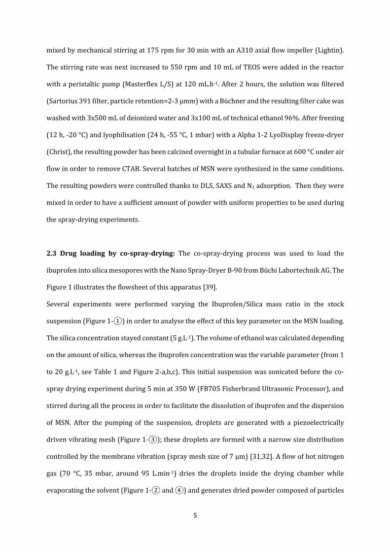
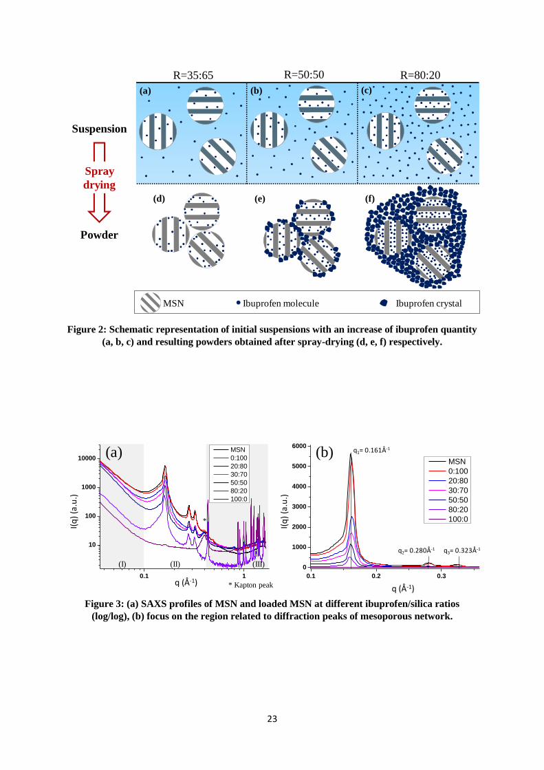
[19,22]. SAXS measurements (Figure 3-b) demonstrate a 2D hexagonal cylindrical network
organization (P6mm) in honeycomb with the indexation of d100, d110 and d200 peaks (respectively
q1, q2 and q3) and allows calculating the centre-to-centre distance, which is about 4.1 nm, in
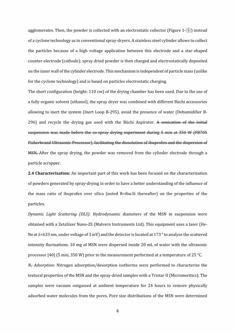
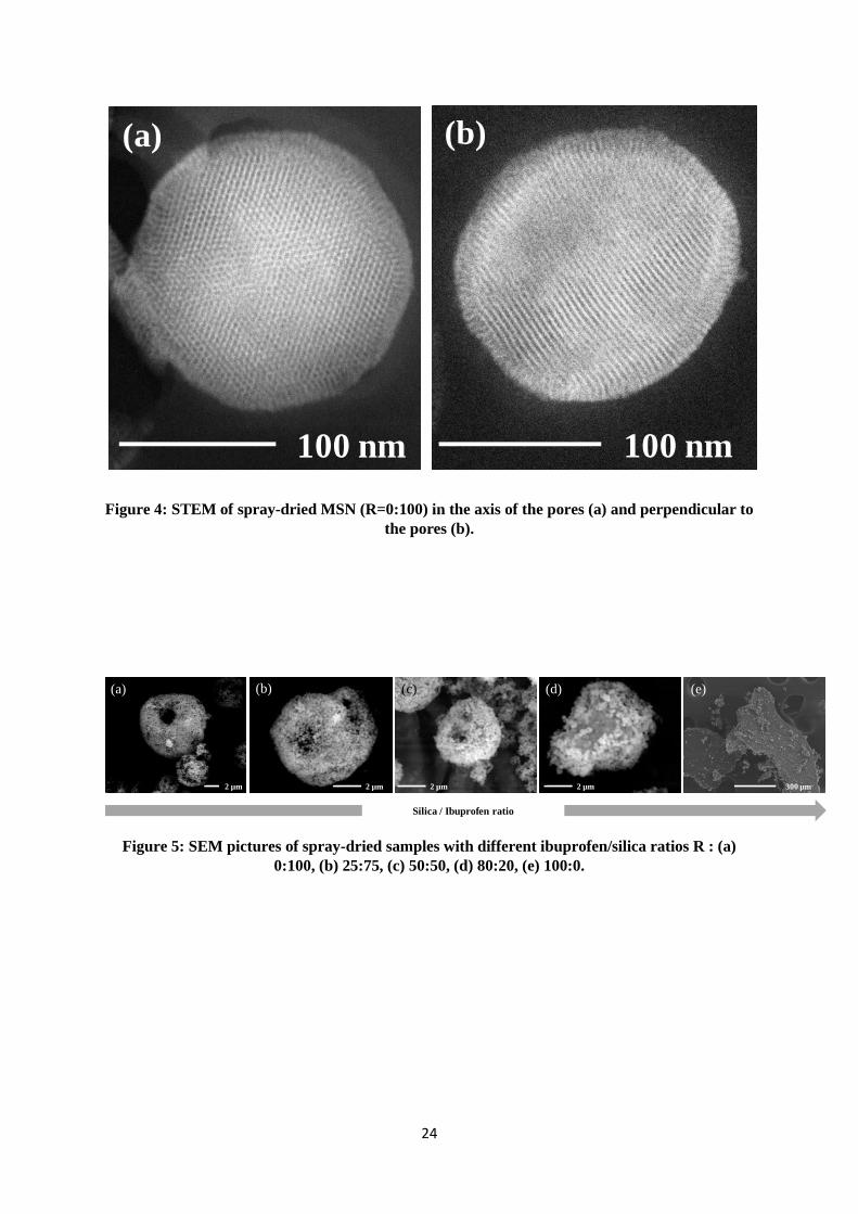
agreement with the literature [44] TEM pictures confirm the MCM-41 derived 2D hexagonal
organization of pores (Figure 4) and highlight the spherical shape of particles. Their diameter
varies from 100 to 250 nm (Appendix, A Figure S2-a). DLS completes this information by giving a
hydrodynamic particle diameter distribution (Appendix A, Figure S2-b) of MSN suspension. Even
if the diameter is overestimated (hydration layer) comparing to TEM, the distribution is
monodisperse and quite narrow (PDI<0.3) with a modal diameter of 301 nm. Although the
diameter of these particles seems to be high considering biomedical applications, they have been

9
chosen for their well-organized porosity. Indeed, smaller nanoparticles lead to disorganized or
worm-like porosity for SBA or MCM-derived silica [45]. Considering that the aim of this article is
the proof of concept of a possible efficient loading, well-defined porous network should be
particularly adapted to characterize the state and organization of molecules into nanoparticles.
3.2 Morphology and composition of resulting agglomerates
After having performed the co-spray drying process with different ibuprofen/silica ratios in the
stock suspension, complementary techniques were used to characterize the hierarchical
organization of resulting dried agglomerates. For the Ibu:Si ratio 100:0 (w/w) (i.e. pure ibuprofen
spray-dried), the quantity used is much higher than other ratios because the yield drastically
dropped, due to the sticky behaviour of the product (Table 1).
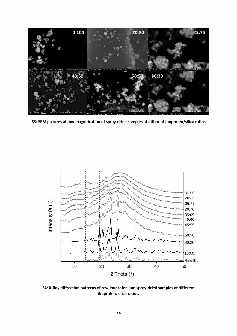
SEM gives information about the spatial localization of silica particles and remaining solid
ibuprofen (if any) into agglomerates. Figure 5 shows high magnification of powder grains for
different ibuprofen/silica ratios (R). For spray-dried pure silica (R=0:100), MSN are spherically
agglomerated and organized with a hollow sphere shape (Figure 5-a). This morphology is typical
of fast solvent evaporation (ethanol) in the drying chamber (Figure 1-④) during the spray-drying
process [46,47,48,49]. When ibuprofen is added in the initial suspension and the ibuprofen
quantity is increased, the previous morphology is preserved for the dried agglomerates formed at
R=25:75 (Figure 5-b) and 50:50 (Figure 5-c), even so the central hole is less well-defined.
Considering low magnification pictures (Appendix A Figure S3), the diameters of agglomerates
are between 1 and 7 µm with a broad size distribution for these three conditions. These results
imply that, for these ratios, ibuprofen molecules are either entrapped inside mesopores or present
as small nanometric entities (undetectable with conventional SEM) between silica nanoparticles.
For higher ratio (R=80:20), agglomerates have irregular shapes (Figure 5-d) with higher particle
sizes between 10 and 40 µm. Interestingly they are mainly composed by a continuous matrix on
which silica particles are agglomerated. Due to the high amount of ibuprofen compared to silica
10
for this sample, we can assume that this matrix is ibuprofen in a solid state. Globally, SEM pictures
suggest that the ibuprofen could get different locations depending on the Ibu:Si ratio.
For pure ibuprofen, the resulting grains are bigger with sizes of several hundred microns with an
important roughness (Figure 5-e). These final grains may have a flat shape due to the surface of
the electrode (flattening of the grain due to the attractive force the electrode).
This could be enhanced by remaining ethanol in ibuprofen during the collection, which make the
powder stickier and more deformable. This phenomenon may have been avoided for samples
containing silica particles, the later increasing the mechanical properties (stiffness) of the
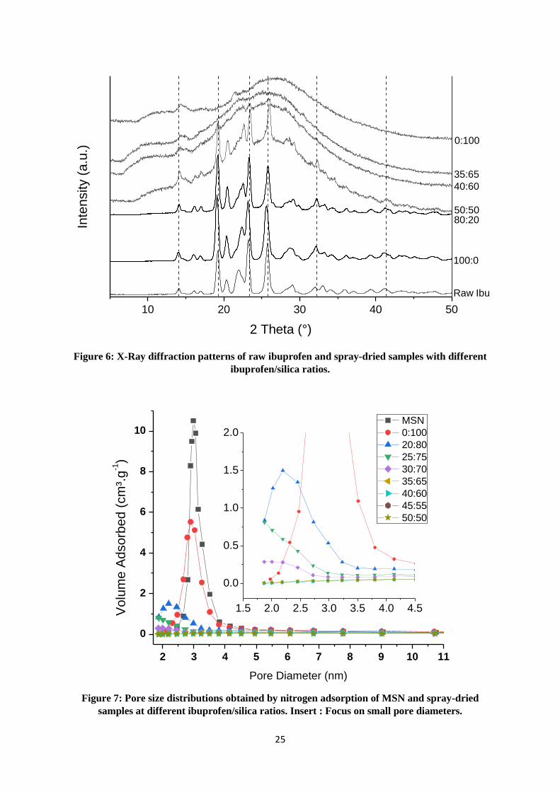
resulting composite. Beyond location of ibuprofen, some selected X-ray diffraction patterns are
presented on Figure 6 (totality of patterns on Appendix A Figure S4) to check its physical state
when dried. For free-ibuprofen sample (R=0:100), a very broad halo centred around 26 ° is
observed. It is characteristic of the short-range order (SiO4 tetrahedra) of amorphous silica. This
broad halo is still present when the ibuprofen amount is increased regardless the sample, except
for the 80:20 ratio. Nevertheless, a series of additional sharp peaks are present from the 40:60 to
the 100:0 ratios. These sharp peaks become more and more intense as the ibuprofen percentage
increases in the stock suspension. These diffraction peaks were attributed to crystalline ibuprofen
(main peaks with following values of 2θ: 14.1, 19.3, 23.4, 25.9 and 32.2°) [19]. It corresponds to
the most thermodynamically stable phase (α) with a P21/c symmetry. These results demonstrate
that below the 40:60 ratio, ibuprofen does not form crystal. It could be correlated with the
previous hypothesis assuming that the molecules are located into mesopores for these ratios. It
has been proved that the crystallization of a molecule can occur in channels only if the channel
diameter/molecular size ratio is more than 20.50 In our case, the pore diameter is about 3 nm
(Table 2, Figure 7) and the ibuprofen molecule is about 11.5 Å long [22]. Thus, the size of
mesopores avoids the formation of ibuprofen crystals inside them. Combining SEM pictures and
XRD, we can assume that beyond the 40:60 ratio, nanocrystals of ibuprofen are formed out of
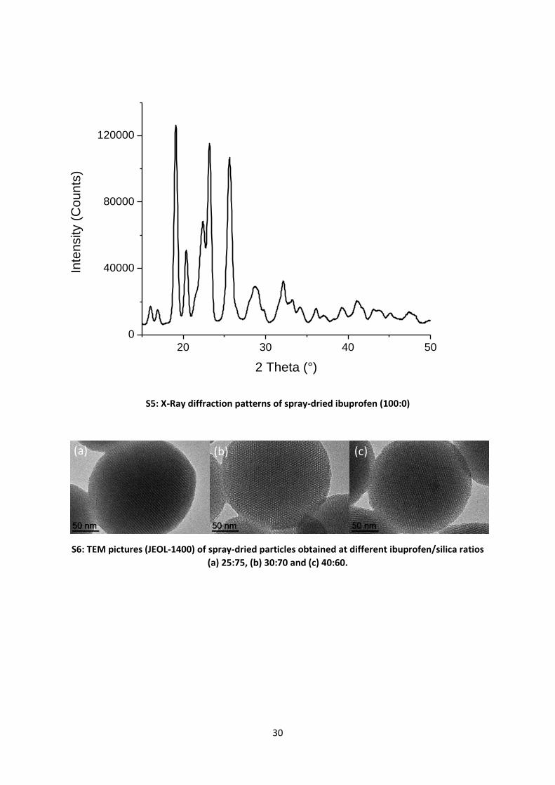
porosity and tends to agglomerate. The hypothesis of the nanoscale of crystals is supported by the

11
width of ibuprofen peaks resulting of small crystalline domains (Appendix A Figure S5). The
crystallite size has been calculated for pure spray-dried ibuprofen thanks to the Scherrer equation
applied to the [200] peak and is around 40 nm. The presence of ibuprofen nanocrystals is
consistent with the spray-drying process. Indeed, the ethanol solvent evaporation is very fast,
promoting nucleation over growth. This trend may have been accentuated for samples containing
silica as each particle could enhance heterogeneous nucleation.
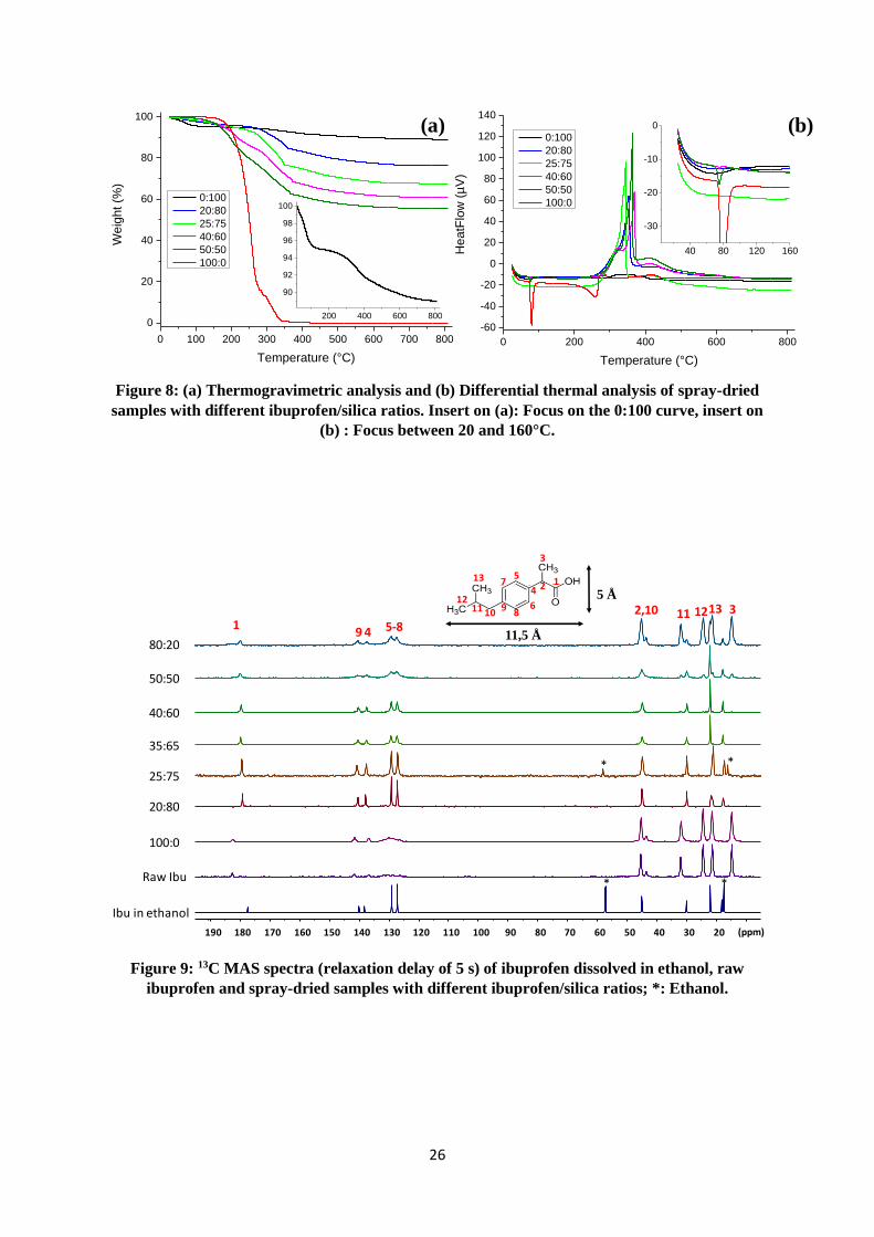
Thermogravimetric Analysis (TGA) has been used to quantify the ibuprofen amount. TGA curves
are presented in Figure 8-a. For pure spray-dried ibuprofen (R=100:0) and considering the
derivative of its curve, two main mass losses occur: the first one between 136 and 280 °C and the
other one between 280 and 345 °C. Three main peaks could be observed on corresponding TGA
results (Figure 8-b): two endothermic peaks, respectively at 80 and 259 °C and an exothermic
peak at 288 °C. According to the literature [51] these peaks can be respectively attributed to the
melting point, the boiling point and the degradation (oxidation) of ibuprofen, meaning that the
first mass loss is due to ibuprofen evaporation and the second one to its decomposition. With the
same methodology, three zones have been determined for pure silica nanoparticles. Below 150
°C, the first zone corresponds to the desorption of physisorbed water or ethanol (endothermic
peak centred around 68 °C). Between 150 °C and 236 °C, a plateau is observed and above 236 °C,
the weight loss is attributable to de-hydroxylation, i.e. the surface silanols condensation [52] For
samples containing both ibuprofen and silica, two domains separated by a clear break can be
distinguished. This break occurs at 355, 344, 370 and 366 °C for respectively 20:80, 25:75, 40:60
and 50:50 samples and is correlated to intense exothermic peaks suggesting that they could be
linked to ibuprofen degradation. It is interesting to note that endothermic peaks related to melting
point of crystalline ibuprofen is only present for samples with initial Ibu:Si ratios above 40:60
(Figure 8-b). These results can be correlated with those of XRD. Indeed, the melting temperature
is only observed when crystals are detected. In any case, several points can be assumed: i) Below
150 °C the mass loss is only due to the removal of physisorbed remaining water or ethanol. ii)
12
Above this value the mass loss is related to ibuprofen removal (evaporation and/or degradation).
iii) At 800 °C the resulting material is de-hydroxylated silica. iv) The amount of silanol groups at
the surface of silica is the same whatever the samples. Indeed, all particles are taken from the
same lot (combination of different synthesis batches) and the conditions in the initial suspension
and during the spray-drying process are not severe enough to remove them (deprotonation,
internal condensation) as demonstrated below by NMR. Considering these hypothesis, it is
possible to calculate the weight percentage of each entity and especially of ibuprofen. Results are
presented in Table 3. Residual water/ethanol amount in the dried samples decreases when
ibuprofen increases. It seems consistent, as the number of ibuprofen molecules interacting with
the silica matrix is increased (assuming that part of ibuprofen is inside the pores). Consequently,
it reduces the probability of silanol interactions with small physisorbed molecules. Moreover, the
ibu:silica weight ratio in spray-dried powders reaches lower values comparing to those in the
stock suspensions. This difference may be explained by the potential loss of isolated nano-
agglomerates of ibuprofen molecules carried away by the gas flow.
3.3 Pore filling and ibuprofen conformation
The honeycomb network of the MSN seems to be unmodified by the presence of ibuprofen (TEM
analysis, Appendix A Figure S6). Although TEM pictures allow directly observing the porous
network, they do not reveal any changes between initial MSN and silica particles co-spray-dried
with ibuprofen. The difficulty in observing it could be explained by the combination of two factors:
i) as ibuprofen is mainly constituted by carbon, the difference in electronic density between
ibuprofen-filled pores and silica network should not exhibit high contrast, ii) at the particle scale,
the superposition of porous channels makes it difficult to distinguish the pore filling.
On the contrary, SAXS reveals important changes when ibuprofen and silica are co-spray-dried.
SAXS curves for MSN, for pure spray-dried silica and for samples with different ratios are
presented on Figure 3-a. Until 0.1 Å-1, the domain (I) reveals that the negative slop of curves is
increased with the increase of ibuprofen ratio. MSN and R=0:100 (spray dried MSN) samples
13
curves have slope close to q-3 whereas that of 80:20 is close to q-4. As these slopes are related to
the surface state in the Porod domain, it could mean that the roughness of silica spheres is step by
step reduced. It is consistent with SEM and XRD analyses. Indeed, between 30:60 and 40:60 ratios,
a layer of agglomerated ibuprofen nanocrystals is progressively formed at the surface of
nanoparticles after this molecule has filled pores. It could explain the “smoothing” of particles
surface due to the homogeneity of this ibuprofen layer with low spatial variations of electronic
density. The domain (II) (approximatively from 0.1 Å-1 to 0.4 Å-1) is the one that is most usually
studied in the field of structured mesoporous materials, as these peaks are due to the diffraction
of x-rays by the empty pores mesostructure due to high contrast of electronic density with silica
matrix. First, these curves confirm that the silica structure is not modified during the co-spray-
drying with or without ibuprofen. As q1, q2 and q3 peaks have the same value for all the samples
and the q2/q1 and q3/q1 values are close to each other (Table 4), we can guess that the P6mm
structure remains the same with similar distance from centre to centre of adjacent cylindrical
pores (about 4.1 nm). However, the peak intensity decreases as the ibuprofen amount increases.
This phenomenon could be explained by the decrease of the electronic density contrast between
silica and ibuprofen (mainly composed of carbon) filling the pores. Thus an intensity decrease
indicates the gradual filling of pores. This kind of contrast decrease has already been described
for micron-scale SBA-15 or MCM-41 particles filled by ibuprofen [20,53,54] or naproxen [26] and
characterized by SAXS or SAXRD. On the contrary, the increase of contrast is associated to the
presence in pores of entities with higher electronic densities like iron oxide [55] or metallic
copper [56]. Finally, domain (III) crosschecks the information obtained by XRD on the structural
organisation of ibuprofen molecules. The filling is still improved for 50:50 and 80:20 ratios,
meaning that the crystallisation of ibuprofen outside of particles revealed by XRD does not occur
because of a lack of free pore surface/volume for molecules.
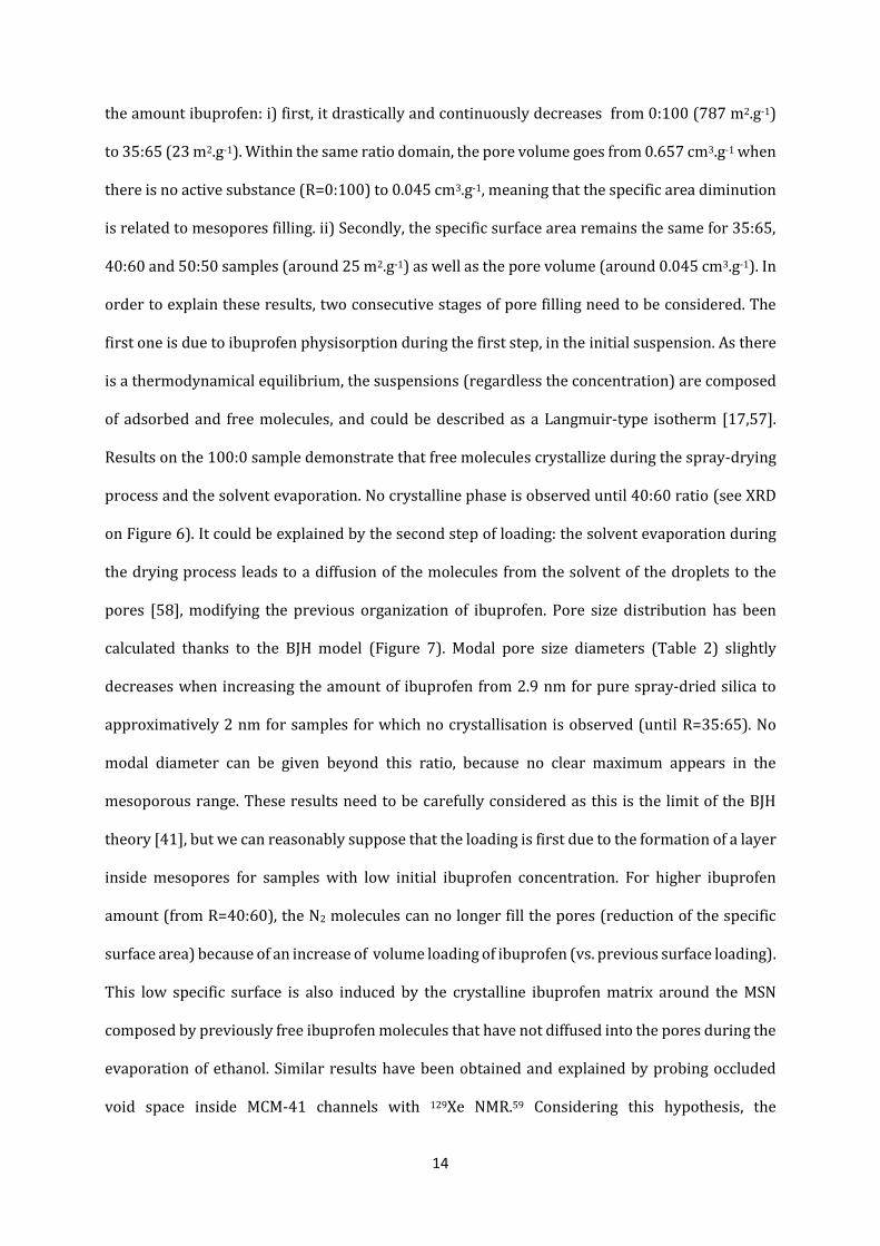
To complete the understanding of pore filling, N2 adsorption measurements have been carried out
(Table 2). BET calculations reveal two tendencies of the specific surface area with an increase in
14
the amount ibuprofen: i) first, it drastically and continuously decreases from 0:100 (787 m2.g-1)
to 35:65 (23 m2.g-1). Within the same ratio domain, the pore volume goes from 0.657 cm3.g-1 when
there is no active substance (R=0:100) to 0.045 cm3.g-1, meaning that the specific area diminution
is related to mesopores filling. ii) Secondly, the specific surface area remains the same for 35:65,
40:60 and 50:50 samples (around 25 m2.g-1) as well as the pore volume (around 0.045 cm3.g-1). In
order to explain these results, two consecutive stages of pore filling need to be considered. The
first one is due to ibuprofen physisorption during the first step, in the initial suspension. As there
is a thermodynamical equilibrium, the suspensions (regardless the concentration) are composed
of adsorbed and free molecules, and could be described as a Langmuir-type isotherm [17,57].
Results on the 100:0 sample demonstrate that free molecules crystallize during the spray-drying
process and the solvent evaporation. No crystalline phase is observed until 40:60 ratio (see XRD
on Figure 6). It could be explained by the second step of loading: the solvent evaporation during
the drying process leads to a diffusion of the molecules from the solvent of the droplets to the
pores [58], modifying the previous organization of ibuprofen. Pore size distribution has been
calculated thanks to the BJH model (Figure 7). Modal pore size diameters (Table 2) slightly
decreases when increasing the amount of ibuprofen from 2.9 nm for pure spray-dried silica to
approximatively 2 nm for samples for which no crystallisation is observed (until R=35:65). No
modal diameter can be given beyond this ratio, because no clear maximum appears in the
mesoporous range. These results need to be carefully considered as this is the limit of the BJH
theory [41], but we can reasonably suppose that the loading is first due to the formation of a layer
inside mesopores for samples with low initial ibuprofen concentration. For higher ibuprofen
amount (from R=40:60), the N2 molecules can no longer fill the pores (reduction of the specific
surface area) because of an increase of volume loading of ibuprofen (vs. previous surface loading).
This low specific surface is also induced by the crystalline ibuprofen matrix around the MSN
composed by previously free ibuprofen molecules that have not diffused into the pores during the
evaporation of ethanol. Similar results have been obtained and explained by probing occluded
void space inside MCM-41 channels with 129Xe NMR.59 Considering this hypothesis, the
15
improvement of filled pores percentage observed by SAXS (decrease of the q1 peak intensity
between R=50:50 and R=80:20) for higher ratios could also be explained even so the specific area
remains the same. Indeed, as the ratio of ibuprofen was higher in the initial suspension, and
considering the equilibrium between free molecules and adsorbed molecules, the amount of
physisorbed molecules should be higher. It could lead to an increased filling rate (Figure 2-d,e,f).
Various NMR experiments have been achieved to gain information on ibuprofen behaviour at the
molecular scale. No clear difference for samples from 20:80 to 100:0 could be observed by 29Si
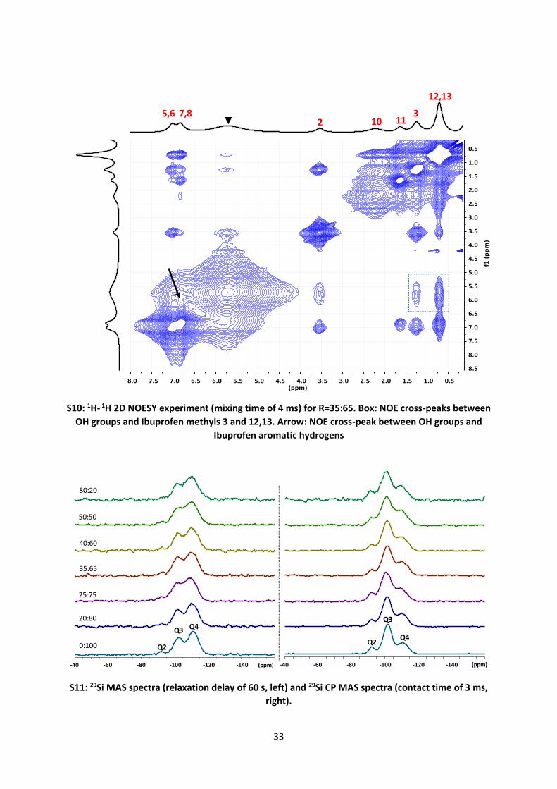
MAS and CP/MAS NMR experiments (Appendix A Figure 7). This confirms that the structure of
mesoporous silica is not modified by the ibuprofen addition. Notably the 29Si CP/MAS
experiments, that enhance the Q2 (Si(OSi)2(OH)2) and the Q3 (Si(OSi)3(OH)) silicon signals
evidence that there is no detectable dehydroxylation of the silica.
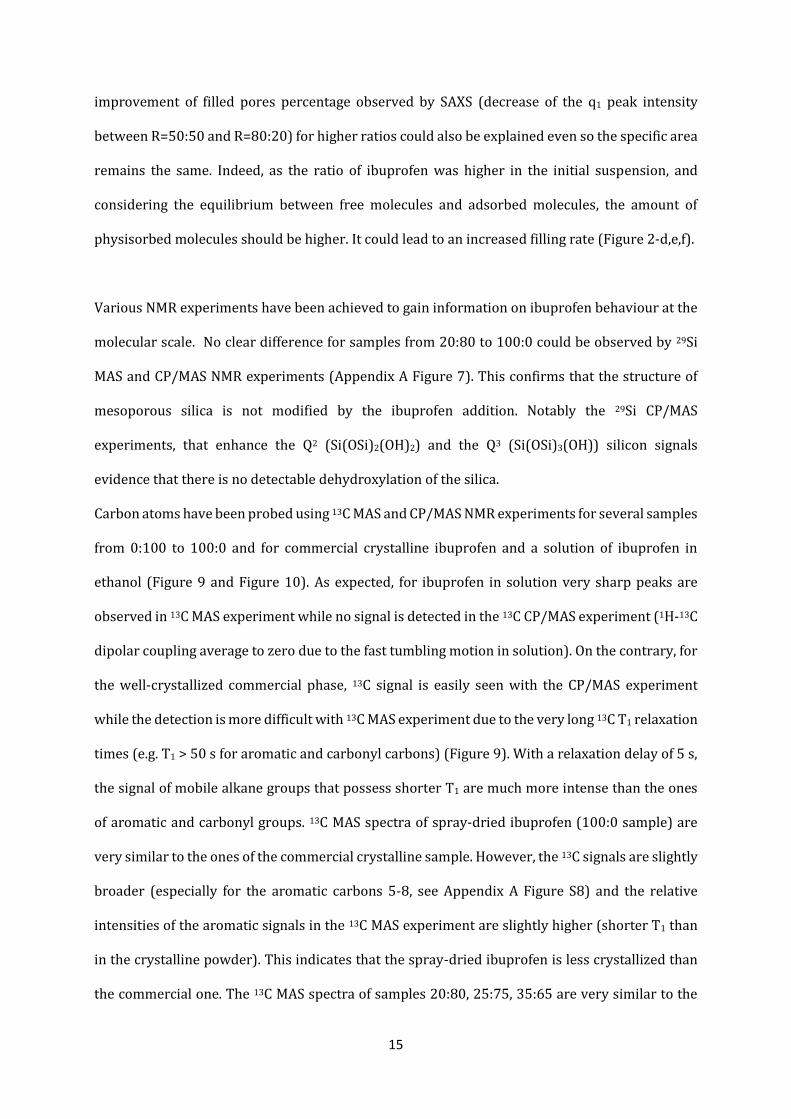
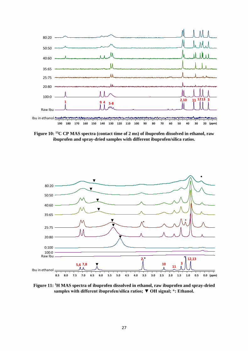
Carbon atoms have been probed using 13C MAS and CP/MAS NMR experiments for several samples
from 0:100 to 100:0 and for commercial crystalline ibuprofen and a solution of ibuprofen in
ethanol (Figure 9 and Figure 10). As expected, for ibuprofen in solution very sharp peaks are
observed in 13C MAS experiment while no signal is detected in the 13C CP/MAS experiment (1H-13C
dipolar coupling average to zero due to the fast tumbling motion in solution). On the contrary, for
the well-crystallized commercial phase, 13C signal is easily seen with the CP/MAS experiment
while the detection is more difficult with 13C MAS experiment due to the very long 13C T1 relaxation
times (e.g. T1 > 50 s for aromatic and carbonyl carbons) (Figure 9). With a relaxation delay of 5 s,
the signal of mobile alkane groups that possess shorter T1 are much more intense than the ones
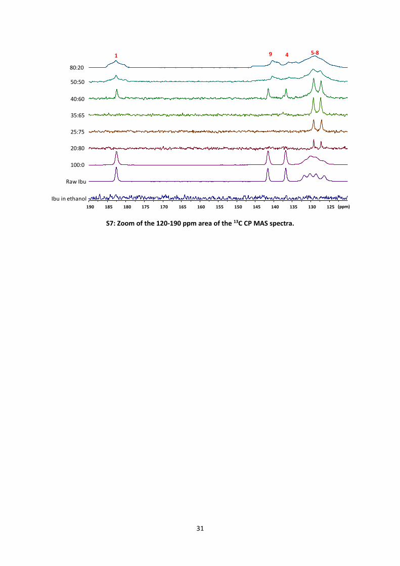
of aromatic and carbonyl groups. 13C MAS spectra of spray-dried ibuprofen (100:0 sample) are
very similar to the ones of the commercial crystalline sample. However, the 13C signals are slightly
broader (especially for the aromatic carbons 5-8, see Appendix A Figure S8) and the relative
intensities of the aromatic signals in the 13C MAS experiment are slightly higher (shorter T1 than
in the crystalline powder). This indicates that the spray-dried ibuprofen is less crystallized than
the commercial one. The 13C MAS spectra of samples 20:80, 25:75, 35:65 are very similar to the
16
dissolved ibuprofen one. This kind of result has been previously described and it is characteristic
of ibuprofen adopting a liquid-like behavior inside the silica mesopores [22]. The most important
change is a shift of the carbonyl signal from 177.4 ppm for ibuprofen in ethanol solution to 179.4
ppm for the 20:80 to 35:65 samples. This shift is probably related to a change in the hydrogen
bond network of the carbonyl group (dimer formation, interaction with ethanol OH groups,..).
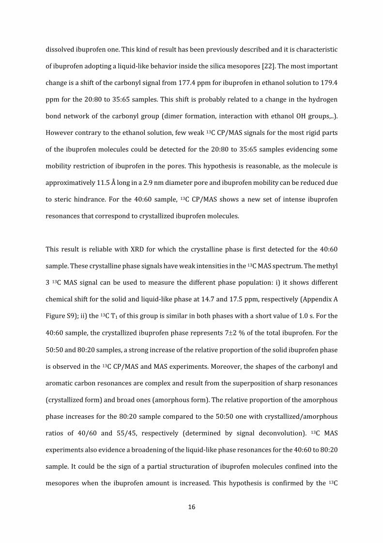
However contrary to the ethanol solution, few weak 13C CP/MAS signals for the most rigid parts
of the ibuprofen molecules could be detected for the 20:80 to 35:65 samples evidencing some
mobility restriction of ibuprofen in the pores. This hypothesis is reasonable, as the molecule is
approximatively 11.5 Å long in a 2.9 nm diameter pore and ibuprofen mobility can be reduced due
to steric hindrance. For the 40:60 sample, 13C CP/MAS shows a new set of intense ibuprofen
resonances that correspond to crystallized ibuprofen molecules.
This result is reliable with XRD for which the crystalline phase is first detected for the 40:60
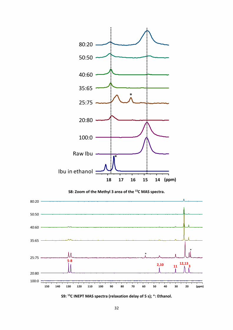
sample. These crystalline phase signals have weak intensities in the 13C MAS spectrum. The methyl
3 13C MAS signal can be used to measure the different phase population: i) it shows different
chemical shift for the solid and liquid-like phase at 14.7 and 17.5 ppm, respectively (Appendix A
Figure S9); ii) the 13C T1 of this group is similar in both phases with a short value of 1.0 s. For the
40:60 sample, the crystallized ibuprofen phase represents 72 % of the total ibuprofen. For the
50:50 and 80:20 samples, a strong increase of the relative proportion of the solid ibuprofen phase
is observed in the 13C CP/MAS and MAS experiments. Moreover, the shapes of the carbonyl and
aromatic carbon resonances are complex and result from the superposition of sharp resonances
(crystallized form) and broad ones (amorphous form). The relative proportion of the amorphous
phase increases for the 80:20 sample compared to the 50:50 one with crystallized/amorphous
ratios of 40/60 and 55/45, respectively (determined by signal deconvolution). 13C MAS
experiments also evidence a broadening of the liquid-like phase resonances for the 40:60 to 80:20
sample. It could be the sign of a partial structuration of ibuprofen molecules confined into the
mesopores when the ibuprofen amount is increased. This hypothesis is confirmed by the 13C

17
INEPT MAS experiments (Appendix A Figure S10) that allow detection of very mobile ibuprofen
molecules. The 13C INEPT signal intensities are strong for 20:80 and 25:75 samples. These signals
then decrease continuously from 35:65 to 80:20 samples indicating a notable diminution of the
mobility of ibuprofen into the mesopores with an increase of ibuprofen amount inside them. It is
consistent with the previous hypothesis (from SAXS and N2 adsorption results, see Figure 2-d,e,f),
in which the amorphous phase results of ibuprofen densification inside the pores. This amorphous
phase is the most dense phase that can be found in the pores, because these don’t reach the
required size to allow ibuprofen crystals formation. As the ibuprofen concentration is increased
in the stock suspension, both solvated and physisorbed ibuprofen amounts are increased due to
the thermodynamic equilibrium. However, this “amorphous condensed phase” has never been
observed for MSN loaded by impregnation. It means that the evaporation process should play a
key role in the densification of ibuprofen into the pores. Then, for ratios higher than 40:60, the
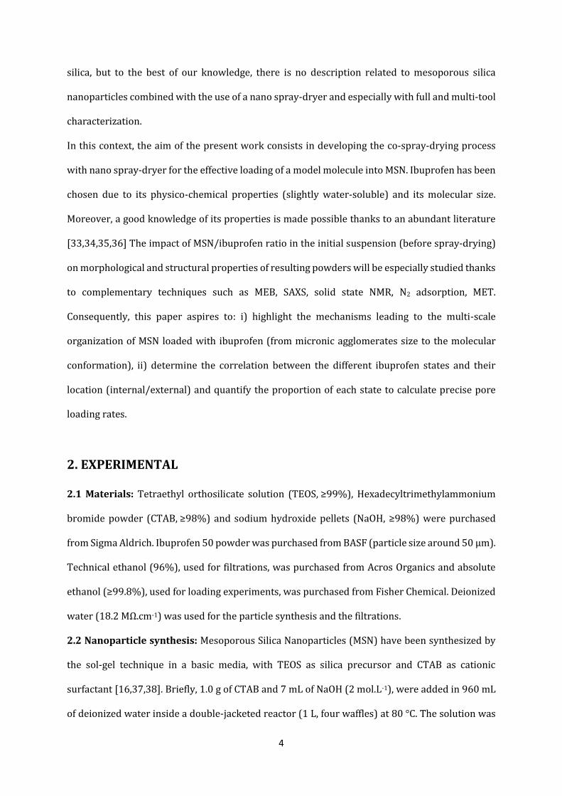
chronological steps of ibuprofen evolution could be the following (Figure 2): i) stock solution with
free and physisorbed (liquid-like) ibuprofen, ii) spray-drying process increasing the amount of
“internal ibuprofen” by densification (solid amorphous phase) due to the diffusion of previously
“free” ibuprofen into the pores induced by the ethanol evaporation, iii) crystallization of
remaining free ibuprofen still during spray-drying. Consequently, for these samples , both
internal (densification from liquid-like to amorphous) and “external” (crystallized) forms are
enhanced. The deconvolution of corresponding solid state NMR peaks allows determining the
proportion of liquid-like, amorphous and crystalline ibuprofen. By combining these results (Table
5) with TGA results (silica/total ibuprofen ratio), it is possible to calculate the ratio (Rexp) between
“internal” ibuprofen (liquid-like + amorphous) and silica. Assuming a total and dense loading of
the porous network, the ibuprofen / silica ratio (Rtheo) can also be approximated : the amount of
ibuprofen is obtained by considering the pore volume of MSN Vp = 0.754 cm3.g-1 and the density
of condensed crystalline ibuprofen i.e. d = 1.076 g.cm-3 [25]. Finally, the pore loading rate is
estimated by the comparison (Rexp/ Rtheo) of these ratios (Table 5). It can be observed that the pore
loading rate quickly increases from R=20:80 to R=40:60. This increase slows down after the
18
apparition of solid ibuprofen phases (amorphous and crystalline), with a pore loading rate barely
to 60% for R=50:50. This good loading rate is close to those described in the literature [16] for
impregnation (approximatively corresponding to 30 wt% of ibuprofen) but in this case with a
continuous, robust and faster process. This loading rate could be improved (and the amount of
crystallized/external ibuprofen decreased) by optimizing some of the parameters of the spray-
drying process such as the drying temperature or the concentration of the initial suspension.
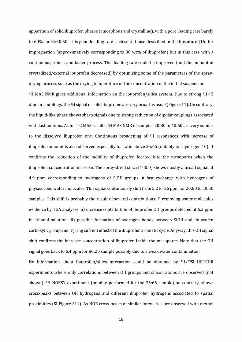
1H MAS NMR gives additional information on the ibuprofen/silica system. Due to strong 1H-1H
dipolar couplings, the 1H signal of solid ibuprofen are very broad as usual (Figure 11). On contrary,
the liquid-like phase shows sharp signals due to strong reduction of dipolar couplings associated
with fast motions. As for 13C MAS results, 1H MAS NMR of samples 20:80 to 40:60 are very similar
to the dissolved ibuprofen one. Continuous broadening of 1H resonances with increase of
ibuprofen amount is also observed especially for ratio above 35:65 (notably for hydrogen 10). It
confirms the reduction of the mobility of ibuprofen located into the mesopores when the
ibuprofen concentration increase. The spray-dried silica (100:0) shows mostly a broad signal at
4.9 ppm corresponding to hydrogens of SiOH groups in fast exchange with hydrogens of
physisorbed water molecules. This signal continuously shift from 5.2 to 6.5 ppm for 20:80 to 50:50
samples. This shift is probably the result of several contributions: i) removing water molecules
evidence by TGA analyses, ii) increase contribution of ibuprofen OH groups detected at 6.2 ppm
in ethanol solution, iii) possible formation of hydrogen bonds between SiOH and ibuprofen
carboxylic group and iv) ring current effect of the ibuprofen aromatic cycle. Anyway, this OH signal
shift confirms the increase concentration of ibuprofen inside the mesopores. Note that the OH
signal goes back to 6.4 ppm for 80:20 sample possibly due to a weak water contamination.
No information about ibuprofen/silica interaction could be obtained by 1H/29Si HETCOR
experiments where only correlations between OH groups and silicon atoms are observed (not
shown). 1H NOESY experiment (notably performed for the 35:65 sample) on contrary, shows
cross-peaks between OH hydrogens and different ibuprofen hydrogens associated to spatial
proximities (SI Figure S11). As NOE cross-peaks of similar intensities are observed with methyl
19
groups on each side of the molecules (methyls 3, 12 and 13) but also with the aromatic hydrogens
(5-8), it can be concluded that there is no preferential orientation of the ibuprofen molecules into
the mesopores. In fact, only the less accessible alkane groups 10 and 11 do not show NOE cross-
peaks with the OH hydrogens. This is in line with the important mobility of ibuprofen in the pores.
Indeed, in the case where strong interaction between ibuprofen and silica may have been present
(through hydrogen bond for example), the ibuprofen mobility should have been weaker and
specific NOE correlations should have been observed.
4. CONCLUSION
An innovative nano-co-spray-drying process has been used for the first time to load mesoporous
silica nanoparticles (diameter of 300 nm/pore size of 2.9 nm) with ibuprofen molecules. Multi-
scale advanced characterization techniques provided key results on the location and the state of
ibuprofen depending on the ibuprofen/silica ratio in the initial suspension:
- Micrometric agglomerates obtained by spray-drying go from spherical hollow spheres at low
ibu:silica weight ratios, to irregular shapes and larger sizes at higher ratios.
- The drug loading of the MSN is the result of two consecutive stages of pore filling: ibuprofen
physisorption in the initial suspension and then diffusion of the ibuprofen molecules into the
pores driven by the solvent evaporation during the spray-drying process.
- Up to 35:65 ibu:silica weight ratio, ibuprofen is encapsulated into the mesopores and adopts a
liquid-like behavior. The amount of encapsulated ibuprofen continuously increases with the initial
ibuprofen concentration.
- At 40:60 ratio, a crystalline ibuprofen phase appears out of the porous network and its amount
increases for higher ratios. A second “solid” phase is detected at the 50:50 ibu:silica ratio and could
be explained by a densification of intraporous ibuprofen.
Beyond mechanistic and fundamental aspects, the identification and quantification of the state of
loaded drugs and the fine calculation of loading rates are of major interest for the industrial
20
transfer of such a process. This work is the first step of the drug loading optimization that will
require an exhaustive study of the effects of other spray-drying parameters.
ACKNOWLEDGMENTS
The authors would like to acknowledge Marianne Clerc-Imperor for helpful discussions about
SAXS results, Gwénaëlle Guittier (LGC) for N2 adsorption measurements and Cédric Charvillat
(CIRIMAT) for XRD and TGA-TDA measurements. They also want to thank Alessandro Pugliara
and Teresa Hungria (Centre de MicroCaractérisation Raimond Castaing UMS 3623) and Stéphanie
Balor (METi) for the TEM analyses. The FERMaT Federation FR3089, Université de Toulouse,
CNRS is acknowledged too for providing Small Angle X-Ray Scattering laboratory facility.
TABLES
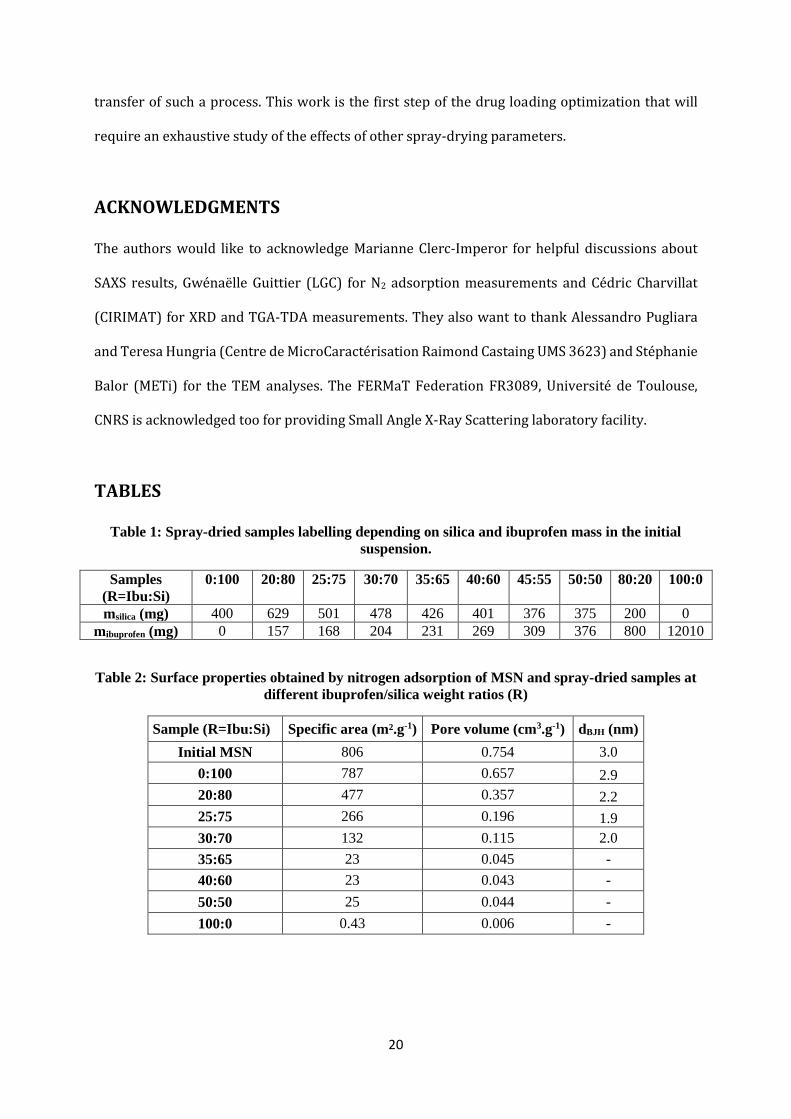
Table 1: Spray-dried samples labelling depending on silica and ibuprofen mass in the initial
suspension.
Samples
(R=Ibu:Si)
0:100 20:80 25:75 30:70 35:65 40:60 45:55 50:50 80:20 100:0
msilica (mg) 400 629 501 478 426 401 376 375 200 0
mibuprofen (mg) 0 157 168 204 231 269 309 376 800 12010
Table 2: Surface properties obtained by nitrogen adsorption of MSN and spray-dried samples at
different ibuprofen/silica weight ratios (R)
Sample (R=Ibu:Si) Specific area (m².g-1) Pore volume (cm3.g-1) dBJH (nm)
Initial MSN 806 0.754 3.0
0:100 787 0.657 2.9
20:80 477 0.357 2.2
25:75 266 0.196 1.9
30:70 132 0.115 2.0
35:65 23 0.045 -
40:60 23 0.043 -
50:50 25 0.044 -
100:0 0.43 0.006 -
21
Table 3: Weight percentages of different entities in spray-dried powders (calculated thanks to
TGA curves). Ibuprofen on silica weight ratios in the spray-dried powders and the initial
suspensions. For spray-dried samples, ratios have been calculated considering that silica weight
is the summation of de-hydroxylated silica and surface hydroxyl groups.
Spray-dried powder Ibu : silica weight ratio
Sample De-hydroxylated SiO2
OH
groups
Physisorbed entities
H2O/EtOH
Ibuprofen Spray-dried
powder
Initial
suspension
wt% wt% wt% wt%
0:100 88.94 6.1 4.96 0.00 0.00 0.00
20:80 76.72 6.1 3.58 13.60 0.16 0.25
25:75 67.89 6.1 3.33 22.68 0.31 0.33
40:60 61.35 6.1 2.89 29.66 0.44 0.67
50:50 55.96 6.1 2.94 35.00 0.56 1.00
Table 4: Ratios of scattering vector values (SAXS) of diffraction peaks (q2/q1 and q3/q1) related
to the mesoporous network and calculated centre-to-centre pores distances (a).
Theoretical values for
hexagonal
organization (P6mm)
MSN 0:100 20:80 25:75 30:70 35:65 40:60 45:55 50:50 75:25 100:0
q2/q1 1.732 1.740 1.731 1.742 1.740 1.720 1.728 1.728 1.728 1.740 1.706 No
silica q3/q1 2.000 2.008 1.996 1.996 2.008 1.985 1.997 1.985 1.985 2.008 1.974
a (nm) - 4.115 4.187 4.180 4.220 4.204 4.237 4.245 4.245 4.220 4.270
Table 5: Quantification of different ibuprofen states by NMR spectroscopy. The pore loading
rate has been calculated combining NMR, TGA and N2 adsorption results.
Theoretical ibu:Si (w/w) ratio 20:80 25:75 40:60 50:50
Ibu:Si (w/w) ratio calculated by TGA 14:86 23:77 30:70 35:65
Internal Liquid-like (%)a 14 23 28 23
Amorphous (%)b n.d. n.d. n.d. 5
External Crystalline (%)b 2 7
« Internal ibu »:Si molar ratio 4:96 8:92 10:90 11:89
Pore loading rate 21% 42% 53% 58%
n.d.: Not detected. a measured from area integration of methyl 3 resonance in 13C MAS experiments. b
determined from signal deconvolution of carbonyl 1 resonance in 13C CP/MAS experiments both
included in solid ibuprofen.
22
FIGURES
Figure 1: Flowsheet of the nano spray-drying process (adapted from 31).
1
InertLoop
Dehumidifier
Aspirator
Tout
Tin
~
DryingChamber
CollectingElectrode
GroundedElectrode
Drying gas
Heater
4
3
2
5
Filter
Gascirculation
23
Figure 2: Schematic representation of initial suspensions with an increase of ibuprofen quantity
(a, b, c) and resulting powders obtained after spray-drying (d, e, f) respectively.
Figure 3: (a) SAXS profiles of MSN and loaded MSN at different ibuprofen/silica ratios
(log/log), (b) focus on the region related to diffraction peaks of mesoporous network.
R=35:65 R=50:50 R=80:20
MSN Ibuprofen molecule Ibuprofen crystal
(a) (b) (c)
(d) (e) (f)
Suspension
Powder
Spray
drying
0.1 1
10
100
1000
10000
MSN
0:100
20:80
30:70
50:50
80:20
100:0
q (Å-1) q (Å-1)
I(q
) (a
.u.)
I(q
) (a
.u.)
q1= 0.161Å-1
q2= 0.280Å-1 q3= 0.323Å-1
(a) (b)
(I) (III)(II)
*
* Kapton peak
0.1 0.2 0.3
0
1000
2000
3000
4000
5000
6000
MSN
0:100
20:80
30:70
50:50
80:20
100:0
24
Figure 4: STEM of spray-dried MSN (R=0:100) in the axis of the pores (a) and perpendicular to
the pores (b).
Figure 5: SEM pictures of spray-dried samples with different ibuprofen/silica ratios R : (a)
0:100, (b) 25:75, (c) 50:50, (d) 80:20, (e) 100:0.
100 nm100 nm
(b)(a)
2 µm 2 µm 2 µm 2 µm
Silica / Ibuprofen ratio
(a) (b) (c) (d)
300 µm
(e)
25
Figure 6: X-Ray diffraction patterns of raw ibuprofen and spray-dried samples with different
ibuprofen/silica ratios.
Figure 7: Pore size distributions obtained by nitrogen adsorption of MSN and spray-dried
samples at different ibuprofen/silica ratios. Insert : Focus on small pore diameters.
10 20 30 40 50
Raw Ibu
100:0
80:2050:50
40:60
35:65
Inte
nsity (
a.u
.)
2 Theta (°)
0:100
2 3 4 5 6 7 8 9 10 11
0
2
4
6
8
10
Vo
lum
e A
dso
rbe
d (
cm
³.g
-1)
Pore Diameter (nm)
MSN
0:100
20:80
25:75
30:70
35:65
40:60
45:55
50:50
1.5 2.0 2.5 3.0 3.5 4.0 4.5
0.0
0.5
1.0
1.5
2.0
26
Figure 8: (a) Thermogravimetric analysis and (b) Differential thermal analysis of spray-dried
samples with different ibuprofen/silica ratios. Insert on (a): Focus on the 0:100 curve, insert on
(b) : Focus between 20 and 160°C.
Figure 9: 13C MAS spectra (relaxation delay of 5 s) of ibuprofen dissolved in ethanol, raw
ibuprofen and spray-dried samples with different ibuprofen/silica ratios; *: Ethanol.
0 100 200 300 400 500 600 700 800
0
20
40
60
80
100W
eig
ht (%
)
Temperature (°C)
0:100
20:80
25:75
40:60
50:50
100:0
200 400 600 800
90
92
94
96
98
100
0 200 400 600 800
-60
-40
-20
0
20
40
60
80
100
120
140
HeatF
low
(µ
V)
Temperature (°C)
0:100
20:80
25:75
40:60
50:50
100:0
40 80 120 160
-30
-20
-10
0(a) (b)
2030405060708090100110120130140150160170180190 (ppm)
Ibu in ethanol
Raw Ibu
100:0
20:80
25:75
35:65
40:60
50:50
80:20
19 4 5-8
2,10
* *
* *
11 1213 3
14
1112
13
3
10
25
6
7
89
11,5 Å
5 Å
27
Figure 10: 13C CP MAS spectra (contact time of 2 ms) of ibuprofen dissolved in ethanol, raw
ibuprofen and spray-dried samples with different ibuprofen/silica ratios.
Figure 11: 1H MAS spectra of ibuprofen dissolved in ethanol, raw ibuprofen and spray-dried
samples with different ibuprofen/silica ratios; ▼ OH signal; *: Ethanol.
2030405060708090100110120130140150160170180190 (ppm)
Ibu in ethanol
Raw Ibu
100:0
20:80
25:75
35:65
40:60
50:50
80:20
1 9 4 5-82,10 11 1213 3
0.00.51.01.52.02.53.03.54.04.55.05.56.06.57.07.58.08.5 (ppm)
Ibu in ethanol
Raw Ibu100:0
20:80
25:75
35:65
40:60
50:50
80:20
5,6
0:100
7,8
2,*
1011
312,13*
*
*
28
APPENDIX A : SUPPLEMENTARY DATA
S1: Nitrogen adsorption/desorption isotherms of MSN and spray-dried samples at different
ibuprofen/silica ratios
S2: a-TEM picture of MSN at low magnification and b- DLS measurements of one sample of MSN
0.0 0.2 0.4 0.6 0.8 1.0
0
100
200
300
400
500
Va (
cm
3(S
TP
).g
-1)
p/p0
MSN
0:100
20:80
25:75
30:70
35:65
40:60
50:50
10 100 1000 10000
0
5
10
15
20
Inte
nsity (
%)
Size (nm)
Measurement 1
Measurement 2
Measurement 3
a b
29
S3: SEM pictures at low magnification of spray-dried samples at different ibuprofen/silica ratios
S4: X-Ray diffraction patterns of raw ibuprofen and spray-dried samples at different
ibuprofen/silica ratios.
0:100
40:60
20:80 25:75
50:50 80:20
10 20 30 40 50
Raw Ibu
25:75
100:0
45:55
80:20
50:50
40:60
35:65
30:70
20:80
Inte
nsity (
a.u
.)
2 Theta (°)
0:100
30
S5: X-Ray diffraction patterns of spray-dried ibuprofen (100:0)
S6: TEM pictures (JEOL-1400) of spray-dried particles obtained at different ibuprofen/silica ratios
(a) 25:75, (b) 30:70 and (c) 40:60.
20 30 40 50
0
40000
80000
120000
Inte
nsity (
Co
un
ts)
2 Theta (°)
(a) (b) (c)
31
S7: Zoom of the 120-190 ppm area of the 13C CP MAS spectra.
125130135140145150155160165170175180185190 (ppm)
Ibu in ethanol
Raw Ibu
100:0
20:80
25:75
35:65
40:60
50:50
80:20
1 9 5-84
32
S8: Zoom of the Methyl 3 area of the 13C MAS spectra.
S9: 13C INEPT MAS spectra (relaxation delay of 5 s); *: Ethanol.
1415161718 (ppm)
Ibu in ethanol
Raw Ibu
100:0
20:80
25:75
35:65
40:60
50:50
80:20
*
*
2030405060708090100110120130140150 (ppm)
100:0
20:80
25:75
35:65
40:60
50:50
80:20
5-82,10 11
12,133
**
33
S10: 1H- 1H 2D NOESY experiment (mixing time of 4 ms) for R=35:65. Box: NOE cross-peaks between
OH groups and Ibuprofen methyls 3 and 12,13. Arrow: NOE cross-peak between OH groups and
Ibuprofen aromatic hydrogens
S11: 29Si MAS spectra (relaxation delay of 60 s, left) and 29Si CP MAS spectra (contact time of 3 ms,
right).
5,6 7,82 10 11
3
12,13
-140-120-100-80-60-40 (ppm)
Q4Q3
Q2
20:80
25:75
35:65
40:60
50:50
80:20
0:100
-140-120-100-80-60-40 (ppm)
Q4
Q3
Q2
34
REFERENCES
[1] T. Lian, J.Y.H Rodney, Trends and Developments in Liposome Drug Delivery Systems. J.
Pharm. Sci. 90 (2001) 667–680, https://doi.org/10.1002/jps.1023
[2] Z. Li, J.C. Barnes, A. Bosoy, J.F. Stoddart, J. Zink, Mesoporous Silica Nanoparticles in
Biomedical Applications. Chem. Soc. Rev, 41 (2012) 2590-2596,
https://doi.org/10.1039/c1cs15246g.
[3] Y. Wang, Q. Zhao, N. Han, L.Bai, J.Li, J.Liu, E.Che, L. Hu, Q. Zhang, T. Jiang, S. Wang,
Mesoporous Silica Nanoparticles in Drug Delivery and Biomedical Applications.
Nanomedicine Nanotechnol. Biol. Med, 11 (2015) 313–327,
https://doi.org/10.1016/j.nano.2014.09.014.
[4] J.L. Paris, M.V. Cabañas, M. Manzano, M. Vallet-Regí, Polymer-Grafted Mesoporous Silica
Nanoparticles as Ultrasound-Responsive Drug Carriers. ACS Nano, 11 (2015), 11023-
11033, https://doi.org/10.1021/acsnano.5b04378.
[5] M. Vallet-Regí, M. Colilla, I. Izquierdo-Barba, M. Manzano, Mesoporous Silica Nanoparticles
for Drug Delivery: Current Insights. Molecules, 23 (2017) 47-53,
https://doi.org/10.3390/molecules23010047.
[6] I.I, Slowing, J.L. Vivero-Escoto, C.W. Wu, V.S.Y. Lin, Mesoporous Silica Nanoparticles as
Controlled Release Drug Delivery and Gene Transfection Carriers. Adv. Drug Deliv. Rev. 60
(2008), 1278-1288, https://doi.org/10.1016/j.addr.2008.03.012.
[7] Y. Zhang, Z. Zhi, T. Jiang, J. Zhang, Z. Wang, S. Wang, Spherical Mesoporous Silica
Nanoparticles for Loading and Release of the Poorly Water-Soluble Drug Telmisartan. J.
Controlled Release, 145 (2010) 257–263, https://doi.org/10.1016/j.jconrel.2010.04.029.
[8] L. Jia, J. Shen, Z. Li, D. Zhang, Q. Zhang, C. Duan, G. Liu, D. Zheng, Y. Liu, X. Tian, Successfully
Tailoring the Pore Size of Mesoporous Silica Nanoparticles: Exploitation of Delivery
Systems for Poorly Water-Soluble Drugs, Int. J. Pharm., 439 (2012), 81–91,
https://doi.org/10.1016/j.ijpharm.2012.10.011.
35
[9] I.I. Slowing, J.L. Vivero-Escoto, B.G. Trewyn, V.S.Y. Lin, Mesoporous Silica Nanoparticles:
Structural Design and Applications. J. Mater. Chem., 37 (2010) 7924-7932,
https://doi.org/10.1039/c0jm00554a.
[10] W. Gao, J.M. Chan, O. Farokhzad, PH-Responsive Nanoparticles for Drug Delivery. Mol.
Pharm. 7 (2010) 1913–1920, https://doi.org/10.1021/mp100253e.
[11] R.R. Castillo, M. Vallet-Regí, Functional Mesoporous Silica Nanocomposites: Biomedical
Applications and Biosafety, Int. J. Mol. Sci., 20 (2019) 929-936,
https://doi.org/10.3390/ijms20040929.
[12] W.D. Bossaert, D.E. De Vos, W.M. Van Rhijn, J. Bullen, P.J. Grobet, P.A. Jacobs, Mesoporous
Sulfonic Acids as Selective Heterogeneous Catalysts for the Synthesis of Monoglycerides, J.
Catal., 182 (1999) 156–164, https://doi.org/10.1006/jcat.1998.2353
[13] M. Vallet-Regí, Ordered Mesoporous Materials in the Context of Drug Delivery Systems and
Bone Tissue Engineering, Chem. Eur. J., 12 (2006) 5934–5943,
https://doi.org/10.1002/chem.200600226.
[14] V. Mamaeva, J.M. Rosenholm, L.T. Bate-Eya, L. Bergman, E. Peuhu, A. Duchanoy, L.E.
Fortelius, S. Landor, D. Toivola, L. Lindén, C. Sahlgren, Mesoporous Silica Nanoparticles as
Drug Delivery Systems for Targeted Inhibition of Notch Signaling in Cancer, Mol. Ther., 19
(2011) 1538–1546, https://doi.org/10.1038/mt.2011.105.
[15] T. Heikkilä, J. Salonen, J. Tuura, N. Kumar, T. Salmi, D.Y. Murzin, M.S. Hamdy, G. Mul, L.
Laitinen, A.M. Kaukonen, J. Hirvonen, V.P Lehto, Evaluation of Mesoporous TCPSi, MCM-41,
SBA-15, and TUD-1 Materials as API Carriers for Oral Drug Delivery, Drug Deliv. 14 (2007)
337–347, https://doi.org/10.1080/10717540601098823.
[16] M. Vallet-Regi, A. Rámila, R.P. del Real, J.A. Pérez-Pariente, New Property of MCM-41: Drug
Delivery System, Chem. Mater., 13 (2001) 308–311, https://doi.org/10.1021/cm0011559.
[17] T. Numpilai, S. Muenmee, T. Witoon, Impact of Pore Characteristics of Silica Materials on
Loading Capacity and Release Behavior of Ibuprofen, Mater. Sci. Eng. C, 59 (2016) 43–52,
https://doi.org/10.1016/j.msec.2015.09.095.
36
[18] R. Mellaerts, J.A. Jammaer, M. Van Speybroeck, H. Chen, J. Van Humbeeck, P. Augustijns, G.
Van den Mooter, J.A. Martens, Physical State of Poorly Water Soluble Therapeutic Molecules
Loaded into SBA-15 Ordered Mesoporous Silica Carriers: A Case Study with Itraconazole
and Ibuprofen, Langmuir 24 (2008) 24 8651–8659, https://doi.org/10.1021/la801161g.
[19] S. Shen, W.K. Ng, L. Chia, Y. Dong, R.B.H. Tan, Stabilized Amorphous State of Ibuprofen by
Co‐Spray Drying With Mesoporous SBA‐15 to Enhance Dissolution Properties, J. Pharm.
Sci., 99 (2010) 1997–2007, https://doi.org/10.1002/jps.21967.
[20] I. Izquierdo-Barba, E. Sousa, J.C. Doadrio, A.L. Doadrio, J.P. Pariente, A. Martínez, F.
Babonneau, M. Vallet-Regí, Influence of Mesoporous Structure Type on the Controlled
Delivery of Drugs: Release of Ibuprofen from MCM-48, SBA-15 and Functionalized SBA-15,
J. Sol-Gel Sci. Technol., 50 (2009), 421–429, https://doi.org/10.1007/s10971-009-1932-3.
[21] F. Babonneau, L. Yeung, N. Steunou, C. Gervais, A. Ramila, M. Vallet-Regi, Solid State NMR
Characterisation of Encapsulated Molecules in Mesoporous Silica, J. Sol-Gel Sci. Technol.,
31 (2004), 219–223, https://doi.org/10.1023/B:JSST.0000047991.73840.8b
[22] T. Azaïs, C. Tourné-Péteilh, F. Aussenac, N. Baccile, C. Coelho, J.M. Devoisselle, F. Babonneau,
Solid-State NMR Study of Ibuprofen Confined in MCM-41 Material, Chem. Mater., 18 (2006)
6382–6390, https://doi.org/10.1021/cm061551c
[23] X. Du, J. He, Regulation role of ibuprofen toward the morphology of porous silica
nanospheres during its in situ encapsulation, J. Coll. Int. Sci, 345 (2010), 269-277.
https://doi.org/10.1016/j.jcis.2010.02.012
[24] Brás, A. R.; Merino, E. G.; Neves, P. D.; Fonseca, I. M.; Dionísio, M.; Schönhals, A.; Correia, N.
T. Amorphous Ibuprofen Confined in Nanostructured Silica Materials: A Dynamical
Approach. J. Phys. Chem. C, 115 (2011), 4616–4623. https://doi.org/10.1021/jp107631m
[25] S.C. Shen, W.K. Ng, L. Chia, J. Hu, R.B.H. Tan, Physical State and Dissolution of Ibuprofen
Formulated by Co-Spray Drying with Mesoporous Silica: Effect of Pore and Particle Size,
Int. J. Pharm., 410 (2011), 188–195. https://doi.org/10.1016/j.ijpharm.2011.03.018.
37
[26] X. Li, X. Du, J. He, Self-Cleaning Antireflective Coatings Assembled from Peculiar
Mesoporous Silica Nanoparticles, Langmuir, 26 (2010) 135258-13534
https://doi.org/10.1021/la1016824
[27] M. Vogt, K. Kunath, J.B. Dressman, Dissolution Enhancement of Fenofibrate by
Micronization, Cogrinding and Spray-Drying: Comparison with Commercial Preparations,
Eur. J. Pharm. Biopharm., 68 (2008) 283–288, https://doi.org/10.1016/j.ejpb.2007.05.010.
[28] M. Fatnassi, C. Tourné-Péteilh, T. Mineva, J.M. Devoisselle, P. Gaveau, F. Fayon, B. Alonso,
Drug Nano-Domains in Spray-Dried Ibuprofen–Silica Microspheres, Phys. Chem. Chem.
Phys., 14 (2012) 12285-12296, https://doi.org/10.1039/c2cp42092a.
[29] X. Li, N. Anton, C. Arpagaus, F. Belleteix, T. Vandamme, Nanoparticles by Spray Drying Using
Innovative New Technology: The Büchi Nano Spray Dryer B-90, J. Controlled Release, 147
(2010) 304–310, https://doi.org/10.1016/j.jconrel.2010.07.113.
[30] K. Bürki, I. Jeon, C. Arpagaus, G. Betz, New Insights into Respirable Protein Powder
Preparation Using a Nano Spray Dryer, Int. J. Pharm., 408 (2011) 248–256,
https://doi.org/10.1016/j.ijpharm.2011.02.012.
[31] C. Arpagaus, Novel Laboratory-Scale Spray Dryer to Produce Nanoparticles. Dry. Technol.,
30 (2012) 1113–1121, https://doi.org/10.1080/07373937.2012.686949.
[32] K. Schmid, C. rpagaus, W. Friess, Evaluation of the Nano Spray Dryer B-90 for
Pharmaceutical Applications, Pharm. Dev. Technol., 16 (2011) 287–294,
https://doi.org/10.3109/10837450.2010.485320.
[33] B. Muñoz, A. Rámila, J. Pérez-Pariente, I. Díaz, M. Vallet-Regí, MCM-41 Organic Modification
as Drug Delivery Rate Regulator, Chem. Mater., 15 (2003), 500–503,
https://doi.org/10.1021/cm021217q.
[34] S.W. Song, K. Hidajat, S. Kawi, Functionalized SBA-15 Materials as Carriers for Controlled
Drug Delivery: Influence of Surface Properties on Matrix−Drug Interactions, Langmuir 21
(2005), 9568–9575, https://doi.org/10.1021/la051167e.
38
[35] C. Tourné-Péteilh, D. Brunel, S. Bégu, B. Chiche, F. Fajula, D.A. Lerner, J.M. Devoisselle,
Synthesis and Characterisation of Ibuprofen-Anchored MCM-41 Silica and Silica Gel, New
J. Chem., 27 (2003), 1415–1418, https://doi.org/ 10.1039/B307046H
[36] P. Yang, Z. Quan, L. Lu, S. Huang, J. Lin, H. Fu, MCM-41 Functionalized with YVO4:Eu3+: A
Novel Drug Delivery System, Nanotechnology, 18 (2007) 235703-235708,
https://doi.org/10.1088/0957-4484/18/23/235703.
[37] I.I. Slowing, B.G. Trewyn, V.S.Y. Lin, Effect of Surface Functionalization of MCM-41-Type
Mesoporous Silica Nanoparticles on the Endocytosis by Human Cancer Cells, J. Am. Chem.
Soc., 46 (2006) 14792–14793, https://doi.org/10.1021/ja0645943.
[38] R. Narayan, U. Nayak, A. Raichur, S. Garg, Mesoporous Silica Nanoparticles: A
Comprehensive Review on Synthesis and Recent Advances. Pharmaceutics, 10 (2018), 118-
125, https://doi.org/10.3390/pharmaceutics10030118.
[39] C. Arpagaus, Nano Spray Dryer B-90: Literature Review and Applications, Dry. Technol. 30
(2011) 1113–1121.
[40] R.C Murdock, L. Braydich-Stolle, A.M. Schrand, J.J. Schlager, S. Hussain, Characterization of
Nanomaterial Dispersion in Solution Prior to In Vitro Exposure Using Dynamic Light
Scattering Technique, Toxicol. Sci., 101 (2008) 239–253,
https://doi.org/10.1093/toxsci/kfm240.
[41] E.P. Barrett, L.G. Joyner, P.P. Halenda, The Determination of Pore Volume and Area
Distributions in Porous Substances. I. Computations from Nitrogen Isotherms, J. Am. Chem.
Soc., 73 (1951) 373–380.
[42] S. Brunauer, P.H. Emmett, E. Teller, Adsorption of Gases in Multimolecular Layers, J. Am.
Chem. Soc., 60 (1938) 309–319.
[43] K.S.W. Sing, D.H. Everett, R.A.W. Haul, L. Moscou, R.A. Pierotti, J. Rouquerol, Siemieniewska,
Reporting Physisorption Data for Gas/Solid Systems, Pure Appl. Chem., 57 (1985) 603-619.
39
[44] A. Abd-Elbary, M.A. El Nabarawi, D.H. Hassen, A.A. Taha, Inclusion and Characterization of
Ketoprofen into Different Mesoporous Silica Nanoparticles Using Three Loading Methods,
Int. J. Pharm. Pharm. Sci., 6 (2014) 183–191.
[45] K. Möller, J. Kobler, T. Bein, Colloidal Suspensions of Nanometer-Sized Mesoporous Silica.
Adv. Funct. Mater., 17 (2007) 605–612, https://doi.org/10.1002/adfm.200600578.
[46] A.B.D. Nandiyanto, K. Okuyama, Progress in Developing Spray-Drying Methods for the
Production of Controlled Morphology Particles: From the Nanometer to Submicrometer
Size Ranges, Adv. Powder Technol, 22 (2011) 1–19,
https://doi.org/10.1016/j.apt.2010.09.011.
[47] Y. Wang, K. Kho, W.S. Cheow, K.A. Hadinoto, Comparison between Spray Drying and Spray
Freeze Drying for Dry Powder Inhaler Formulation of Drug-Loaded Lipid–Polymer Hybrid
Nanoparticles, Int. J. Pharm., 424 (2012) 98–106,
https://doi.org/10.1016/j.ijpharm.2011.12.045.
[48] M. Faustini, M. Giraud, D. Jones, J. Rozière, M. Dupont, T.R. Porter, S. Nowak, M. Bahri, O.
Ersen, O.; C. Sanchez, C. Boissiere, C. Tard, J. Peron, Hierarchically Structured Ultraporous
Iridium-Based Materials: A Novel Catalyst Architecture for Proton Exchange Membrane
Water Electrolyzers, Adv. Energy Mater., 9 (2019) 1802136-1802147,
https://doi.org/10.1002/aenm.201802136.
[49] R. Pérez-Masiá, R. López-Nicolás, M.J Periago, G. Ros, J.M. Lagaron, A. López-Rubio,
Encapsulation of Folic Acid in Food Hydrocolloids through Nanospray Drying and
Electrospraying for Nutraceutical Applications, Food Chem., 168 (2015) 124–133,
https://doi.org/10.1016/j.foodchem.2014.07.051.
[50] M. Sliwinska-Bartkowiak, G. Dudziak, R. Gras, R. Sikorski, R. Radhakrishnan, K. Gubbins,
Freezing Behavior in Porous Glasses and MCM-41, Colloids Surf. Physicochem. Eng. Asp.,
187 (2001) 523–529.
[51] S. Ramukutty, E. Ramachandran, Growth, Spectral and Thermal Studies of Ibuprofen
Crystals, Cryst. Res. Technol., 47 (2012) 31–38, https://doi.org/10.1002/crat.201100394.
40
[52] J. Trébosc, J.W. Wiench, S. Huh, V.S.Y. Lin, M. Pruski, Solid-State NMR Study of MCM-41-Type
Mesoporous Silica Nanoparticles, J. Am. Chem. Soc., 127 (2005) 3057–3068.
[53] C. Charnay, S. Bégu, C. Tourné-Péteilh, L. Nicole, D.A. Lerner, J.M. Devoisselle, Inclusion of
Ibuprofen in Mesoporous Templated Silica: Drug Loading and Release Property, Eur. J.
Pharm. Biopharm., 57 (2004) 533–540, https://doi.org/10.1021/ja043567e.
[54] L. Gao, J. Sun, L. Zhang, J. Wang, B. Ren, Influence of Different Structured Channels of
Mesoporous Silicate on the Controlled Ibuprofen Delivery, Mater. Chem. Phys., 135 (2012)
786–797, https://doi.org/10.1016/j.matchemphys.2012.05.059.
[55] J.G. Li, G. Fornasieri, A. Bleuzen, M. Gich, A. Gloter, F. Bouquet, M. Impéror-Clerc, Alignment
under Magnetic Field of Mixed Fe2O 3 /SiO2 Colloidal Mesoporous Particles Induced by
Shape Anisotropy, Small, 12 (2016) 5981–5988,
https://doi.org/10.1002/smll.201602272.
[56] C.J. Gommes, G. Prieto, P.E. De Jongh, Small-Angle Scattering Analysis of Empty or Loaded
Hierarchical Porous Materials, J. Phys. Chem. C 120 (2016) 1488–1506,
https://doi.org/10.1021/acs.jpcc.5b09556.
[57] J. Andersson, J. Rosenholm, S. Areva, M. Lindén, Influences of Material Characteristics on
Ibuprofen Drug Loading and Release Profiles from Ordered Micro- and Mesoporous Silica
Matrices. Chem. Mater., 16 (2004), 4160–4167, https://doi.org/10.1021/cm0401490.
[58] F. Wan, A. Bohr, M.J. Maltesen, S. Bjerregaard, C. Foged, J. Rantanen, M. Yang, Critical Solvent
Properties Affecting the Particle Formation Process and Characteristics of Celecoxib-
Loaded PLGA Microparticles via Spray-Drying, Pharm. Res., 30 (2013), 1065–1076,
https://doi.org/10.1007/s11095-012-0943-x.
[59] F. Guenneau, K. Panesar, A. Nossov, A.; M.A. Springuel-Huet, T. Azaïs, F. Babonneau, C.
Tourné-Péteilh, J.M. Devoisselle, A. Gédéon, Probing the Mobility of Ibuprofen Confined in
MCM-41 Materials Using MAS-PFG NMR and Hyperpolarised-129Xe NMR Spectroscopy,
Phys. Chem. Chem. Phys. 15 (2013) 18805-18811, https://doi.org/10.1039/c3cp52695j.