Terrace gets rave rev ews from Japanese film crew Shames ...
Upload
khangminh22Category
view
0download
0
E O R G I A N EDICAL
EWSNo 1 (274) 2018ISSN 1512-0112

GEORGIAN MEDICAL
NEWSNo 1 (274) 2018
ЕЖЕМЕСЯЧНЫЙ НАУЧНЫЙ ЖУРНАЛ
ТБИЛИСИ - НЬЮ-ЙОРК
Published in cooperation with and under the patronage of the Tbilisi State Medical University
Издается в сотрудничестве и под патронажем Тбилисского государственного медицинского университета
gamoicema Tbilisis saxelmwifo samedicino universitetTan TanamSromlobiTa da misi patrona;iT

GMN: Georgian Medical News is peer-reviewed, published monthly journal committed to promoting
the science and art of medicine and the betterment of public health, published by the GMN Editorial
Board and The International Academy of Sciences, Education, Industry and Arts (U.S.A.) since
1994. GMN carries original scientific articles on medicine, biology and pharmacy, which are of
experimental, theoretical and practical character; publishes original research, reviews, commentaries,
editorials, essays, medical news, and correspondence in English and Russian.
GMN is indexed in MEDLINE, SCOPUS, PubMed and VINITI Russian Academy of Sciences. The
full text content is available through EBSCO databases.
GMN: Медицинские новости Грузии - ежемесячный рецензируе мый научный журнал,
издаётся Редакционной коллегией и Международной академией наук, образования, искусств и естествознания (IASEIA) США с 1994 года на русском и английском языках в целях поддержки
медицинской науки и улучшения здравоохранения. В журнале публикуются оригинальные
научные статьи в области медицины, биологии и фармации, статьи обзорного характера,
научные сообщения, новости медицины и здравоохранения.
Журнал индексируется в MEDLINE, отражён в базе данных SCOPUS, PubMed и ВИНИТИ РАН.
Полнотекстовые статьи журнала доступны через БД EBSCO.
GMN: Georgian Medical News – saqarTvelos samedicino siaxleni – aris yovelTviuri
samecniero samedicino recenzirebadi Jurnali, gamoicema 1994 wlidan, warmoadgens
saredaqcio kolegiisa da aSS-is mecnierebis, ganaTlebis, industriis, xelovnebisa
da bunebismetyvelebis saerTaSoriso akademiis erTobliv gamocemas. GMN-Si rusul
da inglisur enebze qveyndeba eqsperimentuli, Teoriuli da praqtikuli xasiaTis
originaluri samecniero statiebi medicinis, biologiisa da farmaciis sferoSi,
mimoxilviTi xasiaTis statiebi.
Jurnali indeqsirebulia MEDLINE-is saerTaSoriso sistemaSi , asaxulia
SCOPUS-is, PubMed-is da ВИНИТИ РАН-is monacemTa bazebSi. statiebis sruli teqsti
xelmisawvdomia EBSCO-s monacemTa bazebidan.

МЕДИЦИНСКИЕ НОВОСТИ ГРУЗИИ
Ежемесячный совместный грузино-американский научный электронно-печатный журнал Агентства медицинской информации Ассоциации деловой прессы Грузии,
Академии медицинских наук Грузии, Международной академии наук, индустрии, образования и искусств США.
Издается с 1994 г., распространяется в СНГ, ЕС и США
НАУЧНЫЙ РЕДАКТОР
Лаури Манагадзе
ГЛАВНЫЙ РЕДАКТОР
Нино Микаберидзе
НАУЧНО-РЕДАКЦИОННЫЙ СОВЕТЗураб Вадачкориа - председатель Научно-редакционного совета
Михаил Бахмутский (США), Александр Геннинг (Германия), Амиран Гамкрелидзе (Грузия), Константин Кипиани (Грузия), Георгий Кавтарадзе (Грузия), Георгий Камкамидзе (Грузия),
Паата Куртанидзе (Грузия), Вахтанг Масхулия (Грузия), Тамара Микаберидзе (Грузия), Тенгиз Ризнис (США), Реваз Сепиашвили (Грузия), Дэвид Элуа (США)
НАУЧНО-РЕДАКЦИОННАЯ КОЛЛЕГИЯЛаури Манагадзе - председатель Научно-редакционной коллегии
Архимандрит Адам - Вахтанг Ахаладзе, Амиран Антадзе, Нелли Антелава, Тенгиз Асатиани,Гия Берадзе, Рима Бериашвили, Лео Бокерия, Отар Герзмава, Лиана Гогиашвили, Нодар Гогебашвили,
Николай Гонгадзе, Лия Дваладзе, Манана Жвания, Ирина Квачадзе, Нана Квирквелия, Зураб Кеванишвили,
Гурам Кикнадзе, Палико Кинтраиа, Теймураз Лежава, Джанлуиджи Мелотти, Караман Пагава, Николай Пирцхалаишвили, Мамука Пирцхалаишвили,
Кеннет Уолкер, Рамаз Хецуриани, Рудольф Хохенфеллнер, Кахабер Челидзе, Тинатин Чиковани, Арчил Чхотуа, Рамаз Шенгелия
Website:www.geomednews.org
The International Academy of Sciences, Education, Industry & Arts. P.O.Box 390177, Mountain View, CA, 94039-0177, USA. Tel/Fax: (650) 967-4733
Версия: печатная. Цена: свободная. Условия подписки: подписка принимается на 6 и 12 месяцев.
По вопросам подписки обращаться по тел.: 293 66 78.Контактный адрес: Грузия, 0177, Тбилиси, ул. Асатиани 7, III этаж, комната 313
тел.: 995(32) 254 24 91, 995(32) 222 54 18, 995(32) 253 70 58 Fax: +995(32) 253 70 58, e-mail: [email protected]; [email protected]
По вопросам размещения рекламы обращаться по тел.: 5(99) 97 95 93
© 2001. Ассоциация деловой прессы Грузии© 2001. The International Academy of Sciences,
Education, Industry & Arts (USA)

GEORGIAN MEDICAL NEWS
Monthly Georgia-US joint scientific journal published both in electronic and paper formats of the Agency of Medical Information of the Georgian Association of Business
Press; Georgian Academy of Medical Sciences; International Academy of Sciences, Education, Industry and Arts (USA).
Published since 1994. Distributed in NIS, EU and USA.
SCIENTIFIC EDITORLauri Managadze
EDITOR IN CHIEFNino Mikaberidze
SCIENTIFIC EDITORIAL COUNCILZurab Vadachkoria - Head of Editorial council
Michael Bakhmutsky (USA), Alexander Gënning (Germany), Amiran Gamkrelidze (Georgia), David Elua (USA), Konstantin Kipiani (Georgia), Giorgi Kavtaradze
(Georgia), Giorgi Kamkamidze (Georgia), Paata Kurtanidze (Georgia), Vakhtang Maskhulia (Georgia), Tamara Mikaberidze (Georgia), Tengiz Riznis (USA),
Revaz Sepiashvili (Georgia)
SCIENTIFIC EDITORIAL BOARDLauri Managadze - Head of Editorial board
Archimandrite Adam - Vakhtang Akhaladze, Amiran Antadze, Nelly Antelava, Tengiz Asatiani, Gia Beradze, Rima Beriashvili, Leo Bokeria, Kakhaber Chelidze,
Tinatin Chikovani, Archil Chkhotua, Lia Dvaladze, Otar Gerzmava, Liana Gogiashvili, Nodar Gogebashvili, Nicholas Gongadze, Rudolf Hohenfellner, Zurab Kevanishvili,
Ramaz Khetsuriani, Guram Kiknadze, Paliko Kintraia, Irina Kvachadze, Nana Kvirkvelia, Teymuraz Lezhava, Gianluigi Melotti, Kharaman Pagava, Nicholas Pirtskhalaishvili,
Mamuka Pirtskhalaishvili, Ramaz Shengelia, Kenneth Walker, Manana Zhvania
CONTACT ADDRESS IN TBILISI
GMN Editorial Board7 Asatiani Street, 3th FloorTbilisi, Georgia 0177
Phone: 995 (32) 254-24-91 995 (32) 222-54-18 995 (32) 253-70-58
Fax: 995 (32) 253-70-58
CONTACT ADDRESS IN NEW YORKNINITEX INTERNATIONAL, INC.3 PINE DRIVE SOUTHROSLYN, NY 11576 U.S.A.
Phone: +1 (917) 327-7732
WEBSITE
www.geomednews.org

К СВЕДЕНИЮ АВТОРОВ!
При направлении статьи в редакцию необходимо соблюдать следующие правила:
1. Статья должна быть представлена в двух экземплярах, на русском или английском язы-ках, напечатанная через полтора интервала на одной стороне стандартного листа с шириной левого поля в три сантиметра. Используемый компьютерный шрифт для текста на русском и английском языках - Times New Roman (Кириллица), для текста на грузинском языке следует использовать AcadNusx. Размер шрифта - 12. К рукописи, напечатанной на компьютере, должен быть приложен CD со статьей.
2. Размер статьи должен быть не менее десяти и не более двадцати страниц машинописи,включая указатель литературы и резюме на английском, русском и грузинском языках.
3. В статье должны быть освещены актуальность данного материала, методы и результатыисследования и их обсуждение.
При представлении в печать научных экспериментальных работ авторы должны указывать вид и количество экспериментальных животных, применявшиеся методы обезболивания и усыпления (в ходе острых опытов).
4. К статье должны быть приложены краткое (на полстраницы) резюме на английском,русском и грузинском языках (включающее следующие разделы: цель исследования, материал и методы, результаты и заключение) и список ключевых слов (key words).
5. Таблицы необходимо представлять в печатной форме. Фотокопии не принимаются. Всецифровые, итоговые и процентные данные в таблицах должны соответствовать таковым в тексте статьи. Таблицы и графики должны быть озаглавлены.
6. Фотографии должны быть контрастными, фотокопии с рентгенограмм - в позитивномизображении. Рисунки, чертежи и диаграммы следует озаглавить, пронумеровать и вставить в соответствующее место текста в tiff формате.
В подписях к микрофотографиям следует указывать степень увеличения через окуляр или объектив и метод окраски или импрегнации срезов.
7. Фамилии отечественных авторов приводятся в оригинальной транскрипции.8. При оформлении и направлении статей в журнал МНГ просим авторов соблюдать
правила, изложенные в «Единых требованиях к рукописям, представляемым в биомедицинские журналы», принятых Международным комитетом редакторов медицинских журналов - http://www.spinesurgery.ru/files/publish.pdf и http://www.nlm.nih.gov/bsd/uniform_requirements.htmlВ конце каждой оригинальной статьи приводится библиографический список. В список литера-туры включаются все материалы, на которые имеются ссылки в тексте. Список составляется в алфавитном порядке и нумеруется. Литературный источник приводится на языке оригинала. В списке литературы сначала приводятся работы, написанные знаками грузинского алфавита, затем кириллицей и латиницей. Ссылки на цитируемые работы в тексте статьи даются в квадратных скобках в виде номера, соответствующего номеру данной работы в списке литературы. Большин-ство цитированных источников должны быть за последние 5-7 лет.
9. Для получения права на публикацию статья должна иметь от руководителя работыили учреждения визу и сопроводительное отношение, написанные или напечатанные на бланке и заверенные подписью и печатью.
10. В конце статьи должны быть подписи всех авторов, полностью приведены ихфамилии, имена и отчества, указаны служебный и домашний номера телефонов и адреса или иные координаты. Количество авторов (соавторов) не должно превышать пяти человек.
11. Редакция оставляет за собой право сокращать и исправлять статьи. Корректура авторамне высылается, вся работа и сверка проводится по авторскому оригиналу.
12. Недопустимо направление в редакцию работ, представленных к печати в иныхиздательствах или опубликованных в других изданиях.
При нарушении указанных правил статьи не рассматриваются.

REQUIREMENTS
Please note, materials submitted to the Editorial Office Staff are supposed to meet the following requirements: 1. Articles must be provided with a double copy, in English or Russian languages and typed or compu-ter-printed on a single side of standard typing paper, with the left margin of 3 centimeters width, and 1.5 spacing between the lines, typeface - Times New Roman (Cyrillic), print size - 12 (referring to Georgian and Russian materials). With computer-printed texts please enclose a CD carrying the same file titled with Latin symbols. 2. Size of the article, including index and resume in English, Russian and Georgian languages must be at least 10 pages and not exceed the limit of 20 pages of typed or computer-printed text. 3. Submitted material must include a coverage of a topical subject, research methods, results, and review. Authors of the scientific-research works must indicate the number of experimental biological spe-cies drawn in, list the employed methods of anesthetization and soporific means used during acute tests. 4. Articles must have a short (half page) abstract in English, Russian and Georgian (including the following sections: aim of study, material and methods, results and conclusions) and a list of key words. 5. Tables must be presented in an original typed or computer-printed form, instead of a photocopied version. Numbers, totals, percentile data on the tables must coincide with those in the texts of the articles. Tables and graphs must be headed. 6. Photographs are required to be contrasted and must be submitted with doubles. Please number each photograph with a pencil on its back, indicate author’s name, title of the article (short version), and mark out its top and bottom parts. Drawings must be accurate, drafts and diagrams drawn in Indian ink (or black ink). Photocopies of the X-ray photographs must be presented in a positive image in tiff format. Accurately numbered subtitles for each illustration must be listed on a separate sheet of paper. In the subtitles for the microphotographs please indicate the ocular and objective lens magnification power, method of coloring or impregnation of the microscopic sections (preparations). 7. Please indicate last names, first and middle initials of the native authors, present names and initials of the foreign authors in the transcription of the original language, enclose in parenthesis corresponding number under which the author is listed in the reference materials. 8. Please follow guidance offered to authors by The International Committee of Medical Journal Editors guidance in its Uniform Requirements for Manuscripts Submitted to Biomedical Journals publica-tion available online at: http://www.nlm.nih.gov/bsd/uniform_requirements.html http://www.icmje.org/urm_full.pdfIn GMN style for each work cited in the text, a bibliographic reference is given, and this is located at the end of the article under the title “References”. All references cited in the text must be listed. The list of refer-ences should be arranged alphabetically and then numbered. References are numbered in the text [numbers in square brackets] and in the reference list and numbers are repeated throughout the text as needed. The bibliographic description is given in the language of publication (citations in Georgian script are followed by Cyrillic and Latin). 9. To obtain the rights of publication articles must be accompanied by a visa from the project in-structor or the establishment, where the work has been performed, and a reference letter, both written or typed on a special signed form, certified by a stamp or a seal. 10. Articles must be signed by all of the authors at the end, and they must be provided with a list of full names, office and home phone numbers and addresses or other non-office locations where the authors could be reached. The number of the authors (co-authors) must not exceed the limit of 5 people. 11. Editorial Staff reserves the rights to cut down in size and correct the articles. Proof-sheets are not sent out to the authors. The entire editorial and collation work is performed according to the author’s original text. 12. Sending in the works that have already been assigned to the press by other Editorial Staffs or have been printed by other publishers is not permissible.
Articles that Fail to Meet the Aforementioned Requirements are not Assigned to be Reviewed.

avtorTa sayuradRebod!
redaqciaSi statiis warmodgenisas saWiroa davicvaT Semdegi wesebi:
1. statia unda warmoadginoT 2 calad, rusul an inglisur enebze, dabeWdili standartuli furclis 1 gverdze, 3 sm siganis marcxena velisa da striqonebs Soris 1,5 intervalis dacviT. gamoyenebuli kompiuteruli Srifti rusul da ing-lisurenovan teqstebSi - Times New Roman (Кириллица), xolo qarTulenovan teqstSi saWiroa gamoviyenoT AcadNusx. Sriftis zoma – 12. statias Tan unda axldes CD statiiT. 2. statiis moculoba ar unda Seadgendes 10 gverdze naklebs da 20 gverdze mets literaturis siis da reziumeebis (inglisur, rusul da qarTul enebze) CaTvliT. 3. statiaSi saWiroa gaSuqdes: sakiTxis aqtualoba; kvlevis mizani; sakvlevi masala da gamoyenebuli meTodebi; miRebuli Sedegebi da maTi gansja. eqsperimen-tuli xasiaTis statiebis warmodgenisas avtorebma unda miuTiTon saeqsperimento cxovelebis saxeoba da raodenoba; gautkivarebisa da daZinebis meTodebi (mwvave cdebis pirobebSi). 4. statias Tan unda axldes reziume inglisur, rusul da qarTul enebze aranakleb naxevari gverdis moculobisa (saTauris, avtorebis, dawesebulebis miTiTebiT da unda Seicavdes Semdeg ganyofilebebs: mizani, masala da meTodebi, Sedegebi da daskvnebi; teqstualuri nawili ar unda iyos 15 striqonze naklebi) da sakvanZo sityvebis CamonaTvali (key words). 5. cxrilebi saWiroa warmoadginoT nabeWdi saxiT. yvela cifruli, Sema-jamebeli da procentuli monacemebi unda Seesabamebodes teqstSi moyvanils. 6. fotosuraTebi unda iyos kontrastuli; suraTebi, naxazebi, diagramebi - dasaTaurebuli, danomrili da saTanado adgilas Casmuli. rentgenogramebis fotoaslebi warmoadgineT pozitiuri gamosaxulebiT tiff formatSi. mikrofoto-suraTebis warwerebSi saWiroa miuTiToT okularis an obieqtivis saSualebiT gadidebis xarisxi, anaTalebis SeRebvis an impregnaciis meTodi da aRniSnoT su-raTis zeda da qveda nawilebi. 7. samamulo avtorebis gvarebi statiaSi aRiniSneba inicialebis TandarTviT, ucxourisa – ucxouri transkripciiT. 8. statias Tan unda axldes avtoris mier gamoyenebuli samamulo da ucxo-uri Sromebis bibliografiuli sia (bolo 5-8 wlis siRrmiT). anbanuri wyobiT warmodgenil bibliografiul siaSi miuTiTeT jer samamulo, Semdeg ucxoeli avtorebi (gvari, inicialebi, statiis saTauri, Jurnalis dasaxeleba, gamocemis adgili, weli, Jurnalis #, pirveli da bolo gverdebi). monografiis SemTxvevaSi miuTiTeT gamocemis weli, adgili da gverdebis saerTo raodenoba. teqstSi kvadratul fCxilebSi unda miuTiToT avtoris Sesabamisi N literaturis siis mixedviT. mizanSewonilia, rom citirebuli wyaroebis umetesi nawili iyos 5-6 wlis siRrmis. 9. statias Tan unda axldes: a) dawesebulebis an samecniero xelmZRvane-lis wardgineba, damowmebuli xelmoweriTa da beWdiT; b) dargis specialistis damowmebuli recenzia, romelSic miTiTebuli iqneba sakiTxis aqtualoba, masalis sakmaoba, meTodis sandooba, Sedegebis samecniero-praqtikuli mniSvneloba. 10. statiis bolos saWiroa yvela avtoris xelmowera, romelTa raodenoba ar unda aRematebodes 5-s. 11. redaqcia itovebs uflebas Seasworos statia. teqstze muSaoba da Se-jereba xdeba saavtoro originalis mixedviT. 12. dauSvebelia redaqciaSi iseTi statiis wardgena, romelic dasabeWdad wardgenili iyo sxva redaqciaSi an gamoqveynebuli iyo sxva gamocemebSi.
aRniSnuli wesebis darRvevis SemTxvevaSi statiebi ar ganixileba.

GEORGIAN MEDICAL NEWS No 1 (274) 2018
© GMN 5
Содержание:
Zimlitski M., Natchkebia L., Loria G., Zimlitski G., Gvazava V., Gardeev A.MODIFIED ANTEROLATERAL SURGICAL APPROACH IN TOTAL HIP REPLACEMENT .................................................... 7
Ioffe I., Zelenyi I., Meleshchenko A., Meleshchenko N., Karpenko P.INDEXES OF CYTOKINE PROFILE OF BLOOD IN PATIENTS WITH COMPLICATED ERYSIPELAS ................................ 13
Nikoleishvili D., Koberidze G., Kutateladze M., Zumbadze G., Mariamidze A.BILATERAL ADRENOCORTICAL CARCINOMA: CASE REPORT AND REVIEW OF LITERATURE .................................. 19
Orjonikidze Z., Orjonikidze R., Arutyunov S.PECULIARITIES OF THE OCCLUSION FORMATION IN DENTAL IMPLANT SUPPORTED ARTIFICIAL TEETH ........... 24
Orjonikidze R., Orjonikidze Z, Shirokov I., Arutyunov S.NON-REMOVABLE DENTURE PROTOTYPES, EFFECTIVE IN DENTAL IMPLANTATION ................................................ 31
Шишниашвили Т.Э., Суладзе Т.Д., Махвиладзе М.А., Пхаладзе М.З.МИКРОЭКОЛОГИЧЕСКИЕ ИЗМЕНЕНИЯ КИШЕЧНИКА И ИХ ВЛИЯНИЕ НА СОСТОЯНИЕ ТВЕРДЫХ ТКАНЕЙ ЗУБОВ В ПОДРОСТКОВОМ ВОЗРАСТЕ ................................................................ 38
Smaglyuk L., Solovei K., Liakhovska A.CHARACTERISTICS OF EMG-ACTIVITY OF MASTICATORY MUSCLES IN FUNCTIONAL TREATMENT OF PATIENTS AT RETENTION STAGE ................................................................................ 42
Morchadze L., Margvelashvili V., Taboridze I., Aladashvili L.CORRELATION BETWEEN THE ORAL HYGIENIC CONDITION AND PSYCHO-SOCIAL FACTORS IN THE ELDERLY POPULATION OF IMERETI .............................................................. 48
Котова Н.В., Майчук В.О., Федоренко О.В.ДИФФЕРЕНЦИРОВАННЫЙ ПОДХОД К ПРОФИЛАКТИКЕ ВРОЖДЕННЫХ ДЕФЕКТОВ НЕВРАЛЬНОЙ ТРУБКИ У ДЕТЕЙ ........................................................................................... 52
Chernobay L., Vasylieva O., Isaeva I., Derevianchenko N., Oliynyk A.A STUDY OF ADAPTIVE REACTIONS OF THE CARDIORESPIRATORY SYSTEM IN PSYCHOEMOTIONAL STRESS CONDITIONS ...................................................................................................................... 60
Утеулиев Е.С., Мухамеджанова Г.Е., Бапаева М.К., Сактапов А.К., Низтаева Э.Н., Жаксылык А.А., Атарбаева В.Ш.КОМПЛАЕНТНОСТЬ ВРАЧЕЙ ПРОТОКОЛАМ ДИАГНОСТИКИ И ЛЕЧЕНИЯ АРТЕРИАЛЬНОЙ ГИПЕРТЕНЗИИ .... 64
Сулейманова Г.П., Грехов Р.А., Зборовская И.А.КЛИНИКО-ПСИХОЛОГИЧЕСКИЕ КОРРЕЛЯЦИИ У БОЛЬНЫХ ФИБРОМИАЛГИЕЙ ...................................................... 70
Grekhov R., Suleymanova G., Ramkhelawon M.EFFICIENCY OF BIOFEEDBACK THERAPY IN COMPLEX TREATMENT OF RHEUMATOID ARTHRITIS PATIENTS .. 74
Rekalov D., Prytkova A., Kulynych R., Protsenko G., Protsenko V.NEPHROPATHY IN EARLY RHEUMATOID ARTHRITIS PATIENTS: DOES A SIGNIFICANT RISK EXIST? ...................... 79
Mirijanyan G.PECULIARITIES OF QUALITY OF LIFE IN PATIENTS WITH GASTROESOPHAGEAL REFLUX DISEASE ..................... 88
Заикина Т.С., Бабаджан В.Д., Рындина Н.Г., Борзова Е.Ю., Ковалева Ю.А.ВЛИЯНИЕ ТЕРАПИИ МЕТФОРМИНОМ НА АКТИВНОСТЬ ЭНДОТЕЛИЙ-ЗАВИСИМЫХ МЕДИАТОРОВ У БОЛЬНЫХ ОСТРЫМ ИНФАРКТОМ МИОКАРДА И СОПУТСТВУЮЩИМ САХАРНЫМ ДИАБЕТОМ ТИПА 2 ................................................................................................... 92
Садыкова К.Ж., Шалхарова Ж.Н., Нускабаева Г.О., Жунисова М.Б., Маденбай К.М.СВЯЗЬ СТЕПЕНИ ПРИВЕРЖЕННОСТИ К СРЕДИЗЕМНОМОРСКОЙ ДИЕТЕ С КОМПОНЕНТАМИ МЕТАБОЛИЧЕСКОГО СИНДРОМА СРЕДИ НАСЕЛЕНИЯ В КАЗАХСТАНА .............................. 97

6
МЕДИЦИНСКИЕ НОВОСТИ ГРУЗИИ
CFMFHSDTKJC CFVTLBWBYJ CBF[KTYB
Zemlianitsyna O., Polozova L., Karachentsev I., Sinaiko V., Kravchun N.FEATURES OF EXCRETION OF MELATONIN IN URINE IN PATIENTS WITH TYPE 2 DIABETES MELLITUS AND NON-ALCOHOLIC FATTY LIVER DISEASE WITH MANIFESTATIONS OF FIBROSIS AND ITS RELATIONSHIP WITH CERTAIN METABOLIC AND IMMUNOLOGICAL INDICATORS ................................. 103
Махамбеталиева Н.С., Мершенова Г.Ж., Сейтекова А.Н., Жумабекова И.К., Мирзаева Б.Н.ОЦЕНКА КАЧЕСТВА ЖИЗНИ БОЛЬНЫХ МЕТАБОЛИЧЕСКИМ СИНДРОМОМ ............................................................. 107
Vashakidze E., Imnadze T., Mikadze I.EFFECTIVE TREATMENT OF CHRONIC HEPATITIS C VIRUS INFECTIONS WITH DIRECT ACTING ANTIVIRALS IN PRISONS SYSTEM .................................................................................................112
Сулаева О.Н., Черешнева Е.В., Карташкина Н.Л., Иванова М.Ю., Цомартова Д.А.СЕКРЕТОРНАЯ ФУНКЦИЯ БЕЛОЙ ЖИРОВОЙ ТКАНИ И АДИПОКИНЫ: БИОЛОГИЧЕСКИЕ ЭФФЕКТЫ И КЛИНИЧЕСКОЕ ЗНАЧЕНИЕ (ОБЗОР) ...........................................................................116
Yeryomenko G.THE ROLE OF FRACTALKINE AND MONOCYTE CHEMOATTRACTANT PROTEIN-1 IN THE PROGRESSION OF ASTHMA ................................................................................................................... 125
Butsashvili M., Kamkamidze G., Kajaia M., Nelson K., Triner W., McNutt LA.MEASUREMENT OF PERSONAL RISK BEHAVIOR IN OCCUPATIONAL RISK STUDIES AMONG HEALTH CARE WORKERS .......................................................................................................................................... 130
Wollina U., Gaber B., Koch A.PHOTODYNAMIC TREATMENT WITH NANOEMULSIFIED 5-AMINOLEVULINIC ACID AND NARROW BAND RED LIGHT FOR FIELD CANCERIZATION DUE TO OCCUPATIONAL EXPOSURE TO ULTRAVIOLET LIGHT IRRADIATION ...................................................................... 138
Стравский Т.Я., Гантимуров А.В., Галицкая-Хархалис О.Я., Герасимюк Н.И., Говда Р.В.УЛЬТРАСТРУКТУРНАЯ ХАРАКТЕРИСТИКА ЯИЧЕК КРЫС ПРИ СТЕНОЗЕ СЕМЕННОГО КАНАТИКА И ПОСЛЕ ВОССТАНОВЛЕНИЯ КРОВОТОКА РАЗНЫМИ МЕТОДАМИ ........................................................................... 143
Dekanosidze M., Saganelidze K., Mitagvaria N.EFFECT OF FREE RADICALS ON CALCITONIN-GENE-RELATED PEPTIDE MEDIATED VASODILATION ....................................................................................................................................................... 149
Таганиязова А.А., Султанова Г.Д., Курманалина М.А., Исаева Г.К., Раманкулова А.Б.ВЛИЯНИЕ БАКТЕРИАЛЬНОЙ СЕНСИБИЛИЗАЦИИ НА МОРФОЛОГИЧЕСКИЕ ИЗМЕНЕНИЯ ВНУТРЕННИХ ОРГАНОВ ЭКПЕРИМЕНТАЛЬНЫХ ЖИВОТНЫХ ............................................................ 152
Rigvava S., Kusradze I., Karumidze N., Dvalidze T., Katsitadze M., Barbakadze S., Bolkvadze D., Goderdzishvili M.NEW TEMPERATE ENTEROCOCCUS PHAGE VB_GEC_EFS_2 WITH POTENTIAL OF LYSOGENIC CONVERSION ................................................................................................................ 158
Кинцурашвили Л.Г., Мшвилдадзе В.Д., Суладзе Т.Ш.АЛКАЛОИДЫ ПОДЗЕМНЫХ ОРГАНОВ ACONITUM ORIENTALE MILL. И ACONITUM NASUTUM FISCH. EX REICHEMB. ФЛОРЫ ГРУЗИИ И ИХ БИОЛОГИЧЕСКАЯ АКТИВНОСТЬ ........ 164
Аветисян Э.А., Петросян А.А., Аванесян Л.Г., Шогерян С.А., Саакян Н.А.ВОССТАНОВЛЕНИЕ СИМПАТО-ПАРАСИМПАТИЧЕСКОГО РАВНОВЕСИЯ В ВАРИАБЕЛЬНОСТИ СЕРДЕЧНОГО РИТМА ПРИ РАЗВИТИИ ПСИХОЭМОЦИОНАЛЬНОГО СТРЕССА НА ФОНЕ ПРИМЕНЕНИЯ ТАУРИНА В ЭКСПЕРИМЕНТЕ ......................... 168
Verulava T., Jorbenadze R., Dangadze B.THE ROLE OF NON-PROFIT ORGANIZATIONS IN HEALTHCARE SYSTEM: WORLD PRACTICE AND GEORGIA ........................................................................................ 174

GEORGIAN MEDICAL NEWS No 1 (274) 2018
© GMN 7
Total hip replacement became a treatment of choice for osteoarthritic hip joint, since Sir. John Charnley first introduces this procedure in 1960 year [1]. From that time authors presents us different surgical approaches for hip surgery. All of them have their advantages depending on implants design, patient’s weight and severity of deformi-ty. Tissue dissections based on sound knowledge of ana-tomic orientations is essential for best surgical outcomes.
Material and methods. In this paper, we have studied the difference between classic Watson Jones anterolateral approach and modified anterolateral approach started in our institution from 2004 y. in group of patients with BMI more then 30 - 412 patients, mean age was 66,5 y., male -157 (38.2%), female - 255 (61,8%). Primary diagnosis were: Idiopathic osteoarthritis - 379 (92%), Rheumatoid ostheoarthritis - 16 (4%), DDH - 12 (3%). All surgeries were performed on supine position. All surger-ies were performed by single surgeon. In 321 (78%) of cases modified approach was used. Only uncemented implants from Zimmer-Biomet were used.
Important surgical anatomy landmarks:The antero-lateral approach has no true internervous
plane, but does utilize the intermuscular plane between the Tensor Fascia Lata and the Gluteums medius, both of which are innervated by the superior gluteal nerve. The most important superficial landmarks in this approach are the GT, the anterior border of the femur, and ASIS. The interval between the TFL and GMed can be palpated in most patients who are not obese.
1. The iliotibial tract is classified as a deep fascia of the body, surrounding and connecting the muscles of the body to surrounding tissues. Functionally, the iliotibial tract extends the tensor fascia latae muscle into the lower thigh and leg, allowing it to function as an abductor, me-dial rotator and flexor of the thigh. It also allows the ten-sor fascia latae and gluteus maximus muscles to support the extension of the knee.
2. The Gluteus medius muscle is partially covered, on its lower-third part, by the gluteus maximus muscle. The gluteus medius works to provide rotation of the thigh out-ward from the center of the body, which enables a steady walking gait. Major abductor of thigh; anterior fibers help to rotate hip medially; posterior fibers help to rotate hip laterally. Innervation superior gluteal nerve. Blood supply superior gluteal artery.
3. The gluteus minimus is one of the secondary mus-cles that can produce hip extension. This muscle is lo-cated deep and somewhat anterior to (in front of) the glu-teus medius. The gluteus minimus and gluteus medius are
separated by deep branches of the superior gluteal neu-rovascular bundle, a group of nerves and blood vessels. Along with the gluteus medius and tensor fasciae latae, the gluteus minimus serves as the primary internal rotator of the hip joint.
Anterolateral Approach (Watson-Jones)The anterolateral approach was first described by Wat-
son-Jones and provides good exposure to the hip without trochanteric osteotomy [2]. The approach utilizes the in-termuscular plane between the tensor fascia latae and the gluteus medius. The patient is placed supine on a table with a moveable (kidney rest) segment to allow the but-tock skin to hang freely. The incision is started 2.5 cm posterior and distal to the anterior superior iliac spine. It is then curved distally and posteriorly to the greater tro-chanter and extended 5 cm distal to the greater trochanter along the shaft of the femur (Fig. 1).
Fig. 1. Standard Watson -Jones incision
The interval between the tensor fascia latae and glu-teus medius is then identified and divided midway be-tween the anterior spine and greater trochanter. Dissection is continued proximally to locate and protect the inferior branch of the superior gluteal nerve innervating the tensor fasciae latae. The vastus lateralis and vastus ridge are then identified, and electrocautery is used to reflect the muscle proximally 1-2 cm from its origin. Blunt dissection is then continued to expose the capsule. A retractor is placed over the anterior wall of the acetabulum, and the capsule is in-cised longitudinally across the anterior superior femoral neck (Fig. 2,3).
The femoral head can then be dislocated by external rotation, traction, and adduction of the limb. A femoral neck osteotomy is performed using an oscillating saw. Re-tractors are placed anteriorly, posteriorly, and inferiorly to optimize visualization of the acetabulum. Exposure is
НАУКА
MODIFIED ANTEROLATERAL SURGICAL APPROACH IN TOTAL HIP REPLACEMENT
Zimlitski M., Natchkebia L., Loria G., Zimlitski G., Gvazava V., Gardeev A.
LLC Medical Center "MediClubGeorgia", Tbilisi, Georgia; LLC SofieMedGroup, Aktau, Kazakhstan

8
МЕДИЦИНСКИЕ НОВОСТИ ГРУЗИИ
CFMFHSDTKJC CFVTLBWBYJ CBF[KTYB
then optimized by using a wide retractor to elevate the femoral neck, and a small retractor is used to protect the gluteus medius and minimus. A blunt Au franc retractor can then be hooked under the iliopsoas tendon.
Fig. 2.Capsule incision
Fig. 3. Incision of the capsule
Modified Anterolateral ApproachPatient lies in supine position, with kidney pillow un-
der sacrum, this allows the buttock skin to hang freely, both legs are draped and the hip joint is flexed at 45 de-grees and slightly adducted (Fig. 4). This allows surgeon easily identify great trochanter of the femur, which is more prominent in this position. This position is useful in obese patient.
Oblique incisional line starts from 2,5 cm proximal from the tip of greater trochanter and goes distally paral-lel to the center of femoral bone, it is perpendicular to the line drawn from anterior superior iliac spine to the center of the great trochanter. Total length is 10-12 cm, but it depends on the weight of a patient and if it is demanding it’s easy extendable.
After incision, subcutaneous fat is released from fascia latae to get a mobile window, which helps surgeon to avoid trauma to fascia and deep muscles. Fascia is incised parallel to its fibers, which are easily identified at flexed hip (Fig. 5).
Anterior part of the gluteus medius is detached from the greater trochanter with curved incision 2-3 cm, its muscular part is bluntly dissected about 3 cm from inser-tion. It’s very important not to damage superior gluteal artery and nerve [3] which lies approximately 5-7 cm proximally from greater trochanter. Damage of this nerve leads to Trendelenburg sing [4].
Under gluteus medius surgeons may see the degener-ated gluteus minimus or pre-capsular fat. Gluteus mini-mus is detached from it’s trochanteric insertion and the fat is removed. After two Hoffman retractors are placed and the joint capsule is incised T shape. This leads de-nervation of the capsule. Capsule in not excised, flaps are sutured and retracted.
Fig.4. Patient’s position on the table
Fig. 5. Fascia lata Incision and prominent subcutaneous fat

GEORGIAN MEDICAL NEWS No 1 (274) 2018
© GMN 9
Fig. 6. Anatomical location of the superior gluteal artery and nerve
Fig. 7. Capsule incision and suturing
Fig. 8. Position of the retractors for perfect view of the acetabulum
Further femoral head is dislocated from the joint and the femoral neck resection is performed. Hoffman retrac-tors are placed in acetabulum.
This approach provides a good visualization to both acetabulum and femoral neck. In fourth position the angle of the skin incision makes surgeon easy to insert femoral rasp in the channel, avoiding pressure on the skin and glu-teus medius with Hoffman retractor.
After implantation Capsule is sutured. It is not reat-tached to the trochanter to avoid impingement syndrome . Anterior part of the gluteus medius is sutured to it’s inser-tion and Fascia Lata is closed.
Advantages:The key advantages of the approach include stability,
less chance of posterior dislocation, This is very impor-tant especially in obese patients which has higher rate of dislocation because of stronger leaver force [6], less risk
to sciatic nerve damage unless during dislocation where traction can stretch the nerve. Precautions should also be taken not to damage the superior gluteal nerve.
Disadvantages: One of the key limitations is that possibility of abduc-
tors getting weak during dissection or by denervation of the nerve supply [4].
Damage to superior gluteal nerve and lateral circum-flex femoral artery (LCFA) should be kept in mind. Dam-age to the femoral nerve and vessels are a relatively rare complication.
Results and their discussion. The question regarding which surgical approach to the hip to use to implant an artificial hip prosthesis has been debated extensively. De-spite this, there is no consensus regarding which approach is best for primary THA. The advantages and disadvan-tages of each approach have been well documented and

10
МЕДИЦИНСКИЕ НОВОСТИ ГРУЗИИ
CFMFHSDTKJC CFVTLBWBYJ CBF[KTYB
the choice of which approach to use has largely depended on surgeon preference, which in turn is a reflection of the surgeon’s training and experience.
The posterior approaches for mini-incision hip re-placement surgery still require a posterior capsulotomy and posterior dislocation of the hip to achieve a view for implantation of the prosthesis. Posterior approaches traditionally have been associated with a higher rate of postoperative dislocation [1,12,16]. This complication has been postulated to be caused by division of the pos-terior capsule. To circumvent this problem, investiga-tors have done a repair of the capsule and short external rotators after inserting the implants. This usually con-siderably has decreased the rate of dislocation [8] but in some series, it has been associated with no consider-able change in the dislocation rate and in other studies, the repair has been found incompetent by postoperative radiographic investigation. Even with the best results, posterior dislocation still occurs in some patients. The posterior approach has the benefits of preserving ab-ductor function and providing good exposure of the proximal femur and acetabulum. The main disadvan-tage seems to be the reportedly higher dislocation rates compared with those of other approaches [2,15].
The anterior approaches to the hip have been associ-ated with a lower risk of postoperative dislocation [7,10]. These approaches have been popular for the insertion of hemiarthroplasties and THRs. Anterior approaches have provided very good exposure for the acetabulum and fe-mur for prosthesis implantation, and a number of these approaches have been described [5,9]. They all have in
common the division of the anterior 25 to 50% in the gluteus medius and minimus or reflection of that por-tion of the abductors with a sleeve of vastus lateralis at-tached either through the fascia or bone removed from the greater trochanter. The divided abductors have then been repaired during incision closure. These lateral ap-proaches have eliminated many of the complications associated with complete osteotomy of the greater trochanter, but they present other potential problems. Damage to the superior gluteal nerve frequently is en-countered [1,2,3,13]. Many patients either recover or show few problems, but complete muscle denervation probably is present in 10% of hips postoperatively [3]. This complication always is associated with a notable limp and compromised patient function [3,14]. There is a safe area extending 3 to 5 cm proximal to the tip of the greater trochanter [6]. If the division of the gluteus medius is limited to this area, the possibility of superior gluteal nerve injury is minimized. Given this normally safe area, it may, however, be impossible to eliminate this nerve injury completely because of anatomic varia-tions seen in some patients [6]. Furthermore, when the repaired gluteus medius and minimus fail to heal prop-erly after repair, a persistent limp and positive Tren-delenburg sign occur in an additional group of patients [3]. Obrant et al [11] reported an average 23% decrease in abduction strength after anterolateral exposure. Try-ing to minimize this muscle weakness is one of the fac-tors that has led to the development of so many varia-tions of this procedure. To promote healing of these reattached muscles, surgeons have prescribed the use of crutches for 6 to 8 weeks and delayed patient reha-bilitation. This limited activity is directed at supporting the repaired tendons during healing.
If an anterolateral approach, which splits the vastus lateralis muscle, is used, additional bleeding often is en-countered from injury to a transverse branch of the lateral circumflex femoral artery. Nerve injury to the posterior portions of the vastus lateralis also can occur because the femoral nerve enters the muscle proximally and medially. Division of the muscle can leave the posterolateral por-tion denervated.
With the modification of the Watson-Jones approach described in this article, the musculature around the hip is minimally disrupted. This should lead to a faster and more complete recovery. Injury to the superior gluteal nerve supply to the tenser is unlikely because of the proximal location of the nerve and the limited distal opening in the interval between the tenser and the medius. Additionally, because of the fact that the posterior capsule usually is not disrupted, the incidence of posterior dislocation should be considerably less than with posterior approaches.
One distinct advantage of this modification of the Watson-Jones approach is that it accommodates any de-sign of prosthesis. Any contemporary acetabular or femo-ral prosthesis can be inserted through this exposure. Fully
Fig. 9. Incision provides excellent view for femoral reaming

GEORGIAN MEDICAL NEWS No 1 (274) 2018
© GMN 11
porous coated, proximally porous coated, and grit-blasted taper stems have been used in our patients. This is valid also for cemented implants. To pressurize the cement for femoral fixation, the surgeon must make sure that a long enough pressurization nozzle is used. The exposure is ad-equate to accommodate most current prostheses. This sur-gical approach can be used in almost all patients. For the first few patients operated on using this approach, the sur-geon should choose slender patients with reasonably good range of motion and minimal deformity. In learning the approach, an incision 1 to 2 cm longer in each direction makes the exposure easier. The incision can be shortened as the surgeon’s becomes more experienced. Patients who are extremely obese or very muscular are the most difficult to achieve adequate exposure. In these patients it is often helpful to make the incision a few centimeters longer dis-tally. Additionally, the most important structure to release to assist with exposure and minimize forced exposure is the capsule. If the posterior capsule is not adequately sur-gically released, the tendency is to put more pressure on the femur in adduction, extension and external rotation and this can result in femoral shaft fracture. Patients who have had previous anatomy-changing fractures or have retained internal fixation devices (sliding nail or blade plate) should be done with a larger incision.
REFERENCES
1. Abitbol JJ, Gendron D, Laurin CA, Laurin CA, Beaulieu MA: Gluteal nerve damage following total hip arthroplasty. A pro-spective analysis // J Arthroplasty 1990; 5:319–322.2. Akita K, Sakamoto H, Sato T: The cutaneous branches of the superior gluteal nerve with special reference to the nerve to ten-sor fascia lata // J Anat 1992;180:105–108.3. Baker AS, Bitounis VC: Abductor function after total hip re-placement: an electromyographic and clinica review // J Bone Joint Surg. 1989; 71:47–50.4. Berry DJ, Berger RA, Callaghan JJ, et al. Symposium: Mini-mally invasive total hip arthroplasty // J Bone Joint Surg Am 2003;85:2235-2246.5. Einar Amli, Leif I, Ove Furnes, Oystein Hovik, Sigbjorn Dim-men. Worse patient-reported outcome after lateral approach than after anterior and posterolateral approach in primary hip arthro-plasty A cross-sectional questionnaire study of 1,476 patients 1–3 years after surgery. Received 29 Aug 2013, Accepted 13 Apr 2014; 463-469. 6. Eksioglu F, Uslu M, Gudemez E, Atik OS, Tekdemir I: Reli-ability Clinical Orthopaedics 254 Bertin and Röttinger and Re-lated Research of the safe area for the superior gluteal nerve // Clin Orthop. 2003; 412:111–116.7. Khan T., Knowles D. Damage to the Superior Gluteal Nerve During the Direct Lateral Approach to the Hip: A Cadaveric Study .Central Manchester and Manchester Children’s Univer-sity Hospital, United Kingdom Royal Lancaster Infirmary, Lan-caster, United Kingdom: December 2007.8. Ko CK, Law SW, Chiu KH. Enhanced soft tissue repair using locking loop stitch after posterior approach for hip hemiarthro-plasty // J Arthroplasty. 2001;16:207–211.9. Light TR, Keggi KJ. Anterior approach to hip arthroplasty. Clin Orthop 1980;152:255–260.
10. Lindgren V., Wretenberg P., Kärrholm J., Garellick G., Rolfson O. Patient-reported outcome is influenced by surgical approach in total hip replacementa study of the Swedish Hip Arthroplasty Reg-ister including 42 233 patients // J Published. 2014: 5.11. Obrant J, Rinsberg K, Sanzen L: Decreased abduction strength after Charnley hip replacement without trochanteric os-teotomy // Acta Orthop Scand 1989; 60:305–307.12. Petis S., Howard JL., Lanting BL., Vasarhelyi EM. Surgical approach in primary total hip arthroplasty: anatomy, technique and clinical outcomes // Can J Surg. 2015; 58(2): 128–139.13. Picado Celso H. Garcia, Flávio L., Marques Wilson Jr. Dam-age to the Superior Gluteal Nerve after Direct Lateral Approach to the Hip // Clinical Orthopaedics & Related Research: 2007; 455: 209-211.14. Pospischill M., Kranzl A., Attwenger, B. Knahr K. Minimal-ly Invasive Compared with Traditional Transgluteal Approach for Total Hip Arthroplasty: A Comparative Gait Analysis // Jour-nal of Bone & Joint Surgery - American Volume: 2010; 92(2): 328–337.15. Queen RM., Appleton S., Butler RJ., Newman ET., Kel-ley SS., Attarian DE., Bolognesi MP. Surgical management of recurrent dislocation after total hip arthroplasty // Orthopaedics & Traumatology: Surgery & Research 2014; 100(1).16. Woo RY, Morrey BF: Dislocation after total hip arthroplas-ty. J Bone Joint Surg 64:1295–1306, 1982. 39. Woolston ST, Rahimtoola ZO. Risk factors for dislocation during the first 3 months after primary total hip replacement // J Arthroplasty 1999; 14:662–668.
SUMMARY
MODIFIED ANTEROLATERAL SURGICAL AP-PROACH IN TOTAL HIP REPLACEMENT
Zimlitski M., Natchkebia L., Loria G., Zimlitski G., Gvazava V., Gardeev A.
LLC Medical Center "MediClubGeorgia", Tbilisi, Geor-gia; LLC SofieMedGroup, Aktau, Kazakhstan
In this paper, we have studied the difference between classic Watson Jones anterolateral approach and modified anterolateral approach started in our institution from 2004 y. in group of patients with BMI more then 30 - 412 pa-tients, mean age was 66,5 y., male -157 (38.2%), female - 255 (61,8%). Primary diagnosis were: Idiopathic osteoar-thritis - 379 (92%), Rheumatoid ostheoarthritis - 16 (4%), DDH - 12 (3%). All surgeries were performed on supine position. All surgeries were performed by single surgeon. In 321 (78%) of cases modified approach was used. Only uncemented implants from Zimmer-Biomet were used.
In our study we conclude that the differences between these two approaches were hip flection, gluteus medius partial detachment and oblique incision of the skin, but these three point makes the approach much more easier to perform because in flexed position M.Gluteus Medius and M. Tensor Fascia Lata are more relaxed and in 4th position oblique incision protects the skin from damage caused by femoral rasp.

12
МЕДИЦИНСКИЕ НОВОСТИ ГРУЗИИ
CFMFHSDTKJC CFVTLBWBYJ CBF[KTYB
Keywords: total hip replacement, modified anterolat-eral surgical approach
РЕЗЮМЕ
МОДИФИЦИРОВАННЫЙ ПЕРЕДНЕ-ЛАТЕ-РАЛЬНЫЙ ДОСТУП ПРИ ТОТАЛЬНОМ ЭНДО-ПРОТЕЗИРОВАНИИ ТАЗОБЕДРЕННОГО СУ-СТАВА
Зимлицкий М.Г., Натчкебиа Л.Л., Лория Г.З., Зимлицкий Г.М., Гвазава В.М., Гардеев А.В.
АО Медицинский центр "MediClubGeorgia", Тбилиси, Грузия; АО SofieMedGroup, Актау, Казахстан
Полная замена тазобедренного сустава стала опе-рацией выбора при лечении остеоартроза тазобедрен-ного сустава. Многие хирурги используют различные хирургические доступы к тазобедренному суставу. Все они имеют свои преимущества в зависимости от дизайна имплантов, веса пациента и степени тяже-сти деформации. Хорошая анатомическая ориента-ция при диссекции тканей необходима для достиже-ния лучших результатов хирургического лечения. В проведенном исследовании изучена разница между классическим антеролатеральным подходом Уотсона Джонса и модифицированным антеролатеральным подходом, внедренным нами с 2004 г. у пациентов с индексом массы тела >30. Прооперированы 412 паци-ентов, средний возраст - 66,5 лет, мужчин 157 (38,2%), женщин 255 (61,8%). Первичный диагноз: идиопа-тический остеоартрит - 379 (92%), ревматоидный остеоартрит - 16 (4%), дисплазия сустава - 12 (3%). Операции выполнялись в положении больного лежа на спине. В 321 (78%) случае выполнялся модифици-рованный доступ. Использовались только нецементи-рованные импланты компании Zimmer-Biomet.
Анализ результатов исследования позволяет за-ключить, что различия между представленными двумя хирургическими доступами заключались в сгибании бедра, частичном отслоении ягодичной мышцы и косом разрезе кожи. Эти три момента де-лают доступ намного легче выполнимым, посколь-ку в согнутом положении бедра в тазобедренном
суставе M.Gluteus Medius и M. Fascia Lata более расслаблена и в четвёртой позиции косой разрез кожи защищает ее от повреждений, вызванных бе-дренным рашпилем.
reziume
modificirebuli wina-lateraluri midgoma menj-barZayis saxsris endoproTezirebaSi
m. zimlicki, l. naWyebia, g. loria, g.zimlicki, v. gvazava, a. gardeevi
samedicino centri “mediqlabjorjia”, Tbilisi, saqarTvelo; SofieMedGroup, aqtau, yazaxeTi
SeviswavleT gansxvaveba standartul vat-son-jonsis ganakveTsa da modificirebul vatson-jonsis ganakveTs Soris, romelic Cvens mier gamoiyeneba 2004 wlidan 412 pa-cientSi, romelTa sxeulis masis indeqsi aRemateboda 30-s. saSualo asaki - 66,5 w., ma-makaci - 157 (38,2%), qali - 255 (61,8%). pirve-ladi diagnozebi iyo Semdegi: idiopaTiuri osteoarTrozi - 379 (92%), revmatoiduli ar-Triti - 16 (4%), menj-barZayis saxsris disp-lazia - 12 (3%). yvela operacia Catarebulia pacientis zurgze mdebaroebiT saoperacio magidaze. 321 (78%) pacientebSi gamoyenebul iyo modificirebuli qirurgiuli midgoma. gamoyenebulia mxolod kompania „Zimmer-Biomet“-is ucemento tipis implantebi. Cata-rebuli kvlevis SedegebiT dadginilia, rom standartul da modificirebul ganakveTebs Soris gansxvavebas warmoadgens menj-barZay-is saxsarSi moxra, dundulos saSualo kun-Tis nawilobrivi aSreveba da kanis iribi ganakveTi. es sami gansxvaveba saSualebas aZlevs qirurgs ufro advilad Seasrulos Careva, radganac moxril mdgomarebaSi dun-dulos saSualo kunTi da farTe fasciis damWimavi kunTi modunebulia. kanis iribi ganakveTi ki saSualebas iZleba qirurgi mi-udges barZayis yels Tavisuflad da ar da-azianos kani barZayis raSpiT.

GEORGIAN MEDICAL NEWS No 1 (274) 2018
© GMN 13
INDEXES OF CYTOKINE PROFILE OF BLOOD IN PATIENTS WITH COMPLICATED ERYSIPELAS
Ioffe I., Zelenyi I., Meleshchenko A., Meleshchenko N., Karpenko P.
State Establishment “Lugansk State Medical University”, Rubizhne, Lugansk region, Ukraine
According to data of modern epidemiological studies, the incidence of erysipelas has a clearly pronounced tenden-cy to further increase [3,6]. It is worth noting that also purulent-necrotic complications in patients with erysip-elas often occur along with destructive phlegmonous and gangrenous forms of erysipelas that require surgical treat-ment [7,10-12]. In the preantibiotic period, the most fre-quent systemic complications of erysipelas were pneumo-nia and sepsis, which were often observed in one patient and detected in 30-40% of all patients with complications. Nowadays as purulent-inflammatory complications pre-vail abscesses, phlegmon and thrombophlebitis [5]. This modification of clinical picture of erysipelas is associated with evolution of the infectious pathology in whole [2,8].In this regard, pathophysiological mechanisms of erysip-elas have recently undergone a detailed study, while the essential role of immune disorders in the recurrence and severity of the disease is postulated [4,13,14]. Based on these data, new approaches to treatment and medical reha-bilitation of patients with erysipelas are being developed, and the expediency of including in complex therapy of this disease remedies possessing antioxidative and immu-nomodulating properties is established [1,9].Based on the above, for clinical practice is important to study the indices of the cytokine blood profile in patients with erysipelas with purulent-inflammatory complica-tions, which will allow developing pathogenetically sub-stantiated approaches to the therapy of this pathology.
Purpose – study the indices of cytokine blood profile in patients with erysipelas with complicated forms of this disease.
Material and methods. 157 patients with erysipelas were examined, including 30 patients with such compli-cation as gangrene, 30 with phlegmon, 32 patients with erysipelas complicated by abscess, 30 patients with com-plication of erysipelas with thrombophlebitis of the shin and 35 patients with primary uncomplicated erysipelas. Age of examinees ranged from 25 to 59 years, among them 80 men and 77 women. Localization of the local inflammatory focus in all observed patients was on the lower extremities (shin, back of foot, lower third of the thigh). Informed consent was obtained from each subject.
Treatment was typical - antibacterial, anti-inflamma-tory drugs, vitamins and if necessary, surgical treatment.
Apart from conventional clinical and biochemical studies, all patients were subjected to a special immu-nological study including estimation the concentration of pro- and antiinflammatory cytokines in the serum by ELISA using laboratory equipment manufactured by
Sanofi Diagnostics Pasteur (France) on the enzyme im-munoassay analyzer РR 2100. Concentration of cytokines (TNF-α, IL-1β, IL-2, IL-4, IL-6, IL-10) in blood was de-termined using reagents Protein Contour Company Ltd. (Russian Federation, St. Petersburg).
Statistical processing of research results was carried out using analysis of variance in licensed software pack-age Statistica 12, taking into account the basic principles of usage of statistical methods in clinical trials.
Results and their discussion. The blood content of pro-inflammatory cytokines (IL-1ß, IL-2, TNF-ɑ, IL-6) and anti-inflammatory cytokines (IL-4) in patients with erysipelas was analyzed. It was found that before the start of treatment in an acute period of the disease, was noted a significant increase in the concentration of pro-inflam-matory cytokines in the blood of patients with erysipelas, which was more pronounced in patients with purulent-necrotic complications development (Table).
In the table is shown that in the presence of such com-plication of erysipelas as gangrene, the IL-1ß content in the blood serum is 64.2±1.9 pg/ml that is 3,4-fold higher (P<0,001) compared to norm. The level of another pro-inflammatory cytokine IL-2, was 1,94-fold higher than norm in acute period of erysipelas, complicated by gan-grene (P<0.001). Concentration of TNF-α in patients with erysipelas complicated by gangrene was increased by 2,27 times in average to the corresponding norm value (P<0,001) and serum level of IL-6 was 1,98-fold higher (P<0,001). Level of anti-inflammatory IL-4 in the exam-ined patients with erysipelas, complicated in the acute period of the disease by development of gangrene, was slightly elevated relative to the norm, and namely 1,16-fold (P<0,05). Based on the foregoing, was noted a signif-icant increase of indices of the cytokine blood profile, which characterize the ratio of pro-inflammatory and anti-inflam-matory cytokine, and namely the IL-2/IL-4 index on average was 1,68-fold (P<0,001), IL-1ß/IL-4 - 2,95-fold (P<0,001), TNF-α/IL-4 - 1,95-fold (P<0,001) and the IL-6/IL-4 - 1,69-fold higher (P<0,001) with respect to the norm.
Thus, it was found that in the acute period of ery-sipelas complicated by such purulent-necrotic state as gangrene, a significant increase in serum levels of pro-inflammatory cytokines IL-1ß, IL-2, TNF-α and IL-6 is documented with a slight increase in concentration of anti-inflammatory cytokine IL-4. Due to these chang-es in the cytokine profile of patients with erysipelas complicated by gangrene, the indices characterizing the ratio of pro-inflammatory and anti-inflammatory cytokines in examined patients were significantly in-

14
МЕДИЦИНСКИЕ НОВОСТИ ГРУЗИИ
CFMFHSDTKJC CFVTLBWBYJ CBF[KTYB
creased (P<0,001), which indicates the prevalence of pro-inflammatory properties of serum in this subgroup of patients over anti-inflammatory.
In patients with erysipelas complicated by phlegmon, as a result of the conducted studies were observed similar changes in indexes of cytokine profile of blood. Indeed, in the acute period of erysipelas complicated by phleg-mon, similarly as in patients with gangrene, there was a significant increase in the level of pro-inflammatory cy-tokines in the serum. So, the serum content of IL-1ß had 3,1-fold increase compared to the norm (P<0,001), IL-2 1,92-fold increase (P<0,001), TNF-α 2,0-fold (P<0,001), the level of IL-6 was 1,89-fold higher (P<0,001). Con-centration of anti-inflammatory IL-4 in the examined pa-tients with phlegmonous form of erysipelas was moder-ately increased - on average 1.13 times (P<0.05) relative to the corresponding norm. In patients of this subgroup, the corresponding cytokine indices were increased sig-nificantly, namely the IL-2/IL-4 ratio to 0.75±0.03, that is on average 1.7 times higher than the norm (P<0.001), IL-1ß/IL-4 ratio was 1.09±0.05, which is on average 2.73 times higher than norm (P<0.001). TNF-α/IL-4 ratio in this period was 1.51±0.06, which is 1.8 times higher than the norm (P<0.001). IL-6/IL-4 index in patients with ery-sipelas complicated by phlegmon was 0.86 ± 0.03, that is 1.65 times higher than the norm (P<0.001). Thus, the conducted immunological studies made it possible to es-tablish that in the acute period of erysipelas complicated by phlegmon, serum content of the studied pro-inflamma-tory cytokines (IL-1ß, IL-2, TNF-α, IL-6) is significantly increased, while the level of anti-inflammatory cytokine IL-4 increases very moderately, herewith cytokines indi-ces, which characterize the ratio of pro-inflammatory and anti-inflammatory cytokines in patients with erysipelas complicated by phlegmon, also significantly increase. This indicates prevalence in the serum of patients with erysipelas complicated by phlegmon pro-inflammatory
properties of serum over anti-inflammatory. In compara-tive analysis of the studied parameters of patients with gangrenous and phlegmonous forms of erysipelas, it was established that there were no reliable differences in the analyzed parameters, both in the content of cytokines as well as in the corresponding indices (P>0.05-0.1). Thus, in patients with erysipelas complicated by gangrene or phlegmon were revealed identical changes in the cytokine profile in the form of a significant increase in serum levels of pro-inflammatory cytokines with a relative deficiency of anti-inflammatory ones. The degree of increase in the content of pro-inflammatory cytokines in these two de-structive forms of erysipelas was almost the same.
In patients with a localized form of necrotic-purulent complication (abscess), tendency of changes in the cyto-kines profile was similar to that in patients with destruc-tive forms of erysipelas (phlegmonous and gangrenous), but the extent of the observed shifts was in most cases less pronounced. So, the content of IL-1ß in the serum of patients with erysipelas with abscess was 42.2±1.6 pg/ml, which was 2.24 times higher than the norm (P<0.001), being herewith 1.52 times lower than in patients with gan-grene (P<0.01) and 1.38 times lower than in patients with erysipelas complicated by phlegmon (P<0.01). Level of IL-2 before the start of treatment in the blood of patients with erysipelas complicated by abscess was (32.5±1.5) pg/ml, which is 1.56 times higher than the norm (P<0.01) and in the same time 1,24-fold lower than in patients with erysipelas complicated by gangrene (P<0.05) and 1.23-fold lower than in patients with erysipelas, complicated by phlegmon (P=0.05). Concentration of TNF-α in the serum of patients with erysipelas complicated by abscess was on the average (72.3±1.7) pg/ml, which was 1.83 times higher than the norm of this index (P<0.001), and at that the same time 1.24-fold lower than in patients with gan-grenous form of erysipelas (P<0.05) and 1.11 times lower than in patients whose erysipelas was complicated by
Table. Concentration of cytokines in the sera of patients with erysipelas (M±m)
Indexes of cy-tokine profile
of bloodNorm
Groups of patients with complicated erysipelas primary uncomplicated
erysipelas (n=35)
gangrene (n=30)
phlegmon(n=30)
abscess(n=32)
thrombophlebi-tis (n=30)
IL-1β, pg/ml 18,8±1,7 64,2±1,9*** 58,1±1,7*** 42,2±1,6*** 39,4±1,8*** 32,6±1,6***
ІL-2, pg/ml 20,8±1,4 40,4±1,8*** 39,9±1,6*** 32,5±1,5*** 30,6±1,4** 30,8±1,4**
TNF-α, pg/ml 39,6±2,0 89,8±2,3*** 80,4±1,9*** 72,3±1,7*** 65,6±1,6*** 50,6±2,2*
ІL-6, pg/ml 24,4±1,2 48,2±2,1*** 46,1±1,8*** 40,4±1,5*** 39,2±1,7** 36,2±2,0*
ІL-4, pg/ml 47,2±1,6 54,6±1,8* 53,4±1,5* 52,6±1,3 51,4±1,3 50,3±2,2
ІL-2/ІL-4 0,44±0,03 0,74±0,03*** 0,75±0,03*** 0,62±0,04*** 0,6±0,03*** 0,61±0,03***ІL-1β/ІL-4 0,4±0,03 1,18±0,05*** 1,09±0,05*** 0,8±0,05*** 0,77±0,04*** 0,65±0,04***TNF-α/ІL-4 0,84±0,04 1,64±0,06*** 1,51±0,05*** 1,37±0,03*** 1,28±0,04*** 1,01±0,04**ІL-6/ІL-4 0,52±0,03 0,88±0,04*** 0,86±0,03*** 0,77±0,02** 0,76±0,02** 0,72±0,03**
note: Reliability of difference with respect to the norm * - at Р˂0,05, ** - Р˂0,01, *** - Р˂0,001

GEORGIAN MEDICAL NEWS No 1 (274) 2018
© GMN 15
phlegmon (P=0.05). Concentration of pro-inflammatory IL-6 in the serum of patients with erysipelas complicated by abscess before the start of treatment was (40.4±1.5) pg/ml, which is 1.66 times higher than the norm (P<0.001), 1.2 times lower than in patients with erysipelas, compli-cated by gangrene (P<0.05) and 1.14 times lower than in patients with phlegmon being developed in the acute pe-riod of the disease (P=0.05).
Concentration of anti-inflammatory cytokine IL-4 in patients with erysipelas complicated by abscess was on the average (52.6±1.3) pg/ml, which corresponded to the upper limit of the norm of this index (P>0.05). It was found that there were significant differences between se-rum IL-4 levels in comparison with other complications of erysipelas - phlegmon and gangrene (P>0.05). Cyto-kine profile indices, reflecting the ratio of pro-inflamma-tory and anti-inflammatory cytokines content in the sera of patients with erysipelas with abscess, were increased, although to a lesser degree than in patients with phleg-monous or gangrenous forms. So, IL-2/IL-4 ratio in the patients with erysipelas complicated by abscess aver-aged 0.62±0.04, which is 1.4 times higher than the norm (P<0.001) and simultaneously 1.2 times lower than in patients with gangrene (P<0.05) and 1.21 times lower than the corresponding index in patients with phleg-monous form of erysipelas (P<0.05). IL-1ß/IL-4 ratio was increased in patients with erysipelas complicated by abscess, an average of 0.8±0.05, that is, 2.0 times greater in comparison to the corresponding norm value (P<0.001), at the same time 1.48 times lower relative to the given index in patients with erysipelas compli-cated by gangrene (P<0.01) and 1.36 times lower than in the patients with phlegmonous form of erysipelas (P<0.05). TNF-α/IL-4 index in patients with erysipelas complicated by abscess was 1.37±0.03 before the start of treatment, which is 1.63 times higher than the corre-sponding norm value (P<0.001) and simultaneously 1,2 times lower than this index in patients with gangrenous form of erysipelas (P<0.05) and 1.1 times lower than in patients with erysipelas, complicated by phlegmon (P=0.05). IL-6/IL-4 ratio in patients with erysipelas complicated by abscess averaged 0.77±0.02, which was 1.48 times higher than the norm (P<0.01), and at the same time 1,14 times lower than in patients with erysipelas complicated by gangrene (P<0.05) and 1.12 times lower than in patients with development of such complication as phlegmon (P=0.05).
In case of erysipelas complicated by thrombophlebitis of the subcutaneous veins of the shin, content of IL-1ß in the blood serum of patients was 2.1 times higher than the norm (P<0.001), 1.63 times lower in comparison with gangrenous form of erysipelas (P<0.01) and 1.47 times lower than in patients with development of phlegmon as complication (P<0.001). There were no significant dif-ferences between serum IL-1ß in patients with erysipelas with development of abscess or thrombophlebitis of the subcutaneous veins of the shin (P>0.05). The content of
IL-2 in the serum of patients with erysipelas complicated by thrombophlebitis of the subcutaneous veins of the shin, before the start of treatment averaged (30.6±1.4) pg/ml, which was 1.47 times higher than the corresponding norm value (P<0.001). At the same time, the level of IL-2 in the serum of patients with erysipelas complicated by throm-bophlebitis of the subcutaneous veins of the shin was 1.32 times lower than the corresponding index in patients with erysipelas complicated by gangrenous process (P<0.01) and 1.3 times lower than the content of IL-2 in the blood of patients with erysipelas complicated by phlegmon (P<0.05). There were no significant differences between the level of IL-2 in the blood of patients with erysipelas complicated by abscess or thrombophlebitis of the subcu-taneous veins of the shin.
The level of TNF-α in patients with erysipelas com-plicated by thrombophlebitis of the subcutaneous veins of the shin before the start of treatment was (65.6±1.6) pg/ml, which is 1.66 times higher than normal value (P<0.001), 1.37 times lower than the corresponding index in patients with erysipelas complicated by gangrene (P<0.01) and 1.23 times lower than in patients with such complication of erysipelas as phlegmon (P<0.01). There were no signif-icant differences between serum TNF-α levels in patients with erysipelas complications such as abscess or throm-bophlebitis of the subcutaneous vein of the shin (P>0.05). Content of pro-inflammatory IL-6 in the serum of pa-tients with erysipelas complicated by thrombophlebitis increased by 1.6 times relative to the norm (P<0.01) and averaged (39.2±1.7) pg/ml. At the same time, the level of IL-6 in the serum of patients with erysipelas complicated by thrombophlebitis was 1.23 times lower than before the start of treatment in patients whose erysipelas course was complicated by gangrene (P<0.05) and 1,18 times lower than in patients with erysipelas complicated by phlegmon (P=0.05). There was no significant difference between the level of IL-6 in the sera of patients with erysipelas com-plicated by abscess or thrombophlebitis of the subcutane-ous veins of the lower leg (P>0.05).
Serum content of anti-inflammatory IL-4 in patients with erysipelas complicated by thrombophlebitis cor-responded to the upper limit of the norm of this index (P>0.05), and did not significantly differ from other subgroups of patients with complications of erysipelas (P>0.05). Based on the foregoing, in patients with erysip-elas complicated by thrombophlebitis of the subcutaneous veins of the shin, there was an increase in all analyzed in-dices, which reflect the ratio of pro- and anti-inflammato-ry cytokines in serum. So, the IL-2/IL-4 index in patients with erysipelas complicated by thrombophlebitis averaged 0.6±0.03, which is 1.36 times higher than the norm of this index (P<0.001). At the same time, this index was 1.19 times lower than in patients with erysipelas complicated by gangrene (P<0.05), and 1.25 times lower than in pa-tients whose erysipelas was complicated by appearance of phlegmon (P<0.05). There were no significant differences between the level of this index in patients with erysipelas

16
МЕДИЦИНСКИЕ НОВОСТИ ГРУЗИИ
CFMFHSDTKJC CFVTLBWBYJ CBF[KTYB
with such complications as abscess or thrombophlebitis of the subcutaneous vein of the shin (P>0.05). IL-1ß/IL-4 ratio in patients with erysipelas, who had thrombophle-bitis, was 0.77±0.04 before the start of treatment, which is 1.93 times higher than the corresponding norm value (P<0.001). At the same time, IL-1ß/IL-4 ratio in these patients was 1.53 times lower than in patients with ery-sipelas complicated by gangrene (P<0.01) and 1.42 times lower than in patients with erysipelas complicated by phlegmon (P<0.01). There were no significant differences in this parameter in patients with erysipelas complicated by abscess or thrombophlebitis of the subcutaneous veins of the shin (P>0.05).
TNF-α/IL-4 ratio in patients with erysipelas com-plicated by thrombophlebitis of the subcutaneous veins of the shin was 1.28±0.04 before the start of treatment, which is 1.52 times higher than the norm (P<0.001) and at the same time, below the values of this ratio in patients with erysipelas, complicated by gangrene, by 1.28 times (P<0.05) and phlegmon - by 1.18 times (P=0.05). There were no significant differences between TNF-α/IL-4 ra-tio in patients with erysipelas complicated by abscess or thrombophlebitis (P>0.05). IL-6/IL-4 index was elevated in patients with erysipelas complicated with thrombo-phlebitis of the subcutaneous vein of the shin, in average to 0.76±0.02, which is 1.46 times higher than the normal value (P<0.01). At the same time, this index was signifi-cantly lower than in patients with the presence of destruc-tive forms of erysipelas - phlegmonous or gangrenous (P=0.05). There was no significant difference between the values of the IL-6/IL-4 index in patients with erysipelas complicated by abscess or thrombophlebitis of subcutane-ous veins of the shin (P>0.05).
While analyzing cytokine profile of peripheral blood in patients with primary uncomplicated erysipelas, was also established activation of pro-inflammatory cyto-kines, but significantly less pronounced than in patients with complicated erysipelas. So, before the treatment, the level of IL-1ß in the serum of patients with primary un-complicated erysipelas averaged (32.6±1.6) pg/ml, which is 1.73 times higher than the corresponding norm value (P <0.001). At the same time, the content of IL-1ß in the se-rum of patients with erysipelas complicated by gangrene was an average 1.97 times higher than in patients with a primary uncomplicated erysipelas with a moderate course of the disease (P <0.001). In patients whose erysipelas was complicated by phlegmon, the level of IL-1ß before the start of treatment was 1.78 times higher than in pa-tients with a primary uncomplicated form of erysipelas (P<0.001). In patients with erysipelas complicated by the development of abscess, the content of IL-1ß in the blood serum before the start of treatment was in average 1.29 times higher than in patients with primary uncomplicated erysipelas (P<0.05). With the development of thrombo-phlebitis of the subcutaneous veins of the shin as com-plication of the erysipelas, the serum IL-1ß content was 1.2 times (P=0.05) higher than the corresponding index in
patients with primary erysipelas. Thus, it was found that the content of pro-inflammatory IL-1ß in the serum of pa-tients with complicated forms of erysipelas significantly exceeds the level of this cytokine in patients with uncom-plicated primary erysipelas with its moderate course.
A similar conclusion can be drawn when studying the serum content of another pro-inflammatory cytokine - TNF-α. Indeed, in patients with erysipelas complicated by gangrene, content of this cytokine in serum prior to initiation of treatment was on average 1.77 times higher than in uncomplicated primary erysipelas (P<0.001). In patients with erysipelas complicated by phlegmon, level of TNF-α was 1.59 times higher than before treatment in patients with moderate primary uncomplicated erysipelas (P<0.001). In patients whose erysipelas course was com-plicated by abscess, TNF-α content in serum before the start of treatment was 1.3 times higher than in patients with primary erysipelas without complications (P<0.05).
Level of IL-6 in the serum of patients with erysipelas complicated by gangrene was, on average, 1.33 times higher than in patients with uncomplicated primary ery-sipelas (P<0.05), and in patients with erysipelas com-plicated by phlegmon - 1,27 times higher (Р<0,05). In patients with erysipelas complicated by abscess or throm-bophlebitis of subcutaneous veins, serum IL-6 content in most cases corresponded to the upper limit typical for uncomplicated primary erysipelas, that is, control group (P>0.05). There were no significant differences in anti-inflammatory IL-4 content in the blood serum of patients with erysipelas with purulent-inflammatory complica-tions and a group of patients with uncomplicated form of primary erysipelas (P>0.05).
The indices of cytokine blood profile in most cases in the presence of purulent-inflammatory complications exceeded the corresponding parameters in patients with uncomplicated primary erysipelas. So, IL-1ß/IL-4 ratio in patients with gangrene was 1.82 times higher than in uncomplicated erysipelas (P<0.001), with development of phlegmon - 1.68 times higher (P<0.001), in patients with erysipelas complicated by abscess - 1.23 times higher (P<0.05), in the presence of such a complication of erysipelas as thrombophlebitis of the subcutaneous veins of the shin - 1.18 times higher (P=0.05). TNF-α/IL-4 ratio in patients with erysipelas complicated by gangrene was 1.62 times higher before the start of treatment, compared to uncomplicated primary erysip-elas (P<0.01), in erysipelas with phlegmon 1.5 times higher (P<0.01), in patients who had an abscess - 1.36 times higher (P<0.01) and in the presence of throm-bophlebitis of the subcutaneous veins - 1.27 times higher compared to uncomplicated primary erysipelas (P<0.05). IL-2/IL-4 ratio was significantly elevated relative to this level in patients with uncomplicated primary erysipelas only in destructive forms of erysip-elas - gangrenous (1.21 times on average, P<0.05) and phlegmonous (1.23, times P<0.05), while in case of an erysipelas complicated by abscess or thrombophlebitis

GEORGIAN MEDICAL NEWS No 1 (274) 2018
© GMN 17
of the subcutaneous veins, the value of IL-2/IL-4 ratio was practically the same as in patients with uncom-plicated form of erysipelas (P>0.05). IL-6/IL-4 ratio in patients with erysipelas with severe complications was also significantly higher compared to the group of patients with uncomplicated primary erysipelas: in case of erysipelas with gangrene 1.22 times (P<0.05), phlegmon - 1.2 times (P<0.05). At the same time, in patients with erysipelas complicated by abscess or thrombophlebitis of the subcutaneous veins, the value of the IL-6/IL-4 index corresponded to the upper limit of the level of this coefficient, which was recorded for patients with uncomplicated primary erysipelas (P>0.05).
Conclusions.1. The data obtained indicate that in patients with
complications of erysipelas, was noted a significant in-crease in serum levels of pro-inflammatory cytokines IL-1ß, TNF-α, IL-2, IL-6 with a slight increase in the level of anti-inflammatory cytokine IL-4.
2. Due to these shifts in cytokine blood profile, in pa-tients with complicated forms of erysipelas there is a sig-nificant increase in the indexes reflecting the ratio of pro-inflammatory and anti-inflammatory cytokines, which indicates the prevalence in the blood of the examined patients with complications of erysipelas of pro-inflam-matory properties over anti-inflammatory.
3. More significant increase in the serum level of pro-inflammatory cytokines is typical for patients with destructive forms of erysipelas - phlegmonous and gan-grenous, a slight increase - for patients without purulent-necrotic component (thrombophlebitis of the subcutane-ous veins of the shin).
REFERENCES
1. Белова Е.А., Дворникова Н.Н., Лищук Н.Г. Имуномоду-лирующая терапия больных первичной и рецидивирующей рожей. Вестник новых медицинских технологий. 2007; 14(3):127-128. 2. Богомолов Б.П. Инфекционные болезни: неотложная диа-гностика, лечение, профилактика. М.: Ньюдиамед; 2007. 3. Бубнова Н.А., Симбирцев А.С., Шатиль М.А., Акинчиц Л.Г., Анохина И.Н., Котлов В.О. и др. Рожистое воспале-ние: современный взгляд на проблему и принципы лечения. Вестник лимфологии. 2010; 4: 4-13. 4. Дунда Н.И., Зайцева М.Н. Взаимосвязь динамики ин-фекционного процесса с иммунным статусом больных при первичной роже. Матер. І ежегодн. Всерос. конгресса по инфекционным болезням; 2009 30 марта–1 апреля; Москва. Инфекционные болезни. 2009; 7: 62. 5. Еровиченков А.А., Брико Н.И., Горобченко А.Н. Особен-ности современной клиники рожи как варианта течения стрептококковой инфекции. Врач. 2004; 2:32-35. 6. Пересадин Н.А., Антонова Л.Ф., Юган Я.Л. Рожистая инфекция: современная клиническая характеристика, ана-лиз предрасполагающих и провоцирующих факторов. Український медичний альманах. 2010; 13(6): 119-123. 7. Пересадін М.О. Антонова Л.П., Юган Я.Л. Аналіз
клінічного перебігу бешихи в сучасних умовах. Український медичний альманах. 2011; 14(1):149-152. 8. Покровский В.И., Пак С.Г., Брико Н.И., Данилкин Б.К. Инфекционные болезни и эпидемиология. 2-е изд., испр. и доп. М.: ГЭОТАР-Медиа; 2009. 9. Чернышев О.Б., Петров А.В., Демьянов А.В., Ремезов А.В., Шатиль М.А., Симбирцев А.С. и др. Иммуномодулирующая терапия в лечении рожистого воспаления в остром периоде за-болевания. Инфекции в хирургии. 2009; 7(1): 34-38. 10. Bläckberg A, Trell K, Rasmussen M. Erysipelas, a large retrospective study of aetiology and clinical presentation. BMC Infect Dis. 2015;15:402. 11. Ibrahim F, Khan T, Pujalte GG. Bacterial skin infections. Prim Care.2015; 42:485-499.12. Maxwell-Scott H, Kandil H. Diagnosis and management of cellulitis and erysipelas. Br J Hosp Med (Lond). 2015; 76(8):114-117.13. Mortimer PS, Rockson SG. New developments in clinical aspects of lymphatic disease. J Clin Invest. 2014;124(3):915-21. 14. Nguyen CT, Park SS, Rhee DK. Stress responses in Streptococcus species and their effects on the host. J Microbiol. 2015;53(11):741-9.
SUMMARY
INDEXES OF CYTOKINE PROFILE OF BLOOD IN PATIENTS WITH COMPLICATED ERYSIPELAS
Ioffe I., Zelenyi I., Meleshchenko A., Meleshchenko N., Karpenko P. State Establishment “Lugansk State Medical University”, Rubizhne, Lugansk region, Ukraine
The cytokine blood profile in patients with complicated erysipelas was investigated. It was found that in patients with complications of erysipelas (gangrene, phlegmon, abscess, thrombophlebitis of the subcutaneous veins of the shin) lev-els of pro-inflammatory cytokines IL-1ß, TNF-α, IL-2, IL-6 in serum significantly increase and level of anti-inflamma-tory cytokine IL-4 increases slightly, as well as was found a significant increase in coefficients reflecting the ratio of pro-inflammatory and anti-inflammatory cytokines, which indicates the prevalence in the blood of examined patients with complications of erysipelas an anti-inflammatory prop-erties. A more significant increase in pro-inflammatory cy-tokines serum levels is typical for patients with destructive forms of erysipelas - phlegmonous and gangrenous, a slight increase - for patients without purulent-necrotic component of complication (thrombophlebitis of the subcutaneous veins of the shin). In the future we plan to study pharmacological correction of shifts in cytokine blood profile with drugs with immunomodulating properties in patients with complicated erysipelas.
Keywords: erysipelas, complications, cytokines, pathogenesis.

18
МЕДИЦИНСКИЕ НОВОСТИ ГРУЗИИ
CFMFHSDTKJC CFVTLBWBYJ CBF[KTYB
Изучены показатели цитокинового профиля кро-ви у больных осложненной рожей. Установлено, что у больных с наличием осложнений рожи (гангрена, флегмона, абсцесс, тромбофлебит подкожных вен голени) до начала лечения отмечалось существенное повышение содержания в сыворотке крови провос-палительных цитокинов IL–1ß, TNF-α, IL–2, IL–6 при незначительном увеличении уровня противовоспа-лительного цитокина IL–4, а также достоверное по-вышение коэффициентов, отражающих соотношение провоспалительных и противовоспалительных цито-кинов, что свидетельствует о превалировании в крови
обследованных больных провоспалительных свойств над противовоспалительными. Выраженное повыше-ние содержания в сыворотке крови провоспалитель-ных цитокинов характерно для больных с деструктив-ными формами рожи – флегмонозной и гангренозной, незначительное повышение – для пациентов с отсут-ствием гнойно-некротического компонента осложне-ния (тромбофлебит подкожных вен голени). В даль-нейшем планируется изучение фармакологической коррекции сдвигов в цитокиновом профиле крови препаратами с иммуномодулирующими свойствами у пациентов с осложненной рожей.
РЕЗЮМЕ
ПОКАЗАТЕЛИ ЦИТОКИНОВОГО ПРОФИЛЯ КРОВИ У БОЛЬНЫХ ОСЛОЖНЕННОЙ РОЖЕЙ
Иоффе И.В., Зеленый И.И., Мелещенко А.В., Мелещенко Н.В., Карпенко П.В.
Государственное учреждение «Луганский государственный медицинский университет», Луганская область, Украина
reziume
citokinuri profilis maCveneblebi sisxlSi pacientebSi garTulebuli erizipelasiT
i. iofe, i. zelioni, a. meleSCenko, n. meleSCenko, p. karpenko
saxelmwifo dawesebuleba “luganskis saxelmwifo samedicino universiteti”, luganskis olqi, ukraina
Seswavlilia garTulebuli erizipela-siT avadmyofebis citokinuri profilis maCveneblebi sisxlSi. avadmyofebSi gar-Tulebuli erizipelasiT (gangrena, fleg-mona, abscesi, wvivis kanqveSa venebis Trom-boflebiti) mkurnalobis dawyebamde aRiniSneboda anTebiTi citokinebis IL–1ß, TNF-α, IL–2, IL–6 Semcvelobis mkveTri mateba sisxlis SratSi, anTebissawinaaRmdego IL–4 citokinis donis umniSvnelo mateba, anTebi-Ti da anTebissawinaaRmdego koeficientebis Sefardebis sarwmuno zrda, rac mowmobs sakvlevi avadmyofebis sisxlSi anTebiTi
niSnebis prevalirebas anTebissawinaaRmdego niSnebze.
sisxlis SratSi anTebiTi citokinebis Semcvelobis mkveTri mateba damaxasiaTe-belia avadmyofebisaTvis erizipelasis destruqciuli formiT – flegmonuri da gangrenuli, xolo pacientebSi Cirqovan-nek-rozuli komponentis arsebobiT (wvivis kan-qveSa venebis Tromboflebiti) – umniSvnelo mateba. momavalSi statiis avtorebis mier dagegmilia sisxlis citokinur profilSi Zvrebis farmakologiuri koreqciis Seswav-la imunomodulatoruli preparatebiT.

GEORGIAN MEDICAL NEWS No 1 (274) 2018
© GMN 19
Adrenocortical carcinoma (ACC) is a rare malignan-cy, often associated with a poor prognosis, amounting to 0.02% to 0.2% of all adult cancers. The estimated inci-dence is 1 per million people, with an estimated 5-year survival rate of 16-47% depending on a tumor stage. The female to male ratio is 1.5-2.5:1. The median age of di-agnosis is in the 5th to 6th decade, but ACC is also found in children in which group its incidence is reportedly relatively higher than in adults. Forty to 60% of patients present with symptoms and signs of hormone excess, an-other third have nonspecific symptoms due to local tumor growth, and around 20-30% of ACCs are diagnosed inci-dentally [5,12].
Bilateral malignant adrenal tumors are extremely rare except for those that represent metastases from an extra-adrenal organ. ACCs are bilateral in 2 to 10% of cases [1,5]. Surgical excision is the only curative treatment of ACC. Minimally-invasive techniques has been increas-ingly popular. However, the optimal choice for surgical approach is controversial [5,12,16]. The data on manage-ment of synchronous bilateral ACCs is scarce and the question of simultaneous versus staged surgical approach is open [16]. Given the extreme rarity of the entity and bilateral occurrence, quality comparisons are difficult.
The present paper is a conventional narrative review of the literature on the surgical treatment of bilateral ACC, with reporting of a case from our experience, involving si-multaneous bilateral laparoscopic adrenalectomy in a 65-year old man with bilateral ACC.
Material and methods. In addition to the search re-sults from a few available systematic reviews on ACCs, we queried the relevant English-language literature in Pubmed for the keywords bilateral adrenocortical carci-noma and bilateral adrenal cancer until November 2017. The search was complemented by additional sources de-rived from the reference lists of the relevant articles and guidelines. The data, available online, was summarized in a conventional narrative review. We further report a case of synchronous bilateral ACC with a particularly aggres-sive course.
Case report. Bilateral adrenal masses were incidental-ly found in a 65-year old man on an abdominal ultrasound done for intermittent lower back pain. Contrast-enhanced computed tomography (CT) identified a 58-mm and 76-mm enhancing solid masses in the right and left adrenal, respectively (Fig. 1). Further studies could not identify a possible primary extra-adrenal source. There was no ra-diological evidence of a locally advanced or metastatic disease at that time. Endocrinologic workup (serum cor-tisol, serum androgens, urine catecholamines, and the al-
dosterone to renin ratio) was negative. The presumptive diagnosis of bilateral ACC was made and the patient was admitted for surgery.
Fig. 1. Abdominal CT scan identifying two large adre-nal masses bilaterally
Simultaneous bilateral laparoscopic adrenalectomy was performed with a standard flank transperitoneal tech-nique. The total operative time was 245 min, 140 min and 90 min for the left and right sides, respectively, and ad-ditional 15 min needed for repositioning the patient be-tween the two procedures. Estimated total blood loss was around 90 mL. Final pathology revealed ACC, pathologi-cal tumor stage 3 (pT3) on both sides, with the extension into the surrounding adipose tissue on the left side and through the capsule on the right side as well as lympho-vascular invasion (LVI) bilaterally (pT3LVI) (Fig. 2). Adrenal replacement therapy was immediately instituted. The immediate postoperative course was seemingly un-eventful and the patient was discharged on postoperative day 4. On postoperative day 8, however, he developed in-creasingly intense lower back pain, which was difficult to control with routine analgesics. The spine MRI revealed lytic lesions at T11 through L3, with apparent spinal cord compression at T11. The patient was admitted to a pallia-tive care and succumbed to multi-organ failure within 5 weeks.
Results and their discussion. Epidemiology. ACC is a very rare endocrinological malignancy. The incidence is believed to be 1 to 2 per million per year, but valid data are lacking. The Surveillance, Epidemiology, and End Re-sults (SEER) database estimates its incidence as approxi-mately 0.72 per million cases per year leading to 0.2% of all cancer deaths in the United States [5,12].
BILATERAL ADRENOCORTICAL CARCINOMA: CASE REPORT AND REVIEW OF LITERATURE
1Nikoleishvili D., 1Koberidze G., 1Kutateladze M., 1Zumbadze G., 2Mariamidze A.
1Medical Center "MediClubGeorgia", Tbilisi; 2Pathology Research Center, Tbilisi, Georgia

20
МЕДИЦИНСКИЕ НОВОСТИ ГРУЗИИ
CFMFHSDTKJC CFVTLBWBYJ CBF[KTYB
The median age of diagnosis is in the fifth to sixth decade. A bimodal distribution has been observed, with relatively higher incidence in childhood. ACCs comprise 1.3% of all childhood cancers as opposed to 0.02% to 0.2% of adult cancers. In both age groups, there a predi-lection for the female gender. Germline TP53 mutations are the underlying genetic cause in childhood ACCs. Be-yond the genetic predisposition, no risk factors have been conclusively identified. Smoking in men and contracep-tive use in younger women may have a role [5].
Clinical presentation. According to clinical pre-sentation, the patients can be grouped into three main categories. Around 40% to 60% develop symptoms and signs of hormone excess, most commonly hyper-cortisolism (50%-80% of hormone-secreting ACCs). Another third may have nonspecific symptoms asso-ciated with local tumor growth, such as abdominal or flan pain, or early satiety. Around 20% to 30% of ACCs are incidentally discovered by imaging. Paraneoplastic syndromes are uncommon, but can present as tumor-associated hypoglycemia, hyperreninemic hyperaldo-steronism, erythropoietin-associated polycythemia, and chemokine-mediated leukocytosis [5].
At the time of presentation, many ACCs are large tu-mors, measuring more than 6 cm, with only a small mi-nority presenting as lesions <4 cm. According to tumor size and expansion, the European Network for the Study of Adrenal Tumors (ENSAT) system defines four stages. Stage 1 (≤5 cm) and stage 2 (>5 cm) tumors are confined to the adrenal gland. Stage 3 tumors extend into surround-ing tissue (eg., fat or adjacent organs) or involve regional lymph nodes. Stage 4 denotes a tumor with distant metas-tasis (Table). Due to increasing use of abdominal imaging in everyday medical practice, currently only around 25% of tumors are diagnosed as stage 4 [5].
Workup. At the time of diagnosis, the initial evaluation should include a thorough patient history, physical exami-nation, and initial hormonal evaluation. Staging should include at least abdominopelvic CT or MRI scan and chest
CT. Further imaging such as positron emission tomogra-phy (PET) with [18F]fluorodeoxyglucose (FDG) should be guided by clinical suspicion of disease advancement or to confirm the adrenocortical origin of a tumor. Metas-tases to the liver, lungs, or lymph nodes, and invasion of adjacent organs or venous extension into the renal vein and/or inferior vena cava should be checked [5].
Differential diagnosis. The diagnosis of ACC is often made clinically based on the presence of a large adrenal mass associated with hormone excess. The evaluation of incidentally discovered adrenal masses should follow the well-established guidelines [5,6]. Radiological features are important in this setting. Generally, homogeneous le-sions less than 4 cm with <10 HU or a relative washout >40% are not suspicious for ACC [1,5].
For an adrenal lesion greater than 4 cm, the differen-tial diagnosis should include large adrenal cortical ad-enoma (ACA), adrenal metastasis, pheochromocytoma, adrenal cyst, ganglioneuroma, or other rare tumors, such as sarcomas or lymphomas [5]. Biopsy is indicated only in selected patients and is not recommended if an adrenal mass is likely to be an ACC because of a potential risk of tumor dissemination precluding a resection with negative surgical margins (R0) [6].
Pathology. Of the existing scoring criteria, the Weiss system is widely used in clinical practice and is based on histological parameters such as invasion by tumor into capsule and adjacent vessels, changes in growth patterns, presence of tumor necrosis, increased mitotic rates, and the presence of atypical mitotic fig-ures. According to the Weiss system, tumors with 3 or more of these features can be classified as ACC, where-as tumors without these features (0-2 in the Weiss sys-tem) do not usually metastasize and can be classified as ACA. When using immunohistochemical methods, it is generally accepted that ACCs have a Ki67 label-ing index >5%. ACAs generally present a much lower index [5]. Pathological features of the bilateral ACCs removed in our case are illustrated in Fig. 2.
Table. Staging systems for ACCENSAT stage TNM stage TNM definition
I T1, N0, M0T1, tumor ≤5 cm
N0, no positive lymph nodeM0, no distant metastases
II T2, N0, M0T2, tumor >5 cm
N0, no positive lymph nodeM0, no distant metastases
III T1-T2, N1, M0T3-T4, N0-N1, M0
N1, positive lymph nodeM0, no distant metastases
T3, tumor infiltration into surrounding tissueT4, tumor invasion into adjacent organs or venous tumor
thrombus into vena cava or renal vein IV T1-T4, N0-N1, M1 M1, presence of distant metastases
Abbreviations: ENSAT, European Network for the Study of Adrenal Tumors; TNM, tumor, node, metastasis

GEORGIAN MEDICAL NEWS No 1 (274) 2018
© GMN 21
Management. The only curative approach to localized ACC is complete tumor resection, including of all surround-ing soft tissue and adjacent organs if necessary. Imaging should be obtained as close as possible to the scheduled sur-gery, because many aggressive ACCs grow quickly enough to change the operative plan. Syndromes of hormone excess should be timely and adequately addressed [5].
In our case of bilateral ACC, the adrenal imaging and metastatic workup was done nearly 4 weeks prior to the surgery. The suspected stage 2 disease bilaterally proved to be stage 3 (pT3) on final pathology. In fact, despite preoperative diagnostics, approximately 25% of stage 3 cases are initially suspected to be stage 2 ACC but ulti-mately found to have more extensive disease. This once again highlights the utmost importance of careful opera-tive planning and thorough surgical resection [5,12].
Historically, open surgery was the gold standard for sus-pected ACC by lumbotomy, laparotomy, or the thoracoab-dominal approach. In the past two decades, laparoscopy has emerged as the standard for benign adrenal tumor surgery. Taking into account the advantages of minimally invasive surgery, such as less blood loss, a shorter recovery time, and reduced postoperative pain, there has been consistent at-tempts to apply laparoscopic surgery to ACC management, especially in ENSAT stages 1–2. However, this has been under a long-standing debate due to concerns regarding on-cological safety and effectiveness of laparoscopic adrenalec-tomy (LA) [4-6,12,16]. Current evidence suggests that LA is as effect and oncologically safe for localized ACC as its open counterpart as long as oncological principles are respected and the surgical margins are negative. Surgeon’s experience and expertise is a major issue contributing to the safety of minimally-invasive adrenalectomy. Locally advanced dis-ease or large adrenal masses (>10 cm) should be approached with open surgery. The role of lymph node dissection in lo-calized disease is not known, but it can be performed for at least staging purposes in locally advanced disease [2,12].
Adjuvant therapies aim to decrease the chance of re-currence as even with ostensibly complete resection, rates
of local recurrence is around 20% to 30%. For patients with incomplete resection (eg, R1) local radiation ther-apy has long been used. The adjuvant use of mitotane, an adrenolytic medication, had also been employed for decades. However, neither of these adjuvant treatments has been conclusively proven to be effective [5].
Bilateral disease. In most cases, bilateral adrenal masses represent benign bilateral adrenocortical disease such as bilateral adenomas and macronodular hyperpla-sia. The possibility of metastases, especially in patients with known extra-adrenal malignancy, adrenal lymphoma or bilateral pheochromocytomas should also be consid-ered. Rarely, bilateral adrenal masses may represent co-occurrence of different entities. The patients with bilateral adrenal masses should be assessed according to the same principles as those with unilateral disease to establish if either or both lesions are benign or malignant [6].
The true rate of bilateral ACC is not known. Older series report the presence of synchronous or metachronous contra-lateral tumor in roughly 2% to 10% of ACC patients. Due to the lack of clonal analysis, it is, however, very difficult to determine whether the contralateral carcinoma is an inde-pendent primary tumor or a metastasis from the other adre-nal gland. There are only occasional reports supporting the diagnosis of two different primary ACCs [5]. Extremely rare variants such as bilateral aldosterone-producing adrenocorti-cal carcinoma [3] and bilateral adrenal sarcomatoid carcino-ma [9,15] have been reported. Bilateral ACC can also occur with multiple endocrine neoplasia type 1 (MEN1), although the association remains to be proved [7]. Bilateral primary adrenal non-Hodgkin’s lymphoma (PAL) is another rare and aggressive neoplastic entity. Although they may be dif-ferentiated by CT and MRI [5], preoperative differentiation between ACC and PAL can be a problem in the treatment of potentially malignant adrenal tumors [10,14].
Bilateral adrenal surgery is a complex decision. It is as-sociated with higher morbidity compared with unilateral sur-gery. Lifelong adrenal replacement therapy is inevitable and the patient remains at risk for life-threatening adrenal crisis [6].
Fig. 2. Pathological features of ACC. a. Carcinoma invading the capsule and the surrounding adipose tissue in the left-sided adrenalectomy specimen.
b. Mitotic figures and polymorphisms in the right-sided adrenalectomy specimen

22
МЕДИЦИНСКИЕ НОВОСТИ ГРУЗИИ
CFMFHSDTKJC CFVTLBWBYJ CBF[KTYB
Open anterior transabdominal approach has been most commonly used for bilateral ACC in most cases. It permits an optimal control of vascular anatomy of the adrenal glands, full exposure of peritoneal cavity, and inspection of intra-ab-dominal organs. Moreover, the open approach provides the advantage of exposing both adrenal glands simultaneously without the need to reposition the patient [16].
Bilateral laparoscopic adrenalectomy has been used most commonly for Cushing’s disease (86.7%), followed by bilateral pheochromocytoma (9.8%); bilateral adrenal metastases accounted for the 1% of the procedures. Lateral transabdominal technique is the most common approach for bilateral minimally-invasive adrenalectomy [11,13,16]. However, it is associated with longer operative time when compared with conventional surgery, because of the need to reposition the patient after removing the first gland. To ad-dress the issue of an excessive operating time in some patient groups, eg, in obese patients with Cushing’s disease, some authors recommend staged procedures at intervals of 1 week to 1 month. Posterior retroperitoenoscopic adrenalectomy has recently gained interest for its advantage of avoiding the need to reposition the patient. However, it is applicable only for benign disease. In our case of bilateral laparoscopic ad-renalectomy, the estimated operative time (245 min) was not longer than the mean operative time of 277 min reported in literature [16].
Conclusions. Adrenocortical carcinoma is a rare and aggressive disease. Since the complete surgical excision is the only curative treatment and the disease can progress very rapidly, careful preoperative planning and imaging studies as close as possible to the planned surgery are essential. Lapa-roscopic adrenalectomy is a safe and effective minimally-invasive alternative to traditional open surgery for ACC in experienced hands, but its application may be limited in lo-cally advanced and/or large (>10 cm) tumors due to a risk of breach of surgical principles of oncological safety and in bilateral disease due to the need to reposition a patient and a longer operative time.
REFERENCES
1. Bharwani N, Rockall AG, Sahdev A, Gueorguiev M, Drake W, Grossman AB, Reznek RH. Adrenocortical carcinoma: the range of appearances on CT and MRI. AJR Am J Roentgenol. 2011 Jun;196(6):W706-14.2. Brix D, Allolio B, Fenske W, Agha A, Dralle H, Jurowich C, Langer P, Mussack T, Nies C, Riedmiller H, Spahn M, Weis-mann D, Hahner S, Fassnacht M; German Adrenocortical Carci-noma Registry Group. Laparoscopic versus open adrenalectomy for adrenocortical carcinoma: surgical and oncologic outcome in 152 patients. Eur Urol. 2010 Oct;58(4):609-15.3. Daga G, Sharma S, Mittal V. Bilateral Aldosterone-Producing Adrenocortical Carcinoma: a Rare Entity. Indian J Surg Oncol. 2017 Mar;8(1):88-90.4. Economopoulos KP, Phitayakorn R, Lubitz CC, Sadow PM, Parangi S, Stephen AE, Hodin RA. Should specific patient clini-cal characteristics discourage adrenal surgeons from perform-ing laparoscopic transperitoneal adrenalectomy? Surgery. 2016 Jan;159(1):240-8.
5. Else T, Kim AC, Sabolch A, Raymond VM, Kandathil A, Caoili EM, Jolly S, Miller BS, Giordano TJ, Hammer GD. Ad-renocortical carcinoma. Endocr Rev. 2014 Apr;35(2):282-326.6. Fassnacht M, Arlt W, Bancos I, Dralle H, Newell-Price J, Sahdev A, Tabarin A, Terzolo M, Tsagarakis S, Dekkers OM. Management of adrenal incidentalomas: European Society of Endocrinology Clinical Practice Guideline in collaboration with the European Network for the Study of Adrenal Tumors. Eur J Endocrinol. 2016 Aug;175(2):G1-G34.7. Griniatsos JE, Dimitriou N, Zilos A, Sakellariou S, Evangelou K, Kamakari S, Korkolopoulou P, Kaltsas G. Bilateral adreno-cortical carcinoma in a patient with multiple endocrine neopla-sia type 1 (MEN1) and a novel mutation in the MEN1 gene. World J Surg Oncol. 2011 Jan 25;9:6.8. Gupta P, Bhalla A, Sharma R. Bilateral adrenal lesions. J Med Imaging Radiat Oncol. 2012 Dec;56(6):636-45.9. Ishikawa N, Nagase M, Takami S, Araki A, Ishikawa N, Koike C, Shiina H, Maruyama R. A Case Report of Bilateral Sarcomatoid Carcinoma of Adrenal Glands With Adrenal Insuf-ficiency. Int J Surg Pathol. 2016 Dec;24(8):743-748.10. Joseph FG, Cook S, Gowda D. Primary adrenal lymphoma with initial presentation concerning for bilateral adrenal pheo-chromocytomas. BMJ Case Rep. 2017 Aug 22;2017. pii: bcr-2017-220549.11. Lan BY, Taskin HE, Aksoy E, Birsen O, Dural C, Mitchell J, Siperstein A, Berber E. Factors affecting the surgical approach and timing of bilateral adrenalectomy. Surg Endosc. 2015 Jul;29(7):1741-5. doi: 10.1007/s00464-014-3891-1.12. Langenhuijsen J, Birtle A, Klatte T, Porpiglia F, Timsit MO. Surgical Management of Adrenocortical Carcinoma: Impact of Laparoscopic Approach, Lymphadenectomy, and Surgical Vol-ume on Outcomes-A Systematic Review and Meta-analysis of the Current Literature. Eur Urol Focus. 2016 Feb;1(3):241-250.13. Maccora D, Walls GV, Sadler GP, Mihai R. Bilateral adre-nalectomy: a review of 10 years’ experience. Ann R Coll Surg Engl. 2017 Feb;99(2):119-122.14. Ozimek A, Diebold J, Linke R, Heyn J, Hallfeldt K, Mus-sack T. Bilateral primary adrenal non-Hodgkin’s lymphoma and primary adrenocortical carcinoma--review of the literature preoperative differentiation of adrenal tumors. Endocr J. 2008 Aug;55(4):625-38.15. Turhan Iyidir O, Cerit ET, Özkan Ç, Altınova E, Çimen AR, Sözen S, Kerem M, Aktürk M, Memiş L, Törüner B, Çakır N, Arslan M. A Case Report of Bilateral Adrenal Sarcomatoid Car-cinoma. Case Rep Surg. 2016;2016:3768258.16. Valeri A, Bergamini C, Bellantone R & Lombardi CP. (Eds). Surgery of the Adrenal Gland. Springer-Verlag; 2013.
SUMMARY
BILATERAL ADRENOCORTICAL CARCINOMA: CASE REPORT AND REVIEW OF LITERATURE
1Nikoleishvili D., 1Koberidze G., 1Kutateladze M., 1Zumbadze G., 2Mariamidze A.
1Medical Center "MediClubGeorgia" Tbilisi; 2Pathology Research Center, Tbilisi, Georgia
Adrenocortical carcinoma is a very rare and aggres-sive endocrinological malignancy arising from the adre-nal cortex. The estimated incidence is 1 per million peo-

GEORGIAN MEDICAL NEWS No 1 (274) 2018
© GMN 23
ple, with an estimated 5-year survival rate of 16-47%. It can be bilateral in roughly 2-10% of cases, but the data is scarce and there is no conclusive evidence whether the contralateral mass is an independent tumor or a metasta-sis from the other adrenal gland cancer. Radical surgical excision is the only curative treatment. Therefore, care-ful pre- and intraoperative surgical planning is critically important. Open adrenalectomy has historically been the gold standard approach for surgical treatment of adre-nocortical carcinoma. Laparoscopic adrenalectomy has emerged as a minimally invasive alternative, but its onco-logical safety and effectiveness has long been under de-bate. Current evidence suggests that in experienced hands laparoscopic adrenalectomy is as safe and effective as its open counterpart in the treatment of localized adrenocorti-cal carcinoma and the adrenal masses ≤10 cm. Urologists have been tempted to apply laparoscopy also to bilateral disease, although the need to reposition the patient and a longer operative time can be limitations.
Given the rarity of adrenocortical carcinoma and the lack of quality evidence for the bilateral disease, we used the conventional narrative strategy to review the available literature. We also report a case of nonfunctioning bilat-eral adrenocortical carcinoma in a 65-year old man, who was operated on with simultaneous bilateral laparoscopic adrenalectomy for suspected localized (stage 2) disease, which proved to be bilateral locally advanced carcinoma (stage 3). Postoperatively, the disease rapidly progressed to the fatal outcome. The case once again highlights the importance of detailed operative planning and the need of imaging studies as close as possible to the date of planned surgery.
Keywords: adrenocortical carcinoma, bilateral adre-nal masses, bilateral laparoscopic adrenalectomy.
РЕЗЮМЕ
ДВУСТОРОННЯЯ АДРЕНОКОРТИКАЛЬНАЯ КАРЦИНОМА: ОПИСАНИЕ СЛУЧАЯ И ОБЗОР ЛИТЕРАТУРЫ
1Николеишвили Д.О., 1Коберидзе Г.И., 1Кутателадзе М.Р., 1Зумбадзе Г.М., 2Мариамидзе А.Г.
1Медицинский центр "MediClubGeorgia", Тбилиси;2Научный центр патологии, Тбилиси, Грузия
Адренокортикальная карцинома – злокачествен-ное новообразование, развивающееся из клеток коры надпочечников. Болезнь характеризуется агрессив-ным течением. Двусторонняя опухоль встречается в 2-10% случаев. Множество вопросов, связанных с эпидемиологией и лечением двусторонней карциномы коры надпочечников по сей день не изучены. В боль-шинстве случаев невозможно установить является
ли контралатеральное новообразование независимой опухолью или метастазом рака противоположного надпочечника. Радикальное хирургическое иссечение пораженных тканей остается самым эффективным лечебным вмешательством. Тщательная предопера-ционная подготовка, включая радиологическую пла-нировку, крайне важна для успешного хирургиче-ского вмешательства. Открытая чрезбрюшинная адреналэктомия традиционно считается «золотым стандартом» лечения рака коры надпочечника, яв-ляется малоинвазивным альтернативным доступом. Данные свидетельствуют о том, что в опытных ру-ках лапароскопическая адреналэктомия так же эф-фективна и безопасна, как и «открытая» операция. Местно-распространенный рак и опухоль с макси-мальным диаметром >10 см считаются противо-показанием к малоинвазивной операции при раке коры надпочечника. Двусторонняя лапароскопиче-ская адреналэктомия предусматривает необходи-мость изменения положения тела пациента и боль-шую длительность операции.
В статьей представлен нарративный обзор лите-ратуры по вопросам диагностики и хирургического лечения рака коры надпочечника и двусторонних слу-чаев этой болезни. Описан клинический случай при-менения одномоментной двусторонней лапароскопи-ческой адреналэктомии при гормонально-неактивном раке коры обоих надпочечников.
reziume
ormxrivi adrenokortikaluri karcinoma: SemTxvevis aRwera da literaturis mimo–xilva
1d. nikoleiSvili, 1g. koberiZe, 1m. quTaTelaZe, 1g. zumbaZe, 2a. mariamiZe
1samedicino centri “mediqlabjorjia”, Tbilisi; 2paTologiis kvleviTi centri, Tbilisi, saqarTvelo
adrenokortikaluri karcinoma Tirkmel-zeda jirkvlis qerqovani Sris iSviaTi da agresiuli endokrinologiuri simsivnea. misi sixSire Seadgens daaxloebiT erT avadmyofs milion adamianze, xolo 5-wliani gadarCenis maCvenebeli - 16-47%. daavadeba ormxrivia SemTxvevaTa daaxloebiT 2-10%-Si. arsebuli monacemebi metad mcirea da xSir SemTxvevaSi gaurkvevelia kontralatera-luri Tirkmelzeda jirkvlis warmonaqm-ni damoukidebeli simsivnea, Tu metastazi meore jirkvalSi arsebuli warmonaqmnidan. simsivnis radikaluri qirurgiuli rezeqcia warmoadgens gankurnebis mizniT daavadebis marTvis erTaderT saSualebas; aqedan gamom-

24
МЕДИЦИНСКИЕ НОВОСТИ ГРУЗИИ
CFMFHSDTKJC CFVTLBWBYJ CBF[KTYB
dinare, Carevis pre- da intraoperaciul taqtikis semusavebas udidesi mniSvneloba aqvs. Ria adrenaleqtomia warmoadgens ad-renokortikaluri karcinomis qirurgiuli mkurnalobis tradiciul “oqros stan-darts”. arsebuli monacemebi miuTiTebs, rom laparoskopiuli adrenaleqtomia warmoad-gens Ria operaciis onkologiurad usafr-Txo da efeqtur alternativas qirurgis saTanado gamocdilebis arsebobis SemTx-vevaSi. minimalinvaziuri meTodis gamoye-nebis Cveneba ramdenadme izRudeba adgi-lobrivad gavrcelebuli simsivnis da didi
zomis (>10 sm) warmonaqmnis SemTxvevaSi. la–paroskopiuli meTodis gamoyeneba SesaZloa aseve problemuri iyos ormxrivi daavadebis SemTxvevaSic, radgan aucilebelia Carevebs Soris pacientis repozicia da xangrZlivde-ba operaciis dro.
statiaSi warmoadgenilia adrenokorti-kaluri karcinomis da misi ormxrivi Sem-Txvevebis Sesaxeb arsebuli literaturis naratiuli mimoxilva da aRwerilia klini-kuri SemTxveva 65 wlis mamakacSi hormonu-lad araaqtiuri ormxrivi simsivnis simul-tanur laparoskopiul mkurnalobis Sesaxeb.
PECULIARITIES OF THE OCCLUSION FORMATION IN DENTAL IMPLANT SUPPORTED ARTIFICIAL TEETH
1Orjonikidze Z., 1Orjonikidze R., 2Arutyunov S.
1Tbilisi State Medical University, Georgia; 2FSBEI HE Moscow State University of Medicine and Dentistry of the Ministry of Health of the Russian Federation, Department of Clinical Dentistry at A.I. Evdokimov,
Russian Federation
Modern methods of reconstruction of dental occlusion using dental implants guided by classical dentistry have not been adequately investigated [14,15,20,21].
This is connected to the fact that occlusal character-istics of the dentition with artificial supports are mainly determined by the spatial ratio of dental implant longitu-dinal axis to the direction of functional loads and the relief of the occlusal surface of dental prostheses [3,21].
According to the authors, to avoid the inclination of dental implants to the position of occlusal plane and di-rection of possible applied functional loads, contributing to favorable distribution of stress in the bone tissue was recommended [2,11].
Some authors suggested the method of “minimiza-tion” when forming occlusal surface of the prosthesis to reduce the resultant distributed loads on dental implants leading to an increase in the number of masticatory move-ments and summary loading of the dental prosthesis [6].
The others believe that the tilt-angles of the tubercles of occlusal surface should correspond to the type of man-dible head movement in the fossa of temporal-mandibular joint, occlusal surfaces of the antagonist teeth, the spatial ratio of the alveolar arches of the jaws in the implantation area, and the tone of masticatory muscles [4,10,16].
For analysis and correction of the occlusion of denti-tions with prostheses on artificial supports, the articula-tory paper of various thickness and color allowing reliable determination of only topography of occlusal contact was widely used [7,17-19].
According to some authors, however, to compensate for their lesser mobility in comparison with natural teeth, creation of 8-100 μm disocclusion in the area of implant-supported prostheses was recommended [1,6] (with the help of articulatory paper). Such a wide range of disocclu-sion and a complexity of measuring physiological mobil-ity of both teeth and dental implant for each patient indi-cate to the empirical nature of this approach.
Investigation of the integration process for implant-supported dentures (the main criterion for success), in connection with the quality of occlusal contacts in the field of dentures, will improve the level of patients’ re-habilitation.
However, application of such methods of investigat-ing the integration degree, as clinical, radiologic, echoos-tometry, periotestometry, gnatodynamometry, torque test using dynamometric key in clinical implantology, is often non-informative and even dangerous [5,22,23].
Consequently, the lack of consensus regarding the methods of occluding dentitions at restoration of the de-fects by dental implants, as well as insufficiently substan-tiated recommendations on the arrangement of dental im-plants in accordance with the functional loading indicate to the need for development of effective, accessible and scientifically justified method, considered as urgent issue in dental practice.
The aim of this study was to assess and develop an effective computer technology assisted method for im-proved treatment of dental implant patients.

GEORGIAN MEDICAL NEWS No 1 (274) 2018
© GMN 25
Material and methods. To achieve stated objectives, experimental and clinical studies were carried out. In the experimental study, the mathematical models were devel-oped for calculating and analyzing the tensely deformed condition (TDC) in the system “non-removable prosthe-sis – dental implant – lower jaw “ (PIJ) using the finite element method according to the frontal plane CT data of the patient.
At the same time, the dimensions (geometric mea-surements), characteristic features of mandibular struc-tures of the patients, geometrical features for placement of implants and prostheses and the direction of functional loadings to non-removable prostheses were taken into ac-count. For calculating the features of the structural mate-rials of the dental implants and prostheses, the method of tissue integration of the dental implant has been taken into account. Calculations were carried out using the computer system SPLEN developed at the Moscow State University of Medicine and Dentistry named after A. I. Evdokimov.
To calculate the tensely deformed condition (TDC) of the system “prosthesis – implant – jaw” (PIJ), the data ob-tained after CT and Electronic axiography were matched with the occlusal plane to develop a coordinate system where X-axis was parallel to the occlusal plane and Y-axis – perpendicular to the X axis. Three basic implant posi-tions were examined in the mandibular bony tissue: with maximum inclination up to the cortical layer in the lingual direction (16º to the vertical axis), vertical (0°) and medial location (8º to the vertical axis).
Mean values of the functional load directions on the axiogram corresponding to the minimum and maximum angles at the intersection of the Y-axis along the trajectory of the movement of the lower jaw to the usual occlusion of patients were used:
-Deep incisal overbite (24-30º); - Normal Physiological occlusion (37.5-45º); - Direct incisal occlusion (at incisal edge) (52.5-60º).Clinical studies were carried out at the Moscow State
University of Medicine and Dentistry named after A. I. Evdokimov
Seventy-six (76) patients aged 18-55 y. o. were in-cluded in the study (45 women and 31 men); 51 of them applied for dental care.
After primary examination on the basis of inclusion, non-inclusion and exclusion criteria, 3 groups of patients were formed for treatment and further rehabilitation using dental implants.
Inclusion criteria for the patients of Group 1 (23 pa-tients) was as follows: distally limited and unlimited defects in lateral portions of the mandible (according to Kennedy Classification), absence of 1 to 4 teeth replaced by intraoseal screw implant supported dentures.
Group 2 consisted of 28 patients with similar dentition defects, replaced by screw implant supported dentures, but with resorption of surrounding bone tissue.
Group 3 (25 patients) was formed on the basis of in-clusion criteria (preserved dentition, permanent bite, age:
18 – 24 years old), where the direction of the functional loading in the frontal plane was determined by electronic axiology method. This group was divided into 3 sub-groups (a, b, c), based on the type of occlusion:
а – Deep incisal overbite (maintained incisor-tubercle contact);
b – Normal Physiological occlusion (overbite of man-dibular incisors 1/3);
c – Direct incisal occlusion (occlusion of incisal edges in frontal region).
The degree of dental implants integration was deter-mined by Resonance Frequency Analysis (RFA) using “Osstell ISQ” (Integration Diagnostics, Sweden) based on their attachment rigidity (φ) to bone tissue before and after orthopedic stage of dental rehabilitation.
The study was carried out in three mutually perpen-dicular planes: vestibulo-oral, mesio-distal and parallel-longitudinal axis of the implants in patients of Group 2, which was divided into 3 subgroups depending on the degree of bone receding: subgroup 2a - receding to 1/3 of implant length; subgroup 2b - receding from 1/3 to1/2 of implant length; subgroup 2c - receding beyond 1/2 im-plant length.
Electronic axiography was used to record LJ move-ment during standard tests: angle of the sagittal articular pathway, Bennett angle, angle of the sagittal and lateral incisal pathway, as well as the magnitude of the direct lat-eral displacement.
The obtained data were used for setting fully regu-lated articulator “Protar evo 9” (Kavo, Germany), where the laboratory stage of manufacturing prostheses with implant-supported dentures was performed. In the pro-cess of random tests (at chewing “Orbit sugar free” gum (Wrigley, USA), the LF movements were registered in three mutually perpendicular planes: sagittal, horizontal and frontal.
The direction of functional loading within the fron-tal plane was determined by measuring β-angle formed by the intersection of the Y-axis with the trajectory of LF movement at habitual occlusion. The study was carried out using the device “Arcus Digma II” (Kavo, Germany).
The analysis of occlusal correlations using “T-Scan” device was carried out in patients with primary non-re-movable artificially-supported dentures manufactured at the Medical and Preventive Treatment Facilities (MPTF) in Moscow.
On the occlusiogram, contacts of antagonist teeth were systematized according to the conventional color designa-tion: from blue (the weakest contact) to red (the strongest contact). The degree of implant’s functional loading was determined for each artificial tooth.
For this purpose, the strength of contacts was studied at the maximum intercuspation (MIC) after time delay for its adjustment to the area of implant-supported prostheses.
The statistical processing of the data was carried out by the method of variation statistics: calculation of av-erage arithmetic values of confidence intervals, average

26
МЕДИЦИНСКИЕ НОВОСТИ ГРУЗИИ
CFMFHSDTKJC CFVTLBWBYJ CBF[KTYB
square-law deviation with further calculation of Student’s t-test and the probability of a difference (p), considered as reliable at p <0.05.
Results and their discussions. After providing cal-culations of entire range of functional load directions on the prostheses, taking into account the different height of the tubercles of the artificial teeth occlusal surfaces, the probabilistic characteristics of mandibular bony tissue de-struction were determined.
The obtained data were necessary for the evaluation of tensely deformed condition (TDC), occurred at different positions of the implants, the height of the tubercles of oc-clusal surfaces of the prosthesis, and the direction of func-tional loading. The Schleicher-Nadai’s criterion allowed taking into account inter-influence of tensile, compressive and shear stresses simultaneously in the loaded segment of the biomechanical system.
The results of mathematical calculations of LJ TDC with dental implants were processed and the fields of probabilities of destruction of the PIJ (prosthesis-implant-jaw) system were obtained taking into account local het-erogeneity. Results of the study of TDC in PIJ system are presented in Table 1.
For implant-supported prosthesis, placed in the me-dian position (at 8° angle) at functional load direction: if angle of the direction is greater than 52° - there is a probability of bone tissue destruction with all possible versions of tubercle height of the prosthesis occlusal sur-faces, and at 45° - only with high tubercles. And, finally, for vertically placed implants, the acceptable directions
for functional loading (for varying the height of the oc-clusal surface) are the angles less than or equal to 30°, while at 37.5° and 50° - only use of low tubercles would not cause bone tissue destruction.
As a result of evaluating the rigidity of fixing den-tal implants, it was found that in the subgroup 2a, in the vestibulo-oral direction, φ was equal to 54.6±1.8 conven-tional units on average. In the mesio-distal and along the implant axis directions, φ was more by 6.2% (58.2±2.1 conventional units) and 11.2% (61.5±1.2 conventional units), respectively (p<0.05). In the subgroup 2b, in the vestibulo-oral direction φ was less by 31.1% (17 conven-tional units) compared to subgroup 2a and was equal to 37.6±1.4 conv. units (p<0.05). In the mesio-distal and along the implant axis directions, φ was less by 27.7% (16.1 conditional units) and by 28.8% (17.7 conventional units), respectively, which was 42.1±1.8 and 43.8±2.0 conv. units (p <0.05). In subgroup 2c, φ was even smaller - less by 29.5% (11.1 conventional units); In the mesio-distal and along the implant axis directions - 29.9% (12.6 conventional units) and 29.7% (13.0 conventional units), respectively; in total values - 26.5±0.9; 29.5±0.8 and 30.8±1.2 conv. units (p<0.05).
According to the data of electronic axiology, it was re-vealed that with decrease in LF teeth overlap in the frontal part of the dentition, β-angle is increased, which is formed by the intersection of the Y-axis with the trajectory of LJ movement at habitual occlusion.
The minimum values of β-angle were observed in pa-tients of subgroup 3a: on average, on the left - 22.7°±0.7°,
Table 1. Coefficients of probability for destruction of the system “fixed prosthesis - dental implant - lower jaw”
Tilt of denture support Height of tubercles
Angle of the direction of functional loading in relation to
Y-axis (°)
24 30 37,5 45 52,5 60
tooth
High 0.382 0.509 0.660 0.800 0.930 1.046Average/medium 0.353 0.471 0.611 0.741 0.860 0.967
Low 0.339 0.449 0.581 0.704 0.816 0.917
(0º)
High 0.649 0.860 >1 >1 >1 >1Average/medium 0.596 0.792 >1 >1 >1 >1
Low 0.489 0.650 0.867 >1 >1 >1
(8º)
High 0.473 0.653 0.841 >1 >1 >1Average/medium 0.433 0.599 0.796 0.983 >1 >1
Low 0.409 0.564 0.750 0.923 >1 >1
(16º)
High 0.249 0.355 0.481 0.599 0.706 0.802Average/medium 0.223 0.319 0.435 0.546 0.649 0.741
Low 0.212 0.303 0.412 0.519 0.617 0.705Notes: 0 – minimum probability of destruction; >1 – system destruction.
– values, exceeding safety factor by 25%

GEORGIAN MEDICAL NEWS No 1 (274) 2018
© GMN 27
on the right - 21.8°±0.8°. The maximum values of β-angle were observed in patients of subgroup 3c: on average on the left - 55.5°±1.9°, on the right - 54.7°±2.0°, which were higher on the left by 59.1% and on the right by 60.1% (p<0.05), respectively.
Analyzing mandible movement trajectory during mas-tication in three sections (incisors and canines, premolars, molars) made it possible to reveal increase in β-angle as the chewing gum moves from the incisors to the molars. In patients of subgroup 3a in the area of incisors and ca-nines, on average β-angle was: on the left from 2.9°±0.1º to 8.9°±0.3º, and on the right from 3.1°±0.1º to 8.3°±0.3º.
In the region of premolars, at chewing the β-angle was 4.4 times greater on the left at minimum value (12.9°±0.4°), and 3.0 times greater at maximum value (26.6°±1.1°) (p <0.05). On the right, this angle was 4.4 times greater at minimum value (13.6°±0.4°) and 3.2 times greater at maximum value (26.2º±0.8º) (p<0.05).
In the region of molars, the angle β-was greater in
comparison with premolars: 1.8 times on the left at a minimum value (22.7±0.5°), and 1.5 times at a maximum value (38.7±1.1°) (p<0.05). On the right, at the minimum value, the β-angle was 1.6 times greater (21.8±0.8°), while at maximum value - 1.5 times (38.9±1.2°) (p<0.05). The obtained data made it possible to consider the β-angle as a direction of the main functional load during the chew-ing act.
The results of computer occlusiography performed with use of “T-Scan” device on non-removable implant-supported prostheses made at MPTF of Moscow, revealed the greatest number of artificial teeth contacts with exces-sive occlusal forces: 4 units (59. 4%), 3 units (55.6%) and 1st class defects in the dentitions by Kennedy classifica-tion (60.4%).
The lowest number of contacts with excessive occlu-sal forces was observed on single crowns (16.7%), as well as at 3rd class defects in the dentitions (27.8%) (Table 2, Figs. 1, 2).
Fig. 1. The ratio of artificial teeth having occlusal contacts with different forces in non-removable prostheses of different extensions
– contacts with excessive occlusal forces; – contacts with high occlusal forces; – contacts, slightly superior or equal to occlusal forces; – absence of occlusal contacts
Fig. 2. Percentage ratio between artificial teeth with occlusal contacts of different forces in three classes according to the Kennedy Classification
– contacts with excessive occlusal forces; – contacts with high occlusal forces; – contacts, slightly superior or equal to occlusal forces

28
МЕДИЦИНСКИЕ НОВОСТИ ГРУЗИИ
CFMFHSDTKJC CFVTLBWBYJ CBF[KTYB
The greatest number of contacts with excessive occlu-sal forces were observed in artificial teeth in prostheses with extension of 2 units (37.5%) and in restoration of class II defects by Kennedy (41.4%). The lowest number - in pros-theses with extension of 4 units (25%) and in restoration of class I defects in the dentitions by Kennedy (20.8%).
Occlusion contacts of slightly exceeding or equal forces to the teeth were found mainly on single crowns (33.3%) and in restoration of class III defects in the denti-tions by Kennedy classification (27.8%). The lowest num-ber of contacts, with slightly exceeding or equal forces was seen in artificial teeth in prostheses with extension of 4 units (9.4%) and class I by Kennedy classification (10.3%) (see Table 2, Fig. 1, Fig. 2).
Out of 95 units of artificial teeth at MIC, occlusal con-tacts in 9.5% (9 units) of cases were not observed. Ab-sence of occlusal contacts corresponded to 6.3% of cases (2 units) in prostheses with extention of 4 units, in pros-theses of 3 units - 7.4% (2 units), 2 units - 12.5% (3 units) and single crowns - 5.6% (1 prosthesis), respectively.
Analysis of artificial teeth arrangement without occlu-sal contacts, in prostheses with different extensions and class of defects according to Kennedy, did not reveal any inter-dependencies among these factors.
When studying the growth in the clamping forces from the moment of first contact to the MIC, it was established that delay in growth of contact forces in the area of dental implant-supported prostheses was 0.17±0.02 s. This indi-cated to impossibility of compensating for less mobility of dental implants in comparison with teeth by correcting occlusal contacts without using computer occlusiography.
The analysis showed that super-contacts, with differ-ent occlusive forces at natural occlusion, amounted up to 90.5%. With the increase in extension/length of non-remov-able dental implant-supported prostheses, the super-contact forces, determined by computer occlusiography, have also increased. In addition, a large number of artificial teeth with excessive occlusal forces in natural occlusion and absence of
distal support might be associated with tooth loss in the distal portion/section of the dentition.
As a result of comparative analysis of occlusal contact forces and dental implants rigidity conducted in patients of group 2, it was determined that, the smallest value of φ - on average 22.7 - 35.5 conventional units - corresponded to the prostheses with extension of 1 to 4 units with oc-clusal contacts, characterized by excessive forces.
The maximum indexes of φ - 48 - 59 conventional units were found in the field of artificial teeth in prostheses with extension of 1 - 4 units, having occlusal contacts, slightly exceeding or equal to the contact forces of the teeth.
After the treatment, all patients were scheduled for the follow-up examination at 3, 6 and 9 months, where they underwent computer occlusiography and correction of occlusal relationships to eliminate super-contacts. The quality of treatment was assessed by the rigidity of fixed dental implants.
The analysis of obtained results showed that, due to the use of computer occlusiography for monitoring and correcting occlusal relationships, the necessary delay in the growth of occlusal contact forces was achieved, im-mediately after fixing the prosthesis with cement, which made it possible to avoid developing of super-contacts in the area of implant-supported prostheses.
Monitoring of occlusion and rigidity of the attachment after 3 months in the area of implant-supported prostheses made it possible to reveal that among 23 patients of Group 1, in 3 cases delay time in the growth of occlusal contact forces was decreased by 0.1-0.12 s and was equal to 0.13-0.16 s. In these 3 patients the super-contacts in usual oc-clusion were determined at MIC.
In these patients RFA of the rigidity of fixing dental implants revealed decrease of φ by 4 - 6 conventional units in three inter-perpendicular directions. After 6 months, in the total of 4 patients (same 3 and additional 1 more patient), the delay in growth of occlusal forces of dental implant supported prostheses equaled 0.16-0.19 seconds.
Table 2. Distribution of artificial teeth with occlusal contacts of different forces in prostheses of different extensions and defect classes according to the Kennedy Classification
Extension of the prosthesis (units)
Classes of Defects in the dentitions by Kennedy ClassificationIn Total
I II III
1 1 2 2 1 2 1 – - 1 2 4 42 5 3 1 3 3 – 2 2 2 10 8 33 11 3 2 3 2 1 1 1 1 15 6 44 12 2 1 5 5 1 2 1 1 19 8 3
In total 29 10 6 12 12 3 5 4 5 46 28 14Notes:
– contacts with excessive occlusal forces; – contacts with high occlusal forces; – contacts, slightly superior or equal to occlusal forces

GEORGIAN MEDICAL NEWS No 1 (274) 2018
© GMN 29
Examination conducted at MIC of dentitions in these 4 patients, revealed the super-contacts. RFA of the rigidity of fixing dental implants allowed detecting decrease of φ by 2 - 5 conventional units.
After 9 months, in all patients of the Group 1, the index of occlusion growth delay in the area of implant-supported prostheses was 0.25 ± 0.01 s (p <0.05), while at MIC of dentitions, the occlusal contact forces were equal or insignificantly less in comparison with natural teeth. At RF analysis, φ coefficient remained unchanged in com-parison with φ coefficient obtained at 6 months.
Thus, the proposed method for calculating the angles of dental implants was experimentally and clinically proved. This method gives the possibility to provide in-dividual calculation of the position of the dental implant in the bony tissue of the LJ (mandible), depending on the direction of the functional loading. Modern computer technologies can improve the effectiveness of treatment in patients with dental implants.
REFERENCES
1. Архипов А.В. Сравнительный анализ результатов внутрикостной дентальной имплантации и профилактика осложнений: Автореф. дисс. …канд. мед. наук. – М.: 2005; 18. 2. Борисов А.Г. Влияние направления сил жевательной нагрузки на опорные ткани при протезировании больных с применением дентальных имплантатов: Автореф. дисс. …канд. мед. наук. – М.: 2002; 142.3. Гончаров И.Ю. Планирование хирургического этапа дентальной имплантации при лечении пациентов с различными видами отсутствия зубов: Автореф. дисс... докт. мед. наук. – М., 2009. – 48с.4. Макарьевский И.Г. Особенности ортопедического лечения при использовании дентальных имплантатов // Клиническая имплантология и стоматология. - 2002. - №3-4. - С. 17-25.5. Маркин В.А., Диагностические и прогностические ресурсы современных методов клинической и биомеханической оценки внутрикостных дентальных имплантатов: Автореф. дис. … д-ра мед. наук. – М., 2009. – 45 с.6. Олесова В.Н., Журули Г.Н., Магаметханов Ю.М., Рогатнев В.П., Киселев А.С., Кузнецов А.В., Силаев Е.В. Напряженно-деформированное состояние кортикальной костной ткани в условиях трехмерной математической модели нижней челюсти при нагрузке внутрикостного имплантата в боковом отделе зубного ряда // Стоматология. – 2009. – №6. – С. 60-62.7. Орджоникидзе Р.З. Возможности компьютерного монито-ринга окклюзии с помощью аппарата «T-Scan» (TEKSCAN, CШA). Сб. трудов V Всероссийской научно-практической конференции «Образование, наука и практика в стоматоло-гии» по объединенной тематике «Имплантология в стомато-логии», – 2008. – С.206-207. 8. Перевезенцева А.А. Врачебная тактика выбора конструк-ционного материала временных зубных протезов: Автореф. дисс... канд. мед. наук. – М., 2012. – 107 с.9. Подвижность и несущая способность дентальных им-плантатов / Ерошин В.А., Джалалова М.В., Арутюнов С.Д., Степанов А.Г., Бойко А.В.. - М.: Практическая медицина, 2017. - 127.10. Славичек Р. Жевательный орган. – М.: Азбука, 2008. – 543 с.11. Смирнов Д.А. Биомеханическое обоснование
применения эндооссальных имплантатов для замещения дефектов зубных рядов. Автореф. дис. … канд. мед. наук. – Волгоград. - 2005 – 16 с.12. Стоматологические инновации. Арутюнов С.Д., Лебе-денко И.Ю., Манин О.И., Степанов А.Г. / Под редакцией Арутюнова С.Д. и Лебеденко И.Ю. – М.: ООО «Новик», 2014. - 152 с. 13. Широков И.Ю. Экспериментальное обоснование приме-нения временных зубных протезов при дентальной имплан-тации: Дис. …канд. мед. наук. – М., 2013. – 120 с.14. Carlsson G.E. Dental occlusion: modern concepts and their application in implant prosthodontics // Odontology. - 2009 Vol. 1. - P. 8–17.15. Gross M.D. Occlusion in implant dentistry. A review of the literature of prosthetic determinants and current concepts // Aus-tralian Dent. J. – 2008. – Vol. 53, №1. – Р. 60-68. 16. Hobkirk J.A., Psarros K.J. The influence of occlusal sur-face material on peak masticatory forces using ossteointegrated implants-supported prostheses/ Int J Oral Maxillofac Implants. – 1992. – 7. – P. 345-352.17. Kerstein R.B.: Computerized occlusal analysis in implant prosthondontics // Implant News & Views. - 2000, - Vol. 2, № 1. – Р. 1-2.18. Kerstein R.B. Articulating paper mark misconceptions and computerized occlusal analysis technology // Dent. Implantol. Update. – 2008. - 19(6). – P. 41-46.19. Kerstein R.B. Nonsimultaneous tooth contact in combined implant and natural tooth occlusal schemes // The Journal of Prosthetic Dentistry. – 2009. - Vol. 13, № 9. – P. 751-756.20. Misch C.E, Suzuki J., Misch-Dietsh, M. Francine, Bidez, Martha. A positive correlation between occlusion trauma and peri-implant bone loss: Literature Support // Implant Dentistry. – 2005. – Vol. 14, № 2. – P. 108 - 114.21. Misch C.E. Consideration of biomechanical stress in treat-ment with dental implants // Dent Today. – 2006. - 25(5). – P. 80-85.22. Sennerby L., Persson L.G., Berglundh T., Wennerberg A., Lindhe J. Implant stability during initiation and resolution of ex-perimental periimplantitis: an experimental study in the dog // Clinical Implant Dentistry and Related Research. – 2005. – Vol. 7: №3. – P. 136-140.23. Sennerby L., Meredith N. Implant stability measurements using resonance frequency analysis: biological and biomechani-cal aspects and clinical implications // Periodontology. - 2006. - Vol. 47. - P. 51-66.
SUMMARY
PECULIARITIES OF THE OCCLUSION FORMA-TION IN DENTAL IMPLANT SUPPORTED ARTI-FICIAL TEETH
1Orjonikidze Z., 1Orjonikidze R., 2Arutyunov S.
1Tbilisi State Medical University, Georgia; 2FSBEI HE Moscow State University of Medicine and Dentistry of the Ministry of Health of the Russian Federation, Department of Clinical Dentistry at A.I. Evdokimov, Russian Federation
The aim of this study was to assess and develop an effective computer technology assisted method for im-proved treatment of dental implant patients.

30
МЕДИЦИНСКИЕ НОВОСТИ ГРУЗИИ
CFMFHSDTKJC CFVTLBWBYJ CBF[KTYB
Both an experimental and clinical assessments were conducted.
The experimental part was performed to develop the mathematical models for calculating and analyzing the tensely deformed condition (TDC) in the system “non-removable prosthesis – dental implant – lower jaw “ (PIJ).
Clinical study was conducted on 76 patients with previ-ously placed dental implant supported prostheses in the pos-terior parts of the lower jaw (with and without signs of bone receding around the implants) allocated into three groups.
Results of the study allowed the authors to suggest an effective computer technology supported method for assessing the required angles of dental implants, which provides an opportunity for individual calculation of the dental implant in the bone of the mandible depending on the direction of functional load.
Keywords: tensely deformed condition, prosthesis-implant-jaw.
РЕЗЮМЕ
ОСОБЕННОСТИ ФОРМИРОВАНИЯ ОККЛЮ-ЗИИ ИСКУССТВЕННЫХ ЗУБНЫХ РЯДОВ, ОПИРАЮЩИХСЯ НА ДЕНТАЛЬНЫЕ ИМ-ПЛАНТАНТЫ
1Орджоникидзе З.Р., 1Орджоникидзе Р.З., 2Арутюнов С.Д.
1Тбилисский государственный медицинский универси-тет, Грузия; 2ФГБОУ ВО Московский государствен-ный медико-стоматологический университет Мини-стерства Здравоохранения Российской Федерации, кафедра клинической стоматологии им. А.И. Евдоки-мова, Российская Федерация
Цель исследования - оценка и разработка эффек-тивного метода с использованием компьютерной тех-нологии для усовершенствования лечения пациентов с зубными протезами на имплантированной опоре.
Проведены экспериментальная работа и клиниче-ское исследование.
Экспериментальная часть позволила разработать математические модели для подсчёта и анализа де-формированного состояния системы “несъёмный про-тез - имплантант - нижняя челюсть”.
В клиническое исследование было включено 76 пациентов, с ранее проведённым протезированием с опорой на дентальные имплантанты в боковых отде-лах нижней челюсти (с признаками резорбции кост-
ной ткани вокруг имплантантов и без нее). Основы-ваясь на результатах исследования предложен метод расчета необходимых углов наклона дентальных им-плантантов, который предоставляет возможность ин-дивидуально рассчитать положение каждого из них в костной ткани нижней челюсти в зависимости от на-правления функциональной нагрузки.
reziume
dentalur implantatebze dayrdnobi-li xelovnur kbilTa rigebis okluziis formirebis Taviseburebebi
1r. orjonikiZe, 1z. orjonikiZe, 2s. aruTiunovi
1Tbilisis saxelmwifo samedicino univer-siteti, klinikuri orTopediuli stoma-tologiis departamenti, saqarTvelo; 2ru-seTis federaciis janmrTelobis dacvis saministros moskovis saxelmwifo samedici-no-stomatologiuri universiteti, a. evdoki-movis saxelobis klinikuri stomatologiis kaTedra, ruseTis federacia
kvlevis mizans Seadgenda kompiuteruli teqnologiis gamoyenebiT efeqturi meTo-dis SemuSaveba da Sefaseba implantirebul sayrdeniani kbilis proTezebis mqone paci-entebis mkurnalobisaTvis. Catarebulia eqs-perimentuli da klinikuri kvlevebi.
kvlevis eqsperimentulma nawilma Sesa-Zlebeli gaxada maTematikuri modelebis SemuSaveba sistemis “mouxsneli proTezi – implantanti - qveda yba” deformirebis gamoTvlisa da analizisaTvis.
klinikur kvlevaSi CarTuli iyo 76 paci-enti, romelTac adre Catarebuli hqondaT proTezireba dentalur implantantebze dayrdnobiT qveda ybis gverdiT nawileb-Si (implantantis irgvliv Zvlis qsovilis rezorbciis niSnebiT da mis gareSe).
kvlevis Sedegebze dayrdnobiT, SemoTava-zebulia dentaluri implantantebis daxris aucilebeli kuTxeebis gansazRvris meTodi, romelic iZleva qveda ybis Zvlovan qso-vilSi TiToeuli maTganis mdgomareobis individualuri gamoTvlis saSualebas fun-qciuri datvirTvis mimarTulebis gaTva–liswinebiT.

GEORGIAN MEDICAL NEWS No 1 (274) 2018
© GMN 31
Development of innovative technologies in dentistry made it possible to solve the problem at a new level by replacement of missing teeth and/or their prosthetics. The application of intraosseous dental implants is considered as one of the most distinctive capabilities of the mod-ern dentistry. Denture constructions resting on implants have physiological benefits for the patients maintaining masticatory efficiency as they distribute the masticatory loading/forces directly to the bone tissue of the jaw, in comparison with traditional methods of prosthodontics [4,8,14,21].
At present, the use of dental implantation allows achiev-ing predictable long-term results [6,11,12,15,16,19]. Such progress in implantology led to the raising new questions related to the dental materials and technologies used.
Over the past decade, the application of computerized tomography (CT) and software for planning dental im-plantation has increased significantly. The effectiveness of the provided treatment is associated with development of individual protocols, including the use of prosthetic prototypes, at all clinical stages, starting from the mo-ment of tooth extraction, period for osseointegration of implants, and up to the manufacturing of the final den-tures [1,3,5,13,20,22].
Temporary prostheses transformed into prosthetic prototypes and manufactured by CAD/CAM technologies (computer copy milling) are used to determine the final form of future permanent restoration and to correct occlu-sal relationships, vertical size of non-removable dentures, and their occlusal surfaces.
A progressive loading on bone tissue in the implanta-tion area and soft tissue formations, including interdental papillas have been provided [2,7,9].
Aim of In the present investigation has been to evalu-ate the efficacy of chosen mini-implants and their place-ment, as supports for non-removable denture prototypes for the period of osseointegration of two-stage intraosse-ous dental implants.
Material and methods. Currently, computerized to-mography (CT) scan proved to be the best imaging sys-tem to evaluate the architectonics of bone tissue (quantity and quality).
During the study, the archival CT data/radiographs, obtained on “HiSpeedDX-IPlus” (General Electric), with “DentoScan” software and the following technical set-ting: minimum slice thickness - 1 mm; field of reach - 18-46 cm; standard spatial resolution - 0.54 mm or 9.2 l /cm; high spatial resolution mode - 0, 33 mm, or 15 l/cm.
The software capabilities allow creating 3D (volumet-ric) models of the jaw in various projections, providing for assessment of anatomical and topographic features of the site intended for dental implantation.
For assessment of bone quality Misch and Judy clas-sification [17] has been used versus the X-ray scale data of tissue density assessment, proposed by Hounsfield (in units of Hounsfield - HU) [16]. Information processing was carried out by software package proposed by Micro-soft Corporation - package of Microsoft Office 2007.
In our study we used temporary implants - “Mini” (com-pany - Rusimplant, Russia) made of titanium “Grade-4”, characterized by an optimal combination of biological com-patibility and strength (ISO 5832 / II and ASTM 67-89), good bio-inertness and high mechanical strength: tensile strength - 550 MPa, yield strength 440 - 483 MPa.
Implant parameters: threaded intraosseous part with external diameter 1.8 mm, internal diameter of the rod - 1.4 mm, and different interval lengths - 10, 13 or 16 mm; the thread geometry is characterized with a special profile pitch - 0.95 mm and tapered narrowing of the end section over 3.5 mm, which increases the self-cutting effect.
When developing schematics for resting temporary and permanent implants, as well as designing construc-tions of prosthetic prototypes on temporary (mini) im-plants, a widely applied mathematical and computational modeling procedure, such as the finite difference meth-od (FDM), has been used. This allows investigating the Stress-Strain State (SSS) of temporary prosthesis con-struction at the design phase and makes changes neces-sary to ensure its strength and rigidity.
Using the finite-element method (FEM) during the de-sign phase makes it possible to calculate the stress level in the design of prosthetic prototypes under various types of load applications and fixations. In addition, it becomes possible to choose the structural features ensuring the best functioning of the implant. For Mathematical Modeling of Stress-Strain State (SSS) of temporary implant using FEM method, it is necessary to create its three-dimen-sional geometric model. The geometric model should be developed on the basis of working drawings or schematic design of the implant for a temporary prosthesis in 3D modeling system describing a real construction with a high degree of accuracy. Using mathematical modeling at the design stage allows narrowing down the field of scientific search, significantly reducing the cost and time of development, as well as its effective implementation in practice.
NON-REMOVABLE DENTURE PROTOTYPES, EFFECTIVE IN DENTAL IMPLANTATION
1Orjonikidze R., 1Orjonikidze Z, 2Shirokov I., 2Arutyunov S.
1Tbilisi State Medical University, Georgia; 2FSBEI HE Moscow State University of Medicine and Dentistry of the Ministry of Health of the Russian Federation, Department of Clinical Dentistry at A.I. Evdokimov,
Russian Federation

32
МЕДИЦИНСКИЕ НОВОСТИ ГРУЗИИ
CFMFHSDTKJC CFVTLBWBYJ CBF[KTYB
Modern technologies, such as FEM, allow determin-ing the design of temporary prosthesis, taking into ac-count the properties of elastoplastic materials and contact-ing interaction of the details. There is also the possibility of creating a finite-element model for determining the im-plant interaction with bone tissues of the jaw.
In addition, during the calculation, alike the dental implants, when using special models of a viscoelastic me-dium for the cortical and spongy layers of the jaw, their mechanical properties should be taken into account. Dif-ferent shapes of the jaw and the properties of bone tissues allow taking into account the features of each section of the jaw. The use of FEM method for solving the problems of interaction between the implant and the jaw is the only possible way to determine stress-strains and deformations
developed in the implant and bone tissue nearby. FEM is considered as an algorithmic basis of a large number of omputer applications and software. Through the decades, mathematical modeling has been successfully applied to solve various problems in dentistry [8,15].
Results and their discussion. Placement of a bridge-like prosthetic denture on the permanent two-stage dental implants at an early stage is not possible due to their poor integration into the jawbone.
To replace a dental defect for the period while a two-stage implant (in the present study, d=4.0 mm) undergoes osseointegration, we propose an optimal scheme for the placement of temporary implants (in the present study, d=1.8 mm) thereby unloading the prosthetic bed under a non-removable construction (Fig. 1)
Fig. 1. Schematics for placement of temporary and permanent implants

GEORGIAN MEDICAL NEWS No 1 (274) 2018
© GMN 33
According to the analysis of traditional approaches to this problem, and to the problems associated with insuf-ficient bone volume at the desired implant site, sometimes it is impossible to place temporary implants in the center of the alveolar ridges between permanent implants (1,a).
A particular case of the above-mentioned would be the option 1-1 with one gap between two-stage implants, where the insertion of a temporary implant is not in-tended. The main loading forces acting on the dentition are directed to the bucco-lingual and occlusal directions. Alongside with these operating loads, when placing tem-porary bridge prosthesis on temporary implants, transfer-ring loads to the jaw should be considered as well.
The negative influence of high stress-strains within the bone tissue on the processes of osseointegration is well known. Excessively high stress concentrators in bone tis-sue lead to its resorption.
It should be emphasized, that influence of vertical (oc-clusal) forces, acting on the temporary dental implants within the bone deformation field, is rather limited. At the same time, the load action in bucco-lingual direction, as well as effect of the torque in mesiodistal direction, causes deformations, affecting the areas of bone tissue located right next to the permanent implants. Minimization of the torque, acting over each temporary implant, can be achieved by directing the axes of temporary implants to-wards the central axis of alveolar ridge. Such displace-ments have limitations related to the anatomical features of the jaw for each patient.
In options 2 and 3, possible schemes for placement of temporary implants with shifting in the bucco-lingual direction have been shown, which makes it possible to create a certain arm for forces in the occlusal direction and, with their assistance, to minimize the torque in the mesiodistal direction.
To study the effectiveness of the torque perception in the mesiodistal direction, two options of the displacement of temporary implants were proposed: option 2 - stag-gered displacement, option 3 - U-shaped displacement of implants. Based on the analysis of structural calculations of tensely deformed condition (TDC) within a system “bridge - temporary implant - bone tissue”, the optimal
scheme for execution of temporary-implant placement was determined.
During the work with mathematical modeling using the finite-element method (FEM), a comparative study of the optional schemes for temporary-implant placement was carried out (Fig. 2).
To conduct the investigation, a finite-element model for the system «bridge - temporary implant – jawbone tis-sue” was developed. The problem solution in this system is based on a separation of the tasks related to the interac-tion of non-removable dental prostheses and temporary implants with the jawbone. At the first stage, the rigidity of temporary implant fixation in the jaw was determined, provided that absence of osseointegration. At the second stage, various schematics for temporary implant place-ment in the alveolar ridge were considered and the char-acter of the distribution of vertical (occlusive) and lateral (acting in the bucco-lingual direction) loads between the temporary implants was determined. At the same time, the options for placement of temporary implants, in which they are bearing maximum and minimum load, were de-termined. For implants with the maximum and minimum loads at the third stage of problem solution was used: to determine the field of stresses and deformations in the bone tissue, and also, to assess the effect of stresses aris-ing in the bone tissue under the influence of loads de-livered through temporary implants to the zones of os-seointegration of permanent implants. Consequently, for creation of system finite-element model (FEM), 6 rigidity indexes for the subsystem “temporary implant - bone tis-sue of the jaw” should be determined. The determination of rigidity indices for these subsystems was carried out in accordance with the calculated optional scheme shown in Fig. 3. Contact interaction of the implant with bone tissue was performed along the threaded surface. In the scheme, thread was simplified alike a series of trapezoi-dal rings, spaced approximately equidistant (equal to the thread pitch) from each other. The implant is retained in the bone, mainly by turns of conical thread. It should be also emphasized that, over time, implant undergoes os-seointegration in jawbone tissue, giving additional reten-tion capacity to it.
Fig. 2 The model of bridge-implant placement construction
summation of rigidity
ImplantBridge denture
Occl. Direction
B-L Direction M-D Direction

34
МЕДИЦИНСКИЕ НОВОСТИ ГРУЗИИ
CFMFHSDTKJC CFVTLBWBYJ CBF[KTYB
Occl. Direction
B-L DirectionM-D Direction
Plane B
Contact surface
Temporary implant
Trabecular/spongy bone
Cortical bone
Permanent implant
Fig. 3. Calculated scheme for temporary implant placement
Load action line in the bucco-lingual direction
Intercrown part
Load action line in the occlusal direction
Middle line
Crown
Elastic support modeling fixation
of temporary implant in oral cavity
Fig. 4. Finite-element method (FEM) for studying the schemes of bridge placement on temporary implants
B-L Direction
M-D Direction
Occl. Direction
However, in the study model, when solving the con-tact task determining the rigidity features for axial forces, bending forces and momentums, it is assumed that stress force lifting the implant contact surface off of the bone surface is zero, i.e. the task of ordinary contact between two bodies is considered.
This pattern of interaction between bone tissue and the intraosseous part of the implant corresponds to the com-plete absence of osseointegration but torque for unscrew-ing implant equals zero.
Nevertheless, determining the stiffness of bone tissue when unscrewing the osseointegrated implant is of great interest for further investigation.
In the first approximation, this task can be solved by considering the case of maximum osseointegration, corre-
sponding to the complete adhesion of temporary implant to jawbone.
Therefore, the full model determining the stiffness during the torque action was created taking into account the maximum osseointegration. Results of calculations of stiffness characteristics for the subsystem “implant - jawbone tissue” developed according to the finite-element models (FEM) at loading with bending forces/momentums, axial compressive forces and torque. In the finite-element models of the subsystem, the OX axis cor-responds to the bucco-lingual direction, the OY axis - to the mesiodistal, and the OZ axis - to the occlusal axis. The rigidity/stiffness characteristics and schematics of distri-bution of summary displacements and displacements in the direction of force-factor actions are shown in Fig. 3.

GEORGIAN MEDICAL NEWS No 1 (274) 2018
© GMN 35
During the investigation of the schemes for temporary bridge placement, loading by the force FO=300Н in the occlusal direction, and the force Fb-l =100Н in the bucco- lingual direction have been considered. These loadings were distributed over the entire length of the prosthesis. When applied to a mathematical model, the loads are brought to the midline, which is the geometric center of gravity for dental crown sections and inter-crown part. As a result of calculations, the forces and momentums devel-oped after load application to the temporary dental bridge construction in the points connecting the apexes of tem-porary implants and prosthesis were determined.
The experimental data made it possible to develop a computer model. According to the model operation/be-havior under the experimental conditions, it can be con-cluded that the properties used in the computer model correspond to the properties, describing the real situation in oral cavity, thus, calculations carried out by computer model will reflect behavior/operation of dental prosthe-sis under natural conditions. For the placement of bridges ensuring correction of extensive defects of the dentitions, various schematics can be used. The differences between them consist in the number of permanent implants holding the prosthesis and their relative positions to each other. It was hereinafter assumed that the axial lines of the implant are parallel to the occlusal axis and perpendicular to one mesiodistal arc. To assess the qualitative and quantitative characteristics of jawbone tissue, CT scanning was used, after analysis of which anatomical and topographic char-acteristics of the planned prosthetic bed and field were as-sessed. The possibility of estimating bone density, which allows obtaining objective information on bone quality, was considered as an additional advantage of this diag-nostic method.
It was the qualitative component that represented the greatest value in our study since it has allowed making more precise prediction of dental implant osteointegra-tion at the planning stage. It should be noted that dental implantation planning procedure using CT technology has significant advantages, important for providing high-quality orthopedic treatment. Taking into account the fact that due to the lack of the necessary bone volume, place-ment of temporary implants is not always possible, in this study we tried to justify and substantiate the possibility of applying the proposed method.
Having studied the optional schemes for placement of the bridge prosthesis, its loading with the force FO=300Н in the occlusal direction, and the force Fb-l =100Н in the bucco- lingual direction has been considered. These load-ings were distributed over the entire length of the prosthe-sis. When applied to a mathematical model, the loads are brought to the midline, referred to as the geometric center of gravity for dental crown sections and inter-crown part.
The mentioned loads were reduced to two distributed ones and four concentrated momentums applied to the centers of gravity of the crowns, taking into account the following ratios:
L - length of dental bridge, 31.4 mm;h - distance from the load application line in the buc-
co-lingual direction to the midline, 3.375 mm.The forces and momentums developed at load appli-
cations to the temporary dental bridge construction in the points connecting the apexes of temporary implants to the prostheses were determined. The results of the calculation of dental bridge, with various supporting schemes on tem-porary implants, make it possible to achieve the distribution of forces and momentums, applied to the prosthesis, for the temporary implants. At loads acting on the bridge prosthesis in the occlusal and bucco-lingual directions, the main efforts in temporary implants are forces in the bucco-lingual and oc-clusal directions, as well as the momentum in the mesiodistal direction. As the loads on the bridge prostheses are distrib-utable, the concerted force factors perceived by temporary implants directly depend on the spans between temporary implants and the length of the bridge prostheses. It is evi-dent that for the chosen constructive scheme of a bridge, with a supportive bracket in the zone of the second molar, in most cases maximum load will be perceived by a temporary dental implant within this zone. Minimization of the momentum acting on temporary implants in the mesiodistal direction allows re-ducing stress level in the areas of permanent implant placement.
As a result of experimental studies, the following con-clusions can be drawn: 1. The developed complex math-ematical model of interaction of elements in the system “bridge prosthesis - temporary implants - jawbone tissue” is based on a complex solution of the task of interaction between the construction of bridge prosthesis and temporary implant, as well as the task of interaction of temporary implant and the jaw. For determination of the temporary implant place-ment in the jaw, the elastoplastic problem related to contact interaction of titanium implant with the spongy bone and the cortical layer of the alveolar bone has been solved. The load distribution between temporary implants was determined ac-cording to core model of the bridge prosthesis, taking into account the real sections and properties of construction mate-rial of the temporary non-removable prosthesis.
2. According to the developed complex mathematical model “bridge prosthesis - temporary implant - jawbone tis-sue”, the basic principles of load distribution between tem-porary implants were determined depending on their number and placement. The optional schemes for placement of tem-porary implants in the alveolar ridges with displacement in the bucco-lingual direction, allowing to minimize bending momentum as part of the force in the bucco-lingual direc-tion through redistribution of occlusal loads between the im-plants, were suggested. At placement of temporary implants with displacement in the bucco-lingual direction, a several fold reduction of the forces acting on an implant in the me-siodistal direction can be to achieved, in comparison with the
95540314.0/300 === LFq ÎÎ [Н/м],
7.31840314.0/100 === −− LFq lblb [Н/м],084.04/003375.01004 =⋅== −− lbdÌ FM [Н/м],

36
МЕДИЦИНСКИЕ НОВОСТИ ГРУЗИИ
CFMFHSDTKJC CFVTLBWBYJ CBF[KTYB
traditional schemes for arrangement of temporary implants.3. A picture of tensely deformed condition (TDC) of the
jaw in the zone of temporary implant placement and in the areas of bridging with permanent dental implants was ob-tained. The maximum stresses in the area of osseointegra-tion of permanent-implants resulting from the action of tem-porary implant loads were determined for development of traditional schemes for temporary implants placement along the axis of alveolar ridge as well as the scheme for temporary implants with displacement in the bucco-lingual direction. Due to the optimal arrangement of temporary implants and corresponding minimization of the momentum in the mesio-distal direction, it is possible to reduce the maximum stresses in permanent implant placement area by 20%.
Temporary dental implants are the object of complex loads of different magnitude, duration and direction. The ac-tion of the prosthesis is closely related to the load delivery to the surface of a dental implant between the components of the system “implant-temporary prosthesis”. Minimization of the momentum acting on temporary implants in the me-siodistal direction allows reducing stress level in the areas of permanent implant placement.
REFERENCES
1. Адилханян В.А. Временное протезирование // Институт стоматологии. - 2007. - № 3. - С. 70-72. 2. Антоник М.М., Лебеденко И.Ю., Арутюнов С.Д., Кали-нин Ю.А. Анализ статической и динамической окклюзии зубных рядов на диагностических моделях. //Российский стоматологический журнал – М.: 2011; 1: 4-5.3. Арутюнов А.С. Клинико-организационные основы повыше-ния эффективности ортопедической стоматологической реаби-литации онкологических больных с приобретенными дефектами верхней челюсти. Дис. ... д-ра мед. наук. – М., 2012. – 334 с. 4. Арутюнов С.Д. Комбинированный способ применения временной и двухэтапной имплантации. // Сборник трудов X Всероссийской научно-практической конференции «Об-разование, наука и практика в стоматологии» по единой те-матике «Стоматология и социально-значимые заболевания» (11-13 февраля 2013) – М. – С.21-22.5. Арутюнов С.Д., Ерошин В.А., Степанов А.Г., Бойко А.В., Даов А.Н., Мацаберидзе Л.Г. Новые тенденции использования дентальных мини-имплантатов // Российский вестник ден-тальной имплантологии. – М., – №2(28). – 2013. – С.14-20.6. Арутюнов С.Д., Лебеденко И.Ю., Перевезенцева А.А. Оптимизация протезирования при комбинации временной и двухэтапной дентальной имплантации. // Стоматология. – 2013. – №3(92). – С.21-24.7. Арутюнов С.Д., Муслов С.А., Раимова Д.Б. Сравнительная характе-ристика механических свойств полимерных конструк-ционных материалов для фрезерования прототипов зубных протезов. Стоматология. – М., – 2015. Т. 94. №2. – С. 98-99.8. Арутюнов С.Д., Панин А.М., Антоник М.М., Юн Т.Е., Ада-мян Р.А., Широков И.Ю. Особенности формирования окклюзии искусственных зубных рядов, опирающихся на дентальные им-плантаты. // Стоматология. – М., – 2012. – №1(91). – С.54-58.9. Арутюнов С.Д., Ерошин В.А., Перевезенцева А.А., Бойко А.В., Комаров П.А. Анализ прочностных характеристик кон-струкционных материалов для временных зубных протезов при статических нагрузках. Стоматолог. – М., 2011; 11: .8-11.
10. Жданов Е.В., Шилов Д.А., Корогодин И.В., Хватов А.В. Особенности провизорных реставраций с применением им-плантатов на этапах хирургической подготовки и ортопеди-ческой реабилитации пациентов //Клиническая стоматоло-гия, 2007;3:22-25.11. Ломакин М.В. Новая система стоматологических имплантатов (разработка и клинико-экспериментальное обоснование, клини-ческое внедрение). - Дисс. доктора мед. наук. - М. - 2001. – 249 с.12. Матвеева А.И., Канатов В.А., Гаврюшин С.С. Примене-ние математического моделирования при совершенствова-нии ортопедического лечения концевых дефектов зубных рядов. //Стоматология, 1990, № 1. - С. 48-51.13. Подорванова С.В. Клинико-рентгенологическое обоснова-ние выбора оптимальных конструкций и локализации внутри-костных зубных имплантатов: Дис…к.м.н./Центральный на-учно-исследовательский институт стоматологии. -2003. -175 с.14. Полякова Т.В., Гаврюшин С.С., Арутюнов С.Д. Модели-рование планирования установки временных имплантатов под опору прототипов мостовидных зубных протезов на период остеоинтеграции двухэтапных дентальных имплан-татов. // Инженерный журнал: наука и инновации (электрон-ное научно-техническое издание). – М., 2016. вып. 12. 15. Широков Ю.Е. Системный подход в реабилитации больных с использованием дентальных имплантатов при частичном и пол-ном отсутствии зубов: Дис. ... д-ра мед. наук. – М., – 2007. – 213 с.16. Hounsfield G.N. A Method of and Apparatus for Examina-tion of the Body by Radiation such as X – or Gamma – radiation. BRITISH PATENT 1972; 1: 283. 17. Misch C.E, Judy K.W. Classification of partially edentulous arches for implant dentistry. International journal of oral implan-tology, 1987. – 4(2): 7-13.18. Eroshin V.A., Dzhalalova M.V., Arutyunov S.D., Stepanov A.G., Daov A.N. Mobility of dental implants: new capability of a known de-vice // Russian Journal of Biomechanics. 2015. Vol. 19, No. 3: 236-243.19. Simon H. Use of transitional implants to support a surgical guide: enhancing the accuracy of implant placement.//J Prosthet Dent. 2002; 229-232.20. Sudbrink S.D., Computer-guided implant placement with immedi-ate provisionalization: a case report. J Oral Maxillofac Surg. 2005 Jun;63(6):771-4.21. Sulzer TH, Bornstein MM, Buser D., Indications for oral implan-tology in a referral clinic. A three-year retrospective analysis of 737 pa-tients with 1176 implants Schweiz Monatss-chr Zahnmed. 2004;114(5):444-50. German.22. Weinberg L.A. The Biomechanics of Force Distribution in Im-plant- Supported Prostheses. / Int. J. Oral & Maxillofac. Impl.-1993.-Vol.8.№ 1, р.19-31.
SUMMARY
NON-REMOVABLE DENTURE PROTOTYPES, EFFECTIVE IN DENTAL IMPLANTATION
1Orjonikidze R., 1Orjonikidze Z., 2Shirokov I., 2Arutyunov S.
1Tbilisi State Medical University, Georgia; 2FSBEI HE Moscow State University of Medicine and Dentistry of the Ministry of Health of the Russian Federation, Department of Clinical Dentistry at A.I. Evdokimov, Russian Federation
Temporary prosthesis transformed into a prosthetic prototypes and manufactured by CAD/CAM technologies

GEORGIAN MEDICAL NEWS No 1 (274) 2018
© GMN 37
(computer copy milling) are used to specify the final form of future permanent restoration and to correct occlusal re-lationships, vertical size of non-removable dentures, and their occlusal surfaces.
A progressive loading on bone tissue in the implantation area and soft tissues formation, have been assessed. In this regard, the choice and placement of temporary mini-im-plants, as the supports for non-removable denture prototypes for the period of osseointegration of two-stage intraosseous dental implants, are the issues of great importance. Tempo-rary dental implants are the object of complex loads of different magnitude, duration and direction. The action of the prosthesis is closely related to load delivery to the surface of dental implant between the components of the system - “implant-temporary prosthesis”.
According to the study results, minimization of the momentum acting on temporary implants in the mesio-distal direction allows reducing stress level in the areas of permanent implant placement.
Keywords: CAD/CAM technologies, Temporary dental implants.
РЕЗЮМЕ
НЕСЪЕМНЫЕ ЗУБНЫЕ ПРОТЕЗЫ-ПРОТОТИ-ПЫ, ЭФФЕКТИВНЫЕ ПРИ ДЕНТАЛЬНОЙ ИМ-ПЛАНТАЦИИ
1Орджоникидзе Р.З., 1Орджоникидзе З.Р., 2Широков И.Ю., 2Арутюнов С.Д.
1Тбилисский государственный медицинский универси-тет, Грузия; 2ФГБОУ ВО Московский государствен-ный медико-стоматологический университет Мини-стерства Здравоохранения Российской Федерации, кафедра клинической стоматологии им. А.И. Евдоки-мова, Российская Федерация
Временные протезы, которые трансформировались в протезы-прототипы, изготовленные методом ком-пьютерного фрезерования с помощью CAD/CAM тех-/CAM тех-CAM тех-нологий, используются для уточнения окончательной формы будущей постоянной реставрации, коррекции окклюзионных взаимоотношений, вертикального раз-мера несъемных зубных протезов и их окклюзионной поверхности. В области имплантации опоры протезов-прототипов воздействует прогрессивная нагрузка на костные и мягкие ткани. В связи с этим актуальной про-блемой является выбор и расстановка временных и мини-имплантантов как опоры конструкций несъемных зубных протезов-прототипов на период остеоинтеграции двух-этапных внутрикостных дентальных имплантантов.
Временные дентальные имплантанты являются объектом больших комплексных нагрузок различной величины, длительности и направления. Действие
протеза тесно связано с передачей нагрузки на по-верхности дентального имплантанта и между компо-нентами системы «имплантант – временный протез».
Основываясь на результатах исследования уста-новлено, что минимизация момента, действующего на временные имплантанты в мезиодистальном направ-лении позволяет снизить уровень напряжений в зонах установки постоянных имплантантов.
reziume
dentaluri implantaciisaTvis efeqturi kbi-lis mouxsneli proTezebi-prototipebi
1r. orjonikiZe, 1z. orjonikiZe, 2i. Sirokovi, 2s. aruTiunovi
1Tbilisis saxelmwifo samedicino univer-siteti, klinikuri orTopediuli stoma-tologiis departamenti, saqarTvelo; 2ru-seTis federaciis janmrTelobis dacvis saministros moskovis saxelmwifo samedici-no-stomatologiuri universiteti, a. evdoki-movis saxelobis klinikuri stomatologiis kaTedra, ruseTis federacia
droebiTi proTezebi, transformirebuli proTez-prototipebad daAA damzadebuli CAD/CAM teqnologiiT kompiuteruli Rar-vis (frezirebis) meTodiT gamoiyeneba moma-vali mudmivi restavraciis saboloo formis dasadgenad, okluziuri urTierTobebis, moux-sneli proTezebis vertikaluri zomis da maTi okluziuri zedapiris koreqciisaTvis. proTez-prototipebis implantaciis sayrde-nis midamoSi Zvlis da rbili qsovilebze progresuli datvirTva xorcieldeba. amas-Tan dakavSirebiT, droebiTi da mini-implan-tantebis - rogorc mouxsneli proTezebis konstruqciebis sayrdenisa oretapiani Zval-Sida dentaluri implantantebis osteoin-tegraciis periodSi - SerCeva da daxarisxeba aqtualur problemas warmoadgens.
droebiTi dentaluri implantantebi sxva-dasxva sididis, xangrZlivobis da mimar-Tulebis kompleqsuri datvirTvis obieqtebia. proTezis moqmedeba mWidrodaa dakavSire-buli datvirTvis gadacemasTan dentalur implantantze da “implantant-droebiTi pro-Tezis” sistemis komponentebs Soris.
Catarebuli kvlevis sedegebze dayrdno-biT dadgenilia, rom droebiT implantanteb-ze mediodistaluri mimarTulebiT moqmedi Zalis momentis minimizeba iZleva mudmivi implantantebis midamoSi Zabvis donis Sem-cirebis saSualebas.

38
МЕДИЦИНСКИЕ НОВОСТИ ГРУЗИИ
CFMFHSDTKJC CFVTLBWBYJ CBF[KTYB
Полость рта, как начальный участок желудочно-кишечного тракта, с рождения ребенка и в течение всей его жизни подвергается разнообразным воздей-ствиям и в большей или меньшей степени отражает все физиологические и патологические процессы, происходящие в организме человека [1].
При соматических заболеваниях у детей и под-ростков практически всегда обнаруживаются изме-нения органов полости рта: языка, слизистой оболоч-ки, пародонта, зубов. Выраженность и степень этих изменений зависит от тяжести и продолжительности соматического заболевания. Длительное нарушение физиологического равновесия в организме может усилить разрушение зубов, особенно если этому со-стоянию предшествует пониженная резистентность тканей зуба к кариесу [2].
Человеческий организм является не только слож-ным комплексом органов и систем, функциониру-ющих в строгом взаимодействии, но одновременно служит вместилищем более 500 различных видов микроорганизмов. При этом возникшее в процессе эволюции совместное существование организма и микроорганизмов в физиологических условиях не причиняет человеку каких-либо видимых неудобств: постоянно обнаруживаемые у здоровых людей микро-организмы относятся к нормальной микрофлоре, ко-торая поддерживает биохимическое, метаболическое и иммунное равновесие макроорганизма, необходи-мое для сохранения его здоровья. Наиболее предста-вительной и значимой для человека является микро-флора желудочно-кишечного тракта [3].
Тяжелые острые и хронические заболевания же-лудочно-кишечного тракта у детей и подростков в возрасте, когда происходит формирование, обызвест-вление временных и постоянных зубов, могут вызвать нарушения в процессе одонтогенеза и привести к развитию неспецифической гипоплазии тканей зуба, а также группы зубов, формирующихся в данном воз-растном периоде [11].
В настоящее время отмечается неуклонный рост числа патологических состояний и заболеваний, в развитии которых принимают участие многие пред-ставители нормальной микрофлоры [9]. Качественное и количественное состояние кишечного микробиоце-ноза играет значимую роль в метаболических, физио-логических и иммунологических процессах в орга-низме человека [5].
При микроэкологических изменениях в пищева-рительном тракте повышается высеваемость фермен-тативно-активных микробных ассоциаций и повыша-
ется активность кишечных ферментов в сравнении с нормой [6].
Микробная составляющая организма человека представлена, в основном, в различных отделах же-лудочно-кишечного тракта. Микробиота представля-ет собой уникальную структуру, находящуюся в ор-ганизме здорового человека в состоянии равновесия [10]. Однако, поддержание сбалансированного состо-яния данной системы является сложным процессом, подвергаясь воздействию ряда факторов, таких как ятрогенные воздействия (антибактериальная терапия, лечение гормонами), нарушение питания, стрессы различного генеза, острые инфекционные заболева-ния желудочно-кишечного тракта, неблагоприятные условия окружающей среды [7]
На фоне микроэкологических изменений желу-дочно-кишечного тракта нарушаются минеральный и белковый обмены, происходят биохимические изме-нения фосфорно-кальциевого обмена в костях скелета и твердых тканях зубов [8].
Процесс первичной минерализации и дальнейше-го формирования твердых тканей зубов, риск пораже-ния кариесом, существенно зависит от интенсивности усвоения кальция, так как основным строительным элементов твердых тканей зубов является соединения кальция, который включается в структуру эмали по-ступая из сосудов пульпы и посредством катионного обмена из ротовой жидкости, за счет содержащихся в стимулированной слюне минеральных компонентов. Однако, на всасывание и транспорт кальция в желу-дочно-кишечном тракте влияет ряд факторов, таких как: функциональное состояние микроэкологии ки-шечника, характер питания, иммунный статус паци-ента, состояние эндокринной системы [12].
Согласно вышеизложенному, в последние годы значительно увеличилось число больных среди под-ростков, страдающих патологией пищеварительной системы, в частности микроэкологическими измене-ниями флоры кишечника, что непосредственно от-ражается на состоянии полости рта – минерализации твердых тканей зубов в возрасте, когда происходят активный рост подростка, формирование и минерали-зация твердых тканей постоянных зубов.
Целью исследования явилось определение актив-ности течения кариеса, характеризующегося прогрес-сирующей декальцинацией, в условиях дисбактерио-за кишечника и его влияния на процессы эндогенного усвоения кальция в подростковом возрасте.
Материал и методы. Для выполнения поставлен-ной цели исследованы 68 подростков в возрасте от 11
МИКРОЭКОЛОГИЧЕСКИЕ ИЗМЕНЕНИЯ КИШЕЧНИКА И ИХ ВЛИЯНИЕ НА СОСТОЯНИЕ ТВЕРДЫХ ТКАНЕЙ ЗУБОВ В ПОДРОСТКОВОМ ВОЗРАСТЕ
Шишниашвили Т.Э., Суладзе Т.Д., Махвиладзе М.А., Пхаладзе М.З.
Тбилисский государственный медицинский университет, Грузия

GEORGIAN MEDICAL NEWS No 1 (274) 2018
© GMN 39
до 17 лет, которые в зависимости от степени тяжести изменений микрофлоры кишечника были распределе-ны на две группы: I группу составили 46 подростков с легкой формой дисбиоза (I и II степень), II группу - 22 подростка, у которых диагностированы тяжелые (III-IV степень) микроэкологические изменения ки-шечника. В качестве группы сравнения обследованы 15 подростков того же возраста без патологии желу-дочно-кишечного тракта.
Состав микрофлоры кишечника исследовали в Ин-ституте бактериофагии, микробиологии и вирусологии им. Г. Элиава: определяли общее количество кишечной палочки и лактозодефицитных и гемолитических штам-мов; общее количество бифидобактерий, лактобактерий и кокковых форм; изучали наличие и количество пато-генных и условнопатогенных представителей кишечных микроэлементов в 1 г кала. Данные состава микрофлоры кишечника указывают на степень дисбиоза.
Стоматологическое обследование проводилось по стандартной методике: сбор анамнеза, осмотр, зондирование, перкуссия. Выявление развиваю-щегося кариозного процесса на стадии кариозно-го пятна осуществлялось методом прижизненной окраски. С целью стандартизации оценки стома-тологической заболеваемости, состояние твердых тканей зубов определяли по индексу распростра-ненности (%), интенсивности кариеса (КПУ), а индивидуальный уровень интенсивности кариеса зубов вычисляли по индексу уровня интенсивности кариеса (УИК), с помощью которого выявляется прямопропорциональная зависимость индекса КПУ от возраста. Для определения УИК индекс КПУ де-лится на возраст индивидуума [4].
Данные обследования статистически обрабатыва-лись посредством программы SPSS v21.
Результаты и их обсуждение. Результаты ис-следования показали, что у обследованных I группы встречаемость кариеса постоянных зубов составила 71,8%, у пациентов II группы – 83,4%, а у подростков контрольной группы (без патологии ЖКТ) – 56,7% (диаграмма 1).
Диаграмма 1. Распространенность кариеса зубов у подростков в основной и контрольной группах
Особый интерес представляют результаты анализа данных интенсивности кариеса зубов у обследован-ных подростков. Исследование пациентов с I-II сте-пенью изменений микрофлоры кишечника позволяют заключить, что интенсивность встречаемости кариеса зубов в возрасте с 11-13 лет, в среднем, соответству-ет 3,0±1,00 (р>0,05), а в возрасте 14-16 лет – 3,5±0,98 (р>0,05). У пациентов с III-IV степенью дисбактерио-за отмечались более глубокие повреждения твердых тканей зубов и, в среднем, составили 4,0±1,0, (р>0,05) и 5,89±1,76 (р<0,05), соответственно.
У обследованных лиц с нормальной микрофло-рой кишечника показатели интенсивности кариеса зубов были сравнительно ниже (в среднем, 2,75) и соответствуют компенсированной форме кариеса (диаграмма 2).
Диаграмма 2. Показатели средней интенсивности кариеса зубов
С учетом степени активности кариеса, подростки были распределены следующим образом: с I степенью интенсивности кариозного процесса – 46 (40%), со II степенью – 22 (26%) и с III – 15 (34%) обследованных. Согласно представленным данным, большинство под-ростков с раннего подросткового возраста имели тя-желое поражение кариозной болезнью.
При индивидуальной оценке уровня интенсивности кариеса в начале оценивали возраст пациента и другие факторы, существенно влияющие на риск развития ка-риеса, такие как темп роста в подростковом возрасте, наличие его резких скачков, ортодонтическое лечение, общий анамнез о заболеваниях и принимаемых меди-каментах, рациональном питании, гигиене полости рта.
Оценка взаимосвязи между заболеваемостью кари-есом и патологией желудочно-кишечного тракта у под-ростков выявила прямопропорциональную зависимость индекса КПУ от возраста, однако самая высокая встре-чаемость индивидуального УИК наблюдалась в возрас-те 14-16 лет у пациентов с тяжелой патологией желудоч-но-кишечного тракта (УИК=0,42).
Результаты проведенного исследования позволяет заключить о влиянии патологического состава микро-флоры кишечника на процессы эндогенного усвоения кальция твердыми тканями зубов. Активность течения кариеса, характеризующегося прогрессирующей де-кальцинацией, в основном, определяется снижением

40
МЕДИЦИНСКИЕ НОВОСТИ ГРУЗИИ
CFMFHSDTKJC CFVTLBWBYJ CBF[KTYB
уровня минеральной насыщенности твердых тканей зу-бов вследствие нарушения эндогенного поступления кальция.
Таким образом, в данном исследовании под-твер-ждается тесная связь между соматическим и стоматологическим здоровьем и необходимостью комплексной коррекции здоровья. Процесс первич-ной минерализации и дальнейшего формирования твердых тканей зубов, риск поражения кариесом, существенно зависят от интенсивности усвоения кальция.
Значительные изменения стоматологического статуса, выявляемые у подростков с патологией желудочно-кишечного тракта, и необходимые кор-рекции, подтверждают обоснованность комплекс-ной лечебно-профилактической стоматологической помощи с учетом процессов, происходящих в орга-низме и полости рта. Необходимо оказание высоко-квалифицированной, систематической, целенаправ-ленной стоматологической помощи и выделение пациентов с патологией желудочно-кишечного тракта в отдельную группу.
ЛИТЕРАТУРА
1. Аксамит Л.А., Цветкова А.А. Роль пробиотиков для поло-сти рта в поддержании стоматологического здоровья. Кли-
ническая стоматология 2014; 3(71): 18-20.2. Иванова Е.Н., Кукушкина Е.А. Заболевания слизистой оболочки полости рта. Ростов н/Д; Феникс: 2007; 256.3. Леонтьев В.К., Кисельникова Л.П. Детская терапевтиче-ская стоматология. М.: 2010; 625-626.4. Максимова О.П. Улыбайтесь, господа улыбайтесь. Кли-ническая стоматология 2014; 3(71): 69-73.5. Berg J.Y., Slayton R.L. Early childhood Oral Health – Ames, Iowa: Wiley – Blackwell: 2009; 320.6. Brown K., Decoffe D., Gibson L.D. Diet – Indeced Disbiosis of the Intestinal Mierobiota and effects on immunity and Dis-ease. Nutrients 2012; 4 (1): 1552.7. Coruh T. Dulgergil Mehmet Dalli and Mahmet Mustafa Hamidi, Hakan Golak. Journal of Natural Science. Biologi and Medicine 2013; 4(1): 29-38.8. Cucchiaza S. – The microbiotica in IBD in different Age Groups – Madrid. Spain: 2009; 34.9. De Vrese M., Marteau Ph. K. Probiotics and Prebiotics; Ef-fects on Diarzzhea. J. Nutr 2007; 137: 803-81.10. Horz H.P., Meinelt A., Conrads G. Distribution and persis-tence of probiotic Streptococcus salivarius K 12 in the human oral cavity as determined by real-time quantitative polymerase chain reaction Oral Mierobiol. 2007; 22; 126-30.11. Sauer C.G., Kugathasan S. Pediatric inflammatory bowel disease: highlighting pediatric differences in IBD, Gastroenterl-ogy Clinics of North America 2009; 38(4): 611-628.12. Sun L., Nava G.M. and Steppenbeck Host genetic suscep-tibility, dysbinsis and viral triggers in inflammatory bowel disease. Current Opinion in Gastroenterology 2011; 27(4): 321-32.
SUMMARY
MICROECOLOGICAL CHANGES IN THE INTESTINE AND ITS EFFECT ON THE CONDITION OF HARD TISSUES OF TEETH IN ADOLESCENCE
Shishniashvili T., Suladze T., Makhviladze M., Pkhaladze M.
Tbilisi State Medical University, Georgia
On the background of microecological changes in the gastrointestinal tract, mineral and protein metabolism is disrupted, biochemical changes occur in the phosphorus-calcium metabolism in the bones of the skeleton and hard tissues of the teeth.
The aim of the research was to study the activity of the caries process, characterized by progressive decalcification, under conditions of microecological changes in the intestine and its effect on the processes of endogenous calcium as-similation in adolescence. Within the frames of the research 68 adolescents aged 11 to 17 years with various degrees of pathology of the gastrointestinal tract were examined.
The data of the performed studies showed that in pa-tients with more severe changes in the intestinal micro flora, the prevalence and intensity of dental caries are sig-nificantly higher (83.4% on average DMFT 5.0) than in adolescents with mild dysbiosis (71.8% DMFT 3.2% ), and in the patients with normal intestinal micro flora - the parameters were minimal.
Estimating and comparing the interrelation between the incidence of caries and the presence of diseases of the gastrointestinal tract in adolescents, a directly proportion-al dependence of the DMF index on age was found, but the greatest increase in the individual level of incidence of dental caries (SIC) was observed in adolescents aged 14-16 (SIC=0,42) with severe gastrointestinal pathology GIT.
Thus, significant changes in dental status detected in adolescents with pathology of the gastrointestinal tract confirm the validity of complex treatment and preven-tive dental care, considering the processes occurring in the body and the oral cavity, as well as the systematic, purposeful dental care and the allocation of patients with gastrointestinal pathology (GIT) in a separate group.
Keywords: Intestinal microecology, dental caries, ado-lescents.

GEORGIAN MEDICAL NEWS No 1 (274) 2018
© GMN 41
РЕЗЮМЕ
МИКРОЭКОЛОГИЧЕСКИЕ ИЗМЕНЕНИЯ КИ-ШЕЧНИКА И ИХ ВЛИЯНИЕ НА СОСТОЯНИЕ ТВЕРДЫХ ТКАНЕЙ ЗУБОВ В ПОДРОСТКОВОМ ВОЗРАСТЕ
Шишниашвили Т.Э., Суладзе Т.Д., Махвиладзе М.А., Пхаладзе М.З.
Тбилисский государственный медицинский универси-тет, Грузия
На фоне микроэкологических изменений в желу-дочно-кишечном тракте нарушаются минеральный и белковый обмены, происходят биохимические изме-нения фосфорно-кальциевого обмена в костях скелета и твердых тканях зубов.
Целью исследования явилось определение актив-ности течения кариеса, характеризующегося прогрес-сирующей декальцинацией, в условиях дисбактерио-за кишечника и его влияния на процессы эндогенного усвоения кальция в подростковом возрасте. Обследо-вано 68 подростков в возрасте 11-17 лет с различной степенью патологии желудочно-кишечного тракта.
Данные исследования показали, что у пациентов с более тяжелыми изменениями микрофлоры кишечни-ка распространенность и интенсивность кариеса зу-бов значительно выше (83,4%, в среднем, КПУ 5,0), чем у подростков с легкой формой дисбактериоза (71,8% КПУ – 3,2); у пациентов с нормальной микро-флорой кишечника эти показатели минимальны.
Оценика взаимосвязи между заболеваемостью ка-риесом и наличием патологии желудочно-кишечного тракта у подростков выявила прямопропорциональную зависимость индекса КПУ от возраста; максимальный уровень интенсивности кариеса (УИК) наблюдали у подростков в возрасте 14-16 лет (УИК=0,42) с тяжелой патологией желудочно-кишечного тракта.
Таким образом, изменения стоматологического статуса у подростков с патологией желудочно-кишеч-ного тракта подтверждают обоснованность комплекс-ной лечебно-профилактической стоматологической помощи с учетом процессов, происходящих в орга-низме и полости рта.
reziume
nawlavuri mikroekologiuri cvlilebebis gavlena kbilis magar qsovilTa mdgomareo-baze mozardTa asakSi
T. SiSniaSvili, T. sulaZe, m. maxvilaZe, m. fxalaZe
Tbilisis saxelmwifo samedicino univer-siteti, saqarTvelo
kuW-nawlavis traqtSi mimdinare mikro-ekologiuri disbalansis fonze icvleba mineraluri da cilovani cvlis procesebi, ConCxsa da kbilis magar qsovilebSi irRveva fosfor-kalciumis cvlis bioqimiuri metabo-lizmi.
kvlevis mizans warmoadgenda kbilis kariesis gavrcelebis maCveneblebis Seswav-la nawlavuri mikroekologiuri cvlile-bebis dros, rac xasiaTdeba kalciumis endo-genuri SeTvisebis moSliT da progresuli dekalcinaciiT. gamokvleul iqna 11-17 wlis asakis 68 mozardi, romelTac laboratori-uli kvleviT daudgindaT disbaqteriozis sxvadasxva xarisxi.
gamokvlevis Sedegebis analizma gamoav-lina, rom pacientebSi nawlavis mikroflo-ris mZime darRvevebiT (III-IV xarisxi) kbilis kariesis gavrceleba da intensivoba sar-wmunod maRalia (83,4%, kba - 5,0), vidre mo-zardebSi disbaqteriozis msubuqi formiT (71,8%, kba – 3.2). sakontrolo jgufis mo-zardebSi ki (kuW-nawlavis traqtis normal-uri mikrofloriT) kbilis kariesis maCvene-blebi mini-maluria.
amrigad, stomatologiuri statusis gamoxatuli cvlilebebi kuW-nawlavis traqtis paTologiis mqone mozardebSi saWiroebs kompleqsuri samkurnalo-pro-filaqtikuri stomatologiuri daxmarebis dagegmvasa da realizacias.

42
МЕДИЦИНСКИЕ НОВОСТИ ГРУЗИИ
CFMFHSDTKJC CFVTLBWBYJ CBF[KTYB
The period of active orthodontic treatment is a com-plex treatment of patients with malocclusion aimed at preserving the somatic and dental health of the patient, eliminating risk factors, normalizing occlusion and creating physiological conditions for the functioning of the dento-jaw system [2,6,10]. The main aim of orthodontic treatment is the achievement of functional, morphological and aesthetic optimum of the maxillofa-cial area [2]. Retention is a period that occurs after the stage of active treatment, aimed to keep all new posi-tions of the teeth. Failure in conducting this phase of treatment, in most cases, has the probability of relapse. All this is due to the fact that bone and soft tissue re-quire time to adapt to new occlusion [3]. Not only the duration of the retention period, but also the achieve-ment of a balance between morphological changes in occlusion and the functional adaptation to these chang-es of the human body play a role in obtaining a stable effect of orthodontic treatment [4,10]. Tension (tone) of masticatory muscles can cause changes in occlusion and relapse of orthodontic pathology [2].
The aim of the research was to study changes of EMG-activity of masticatory muscles after functional therapy at the retention stage after active orthodontic treatment.
Material and methods. The study included 55 peo-ple who completed orthodontic treatment with a brack-et system. They were divided into 2 groups which dif-fered according to the methods of retention that were used. The study group consisted of 25 persons and the control group included 30 patients. The average age of patients in the study group was 25.9 ± 0.8 years, of which 24.0% were patients of 19-23 years old, 48.0% – patients of 24-28 years old and 28.0% – patients of 29-33 years. Women were 14, men were 11. The av-erage age of patients in the control group was 26.7 ± 0.8 years and it had the following age distribution: pa-tients 19-23 years old – 23.3%, patients 24-28 years old – 36.7%, patients 29-33 years – 40.0%. Women in the control group were 12, men were 18. Before the orthodontic teratment patients of the study and control groups had malocclusion of the I class by Angle (neu-tral occlusion) with anomalies of position of individual teeth in the vertical, transversal or sagittal planes.
In the study group the proposed method of function-al adaptation of dento-jaw system to the newly estab-lished occlusion was carried out at the retention stage. The control group used the standard retention method.
Clinical examination of all patients was performed according to the standard algorithm for the examina-
tion of the orthodontic patient. EMG of masticatory muscles was performed according to the recommenda-tions Sforza et al. and Tartaglia et al [7,9] at the begin-ning of the retention stage and in tree months. The mas-seter and anterior temporal muscles of both sides (left and right) were examined. Disposable silver chloride bipolar surface electrodes (diameter 10 mm, Neirosoft, Russia) were positioned on the muscular bellies paral-lel to muscular fibers [5]. The skin was cleaned with 70% alcohol prior to the placement of the electrodes. In particular, on the anterior temporalis muscles, the electrodes were positioned vertically, 3 cm of the zy-gomatic arch, just lateral to the eyebrow (lateral to the orbit of the eye); on the masseter muscles, the elec-trodes were parallel to muscular fibers, between the cheek bone and the corner of the jaw, with the upper pole of the electrode at the intersection between the tragus-labial commissure and the exocanthion–gonion lines. A disposable reference electrode was applied to the forehead. sEMG activity was recorded using a com-puterized instrument Synapsis and software by Neiro-tech (Russia). The analog EMG signal was amplified and digitized. Patients were sitting in a natural position without muscular tension, arms, legs were not crossed, head was held equally without support. Lips were kept closed slightly, tooth – in physiological rest. To avoid the effect of fatigue, there was three minutes-rest be-tween each test. EMG-activity was recorded in 3 tests, lasted 10s for each one. The first test or the maximum voluntary clenching (MVC) was performed in intercus-pal position (without any material placed on the molar teeth) for evaluation of symmetry of the masseter and anterior temporalis muscles of the left and right sides. The second and third tests were one-side clenching, us-ing cotton rolls on right and left sides respectively for evaluation of EMG-activity of masticatory muscles on working and balancing sides.
The procedures received approval from the Bioeth-ics Committee of the Ukrainian Medical Stomatological Academy (Poltava, Ukraine). All patients signed a state-ment of informed consent.
The obtained data was statistically analyzed using the Student’s t-test and the Fisher’s criterion X2. The hypotheses were verified at the level of significance p<0,05 [9].
Results and their discussion. The results of EMG of masticatory muscles in patient of the both groups in the test of clenching teeth on the left side are presented in the Table 1.
CHARACTERISTICS OF EMG-ACTIVITY OF MASTICATORY MUSCLES IN FUNCTIONAL TREATMENT OF PATIENTS AT RETENTION STAGE
Smaglyuk L., Solovei K., Liakhovska A.
Higher State Educational Establishment of Ukraine. Ukrainian Medical Stomatological Academy, Orthodontics department, Poltava, Ukraine

GEORGIAN MEDICAL NEWS No 1 (274) 2018
© GMN 43
Table 1. EMG activity of masticatory muscles in the study and control groups in the test of clenching teeth on the left side
IndicesControl group (n=30) Study group (n=25)
m.temporalis m.masseter m.temporalis m.masseterdextra sinistra dextra sinistra dextra sinistra dextra sinistra
maximum am-plitude, μV
1323,65±59,98
1412,49±64,89
1812,24±86,45
1907,43±106,57
1392,34±62,52
1425,28±72,17
1916,57±107,43
2095,38±118,62
р >0,05 >0,05 >0,05 >0,05average am-plitude, μV
319,67±24,53
331,39±22,87
378,54±27,65
396,23±29,01
334,45±27,63
353,17±28,19
389,64±28,01
402,32±27,64
р >0,05 >0,05 >0,05 >0,05
Table 2. EMG activity of masticatory muscles in the study and control groups in the test of clenching teeth on the right side
IndicesControl group (n=30) Study group (n=25)
m.temporalis m.masseter m.temporalis m.masseterdextra sinistra dextra sinistra dextra sinistra dextra sinistra
maximum am-plitude, μV
1398,43±61,36
1302,34±58,76
1935,67±98,78
1894,89±94,61
1431,32±63,29
1354,39±59,81
2045,28±109,87
1923,45±98,34
р р>0,05 р>0,05 р>0,05 р>0,05average am-plitude, μV
312,65±26,29
297,62±26,37
365,78±33,21
342,81±32,14
324,13±25,09
302,65±23,09
398,5431,02
367,34±27,08
р р>0,05 р>0,05 р>0,05 р>0,05
Pic. 1. A fragment of EMG in the test of clenching teeth on the left side in patient B., 23 years old (study group, card 27). Increased muscle activity from the balancing side (right)
Pic. 2. A fragment of EMG in the test of clenching teeth on the right side in patient M., 26 years old (control group, card 31). Increased muscle activity from the balancing side (left)

44
МЕДИЦИНСКИЕ НОВОСТИ ГРУЗИИ
CFMFHSDTKJC CFVTLBWBYJ CBF[KTYB
The results of EMG of masticatory muscles in patients of the both groups in the test of clenching teeth on the right side are presented in the Table 2.
At the beginning of the retention stage in the ex-amined two groups of patients the EMG-activity of the temporal and masseter muscles in the tests of clench-ing teeth on the left and right sides was greater on the working side than the balancing one, but the dif-ference in the indices of both groups was not signifi-cant (p>0.05). From the data of literary sources, in the physiological functioning of the masticatory muscles in one-side teeth clenching, the activity of the muscles of the working side should exceed the indices of the balancing [7,8]. An example of the EMG in the one-side clenching test in patients of both groups is pre-sented in the pictures 1 and 2.
Table 3 displays values of EMG-activity of the mas-ticatory muscles in the test of maximum teeth clenching (bruxism test) in the study and control groups.
In the test of maximal teeth clenching, the same chang-es of EMG-activity of masticatory muscles were observed in patients of the study and control groups. Thus, in all observations, the higher indices of the maximum and average amplitude of bioelectrical activity of the mas-seter muscle, in contrast to the temporal (p <0.01), were noted. At the same time there was an asymmetric activity of masseter muscles on the right and left sides (p <0.01).
As for the temporal muscles, imbalance in their work was rare and the difference in EMG-activity on the left and right sides was not significant (p> 0.05). It should also be noted that bioelectric activity of masticatory muscles had high rates (more than 2000 μV), which approved their hyperactivity.
There is an example of EMG in the maximum clench-ing test in patients of the study group.
In connection with the determined functional disor-ders of the EMG-activity of masticatory muscles in the beginning of the retention stage, the need for functional therapy, aimed to adapting the dento-jaw region to the newly established occlusion and ensuring muscular bal-ance, has been proved.
We proposed a new design of the retention appliance (patent for utility model № 75942 « Corrector of the posi-tion of lower jaw» authors Smaglyuk L.V., Fetisova G.L., Solovei K.O., 25Dec2012). Corrector of the position of lower jaw is fixed on the lower jaw, has a shortened basis. This appliance contributes to the preservation of the func-tional load on the occlusive surface of the teeth. Corrector of the position of lower jaw doesn’t reduce the volume of the oral cavity, and can also be used for therapeutic exercises for the muscle of the tongue and provides cor-rection of the position of the lower jaw in three mutually perpendicular planes.
In the manufacture of this appliance for patients in ex-
Table 3. EMG activity of masticatory muscles in the study and control groups in the test of maximum teeth clenching
IndicesControl group (n=30) Study group (n=25)
m.temporalis m.masseter m.temporalis m.masseterdextra sinistra dextra sinistra dextra sinistra dextra sinistra
maximum am-plitude, μV
1325,34±77,29
1312,75±75,23
2028,76±98,45
1765,34±88,75
1356,94±78,29
1341,27±75,21
2165,76±121,82
1853,32±97,32
р >0,05 <0,01 >0,05 <0,01
average am-plitude, μV
321,67±28,75 318,73±
26,98478,35±32,86
419,54±30,28
325,86±27,38
329,67±29,81
496,53±34,24
423,17±31,31
р >0,05 <0,01 >0,05 <0,01
Pic. 3. A fragment of EMG in the test of maximum clenching in patient N., 25 years old (study group, card 27). Increased EMG-activity of right masseter muscle

GEORGIAN MEDICAL NEWS No 1 (274) 2018
© GMN 45
perimental group, we use the method of determining the optimal individual interocclusion height (patent for util-ity model № 100624 «Method of determining the opti-mal height between occlusal ratio of dental rows» authors Smaglyuk L.V., Sheshukov D.V., Fetisova G.L., Solovei K.O., 10Aug2015). For this method, postural kinesio-logical tests were used, when physiologically resting in state of physiological rest and locked in the state of the usual occlusions of the patient’s teeth. When conducting the tests, the angle of inclination of the patient’s hands down to the doctor when pressed on the back surface of the hands of the patient, as well as the degree of resis-tance by pressing. Further tests are performed at opening of the dental rows of the patient for 1 mm, 1.5 mm, 2 mm, 2.5 mm and others. For opening, plastic occlusion plates, which were located between the dental rows in the lateral areas, were used.
This method allows you to pick up the height of the jaws closure, which provides the most optimal stability of the body in space. The next stage of our study was the manufacture of a correction of the position of the low-er jaw with an individually determined interocclusion height. After fixation of the appliance in the oral cavity, the patient was given a recommendation on the use of the appliance.
The next visit was scheduled after 3 month. During control visits in patients, the primary and control groups performed EMG of masticatory muscles.
The data of the EMG of the masticatory muscles of the study and control groups in 3 months from the beginning of the retention stage are showed in Table 4.
In 3 months, in the test of clenching teeth on the left side the maximum and average amplitude of the activity of temporal and masseter muscles on both sides reached
Table 4. EMG activity of masticatory muscles in the study and control groups in 3 months
Показники
Control group (n=30) Study group (n=25)m.temporalis m.masseter m.temporalis m.masseter
dextra sinistra dextra sinistra dextra sinistra dextra sinistraclenching teeth on the left side
maximum am-plitude, μV
1232,54±54,23
1378,65±52,59
1721,29±78,62
1871,325±79,38
925,52±51,28
1127,89±57,54
1312,26±61,25
1525,34±98,43
р >0,05 >0,05 <0,05 <0,05р1 >0,05 >0,05 >0,05 >0,05 <0,05 <0,05 <0,01 <0,01
average am-plitude, μV
306,44±22,98
333,4523,41
355,91±24,31
387,6526.29
289,65±23,21
303,56±24,98
301,58±23,87
354,87±26,75
р >0,05 >0,05 >0,05 <0,05р1 >0,05 >0,05 >0,05 >0,05 >0,05 <0,05 <0,05 <0,05
clenching teeth on the right sidemaximum am-
plitude, μV1197,38±51,38
1287,28±58,65
1576,46±67,38
1804,55±79,02
564,24±37,86
735,45±45,82
629,3441,28
987,23±53,67
р >0,05 >0,05 <0,01 <0,01р1 >0,05 >0,05 >0,05 >0,05 <0,01 <0,01 <0,001 <0,01
average am-plitude, μV
312,38±16,77
321,32±23,76
354,29±26,38
388,6527,56
219,45±19,64
254,34±21,09
232,26±20,86
273,49±22,13
р >0,05 >0,05 <0,05 <0,01р1 >0,05 >0,05 >0,05 >0,05 <0,01 <0,01 <0,001 <0,01
maximum teeth clenching maximum am-
plitude, μV1286,54±55,83
1423,65±63,29
1765,84±82,19
1865,45±85,43
989,43±38,21
987,28±38,66
1118,87±46,28
1106±45,28
р >0,05 >0,05 <0,05 <0,05р1 >0,05 >0,05 >0,05 >0,05 <0,05 <0,05 <0,01 <0,01
average am-plitude, μV
309,54±23,48
328,2821,79
368,91±78,56
387,6527,95
298,76±22,09
293,29±21,87
315,28±25,14
309,46±23,76
р >0,05 >0,05 <0,01 <0,01р1 >0,05 >0,05 >0,05 >0,05 <0,01 <0,01 <0,001 <0,01
1) p1 – the level of significance between the indicators at the beginning of the retention stage and after 3 months of functional therapy

46
МЕДИЦИНСКИЕ НОВОСТИ ГРУЗИИ
CFMFHSDTKJC CFVTLBWBYJ CBF[KTYB
the norm values, in contrast to the control group, in which the indices did not significantly differ from the initial val-ues (p>0,05). The EMG-activity of the masticatory mus-cles in patients of the control did not change significantly and was asymmetrical and asynchronous on the right and left sides.
It should be noted, that in patients of the study group during functional therapy EMG-activity of masticatory muscles on the working side (left) was significantly high-er, than on the balancing side (right), indicating the physi-ological balance of muscles of the dento-jaw area.
In the test of clenching teeth on the right side the EMG-activity of the masseter and temporal muscles de-creased and reached normal values in all patients of the study group in 3 month of the functional therapy. More-over, the EMG-activity of the masticatory muscles was higher on the right side (working) than on the left (bal-ancing), which proves the harmony and synchrony of the muscles activity.
In the patients of the study group, in 3 months of the retention and functional therapy, the bioelectric activ-ity of the masticatory muscles significantly decreased (p<0.05), and their synchronous and symmetrical work was determined (Pic. 4). The EMG-activity of the mas-ticatory muscle was slightly higher than the temporal, indicating the physiological mass-type type of chew-ing. In the control group, the activity of the muscles decreased insignificant, disharmony in the activity on left and right sides was kept.
The algorithm of functional therapy using of the pro-posal corrector of a law jaw position, allowed to achieve the physiological functional balance of the masticatory muscles at the beginning of retention stage. Such method provides a stable result of the orthodontic treatment.
Conclusion. Significant difference of EMG-activity before and after functional therapy at the retention stage after active orthodontic treatment was found. At the be-ginning of the retention stage increased and asymmetrical EMG-activity of masseter and temporal muscles on the right and left sides was observed. In 3 month of functional correction EMG-activity of the masticatory muscles sig-
nificantly decreased and became synchronous and sym-metrical. In the control group, in which standart method of retention was used, EMG-activity of the masticatory muscles decreased insignificant and there was not differ-ence in activity between working and balancing sides. This proves the effectiveness of the method of functional therapy at the beginning of retention stage after active orthodontic treatment.
REFERENCE
1. Славин М.Б. Методы системного анализа в медицинских исследованиях. М.: Медицина; 1989.2. Смаглюк Л.В. Лікування дистального відкритого прикусу з використанням пристрою для корекції положення язика запропонованої конструкції // Український стоматологічний альманах. 2003; 4: 50-52.3. Alexander ‘Wick’ RG. The Alexander Discipline: Long-term Stability. Volume 2. U.S.: Quintessence Publishing Co Inc; 2013. 4. Forde K, Storey M, Littlewood SJ, Scott P, Luther F, Kang J. Bonded versus vacuum-formed retainers: a randomized con-trolled trial. Part 1: stability, retainer survival, and patient satis-faction outcomes after 12 months // Eur J Orthod. 2017 Oct 20. doi: 10.1093/ejo/cjx058.5. Hermens HJ, Freriks B, Disslhorst-Klug C, Rau G. Develop-ment of recommendations for SEMG sensors and sensor place-ment procedures. J Electromyogr Kinesiol 2000;10:361–374.6. Johnston CD, Littlewood SJ. Retention in orthodontics // Br Dent J. 2015 Feb 16;218(3):119-22. 7. Sforza C, Montagna S, Rosati R, DE Menezes M. Immedi-ate effect of an elastomeric oral appliance on the neuromuscular coordination of masticatory muscles: a pilot study in healthy subjects // J Oral Rehabil. 2010 Nov; 37(11):840-7. 8. Smaglyuk L.V., Liakhovska A.V. Evaluation in EMG-activity of masticatory muscles in the functional correction in adolescent girls with disorders of sexual development // British Journal of Educational and Scientific Studies. 2016; 1(23):797-802.9. Tartaglia GM, Lodetti G, Paiva G, De Felicio CM, Sforza C. Surface electromyographic assessment of patients with long lasting temporomandibular joint disorder pain // J Electromyogr Kinesiol. 2011 Aug;21(4):659-64.10. Zachrisson BU. Multistranded wire bonded retainers: from start to success // Am J Orthod Dentofacial Orthop; 2015 Nov; 148(5):724-727.
Ts(24,5%)Td(24,3%)
Md(25,7%)Ms(25,5%)
Pic. 4. EMG-activity of the temporal and massete muscles in A. patient, 23 y.o., the study group (card 26) in 3 months of functional therapy in the maximum clenching test

GEORGIAN MEDICAL NEWS No 1 (274) 2018
© GMN 47
SUMMARY
CHARACTERISTICS OF EMG-ACTIVITY OF MASTICATORY MUSCLES IN FUNCTIONAL TREATMENT OF PATIENTS AT RETENTION STAGE
Smaglyuk L., Solovei K., Liakhovska A.
Higher State Educational Establishment of Ukraine. Ukrainian Medical Stomatological Academy, Orthodon-tics department, Poltava, Ukraine
The aim of the research was to study changes of EMG-activity of masticatory muscles after functional therapy at the retention stage after active orthodontic treatment.
Material and methods. The study included 55 people at the beginning of the retention stage after active orth-odontic treatment. Patients of the study group were carried out functional therapy using proposed appliance during 3 month. The control group used the standard retention method. Clinical examination and EMG of masticatory muscles was performed to all patients at the beginning of the retention and in tree months.
Results and conclusion. In 3 month of functional cor-rection EMG-activity of the masticatory muscles sig-nificantly decreased and became synchronous and sym-metrical in patients of the study group, that proved the effectiveness of the method of functional therapy at the beginning of retention stage after active orthodontic treat-ment.
Keywords: orthodontic treatment, retention. functional therapy, orthodontic appliance, position of a law jaw.
РЕЗЮМЕ
ХАРАКТЕРИСТИКА ЭЛЕКТРОМИОГРАФИЧЕС-КОЙ АКТИВНОСТИ ЖЕВАТЕЛЬНЫХ МЫШЦ В ПРОЦЕССЕ ФУНКЦИОНАЛЬНОЙ ТЕРАПИИ У ПАЦИЕНТОВ НА СТАДИИ РЕТЕНЦИИ
Смаглюк Л.В., Соловей К.А., Ляховская А.В.
Высшее государственное учебное заведение Украины «Украинская медицинская стоматологическая акаде-мия», кафедра ортодонтии, Полтава, Украина
Целью исследования явилось изучение изменений электромиографической активности жевательных мышц на стадии ретенции после стадии активного ортодонтического лечения на фоне функциональной терапии.
В исследование включены 55 пациентов в начале
стадии ретенции после активного ортодонтическо-го лечения. Пациентам основной группы проводили функциональную терапию ортодонтическим аппара-том предложенной конструкции в течение 3 месяцев. В контрольной группе использован стандартный ме-тод ретенции. Клиническое обследование и электро-миография (ЭМГ) жевательных мышц выполнены всем пациентам в начале стадии ретенции и спустя 3 месяца.
После 3 месяцев функциональной коррекции у пациентов основной группы ЭМГ-активность же-вательных мышц значительно уменьшилась и стала симметричной, что доказывает эффективность метода функциональной терапии в начале стадии ретенции после активного ортодонтического лечения для адап-тации к новосозданной окклюзии.
reziume
saReWi kunTebis eleqtromiografiuli daxasiaTeba pacientebSi retenciis stadia-Si funqciuri Terapiis pirobebSi aqtiuri orTodontuli mkurnalobis Semdeg
l. smagliuki, k. solovei, a. liaxovskaia
ukrainis umaRlesi saxelmwifo saswavlo dawesebuleba “ukrainis samedicino stoma-tologiuri akademia”, poltava, ukraina
kvlevis mizans warmoadgenda saReWi kun-Tebis eleqtromiografiuli aqtivobis Se-faseba funqciuri Terapiis fonze retenciis stadiaSi, aqtiuri orTodontuli mkurnalo-bis Semdeg.
kvlevaSi CarTuli iyo 55 pacienti reten-ciis stadiis dasawyisSi aqtiuri orTodon-tuli mkurnalobis Semdeg. ZiriTadi jgu-fis pacientebs Cautarda funqciuri Terapia SemoTavazebuli orTodontuli aparatiT 3 Tvis ganmavlobaSi. sakontrolo jgufSi gamoyenebuli iyo retenciis standartuli meTodi. klinikuri gasinjva da saReWi kun-Tebis eleqtromiografia ganxorcielda re-tenciis stadiaze da 3 Tvis Semdeg.
funqciuri koreqciidan 3 Tvis Semdeg ZiriTadi jgufis pacientebSi saReWi kun-Tebis eleqtromiografiuli aqtivoba mniS-vnelovnad Semcirda da gaxda simetriuli, rac metyvelebs retenciis sawyis stadiaze funqciuri Terapiis meTodis gamoyenebis efeqturobaze orTodontuli mkurnalobis Semdeg axlad Seqmnili okluziis saukeTe-so adaptaciis mizniT.

48
МЕДИЦИНСКИЕ НОВОСТИ ГРУЗИИ
CFMFHSDTKJC CFVTLBWBYJ CBF[KTYB
Population aging is a characteristic demographic phe-nomenon of modern epoch [9]. In America the aged people are those, who are over 65. Most of them are independent, healthy, active and productive [3]. For Georgia, as well as for many other countries, the increase in the number of elderly people is very common.By 2025 the part of the population aged 65 and more in Georgia will be 16% [1].
The aim of gerontostomatology is to preserve the teeth in elderly age, which will help to maintain not only the aesthetic and phonetic abilities, but also a perfect chew [2]. The health of teeth and toothpaste tissues is largely dependent on oral hygiene.
medical and dental aspects of aging, but also factors such as movement, independent life, socialization and sensory functions [14].
Most of the authors refer to the attitude of the behavior of the oral cavity to self-efficacy [8]. The frequency of oral care and visits to the dentists depend on social and economic conditions [5].
The data indicate low levels of dental activity in the elderly population of Imereti and negative attitude to-wards hygienic habits.
The lack of access to dental services was mostly re-lated to material hardship - 55.3% reported the lowest income of a family with a stomach, 5.3% complained of fear in dentistry procedures, 2.8% inadequate insurance, 36.6% refused to visit a dentist without needing pain or discomfort[13]. Oral hygiene habits are associated with the frequency of dental illnesses[12], so it is important to study oral cavity hygiene in older persons.
The goal of our research is to determine the correla-tion between the oral hygienic condition and psycho-so-cial factors in the elderly population of Imereti.
Material and methods. 374 persons who were older than 50 years of age from the contingent of Kutaisi Region-al Dental Center and Kutaisi Regional Dental Center and Kutaisi boarding house. Among them 50-59 years were 61, 60-69 years old. Age - 92, 70-79 years Age - 62, 80-89 years - 79 and older than 80 years - 80.
Involvement criteria - Permanent residence and in-formed consent in Imereti Region for participation in the survey. Exclusion criteria - Severe systemic diseases, el-derly dementia. We have learnt about hygiene levels, so-cial and behavioral abilities, possible causes of change of personal hygiene habits.
The oral hygienic level was determined by the method of Greene Vermilion[7], and we used the E. Ambjornsen method to evaluate hygienic conditions of removable prostheses[4].
The difference between the groups was determined by F Fischer’s criterion; The relationship between the fac-tors was determined by the correlation analysis - the Spir-erman’s Random Test. The mathematical provision was implemented using SPSS 22 of the programs package.
Results and their discussion. The oral hygienic con-dition has a special influence on the dental status.
The oral hygienic status of the elderly population of Imereti is shown on Fig. 1.
Fig. 1.Oral hygienic status in the elderly population of Imereti
As shown in the diagram, the elderly population pre-vents poor and satisfactory hinterver levels and is a good level.
The distribution of hygienic levels according to ages is shown on diagram 2.
A good level of oral hygiene is low in every age group - p> 0.05, with high frequency of poor and satisfactory hygienic level, with age at the same time decreasing sat-isfactory levels and increasingly poor level p<0.05. The unsatisfactory level of frequency is reduced to 80-89 and 90 years of age at poor levels.
Fig. 2.Distribution of oral hygiene level according to the age in the elderly population of Imereti region
CORRELATION BETWEEN THE ORAL HYGIENIC CONDITION AND PSYCHO-SOCIAL FACTORS IN THE ELDERLY POPULATION OF IMERETI
Morchadze L., Margvelashvili V., Taboridze I., Aladashvili L.
Tbilisi State Medical University; I. Javakhishvili Tbilisi State University; Grigol Robakidze University;University Geomed, Georgia

GEORGIAN MEDICAL NEWS No 1 (274) 2018
© GMN 49
Table.Correlation between hygiene levels and psycho-social and behavioral factors Good level Satisfactory Unsatisfactory Bad
Self-efficacy r 0.113* 0.298** 0.084 -0.438**
p 0.029 0.000 0.103 0.000Self-control r 0.104* 0.159** 0.077 -0.307**
p 0.045 0.002 0.138 0.000Optimism r 0.144** 0.359** -0.044 -0.408**
p 0.005 0.000 0.398 0.000Hostility r -0.111* 0.039 -0.031 -0.046
p 0.031 0.448 0.555 0.380Social abilities r 0.144** 0.240** 0.082 -0.434**
p 0.005 0.000 0.115 0.000Psychosocial function r 0.227** 0.095 0.097 -0.394**
p 0.000 0.067 0.061 0.000Self-service skills r 0.127* 0.256** 0.038 -0.362**
p 0.014 0.000 0.465 0.000Motor function r 0.128* 0.260** 0.041 -0.370**
p 0.013 0.000 0.426 0.000Ability to adapt r 0.076 0.190** -0.119* -0.344**
p 0.142 0.000 0.021 0.000Need for other medical
carer -0.107* -0.166** 0.108* 0.327**
p 0.039 0.001 0.036 0.000Violation of behavior r -0.126* -0.275** -0.031 0.378**
p 0.015 0.000 0.555 0.000Chronic pain r -0.140** -0.370** -0.044 0.484**
p 0.007 0.000 0.400 0.000Decreased mobility r -0.181** -0.417** 0.025 0.492**
p 0.000 0.000 0.624 0.000Inconvenience r -0.169** -0.386** -0.063 0.533**
p 0.001 0.000 0.224 0.000Physical weakness r -0.193** -0.392** 0.015 0.487**
p 0.000 0.000 0.769 0.000Disorientation r -0.134** -0.298** -0.041 0.403**
p 0.009 0.000 0.430 0.000Bad memory r -0.132* -0.292** -0.049 0.417**
p 0.011 0.000 0.343 0.000Psychological weakness r -0.145** -0.395** 0.023 0.462**
p 0.005 0.000 0.664 0.000Lack of support r -0.152** -0.377** .164** 0.318**
p 0.003 0.000 0.001 0.000To find out what causes deterioration of hygiene levels
with age, we have correlated analysis between hygiene levels and behavioral factors that can cause changes in hygiene habits (Table).
Positive correlation with good hygienic levels demonstrates: Self-efficacy; Self control Optimism; Social opportunities; Psy-chosocial functioning; Self-service; Motor function;
Positive correlation with a satisfactory hygienic level demonstrates: - Self-efficacy; Self control Optimism; Social opportunities; Self-service; Motor function; Adaptation;
Non-satisfactory hygienic levels of trusted positive correlation demonstrate the absence of support.
Reliable positive correlation with a bad hygienic level demonstrates the need for other medical care; Disorders

50
МЕДИЦИНСКИЕ НОВОСТИ ГРУЗИИ
CFMFHSDTKJC CFVTLBWBYJ CBF[KTYB
of behavior; Chronic pain Impaired mobility; Inconve-nience; Physical weakness; Disorientation; Bad memory; Psychological weakness; Lack of support.
The elderly is characterized by polymorbidity, reduc-tion of vital functions, workload, physical and mental ac-tivity [2].
Oral hygiene habits differ in the elderly age according to the population [11]. In the elderly population of Imereti, the oral hygienic level worsens with age rising associated with changes in psycho-social and behavioral factors.
Oral care problems are related to the lack of mobility and energy, depression and lack of social support [10].
Our research has revealed that poor hygienic levels show positive correlation with violations of behavior; Chronic pain Reduced mobility; Inconvenience; Physical weakness; Disorientation; Bad memory Psychological weakness; Ab-sence of support;Among the elderly there are a relatively large number of tooth extractions that determine the general condition of the patient, the need for treatment, the ability to care for the oral cavity, the possibility of patient adaptation1. According to our research, the bad hygienic level correlates with the need for other medical care; While a good hygienic level is associated with self-efficacy; Self control Optimism; Social opportunities Psychosocial functioning; Self-service; Motor function.
Conclusion. The oral hygienic level in the elderly persons of the Imereti Region is correlated with psycho-social and behavioral factors.
REFERENCES
1. წულაძეგ. საქართველოსდემოგრაფიულიპერსპექტივები 2015-2300 წლებში, თბ.: 2013; 39.2. Сапронова, О. Н. Особенности соматического и стоматоло-гического статуса пациентов старших возрастных групп // На-учные ведомости Белгородского государственного универси-тета. Серия: Медицина. Фармация2012, 20(22): 141.3. Administration on Aging. A statistical profile of older Americans, 2008. At: ww.aoa.gov/prof/Statistics/profile/ profiles.asp. Accessed: August 25, 2009.4. Ambjornsen E, Valderbaug I., Norheim P.W., Floystand F.. Assessment of an additive index for plaque accumulation on complete maxillary dentures // Acta Odontol. Scand. 1982; 40(4): 203-208.5. Celeste RK, Nadanovsky P, Fritzell J: Trends in socioeco-nomic disparities in the utilization of dental care in Brazil and Sweden // Scand J Public Health 2011; 39(6):640–648.6. de Baat C. Elderly people and removable partial dentures // Ned Tijdschr Tandheelkd2009; 116(12): 665-8.7. Greene JC, Vermillion JR. The simplified oral hygiene index // J Am Dent Assoc.1964;68:7–13.8. Kakudate N, Morita M, Fukuhara S, Sugai M, Nagayama M, Kawanami M, Chiba I. Application of self-efficacy theory in dental clinical practice // Oral Dis. 2010;16(8):747–752. 9. Liverman Catharyn T., Kristine Yaffe, Dan G. Blazer, eds.Cognitive aging: Progress in understanding and opportunities for action // National Academies Press: 2015.10. Niesten D, van Mourik K., van der Sanden W. The impact of frailty on oral care behavior of older people: a qualitative study // BMC oral health 2013; 13(1): 61.
11. Petersen P. E., Kandelman D., Arpin S., Ogawa H. Global oral health of older people-call for public health action //Community Dental Health. - 2010;27(4).12. Stromberg E, Hagman-Gustafsson ML, Holmen A, Wardh I, Gabre P: Oral status, oral hygiene habits and caries risk factors in home-dwelling elderly dependent on moderate or substantial supportive care for daily living // Community Dent Oral Epidemiol. 2012; 40(3): 221-229.
SUMMARY
CORRELATION BETWEEN THE ORAL HYGIEN-IC CONDITION AND PSYCHO-SOCIAL FACTORS IN THE ELDERLY POPULATION OF IMERETI
Morchadze L., Margvelashvili V., Taboridze I., Aladashvili L.
Tbilisi State Medical University; I. Javakhishvili Tbilisi State University; Grigol Robakidze University;University Geomed, Georgia
The purpose of the study is to determine the correla-tion between the oral hygienic condition and the psycho-social factors in the elderly population of Imereti.
The research was based on the findings of 374 persons who were older than 50 years of age from the contingent of Kutaisi Regional Dental Center and From the con-tingent of the boarding house of the Kutaisi elderly and disabled people. The oral hygienic level was determined by the method of GreeneVermilion, and we used the E. Ambjornsen method to evaluate hygienic conditions of removable prostheses.
The difference between the groups was determined by F Fischer’s criterion; The relationship between the factors was determined by the correlation analysis - the Spearerman’s Rank Test.
Correlation analysis shows the level of hygiene and be-havioral factors that show positive correlation with good hygienic levels of the mouth: self-efficiency r=0.113; P=0.029; Self control r=0.104; P=0.045; Optimism r=0.144; P=0.005; Social opportunities r=.144; P=0.005; Psychosocial function r=0.227; P=0.000; Self-service r=0.127; P=0.014; Motor function r=0.128; P=0.013;
Reliable positive correlation with a bad hygienic level - the need for other medical assistance - r=0.327; P=0.000; Disorders of behavior - r=0.378; P=0.000; Chronic pain - r=0.484; P=0.000; Decreased mobility - r=0.492; P=0.000; Inconvenience - r=0.533; P=0.000; Physical weakness - r=0.487; P=0.000; Disorientation - r=0.403; P=0.000; Bad memory - r=0.417; P=0.000; Psy-chological weakness - r=0.462; P=0.000; Lack of support - r=0.318; P=0.000;
The oral hygienic level in elderly persons correlates with psycho-social and behavioral factors.
Keywords: Oral hygienic level, psycho-social and be-havioral factors.

GEORGIAN MEDICAL NEWS No 1 (274) 2018
© GMN 51
РЕЗЮМЕ
КОРРЕЛЯЦИЯ МЕЖДУ ГИГИЕНИЧЕСКИМ СОСТОЯНИЕМ ПОЛОСТИ РТА И ПСИХО-СО-ЦИАЛЬНЫМИ ФАКТОРАМИ В ПОЖИЛОЙ ПО-ПУЛЯЦИИ ИМЕРЕТИНСКОГО РЕГИОНА
Морчадзе Л.А., Маргвелашвили В.В., Таборидзе И.И., Аладашвили Л.Т.
Тбилисский государственный медицинский универ-стет; Тбилисский государственный универстет им. И. Джавахишвили; Универстет им. Гр. Робакидзе; Универстет Геомеди, Грузия
Цель исследования - определить соотношение между гигиеническим состоянием полости рта и пси-хосоциальными факторами в пожилой популяции Имерети.
Исследовано 374 лиц старше 50 лет из контин-гента Кутаисского регионального стоматологи-ческого центра и Пансионата престарелых и ин-валидов г. Кутаиси. Уровень гигиены полости рта определяли по методу Greene Vermilion, для оценки гигиенических условий съемных протезов исполь-зовали метод E. Ambjornsen.
Разница между группами определялась по крите-рию Фишера; Связь между факторами определялась корреляционным анализом – ранговым тестом Спир-мена.
Корреляционный анализ показал, что с хорошим гигиеническим уровнем полости рта значимую по-ложительную корреляцию проявляют: самоэффек-тивность - r=0.113; P=0,029; самоконтроль - r=0,104; P=0,045; оптимизм - r=0.144; P=0,005; социальные возможности - r=0.144; P=0,005; мобильность - r=0,128; P = 0,013.
Значимая положительная корреляция с плохим ги-гиеническим уровнем - потребность в другой меди-цинской помощи - r=0,377; P=0,000; нарушения пове-дения - r=0,378; P=0,000; хроническая боль - r=0,484; P=0,000; снижение подвижности - r=0,492; P=0,000; неудобство - r=0,533; P=0,000; физическая слабость - r=0,487; P=0,000; дезориентация - r=0,403; P=0,000; плохая память - r=0,417; P=0,000; психологическая слабость - r=0,462; P=0,000; отсутствие поддержки - r=0,318; P=0,000.
Результаты проведенного исследования позволяют заключить, что уровень гигиены полости рта у пожи-лых людей коррелирует с психосоциальными и пове-денческими факторами.
reziume
korelacia piris Rrus higienur mdgomareo-basa da fsiqosocialur faqtorebs Soris imereTis asakovan populaciaSi
l. morCaZe v. margvelaSvili i. TaboriZe, l. aladaSvili
Tbilisis saxelmwifo samedicino univer-siteti; iv. javaxiSvilis sax. universiteti; grigol robaqiZis sax. univeriteti; univer-siteti geomedi, saqarTvelo
kvlevis mizans wrmoadgenda korelaciis dadgena piris Rrus higienur mdgomareobasa da fsiqosocialur faqtorebs Soris imere-Tis asakovan populaciaSi.
kvlevas safuZvlad daedo 50 welze ufro-si asakis 374 piris stomatologiuri maxa-siaTeblebis gamokvlevis Sedegebi quTaisis regionuli stomatologiuri centris da quTaisis moxucTa da invalidTa pansionatis kontingentidan. piris Rrus higienuri done isazRvreboda Grin Vermilion-is meTodiT, mixed-viT, xolo mosaxsneli proTezebis higienuri mdgomareoba - E. Ambjornsen-is meTodiT.
jgufebs Soris gansxvaveba dadginda F fiSeris kriteriumiT, faqtorebs Soris urTierTkavSiri - korelaciuri analizis saSualebiT, spirmenis ranguli testiT.
korelaciurma analizma higienis done-sa da qceviT faqtorebs Soris gamoavlina, rom piris Rrus karg higienur donesTan sarwmuno dadebiT korelacias amJRavnebs: TviTefeqturoba r=0.113; p=0.029; TviTkontr-oli r=0.104; p=0.045; optimizmi r=0.144; p=0.005; socialuri SesaZleblobebi r=0.144; p=0.005; fsiqosocialuri funqcionireba r=0.227; p=.000; TviTmomsaxureba r=0.127; p=0.014; mo-toruli funqcia r=0.128; p=0.013.
cud higienur donesTan sarwmuno dade-biT korelacias amJRavnebs: sxva samedicino daxmarebis saWiroeba - r=0.327; p=0.000; qce-vis darRvevebi - r=0.378; p=0.000; qronikuli tkivili - r=0.484; p=0.000; daqveiTebuli mo-biluroba - r=0.492; p=0.000; mouxerxebloba - r=0.533; p=0.000; fizikuri sisuste - r=0.487; p=0.000; dezorientacia - r=0.403; p=0.000; cudi mexsiereba - r=0.417; p=0.000; fsiqolo-giuri sisuste - r=0.462; p=0.000; mxardaWeris ararseboba - r=0.318; p=0.000.
amrigad, asakovan pirebSi piris Rrus hi-gienuri done korelirebs fsiqosocialur da qceviT faqtorebTan.

52
МЕДИЦИНСКИЕ НОВОСТИ ГРУЗИИ
CFMFHSDTKJC CFVTLBWBYJ CBF[KTYB
По данным ВОЗ, ежегодно 3,2 миллиона детей рождаются с врожденными пороками развития (ВПР), которые являются причиной инвалидности детей и негативно влияют на их здоровье и благополучие их семей, систему здравоохранения и общество. Врож-денные дефекты невральной трубки (ВДНТ), включая spina bifida и анэнцефалию, занимающие второе ме-сто по частоте среди всех пороков развития, относят-ся к наиболее тяжелым порокам [1]. Показатели рас-пространенности ВДНТ на сегодняшний день самые высокие в Украине, в сравнении со странами Европы (18 на 10000 населения) [4].
Выявлен целый ряд факторов риска формирования ВДНТ у плода - экологическая ситуация, стиль жизни и питание женщин, наследственная предрасположен-ность [1]. Одним из самых частых факторов риска формирования ВДНТ у плода является гипергомоци-стеинемия (ГГЦ) [2]. Установлено, что полиморфизм гена фермента 5,10-метилентетрагидрофолатредукта-за (MTHFR) ассоциируется с нарушением фолатного обмена в организме матери и с развитием гипергомо-цистеинемии (ГГЦ) [9].
Профилактический прием всеми женщинами ре-продуктивного возраста фолиевой кислоты в дозе 0,4 мг позволяет снизить риск развития ВДНТ на 70-80% [3]. По данным Центра контроля и профилактики за-болеваний, с момента внедрения фолатпрофилактики в США в 1992 г. за последующие 5 лет показатель рас-пространенности ВДНТ снизился на 31%. Фортифи-кация продуктов питания (муки) в США и Канаде по-зволила снизить частоту формирования ДНТ на 50%. В странах Европы и Азии, где нет законодательно закрепленной полномасштабной фортификации про-дуктов, показатели распространенности ВДНТ более высокие [13]. Прием фолиевой кислоты в стандарт-ной дозе и / или использование фортифицированных продуктов питания не позволяет достичь защитного уровня фолиевой кислоты в организме женщины при генетической детерминованности нарушения фолат-ного обмена или при сочетании нескольких факторов риска [5]. Доказано, что женщинам, у которых уже была беременность или роды ребенком со спинальной дизрафией или они принимают препараты – антагони-сты фолиевой кислоты (например, противосудорож-ные или триметаприм) рекомендована профилактиче-ская доза фолиевой кислоты 4–5 мг в сутки [11,14]. Однако существует множество других экологических или генетических факторов с высоким риском рожде-ния ребенка с ВДНТ [7,9].
Целью исследования явилась разработка алгорит-ма выявления женщин репродуктивного возраста с риском рождения ребенка с врожденными дефектами невральной трубки и, в случае выявления, определе-ние профилактической дозы фолиевой кислоты.
Материал и методы. Проведен ретроспективный анализ случаев ВДНТ в Одесской области (Украина) за 2000-2013 гг. Изучалась частота рождения детей с ВПР ЦНС и ВДНТ, факторы риска формирования ВДНТ у детей. Данные матерей 175 детей с открыты-ми формами спинальных дизрафий (І группа) сравни-вали с таковыми матерей 60 детей без пороков раз-вития (ІІ группа). Хромосомных аберраций у детей не выявлено. В 2009-2013 гг. проспективно изучали уровень фолиевой кислоты, гомоцистеина и наличие полиморфизмов C677T и A1298C гена MTHFR у 59 детей с ВДНТ (ІА группа) и их матерей (ІБ группа) и 45 детей без врожденных пороков развития (ІІА группа) и их матерей (ІІБ группа). Уровень фолие-вой кислоты определяли методом иммунохимической электрохемилюминисцентной детекции (ECLIA) на анализаторе Cobas 6000 (Roche, Germany); уровень гомоцистеина - методом высокоэффективной жид-костной хроматографии; полиморфизмы гена MTHFR в венозной крови - методом полимеразной цепной ре-акции с использованием комплекта реагентов для ам-плификации “SNP-ЭКСПРЕСС”.
Обработка результатов исследования осуществля-лась с использованием программ Excel, STATISTICA 7.0 и on-line-калькулятор SISA (Simple Interactive Statistical Analysis), рассчитывали t-критерий Стью-дента или критерий χ2, разница считалась достовер-ной при p≤0,01 или р<0,05. Взаимосвязь двух призна-ков рассчитывали методом корреляционного анализа по Пирсону (r), связь (ассоциацию) двух качествен-ных признаков оценивали по методу Кендалла (t), ка-чественный и количественный признаки – по методу Спирмена (r). По показателям корреляции или ассоци-ации оценивали прямую или обратную силу взаимос-вязи: до 0,25 – слабая, от 0,25 до 0,75 – умеренная, 0,75 и больше – сильная. Для выявления факторов риска рассчитывались отношение шансов (ОШ), 95% дове-рительный интервал (ДИ) и показатель атрибутивного риска (АР) возникновения ВДНТ у детей, регрессию определенных факторов риска рассчитывали посред-ством «Feature Selection and Variable Filtering” моду-ля интеллектуального анализа данных (Datamining). С помощью уравнения Харди-Вайнберга описывали соответствие распределения частот генотипов в ис-
ДИФФЕРЕНЦИРОВАННЫЙ ПОДХОД К ПРОФИЛАКТИКЕ ВРОЖДЕННЫХ ДЕФЕКТОВ НЕВРАЛЬНОЙ ТРУБКИ У ДЕТЕЙ
Котова Н.В., Майчук В.О., Федоренко О.В.
Одесский национальный медицинский университет, Украина

GEORGIAN MEDICAL NEWS No 1 (274) 2018
© GMN 53
следуемой выборке закономерности, в соответствии с которой частоты генотипов приходят в равновесное состояние за 1 поколение для 1 локуса или без сим-птомов для более, чем 1 локуса.
Результаты и их обсуждение. За 14-летний пери-од у детей, рожденных живыми, зарегистрировано 515 случаев ВПР ЦНС (14,4 на 10000 новорожденных). Spina bifida aperta выявлена у 175 новорожденных (4,9 на 10000 новорожденных; 33,8% всех врожденных пороков развития ЦНС). С 2000 г. до 2013 г. частота рождения детей ВПР ЦНС снизилась (ОШ 1,97; 95% ДИ 1,27–3,06), а частота рождения детей с ВДНТ не изменилась (ОШ 1,82; 95% ДИ 0,65–5,10). В 34,4% случаев ВДНТ выявлен антенатально при ультразву-ковом исследовании плода, в остальных случаях диа-гноз установлен после рождения ребенка. Наблюда-лись следующие формы ВДНТ: рахишизис – 29,7%; менингомиелорадикулоцеле – 37,5%; менингорадику-лоцеле – 25%; миелоцистоцеле – 3,12%; менингоцеле – 4,7%. По локализации преобладали расщелины по-звоночника пояснично-крестцовой области (78%). За период исследования выявлено увеличение удельного веса тяжелых форм ВДНТ (рахишизис, комбиниро-ванные формы и энцефалоцеле) и уменьшение удель-ного веса легких оболочечных и корешковых форм (менингоцеле и менингорадикулоцеле). Показатель летальности детей с ВПР ЦНС по годам колебался в пределах от 2,17% до 13,04% и находился в прямой корреляции с увеличением числа тяжелых форм по-роков, таких как энцефалоцеле и рахишизис.
По распределению частот рождения детей со spina bifida aperta в Одесской области выделены три тер-риториальных домена. В первом территориальном домене зарегистрирована высокая частота рождения детей с ВДНТ: 37–45 на 10 000 новорожденных в г. Одессе и близь лежащих к городу районах, а также 3 других крупных городах юга области (г. Белгород-Днестровский, г. Ильичевск, г. Южный). Эти города и районы области характеризуются развитой промыш-ленностью и значительным загрязнением окружаю-щей среды, прежде всего, воздуха и прибережных вод Черного моря. Во втором территориальном домене выявлены средние показатели частоты ВДНТ (19 на 10 000 новорожденных). В этот территориальный до-мен вошли аграрные районы Придунавья и Бессара-бии, которые характеризуются низким качеством пи-тьевой воды и значительной пестицидной нагрузкой. В остальных районах Одесской области зарегистри-рованы спорадические случаи ВДНТ. В этих сельских районах отсутствуют крупные промышленные пред-приятия, слабо развит агропромышленный комплекс и экологически чистая питьевая вода.
Оценивались факторы риска развития ВДНТ, вы-бранные эмпирически с учетом данных анализа лите-ратуры. В результате статистической обработки вы-явлен ряд прогностически значимых факторов риска формирования ВДНТ у детей (таблица 1).
Сравнение результатов исследования уровня фо-лиевой кислоты и гомоцистеина в венозной крови с помощью расчета критерия Стьюдента выявило, что у матерей ІБ группы по сравнению с матерями IIБ группы понижен уровень фолиевой кислоты (t=-2,16, p<0,01) и повышен уровень гомоцистеина (t=-4,95, p<0,001). Выявлена сильная обратная корреляцион-ная связь между этими показателями (r=-0,71; p˂0,01). Данные представлены в таблице 2.
Выявленные у матерей ІБ группы изменения био-химических маркеров нарушения фолатного обмена коррелировали с изменениями генотипов по полимор-физму гена MTHFR C677T и А1298С (таблица 3).
Распределение генотипов по полиморфизму C677T гена MTHFR у детей ІА группы со spina bifida aperta: ожидаемая гетерозиготность составила для данного массива 28,8 при наблюдаемой – 40,0, а распределе-ние в целом не отвечало уравнению Харди-Вайнберга (χ2=9,07 p <0,01). При этом частота мажорного аллеля С / С составила 0,60, а мутантного – 0,40. Распреде-ление частот генотипов у матерей (ІБ группа) и детей с spina bifida aperta (ІА группа) практически не отли-чалось (χ2=2,09 p>0,05). При этом частота мажорного С / С аллеля составила 0,61, а мутантного – 0,39. При оценке распределения соответствующих генотипов в контрольной группе установлено, что как у матерей (ІІБ группа) так и у их здоровых детей (ІІА группа), он был равновесным и отвечал уравнению Харди-Вайнберга (соответственно χ2=0,7; p<0,05 и χ2=3,67; p>0,05). При этом значение ожидаемой гетерозигот-ности составило, соответственно, 8,89 и 15,56 (ча-стота мажорного аллеля составила у матерей 0,89, а у детей – 0,78, частота мутантного – соответственно 0,39 и 0,22).
Распределение генотипов детей с spina bifida aperta (ІА группа) по полиморфизму А1298С отвечало урав-нению Харди-Вайнберга (χ2=0,13; p<0,05), при часто-те мажорного аллеля 0,93. Распределение генотипов матерей детей с spina bifida aperta (ІБ группа) было неравновесным (χ2=4,33; p<0,05), что соответствует частоте мажорного аллеля 0,64 при ожидаемой гете-розиготности 27,59. Установлено неравномерное рас-пределение частоты генотипов по функциональному полиморфизму A1298C гена MTHFR в ІІ группе: у матерей расчетный критерий составил χ2=5,0 при кри-тическом уровне 3,8, а у их детей - χ2=15,18 (p<0,05), а частота мажорного аллеля А составила соответствен-но 0,82 и 0,87 при ожидаемом количестве гетерозигот в выборке 14,69 и 11,28.
Установлена корреляционная связь между уров-нем гомоцистеина и полиморфизмами гена MTHFR. В ІБ группе отмечается достоверная ассоциация между гетерозиготным состоянием по аллелю С677Т гена MTHFR и высоким уровнем гомоцистеина (τ=-0,67; p<0,01). При этом изолированное генотипирование по одному из полиморфизмов с целью прогнозирования развития ВДНТ у плода недостаточно обоснованно,

54
МЕДИЦИНСКИЕ НОВОСТИ ГРУЗИИ
CFMFHSDTKJC CFVTLBWBYJ CBF[KTYB
Таблица 1. Факторы риска развития врожденных дефектов невральной трубки у плода
Факторы риска Основная группа (n=175)
Контрольная группа (n=60) ОШ ДИ (95%) χ2 р
n % n %Отягощенный семейный анамнез по ВПР (сердце, пищеварительный тракт, волчья пасть)
30 17,1 3 5 3,9 1,1- 13,3 4,5 0,03
ВПР у других детей в этой семье 23 13,1 2 3,3 4,36 1,0- 19,2 3,6 0,05
Отягощенный семейный анамнез по инсульту, инфаркту, варикозной болезни, тромбоэмболии, тромбозам сосудов
71 40,6 11 18,3 3,04 1,4-6,2 8,8 0,004
Отягощенный семейный анамнез по онкологической патологии пищеварительного тракта, дыхательной и/или репродуктивной системы
59 33,7 9 15 2,9 1,3-6,2 6,7 0,01
Возраст матери больше 35 лет 57 32,5 11 18,3 2,1 1,0-4,4 3,7 0,05
Ожирение у матери (ИМТ≥30) 38 21,7 7 11,6 2,1 0,8- 4,9 2,3 0,12
Сахарный диабет 2 типа у матери 29 16,5 4 6,6 2,7 0,9-8,2 2,8 0,09
Варикозная болезнь у матери 21 12 5 8,3 2,7 0,5-4,1 0,3 0,58
Отсутствие профилактического приема фолиевой кислоты 123 70,2 25 41,6 1,1 1,8- 6,0 14,5 0,0008
Активное курение матери 11 6,2 2 3,3 3,3 0,4-9,0 0,3 0,59
Экспозиция вторичного табачного дыма (пассивное курение)
96 54,8 22 36,6 1,9 1,1-3,8 5,2 0,02
Употребление матерью больше, чем 1 чашки кофе в день 34 19,4 7 11,6 2,09 0,7-4,3 1,4 0,24
Лихорадка / прием горячих ванн в I триместре беременности 49 28 6 10 1,8 1,4- 8,6 7,1 0,008
Проживание в помещении с печным отоплением углем 31 17,7 6 10 1,9 0,7-5,5 1,5 0,2
Использование колодезной воды при приготовлении еды 94 53,7 18 30 2,7 1,4-5,0 9,1 0,003
Работа матери на вредном производстве 26 14,8 3 >5 3,3 0,9- 11,3 3,2 0,07
Отягощенный акушерский анамнез 81 46,2 12 20 3,4 1,7- 6,9 11,8 0,001

GEORGIAN MEDICAL NEWS No 1 (274) 2018
© GMN 55
что, вероятно, объясняется малыми размерами вы-борки (ОШ 7,25; 95% ДИ 0,09-610,34). При наличии обоих мутантных аллелей даже в гетерозиготном со-стоянии прогностическая точность генотипирования увеличивается – риск формирования ВДНТ у детей таких женщин увеличивается на порядок (ОШ 10,8; 95% ДИ 7,1-16,5). Это позволяет рассматривать ге-нотипирование по функциональным полиморфизмам C677T и A1298C гена MTHFR как ценный диагно-стический инструмент, который дает возможность прогнозировать риск возникновения различных форм спинальных дизрафий и корректировать тактику про-филактики фолиевой кислотой.
Спинальные дизрафии являются распространен-ной патологией в Украине и встречаются значительно чаще, чем в других экономически развитых странах Европы и мира. Наряду с продолжением государ-ственных программ первичной профилактики spina bifida (популяризация здорового питания, фортифи-кация продуктов питания и прием женщинами фер-тильного возраста фолиевой кислоты в дозе 0,4 мг в сутки), одним из путей повышения эффективности профилактики ВДНТ является оценка индивидуаль-ного риска будущих матерей для выявления среди них женщин с высоким риском, что требует повышения профилактической дозы фолиевой кислоты. В эту группу риска могут входить женщины с генетически детерменированными нарушениями фолатного обме-на, или наличием экзогенных факторов риска.
В ходе исследования выявлена неравномерность рас-пределения частоты рождения детей с данной патологи-ей в Одесской области (Украина) и установлена прямая связь частоты ВДНТ у детей с проживанием матерей на территориях с развитой промышленностью, а также в меньшей степени – в сельских районах с недостаточным качеством питьевой воды, что подтверждает данные ли-тературы о влиянии состояния окружающей среды на риск формирования ВДНТ [6,10].
Анализ факторов риска также подтвердил, что на формирования ВДНТ влияют факторы окружающей среды, особенности питания женщины, ее заболева-ния / прием медикаментов в связи с этими заболева-ниями, генетические нарушения фолатного обмена. Все выявленные нами факторы риска формирования ВДНТ у плода можно разделить на 2 большие группы: 1) те, что можно элиминировать при планировании беременности; 2) те, что нельзя элиминировать при планировании беременности, в том числе, наслед-ственные.
Прогностически значимые факторы риска, такие как отсутствие преконцепционной профилактики ВДНТ фолиевой кислотой (АР 0,4), экспозиция вто-ричного табачного дыма (АР 0,33), лихорадка/прием горячих ванн в I триместре беременности (АР 0,64), использование колодезной воды для приготовления пищи (АР 0,44) могут быть элиминированы. При вы-явлении у женщины этих факторов риска формирова-ния ВДНТ у плода необходимо провести ее консуль-тирование, которое должно мотивировать женщину принимать с целью профилактики фолиевую кислоту / поливитамины, содержащие фолиевую кислоту, ра-ционально питаться, использовать качественную пи-тьевую воду для приготовления пищи, элиминировать влияния вторичного табачного дыма.
Прогностически значимые факторы риска - отя-гощенный семейный анамнез по инсульту, инфаркту, тромбозам, ВПР, злокачественным новообразовани-ям (АР 0,54–0,7), отягощенный акушерский анамнез (АР 0,56), заболевания матери (например, варикозная болезнь), наличие ВПР у других детей в этой семье (АР 0,74), нельзя элиминировать до наступления сле-дующей беременности. Целым рядом исследований доказано, что возникновение и развитие данной па-тологии связано с мутациями гена MTHFR, поэтому данные факторы риска следует рассматривать, как предикторы наследственной предрасположенности
Таблица 2. Уровень фолиевой кислоты и гомоцистеина в венозной крови у детей и их матерейГруппа Фолиевая кислота (нг/мл) Гомоцистеин (мкмоль/л)
ІА (n=59) 8,2±0,6 7,2±0,4ІБ (n=59) 6,8±0,6 11,3±0,4ІІА (n=45) 9,3±0,5 6,6±0,4ІІБ (n=45) 8,7±0,7 7,7±0,5
Таблица 3. Генотипы детей и матерей в группах исследования по полиморфизмам гена MTHFR
ГруппыПолиморфизм гена MTHFR C677T Полиморфзм гена MTHFR A1298CГенотип
C/CГенотип
C/TГенотип
Т/ТГенотип
A/AГенотип
A/CГенотип
C/C
І (n=123)ІА 17 38 4 21 34 4ІБ 16 44 4 24 36 4
ІІ (n=90)ІІІА 35 10 0 35 10 0ІІІБ 25 20 0 40 5 0

56
МЕДИЦИНСКИЕ НОВОСТИ ГРУЗИИ
CFMFHSDTKJC CFVTLBWBYJ CBF[KTYB
к развитию ВДНТ у детей [6,7,9]. Выявление одного из этих факторов риска является поводом для меди-ко-генетического консультирования и дополнительно-го обследования женщин, цель которого – выявление гипергомоцистеинемии и генетической предраспо-ложенности к нарушению фолатного обмена путем изучения функциональных полиморфизмов C677T и A1298C гена MTHFR.
Исходя из изложенного выше, предлагаем алгоритм оценки индивидуального риска женщин репродуктив-ного возраста по развитию ВДНТ у их детей и диф-ференцированного назначения фолиевой кислоты для первичной профилактики данной патологии. Алгоритм предусматривает оценку и выявление факторов риска развития ВДНТ у детей на этапе первичной медицин-ской помощи. Всех женщин репродуктивного возраста необходимо консультировать по вопросам профилак-тики ВДНТ у плода и всем им следует рекомендовать принимать фолиевую кислоту в стандартной дозе, раци-онально питаться и элиминировать имеющиеся факторы риска, обусловленные стилем жизни. Если у женщины выявлены факторы генетической предрасположенности к нарушению фолатного обмена или средовые факто-ры риска, которые невозможно элиминировать, то ей рекомендуется дополнительное обследование, которое
включает определение уровня гомоцистеина, а при на-личии ГГЦ и полиморфизмов гена MTHFR.
Всем женщинам рекомендуется употреблять в пищу продукты, богатые фолатами, однако фолие-вая кислота/поливитамины, содержащие фолиевую кислоту необходимы для достижения в крови уровня фолиевой кислоты, достаточного для профилактики ВДНТ у плода (доказательность рекомендаций – III, настоятельность рекомендаций – A). В научной лите-ратуре и профилактических рекомендациях разных стран широко обсуждается вопрос выбора профилак-тической дозы фолиевой кислоты. Поскольку более, чем 50% случаев беременности наступает незаплани-рованно, в большинстве стран женщинам фертиль-ного возраста рекомендуют ежедневный прием фо-лиевой кислоты в дозе 0,4–0,8 мг (доказательность рекомендаций – II-2, настоятельность рекомендаций – А). Женщинам, которые планируют беременность (при отсутствии в анамнезе беременность плодом с ВДНТ) рекомендуется прием фолиевой кислоты или поливитаминов, содержащих фолиевую кислоту, в дозе 0,4 – 0,8 мг ежедневно, начиная за 3 месяца до зачатия и в течение первого триместра беременности (доказательность рекомендаций – II-2, настоятель-ность рекомендаций – А) [3,5,11].
примечание: * - прием фолиевой кислоты рекомендуется за 3 мес до планируемой беременности и в ее І триместре
Рис. Алгоритм определения индивидуального риска женщины по рождению ребенка со Spina bifida и дифференцированного подхода к профилактическому назначению фолиевой кислоты

GEORGIAN MEDICAL NEWS No 1 (274) 2018
© GMN 57
Установлено, что прием фолиевой кислоты в стан-дартной дозе не позволяет достичь защитных уровней фолиевой кислоты в организме и случаях генетической детерминированности нарушения фолатного цикла или при сочетании генетических и экзогенных неблагопри-ятных факторов [8,9]. Поэтому женщинам, которые име-ли беременность плодом или ребенка с любой формой ВДНТ, имеют ожирение, или сахарный диабет, прини-маю медикаменты – антагонисты фолиевой кислоты, или принадлежат к этнической группе высокого степени риска (например, сикхи) рекомендуется использование фо-лиевой кислоты в дозе 4-5 мг в сутки на преконцепционном этапе и в первом триместре беременности, а так же диета с пониженным содержанием метионина; с 12 недель после оплодотворения в течение беременности и в послеродовом периоде (4-6 недели или весь период грудного вскармли-вания) рекомендуется употреблять поливитамины с фоли-евой кислотой в дозе 0,4-1,0 мг в сутки (доказательность рекомендаций – II-2, настоятельность рекомендаций – А) [2,5,11]. Вопрос о назначении более высоких доз фолие-вой кислоты также обсуждается, если женщины имеют сиблингов или близких кровных родственников с данной патологией. В таких случаях рекомендуется принимать фолиевую кислоту в дозе 0,8 – 1,0 мг в сутки сутки (дока-зательность рекомендаций – II-2, настоятельность рекомен-даций – А). Выявление мутаций гена MTHFR также явля-ется показанием для приема более высоких доз фолиевой кислоты при планировании беременности, однако дозы не уточняются [8,11].
В случае обнаружения ГГЦ, не обусловленной ге-нетическими мутациями, доза фолиевой кислоты и сроки ее профилактического приема не изменяются, но при этом рекомендована диета с ограниченным со-держанием метионина и контроль уровня гомоцисте-ина в крови [12].
Учитывая все выше сказанное, предлагаем алго-ритм определения индивидуального риска женщины репродуктивного возраста по рождению ребенка с ВДНТ и дифференцированного подхода к профилак-тическому назначению фолиевой кислоты, который позволит повысить эффективность профилактики спинальных дизрафий у детей (рис.).
Заключение. Для повышения эффективности профилактики формирования ВДНТ у детей целе-сообразно дифференцированно подходить к выбору дозы фолиевой кислоты у женщин. Необходимо всем женщинам репродуктивного возраста рекомендовать здоровое питание с достаточным содержанием фола-тов и прием фолиевой кислоты ежедневно в дозе 0,4 мг. При этом при планировании беременности необ-ходимо оценивать индивидуальный риск каждой жен-щин по развитию у ее плода спинальной дизрафии. Для определения индивидуального риска необходи-мо выявлять экзогенные факторы риска по наличию генетической предрасположенности к нарушению фолатного обмена и неустраняемые факторы риска. Выявление биохимических и молекулярно-генетиче-
ских маркеров нарушения фолатного обмена позволит индивидуально подбирать оптимальную профилакти-ческую дозу фолиевой кислоты.
ЛИТЕРАТУРА
1. Пороки развития. ВОЗ: Информационный бюллетень N370, 2015 г.2. Ansari R., Mahta A., Mallack E., Luoa J.J. Hyperhomocys-teinemia and Neurologic Disorders: a Review // J Clin Neurol. 2014; 10(4): 281–288.3. De-Regil L.M., Peña-Rosas J.P., Fernández-Gaxiola A.C., Rayco-Solon P. Effects and safety of periconceptional oral fo-late supplementation for preventing birth defects. Cochrane Da-tabase of Systematic Reviews 2015; 12: Art. No.: CD007950.4. European Surveillance of Congenital Anomalies. http://www.eurocat-network.eu/accessprevalencedata/prevalencetables. data uploaded 23/01/2018.5. Global Birth Prevalence of Spina Bifida by Folic Acid Fortifi-cation Status: A Systematic Review and Meta-Analysis. Ameri-can Journal of Public Health (AJPH) 2016; 106(1A): e24-e34.6. Greene N.D.E., Copp A.J. Neural Tube Defects // Annu Rev Neurosci. 2014; 37: 221–242.7. Hickey S.E. et al. Lack of evidence for MTHFR polymorphism testing // ACMG Practice Guidelines 2013: 15(2): 153-156. 8. Kennedy D., Koren G. Identifying women who might benefit from higher doses of folic acid in pregnancy. // Canadian Family Physician. 2012; 58(4): 394–397.9. Liew S.C., Das–Gupta E. Methylenetetrahydrofolate reduc-tase (MTHFR) C677T polymorphism: Epidemiology, metabo-lism and the associated diseases // European Journal of Medical Genetics 2015; 5: 1–10. 10. Nieuwenhuijsen M.J., Dadvand P., Grellier J. et al. Environ-mental risk factors of pregnancy outcomes: a summary of re-cent meta-analyses of epidemiological studies. // Environmental Health 2013: 12: 6.11. Pre-conception Folic Acid and Multivitamin Supplementa-tion for the Primary and Secondary Prevention of Neural Tube Defects and Other Folic Acid-Sensitive Congenital Anomalies. SOGC clinical practice guideline. 2015; 324: 1-16.12. Riddell L.J., Chisholm A., Williams S., Mann J. Dietary strategies for lowering homocysteine concentrations. Am J Clin Nutr 2000;71:1448–54.13. Spina bifida: Data and Statistics. https://www.cdc.gov/ncbddd/spinabifida/data.html14. WHO: Integrated management of pregnancy and childbirth (IMPAC). Prevention of neural tube defects.
SUMMARY
THE DIFFERENTIATED APPROACH TO PRE-VENTION OF NEURAL TUBE DEFECTS IN CHIL-DREN
Kotova N., Maichuk V., Fedorenko O.
Odessa National Medical University, Ukraine
Neural tube defects occupy second place in frequency after the defects of the cardiovascular system. The fo-late metabolism violation and hyperhomocysteinemia in

58
МЕДИЦИНСКИЕ НОВОСТИ ГРУЗИИ
CFMFHSDTKJC CFVTLBWBYJ CBF[KTYB
women are proved to be the leading risk factors for the NTD of the fetus. Polymorphism of the 5,10-methylene-tetrahydrofolate reductase gene (MTHFR) is a genetic determinant of folate metabolism violation. Admission of folic acid in a standard dose of 0.4 mg and / or the use of fortified foods does not allow reaching the protective level of folic acid if there is a mutation of the MTHFR gene or when several risk factors combine, which requires a higher dose of folic acid.
The aim of the study is to develop an algorithm for the identification of women of reproductive age with the risk of having a child with NTD and to apply differentiated approach to the choice of a preventive dose of folic acid.
A retrospective analysis of NTD cases in the Odes-sa region (Ukraine) for 2000-2013 was carried out. The frequency of the birth of children with CNS defects and NTD, risk factors of NTD in children were studied. Moth-ers and their children with NTD were evaluated for the level of folic acid, homocysteine and the presence of C677T and A1298C MTHFR polymorphisms.
The incidence of spina bifida aperta is 4.9 per 10,000 newborns. Two groups of significant risk factors for the NTD in children were identified: 1) risk factors that can be eliminated - the absence of preconceptional pre-
vention of NTD with folic acid (AR 0.4), second-hand tobacсo smoking (AR 0.33), fever/hot baths in the first trimester of pregnancy (AR 0.64), use of well water for cooking (AP 0.44); 2) risk factors that can not be eliminated, and which indicate a genetic risk of NTD - a family history of a stroke, heart attack, thrombosis, congenital malformations, malignant tumors (AR 0.54-0.7), an obstetrical history of miscarriage (AR 0.56 ), mother’s diseases (varicose disease, obesity), NTD in other children in this family (AR 0.74). The mothers of children with NTD showed a decreased level of folic acid and an increased level of homocysteine in addition to the correlation of hyperhomocysteinemia with the mutations of the MTHFR gene.
The algorithm for assessing the individual risk of having a child with NTD includes the evaluation of risk factors. If a genetic factor of folate metabolism violation or environmen-tal risk factors that can not be eliminated are found, we rec-ommend an additional examination. It includes determining the level of homocysteine and the MTHFR polymorphisms (in the case of hyperhomocysteinemia), which will identify the required dose of folic acid.
Keywords: spina bifida, risk factors, prevention.
РЕЗЮМЕ
ДИФФЕРЕНЦИРОВАННЫЙ ПОДХОД К ПРОФИЛАКТИКЕ ВРОЖДЕННЫХ ДЕФЕКТОВ НЕВРАЛЬНОЙ ТРУБКИ У ДЕТЕЙ
Котова Н.В., Майчук В.О., Федоренко О.В.
Одесский национальный медицинский университет, Украина
Врожденные дефекты невральной трубки (ВДНТ) занимают второе место по частоте после пороков сер-дечно-сосудистой системы. Доказано, что нарушение у женщин фолатного обмена и гипергомоцистеине-мия, как его следствие, являются ведущими фактора-ми риска формирования ВДНТ плода. Полиморфизм гена фермента 5,10-метилентетрагидрофолатредукта-зы (MTHFR) является генетической детерминантой нарушения фолатного обмена. Прием фолиевой кис-лоты в стандартной дозе 0,4 мг и/или использование фортифицированных продуктов питания не позволяет достичь защитного уровня фолиевой кислоты в орга-низме женщины при наличии мутации гена MTHFR или при сочетании нескольких факторов риска, что требует назначения более высокой дозы фолиевой кислоты.
Целью исследования явилась разработка алгорит-ма выявления женщин репродуктивного возраста с риском рождения ребенка с врожденными дефектами невральной трубки и, в случае выявления, определе-ние профилактической дозы фолиевой кислоты.
Проведен ретроспективный анализ случаев ВДНТ в Одесской области (Украина) за 2000–2013 гг. Из-
учалась частота рождения детей с врожденными по-роками развития (ВПР) ЦНС и ВДНТ, факторы риска формирования ВДНТ у детей. У матерей и их детей с ВДНТ определяли уровень фолиевой кислоты, гомо-цистеина и наличие полиморфизмов C677T и A1298C гена MTHFR.
Частота spina bifida aperta - 4,9 на 10000 новорож-денных. Выявлено 2 группы прогностически значи-мых факторов риска формирования ВДНТ у детей: 1) факторы риска, которые можно элиминировать – от-сутствие преконцепционной профилактики ВДНТ фолиевой кислотой (АР 0,4), экспозиция вторичного табачного дыма (АР 0,33), лихорадка/прием горячих ванн в I триместре беременности (АР 0,64), исполь-зование колодезной воды для приготовления пищи (АР 0,44); 2) факторы риска, которые нельзя элими-нировать и указывающие на генетический риск ВДНТ – отягощенный семейный анамнез по инсульту, ин-фаркту, тромбозам, ВПР, злокачественным новооб-разованиям (АР 0,54–0,7), отягощенный акушерский анамнез (АР 0,56), заболевания матери (варикозная болезнь), наличие ВПР у других детей в этой семье (АР 0,74). У матерей детей с ВДНТ выявлено сниже-

GEORGIAN MEDICAL NEWS No 1 (274) 2018
© GMN 59
ние уровня фолиевой кислоты, повышение уровня гомоцистеина и корреляция гипергомоцистеинемии с мутациями гена MTHFR.
Анализ результатов проведенного исследования позволяет заключить, что алгоритм оценки индивиду-ального риска женщин по развитию ВДНТ у их детей предусматривает оценку факторов риска. При выяв-лении фактора генетической предрасположенности к нарушению фолатного обмена или средовых факто-ров риска, не поддающихся элиминации, рекоменду-ется дополнительное обследование, которое включает определение уровня гомоцистеина, а при наличии ги-пергомоцистеинемии – полиморфизмов гена MTHFR, что позволит определить необходимую дозу фолиевой кислоты.
reziume
nevraluri milis Tandayolili defeqtebis profilaqtikisadmi diferenciuli midgoma
n. kotova, v. maiCuki, o. fedorenko
odesis erovnuli samedicino universiteti, ukraina
nevraluri milis Tandayolili defeqte-bi ganviTarebis sixSiriT meore adgilzea gulis sisxlZarRvTa sistemis mankis Semdeg. dadgenilia, rom folaturi cvlis darRveva da hipergomocistenemia, rogorc misi Sedegi, qalebSi warmoadgens nayofis nevraluri mi-lis Tandayolili defeqtebis (nmTd) ganvi-Tarebis wamyvan risk-faqtors. aqedan gamom-dinare, kvlevis mizans warmoadgenda iseTi algoriTmis SemuSaveba, romlis meSveobiT SesaZlebeli iqneba reproduqciuli asakis qalebSi nevraluri milis Tandayolili de-feqtebis riskis gamovlena da, gamovlenis SemTxvevaSi ki, foliis mJavas (folatis) sa-Tanado profilaqtikuri dozis SerCeva.
Catarda nmTd-iT dabadebuli bavSvTa SemTxvevebis retrospeqtuli analizi 2000-2013 ww. odesis olqSi. Seswavlilia cen-traluri nervuli sistemis Tandayolili mankiT da nmTd-iT bavSvTa dabadebis six-
Sire da am paTologiebis ganviTarebis risk-faqtorebi. dedebSi da maT SvilebSi xde-boda folatis, homocisteinis maCveneblebis Seswavla MTHFR genis C677T da A1298C po-limorfizmebis fonze.
Catarebulma kvlevam gamoavlina 10000 axalSobilidan 4,9 SemTxvevaSi spina bi-fida apert-s sixSire, agreTve, prognozulad mniSvnelovani nmTd-s riskis faqtorebis ori jgufi: 1) riskis faqtorebi, romelTa eliminireba aris SesaZlebeli - nmTd-s prekoncefciuri profilaqtikis SesaZleb-loba foliis mJavis saSeulabiT (АР 0,4), Tambaqos pasiuri zemoqmedebis eqspozicia (АР 0,33), cieba da cxeli abazanebis miReba orsulobis I trimestrSi (АР 0,64), Wis wylis gamoyeneba saWmlis momzadebaSi (АР 0,44); 2) risk-faqtorebi, romelTa eliminacia SeuZ-lebelia da romlebic miuTiTeben nmTd-s genetikur riskebze – mZime memkvidreobiTi anamnezi – insulti, infarqti, Trombozebi, Tandayolili mankebis ganviTareba, avTvise-biani warmonaqmnebi (АР 0,54-0,7), mZime sameano anamnezi (АР 0,56), dedis daavadebebi (variko-zuli), am ojaxis sxva bavSvebSi Tandayoli-li mankebis ganviTarebiT (АР 0,74).
nmTd-iT bavSvebis dedebSi kvlevis Sede-gad aRmoCnda foliis mJavas donis daqvei-Teba, xolo homocisteinis donis mateba da hiperhomocisteinemiis korelacia MTHFR genis mutaciebTan.
Catarebuli kvlevis Sedegad avtorebis mier gamotanilia daskvna imis Sesaxeb, rom qalebSi nevraluri milis Tandayolili de-feqtebis riskis Sefasebis SemoTavazebuli algoriTmi iTvaliswinebs risk-faqtorebis Sefasebas. folaturi cvlis darRvevis gene-tikuri midrekilebis faqtoris an garemos risk-faqtorebis, romlebic ar eqvemdebareba eliminacias, gamovlenis SemTxvevaSi, reko-mendebulia damatebiTi gamokvleva, romelic moicavs homocisteinis donis gansazRvras, an homocisteinemiis SemTxvevaSi - MTHFR genis polimorfizmebis gamovlenas, rac foliis mJavas saTanado dozis SerCevis saSualebas iZleva.

60
МЕДИЦИНСКИЕ НОВОСТИ ГРУЗИИ
CFMFHSDTKJC CFVTLBWBYJ CBF[KTYB
It is known that at present time the mechanisms of ad-aptation to stress influences are mainly related to a nar-row circle of experimental conditions, which limits the interpretation of the results and their clinical extrapolation [1,2]. Such thing is usually ignored: the complex nature of the action of circumstantial factors: the social factors that create the nervous-psychic tension (stress), which at pres-ent time has acquired a chronic condition and also evolu-tionarily unusual influences of technogenic origin [3-5].
A promising way of studying the functional state of an organism is the assessment of its integrity, unity and coherence of regulatory mechanisms [6,7]. Physiological basis of an organism unity is an intersystem integration, which causes successful adaptation to the psycho-emo-tional stress that appears in the conditions of studying in higher educational institutions [8,9].
Previous studies of psychoemotional stress effect on medical students in a state of rest have shown a dynamic and multifactoral nature of adaptation to the training load [10]. But for the purpose of more immersed determination of adaptation patterns that are hidden in a state of rest, this study used a physical load to failure (determination of physical endurance).
Material and methods. The subjects of the study were female students of the main group of I-III courses. The re-search was conducted during the educational process, which simulated the psycho-emotional load of a dynamic nature.
The female students who were enrolled to the 1st year of the medical university made up a control group. The experimental groups included female students of I, II and III courses, whose medical examination was conducted at the end of the school year. The study of the peculiarities of intersystem integration of the cardiorespiratory system of the organism and the integrative activity of the brain was performed during the examination according to the defined scheme. In order do it, an individual map of the survey was developed that reflected the parameters of the investigated and registered functions, as well as anthropo-metric data in the initial state and after physical activity.
Physical load was created during work on a bicycle ergometer to failure. The study used a bicycle ergometer ERG-3, constant strength of 200 watts with a rotational pedals speed of 60 Hz.
The hemodynamic indices of students have been studied - pulse rate palpation; arterial pressure (systolic, diastolic, pulse, average) according to N.S. Korotkov’s method. In order to study the condition of the respira-tory system, respiratory rates, respiratory function tests of Stange’s (breath holding at inhalation) and Genci’s (breath holding at exhalation) were determined.
For the analysis of intersystem relationships between the cardiovascular and respiratory systems, the “index of the combination” (IC), as the ratio of the heart rate (HR) to the breathing rate (BR), was calculated. Calculation was done in the final state right after the end of physical activity.
The state of intellectual ability and sustainability of at-tention in the terms of correctional test was studied. Its in-dicators were the total percentage of completed work, the number of errors for 2 minutes of the test, as well as for each minute. 217 Kharkiv National Medical University students participated in the survey. The survey was con-ducted at the beginning of the classes (control group), and then at the end of the 1st, 2nd and 3rd year (main group).
Results and their discussion. The research was car-ried out within the scientific comprehensive research work of physiology department of Kharkiv National Medical University “Peculiarities of integrative and au-tonomic functions in process of adaptation to intellec-tual, emotional and physical loads” (№ of registration 0115U000239, execution period 2015-2017).
The study of integrative brain functions indicators in a state of rest, conducted before the beginning of class-es on the 1st course, showed that the entire contingent of the examined students in the total volume performed in 2 minutes of intellectual work, according to the cor-rectional test, can be divided into three groups. I group - poor performance, the volume of performed work did not exceed 30% (26,48±1,7%). II group - average working capacity, the volume of performed work exceeded 30% (33,4±2,36%) and III group – high efficiency, the volume of performed work exceeded 40% (43,5±3,77%).
In order to identify the patterns of adaptation to the training load, hidden in a state of rest, physical load to failure was implied.
The study of physical endurance during the time of work on a bicycle ergometer before the beginning of stud-ies in high educational institution showed that the most enduring are the female students of I group (167.5 st). In II and III groups there was a decrease of this indica-tor by 23.2% and 44.5% respectively in comparison with I group. At the end of the 1st year, physical endurance increases in comparison with the final level in all three groups: 15.3% - I group and in II and III groups - by 52.5% and 84.3% respectively.
It is set that by the end of the 2nd year among I and II groups there is a reverse redistribution of physical endur-ance: in I group, the time of work on bicycle ergometer increases by 34%, and in II group only by 13%. Stable high endurance is shown by the female students of III
A STUDY OF ADAPTIVE REACTIONS OF THE CARDIORESPIRATORY SYSTEM IN PSYCHOEMOTIONAL STRESS CONDITIONS
Chernobay L., Vasylieva O., Isaeva I., Derevianchenko N., Oliynyk A.
Kharkiv National Medical University, Ukraine

GEORGIAN MEDICAL NEWS No 1 (274) 2018
© GMN 61
group - 215 st., which is 95.5% higher than before the beginning of classes.
By the end of the 3rd year the female students with low intellectual ability (I group) showed again the smallest result among the groups and reduced it by 22% compared with the initial level. Working capacity in II group has been recov-ered to the initial level, and in III group has been increased by 83.9%. The most enduring are the female students of III group, the female students of I and II groups are observed fluctuations, I group is the least enduring.
From the cardiovascular and respiratory systems, in all groups of students before the beginning of studies in high educational institutions in response to physical ac-tivity, an adequate reaction and normative restoration of indicators in the process of rest was noted. The analysis of the reactions of the cardiorespiratory system before the beginning of studies showed that the increase in systolic blood pressure (BP) is noted in the female students of all three groups, but most of all - in I group 41% compared with 33% and 30% respectively in II and III groups. The lowest values of diastolic blood pressure were found in the female students of I and III groups: by 14.7% and 10.2% compared with the female students of II group. The reduction of diastolic blood pressure may be asso-ciated with a decrease in the overall peripheral vascular resistance and vasodilatation under conditions of physical activity. The proof of the latter is that the blood pressure pulse increases by an average of 2 - 2.5 times for students of all groups. This provides a compensatory increase in the linear and volume velocity of blood circulation. This change is most pronounced in the female students of ex-treme groups: 39% and 31% respectively. In the female students of II group, this indicator was increased by 20%.
It is proved that an increase in BR is proportional to the increase in HR in training dynamics. Thus, in the final state, the BR was increased by 39%, 68% and 37.0% in I, II and III groups respectively. At the end of the 1st year, the increase in BR was the lowest, at the end of the 2nd year - by 69%, 87% and 67% more than the final level respectively in the exam-ined groups. At the end of the 3rd year, the BR in response to physical activity increases by 47% in I group, by 67% in II and 66% in III group. That is to say, at the end of the 2nd year this increase is the most pronounced.
It has been set that the corresponding reaction of the cardiorespiratory system to physical activity by studying years in all three groups varies in two variants. The first variant of the reaction is noted in the female students of the 1st group: they have a tendency of gradual decrease of the whole complex of the cardiorespiratory system functional indicators in response to physical activity. This tendency is the most pronounced at the end of the 1st year and the smallest at the end of the 3rd year.
The second variant of the reaction occurs in the female students of the II and III groups. The corresponding reac-tion in them is two-phase. At the end of the 1st year of studying, an increase in the corresponding response for all indicators compared with the state of rest on average
by 15-20% is noted, followed by a gradual decrease in the severity of all of the cardiorespiratory system indicators on the average by 17-20%.
The proportionality of changes in the functional pa-rameters of the cardiorespiratory system in comparison with the state of rest by years of studying is improving. This is traced to all groups in the dynamics of the first two years of studying. IC is normal in the female students of II and III groups, and in I group it is 25% less than in a state of rest and deviates from the norm. At the end of the 3rd year of physical activity integrated effect isn’t marked: IC in all groups decreases again by more than 20% of the normal level.
The study of intellectual ability to work against the background of physical activity in the dynamics of study-ing proved that the number of errors committed against the background of physical activity decreases, but this re-action arises in the dynamics of training at different times in groups: in I group - at the end of the 2nd year, and in II and III - at the end of the 1st year of studying. But the improvement tendency is continued in the studying dy-namics in all groups.
Conclusions. Studying the complex of psychophysi-ological indices in the female students during the three years of studying in higher educational institution has shown that it is accompanied by the formation of chronic emotional stress, the degree of expressiveness of which at the same load depends on the individual characteris-tics of the students’ psychophysiological status, as well as the level of physical activity. The use of physical ac-tivity as a provocative factor has made it possible to set that the formation of adaptation to educational stress is of a static nature. The initial period of studying in higher educational institution is accompanied by the activation of nonspecific adaptation mechanisms almost in all of the female students (1st stage).
Starting from the 2nd year, three variants of the adap-tation development process were revealed: in the first part of the female students (40%) there is formation of an ad-equate adaptation to the studying load. This is shown up by an increase in intellectual and physical ability to work, rationalization of vegetative provision of both physical and intellectual work. An objective criterion for the for-mation of an adaptation optimum is the rapid formation of a stable infrastructure of intersystem connections, which reflects the increase in the organism integrity, and an in-crease in the strength of positive correlations between functional indicators of the organism (2nd stage).
In the second part of the female students (17%) the activation stage of nonspecific adaptation mechanisms immediately changes their exhaustion (3rd stage), which is accompanied by a decrease in intellectual and physical capacity, and the lack of their vegetative provision. These phenomena are manifested in the absence of a stable in-frastructure of correlation relations that indicates to the superiority of disintegration phenomena in the organism, a lack of adaptive capacity.

62
МЕДИЦИНСКИЕ НОВОСТИ ГРУЗИИ
CFMFHSDTKJC CFVTLBWBYJ CBF[KTYB
In the third part of the female students (43%), the primary activation of nonspecific adaptation mechanisms (stage 1) in the second year of studying in higher educational institution passes into the stage of a specific adaptation formation (stage 2). However, the severity of changes in psychophysiologi-cal indicators has an excessive character. The expanse of an excessive amount of organism resources makes the “price” of adaptation very high and naturally leads to the third stage - the exhaustion of adaptation possibilities.
The failure of adaptation in this category of students is accompanied by destabilization of vegetative reactions, decreased physical and intellectual capacity and general neurotization. Infrastructure of intersystem communica-tions, which was formed at the 2nd stage of adaptation to the studying load, is destroyed. The total number of cor-relations and the number of negative connections grows.
Thus, the formation of adaptive optimum is found only in (40) % of the female students. The large part (60%) of the female students reveals either inherently lack of adap-tive capacity or excessive adaptation. As insufficient so excessive adaptation naturally leads to psychological and vegetative violations with the subsequent occurrence of psychosomatic neurotic manifestations.
The prospects for further research, in our opinion, is the further study of the gender peculiarities of adaptive respons-es development to psychoemotional stress that develops in the conditions of studying in higher educational institutions. This will give an opportunity to identify the features of sta-bility formation mechanisms, depending on the gender.
REFERENCES
1. Бабич А.М., Ковбан О.Л., Якимишин І.Д., Ковтун А.А. Здо-ровий спосіб життя, як основний компонент стану здоров’я населення // Науковий вісник Львівського національного університету ветеринарної медицини та біотехнологій ім. Гжицького. – 2014. – Т. 16, № 3(5). – С. 240-245.2. Баевский Р.М. Оценка адаптационных возможностей ор-ганизма и риск развития заболеваний. – М.: Медицина. – 1997. – 265 с.3. Кошманюк М.В. Особенности формирования здорового способа жизни студентов в условиях высшего учебного за-ведения. Ужгород: Говерла // Научный вестник Ужгородско-го университета: Серия: Педагогика. Социальная работа. – Ужгород: Говерла, 2014. – Вып. 30. – С. 69–73. 4. Мартинів О. М. Аналіз стану готовності студентів до здорового способу життя як показника культури здоров’я збереження // Науковий вісник Ужгородського університету : Серія: Педагогіка. Соціальна робота / гол. ред. І.В. Козу-бовська. – Ужгород: Говерла, 2013. – Вип. 28. – С. 100–103.5. Онищенко Н. П. Особливості застосування здоров’я збережувальної педагогіки у навчально-виховному процесі вищих навчальних закладів // Науковий вісник Ужгородсько-го національного університету : Серія: Педагогіка. Соціальна робота / гол. ред. І. В. Козубовська. – Ужгород: Видавництво УжНУ «Говерла», 2014. – Вип. 34. – С. 129–132.6. Попова А.В., Шнейдер О.С. Здоровый образ жизни сту-дентов : учеб. Пособие. – Хабаровск : Изд-во ДВГУПС. – 2012. – 88 с.7. Федорович О.В., Сотник Ж.Г. Здоровий спосіб життя –
здорова нація // Психолого-педагогічні основи гуманізації навчально-виховного процесу в школі та ВНЗ. – 2013. – Вип. 1. – С. 231-238.8. Marakushin D.I., Vasylieva О.V., Vashuk N.A., Khalil El-Man-souri. Adaptation of foreign medical students to the educational process in higher educational institutions of Ukraine // Current issues of science and education in conditions of globalization. Collection of Conference Papers of International Scientific-Practical Conference (12.06.2015, the United Kingdom, London). – u-conferences.org / Centre for Scien-tific and Practical Studies, 2015. – P. 19-21.9. Shakirova O.O., Chernobay L.V., Maslova N.M. A study of intersystem relations in conditions of emotional stress in medi-cal students // Abstracts of the IIId All-Ukrainian scientific con-ference in physiology with international participation «Physiol-ogy in medicine, pharmacy and pedagogy: current problems and contemporary achievements» (18.05.2016, Kharkiv, Ukraine). – 2016. – P. 56-57.10. Marakushin D., Chernobay L., Vasylieva O., Karmazina I. Intersystem integration in terms of the educational process in the initial courses of higher medical school // Georgian Medical News 2016; №7-8 (256-257): 88-92.
SUMMARY
A STUDY OF ADAPTIVE REACTIONS OF THE CARDIORESPIRATORY SYSTEM IN PSY-CHOEMOTIONAL STRESS CONDITIONS
Chernobay L., Vasylieva O., Isaeva I., Derevianchenko N., Oliynyk A.
Kharkiv National Medical University, Ukraine
The article presents modern data on the physiological mechanisms study of the stress resistance development, which occurs in medical students in the dynamics of study-ing in higher educational institution in junior courses (1-3 courses). The adaptive reactions of the cardiorespiratory sys-tem in conditions of psychoemotional load were studied.
Results showed that development of adaptation syn-drome in those conditions greatly depends on speed of formation and intensity level of intersystem integration in cardiorespiratory system. That relation is revealed most of all on the background of physical activity (load on bicycle ergometer to the full). The variability of stages of adapta-tion development was found out.
In first group of students the first stage of adaptive syndrome is characterized by activation of non-specific adaptive mechanisms (1st year of studies), that is naturally replaced by second stage – specific adaptive reactions, when the intellectual work capacity increases on the back-ground of rationalization of its autonomic supply.
In second group of students the second variant of adaptation development occurs – non-specific adap-tive mechanisms (1st stage) on the 2nd year of study are changed by the stage of specific adaptation (2nd stage), that is accompanied by excessive psychophysiological in-dexes. On the 3rd year of study it leads to the depletion of adaptive capabilities (3rd stage).

GEORGIAN MEDICAL NEWS No 1 (274) 2018
© GMN 63
In third group of students the stage of activation of non-specific adaptive mechanisms was rapidly changed by their depletion, that was accompanied by decrease of intersystem integration and intellectual workability. Therefore, the formation of adaptive optimum occurs only in 40% of students. The majority (60%) of students-young women show either the initially insufficient adaptive ca-pabilities, or excessive adaptation. Both insufficient and excessive adaptation naturally leads to psychological and autonomic disorders followed by appearance of psycho-somatic neurotic manifestations.
The prospects of future research from our point of view should lie in further investigation of gender pecu-liarities of adaptive reactions development up to psycho-emotional stress that develops in conditions of studying in university. That will make an opportunity to reveal the features of mechanisms of resistance formation depend-ing on gender.
Keywords: adaptive reactions, cardiorespiratory sys-tem, psychoemotional loads, medical students, intellec-tual performance.
РЕЗЮМЕ
ИССЛЕДОВАНИЕ АДАПТИВНЫХ РЕАКЦИЙ КАРДИОРЕСПИРАТОРНОЙ СИСТЕМЫ В УС-ЛОВИЯХ ПСИХОЭМОЦИОНАЛЬНЫХ НА-ГРУЗОК
Чернобай Л.В., Васильева О.В., Исаева И.Н., Деревянченко Н.В., Олейник А.О.
Харьковский национальный медицинский универси-тет, Украина
В статье приведены современные данные об ис-следовании физиологических механизмов развития устойчивости к стрессу, который возникает у студен-тов-медиков в динамике обучения в ВУЗе на младших курсах (1-3 курс). Исследованы адаптивные реакции кардиореспираторной системы в условиях психоэмо-циональных нагрузок.
Результаты показали, что развитие адаптационно-го синдрома в этих условиях зависит от скорости фор-мирования и интенсивности уровня межсистемной интеграции в кардиореспираторной системе. Эти от-ношения проявляются более четко на фоне физичес-кой активности (полная нагрузка на велоэргометре). Обнаружена изменчивость этапов развития адапта-ции.
Формирование адаптивного оптимума происходи-ло только у 40% студентов. Большинство (60%) сту-дентов - молодых женщин показали либо изначально недостаточные адаптивные возможности, либо чрез-
мерную адаптацию. Как недостаточная, так и чрез-мерная адаптация приводит к психологическим и ве-гетативным расстройствам, которые сопровождаются появлением психосоматических невротических про-явлений.
Перспективы будущих исследований, с точки зре-ния авторов, должны заключаться в дальнейшем изу-чении гендерных особенностей развития адаптивных реакций к психоэмоциональному стрессу, который развивается во время учебы в университете.
reziume
kardiorespiraciuli sistemis adaptaciuri reaqciebis kvleva fsiqoemociuri datvirT-vis pirobebSi
l. Cernobai, o. vasilieva, i. isaeva, n. derevianCenko, a. oleiniki
xarkovis erovnuli samedicino universite-ti, ukraina
statiaSi moyvanlia Tanamedrove monace-mebi student-medikosebSi swavlebis sawyis (1-3) kursebze swavlis dinamikaSi ganviTa-rebuli stresisadmi mdgradobis ganmsaz-Rvreli fiziologiuri meqanizmebis kvlevis Sesaxeb. Seswavlilia kardiorespiraciuli sistemis adaptaciuri reaqciebi fsiqoemoci-uri datvirTvis pirobebSi.
kvlevis Sedegebma aCvena, rom adaptaci-uri sindromis ganviTareba aseT pirobebSi mniSvnelovnad aris damokidebuli kardio-respiraciul sistemaSi sistemaTSorisi in-tegraciis formirebis siCqaresa da intensi-vobis xarisxze. es damokidebuleba yvelaze metad vlindeba fizikuri aqtivobis fonze (sruli datvirTva veloergometrze). dadge-nilia adaptaciis ganviTarebis etapebis cva-lebadoba.
adaptaciuri optimumi ganuviTarda mxo-lod studentebis 40%-s. studenti-go-gonebis umetesoba (60%) avlenda imTaviTve arasakmaris adaptaciur SesaZleblobebs, an Warb adaptacias. rogorc arasakmarisi, aseve Warbi adaptacia ganapirobebs fsiqologi-uri da vegetaturi darRvevebis ganviTare-bas, Tanxvedrili fsiqosomaturi nevrozuli gamovlinebiT.
momavali kvlevebis perspeqtiva, avtorebis azriT, mdgomareobs universitetSi swavlis pirobebSi fsiqoemociuri stresisadmi adap-taciuri reaqciebis genderuli Tavisebure-bebis kvlevaSi.

64
МЕДИЦИНСКИЕ НОВОСТИ ГРУЗИИ
CFMFHSDTKJC CFVTLBWBYJ CBF[KTYB
КОМПЛАЕНТНОСТЬ ВРАЧЕЙ ПРОТОКОЛАМ ДИАГНОСТИКИ И ЛЕЧЕНИЯ АРТЕРИАЛЬНОЙ ГИПЕРТЕНЗИИ
Утеулиев Е.С., Мухамеджанова Г.Е., Бапаева М.К., Сактапов А.К., Низтаева Э.Н., Жаксылык А.А., Атарбаева В.Ш.
Казахстанский медицинский университет «ВШОЗ», Алматы, Казахстан Артериальная гипертензия (АГ) является ведущей
неинфекционной пандемией, определяющей струк-туру сердечно-сосудистой заболеваемости и смерт-ности. «Тихий убийца», как ее окрестили кардиоло-ги, ежегодно забирает миллионы жизней. Инфаркты и инсульты как следствие АГ являются причиной смертности около 70% населения в мире. Семеро больных из десяти умирают вследствие закупорки артерий сердца или мозга. Во всех случаях причиной смертности являются скачки давления [1].
В Казахстане, как и в большинстве стран мира, ар-териальная гипертензия - одно из распространенных заболеваний [2]. По данным Республиканского центра развития Министерства Здравоохранения Республики Казахстан (МЗ РК) в 2011 г. в Казахстане зарегистри-ровано 921 225 больных АГ [3].
Основная цель лечения больного АГ состоит в максимальном снижении риска развития сердеч-но-сосудистых осложнений и смертности. Для ее достижения требуется не только достижение це-левого уровня артериального давления (АД), но и коррекция всех модифицируемых факторов риска, замедление темпа прогрессирования или умень-шение поражения органов мишеней, лечение со-путствующих заболеваний. Конечной целью ле-чения больных АГ является улучшение качества жизни, предотвращение развития осложнений и увеличение продолжительности жизни. Слож-ность лечения АГ состоит в многогранном меха-низме ее развития, исходя из этого, монотерапия эффективна лишь только в 40% случаев. Одним из путей улучшения ситуации является расширение показаний для использования комбинированной антигипертензивной терапии, при этом у большин-ства больных требуется изменение стратегии лече-ния. АГ является ведущим фактором риска развития ишемического инсульта с летальным исходом [13].
Основные антигипертензивные препараты (АГП) имеют доказательную базу (класс 1, уровень А). Вы-бор рациональных и возможных комбинаций АГП за-висит от клинической ситуации. У большинства па-циентов с АГ эффективного контроля артериального давления возможно достигнуть только при комбини-рованной терапии. Предпочтительно использование фиксированных комбинаций антигипертензивных препаратов. Комбинированная терапия является ос-новой современной стратегии ведения пациентов, ре-ально обеспечивающей возможность достижения це-левых уровней АД [4,6].
Фиксированные комбинации необходимо широ-ко использовать в лечении больных АГ, в том числе и трудноконтролируемой, поскольку обладают хоро-шей эффективностью и переносимостью, связанной с низкой дозировкой составляющих компонентов и простотой использования, способной улучшить дол-госрочную приверженность пациентов к лечению [8].
Лечение АГ в западных странах является прерога-тивой врачей амбулаторного звена здравоохранения. Необходимость стационарного лечения пациентов с АГ обусловлена либо злокачественным течением за-болевания, либо сложностью подбора антигипертен-зивных препаратов в амбулаторных условиях [5].
Среди причин неконтролируемой АГ следует вы-делить три основные их группы: причины, связанные с врачом, пациентом и организацией медицинской по-мощи.
Значимым фактором, влияющим на достижение целевого уровня АД, является низкая приверженность пациентов к лечению [9,14], что обусловлено низкой солидарной ответственностью со стороны пациентов [10,12].
Так, на примере кардиологического отделения многопрофильной городской клинической больницы г. Алматы частота госпитализированных больных АГ со-ставила в 2012 г.- 320 из 1100 больных общего числа го-спитализированных, в 2013 г. - 335 из 1100 пациентов общего числа госпитализированных, что составляет 1/3 больных АГ. Следовательно, изучение причин частоты госпитализации больных АГ в разрезе половозрастного состава является весьма актуальным.
С 2006 г. Министерством здравоохранения РК еже-годно пересматриваются и утверждаются протоколы диагностики и лечения артериальной гипертензии, тем не менее, наблюдаются случаи когда в практи-ческой деятельности врачи опираются не на данные доказательной медицины, а на собственный опыт и информацию, полученную на симпозиумах и лекци-ях, организованных фирмами-производителями ле-карственных препаратов [7].
В частности, при изучении соответствия, прово-димых врачами поликлиник, диагностики и лечения неинфекционных заболеваний национальным клини-ческим протоколам РК, наибольшее несоответствие выявлено при АГ – 36,1 % (частичное соответствие - 69,5%, полное соответствие - 30,5%), по лечебным мероприятиям несоответствие составило - 16,4% (ча-стичное соответствие – 61,5%, полное соответствие - 38,5%) [3,11].

GEORGIAN MEDICAL NEWS No 1 (274) 2018
© GMN 65
Исходя из вышеизложенного, целью исследования явилось изучение приверженности врачей протоколам диагностики и лечения артериальной гипертензии.
Материал и методы. Проведено исследование по изучению комплаентности врачей протоколам диа-гностики и лечения артериальной гипертензии в ам-булаторных условиях.
Для изучения тактики врача по ведению и лечению больных АГ проведен опрос среди участковых врачей и кардиологов поликлиник Бостандыкского района г. Алматы. В анкетировании приняли участие 114 вра-чей поликлинического звена.
С целью определения приверженности к протоко-лам диагностики и лечения АГ в анкеты были вклю-чены следующие вопросы: как часто используются протоколы диагностики и лечения, как оценивает-ся соответствие протоколов диагностики и лечения принципам доказательной медицины, повышают ли протоколы диагностики и лечения качество медицин-ской помощи, как осуществляется контроль использо-вания протоколов диагностики и лечения.
Анкетирование участковых терапевтов проводилось в поликлиниках Бостандыкского района г. Алматы.
Опрос участковых терапевтов проводился аноним-но, добровольно. Перед анкетированием было полу-чено устное добровольное согласие для участия в ис-следовании.
Вопросы касались возраста, стажа работы, катего-рии, ведения и лечения больных АГ согласно протоко-лам диагностики и лечения.
Проведено анкетирование 114 участковых терапев-тов поликлиник г. Алматы, анализ 165 амбулаторных карт, директивной, нормативной, отчетной и текущей документации. Возраст врачей составил от 25 до 68 лет, средний возраст- 46,5 ±1,2 года. Общий стаж работы ко-лебался в пределах от 1 до 45 лет и, в среднем, составил 11,4±1,2 года.
Расчет числа наблюдений, необходимых для полу-чения достоверных относительных величин, был про-
изведен по формуле 1:
(1)
где N – требуемое число наблюдений; t – критерий достоверности, равный 2 (при р = 95,0%); р – показатель распространенности явления; q = 100 - р;∆ – доверительный интервал.
Для определения достоверности полученных по-казателей рассчитывалась средняя ошибка показателя по формуле 2:
m = ±
где m – средняя ошибка показателя;p – величина показателя;q – величина, обратная показателю;n – число наблюдений.
Статистическая и математическая обработка дан-ных проводилась с использованием пакета приклад-ных программ SPSS версия 22.0, Statistica версия 6.0 на персональном компьютере Aspire E 15 Intel Core i5 2,8 ГГц.
Результаты и их обсуждение. Результаты опроса показали, что врачи в 100% случаях владеют принци-пами протоколов диагностики и лечения АГ. Причем, 86,3% врачей регулярно применяют их в практиче-ской деятельности, а 13,7% - по мере необходимости, p<0,05.
Врачи высшей категории (91,7%) отметили пол-ное соответствие протоколов диагностики и лечения принципам доказательной медицины и широко ис-пользуют их в практической деятельности. У врачей I и II категорий комплаентность составила 79,3% и 64,0%, соответственно. Низкая приверженность (55,6%) к применению проколов диагностики и ле-чения АГ наблюдается у лиц без квалификационных категорий, т.е. у лиц с врачебным стажем до 5 лет. (та-блица 1).
nqp ⋅
(2)
Таблица 1. Показатели применения протоколов диагностики и лечения врачами с учетом их категорииВрач/Категория Применение протоколов Не применение протоколов36- без категории 20 (55,6%) 16 (44,4%)25 - 2 категория 16 (64,0%) 9 (36%)29 - 1 категория 23 (79,3%) 6 (20,7%)
24 - высшая категория 22 (91,7%) 2 (8,3%)Всего – 114 врачей 81 (71,1%) 33 (28,9%)
Таблица 2. Показатели соответствия ведения больных АГ протоколам диагностики и леченияВозраст
пациентовКоличество пациентов
Соответствие протоколу диагностики Соответствие протоколу леченияДа нет да нет
До 35 лет 6 6 (3,6±1,45%) 0 (0%) 4 (2,4±1,19%) 2 (1,2±0,85%)35 – 55 лет 75 66 (40,0±3,81%) 9 (5,4±1,77%) 69 (41,8±3,84%) 6 (3,6±1,45%)
55 лет и более 84 72 (43,7±3,86%) 12 (7,3±2,03%) 75 (45,5±3,88%) 9 (5,5±1,77%)Всего 165 144 (87,3±2,59%) 21 (12,7±2,59%) 148 (89,7±2,37%) 17 (10,3±2,37%)

66
МЕДИЦИНСКИЕ НОВОСТИ ГРУЗИИ
CFMFHSDTKJC CFVTLBWBYJ CBF[KTYB
В процессе анализа амбулаторных карт выявлено, что практические врачи в процессе ведения пациен-тов невсегда придерживаются протоколов диагности-ки и лечения, утвержденных МЗ РК.
Проведен анализ 165 амбулаторных карт больных АГ. Результаты анализа показали что, 87,3±2,59% и 89,7±2,37% пациентов ведутся в полном соответствии с протоколами диагностики и лечения, не полное со-ответствие выявлено у 12,7±2,59% и 10,3±2,37%, со-ответственно, p<0,05 (таблица 2).
Установлено соответствие протоколам диагностики у 6 (3,6±1,45%) пациентов до 35 лет, несоответствие – 0. В возрасте 35-55 лет: соответствие - у 66 (40±3,81%) больных, несоответствие - у 9 (5,4±1,77%) пациентов. В возрасте 55 и старше лет соответствие протоколам диа-гностики выявлено у 72 (43,7±3,86%) больных, несоот-ветствие – у 12 (7,3±2,03%) пациентов.
Соответствие протоколам лечения в возрастной группе до 35 лет выявлено у 4 (2,4±1,19%) пациен-тов, у 2 (1,2±0,85%) – не соответствовало; в возрас-те 35-55 лет соответствие протоколам лечения вы-явлено у 69 (41,8±3,84%) больных, не соответствие - у 6 (3,6±1,45%) пациентов; в возрастной группе 55 лет и более соответствие протоколам лечения - 75 (45,5±3,88%) случаев, не соответствие – 9 (5,5±1,77%) (таблица 2).
С целью улучшения прогноза заболевания, пре-дотвращения развития инфаркта миокарда, внезапной смерти, цереброваскулярных осложнений, врачи в ле-чении пациентов с АГ стремятся к целевому уровню АД <140/80 мм рт.ст.
Анализ амбулаторных карт показал, что в результате проведенного лечения 28 (24,6±4,03%) врачей, достигли целевого значения АД <140/80 мм рт.ст. у 30% пациентов с АГ, 24 врача (21,0±3,82%) – у 25%, 17 (14,9±3,34%)- у 20%, 14 (12,3±3,08%) – у 15%, 19 (16,7±3,49%) – у 10%, 12 (10,5±2,87%) – у 5% (таблица 3).
Согласно клиническим протоколам диагностики и лечения АГ при назначении антигипертензивной те-рапии плановые визиты больного к врачу для оценки переносимости, эффективности и безопасности лече-ния, а также контроля выполнения полученных ре-комендаций, проводятся с интервалом 2-4 недели до достижения целевого уровня АД (отсроченный ответ может постепенно развиваться на протяжении первых двух месяцев). После достижения целевого уровня АД на фоне проводимой терапии следующие визиты для пациентов со средним и низким риском, плани-руются с интервалом в 6 месяцев.
В целях определения соблюдения протоколов ве-дения лиц с АГ проведен анализ посещений пациен-тами врачей, что позволило оценить приверженность соблюдения стандартов диагностики и лечения боль-ных АГ.
Из таблицы 4 явствует, что 1 раз в неделю, 1 раз в 2 недели и раз в месяц врача посещают 112 (67,9%) пациентов. Низкая ответственность за здоровье со стороны пациентов и врачей наблюдается в 53 (32,1%) случаях.
Как известно, основными группами препаратов, которые подходят и рекомендуются для начальной и поддерживающей антигипертензивной терапии в
Таблица 3. Пациенты, достигшие целевой уровень артериального давления
Достижение целевого уровня АД среди пациентовКоличество врачей
Абс. %5% 12 10,5±2,8710% 19 16,7±3,4915% 14 12,3±3,0820% 17 14,9±3,3425% 24 21,0±3,8230% 28 24,6±4,03
Таблица 4. Частота посещаемости больными с АГ участкового врача
Частота посещенийКоличество пациентов
Абс. %1 раз в неделю 8 4,8±1,66
1 раз в 2 недели 27 16,4±2,881 раз в месяц 77 46,7±3,88
1 раз в 6 месяцев 28 17,0±2,921 раз в год 18 10,9±2,43
Менее 1 раза в год 7 4,2±1,56Всего 165 100

GEORGIAN MEDICAL NEWS No 1 (274) 2018
© GMN 67
виде монотерапии или в определенных комбинациях являются: диуретики (тиазиды, хлорталидон и инда-памид), бета-блокаторы, антагонисты кальция, инги-биторы АПФ и блокаторы рецепторов ангиотензина. В этой связи проведен анализ применения антигипер-тензивных препаратов. Данное исследование прове-дено множественным выбором, так как при анализе полученных данных врачами были использованы не-сколько препаратов, относящихся к разным группам. Врачи часто назначают ингибиторы АПФ – 88,6±3,0%, мочегонные средства и бета-блокаторы – 86,0±2,4%, затем следуют блокаторы ангиотензина – 32,5±4,4%, гиполипидемические средства – 28,1±4,2%, препараты центрального действия – 23,7±4,0% и антагонисты каль-ция – 14,0±4,3%. Реже назначаются нитраты – 9,6±2,8%, пищевые добавки – 0,9±0,9% и другие препараты - вали-дол, корвалол, баралгин (7,0±2,4%), рис. 1.
В протоколе оговорено, что монотерапия может эффективно снизить АД лишь у ограниченного числа больных АГ (низкий и умеренный сердечно-сосуди-стый риск), а большинству пациентов для достижения контроля АД требуется комбинация как минимум из двух препаратов.
В исследуемой группе при АГ врачи предпочитают комбинацию препаратов, о чем сообщили 58,8±4,6% врачей, монотерапию используют 27,2±4,2%, 14,0±3,2% врачей не смогли ответить на данный во-прос (рис. 2).
При выборе антигипертензивного препарата по-сле выписки больного из стационара 74,6±4,1% вра-чей руководствуются рекомендациями стационара, 20,2±3,8% врачей выбирают препарат по факту его наличия в списке лекарств по бесплатному отпуску. 2,6±1,5% врачей руководствуются предпочтением па-циента, столько же врачей учитывают финансовый аспект при выборе препарата для лечения АГ.
Рис. 2. Применяемость тактики лечения АГ врачами-респондентами
93,9±2,2% врачей-респондентов в своей повсед-невной практике используют данные самоконтроля артериального давления пациентами для решения во-проса о коррекции лечения.
В общепринятой практике, выбор тактики лече-ния зависит от исходных данных пациента, а также его ответа на лечение: при небольшом повышении АГ и низком риске сердечно - сосудистых осложнений чаще применяется монотерапия, при недостаточной эффективности которой происходит замена на другой препарат либо повышается доза, если эта мера ока-жется неэффективной, применяется монотерапия в максимальной дозе либо подбирается комбинация с другим препаратом. При АГ II-III степени и высоком сердечно-сосудистом риске чаще назначается комби-нация 2 препаратов, которая при неэффективности повышается до максимальной дозы или заменяется на другую комбинацию, также возможно добавление третьего препарата.
В проведенном исследовании, при недостаточ-ной эффективности низких и средних доз препарата, 69,3±4,3% врачей увеличивали дозировку, 21,1±3,8% - меняли препарат. 6,1±2,2% врачей в таких случа-ях добавляли небольшую дозу второго препарата, а
Рис. 1. Частота использования препаратов при АГ

68
МЕДИЦИНСКИЕ НОВОСТИ ГРУЗИИ
CFMFHSDTKJC CFVTLBWBYJ CBF[KTYB
3,5±1,7% выбирали другие подходы к лечению - до-бавление третьего препарата, замена на другую ком-бинацию, добавление нитратов, обладающих вазоди-лятационными свойствами (рис. 3).
Рис. 3. Тактика лечения при недостаточной эффек-тивности низких и средних доз препарата
В ходе исследования изучен источник получения информации, которым руководствуются врачи при выборе антигипертензивной терапии. Особое внима-ние уделяется приверженности соблюдения нацио-нальных и международных протоколов диагностики и лечения. В основном, руководствовались националь-ными и международными стандартами лечения, так как они обеспечивают оптимальный выбор применя-емых антигипертензивных препаратов в зависимости от этиологии и патогенеза АГ, основанных на принци-пах доказательной медицины.
Рис. 4. Распределение выбора источников информа-ции врачами
Согласно данным рис. 4, большинство врачей (82,0±3,6%) следуют стандартам лечения при назна-чении антигипертензивного препарата, 9,1±2,69% в этом вопросе доверяют личному опыту, 5,6±2,15% - материалам конференций и симпозиумов, 1,2±1,02% - информации, представленной медицинскими пред-ставителями, а 2,1±1,34% врачей прислушиваются к советам коллег, устным рекомендациям экспертов.
На основании результатов проведенного исследо-вания следует заключить:
1. Врачи в 100% случаев ознакомлены и владеют протоколами диагностики и лечения, 86,3% врачей регулярно применяют их в практической деятель-ности.
2. Определено, что при проведении диагностиче-ских мероприятий в 87,3±2,59% и лечебных манипу-ляций в 89,7±2,37% случаев соблюдаются протоколы диагностики и лечения, основанные на принципах до-казательной медицины.
Выявлены случаи несоблюдения стандартов диагностики в 12,7±2,59% случаев и лечения - в 10,3±2,37%.
Результаты проведенного исследования диктуют необходимость повышения санитарной грамотности по вопросам рисков АГ среди населения и совершен-ствования мотивационных работ среди врачей по фор-мированию приверженности соблюдению принципов доказательной медицины.
На основании полученных данных определен не-достаточный уровень солидарной ответственности, как со стороны пациентов, так и со стороны врачей.
ЛИТЕРАТУРА
1. Бойцов С.А., Шальнова С.А. и соавт. Артериальная гипертония среди лиц 25–64 лет: распространенность, осведомленность, лечение и контроль. Кардиоваску-лярная терапия и профилактика 2014, 14: 4–142. Казымов М.С. Этнические особенности распростра-ненности артериальной гипертензии и ее факторов ри-ска: док. мед. наук: 14.00.05. – Новгород, 2009. – С. 4-5.3. Кумар А.Б. Оценка практики госпитализаций больных и уровня внедрения принципов доказательной медицины в Республике Казахстан. Автореф. на соискание ученой степени док. философии (Phd) 6D110200-общественное здравоохрание г. Алматы, 2013. – С. 51-53. 4. Mancia G., DeBacker G.,Dominiczak A. et al.2007 Guidelines for the Management ofArterial Hypertension: The Task Force for the Management of Arterial Hypertension of the European Society of Hypertension (ESH) and of the European Society of Cardiology (ESC). J Hypertens 2007; 25(6):1105-875. Моисеев В.С., Кобалава Ж.Д. Аргус. Артериальная гипертония у пожилых. - Москва. – 2002. 442 с. Миль-то А.С. Глава. Особенности лечения артериальной ги-пертонии у больных пожилого возраста.6. National guidelines for diagnosis and treatment of hypertension. (Национальные рекомендации по диа-гностике и лечению артериальной гипертонии. Кар-диоваскулярная терапия и профилактика 2008; 7(6) Приложение 2; 1-32).7. Оганисян Н.С. Особенности фармакотерапии и состо-яние вторичной профилактики у больных артериальной гипертонией амбулаторно-поликлинического звена. Ав-тореферат диссертации канд. Мед. наук. М.: 2007.8. Пантелеева Л.Р. Стародубцев А.К. Максимов М.Л., Ермолаева А.С. Современные подходы к рациональ-ному лечению артериальной гипертензии. Россий-ский Медицинский Журнал №15 2015. С-866.9. Поддубская Е. А., Косматова О. В., Скрипникова И. А. Приверженность лечению пациентов с остеопоро-
69.3
21.1
6.1 3.5увеличение дозировки
замена препарата
добавление малой дозы второго препаратадругие подходы к лечению

GEORGIAN MEDICAL NEWS No 1 (274) 2018
© GMN 69
зом и возможные пути ее повышения. // Профилакти-ка заболеваний и укрепление здоровья. – 2008. – № 3. – С. 19–24.10. Сапабекова З., Утеулиев Е., Айтамбаева Л. Міндетті әлеуметтік медициналық сақтандыруды енгізуге қатысты дайындықты бағалау // Вестник КазНМУ №3 2017 С – 451.11. Тюлегенбаева Б., Утеулиев Е., Айтамбаева Л. Теку-щее состояние качества оказания медицинских услуг на уровне ПМСП // Вестник КазНМУ №3 2017 С- 460.12. Утеулиев Е.С., Аменов А.И., Асен А.А., Сактапов А.К., Искакова Н.Н., Аманжолова У.Ш. Проблемы иммунопрофилактики и солидарной ответственности на уровне первичной медико-санитарной помощи // Georgian Medical News – 2017, №11 – С. 9613. Утеулиев Е.С., Конысбаева К.К., Текебаева Л.А., Алибеков Б.Д., Токмурзиева Г.Ж. – Причины леталь-ности от ишемического инсульта и пути усовершен-ствования методов профилактики // Georgian Medical News – 2017, №11 – С. 4814. Шальнова С., Кукушкин С., Маношкина Е., Тимо-феева Т. Артериальная гипертензия и приверженность терапии // Врач. – 2009. – № 12. – С. 39–42.
SUMMARY
COMPLIANCE OF DOCTORS TO PROTOCOLS IN DIAGNOSTICS AND TREATMENT OF ARTE-RIAL HYPERTENSION
Uteuliyev Y., Mukhamejanova G., Bapaeva M., Saktapov A., Niztayeva E., Zhaksylyk A., Atarbayeva V.
Kazakhstan’s medical university «KSPH», Almaty, Ka-zakhstan
Based on the study, data on the compliance of physi-cians in compliance with national and international pro-tocols for the diagnosis and treatment of hypertension based on the principles of evidence-based medicine were revealed.
The obtained data testify to the presence of facts not observing the recommended protocols.
In addition, on the basis of the data obtained, insuffi-cient level of joint responsibility was determined, both on the part of patients and on the part of doctors.
The result of not full compliance with the protocols for diagnosis and treatment of hypertension, as well as insuf-ficient adherence to the principles of joint responsibility were facts not reaching the target level of blood pressure.
This work allows us to determine the further path of development of studies on arterial hypertension and en-suring all levels of prevention.
Keywords: Arterial hypertension, compliance, joint re-sponsibility, antihypertensive therapy, arterial pressure.
РЕЗЮМЕ
КОМПЛАЕНТНОСТЬ ВРАЧЕЙ ПРОТОКОЛАМ ДИАГНОСТИКИ И ЛЕЧЕНИЯ АРТЕРИАЛЬ-НОЙ ГИПЕРТЕНЗИИ
Утеулиев Е.С., Мухамеджанова Г.Е., Бапаева М.К., Сактапов А.К., Низтаева Э.Н., Жаксылык А.А., Атарбаева В.Ш. Казахстанский медицинский университет «ВШОЗ», Алматы, Казахстан
На основании проведенного исследования выяв-лены данные о комплаентности врачей в соблюдении национальных и международных протоколов диагно-стики и лечения артериальной гипертензии, основан-ных на принципах доказательной медицины. Полу-ченные данные свидетельствуют о наличии фактов не соблюдения рекомендуемых протоколов.
На основании полученных данных определен не-достаточный уровень солидарной ответственности, как со стороны пациентов, так и врачей.
Результатом неполноценного соблюдения про-токолов диагностики и лечения артериальной гипер-тензии, а также недостаточной приверженности к принципам солидарной ответственности послужили факты не достижения целевого уровня артериального давления.
Данная работа позволяет определить дальнейшее направление развития тактики лечения артериальной гипертензии и обеспечения всех уровней профилак-тики.
reziume
eqimebis komplaentoba arteriuli hiperten-ziis diagnostikisa da mkurnalobis proto-kolebTan mimarTebiT
e. uteulievi, g. muxamedjanova, m. bapaeva, a.saktapovi, e.niztaeva, a.Jaksiliki, v.atarbaeva
yazaxeTis samedicino universiteti, almati, yazaxeTi
Catarebuli kvlevis safuZvelze dadge-nilia monacemebi eqimebis komplaentobis Sesaxeb arteriuli hipertenziis diagnos-tikisa da mkurnalobis erovnuli da saerTa-Soriso protokolebis dacvasTan mimarTe-biT, romelic efuZneba mtkicebiTi medicinis principebs. miRebuli Sedegebi miuTiTebs rekomendebuli protokolebis daucvelobis faqtebis arsebobis Sesaxeb.
amas garda, miRebuli Sedegebis safuZvel-ze dadgenilia solidaruli pasuxismgeb-

70
МЕДИЦИНСКИЕ НОВОСТИ ГРУЗИИ
CFMFHSDTKJC CFVTLBWBYJ CBF[KTYB
lobis arasakmarisi xarisxi rogorc pacien-tebis, aseve, eqimebis mxridan.
arteriuli hipertenziis diagnostikisa da mkurnalobis erovnuli da saerTaSo-riso protokolebis arasruli dacvis, aseve, solidaruli pasuxismgeblobis principebis
arasakmarisi gaziarebis Sedegs warmoadgens arteriuli wnevis samizne maCveneblebis mi-uRwevloba.
Catarebuli kvlevis Sedegebi saSualebas iZleva SemuSavdes arteriuli hipertenziis mkurnalobis da profilqtikis taqtika.
КЛИНИКО-ПСИХОЛОГИЧЕСКИЕ КОРРЕЛЯЦИИ У БОЛЬНЫХ ФИБРОМИАЛГИЕЙ
1,3Сулейманова Г.П., 1,2Грехов Р.А., 1,2Зборовская И.А.
1Федеральное государственное бюджетное научное учреждение «Научно-исследовательский институт клинической и экспериментальной ревматологии им. А.Б. Зборовского», Волгоград;
2Федеральное государственное бюджетное образовательное учреждение высшего образования «Волгоградский государственный медицинский университет» Министерства здравоохранения
Российской Федерации; 3Федеральное государственное автономное образовательное учреждение высшего образования «Волгоградский государственный университет», Россия
Фибромиалгия (ФМ) представляет собой вне-суставное ревматическое заболевание, которое ха-рактеризуется генерализованной мышечной болью, утомляемостью скелетной мускулатуры и снижением уровня болевого порога при пальпации в определен-ных чувствительных точках [7,8,10].
Согласно данным литературы, очевидными пре-дикторами развития и клинической декомпенсации течения фибромиалгии могут являться психологиче-ские факторы различного плана. На психосоматиче-ский характер заболевания указывает высокая частота у больных фибромиалгией сопутствующих невроти-ческих расстройств (депрессия, тревога, астения); от-сутствие видимых органических причин возникнове-ния боли и других клинических симптомов, а также трудности контролирования и сдерживания заболева-ния при изолированной лекарственной терапии [6-9].
Перспективным представляется изучение личност-ных особенностей пациентов и их влияния на развитие фибромиалгии. Именно личностный аспект реакции на болезнь занимает центральное место в основе формиро-вания внутренней картины болезни (ВКБ) [2]. Патоло-гическая реакция личности на заболевание часто высту-пает в качестве декомпенсирующего фактора, запуская психосоматические механизмы развития болезни [4].
Исследование психосоматических соотношений при фибромиалгии позволит: 1) выявить психотера-певтические «мишени» у больных фибромиалгией; 2) повысить индивидуальные адаптивные возможности пациента; 3) разработать целостный терапевтический подход, позволяющий гармонично учитывать и ис-пользовать медикаментозные, психологические и со-циальные возможности лечения.
Целью исследования явилось изучение особен-ностей психологического реагирования, в частности,
интернальности в отношении здоровья и уровня не-вротических нарушений у больных фибромиалгией в зависимости от интенсивности проявления основных клинических симптомов заболевания.
Материал и методы. Проведено комплексное клинико-психологическое обследование 100 жен-щин, больных фибромиалгией, в возрасте от 24 до 51 года. Так как фибромиалгия не имеет общепри-знанной классификации по тяжести течения, в нашем исследовании интенсивность клинических проявле-ний ранжировалась с помощью визуальной анало-говой шкалы (ВАШ) и оценивалась в баллах от 0 до 10. Диапазон 0-3 баллов соответствовал отсутствию или слабой выраженности симптома, диапазон 4-6 баллов – умеренной интенсивности и диапазон 7-10 баллов – высокой интенсивности клинических про-явлений. Интенсивность основных симптомов забо-левания оценивалась субъективно самим пациентом: изучалась интенсивность мышечной и головной боли, утомляемости, скованности, нарушение сна, количе-ство диагностических болезненных точек.
Изучение уровня субъективного контроля прово-дилось с помощью психодиагностической методики «УСК», уровень невротических нарушений изучался по методике «УНП» [1,3]. Статистический анализ экспериментальных дан-ных выполнялся с помощью программного пакета STATISTICA 6.0 for Windows. Выборочные харак-теристики выражались как среднее ± стандартное отклонение (M±δ). Связь между количественными признаками, распределение которых подчинялось нормальному закону, определяли коэффициентом корреляции Пирсона (Pearson) r, если распределение не соответствовало нормальному, то применяли коэф-фициент ранговой корреляции Спирмена (Spearman).

GEORGIAN MEDICAL NEWS No 1 (274) 2018
© GMN 71
Результаты и их обсуждение. Анализ получен-ных результатов показал, что большинство средних значений изученных клинических проявлений фи-бромиалгии находятся в диапазоне 7 баллов, что со-ответствует их высокой интенсивности: болевой син-дром – 7,2±0,16 баллов, утомляемость – 7,14±0,14, интенсивность головных болей – 6,82±0,23 баллов, нарушение сна – 6,66±0,27; количество диагностиче-ских болевых точек, в среднем, составило 14,27±0,25. Исключение составили средние значения по шкале скованности, где средний числовой диапазон не пре-вышал 5 баллов (4,8±0,23), соответствуя умеренной интенсивности симптомов.
Доля лиц с клиническим уровнем невротических расстройств (свыше 6 баллов) составила 62,25%. У 33,5% пациентов регистрировались субклинические показатели невротических расстройств (3-6 баллов). В целом, усиление невротических симптомов до кли-нически выраженного уровня отмечалось по шкалам депрессии (6,46±0,15), астении (6,72±0,17) и тревоги (6,8±0,98).
Средние значения уровня субъективного контроля по шкале здоровья и болезни располагались в экстер-нальном диапазоне, составляя, в среднем, 2,87±0,32
баллов. Отмеченные показатели уровня субъективно-го контроля могут свидетельствовать о том, что боль-шинство больных фибромиалгией не считают себя способными контролировать симптомы заболевания, как и другие события, происходящие в их жизни; сни-мают с себя ответственность за результаты лечения, возлагая ответственность и надежды на врачей, бога, судьбу. Таким образом, больной занимает пассивную позицию в борьбе со своим заболеванием, не желает придерживаться назначенного курса лечения; не про-являет активности в отношении выполнения профи-лактических мероприятий.
Статистический расчет корреляционных взаимос-вязей между уровнем субъективного контроля (по шкале здоровья и болезни) и невротическими рас-стройствами проводился в группах больных фибро-миалгией с умеренным (<7 баллов) и интенсивным (≥7 баллов) проявлением основных клинических сим-птомов заболевания. Полученные данные приведены в таблице.
Согласно полученным данным, у больных фи-бромиалгией с умеренно выраженной симптома-тикой на взаимосвязь между изучаемыми психо-логическими характеристиками оказывал влияние
Таблица. Корреляционные взаимосвязи интернальности по отношению к здоровью и невротических нарушений у больных фибромиалгией
в зависимости от интенсивности клинических проявлений
Группы больных с умеренной выраженностью основных симптомов фибромиалгии
Группы больных с интенсивным проявлением основных симптомов фибромиалгии
Утомляемость < 7 баллов (N=46) Утомляемость ≥ 7 баллов (N=54)А Д Т И А Д Т И
Из r=0,081р=0,52
r=-0,292р=0,088
r=-0,033р=0,793
r=-0,254р=0,06
r= 0,124р=0,647
r=-0,090р=0,713
r=-0,524*р=0,037
r=-0,396р=0,039*
Нарушение сна < 7 баллов (N=55) Нарушение сна ≥ 7 баллов (N=45)
Из r=-0,030р=0,823
r=-0,043р=0,779
r=-0,126р=0,358
r=-0,07р=0,604
r= 0,146р=0,338
r=-0,336р=0,012*
r=-0,135р=0,373
r=-0,205р=0,175
Мышечная боль < 7 баллов (N=57) Мышечная боль ≥ 7 баллов (N=43)
Из r=-0,271р=0,004*
r=-0,360р=0,031*
r= 0,091р=0,474
r=-0,276р=0,004*
r=0,132р=0,681
r=-0,688р=0,013*
r=-0,641*р=0,029
r=0,464р=0,128
Диагностические точки ≤ 14 (N=37) Диагностические точки ≥ 14 (N=63)
Из r=-0,532р=0,128
r=-0,177р=0,578
r=0,164р=0,200
r=-0,082р=0,771
r=-0,032р=0,765
r=-0,368р=0,043*
r=-0,067р=0,867
r=-0,365р=0,026*
Головная боль < 7 баллов (N=55) Головная боль ≥ 7 баллов (N=45)
Из r=-0,016р=0,907
r=-0,109р=0,425
r=-0,223р=0,101
r=-0,645р=0,132
r= 0,154р=0,31
-0,21р=0,166
r=-0,682р=0,231
r= 0,086р=0,97
Скованность < 7 баллов (N=90) Скованность ≥ 7 баллов (N=10)
Из r=-0,088р=0,405
r= 0,329р=0,353
r=-0,009р=0,352
r=-0,016р=0,964
r= 0,037р=0,917
r=-0,287р=0,006*
r=-0,182р=0,614
r=-0,224р=0,063
* - р < 0,05; Из - интернальность в отношении здоровья, А – астения, Д – депрессия, Т – тревога, И – ипохондрия

72
МЕДИЦИНСКИЕ НОВОСТИ ГРУЗИИ
CFMFHSDTKJC CFVTLBWBYJ CBF[KTYB
только фактор мышечной боли. При этом экстер-нальность в сфере здоровья и болезни отрица-тельно коррелировала с астенией, депрессией и ипохондрией. Таким образом, ранняя диагностика заболевания, своевременное назначение адекватной терапии, в том числе направленной на коррекцию аффективных нарушений у пациентов, могут спо-собствовать повышению уровня субъективного кон-троля у пациента по отношению к своему заболева-нию и повышению его комплаенса.
Увеличение интенсивности (≥7 баллов) мышеч-ных болей, нарушения сна, скованность, количество диагностических точек более 14, уровень депрессии отрицательно коррелировали с интернальностью в отношении здоровья. Уровень тревоги обратно кор-релировал с интернальностью в отношении здоровья при усилении (≥7 баллов) утомляемости и мышеч-ных болей, а также отрицательные корреляционные взаимосвязи наблюдались по шкале ипохондрии при увеличении утомляемости и количества болезненных точек более 14.
Таким образом, усиление клинических и аффек-тивных симптомов заболевания (депрессия, тревога, ипохондрия) способствуют углублению экстерналь-ности уровня субъективного контроля у больных фи-бромиалгией. У пациентов снижается мотивация к ле-чению, выполнению необходимых профилактических мероприятий, обращению за медицинской помощью, что, в целом, способствует снижению эффективности проводимой терапии и социально-психологической дезадаптации больных.
В то же время, неудачи в лечении вызывают усиление ощущения беспомощности пациентов, а, следовательно, опосредовать рост невротиче-ских акцентов заболевания. Отмеченные психо-соматические взаимосвязи у больных фиброми-алгией свидетельствуют о формировании у них недостатков, свойственных состоянию «выучен-ной беспомощности», описанной Мартином Се-лигманом [5].
Выводы. Проведенное исследование указыва-ет на присутствие тесных психосоматических вза-имозависимостей у больных фибромиалгией, что диктует необходимость индивидуального подхода к пациентам, учета специфической клинико-пси-хологической картины заболевания, разработки расширенных реабилитационных программ, вклю-чающих, помимо фармпрепаратов, психотерапев-тические методы: например, гипносуггестивную терапию, аутогенную тренировку, метод биологи-ческой обратной связи, а также методы групповой, рациональной и поведенческой психотерапии, соз-дание так называемых школ для больных с фибро-миалгией, где пациенты могли бы получать полную информацию о своем заболевании и обучаться кон-тролировать его симптомы.
ЛИТЕРАТУРА
1. Барканова О.В. Методики диагностики эмоциональной сферы: Психологический практикум. Красноярск: Литера-Пресс; 2009: 237. 2. Бахтояров С.Е., Латцердс Н.В. Внутренняя картина болезни в терапевтической практике // Медицина: вызовы сегодняшне-го дня: Материалы Междунар. науч. конф. (г. Челябинск, июнь 2012 г.). Челябинск: Два комсомольца; 2012: 1-3.3. Грецов А.Г., Азбель А.А. Психологические тесты для старшеклассников и студентов. Санкт-Петербург; Питер: 2012; 208. 4. Кулганов В.А., Белов В.Г., Парфёнов Ю.А. Приклад-ная клиническая психология: Учебное пособие. Санкт-Петербург: СПбГИПСР; 2012: 444.5. Селигман М. Как научиться оптимизму: Измените взгляд на мир и свою жизнь. М.: Альпина Паблишер; 2013: 338. 6. Clauw D. Fibromyalgia: A clinical review. Journal of the American Medical Association 2014; 311: 1547-1555.7. Crofford L.J. Textbook of Rheumatology (Tenth Edition). In Kelley and Firestein (Eds.), Fibromyalgia; Elsevier: 2017; 768–783.8. Sumpton J.E., Moulin D.E. Handbook of clinical neurology. In R.F.Spetzler, K.Moon & R.O.Almefty (Eds.), Fibromyalgia; Elsevier: 2014; 513–527.9. Wang S.M., Han C., Lee S.J., Patkar A.A., Masand P.S. Fi-bromyalgia diagnosis: A review of the past, present and future. Expert Review of Neurotherapeutics 2015; 15(6): 667-679.10. Wolfe F., Clauw D.J., Fitzcharles M.A., Goldenberg D.L., Katz R.S., Mease P., Russell A.S., Russell I.J., Winfield J.B., Yunus M.B. The American College of Rheumatology prelimi-nary diagnostic criteria for fibromyalgia and measurement of symptom severity. Arthritis Care Res (Hoboken) 2010; 62(5): 600-610.
SUMMARY
CLINICAL AND PSYCHOLOGICAL CORRELA-TIONS IN FIBROMYALGIA PATIENTS
1,3Suleymanova G., 1,2Grekhov R., 1,2Zborovskaya I.
1A.B.Zborovski Research Institute for Clinical and Ex-perimental Rheumatology, Volgograd; 2Volgograd State Medical University; 3Volgograd State University, Russian Federation
The features of clinical symptoms, neurotic disorders and the level of subjective control were studied in patients with fibromyalgia. The analysis of relationship between the level of subjective control and neurotic symptoms (as-thenia, depression, anxiety, hypochondria) depending the severity of main clinical manifestations of the disease was carried out. It was found that high intensity of fatigue, muscle pain, stiffness, insomnia, and an increase in the number of diagnostic tender points contribute to the for-mation of inverse correlation between the level of subjec-tive control and neurotic disturbances. Thus, the increase of the externality of the level of subjective control allows

GEORGIAN MEDICAL NEWS No 1 (274) 2018
© GMN 73
indicating to the formation of patients’ passivity in rela-tion to their disease, the lack of adherence to prescribed course of treatment (low compliance). Although drug therapy is the main component of complex treatment of fibromyalgia patients, patients require significantly more – successful treatment requires active involvement of pa-tients in the therapy process, as well as changes in their at-titudes and lifestyle, which can be achieved by training in so-called “schools” for patients, use of psychotherapeutic methods.
Keywords: fibromyalgia, level of subjective control, anxiety, depression, asthenia, hypochondria.
РЕЗЮМЕ
КЛИНИКО-ПСИХОЛОГИЧЕСКИЕ КОРРЕЛЯ-ЦИИ У БОЛЬНЫХ ФИБРОМИАЛГИЕЙ
1,3Сулейманова Г.П., 1,2Грехов Р.А., 1,2Зборовская И.А.
1Федеральное государственное бюджетное научное учреждение «Научно-исследовательский институт клинической и экспериментальной ревматологии им. А.Б. Зборовского», Волгоград; 2Федеральное государ-ственное бюджетное образовательное учреждение высшего образования «Волгоградский государствен-ный медицинский университет» Министерства здра-воохранения Российской Федерации; 3Федеральное государственное автономное образовательное уч-реждение высшего образования «Волгоградский госу-дарственный университет», Россия
У больных фибромиалгией изучены особенности клинических симптомов, невротических нарушений и уровня субъективного контроля. Проведен анализ взаимосвязи уровня субъективного контроля и не-вротических симптомов (астения, депрессия, тревога, ипохондрия) в зависимости от степени выраженно-сти основных клинических проявлений заболевания. Установлено, что высокая интенсивность утомляе-мости, мышечных болей, скованности, диссомнии, а также увеличение количества диагностических бо-лезненных точек способствует формированию обрат-ных корреляционных взаимосвязей между уровнем субъективного контроля и невротическими расстрой-ствами. Таким образом, углубление экстернальности уровня субъективного контроля позволяет указать на формирование тенденции к пассивности пациен-тов в борьбе со своим заболеванием, нежелание при-держиваться назначенного курса лечения (низкий комплаенс). Хотя медикаментозная терапия является основным компонентом комплексного лечения боль-ных фибромиалгией, пациенты требуют значительно большего – успешное лечение требует активного во-влечения пациентов в процесс терапии, а также изме-
нения их установок и образа жизни, что может быть достигнуто путем обучения в так называемых «шко-лах» для пациентов, использованием психотерапевти-ческих методов лечения.
reziume
fibromialgiiT daavadebul pacientTa klinikur-fsiqologiuri korelaciebi
1,2g. suleimanova, G1,2r. grexovi,P1,2i. zborovskaia
1federaluri saxelmwifo sabiujeto sa-mecniero dawesebuleba “a. zborovskis sax. klinikuri da eqsperimentuli revmatologi-is samecniero-kvleviTi instituti”, volgo-gradi; 2federaluri saxelmwifo umaRlesi ganaTlebis saganmanaTleblo dawesebuleba ruseTis jandacvis saministros “volgogra-dis saxelmwifo samedicino universiteti”; 3umaRlesi ganaTlebis federaluri saxelm-wifo avtonomiuri saganmanaTleblo dawese-buleba “volgogradis saxelmwifo univer-siteti”, ruseTi
fibromialgiiT daavadebul pacientebSi Seswavlilia klinikuri simptomebis, nev-rotuli darRvevebisa da subieqturi kon-trolis donis Taviseburebani. Catarebulia subieqturi kontrolis donisa da nevrotu-li simptomebis (asTenia, depresia, SfoTva, ipoqondria) urTierTkavSiris analizi da-avadebis ZiriTadi klinikuri gamovline-bebis xarisxTan mimarTebaSi. dadginda, rom daRlilobis, kunTebis tkivilis, SeboWilo-bis, disomniis, maRali intensivoba da sadiag-nozo mtkivneuli wertilebis ricxvis gaz-rda xels uwyobs ukukorelaciuri kavSiris formirebas subieqturi kontrolis donesa da nevrotul aSlilobas Soris. amrigad, subieqturi kontrolis donis eqsternalo-bis gaRrmaveba miuTiTebs pacientis da-avadebasTan brZolis pasiurobis zrdisa da mkurnalobis uaryofis (dabali komplaensi) tendenciaze. miuxedavad imisa, rom medika-mentozuri Terapia fibromialgiiT daavade-bulTa kompleqsuri mkurnalobis ZiriTadi komponentia, aRniSnuli daavadebis mqone pa-cientebis warmatebiT mkurnaloba iTvalis-winebs gacilebiT mets. kerZod, pacientebis mkurnalobis procesSi aqtiur CarTvas da maTi yofacxovrebis stereotipis Secvlas, rac SesaZlebelia miRweul iqnas pacien-tebis treiningiT specialur skolebSi, sa-dac swarmoebs mkurnalobis fsiqoTerapi-uli meTodebis gamoyeneba.

74
МЕДИЦИНСКИЕ НОВОСТИ ГРУЗИИ
CFMFHSDTKJC CFVTLBWBYJ CBF[KTYB
Rheumatoid arthritis (RA) is a serious chronic inflam-matory disease of the joints with symptoms of persistent pains, deformation, destruction and dysfunction of the in-volved joints.
Though the prevalence of RA is not as elevated as compared to osteoarthritis, it is considered as one of the most serious rheumatic diseases due to steadily progress-ing course which leads to dysfunction, ankylosis and early disability of the joints which goes permanent, and even-tually premature death of the patients.
High socio-economic importance of RA is associated with the fact that it mainly affects people of working age and huge amounts of money is spent on the treatment of the patients which ultimately leads to exceeded budget of other chronic diseases.
In regard to this, it is urged to develop actual scien-tific and practical rationale to be used in RA. Thereupon, substantiation application of innovative, safe and effec-tive treatment and rehabilitation technologies aimed at improving the functionality and adaptive capabilities of patients [1,6,8,9,11,17,19].
Very promising in this regard is the use of biofeedback techniques with biofeedback therapy mainly aimed at ac-tive participation of the patient in the treatment and train-ing to manage own physiological functions [2,4,10,12,23]. The method of biofeedback therapy is based on the prin-ciple of effectiveness of self-regulation of involuntary functions of organs and systems using external feedback. Thus, biofeedback therapy to the patient opens the pos-sibility to realize the mechanisms of self-control and self-regulation, to obtain sustainable positive changes in the activities of the various physiological systems of the body [7,13-16,21].
The purpose of our work was to increase the efficiency of complex RA treatment by means of biofeedback ther-apy.
Material and methods. We observed 90 patients with diagnosis of authentic RA: 65 women (72,2%) and 25 men (27,8%) aging from 18 to 66 years. The minimal (1st degree) of RA activity is found in 19 (21,1 %), moderate (2nd degree) - in 58 (64,4%) and high (3rd degree) - in 13 (14,5%) patients. 1st stage of RA according to The Stein-broker criteria was established in 25 (27,8%) patients, 2nd - in 21 (23,3%), 3rd - in 44 (48,9%) after X-ray examina-tion of joints. The 1st functional class was revealed in 32 (35,6%) patients, 2nd - in 52 (57,7%) and only 6 (6,7%) patients had no dysfunction of joints. Slowly progressive course of RA was observed in 63 people (70%), rapidly
progressive - in 27 (30%). Systemic manifestations of the disease were detected in 20 patients (22.2%), and are most often observed in patients with moderate to high degrees of activity of the process.
Criteria included in the research were: verbal consent of the patient; age over 18 years old; corresponding to the diagnostic criteria of the American College of Rheumatol-ogy 1991 (ACR); RA 1st – 3rd radiological stage of RA by Steinbroker.
Criteria excluded were: age of patients’ at least 18 years or more than 70 years; 4th stage of RA by Steinbro-ker; cardiac arrhythmia, the presence of a pacemaker, co-agulopathy, and concomitant serious medical pathology.
According to the objectives, RA patients were divided randomly into two groups, according to gender, age and duration of disease: main (n=60) and control (n=30). RA patients of the main and control groups received similar medicament and physiotherapeutic treatment. Besides, patients of main group received in addition 12-14 daily sessions of biofeedback therapy using Reacor - reha-bilitation psycho-physiological complex by «Medicom MTD» (Taganrog), certificated medical products (Reg-istration certificate Ministry of Health of Russian Fed-eration #97/17-106; Patents of the Russian Federation ##2076625, 2102004, 2079284). Training based on the parameters of brain electrical activity (relaxation on elec-troencephalogram), aimed at increasing alpha activity of the brain was used. Procedure was carried out after single session «Topics of alpha activity» in order to determine the areas of greatest intensity of alpha rhythms.
The psychological status of patients was estimated by means of «Level of subjective control» questionnaire [3]. We studied the level of subjective control (LSC) as an in-tegral characteristic, predetermining the personal charac-teristics of the patient and having a significant impact on the selection of certain individual strategies of behavior [5]. If a person takes most of the responsibility for the events taking place in his life for themselves, explaining them by their conduct, character, ability, it shows the pres-ence of his inner (internalizing) control. If he is inclined to attribute the responsibility for all the surrounding fac-tors, finding the causes of other people in the environ-ment, or in the fate of the case, it indicates the presence of his external (externalities) control.
Efficiency of treatment was estimated by means of OMERACT III unified criteria (Outcome Measures in Ar-thritis Clinical Trials). The main list of criteria included the following indices: general assessment of patient’s con-
EFFICIENCY OF BIOFEEDBACK THERAPY IN COMPLEX TREATMENT OF RHEUMATOID ARTHRITIS PATIENTS
1,2Grekhov R., 1,3Suleymanova G., 1Ramkhelawon M.
1A.B.Zborovski Research Institute for Clinical and Experimental Rheumatology, Volgograd; 2Volgograd State Medical University; 3Volgograd State University, Russian Federation

GEORGIAN MEDICAL NEWS No 1 (274) 2018
© GMN 75
dition; physical function; radiological research; expres-siveness of pain in joints by VAS (visual analog scale); index of activity of illness - DAS28 (joint count, number of the swollen joints, erythrocytes sedimentation rate (ESR)); the general assessment of health by VAS); cri-teria of efficiency of anti- rheumatic therapy carried-out - ACR; and laboratory indices also (C-reactive pro-tein (CRP) and circulating immune complexes (CIC)). According to recommendations of the European league against rheumatism (EULAR), DAS > 1,2 corresponds to good response to treatment, 0,6<DAS<1,2 - moder-ate response to treatment; DAS<0,6 - absence of effect. ACR 20, ACR 50, ACR 70 indicates respectively 20%, 50% and 70% of improvement. Statistical processing of the received results was carried out with STATIS-TICA 6.0 for Windows package and Biostatistics 4.03 for Windows.
Results and their discussion. Analysis of the dy-namics of compulsory clinical-laboratory parameters effectiveness and their comparison in patients of the main and control groups was carried out. Score of treatment ef-ficacy was assessed by DAS 28 criteria (Fig. 1) and ACR (Table 1).
Fig. 1. Evaluating the effectiveness of the treatment of pa-tients with rheumatoid arthritis using the criterion DAS 28
Evaluating the effectiveness of therapy by ACR 20 criteria in the main group was significantly higher than in control (p=0,04). At the same time, DAS 28 authentically changed in patients of both groups (at p<0,001), but these
changes corresponded to 1,6 points in the main group, and - 1,2 in the control that corresponds to moderate effect of anti-rheumatic therapy carried-out.
Efficiency of biofeedback therapy in complex treat-ment of RA patients in comparison with conventional treatment, depending on degree of activity and disease stage was analyzed. Efficiency of combined therapy by ACR criteria was higher in patients with 1st and 2nd degree of RA activity, however ACR 50 (χc2=4, 09 at p=0,043) meets at the 1st degree of RA (Table 2) signifi-cantly more often.
At the same time, a positive effect from carried-out therapy was observed in patients with the 1st degree of activity of RA, according to criterion of DAS 28 (Fig. 2) of which the changes corresponded to 1, 6 points, these changes corresponded to 0, 9 points (moderate effect), in group of patients with 2nd degree of RA activity, and - 0, 2 points that corresponds to absence of effect in patients with the 3rd degree.
Fig. 2.Evaluating the effectiveness of the treatment of pa-tients with rheumatoid arthritis using the criterion DAS 28 depending on the degree of disease activityNote: * - p <0,01, ** - p <0,001
The obtained data on set of estimated indicators tes-tify that results of treatment were essentially better in the group of patients with RA I and the II degree of activity. In evaluating the effectiveness of biofeedback therapy de-pending on radiological stages of the disease following results were obtained (Table 3 and Fig. 3).
Table 1. Evaluating the effectiveness of treatment of RA patients with ACR criteria
Estimated Parameter Main Group(n=60) Control Group (n=30) Reliability
(χ2, p)ACR 20ACR 50ACR 70
39 (65%)12 (20%)
–
13 (43,3%) 5 (16,6%)
–
χ2 = 4,12, p = 0,04χ2 = 0,09, p = 0,76
–
Table 2. Evaluation of treatment efficacy using the ACR criteria, depending on the degree of disease activity
Estimated ParameterI Degree of activity
(n =12)II Degree of activity
(n =39)III Degree of activity
(n =9)(absolute value and percentage)
ACR 20ACR 50ACR 70
6 (50%)6 (50%)
–
28 (71,8%) 6 (15,3%)
–
6 (66,6%)––

76
МЕДИЦИНСКИЕ НОВОСТИ ГРУЗИИ
CFMFHSDTKJC CFVTLBWBYJ CBF[KTYB
On the basis of the data received it is possible to draw a conclusion that efficiency of the combined therapy by criteria of ACR is higher in patients with I and the II stage of RA. Patients with stage I RA, a change according to criterion DAS 28, scores cor-responded to 1.5 at p<0,001, the group of patients with stage II RA, these changes conform 1,1 scores (p<0,05), and with III - 0.5 (p<0,05). On sets of esti-mated indicators, results of treatment were essential-ly better in group of RA patients with I radiographic stage. While the use of biofeedback therapy in the treatment of patients with stage III RA has been rela-tively not been a success.
Fig. 3. Evaluating the effectiveness of treatment in pa-tients with rheumatoid arthritis using the criterion DAS 28, depending on the stage of the diseaseNote: *-p <0,05, ** - p <0,001
Dynamics of indicators of level of subjective control in the main and control groups (Figs. 4,5) were besides studied.
In the main group of patients, after using method of biofeedback therapy, a significantly strong trend towards internality (performance improvement) on the scale of developments in the field and relationship to the disease was seen. In the group of patients treated only with con-ventional therapy, the results of the dynamics of the USK were not statistically significant.
Thus, complex treatment, including using the meth-od of biofeedback therapy was more effective than the traditional treatment of RA patients. When using biofeedback therapy there was a decrease of intensity of pain eventually bringing to relief and a decrease in exudative phenomena in the joints, increased joint mobility, and improved overall well-being of patients. Apparently, the mechanism of therapeutic effect of bio-feedback therapy should be found not only in chang-es related physiological systems. One of the possible mechanisms of therapeutic action is the effect of cog-nitive experiences biofeedback training in self-control, the re-orientation of external level of subjective control on internalising (which is confirmed by the results of the present study). With the help of biofeedback thera-py, patients gained a sense of controllability of their ill-ness, which before they were not able to do and hence adhere to stricter treatment regime, taking responsibil-ity for their health.
Table 3. Evaluating the effectiveness of treatment of RA patients with ACR criteria, depending on the stage of the disease
Estimated ParameterI Stage (n=16) II Stage (n=14) III Stage (n =30)
(absolute value and percentage)ACR 20ACR 50ACR 70
9 (56,25%)7 (43,75%)
–
9 (64,28%) 5 (35,72%)
–
19 (63,3%)––
Fig. 4. Indicators of subjective control in patients with Rheumatoid arthritis, the main group before and after treatmentNote: *-p <0,05, ** - p <0,01. GI - the general internality; IA - internality in the field of achievements;
IF - internality in the field of failures; IFR - internality in the family relations; IPR - internality in relations of production; IPPR - internality in the field of the interpersonal relations;
IH – internality concerning health and an illness

GEORGIAN MEDICAL NEWS No 1 (274) 2018
© GMN 77
Perhaps these aspects of biofeedback therapy lead to improved results of the therapeutic process. We can say that in this case that not the degree of physiological change serves as a critical value, but the degree of belief in the patient to take control over the disease symptoms. Biofeedback training gives the patient the opportunity to receive positive reinforcement through feedback, inform-ing them about the development of skills of self-regula-tion, the patient brings a sense of satisfaction associated with the completion of self-voluntary efforts, and the awareness of the possibility of improvement. Sense of hopelessness and helplessness is replaced by feeling of the prospect, a new possibility that promotes the reduc-tion of pain syndrome and a decrease in depression and frustration [18,20,22].
It should be noted that the few cases of ineffectiveness of biofeedback therapy were observed only in patients with maximum activity and stage III of radiographic RA as the stage of progression of the rheumatoid process was accompanied by the development of irreversible organic lesions of joints and high impaired immunological activ-ity requiring use of powerful immunosuppressive agents, hormonal and, methods of extracorporeal therapy.
Conclusions. Application of biofeedback therapy en-hances the effectiveness of integrated treatment of RA pa-tients by reducing pain and joint syndrome, promotes ac-tive and conscious participation of the subject in the course of therapy on the basis of self-regulation and self-control, and therefore mobilized will potential and increased self-esteem of patients. The method of biofeedback therapy is an affordable and safe treatment that can be recommended for widespread use in rheumatology practice.
REFERENCES
1. Грехов Р.А. Психосоматические соотношения и внутрен-няя картина болезни у ревматологических пациентов: Авто-реф. дисс. … док. мед. наук. Волгоград, 2010. 51 С.2. Грехов Р.А., Харченко С.А., Сулейманова Г.П., Слюсарь О.П. Лечение ревматоидного артрита: Рекомендации для па-
Fig. 5. Indicators of subjective control in patients with Rheumatoid arthritis of the control group before and after treatment
Note: GI - the general internality; IA - internality in the field of achievements; IF - internality in the field of failures; IFR - internality in the family relations; IPR - internality in relations of production;
IPPR - internality in the field of the interpersonal relations; IH–internality concerning health and an illness
циентов. Волгоград: НИИ КиЭР РАМН, 2011. 56 С.3. Грецов А.Г., Азбель А.А. Психологические тесты для старшеклассников и студентов. Санкт-Петербург, Питер, 2012. 208 С. 4. Карвасарский Б.Д. Психотерапия: Учебник для вузов. 4-е издание. Санкт-Петербург, Питер, 2012. 672 С. 5. Майерс Д. Социальная психология. 7-е издание. Санкт-Петербург, Питер, 2015. 800 C.6. Насонов Е.Л. Ревматология: Клинические рекомендации. 2-е издание. Москва, ГЭОТАР Медиа, 2011. С. 111–112.7. Astor P.J., Adam M.T., Jerčić P., Schaaff K., Weinhardt C. (2013) Integrating biosignals into information systems: A Neuro is tool for improving emotion regulation. Journal of Manage-ment Information Systems, 30 (3), 247–277.8. Barsky et al. (2010) A randomized trial of three psychosocial treatments for the symptoms of rheumatoid arthritis. Seminars in Arthritis and Rheumatism, 40(3), 222–232.9. Chandrasekar R. and Chandrasekar S. Natural herbal treat-ment for rheumatoid arthritis - a review. International Journal of Pharmaceutical Sciences and research 2017: 8(2); 368-384.10. Eid M.A.M., Aly S.M., El-Shamy S.M. Effect of Electro-myographic Biofeedback Training on Pain, Quadriceps Muscle Strength, and Functional Ability in Juvenile Rheumatoid Arthri-tis. American Journal of Physical Medicine & Rehabilitation 2016; 95(12): 921–930.11. Ernst E., Posadzki P. Complementary and alternative medicine for rheumatoid arthritis and osteoarthritis: an overview of system-atic reviews. Curr Pain Headache Rep. 2011; 15(6): 431-437.12. Frank D.L., Khorshid L., Kiffer J.F., Moravec C.S., McK-ee M.G. Biofeedback in medicine: who, when, why and how? Ment Health Fam Med. 2010; 7(2): 85-91.13. Henriques G., Keffer S., Abrahamson C., Horst S.J. Exploring the effectiveness of a computer-based heart rate variability biofeed-back program in reducing anxiety in college students. Applied Psy-chophysiology and Biofeedback 2011; 36(2): 101–112.14. Kemp A.H., Quintana D.S. The relationship between mental and physical health: Insights from the study of heart rate variability. International Journal of Psychophysiology 2013; 89(3): 288–296.15. Krygier J.R., Heathers J.A.J., Shahrestani S., Abbott M., Gross J.J., Kemp A.H. Mindfulness meditation, well-being, and heart rate variability: A preliminary investigation into the impact of intensive Vipassana meditation. International Journal of Psychophysiology 2013; 89(3): 305–313.16. Kudo N., Shinohara H., Kodama H. Heart rate variability bio-

78
МЕДИЦИНСКИЕ НОВОСТИ ГРУЗИИ
CFMFHSDTKJC CFVTLBWBYJ CBF[KTYB
feedback intervention for reduction of psychological stress during the early postpartum period Applied Psychophysiology and Bio-feedback 2014; 39(3–4): 203–211.17. Panush R.S. C›mon, CAM. J Rheumatol. 2013; 40(5): 544-546.18. Prokop C.K., Bradley L.A. Medical psychology: Contributions to behavioral medicine. New York, Academic Press: 2013; 544.19. Rajbhandary R., Bhangle S., Patel S., Sen D., Perlman A., Pa-nush R.S. Perspectives about complementary and alternative medi-cine in rheumatology. Rheum Dis Clin North Am 2011; 37(1): 1-8.
20. Strehl U. What learning theories can teach us in designing neurofeedback treatments. Frontiers in human neuroscience 2014; 8(894): 1-8.21. Sutarto A.P., Wahab M.N., Zin N.M. Resonant breathing bio-feedback training for stress reduction among manufacturing opera-tors. Int J Occup Saf Ergon. 2012; 18(4): 549–561.22. Yates A.J. Biofeedback and the modification of behavior. New York, Plenum Press: 2012; 524.23. Yilmaz et al. Efficacy of EMG-biofeedback in knee osteo-arthritis. Rheumatology International. 2010; 30(7): 887–892.
SUMMARY
EFFICIENCY OF BIOFEEDBACK THERAPY IN COMPLEX TREATMENT OF RHEUMATOID ARTHRITIS PATIENTS
1,2Grekhov R., 1,3Suleymanova G., 1Ramkhelawon M.
1A.B.Zborovski Research Institute for Clinical and Experimental Rheumatology, Volgograd; 2Volgograd State Medical University; 3Volgograd State University, Russian Federation
Biofeedback is the process of gaining greater awareness of many physiological functions primarily using instruments that provide information on the ac-tivity of those same systems, with a goal of being able to manipulate them at will. In this work, the analysis of efficiency of biofeedback therapy in treatment of pa-tients suffering from rheumatoid arthritis (RA) is car-ried out. This analysis was fulfilled by studying the dy-namics of clinical, laboratory and psychological (level of subjective control) indices and their comparison in patients of main and control groups before and after
the treatment. During the course of combined therapy alongside biofeedback therapy, an analgesic and anti-inflammatory effects, and improved functional capabil-ities were noticed in the patients. Thus, the additional use of biofeedback mechanism in complex treatment of RA patients promote increased efficiency of medical and rehabilitation process and improvement of level of self-control in the patients.
Keywords: biofeedback therapy, rheumatoid arthritis, level of subjective control.
РЕЗЮМЕ
ЭФФЕКТИВНОСТЬ БИОЛОГИЧЕСКОЙ ОБРАТНОЙ СВЯЗИ В КОМПЛЕКСНОМ ЛЕЧЕНИИ БОЛЬНЫХ РЕВМАТОИДНЫМ АРТРИТОМ
1,2Грехов Р.А., 1,3Сулейманова Г.П., 1Рамхелавон М.Б.
1Федеральное государственное бюджетное научное учреждение «Научно-исследовательский институт клинической и экспериментальной ревматологии им. А.Б. Зборовского», Волгоград;
2Федеральное государственное бюджетное образовательное учреждение высшего образования «Волгоградский государственный медицинский университет» Министерства здравоохранения
Российской Федерации; 3Федеральное государственное автономное образовательное учреждение высшего образования «Волгоградский государственный университет», Россия
Биологическая обратная связь заключается в про-цессе достижения пациентом большей осведомлен-ности о многих физиологических функциях, пре-жде всего, с использованием инструментов, которые предоставляют ему информацию о деятельности этих систем, с целью получения возможности управлять ими по своему усмотрению. В исследовании прове-ден анализ эффективности терапии с использованием биологической обратной связи при лечении пациен-тов, страдающих ревматоидным артритом (РА). Ана-лиз проведен путем изучения динамики клинических, лабораторных и психологических (уровень субъек-
тивного контроля) показателей и их сравнения у па-циентов основной и контрольной групп до и после ле-чения. В ходе комплексной терапии с использованием метода биологической обратной связи у пациентов от-мечались анальгетические и противовоспалительные эффекты и улучшение функциональных возможно-стей. Таким образом, дополнительное использование механизмов биологической обратной связи в ком-плексном лечении пациентов с ревматоидным артри-том содействует повышению эффективности лечеб-ного и реабилитационного процессов и повышению уровня самоконтроля у больных.

GEORGIAN MEDICAL NEWS No 1 (274) 2018
© GMN 79
biologiuri ukukavSiri mdgomareobs pacientis mier sxvadasxva fiziologiuri funqciebis gacnobierebaSi xerxebis gamo-yenebiT, romlebic iZlevian informacias am funqciebis mkurnalobis procesSi saTanado gamoyenebis Sesaxeb yvela niuansis gaTva-liswinebiT.
kvlevis procesSi Catarda revmatoi-duli arTritiT daavadebuli pacientebis biologiuri ukukavSiris gamoyenebiT mkurnalobis efeqturobis analizi. ana-lizi Catarda klinikuri, laboratoriuli da fsiqologiuri (subieqturi kontrolis done) maCvenebelebis dinamikis Seswavlis da maTi or jgufs Soris (ZiriTadi da sa-
kontrolo) Sedarebis gziT mkurnalobam-de da mkurnalobis Semdeg. kompleqsuri Terapiis msvlelobaSi aRiniSna analge-tikuri da anTebissawinaaRmdego efeqtebi avadmyofebis funqciuri SesaZleblobebis gaumjobesebiT.
Catarebuli mkurnalobis Sedegebze dayrdnobiT, statiis avtorebis mier gamota-nilia daskvna, rom revmatoiduri arTritiT daavadebuli pacientebis kompleqsuri mkurnalobis dros biologiuri ukuqmedebis meqanizmebis damatebiTi gamoyeneba uzrun-velyofs samkurnalo da sareabilitacio procesebis gaumjobesebas da avadmyofebis TviTkontrolis donis amaRlebas.
reziume
biologiuri ukukavSiris efeqturoba revmatoiduli arTritiT avadmyofebis kompleqsuri mkurnalobis dros
1,2r. grexovi, 1,3g. suleimanova, 1m. ramxelavoni
1federaluri saxelmwifo sabiujeto samecniero dawesebuleba “a. zborovskis sax. klinikuri da eqsperimentuli revmatologiis samecniero-kvleviTi instituti”; 2federaluri saxelmwifo umaRlesi ganaTlebis saganmanaTleblo dawesebuleba
ruseTis jandacvis saministros “volgogradis saxelmwifo samedicino universiteti”; 3umaRlesi ganaTlebis federaluri saxelmwifo avtonomiuri saganmanaTleblo
dawesebuleba “volgogradis saxelmwifo universiteti”, ruseTi
NEPHROPATHY IN EARLY RHEUMATOID ARTHRITIS PATIENTS: DOES A SIGNIFICANT RISK EXIST?
¹Rekalov D., 3Prytkova A., 2Kulynych R., 4Protsenko G., 5Protsenko V.
¹Zaporizhzhia State Medical University, Department of Internal Diseases-3; 2Department of pharmacology; 3Communal Utility “Zaporizhzhia Regional Clinical Hospital” of Zaporizhzhia Regional Council Hospital,
Department of Rheumatology; 4National Scientific Center “M.D. Strazhesko Insitute of Cardiology, MAS of Ukraine Ukraine”, Department of Myocardial Diseases and Clinical Rheumatology, Kyiv;
5Ukrainian National Cancer Institute, Kyiv, Ukraine
Rheumatoid arthritis (RA) is autoimmune disease which is characterized by chronic inflammation and for-mation of irreversible organic changes in the joints.
The systemic inflammatory process in the body is maintained primarily by synovial tissue macrophages, which produce the tumor necrosis factor alpha (TNF-α) with higher the detection of this marker as inflammatory activity grows. TNF-α mediates the production of the osteoprotegerin ligand (osteoclast differentiation factor), adhesion molecules, metalloproteinases, collagenases, chemokines and prostaglandins increase. Osteoproteger-in mediates the resorption of bone tissue in the affected joints [2,4,5,7,9,11].
RA is characterized by the defeat not only of the joints, but also of other organs and systems, particularly, skin, lungs, heart, blood vessels, kidneys, eyes, hematopoietic organs. Nephropathy is currently the leading symptomatic complex of RA, having a 36 to 73% occurrence frequency, being the third world widespread reason for kidney dam-age. Renal pathology is a prognostic criterion of disease severity, determining further prognosis and outcome of the disease. The development of renal insufficiency, as a rule, is the main cause of the fatal outcome of RA without possibility of regular hemodialysis to such patients. Com-plications of this situation are the low level of diagnosis of nephropathy in the patients with RA - only in every

80
МЕДИЦИНСКИЕ НОВОСТИ ГРУЗИИ
CFMFHSDTKJC CFVTLBWBYJ CBF[KTYB
second patient the pathology of the kidneys is determined clinically before autopsy results.
In RA the etiological factors of kidney damage can have the following features. First of all, nephropathy can be a clinical manifestation of RA. Second, the development of such systemic complications as amyloidosis, leads to the for-mation of renal failure. And the third factor of nephropathy is the toxic effect of a number of anti-inflammatory drugs used in RA treatment, which, moreover, have a pronounced antiaggregant effect, which significantly affects the system hemostasis and nephrons blood supply.
Nephrosclerosis is formed as a result of rheumatoid ne-phropathy, with sharp reduce of nephrons quantity, which can fully carry out their function, eventually leading to chronic renal failure. This complication of RA determines corresponding therapeutic approach and diagnostic crite-ria [1]. Chronic autoimmune inflammation leads to micro-circulatory changes, violating blood rheological qualities, and forms endothelial inflammatory reaction, leading to renal arteries vasculitis [3].
Serum creatinine, glomerular filtration rate, serum electrolyte concentrations dynamics are diagnostic crite-ria of renal decompensation with RA background. Severe RA is often associated with nephropathy [14].
The detailed study of the pathogenic features, clini-cal manifestations and therapeutic approaches to kidney damage in the background of RA is an important and timely issue of contemporary rheumatology. Up-to-date reference data is rather often contradictory and not unam-biguous, thus, determining the relevance of the problem.
Our study aimed at assessing the pathophysiological relations between activation of cell-mediated immunity (tumor necrosis factor-alpha (TNF-α) inflammatory cyto-kines) and renal dysfunction in the patients with early RA.
Material and methods. Data from 35 early RA pa-tients (duration of symptoms below 12 months) in the mean age of 50,71±2,25 years (ranged 18 – 76 years, 80% of women) with mean duration of the disease 9,21±0,43 months by the study initiation was analyzed. Concomi-tant pathologies potentially capable to affect the studied parameters (i.e., arterial hypertension) were the exclusion criteria (see below).Inclusion Criteria 1. Adult male or female, at least 18 years old. 2. Diagnosis of RA for ≤12 months who also fulfill the 2010 ACR/EULAR classification criteria for RA. 3. Naïve to csDMARDs. 4. ≥1 bone erosion on x-ray (by local reading) OR in the absence of documented bone erosion, both positive rheu-matoid factor (RF) and positive anti-cyclic citrullinated peptide (anti-CCP). 5. Stable dose of non-steroidal anti-inflammatory drugs (NSAIDs), acetaminophen less then 4 weeks. 6. Subjects must voluntarily sign and date an informed consent, approved by an Independent Ethics Committee (IEC)/Institutional Review Board (IRB), prior to the ini-tiation of any screening or study-specific procedures.
Exclusion Criteria 1. Prior exposure to any csDMARDs or bDMARD(s). 2. History of inflammatory joint disease other than RA (including but not limited to gout, systemic lupus erythe-matosus, psoriatic arthritis, axial spondyloarthritis includ-ing ankylosing spondylitis and non-radiographic axial spondyloarthritis, reactive arthritis, overlap connective tissue diseases, scleroderma, polymyositis, dermatomyo-sitis, fibromyalgia [currently with active symptoms], or any arthritis with onset prior to age 17 years). History of secondary Sjogren’s Syndrome is permitted. 3. Female who is pregnant, breastfeeding or considering becoming pregnant during the study. 4. Male subject who is considering fathering a child or donating sperm during the study. 5. Any active or recurrent viral infection that, based on the Investigator’s clinical assessment, makes the subject an unsuitable candidate for the study, including hepatitis B virus (HBV) or hepatitis C virus (HCV), recurrent or dis-seminated (even a single episode) herpes zoster, dissemi-nated (even a single episode) herpes simplex, or known history of human immunodeficiency virus (HIV). 6. Subject has active TB. 7. History of any kidneys disease.8. History of clinically significant medical conditions that can relate on kidneys function.9. History of clinically significant drug or alcohol abuse within the last 6 months. 10. History of gastrointestinal perforation less than 6 weeks before start treatment. 11. Subject has been a previous recipient of an organ transplant. 12. History of any malignancy except for successfully treated NMSC or localized carcinoma in situ of the cervix.13. Infection(s) requiring treatment with parenteral anti-infectives within 30 days prior the treatment. 14. Laboratory values meeting the following criteria with-in the Screening period: - Serum aspartate transaminase (AST) > 2 × ULN - Serum alanine transaminase (ALT) > 2 × ULN - Total white blood cell count (WBC) < 2,500/µL - Absolute neutrophil count (ANC) < 1,500/µL - Absolute lymphocyte count < 850/µL- Thrombocytopenia or significant anemia.
Urine and blood tests were performed to verify the main kidney function indicators and inflammation cyto-kines significant interaction.
Most patients had a seropositive variant of RA (for RF, and/or ACCP). The clinical activity of the process was re-lated to “high” according to DAS28 index of activity, with a moderate disruption of the functional status according to the HAQ questionnaire.
At the baseline, 24 patients received non-steroid anti-inflammatory drugs (NSAID’s) in the standard dosages, other patients were administered glucocorticoids.
Clinical ranges of TNF-α, C-reactive protein (CRP) were measured in all the patients.

GEORGIAN MEDICAL NEWS No 1 (274) 2018
© GMN 81
Recombinant human TNF-α (ELISA Kits, R&D Sys-tems (USA)) induces cytotoxicity in the L-929 mouse fibroblast cell line in a dose-dependent manner (orange line on Fig.). Cytotoxicity elicited by recombinant human TNF-α (0.25 ng/mL) is neutralized (green line on fig.) by increasing concentrations of mouse anti-human TNF-α monoclonal antibody (Catalog # MAB610). The ND50 is typically 0.01-0.04 µg/mL in the presence of the meta-bolic inhibitor actinomycin D (Fig. 1).
Fig. 1. Cytotoxicity induced by TNF-α and neutralization by human TNF-α antibody
High-sensitivity CRP (hs-CRP) was measured by ul-trasensitive solid-phase enzyme-linked immunosorbent assay (Biomerica, USA). The procedure had a sensitivity of 0.1 mg/L. Expected values were classified as: low (<1 mg/L), moderate (1-3 mg/L), and high risk (>3 mg/L).
Pharmacokinetic data analysis of albumin and β2-microglobulin urine excretion was examined by ELISA, using the kits of DRG International, Inc. (USA) ac-cording to the provided instructions. Albumin and β2-microglobulin ELISA test is based on the principle of a solid phase enzyme-linked immunosorbent assay (ELI-SA). The assay system utilizes a monoclonal anti-β2-microglobulin antibody and anti-albumin antibody for solid phase immobilization (on the microtiter wells). A sheep anti-β2-microglobulin antibody and anti-albumin antibody is in the antibody-enzyme (horseradish per-oxidase) conjugate solution. The diluted test sample is allowed to react first with the immobilized antibody for 30 minutes at 37°C. The sheep anti-β2-microglobulin-HRPO and anti-albumin-HRPO conjugate is then added and reacted with the immobilized antigen for 30 minutes at 37°C, resulting in the β2-macroglobulin and albumin molecules being sandwiched between the solid phase and enzyme-linked antibodies. The wells are washed with water to remove unbound labeled antibodies. A solution of TMB is added and incubated for 20 minutes at room temperature, resulting in the development of a blue color. The color development is stopped with the addition of 1N HCl, changing the color to yellow. The concentration of β2-microglobulin and albumin are directly proportional to
the color intensity of the test sample. Absorbance is mea-sured spectrophotometrically at 450 nm.
The albumin-creatinine (A/C) ratios of the urine morning portion and the glomerular penetration index (GPI), reflecting the average concentration of albumin in the glomerular ultrafiltrate, were calculated by the for-mula: GPI = P×V / GFR, where P is the concentration of excreted albumin in urine (mg/l); V - minute diuresis (ml/min). The concentration of creatinine in serum and urine was determined by a unified method using the color reac-tion Jaffe (Popper method).
The GFR was calculated using the Cockroft-Gault for-mula:
An analysis of the normality of the distribution was evaluated by the Shapiro-Wilk (W) criterion. Data are presented as an average and standard error of representa-tiveness of the sample mean (M±m) or median and inter-quartile range - 25-75 percentiles (Me (Q25; Q75)).
To assess the effect of RA activity on the risk of ne-phropathy (albuminuria level), we performed a variance analysis in a one-factor scheme. To assess the presence of varying degrees of severity of a potential statistically sig-nificant relationship between the studied indicators under the condition of normal distribution of data, single-factor analysis of variance was used to establish not only the ex-istence of a relationship between the characteristics stud-ied, but also the direction and severity of the effect of vari-ous factors on the response of the dependent variable - the amount of urine albumin excretion in RA. As per Analysis of Variation (ANOVA), the activity of inflammatory pro-cess was considered as a grouping (factorial) feature, and an effective indicator, affected by an independent determi-nant (or as a consequence of a change in an independent argument), and measure reflecting the severity of renal glomerular impairment - the level of albuminuria.
To determine the relationship between a set of inde-pendent factors and a dependent variable, a multiple step-by-step regression analysis procedure was used (a step-wise sequential elimination method by removing from a predefined set of variables one that had the smallest par-tial multiple correlation coefficient).
Fisher’s exact test with the analysis of conjugacy ta-bles was used for comparison of the groups by the qualita-tive trait, as well as in the study of the frequency of occur-rence of the indicators.
The results of the study were processed using the statistical package of the licensed program “STATIS-TICA® for Windows 6.0” (StatSoft Inc., # AXXR712D-833214FAN5), as well as “SPSS 17.0”and “Microsoft Ex-cel 2010”. For all types of analysis p<0.05 was considered as a statistically significant difference.
Results and their discussion. By ANOVA, the evalu-ated coefficient of determination, statistically significant-ly determined more than 38% of the cumulative variation

82
МЕДИЦИНСКИЕ НОВОСТИ ГРУЗИИ
CFMFHSDTKJC CFVTLBWBYJ CBF[KTYB
in the value of the indicator characterizing the presence and severity of nephropathy according to the albuminuria marker (р<0,05). A fairly high value of the F-ratio index 21,33 was reported, as well as the presence of a close re-liable functional relationship between the degree of RA activity and the risk of nephropathy development. This is evidenced by a rather high index of the calculated empiri-cal correlation ratio (0.54), indicating a pathogenic rela-tionship between these signs of the pathological process.
The data obtained in the process of regression analysis (Fig. 2) confirms the relationship between urine excre-tion of microglobulin degree and TNF-α expression, most adequately characterized by the logarithmic regression model: MGU = -481 + 937 * log10 (TNF-α) at F=11.7, p <0.01. This result indicates that the level of the leading in-flammatory marker of the immune system, reflecting the activity of the systemic inflammatory process, according to the calculated logarithmic equation, determines more than 25% of the total dispersion of the leukocyte count (R=0.51, R2=0.26, normalized R2=0.23 for p<0.05).
Fig. 2. Regression analysis data of the relationship be-tween the microglobulin urine and TNF-α ranges
In general, an insignificant dispersion of the micro-globulin urine index from the approximation curve was de-termined. With respect to the constant, we have statistically valid data to state that the obtained value of 481, which must be summarized in the presented equation, is necessary even if we consider not this limited population presented in our
work, but a much larger set of subjects with RA. This is con-firmed by T-statistics data: t=4.18, p<0.05.
If in the indicated transcendental expression of the base of the presented logarithm is 10 (decimal logarithm), then it can be noted that at a TNF-α value of more than 250 pg/ml, hyper-β2-microglobulin urine was observed in 100% of patients (Fig. 2). The dependence described in-dicates that the dynamics of the manifestation of tubular disorders in RA is characterized by progressive deteriora-tion, depending on the degree of expression of TNF-α. he greatest increase in function (change in the value of microglobulinuria) is noted at a segment from 200 to 350 pg / ml (the argument is “TNF-α value”). The result ob-tained is an important diagnostic and pathogenic predictor of nephropathy development in connection with systemic cytokine activation [10].
For greater clarity, we calculated the MGU level values separately by the quartiles of the TNF-α value (Table 1).
Graphically, the differences in the initial data of β2-MGU, depending on the gradation in quartiles of the TNF-α, were as follows (Fig. 3).
Fig. 3. The value of β2- microglobulin urine as a func-tion of the ranking of TNF-α quartiles
The results show that the highest excretion of β2-microglobulin in the urine was observed in the group of patients with the maximal values of the key TNF-α cyto-kine.
At the next stage, we divided all the patients into 2 groups (50 TNF-α percentages, rounded off to 170 pg/ml) and analyzed the nephropathy marker values depending on the initial values of the TNF-α inflammatory indicator. The data are presented in Table. 2.
b2-microglobulinuria = -481,4374+937,1231*log10(x)
50 100 150 200 250 300 350 400 450
TNF-a
600
800
1000
1200
1400
1600
1800
2000
2200
2400
b2-m
icro
glob
ulin
uria
Table 1. The value of β2- microglobulin urine in quartiles of the TNF-α value (Ме(Q25;Q75))
β2- MGU unit of measurement
I quartile(Q25, TNF-α less
then 123,17 pg/ml, n=9)
II quartile(Q25-Q50, TNF-α
123,17–169,39 pg/ml, n=9)
III quartile(Q50-Q75, TNF-α
169,39 – 247,63 pg/ml, n=9)
IV quartile(Q75, TNF-α more then 247,63 pg/ml,
n=8)1 2 3 4
The urine β2-microglobulin excretion, μg/l
1048 (780 – 1227)
1160 (1020 – 1452)
1379 (1322,5 – 1475,5)*
1770 (1427,25 – 1929)* #
note: * - p <0.05 when compared to patients in subgroup 1; # - p <0.05 when compared to patients from 2 subgroups

GEORGIAN MEDICAL NEWS No 1 (274) 2018
© GMN 83
Table. 2. Differences in the renal function features in the patients with early RA depending on the TNF-α expression (М±m (95%- confidence interval))
Indicator, units of measure
Patients with RA (n=35) The level of sig-nificance of dif-
ferences between groups
The TNF-α level less than the median 170
pg/ml (n=18)
The TNF-α level higher than the median 170 pg/
ml (n=17)
In general, for the group (n=35)
GFR Cockroft-Gault, ml / min 135,66±4,15 121,58±5,55 129,54±3,95 р<0,05
Albuminuria, mg/L 70,48±15,09(40,3-100,67)
124,27±15,02(94,22-154,31)
70,48±15,09(40,3-100,67) р<0,05
β2-microglobulinuria, μg/L 983,28±72,39(758,5-1128,06)
1438,53±100,21(1238,11-1638,95)
1283,28±72,39(1138,5-1428,06) р<0,05
The ratio of albumin / creati-nine urine, mg/g creatinine
38,79±3,8(31,2-56,39)
95,93±14,9(71,13-110,73)
83,83±9,63(64,58-103,09) р<0,05
Glomerular penetration index, mg/l
0,42±0,04(0,34-0,5)
1,23±0,24(0,74-1,72)
0,81±0,14(0,54-1,09) р<0,05
Table 3.Table of the contingency of Microalbuminuria, with increasing cytokine TNF-α activity
Factor No. of patients with MAU, pers.
No. of patients without MAU, pers. Total, pers.
TNF-α is less than 250 pg/ml 2 25 27
TNF-α is more than 250 pg/ml 6 2 8
35
Table 4.Criteria for assessing albuminuria significance depending on the effect of TNF-α overexpressionCriterion Value Significance
Criterion χ2 15,991 p<0,01Criterion χ2 with the Yates correction 12,34 p<0,01Criterion χ2 with the likelihood correction14,372p<0,01Fisher’s exact test (two-sided) 0,00043 p<0,05 The minimum value of the expected phenomenon is 1.83
Fig. 4. Differences in values of nephropathy markers in the patients with early RA, depending on TNF-α level
The statistical significance, directivity and magni-tude of differences in the patients with RA and high level of TNF-α expression compared to the subgroup with a TNF-α level below 170 pg / ml is clearly shown in Fig. 4.
Based on the results obtained, individuals with TNF-α values elevation showed more significant changes in kidneys functional status. It was noted that the level of GFR was lower in 1st subgroup by 12% (p <0.05). The degree of albumin and β2-microglobulin urine excretion also proved to be significantly higher (43.28% and 46.3%, respectively) in individuals with excessive TNF-α expres-sion. A similar dynamics was observed for A/C ratios and GPI (the differences were 59.56 and 65.85%, respective-ly). These changes indicate a reliable pathogenic conju-

84
МЕДИЦИНСКИЕ НОВОСТИ ГРУЗИИ
CFMFHSDTKJC CFVTLBWBYJ CBF[KTYB
gation of the inflammatory processes in RA and markers of renal dysfunction in the process of formation of ne-phropathy.
In order to test the hypothesis of a connection between these two signs (MAU and TNF-α), we analyzed the con-jugacy table using the exact Fisher test (Table 3).
The analysis shows that in individuals with TNF-α level above 250 pg/ml the number of recorded cases of microalbuminuria was significantly higher compared to those with a lower TNF-α levels (Table 4).
A distinctive feature of nephropathy was its latency, with almost complete absence of significant clinical symptoms, such as hematuria, acute anuria or edema. Ne-phropathy in the patients with early RA is associated with the main pathology severity, as well as the prognosis and general outcome.
The literature data indicates that in early RA, both pro-inflammatory and anti-inflammatory cytokines are activated simultaneously. It can be assumed that the goal of such mutually opposite activation of the systemic reac-tion of the organism is necessary, first of all, for limitation of inflammation activity. Accordingly, presence of certain equilibrium between antagonizing cytokines determines the disease features and outcome of the entire autoim-mune inflammatory process.
In terms of vascular changes in early RA, angiogen-esis activation and resulting hypervascularization is noted in connection with an increase in the production of certain pro-inflammatory cytokines and angiogenesis stimulants. These markers have a significant clinical and prognostic value in the circumstances of RA and especially in ne-phropathy. It was noted that the cytokines IL-1β, IL-6, TNF-α and IL-8 can stimulate the formation of proangio-genic molecules (TGFP) [14]. This pathogenic feature of autoimmune inflammatory process with this pool cyto-kines hyperproduction determines the debut of RA as of synovitis. In the later future we usually observe the in-volved in the pathological process bone and cartilaginous tissue destruction.
Our data also confirms the theory of activated T-lymphocytes effect of cellular immune system on joints structural formations. A key feature of RA early stages is the different specific gravity of “functional” (T-cell-macrophage) and autonomous synovial-associated muta-tions, being the mechanisms of rheumatoid synovitis. The severity of vasculitis in early RA depends on which vessel is involved in the pathological process (localization, di-ameter, extent, quantity, etc.). The severity of destructive process within vascular wall determines the clinical and prognostic markers of early RA. The higher are the lev-els of such inflammatory process activity indices, as ESR, CRP, fibrinogen, γ-globulins of blood, the more actively will the destruction of joint surfaces progress. The main clinical and laboratory feature of early RA is character-ized by these markers normal values. It is noted that such changes as a decrease in GFR, tubular dysfunction, dis-ruption of electrolytes are markers of renal dysfunction.
We identified that the most common change in renal function in early RA patients is the variety in GFR by MDRD and by Cockroft-Gault, which fully reflect renal dysfunction. In addition, microalbuminuriais also an im-portant criterion in terms of amyloidosis verification.
We performed standard testing of kidneys function: serum creatinine evaluation, proteinuria and microalbu-minuria ranges, GFR by the MDRD or Cockroft-Gault.
We consider that determination of early signs of re-nal dysfunction is an obligatory criterion while examin-ing patients with RA due to frequent subclinical course of kidney damage in the absence of pronounced clinical and laboratory changes. But this absence does not exclude presence of reduced renal perfusion and glomerular isch-emia, which ultimately leads to chronic renal failure de-velopment.
On the base of early clinical markers of renal dysfunc-tion in the background of RA we can comprehensively verify renal function changes before the signs of advanc-ing severity and decompensation of this chronic disease. The theoretical and practical significance of the revealed combinations of permanent associations between the manifestations of renal dysfunction is very strong, leading to joints chronic inflammatory disease progression. The multifactor approach we have described in terms of di-agnosing kidneys functional disorders in accordance with the anatomical and functional classes of RA and disease course features contributes to improving quality and ac-curacy in predicting RA outcomes with the possibilities of its unfavorable progression active prevention.
The use of biologic drugs has been linked with the paradoxal development of systemic and organ specific autoimmune processes. The aim of this study was to de-scribe the features of biologics-induced autoimmune re-nal disorders (AIRD) through a systematic review and a cohort study of 707 adult patients affected with Rheu-matoid Arthritis (RA), Ankylosing Spondylitis (SA) and Psoriatic Arthritis (PsA) [12]. The literature search identi-fied 2687 articles of which 21 were considered relevant for the present study, accounting for 26 case reports. The cohort analysis retrieved 3 cases. According to clinical manifestations and kidney histology the identified AIRD cases were classified as: a) glomerulonephritis associated with systemic vasculitis (GNSV), b) glomerulonephritis in lupus-like syndrome (GNLS), c) isolated autoimmune renal disorders (IARD). Twenty-two out of 29 cases with AIRD were reported in the patients affected by RA, 5 in AS and 2 in PsA. The biologic drug most frequently as-sociated with development of AIRD was Etanercept (15 cases, 51.7%), followed by Adalimumab (9 cases, 31.0%) and Infliximab (3 cases, 10.3%) while Tocilizumab and Abatacept were reported in 1 case (3.4%) for each. Thir-teen out of 29 (44.8%) cases were classified as affected by IARD, 12 (41.3%) as GNSV and 4 (13.9%) as GNLS. Worse prognosis was associated with GNSV and lack of biologic withdrawal. Although rare, AIRD may be life-threatening and may lead to renal failure and death. If

GEORGIAN MEDICAL NEWS No 1 (274) 2018
© GMN 85
AIRD occurs, biologic drugs must be stopped and patient should be treated according to clinical manifestations and kidney biopsy findings.
Rheumatoid arthritis, a prototypic systemic autoim-mune inflammatory condition, confers an increased risk of chronic kidney disease (CKD). This study aimed to examine the combined effects of CKD in RA patients [12]. In this retrospective evaluation of 428 RA patients, CKD was defined as an estimated glomerular filtration rate of <60 mL/min/1.73 m2 and/or positive dipstick tests for proteinuria of ≥3 months duration. C-reactive protein (CRP) was used as an inflammation marker, and a high CRP level was defined as a mean CRP value of ≥0.57 mg/dL during the first 6 months of follow-up. Pa-tients were categorized as follows: non-CKD with low CRP, non-CKD with high CRP, CKD with low CRP, and CKD with high CRP. During a median follow-up of 89 months, 67 patients (16%) had CKD. Using patients with non-CKD and low CRP as a reference group, the adjusted hazard ratios (HR, 95% confidence interval) for patients with CKD/low CRP and 9.71 (3.27–31.97) for those with CKD/high CRP.
Although chronic kidney disease (CKD) may consti-tute a chronic inflammatory state indicated by elevated inflammatory mediators such as tumor necrosis factor alpha (TNF-α), the impact of anti-TNF-α therapy on pro-gression of CKD in the patients with rheumatoid arthritis (RA) is unclear [6]. Seventy patients with RA and CKD were retrospectively analyzed. Outcomes were evaluated using the difference in the annual change of estimated glomerular filtration rate (eGFR) between patients treated with anti-TNF-α or without. Anti-TNF-α therapy signifi-cantly decreased disease activity score (DAS) 28 from 5.32±0.78 to 3.59±0.85 (p<0.001). There was a tenden-cy toward stabilization of eGFR after a mean of 2.9±1.1 years from 50.3±8.4 ml/min/1.73m(2) to 54.5±16.0 ml/min/1.73 m(2) in the patients received anti-TNF-α ther-apy along with decreased DAS28 (p=0.084). Conversely, eGFR decreased significantly in the patients not receiving anti-TNF-α therapy after a mean of 2.8±1.7 years from 52.6±7.5 ml/min/1.73 m(2) to 46.5±11.5 ml/min/1.73 m(2) (p=0.041) without significant DAS28 change (p=0.078). The annual change of eGFR was significantly different between patients treated with anti-TNF-α drugs and without (2.0±7.0 ml/min/1.73 m(2)/year vs. -1.9±4.0 ml/min/1.73 m(2)/year; difference in mean vs. -3.9±7.3 ml/min/1.73 m(2)/year; p=0.006). Use of anti-TNF-α drugs was significantly associated with positive annual change of eGFR in multivariate logistic regression analy-sis (p=0.019). Among patients with RA and CKD, treat-ment with anti-TNF-α drugs was associated with less re-nal function decline. Anti-TNF-α drugs may be beneficial for managing RA combined with CKD.
Kidneys proximal tubules carry out dominant function in the excretion of different enzymes in the urine. These enzymes can be used as markers for secondary renal dam-age under the influence of different diseases, medicines,
and toxins. The aim of this study was to evaluate the val-ues of alanine aminopeptidase (AAP), gamma-glutamyl transferase (gamma-GT), and beta2 microglobulin (be-ta2m) in urine of patients with untreated rheumatoid ar-thritis (RA) and to define the possible association between untreated rheumatoid arthritis and tubular function at the brush border region [13]. A kinetic assay for AAP, stan-dard methods by the International Federation for Clinical Chemistry (IFCC) for gamma-GT and Microparticle En-zyme Immunoassay (MEIA), (Abbott A(x)SYM System) for the determination of beta2m in urine of 70 partici-pants (35 untreated RA patients and 35 healthy volunteers (HC)) were used. From the total of 35 RA patients, AAP enzymuria was found in 24 patients with test sensitivity (68.57%), gamma-GT in 16 patients with test sensitiv-ity (45.71%), while the presence of urinary beta2m was found in a very low percentage of cases. Out of 18 rheu-matoid factor (RF) negative patients, 14 patients were AAP and 10 patients were gamma-GT positive, while the presence of beta2m in urine was not detected. Among 17 RF positive RA patients, the presence of AAP and gam-ma-GT was noticed in 10 and 6 patients, respectively, while the presence of beta2m in urine was not detected. In conclusion, AAP had a higher sensitivity than gamma-GT and beta2m in detection of asymptomatic renal lesions in untreated RA.
Conclusions.1. Renal dysfunction occurred in the majority of pa-
tients with early RA at the baseline reflecting decrease in GFR and albumin excretion increase, being definitely connected with inflammation severity.
2. Reliable correlation (r=0.51, p<0.05) between beta-2-microglobulinuria and TNF-α levels was clearly shown, revealing the relationship described by the formula MGU = -481+937×log10 (TNF-α) as per regression analysis.
3. The severity of tubular damage in early RA is as-sociated with TNF-α expression, especially in the patients with TNF-α above 250 pg/ml, when microalbuminuria rates were significantly higher (p = 0.00043).
REFERENCES
1. Уразаева Л.И., Максудова А.Н. Биомаркеры раннего по-вреждения почек: обзор литературы // Практическая меди-цина 2014; №4(80), тоm1: 125-130.2. Chiu H.Y., H.L. Huang, C.H. Li Increased Risk of Chronic Kidney Disease in Rheumatoid Arthritis Associated with Car-diovascular Complications - A National Population-Based Co-hort Study // PLoS One. 2015; 10(9):e0136508. 3. Faisal R., L. Shinwari, T. Jehangir. Comparison of the Thera-peutic Effects of Thymoquinone and Methotrexate on Renal In-jury in Pristane Induced Arthritis in Rats // J Coll Physicians Surg Pak. 2015; 25(8): 597-601. 4. Hashimoto A., Y. Kanisawa, A. Fujimi et al. Thrombocytope-nia and Anemia with Anti-c-Mpl antibodies Effectively Treated with Cyclosporine in a Patient with Rheumatoid Arthritis and Chronic Renal Failure // Intern Med. 2016; №55(6): 683-687. 5. Isaacs J.D., A. Zuckerman, S. Krishnaswami et al. Changes in serum creatinine in the patients with active rheumatoid arthri-

86
МЕДИЦИНСКИЕ НОВОСТИ ГРУЗИИ
CFMFHSDTKJC CFVTLBWBYJ CBF[KTYB
tis treated with tofacitinib: results from clinical trials // Arthritis Res Ther. 2014; 16(4): 158. 6. Kim HW, Lee CK, Cha HS, Choe JY, Park EJ, Kim J. Effect of anti-tumor necrosis factor alpha treatment of rheumatoid arthritis and chronic kidney disease // Rheumatol Int. 2015; 35(4):727-34. 7. Kizaki K., Yamashita, T. Hayashi et al. Infliximab is equiva-lently suppressing oxidative stress compared to tocilizumab among well-controlled patients with rheumatoid arthritis // Int J Rheum Dis. 2016.8. Kochi M, Kohagura K, Shiohira Y, Iseki K, Ohya Y. Chronic kidney disease, inflammation, and cardiovascular disease risk in rheumatoid arthritis // J Cardiol. 2017 29: S0914-5087(17).9. Kohara M., S. Takeda, T. Miki et al. Development of Acute Pericarditis Associated with New-onset Rheumatoid Arthritis in a Diabetic Patient with Renal Impairment: The Elusive Nature of Uremia // Intern Med. 2016; №55(8). 955-959. 10. Orosa B., S. García, C. Conde. The autotaxin-lysophospha-tidic acid pathway in pathogenesis of rheumatoid arthritis // Eur J Pharmacol. 2015; 765: 228-233. 11. Park J.S., M.C. Park, Y.B. Park et al. Concurrent use of methotrexate and celecoxib increases risk of silent liver fibrosis in rheumatoid arthritis patients with subclinical reduced kidney function // Clin Rheumatol. 2014; 33(10): 1415-1423. 12. Piga M, Chessa E, Ibba V, Mura V, Floris A, Cauli A, Ma-thieu A. Biologics-induced autoimmune renal disorders in chronic inflammatory rheumatic diseases: systematic literature review and analysis of a monocentric cohort // Autoimmun Rev. 2014; 13(8):873-9. 13. Spasovski D, Masin-Spasovska J, Nada M, Calovski J, Sand-evska E, Osmani B, Sotirova T, Balkanov S, Dukovski D, Ljatifi A, Spasovski G. Diagnostic value of brush border enzymes of the proximal renal tubules in rheumatoid arthritis // Clin Lab. 2011; 57(5-6):305-14.14. Yukawa N., T. Mimori. Hematologic, biochemical, and im-munological tests in clinical practice of rheumatoid arthritis // Nihon Rinsho. 2013; №71(7): 1178-1182.
SUMMARY
NEPHROPATHY IN EARLY RHEUMATOID AR-THRITIS PATIENTS: DOES A SIGNIFICANT RISK EXIST?
¹Rekalov D., 3Prytkova A., 2Kulynych R., 4Protsenko G., 5Protsenko V.
¹Zaporizhzhia State Medical University, Department of Internal Diseases-3; 2Department of pharmacology; 3Communal Utility “Zaporizhzhia Regional Clinical Hos-pital” of Zaporizhzhia Regional Council Hospital, De-partment of Rheumatology; 4National Scientific Center “M.D. Strazhesko Insitute of Cardiology, MAS of Ukraine Ukraine”, Department of Myocardial Diseases and Clini-cal Rheumatology, Kyiv; 5Ukrainian National Cancer In-stitute, Kyiv, Ukraine
Objective of the study layed in assessment of the pathophysiological relation between cell-mediated immu-nity (tumor necrosis factor-alpha (TNF-α) inflammatory cytokine) activation and renal dysfunction in the patients with early rheumatoid arthritis.
We analyzed the data from 35 early rheumatoid ar-thritis (RA) patients of average age of 50,71±2,25 years (ranged 18-76 years, 80% of women) with 9,21±0,43 months mean duration of the disease by the time of the study initiation. Urine and blood tests were performed to verify the main indicators of kidney function and inflam-mation cytokines significant interaction.
All signs of renal dysfunction at the baseline in the patients with early RA were associated with glomerular filtration rate decrease and excretion of urine protein increase. Dynamics of albuminuria, according to the analysis of variance for one-factor scheme, were sig-nificantly determined by the state of disease activity, reflecting the severity of joint damage. High urine β-2-microglobulin level was significantly associated with the expression rate of main inflammatory cytokines as per binary regression analysis.
The obtained dependence showed the dynamics of ex-pression of tubular disorders in early RA with a progres-sive deterioration which did associate with the levels of TNF-α expression, and variety of the urine miсroglobulin rates in the interval 200–350 μg/L. Reliable correlation (r=0.51, p<0.05) between beta-2-microglobulinuria and TNF-α levels was clearly shown, revealing the relation-ship described by the formula MGU=– 81+937×log10 (TNF-α) as per regression analysis. The severity of tu-bular damage in early RA is associated with TNF-α ex-pression, especially in the patients with TNF-α above 250 pg/mL, when microalbuminuria rates were significantly higher (p=0.00043). We identified robust data that in the early RA patients with high TNF-α, the number of report-ed cases of microalbuminuria was significantly higher than in those with low levels.
Keywords: beta-2-miсroglobulinuria, early rheuma-toid arthritis, regression analysis, tumor necrosis factor-α, nephropathy.
РЕЗЮМЕ
РИСК РАЗВИТИЯ НЕФРОПАТИИ ПРИ РАННЕМ РЕВМАТОИДНОМ АРТРИТЕ
¹Рекалов Д.Г., 3Прыткова А.В., 2Кулинич Р.Л.,4Проценко Г.А., 5Проценко В.В.
¹Запорожский государственный медицинский универ-ситет, департамент внутренних болезней-3; 2депар-тамент фармакологии; 3Запорожский региональный госпиталь, департамент ревталогии;4Национальный научный центр «Институт кардиологии им. акад. М.Д. Стражеского» НАМН Украины, Киев; 5Нацио-нальный институт рака, Киев, Украина
Поражение почек ассоциируется с тяжелым тече-нием ревматоидного артрита (РА), что диктует необ-ходимость мониторинга функции почек у больных,

GEORGIAN MEDICAL NEWS No 1 (274) 2018
© GMN 87
обусловливает научный интерес к нефрологическим проблемам при РА. Исследования, посвященные из-учению патологии почек у больных РА, отражающих влияние клинических, лабораторных и инструмен-тальных данных на формирование почечной дисфунк-ции малочисленны либо противоречивы, что опреде-ляет актуальность изучения данной проблемы.
Целью исследования явилось изучение патофи-зиологической сопряженности активации цитокино-вого звена иммунитета – фактора некроза опухоли-α (ФНО-α) и нарушения функционального состояния почек у больных в дебюте ревматоидного артрита.
В исследование включены 35 больных ранним РА и продолжительностью симптомов заболевания не более 12 месяцев. Возраст пациентов составил, в среднем, 50,71±2,25 (18-76 лет), 28 (80%) из них женщины, сред-няя продолжительность заболевания на момент первич-ного исследования составила 9,21±0,43 месяца.
При первичном обследовании у изучаемых па-циентов с ранним РА выявлена ренальная дисфунк-ция, которая проявляется в снижении скорости
клубочковой фильтрации и повышении экскреции микропротеинов с мочой. Динамика альбумину-рии, согласно данным дисперсионного анализа по однофакторной схеме, достоверно указывает на степень активности заболевания, отражающую выраженность суставного воспаления. Гипербета-2-микроглобулинурия оказалась статистически значимо взаимосвязана с уровнем экспрессии клю-чевого цитокина – ФНО-α согласно бинарного ре-грессионного анализа, причем взаимосвязь носила логарифмическую зависимость.
Полученная зависимость свидетельствует, что ди-намика выраженности тубулярных нарушений при РА характеризуется прогрессивным ухудшением в зави-симости от степени выраженности экспрессии ФНО-α, причем наибольший прирост функции (изменение ве-личины микроглобулинурии) отмечается на отрезке от 200 до 350 пг/мл уровня ФНО-α. Согласно критерию χ2, у лиц с ФНО-α более 250 пг/мл количество регистриру-емых случаев микроальбуминурии достоверно больше, нежели при меньшем уровне ФНО-α (р<0,01).
reziume
nefropatiis ganviTarebis riskebis gansazRvra adreuli revmatoiduli artritis SemTxvevaSi
1d. rekalovi, 3a. pritkova, 2r. kuliniCi, 4g. procenko, 5v. procenko,
1zaporoJies saxelmwifo samedicino universiteti, Sinagan sneulebaTa departamenti; 2farmakologiis departamenti; 3zaporoJies regionluri hospitali,
revmatologiis departamenti; 4ukrainis mecnierebaTa akademiis samedicino mecnierebis erovnuli samecniero centri “m. straJevskis sax. kardiologiis instituti”,
kievi; kibos erovnuli instituti, kievi, ukraina
Tirkmelebis dazianeba asocirdeba revma-toiduli artritis (ra) mZime mimdinareobas-Tan, rac moiTxovs avadmyofebis Tirkmele-bis funqciis monitorings da ganapirobebs gansakuTrebul samecniero interess. ra-s dros nefrologiuri problemebis Sesaxeb arsebuli literaturuli monacemebi aRniS-nuli sakiTxis mimarT metad mcirea, rac miuTiTebs am problemis Seswavlis aqtua-lobaze.
kvlevis mizans warmoadgenda imunitetis citokinuri rgolis – simsivnuri nekrozis faqtoris – snf-α da Tirkmelebis funqci-uri darRvevis paTofiziologiuri Tanxved-ris Seswavla avadmyofebSi revmatoiduli artritis dasawyisSi.
kvlevaSi CarTuli iyo 35 avadmyofi ad-reuli ra-T (daavadebis xangrZlivoba arau-metes 12 Tvisa), saSualo asaki – 50,71±2,25 w. (18-dan 76 wlamde), 28 (80%) iyo qali, da-avadebis saSualo xangrZlivoba – 9,21±0,43 Tve.
avadmyofTa pirveladma Seswavlam ga-moavlina, rom adreuli ra-T avadmyofebs axasiaTebs renaluri disfunqciis niSnebi, rac gamoixateba boWkovani filtraciis sis-wrafis daqveiTebiT da SardiT mikropro-teinebis eqskreciiT. dispersiuli analizis monacemebis da erTfaqtoruli sqemis Tanax-mad, albuminuriis dinamika sarwmunod gani-sazRvreba daavadebis xarisxis aqtivobiT, rac miuTiTebs saxsris anTebaze. binaluri regresiuli analizis Tanaxmad, hiperbeta-2-mikroglobulinuria statistikurad mniS-vnelovnad dakavSirebuli aRmoCnda sawyisi citokinis - α simsivnuri nekrozis faqtoris eqspresiis donesTan. urTierTkavSiri xasiaT-deboda logoriTmuli damokidebulebiT.
gamovlenili monacemebi metyvelebs tu-bularuli darRvevebis gamoxatvis dinami-kis progresul gauaresebaze snf-α eqspresi-is xarisxisagan damokidebulebiT, funqciis gansakuTrebuli mateba (mikroglobulin-uriis maCveneblis cvlileba) aRiniSneba

88
МЕДИЦИНСКИЕ НОВОСТИ ГРУЗИИ
CFMFHSDTKJC CFVTLBWBYJ CBF[KTYB
snf-α 200-350 pg/ml-is monakveTze. χ2 kriteri-umis Tanaxmad, pacientebSi snf-α 250 pg/ml-ze ufro maRali maCvenebliT, mikroalbuminuri-
is daregistrirebuli SemTxvevebis raode-noba sarwmunod meti iyo, vidre snf-α-s ufro dabal doneze (р<0,01).
PECULIARITIES OF QUALITY OF LIFE IN PATIENTS WITH GASTROESOPHAGEAL REFLUX DISEASE
Mirijanyan G.
Yerevan State Medical University, Healthcare Management and Economics Department, Armenia
In recent years, quality of life (QL) assessment has been widely spread in developed countries for evaluating health care productivity. QL is an integrated characteristic, which gives an opportunity to assess man’s physical, emotional and social functions [3,4]. According to WHO health is a physi-cal, mental and social well-being, and not just the absence of disease and physical defect. That’s why QL is connected with health and should be regarded as one of the most im-portant postulates of modern medical science, with the help of which one can assess not only man’s physical health,but mental as well [2,5]. The method of study of life quality has been widely used in Gastroenterology since 90’s of the 20th century. It is important to pay attention to the fact that the number of articles about QL surveys has increased since 2000. The results show that this method has great opportuni-ties in clinical trials and clinical practice.
The assessment of QL in cases of various diseases is an indisputable fact. From the one hand QL survey gives an opportunity to determine how the patient is bearing the disease, from the other hand, it’s crucial for solving the problems occurring during treatment. The level of QL can be used as a supporting criterion for the development of individual treatment schemes, effective assessment of treatment, and the efficiency analysis of primary and sec-ondary preventive activities.
SF-36 is most commonly used general questionnaire in gastroenterological practice. QL assessment in gastro-enterology is important in the functional pathology of di-gestive tract, as laboratory-instrumental control has fewer possibilities in the course of the disease, so this method can practically become unique in assessing the effective-ness of therapeutic measures[1,6,7,8].
During Gastroesophageal Reflux Disease (GERD) low scores of QL are considered to be a serious problem [9]. It has been proved that the quality of life suffers more at this pathology than during stenocardia [11]. During GERD QL pain and physical activity scales are mainly [10].
Hence, the survey of life quality has peculiarities in cases of digest tract diseases and this fact should be taken into consideration for evaluating the effectiveness of pre-ventive and therapeutic measures.
The current study aimed to determine the QL changes in patients with GERD.
Material and methods. Gastroenterology divisions of large multi-profile hospitals had served as the main base, where the optimization issues of the work of the department were processed. Gastroenterological patients’ historiography has also been used as a research object.
Patients were selected randomly. The sample con-sisted of 100 patients with GERD. We also formed con-trol group, which consist of 50 practical healthy patients (without GERD). In case group we formed two subgroups- GERD with Esophagitis and GERD without Esophagitis.
For QL assessment of patients with GERD was used SF-36 questionnaire. SF-36 questionnaire (The Short Form-36) is a commonly used general questionnaire to assess the quality of life of population. It’s widely used in Europe and USA. Each scale has been scored 0-100, in which a higher score representing better quality of life. Two parameters of health status are formulated from these-Mental Health (MH) and Physical Health (PH) components. The following subscales were analyzed for the calculation of these two components: 1. physical functioning (FF); 2. role physical functioning (RP); 3. Bodily pain (BP); 4. general health (GH); 5. vi-ability (VT); 6. social functioning (SF); 7. role emotional functioning (RE); 8. mental health (MH).
The data is evaluated using a 100-point scale. In addition, the higher the cumulative value, the higher the QL score.
GerdQ questionnaire consists of 6 questions and is used in GERD diagnosis. If the total score is 8 or higher, the probability of GERD is higher in the patient.
The body mass index was used to estimate the physi-cal development harmony (BMI).1. Overweight, if BMI=25-29,9 kg/m2,2. Obesity at 1st degree if BMI =30,0-34,5 kg/m2,3. Obesity at 2nd degree if BMI =35,0-39,9 kg/m2,4. Obesity at 3rd degree if BMI > 40, kg/m2.
Statistical analyses of Data were done with the help of Microsoft Excel and 2000 Statistica 6.0 for Windows pro-grams. All data were subjected to analysis of their distribu-tion. Normal distribution testing was done by the following

GEORGIAN MEDICAL NEWS No 1 (274) 2018
© GMN 89
methods: graphic, Kolmogorof-Smirnov, the significance of Excess and Asymmetric indicators. The calculation of mean values, extensive and intensive indicators, their average mis-takes, as well as the assessment of the reliability of different mean values and indices were widely used from medical sta-tistics data with the help of Students T-test.
The relationships between chances and confidence limits have been calculated to detect the risk factors for the GERD. If chances are 0-1 interval, it fits at low risk level, if greater than 1, high risk, if equal to 1, the effect is absent.
Chance ratio=(A*D)/(B*C), whereA those patients who have a disease and risk factor,B those patients who have a disease, but the risk factor
is missing,C those patients who do not have a disease, but the
risk factor exists,D those patients who do not have a disease and lack
the risk factor.Test Sensitivity = (a / a + b) * 100.The test specification = (d / c + d) * 100, where a is
the correct positive result, b is wrong negative, d is correct negative, c is wrong positive. For the significance of clini-cal, laboratory-instrumental and functional indicators for early diagnostics and corresponding mathematical model-ing of GERD a multiconductive dispersion analysis was carried, the level of informativeness of each of the char-acteristics was assessed.
Results and their discussion. In case group 58 pa-tients were male and 42 were female. GERD without Esophagitis subgroup included 71 cases, 41 of which were diagnosed in men and 30 in women. The mean age of this subgroup was 35.3±5.2 years. The second clinical subgroup is gastroesophageal reflux, with eosophthalic patients, which consisted of 29 cases. 17 patients in this subgroup were male and 12 female. The mean age of this subgroup was 35,4±5,3 years. Comparing of QL scores of case and control groups presented in Fig. 1.
Fig. 1. QL scores of patients with GERD compared with the control group
The most affected subscales of patients with GERD were “Role physical functioning”, “General health”, “Role emotional functioning” comparing with control group according SF-36 questionnaire.
In our research we also studied QL score peculiari-ties depending on the type of GERD (two subgroups). It has been found that in the case of gastroesophageal reflux without esophagus QL scores were lower than it in the GERD with Esophagitis subgroup (Fig. 2).
Fig. 2. Features QL scores depending on the type of GERD according to SF-36 questionnaire
In the case of GERD without an esophagus, the most affected were “Role physical functioning” and “Role emotional functioning” subscales. Regardless of the type
Table 1. Data of the SF-36 questionnaires for patients with GERD depended of BMI (Me, Q 25 –Q 75)SF-36 indicators Overweight Healthy weight P
PF 91 [89, 102] 92 [85, 95] 0,3RP 98 [74, 121] 120 [73, 128] 0,04BP 48 [47, 69] 48 [41, 75] 0,8GH 44 [32, 50] 51 [42, 75] 0,002
VT54 [48, 79]54 [35, 71]
0,05
SF 48 [24, 51] 35 [22, 53] 0,7RE 98 [31, 101] 98 [32, 99] 0,4MH 59 [54, 71] 61 [49, 69] 0,3
Physical component 44 [42, 54] 51 [41, 59] 0,04Psychological component 39 [31, 43] 42 [30, 47] 0,2

90
МЕДИЦИНСКИЕ НОВОСТИ ГРУЗИИ
CFMFHSDTKJC CFVTLBWBYJ CBF[KTYB
of subgroup, QL “Bodily pain” and “General health” sub-scal’s scores were low.
Thus, based on the data obtained, it can be inferred that the changes of QL during GERD have nosologi-cal features; it is more likely to suffer in the case of non-esophageal GERD, besides the description of QL changes patterns is closely connected with psycho-somatic equivalent of illness, particularly in the case of depression suffer mental health and role emotional functioning scales, in the case of anxiety, bodily pain and general health scales.
In Table 1 the results of life quality criteria for pa-tients with GERD, depending on the amount of BMI are presented. In the group of overweight, compared with the group of healthy weight, the following scales of QL were lower: role physical functioning (RP), gen-eral health (GH) and vitality (VT), which affected the physical component of health state and weight gain obesity group (P<0.05).
According to the survey data, there was no dif-ference between men’s and women’s QL indicators (P>0,05).
With the growth of the ratio between body circum-ference and circumference of the hinge, the physical functioning (PF) scale of the SF-36 questionnaires (r=-0,51, P <0,05) decreases in the comparable groups (r=-0,51, P<0,05). In the group being compared with the increase of BMI (r=-0,35, P<0,05) and body circumfer-ence (r=-0,41, P<0,05) decreases the scale of mental health (MH). The results of the survey did not record a link between the indicators of SF-36 and GerdQ ques-tionnaires (P>0,05).
The evaluation of the QL of younger patients by the SF-36 questionnaires revealed the following cor-relations: in the study group physical functioning de-creased in the case of increased inflammatory process-es of mucous membrane PF) (r=-0,4, P<0,05), parallel
to the increase in epiphylial hyperplasia of the inflam-matory mucosa in epithelial hypertrophy, social func-tioning (SF) (r=-0,6, P<0,05), as well as role emotional functioning (RE) (r=-0,4, P <0,05) decrease in the com-pared group.
Thus, the quality of life for younger patients with GERD in case of BMI> 25 kg/m2 is statistically lower in comparison with the patients with GERD when BMI<25 kg/m2. With the increase in obesity, the physical func-tioning of life quality (PF) is reduced. According to the GerdQ questionnaire, GERD BMI>25 kg/ m2 symptom-atic symptoms of acne reflexes become more vivid and parallel to this increases GerdQ score.
The multivariate discriminant analysis method was used to construct GERD early diagnostic model. Of all the clini-cal, laboratory-functional indicators studied, 5 were taken into consideration, in case of which P<0.05 (Tables 2,3):
Group 1 - gastroesophageal reflux, without esophagi-tis (inflammation of mucous membranes), group 2 - gas-troesophageal reflux with esophagitis.
Two discrete linear functions have been formed from here:F1 =-102.7+9.5*X1+10.1* X2 +14.1 X3 +0.9 X4 -2.9 X5F2 =-150.7+11.5*X1+17.1* X2 +19.4 X3 +1.54 X4 -5.9 X5X1 burning expressionX2 ghostsX3 body circumference / hinge circumference X4 SF 36 psychological componentX5 presence of night patchy acid refluxIf F2> F1, so the patient has GERD.
Conclusion. Patients with GERD, who have BMI >25 kg/m2 have low indicators of QL compared to the group GERD BMI<25 kg/m2, especially health physical component level is low. With the addition of abdominal fat, the level of physical functioning (PF) indicator de-creases. In cases of various types of GERD the descrip-tion of QL scale change patterns is closely connected with psychosomatic equivalent of the illness, particularly in
Table 2. The assessment of indicators’ informativness Feature λ Uilksi Private F P
Burns, scores 0.39 0.68 10.9 0.01Nutritional ghosts, score 0.37 0.81 9.8 0.02Waist circumference / Hinge circumference 0.36 0.80 5.98 0.01SF 36 psychological component, score 0.51 0.69 13.56 0.001Night patchy acid reflux,yes-1, no-2 0.41 0.87 1.98 0.03
Table 3. Calculation of the coefficients of linear function
Feature 1st group 2nd group
Burns, scores 9.5 11.5Nutritional ghosts, score 10.1 17.1Waist circumference / Hinge circumference 14.1 19.4SF 36 psychological component, score 0.9 1.54Night patchy acid reflux,yes-1, no-2 -2.9 -5.98

GEORGIAN MEDICAL NEWS No 1 (274) 2018
© GMN 91
case of depression, scales of mental health and emotional functioning are affected, in case of anxiety, pain and gen-eral health scales are affected. When planning the devel-opment of a specialized care system for patients with dis-eases of the digestive system, take into consideration the specificity of the disease and QL.
REFERENCES
1. Миронов В.И., Дворников Н.В. и соавт. Клинические особен-ности гастродуоденальных язв у больных пожилого и старческого возраста // Актуальные проблемы геронтологии и гериатрии. — Н. Новгород : Б.и, 1999. - 88-89.2. Новик А.А. Руководство по исследованию качества жизни в ме-дицине / А.А. Новик, Т.И. Ионова. – СПБ.: «Изд. дом Нева», М.: «ОЛМА-ПРЕСС Звёздный мир», 2002. – 320.3. Новик А.А., Ионова Т.И Возможности метода исследования качества жизни в гастроэнтерологии // Рук-во по исследова-нию качества жизни в медицине. п/р Ю.Л. Шевченко, 2007.4.Новик А.А., Ионова Т.И. Руководсво по изучению каче-ства жизни в медицине. М.-2007.-320с.5. Шевченко Ю.Л., Концепция исследования качества жиз-ни в здравоохранении России. Материалы Всеросс. науч.-практ. конф. СПб: - 2000. - С. 3-22.6. Dimenäs Elof, Svedlund Jan, Wiklund Ingela Gastrointestinal Symptom Rating Scale - Self-Administered Version (GSRS-Self)// GSRS Self © AstraZeneca R&D, 1995.7. Elshtein N. Polymorbidity in gastroenterological practice.Acta Medico Baltica 1996, 1:70-3 8. Elshtein, N. Kaasaegne gastroenterologia : keerukad ja lahen-damata kusimused / N. Elshtein // 9 th Congress of Estonian So-ciety of Internal Medicine. - Pamu : Abstract book, 1995. - A9.9. Glise H. Quality of life: a reflection of symptoms and con-cerns / H. Glise, B. Hallerback, I. Wiklund // Scand. J. Gastroen-terol. – 2006. Vol. 31, Suppl. 221. – P.14-17.10. Quintero E, Alarcón-Fernández O, Jover R. Colonoscopy qual-ity control as a requirement of colorectal cancer screening. // Gas-troenterol Hepatol. 2013 Nov,36(9):597-605.11. Solaymany-Dodaran M. Cause-Specific mortality of people with Barrett’s esoph-agus compared with the general popula-tion: a population-based cohort study / M. Solaymany-Dodaran, T.R. Card, О. West // Gastroenterology. – 2013. - №144 (7). Р.1357–1383.
SUMMARY
PECULIARITIES OF QUALITY OF LIFE IN PA-TIENTS WITH GASTROESOPHAGEAL REFLUX DISEASE
Mirijanyan G.
Yerevan State Medical University, Healthcare Manage-ment and Economics Department, Armenia
Many studies showed, that quality of life (QL ) is affected in case of gastroesophageal reflux disease (GERD). Numerous questionnaires with a wide variety of characteristics have been developed for the assess-ment of GERD.
The current study aimed to determine the QL changes of patients with GERD.
The sample consisted of 100 patients with GERD. We also formed control group, which consist of 50 practi-cal healthy patients (without GERD). In case group we formed two subgroups- GERD with Esophagitis and GERD without Esophagitis. For QL measure we used SF-36 questionnaire. In case group 58 patients were male and 42 were female. GERD without Esophagitis subgroup included 71 cases, 41 of which were diagnosed in men and 30 in women. The mean age of this subgroup was 35.3±5.2 years. The second clinical subgroup is gastro-esophageal reflux, with eosophthalic patients, which consisted of 29 cases. 17 patients in this subgroup were male and 12 female. The mean age of this subgroup was 35,4±5,3 years.
The most affected subscales of patients with GERD were “Role physical functioning”, “General health”, “Role emotional functioning” comparing with control group ac-cording SF-36 questionnaire. In the case of GERD with-out an esophagus, the most affected were “Role physical functioning” and “Role emotional functioning” subscales. Regardless of the type of subgroup, QL “Bodily pain” and “General health” subscal’s scores were lowIn case group patients, who have BMI>25 kg/m2 have lower scores of QL compared to the group GERD BMI<25kg/m2.
Keywords: Gastroesophageal reflux disorder, quality of life.
РЕЗЮМЕ
ОСОБЕННОСТИ КАЧЕСТВА ЖИЗНИ У ПАЦИ-ЕНТОВ С ГАСТРОЭЗОФАГЕАЛЬНЫМ РЕФ-ЛЮКСОМ
Мириджанян Г.М.
Ереванский государственный университет, кафедра управления и экономики здравоохранения, Армения
Многие исследования показали, что гастроэзо-фагеальная рефлюксная болезнь (ГЭРБ) влияет на качество жизни (КЖ). Для оценки КЖ разработаны многочисленные анкеты с широким спектром харак-теристик.
Целью данного исследования явилась оценка осо-бенностей изменения показателей качества жизни у пациентов с гастроэзофагеальной рефлюксной болез-нью. Наблюдались 100 пациентов с ГЭРБ (основная группа), контрольную группу составили 50 практиче-ски здоровых пациентов (без ГЭРБ). Основная группа была разделена на две подгруппы: ГЭРБ с эзофагитом и ГЭРБ без эзофагита. Для оценки КЖ использова-ли международный опросник SF-36. В исследуемую группу были включены 58 мужчины и 42 женщин. ГЭРБ без эзофагита включала 71 случай (41 мужчина,

92
МЕДИЦИНСКИЕ НОВОСТИ ГРУЗИИ
CFMFHSDTKJC CFVTLBWBYJ CBF[KTYB
30 женщин), средний возраст больных составил 35,3±5,2 г. Вторую клиническую подгруппу составили пациенты с ГЭРБ с эзофагитом (n=29), 17 мужчин, 12 женщин, средний возраст - 35,4±5,3 г.
Результаты исследования показали, что среди паци-ентов с ГЭРБ с эзофагитом наиболее низкие показатели в сравнении с контрольной группой выявлены по шка-лам «ролевое физическое функционирование», «общее состояние здоровья», «ролевое эмоциональное функци-онирование»; в группе ГЭРБ без эзофагита - «ролевое физическое функционирование» и «ролевое эмоци-ональное функционирование». Независимо от под-группы низкие показатели отмечались по шкале «бо-лезненность» и «общее здоровье». В группе пациентов с индексом массы тела (ИМТ)>25 кг/м2 были более низкие баллы КЖ по сравнению с группой ИМТ<25 кг/м2.
reziume
gastroezofaguri refluqsis mqone pacien-tebis sicocxlis xarisxis Taviseburebebi
g. mirijaniani
erevnis saxelmwifo universiteti, janmrTe-lobis dacvis marTvisa da ekonomikis ka-Tedra, somxeTi
kvlevebis Sedegad naCvenebia, rom gas-troezofaguri refluqsuri daavadeba (gerd) moqmedebs pacientis sicocxlis xarisxze (sx). am zemoqmedebis Sesafaseb-
lad SemuSavebulia mravali anketa, maCve-neblebis farTo speqtriT. winamdebare kvlevis mizans Seadgenda sicocxlis xarisxis maCveneblebis cvlilebebis Se-faseba pacientebSi gastroezofaguri re-fluqsuri daavadebiT. gamokvleulia 100 pacienti gerd-iT da 50 pacienti gerd-is gareSe. pacientebi daiyo or qvejgufad: gerd ezofagitiT da gerd ezofagitis ga-reSe. sx-is SefasebisaTvis gamoyenebuli iyo saerTaSoriso kiTxvari SF-36. sakv-lev jgufSi iyo 58 mamakaci da 42 qali. qvejgufi gerd ezofagitiT moicavda 71 SemTxvevas (41 mamakci, 30 qali), saSualo asaki - 35,3±5,2 w.; xolo qvejgufi gerd ezofagitis gareSe - 29 SemTxvevas (17 ma-makci, 12 qali), saSualo asaki - 35,4±5,3 w. kvlevis Sedegebma aCvena, rom gerd-iT pa-cientebSi ezofagitiT, sakontrolo jguf-Tan SedarebiT, yvelaze metad dazaralda Skala “roluri fizikuri funqcionireba”, “janmrTelobis saerTo mdgomareoba”, “ro-luri emociuri funqcionireba”. qvejgufSi gerd ezofagitis gareSe metad dazaralda Skalebi “roluri fizikuri funqcioni-reba” da “roluri emociuri funqcionire-ba”. qvejgufisgan damoukideblad, aRiniS-na dabali Skalebi “avadoba” da “saerTo janmrTeloba”. pacientebis jgufSi sxeu-lis masis indeqsiT >25 kg/m2, sxeulis masis indeqsiT <25 kg/m2-Tan SedarebiT, aRiniS-neboda QL-is ufro dabali qulebi.
ВЛИЯНИЕ ТЕРАПИИ МЕТФОРМИНОМ НА АКТИВНОСТЬ ЭНДОТЕЛИЙ-ЗАВИСИМЫХ МЕДИАТОРОВ У БОЛЬНЫХ ОСТРЫМ ИНФАРКТОМ МИОКАРДА
И СОПУТСТВУЮЩИМ САХАРНЫМ ДИАБЕТОМ ТИПА 2
Заикина Т.С., Бабаджан В.Д., Рындина Н.Г., Борзова Е.Ю., Ковалева Ю.А.
Харьковский национальный медицинский университет, Украина
Повреждение эндотелия является не только пуско-вым механизмом развития острой окклюзии венечной артерии, но и независимым предиктором неблагоприят-ного течения острого инфаркта миокарда (ОИМ) [2,17].
Предполагается, что в его патогенезе особая роль принадлежит эндотелий-зависимым медиаторам ‒ sCD40-лиганду и sVE-кадгерину. sCD40-лиганд ‒ уни-кальное соединение, которое, экспрессируясь на поверх-ности активированных тромбоцитов, эндотелиальных клетках, Т-лимфоцитах [16] играет значимую роль в
процессах активации лейкоцитарно-тромбоцитарного взаимодействия, опосредуя иммуновоспалительную ре-акцию и тромбоз [1]. Установлено, что гипергликемия способна повышать экспрессию sCD40-лиганда, таким образом формируя провоспалительное и протромбоген-ное состояние. Доказано, что sCD40L достоверно повы-шает проникновение липидов через эндотелиальную стенку и их трансформацию в пенистые клетки [22], что инициирует формирование и прогрессирование атеро-склероза [13].

GEORGIAN MEDICAL NEWS No 1 (274) 2018
© GMN 93
Освободившийся из плазматической мембра-ны разрушенного эндотелиоцита в сосудистое русло sVE-кадгерин трансформируется в так на-зываемые эндотелиальные микрочастицы, кото-рые способны стимулировать внутрисосудистое воспаление, вмешиваться в процессы свертыва-ния крови и регуляции сосудистого тонуса [23]. J.M. Sinning с соавт. [19] выявили ассоциацию его повышенного уровня с частотой возникновения неблагоприятных сердечно-сосудистых и цере-броваскулярных событий.
Значимым аспектом эффективности терапии боль-ных ОИМ и сахарным диабетом (СД) 2-го типа явля-ется выбор сахароснижающего препарата, который способствует не только нормализации уровня глюко-зы крови, но и владеет дополнительными кардио-про-тективными эффектами.
Учитывая наличие у больных ОИМ и СД 2-го типа гиперинсулинемии, дополнительное введение инсулина с целью интенсивного контроля глике-мии кажется нерациональным, тогда как терапия метформином может иметь ряд преимуществ. Не-давние исследования на животных продемонстри-ровали наличие выраженного кардиопротективного эффекта у метформина при условии введения его в начальном реперфузионном периоде инфаркта ми-окарда [8], что подтверждено результатами много-центровых исследований [3,5,12], в которых про-демонстрировано положительное влияние терапии метформином у больных с ишемической болезнью сердца. Многочисленные клинические исследова-ния, изучавшие влияние интенсивности гипоглике-мической терапии у пациентов с острым инфарктом миокарда, продемонстрировали противоречивые результаты, в частности преимущества агрессив-ной инсулинотерапии, выявленные в исследовании DIGAMI, были опровергнуты в клиническом иссле-довании DIGAMI-2 [14]. В исследованиях Gerstein H.C., и соавт. [9] и Duckworth W. [6] установлено достоверное по-вышение уровня кардиоваскулярной смертности на 35% в данной когорте больных. Назначение метформина пациентам, в особенности с повышенной массой тела, способствует снижению риска кардиоваскулярной смерти на 36% [15]. Подобное влияние метформина, по всей вероятности, связано с его эндотелий-протек-торными эффектами.
Отсутствие данных относительно влияния мет-формина на sCD40L и sVE-кадгерин у больных с ОИМ и СД 2-го типа стимулирует дальнейший на-учный поиск.
Целью исследования явилась оценка влияния терапии метформином на уровни sCD40-лиганда и sVE-кадгерина у больных острым инфарктом ми-окарда и сопутствующим сахарным диабетом 2-го типа.
Материал и методы. В исследовании приняли участие 44 больных ОИМ и сахарным диабетом 2-го типа, находившиеся на стационарном лечении в инфарктном отделении Харьковской городской клинической больницы №27, которая является ба-зовым лечебным учреждением кафедры внутренней медицины №2 и клинической иммунологии и ал-лергологии Харьковского национального медицин-ского университета.
Для оценки влияния метформина на активность эндотелий-зависимых медиаторов, больные ОИМ и СД 2-го типа были разделены на две группы: I груп-па ‒ 21 больной, которым назначен метформин в дозе 1000 мг в сутки; II группа ‒ 23 больных, которым про-водилась терапия инсулином короткого действия в среднесуточной дозе 14-20 ЕД подкожно.
Критериями назначения метформина в качестве сахароснижающего препарата у больных острым инфарктом миокарда и сахарным диабетом 2-го типа были: повышенная масса тела, отсутствие тя-желой почечной (СКФ>60 мл/мин) и печеночной недостаточности, возраст менее 80 лет.
Участникам исследования дважды с интерва-лом в 10-14 дней проводилось определение уров-ня sCD40L с использованием коммерческой тест-системы производства «YH Biosearch Laboratory» (Китай) и уровня sVE-кадгерина с использова-нием коммерческой тест-системы производства «Bender MedSystem» (Австрия). Расчет глюкозы крови проводился глюкозооксидазным методом с использованием коммерческой тест-системы фирмы «Филисит-Диагностика» (Украина). Опре-деление уровня гликозилированного гемоглобина (HbA1c) осуществлялось хроматографическим методом с использованием коммерческой тест-системы фирмы «Реагент» (Украина), инсулина крови - с использованием коммерческой тест-системы фирмы «DRG Instruments GmbH» (Гер-мания). Математическая компьютерная обработка результатов проведена с помощью программного пакета SРSS и Microsoft Excel. Рассчитывались: среднее арифметическое (М), ошибка средней арифметической (m), вероятность и уровень зна-чимости (p). Статистически значимым являлся уровень р˂0,05.
Результаты и их обсуждение. Анализ исходных данных углеводного обмена в обследуемых группах больных выявил, что по уровню гликозилированно-го гемоглобина (7,6±0,5% и 8,6±0,4% соответствен-но; р>0,05), инсулина крови (37,7±1,7 мклЕД/мл и 38,9±1,4 мклЕД/мл соответственно; р>0,05) и глю-козы крови (10,4±1,1 ммоль/л и 11,1±0,8 ммоль/л соответственно; р>0,05) они не отличались, что подтверждает сопоставимость групп сравнения (та-блица 1).

94
МЕДИЦИНСКИЕ НОВОСТИ ГРУЗИИ
CFMFHSDTKJC CFVTLBWBYJ CBF[KTYB
Выявлено, что группы сравнения достоверно различались по ряду антропометрических показате-лей (таблица 2), а именно объем талии (ОТ), объем бедер (ОБ), индекс массы тела (ИМТ) были досто-верно выше в группе больных, которым в качестве сахароснижающего препарата назначен метформин по сравнению с больными, которым была рекомен-дована инсулинотерапия (86,1±0,9 см и 82,3±1,0 см соответственно; 100,8±0,5 см и 97,2±0,7 см соот-ветственно; 26,8±0,2 кг/м2 и 23,3±0,3 кг/м2 соответ-ственно; р˂0,05).
В течение последующего 10-14-дневного периода комплексной терапии с включением сахароснижа-ющего препарата (метформин или инсулин) во всех
исследуемых группах удалось снизить уровень глю-козы крови (6,5±0,8 ммоль/ и 6,8±0,6 ммоль/л, соот-ветственно; р>0,05). В то время как снижение уровня инсулина крови среди больных, получавших метфор-мин, было более значимо (-25,4% и -18,8%, соответ-ственно; р1-2<0,05) (таблица 3).
Анализ динамики антропометрических показа-телей (таблица 4), выявил более значимую тенден-цию снижения ИМТ в группе больных, получавших метформин (-3,4% и -1,7%, соответственно; р˂0,05). Однако необходимо отметить недостаточность этого периода наблюдения для установления достоверных ассоциаций между назначением метформина и сниже-нием массы тела .
Таблица 1. Исходные показатели углеводного обмена у больных ОИМ и СД 2 типа
ПоказательБольные с ОИМ и СД 2-го типа, n=44
рМетформин, n=21 Инсулинотерапия, n=231 2
HbA1c, % 7,6±0,5 8,6±0,4 р1-2>0,05Инсулин крови, мклЕД/мл 37,7±1,7 38,9±1,4 р1-2>0,05Глюкоза крови, ммоль/л 10,4±1,1 11,1±0,8 р1-2>0,05
примечание: *– р˂0,05
Таблица 2. Исходные антропометрические показатели у больных ОИМ и СД 2 типа
ПоказательБольные ОИМ и СД 2 типа, n=44
рМетформин, n=21 Инсулинотерапия, n=231 2
Объем талии , см 86,1±0,9* 82,3±1,0* р1-2˂0,05Объем бедер, см 100,8±0,5* 97,2±0,7* р1-2˂0,05
Индекс массы тела, кг/м2 26,8±0,2* 23,3±0,3* р1-2˂0,05* - р˂0,05
Таблица 3. Показатели углеводного обмена у больных ОИМ и СД 2 типа в зависимости от тактики сахароснижающей терапии на 10-14-й дни болезни
ПоказательБольные ОИМ и СД 2-го типа, n=44
рМетформин, n=21 Инсулинотерапия, n=231 2
Инсулин крови, мклЕД/мл 30,1±1,8 (-25,4%)* 32,7±1,4 (-18,8%)* р1-2˂0,05Глюкоза крови, ммоль/л 6,5±0,8 (-40,0%) 6,8±0,6 (-33,8%) р1-2>0,05
примечание: *– р˂0,05
Таблица 4. Антропометрические показатели у больных ОИМ и СД 2 типа на 10-14-й дни болезни в зависимости от тактики сахароснижающей терапии
ПоказательБольные с ОИМ и СД 2-го типа, n=44
рМетформин, n=21 Инсулинотерапия, n=231 2
ОТ, см 84,9±0,9 (-1,4%) 81,3±1,0 (-1,2%) р1-2>0,05ОБ, см 98,4±0,5 (-2,4%) 94,9±0,7 (-2,4%) р1-2>0,05
ИМТ, кг/м2 25,9±0,2 (-3,4%)* 22,9±0,3 (-1,7%)* р1-2˂0,05примечание: *– р˂0,05

GEORGIAN MEDICAL NEWS No 1 (274) 2018
© GMN 95
Установлено, что назначение метформина в каче-стве сахароснижающего препарата больным ОИМ и СД 2 типа способствовало более значимому сниже-нию уровня sCD40L (-29,3% и -24,4% соответствен-но; р1-2<0,05) в сравнении с больными, получавшими инсулинотерапию (таблица 5).
Анализ литературных данных подтверждает полу-ченные результаты. Так, в работах Kebapcilar L. [10], продемонстрировано убедительное снижение уровня sCD40-лиганда на фоне терапии метформином. Поло-жительное влияние терапии метформином на уровни sCD40-лиганда, вероятно, реализуется путем угнете-ния фосфорилирования Akt-киназы, ответственной за активацию ядерного фактора каппа-би, который контролирует экспрессию генов иммунного ответа, в частности CD40. Это приводит к уменьшению актив-ности иммунновоспалительной сосудистой реакции путем угнетения синтеза провоспалительных цитоки-нов (ИЛ-1β, ИЛ-2, ИЛ-6, ФНО-α), угнетению тромбо-образования de novo и замедлению прогрессирования атеросклероза [18]. Более того, результаты нескольких недавних исследований продемонстрировали особую роль ингибирования ядерного фактора каппа-би в ре-дукции зоны некроза миокарда путем активации аде-нозинмонофосфат-активированной протеинкиназы [21], в улучшении вазодилатирующих свойств эндо-телия за счет активации индуцибельной NO-синтазы [24], а также повышения адаптационных возможно-стей кардиомиоцитов к развитию ишемии благодаря активации RISK-киназы [20].
Анализируя динамику снижения уровня sVE-кадгерина в исследуемых группах на фоне терапии с включением мет-формина, выявленные различия не имели статистической значимости (-22,4% и -19,2% соответственно; р1-2>0,05). Вероятно, это связано с тем, что образование эндотели-альных микровезикул из циркулирующего sVE-кадгерина опосредуется не только активацией ядерного фактора каппа-би [7], на который оказывает влияние метформин, но и иными молекулярными механизмами, вызывающими глубокие нарушения цитоскелета эндотелиоцитов в усло-виях ишемического повреждения.
Учитывая важное значение циркулирующих эн-дотелиальных микровезикул в феноменах активации
моноцитов с последующим прогрессированием ате-росклероза, агрегации тромбоцитов, модулировании коагуляции, необходимо продолжить поиск возмож-ных путей воздействия.
Таким образом, в работе продемонстрировано, что терапия метформином у больных ОИМ и сопутствую-щим СД 2 типа приводит к более значимому снижению уровня инсулина крови, sCD40-лиганда, что, в свою оче-редь, может способствовать угнетению иммуновоспали-тельной реакции, тромбообразования de novo.
ЛИТЕРАТУРА
1. Акимцева Е.А. Маркеры эндотелиальной дисфункции как предикторы развития ретромбозов коронарных стентов. Фундаментальные исследования 2012; 8: 271-273.2. Березин А.Е. Циркулирующие эндотелиальные апоптиче-ские микрочастицы как новый маркер кардиоваскулярного риска (обзор литературы). Український медичний часопис 2014; 5(103): 83-88. 3. Bartnik M., Ryden L., Ferrari R. et al. The prevalence of ab-normal glucose regulation in patients with coronary artery dis-ease across Europe. The Euro Heart Survey on diabetes and the heart. European Heart Journal 2004; 25 (21): 1880-1890.4. Diaz R., Goyal A., Mehta S.R. et al. Glucose–insulin–potas-sium therapy in patients with ST-segment elevation myocardial infarction // JAMA 2007; 298: 2399-2405.5. Duckworth W.C., Abrarira C., Moritz T. et al. Glucose control and vascular complications in veterans with type 2 diabetes. N. Engl. J. Med. 2009; 360: 129-139.6. Duckworth W. VADT: results. Available from http://webcasts.prous.com/netadmin/webcast Last ubdate: 20 March 2009.7. Faille D., El-Assaad F., Mitchell A.J. et al. Endocytosis and intracellular processing of platelet microparticles by brain endo-thelial cells. J. Cell. Mol. Med. 2012; 16 (8): 1731-1738.8. Foretz M., Guigas B., Bertrand L. et al. Metformin: from mecha-nisms of action to therapies. Cell Metabolism 2014; 20: 953-966.9. Gerstein H.C., Miller M.E. et al. Action to Control Cardiovas-cular Risk in Diabetes Study Group, ACCORD Collaborative Group. ACCORD Collaborative Group // N. Engl. J. Med. 2008; 358(24): 2545-2559.10. Kebapcilar L., Kebapcilar AG., Bilqir O. et al. Metformin plus oral contraceptive may decrease plasma sCd40 ligand in women with PCOS patients. Gynecol. Endocrinology 2011; 27(2): 91-95.11. Kosiborod M., McGuire D.K. Glucose-lowering targets for patients with cardiovascular disease: focus on inpatient manage-
Таблица 5. Уровни эндотелий-зависимых медиаторов у больных ОИМ и СД 2 типа в зависимости от тактики сахароснижающей терапии
Показатель
Больные с ОИМ и СД 2-го типа, n=44
рМетформин, n=21 Инсулинотерапия, n=23
1 21 день 10-14 день 1 день 10-14 день
sCD40L, нг/мл 3,84±0,082,97±0,12
3,82±0,043,07±0,09
р1-2<0,05(-29,3%)* (-24,4%)*
sVE-кадгерин, нг/мл 1,80±0,06
1,47±0,051,74±0,05
1,46±0,05р1-2>0,05
(-22,4%) (-19,2%)примечание: *– р˂0,05

96
МЕДИЦИНСКИЕ НОВОСТИ ГРУЗИИ
CFMFHSDTKJC CFVTLBWBYJ CBF[KTYB
ment of patients with acute coronary syndromes // Circulation 2010; 122: 2736-2744.12. Kronmal R.A., Barzilay J.I., Smith N.L. et al. Mortality in pharmacologically treated older adults with diabetes: the Car-diovascular Health Study, 1989-2001. PLoS.Med. 2006; 3: e400.13. Leroyer A.S., Rautou P.E., CD40 ligand+ microparticles from human atherosclerotic plaques stimulate endothelial proliferation and angiogenesis a potential mechanism for intraplaque neovascu-larization. J. Am. Coll. Cardiol. 2012; 52(16): 1302-1311. 14. Malmberg K, Ryden L, Hamsten A, Herlitz J, Waldenstrom A, Wedel H, Welin L: Randomized trial of insulin-glucose in-fusion followed by subcutaneous insulin treatment in diabetic patients with acute myocardial infarction (DIGAMI study): ef-fects on mortality at 1 year. J Am Coll Cardiol . 1995; 26:56-65.15. Patel A., MacMahon S. et al. Action to Control Cardiovas-cular Risk in Diabetes Study Group, ADVANCE Collaborative Group, Intensive blood glucose control and vascular outcomes in patients with type 2 diabetes. ADVANCE Collaborative Group // N. Engl. J. Med. 2008; 358(24): 2560-2572.16. Quezada S.A., Jarvinen L.Z., Lind E.F. et al. CD40/ CD154 interactions at the interface of tolerance and immunity. Annu Rev Immunol. 2004; 22: 307-328.17. Rajendran P., Rengarajan T., et al. The vascular endothelium and human disease. Int. J. Biol. Sci. 2013; 9(10): 1057-1069.18. Seijkens T., Kusrers P., Engel D. et al. CD40-CD40L: link-ing pancreatic, adipose tissue and vascular inflammation in type 2 diabetes and its complications. Diabetes and vascular Disease Research 2012; 10(2): 115-122.19. Sinning J.M., Losch J. et al. Circulating CD31+/Annexin V+ microparticles correlate with cardiovascular outcomes. Eur Heart J. 2011; 32: 2034-2041. 20. Thiemermann C. Inhibition of the activation of nuclear fac-tor kappa B to reduce myocardial reperfusion injury and infarct size. Cardiovascular Research 2004; 63: 8-10.21. Yin M., Van der Horst I. C.,Van Melle J.P. et al. Metformin im-proves cardiac function in a nondiabetic rat model of post-MI heart failure. Am. J. Physiol. Heart Circ. Physiol. 2011; 301: 459-468.22. Yuan M., Fu H., Ren L. et al. Soluble CD40 ligand promotes macrophage foam cell formation in the etiology of atherosclero-sis. Cardiology 2015; 131 (1): 1-12.23. Wu Z.N., Ji C.L. et al. Membrane microparticles and diseases. European Rev. Med. Pharmacol. Science. 2013; 17: 2420-2427.24. Zhao T.C., Kukreja R.C. Late preconditioning elicited by ac-tivation of adenosine A (3) receptor in heart: role of NF-kappa B, I NOS and mitochondrial K (ATP) channel. J. Mol. Cell Cardiol. 2002; 34:263-277.
SUMMARY
INFLUENCE OF THE METFORMIN THERAPY ON THE ACTIVITY OF ENDOTHELIAL-DEPEN-DENT MEDIATORS AMONG PATIENTS WITH ACUTE MYOCARDIAL INFARCTION AND CON-COMITANT TYPE 2 DIABETES MELLITUS
Zaikina Т., Babadjan V., Ryndina N., Borzova O., Kovalyova Yu.
Kharkov Natonal Medical University, Ukraine
Aim of study‒ estimate the influence of the metfor-min therapy on the sCD40-ligand and sVE-cadherinlevels
among patients with acute myocardial infarction and con-comitant type 2 diabetes mellitus.
The study included44patients with AMI and type 2 di-abetes whichweredividedintotwo groupsdependingonthe glucose lowering drugs they have been taken: I group ‒ 21patients who have been taken metformin; II group ‒ 23patients, who have been taken short-acting insulin.
Accordingtotheobtainedresults using of metformin as a glucose lowering drug in comparison with patients who have been taken short-acting insulin causes faster de-creasing of the sCD40L level (-29,3% and -24,4% accord-ingly; р<0,05). Whereas there was no significant differ-encesin the dynamics of sVE-cadherinlevels (-22,4% and -19,2% accordingly; р>0,05). Positive influence of them et form in on thes CD40-ligand level probably is caused by the inhibition of the Akt-kinase phosphorylation re-sponsible for the activation of the nuclear factor kappa-b which controls expression of the immune response genes, particulary CD40. It leads to the inhibition of the throm-bocytes activation and differentiation of the monocytes to the macrophages able to product proatherogenic factors.
It was established that metformin therapy among pa-tients with acute myocardial infarction and diabetes mel-litus type 2 leads to the faster decreasing of sCD40-ligand in comparison with insulin therapy, which can contribute to the improvemenet of the prognosis in this cohort.
Keywords: metformin, acute myocardial infarction, diabetes mellitus type 2, sCD40-ligand, sVE-cadherin.
РЕЗЮМЕ
ВЛИЯНИЕ ТЕРАПИИ МЕТФОРМИНОМ НА АКТИВНОСТЬ ЭНДОТЕЛИЙ-ЗАВИСИМЫХ МЕДИАТОРОВ У БОЛЬНЫХ ОСТРЫМ ИНФАР-КТОМ МИОКАРДА И СОПУТСТВУЮЩИМ СА-ХАРНЫМ ДИАБЕТОМ ТИПА 2
Заикина Т.С., Бабаджан В.Д., Рындина Н.Г., Борзова Е.Ю., Ковалева Ю.А.
Харьковский национальный медицинский универси-тет, Украина
Цель исследования ‒ оценка влияния терапии мет-формином на уровни sCD40-лиганда и sVE-кадгерина у больных острым инфарктом миокарда и сопутству-ющим сахарным диабетом 2 типа.
В исследовании приняли участие 44 больных острым инфарктом миокарда и сопутствующим са-харным диабетом 2-го типа, они были разделены на группы в зависимости от тактики сахароснижающей терапии: I группа ‒21 больной, которым назначался метформин; II группа ‒ 23 больных, которым прово-дилась терапия инсулином короткого действия.
Установлено, что назначение метформина в ка-честве сахароснижающего препарата в сравнении с

GEORGIAN MEDICAL NEWS No 1 (274) 2018
© GMN 97
больными, которые получали инсулинотерапию, при-водило к более быстрому снижению уровня sCD40L (-29,3% и -24,4% соответственно; р1-2<0,05), в то вре-мя как достоверных различий в исследуемых группах по уровню sVE-кадгерина ( -22,4% и -19,2% соответ-ственно; р1-2>0,05) выявлено было. Положительное вли-яние терапии метформином на уровни sCD40-лиганда, по всей вероятности, реализуется благодаря угнетению фосфорилирования Akt-киназы, ответственной за акти-вацию ядерного фактора каппа-би, контролирующего экспрессию генов иммунного ответа, в частности CD40. Это приводит к угнетению процессов агрегации тромбо-цитов, а также процессов дифференциации моноцитов в макрофаги, которые способны активно продуцировать проатерогенные факторы.
Установлено, что терапия метформином у боль-ных острым инфарктом миокарда и сопутствующим сахарным диабетом 2-го типа приводит к более зна-чимому снижению уровня инсулина крови, sCD40-лиганда, что, в свою очередь, способствует улучше-нию прогноза выживаемости в целом.
reziume
metforminiT Terapiis gavlena endoTelium-damokidebul mediatorebze miokardiumis mwvave infarqtiT da Tanmxlebi Saqriani diabeti tipi 2-iT avadmyofebSi
t. zaikina, v. babajani, n. rindina, e. borzova, i. kovaliova
xarkovis erovnuli samedicino universite-ti, ukraina
kvlevis mizans Seadgenda metforminiT Terapiis gavlenis Sefaseba sCD40-ligandis
da sVE-kadgerinis doneze avadmyofebSi miokardiumis mwvave infarqtiT da Tanmx-lebi Saqriani diabeti tipi 2-iT.
kvlevaSi monawileobda 44 avadmyofi mio-kardiumis mwvave infarqtiT da Tanmxlebi Saqriani diabeti tipi 2-iT, romelnic, Saqar-damaqveiTebeli Terapiis taqtikis mixed-viT, daiyo jgufad: I jgufi – 21 avadmyofi, romelTac daeniSnaT metformini; II jgufi – 23 avadmyofi - mkurnaloba xanmokle moq-medebis insuliniT.
dadgenilia, rom SaqardamaqveiTebeli pre-paratis saxiT metforminis daniSvna, insuli-noTerapiasTan SedarebiT, iwvevs sCD40L-is donis ufro swraf Semcirebas (Sesabamisad, -29,3% da -24,4%; р1-2<0,05); sVE-kadgerinis do-nesTan mimarTebiT sakvlev jgufebSi sarw-muno gansxvaveba dadgenili ar iqna (Sesaba-misad, -22,4% da -19,2%; р1-2<0,05).
savaraudod, metforminiT Terapiis dadebiTi gavlena sCD40-ligandis doneze realizdeba Akt-kinazas fosforilirebis daTrgunvis meSveobiT, romelic pasuxismge-belia imunuri pasuxis makontrolebeli genebis eqspresiis (kerZod, CD 40) birTvu-li kapa-bi faqtoris aqtivaciaze. es iwvevs Trombocitebis agregaciis procesebis, aseve, monocitebis proaTerogenuri faqtorebis aqtiurad maproducirebel makrofagad diferenciaciis procesebis daTrgunvas.
dadgenilia, rom metforminiT Terapia avadmyofebSi miokardiumis mwvave infarq-tiT da Tanmxlebi Saqriani diabeti tipi 2-iT iwvevs sisxlSi insulinis donis da sCD40-ligandis ufro mniSvnelovan klebas, ramac, SesaZloa, xeli Seuwyos gadarCenis prognozis maCveneblis gaumjobesebas.
СВЯЗЬ СТЕПЕНИ ПРИВЕРЖЕННОСТИ К СРЕДИЗЕМНОМОРСКОЙ ДИЕТЕ С КОМПОНЕНТАМИ МЕТАБОЛИЧЕСКОГО СИНДРОМА
СРЕДИ НАСЕЛЕНИЯ В КАЗАХСТАНА
Садыкова К.Ж., Шалхарова Ж.Н., Нускабаева Г.О., Жунисова М.Б., Маденбай К.М.
Международный Казахско-турецкий университет им. Х.А. Ясави, Казахстан
Метаболический синдром (МС) в настоящее вре-мя вызывает большой интерес как среди исследова-телей, так и практикующих врачей, что обусловлено достаточно высокой распространенностью данного симптомокомплекса и значительным риском разви-тия связанных с ним фатальных кардиоваскулярных осложнений [19]. Частота встречаемости МС в мире
колеблется, в среднем, в пределах от 25% до 40% и зависит от этнических особенностей, социально-де-мографических факторов, привычек питания, уров-ня физической активности, а также от используемых критериев диагностики МС [14,15,17]. Распростра-ненность МС в Европе по результатам крупномас-штабного исследования 34 821 лиц из 10 стран состав-

98
МЕДИЦИНСКИЕ НОВОСТИ ГРУЗИИ
CFMFHSDTKJC CFVTLBWBYJ CBF[KTYB
ляет 24,3% [22], в США данный симптомокомплекс выявлен более чем у четверти взрослого населения [5]. Согласно мета-анализу поперечных исследова-ний, проведенных в странах Ближнего Востока, рас-пространенность МС в Турции составляет 2,2-44%, в Саудовской Аравии – 16-41%, в Пакистане – 14-63%, в ОАЭ – 22-50%, в Катаре 26-33% [1]. В Палестине данный показатель варьирует от 17 до 59,5% в зави-симости от критериев определения МС [3], в иран-ской популяции составляет 28-35% [2].
По результатам мета-анализа 20 проспективных когортных исследований МС ассоциирован с более высоким риском летального исхода как от всех при-чин, так и от ССЗ, при этом относительный риск развития смерти составил 1,23 и 1,24, соответственно. Показано, что риск смерти как от всех причин, так и от ССЗ, статистически значимо выше у лиц с такими от-дельными компонентами МС, как абдоминальное ожи-рение (АО), нарушение углеводного обмена (НУО) и гипоальфахолестеринемия (Гипо-α-ХС) [10].
Показатель распространенности МС и СД 2 типа в Казахстане остается на высоком уровне [16,21].
Одной из причин сложившейся ситуации по МС, на наш взгляд, является особенность питания казахстанской популяции. Ряд исследований так-же выявляют связь между особенностями питания и развитием ССЗ. Так, согласно результатам муль-тицентрового, рандомизированного исследования PREDIMED, в ходе которого проводилась профи-лактика ССЗ среди лиц высокого риска путем мо-дификации диеты, богатая антиоксидантами и не-насыщенными жирами диета была рекомендована в качестве эффективного средства профилактики ССЗ [20,23]. На сегодняшний день одной из эффек-тивных диет для профилактики и лечения МС при-знана Mediterranean Diet (MedDiet), что подтверж-дается результатами проспективных исследований и мета-анализа [6,12]. Необходимо признать, что в нашем регионе роль правильного питания в профи-лактике и лечении МС, а также ассоциированных с ним СД 2 типа и ССЗ недооценивается. Считаем, что необходимо проводить профилактику развития не только целостного синдрома, но и отдельных его компонентов, так как они изолированно также явля-ются факторами развития ССЗ.
Целью исследования явилось изучение степени приверженности наших пациентов к MedDiet с по-мощью валидизированного опросника MedDietScore (MDS) и оценка связи показателей MDS с отдельными компонентами МС являются весьма актуальными для разработки профилактических рекомендаций.
Материал и методы. Для достижения цели иссле-дования проведено поперечное (одномоментное) иссле-дование, в ходе которого было проведено общеклини-ческое и лабораторное обследованиие 839 пациентов из прикрепленного контингента клиники Международного казахско-турецкого университета им. Х.А. Ясави.
Сбор данных осуществлялся при помощи карт об-следования пациентов. Общеклинические исследова-ния включали антропометрические исследования, из-мерение артериального давления (АД), объективное обследование для верификации диагноза МС.
Забор проб крови на лабораторные анализы про-водился у всех больных из локтевой вены после 12-часового голодания. Биохимические исследования показателей липидного спектра крови включали опре-деление общего холестерина (ОХС), триглицеридов (ТГ), холестерина липопротеинов высокой плотности (ХС ЛПВП) стандартными энзиматическими метода-ми на биохимическом анализаторе Cobas Integra-400 фирмы Roche (Германия). Определение глюкозы на-тощак производилось после 12-часового голодания забором капиллярной крови из пальца экспресс-ме-тодом при помощи откалиброванного на плазму ве-нозной крови глюкометра OptiumXceed фирмы Abbott Diabetes Care Ltd. (Великобритания).
МС диагностировался по критериям IDF (2005): абдоминальное ожирение (АО) - окружность талии у мужчин ≥94 см, у женщин ≥80 см и любые два из ниже перечисленных признаков: 1) гипертриглицеридемия (ГиперТГ) при уровне ТГ≥1,7 ммоль/л; 2) гипоальфа-холестеринемия (Гипо-α-ХС) при ХС ЛПВП у мужчин <1,03 ммоль/л, у женщин < 1,29 ммоль/л или гиполи-пидемической терапии; 3) артериальная гипертензия (АГ) при САД ≥130 или ДАД ≥85 мм.рт. ст. или наличии антигипертензивной терапии; 4) нарушение углеводного обмена (НУО) при гликемии натощак ≥ 5,6 ммоль/л или факте ранее диагностированного СД 2 типа [9].
Все пациенты самостоятельно заполняли ва-лидизированный опросник приверженности к Mediterranean Diet (MedDiet) MedDietScore (MDS). Опросник состоит из 11 шкал, каждая из которых от-ражает определенную группу продуктов питания и частоту их употребления в месяц с указанием опреде-ленного балла.
Расчет приверженности к MedDiet проводил-ся по 11 шкалам по методике, разработанной D.B. Panagiotakos [18]. При употреблении продуктов, ре-комендуемых по MedDiet для частого употребления, присваивался 0 балл за редкое потребление, 1 балл за употребление от 1 до 4 раз/месяц, 2 балла за прием от 5 до 8 раз/месяц, 3 балла за прием 9 до 12 раз/месяц, 4 балла за потребление от 13 до 18 раз/месяц и 5 баллов при почти ежедневном употреблении. С другой сто-роны, для нерекомендуемых или рекомендуемых для редкого употребления продуктов питания, в том числе для красного мяса и мясных продуктов, использова-лась противоположная шкала оценки (то есть 0 бал-лов для почти ежедневного потребления и 5 баллов для редкого употребления). Таким образом, диапазон оценки приверженности к MedDiet составлял от 0 до 55 баллов. Более высокие баллы расценивались как высокая приверженность к MedDiet, низкие баллы указывали на низкую приверженность к MedDiet.

GEORGIAN MEDICAL NEWS No 1 (274) 2018
© GMN 99
Этическое одобрение исследования получено от этического комитета Международного казахско-ту-рецкого университета им. Х.А. Ясави г. Туркестан (Протокол №1 от 01.10.2012 г.). Все участники иссле-дования подписывали информированное согласие для участия в проекте.
Полученные при исследовании данные были подвергнуты статистической обработке методом ва-риационной статистики с использованием пробной версии программы SPSS (SPSS 20 Inc, Chicago, IL, USA). Проверка нормальности распределения произ-водилась с помощью описательной статистики, кван-тильных диаграмм. Если данные сравниваемых групп отличались от нормального распределения в качестве меры центральных тенденций указывались медиана (Ме), 25 и 75 процентили, при этом для сравнения двух независимых групп использовался непараметри-ческий критерий Манна-Уитни U.
Для оценки связи между МС, его отдельными компо-нентами и средним баллом MDS использовался много-мерный логистический регрессионный анализ. В ходе регрессионного анализа была сделана коррекция на потенциальные конфаундинг-факторы. В качестве мер эффекта в многомерном логистическом регрессионном анализе рассчитывали нескорректированные (нОШ) и скорректированные (сОШ) отношения шансов с 95% доверительными интервалами (ДИ). Для ранговых не-зависимых переменных оценивали тест для линейного тренда путем введения категорий ранговой переменной в модель в виде непрерывной переменной. Критический уровень значимости (р) при проверке статистических гипотез принимался за 0,05.
Результаты и их обсуждение. Распространен-ность МС по критериям IDF в исследованной попу-ляции составила 25,4%. В исследованной выборке компоненты МС распределились следующим обра-зом: абдоминальное ожирение (АО) выявлено у 73,3% пациентов, артериальная гипертензия (АГ) – у 44,7%, НУО отмечались у 25,5% участников проекта, Ги-
перТГ - у 17,9% и гипоальфахолестеринемия (Гипо-α-ХС) у 11,9% пациентов.
Средний балл MDS в исследованной нами популя-ции составил 20,91(Ме=21,00; 25 процентиль-19,00; 75 процентиль-23,00). Так как максимальный балл MDS составляет 55,00 баллов, приверженность к MDS в исследованной нами выборке составила 38%. Авто-рами MDS было предложено 5 градаций баллов MDS: 0-11, 12-22, 23-34, 35-44 и 45-55. Для каждого класса были оценены шансы наличия ишемической болез-ни сердца (ИБС). У лиц с приверженностью к MDS от 0 до 11 баллов по сравнению с лицами с высоким баллом MDS (45-55) шансы наличия ИБС были 2,17 раз выше, для лиц с баллами 12 и 22 шансы наличия ИБС были 1,63 раз выше, аналогичный показатель для лиц с баллом MDS 23-34 составил 1,42, в то время как для лиц с баллами MDS в диапазоне 35-44 отношения шансов были равны 1. Следовательно, средний балл MDS и степень приверженности к MedDiet в исследу-емой популяции являются достаточно низкими.
Средний балл MDS у пациентов с МС был стати-стически значимо ниже в сравнении с пациентами без МС, составив 18,85 (3,26) и 21,64 (2,24), соответствен-но (p<0,001). Для подтверждения выявленных различий проведен логистический регрессионный анализ с учетом влияния таких конфаундинг-факторов, как социально-демографические показатели, ИМТ, АЛТ, АСТ. Показа-тели MDS были разделены на терцили (TR): TR1≤19,00 балла; TR2 - 19,01- 21,99 балла; TR3≥22,00 балла. В ка-честве референтной группы рассматривалась группа с высшей терцилью (TR) MDS, так как она признана про-гностически наиболее благоприятной. Скорректирован-ный логистический регрессионный анализ выявил ста-тистически значимую обратнопропорциональную связь между терциллями MDS и наличием МС, при этом сОШ наличия МС для пациентов с низшей терцилью балла MDS по сравнению с лицами с высшей терцилью после коррекции на учтенные конфаундинг-факторы состави-ло 9,34 (ДИ: 4,59; 18,99).
Таблица. Средний балл MedDietScore в зависимости от наличия компонентов метаболического синдрома
Компоненты МС nСредний балл MDS
р (критерий Манна-Уитни U)Ме 25 процен-
тиль75 процен-
тиль
АОнет 225 22,00 20,00 24,00
<0,001есть 614 21,00 18,00 23,00
АГнет 464 22,00 20,00 24,00
<0,001есть 375 20,00 18,00 22,00
ГиперТГнет 689 21,00 19,00 23,00
<0,001есть 150 20,00 17,00 22,00
Гипо-α-ХСнет 739 21,00 19,00 23,00
<0,001есть 100 19,5 17,00 21,00
НУОнет 625 21,00 19,00 23,00
<0,001есть 214 20,00 17,00 22,00

100
МЕДИЦИНСКИЕ НОВОСТИ ГРУЗИИ
CFMFHSDTKJC CFVTLBWBYJ CBF[KTYB
Проведено также определение среднего балла MDS при наличии отдельных компонентов МС (таблица). Сравнение показателей MDS между группами лиц с на-личием определенного компонента МС или без его на-личия проводилось с помощью критерия Манна-Уитни U, так как распределение было неправильным. Выявле-но, что средний балл MDS был статистически значимо ниже у пациентов с наличием АО, АГ, ГиперТГ, Гипо-α-ХС и НУО в сравнении с пациентами, у которых от-сутствовали данные компоненты МС.
Для подтверждения вывленных различий нами был проведен логистический регрессионный анализ между терциллями MDS и отдельными компонента-ми МС с коррекцией на социально-демографические конфаундинг факторы, а также на ИМТ, АЛТ, АСТ.
При оценке связи между отдельными компонента-ми МС и средним баллом MDS путем регрессионного логистического анализа с коррекцией на потенциаль-ные конфаундинг-факторы выявлены обратнопро-порциональные связи (рис. 1). Между АО и средним баллом MDS определяется статистически значимая отрицательная связь после коррекции на социаль-но-демографические факторы (сОШ=1,81; ДИ: 1,20; 2,72, при Т1 против Т3), но она теряет статистическую значимость после коррекции на ИМТ (сОШ=1,61; ДИ:0,92; 2,8, при Т1 против Т3), т.е. данная связь пре-жде всего объясняется повышением ИМТ у пациен-тов с низкой приверженностью к MDS.
Рис. 1. Оценка связи между абдоминальным ожире-нием, гипертриглицеридемией, гипоальфахолестери-немией и средним баллом MDS
Рис. 2. Оценка связи между гипергликемией, артери-альной гипертензией и средним баллом MDSпримечание: показаны скорректированные отноше-ния шансов (сОШ) с коррекцией на пол, этническую принадлежность, курение и употребление алкоголя, ИМТ, АЛТ, АСТ
Между средним баллом MDS и всеми осталь-
ными компонентами МС выявляется обратно про-порциональная связь, сохраняющая статистическую значимость после коррекции на все рассматриваемые факторы. Так, у пациентов с низшей терцилью балла MDS по сравнению с лицами с высшей терцилью бал-ла MDS шансы наличия ГиперТГ повышаются более чем в 2 раза (сОШ=2,19; ДИ: 1,40; 3,41). Шансы нали-чия Гипо-α-ХС у пациентов с низшими баллами MDS по сравнению с лицами с высшими баллами повыша-ются более чем в 2,5 раза (сОШ=2,82; ДИ: 1,60; 4,96).
Аналогичные связи определяются между средним баллом MDS и гипергликемией и АГ (рисунок 2). У пациентов с низкой приверженностью к MedDiet по сравнению с более приверженными к данной диете лицами шансы наличия гипергликемии повышают-ся более чем в 2 раза (сОШ=2,43; ДИ: 1,61; 3,67), а шансы наличия АГ повышаются более чем в 2,5 раза (сОШ=2,84; ДИ: 1,89; 4,28). При этом выявленные связи статистически значимы.
Выявленные в результате проведенного анали-за обратнопропорциональные связи между компо-нентами МС и средним баллом MDS согласуются с данными проведенных аналогичных исследований. В результате мета-анализа с охватом 8 поперечных и 4 проспективных исследований показана обратно-пропорциональная связь между степенью привержен-ности к MedDiet и риском развития МС (RR: 0.81, 95%CI: 0.71, 0.92). Выявлены отрицательные связи между степенью приверженности к MedDiet и ОТ, уровнем АД и низкими значениями ХС ЛПВП [8].
Обратнопропорциональные связи между степенью приверженности к MedDiet и отдельными компонен-тами МС, как Гипо-α-ХС, ГТГ, АГ и НУО находятся в соответствии с результатами проведенных попереч-ных и проспективных исследований. Согласно данным Kastorini C.M. и соавт. [11], высокая приверженность к MedDiet улучшала показатели ОТ (-0.42 см, 95% ДИ: -0.82; -0.02), ХС ЛПВП (1.17 мг/дл, 95% ДИ: 0.38; 1.96), ТГ (-6.14 мг/дл, 95% ДИ: -10.35; -1.93), систолического (-2.35 мм рт ст, 95% ДИ: -3.51; -1.18) и диастолического артериального давления (-1.58 мм рт ст, 95% ДИ: -2.02; -1.13), уровня глюкозы натощак (-3.89 мг/дл, 95% ДИ:-5.84; -1.95). В результате исследования HAPIEE, прове-денного в Польше выявлены статистические значимые отрицательные связи между средним баллом MDS и ОТ (β=−0.307±0.239 см), уровнем систолического артериаль-ного давления (β =−0.440±0.428 мм рт ст) и уровнем ТГ (β =−0.021±0.016 ммоль/л). После скорректированного анализа было показано, что у лиц с высшей квартилью среднего балла MDS шансы наличия МС, АО, ГиперТГ и АГ были статистически значимо ниже [7].
Выявленная обратнопропорциональная связь между средним баллом MDS и НУО, а также эффек-тивность профилактики СД 2 типа подтверждается данными мета-анализа результатов 10 проспективных исследований [13].
Результаты проведенного исследования свидетель-

GEORGIAN MEDICAL NEWS No 1 (274) 2018
© GMN 101
ствуют о том, что приверженность к MedDiet оказывает благоприятное влияние на АО, уровни липидов и ар-териального давления, метаболизм глюкозы, что объ-ясняется антиоксидантным и противовоспалитель-ным действием этой диеты в целом и отдельных ее компонентов, в частности оливкового масла, фруктов и овощей, цельнозерновых продуктов и рыбы на все компоненты MС, каждый из которых, в свою очередь, является самостоятельным фактором риска развития ССЗ, резистентности к инсулину и СД 2 типа [4].
Таким образом, полученные результаты свиде-тельствуют, что высокая приверженность к MedDiet благоприятно влияет на липидный обмен, углеводный профиль и уровень артериального давления, что, в свою очередь, снижает риски развития МС. Однако для подтверждения полученных данных необходимы дальнейшие проспективные исследования с участием представителей казахстанской популяции.
ЛИТЕРАТУРА
1. Ansarimoghaddam A, Adineh H.A., Iraj Z., Sohrab I., Ali H. Prevalenceof metabolic syndrome in Middle-East countries: Meta-analysis of cross-sectional studies // Diabetes Metab. Syn-dr. – 2017. - S1871-4021(17)30350-8. 2. Dalvand S., Niksima S.H., Meshkani R., Ghanei Gheshlagh R., Sadegh-Nejadi S., Kooti W., Parizad N., Zahednezhad H., Afrisham R. Prevalence of Metabolic Syndrome among Iranian Population: A Systematic Review and Meta-analysis // Iran J Public Health. – 2017. – Vol. 46(4): - P. 456-467.3. El Bilbeisi A.H., Shab-Bidar S., Jackson D., Djafarian K. The Prevalence of metabolic syndrome and its related factors among adults in Palestine: A Meta-Analysis // Ethiop J Health Sci. – 2017.- Vol. 27(1). - P. 77-84.4. Esposito K., Maiorino M.I., Bellastella G., Chiodini P., Pan-agiotakos D., Giugliano D.A. Journey into a Mediterranean diet and type 2 diabetes: a systematic review with meta-analyses // BMJ Open. – 2015. – Vol.5(8). - e008222. 5. Falkner B., Cossrow N.D. Prevalence of metabolic syndrome and obesity-associated hypertension in the racial ethnic minori-ties of the United States // Curr Hypertens Rep. – 2014. – Vol. 16(7).- P. 449.6. Garcia M., Bihuniak J.D., Shook J., Kenny A., Kerstetter J., Huedo-Medina T.B. The Effect of the Traditional Mediterra-nean-Style Diet on Metabolic Risk Factors: a meta-analysis // Nutrients. - 2016. – Vol. 8 (3). 7. Grosso G., Stepaniak U., Micek A. et al. A Mediterranean-type diet is associated with better metabolic profile in urban Polish adults: Results from the HAPIEE study // Metabolism. – 2015. - Vol. 64 (6). - P. 738-746.8. Godos J., Zappalà G., Bernardini S., Giambini I., Bes-Ras-trollo M., Martinez-Gonzalez M. Adherence to the Mediterra-nean diet is inversely associated with metabolic syndrome oc-currence: a meta-analysis of observational studies // Int J Food Sci Nutr. – 2017. – Vol. 68(2). – P. 138-148. 9. International Diabetes Federation. Worldwide definition of the metabolic syndrome. Available at: http://www.idf.org/webdata/docs/IDF_ Metasyndrome_ definition.pdf (accessed 24.08.2005).10. Ju S.Y., Lee J.Y., Kim D.H. Association of metabolic syn-drome and its components with all-cause and cardiovascular
mortality in the elderly: A meta-analysis of prospective cohort studies // Medicine (Baltimore). – 2017. – Vol. 96(45). - e8491.11. Kastorini C.M., Milionis H.J., Esposito K., Giugliano D., Goudevenos J.A., Panagiotakos D.B. The effect of Mediter-ranean diet on metabolic syndrome and its components: a meta-analysis of 50 studies and 534,906 individuals // J Am CollCar-diol. – 2011. – Vol. 57, № 11. – P. 1299-1313.12. Kastorini C.M., Panagiotakos D.B., Chrysohoou C. ATTICA Study Group et al. Metabolic syndrome, adherence to the Medi-terranean diet and 10-year cardiovascular disease incidence: The ATTICA study // Atherosclerosis. – 2016. – Vol. 246. – P. 87-93.13. Koloverou Efi, Katherine Esposito, Dario Giugliano, Dem-osthenes Panagiotakos The effect of Mediterranean diet on the development of type 2 diabetes mellitus: A meta-analysis of 10 prospective studies and 136,846 participants // Metabolism. – 2014. - Vol. 63.- P. 903-911.14. Lim S., Shin H., Song J.H, Kwak S.H, Kang S.M, Won Yoon J. et al. Increasing prevalence of metabolic syndrome in Korea: The Korean National Health and Nutrition Examination Survey for 1998-2007 // Diabetes Care. - 2011. –Vol. 34. - P.1323-1328.15. Liu M., Wang J., Jiang B., Sun D., Wu L., Yang S. et al. In-creasing prevalence of metabolic syndrome in a Chinese elderly population: 2001-2010 // PLoS One. - 2013. – Vol. 8. // https://www.ncbi.nlm.nih.gov/pmc/articles/PMC3688 874/ - е66233.3.16. Madenbay Kamshat, Zhunissova Mira, Sadykova Karly-gash, Sadykova Assel, Shalkharova Zhanar, Shalkharova Zhan-at, Grjibovski Andrej M. Prevalence of the metabolic syndrome and its components in Southern Kazakhstan: a cross-sectional study using three international criteria // Abstracts of European Congress of epidemiology “Healthy living”, Maastrich, The Ni-derlands, 2015, June 25-27th - P. 713-714.17. Mozumdar A, Liguori G. Persistent increase of prevalence of metabolic syndrome among U.S. adults: NHANES III to NHANES 1999-2006//Diabetes Care.- 2011. - Vol. 34 - P. 216-219.18. Panagiotakos D.B., Pitsavos C, Arvaniti F, Stefanadis C: Adherence to the Mediterranean food pattern predicts the preva-lence of hypertension, hypercholesterolemia, diabetes and obe-sity, among healthy adults; the accuracy of the MedDietScore // Prev Med. – 2007. – Vol. 44. – P. 335-340.19. Ramudo Cela L., Ávila González M.J., Yáñez Rubal J.C., Díaz Platas L.M., Martín Herranz M.I., Díaz Del Valle J.C. Coronary vascular disease event risk and metabolic syndrome prevalence in patients enrolled in an assertive treatment commu-nity program // Compr Psychiatry. – 2017. –Vol. 81. – P. 28-32.20. Ros E., Martínez-González M.A., Estruch R., Salas-Salvadó J., Fitó M, Martínez J.A., Corella D. Mediterranean diet and car-diovascular health: Teachings of the PREDIMED study // Adv Nutr. – 2014. – Vol. 5(3). – P. 330S-336S. 21. Sadykova R.Zh., Shalkharova Zh.N., Shalkharova Zh.S., Madenbay K., Zhunissova M., Nuskabaeva G., Sadykova A., Grjibovski Andrej M. Prevalence of aenemia, its social-demo-graphic determinants and possible link with metabolic syndrome in population of Turkestan, South Kazakhstan // Human ecology – 2015. – Vol. 8. – P. 58–64 (Scopus).22. Scuteri A., Laurent S., Cucca F. et al. Metabolic Syndrome and Arteries Research (MARE) Consortium. Metabolic syn-drome across Europe: different clusters of risk factors // Eur J Prev Cardiol. – 2015. – Vol. 22(4). – P. 486-491. 23. Thanasis G. Tektonidis, Agneta Åkesson, Bruna Gigante, Alicja Wolk, Susanna C. Larsson A Mediterranean diet and risk of myo-cardial infarction, heart failure and stroke: A population-based co-hort study // Atherosclerosis. – 2015. – Vol. 243. – P. 93-98.

102
МЕДИЦИНСКИЕ НОВОСТИ ГРУЗИИ
CFMFHSDTKJC CFVTLBWBYJ CBF[KTYB
SUMMARY
RELATIONSHIPS BETWEEN ADHERENCE TO MEDITERRANEAN DIET AND COMPONENTS OF THE METABOLIC SYNDROME IN THE KA-ZAKHSTANI POPULATION
Sadykova K., Shalkharova Zh., Nuskabayeva G., Zhunissova M., Madenbay K.
Kh. A. Yassawi International Kazakh-Turkish University, Kazakhstan
The urgency of the problem of metabolic syndrome (MS) is due to the high prevalence and risk of developing cardiovascular complications.
Purpose of the study - to investigate the degree of ad-herence to the Mediterranean diet of the studied sample using the MedDietScore (MDS) questionnaire and to as-sess the association of score of MDS with the components of metabolic syndrome.
A cross-sectional study was conducted, during which a clinical, laboratory examination and questioning of 839 pa-tients was carried out. For assessment of the relationship be-tween the MS, its components and the average MDS score multivariate logistic regression analysis was used.
The average score of MDS in the studied population was 20.91 (Me=21.00). The mean MDS score was statistically significantly lower in patients with MS components, com-pared with patients who did not have these components.
The inverse association between adherence to the Mediterranean diet and the components of the MS was revealed.
Keywords: Mediterranean diet, metabolic syndrome and its components, MedDietScore, prevention.
РЕЗЮМЕ
СВЯЗЬ СТЕПЕНИ ПРИВЕРЖЕННОСТИ К СРЕ-ДИЗЕМНОМОРСКОЙ ДИЕТЕ С КОМПОНЕН-ТАМИ МЕТАБОЛИЧЕСКОГО СИНДРОМА СРЕ-ДИ НАСЕЛЕНИЯ В КАЗАХСТАНА
Садыкова К.Ж., Шалхарова Ж.Н., Нускабаева Г.О., Жунисова М.Б., Маденбай К.М.
Международный Казахско-турецкий университет им. Х.А. Ясави, Казахстан
Цель исследования - изучение степени привер-женности исследуемой выборки к Mediterranean diet с помощью опросника MedDietScore (MDS) и оценка
связи показателей MDS с отдельными компонентами метаболического синдрома.
Наблюдались 839 пациентов, которым проведено общеклиническое, лабораторное обследование и анке-тирование. Для оценки связи между метаболическим синдромом (МС), его отдельными компонентами и средним баллом MDS использовался многомерный логистический регрессионный анализ.
Средний балл MDS в исследованной популяции составил 20,91 (Ме=21,00). Средний балл MDS был статистически значимо ниже у пациентов с наличием компонентов МС в сравнению с пациентами, у кото-рых они отсутствовали.
Выявлены обратнопропорциональные связи меж-ду приверженностью к Mediterranean diet и компонен-тами МС.
reziume
xmelTaSua zRvis dietis gaziarebis xaris-xis korelacia metaboluri sindromis kom-ponentebTan yazaxeTis mosaxleobaSi
k. sadikova, J. Salxarova, g. nuskabaeva, m. Junisova, k. madenbai
yazaxeT-TurqeTis x. iasavis sax. saerTaSor-iso universiteti, yazaxeTis respublika
kvlevis mizans warmoadgenda gamo-sakvlevi kontingetnis mier Mediterranean diet (xmelTaSua zRvis dietis) gaziarebis xarisxis Sefaseba MedDietScore (MDS)-kiTx-varis gamoyenebiT da MDS-is maCveneblebis kavSiris gansazRvra metaboluri sindromis calkeul komponentebTan.
Catarebulia erTmomentiani kvleva, rom-lis farglebSic Catarda 839 pacientis saerTo klinikuri, laboratoriuli gamokv-leva da anketireba. metabolur sindroms, mis calkeul komponentsa da MDS-is saSua-lo qulis maCveneblebs Soris kavSiris Se-fasebisaTvis gamoyenebuli iyo mravalfaq-toruli logistikuri regresiuli analizi.
MDS--is saSualo qula gamokvleul popu-laciaSi Seadgenda 20,91(Ме=21,00) –s. MDS--is saSualo qula statistikurad sarwmunod naklebi iyo pacientebSi metaboluri sin-dromis niSnebiT, vidre pacientebSi am kom-ponentis gareSe.
dadgenilia ukuproporciuli kavSiri Mediterranean diet-is gaziarebis xarisxsa da metaboluri sindromis komponentebs Soris.

GEORGIAN MEDICAL NEWS No 1 (274) 2018
© GMN 103
Fibrosis plays an important role in developing patho-logical process in the liver tissue. Evaluation of morpho-logical changes is carried out after a liver biopsy and is considered as a “Gold standard”. Minimal invasive meth-ods for determination of the fibrotic changes in the hepatic tissue include direct and indirect fibrosis markers. In re-cent years, non-invasive methods have drawn much inter-est. The method is based on the study of tissue elasticity, which depends on tissue density, and hence on the level of fibrogenesis. Non-invasive methods include elastography and elastometry [9].
Currently, a unified approach using standardized techniques is commonly used and several indicators of indirect fibrosis markers are reduced to absolute units, with transfer to the stage and degree of activity ac-cording to METAVIR(scoring system for assessing the degree of fibrosis ). International requirements for the morphological assessment of liver fibrosis include the conclusion of the METAVIR stage of fibrosis and the degree of its activity [4].
Comorbidity of diabetes mellitus (DM) with other diseases leads to worsen carbohydrate and lipid metabo-lism, immune system response and antioxidant system. In the case of non-alcoholic fatty liver disease (NAFLD), dyslipidemia is revealed by activation of free fatty ac-ids synthesis , leading to stimulation of lipid peroxida-tion and, as a result, to inflammatory necrotic and fibrotic changes in hepatocytes [1].
Therefore, one of the priority tasks is to determine the mechanisms of immune response disorders in the comor-bid diseases of DM.
The hormone of the epiphysis and extra pineal tissues - melatonin is considered to be one of the most impor-tant indicators of nonspecific defense. Existing data show the stimulating effect of melatonin on the immune system function. A number of studies shows that in conditions of immune dysregulation, melatonin manifests immuno-tropic properties [3].
Melatonin shows antioxidant activity, which makes it possible to use it in cases of free radical damage to DNA, proteins and lipids. Melatonin can be considered as im-mune buffer that exhibits immunostimulatory activity in conditions of immunosuppression or has anti-inflammato-ry properties in cases of acute inflammation [2].
There are data on the possible use of melatonin for the prevention of liver damage with circulation disorders
leading to hepatocyte dystrophy and preventing the prolif-eration of connective tissue in the triad region, sclerosing the blood vessels of the hepatic veins, resulting dystro-phic-degenerative change. Possibility of using melatonin for the correction of morphological liver disorders in ali-mentary obesity was studied [5].
There is an assumption that the secretion of melatonin directly affects the secretion rhythms not only endocrine, but also on immune system [6]. Data are presented on the expression of melatonin receptors in β-cells of pan-creatic islets, mainly MT2 receptors. It is suggested that melatonin may contribute to the optimization of carbohy-drate metabolism in conditions of low secretion of insulin [7,8,10]. At the same time, there is a point of view that diabetes mellitus (DM) shows a oppositely directed physi-ological action of hormones - insulin and melatonin. The activity of insulin decreases at night, and the concentration of melatonin in this pathology, on the contrary - increases.
However, the effect of the progression of hepatocyte fibrosis on the secretion of melatonin in patients with type 2 DM and NAFLD is still unexplored.
Objective - to study the peculiarities of melatonin se-cretion in the urine in patients with diabetes mellitus and non-alcoholic fatty liver disease with manifestations of fibrosis and its relationship with some metabolic and im-munological indicators.
Material and methods. To achieve this goal, 23 pa-tients with type 2 DM and NAFLD were examined. The degree of fibrosis in patients was diagnosed on the basis of static elastography and indirect fibrosis markers.
Evaluation of the presence and extent of fibrosis was carried out using the classification (discriminant) Bonaci-ni scoring scale. INR, ALT/AST and platelet ratio were measured. Based on the values of the fibrosis index, an evaluation of the intensity of fibrosis was performed and the correspondence of the fibrosis stage to the results of the histological evaluation of METAVIR was determined.
In 16 patients (69.6%), NAFLD was diagnosed in the stage of mild fibrosis - the Bonacini index was 1 to 3 points, which corresponded to F0-F1 on the METAVIR scale (in 9 patients Bonacini index was 1-2 points, and in 7 patients - 3 points). In 5 patients (21.7%), whose Bonacini index exceeded 3 points, a moderate degree of fibrosis was diagnosed. Only 2 (8.7%) patients did not have any fibrotic disorders, so they were excluded from the further study.
FEATURES OF EXCRETION OF MELATONIN IN URINE IN PATIENTS WITH TYPE 2 DIABETES MELLITUS AND NON-ALCOHOLIC FATTY LIVER DISEASE
WITH MANIFESTATIONS OF FIBROSIS AND ITS RELATIONSHIP WITH CERTAIN METABOLIC AND IMMUNOLOGICAL INDICATORS
1,2Zemlianitsyna O., 2Polozova L., 2Karachentsev I., 1Sinaiko V., 2Kravchun N.
1Kharkiv National Medical University; 2SI «V. Danilevskiy Institute for Endocrine Pathology Problems at AMS of Ukraine, Kharkiv, Ukraine

104
МЕДИЦИНСКИЕ НОВОСТИ ГРУЗИИ
CFMFHSDTKJC CFVTLBWBYJ CBF[KTYB
All patients were assessed for the excretion of melato-nin and albumin in daily urine, as well as the determina-tion of homocysteine in the blood. The determination of melatonin (n-acetyl-5-methoxytryptamine) in daily urine was carried out. The normative values were 36-51 nmol/day for females over the age of 13 years, for males older than 15 years - 62-84 nmol/day [2].
The level of L-homocysteine in human serum was determined by the enzymatic method on Roche / Hitachi cobas c analyzers.
The excretion of albumin in the urine was determined by immunoenzymatically.
Evaluation of the inflammatory status of patients was carried out by means of calculation of the index of segment-ed neutrophils / lymphocytes (ISNL), which shows the rela-tionship between nonspecific and specific protection.
ISNL = stab neutrophils + segmented neutrophils / lymphocytes. Normal index is 2.47
To determine the relationship between level of mela-tonin and fibrosis, a correlation analysis was performed using the Spearman rank correlation coefficient.
To determine the relationship between the level of melatonin and other indicators, a correlation analysis was performed using the Pearson correlation coefficient.
Results and their discussion. The melatonin excre-tion in urine in patients with type 2 DM and NAFLD was not relevant to the degree of fibrosis, its average level was 89.50±16.66 mmol/day, which exceeded the reference values (Table 1).
To assess the state of inflammation in 21 patients with type 2 diabetes and NAFLD in the stage of fibro-sis, the ISNL index was determined.
Only 6 patients (28.6%) who were diagnosed with the mild degree of fibrosis exceeded the normal range by 2.47, which confirmed the presence of an inflamma-tory reaction in these patients. The average values of ISNL are represented in Table 2.
When performing the correlation analysis, a weak inverse correlation was established between the mela-tonin level and the ISNL (r=-0.14). Thus, the increase of melatonin level in patients with type 2 DM and NAFLD is associated with the presence of fibrotic changes in the liver and a decrease of inflammatory process.
Some biochemical, hormonal and immunological pa-rameters were studies as well (Table 3).
There were no significant differences in the param-eters studied, depending on sex.
Correlation between the melatonin level and the re-maining indicators was also interesting for us (Table 4).
Thus, a direct correlation between the METAVIR fi-brosis stage and the level of melatonin in urine (r=0.24) was revealed.
In addition, during the correlation analysis, a strong inverse correlation between the melatonin level and T-lymphocyte helper cells (CD4) - (r=-0.44) was also seen, as well as a direct medium correlation between melatonin and T-lymphocytes (CD16) - (r=0.32).
Table 1. Excretion level of melatonin in urine in patients with type 2 DM and NAFLD
Index Males, n=8
Females,n=13
Total,n=21
Melatonin in urine, mmol/day 83,0±29,87 92,5±23,28 89,5±16,66
Table 2. The index of ISNL in patients with type 2 diabetes in combination with NAFLD
Index Males,n=8
Females,n=13
Total, n=21
ISNL 1,87±0,1 2,3±0,15 2,04±0,1
Table 3. Some biochemical, hormonal and immunological parameters in patients with type 2 DM and NAFLD
Index Males,n=8
Females,n=13
Total, n=21
Gamma-glutamyltransferase (GGT), unit/L 56±6,2 49,9±10,95 52,3±7,09
Insulin, mC/ml 25,86±3,5 22,7±8,4 23,95±5,2
Homocysteine, μmol/L 12,4±8,5 10,37±3,26 11,19±2,29Albumin in urine, μg/ml 29,9±19,7 2,4±0,68 14,24±7,1T-lymphocytes Common 61,14±2,1 58,6±1,06 59,8±1,08T-lymphocytes helpers (CD4) 45,6±1,8 43,9±0,4 44,7±0,9T-lymphocytes suppressors (CD8) 13,3±0,45 12,0±1,06 12,6±0,6Т- lymphocytes activated (CD16) 41,1±1,7 39,25±1,46 40,1±0,8Immunoregulatory index 3,45±0,14 3,89±0,48 3,68±0,28

GEORGIAN MEDICAL NEWS No 1 (274) 2018
© GMN 105
Table 4. Correlation coefficients of melatonin level with some biochemical and immunological parameters in patients with type 2 diabetes and NAFLD
Index Correlation coefficient Error of correlation coefficient t-testIndex of fibrosis 0,24 0,07 3,52Gamma-glutamyltransferase (GGT) -0,58 0,06 -9,96Insulin -0,37 0,07 -5,66International normalized ratio (INR) -0,09 0,07 -1,29ALT -0,27 0,07 -3,99AST -0,32 0,07 -4,67ALT/AST 0,10 0,07 1,38Platelets 0,07 0,07 1,02ISNL -0,14 0,07 -1,91T-lymphocytes common -0,07 0,07 -0,98T-lymphocytes helpers (CD4) -0,44 0,06 -6,82T-lymphocytes suppressors (CD8) -0,12 0,07 -1,71Т- lymphocytes activated (CD16) 0,32 0,07 4,66Immunoregulatory index -0,01 0,07 -0,09Homocysteine 0,43 0,06 6,67Albumin in urine 0,20 0,07 2,79
Thus, an increased level of urinary melatonin excre-tion in patients with DM type 2 and NAFLD in the stage of fibrotic changes was associated with a decrease of adap-tive link of cellular immunity (CD4) and activation of the T-cell link that is responsible for the progression of apoptosis (CD16). These findings may indicate a decrease of the ef-fects of melatonin as a factor of nonspecific defense, despite its high level of excretion in the urine, which is most likely due to a disorders of melatonin metabolism in the liver in the presence of fibrosis. All of the above raises doubts about the effectiveness of therapy with melatonin drugs in patients with DM type 2 and NAFLD in the stage of fibrosis.
In addition, there were some other direct correlations, which drew our attention, for instance the one revealed, between melatonin excretion and homocysteine (r=0.43), as well as between melatonin and albumin excretion in the urine (r=0.20).
Thus, an increased level of melatonin excretion in the urine can be not only a marker of liver fibrosis, but also a predictor of cardiovascular disorders in patients with DM type 2 diabetes and NAFLD. We hope to determine the above mentioned issue on the further stages of our study.
Conclusions.- In patients with DM type 2 and NAFLD with mani-
festations of fibrosis, a direct correlation between META-VIR fibrosis stage and the level of urinary melatonin ex-cretion was found.
- In patients with DM type 2 diabetes and NAFLD with manifestations of fibrosis, a weak correlation be-tween the level of melatonin and ISNL is determined.
- Urine excretion level of melatonin in patients with DM type 2 and NAFLD in the stage of fibrotic changes
was associated with a decrease of adaptive link of cellular immunity (CD4) and activation of the T-cell link, which is responsible for the progression of apoptosis (CD16).
In patients with DM type 2 diabetes and NAFLD with fibrosis manifestations, a direct correlation between mela-tonin excretion and indicator of endothelial dysfunction: homocysteine and albuminuria was revealed.
REFERENCES
1. Арушанян Э.Б., Бейер Э.В. Иммунотропные свойства эпифизарного мелатонина // Эксперим. и клинич. фармакология 2002; 65(5): 73–80.2. Беспятых А.Ю., Бродский В.Я., Бурлакова О.В., Голиченков В.А., Вознесенская Л.А., Колесников Д.Б., Молчанов А.Ю., Рапопорт С.И. Под ред. С.И. Рапопорта, В.А. Голиченкова Мелатонин: теория и практика. М.: ИД «Медпрактика-М»; 2009: 99 с.3. Буеверов А.О., Богомолов П.О., Маевская М.В. Патогенетическое лечение неалкогольного стеато-гепатита: обоснование, эффективность, безопасность // Терапевтический архив 2007; 79(8): 1–4. 4. Жураковский И.П., Архипов С.А., Пустоветова М.Г., Кунц Т.А., Битхаева М.В., Маринкин И.О Антифибротический эффект мелатонина при формировании синдрома сочетанных дистрофически-дегенеративных изменений мезенхимальных производных при локальном хроническом воспалительном процессе // Сибирское Медицинское Обозрение 2011; 3(69). 5. Зубкова Г.В. Сб. Метод Друэкса в модификации научных трудов. 1974. В.109. С.77-81.6. Мичурина С.В., Васендин Д.В., Ищенко И.Ю. Структурно-функциональные изменения в печени крыс вистар с экспериментальным ожирением и коррекцией мелатонином//

106
МЕДИЦИНСКИЕ НОВОСТИ ГРУЗИИ
CFMFHSDTKJC CFVTLBWBYJ CBF[KTYB
Известия Тульского государственного университета Естественные науки 2014; Вып. 1. Ч.1: 291–299. 7. Попов С.С., Пашков А.Н., Есауленко И.Э., Шульгин К.К., Агарков А.А. Применение мелаксена на фоне базисного лечения при терапии хронического алкогольного гепатита в стадии обострения // Вестник новых медицинских технологий 2013; 20(2): 134–138.8. Lyssenko V. et al. Common variant in MTNR1B associated with increased risk of type 2 diabetes and impaired early insulin secretion // Nat. Genet. 2009; 41(1): 82–88.9. Meng Yin, Javant A. Talwalkar, Kevin J. Glaser et al. A Pre-liminary Assessment of Hepatic Fibrosis with Magnetic Resonance Elastography //Clin Gastroenterol Hepatol 2007; 5(10): 1207–1213.10. Vico A.C., Lardone P.J., Álvarez-Sánchez N., Rodríguez-Rodríguez A., Guerrero J.M. Melatonin: Buffering the Immune System // Int J Mol Sci. 2013; 14(4): 8638–8683.
SUMMARY
FEATURES OF EXCRETION OF MELATONIN IN URINE IN PATIENTS WITH TYPE 2 DIABE-TES MELLITUS AND NON-ALCOHOLIC FATTY LIVER DISEASE WITH MANIFESTATIONS OF FIBROSIS AND ITS RELATIONSHIP WITH CER-TAIN METABOLIC AND IMMUNOLOGICAL IN-DICATORS
1,2Zemlianitsyna O., 2Polozova L., 2Karachentsev I., 1Sinaiko V., 2Kravchun N.
1Kharkiv National Medical University; 2SI «V. Danilevs-kiy Institute for Endocrine Pathology Problems at AMS of Ukraine, Kharkiv, Ukraine
To study the features of secretion of melatonin in the urine in patients with DM type 2 and NAFLD with mani-festations of fibrosis and its relationship with some meta-bolic and immunological parameters, 23 patients with DM type 2 and NAFLD were examined. The degree of fibrosis in patients was diagnosed on the basis of static elastography and the study of indirect fibrosis markers 16 persons (72%) diagnosed with mild fibrosis (F0-F1 on METAVIR), 5 people (18.2%) – with moderate fibrosis (F2-F3 on METAVIR). Only 2 (8.7%) patients did not have any fibrotic disorders, so they were excluded from the further study. All patients underwent determination of melatonin excretion of albumin and in daily urine, as well as the determination of homocysteine in the blood.
The level of excretion of melatonin in the urine in pa-tients with DM type 2 and NAFLD did not depend on the degree of fibrosis and on the average was 89.50±16.66 mmol/day, which exceeded the reference values. It has been established that the increase in melatonin level in patients with DM type 2 and NAFLD is associated with the presence of fibrotic changes in the liver and a decrease in the activity of the inflammatory process. In addition, a direct correlation was found between the excretion of melatonin and homocysteine (r=0.43), as well as between melatonin and albumin excretion in the urine (r=0.20).
Thus, an increased level of excretion of melatonin in the urine can be not only a marker of liver fibrosis, but also a predictor of cardiovascular disorders in patients with DM type 2 and NAFLD.
Key words: excretion of melatonin, melatonin in the urine, diabetes mellitus type 2, cardiovascular disorders, liver fibrosis
РЕЗЮМЕ
ОСОБЕННОСТИ ЭКСКРЕЦИИ МЕЛАТОНИНА С МОЧОЙ И ЕЕ ВЗАИМОСВЯЗЬ С НЕКОТО-РЫМИ МЕТАБОЛИЧЕСКИМИ И ИММУНОЛО-ГИЧЕСКИМИ ПОКАЗАТЕЛЯМИ У БОЛЬНЫХ ДИАБЕТОМ 2 ТИПА В СОЧЕТАНИИ С НЕАЛ-КОГОЛЬНОЙ ЖИРОВОЙ БОЛЕЗНЬЮ ПЕЧЕНИ С ПРОЯВЛЕНИЯМИ ФИБРОЗА
1,2Земляницына О.В., 2Полозова Л.Г., 2Кравчун Н.А., 2Караченцев Ю.И., 2Синайко В.М.
1Харьковский национальный медицинский университет; 2ГУ «Институт проблем эндокринной патологии им. В.Я. Данилевского АМН Украины», Харьков, Украина
Для изучения особенностей секреции мелатонина с мочой у больных сахарным диабетом (СД) 2 типа и неалкогольной жировой болезнью печени (НАЖБП) с проявлениями фиброза и его взаимосвязи с некоторыми метаболическими и иммунологическими показателя-ми обследовано 23 пациента с СД 2 типа в сочетании с НАЖБП. Степень фиброза у пациентов диагностирова-лась на основании данных статической эластографии и изучения непрямых маркеров фиброза. У 16 (69,6%) па-циентов диагностирована НАЖБП в стадии слабого фи-броза (F0-F1 по шкале METAVIR), у 5 (21,7%) пациен-тов – умеренная степень фиброза. У 2 (8,7%) пациентов фиброзных проявлений не выявлено и они были исклю-проявлений не выявлено и они были исклю-чены из исследования.
Всем пациентам определяли экскрецию мелатони-на и альбумина в суточной моче, а также содержание гомоцистеина в крови.Уровень экскреции мелатонина с мочой у больных СД 2 типа и НАЖБП не зависел от степени фиброза и составил, в среднем, 89,50±16,66 ммоль/сутки, что превышает референтные значения. Установлено, что повышение уровня мелатонина у больных СД 2 типа в сочетании с НАЖБП связано с фиброзными изменениями в печени и снижением активности воспалительного процесса. Выявлена прямая корреляционная связь между экскрецией ме-латонина и гомоцистеина (r=0.43), а также мелато-нина и альбумина с мочой (r=0.20). Таким образом, увеличенный уровень экскреции мелатонина с мочой, по всей вероятности, является не только маркером фи-броза печени, но и предиктором сердечно-сосудистых нарушений у больных СД 2 типа и НАЖБП.

GEORGIAN MEDICAL NEWS No 1 (274) 2018
© GMN 107
reziume
SardiT melatoninis eqskreciis Tavise-burebis da misi sxvadasxva metabolur da imunologiur parametrebTan kavSiris gansazRvra pacientebSi Saqriani diabeti tipi 2-iT, Tanxvedrili araalkoholuri cxi-movani hepatoziT da RviZlis manifestire-buli fibroziT
1,2o. zemlianicina, 2l. polozova, 2n. kravCuni, 2iu. karaCencevi, 2v. sinaiko
1xarkovis erovnuli samedicino universite-ti; 2saxelmwifo dawesebuleba “v. danilevs-kis sax. ukrainis endokrinuli paTologiis problemebis instituti”, xarkovi, ukraina
kvlevis mizans warmoadgenda SardSi me-latoninis eqskreciis Taviseburebis Ses-wavla da misi kavSiris dadgena sxvadasxva metabolur da imunologiur parametrebTan pacientebSi Saqriani diabeti tipi 2-iT, Tan-xvedrili araalkoholuri cximovani hepatozT da RviZlis manifestirebuli fibroziT.
kvlevaSi CarTuli iyo 23 pacienti Saqri-ani diabeti tipi 2-iT, araalkoholuri cxi-movani hepatoziT da RviZlis manifestire-buli fibroziT.
RviZlis fibrozis xarisxi diagnostire-buli iyo elastografiis statistikuri monacemebisa da fibrozis arapirdapiri
markerebis safuZvelze. 16 (69.6%) pacientTan diagnostirda araalkoholuri hepatozi fib-rozis sawyisi stadiiT (F0-F1 METAVIR-is Ska-liT), 5 (21,7%) pacientTan - fibrozis mZime forma. 2 pacientTs fibrozi ar gamouv-linda da isini gamoiricxa kvlevian. yvela pacientTan Sefasda melatoninis da albu-minis yoveldRiuri eqskrecia SardiT da homocisteinis done sisxlSi. pacientebSi Saqriani diabeti tipi 2-iT, araalkoholuri cximovani hepatoziT da RviZlis manifes-tirebuli fibroziT melatoninis SardiT eqskreciis done ar iyo damokidebuli fi-brozis xarisxze da saSualod Seadgenda 89.50±16.66 mmol/dReSi, rac aRemateba refe-rensul maCvenebels. dadgenilia, rom mela-toninis donis mateba pacientebSi Saqriani diabeti tipi 2-iT, Tanxvedrili araalko-holuri cximovani hepatoziT da manifes-tirebuli fibrozis stadiiT ukavSirdeba RviZlSi mimdinare fibrozul cvlilebebs da anTebiTi procesis Semcirebas. aRmoCenilia pirdapiri korelacia SardSi melatoninis da homocisteinis (r=0.43) da metaltoninsa da albuminis eqskreciebs Soris (r=0.20).
savaraudoa, rom pacientebSi Saqriani di-abeti tipi 2-iT, araalkoholuri cximovani hepatoziT da RviZlis manifestirebuli fibroziT SardiT melatoninis eqskreciis donis mateba aris ara mxolod RviZlis fi-brozis markeri, aramed gul-sisxlZarRvTa sistemis darRvevis prediqtoric.
ОЦЕНКА КАЧЕСТВА ЖИЗНИ БОЛЬНЫХ МЕТАБОЛИЧЕСКИМ СИНДРОМОМ
Махамбеталиева Н.С., Мершенова Г.Ж., Сейтекова А.Н., Жумабекова И.К., Мирзаева Б.Н.
Карагандинский государственный медицинский университет, Казахстан
Распространенность метаболического синдрома (МС) в два раза превышает заболеваемость сахарным диабетом, и в ближайшие 25 лет ожидается увеличе-ние темпов его роста на 50% [1]. У пациентов с МС, представляющим собой совокупность ассоциирован-ных заболеваний, таких как артериальная гипертензия (АГ), сахарный диабет (СД) и ожирение риск инвали-дизации увеличивается в несколько раз, трудоспособ-ность снижается и сокращается продолжительность жизни, в среднем, на 7-12 лет [2].
Во всем мире проводятся работы по эпидемиоло-гии метаболического синдрома. В Казахстане метабо-лический синдром распространен у 30-40% взрослой популяции. Распределение МС по возрастным катего-
риям выглядит следующим образом: мужчины в воз-расте от 30 до 39 лет - в 3,2% случаев, от 40 до 49 лет - 12,2%, от 50 до 59 - 14,6%, от 60 до 69 лет - 22,6%; женщины: от 30 до 39 лет - менее, чем в 5% случаев, от 40 до 49 лет - 17%, от 50 до 59 - 30,8%, от 60 до 69 лет - 36,7%. У женщин показатели МС в возрасте 50-59 (30,8%) и 60-69 лет (36,7%) выше в сравнении с таковыми у мужчин в тех же возрастных категориях (р<0,002 и р<0,05, соответственно) [3,4]. В репрезен-тативной выборке среди взрослого населения г. Кара-ганды распространенность МС составила 22% [5].
Целью исследования явилась оценка физического и психологического состояния здоровья больных ме-таболическим синдромом.

108
МЕДИЦИНСКИЕ НОВОСТИ ГРУЗИИ
CFMFHSDTKJC CFVTLBWBYJ CBF[KTYB
Материал и методы. Общее количество участ-ников исследования составило 300 человек. Все ре-спонденты были разделены на три группы. I группа включала 100 практически здоровых, II группу со-ставили респонденты с абдоминальным ожирением (АО) и одним из критериев МС (АГ, гипергликемия
натощак, нарушение толерантности к глюкозе гипертриглицеридемия, снижение уровня ХС ЛПВП)III группа - 100 больных с достоверным диагнозом метаболического синдрома. Общая характеристика пациентов, включенных в исследование, представ-лена в таблице 1.
Таблица 1. Общая характеристика респондентовХарактеристика
групппрактически здоровые
(I группа)без МС с АО(II группа)
с метаболическим синдромом (III группа)
Количество пациентов, n 100 100 100Возраст (средний), года 44,5 ±9 46±2 48±8,5
ПолМ, n 56 42 49Ж, n 44 58 51
Таблица 2. Сравнительная характеристика II и III группы по критериям МС
Критерии МС Респонденты без МС с АО
(II группа)Респонденты с МС
(III группа)мужчины n=42 женщины n=58 мужчины n=49 женщины n=51
Окружность талии 42 58 49 51АГ 19 23 49 51Гипертриглицеридемия 5 12 1 3Гипергликемия натощак 7 9 27 29Нарушение толерантности к глюкозе 1 2 9 11
Снижение ХС ЛПВП 4 4 8 3Повышение ХС ЛПНП 6 8 4 5
Таблица 3. Показатели физического компонента в исследуемых группах (в процентах)
Показатели физического компонента КЖI группа -
практически здоровые (n=100)
II группа - без МС (n=100)
III группа - с МС (n=100)
Физическое функционирование (ФФ) 96,0 (±4,0) 40,0 (±0,0) ** 35,0 (±5,0) ***Ролевое функционирование (РФ) 100,0 50,0 (±2,5) * 25,0 (±5,0) ***Интенсивность боли (ИБ) 100 52,0 (±10,0) ** 41,0 (±2,2) ***Общее состояние здоровья (ОСЗ) 98 (±2,0) 60,0 (±10,0) 33,0 (±5,0) **
примечание: * - наличие статистически значимых различий по данному параметру между пациентами II и III групп в сравнении с I группой р<0,05*; р<0,01**; р<0,001***
Сравниваемые группы были сопоставимы по полу и возрасту - 153 (51%) женщины и 147 (49%) мужчин.
Сравнительная характеристика II и III групп пред-ставлена в таблице 2.
Для оценки физического и психологического ком-понентов КЖ использован опросник «SF-36 Health Status Survery».
Результаты и их обсуждение. Сравнение резуль-татов оценки физического компонента КЖ контроль-ной, II и III групп представлено в таблице 3.
Согласно данным таблицы 3, показатели физиче-ского компонента респондентов без МС и больных МС в 2-3 раза понижены в сравнении с контрольной группой и достоверно различны по всем показателям: физическое функционирование во II группе снижено
на 56%, в III группе - на 61%, ролевое функциониро-вание во II группе снижено на 50%, в III группе - на 75%, интенсивность боли - на 48% и 59%, соответ-ственно, общее состояние здоровья снижено на 38% во II группе и на 65% в III группе.
Сравнение показателей физического компонента КЖ больных II и III групп выявило отличие по всем показа-телям физического компонента на 10-25%. Существен-ная разница отмечалась в порядке уменьшения по сле-дующим показателям: физическое функционирование на 5%, интенсивность боли на 11%, ролевое функцио-нирование на 25%, общее состояние здоровья на 27%.
Проведено также сравнение результатов оценки психологического компонента, которое представлено в таблице 4.

GEORGIAN MEDICAL NEWS No 1 (274) 2018
© GMN 109
Согласно результатам оценки психологическо-го компонента здоровья, показатели КЖ у больных II и III групп достоверно понижены в сравнении с данными контрольной группы в следующей после-довательности: жизненная активность на 45,0% во II группе, на 75% - в III группе, ролевое функци-онирование, обусловленное эмоциональным состо-янием - на 51,6% во II группе, на 71,6% в III, пси-хическое здоровье на 30% и 61%, соответствено, социальное функционирование на 49,5% и на 79,5% соответствено (таблица 4).
При сравнении показателей психологического компонента у больных II и III групп существенная разница отмечалась в порядке уменьшения по сле-дующим показателям: ролевое функционирование, обусловленное эмоциональным состоянием на 20%, социальное функционирование на 30%, психическое здоровье на 31%, жизненная активность и социальное функционирование на 30%. Снижение показателей физического и психологического компонентов КЖ у обследованных больных представлено рис. 1.
Снижение показателей физического и психологи-ческого компонента КЖ у больных II и III групп пред-ставлены на рис 2 и 3.
Снижение показателей физического и психологи-ческого компонента КЖ у респондентов без МС со-ставило 49,5% и 46%.
Снижение показателей физического и психологи-ческого компонента КЖ у респондентов с МС соста-вило 66,5% и 74%, соответственно.
С целью установления взаимосвязи симптомов депрессии и уровня тревоги, выявляемых при мета-болическом синдроме с показателями качества жиз-ни, пациентам проведено анкетирование по специ-альному опроснику Госпитальной шкалы тревоги и депрессии (HADS). Показатели уровня тревоги и депрессии в сравниваемых группах представлены на рис. 4 и 5.
У респодентов I группы признаки субклинической тревоги и депрессии отмечались в 21% случаев, при-знаки клинически тревоги и депрессии не выявлены.
У больных II группы без МС субклинические при-знаки тревоги и депрессии отмечались в 32% случаев, а клинически выраженные признаки тревоги и де-прессии - в 23%. У больных III группы с МС субкли-нические и клинические признаки отмечались в 48% и 52%, соответственно.
В 45% случаев у больных II группы отсутствовала
Таблица 4. Показатели психологического компонента в исследуемых группах (в процентах)
Показатели психологического компонента КЖI группа -
практически здоровые (n=100)
II группа - без МС (n=100)
III группа - с МС (n=100)
Жизненная активность (ЖА) 100 55,0 (±2,0) * 25,0 (±2,5) ***Ролевое функционирование, обусловленное эмоциональным состоянием (РЭ) 96,6(±3,3) 45,0 (±3,3) 25,0 (±3,3) **
Психическое здоровье (ПЗ) 97,0 (±2,8) 67,0 (±2,4) ** 36,0 (±1,2) ***Социальное функционирование (СФ) 99,5 (±5,0) 50,0 (±1,2) * 20,0 (±2,5) ***
примечание: * - наличие статистически значимых различий по данному параметру между пациентами II и III групп в сравнении с I группой: р<0,05*; р<0,01**; р<0,001***
Рис. 1. Снижение показателей физического и психологического компонентов КЖ у обследованных больных
Рис. 2. Снижение показателей физического и психо-логического компонентов КЖ у респондентов без МС
Рис. 3. Снижение показателей физического и психо-логического компонентов КЖ у респондентов с МС

110
МЕДИЦИНСКИЕ НОВОСТИ ГРУЗИИ
CFMFHSDTKJC CFVTLBWBYJ CBF[KTYB
тревога и депрессия, тогда как у всех больных МС от-мечались признаки тревоги и депрессии.
Результаты исследования выявили, что показатели физического и психологического компонентов КЖ у больных МС в сравнении с контрольной группой были снижены в 3 раза (р<0,05), что свидетельствует о влия-нии МС на КЖ.
В заключение следует отметить, что результаты анкетирования по специальному опроснику Госпи-тальной шкалы тревоги и депрессии (HADS) выяви-ли, что уровень тревоги и депрессии по шкале HADS зависели от продолжительности и компонентов МС.
У здоровых лиц признаки субклинической тревоги и депрессии отмечались в 21% случаев, у больных без МС и с АО субклинические признаки тревоги и депрессии выявлены в 32%, а клинически выраженные признаки тревоги и депрессии - в 23% при продолжительности болезни до 5 лет.
Выводы. 1. Показатели физического и психологи-ческого компонентов КЖ у больных МС достоверно (р<0,05) ниже в сравнении с контрольной группой (практически здоровые) и группой без МС.
2. Снижение физического (66,5%) и психологиче-ского (74%) показателя КЖ респондентов с МС зави-сит от продолжительности болезни и ее компонентов.
ЛИТЕРАТУРА
1. Абылайұлы Ж., Джусипов А.К., Беркинбаев С.Ф., Шал-харова Ж.С., Ошакбаев К.П. «Проект консенсуса по диагно-стике и ведению метаболического синдрома в Республике Казахстан.Республиканской научно-практической конфе-ренции «Метаболический синдром. Реалии и перспективы в кардиологии и эндокринологии» // «Терапевтический вест-ник» 2008; 4(20): 27-32.
2. Аязбаиева Н.А. Метаболический синдром в практике те-рапевта // Терапевтический вестник – 2010; 3: 89-90.3. Бритов А.Н. Метаболический синдром (риск сердечно-сосудистых заболеваний). 2005; 69-75 4. Беленков Ю.Н. Национальное руководство, 2008.5. Белобородова А.В. Метаболический синдром и пораже-ние: рациональный выбор фармакотерапии // Лечащий врач – 2010; - №2: - С. 23-28. 6. Блинов, А.С. «Клинические особенности метаболическо-го синдрома на амбулаторном этапе» // Журнал «Здоровье и болезнь» 2001; №3(98): 27-30.7. Власова А.В., Алиханова К.А. «Распространенности мета-болического синдрома». Автореф. Диссертации; КГМУ: 2016. 8. Волков В.С. Эссенциальная артериальная гипертония при метаболическом синдроме // Клиническая медицина. – 2011; - №2: - С. 64-65.9. Маколкин В.И. Метаболический синдром. - Медицинское информационное агентство: 2010; - 144. 10. Ройтберг Г.Е Интегральный подход к диагностике мета-болического синдрома в клинической практике // Кардиоло-гия. – 2012; 10: - С. 45-50.11. Шалхарова Ж.С. «Метаболический синдром: эпидеми-ология, диагностика, клиника и лечение». – Алматы: 2006.
SUMMARY
EVALUATION OF QUALITY OF LIFE IN PA-TIENTS WITH METABOLIC SYNDROME
Mahambetalieva N., Mershenova G., Seytekova A., Zhumabekova I., Mirzaeva B.
Karaganda State Medical University, Kazakhstan
The aim of the research - to assess physical and psycho-logical state of health of patients with metabolic syndrome.
The total number of participants in the study was 300
Рис. 4. Уровень тревоги и депрессии в сравниваемых группах
Рис. 5. Уровень тревоги и депрессии в сравниваемых группах

GEORGIAN MEDICAL NEWS No 1 (274) 2018
© GMN 111
people. All respondents were divided into three groups. The first group included 100 people practically healthy, the second group 100 people without MS and the third group 100 people with a reliable diagnosis of the meta-bolic syndrome.
To assess the physical and psychological components of the QOL, the «SF-36 Health Status Survery» question-naire was used.
The results of the study showed that the indices of the physical and psychological components of QOL in patients with metabolic syndrome were reduced by 3 (p<0.05) compared to the control group, which indicated the effect of the metabolic syndrome on QL. In patients with MS compared with the control group (1 group), the psychological component as a vital activity was reduced by 45%, role functioning due to emotional state by 55%, mental health by 33%, social functioning by 50%.
The results of the questionnaire on the Special Ques-tionnaire of the Hospital Scale of Anxiety and Depression (HADS) for the purpose of a detailed analysis of anxiety and depression showed that the level of anxiety and de-pression on the HADS scale depended on the duration and components of the MS.
Indices of the physical and psychological component of QL in patients with MS significantly lower (p <0.05) comparing with the control group (practical healthy) and with the group without MS.
The decrease in the physical (66.5%) and psychological (74%) state of QL health depends on the duration of the dis-ease and on the components of the metabolic syndrome.
Keywords: life quality, metabolic syndrome, abdomi-nal obesity, type 2 diabetes, arterial hypertension, the questionnaire of hospital Scale of Anxiety and Depression (HADS).
РЕЗЮМЕ
ОЦЕНКА КАЧЕСТВА ЖИЗНИ БОЛЬНЫХ МЕТАБОЛИЧЕСКИМ СИНДРОМОМ
Махамбеталиева Н.С., Мершенова Г.Ж., Сейтекова А.Н., Жумабекова И.К., Мирзаева Б.Н.
Карагандинский государственный медицинский уни-верситет, Караганда, Казахстан
Целью исследования явилась оценка физического и психологического состояния здоровья больных ме-таболическим синдромом.
Исследовано 300 больных, которые были разделе-ны на три группы: I группа - 100 практически здоровых лиц, II группу составили респонденты с абдоминаль-ным ожирением (АО) и одним из критериев МС (АГ, гипергликемия натощак, нарушение толерантности к глюкозе гипертриглицеридемия, снижение уровня ХС ЛПВП). III группа - 100 больных с достоверным диа-гнозом метаболического синдрома.
С целью оценки физического и психологическо-го компонентов качества жизни (КЖ) использован опросник «SF-36 Health Status Survery».
Результаты исследования выявили, что показа-тели физического и психологического компонентов КЖ больных МС в сравнении с контрольной группой были в 3 раза снижены (р<0,05), что свидетельствует о влиянии МС на КЖ.
Результаты анкетирования по специальному опрос-нику Госпитальной шкалы тревоги и депрессии (HADS) с целью детального анализа тревоги и депрессии пока-зали, что уровень тревоги и депрессии по шкале HADS зависят от длительности и компонентов МС.
На основании проведенного исследования сле-дует заключить, что показатели физического и пси-хологического компонентов КЖ больных МС до-стоверно (р<0,05) ниже в сравнении с контрольной группой (практически здоровые) и с группой с ожи-рением и без МС. Снижение физического (66,5%) и психологического (74%) состояния зависят от про-должительности метаболического синдрома и его компонентов.
reziume
metaboluri sindromiT pacientebis fizikuri da fsiqologiuri janmrTelobis Sefaseba
n. maxambetalieva, g. merSenova, a. seitekova, i. Jumabekova, b. mirzaeva
yaragandis saxelmwifo samedicino univer-siteti, yaraganda, yazaxeTi
kvlevis mizans Seadgenda metabolu-ri sindromiT pacientebis fizikuri da fsiqologiuri janmrTelobis mdgomareobis Sefaseba.
gamokvleulia 300 avadmyofi, romelic daiyo sam jgufad: I jgufi – 100 praqtiku-lad janmrTeli piri, II jgufi – 100 piri me-taboluri sindromis gareSe da abdominuri simsuqniT, III jgufi - 100 piri metaboluri sindromis sarwmuno diagnoziT.
sicocxlis xarisxis fizikuri da fsiqologiuri komponentebis SefasebisaT-vis gamoiyenebuli iyo kiTxvari «SF-36 Health Status Survery».
kvlevis Sedegebma gamoavlina, rom meta-boluri sindromiT pacientebis sicocxlis xarisxis fizikuri da fsiqologiuri kom-ponentebi, sakontrolo jgufTan SedarebiT, Semcirebulia 3-jer (р<0,05), rac miuTiTebs metaboluri sindromis gavlenaze sicocx-lis xarisxze.
SfoTvisa da depresiis hospitaluri Skalis specialuri kiTxvariT (HADS) anke-

112
МЕДИЦИНСКИЕ НОВОСТИ ГРУЗИИ
CFMFHSDTKJC CFVTLBWBYJ CBF[KTYB
tirebis Sedegebma aCvena, rom SfoTvisa da depresiis done HADS-is SkaliT damokide-bulia metaboluri sindromis xangrZlivo-basa da komponentebze.
Catarebuli kvlevis safuZvelze av-torebi daaskvnian, rom metaboluri sin-dromiT pacientebis sicocxlis xarisxis fizikuri da fsiqologiuri komponentebis
maCveneblebi sarwmunod (р<0,05) dabalia sakontrolo jgufTan (praqtikulad janmrTelebi) da metaboluri sindromis armqoneTa jgufTan SedarebiT. fizikuri (66,5%) da fsiqologiuri (74%) mdgomareo-bis Semcireba damokidebulia daavadebis xangrZlivobasa da metaboluri sindromis komponentebze.
EFFECTIVE TREATMENT OF CHRONIC HEPATITIS C VIRUS INFECTIONS WITH DIRECT ACTING ANTIVIRALS IN PRISONS SYSTEM
Vashakidze E., Imnadze T., Mikadze I.
Tbilisi State Medical University, Department of Infectious Diseases; Ministry of Corrections of Georgia, Medical Department
Hepatitis C virus (HCV) belongs to one of the most widespread parenteral viral infections, persisting in 3% of world population. According to WHO data, over 170-300 million of people are infected by HCV presently. Hepa-titis C follows acute course in 15% of patients, while in other 80-85% of patients it tends to go to chronic phase, which accounts for the fact that chronic hepatitis appears as a basic pattern of HCV infection.
Currently Hepatitis C virus accounts for the etiology of hepatic cirrhosis in 40% of cases, hepatocellular carci-noma in 60% of cases and the chief reason for liver trans-plantation in 30% of cases, respectively [1,2].
The incidence of HCV infection is highest in popu-lation of Georgia compared with other eastern European countries. Nowadays, hepatitis C is one of the most actual problems of healthcare in the country.
This problem is particularly severe in imprisoned pa-tients. In general, the incidence of various infectious dis-orders is highest in this cohort, including HIV, Hepatitis B and C and tuberculosis. Such high incidence of infections compared with general population is conditioned by the behavior of prisoners and social economical problems, also the high transmission rate of these diseases among the prisoners [7].
Hepatitis C unquestionably represents the most fre-quent infective disease in correction department. The fre-quency of HCV in imprisoned subjects is 3-4 times higher compared with general population. Meta analysis of the studies conducted in different countries has revealed the direct correlation of HCV infection with intravenous drug abuse in imprisoned males.
Based on the problem urgency, the state program of Hepatitis C prevention, diagnosis and treatment was launched in the correction system of Georgia since 2014,
which implicated the identification of HCV virus infec-tion level in imprisoned patients, treatment of infected individuals with latest medications and prevention of fur-ther spread of the disease.
The first stage of the study included treatment of pa-tients with antiviral combined therapy (pegylated interfer-on α – 2a Pegferon and Ribavirin – Copegus). The results are published [11].
Since 2016, the treatment of prisoners within the pro-gram frames was continued by the medication Harvoni.
Harvoni represents combined drug with fixed dose (Ledipasvir 90 mg / Sophosbuvir 400 mg), which is ad-ministered orally once a day.
Ledipasvir represents the inhibitor of HCV virus NS5A, while Sophosbuvir is HCV nucleoside analogue NS5B polymerase inhibitor.
Harvoni is used in genotype I without Ribavirin (and its efficacy is higher than 95%); while in genotype II and III, it is used in combination with Ribavirin (efficacy 85-90%).
Harvoni has been used in USA since 2014. Main ad-vantage of these regimens is the lack of side effects and contraindications for treatment; they can be used even in patients with decompensated hepatic cirrhosis (Harvoni is antiviral drug manufactured by United States pharmaceu-tical company “Gilead Sciences”) [3,9].
The aim of the study was to identify the effect of antiviral therapy with latest direct antiviral drugs in HCV infected im-prisoned patients and management of treatment side effects in this cohort. The patient data were analyzed according to sex, age, HCV virus genotype and hepatic lesion.
Material and methods. 30 patients with chronic hepatitis C were taken under observation. The inclusion criteria for the study involved: adult age (> 18 years), pa-tients of both sex, whose blood was identified positive for

GEORGIAN MEDICAL NEWS No 1 (274) 2018
© GMN 113
anti-HCV antibodies by ELISA-3 and the diagnosis of ac-tive hepatitis C was additionally verified by identification of HCV RNA by PCR. Exclusion criteria included: in-fection with other hepatotropic virus – HDV, HBV, HIV; advanced concomitant diseases, pregnancy, also the dis-ease with advanced stage of hepatic cirrhosis. From 300 patients involved in the study 190 were males and 110 females; the age ranged between ≈ 25 – 55 years.
The studied patients were divided into 3 groups ac-cording to HCV genotype:I group: infected by 1a/1b genotype – 150 patientsII group: infected by 2a/2c genotype – 20 patientsIII group: infected by 3a genotype – 130 patients. 2 subgroups were identified in each group (based on Fib4):a) Patients with low fibrosis degree (Fib4 - <1.45)b) Patients with high fibrosis degree (Fib4 - >3.25).Elastography procedure was applied in order to identify the fibrosis degree for Fib4 index – 1.45 – 3.25.
The patients were treated by combined regimen with Harvoni and Ribavirin. 1a/1b genotype – Harvoni;2a/2c genotype – Harvoni + Ribavirin (<75 kg - 1000 mg; >75 kg – 1200 mg);3a genotype – Harvoni + Ribavirin.
Depended on the fibrosis level, the treatment course would take 12 or 24 weeks (the study did not involve the patients with decompensated hepatic cirrhosis) [5,6].
The table below gives the starting biochemical indices collected from the study subjects.
ANOVA statistical test was applied to identify the reli-ability and significance of all acquired data (qualitative in its essence) depicted in percentage format.
Results and their discussion. Monitoring of side ef-fects was included at every step of the treatment, which was nominally started as following: detailed analysis of the patient’s medical history and defining the risk factors for the antiviral therapy side effects in given case; devel-opment of individual treatment regimen for each patient for successful management of side effects, involvement of other clinical specialty consultants per needed; explana-tion of side effects of the therapy and their management to the patients; monitoring of the patients and management of the side effects [10].
General side effects included asthenia, fatigue, tired-ness, headache, insomnia, pruritus, rash, dry skin.
One of the most frequently encountered symptoms was fatigue, which was seen at any stage of treatment in 80% of patients. The possible causes of fatigue were routinely ex-cluded; the mentioned suspected causes involved anemia, depression, thyroid dysfunction, raised blood glucose levels.
The clinical laboratory studies were applied to the pa-tient at different steps, including: at 4th week of treatment –rapid viral response (RVR); at 12th week of treatment –end of treatment response (EOT); in case if needed, this was also done during 24 week antiviral therapy course. The study was performed at 4th week from beginning of therapy (RVR); 12th week – early viral response (EVR); at 24th week for end of treatment response (EOT) andin all cases after 3 months of completion of treatment for stable viral response (SVR). The patients with negative stable viral response were regarded as cured.
The biochemical markers received at the stage of treat-ment when RVR was measured in patients appeared to be improved compared with the indices found at 4th week.
In I and II subgroups from the 1st group (1a/1b, Fib4 - <1.45; Fib4 - >3.25) the rapid viral response was negative in all patients in 100% of cases.
The biochemical markers identified by the end of treatment was even more improved compared with the indices found at 12th week.
The EOT in I and II subgroups from 1st group was found negativein all patients (100%).
3 months after completion of therapy the stable viral response was found in 149 patients (99.3%) and only 1 patient showed virus detectable by PCR test indicating on relapse (0.7%).
I and II subgroups from 2nd group (2a/2c Fib4 - <1.45; Fib4 - >3.25) rapid viral response (RVR) was negative in all patients(100%).
The patients from I and II subgroup of 2nd group dem-onstrated biochemical markers to be decreased compared with the initial levels.
At this stage of treatment, the RVR biochemical mark-ers acquired from these patients were markedly improved compared with the indices found at 4th week.
In I and II subgroups from the 2nd group (2a/2c Fib4 - <1.45; Fib4 - >3.25) the end of treatment response (EOT) was found negative in all patients (100%). The biochemical markers taken by the end of treatment were markedly im-proved compared with the same indices taken at 12th week.
Table 1. Starting biochemical indices collected from the study subjects
Biochemical indices
I genotypeMean /standard
deviation
II genotypeMean / standard
deviation
III genotypeMean / standard
deviationF P
ALT 80.41±37.63 75.35±41.77 80.13±41.32 0.147 0.863AST 48.88±25.96 43.9±29.01 49.05±27.51 0.333 0.717GGT 73.94±27.55 67.9±23.26 70.66±28.65 0.732 0.482T.BIL 21.41±5.33 21.95±3.94 21.8±4.84 0.260 0.771
Albumin 34.6±3.47 34.15±1.95 35.64±2.97 4.508 0.012Pt 74.1±7.69 70.6±3.41 71.76±5.23 5.787 0.003

114
МЕДИЦИНСКИЕ НОВОСТИ ГРУЗИИ
CFMFHSDTKJC CFVTLBWBYJ CBF[KTYB
In I and II subgroups from the 2nd group (2a/2c Fib4 - <1.45; Fib4 - >3.25) the stable viral response (SVR) was negative in all patients (100%).
In I and II subgroups of the 3rd group (3a Fib4 - <1.45; Fib4 - >3.25) the rapid viral response (RVR) was negative in 100% of cases.
In I and II subgroups of the 3rd group the biochemi-cal markers were markedly decreased compared with the initial findings.
In I and II subgroups of the 3rd group (3a Fib4 - <1.45; Fib4 - >3.25) the stable (rapid) viral response (RVR) was found in all patients (100%).
In I and II subgroups of the 3rd group (3a Fib4 - <1.45; Fib4 - >3.25) the end of treatment response was negative in all patients (100%).
At the completion of treatment, biochemical markers were found to be improved compared with the indices ob-served at 12th week.
The table below depicts the final biochemical markers acquired from the study groups.
In I and II subgroups of the 3rd group (3a Fib4 - <1.45; Fib4 - >3.25) stable viral response (SVR) was found in 126 patients (97%). After 3 months from completion of treatment the virus was detectable with PCR in 4 patients. Therefore, only 126 patients out of 130 in the 3rd group completed the treatment successfully (97%) with stable viral response. In 4 patients (3%) there was a relapse [6,8].
Thus, in patients with type I genotype (1a/1b Fib4 - <1.45; Fib4 - >3.25) the rapid viral response (RVR) was achieved in 100%, end of treatment response (EOT) was also achieved in 100% and stable viral response (SVR) was achieved in 99.3%.
In patients with type II genotype (2a/2c Fib4 - <1.45; Fib4 - >.25) the rapid viral reponse (RVR) was negative in 100%; end of treatment response (EOT) was negative in 100% and stable viral response (SVR) was achieved in 100%.
In patients with type III genotype (3a Fib4 - <1.45; Fib4 - >3.25) the rapid viral response (RVR) was nega-tive in 100%; end of treatment response (EOT) was negative in 100% and stable viral response (SVR) was achieved in 97%.
Therefore, successful completion of treatment was achieved in 149 patients from type I genotype, 20 pa-tients from type II genotype and 126 patients from type
III genotype subgroups. Thus, HCV infection is widespread in correction
department institutions. The diagnosis and treatment program for Hepatitis C is not applied in many coun-tries. According to our study results, the final outcomes of antiviral treatment completed within the frames of this program, side effects developed during the course of treatment and their management are not significantly different from the results of antiviral treatment con-ducted in other population. Regardless of different psy-chological setting present in this type of institutions, this program is ongoing successfully and the observed results indicate on the importance of its continuation, as it will improve not only the life quality of the im-prisoned patients involved in the program, but also will significantly decrease the overall medical expenses as a result of prevention of the disease progression and further infection transmission respectively, both within the correction departments as well as throughout the country.
REFERENCES
1. Dumont DM, ALLEN SA, Brockmann BW, Alexander NE, Rich JD. Incarceration, community health and racial disparities // J. Health Cave Poor Underserved. 2013; 24:778-88. 2. EASL Clinical Practice Guidelines. Management of hepatitis C Virus infectios // J. Hepatology 2011; 55: 245-264.3. EASL Recommendations on Treatment of Hepatitis C 2016 // Hepatology, 2017; 66: 153-194.4. Fazel S, Gram M, Kling B, Hawton K. Prison suicide in 12 countries: an ecological study of 861 suicide during 2003-2007 // Soc psychiatry Psychiatry Epidemiol 2011; 46:191-5. 5. Kirwan P, Evans B. Hepatitis C and B testing in English prision-ers is low but increasing // J. Public. Health (oxf) 2011; 3:197-204. 6. Ly KN, Xing I., Klevens RM at all. The increasing burden of moztality fozfality from viral hepatitis in USA // Ann Intern Med 2012: 156; 271-278. 7. Messina, J.P., et al, Clobal distribution and prevalence of hepatitis C virus genotypes // Hepatology 2015; 61(1); 77-87.8. Mitchell AE, Colvin HM, Ralmez Beasley R. Institute of medicine recommendation for the prevention an control of HBV and HCV // Hepatology 2010; 51: 729-733. 9. Serfaty, L; Follow-up of patients with chronic hepatitis C and a sustained viral response // Liver Int, 2016; 36(1): 67-71.10. United Nations 45/111 Basic principles for the treatment of
Table 2. The final biochemical markers acquired from the study groups
Biochemical indices
I genotypeMean / standard
deviation
II genotypeMean / standard
deviation
III genotypeMean / standard
deviationF P
ALT 47.03±20.23 39.55±2.61 42.44±7.15 4.388 0.013AST 31.65±17.16 23.05±5.81 26.8±10.27 6.102 0.003GGT 52.61±18.35 48.5±11.82 45.04±15.73 7.014 0.001T.BIL 19.23±2.98 19.25±1.41 19.25±2.18 0.004 0.996
Albumin 36.13±3.07 36.35±1.93 37.25±2.59 5.662 0.004Pt 75.96±6.22 71.75±2.71 74.08±3.85 6.602 0.002

GEORGIAN MEDICAL NEWS No 1 (274) 2018
© GMN 115
prisioners, 1990. Available at: http://www.un.org.documents/ga/ves45/111.htm.Assessed 15 feb. 2013. 11. Vashakidze E., Imnadze T. Combined antiviral treatment of Hepatitis C virus infection with (peg – interferon – a pegferon and Ribavirin – copegus) in inmates // Georgian Medical News 2017; 3(264): 86-90.
SUMMARY
EFFECTIVE TREATMENT OF CHRONIC HEPA-TITIS C VIRUS INFECTIONS WITH DIRECT ACTING ANTIVIRALS IN PRISONS SYSTEM
Vashakidze E., Imnadze T., Mikadze I.
Tbilisi State Medical University, Department of Infectious Diseases; Ministry of Corrections of Georgia, Medical Department
Hepatitis C is a widespread infectious disorder, ac-counting for chronic hepatic inflammatory condition – hepatitis, hepatic cirrhosis and hepatocellular carcinoma throughout the world.
In Georgia, where the incidence of HCV infection in population is highest among eastern European countries, hepatitis C represents one the most actual problems of healthcare system.
The aim of the study was to identify the efficacy of antiviral therapy and management of side effects in HCV infected patients.
In the patients under observation with type I genotype, the rapid viral response (RVR) was negative in 100%. End of treatment response (EOT) was negative in 100%, and stable viral response (SVR) was negative in 99.3%.
In patients with type II genotype the rapid viral re-sponse (RVR) was negative in 100%, end of treatment response (EOT) was negative in 100% and stable viral response (SVR) was negative in 100%.
In patients with type III genotype: the rapid viral re-sponse (RVR) was negative in 100%. End of treatment response (EOT) was negative in 100% and stable viral response (SVR) was negative in 97%.
Thus, HCV infection is widespread in correction department institutions. According to our study results, the final outcomes of antiviral treatment completed within the frames of this program, side effects devel-oped during the course of treatment and their manage-ment are not significantly different from the results of antiviral treatment conducted in other population. Re-gardless of different psychological setting present in this type of institutions, this program is ongoing suc-cessfully and the observed results indicate on the im-portance of its continuation.
Keywords: hepatitis C, harvoni, ribavirin, genotype, prison.
РЕЗЮМЕ
ЭФФЕКТИВНОЕ ЛЕЧЕНИЕ ГЕПАТИТА С АН-ТИВИРУСНЫМИ АГЕНТАМИ ПРЯМОГО ДЕЙ-СТВИЯ В ТЮРЬМАХ
Вашакидзе Э.Т., Имнадзе Т.М., Микадзе И.Т.
Тбилисский государственный медицинский универ-ситет, департамент инфекционных заболеваний; Министерство по исполнению назаканий и пробации, Грузия
Целью исследования явилась оценка эффектив-ности антивирусного лечения HCV-инфицированных пациентов и менеджмент побочных эффектов.
Среди обследованных пациентов с I генотипом быстрый вирусный ответ был отрицательным в 100%, ответ в конце лечения - в 100%, стойкий вирусный от-вет - в 99,3%.
Среди пациентов с II генотипом быстрый вирус-ный ответ был отрицательным в 100%, ответ в конце лечения - в 100%, SVR – стойкий вирусный ответ - в 100%.
Среди пациентов с III генотипом быстрый вирус-ный ответ был отрицательным в 100%, ответ в конце лечения - в 100%, стоикий вирусный ответ - в 97%.
На основании полученных результатов следу-ет заключить, что программа антивирусной терапии HCV-инфицированных пациентов способствует улуч-шению качества жизни заключенных, сокращению медицинских расходов вследствие прогрессирования заболевания и препятствует распространению гепати-та С как в учреждениях принудительного заключения, так и в масштабах всей страны.
reziume
qronikuli C hepatitis efeqturi mkurnalo-ba pirdapiri moqmedebis antivirusuli agen-tebiT sapatimroSi
e. vaSakiZe, T. imnaZe, i. miqaZe
Tbilisis saxelmwifo samedicino univer-siteti, infeqciur sneulebaTa departamenti; saqarTvelos sasjelaRsrulebis da proba-ciis saministro samedicino departamenti, saqarTvelo
Sromis mizans warmoadgenda HCV infici-rebuli pacientebis antivirusuli Terapiis efeqturobis dadgena da gverdiTi efeqtebis marTva.
dakvirvebis qveS myofi I genotipiT paci-entebSi swrafi virusuli pasuxi uaryofiTi

116
МЕДИЦИНСКИЕ НОВОСТИ ГРУЗИИ
CFMFHSDTKJC CFVTLBWBYJ CBF[KTYB
iyo 100%-Si; mkurnalobis bolos pasuxi – 100%-Si, myari virusuli pasuxi - 99.3%-Si.
II genotipiT pacientebSi swrafi virusu-li pasuxi uaryofiTi iyo 100%-Si, mkurnalo-bis bolos pasuxi – 100%-Si; myari virusuli pasuxi – 100%-Si.
III genotipiT pacientebSi swrafi virusu-li pasuxi uaryofiTi iyo 100%Si, mkurnalo-bis bolos pasuxi - 100%-Si; myari virusuli pasuxi – 97%-Si.
kvlevis Sedegad miRebuli monacemebis safuZvelze avtorebs gamotanili aqvT daskvna, rom HCV inficirebuli pacientebis antivirusuli Terapiis programa efeqtu-ria, uzrunvelyofs patimrebis cxovrebis xarisxis gaumjobesebas, daavadebis pro-gresirebis gamo samedicino danaxarjebis da C hepatitis gavrcelebis Semcirebas, ro-gorc sasjelaRsrulebis dawesebulebebSi, aseve mTeli qveynis maStabiT.
Помимо участия в депонировании и метаболиз-ме триглицеридов, жировая ткань (ЖТ) является общепризнанным активным эндокринным органом [1,14,16,30]. Нарушение функционирования жировой ткани связывают с развитием широкого спектра забо-леваний [35]. Последнее десятилетие белая жировая ткань рассматривается как «ключевой орган, кон-тролирующий продолжительность жизни» [13,20]. В принципе, связь между старением и жировой тканью является очевидной, поскольку с возрастом происхо-дит увеличение массы тела, особенно в пострепро-дуктивном периоде [13,33], при этом эффект сниже-ния продолжительности жизни при увеличении массы жировой ткани связывают с изменением экспрессии и спектра продуцируемых адипоцитами биологи-чески активных молекул [19]. С ними же связывают повышение риска развития таких ассоциированных с возрастом нарушений, как метаболический синдром, сердечно-сосудистые заболевания, сахарный диабет типа 2 (СД2) и злокачественные новообразования [5,7-9,32,34]. Следует отметить, что за последние годы список секреторных продуктов адипоцитов су-щественно пополнился, а многочисленные экспери-ментальные и клинические исследования расширили представления о механизмах действия и биологиче-ских эффектах адипокинов [10,17,18,27,30]. В данном обзоре систематизированы данные о биологических эффектах и клиническом значении хорошо известных и недавно открытых адипокинов.
Секреторные продукты белой жировой ткани. На сегодня описан широкий список секреторных продук-тов адипоцитов, насчитывающий несколько десятков
биологически активных молекул [1,10,14,27]. К ним, помимо элементов внеклеточного матрикса (коллаген I и III типов, протеогликаны) и ферментов их дегра-дации (матриксные металлопротеиназы), относятся про- и противовоспалительные цитокины, хемокины, липидные медиаторы воспаления, факторы роста и ангиопоэтические факторы, а также гормонально ак-тивные молекулы – адипокины [1,11,27,30]. Помимо хорошо известных адипонектина и лептина, за по-следнее десятилетие список адипокинов пополнился такими молекулами, как резистин, химерин, глипи-кан 4, висфатин, васпин, оментин (рис.). Адипокины реализуют свои эффекты посредством аутокринного, паракринного и эндокринного механизмов, и играют значимую роль в регуляции метаболизма и энерге-тического обмена. Они являются системными моду-ляторами голода и насыщения, пищевого поведения, метаболизма глюкозы и липидов, регулируют состо-яние гипоталамо-гипофизарно-гонадальной оси, про-дукцию половых стероидов, влияют на секрецию и реализацию сигнального каскада инсулина [9,13,17,30]. Одним из наиболее значимых клинических эффектов адипокинов является их участие в патогенезе инсулино-резистентности (ИР) и воспаления, что непосредствен-но определяет интерес к роли адипокинов в патогенезе ожирения, метаболического синдрома, СД2, сердечно-сосудистых заболеваний, патологии опорно-двигатель-ного аппарата, синдрома поликистоза яичников, злока-чественных новообразований [3,5,9,11,21,26,31]. Стоит отметить, что адипоциты и секретируемые ими продук-ты существенно различаются – этот фактор определяет-ся их локализацией.
СЕКРЕТОРНАЯ ФУНКЦИЯ БЕЛОЙ ЖИРОВОЙ ТКАНИ И АДИПОКИНЫ: БИОЛОГИЧЕСКИЕ ЭФФЕКТЫ И КЛИНИЧЕСКОЕ ЗНАЧЕНИЕ (ОБЗОР)
1Сулаева О.Н., 2Черешнева Е.В., 2Карташкина Н.Л., 2Иванова М.Ю., 2Цомартова Д.А.
1Патоморфологическая лаборатория “CSD Health Care”, Киев, Украина; 2Федеральное государственное автономное образовательное учреждение высшего образования Первый Московский государственный медицинский университет им. И.М. Сеченова Министерства здравоохранения Российской Федерации
(Сеченовский университет), Москва, Российская Федерация

GEORGIAN MEDICAL NEWS No 1 (274) 2018
© GMN 117
Депо-специфические особенности адипоцитов и спектра секретируемых адипокинов. Несмотря на сходную морфологию, адипоциты подкожной жи-ровой ткани (ПЖТ) и висцеральной жировой ткани (ВЖТ) существенно различаются. Различия эти связа-ны с набором рецепторов на поверхности разных ади-поцитов, особенностями метаболизма, компенсатор-но-приспособительными реакциями при усиленном накоплении жира и спектром секретируемых адипо-кинов [4,25]. Типичными адипокинами ПЖТ являют-ся адипонектин, лептин, апелин и глипикан 4 (Gpc4). Тогда как адипоциты ВЖТ экспрессируют оментин, васпин, вистатин, резистин, интерлейкин 6 (IL-6), фактор некроза опухолей α (TNFα) [4,10,23]. Описаны также половые особенности экспрессии адипокинов. У женщин отмечена более высокая продукция васпи-на, висфатина и лептина [35]. Тогда как у мужчин при ожирении более значимо повышаются уровни IL-6, резистина и оментина [17]. Кроме того, химерин и ли-покалин у мужчин экспрессируется в ВЖТ, тогда как у женщин экспрессия данных адипокинов отмечена преимущественно в ПЖТ [18,34]. Не менее важным яв-ляется способ адаптации адипоцитов к увеличению по-требления жира и калорий. Оказалось, что объем ПЖТ увеличивается за счет гиперплазии, тогда как для ВЖТ более характерной реакцией на избыточное потребле-
ние калорий является развитие гипертрофии адипоци-тов [4,8,23]. При этом гипертрофия адипоцитов в ВЖТ ассоциирована с утратой способности клеток накапли-вать избыток жира, что ведет его эктопическому отло-жению в других тканях и сопровождается феноменом липотоксичности [32]. При выраженной гипертрофии адипоциты также попадают в условия ограниченно-го кровоснабжения и гипоксии, что ведет к измене-нию экспрессии порядка 1300 генов [16]. Ключевую роль при этом играет активация экспрессии HIF-1α, включающая каскад сигнальных событий, ведущих к активации ангиогенеза и воспаления [6,25]. Помимо гипертрофированных адипоцитов, на гипоксию реа-гируют также преадипоциты и макрофаги, и эта ре-акция проявляется повышением продукции провоспа-лительных факторов [2,35]. Вследствие этого именно ВЖТ является источником провоспалительных цито-кинов (IL-6, TNFα, резистин, васпин), которые вносят вклад в развитие ИР [11,12,21,35].
Адипокины и воспаление. В норме адипоциты се-кретируют преимущественно адипонектин, способ-ствующий поддержанию М2 (противовоспалитель-ного) фенотипа макрофагов [3]. При гипертрофии и гипоксии в адипоцитах повышается экспрессия таких адипокинов, как лептин, резистин, IL-1β, IL-6, TNFα, фактор роста эндотелия сосудов (VEGF) и матрикс-
Рис. Секреторные продукты адипоцитов и заболевания, связанные с их дисфункцией

118
МЕДИЦИНСКИЕ НОВОСТИ ГРУЗИИ
CFMFHSDTKJC CFVTLBWBYJ CBF[KTYB
Таблица. Биологические эффекты и клиническое значение адипокинов
Адипокин При ожирении Биологический эффект
Адипонектин ↑
Улучшает энергетический обмен: ↑ чувствительность к инсулину ↑ окисление липидов ↓ накопление жира, Антиатерогенные эффекты: ↓ адгезию моноцитов ↓ апоптоз, ↑ ангиогенез↓ пролиферацию гладких миоцитов стенки сосудов↓ ЕТ-1 индицированную гипертрофию кардиомиоцитов↓ повреждение миокарда при ишемии
Апелин ↑
Суммарный антидиабетический эффект ↑ чувствительность к инсулинуАнтиатерогенный эффект↓ развитие атеросклеротических бляшек↓ АД, ↓ сердечную недостаточность↓ повреждение кардиомиоцитов при ишемииКардиопротективный эффект↑ сокращение миокарда
Васпин ↑
Висцеральный адипокин↑ захват глюкозы адипоцитамиПротивовоспалительный эффект ↑ секрецию адипонектина↑ чувствительность к инсулину
Висфатин ↑
Клинический маркер атеросклероза ↑ воспаление сосудистой стенки, ↑ дестабилизацию атеросклеротической бляшки.↑ рост гладких миоцитов сосудистой стенки↑ пролиферацию эндотелиоцитов↑ эндотелий-зависимую вазодилятацию↓ апоптоз гладких миоцитов и эндотелиоцитов
ная металлопротеиназа-2 (MMP-2), но снижается продукция адипонектина [5,20]. Продуцируемые про-воспалительные цитокины стимулируют активацию в макрофагах NF-kB и их трансформацию в провос-палительный М1-фенотип с параллельной активацией рекрутирования цитотоксических СD8 лимфоцитов [25]. В норме М1 тип макрофагов формируется при остром повреждении и действии классических актива-торов, включая интерферон γ (ИНФγ), бактериальный липополисахарид, TNFα [2]. Однако при ожирении индуктором трансформации М2 макрофагов в М1 тип является гипоксия, активирующая (прямо и через из-менение спектра адипокинов) в макрофагах экспрес-сии NF-kB, считающегося классическим регулятором провоспалительных событий [3]. Результатом этого является активация продукции активных радикалов кислорода, провоспалительных цитокинов – IL-1β, IL-6, IL-12, IL-23, а также хемокинов, привлекающих в зону повреждения нейтрофилы, эозинофилы и моно-циты, а также разные подтипы лимфоцитов [17,25]. Не удивительно, что висцеральное ожирение сопрово-
ждается увеличением уровня воспалительных марке-ров, включая С-реактивный белок. Закономерно, что хирургическое удаление ВЖТ у животных и людей нивелирует признаки воспаления и резистентность к инсулину [4,12]. Напротив, селективное удаление подкожного жира, например, при липосакции сопро-вождается ухудшением метаболического профиля. В экспериментальных исследованиях показано, что при удалении 50% ПЖТ отмечено увеличение ВЖТ, что сопровождалось повышением резистентности к инсу-лину и выраженной тенденцией увеличения отложе-ния жира в печени [23,25].
Адипокины – биологические эффекты и клини-ческое значение. Адипонектин является одним из ключевых адипокинов жировой ткани и играет роль физиологического сенсибилизатора инсулина (та-блица). Адипонектин экспрессируется в основном в адипоцитах глютео-феморальной ПЖТ, и его уровень выше у женщин [17]. Адипонектин является одним из немногих адипокинов, уровень которого находится в обратной зависимости от объема жировой ткани.

GEORGIAN MEDICAL NEWS No 1 (274) 2018
© GMN 119
Глипикан-4 ↑ в ВЖТ↓ в ПЖТ
↑сигнализацию инсулина ↑ дифференцировку адипоцитов
DPP4 ↑ ↓ инсулиновую сигнализацию в адипоцитах
PAI1 ↑ ↑ отложение жира ↑ резистентность к инсулину.
IL-6 ↑
Провоспалительный цитокин↓ аппетит, ↑ затраты энергии, ↓ чувствительность к инсулину.
Лептин ↑
Регулирует аппетит ↑ окисление жиров и расход энергии, ↑ чувствительность к инсулину. Проатерогенный эффект за счет:↑ воспаления в стенке сосудов↑ эндотелиальной дисфункции
Липокалин-4 ↑ ↑ транспорт липофильных веществ (ретиноиды, стероиды, арахидоновая кислота).
Оментин ↓
↑ захват глюкозы адипоцитами↑ эндотелий-зависимую вазодилятацию↓ воспаление в стенке сосудов↑ чувствительность к инсулину↓ миграцию эндотелиоцитов и ангиогенез
Резистин ↑
Провоспалительный цитокин↑ экспрессию молекул клеточной адгезии↑ инсулинорезистентность↑ секрецию TNFα↑ риска коронарной патологии↑ пролиферации эндотелиоцитов коронарных артерий ↑ пролиферации гладких миоцитов стенки аорты↑ проатерогенных воспалительных маркеров↓ брадикинин-индуцированную дилятацию коронарных артерий
RBP 4 ↑ ↑ инсулинорезистентность
TNFα ↑
↓ аппетит, вызывает кахексию, ↓ секрецию инсулина↓ чувствительность к инсулину.↑ липолиз
Химерин ↑
Модулятор воспаления ↓ воспаление в стенке сосудов ↑ адипогенезРегулирует метаболизм глюкозы↑ ангиогенез↑ жесткости сосудов
SERPs
SERP1 ↓ в ПЖТ
SERP2-4 ↑в ПЖТ
и ↑ в ВЖТ
↑ секрецию адипонектина ↑ адипогенезSERP1 – противовспалительный эффект SERP2 – антагонист SERP1
примечание: ВЖТ – висцеральная жировая ткань, ПЖТ – подкожная жировая ткань, ↑ - увеличение, ↓ - снижение, RBP4 – ретинол-связывающий белок, SERP - секретируемые frizzled-related белки,
DPP4 – дипепетидил пептидаза 4, TNFα – фактор некроза опухоли α, PAI1 - ингибитор активатора плазминогена, IL-6 – интерлейкин 6

120
МЕДИЦИНСКИЕ НОВОСТИ ГРУЗИИ
CFMFHSDTKJC CFVTLBWBYJ CBF[KTYB
Увеличение объема ВЖТ сопровождается снижени-ем экспрессии адипонектина [5,9]. Среди благопри-ятных эффектов адипонектина следует отметить вы-раженное противовоспалительное действие, а также антиапоптотический и проангиогеный эффекты [24]. Он оказывает значимое протективное влияние на раз-витие широкого спектра хронических заболеваний и играет ключевую роль в развитии антидиабетиче-ского потенциала антагонистов PPARγ, особенно в условиях диеты с высоким содержанием жиров [6,24,31]. Описан вариант генетически детермини-рованной гипоадипонектинемии. Носители такой мутации имеют выраженную склонность к разви-тию метаболического синдрома. Снижение уровня адипонектина сегодня считают информативным критерием повышенного риска развития атероскле-роза, ожирения, ревматоидного артрита, остеоар-трита, СД2, поражения печени, кардиоваскулярной патологии, включая артериальную гипертензию и коронарную патологию сердца [6,13,28].
Лептин – пептидный гормон, регулирующий энергетический обмен, вырабатывается преимуще-ственно в подкожной жировой клетчатке и в мень-шей степени висцеральными адипоцитами [13]. Относится к адипокинам, оказывает анорексиген-ное действие, подавляя аппетит через модуляцию уровня нейропептида Y. Лептин синтезируется пре-имущественно в ПЖТ, особенно активно у женщин [5,25]. Его экспрессия в ВЖТ крайне низкая. Хотя при ожирении уровень лептина повышается, данное состояние сопровождается развитием резистентно-сти к лептину [26], соответственно, при ожирении функциональных эффекты лептина ограничены. Следует отметить, что большинство эффектов леп-тина на метаболизм реализуется через центральные механизмы [9]. В экспериментальных работах по-казано, что восстановление сенситивности рецеп-торов лептина в мозге у db/db мышей при полном выключении рецепторов лептина на периферии вы-зывает нормализацию метаболического фенотипа [10]. Помимо влияния на аппетит и потребление пищи, лептин играет значимую роль и в деятельно-сти других органов и систем. Так, доказаны эффек-ты лептина на скелетные ткани: данный адипокин регулирует ремоделирование кости и дифференци-ровку хондроцитов [13,22]. Кроме того, мишенью лептина являются иммунокомпетентные клетки. К иммуномодулирующим эффектам лептина отно-сят следующие: индукция экспрессии цитокинов; активация макрофагов; повышение цитотоксично-сти NK; стимуляция Th1 при ингибировании Th2, активация дендритных клеток; увеличение про-дукции ИЛ-2 и усиление пролиферации наивных Т-лимфоцитов [20,23,27]. Комбинированное вли-яние лептина на иммунные клетки и ангиогенные предшественники определяет потенциальную роль лептина в развитии и прогрессировании опухолей
(например, опухолей грудной железы) [7,8]. Повы-шение уровня лептина ассоциировано с развитием ИР, ревматоидного артрита, остеоартрита, гепатита, воспалительных заболеваний кишечника [9,11,35].
Резистин – уникальный регулятор, который отно-сится к не так давно описанному семейству полипеп-тидов - резистин-подобных молекул (RELMs) [5,10]. Известно, что резистин может вызывать ИР и оказы-вать влияние на иммунные клетки. В исследованиях in vitro показано повышение секреции резистина ади-поцитами при обработке их эндотоксином (LPS) [12]. Кроме того, введение в культуру адипоцитов рекомби-нантного резистина приводит к повышению уровня IL -6, TNFα, а также экспрессии TLR2, IKKβ и JNK, что свидетельствует о возможной роли резистина в про-воспалительных событиях через активацию NF-κB и JNK путей [32]. Повышение уровня резистина зареги-стрировано при ожирении, СД2, синдроме апноэ, хро-нических заболеваниях почек, атеросклерозе, артри-те, жировом поражении печени, кардиоваскулярной патологии [21,35].
Ретинол связывающий белок 4 (RBP4), ассоции-рованный с развитием ожирения и ИР, продуцирует-ся преимущественно адипоцитами ВЖТ и является маркером висцерального ожирения [4,21]. При СД2 отмечено повышение в плазме крови уровня RBP4, а также транстиретина – молекулы, стабилизирую-щей RBP4 и повышающей период его полужизни [8]. Ряд исследований подтвердили наличие пози-тивной связи между концентрацией RBP4 и пока-зателями, отражающими развитие метаболического синдрома. В связи и этим уровень RBP4 в плазме крови считается одним из информативных индика-торов развития метаболического синдрома [12].
Экспрессия TNFα и IL6 – хорошо известных провоспалительных цитокинов, увеличивается в адипоцитах в условиях гипоксии, наблюдаемой при гипертрофии адипоцитов ВЖТ [10], что приводит к развитию субклинического воспаления, наблю-даемого у пациентов с ожирением. Хотя исходно считается, на сегодняшний день установлено, что основным источником TNFα являются макрофаги жировой ткани [3,29]. При ожирении уровень TNFα позитивно коррелируют с другими маркерами вос-паления и ИР [6,24,32]. Применение ингибитора TNFα у пациентов с ожирением и метаболическим синдромом ведет к уменьшению уровня маркеров воспаления, нормализации уровня глюкозы, а так-же повышению уровня адипонектина, позитивно влияющего на чувствительность клеток-мишеней к инсулину [4]. Стоит отметить, что длительное время при избыточном потреблении калорий и вис-церальном ожирении, TNFα действует преимуще-ственно локально, и его системная концентрация может оставаться в пределах нормы [24]. В отличие от этого, IL-6 освобождается гораздо более актив-но, его уровень коррелирует с увеличением ИМТ
GEORGIAN MEDICAL NEWS No 1 (274) 2018
© GMN 121
и плазменным уровнем свободных жирных кислот [10]. При этом источником IL-6 являются как ади-поциты ВЖТ, так и макрофаги, продуцирующие данный цитокин в систему портальной вены, откуда он попадает в печень, стимулируя продукцию гепа-тоцитами С-реактивного белка, являющегося одним из маркеров воспаления [2,17]. В жировой ткани и ткани печени, IL6 повышает резистентность к инсу-лину за счет стимуляции супрессора цитокинового сигнала 3 (SOCS3), который, в свою очередь, блоки-рует фосфорилирование инсулинового рецептора и активацию его сигнальных путей [14].
Висфатин также известен как колоние-стимули-рующий фактор пре-B клеток (PBEF) или Никотина-мид фосфорибозилтрансфераза (NAMPT). Висфатин не является специфическим секреторным продуктом адипоцитов [4]. Помимо жировой ткани, он экспрес-сируется также лейкоцитами, мышечными клетками и гепатоцитами. При ожирении, основным источни-ком висфатина могут быть не столько адипоциты, сколько провоспалительные макрофаги, инфильтри-рующие жировую ткань [35]. Роль и механизмы дей-ствия висфатина по сей день не изучены. Известно, что экспрессия висфатина повышается при ожирении, однако механизмы его участия в развитии ИР и СД2 требуют дальнейшего выяснения. Тем не менее, до-казана ассоциация повышения уровня висфатина с развитием СД2, ревматоидного артрита, септического шока, поражения легких [35].
Оментин секретируется, в основном, клетками стромальной васкулярной фракции (SVF), которые считаются источником формирования новых ади-поцитов [32]. Уровень циркулирующего оментина снижается у пациентов с ожирением и СД2 [11]. Экспрессия гена оментина выше в ВЖТ, чем в ПЖТ [33] как у мужчин, так и у женщин. Не исключает-ся влияние на его экспрессию андрогенов [30]. По-казано, что низкая концентрация тестостерона со-пряжена с более высоким уровнем оментина [11]. Оментин повышает захват глюкозы в адипоцитах in vitro через повышение фосфорилирования протеин киназы B (AKT) и трансдукции сигнала инсулина [27]. Кроме того, оментин считается противовос-палительным цитокином, который ограничивает эффекты TNFα- через ингибирование ERK/NF-κB пути в эндотелиоцитах [24], а в гладких миоцитах оментин ингибирует действие TNFα через ингиби-рование p38 и JNK путей [29]. Эти данные свиде-тельствуют о важной роли оментина в увеличении сенситивности к инсулину и ограничении воспале-ния, однако клиническое значение данного адипо-кина требует дальнейшего изучения.
Апелин экспрессируется в различных тканях, в том числе в жировой, и оказывает позитивный эф-фект на чувствительность к инсулину, захват глю-козы и липолиз в скелетных мышцах и в жировой ткани [11,18]. Кроме того, апелин является одним
из регуляторов артериального давления и оказывает влияние на несколько типов иммунокомпетентных клеток. Необходмио подчеркнуть про-ангиогенный эффект апелина. Во многом его действие сходно с эффектами VEGF, одного из ключевых стимуля-торов ангиогенеза [18]. При быстром увеличении объема жировой ткани проангиогенные факторы играют ключевую роль в поддержании адекватно-го кровотока, доставки нутриентов и кислорода, а также ремоделировании ЖТ. Кроме того, показано провоспалительное действие апелина и позитивная связь между уровнями апелина и TNFα, равно как и других провоспалительных цитокинов [14,33] как на системном, так и на локальном уровнях – в пре-делах жировой ткани. В дальнейшем выявлено,что апелин стимулирует экспрессию адгезионных мо-лекул (VCAM и ICAM) через стимуляцию NF-κB и JNK сигнального пути, что подтверждает роль апе-лина как провоспалительного адипокина [12,34]. При ожирении, нарушении толерантности к глю-козе и СД2 отмечено увеличение уровня апелина в плазме крови, тогда как ограничение потребления калорий и снижение веса сопровождалось снижени-ем уровня апелина в плазме крови [34].
Ингибитор активатора плазминогена 1 (PAI1), ин-гибирующий фибринолиз, также является продуктом секреторной активности, в основном, адипоцитов ВЖТ. Уровень PAI1 тесно коррелирует с массой висцерально-го жира при ожирении, демонстрируя связь между ожи-рением и тромботическими нарушениями [10,31].
Экспрессия белка дипептидил пептидазы (DPP)-4, нового адипокина, роль которого ранее описана в инкретиновой системе, значительно повышается в ВЖТ при ожирении [12,23]. Основным клеточным источником секреции DPP4 являются дифференци-рованный адипоциты. В гораздо меньшем количестве этот адипокин экспрессируется в клетках SVF и ма-крофагах. DPP4 медиирует ИР [14,19]. Повышение уровня этого адипокина снижает эффекты инсулина на адипоциты через снижение инсулин-стимулиро-ванного фосфорилирования AKT [11]. Сывороточный уровень DPP4 напрямую ассоциирован с гипертрофи-ей абдоминальных адипоцитов, ИР и метаболическим синдромом [32,35].
Химерин, также известный как белок, обеспе-чивающий ответ при активации рецептора ретино-ла (retinoic acid receptor responder 2 - RARRES2), исходно синтезируется в адипоцитах в виде неак-тивного прекурсора – прохимерина [34]. Послед-ний превращается в химерин путем отщепления С-терминального фрагмента сериновыми протеаза-ми, исключительно в условиях воспаления. Ключе-вым стимулятором продукции химерина в адипоци-тах является TNFα [29]. Химерин играет значимую роль в регуляции адипогенеза и контроле метаболи-ческих процессов в адипоцитах, а также регулирует гомеостаз глюкозы и модулирует воспаление [32].

122
МЕДИЦИНСКИЕ НОВОСТИ ГРУЗИИ
CFMFHSDTKJC CFVTLBWBYJ CBF[KTYB
Эффект химерина заключается в индукции мигра-ции лейкоцитов, повышение адгезии макрофагов к VCAM-1 и фибронектину, что потенцирует локаль-ное воспаление. В активированных макрофагах хи-мерин стимулирует фагоцитоз [11]. У пациентов с коронарной патологией и СД2, повышение уровня химерина позитивно коррелирует с маркерами вос-паления, включая TNFα, IL6, C-реактивный белок, лептин и резистин [5,11,16].
Липокалин 2, также известный как липокалин, ас-социированный с желатиназой нейтрофилов и 24p3, относится к суперсемейству липокалиновых белков [11]. Липокалины связывают и транспортируют липо-фильные вещества, такие как ретинол, арахидоновая кислота, и стероиды. Однако, высокоаффинный ли-ганд для липокалина 2 пока не известен. Циркулиру-ющий уровень липокалина 2 позитивно ассоциирован с избыточным весом, однако уровень данного адипо-кина может меняться также под влиянием метаболи-ческой эндотоксемии и при избыточном потреблении насыщенных жиров [27]. Эти факторы, и возможно другие механизмы, по всей вероятности, объясняют связь повышенного уровня липокалина 2 с развитием синдрома поликистозных яичников и ИР. Экспрессия липокалина 2 в жировой ткани повышается при ожи-рении под действием воспалительных стимулов [12]. Липокалин-2, как и химерин у мужчин экспрессиру-ется в большей степени в ВЖТ, чем в абдоминальной ПЖТ, тогда как у женщин - наоборот [1].
Поверхностный протеогликан глипикан-4 (Gpc4) освобождается первично адипоцитами, подвергаясь протеолитическому расщеплению липазами, включая инсулин-регулируемую гликозилфосфатидилинози-тол-специфическую фосфолипазу D (GPLD1) [26]. У лиц с нормальным весом экспрессия глипикана-4 выше в абдоминальной ПЖТ по сравнению с ВЖТ [25]. Однако при повышении индекса массы тела и окружности талии уровень мРНК Gpc4 в абдоминаль-ной ПЖТ снижается, а в ВЖТ – нарастает, достигая максимальных значений при ожирении [33].
Семейство пяти секретируемых frizzled-related белков, называемых SFRP1–5, вовлечено в регуляцию адипогенеза. Уровень мРНК SFRP1 и SFRP 4 повы-шается во время адипогенной дифференцировки, что может свидетельствовать о проадипогенной роли данных факторов [16]. При ожирении уровень SFRP1 снижается в абдоминальной ПЖТ, но не меняется в ВЖТ, тогда как уровни SFRP2–4 повышаются более выраженно в ВЖТ, а SFRP5 – не меняется. SFRP1 в абдоминальной ПЖТ негативно связан с ИМТ и ИР, он ограничивает секрецию провоспалительных ци-токинов и увеличивает освобождение адипонектина. Плазменный уровень SFRP5 у женщин выше, чем у мужчин [33]. Однако интерпретация роли SFRP тре-бует дальнейшего изучения.
Васпин (ингибитор сериновых протеаз из висце-ральной жировой ткани, или серпинA12) является
адипокином повышающим чувствительность к ин-сулину [35]. У здоровых людей с нормальным весом уровень мРНК васпина не определяется. Индукция экспрессии васпина происходит при избыточном весе и ожирении в ВЖТ и/или в ПЖТ [33]. Введе-ние васпина мышам с ожирением, вызванным дие-той, восстанавливает чувствительность к инсулину. Этот эффект частично связан с нормализацией на-рушенной экспрессии генов, связанных с развитием ИР, включая лептин, резистин, TNFα, транспортер глюкозы 4, и адипонектин [13,17,21]. Ряд исследо-ваний показал, что индукция экспрессии гена ва-спина в жировой ткани является компенсаторным механизмом, обеспечивающим естественную защи-ту от развития ИР [18].
Таким образом, секреция адипоцитов представле-на широким спектром адипокинов, различающихся депо- и пол-специфической экспрессией, биологиче-скими эффектами и ассоциацией с развитием различ-ной патологии. Тесная связь адипокинов с регуляцией метаболизма, ангиогенеза и воспаления, а также им-муномодулирующие эффекты данных молекул предо-пределяют интерес к адипокинам в плане разработки новой стратегии коррекции метаболических наруше-ний. В целом адипокины представляют собой группу признанных и потенциальных биомаркеров для диа-гностики метаболических нарушений и прогнози-рования течения широкого спектра заболеваний включая СД2, сердечно-сосудистые заболевания, а также опухолевую патологию. Изучение механиз-мов действия адипокинов открывает перспективы открытия новых мишеней коррекции метаболиче-ских нарушений.
ЛИТЕРАТУРА
1. Ahima RS. Adipose tissue as an endocrine organ. Obesity. 2006; 14(5):242–249.2. Barinov EF, Sulaieva OM, Barinova ME. Molecular mecha-nisms of the iNOS regulation disorders in monocytes of patients with diabetic foot syndrome. Klin khirurhiia. 2010;4:40-4. 3. Barinov EF, Sulaieva ON, Barinova ME. Blood monocytic L-arginine metabolic changes in diabetic foot syndrome. Klin-icheskaia laboratornaia diagnostika. 2010;5:16-9.4. Bays HE, Gonzalez-Campoy JM. Pathogenic potential of adi-pose tissue and metabolic consequences of adipocyte hypertro-phy. Expert Rev Cardiovasc Ther. 2008;6(3):343-68. 5. Bednarska-Makaruk M, Graban A. Association of adiponec-tin, leptin and resistin with inflammatory markers and obesity in dementia. Biogerontology. 2017;18(4):561-580. 6. Bik W, Baranowska B. Adiponectin—a predictor of higher mortality in cardiovascular disease or a factor contributing to longer life. Neuroendocrinol Lett. 2009;30(2):180–184.7. Blecher C, Stadler SC. Obesity and Breast Cancer: Current Insights on the Role of Fatty Acids and Lipid Metabolism in Promoting Breast Cancer. Front Endocrinol. 2017;8:293. 8. Choi J, Cha YJ, Koo JS. Adipocyte biology in breast cancer: From silent bystander to active facilitator. Prog Lipid Res. 2017;69:11-20. 9. Deng Y, Scherer PE. Adipokines as novel biomarkers and

GEORGIAN MEDICAL NEWS No 1 (274) 2018
© GMN 123
regulators of the metabolic syndrome Ann N Y Acad Sci. 2010 Nov; 1212: E1–E19.10. Esteve R. Adipose tissue: cell heterogeneity and functional diversity. Endocrinol Nutr. 2014;61(2):100-12. 11. Flehmig G, Scholz M, Kleting N. Identification of adipokine clusters related to parameters of fat mass, insulin sensitivity and inflammation. PLoS One. 2014;9(6):e99785. 12. Gabriely I, Ma XH, Yang XM. Removal of visceral fat pre-vents insulin resistance and glucose intolerance of aging: an adipokine-mediated process. Diabetes. 2002;51(10):2951-8.13. Gomez-Serrano M, Camafeita E. Proteome-wide alterations on adipose tissue from obese patients as age-, diabetes- and gen-der-specific hallmarks. Sci Rep. 2016;6:25756. 14. Hepler C., Vishvanath L., Gupta R. K. Sorting out adipocyte precursors and their role in physiology and disease. Genes Dev. 2017; 31(2): 127-140. 15. Hernandez TL, Kittelson JM, Law CK, Ketch LL. Fat redis-tribution following suction lipectomy: defense of body fat and patterns of restoration. Obesity. 2011;19(7):1388-95. 16. Lawler HM. Adipose Tissue Hypoxia, Inflammation, and Fi-brosis in Obese J Clin Endocrinol Metab. 2016;101(4):1422-8. 17. Lee MJ, Wu Y, Fried SK. Adipose tissue heterogeneity: im-plication of depot differences in adipose tissue for obesity com-plications. Mol Aspects Med. 2013;34(1):1-11. 18. Masri B, Berghe LVD, Sorli C. Apelin signalisation and vas-cular physiopathology. J Soc Biol.2009;203(2):171–179.19. Palmer AK, Kirkland JL Aging and adipose tissue: poten-tial interventions for diabetes and regenerative medicine. Exp Gerontol. 2016;86:97-105.20. Pararasa C. Ageing, adipose tissue, and inflammation. Bio-gerontology. 2015;16(2):235-48. 21. Rehman K, Akash MS. Mechanisms of inflammatory re-sponses and development of insulin resistance: how are they interlinked? J Biomed Sci. 2016;23(1):8722. Sanchez-Gurmaches J, Hung CM, Guertin DA. Emerging Complexities in Adipocyte Origins and Identity. Trends Cell Biol. 2016;26(5):313-32623. Schlecht I, Fischer B. Relations of Visceral Adipose Tissue, Body Mass Index to Serum Concentrations of Parameters of Chronic Inflammation. Obes Facts. 2016;9(3):144-57. 24. Stanley TL, Zanni MV. TNF-α antagonism with etanercept decreases glucose and increases adiponectin in obese subjects. J Clin Endocrinol Metab. 2011;96(1): E146–E150.25. Sulaieva O.N., Belemets N.I. Sex differences in meta-bolic control and obesity. World of Medicine and Biology. 2017;13(61):185-18926. Sun Q, Shi L. Plasma retinol-binding protein 4 levels and risk of coronary heart disease: a prospective analysis among women. Circulation. 2013;127(19):1938–1947.27. Tilg H, Moschen R. Adipocytokines: mediators linking adi-pose tissue, inflammation and immunity. Nature Rev. Immunol. 2006; 6(10):772–783. 28. Turer AT, Scherer PE. Adiponectin: mechanistic insights and clinical implications. Diabetologia.2012;55(9):2319–2326.29. Tzanavari T, Giannogonas P, Karalis KP. TNF-α and obesity. Current Directions in Autoimmunity. 2010;11:145–156.30. Vegiopoulos A, Rohm M, Herzig S. Adipose tissue: between the extremes. EMBO J. 2017;36(14): 1999-2017.31. Wang ZV, Scherer PE. Adiponectin, the past two decades. J Mol Cell Biol. 2016 Apr;8(2):93-100. 32. White UA, Fitch MD. Association of in vivo adipose tissue cellular kinetics with markers of metabolic health in humans. J Clin Endocrinol Metab. 2017. doi: 10.1210/jc.2016-4000.
33. White UA, Tchoukalova YD. Sex dimorphism and depot differences in adipose tissue function. Biochim Biophys Acta. 2014;1842(3):377-92. 34. Yu S, Zhang Y, Li MZ, Xu H, Wang Q Chemerin and apelin are positively correlated with inflammation in obese type 2 dia-betic patients. Chin Med J. 2012;125:3440–344435. Zhang M, Hu T. Associations of Different Adipose Tissue Depots with Insulin Resistance: A Systematic Review and Meta-analysis of Observational Studies. Sci Rep. 2015;5:18495.
SUMMARY
SECRETORY FUNCTION OF WHITE ADIPOSE TISSUE AND ADIPOKINES: BIOLOGICAL EF-FECTS AND CLINICAL SIGNIFICANCE (RE-VIEW)
1Sulaieva O., 2Chereshneva Y., 2Kartashkina N., 2Ivanova M., 2Tsomartova D.
1Pathomorphological laboratory "CSD Health Care", Kiev, Ukraine; 2Federal State Autonomous Institution for Higher Education, I.M. Sechenov First Moscow State Medical Uni-versity at Ministry of Healthcare of the Russian Federation (Sechenov University), Russian Federation
In addition to accumulation and metabolism of tri-glycerides, white adipose tissue is recognized as the active endocrine organ, whose dysfunction is associ-ated with the development of a wide range of diseases. The secretome of adipocytes is represented by a wide range of adipokines, which vary in depot and sex-specific manner. In addition, adipokines have diverse biological effects, correlations with different metabolic features and functions. In this review, the data on bio-logical effects, origin and the clinical significance of adipokines are discussed. The influence of adipokines on metabolism, sensitivity to insulin, vascular homeo-stasis, angiogenesis, repair, inflammation and immune cells are shown. Visceral adipose tissue accumulation is accompanied with adipocytes hypertrophy and over-production of such proinflammatory and proaterogenic molecules like resistin, visfatin, vaspin, tumor necrosis factor, interleukin 6, lipocalin, glypican 4, RBP4 etc. There is a tight correlation between these adipokines level and development of insulin resistance, type 2 dia-betes, cardiometabolic complications and cancer. Thus, adipokines represent a group of informative biomark-ers for the diagnostics of metabolic disorders and the prediction of the outcome of the wide range of dis-eases. The study of the effects and mechanisms of the action of adipokines is the basis for determining new targets for therapy.
Keywords: adipose tissue, adipocytes, macrophages, adipokines, obesity, insulin resistance, type 2 diabetes, cancer

124
МЕДИЦИНСКИЕ НОВОСТИ ГРУЗИИ
CFMFHSDTKJC CFVTLBWBYJ CBF[KTYB
РЕЗЮМЕ
СЕКРЕТОРНАЯ ФУНКЦИЯ БЕЛОЙ ЖИРО-ВОЙ ТКАНИ И АДИПОКИНЫ: БИОЛОГИЧЕ-СКИЕ ЭФФЕКТЫ И КЛИНИЧЕСКОЕ ЗНАЧЕ-НИЕ (ОБЗОР)
1Сулаева О.Н., 2Черешнева Е.В., 2Карташкина Н.Л., 2Иванова М.Ю., 2Цомартова Д.А.
1Патоморфологическая лаборатория “CSD Health Care”, Киев, Украина; 2Федеральное государственное автономное образовательное учреждение высшего образования Первый Московский государственный медицинский университет им. И.М. Сеченова Мини-стерства здравоохранения Российской Федерации (Сеченовский университет), Российская Федерация
Помимо участия в депонировании и метабо-лизме триглицеридов, жировая ткань является общепризнанным активным эндокринным орга-ном, а ее дисфункция ассоциирована с развитием широкого спектра заболеваний. Секреция адипо-цитов представлена широким спектром адипоки-нов, различающихся депо- и пол-специфической экспрессией, биологическими эффектами и ассо-циацией с развитием различных заболеваний. В данном обзоре систематизированы данные о био-логических эффектах и клиническом значении ади-покинов.
В работе обсуждаются влияние адипокинов на метаболизм, сенситивность к инсулину, состояние сосудистой стенки, ангиогенез, репарацию, вос-паления, а также иммуномодулирующие эффекты данных молекул. Показаны депо-специфические особенности экспрессии адипокинов, связь ади-покинового профиля с развитием инсулинорези-стенстности, сахарного диабета 2 типа, кардио-метаболических осложнений, злокачественных новобразований.
Таким образом, адипокины являются информатив-ными биомаркерами развития метаболических нару-шений, и применимы для прогнозирования течения широкого спектра заболеваний. Изучение механизмов действия адипокинов открывает перспективы откры-тия новых мишеней коррекции метаболических нару-шений.
reziume
TeTri cximovani qsovilis sekreciuli fun-qcia da adipokinebi: biologiuri efeqtebi da klinikuri mniSvneloba (mimoxilva)
1o. sulaeva, 2e. CereSneva, 2n. kartaSkina, 2m. ivanova, 2d. comatova
1patomorfologiuri laboratoria “CSD Health Care”, kievi, ukraina; 2umaRlesi ganaT-lebis federaluri saxelmwifo avtonomi-uri saganmanaTleblo dawesebuleba “ru-seTis federaciis jandacvis saministros moskovis i. seCenovis sax. pirveli samedi-cino universiteti”, ruseTis federacia
cximovani qsovili trigliceridebis metabo-lizmsa da deponorebaSi monawileobios garda, warmoadgens aRiarebul aqtiur en-dokrinul organos, misi disfunqcia ki aso-cirebulia daavadebaTa ganviTarebis farTo speqtrTan. adipocitebis sekrecia warmod-geneli aris adipokinebis farTo speqtriT, romlebic gansxvavdebian depo- da poli-specifiuri eqspresiiT, biologiuri efeqte-biT da sxvadasxva daavadebis ganviTarebis asociaciiT. mimoxilvaSi sistematizirebu-lia monacemebi adipokinebis biologiuri efeqtebis da klinikuri mniSvnelobis Sesa-xeb; gaanalizebulia adipokinebis gavlena metabolizmze, insulinisadmi mgrZnobeloba, sisxlZarRvovani kedlis mdgomareoba, an-giogenezi, reperacia, anTebebi da aRniSnuli molekulebis imunomamodulirebeli efeqte-bi. naCvenebia adipokinebis eqspresiis depo-specifiuri Taviseburebebi, adipokinebis profilis kavSiri insulinrezistentobis, Seqriani diabeti tipi 2-is, kardiometabolu-ri garTulebebis da avTvisebiani warmonaqm-nis ganviTarebasTan. amrigad, gamomdinare Catarebuli kvlevis Sedegebidan avtorebs gamotanili aqvT daskvna, rom adipokinebi warmoadgenen metaboluri darRvevebis gan-viTarebis biomarkerebs da SeiZleba gamo-yenebuli iqnas farTo speqtris daavadebaTa mimdinareobis prognozirebisaTvis. adipoki-nebis moqmedebis meqanizmis Seswavla uzrun-velyofs metaboluri darRvevebis koreqci-is axali samizneebis dadgenis perspeqtivas.

GEORGIAN MEDICAL NEWS No 1 (274) 2018
© GMN 125
Asthma remains one of the most pressing medical and social issues of our time, primarily due to extremely high prevalence of the disease. Thus, according to international experts (The Global Asthma Report of 2014) [10], no less than 334 million people, approximately 5% of the popula-tion of our planet, suffer from asthma. This is the average rate, while the prevalence is even higher among children and young adults (14 and 8.6%, respectively). A few years ago the experts estimated that 235 million people in the world are affected by asthma (The Global Asthma Report of 2011) [11,25].The proportion of severe forms of the disease is increasing. It can be accounted for the environ-mental deterioration in the world, progressive urbaniza-tion and poorly controlled chemization of agriculture, the unhealthy lifestyle of a modern man, theincrease in the frequency of genetic defects associated with the develop-ment of asthma and its severe course. According to the official statistics of2015, the spreading of asthma is 489.8 cases per 100 thousand of urban population (it encom-passes about 0.5% of the entire population), 23 cases of the incidence per 100 thousand people[3]. The important problem of a modern approach to the disease treatment is to study the mechanisms of evolution of progression of chronic disease, mastering of thedisease prevention meth-ods, maximum preservation of the functions of affected organs and systems. It fully concerns asthma, hyperten-sion, obesity, type 2 diabetes mellitus, because the spread of these diseases is increasing in developed countries [5].
The current stage of asthma investigation is notable for finding sensitive and specific markers that allow to im-prove diagnosis at early stages of disease complications and to optimize treatment [9].Timely diagnostics and composition of the disease course prognosis are essential elements of therapeutic-diagnostic process, which deter-mine the treatment strategy and tactics[11].
Certain changes in asthma pathogenesis concepts have occurred recently. From the viewpoint of pathophysiolog-ical process, asthma is not viewed any more as variable bronchi narrowing, but considered as chronic inflamma-tion of airways, regulated by various cellular elements and inflammation mediators [20,29, 30].Asthma as a disease is essentially a rather complicated condition. Scientists worldwide try to achieve clear notions of the mechanism of pathological process development in asthma, which results in generation of various theories and hypotheses. Nevertheless, as for today, no distinct understanding of asthma pathogenesis is available. Endothelial function [8,26,28], in particular, regulation of chemotaxic, inflam-matory, and regenerative processes as response to local lesion [9] is of special interest in the study of mechanisms
of inflammatory process formation in asthma[14,15]. The triggers of pathological process can include hypoxemia, hypercapnia, bronchial obstruction and related ventilation disorders, pathological effect of bioactive substances, dis-orders of blood rheological properties etc. [23].
Vascular endothelium participates in regulation of vascular tone due to release of vasodilator and vasocon-strictor substances [12,28], i.e. the formation of endothe-lial dysfunction results in development of vasoconstric-tion, inflammation, and proliferation in vascular wall [3].
Complicated mechanism causing the affection of endothelial function can form pathological chain and promote maintenance and progression of further in-flammation manifestations, and, concomitantly, the disease as a whole. It is worth mentioning that deter-mination of the role of these mechanisms in asthma has not been appropriately justified. In view of this fact, the issue of diagnostics of endothelial function condi-tion and its effect on the asthma course requires further thorough studies [27]. In view of the above, we should mention that the condition of histohematic barrier, in particular, its endothelial component, can not be disre-garded in the study of asthma formation and progres-sion. Endothelium, being spread together with vessels in all tissues and having weight of about 1.8 kg, can be deemed the largest endocrine organ in the human body [6, 9], which performs variety of essential func-tions, includingbarrier, transport, synthesis of proteins and vasoactive substances, actively participates in an-giogenesis, blood clotting processes, regulates vascular tone and immune inflammatory reactions. Regulation of vascular tone and endothelial condition is performed due to balanced generation and release of vasodilator and vasoconstrictor substances [4,30].
It has been shown during the last decade that one of leading molecular markers of vasculature endothelium damage can be deemed monocyte chemoattractant pro-tein-1 (МСР-1) and fractalkine (CX3CL1), which are produced by monocytes, T-lymphocytes, fibroblasts, vascular endothelial cells, epithelial and smooth muscle bronchi cells [1,4,16,17]. It is known that CX3CL1 is the only chemokine associated with cellular membrane. It is expressed on the membrane, predominantly of brain, lungs, and heart cells. Thus, both CX3CL1 and MCP-1 belong to pro-inflammatory cytokines and are markers of endothelial dysfunction [18].
Purpose: to increase the effectiveness of diagnosis and prediction of adverse asthma comorbid conditions based on the study of clinical manifestations features, functional disorders of the airways and endothelial dysfunction.
THE ROLE OF FRACTALKINE AND MONOCYTE CHEMOATTRACTANT PROTEIN-1 IN THE PROGRESSION OF ASTHMA
Yeryomenko G.
Kharkiv National Medical University, Ukraine

126
МЕДИЦИНСКИЕ НОВОСТИ ГРУЗИИ
CFMFHSDTKJC CFVTLBWBYJ CBF[KTYB
Material and methods. 79 patients were selected for the study. All patients were divided into 3 groups: group I - patients with asthma (n=22); group II (n=24) - patients with asthma + diabetes mellitus type 2; group III (n=33) - patients with asthma + obesity + arterial hypertension (AH), control group (n=17) included healthy people. The diagnosis was based on the early stages of examination and treatment of patients.At most, the age of the examined patients in the whole group was 43.4±4.4; women domi-nated (60.9% - 42 patients).
The criteria for participants’ inclusion to the study were as follows: the presence of chronic respiratory disease with different degrees of control, which includes the studied dis-eases – diabetes mellitus type 2, AH and obesity. The con-trol group was made up of healthy volunteers who never had aggravating history and studied diseases, taking into account the principle of correlation according to anthropo-demographic indicators. An essential condition for the par-ticipants was their willingness to take part in the experiment. The patients who suffered from any kind of pathology which could result in the further research outcomes but did not wish to participate in the study did not take part in it.
All the patients underwent clinical examination [13] . Spirography indicators were tested, and full patients’ exami-nation was performed [4]. CX3CL1 and MCP-1 levels were measured via immune-enzyme analysis using the system Ray Bio® Human Fractalkine from Ray Biotech, Inc., USA; "Human MCP-1" ( eBioscience, Austria) respectively [6].
Statistical processing of the study materials was per-formed using the software SPSS 19 for Windows. All sta-tistical analyses were carried out in SPSSVersion 19. Con-tinuous variables were presented in the form of median and 25th–75th percentile values — Me [Q1–Q3].
Mann-Whitney U-criterion was used for determina-tion of differences between independent samples. Krus-kal-Wallis nonparametric test was used in the study of МСР-1 and CX3CL1 effects on endothelial function and external respiration function in tested comorbid condi-tions. The critical significance level in testing of the sta-tistical hypotheses was taken equal to 0.05.
Results and their discussion. In bioethical aspect, the study was conducted in accordance with requirements of the Law of Ukraine “On Medicinal Products”, 1996, ar-ticles 7, 8, 12, principlesof ІСН GСP (2008), GLР (2002), “Procedure of Medicinal Products Clinical Trials and Ex-pert Evaluation of Clinical Trial Materials” and “Typical Provision on Ethics Commission”, approved by the or-ders of the Ministry of Health of UkraineNo. 523 dated 12.07.2012 and No. 616 dated 03.08.2012. The study was conducted with minimum psychological losses for the patients. The patients were fully informed of the study methods and scope. The study compliance with mod-ern international and national requirements to bioethical norms has been approved by Ethics and Bioethics Com-mission of Kharkiv National Medical University (meeting minutes No. 6 dated 03.06.2015).
Fig. 1. Values of МСР-1 parameter in the studied groups Fig. 2. Values of CX3CL1 parameter in the studied groups
Table 1.Values ofМСР-1 parameter in the studied groups (Me [Q25; Q75])
Group МСР-1, ng/mlI (n=27) 62.00 [61.00;66.00]II (n=29) 806.14[768.36;904.37]III (n=29) 130.12[112.22;149.94]c (n=17) 52.11 [49.62;53.73]
note: р 1-2˂0.001;p1-3˂0.001; p1-c˂0.001; p2-3˂0.001; p2-c˂0.001; p3-c˂0.001, where p – probability of differenc-es in the parameter values between the relevant groups: 1, 2, 3 and control (с) groups
Table 2.Values of CX3CL1 parameter in the studied groups (Me [Q25; Q75]) Group Fractalkine, ng/mlI (n=27) 46.00 [44.60; 48.10]II (n=29) 97.60 [94.55; 99.25]III (n=29) 79.14 [77.03; 81.44]c (n=17) 44.12 [42.13;42.13]
note: р 1-2˂0.001; p1-3˂0.001; p1-c˂0.002; p2-3˂0.001; p2-c˂0.001; p3-c˂0.001, wherep – probability of differences in the parametervalues between the relevant groups: 1, 2, 3 and control (с) groups

GEORGIAN MEDICAL NEWS No 1 (274) 2018
© GMN 127
Statistical processing of endothelium function parame-ters МСР-1 and CX3CL1 depending on comorbid pathology has revealed statistically significant elevated level of these parameters in all studied groups (Figs.1, 2). The most essen-tial changes of the se parameters were registered in patients of the group 2, indicative of them ost pronounced endothe-lial dysfunction manifestations in this patient group (Tables 1, 2). The level of tested parameters depended on the effect of concomitant pathology on asthma (patient’s belonging to one group or another) (Table 2).
The assessment of dependence between endothelium dysfunction markers and external respiration function was performed. It has been found out that МСР-1 af-fects vital pulmonary capacity (χ2=14.466; р=0.002) and FEV1 (χ2=8.471; р=0.037). CX3CL1 exerts the influ-ence on these parameters as well (χ2=19.385, р=0.001); (χ2=11.476, р=0.009), respectively.
The obtained data are indicative not only of endo-thelial dysfunction in asthma patients with comorbid pa-thology, but also of the presence of interrelation between vascular endothelium function disorder and bronchial ob-struction syndrome severity.
The decrease of rate parameter values, in particular, FEV1, MEF 25, MEF 50,and MEF 75 was identified in all patient groups, which is indicative of obstructive disor-ders of external respiration function and typical of severe disease course or asthma exacerbation. Nevertheless, only values of FVC% and FEV1 % parameters were reliably different in three studied groups (Table 3).
Elevated CX3CL1 and MCP-1 levels were observe-din all the surveyed patients. CX3CL1level ingroup I pa-tients was 52.69 ± 0.71 ng/ml (p <0.05), group IIpatients – 96.88±1.26 ng/ml (p<0.001), andgroup III patients – 79.56 ± 0.46 ng/ml (p<0.01). As for MCP-1, its level in group I patients was 68.05 ± 0.87 ng/ml (p<0.05), II patients – 839 ± 24.62 pg/ml (p<0.0001), and group III patients – 137.98 ± 3.901 ng/ml (p<0.001).
The obtained resultswere used to plot a scatter diagram which vividly demonstratedCX3CL1 and MCP-1 distri-bution levels and their dependence on comorbidpathology
(Table 1).According to this model,a specific regularity has been obtained: all the three groups were split into three clouds in terms of the results of laboratory assessment and functional state of lungs. Such dispersion enables to suggest CX3CL1 and MCP-1 influence on comorbidity at the endothelial level in asthma patients and determine the likelihood of disease progression associated with endo-thelial dysfunction.
This distribution allows to predict the severity of co-morbid pathology and the degree of endothelial affection in patients with asthma and comorbid conditions [8].
The highest rates of CX3CL1 and MCP-1 have been reported in group II patients. According to the scientific sources, it happens due to several factors. One of these reasons is a genetic disposition. A version of MCP-1, G-2518 gene, which is expressively correlated with MCP-1 serum level, has been reported in many Europeans. An-other reason is elevated glucose level, which, in its turn, increases the production of vascular endothelial CX3CL1 and MCP-1 by cells [7].
Conclusion: based on these data, we can assume that patients with a combination of asthma and diabetes mel-litus type 2 demonstrate more cases of endothelial mal-function, leading to the worsening of respiratory function, compared to the group with asthma, obesity and arterial hypertension and the group without comorbidity.
Abbreviations:Fractalkine: CX3CL1 MCP-1:Monocyte Chemoatrac-
tant Protein-1FEV1: Forced expiratory volume in 1 sek. FVC: Forced vital capacity MEF: maximum expiratory flow AH: arterial hypertension ELISA: Enzyme-linked Immunosorbent Assay
REFERENCES
1. Antonovych Zh.V. Natural regulatory T-cells and cytokines in bronchial asthma patients at different disease periods. Immuno-pathology, Allergology, Infectology/ AntonovychZh.V.,Tsariov V.P., Honcharova N.V. // Immunopathology, Allergology, Infec-tology– 2012; 4: 35–44.
Table3. Statistical characteristics of external respiration function parameters depending on comorbid conditions
Parameters
GroupI
n=27II
n=29III
n=29Me [Q25; Q75] Me [Q25; Q75] Me [Q25; Q75]
FVC, % 65.500 [60.000; 82.000]*. # 61.250 [54.250; 73.750]*.& 82.000 [58.950; 92.550]#.&
FEV1, % 52.250 [47.250; 64.350]# 51.500 [43.500; 61.750]& 65.000 [44.500; 87.000]#.&
FEV1/FVC,% 69.000 [61.880; 78.000] 70.000 [62.000; 78.620] 72.700 [62.000; 81.235]MEF 25 48.950 [38.175; 72.800] 42.300 [30.000; 77.000] 65.000 [33.300; 92.100]MEF 50 41.000 [26.875; 61.575] 41.000 [21.900; 74.200] 44.100 [26.900; 75.650]MEF 75 53.950 [30.000; 75.900] 50.000 [27.100; 73.500] 44.000 [29.100; 80.500]
note:* – р1-2˂0.05, # – р1-3˂0.05;& – р2-3˂0.01

128
МЕДИЦИНСКИЕ НОВОСТИ ГРУЗИИ
CFMFHSDTKJC CFVTLBWBYJ CBF[KTYB
2. Belovol A.N. Endothelial function: focus on nitrogen oxide /A.N. Belovol, I.I. Kniazkova // Health of Ukraine.– 2012. – №1(21): Cardiology. Rheumatology. Cardiosurgery. – P.50-51.3. Holovchenko Yu. I., Treshchinska M. A. Are view of modern concepts ofendothelialdysfunction/ Yu.I. Holovchenko, M.A. Treshchinska // Rheumatology / Heumatology / Neurology. – 2008.– Vol. 2, No. 11.–P. 38-40.4. Yelskii V.N. The role of endothelial dysfunction in genesis of cardiovascular diseases / V.N. Yelskii, N.T. Vatutin, N.V. Ka-linkina, A.M. Salakhova // Journal of Academy of Medical Sci-ences of Ukraine. – 2008. – Vol. 14, No. 1. – P. 51-62.5. Zaripova T.N. Comorbid conditions: their effects on inflam-matory process in airways of bronchial asthma patients / T.N. Zaripova, I.I. Antipov // Cytokines and Inflammation.– 2015.– No. 4.– P. 23-28.6. Kade A. Kh. Physiological functions of vascular endotheli-um /A.Kh. Kade, S.A. Zanin, Ye.A. Hubareva, A.Yu. Turovaia, Yu.A. Bohdanova, S.O. Apsaliamova, S.N. Merzliakova// Fun-damental Studies.– 2011.–No. 11-3. –P. 611-617.7. Kochemasova T.V. Endothelial condition and adhesion of leu-kocytes in diabetes mellitus // Diabetes Mellitus. – 2000. – No. 3.–P.59-62.8. Poliakov V.V. Clinical value of endothelial dysfunction in children with recurrent obstructive bronchitis and bronchial asthma / V.V. Poliakov, A.S. Senatorova // International Medical Journal. – 2012. –No. 2. –P.32-35.9. Popova A. A. Endothelial dysfunction and its formation mechanisms /A.A. Popova, Ye.N. Berezikova, S.D. Maianskaia // Siberian Medical Survey. – 2010. –Vol. 64, No. 4.– P.7-11.10. Feshchenko Yu.I. Bronchial asthma world wide and in Ukraine: How can its control beimproved? /Yu.I. Feshchenko // HealthofUkraine, 2016.– No. 3 (35).– P. 11.11. ChikinaS.Yu. Newlookatbronchialasthmaphenomena(a liter-aturereview) //Practical Pulmonology. – 2012. – No. 2. – P. 2-6.12. ShyshkinA.N. Endothelial dysfunction and arterial hyper-tension / A.N. Shyshkin, M.L. Lyndina//Arterial Hypertension. – 2008. – Vol. 14, No. 4.–P.315-319.13. Asthma control test and asthma quality of life questionnaire association in adults / A.O. Alpaydin, M. Bora, A. Yorgancioglu [et al.] // Iran J. Allergy Asthma Immunol. – 2012. –Vol. 11, No. 4. – P. 301-30714. Insuela D.B.R., Silva P.M.R., Martins M.A., Carvalho V.F. The Yin Yang of hormones that control glucose homeostasis in asthma // J. Allergy Ther. 2013; 11: P.3–8.15. Jani P.K.Complement MASP-1 enhances adhesion between endothelialcells and neutrophils by up-regulating E-selectin expression/P.K. Jani,E. Schwaner, E. Kajdácsi,M.L.Debreczeni, R.Ungai-Salánki,J.Dobó,Z.Doleschall,J.Jr.Rigó, M. Geiszt, B. Szabó, P. Gál, L. Cervenak//MolImmunol.-2016. – Vol.75.–P.38-47. 16. Jones BA, Beamer M, Ahmed S. Fractalkine / CX3CL1: a potential new target for inflammatory diseases. Mol Interv 2010; 10(5): 263-70.17. Jun Panee. Monocyte Chemoattractant Protein 1 (MCP-1) in obesity and diabetes / Jun Panee // Cytokine –Issue 1, Oct. 2012 – Vol. 60, P.1–1218. Kolářová H, Ambrůzová B, Svihálková Šindlerová L, Klinke A, Kubala L. Modulation of Endothelial Glycocalyx Structure under Inflammatory Conditions. Mediators Inflamm. 2014: 69431219. Luo S.Isolation of Mouse CoronaryEndothelialCells/S. Luo,A.H.Truong,A. Makino//JVisExp. –2016. –Vol. 113. –doi: 10.3791/53985.20. Rai N. Cellular and molecular mechanisms of action of poly-
herbal preparation UNIM-352 in experimental models of bron-chial asthma / Rai N., RayA., JamilS.S., Gulati K. //Indian J Exp Biol.– 2015. – Vol. 53, No. 10.–Р.625-631.21. Salvador B. Modulation of endothelial function by Toll like receptors / B. Salvador, A.Arranz, S.Francisco, L. Córdoba, C. Punzón, M.Á. Llamas, M.Fresno // Pharmacol Res.– 2016. – Vol.108. – P.46-56.22. Sayner S.L. Emerging themes of cAMP regulation of the pulmonary endothelial barrier //American Journal of Physiolo-gy-Lung Cellular and Molecular Physiology. – 2011. – Vol. 300, No. 5. – P. 667-678.23. T cells and cytokines in bronchoalveolar lavage fluid after segmental allergen provocation in atopic asthma / VirchowJ.C.Jr, Walker C., Hafner D. [et al] // Am J RespirCrit Care Med. – 1995. – Vol. 151. – P. 960–968.24. Mehrad, B. Chemokines as mediators of angiogenesis/ B. Mehrad, M.P. Keane, R.MStrieter // Thromb. Haemost. – 2007. – 97(5). – P. 755-762.25. The Global Asthma Report 2011. Paris, France: The Interna-tional Union Against Tuberculosis and Lung Disease.26. The Global Asthma report 2014. Auckland, New Zealand: Global Asthma Network, 2014.27. VijayK. T. Angiogenesis and vascular remodelingin chronic airway diseases/K.T. Vijay, WillemI. DeBoer,K.M. Virendra,J.M. Wolter,S.S. Hari // Cell Biochem Biophys. –2013. – Vol. 67. –Р.219-234.28. WannerA.Airway Endothelial Dysfunction in Asthma and Chronic Obstructive Pulmonary Disease: A Challenge for Fu-ture Research/A.Wanner, E. S. Mendes // Am. J. of Respiratory and Crit. Care Med. – 2010. – Vol. 182. –P. 1344–1351.29. Yu.M. Aberrant purine metabolism in allergic asthma re-vealed by plasma metabolomics/M. Yu,Feng-Xia. Cui, H-M. Jia,Ch. Zhou,Y. Yang,H-W. Zhang,G. Ding,Zhong-Mei Zou //Journal of pharmaceutical and biomedical analysis. – 2016. – Vol. 120. – P. 181-189.30. Zuo L. Molecular Regulation of Toll-like Receptors in Asth-ma and COPD/ L.Zuo, K. Lucas, C.A. Fortuna, C-C. Chuang, T.M.Best//Frontiers in Physiology. –2015. – Vol.6. – P.312. –doi:10.3389/fphys.2015.00312.
SUMMARY
THE ROLE OF FRACTALKINE AND MONOCYTE CHEMOATTRACTANT PROTEIN-1 IN THE PRO-GRESSION OF ASTHMA
Yeryomenko G.
Kharkiv National Medical University, Ukraine
To improve the effectiveness of diagnosis and predic-tion of adverse asthma comorbid conditions based on re-search of clinical manifestations features, functional dis-orders of the airways and endothelial dysfunction.
79 patients were selected for the study. All the pa-tients were divided into 3 groups: group 1 – patients with asthma (n-22); group II (n-24) – patients with asthma + diabetes mellitus type 2; group III (n-33) - patients with asthma + obesity + arterial hypertension (AH) and control group (n-17) – healthy people. All the patients underwent clinical examination. Spirography indicators were tested,

GEORGIAN MEDICAL NEWS No 1 (274) 2018
© GMN 129
and full patients’ examination was conducted. The levels of fractalkine (CX3CL1) and Monocyte Chemoattractant Protein – 1(MCP-1) were determined by Enzyme-linked immunosorbent assay (ELISA) test using Ray Bio® Hu-man Fractalkine system manufactured by Ray Biotech, Inc., USA; "Human MCP-1" (eBioscience, Austria), re-spectively. Statistical processing of parameters of endo-thelial function, МСР-1 andCX3CL1, depending on co-morbid pathology, has identified statistically significant elevated level of the separametersinall studied groups. The most pronounced changes of both parameters were registered in group 2 patients, indicative of the most sig-nificant manifestations of endothelial dysfunction in this patient group.Assessment of endothelium dysfunction markers depending on external respiration function was performed. It has been found out that МСР-1 affects vital pulmonary capacity (χ2=14.466; р=0.002) and forced ex-piratory volume in 1 second (FEV1) (χ2=8.471; р=0.037). Besides, CX3CL1 exerts influence on these parameters as well (χ2=19.385, р=0.001); (χ2=11.476, р=0.009), re-spectively. The decrease in rate parameters, in particular, FEV1, MEF 25, MEF 50, and MEF 75, was identified in all patient groups. Based on these data, we can assume that patients with a combination of asthma and diabetes mellitus type 2 demonstrate more cases of endothelial malfunction, leading to the worsening of respiratory func-tion, compared to the group with asthma, obesity and arte-rial hypertension and a group without comorbidity.
Keywords: asthma, diabetes mellitus type 2, obesity, arterial hypertension, monocyte chemoattractant pro-tein-1, fractalkine
РЕЗЮМЕ
РОЛЬ ФРАКТАЛКИНА И МОНОЦИТАРНОГО ХЕМОТАКСИЧЕСКОГО ФАКТОРА-1 В ПРО-ГРЕССИРОВАНИИ БРОНХИАЛЬНОЙ АСТМЫ
Еременко Г.В.
Харьковский национальный медицинский универси-тет, Украина
Цель исследования - повышение эффективности диагностики и прогнозирования неблагоприятных коморбидных состояний при бронхиальной астме на основе исследования особенностей клинических про-явлений, функциональных нарушений дыхательных путей и эндотелиальной дисфункции.
В исследовании учавствовали 79 пациентов. Па-циенты были разделены на 3 группы: I группу (n=22) составили пациенты с астмой; II группу (n=24) - па-циенты с астмой и сахарным диабетом 2-го типа; III группу (n=33) - пациенты с астмой, ожирением и ар-териальной гипертензией (АГ); контрольную группу (n=17) - здоровые лица. У пациентов оценены пока-
затели спирографии, проведено полное обследование пациентов. Уровни фракталкина (CX3CL1) и моноци-тарного хемотаксического фактора-1 (MCP-1) опреде-ляли методом иммуноферментного анализа (ELISA) с использованием системы RayBio® HumanFractalkine производства RayBiotech, Inc., США и "Human MCP-1" (eBioscience, Austria), соответственно.
Статистическая обработка параметров эндотелиа-льной функции МСР-1 и CX3CL1, в зависимости от сопутствующей патологии, выявила статистически значимый повышенный уровень этих показателей во всех изучаемых группах. Наиболее выраженные из-менения обоих параметров регистрировались у паци-ентов II группы, что свидетельствует о наиболее зна-чительных проявлениях эндотелиальной дисфункции в этой группе пациентов. Проведена оценка маркеров дисфункции эндотелия в зависимости от функции внешнего дыхания. Обнаружено, что уровень МСР-1 влияет на величины жизненной емкости легких (χ2=14,466, р=0,002) и объем форсированного выдоха за 1 секунду (FEV1) (χ2=8,471, р=0,037). CX3CL1 ока-зывает влияние на указанные параметры (χ2=19,385, р=0,001); (χ2=11,476, р=0,009), соответственно. Сни-жение параметров скорости, в частности, FEV1, MEF 25, MEF 50 и MEF 75 выявлено во всех группах па-циентов.
Исходя из полученных данных, авторы предпола-гают, что пациентам с сочетанием астмы и сахарного диабета 2-го типа свойственно нарушение функции эндотелия, что приводит к ухудшению вентиляцион-ной функции легких в сравнении с больными бронхи-альной астмой, ожирением и артериальной гипертен-зией и без сопутствующих заболеваний.
reziume
fraqtalkinisa da monocitebis qemotaqsi-uri faqtori 1-is roli bronquli asTmis progresirebaSi
g. eriomenko
xarkovis erovnuli samedicino universite-ti, ukraina
kvlevis mizans warmoadgenda bronquli asTmis Tanmxlebi qronikuli mdgomareobebis efeqturi diagnostireba da prognozireba klinikuri gamovlinebebis Taviseburebebze, sasunTqi gzebis funqciur darRvevebsa da endoTelur disfunqciaze dayrdnobiT.
kvlevisTvis SerCeuli iyo 79 pacienti, romlebic gadanawilda 3 jgufSi: I jgufi (n=22) - pacientebi bronquli asTmiT; II jgu-fi (n=24) - pacientebi bronquli asTmiTa da Tanmxlebi Saqriani diabeti tipi 2-iT; III jgufi (n=33) - pacientebi asTmiT, Tanmxlebi

130
МЕДИЦИНСКИЕ НОВОСТИ ГРУЗИИ
CFMFHSDTKJC CFVTLBWBYJ CBF[KTYB
simsuqniT da arteriuli hipertenziiT. sa-kontrolo jgufi Seadgina 17 janmrTelma pirma. yvela maTgans Cautarda sruli kvle-va da ganisazRvra spirografiuli maCveneb-lebi. fraqtalkinis (CX3CL1) da monocitis qemotaqsisuri faqtori 1-is (MCP-1) done ga-nisazRvra imunofermentuli analizis (ELI-SA) Ray Bio® Human Fractalkine-isa (Ray Biotech, Inc, USA) da "Human MCP-1" (eBioscience, Austria) testebis gamoyenebiT.
endoTeluri funqciis parametrebis (МСР-1 da CX3CL1) statistikurma damuSavebam, Tan-mxleb paTologiebze damokidebulebiT, ga-moavlina am parametrebis mniSvnelovnad gazrdili done yvela sakvlev jgufSi. cvli-lebebi yvelaze mkveTri iyo meore jgufSi, rac miuTiTebs mniSvnelovan endoTelur disfunqciaze am jgufis individebSi.
endoTeluri disfunqciis markerebis Ses-wavla moxda gareTa sasunTqi funqciis Se-fasebis mixedviT. gamovlinda, rom МСР-1 gavlenas axdens filtvebis sasicocxlo tevadobaze (χ2=14.466; р=0.002) da forsire-buli amosunTqvis moculobaze 1 wamSi (FEV1) (χ2=8.471; р=0.037). garda amisa, aRniS-nul parametrebze CX3CL1-ic axdens gavle-nas (χ2=19.385, р=0.001); (χ2=11.476, р=0.009). pa-cientTa yvela jgufSi gamovlinda sunTqvis siCqaris parametrebis (FEV1, MEF 25,MEF 50 da MEF 75) Semcireba.
miRebul monacemebze dayrdnobiT avtore-bi daadgenen, rom endoTeluri disfunqciis ufro meti SemTxveva gamovlinda bronquli asTmisa da Saqriani diabeti tipi 2-is kombina-ciis mqone pacientebSi, rac, Tavis mxriv, sunTq-vis funqciis gauaresebis mizezs warmoadgens.
MEASUREMENT OF PERSONAL RISK BEHAVIOR IN OCCUPATIONAL RISK STUDIES AMONG HEALTH CARE WORKERS
1Butsashvili M., 1Kamkamidze G., 1Kajaia M., 2Nelson K., 3Triner W., 4McNutt LA.
1Health Research Union, Tbilisi, Georgia; 2Johns Hopkins Bloomberg School of Public Health, Baltimore; 3Department of Emergency Medicine, Albany Medical College, Albany, New York; 4School of Public Health,
University at Albany, State University of New York, Rensselaer, USA
In much of Eastern Europe and Central Asia, injection drug users (IDUs) form an important reservoir for hepa-titis C virus (HCV) infection. Prevalence of HCV among IDUs varies by country but typically falls in the range of 70% to 90% [1,19,24]. Because IDU is associated with such a high risk of HCV, adjusting for it is imperative when studying other risk factors for HCV, including noso-comial, household and sexual transmission. Most studies on non-IDU risk factors for HCV measure IDU history through self-report. Unfortunately, IDU and other illicit activities are notoriously difficult to measure through self-report in both high risk groups and the general popu-lation. While questions about behavioral risk factors are widely employed in research, questions about socially disapproved behaviours are commonly under-endorsed. This can result in substantially biased estimates of HCV infection among non-IDUs. The significance of this issue cannot be overstated for Eastern Europe and Central Asia.
Infection with blood borne pathogens is considered a major occupational hazard for health care workers (HCWs), thus quantifying the risk of infection associated with various activities is essential to target interventions and develop thoughtful evaluation protocols [6,11,23].
Hepatitis C virus (HCV) is particularly useful in study-ing exposure to occupational risks because it has limited modes of transmission, making it easier to characterize instances of nosocomial infection. The primary alterna-tive mode of transmission in much of the world is IDU. Thus, studies of occupational risk of blood borne patho-gen infections need to measure this important potential confounder behaviour so that risk due to personal behav-iour is not misattributed to occupational exposure.
In Eurasia, it is hypothesized that blood borne pathogens such as HCV are frequently transmitted in routine medical care, making HCV an important marker of potential breaks in universal precautions. In Georgia, anecdotal reports sug-gest that injection drug use is common in privileged young men and a growing number of young women. While data on IDU is scant here, it is roughly estimated that more than 350,000 people use injection drugs, with a very high preva-lence of this behaviour among young adults. Approximately 58% of IDUs are infected with HCV [12]. Thus, IDU may be an important confounder in occupational studies in Georgia and throughout the region.
No validated questions or scales exist to measure injection drug use and other sensitive behaviors among

GEORGIAN MEDICAL NEWS No 1 (274) 2018
© GMN 131
health professionals or the general population in Georgia, or to our knowledge, anywhere. Given the constraints of most occupational research, we studied the literature and asked experts to identify possible self-report measures that may be useful. None were identified. We decided that using a marker may be sufficient to identify possible IDUs for the purpose of controlling confounding by this factor. It is known that a risk factor for IDU is associating with individuals or groups of IDUs. More generally, risk be-haviors, especially substance use (e.g., smoking, alcohol consumption, illegal drug use) are often shared by family members and close peers. We hypothesized that asking about the existence of friends, family and colleagues who are IDUs would be a particularly useful marker of IDU [7,9,17]. This hypothesis was based on research showing that indirect measures of friends’ behaviors likely provid-ed a better measure then self-report [11]. If our hypothesis is correct, then the factors associated with HCV infection would be more similar to the group of self-reported IDUs than those who report no injection drug use and having no family, friends or colleagues being IDUs.
Material and methods. A cross-sectional survey of physicians and nurses was conducted in five cities in Georgia. The seven hospitals (three located in Tbilisi, two in Western Georgia and two in Eastern Georgia) were selected to be representative of hospitals in the country. Potential study participants were randomly selected from the list of nurses and physicians in each hospital. Selected HCWs were invited to participate in the study and pro-vided informed consent. Consenting HCWs were asked to complete a self-administered survey and provide blood for testing of HIV and hepatitis B and C.
The survey contained questions on demographic, pro-fessional, and clinical characteristics, and occupational and personal risk behaviors. Demographic factors collect-ed included gender, age, and marital status. Professional characteristics included position (e.g., physician, nurse), year of graduation, and specialty (e.g., surgery, obstet-rics). Occupational risk factors potentially related to ex-posure include frequency of medical procedures conduct-ed, consistent use of gloves and protective equipment, and specific information on any exposures to blood and body fluids. Clinical risk factors include a history of medical care (e.g., surgery). Personal risk behaviors included use of injection drugs and having multiple sex partners.
To address the likelihood for under-reporting person-al risk behaviors, we also asked about the proportion of friends, family and colleagues engaged in risk behaviors to determine if this indirect assessment may serve as a good proxy for personal risks. Three risk groups were cre-ated: self-reported IDU (Self IDU); at least one friend, family, or colleague uses injection drugs but no report of personal IDU (Associate IDU); and no report of self or associates using injection drugs (No IDU).
Approximately 2-3 ml venous blood was drawn for seroprevalence of anti-HIV, anti-HCV, anti-HBc, HBsAg using third-generation ELISA methods.
Approval by ethics committees in both Georgia and the United States were obtained prior to the collection of data. To protect confidentiality and guard against employ-ment loss due to test results, all results were provided in person and all data was coded and immediately unlinked from the patient’s name.
Data management and statistical analyses was performed using SPSS 20.0 and SAS. Descriptive statistics were used for characterisation of social-demographic data and blood and body fluid precautions. The analyses focus on anti-HCV because no staff was positive for HIV, and HBV is prevalent due to transmission via multiple types of exposure, making it a poor marker for occupational exposure to blood in Geor-gia. Further, HCV is rarely transmitted outside the injecting drug using population except for (hypothesized) exposures to contaminated equipment in medical care.
Bivariate analyses between IDU risk group and HCV prevalence included estimation of prevalence ratios with 95% confidence intervals. Log-binomial regression analy-sis was utilized to estimate the adjusted prevalence ratios between IDU risk group and HCV prevalence, adjusted for gender, age, occupation, job type, and type of procedures per-formed. We also estimated the distribution of self-reported behaviours at the workplace, frequency and circumstances of percutaneous injury or other exposure to blood and body flu-ids among the three IDU risk groups. Associations between IDU risk groups and HBV were computed as a control analy-sis and it was hypothesized no association would exist.
Results and their discussion. Of the 1600 HCWs randomly sampled, 1386 (86.6%) participated in the study, and 1312 (82.0% overall, 94.7% of participants) had sufficient information about injection drug use and friends, family and colleagues’ drug use to include in the analyses. Approximately 80% of study participants were women; 51.4% were over 45 years of age and half were nurses (53.7%). The largest group of participants worked in surgery (28.7%) followed by internal medicine (19.5%) and the intensive care unit (ICU) (18.1%).
Only 10 HCWs reported ever using illicit injecting drugs. Among HCWs not reporting IDU, 75 participants (5.7%) said they had at least one friend, family member or colleague who used injection drugs. Bivariate analysis found antibod-ies to HCV were associated with IDU risk group with 20.0% among those reporting Self IDU, 9.3% for Associate IDU, and 4.6% for No IDU (p=0.016). This association appears to be largely driven by women’s reports. Among women, an-tibodies to HCV were associated with IDU risk group with 10.3% for Associate IDU, and 3.6% for No IDU. Among men, the HCV prevalence was 9.8% for Associate IDU and 8.7% for No IDU. As expected, no association with HBV markers was identified (Table 1).
Bivariate associations of demographic, behavioral and occupational factors by IDU risk group are presented in Table 2. Compared to nurses, physicians were more likely to report having drug using associates; the proportion of IDU associates was highest among surgeons (10.1%), and male surgeons (n=29, 21.6%) were about twice as

132
МЕДИЦИНСКИЕ НОВОСТИ ГРУЗИИ
CFMFHSDTKJC CFVTLBWBYJ CBF[KTYB
likely to have drug using associates compared to female surgeons (n=5, 10.9%). Additionally, male surgeons were 4.8 times as likely to have HCV infection compared to
female surgeons (p=0.07), despite the fact that they likely have similar occupational exposures to blood borne vi-ruses (Table 3).
Table 1. Bivariate analysis of injection drug use exposure and hepatitis infection markersHepatitis Marker
N n % PR 95% CIAnti-Hepatitis C Virus
Self-reported IDU* 10 2 20.0 4.38 1.24 , 15.54Associates IDU 75 7 9.3 2.04 0.97 , 4.33
No IDU exposure 1227 56 4.6 1 --
Anti-HBc Self-reported IDU* 10 3 30.0 1.03 0.40 , 2.67
Associates IDU 75 21 28.0 0.96 0.66 , 1.40No IDU exposure 1227 357 29.3 1 --
HBsAgSelf-reported IDU* 10 0 0 -- --
Associates IDU 75 3 4.0 2.04 0.63 , 6.64No IDU exposure 1227 24 2.0 1 --* - Three risk groups were created: self-reported IDU (Self IDU); at least one friend, family,
or colleague uses injection drugs but no report of personal IDU (Associate IDU); and no report of self or associates using injection drugs (No IDU)
Health care workers in the Associate IDU risk group tended to have higher rates of risky sexual behavior com-pared to those in the No IDU risk group (Table 2). About a fifth of those in the Self IDU and Associate IDU risk groups reported having two or more partners compared to about 4% of those in the No IDU Risk group (PR 5.05; 95% CI:3.0 – 8.55).
The frequency of self-reported occupational exposure to blood/body fluids was highest in the Associate IDU risk group: more than 1 in 4 reported frequent (>=5) blood splashes in their career (27.4%) compared to 13.7% in the No IDU risk group. Similarly, more than 5 cases of needle-stick injury were reported by 30.1% of the Asso-ciate IDU risk group compared to 19.2% of the No IDU risk group. A similar distribution was observed for unin-tentional cut with contaminated instrument (56.2% versus 37%, respectively).
A profound limitation in many studies designed to es-timate the risk of HCV infection due to exposures other than IDU is the absence of a valid measure of ever us-ing illegal injecting drugs. This is particularly important in regions like Eurasia where the IDU community is an important reservoir for HCV and thus a key confounder in risk factor studies. Many studies utilize self-report of IDU behavior and accept the limitation of under-reporting [4,14,15,20]. However, the under-endorsement of IDU behavior may sabotage studies in groups where there is a relatively high probability that several, possible many, of the HCV infections are tied to IDU behavior. Few re-searchers have acknowledged the potential for under-en-
dorsement of IDU behavior as a possible reason for the findings, more often hypothesizing that other routes of transmission are plausible (e.g., non-injecting drug equip-ment, sexual transmission, shared household items such as razors and nail clippers) [13,16,22]. Yet, given that IDU just once in a lifetime is such an important risk factor for HCV in Eurasia and in some high risk groups world-wide, it is imperative that better measures be developed to remove the effect of confounding of IDU exposure to more correctly identify and quantify the risk of HCV transmission due to other mechanisms.
This study represents a milestone for measurement development by assessing the potential usefulness of a marker (social network use) to measure IDU among HCWs for the purpose of reducing confounding in occu-pational studies. We hypothesized that the measure would be associated with HCV prevalence. The study revealed higher HCV prevalence among persons reporting IDU among friends, family member or colleague compared to those who did not (9.3% versus 4.6%). While we fully ac-knowledge such measures will also have misclassification error, they may be a useful tool to reduce confounding by IDU through stratification.
The usefulness of this first step appears to have a gen-der differential. The association between IDU and HCV appears limited to women, but this is potentially because the vast majority of our respondents were women. Given the prevalence of IDU among Eurasian men, and the ap-parent willingness to report risky sexual behaviors, un-documented IDU is far more likely the reason for HCV

GEORGIAN MEDICAL NEWS No 1 (274) 2018
© GMN 133
Table 2. Demographic, occupational and behavioral factors by IDU risk group* Self-reported IDU Associates IDU No IDU exposure
Characteristics N % N % N % p-valueAge
18-24 25-34 35-44 45+
1180
10.010.080.0
0
1727247
22.736.032.09.3
55191303597
4.8016.726.452.1
0.03
Gender Male
Female6 4
60.040.0
4233
56.044.0
2121013
17.382.7 <0.0001
Occupation Nurse
Physician28
20.080.0
1857
24.076.0
681539
55.844.2 <0.0001
Job type Surgery
ICU Dialysis
Int. medicine Lab
OBGYN Pediatrics
5002110
55.600
22.211.111.1
0
3813280104
50.717.32.710.7
013.35.3
333224902455719385
27.118.37.320.04.615.76.9
0.016
Ever diagnosed or thought had STD 0 0 12 16.0 40 3.3 <0.0001
Number of sex partners in the past None One 2+
242
25.050.025.0
203916
26.752.021.3
46861142
41.754.53.7
0.016
Frequency of blood splashNo
RarelyFrequently
522
55.622.222.2
282520
38.434.227.4
641384163
54.032.313.7
0.044
Frequency of needle stick injuries
NoRarely
Frequently
523
50.020.030.0
262522
35.634.230.1
649305227
55.025.819.2
0.005
Frequency o f cut with contaminated instrument
NoRarely
Frequently
82-
80.020.0
-
61111
83.515.11.4
10241387
88.911.80.6
0.011
* - Three risk groups were created: self-reported IDU (Self IDU); at least one friend, family, or colleague uses injection drugs but no report of personal IDU (Associate IDU); and no report of self or associates using injection drugs (No IDU)
infection than heterosexual transmission. Thus, differen-tial reporting of risks by gender may be what makes sexu-al behavior appear to be an important personal behavioral risk for HCV.
Clearly, additional qualitative research is needed to refine an indirect measure of IDU that would be sensi-tive enough for both genders to be useful in research. We have started to discuss additional items that may be
helpful, including questions about the age at first time illegal drugs were offered to the individual, age first time he/she saw someone use illegal injecting drugs, etc. We anticipate the road to developing an indirect measure useful in the general population is a long and crooked one. However, it is essential to identify IDU behavior to develop risk estimates for HCV attributable to other exposures.

134
МЕДИЦИНСКИЕ НОВОСТИ ГРУЗИИ
CFMFHSDTKJC CFVTLBWBYJ CBF[KTYB
Table 3. HCV prevalence among surgeons
T Total Anti-HCV(+) p-value
N n %
Males Females
14047
141
10.02.1
0.07
Age 18-24 25-34 35-44 45+
1344985
0168
02.912.29.4
0.43
Ever diagnosed or thought had STD 17 1 5.9 0.51
Number of sex partners in the past None One 2+
328939
067
06.717.9
0.08
Frequency of blood splash No
Rarely Frequently
606453
543
8.36.25.7
0.83
Frequency of needle stick injuries No
Rarely Frequently
794750
435
5.16.410
0.55
Frequency of cut with contami-nated instrument
No Rarely
Frequently
676347
365
4.59.510.6
0.41
Because substantial research on the measurement of drug use and related high-risk behaviors has primarily been conducted among high-risk groups (e.g., HIV-pos-itive patients), there is limited data for other population groups, such as HCWs [2,3,8,21]. To improve the valid-ity of sensitive behavioral risk information in other set-tings, researchers use a variety of methods to validate the data, such as urine or hair analysis [5,10,15,25]. In Eurasia, though, use of injecting drugs only once in a lifetime is sufficient to increase the probability of HCV infection. Thus, biologic measures useful for current and recent drug use may likewise misclassify these individu-als. Additionally, health professionals may be unwilling to submit to biologic tests to validate IDU self-report data. Finally, given the scant public health resources in devel-oping countries, such extensive testing is impractical for confounder measurement.
A large literature on measurement of social desir-ability bias exists in the social science literature. These studies tend to ask questions about one person, usually a best friend, to measure underreporting of sensitive behaviours [18,26] The behaviours studied are gener-ally sensitive (eg, sexual) in nature but not as sensi-tive as illegal drug use among the general population
or professional subgroups. In our pilot study we asked about the closest five friends and obtained no reports of IDU. We learned that moving the reference point to include individuals that have a further distance from the study participants was needed to obtain any posi-tive reports. It is this reason that we believe that very sensitive behaviours, such as illegal and career-ending behaviours, may need a more complex measurement to be sufficient for epidemiologic research.
Data for surgeons was carefully analyzed because of their inherent risk for nosocomial infections and because they reported the largest proportion IDU friends, family members and colleagues. Male surgeons were more likely to have HCV infection compared to female surgeons, yet both groups reported substantial occupational exposures. A strong relationship was detected between types of risky behaviors: many physicians not reporting any IDU con-nection did report high risk personal behaviors and also reported the highest levels of occupational risks. This as-sociation illustrates the difficulty in measuring occupa-tional exposure to HCV: people with a high-risk nature don’t appear to differentiate between personal and profes-sional risks, and for them it may be difficult or impossible to unravel the cause of infection. Yet this challenge also

GEORGIAN MEDICAL NEWS No 1 (274) 2018
© GMN 135
presents an opportunity: nosocomial risk factors could be estimated for those who do not have personal risk factors for HCV.
The major limitation of the study is the lack of validation of self-reported data. We assume that under-endorsement still occurred in this study. We did not conduct observations at the departments selected to participate in the survey. Another limitation is that the data are not available on selected study subjects who did not participate. There is a chance that those who chose not to be included in the study knew their HCV status and avoided the test, particularly in Tbilisi where screening at some hospitals was conducted a few years prior.
In summary, using self-reported IDU behavior in HCV risk factor studies may leave substantial con-founding and biased results. This may be particularly true in Eastern Europe and Central Asia where IDU use and hepatitis are both prevalent. This benchmark study suggests that simple indirect measures of IDU may likewise be unsuccessful. However, we hypothe-size that more complex and detailed measures, partially based on indirect measures of ICU, will need to be de-veloped to successfully remove sufficient confounding to reasonably estimate the risk of HCV due to other causes. While this study is not conclusive, it does pro-mote a much needed discussion on how to deal with IDU when it is a potential confounder in studies of the general population or high risk occupational groups.
Acknowledgements. The study was supported Civil-ian Research and Development Foundation (GEB2-2636-TB-05); Fogarty International Center, National Insti-tutes of Health (2D43TW000233 and 5D43TW007384); Shota Rustaveli National Science Foundation (project # 217998).
REFERENCES
1. Amin J, Kaye M, Skidmore S et al. HIV and hepatitis C co-in-fection within the CAESAR study. HIV Med. 2004; 5:174-179.2. Avalos LA., Mertens JR, Ward CL et al. Stress, substance use and sexual risk behaviors among primary care patients in Cape Town, South Africa. AIDS Behav. 2010; 14(2):359-370. 3. Bassols AM, De Boni R, Pechansky F. Alcohol, drugs, and risky sexual behavior are related to HIV infection in female ado-lescents. Rev Bras Psiquiatr. 2010; 32(4):361-368. 4. Delaney-Black V, Chiodo LM, Hannigan JH et al. Just say “I don’t”: lack of concordance between teen report and biological measures of drug use. Pediatrics. 2010; 126:887-893.5. Dillon FR, Turner CW, Robbins MS et al. Concordance among biological, interview, and self-report measures of drug use among African American and Hispanic adolescents re-ferred for drug abuse treatment. Psychol Addict Behav. 2005; 19(4):404-413.6. Gerberding JL, Henderson DK. Management of occupational exposures to blood borne pathogens: hepatitis B virus, hepatitis C virus, and human immunodeficiency virus. Clin Inf Dis. 1992; 14(6):1179-1185.7. Harrison L, Hughes A. The Validity of Self-Reported Drug
Use: Improving the Accuracy of Survey Estimates. NIDA Re-search Monograph; 1997.8. Hosseini M, Alinaghi S, Kheirandish P et al. Prevalence and correlates of co-infection with human immunodeficiency virus and hepatitis C virus in male injection drug users in Iran. Arch Iran Med. 2010; 13(4):318-323.9. Ianotti RJ, Bush PJ. Perceived vs actual friend’s use of al-cohol, cigarettes, marijuana, and cocaine: which has the most influence? J youth and Adolesc. 1992; 21:375-389.10. Johnson MB, Voas RA, Miller BA et al. Predicting drug use at electronic music dance events: self-reports and biological measurement. Eval Rev. 2009; 33(3):211-225.11. Khan S, Attaullah S, Ayaz S et al. Molecular epidemiology of HCV among health care workers of Khyber Pakhtunkhwa. Virol J. 2011; 8:105.12. Kuniholm MH, Aladashvili M, Del Rio C et al. Not all injec-tion drug users are created equal: heterogeneity of HIV, hepatitis C virus, and hepatitis B virus infection in Georgia. Subst Use Misuse. 2008; 43(10):1424-1437.13. Ledgerwood DM, Goldberger BA, Risk NK et al. Compari-son between self-report and hair analysis of illicit drug use in a community sample of middle aged men. Addict Behav. 2008; 33(9):1131-1139.14. Magura S. Validating self-reports of illegal drug use to eval-uate National Drug Control Policy: a reanalysis and critique. Eval Program Plann. 2010; 33(3):234-237.15. Magura S, Kang SY. Validity of self-reported drug use in high-risk populations: a meta analysis review. Substance Use and Misuse. 1996; 31:1131-1153.16. Millson P, Leonard L, Remis RS et al. Injection Drug Use, HIV and HCV Infection in Ontario: The Evidence 1992 to 2004. IDU Situation report; 2004.17. O’Malley PM, Bachman JG, Johnston LD. Reliability and consistency in self-reports of drug use. Int J Addict. 1984; 18:805-824.18. Steenkamp J, De Jong M, Baumgartner H. Socially desirable tendencies in survey research”, Journal of Marketing Research. Article postprint. Vol. XLVI; 2009.19. Strader DB. Co-infection with HIV and hepatitis C virus in injection drug users and minority populations. Clin Inf Dis. 2005; 41(1):S7-S13.20. Substance Abuse and Mental Health Services Administra-tion (SAMHSA). National survey on drug use and health; 2003. 21. Tanney MR, Naar-King S, Murphy DA et al. Multiple risk behaviors among youth living with human immunode-ficiency virus in five U.S. cities. J Adolesc Health. 2010; 46(1):11-16.22. Van den Berg CH, Van de Laar TJ, Kok A et al. Never inject-ed, but hepatitis C virus-infected: a study among self-declared never-injecting drug users from the Amsterdam Cohort Studies. J Viral Hepat. 2009; 16(8):568-577.23. Varghese GM, Abraham OC, Mathai D. Post-exposure pro-phylaxis for blood borne viral infections in health care workers. Postgrad Med. 2003; 79:324-328.24. Verucchi G, Calza L, Manfredi R et al. Human immunode-ficiency virus and hepatitis C virus co-infection: epidemiology, natural history, therapeutic options and clinical management. Infection. 2004; 32:33-46.25. Vignali C, Stramesi C, Vecchio M et al. Hair testing and self-reported cocaine use. Forensic Sci Int. 2012; 215(1-3):77-80.26. Yeatman S, Trinitapoli J. Best-friend reports: a tool for measuring the prevalence of sensitive behaviors. Am J Public Health. 2011; 101(9):1666-1667.

136
МЕДИЦИНСКИЕ НОВОСТИ ГРУЗИИ
CFMFHSDTKJC CFVTLBWBYJ CBF[KTYB
SUMMARY
MEASUREMENT OF PERSONAL RISK BEHAVI-OR IN OCCUPATIONAL RISK STUDIES AMONG HEALTH CARE WORKERS
1Butsashvili M., 1Kamkamidze G., 1Kajaia M., 2Nelson K., 3Triner W., 4McNutt LA.
1Health Research Union, Tbilisi, Georgia; 2Johns Hop-kins Bloomberg School of Public Health, Baltimore; 3Department of Emergency Medicine, Albany Medical College, Albany, New York; 4School of Public Health, University at Albany, State University of New York, Rensselaer, USA
Risky behaviours, particularly illegal and heavily stigmatized behaviours, are difficult to measure through self-report in both high risk groups and the general popu-lation. Underreporting can result in substantially biased estimates of non-injection drug use (IDU) risk of hepati-tis C virus (HCV) infection. We hypothesized that asking about the existence of social networks injection drug use may be a useful marker of IDU.
A cross-sectional survey of physicians and nurses was conducted in seven hospitals in Georgia. Based on survey responses participants were categorized into three IDU risk groups: ever used injecting drugs (Self IDU), reported a friend, family member or colleague used injecting drugs (Associate IDU), or reported neither (No IDU). Testing on anti-HCV was done using third generation ELISA methods. Both unadjusted and adjusted prevalence ratios between IDU risk groups and HCV prevalence were es-timated.
Of the 1312 (82.2%) participants, 10 (0.8%), 75 (5.7%), and 1227 (93.5%) were categorized as Self IDU, Associate IDU and No IDU, respectively; with HCV prev-alence of 20%, 9.3% and 4.6%, respectively (p=0.016). The association was due primarily to women’s reports. Those who reported some IDU risk were more likely to report other personal risk behaviors (e.g., multiple sex partners) and occupational risk behaviors (e.g., frequent exposure to blood and body fluids).
This study represents a start of measurement develop-ment by assessing the potential usefulness of a marker to measure of IDU. Improved measurement of stigmatized behaviors is needed for confounding adjustment to im-prove estimates of occupational risks of blood-borne in-fections.
Keywords: injection drug use, blood-borne infec-tions, disclose.
РЕЗЮМЕ
ИЗМЕРЕНИЕ ПЕРСОНАЛЬНОГО РИСКОВАН-НОГО ПОВЕДЕНИЯ В ИССЛЕДОВАНИЯХ ПРО-ФЕССИОНАЛЬНОГО РИСКА СРЕДИ РАБОТ-НИКОВ ЗДРАВООХРАНЕНИЯ
1Буцашвили М.Д., 1Камкамидзе Г.К., 1Каджаиа М.Ш., 2Нельсон К.Е., 3Тринер В.,4Макнат Л.А.
1Научно-исследовательский союз здравоохранения, Тбилиси, Грузия; 2Школа Общественного Здравоохра-нения имени Блумберга, Университет Джона Хопкин-са, Балтимор; 3Отделение Неотложной Медицины, Медицинский Колледж Олбани, Нью-Йорк; 4Школа Общественного Здравоохранения Олбани, Государ-ственный университет Нью-Йорка, США
Рискованное поведение, особенно незаконное и сильно стигматизированное, трудно определить по-средством самоотчета как в группах высокого риска, так и среди всего населения. Неполный учет может привести к существенно искаженным оценкам риска заражения вирусом гепатита C (ВГС), не связанного с инъекционным употреблением наркотиков. Предпо-лагается, что вопрос о существовании социальных си-стем среди потребителей инъекционных наркотиков (ПИН) может послужить полезным маркером в подоб-ных исследованиях. В ходе исследования проведен опрос врачей и медсестер в семи больницах Грузии. На основании результатов опроса участники разделе-ны на три группы риска ПИН: когда-либо использовав-шие инъекционные наркотики (Само-ПИН), которые сообщили, что друг, член семьи или коллега употре-бляли инъекционные наркотики (Ассоциированный-ПИН), или те, которые не сообщали ни об одном из перечисленных факторов (Не-ПИН). Тестирование на анти-ВГС проводили с использованием методов ELISA (ИФА) третьего поколения. Оценены как не-скорректированные, так и скорректированные ко-эффициенты распространенности между группами риска ПИН и превалентность ВГС. Из 1312 (82,2%) участников 10 (0,8%), 75 (5,7%) и 1227 (93,5%) были категоризированы как Само-ПИН, Ассоциированный- ПИН и Не-ПИН, соответственно; с распространенно-стью ВГС 20%, 9,3% и 4,6% соответственно (р=0,016). Ассоциация была связана, в основном, с докладами участников женского пола. Те, кто сообщил о некото-рых рисках ПИН, с большей вероятностью сообщали и о других личных рискованных поведениях (напри-мер, о множественных сексуальных партнерах) и о профессиональных видах рискованного поведения

GEORGIAN MEDICAL NEWS No 1 (274) 2018
© GMN 137
(например, частая экспозиция кровью и другими биологическими жидкостями). Данное исследование является стартом для разработки ряда методов опре-деления ПИН и стигматизированного поведения, а также оценки профессиональных рисков заражения инфекциями, передающимися через кровь.
reziume
personaluri sarisko qcevis gansazRvra jandacvis muSakebSi profesiuli riskis Semswavlel kvlevebSi
1m. buwaSvili, 1g. kamkamiZe, 1m. qajaia, 2k. nelsoni, 3u. trineri, 4l. maknati
1janmrTelobis kvlevis kavSiri, Tbilisi, saqarTvelo; 2jon hopkinsis universitetis, blumbergis sazogadoebrivi janmrTelobis skola, baltimori; 3gadaudebeli medicinis departamenti, olbanis samedicino koleji, olbani, niu-iorki,; 4olbanis sazogadoebri-vi janmrTelobis skola, niu-iorkis saxelm-wifo universiteti, aSS
sarisko qcevebis, gansakuTrebiT aralega-luri da stigmatizebuli qcevebis, gansaz-Rvra rTulia TviTangariSgebis meTodiT rogorc maRali riskis jgufebSi, ise zogad populaciaSi. arasrulma angariSgebam Sei-Zleba gamoiwvios cdomileba araineqciuri narkotikebis moxmarebis riskis Sefase-baSi C hepatitis virusiT (HCV) infeqcias-Tan mimarTebiT. savaraudoa, rom respon-dentisTvis SekiTxvebis dasma ineqciuri narkotikebis momxmarebelTa socialuri wris Sesaxeb, warmoadgendes narkotikebis ineqciuri moxmarebis mniSvnelovan mar-kers. Catarda jvaredin-seqciuri kvleva saqarTvelos Svid saavadmyofoSi. gamokiTx-vis dros SekiTxvebze gacemuli pasuxebis mixedviT kvlevis monawileebi dayofili iyo narkotikebis ineqciuri moxmarebis sam risk
jgufad: I - pirebi, visac odesme mouxmaria ineqciuri narkotiki (narkotikis ineqciuri TviTmoxmareba); II - megobars, ojaxis wevrs an kolegas mouxmaria ineqciuri narkotiki (asocirebuli narkotikis ineqciur moxmare-basTan); III - aranairi kavSiri narkotikis in-eqciur moxmarebasTan. kvlevis monawileebs HCV sawinaaRmdego antisxeulebze testire-ba CautardaT mesame Taobis ELISA meTodiT. dadginda dauzustebeli da dazustebuli prevalentobis Tanafardoba narkotikebis ineqciuri moxmarebis risk jgufebsa da HCV prevalentobas Soris.
kvlevis 1312 (82.2%) monawilidan 10-s (0.8%) mieniWa narkotikis ineqciuri TviT-moxmarebis kategoria, 75-s (5.7%) - narkoti-kis ineqciur moxmarebasTan asocirebulis kategoria da 1227-s (93.5%) - narkotikis ineqciur moxmarebasTan aranair kavSirSi myofis kategoria. HCV prevalentoba nar-kotikis ineqciuri TviTmoxmarebis katego-riaSi Seadgenda 20%-s, narkotikis ineqciur moxmarebasTan asocirebulis kategoriaSi - 9.3%-s da narkotikis ineqciur moxmare-basTan aranair kavSirSi myofis kategori-aSi - 4.6%-s (p=0.016). aRniSnuli asociacia ZiriTadad emyareboda qalebis pasuxebs. is respondentebi, romlebic aRniSnavdnen nar-kotikebis ineqciuri moxmarebis garkveul risks, ufro metad aRniSnavdnen sxva persona-lur (mag., mravlobiTi sqesobrivi partnio-rebi) da profesiul (mag., xSiri eqspozireba sisxlTan da sxva biologiur siTxeebTan) sarisko qcevebs. aRniSnuli kvleva erT-erTi pirvelia, romelic gansazRvravs nar-kotikebis intravenuri moxmarebis markeris potenciuri gamoyenebis mniSvnelobas. Cata-rebuli kvleva warmoadgens erTgvarovan starts narkotikis ineqciuri moxmarebis da stigmatizebuli qcevebis gansazRvris meTo-debis gamomuSavebisaTvis da aseve sisxliT gadamcemi infeqwciebis profesiuli riskis SefasebisTvis.

138
МЕДИЦИНСКИЕ НОВОСТИ ГРУЗИИ
CFMFHSDTKJC CFVTLBWBYJ CBF[KTYB
Actinic keratosis (AK) is an epithelial lesion of chron-ically sun-exposed skin. Exogenous risk factors for the development of a AK’s are natural ultraviolet (UV)-light exposure, metal arc-welding and tanning beds. AK’s are potential precursors of cutaneous squamous cell carcino-ma [1-3]. Multiple AK’s are known as field cancerization. Both visible lesions and surrounding cells harboring UV-induced tumorigenic mutations are combined. Therefore, treatment should be aimed at field cancerization instead of individual lesions.
Treatment of AK’s is most often topical with vari-ous compounds such as 5-fluorouracil, ingenol mebutate, imiquimod or diclofenac sodium but non-adherence and non-persistence with these treatments may be as high as 88% in clinical practice [4].
Photodynamic therapy (PDT) combines a photo-sen-sitizer with subsequent irradiation, which leads to pres-ence of oxygen in formation of reactive oxygen species (ROS). ROS kill the target cells [5,6]. With PDT, the skin is treated with either aminolevulinic acid or methyl ami-nolevulinate which is preferentially accumulated by atyp-ical keratinocytes. In this study, we used a nanoemulsion of 5-aminolevulinic acid (5-ALA) in combination with narrow band red light to treat occupational field cancer-ization.
Material and methods. This is a monocentric pro-spective uncontrolled study. Patients with field cancer-ization due to occupational exposure to natural UV-light and/or UV-light exposure during welding were included, if they did not respond to topical 3% diclofenac sodium gel (Solaraze gel®, Almirall Hermal GmbH, Reinbeck, Germany) twice daily for multiple AK’s.
AK lesions were clinically graded on the basis of their thickness using the Olsen classification system [7]. Grade 1 lesions are slightly palpable, grade 2 lesions are mod-
erately thick, and grade 3 lesions are very thick and hy-perkeratotic. 95% of lesions in this study were of grade I and II.
After written and verbal information and written con-sent, patients were treated with PDT. Each session started with roughing of the actinic damaged skin with a mono-filamentous fiber pad (Debrisoft®, Lohmann & Rauscher GmbH & Co. KG, Neuwied, Germany). The area was disinfected with Octenisept® solution (SCHÜLKE & MAYR GmbH, Norderstedt, Germany). Thereafter, a gel containing 78 mg 5-ALA in a nanoemulsion (Ameluz®, Biofrontera AG, Leverkusen, Germany) was applied. The area was covered by aluminum foil for 3 hours. Before ir-radiation, the gel residues were removed with a wet gauze compress. Irradiation was performed with a BF-RHODO LED® (Biofrontera) device emitting red light with a peak wavelength of 635 nm for 10 min (= 37 J/cm2). During irradiation, the skin surface was treated by cooled air and sometimes with an ice-spray. After PDT, we recommend-ed skin care with a cream containing an extract of Ma-honia aquifolium (Beliox® cream, Biofrontera AG) for at least two weeks. In case of several sessions, the time between sessions was about 6 weeks.
We analyzed the data during March 2014 to July 2017 with a follow-up of at least 6 months.
Erythema after PDT was scored semi-quantitative: 0 (no erythema), + (mild erythema), ++ (moderate ery-thema) to +++ (severe erythema). Pain during PDT was scored on a visual analogue scale form 0 (no pain at all) to 10 (unbearable).
Results and their discussion. A total of 30 patients have been included in the study: 26 males and 4 females (Mean age was 77±10.5 years). Occupation areas were construction industry, farming and gardening. Localization of multiple AK’s was mainly on the head and face (Table).
PHOTODYNAMIC TREATMENT WITH NANOEMULSIFIED 5-AMINOLEVULINIC ACID AND NARROW BAND RED LIGHT FOR FIELD CANCERIZATION DUE
TO OCCUPATIONAL EXPOSURE TO ULTRAVIOLET LIGHT IRRADIATION
Wollina U., Gaber B., Koch A.
Department of Dermatology and Allergology, Academic Teaching Hospital Dresden, Dresden, Germany
Table. Localization of multiple actinic keratosis (a single patient could present with several localizations)Location Number of Olsen grade II actinic keratosis
Capillitium (Bald head):Forehead: 31
Nose and cheeks: Temples 11
Ears 12Neck 5
Hands and lower arms 16Total 148

GEORGIAN MEDICAL NEWS No 1 (274) 2018
© GMN 139
Eleven patients could be treated with a single session of PDT, 6 patients needed 2 sessions, 10 patients got 3 to 5 sessions, and 3 patients got 6 to 9 sessions. A single area was treated once or twice depending on the clinical response about 6 weeks apart. The higher number of ses-sions in some patients was related to the number of areas to be treated. Immediately after PDT, the erythema was moderate in most patients. Mean pain during PDT was scored 6.8±2.8. Erythema was moderate in 66.7 %, strong in 16.7%, and absent or mild in 16.6% of patients.
Complete clearance of field cancerization could be achieved in 26 patients (Fig.). Almost complete clearance, i.e. >90 % of treated areas found free of AK, was achieved in 2 patients. Two patients could not tolerate the pain during treatment. They achieved a clearance > 50%. It was obvious that not only the removal of pre-cancerous lesions but an im-provement of skin quality could be achieved.
PDT is an effective method to treat patients with mul-tiple actinic keratosis known as a field cancerization. We
performed PDT with nanoemulsified 5-ALA and narrow band irradiation. 5-ALA acid is metabolized to protopor-phyrin IX, a photoactive compound accumulating intra-cellularly in AK. Protoporphyrin IX is activated by illu-mination with red light with a wavelength around 630 nm. In the presence of oxygen, reactive oxygen species are formed which destroy the pre-malignant keratinocytes. The benefit with 5-ALA is its ability to improve the com-plete response rate of actinic keratosis while there is no systemic toxicity [8].
5-ALA in a nanoemulsion has improved qualities compared to 5-ALA alone. The lipid vesicles have a mean diameter of approximately 20 nm. They are composed of a monolayer of phosphatidylcholine molecules filled with a mixture of C8 and C10 fatty acid molecules (Miglyol® 812). There are no solid nanoparticles in this mixture. Phosphatidylcholine is the major component of the emul-sifier monolayer and becomes completely metabolized [8]. The nanoscale lipid-vesicle formulation improves sta-
Fig. Examples of photodynamic therapy for field cancerization using nanoemulsified 5-aminolevulinic acid and narrow band red light irradiation (all examples show pictures taken before treatment (a, c, e)
and 6-8 weeks after a single session of PDT (b, d, f)).(a & b) An 81-year-old male patient with field cancerization of the bald head.
(c & d) A 78-year-old male patient with field cancerization of the forehead and bald head.(e & f) A 74-year-old male patient with multiple actinic keratosis on his capillitium.
The hair was cut on site of field cancerization to obtain better treatment results

140
МЕДИЦИНСКИЕ НОВОСТИ ГРУЗИИ
CFMFHSDTKJC CFVTLBWBYJ CBF[KTYB
bility of 5-ALA significantly [9]. In the skin-fold chamber model, nanoemulsified 5-ALA leads to more protopor-phyrin IX fluorescence at depth in the skin compared to ALA suggesting an enhanced penetration [10].
PDT with nanoemulsified 5-ALA is superior to place-bo in treatment of AK’s [11]. Two randomized controlled trials reported a higher response rate to nanoemulsion for-mulation of 5-ALA in comparison to methyl aminolevu-linate. Narrow spectrum irradiation was more effective than broad band irradiation [12](Dirschka et al. 2013). A randomized, double-blind, phase III, multicenter study reported a complete clearance rate of 91% after only 2 PDT sessions with nanoemulsified 5-ALA [13]. Schmitz et al. (2017) reported a decrease in the actinic keratosis area and severity index in 97% of their patients using a nanoemulsion of 5-ALA and narrow band red light [14].
In general, randomized controlled trials with PDT for AK’s reported overall lesion clearance rates of 82–91% [12, 15-21] and 12-month recurrence rates of 53–64% de-pending on the type of a photosensitizer used [12].
The major adverse effect during treatment is pain. In an intra-individual study, no significant difference between the pain response during PDT using methyl aminolevu-linate and 5-ALA has been observed [22]. Nerve block, subcutaneous infiltration anesthesia, cold analgesia, and transcutaneous electrical nerve stimulation, but not topi-cal anesthetic gels, are associated with less PDT-related pain [23]. Cold-air analgesia achieves the best pain relief [24]. We also used cold-air analgesia in the present study.
PDT does not only affect AK’s but improves cosmetic appearance. In particular, skin surface and pigmentation parameters are greatly improved [25].
In the present study, field cancerization due to occu-pational UV-light exposure was successfully treated by nanoemulsified 5-ALA combined with narrow band red light with a peak wavelength of 635 nm. Treatment was well tolerated with pain during the PDT session as the major adverse event. In Germany, UV-light induced skin cancer (AK’s and squamous cell carcinoma) is recognized as an occupational disease [26,27]. PDT is a possible op-tion for treatment of occupational skin cancer covered by the trade associations (Berufsgenossenschaften) after ap-plication. The patients have been included in the study only after topical therapy with 3% diclofenac sodium gel had failed. The 93.3% of complete clearance among the patients who tolerated PDT demonstrates the high effi-cacy of PDT with nanoemulsified 5-ALA combined and narrow band red light irradiation.
REFERENCES
1. Fernandez Figueras MT. From actinic keratosis to squamous cell carcinoma: pathophysiology revisited. J Eur Acad Dermatol Venereol. 2017;31 Suppl 2:5-7. 2. John SM, Trakatelli M, Gehring R, Finlay K, Fionda C, Wit-tlich M, Augustin M, Hilpert G, Barroso Dias JM, Ulrich C, Pel-lacani G. CONSENSUS REPORT: Recognizing non-melanoma skin cancer, including actinic keratosis, as an occupational dis-
ease - a call to action. J Eur Acad Dermatol Venereol. 2016;30 Suppl 3:38-45.3. Wollina U. Berufsbedingter Hautkrebs durch Lichtbogensch-weißen. Dermatol Beruf Umwelt. 2010;58:28-30.4. Foley P, Stockfleth E, Peris K, Basset-Seguin N, Cerio R, An-tonio Sanches J, Guillen C, Farrington E, Lebwohl M. Adher-ence to topical therapies in actinic keratosis: A literature review. J Dermatolog Treat. 2016;27(6):538-45.5. Gomer CJ, Ferrario A, Hayashi N, Rucker N, Szirth BC, Mur-phree AL. Molecular, cellular, and tissue responses following photodynamic therapy. Lasers Surg Med. 1988;8(5):450-63.6. Fonda-Pascual P, Moreno-Arrones OM, Alegre-Sanchez A, Saceda-Corralo D, Buendia-Castaño D, Pindado-Ortega C, Fer-nandez-Gonzalez P, Velazquez-Kennedy K, Calvo-Sánchez MI, Harto-Castaño A, Perez-Garcia B, Bagazgoitia L, Vaño-Galvan S, Espada J, Jaen-Olasolo P. In situ production of ROS in the skin by photodynamic therapy as a powerful tool in clinical der-matology. Methods. 2016;109:190–202.7. Olsen EA, Abernethy ML, Kulp-Shorten C, Callen JP, Glazer SD, Huntley A, McCray M, Monroe AB, Tschen E, Wolf JE Jr. A double-blind, vehicle-controlled study evaluating masoprocol cream in the treatment of actinic keratoses on the head and neck. J Am Acad Dermatol. 1991;24(5 Pt 1):738-43.8. Tzogani K, Straube M, Hoppe U, Kiely P, O’Dea G, Enzmann H, Salmon P, Salmonson T, Pignatti F. The European Medicines Agency approval of 5-aminolaevulinic acid (Ameluz) for the treatment of actinic keratosis of mild to moderate intensity on the face and scalp: summary of the scientific assessment of the Committee for Medicinal Products for Human Use. J Dermato-log Treat. 2014 Oct;25(5):371-4.9. Maisch T, Santarelli F, Schreml S, Babilas P, Szeimies RM. Fluorescence induction of protoporphyrin IX by a new 5-aminolevulinic acid nanoemulsion used for photodynamic therapy in a full-thickness ex vivo skin model. Exp Dermatol. 2010;19(8):e302–5.10. de Bruijn HS, Brooks S, van der Ploeg-van den Heuvel A, Ten Hagen TL, de Haas ER, Robinson DJ. Light Fractionation Significantly Increases the Efficacy of Photodynamic Ther-apy Using BF-200 ALA in Normal Mouse Skin. PLoS One. 2016;11(2):e0148850.11. Szeimies RM, Radny P, Sebastian M, Borrosch F, Dirschka T, Krähn-Senftleben G, Reich K, Pabst G, Voss D, Foguet M, Gahlmann R, Lübbert H, Reinhold U. Photodynamic therapy with BF-200 ALA for the treatment of actinic keratosis: results of a prospective, randomized, double-blind, placebo-controlled Phase III study. Br. J. Dermatol. 2010;163(2):386–94.12. Dirschka T, Radny P, Dominicus R, Mensing H, Brüning H, Jenne L, Karl L, Sebastian M, Oster-Schmidt C, Klövekorn W, Reinhold U, Tanner M, Gröne D, Deichmann M, Simon M, Hübinger F, Hofbauer G, Krähn-Senftleben G, Borrosch F, Reich K, Berking C, Wolf P, Lehmann P, Moers-Carpi M, Hönigsmann H, Wernicke-Panten K, Hahn S, Pabst G, Voss D, Foguet M, Schmitz B, Lübbert H, Szeimies RM; AK-CT002 Study Group; AK-CT003 Study Group. Long-term (6 and 12 months) follow- up of two prospective, randomized, controlled phase III trials of photodynamic therapy with BF-200 ALA and methyl aminolaevulinate for the treatment of actinic keratosis. Br J Dermatol. 2013;168(4):825-36.13. Reinhold U, Dirschka T, Ostendorf R, Aschoff R, Berking C, Philipp-Dormston WG, Hahn S, Lau K, Jäger A, Schmitz B, Lübbert H, Szeimies RM. A randomized, double-blind, phase III, multicentre study to evaluate the safety and efficacy of BF-200 ALA (Ameluz®) vs. placebo in the field-directed treatment

GEORGIAN MEDICAL NEWS No 1 (274) 2018
© GMN 141
of mild-to-moderate actinic keratosis with photodynamic thera-py (PDT) when using the BF-RhodoLED(®) lamp. Br J Derma-tol. 2016;175(4):696-705.14. Schmitz L, von Dobbeler C, Gupta G, Gambichler T, Szeimies RM, Morton CA, Dirschka T. Photodynamic therapy leads to significant improvement of actinic keratosis area and severity index (AKASI). Photodiagnosis Photodyn Ther. 2017; pii: S1572-1000(17)30389-7.15. Pariser DM, Lowe NJ, Stewart DM, Jarratt MT, Lucky AW, Pariser RJ, Yamauchi PS. Photodynamic therapy with topical methyl aminolevulinate for actinic keratosis: results of a pro-spective randomized multicenter trial. J Am Acad Dermatol. 2003;48(2):227-32.16. Pariser D, Loss R, Jarratt M, Abramovits W, Spencer J, Geronemus R, Bailin P, Bruce S. Topical methyl-aminolev-ulinate photodynamic therapy using red light-emitting diode light for treatment of multiple actinic keratoses: A randomized, double-blind, placebo-controlled study. J Am Acad Dermatol. 2008;59(4):569-76.17. Freeman M, Vinciullo C, Francis D, Spelman L, Nguyen R, Fergin P, Thai KE, Murrell D, Weightman W, Anderson C, Reid C, Watson A, Foley P. A comparison of photodynamic therapy using topical methyl aminolevulinate (Metvix) with single cycle cryotherapy in patients with actinic keratosis: a prospective, ran-domized study. J Dermatolog Treat. 2003;14(2):99-106.18. Piacquadio DJ, Chen DM, Farber HF, Farber HF, Fowler JF Jr, Glazer SD, Goodman JJ, Hruza LL, Jeffes EW, Ling MR, Phillips TJ, Rallis TM, Scher RK, Taylor CR, Weinstein GD. Photodynamic therapy with aminolevulinic acid topical solu-tion and visible blue light in the treatment of multiple actinic keratoses of the face and scalp: investigator-blinded, phase 3, multicenter trials. Arch Dermatol. 2004;140(1):41-6.19. Hauschild A, Stockfleth E, Popp G, et al. Optimization of photodynamic therapy with a novel self-adhesive 5-aminolaevu-linic acid patch: results of two randomized controlled phase III studies. Br J Dermatol. 2009;160(5):1066-74.20. Rubel DM, Spelman L, Murrell DF, See JA, Hewitt D, Fol-ey P, Bosc C, Kerob D, Kerrouche N, Wulf HC, Shumack S. Daylight photodynamic therapy with methyl aminolevulinate cream as a convenient, similarly effective, nearly painless al-ternative to conventional photodynamic therapy in actinic kera-tosis treatment: a randomized controlled trial. Br J Dermatol. 2014;171(5):1164-71.21. Lacour JP, Ulrich C, Gilaberte Y, Von Felbert V, Basset-Se-guin N, Dreno B, Girard C, Redondo P, Serra-Guillen C, Synner-stad I, Tarstedt M, Tsianakas A, Venema AW, Kelleners-Smeets N, Adamski H, Perez-Garcia B, Gerritsen MJ, Leclerc S, Ker-rouche N, Szeimies RM. Daylight photodynamic therapy with methyl aminolevulinate cream is effective and nearly painless in treating actinic keratoses: a randomised, investigator-blinded, controlled, phase III study throughout Europe. J Eur Acad Der-matol Venereol. 2015;29(12):2342-8.22. Yazdanyar S, Zarchi K, Jemec GBE. Pain during topical pho-todynamic therapy - comparing methyl aminolevulinate (Met-vix®) to aminolaevulinic acid (Ameluz®); an intra-individual clinical study. Photodiagnosis Photodyn Ther. 2017;20:6-9.23. Ang JM, Riaz IB, Kamal MU, Paragh G, Zeitouni NC. Pho-todynamic therapy and pain: A systematic review. Photodiagno-sis Photodyn Ther. 2017;19:308-44. 24. Wang B, Shi L, Zhang YF, Zhou Q, Zheng J, Szeimies RM, Wang XL. Gain with no pain? Pain management in dermatologi-cal photodynamic therapy. Br J Dermatol. 2017;177(3):656-65.25. Reinhold U. A review of BF-200 ALA for the photodynamic
treatment of mild-to-moderate actinic keratosis. Future Oncol. 2017;13(27):2413-28.26. Diepgen TL, Brandenburg S, Aberer W, Bauer A, Drex-ler H, Fartasch M, John SM, Krohn S, Palfner S, Römer W, Schuhmacher-Stock U, Elsner P. Skin cancer induced by nat-ural UV-radiation as an occupational disease—requirements for its notification and recognition. J Dtsch Dermatol Ges. 2014;12(12):1102-6. 27. Fartasch M, Diepgen TL, Schmitt J, Drexler H. The relation-ship between occupational sun exposure and non-melanoma skin cancer: clinical basics, epidemiology, occupational disease eval-uation, and prevention. Dtsch Arztebl Int. 2012;109(43):715-20.
SUMMARY
PHOTODYNAMIC TREATMENT WITH NANO-EMULSIFIED 5-AMINOLEVULINIC ACID AND NARROW BAND RED LIGHT FOR FIELD CAN-CERIZATION DUE TO OCCUPATIONAL EXPO-SURE TO ULTRAVIOLET LIGHT IRRADIATION
Wollina U., Gaber B., Koch A.
Department of Dermatology and Allergology, Academic Teaching Hospital Dresden, Dresden, Germany
Actinic keratosis (AK) is a premalignant epithelial disease that develops on skin chronically exposed to ultra-violet-light. For outdoor workers, AK is an occupational hazard with a potential for malignant transformation into squamous cell carcinoma. Various topical treatments are used to treat AK’s. Photodynamic therapy (PDT) prom-ises the highest cure rates.
We performed a prospective single center trial evalu-ating effects of PDT treatment for field cancerization due to occupational exposure to ultraviolet light irradiation in patients unresponsive to the treatment regimen with topical diclofenac gel. For PDT, a nanoemulsified 5-ami-nolevulinic acid (5-ALA) formulation was combined with narrow band red light irradiation with a peak wavelength of 635 nm and a total light intensity of 37 J/cm2. Clear-ance rate and adverse events were recorded.
Thirty patients with a mean age of 77±10.5 years (26 males and 4 females) were included in the trial. They re-ceived between one and nine PDT sessions. Mean pain during PDT was scored at 6.8±2.8 using the visual ana-logue scale. Patients exhibited a mild immediate erythe-ma after PDT. Among the patients, who tolerated PDT, a complete clearance was achieved in 93.3%, and >90% clearance - in 6.7%. Pain management was performed by cold-air analgesia.
In conclusion, PDT with nanoemulsified 5-ALA and narrow band red light irradiation is an effective and safe treatment for occupational field cancerization without systemic toxicity. Adherence to this treatment is much better than to topical treatment.
Keywords: Actinic keratosis, field cancerization, oc-

142
МЕДИЦИНСКИЕ НОВОСТИ ГРУЗИИ
CFMFHSDTKJC CFVTLBWBYJ CBF[KTYB
cupational disease, photodynamic therapy, nanoemulsi-fied 5-aminolevulinic acid.
РЕЗЮМЕ
ФОТОДИНАМИЧЕСКАЯ ТЕРАПИЯ С ПРИМЕ-НЕНИЕМ 5-АМИНОЛЕВУЛИНОВОЙ КИСЛО-ТЫ И КРАСНОГО СВЕТА УЗКОГО ДИАПАЗОНА В ЛЕЧЕНИИ КАНЦЕРИЗАЦИИ ОБШИРНЫХ ОБЛАСТЕЙ КОЖИ У ПРЕДСТАВИТЕЛЕЙ ПРО-ФЕССИЙ, ПОДВЕРГАЮЩИХСЯ УЛЬТРАФИО-ЛЕТОВОМУ ИЗЛУЧЕНИЮ
Воллина У., Габер Б., Кох А.
Академический учебный госпиталь Дрездена, депар-тамент дерматологии и аллергологии, Дрезден, Гер-мания
Актинический кератоз (АК) является предраковым эпителиальным заболеванием кожи, хронически под-верженной ультрафиолетовому излучению. Для ра-ботников под открытым небом АК является професси-ональным риском с потенциальной злокачественной трансформацией в плоскоклеточный рак. Для лече-ния АК в настоящее время используются различные эффективные процедуры. На сегодняшний день при применении фотодинамической терапии (ФДТ) до-стигаются самые высокие показатели излечения.
Проведено проспективное одноцентровое иссле-дование, оценивающее эффекты ФДТ при лечении обширных областей канцеризации кожи в связи с про-фессиональным ультрафиолетовым облучением у па-циентов, не реагирующих на режим местного лечения диклофенак-гелем. Для проведения ФДТ, применение наноэмульгированного препарата 5-аминолевулино-вой кислоты (5-ALA) сочетали с облучением с узким диапазоном красного света с максимальной длиной волны 635 нм и полной интенсивностью света 37 Дж/см2. Регистрировали как степень улучшения состо-яния, так и неблагоприятные побочные воздействия лечения.
В исследование были включены 30 пациентов со средним возрастом 77±10,5 лет (26 мужчин и 4 жен-щины). Пациентам проведены от одного до девяти се-ансов ФДТ. Средний показатель боли при ФДТ оцени-вался на уровне 6,8±2,8 с использованием визуальной аналоговой шкалы. Непосредственно после ФДТ у пациентов возникла эритема средней степени. Среди пациентов, толерантных к ФДТ 100%-ое восстановле-ние было достигнуто у 93,3% пациентов, и восстанов-ление, превышающее 90% - у 6,7%. Анальгезию осу-ществляли при помощи обезболивания охлаждённым воздухом.
В заключение, ФДТ с применением наноэмульги-рованной 5-ALA и излучения красного света узкого диапазона является эффективным и безопасным мето-
дом лечения канцеризации обширных областей кожи, и не проявляет системной токсичности. Привержен-ность пациентов к этому лечению намного лучше, чем к другим видам местного лечения.
reziume
ultraiisferi gamosxivebis zemoqmedebiT gamowveuli kanis didi farTobis kanceriza-ciis mkurnaloba fotodinamiuri TerapiiT 5-aminolevulinis mJavas da viwro diapazo-nis Suqis gamoyenebiT Ria cis qveS momuSave personalSi
u. volina, b. gaberi, a. koxi
drezdenis akademiuri saswavlo hospitali, dermatologiisa da alergologiis depar-tamenti, drezdeni, germania
aqtinuri keratozi (ak) warmoadgens ul-traiisferi gamosxivebis qronikuli zemoq-medebis pirobebSi myofi kanis kiboswina epiTelur daavadebas. Ria cis qveS momuSa-veTaTvis ak warmoadgens profesiul risks potenciuri avTvisebiani transformaciisaT-vis brtyelujredovan kibod. ak-is sam-kurnalod gamoiyeneba rigi efeqturi pro-cedurebi. sadReisod gankurnebis yvelaze maRali maCveneblebi miiRweva fotodinami-uri Terapiis (fdT) gamoyenebiT.
Catarebulia prospeqtuli erTcentru-li kvleva, romliTac Sefasebulia fdT-is efeqtebi kanis didi farTobis midamoebis kancerizaciis mkurnalobaSi ultraiis-feri gamosxivebis zemoqmedebis pirobebSi momuSave profesiis pacientebSi, romelnic ar reagirebdnen adgilobrivi mkurnalobis reJimze diklofenak-geliT. fdT-is Cata-rebisaTvis 5-aminolevulinis mJavas (5-ALA) nanoemulgirebuli preparatis gamoyeneba SeuRlebuli iyo viwro diapazonis wiTeli sinaTlis dasxivebasTan talRis maqsimalu-ri sigrZiT 635 nm da sinaTlis srul inten-sivobiT 37 j/sm2. tardeboda mdgomareobis gaumjobesebis xarisxis da, aseve, mkurnalo-bis arasasurveli gverdiTi efeqtebis re-gistrireba.
kvlevaSi CarTuli iyo 30 pacienti, sa-Sualo asakiT 77±a10,5 weli (26 mamakaci, 4 qali). pacientebs CautardaT fdT-is 1–9 se-ansi. tkivilis saSualo maCvenebeli fdT-is dros vizualuri analoguri Skalis gamo-yenebiT fasdeboda 6,8±2,8-is doneze. uSualod fdT-is Semdeg pacientebs ganuviTarda sa-Sualo xarisxis eriTeama.
100%-iani aRdgena miRweul iqna paci-

GEORGIAN MEDICAL NEWS No 1 (274) 2018
© GMN 143
entebis 93,3%-Si, 90%-ze metiT aRdgenis xarisxi - 6,7%-Si. analgezia xorcieldebo-da gautkivarebiT civi haeriT.
amrigad, fdT nanoemulgirebuli 5-ALA-s da viwro diapazonis wiTeli sinaTlis gamo-yenebiT warmoadgens kanis didi farTobis
midamoebis kancerizaciis mkurnalobis efeq-tur da usafrTxo meTods, ar avlens siste-mur toqsikurobas. pacientebis damokidebu-leba am mkurnalobisadmi bevrad ukeTesia, vidre adgilobrivi mkurnalobis sxva meTo-debisadmi.
УЛЬТРАСТРУКТУРНАЯ ХАРАКТЕРИСТИКА ЯИЧЕК КРЫС ПРИ СТЕНОЗЕ СЕМЕННОГО КАНАТИКА И ПОСЛЕ ВОССТАНОВЛЕНИЯ КРОВОТОКА РАЗНЫМИ МЕТОДАМИ
Стравский Т.Я., Гантимуров А.В., Галицкая-Хархалис О.Я., Герасимюк Н.И., Говда Р.В.
Госудрственное высшее учебное заведение «Тернопольский государственный медицинский университет им. И.Я. Горбачевского Министерства здравоохранения Украины», Тернополь, Украина
Как известно, процесс сперматогенеза является чрезвычайно динамичным. Поэтому даже временные эпизоды ишемии в яичке вызывают циркуляторную гипоксию с соответствующими нарушениями микро-циркуляции и реорганизацию терминального сосуди-стого русла. Анатомические особенности строения кровеносных сосудов яичка (узкие, извилистые, от-даленные от магистрального сосудистого ствола) при компрессии способствуют возникновению ишемии с последующими значительными нарушениями струк-туры и функции органа [7]. Острое нарушение арте-риального кровоснабжения яичка встречается срав-нительно редко, связано с перевязкой и тромбозом яичковой артерии. Компрессия семенного канатика закономерно возникает при косых паховых грыжах. Паховые грыжи являются распространенным хирур-гическим заболеванием, составляя 50,7-75%, а, по другим сведениям - 75-78% в общей структуре наруж-ных грыж живота [1]. Паховые грыжи в большинстве случаев (85-90%) диагностируют у мужчин. В Укра-ине по поводу паховых грыж ежегодно выполняется приблизительно 40000 операций, в России – 200 000, в Англии это число превышает 80 000, в США - 700 000 [10]. При паховых грыжах грыжевой мешок на-ходится в тесном контакте с элементами семенного канатика (артерии, вены и семявыносящий проток). Подобным анатомо-топографическим расположением грыж объясняется ее негативное влияние на сосуди-сто-нервный комплекс органов в целом. Что касается патогенеза развития бесплодия вследствие гемодина-мических нарушений в яичке, то он изучен не полно-стью и имеет сочетанный характер [8].
Опираясь на данные проведенного литературного анализа, можно предположить, что гемодинамичес-кие расстройства в яичках вызывают хроническую гипоксию органа с повреждением стромы и канальцев половой железы. Указанный механизм играет веду-щую роль в патогенезе развития тестикулярной фор-
мы мужского бесплодия [9,11]. Однако, в подавляющем большинстве научных работ [6,14], проводимых в этом направлении, обсуждается целесообразность быстрого оперативного лечения паховых грыж с применением новейших методов пластики и установление особенно-стей морфологической перестройки ткани и сосудистого русла яичка в условиях циркуляторной гипоксии и после восстановления кровотока в органе.
Необходимо отметить, что методика лечения ко-сых паховых грыж не предусматривает коррекции воз-никновения возможных реперфузионных изменений. Критериями оценки качества лечения являются частота возникновения рецидивов и улучшение репродуктивной функции по результатам спермограммы или исследова-ния гормонального состояния пациента [5,12].
Материал и методы. Экспериментальное иссле-дование проведено на 84 белых беспородных поло-возрелых крысах-самцах. Животные были разделены на четыре группы: контрольную и три эксперимен-тальных. Контрольную группу составили 12 интакт-ных животных. Остальные 72 составили основную группу, которая была разделена на 3 подгруппы: со стенозом семенного канатика (48 животных), со сте-нозом семенного канатика и его реканализацией на 3 сутки без коррекции (12 животных) и со стенозом семенного канатика и его реканализацией на 3 сутки с коррекцией (12 животных). Продолжительность на-блюдения составила 1, 3, 7 и 14 суток.
Животных содержали в виварии Тернопольско-го государственного медицинского университета им. И.Я. Горбачевского МОЗ Украины. Манипуляции и уход за животными осуществлялись в соответствии с положением «Европейской конвенции о защите позво-ночных животных, используемых для экспериментов и с другой научной целью», (Страсбург, 1986 г.), и в соот-ветствии с положениями «Общие этические принципы экспериментов на животных», утвержденными Первым национальным конгрессом по биоэтике (Киев, 2001 г.).

144
МЕДИЦИНСКИЕ НОВОСТИ ГРУЗИИ
CFMFHSDTKJC CFVTLBWBYJ CBF[KTYB
Дозированную компрессию структурных элемен-тов семенного канатика выполняли на 1/3 диаметра путем наложения на него шелковой лигатуры, степень сужения регулировали с помощью металлического зонда с конусообразным наконечником по известной методике [2].
Коррекцию реперфузионных изменений живот-ным третьей экспериментальной группы проводили предложенным устройством для дозированного су-жения диаметра трубчатых биологических объектов [3], который состоит из калибровочных элементов в виде трех пластинчатых колец разного диаметра с незамкнутыми концами, с отверстиями, сквозь кото-рые проводят лигатуры, перекрещивают и прошивают ими противоположные края операционной раны. На-чиная с 3 суток эксперимента каждые 24 часа снимали по одной лигатуре, начиная с кольца с наименьшим диаметром просвета, что позволило осуществить по-степенную плавную декомпрессию.
Дозированный стеноз семенного канатика с по-следующей реканализацией проводили также выше-приведенным устройством [3], однако лигатуру про-водили тольков в кольцо с наименьшим диаметром просвета, снимая ее в установленные сроки.
Ультратонкие срезы выполняли с помощью уль-трамикротома LKB-3. Полученный материал окра-шивали 1% водным раствором уранилацетата и контрастировали цитратом свинца согласно методу Рейнольдса [4]. Изучение препаратов проводили с ис-пользованием электронного микроскопа ПЭМ-125К (г. Сумы Украина).
Результаты и их обсуждение. Начиная с тре-тьих суток моделируемого дозированного стеноза семенного канатика, в извилистых семенных ка-нальцах субмикроскопически наблюдалось утон-чение сперматогенного эпителия, обнаруживалась деструкция его клеток, особенно в стадии форми-рования. Выявлены патологические изменения го-ловок сперматозоидов, в частности, измененные формы их ядер, цитоплазма осмиофильная, в ней визуализировались вакуолевидные структуры с мелкозернистым содержанием. Выявлены измене-ния акросомы, которые проявлялись в ее отеке и ва-куолизации. Нарушалось формирование хвостового отдела сперматогенных клеток.
Спустя семь суток после эксперимента, ранее вы-явленные патологические изменения становились более выраженными. Для сперматогенного эпителия более характерным являлось нарушение структуры его клеток, особенно сперматидов и сперматозоидов. Наблюдались удлиненные сперматиды с небольши-ми уплотненными ядрами и осмиофильной карио-плазмой. Встречались сперматиды, в ядраx которых содержался гранулярный и диффузно размещенный эухроматин, отдельные вакуоли неправильной фор-мы. Головки сперматозоидов не деформированны, на-блюдался отек акросомы (рис. 1).
Рис. 1. Ультраструктура сперматогенного эпителия канальцев яичка крысы спустя 7 суток после экспери-ментального стеноза семенного канатика. Ув.×5000: 1 -поврежденные сперматозоиды; 2 – сперматиды
В отдаленные сроки наблюдения при электронно-микроскопическом исследовании структуры половой железы выявлены значительные нарушения сперма-тогенеза. Деструктивные изменения сперматидов и сперматозоидов сочетались с выраженным очаговым внутриклеточным и внеклеточным отеком. Спермато-зоиды окружены преимущественно электронно-свет-лыми зонами. В части клеток головки деформирова-ны, акросомы утончены и фрагментированны.
В случаях одномоментной декомпрессии предва-рительно стенозированного семенного канатика обна-руживаются значительные изменения ультраструкту-ры гемомикроциркуляторного русла яичка на стороне вмешательства. Спустя 7 суток после эксперимента, 4 суток декомпрессии без коррекции, в данной серии животных электронно-микроскопическое исследова-ние выявило неравномерное утолщение стенки крове-носных капилляров за счет отека цитоплазмы эндоте-лиальных клеток (рис. 2).
Рис. 2. Субмикроскопическое состояние кровеносно-го капилляра семенника животного спустя 7 суток после экспериментальной реперфузии семенного ка-натика. Ув.×7000: 1 - широкий кровенаполненный просвет гемокапилляра; 2 - ядро; 3 - цитоплазма эн-дотелиоцита; 4 - базальная мембрана
4
3
2
1

GEORGIAN MEDICAL NEWS No 1 (274) 2018
© GMN 145
Значительно измененные эндотелиоциты выявле-ны в пикнотических ядрах неправильной формы, в ка-риоплазме которых визуализируются осмиофильные грудки периферийно расположенного гетерохромати-на. Неравномерная кариолема образует глубокие ин-вагинации. Цитоплазма включает электронно-светлые зоны с поврежденными органеллами, расположенны-ми парануклеарно. Базальная мембрана неравномер-но утолщена с нечеткими контурами. Периваскуляр-ные пространства неравномерно расширенны.
Спустя 14 дней (11 суток декомпрессии без кор-рекции) субмикроскопически установлено дальней-шее нарастание деструктивных изменений в органе. Просветы гемокапилляров расширены, неправиль-ной формы, включают форменные элементы крови (эритроциты, тромбоциты). В просвете сосудов на-блюдается выпячивание ядерного участка эндоте-лиоцитов. Ядра эндотелиоцитов имеют удлиненную форму, контур их кариолемы также неровный за счет многочисленных выпячиваний и инвагинаций. Базальная мембрана неравномерно утолщена, ее контуры нечеткие, местами нарушена целостность. В околососудистом пространстве обнаруживаются коллагеновые волокна и электронно-прозрачный аморфный компонент. Периваскулярные простран-ства значительно расширены с просветленными участками (рис. 3).
Рис. 3. Субмикроскопические изменения состояния кровеносного гемокапилляра семенника животного спустя 14 дней после экспериментальной реперфузии семенного канатика. Ув.×7000: 1 - просвет гемока-пилляра с форменными элементами крови; 2 - ядро эндотелиоцита; 3 -цитоплазма эндотелиоцита; 4 - неравномерная базальная мембрана; 5 -периваскуляр-ное пространство
Спустя 7 и 14 дней (4 и 11 дней декомпрессии без коррекции) в извитых семенных канальцах выявлено значительное повреждение клеток сперматогенного эпителия. Сперматозоидные скопления, нарушение формирования их головки и хвостового отдела. Ви-зуализировались электронно-светлые зоны, возникшие вследствие выраженного межклеточного отека (рис. 4).
Рис. 4. Ультраструктура сперматогенного эпите-лия канальцев семенника животного спустя 14 дней после экспериментальной реперфузии семенного ка-натика. Сб. × 17000: 1 - деструктивно измененные сперматозоиды; 2 - межклеточный отек
Дозированная декомпрессия семенного канатика ока-зывает более положительное влияние на восстановление органного кровообращения в сравнении с одномомент-ной декомпрессией. Уже на седьмые сутки отмечается незначительное улучшение структурной организации сосудов гемомикроциркуляторного русла. Незначитель-но расширенные просветы гемокапилляров включают форменные элементы крови (эритроциты, тромбоциты), которые не столь расширенны и кровенаполненны, как в группе животных с одномоментной реперфузией. В составе их стенки отмечаются увеличенные эндотели-оциты с ядрами продолговатой формы и развитой ци-топлазмой. Цитоплазматические участки включают ва-куоли и множество пиноцитозных пузырьков. Местами утолщенная базальная мембрана четко контурирована. Люминальная поверхность эндотелиоцитов включает одиночные микроворсинки, цитоплазматические выпя-чивания и инвагинации (рис. 5).
Рис. 5. Субмикроскопическое состояние кровенос-ного гемокапилляра семенника животного спустя 7 дней после экспериментальной реперфузии семенно-го канатика с применением коррекции. Ув. × 12000: 1 - просвет капилляра с эритроцитами; 2 - ядро; 3 - цитоплазма эндотелиоцита; 4 - базальная мембрана
2
3
4
1
2
4
3
1
2

146
МЕДИЦИНСКИЕ НОВОСТИ ГРУЗИИ
CFMFHSDTKJC CFVTLBWBYJ CBF[KTYB
Спустя 14 дней после эксперимента с применением регулированной реперфузии (7 дней после примене-ния коррекции) установлена нормализация структур-ной организации гемокапилляров. Просветы боль-шинства сосудов небольшие, включают форменные элементы крови, стенка большинства гемокапилляров четко структурирована. В составе эндотелиоцитов от-мечаются ядра продолговатой формы с небольшой ин-вагинацией кариолемы, в их кариоплазме преобладает эухроматин. Цитоплазматические участки эндотелио-цитов умеренно утолщенны с хорошо выраженными пиноцитозными пузырьками и кавеолами. Базальная мембрана, в основном, равномерная, на отдельных участках несколько утолщена. Отека периваскуляр-ной соединительной ткани не наблюдалется.
В условиях применения дозированной декомпрес-сии семенного канатика с целью коррекции реперфузи-онных изменений, при электронно-микроскопическом исследовании к завершению эксперимента отмечались сравнительно небольшие расстройства сперматогенеза. Клетки сперматогенного эпителия и их фазовый ха-рактер роста сохранены. Так, в стадиях роста и фор-мирования неизмененными оказались сперматоциты I и II порядков, а также сперматиды. У последних в со-ставе головки хорошо структурировались ядро и акро-сома, наблюдается формирование хвостового отдела. В условиях дозированной декомпрессии семенного канатика на субмикроскопическом уровне обнаружи-вается улучшенное состояние сосудов гемомикроцир-куляторного русла, что положительно влияет на уль-траструктуру сперматогенного эпителия (рис. 6).
Рис. 6. Ультраструктура сперматогенного эпите-лия извитого семенного канальца яичка животного спустя 14 дней после экспериментальной реперфу-зии семенного канатика с применением коррекции. Ув.×7000: 1 - сперматозоид на стадии формирова-ния; 2 - сперматиды
На основании результатов проведенного экспери-ментального исследования можно предположить, что расстройства органной гемодинамики играют ведущую роль в возникновении морфофункциональных рас-стройств половой железы. При одномоментной рекана-лизации предварительно стенозированного семенного канатика возникают изменения, динамика развития ко-
торых характерна для «реперфузионного синдрома».В находящемся в условиях циркуляторной гипок-
сии органе, при восстановлении кровотока возникает ряд реакций, связанных с доставкой оксигенированной крови к предварительно ишемизированной ткани, в ре-зультате чего патологические состояния, возникающие в период ишемии, обостряются; вследствие развивается защитная реакция, проявляющаяся в уменьшении и не-полном восстановлении локального кровотока. После устранения компрессионного фактора развивается «no-reflow феномен», причинные механизмы возникновения которого, в первую очередь, связаны со спазмом мелких интраорганных сосудов и интерстициальным отеком [13]. Значимую роль в развитии этого, по всей вероят-ности, играют тромбоциты и медиаторы воспаления, ко-торые накапливаясь, попадают в сосудистое русло. Вы-шеизложенное приводит к увеличению проницаемости эндотелия сосудистой стенки с выходом жидкости из эндотелиальных клеток в интерстициальное простран-ство, в результате чего возникает его отек и происходит внутрисосудистое сгущение крови, диаметр просвета таких сосудов уменьшается, соответственно снижается и перфузия тканей.
Применение метода дозированной декомпрессии се-менного канатика способствует улучшению гемодинами-ческих условий органа в реперфузионный период. При этом выявленные изменения паренхимы яичка и его ин-траорганных сосудов менее выраженны, чем при стенози-ровании семенного канатика и его одномоментной деком-прессии. Функциональная активность половой железы при коррекции реперфузионных изменений нарушалась не-значительно, о чем свидетельствует наличие зрелых форм сперматозоидов в просветах канальцев. Следует предпо-ложить, что при дозированном восстановлении органного кровотока половой железы орган в меньшей степени под-вергается внезапной, чрезмерной оксигенации, в меньшей степени к нему поступают накопленные медиаторы воспа-ления, поэтому реакция сосудистого русла, а также измене-ния паренхимы органа не столь выражены.
На основании полученных в результате проведен-ного исследования данных следует заключить, что при-менение предложенного нами устройства для сужения диаметра трубчатых биологических объектов с последу-ющей дозированной декомпрессией семенного канатика обеспечивает коррекцию реперфузионных изменений и позволяет достичь положительной динамики пере-стройки сосудистого русла и паренхимы органа.
Выводы.1. При дозированном стенозе семенного канатика
вследствие гемодинамических расстройств возни-кают выраженные признаки поражения паренхимы яичка, степень проявления которых увеличивается в отдаленных сроках наблюдения.
2. Гемодинамический фактор играет ведущую роль в развитии «реперфузионного синдрома» после восстановления проходимости структурных элемен-тов семенного канатика.
1
2

GEORGIAN MEDICAL NEWS No 1 (274) 2018
© GMN 147
3. При одномоментном устранении компрессии семенного канатика яичко подвергается выраженным морфофункциональным изменениям, которые усугу-бляются в отдаленных сроках наблюдения.
4. В реперфузионный период развиваются значи-тельные изменения паренхимы органа, и, как след-ствие, расстройства сперматогенеза.
5. При применении метода дозированной деком-прессии возникают изменения противоположные реперфузионным, при этом сохраняется функцио-нальная активность органа, однако полного восста-новления его структуры не наблюдается.
ЛИТЕРАТУРА
1. Алиев С.А., Алиев Э.С. Приоритетные подходы к хирурги-ческому лечению паховых грыж в свете современных пред-ставлений о герниогенезе: обзор литературы. // Вестник хирур-гии им. И.И. Грекова. – 2012. – Т. 10, № 5. – С. 111–114.2. Герасимюк Н.І., Пилипко І.В., Островська Л.О. Пат. 58427 Україна, МПК А61 М5/00. Пристрій для дозованого звужен-ня судин. заявник та патентов ласник Тернопільський дер-жавний медичний університет імені І.Я. Горбачевського. – № u201011700; заявл. 01.10.2010 ;опубл. 11.04.2011, Бюл. № 7.3. Герасимюк І.Є., Стравський Т.Я., Криницька І.Я. Пат. № 92670 Україна, МПК А 61 17/00. Пристрій для дозова-ного звуження діаметра трубчастих біологічних об’єктів. Заявник та патентов ласник ДВНЗ «Тернопільський дер-жавний медичний університет імені І.Я. Горбачевського МОЗ України». – № u201403461; заявл. 04.04.2014 ;опубл. 26.08.2014, Бюл.№ 16.4. Горальський Л.П., Хомич В.Т. Основи гістологічної техніки і морфофункціональної методи до сліджень у нормі та при патології. – Житомир: Полісся, 2011. – 288.5. Мірошников Я.О., Дриманова В.В. Оцінка ефективності комплексного лікування хворих на варікоцелє з порушен-ням функції репродуктивної системи. // Практична медици-на. – 2009. – Т. 15, № 2. – С. 42–47.6. Рыбалкин С.П., Ковалева Е.В., Гуськова Т.А. и др. Экспе-риментальная оценка влияния препарата Кагоцел на генера-тивную функцию животных // Токсикологический вестник. – 2013. – № 2. – С. 33–38.7. Толкачёв К.С. Щербатых А.В. Влияние паховой грыжи и герниопластики на состояние репродуктивного здоровье муж-чин. // Сибирский медицинский журнал. – 2013. – № 13. – 5–8.8. Тутченко М.І., Піотрович С.М., Васильчук О.В. Оптимізація хірургічного лікування хворих з приводу рецидивної пахвинної грижі. // Клінічнахірургія. – 2011. – №8. – С. 46–48.9. Федосеев А.В., Муравьев С.Ю., Успенский И.И. и др. Состояние соединительной ткани и кровотока в семенном канатике у грыженосителей, как факторы, определяющие хирургическую тактику лечения. // Российский медико–биологический вестник имени академика И.П. Павлова. – 2012. –№ 4. – С. 123–130.10. Фелештинський Я.П. Сучасні способи хірургічного лікування пахвинних гриж.//Здоров’яУкраїни.–2012.– 17.–22–25.11. Bharathi R.S., Arora M., Baskaran V. How we «SEAL» internal ring in pediatric inguinal hernias. // Surgical Laparoscopy Endosco-py & Percutaneous Techniques. – 2008. – Vol. 18, № 2. – 192–194.12. Esteves S., Agarwal A. Afterword to varicocele and male in-fertility: current concepts and future perspectives // Asian Jour-nal Andrology. – 2016. – Vol. 18, № 2. – P. 319–322.
13. Ouh I., Seo M., Shah F. et al. Proteomic analysis of testicu-lar ischemia-reperfusion injury in rats. // Journal of Veterinary Medical Science. – 2014. – Vol. 76, № 3. – P. 313–321.14. Shridharani A., Owen R., Elkelany O. et al. The significance of clinical practice guidelines on adult varicocele detection and man-agement. // Asian Journal Andrology. – 2016. – 18(2). – 269–275.
SUMMARY
ULTRASTRUCTURAL CHARACTERISTICS OF RATS TESTICLES UNDER SPERMATIC CORD STENOSIS AND AFTER BLOOD FLOW RESTO-RATION BY DIFFERENT METHODS
Stravskyy T., Hantimurov A., Halytska-Kharkhalis O., Herasymiuk N., Hovda R.
State Higher Educational Institution “I. Ya. Horbachevsky Ternopil State Medical University of Ministry of Health Ukraine”, Ukraine
Hemodynamic disorders in the testicles cause chronic organ hypoxia with damage of its stroma and seminifer-ous tubules, which plays a leading role in the pathogenesis of the testicular form of male infertility development. The aim of the work was to establish the features of ultrastruc-tural reorganization of the testicles tissue and its vascular bed under circulatory hypoxia conditions and after resto-ration of blood flow in the organ. The study was conducted on 84 white adult male rats. The control group consisted of 12 intact animals. The experimental group was divided into 3 series: with stenosis of the spermatic cord (48 ani-mals), with stenosis of the spermatic cord and its recanali-zation on the 3rd day without correction (12 animals) and with stenosis of the spermatic cord and its recanalization with correction on the 3rd day (12 animals). Under condi-tions of dosage spermatic cord stenosis (when the ligature is applied) in the remote monitoring period, with electron microscopic study, were detected destructive changes of spermatid and spermatozoa, which were combined with significant focal intracellular and extracellular edema. In part of the cells heads were deformed, the acrosomes were sophisticated and fragmented. At one-stage decom-pression of the spermatic cord (removal of the stenosing ligature on the third day), the above-described changes in accordance were deepened. At 14th day of the obser-vation, collagen fibers and an electron-transparent amor-phous component were found in the perivascular space. When the proposed method of dosed decompression of the spermatic cord (successive removal of three stenosing ligatures of different diameters) has been applied changes in the testicles parenchyma and its intraorganic vessels were less severe. Functional activity of the testicle after correction of reperfusion changes, was not significantly affected, what was proved by the presence in seminifer-ous tubules lumen of spermatozoa mature forms.
Keywords: ultrastructure of testicles tissue, vascular bed, circulatory hypoxia, male infertility.

148
МЕДИЦИНСКИЕ НОВОСТИ ГРУЗИИ
CFMFHSDTKJC CFVTLBWBYJ CBF[KTYB
РЕЗЮМЕ
УЛЬТРАСТРУКТУРНАЯ ХАРАКТЕРИСТИКА ЯИЧЕК КРЫС ПРИ СТЕНОЗЕ СЕМЕННОГО КАНАТИКА И ПОСЛЕ ВОССТАНОВЛЕНИЯ КРОВОТОКА РАЗНЫМИ МЕТОДАМИ
Стравский Т.Я., Гантимуров А.В., Галицкая-Хар-халис О.Я., Герасимюк Н.И., Говда Р.В.
Госудрственное высшее учебное заведение «Терно-польский государственный медицинский университет им. И.Я. Горбачевского Министерства здравоохране-ния Украины», Тернополь, Украина
Гемодинамические расстройства в яичках вызы-вают хроническую гипоксию органа с повреждением его стромы и канальцев, что играет ведущую роль в патогенезе развития тестикулярной формы мужского бесплодия. Целью исследования явилось установле-ние особенностей ультраструктурной перестройки ткани и сосудистого русла яичек в условиях циркуля-торной гипоксии после восстановления кровотока в органе. Исследование проведено на 84 белых беспо-родных половозрелых крысах-самцах. Контрольную группу составили 12 интактных животных. Основная группа была разделена на 3 подгруппы: со стенозом семенного канатика (48 животных), со стенозом се-менного канатика и его реканализацией на 3 сутки без применения коррекции (12 животных) и со стенозом семенного канатика и его реканализацией на 3 сутки с применением коррекции (12 животных). В условиях дозированного стеноза семенного канатика, при на-ложении лигатуры, в отдаленные сроки наблюдения электронномикроскопическое исследование выявило деструктивные изменения сперматидов и сперматозо-идов, сочетанные со значительным очаговым внутри-клеточным и внеклеточным отеком. В части клеток головки были деформированы, акросомы утончены и фрагментированы. При одномоментной декомпрессии семенного канатика (снятие стенозирующей лигатуры на третьи сутки) вышеописанные изменения усугу-блялись. На 14 сутки наблюдения в околососудистом пространстве обнаруживались коллагеновые волокна и электронно-прозрачный аморфный компонент. При применении предложенного метода дозированной де-компрессии семенного канатика (последовательное снятие трех стенозирующих лигатур разного диаме-тра) изменения паренхимы и интраорганных сосудов яичка были менее выраженны. Функциональная ак-тивность половой железы при коррекции реперфузи-онных изменений нарушалась незначительно, о чем свидетельствует наличие зрелых форм сперматозои-дов в просветах канальцев.
reziume
virTagvebis saTesleebis ultrastruqtu-ruli maxasiaTeblebi saTesle bagirakis stenozis da sisxlis mimoqcevis sxvadasxva meTodiT aRdgenis Semdeg
t. stravski, a. gantimurovi, o. galickaia-xar-xalisi, n. gerasimiuki, r. govda
ukrainis janrTelobis dacvis saministros ternopolis i. gorbaCevskis sax. saxelmwifo samedicino universiteti, ternopoli, ukraina
kvlevis mizans Seadgenda saTesles qso-vilis da sisxlZarRvovani qselis ultra-struqturuli cvlilebebis Tavisebure-bebis dadegena cirkulaciuri hipoqsiis dros da organoSi sisxlis mimoqcevis aR-dgenis Semdgom. kvleva Catarda 84 TeTr zrdasrul mamr virTagvaze. sakontrolo jgufi Seadgina 12 intaqturma cxovelma. sakvlevi jgufi daiyo sam seriad: saTesle bagirakis stenoziT (48 cxoveli), saTesle bagirakis stenoziT da mesame dRes misi rekanalizaciiT koreqciis gamoyenebis gar-eSe (12 cxoveli), saTesle bagirakis stenoz-iT da mesame dRes misi rekanalizaciiT ko-reqciis gamoyenebiT (12 cxoveli). saTesle bagirakis dozirebuli stenozis pirobebSi (ligaturiT) dakvirvebis mogvianebiT vadaze eleqtronulmikroskopiuli kvleviT dad-ginda spermatidebis da spermatozoidebis destruqciuli cvlilebebi, Serwymuli mniS-vnelovan ujredSida da ujredgare SeSu-pebasTan. ujredTa nawilSi Tavebi iyo de-formirebuli, akrosomebi - dawvetebuli da fragmentirebuli. saTesle bagirakis erTmo-mentiani dekompresiis dros (mastenozebeli ligaturis moxsniT mesame dRes) zemoaRniS-nuli cvlilebebi gaRrmavda. me-14 dRes sisxlZarRvTa mimdebare sivrceSi aRiniSna kolagenuri boWkoebi da eleqtronul-gam-Wvirvale amorfuli komponenti. saTesle bagirakis dozirebuli dekompresiis Semo-Tavazebuli meTodis gamoyenebis pirobebSi ki (sxvadasxva diametris sami mastenozebeli ligaturis Tanmimdevruli moxsna) saTesles parenqimisa da organosSida sisxlZarRvebis cvlilebebi naklebad gamoxatuli iyo; re-perfuziuli cvlilebebis koreqciis dros sasqeso jirkvlis funqciuri aqtivoba um-niSvnelod irRveoda, rac saTesle bagi-rakebis sanaTurSi spermatozoidebis mom-wifebuli formebis arsebobaze miuTiTebs.

GEORGIAN MEDICAL NEWS No 1 (274) 2018
© GMN 149
Neurogenic vasodilation in many vertebrates’ vascu-lar beds is mediated mainly by Nitric Oxide (NO) [10]. Investigation of the nature of the neurogenic vasodilation of the rabbit lingual artery [1], which by means of nu-merous branches provides blood supply to the tongue and gums, has shown that it has a two-component character – the first component is atropine sensitive, and the second one – is non-cholinergic. Such a mixed dilator response is not unusual and has been described for various tis-sues [3]. On the dog lingual artery preparations it was found that not the NO, but the Calcitonin-Gene-Relat-ed Peptide (CGRP) released from non-adrenergic and non-cholinergic nerves in response to stimulation leads to relaxation of the canine’s lingual artery, which in turn is mediated by the activation of CGRP1- receptors [5,9]. CGRP, the 37-amino acid peptide, is a widely distributed peptide not only in the central and periph-eral nervous systems but is also present in nerve fibers along the entire cardiovascular system [5].
It is known that in some pathological conditions, due to the formation of a large number of free oxygen radi-cals, the cardiovascular system is severely affected [7]. However, the effect of free radicals on CGRP-mediated vasodilation remains unclear.
The aim of this work was to study the effect of free radicals on CGRP-mediated neurogenic vasodilation on preparations of an isolated rabbit lingual artery.
Material and methods. The experiments were per-formed on the lingual artery preparations of 6 rabbits of the Chinchilla breed of both sexes, weighing 3.5-4.0 kg.
In accordance with I. Beritashvili Center for Experi-mental Biomedicine institutional Animal Care Committee guidelines, the animals were anesthetized with Nembu-tal (40 mg/kg) and then sacrificed by rapid exsanguina-tion. The lingual artery - the largest collateral branch of the external carotid artery, was carefully isolated under a binocular microscope and divided into several annular segments (about 1.5 mm in length) that were immediately placed in a cooled Ringer-Krebs solution (118.3mM NaCl, 4.7mM KCl; 2.5mM CaCl2, 1.2mM MgSO4, 1.2mM KH-2PO4, 25.0mM NaHCO3, 0.026mM calcium disodium eth-ylenediaminetetraacetic acid, 11.1mM glucose, saturated with gas mixture - 95%O2/5%CO2, pH 7.2-7.3). From the most of the annular segments the endothelial layer was separated by careful rubbing with a special wooden tool, and then placed in a small flow bath filled with the same Ringer-Krebs solution at a temperature of 370C.
The contractile-relaxation activity of isolated prepa-rations, both with intact endothelial layer and deendotel-ized, in isometric mode were studied on a strain-gauge unit using mechanotrons of the 6 MX1C types [2]. The
preparation was fixed on the two platinum hooks (Fig. 1), one of which is rigidly attached to the mechanotron rod. In order to establish equilibrium states, the preparation re-mained at rest for 90 minutes and was only subjected to a preliminary passive tension of 15.3 mN (1.5 g). During this period, the Ringer-Krebs solution in the unit was re-newed every 10-15 minutes. The annular preparation was stimulated for 45 seconds by a sequence of 2ms rectangu-lar electric pulses with a frequency of 8 Hz, amplitude of 10V by means of an electrostimulator ЭСЛ-2.
Fig. 1. Block diagram of the mechanotron unit: 1-mecha-notron, 2-mechanism of tension and calibration; 3-ther-mostated chamber; 4-flasks for feeding the substances under study; 5-working chamber; 6-ultrathermostat; 7-amplifier; 8-recorder; 9-electrostimulator
In all the experiments, the adrenergic blocker Guanethi-dine (5x10⁻⁶M) was added to the bath to suppress neurogenic vasoconstriction. For preliminary increasing of the tonus of the preparations, Norepinephrine (10-5M) was used, after in-troduction of which into the preparation washing solution the adequacy of deendothelization was checked by the absence of relaxation in response to the introduction of acetylcholine (10-5M). Preliminary measurements showed that the relax-ation caused by electrical stimulation of the preparations ex-posed to Guanethidine and activated by Norepinephrine is stable and lasts about 5 hours.
As is known, oxygen radicals after the Н2О2 dismu-tation form hydroxyl radicals HO, which are a powerful oxidant and, according to experimental data [4], lead to prolonged vasodilation and oxidative vascular injury. In our experiments, the Fenton reaction was used as the HO-generating system. Mixture of Н2О2 and iron salt forms HO, it was shown that, immediately after addition of Н2О2/FeSO4 to the solution, НО generation starts with maximum intensity and lasts for about 40 minutes [8].
Results and their discussion. Arterial segments for 45 seconds were subjected to electrical stimulation (8Hz,
EFFECT OF FREE RADICALS ON CALCITONIN-GENE-RELATED PEPTIDE MEDIATED VASODILATION
1Dekanosidze M., 1Saganelidze K., 2Mitagvaria N.
1New Vision University, Tbilisi; 2I. Beritashvili Center for Experimental Biomedicine, Tbilisi, Georgia

150
МЕДИЦИНСКИЕ НОВОСТИ ГРУЗИИ
CFMFHSDTKJC CFVTLBWBYJ CBF[KTYB
10V, 2ms) as a result of which a contractile reaction de-veloped, followed by relaxation of the preparation and restoration of the original tonus (Fig. 2). Such a tran-sient contractile reaction was completely suppressed by the preliminary introduction of Guanethidine (5x10⁻⁶M) into the wasting solution. Against the background of Nor-epinephrine (10-5M) increased tonus, the same electrical stimulation leads to relaxation of the preparation (Fig. 3).
Fig. 2. The effect of electrical stimulation (S) on vasomo-tor reactions of the annular segment of the rabbit lingual artery before and after using of the adrenoblocker Gua-nethidine (G) against the background of exposure to Nor-epinephrine (NE) after use of Guanethidine
Fig. 3. The effect of electrical stimulation (S) on vasomo-tor reactions of the annular segment of the rabbit lingual artery against the background of Norepinephrine (NE) exposure after using the adrenoblocker Guanethidine (G)
The described reactions were obtained by us both on the annular segments with the intact endothelial layer and after their deendothelization.
In order to completely exclude the involvement of the endothelium-dependent relaxing factor in the recorded va-sodilation, further series of experiments were performed only on the deendotelized preparations.
Just like in the previous series of experiments, all the preparations after deendothelization were previously ex-posed to the action of Guanethidine. After this, the tonus of the annular segments was increased by the action of Norepinephrine, and upon termination of the transient process and tonus stabilization, Calcitonin Gene-Related Peptide (CGRP) 10-8M was administered into the washing solution.
As can be seen from the Fig. 4 (A), CGRP leads to a rapid relaxation of the preparation. This reaction is significantly suppressed if a CGRP antagonist (2.10-8M,
CGRP(8-37)) is added to the wash solution 30 minutes prior to the introduction of Norepinephrine. A similar effect (suppression of CGRP-induced relaxation) is caused by relatively long (up to 80 minutes) exposure of the annular arterial segment to hydroxyl radicals (HO) (Fig. 4 (B)). For this purpose, 3.10-4М Н2О2 and 2.10-4М FeSO4 were introduced into the washing solution and caused a known Fenton reaction.
Fig. 4. The effect of the CGRP antagonist (A) and hydroxyl radicals (B) on the relaxation response of the deendotel-ized annular segments of the rabbit lingual artery under the Norepinephrine (NE) tonus in response to the exog-enous effect of Calcitonin Gene-Related Peptide (CGRP)
The use of the experimental model of the deendotel-ized arterial annular segments in our experiments showed that possible participation of the endothelium-dependent relaxing factor in dilation of the lingual artery can be com-pletely excluded and, consequently, the mediator role of endothelial Nitric Oxide in this process can be excluded.
We have already noted that CGRP, in addition to the central and peripheral nervous systems, is also found in nerve fibers along the entire cardiovascular system [5]. The data obtained by us confirmed the earlier assumption that CGRP might participate in non-cholinergic and non-adrenergic vasomotion in response to perivascular electri-cal stimulation of the dog lingual artery annular segment. Relaxation of the preparation, recorded by us upon intro-duction of CGRP into the washing solution, was practi-cally inhibited by the CGRP antagonist.
It is known that active forms of oxygen can have a significant effect on the reactivity of arteries. Even with an increase in systemic arterial pressure, there may be an increase in the production of superoxide (due to ex-cessive activation of arachidonic acid), which in turn, after the dismutation of Н2О2, forms a hydroxyl radical,
A
B

GEORGIAN MEDICAL NEWS No 1 (274) 2018
© GMN 151
which is a potential oxidant leading to prolonged va-sodilation and oxidative vascular lesions [4]. Vascular reactions to oxygen radicals are the subject of intensive research, as the active forms of oxygen are manifested in many types of pathological conditions and can lead, as already mentioned, to significant vascular lesions. It was shown that damage to the endothelial cells by hydroxyl radicals disrupts the functioning of the en-dothelium-dependent relaxing mechanism [6]. In this regard, the point arose about the possible effect of free radicals and the CGRP-mediated vascular reaction. As our experiments showed, a relatively prolonged ex-posure to hydroxyl radicals results in suppression of CGRP-mediated neurogenic relaxation of the rabbit lingual artery annular segment, similar to the effect of the CGRP antagonist peptide, which confirms the data previously described by Norisue M, Todoki [9] on ca-nine’s lingular artery. Thus, it can be argued that free radicals can disrupt the reactivity of the vascular wall both in the presence and in the absolute absence of an endothelium-dependent relaxation factor.
REFERENCES 1. Brayden J.E., LargeW.A. Electrophysiological analysis of neurogenic vasodilatation in the isolated lingual artery of the rabbit. British Journal of Pharmacology, 1986, 89, 163-171; 2. Chikobava N., Dekanosidze M.,. Mitagvaria N. Direct mea-surement of Contractility of Isolated Small Arteries Prepara-tions. Bulletin of Georgian National Academy of Sciences, 2017, vol 11, 3, 146-150. 3. Eccles R., Wilson H. The autonomic innervation of the nasal blood vessels of the cat. J. Physiol. (Lond.), 1974, 238, 549-560; 4. Fujiwara H, Wake Y, Hashikawa-Hobara N, Makino K, Taka-tori S, Zamami Y, Kitamura Y, Kawasaki H. Endothelium-de-rived relaxing factor-mediated vasodilation in mouse mesenteric vascular beds. J Pharmacol Sci. 2012; 118, 3, 373-381. 5. Iyengar, Smriti; Ossipov, Michael H.; Johnson, Kirk W. The role of calcitonin gene–related peptide in peripheral and central pain mechanisms including migraine. PAIN: 2017, 158, 4, 543–559.6. Jane A. Leopold. Antioxidants and Coronary Artery Disease: From Pathophysiology to Preventive Therapy. Coron Artery Dis. 2015, 26, 2, 176–183. 7. Joel D. Trinity, Ryan M. Broxterman, and Russell S. Richard-son. Regulation of Exercise Blood Flow: Role of Free Radicals. Free Radic Biol Med. 2016, 98, 90–102.8. Lee MC, Velayutham M, Komatsu T, Hille R, Zweier JL. Measurement and characterization of superoxide generation from xanthine dehydrogenase: a redox-regulated pathway of radical generation in ischemic tissues. Biochemistry. 2014, 21, 53, 6615-6623. 9. Norisue M, Todoki K and E. Okabe. Inhibition by Hydroxyl Radicals of Calcitonin Gene-Related Peptide-Mediated Neuro-genic Vasorelaxation in Isolated Canine Lingual Artery Journal of Pharmacology and Experimental Therapeutics, 1997, 280, 1, 492-500. 10. Tousoulis D, Kampoli AM, Tentolouris C, Papageorgiou N, Stefanadis C.The role of nitric oxide on endothelial function. Curr Vasc Pharmacol. 2012; 10(1): 4-18.
SUMMARY
EFFECT OF FREE RADICALS ON CALCITO-NIN-GENE-RELATED PEPTIDE MEDIATED VASODILATION
1Dekanosidze M., 1Saganelidze K., 2Mitagvaria N.
1New Vision University, Tbilisi; 2I. Beritashvili Center for Experimental Biomedicine, Tbilisi, Georgia
It is known that in some pathological conditions, due to the formation of a large number of free oxygen radicals, the cardiovascular system is severely affected. However, the effect of free radicals on CGRP-mediated vasodilation remains unclear.
The aim of this work was to study the effect of free radicals on CGRP-mediated neurogenic vasodilation on preparations of an isolated rabbit lingual artery. The ex-periments were performed on the lingual artery preparations of 6 rabbits of the Chinchilla breed of both sexes.
The contractile-relaxation activity of isolated prepa-rations, both with intact endothelial layer and deendotel-ized, were studied in isometric mode on a strain-gauge unit using mechanotrons of the 6 MX1C type.
Our experiments showed that free radicals can disrupt the reactivity of the vascular wall both in the presence and in the absence of endothelium-dependent relaxation fac-tors and that is might be considered as a main conclusion of this study.
Keywords: CGRP, lingual artery, free radicals, vaso-dilation.
РЕЗЮМЕ
ЭФФЕКТ СВОБОДНЫХ РАДИКАЛОВ НА ВАЗОДИЛАТАЦИЮ,ОПОСРЕДОВАННУЮ КАЛЬЦИТОНИН-ГЕН-ЗАВИСИМЫМ ПЕПТИДОМ
1Деканосидзе М.С., 1Саганелидзе Х.З., 2Митагвария Н.П. 1Университет Нью Вижен, Тбилиси; 2Центр экспери-ментальной биомедицины им. И. Бериташвили, Тби-лиси, Грузия
Известно, что в условиях некоторых патологий, ввиду образования большого количества свободных кислородных радикалов, значительно повреждается сердечно-сосудистая система, в частности, дилатаци-онная функция сосудистых стенок. Возникла необхо-димость уточнения эффекта свободных радикалов на вазодилатацию, опосредованную кальцитонин-ген-зависимым пептидом (CGRP).
Целью данного исследования явилось изучение действия свободных радикалов на нейрогенную ва-

152
МЕДИЦИНСКИЕ НОВОСТИ ГРУЗИИ
CFMFHSDTKJC CFVTLBWBYJ CBF[KTYB
зодилатацию, опосредованную кальцитонин-ген-зависимым пептидом.
Экспериментальные исследования проводились на препаратах лингвальной артерии кроликов породы шиншилла обеих полов. На тензометрической уста-новке с использованием мехатронов типа 6MX1C в изометрическом режиме изучена сократительно-ре-
лаксационная активность изолированных препаратов как с интактным эндотелиальным слоем, так и деэн-дотелизированным.
Установлено, что свободные радикалы нарушают реактивность сосудистой стенки как при наличии, так и при отсутствии эндотелий-зависимых факторов ре-лаксации.
reziume
Tavisufali radikalebis efeqti kalcitoninis genTan dakavSirebuli peptidiT gaSualedebul vazodilataciaze
1m. dekanosiZe, 1x. saganeliZe, 2n. miTagvaria
1universiteti niu viJeni, Tbilisi; 2i. beritaSvilis eqsperimentuli biomedicinis centri, Tbilisi, saqrTvelo
cnobilia, rom zogierTi paTologiis piro-bebSi, Tavisufali radikalebis didi raode-nobiT formirebis gamo, gul-sisxlZarRvTa sistema mniSvnelovnad ziandeba, kerZod, irRve-va sisxlZarRvTa dilataciuri funqcia. da-zustebas saWiroebs Tavisufali radikalebis efeqti kalcitoninis genTan dakavSirebuli peptidiT (CGRP) gaSualedebul vazodilata-ciaze. kvlevis mizans warmoadgens Tavisufa-li radikalebis moqmedebis efeqtis Sefaseba CGRP-iT gaSualedebul neirogenul vazodi-lataciaze. eqsperimentuli kvlevebi Catar-
da SinSilas jiSis orive sqesis bocverebis enis arteriis preparatebze. tenzometriul danadgarze 6MX1C tipis meqanotronebis ga-moyenebiT izometrul reJimSi Seswavlilia izolirebuli, deendoTelizebuli da endo-Telium-intaqturi preparatebis kumSvad-re-laqsaciuri aqtivoba.
dadgenilia, rom Tavisufali radikalebi arRvevs sisxlZarRvis kedlis reaqtiulo-bas rogorc endoTelium-damokidebuli faq-toris arsebobis, aseve, misi ararsebobis pi-robebSi.
ВЛИЯНИЕ БАКТЕРИАЛЬНОЙ СЕНСИБИЛИЗАЦИИ НА МОРФОЛОГИЧЕСКИЕ ИЗМЕНЕНИЯ ВО ВНУТРЕННИХ ОРГАНАХ ЭКПЕРИМЕНТАЛЬНЫХ ЖИВОТНЫХ
Таганиязова А.А., Султанова Г.Д., Курманалина М.А., Исаева Г.К., Раманкулова А.Б.
Западно-Казахстанский государственный медицинский университет им. Марата Оспанова, Актобе, Республика Казахстан
Несмотря на многочисленные исследования в об-ласти экспериментальной и клинической медицины, сформировать единую концепцию патогенеза тяже-лой гнойно-септической инфекции челюстно-лице-вой области (ЧЛО) по сей день не удалось.
Данные литературы свидетельствуют, что состоя-ние иммунологической реактивности организма явля-ется значимым фактором патоморфоза воспалитель-ных заболеваний ЧЛО [1,7,12,14]. Роль механизмов
сенсибилизации к антигенам «местной» микрофло-ры несомненна, что позволяет рассматривать острое воспаление одонтогенной этиологии как результат аллергической реакции организма, опосредованной механизмами гиперчувствительности замедленного действия и немедленного типа [3,5,8,15].
Поскольку обсуждение общих концептуальных вопросов воспаления по сей день продолжается, из-учение некоторых фундаментальных аспектов бакте-

GEORGIAN MEDICAL NEWS No 1 (274) 2018
© GMN 153
риальной сенсибилизации организма в клиническом течении инфекционно-воспалительного процесса в ЧЛО для анализа имеющихся медико-биологических теорий и разработки новых научных подходов и кон-цепций, представляется весьма актуальным.
Целью исследования явилось определение влия-ния уровня бактериальной сенсибилизации на органы естественной защиты животных в условиях экспери-мента.
Материал и методы. Экспериментальное гнойно-деструктивное воспаление нижней челюсти и около-челюстных мягких тканей воспроизведено по разра-ботанной нами оригинальной методике (предпатент №19378, заявка № 2006≈1256.1 от 14.11.2006), на 100 морских свинках самцах в весенне-летний период, с массой тела 475,0±25,0 гр.
Воспроизведение модели инфекционно-воспа-лительного процесса в ЧЛО осуществляли на базе иммунологической лаборатории НЦ ЗКГМА им. М. Оспанова.
Материалом для инфицирования служила ми-кробная взвесь суточной культуры St. аureus FDA 209-P, содержащая 1 миллиард микробных тел в 1,0 мл. Животные сенсибилизированы путем одно-, дву-, трех- и четырехкратного введения под над-костницу вестибулярной поверхности нижней че-люсти морской свинки 0,5 мл микробной взвеси суточной культуры St. аureus и воздействия на очаг инфицирования постоянным электрическим током положительной полярности, силой 5-10 мкА в те-чение 3-5 минут, под общим эфирным наркозом. Интервал между 1 и 2, 2 и 3, 3 и 4 введением бак-териального инфекта составлял 7 дней. Инъекцию разрешающей дозы стафилококкового антигена в первичный инфекционный очаг осуществляли на 7, 14, 21 и 28 сутки в объеме 0,5 мл, концентрации 0,5 миллиарда микробных тел (м.т/мл).
В ходе исследования сформированы 4 группы экс-периментальных животных (по 20 свинок в каждой): I группа – животные с четырехкратным инфицировани-ем, II группа – с трехкратным инфицированием и III группа – с двукратным инфицированием. Животные с однократным инфицированием составили IV группу (таблица 1).
В качестве контроля к этой серии опытов служили 20 интактных животных, по 5 из которых выводились из эксперимента в назначенные сроки исследования.
Развитие инфекционно-воспалительного процесса
в челюсти и околочелюстных мягких тканях оценива-ли по следующим критериям:
1. Клинические проявления – местные признаки инфекционно-воспалительной реакции: размеры и площадь инфильтрации мягких тканей в зоне воспа-ления, некротизация, сроки формирования наружных свищей, характер отделяемого.
2. Лабораторные исследования – забор крови осу-ществляли посредством внутрисердечной пункции под общим эфирным наркозом.
Уровень сенсибилизации оценивали апробирован-ными аллерготестами лейкоцитов in vitro: реакцией специфической аггломерации лейкоцитов (РСАЛ), реакцией специфического лизиса лейкоцитов (РСЛЛ), реакцией дегрануляции тучных клеток (РДТК) [2,9,10] и реакцией специфической бласттрансформа-ции (РСБТЛ) [8,11].
На 7 сутки после введения разрешающей дозы микробной культуры животных выводили из опыта. Материалы фиксировали в 10% нейтральном раство-ре формалина, заливали в парафин, срезы окрашивали гематоксилином и эозином.
Результаты и их обсуждение. В ходе исследо-вания установлено, что у всех животных I, II, III, IV групп на месте введения разрешающей дозы микроб-ной культуры развился инфекционно-воспалитель-ный процесс разной степени выраженности и распро-страненности в зависимости от кратности введения бактериального агента. Причем, ответная реакция ор-ганизма у всех животных достигала максимума на 2 сутки после инфицирования.
Клиническая картина проявлялась укорочением острой фазы гнойно-воспалительного процесса и быстрой его хронизацией. Причем, оценка динамики развития местных признаков воспаления ЧЛО в экс-перименте показала широкую ее вариабельность в различных группах животных.
Анализ полученных данных показал, что бакте-риальная сенсибилизация развилась во всех группах экспериментальных животных. Ответная инфекцион-но-воспалительная реакция при этом достигала мак-симума спустя 48 часов.
В результате проведенных исследований установ-лено, что при увеличении сроков контакта с бактери-альным липополисахаридом (ЛПС) нарастает уровень сенсибилизации к нему, что проявляется как в увели-чении количественных значений, так и процента вы-являемости позитивных проб (таблица 2).
Таблица 1. Распределение экспериментальных животных по группам
Группы Кратность веденияSt. aureus
Сроки введения разрешающей дозы
Сроки выведения животных из опыта
I (n=20) Четырехкратно (1-7-14-21 сутки) 28 сутки 35 суткиII (n=20) Трехкратно (1-7-14 сутки) 21 сутки 28 суткиIII (n=20) Двухкратно (0-7 сутки) 14 сутки 21 суткиIV (n=20) Однократно (1 сутки) 7 сутки 14 сутки

154
МЕДИЦИНСКИЕ НОВОСТИ ГРУЗИИ
CFMFHSDTKJC CFVTLBWBYJ CBF[KTYB
Нарастание как количественных значений, так и процента позитивных проб, использованных аллерго-тестов, наиболее выражено у животных I группы. По мере уменьшения времени контакта с ЛПС снижается и выраженность аллергизирующего эффекта.
Каждая группа животных с моделью инфекцион-но-воспалительного процесса ЧЛО характеризова-лась своеобразием гистопатологических изменений в соответствии с ростом уровня бактериальной сен-сибилизации и морфологической трансформации. Результаты морфологических исследований выявили генерализованные повреждения сосудистого и парен-химатозного компонентов внутренних органов.
Поскольку для исследуемых животных был типи-чен однородный характер повреждений, в целях до-стоверности полученных данных обобщены описания морфологических изменений во внутренних органах животных I и III групп в разные сроки наблюдения.
В I группе экспериментальных животных с моде-лью гнойно-деструктивного остеомиелита нижней челюсти повреждения носили диффузный и мел-коочаговый характер с истощением компенсатор-но-восстановительных процессов пролиферации, эухроматизации гетерохроматина, гиперплазии и ги-пертрофии субклеточных элементов.
Морфологическое исследование регионарных лимфатических узлов выявило тотальное истощение паракортикальной зоны, нарушение соотношения коркового и мозгового слоев, светлые центры фолли-кулов с выраженным истощением лимфоцитов. Кап-сула лимфоузлов резко отечна, разволокнена.
В биоптатах, забранных из очага воспаления, вы-явлены стазы, сладжи и образование тромбов в про-свете сосудов околочелюстных мягких тканей, отме-чались явления тромбофлебита. В биоптатах тканей печени гепатоциты были в состоянии выраженной белковой дистрофии, пространства Диссе расширены, отечны. Сосуды расширены, стенки их разволокнены, фрагментированы, в отдельных полях зрения - в про-свете сосудов прослеживаются явления сладжиро-вания и тромбообразования (рис. 1). Вокруг сосудов наблюдались периваскулярное воспаление, а также - инфильтрация желчного капилляра.
В биоптатах тканей легких имели место очаговые нарушения кровообращения в виде стазов и крово-излияний. Стенки кровеносных сосудов утолщены, разволокнены за счет отека, выстилающие их эндо-телиальные клетки набухшие, местами слущивались в просвет. Периваскулярно располагались лимфо-ги-
стиоцитарные инфильтраты. Легочная ткань на боль-шом протяжении была неравномерно воздушна, мно-гие альвеолы эмфизематозно расширены, стенки их истончены. Другие, напротив, сдавлены очаговыми уплотнениями легочной ткани. Альвеолярные перего-родки в таких участках утолщены за счет инфильтра-ции их воспалительными элементами и пролифераци-ей местных клеток. В просвете альвеол содержится отечная жидкость с примесью единичных лейкоцитов и слущеных клеток альвеолярного эпителия, местами с явлениями абсцедирования. Наблюдаются резкие нарушения кровообращения в сосудах легких (ста-зы, сладжи, тромбы). Межальвеолярные перегородки утолщены (рис. 2). Слизистая оболочка бронхов отеч-на, клетки - в состоянии белковой дистрофии, места-ми эпителий слущен, просвет бронха сужен.
Рис. 1. Морфология ткани печени животных I группы с четырехкратным инфицированием. Прослежива-ется синдром «сладжирования». Окраска гематокси-лином и эозином. Ув.х200
Рис. 2. Морфология ткани легких у животных I группы с четырехкратным инфицированием. Очаговая пневмо-ния с абсцедированием. Окраска гематоксилином и эо-зином. Ув.х200
Таблица 2. Уровень бактериальной сенсибилизации в группах экспериментальных животныхГруппа РСПЛ≤10% РСАЛ≤1,4 РДТК≤10% РСБТЛ≤15%
I 14,2±2,0 1,8±0,17 16,4±0,8 15,8±0,9II 15,6±1,9 1,6±0,15 12,4±0,6 10,1±1,4III 12,0±2,1 1,2±0,17 9,1±0,8 6,8±1,1IV 11,3±2,4 1,1±0,19 8,7±0,7 6,6±0,9

GEORGIAN MEDICAL NEWS No 1 (274) 2018
© GMN 155
В биоптатах тканей почек обнаружены морфологи-ческие признаки «шоковой почки»: резкое нарушение кровообращения в виде полнокровия сосудов, в том числе клубочкового аппарата. В капиллярах клубочко-вого аппарата определяются сладжи, в сосудах стромы - сладжи и микротромбы, очаговые кровоизлияния. В мозговом слое почек отмечается полнокровие (рис. 3), в канальцах – очаговые некрозы, характерные для некро-тического нефроза, а в межуточной ткани – воспаление.
Рис. 3. Морфология ткани почек у животных I группы с четырехкратным инфицированием. Син-дром «шоковых почек». Окраска гематоксилином и эозином. Ув.х200
Таким образом, в течении инфекционно-воспа-лительного процесса у лабораторных животных I группы с выраженной бактериальной сенсибили-зацией организма выявлена морфологическая кар-тина бактериального шока, проявлявшегося резким нарушением органного кровообращения и син-дромами «шокового легкого» и «шоковых почек». Данный факт свидетельствует о формировании от-даленных очагов генерализованного воспаления и развития полиорганной недостаточности.
Исследование биоптатов тканей из первичного очага инфекционно-воспалительного процесса и вну-тренних органов у животных II группы с трехкратным инфицированием, развитым в результате разлитого гнойного воспаления околочелюстных мягких тканей ЧЛО, позволили установить следующие характерные морфологические изменения: в биоптатах тканей пе-чени у этой категории животных обнаруживается бел-ковая дистрофия гепатоцитов, отек стромы, расшире-ние пространства Диссе. Стенки сосудов портального тракта фрагментированы, местами разрушены, в про-свете сосудов - стазы, полнокровие.
В легких определяется морфологическое про-явление интерстициальной пневмонии с явлениями перибронхита, нарушения кровообращения в сосудах стромы.
В клубочках почек морфологически выявляются отек, пролиферация мезенхиальных клеток, очаговые некрозы петель капилляров сосудистого клубочка, разволокнение капсулы клубочков.
Таким образом, морфологические исследования биоптатов из очагов гнойного воспаления в ЧЛО и внутренних органов у экспериментальных животных II группы с трехкратным инфицированием позволили установить характерные воспалительные изменения стромы органов и выраженную сосудистую реакцию на фоне снижения функции Т-звена иммунного ответа.
Морфологические исследования биоптатов у жи-вотных III группы с двухкратным инфицированием вы-явили умеренные изменения во внутренних органах, характеризующиеся неспецифическими общепатологи-ческими процессами в виде дистрофии, очагового не-кроза и нарушения органного кровообращения.
В регионарных лимфатических узлах наблюдается отек капсулы. В паракортикальной зоне умеренное ис-тощение лимфоцитов. Соотношение коркового и моз-гового слоев не нарушены.
Морфология печени проявлялась умеренной бел-ковой дистрофией, отеком в цитоплазме гепатоцитов с сохранением структурной организации (рис. 4). Ме-стами отмечалось очаговое воспаление.
Рис. 4. Морфология ткани печени у животных III группы с двукратным инфицированием: умеренная белковая дистрофия с сохранением структурной ор-ганизации гепатоцитов. Местами наблюдается оча-говое воспаление. Окраска гематоксилином и эозином.Ув.х200
В клубочках почек определяется отек, просвет их расширен, вокруг клубочка – воспалительная инфиль-трация, сосуды полнокровны. В строме клубочков – отек, канальцы расширены, клетки эпителия в состоя-нии белковой дистрофии (рис. 5).
Рис. 5. Морфология ткани почек у животных III груп-пы с двукратным инфицированием. Вокруг клубочка - воспалительная инфильтрация. Окраска гематокси-лином и эозином. Ув.х200

156
МЕДИЦИНСКИЕ НОВОСТИ ГРУЗИИ
CFMFHSDTKJC CFVTLBWBYJ CBF[KTYB
В паренхиме легких наблюдается очаговое воспа-ление, структура сохранена. Стенки альвеол неравно-мерно утолщены за счет очаговой инфильтрации, про-свет альвеол свободен, межальвеолярные перегородки утолщены. Стенка сосуда утолщена, просвет ее сужен (рис. 6). В перибронхиальной и периваскулярной тка-ни отмечается небольшая инфильтрация.
Рис. 6. Морфология ткани легких у животных III группы с двукратным инфицированием. В паренхиме легких - очаговое воспаление. Окраска гематоксили-ном и эозином. Ув.х200
Эпителий бронхов набухший, изредка в мелких бронхах наблюдается слущивание эпителия в просвет.
Морфологические исследования легких, печени и почек у лабораторных животных IV группы выявили полнокровие органов и появление лимфо-гистиоци-тарных инфильтратов.
Интактные животные, составившие группу кон-троля (20 морских свинок), были забиты для мор-фологического исследования почек, печени, легких и селезенки с целью учета возможных вариантов и спонтанных изменений.
Макро- и микроскопические отклонения от нормы в исследуемых органах не обнаружены.
В печени строение органа не нарушено. Отмечает-ся общее умеренное, равномерное полнокровие тка-ней. Структура желчных капилляров сохранена.
Бактериологическое исследование тканевого гомо-гената печени роста St.aureus не выявило.
При гистологическом исследовании почек строе-ние органа не нарушено. В тканях - умеренное кро-воизлияние. Корковый и мозговой слои четко раз-личимы. Клубочки почек одинаковых размеров, не изменены. Просветы капсул клубочков свободные. Эпителий проксимального и дистального отделов не-фрона без особенностей. Границы между клетками хорошо выражены, цитоплазма их окрашена равно-мерно, ядра клеток округлые, четко контурированы.
При культуральном исследовании тканевого гомо-гената почки роста St.aureus не обнаружено.
Ткань легких умеренного кровенаполнения. Струк-турных изменений не обнаружено. Воздушность сохра-нена на всем протяжении. Альвеолярные перегородки равномерной толщины, просвет их свободен.
Высев из тканевого гомогената легких на специ-фическую среду роста St.aureus не выявил.
В результате морфологических исследований лег-ких, печени и почек у интактных животных спонтан-ных изменений не обнаружено.
Результаты проведенного исследования подтверди-ли, что повреждение тканевых элементов внутренних органов является неотъемлемым компонентом ответной реакции организма на бактериальные вмешательства.
Несомненно, «иммунологическому феномену» по-вреждения, обусловленного специфической бактери-альной сенсибилизацией организма, следует отвести ведущее место и в патогенезе инфекционно-воспали-тельного процесса в ЧЛО [4,6,13].
Как показали результаты проведенных морфологи-ческих исследований, интенсивность патофизиологиче-ских реакций «иммунологического феномена» повреж-дения зависит от степени выраженности бактериальной сенсибилизации организма экспериментальных живот-ных. Установлено, что чем выше уровень бактери-альной сенсибилизации, тем более выражен эффект острой инфекционной “агрессии”. Исходя из выше-изложенного, состояние “патологической настроен-ности организма”, по всей вероятности, определяет характер течения гнойно-воспалительного процесса, соответственно тяжесть и прогноз заболевания.
Таким образом, течение инфекционно-воспали-тельного процесса в ЧЛО на фоне бактериальной сен-сибилизации носит гнойно-септический характер и морфологически проявляется резкими нарушениями органного кровообращения вплоть до развития син-дромов «шокового легкого», нарастания печеночной и почечной недостаточности, способствуя тем самым формированию отдаленных очагов воспаления и раз-витию синдрома полиорганной недостаточности.
В заключении следует отметить, что:1. Модель инфекционно-воспалительного процес-
са нижней челюсти и околочелюстных мягких тканей в условиях предварительной бактериальной сенсиби-лизации закономерно воспроизводит динамику пато-морфологических изменений во всех внутренних ор-ганах.
2. В условиях сенсибилизации организма вследствие многократного контакта микроба с макроорганизмом из-меняется динамика и морфологическая картина основ-ного, типичного для данной болезни процесса в сторону гиперергии и развития осложнений.
3. Тканевая реакция на возбудитель инфекции в условиях нарастающей бактериальной сенсибилиза-ции организма характеризуется своеобразием гисто-патологических изменений и выражается развитием гнойно-септического воспаления с ДВС-синдромом. Развитие инфекционно-воспалительного процесса в челюстно-лицевой области по гиперергическому типу влечет за собой глубокие структурные пораже-ния внутренних органов (легкие, печень и почки) с развитием обширных зон некроза.

GEORGIAN MEDICAL NEWS No 1 (274) 2018
© GMN 157
ЛИТЕРАТУРА
1. Aдo A.Д. Некоторые вопросы патогенеза воспаления в свете современных данных // Акуш. и гинек. – 1970. - №6. – С. 8-10.2 Алексеева О.Г., Дуева Л.А. Аллергия к промышленным химическим соединениям. – М.: Медицина, 1978. – 270с.3. Галанкин В.Н., Токмаков А.М. Проблемы воспаления с позиций теории и практики. – М., 1991. - 120с.4. Гусев Е.Ю., Черешнев В.А. Иммунологические и патофи-зиологические механизмы системного воспаления. –Меди-цинская иммунология.-2012.5. Дудка В.Т. Морфология иммунопатологических реакций при гнойно-септических процессах в клинике и эксперименте: автореф. ... канд. мед. наук:.14.00.30. – М.: МГМСУ, 2000. – 21.6. Донецкова А.Д., Никонова М.Ф., Шарова Н.И. Роль тиму-са в процессе восстановления Т-лимфоцитов после воздей-ствия различных повреждающих факторов. –Медицинская иммунология.-2015.7. Серов В.В., Кактурский Л.В. Гиперчувствительность за-медленного типа //Арх. пат. – 1973. - №6. – С. 3-19. 8. Струков А.И., Пауков В.С., Кауфман О.Д. Воспаление (общая патология человека). М.: Медицина; 1990: – С. 3-73.9. Amin K. The role of mast cells in allergic inflammation //Re-spiratory medicine. – 2012. – Т. 106. – №. 1. – С. 9-14. 10. Dvorak A.M. Basophil and mast cell degranulation and re-covery. – Springer Science & Business Media: 2013; Т. 4.11. Eijsvoogel V. P. (ed.). Leukocyte membrane determinants regulating immune reactivity. – Elsevier: 2012.12. Gawade L., Dadarkar S.S., Husain R., Gatne M. A detailed study of developmental immunotoxicity of imidacloprid in Wis-tar rats. Food and chemical toxicology 2013; 51:61-70. 13. Gendron R., Daniel G., Léo-François Maheu-Robert. The oral cavity as a reservoir of bacterial pathogens for focal infec-tions. Microbes and infection 2000: 2(8): 897-906. 14. Gupta S.S., Shetty D.C., Urs A.B., Nainani P. Role of inflam-mation in developmental odontogenic pathosis. Journal of Oral and Maxillofacial Pathology: JOMFP 2016; 20(1): 164. 15. Lamont RJ., George N. Howard F. Jenkinson. Oral microbi-ology and immunology. No. Ed. 2. ASM Рress: 2014.
SUMMARY
IMMUNOMORPHOLOGICAL ASPECTS OF BAC-TERIAL SENSITIZATION IN THE EXPERIMENT
Taganiyazova A., Sultanova G., Kurmanalina M., Isaeva G., Ramankulova A.
West Kazakhstan Marat Ospanov State Medical Univer-sity, Aktobe, Republic of Kazakhstan
This study investigates the morphological structure of lungs, liver and kidneys of the experimental animals. To reproduce the inflammation in maxilla-facial region under the periosteum of guinea pigs’ mandible a suspen-sion of S.aureus was inoculated. The infected site was exposed by a constant electric current of positive polar-ity of 5-10 μA strength. Allergotests were used to deter-mine the level of sensitization. Structural changes in the organs were determined by microscopic examination. The results of the study showed that as the contact with the
antigen increased, the number of positive allergic tests also increased. Histologically, a generalized damage to the microstructure of the internal organs, depending on the degree of bacterial sensitization of the animals was observed.
Keywords: purulent-destructive inflammation, ex-periment, animals, sensitization of the organism, morpho-logical structure of lungs, liver and kidneys.
РЕЗЮМЕ
ВЛИЯНИЕ БАКТЕРИАЛЬНОЙ СЕНСИБИЛИ-ЗАЦИИ НА МОРФОЛОГИЧЕСКИЕ ИЗМЕНЕ-НИЯ ВО ВНУТРЕННИХ ОРГАНАХ ЭКПЕРИ-МЕНТАЛЬНЫХ ЖИВОТНЫХ
Таганиязова А.А., Султанова Г.Д., Курманалина М.А., Исаева Г.К., Раманкулова А.Б.
Западно-Казахстанский государственный медицин-ский университет им. Марата Оспанова, Актобе, Республика Казахстан
Исследована морфологическая структура лег-ких, печени и почек экспериментальных животных. С целью воспроизведения гнойно-воспалительного процесса в челюстно-лицевой области под надкост-ницу нижней челюсти морских свинок вводили су-спензию S.aureus. Очаг инфицирования подвергался воздействию постоянного электрического тока с по-ложительной полярностью 5-10 мкА. Для определе-ния уровня сенсибилизации применяли аллерготесты. Структурные изменения в органах определяли при помощи микроскопического исследования. Результа-ты исследования показали, что по мере увеличения контакта с антигеном повышается количество поло-жительных аллерготестов. Гистологически выявлено, что генерализованное повреждение микроструктуры внутренних органов зависит от степени бактериаль-ной сенсибилизации животных.
reziume
baqteriuli sensibilizaciis imunomor-fologiuri aspeqtebi eqsperimentSi
a. taganiazova, g. sultanova, m. kurmanalina, g. isaeva, a. ramankulova
dasavleT yazaxeTis marat ospanovis sax. saxelmwifo samedicino universiteti, akto-be, yazaxeTis respublika
naSromSi Seswavlilia eqsperimentuli cxovelebis filtvebis, RviZlis da Tirk-mlebis morfologiuri struqtura. Cirqovan-

158
МЕДИЦИНСКИЕ НОВОСТИ ГРУЗИИ
CFMFHSDTKJC CFVTLBWBYJ CBF[KTYB
anTebiTi procesis reproducirebis mizniT zRvis goWebis yba-saxis midamoSi, Zvalsaz-rdelas qveS xdeboda S. aureus-is suspenziis Seyvana. infeqciis kera imyofeboda 5-10 μA-is dadebiTi polarobis mudmivi denis ze-moqmedebis qveS. sensibilizaciis xarisx-is gansazRvrisaTvis gamoiyenebuli iyo alergotestebi. struqturuli cvlile-
bebi organoebSi Sefasda mikroskopiuli kvleviT. kvlevis Sedegebi miuTiTebs, rom antigenTan kontaqtis zrdis Sesabamisad matulobs dadebiTi alergotestebis raode-noba. histologiurad aRiniSna Sinagani or-ganoebis mikrostruqturis generalizebuli dazianeba, rac damokidebulia cxovelTa baqteriuli sensibilizaciis xarisxze.
NEW TEMPERATE ENTEROCOCCUS PHAGE VB_GEC_EFS_2 WITH POTENTIAL OF LYSOGENIC CONVERSION
1Rigvava S., 1Kusradze I., 1Karumidze N., 1,2Dvalidze T., 1Katsitadze M., 1Barbakadze S., 1Bolkvadze D., 1Goderdzishvili M.
1G.Eliava Institute of Bacteriophages, Microbiology and Virology, Tbilisi; 2Ivane Javakhishvili Tbilisi State University, Georgia
Bacteriophages represent the widest group of vi-ruses, like all viruses they are absolute parasites as they don’t have machinery for generating energy and ribosomes for making proteins, they direct their own reproduction in an appropriate host. It’s well-known that virulent phages are used as antibacterial agent be-cause of their high level of specificity, host range and advantages over antibiotics. But the picture in the case of temperate phage is absolutely different, besides the fact that they are not used in phage therapy, they can worse bacterial infection process. In particular, during life cycle their viral genome is able to integrate into the bacterial chromosome as a prophage [14]. Many ly-sogenic phages express gene products that have subtle effects on the phenotype of the host cell. This process is called lysogenic conversion. Lysogenic phages play an important role in adaptation of the pathogen, which in some cases leads to the spread of epidemic clones. Lysogeny often involves the integration of the temper-ate phage genome in the host chromosome, even if a number of prophages are found to replicate in cells ad plasmids. The identification of determinants of the de-cision between lysis and lysogeny can illustrate the role of lysogenic conversion [3,16]. Lysogenic conversion is quite widespread, but all mechanisms of this phe-nomenon still are subject of study [6]. Enterococcus faecium phage vB_GEC_EFS_2 from Eliava Institute collection became the subject of interest as Analysis of genome has shown that it contains gene integrase that indicates its lysogenic nature. It was found that vB_GEC_EFS_2 phage one holin gene contains “Hae-molysin XhIA” domain. XhIA is a cell-surface associ-ated haemolysin which causes lysis of red blood cells in rabbits and horses. Hemolytic nature of XhIA first
was shown in Xenorhabdus nematophilia by Kimberly M., 2005. This protein represented virulent factor as well [16]. Extra scientific research about this functional domain does not exist. We found phages with XhIA ho-lin in NGBI, HMMER databases, but nonetheless XhIA holin is not characterized in scientific papers. It is in-teresting to note that all this holins are from bacteria prophages. Phage containing XhIA holin has not been characterized in details yet. In 2015 are published ar-ticle about the prophage of Mannheimia haemolytica which contains “hypothetical protein” with the domain of XhIA and the authors discuss the possibility of lyso-genic conversion and only predict that high virulence of strain is XhIA-related [13] A2 (587A and 1127A. In da-tabase also have been found lysogenic phages (Sulfito-bacter phage NYA-2014a, gene bank: NC_027299.1; Glostridium phage phi GTiGT19406C, gene bank: NG: NG_029006.1) with some proteins (not holin) contain-ing XhIA domain, which does not exclude the possi-bility of lysogenic conversion. Existence of this func-tional domain in holin causes our great interest to the phage vB_GEC_EFS_2. In present study, we character-ized temperate phage vB_GEC_EFS_2 and studied of XhIA domain-containing protein in the development of lysogenic conversion. In particular, isolation of XhIA Holin gene from the phage vB_GEG_EFS_2 and study its haemolytic activity in vitro.
Material and methods. Bacterial strains, Bacterio-phage, plasmids. Phage vB_GEG_EFS_2 from G. Elia-va Institute collection was isolated from river Mtkvari in 2007; Phage isolation, concentration and purification were carried out by standard methods [12]. It’s host strain Enterococcus faecium 12 from the G. Eliava Institute col-lection was isolated from patients urine. Strain was grown

GEORGIAN MEDICAL NEWS No 1 (274) 2018
© GMN 159
in Brain Heart Infusion (BHI) media at 370 C with aera-tion. For transformation, chemically competent E. coli TOP10 and E. coli BL21(DE3) cells (Invitrogen) were prepared for cloning and recombinant protein expression.
Isolation, Propagation and purification of the phage. Phage vB_GEC_EfS_2 was isolated from River Mtkvari. After overnight incubation of the sewage samples with microbial culture in Brain Heart Infusion Broth (BHIB) at 370C, samples were centrifuged at 5000g for 30 min and filtrated and the supernatant was checked for phages by a standard spot test: overnight host strain diluted in BHIB 108 cfu ml-1 were streaked onto a Petri plate with 1.5% agar. After drying of the lines (15-20 min), 0.05 ml of each supernatant was spotted onto each line. Plates were incubated at 370C for 18 h. After incubation, lysis zones on the bacterial lines shows the presence of phages [11]. Strain Enterococcus faecium 12 from Eliava institute col-lection was used as the propagation host of the phage vB_GEG_EFS_2. For propagation concentrate method was used: 1ml culture added diluted phage 0.1ml, the mixtures were added to 5 mL of molten semisolid medium with soft agar and poured onto solid medium and incubated at 37°C overnight. Plaque Purification of bacteriophage was re-peated 15-20 times. Purified phage was amplified and titer in the filtrate was assessed by using the double- layer agar method [12]. The phage lysate was stored at 40C and the phage preparations were stored at 4°C until further use.
Electron Microscopy. Phage morphology was studied by transmission electron microscopy; phages were cen-trifuged and washed with 0.1 mol/L ammonium acetate. 300-mesh copper grids were placed in 20 L of a high-titer phage lysate for 3 min, then rinsed with distilled water and dried. Negative staining was performed by placing grids in 1% uranyl acetate solution for 2 min. Electron micrographs were taken with a JEM 1400 transmission electron microscope (JEOL) at 100 kV[15].
One step growth curve and Phage attachment rate. One-step growth curve experiments were performed as previously described [5]. Briefly, 0.1 mL of phage (2×10⁸ pfu/mL) was added to 0.9 mL of mid-exponential-phase bacterial culture (5×10⁸ colony-forming units (cfu)/mL; multiplicity of infection = 0.4). This mixture was incu-bated at 37 °C for 10 min to allow phage adsorption. After 10 min, the mixture was centrifuged at 6000g for 5 min, then the supernatant was discarded and the pellet was re-suspended in 10 mL of BHI broth. Subsequently, 0.1 mL samples were taken at 0, 10, 20, 30, 40, 50, 60, 70, 80, and 90 min, and titered using the agar overlay method. We determined the rates of bacteriophage attachment to host cells by measuring their disappearance over time from a solution containing bacterial hosts; modified from Stent’s procedure [5]. We combined phage and bacterial cultures such that the multiplicity of infection was 0.1. Culture tubes were then placed in a water bath at 37 °C. After 0, 5, 10, and 12 min, 0.1 mL samples were taken and trans-ferred into tubes with 9.9 mL of BHI broth supplemented with 0.4 mL of chloroform. The mixture in the tubes was
actively shaken then placed on an ice bath for 10 min. Subsequently, each sample was diluted 100-fold, and 0.1 mL was plated on lawns of the respective host bacteria. Following 18-24 h of incubation at 37 °C, the titer of unat-tached phages was determined by counting plaques on the bacterial lawn. Serial dilution of phages without chloro-form treatment was used as a control [15].
DNA extraction, Sequencing. Data analysis. Phage DNA was isolated using viral DNA extraction Invitrogen PureLink Viral RNA/DNA Mini Kit according manu-facturers recommendation. Phage genome sequence was performed by a single-molecule real-time sequencing method, which is considered a third generation sequenc-ing method. Sequencing assemble was done with the Newmbler 2.3 program.
The following programs were used to identify Open Reading Frames (ORFs): Artemis [1]; Genemark [8]; Glimmer [9]. Promoter and terminator areas were stud-ied by ARNOLD and BPROM, BDGP programs. The initial annotation of open reading frames was carried out by blastp and PSIblast. The Artemis program was used to create a genome map, Artemis Act for comparison of genomes.
Primer designing. For primer design internet-based bioinformatics programs were used, such are Primer-BLAST, Primer3Plus. F-primer was made by default, from the gene start codon. In case of R primer, the C-ter-minal 6x his Tag section was added to the bottom. Conse-quently, R-primer came out in such a sequence: TCAAT-GATGATGATGATGATG.
PEG DNA precipitation. The 15μL PCR product was transferred to 1.5ml eppendorf tubes added same amount PEG 8000/2.5 M NaCl and mixed by pipetting up & down very well. Then the PCR + PEG mixture was incubated at 37°C for 15 min. Centrifuged at 14000rpm for 8 min and using a P200 pipette supernatant was pull off carefully and discarded. 100 μL of cold 80% EtOH was added to the tube and centrifuged at 14000rpm for 3min. The EtOH was dry off in incubator for 15-20 min with low heat. PCR product was dissolved in 10 μL dH2O.
Cloning, protein expression and purification. Poly-merase chain reaction was used to obtain gene of interest. Stimulation of Protein expression were carried out in a special competent cells-BL21 (DE3) pLysS, this method involves the preparation of culture, growth, induction and lysis steps. First we used primary transformation, which involves following procedures: Obtaining PCR product →Transformation → TOPO cloning reaction → testing transfers on ampicillin media → testing transfers by PCR and Sequencing → isolation of plasmids from transfer. For chemical transformation from TOPO clones mix 2-6 μl were transferred to competent cells which were fol-lowed by incubation on ice for 30min, heat shock samples on 420 C for 30sec and immediately were transferred to ice. 250 μl S.O.C media was added and incubated with shaking at 370 C, after samples were transferred to 100µg/ml ampicillin/ 34µg/ml chloramphenicol

160
МЕДИЦИНСКИЕ НОВОСТИ ГРУЗИИ
CFMFHSDTKJC CFVTLBWBYJ CBF[KTYB
LB agar plates and incubated overnight at 370 C [2]. Next day colonies- expected transformants grown on ampicillin/chloramphenicol agar plates were tested by specific primers. From positive transformants plasmids were isolated with Purelink Quick Plasmid Miniprep kit according manufacturer’s manual. The recombinant protein was induced to be expressed in E. coli BL21(DE3)plysS with 1 mM IPTG for the final concentration at 37°C for 3 h and the induction was stopped by centrifugation for 20min at 5000x g, pallets were stored at -800 C[14].
Sonication and His-Trap protein purification. After in-duction, for the initial process of breaking down the cells lysis buffer (20mM NaHPO4 H2O/0.5M NaCl, pH=7.4) was used. 6ml of lysis buffer was added to the pellet and was followed by sonication. Sonication of the sample was done for 8 cycles with 60% amplitude, each cycle consist-ing of 10 sec of pulse and 30 sec of rest. After samples were centrifuged at 5000 rpm for 30min at -40 C. Super-natant then was filtered and HisTrapTM HP columns were used to purify histidine-tagged protein according to man-ufacturer’s recommendations.
SDS–PAGE. Sodium dodecyl sulfate-polyacrylamide gel electrophoresis (SDS-PAGE) was used for visualiza-tion and determination of denatured proteins subunits. Proteins were denatured at 100°C for 10 min with sample buffer before were used in SDS-PAGE, 10% resolving
and 3% stacking gels were used. Separated proteins were fixed by immersing the gels in a 10% (v/v) trichloroacetic acid solution for 1 h and stained overnight in Coomassie blue. Prestained protein marker (SeeBlue Plus2; novex by Life technologies) was used to indicate the molecular weight of desired protein.
Protein activity characterization in vitro. To deter-mine haemolytic activity materials were prepared as fol-lows: 100uL ml investigational substance were added to 1 ml of blood and count with the automatic counting device HumaCount30ts, blood with any addition were used as a control for each experimental samples. In parallel, RBC was counted under light microscope.
Results and their discussion. Morphological proper-ties of phage vB_GEC-EfS_2. Bacteriophage was isolated from sewage water of river Mtkvari using enrichment culture. On the agar plate phage forms small size colo-nies (Fig. 1a). Electron microscopy revealed that bacterio-phage vB_GEC-EfS_2 belongs to the Syphoviridae fam-ily. This phage had a 60 nm diameter icosahedral head and a 180 nm noncontractile tail (Fig. 1b).
Life history parameters. We performed one-step growth assays to determine the length of the latent period and the burst size of each phage strain. Phage vB_GEC-EfS_2 had a 73 (±5) min latent period and a burst size of 122 (±25) prog-eny per infected host cell. The time for attachment of 90% of host was approximately 10 min (Fig.2a, 2b).
Fig.1a. Phage vB_GEC_EfS_2 plaques on 1,5% BHI agar. Fig. 1b. Transmission electron micrographs of phages vB_GEC_EfS_2 stained with 1% uranyl acetate and observed at 100 kV. Magnification is 250 000×.
Bars = 100 nm
Fig. 2a. Phage vB_GEC-EfS_2 attachment rate 90% in 10min. 2b. Latent period 73min and burst size 122 progeny per infected host cell
a
a
b
b

GEORGIAN MEDICAL NEWS No 1 (274) 2018
© GMN 161
Sequencing and Data analysis. Whole genome of phage vB_GEC-EfS_2 was sequenced and analyzed. Total length of phage vB_GEC_EFS_2 is 38508bp, The genome encodes 65 open reading frames(ORFs), among them - 3 lysis genes , 13 structural proteins, 1 DNA rep-lication associated, 1 integration , 3 lysis-lysogenic cycle regulation, 43 hypothetical proteins (Fig. 3).
Fig. 4. vB_GEC_EFS_2 and phiEf11 genome resemblance
Fig. 5a. White colonies on ampicillin plate - Transfor-mants for Enterococcus phage vB_GEC_EfS_2; 5b-Con-firmation of constract-positive transformants (1-1kb lad-der; 2,3,4-positive transformants)
Phage vB_GEC_EFS_2 is an obligate lysogenic phage (contains 1gene for integration) and proteins shares homologies with E. faecalis specific phage ȹEf11, how-ever unlike phage ȹEf11, vB_GEC_EFS_2 contains ho-lin with haemolysin domain- XhlA (Fig. 4).
Cloning and expression of holin in BL21(DE3) pLysS. 6xHis-tagged recombinant protein was cloned in pEXP5-CT/TOPO vector and the recombinant plasmid was trans-formed into TOP10 Chemically Competent E. coli cells. Detection of transformants were carry out on ampicillin agar plates, transformants were analyzed with specific primers T7 forward primer and T7 term reverse primer, PCR was per-formed with following pairs: T7F/R-long(his) and F-start/T7-reverse primer. Presence of 300 bp band on gel (Fig 5a and 5b) confirmed positive transformation.
The correct clones were selected; plasmid DNAs were isolated and sequenced verified using the T7 universal primers. Sequence alignment showed that phage Entero-coccus vB_GEC-EfS_2 holin gene is matching with its original gene sequence (Fig. 6).
Fig. 6. Enterococcus phage vB_GEC-EfS_2 alignment
Recombinant construct was successfully transformed and expressed in E.coli BL21(DE3) pLysS strain. Induction of exponentially growing cells (OD600nm = 0.6) was per-formed with 0.1 M isopropyl-beta-D-thiogalactopyranoside (IPTG) to get the final concentration of 0.5 and 1mM. After induction sonication was carried out; expression of gene was stopped by centrifugation and purified with HisTrap HP pro-tein purification columns and has been confirmed on SDS PAGE. The estimated molecular weight of purified protein with 6xhis tag on SDS-PAGE was approximately 15kDa.
Hemolytic activity of holin gene. The study of hemo-lytic activity of phage vB_GEC_EFS_2 was determined by CBC analysis and detection of number of erythrocytes by Humacount30. The test was conducted 20 times, with dif-ferent time intervals and amount of investigational protein and phage lysate in parallel. For final measurement we used following combination: 1ml blood with 200 μl investiga-
Fig.3. Genetic map of contig 1 and 2
a
b

162
МЕДИЦИНСКИЕ НОВОСТИ ГРУЗИИ
CFMFHSDTKJC CFVTLBWBYJ CBF[KTYB
tional protein and samples were checked immediately and after 0,5h by Humacount30. We estimated that the amount of erythrocytes decreases by 10% in both cases compared to control - a normal blood without any addition.
Temperate phages are basically bacteriophages which can choose between a lytic and lysogenic pathway of devel-opment. Infection by a temperate phage can lead to death of the bacterial cell, but sometimes these phages integrate into the bacterial chromosome, offering the potential for a more long-lasting relationship to be established. The lytic pathway is similar to this of virulent phages. In the lysogenic pathway virus remains dormant until induction. Temperate bacteriophages start their life cycle when they adsorb to per-missive host [10], but sometimes these phages integrate into the bacterial chromosome, offering the potential for a more long-lasting relationship to be established. Here we define three major ecological and evolutionary benefits of temper-ate phage for bacteria: as agents of horizontal gene transfer (HGT. After injecting their genome into the host cell, they produce a set of early proteins and a few copies of their ge-nome. On this stage a decision “lysis versus lysogeny” is made. Usually in poor growth conditions of the host cell, a phage chooses lysogenic pathway, because the number of progeny it can produce in such cell is usually low. Alterna-tively, lysogenic growth results in integration of the phage DNA into the host chromosome or stable replication as a plasmid. Most of the gene products of the lysogenic phage remain dormant until it is induced to enter the lytic cycle. However, many lysogenic phages express gene products that have subtle effects on the phenotype of the host cell. This process is called lysogenic conversion.
We isolated and characterized Biological and genetic properties of new temperate Enterococcus phage vB_GEC_EFS_2.In general, unlike other temperate phages phage vB_GEC_EFS_2 has high reproductive rate and in spite of its similarity to enterococcus phage ȹEf11 sequence analysis revealed that phage vB_GEC_EFS_2 contains ho-lin with haemolysin domain- XhlA. We found phages with XhIA holin in NGBI, HMMER databases, but nonetheless XhIA holin is not characterized in scientific papers. In case of vB_GEG_EFS_2 phage lysogenic conversion will not be revealed, because the holin gene expresses only in case of lytic life cycle. While lysogenic life cycle the holin gene is blocked, correspondingly in case of integration with the host strain this phage cannot be reason of hemolytic activity of the strain and on the contrary, synthesized holin gene by phage vB_GEG_EFS_2 lytic cycle represent the potential source of hemolysis. We isolated pure holin by his-tag purification system and detected its hemolysis activity in vitro. In Parallel we used phage vB_GEC_EFS_2 lysate to determine the hae-molytic activity; we had the different percentage of RBC de-crease in blood test with lysate and pure protein. Erythrocyte decrease was almost same in samples with phage lysate and pure protein. Because we had low decrease of erythrocytes in samples with lysis buffer we hypothesized that lysis buffer which was used for protein purification by its composition can be the reason of RBC destruction. Despite this we
can assume that we expressed pure holin gene from phage vB_GEC_EFS_2 which revealed low hemolytic activity and we studied the mechanism of it which was just predicted and hypnotized previously by different authors. Also the special primers which were designed during this research were used for other enterococcus phages as a marker for XhIA domain detection and they can be used in future for characterization newly isolated phages.
Acknowledgement. This work was supported by Shota Rustaveli National Science Foundation (SRNSF) [N 217874 The role of XhIA domain containing protein in the development of lysogenic conversion].
We are grateful to Professor Hiroshi Mamitsuka (Bio-informatic Center, Institute for Chemical Research, Kyoto University, Kyoto, Japan) for providing help in phage ge-nome annotation.
REFERENCES
1. Artemis. http://www.sanger.ac.uk/resources/software/artemis2. Briers Y. et al. Engineered Endolysin Based Artilysins To Combat Multidrug Resistant GramNegative pathogens. MBio 2014; 5(4):1–10.3. Bondy-Denomy J., Davidson A.R. When a virus is not a para-site: The beneficial effects of prophages on bacterial fitness. J. Microbiol. 2014; 52(3): 235–242.4. Cowles K.N., Goodrich-Blair H. Expression and activity of a Xenorhabdus nematophila haemolysin required for full virulence towards Manduca sexta insects. Cell. Microbiol. 2005;209-219.5. Dennehy J.J., Turner P.E. Reduced fecundity is the cost of cheat-ing in RNA virus phi6. Proc. Biol. Sci. 2004; 271: 2275-2282.6. Ellis E.L. THE GROWTH OF BACTERIOPHAGE (From the William G. Kerckhoff Laboratories of the Biological Scienc-es, California Institute of Technology, Pasadena. Accepted for publication, September 7, 1938. Certain large protein molecules (viruses) possess. 365–384.7. Fortier L.C., Sekulovic O. Importance of prophages to evolution and virulence of bacterial pathogens. Virulence 2013; 354-365.8. Genemark. http://exon.gatech.edu/gmhmm2_prok.cgi9. Glimmer. http://www.ncbi.nlm.nih.gov10. Harrison E., Brockhurst M.A. Ecological and Evolutionary Benefits of Temperate Phage: What Does or Doesn’t Kill You Makes You Stronger. BioEssays. 2017; 39;1-6.11. Karumidze N., Kusradze I., Rigvava S., Goderdzishvili M., Rajakumar K., Alavidze Z. Isolation and characterisation of lytic bacteriophages of klebsiella pneumoniae and klebsiella oxytoca. Curr. Microbiol. 2013;3: 251-258.12. Kutter E., Sulakvelidze A. Bacteriophages: biology and ap-plications. 2004.13. Niu Y.D. et al. Comparative analysis of multiple inducible phag-es from Mannheimia haemolytica. BMC Microbiol. 2015; 15; 175.14. Oliveira H. et al. Structural and enzymatic characterization of ABgp46, a novel phage endolysin with broad anti-gram-nega-tive bacterial activity. Front. Microbiol. 2016; vol. 7: 1–9.15. Rigvava S., Tchgkonia I., Jgenti D., Dvalidze T., Carpino J., Goderdzishvili M. Comparative analysis of the biological and physical properties of Enterococcus faecalis bacteriophage vB_EfaS_GEC-EfS_3 and Streptococcus mitis bacteriophage vB_SmM_GEC-SmitisM_2. Can. J. Microbiol. 2013; 59: 18-21.16. Touchon M., Moura J.A. de Sousa, Rocha E.P. Embracing the enemy: The diversification of microbial gene repertoires by phage-mediated horizontal gene transfer. Current Opinion in Microbiology 2017; 38: 66-73.

GEORGIAN MEDICAL NEWS No 1 (274) 2018
© GMN 163
SUMMARY
NEW TEMPERATE ENTEROCOCCUS PHAGE VB_GEC_EFS_2 WITH POTENTIAL OF LYSO-GENIC CONVERSION
1Rigvava S., 1Kusradze I., 1Karumidze N., 1,2Dvalidze T., 1Katsitadze M., 1Barbakadze S., 1Bolkvadze D., 1Goderdzishvili M.
1G.Eliava Institute of Bacteriophages, Microbiology and Virology, Tbilisi; 2Ivane Javakhishvili Tbilisi State Uni-versity, Georgia
Bacteriophages represent the widest group of viruses, from which only virulent phages are used as antibacterial agent. But the picture in the case of temperate phage is abso-lutely different; many lysogenic phages express gene prod-ucts that have subtle effects on the phenotype of the host cell. This process is called lysogenic conversion. In present study we characterized new temperate Enterococcus faecium phage vB_GEC_EFS_2, which was isolated from river Mt-kvari. The phage is a member of Siphoviridae family. Whole genome of phage vB_GEC-EfS_2 was sequenced and ana-lyzed. Total length of the genome of phage vB_GEC_EFS_2 is 38 508bp, The assembly contains 65 ORFs, among them - 3 lysis genes , genes coded 13 structural proteins, 1 DNA replication-associated gene, 1 gene coded integration, 3 - ly-sis-lysogenic cycle regulation, 43 hypothetical proteins. One holin gene contained “Haemolysin XhIA” domain which is surface associated haemolisyn. We isolated and purified holin gene and determine its haemolitic activity alongside with vB_GEC_EfS_2 phage lysate. We clarified the XhIA domain function and role in protein’s haemolytic nature and described another kind of lysogenic conversion.
Keywords: Bacteriophage, Lysogenic conversion, XhIA-doman.
РЕЗЮМЕ
НОВЫЙ УМЕРЕННЫЙ ЭНТЕРОКОККОВЫЙ БАКТЕРИОФАГ VB_GEC_EFS_2 С ПОТЕНЦИА-ЛОМ ЛИЗОГЕННОЙ КОНВЕРСИИ
1Ригвава С.Г., 1Кусрадзе И.Г., 1Карумидзе Н.И., 1,2Двалидзе Т.А., 1Кацитадзе М.П., 1Барбакадзе С.О., 1Болквадзе Д.В., 1Годердзишвили М.Г.
1Институт бактериофагии, микробиологии и виру-сологии им. Г. Елиава, Тбилиси; 2Тбилисский государ-ственный университет им. И. Джавахишвили, Грузия
Бактериофаги представляют самую большую группу вирусов. Из них, как антибактериальное ле-чебно-профилактическое средство, используются только вирулентные фаги. В отличии от них, уме-
ренные фаги не пригодны для фаготерапии. Они, на-оборот, могут осложнить течение бактериальной ин-фекции. В частности, в процессе жизненного цикла возможна интеграция ДНК фага в геном бактерии в виде профага. В результате происходит изменение свойств бактериальной клетки ввиду экспрессии ге-нов профага. Это явление известно как лизогенная конверсия. В ходе исследований охарактеризован умеренный фаг энтерококка vB_GEC_EFS_2, кото-рый выделен из сточных вод реки Кура. Он является представителем семейства сифовириде. После прове-дения полного секвенса ДНК фага и анализа данных установлено, что длина генома 38508bp. Он включа-ет в себя 65 открытых рамок считывания, из них 3 гена лизиса, 13 структурных генов, 1 ген репликации, 1 обусловливает лизогению (интеграз), 3 гена регули-руют литический цикл, а остальные 43 гена кодиру-ют белки под названием гипотетические протеины, функции которых до конца не изучены. В геноме фага vB_GEC_EFS_2 находится эндолизин и 2 гена холина При изучении функционально активных доменов в каждом гене оказалось, что Efph2_con1_42 включает в себя „Haemolysin XhIA” участок, который является связанным с поверхностью гемолизином. Выделен и очищен ген холина, изучена его гемолитическая ак-тивность. В исследовании определена функция XhIA домена, гемолитический характер белка и описана ли-зогенная конверсия другого типа.
reziume
lizogenuri konversiis potencialis mqone axali enterokokis zomieri fagi vB_GEC_EFS_2
1s. rigvava, 1i. kusraZe1, 1n. qarumiZe, 1,2T. dvaliZe, 1m. kacitaZe, 1s. barbaqaZe, 1d. bolqvaZe, 1m. goderZiSvili
1g. eliavas sax. baqteriofagiis, mikrobiolo-giisa da virusologiis instituti, Tbilisi; 2i. javaxiSvilis sax. Tbilisis saxelmwifo universiteti, saqarTvelo
baqteriofagebi virusTa yvelaze farTo jgufs warmoadgenen, romelTagan rogorc antibaqteriuli samkurnalo-profilqti-kuri saSualeba mxolod virulenturi fagebi gamoiyeneba. sruliad sxva suraTia zomieri fagebis SemTxvevaSi. garda imisa, rom isini ar gamodgebian fagoTerapiaSi, SesaZlebelia maT, piriqiT, gaarTulon baq-teriuli infeqciis mimdinareoba.kerZod, sa-sicocxlo ciklis dros SesaZlebelia fa-gis dnm-is integrireba baqteriis genomSi profagis saxiT. Sedegad xdeba profagis garkveuli genebis eqspresia, rac baqteriul

164
МЕДИЦИНСКИЕ НОВОСТИ ГРУЗИИ
CFMFHSDTKJC CFVTLBWBYJ CBF[KTYB
ujreds garkveul Tvisebebs sZens. es mov-lena lizogenuri konversiis saxeliT aris cnobili. kvlevaSi daxasiaTebulia entero-kokis zomieri fagi vB_GEC_EFS_2, romelic gamoyofilia mdinare mtkvris Camdinare wylidan. es fagi aris Siphoviridae-s ojaxis warmomadgenli. fagis dnm-is sruli seqven-sis da masalis analizis Sedegad dadginda, rom fagi vB_GEC_EFS_2-is genomis sigrZe Seadgens 38508 nw, is Seicavs 65 Ria wakiTxvis CarCos, aqedan 3 lizisis genia, 13 warmoadgens struqturul gens, 1 replikaciis gens, 1 geni gansazRvravs lizogenias (integraza), 3 geni aregulirebs lizisur cikls, danarCeni 43
geni ki akodirebs cilebs, romelTa funqcia bolomde ar aris Seswavlili da romlebic moixsenebian rogorc savaraudo proteinebi. fagi vB_GEC_EFS_2-is genomi Seicavs 1 en-dolizins da holinis 2 gens. TiToeul genSi fuqnciurad aqtiuri domenebis Seswavlisas aRmoCnda, rom Efph2_con1_42 Seicavs „Haemolysin XhIA” ubans, romelic warmoadgens ujredis zedapirTan asocirebul hemolizins. kvlevis msvlelobaSi gamoyofili da gasufTavebulia holinis geni, Seswavlilia misi hemolizuri aqtivoba; gansazRvrulia XhIA domenis funqcia, cilis hemolizuri xasiaTi da aRwerilia lizogenuri konversiis axali tipi.
АЛКАЛОИДЫ ПОДЗЕМНЫХ ОРГАНОВ ACONITUM ORIENTALE MILL. И ACONITUM NASUTUM FISCH. EX REICHEMB. ФЛОРЫ ГРУЗИИ
И ИХ БИОЛОГИЧЕСКАЯ АКТИВНОСТЬ
Кинцурашвили Л.Г., Мшвилдадзе В.Д., Суладзе Т.Ш.
Тбилисский государственный медицинский университет, Институт фармакохимии им. И. Кутателадзе, Грузия
Растения рода Aconitum относятся к семейству Helleboraceae. На территории Грузии произраста-ют виды Aconitum orientale Mill. и Aconitum nasutum Fisch. ex Reichemb. [16]. Виды рода Aconitum явля-ются богатым источником дитерпеновых алкалоидов, которые характеризуются широким спектром фарма-кологической активности - антиаритмическим, не-иротоксическим, стимулирующим ЦНС, а по данным литературы в народной и традиционной медицине на-стойки и мази этих видов растений обладают проти-воопухолевой активностью [6,11,13-15,17].
Целью исследования явилось изучение подземных органов Aconitum orientale Mill. и Aconitum nasutum Fisch. ex Reichemb. на содержание биологически ак-тивных дитерпеновых алкалоидов.
Материал и методы. Объектами исследова-ния были подземные органы Aconitum orientale Mill. и Aconitum nasutum Fisch. ex Reichemb. (сем. Helleboraceae), собранные в Цихиджвари в фазе пло-доношения в 2015 г.
Получение суммы алкалоидов из подземных орга-нов Aconitum orientale Mill. и Aconitum nasutum Fisch. ex Reichemb.
Воздушно-сухие измельченные подземные части растения (0,5 кг) подщелачивали 5% раствором соды и алкалоиды экстрагировали хлороформом. Хлоро-формные извлечения сгущали до 1/5 первоначального объема и алкалоиды извлекали 5% водным раствором серной кислоты. Кислый экстракт промывали эфиром,
затем при охлаждении подщелачивали карбонатом натрия до pH 8 и алкалоиды экстрагировали хлоро-формом. После обезвоживания безводным сульфатом натрия и сгущения под вакуумом, получали 5,5 г тре-тичной суммы дитерпеновых алкалоидов из аконита восточного и 5,0 г из аконита носатого.
Полученные суммы алкалоидов фракционировали по силе основности со ступенями pH 10,0-2,0.
Качественный анализ состава алкалоидов и иден-тификацию проводили при хроматографировании в тонком слое на пластинках silicagel 254 Merck в систе-мах: I- хлороформ-метанол (6:1), II- хлороформ-мета-нол (4:1), III- хлороформ-бензол-этанол 95%-аммиак 25% (40:40:10:0,2), в сравнении с достоверными об-разцами аконитина, метилликаконитина, лаппако-нитина, аконосина, талатизамина, изоталатизидина, атизина, караколина, каммаконина, делькозина, гигак-тонина, ранаконитина, ликоктонина [4]. Детектор - ре-актив Драгендорфа [10].
Aconitum orientale Mill.: при полибуферном раз-делении суммы алкалоидов выделили I, II и IV ос-нования. Фракции с pH 5-6 делили на колонке си-ликагеля (100/400), элюируя хлороформом, смесью хлороформ-метанола (99:1; 98:2; 95:5; 90:10; 80:20), при сгущении которых выделили основание III.
Маточники объединили и хроматографировали на колонке с окисью алюминия (100/160, активность II), элюируя бензолом, смесью бензол-метанола (100:1; 50:1; 25:1; 10:1; 5:1; 1:1). При элюировании бензол-ме-

GEORGIAN MEDICAL NEWS No 1 (274) 2018
© GMN 165
танолом (100:1), выделили основание V, а при элюиро-вании бензол-метанолом (50:1) получили основание VI. Aconitum nasutum Fisch. ex Reichemb. Основания I, II и IV выделили аналогичным способом колоночного разделения Aconitum orientale Mill . Фракции с pH 5-6 делили на колонке силикагеля (100/400), элюируя хло-роформом, смесью хлороформ-метанола (99:1; 98:2; 95:5; 90:10; 80:20) и выделили кристаллическое осно-вание III.
Фракции 8 и 9 хроматографировали на колонке с окисью алюминия (100/160, активность II), элюируя бензолом, смесью бензол-метанола: (100:1; 50:1; 25:1; 10:1; 5:1; 1:1). При элюировании смесью бензол-мета-нол (50:1, 25:1) выделили основание V, а при элюиро-вании смесью бензол-метанол (50:1) получили осно-вание VI.
Полученные суммы алкалоидов из подземных органов Aconitum orientale Mill. и Aconitum nasutum Fisch. ex Reichemb. изучены на цитотоксичную ак-тивность на клеточных культурах карциномы легкого (A-549), аденокарциномы толстой кишки (DLD-1) и нормальных фибробластов человека (WS-1) методом восстановления резазурина. В качестве стандарта ис-пользован этопозид [12]. Цитотоксичная активность выражена как концентрация алкалоидов, препятству-ющая росту клеток на 50% (IC50). Исследование про-ведено в лаборатории LASEVE, в отделении фунда-ментальных наук Университета г. Шикутими (Квебек, Канада).
Результаты и их обсуждение. Aconitum orientale Mill.:
Основание I – C32H44N2O9. т. пл. 131-134ºС (ацетон), +33º(хлф).
Подвижность I на тонкослойной хроматографии, отсутствие депрессии в точке плавления смешанной пробы I со свидетелем ранаконитина позволило I ото-ждествить с ранаконитином [1].
Основание II – C34H47NO11, т. пл. 202-205ºС(метанол), +19º(хлф).
Подвижность II на ТСХ, отсутствие депрессии в точке плавления смешанной пробы II со свидетелем аконитина подтвердили идентичность II с алкалоидом аконитином [8].
Основание III - C22H35NO4 , т. пл. 183-184ºС ( аце-тон.). –10ºС(метанол).
Подвижность III на ТСХ, отсутствии депрессии в точке плавления смешанной пробы III со свидетелем караколином позволило идентифицировать основание III с алкалоидом караколином [7].
Основание IV – C24H39NO7, т. пл. 168-169 ºС (разл. EtOH). +49º ( этанол). Подвижность IV на ТСХ, отсутствие депрессии в точке плавления смешенной пробы IV с гигактонином подтвердило идентичность IV с алкалоидом гигактонином [12].
Основание V - C32H44N2O8, т. пл. 216-217ºС(метанол), +27º ( этанол). Совокуп-ность полученных данных, подвижность на ТСХ,
отсутствие депрессии в точке плавления смешанной пробы V со свидетелем лапаконитином подтвердили идентичность V с алкалоидом лапаконитином [6,18].
Основание VI - C25H41NO7, т. пл. 136-140ºС (эта-нол), +52º (абс. спирт).
Подвижность VI на ТСХ, отсутствие депрессии в точке плавления смешанной пробы VI со свидетелем ликоктонина подтвердили идентичность VI с алкало-идом ликоктонином [5].
Aconitum nasutum Fisch. ex Reichemb.: Основание I- C24H39NO5 , т. пл. 140-145ºС (ацетон),
+33º(хлф).Подвижность I на ТСХ, отсутствие депрессии в
точке плавления смешанной пробы I со свидетелем талитизамина позволило I отождествить с талитиза-мином [3].
Основание II – C34H47NO11, т. пл. 202-205ºС(метанол, +19º(хлф).
Подвижность II на ТСХ, отсутствие депрессии в точке плавления смешанной пробы II со свидетелем аконитина подтвердили идентичность II с алкалоидом аконитином [8].
Основание III - C22H35NO4 , т. пл. 183-184ºС ( аце-тон). –10ºС(метанол).
Подвижность III на ТСХ, отсутствие депрессии в точке плавления смешанной пробы III со свидетелем караколином позволило идентифицировать основание III с алкалоидом караколином [7].
Основание IV – C22H35NO4, т.пл.148ºС (гексан). –21ºС(метанол).
Подвижность IV на ТСХ, отсутствие депрессии в точке плавления смешенной пробы IV с аконосином подтверди-ло идентичность IV с алкалоидом аконосином [2].
Основание V - C32H44N2O8, т. пл. 216-217ºС(метанол), +27º ( этанол).
Совокупность полученных данных, подвижность на ТСХ, отсутствие депрессии в точке плавления смешан-ной пробы V со свидетелем лапаконитином подтверди-ли идентичность V с алкалоидом лапаконитином [6,18].
Основание VI – C23H37NO5, т. пл. 135-137ºС(этанол), – 4.48ºС(метанол).
Подвижность VI на ТСХ, отсутствие депрессии в точке плавления смешанной пробы VI со свидетелем каммаконином подтвердили идентичность VI с алка-лоидом каммаконином [4,9].
Таким образом, установлено, что в обоих видах Aconitum флоры Грузии присутствуют алкалоиды: аконитин, лаппаконитин, караколин. Спектры ал-калоидов видов Aconitum orientale Mill. и Aconitum nasutum Fisch. ex Reichemb. отличаются по составу: в Aconitum orientale Mill. присутствуют основания: раннаконитин, гигактонин, ликоктонин, а в Aconitum nasutum Fisch. ex Reichemb. - талитизамин, каммако-нин, аконосин.
Результаты исследования in vitro, цитотоксичной активности против A-549 (карцинома легких) и DLD-1 (аденокарцинома кишечника) приведены в таблице.
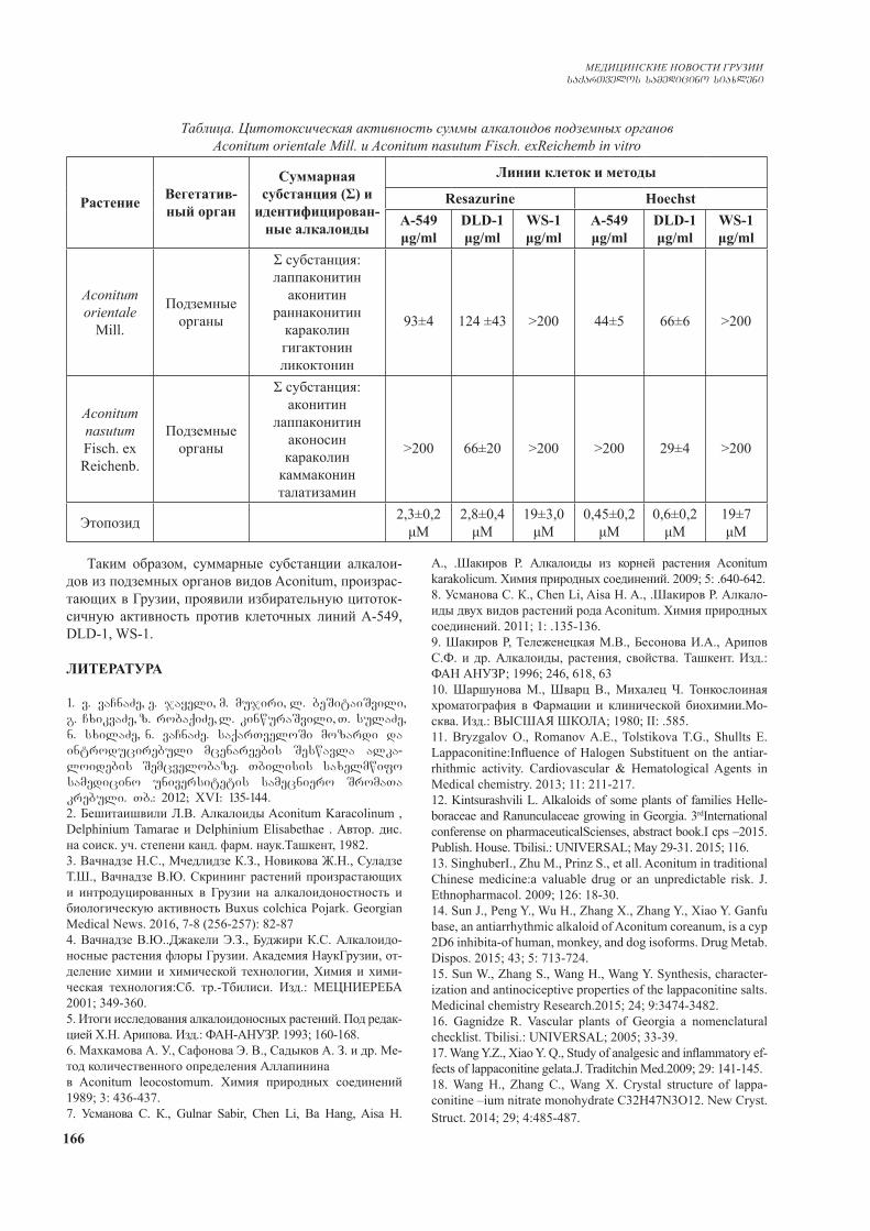
166
МЕДИЦИНСКИЕ НОВОСТИ ГРУЗИИ
CFMFHSDTKJC CFVTLBWBYJ CBF[KTYB
Таким образом, суммарные субстанции алкалои-дов из подземных органов видов Aconitum, произрас-тающих в Грузии, проявили избирательную цитоток-сичную активность против клеточных линий A-549, DLD-1, WS-1.
ЛИТЕРАТУРА
1. v. vaCnaZe, e. jayeli, m. mujiri, l. beSitaiSvili, g. CxikvaZe, z. robaqiZe, l. kinwuraSvili, T. sulaZe, n. sxilaZe, n. vaCnaZe. saqarTveloSi mozardi da introducirebuli mcenareebis Seswavla alka-loidebis Semcvelobaze. Tbilisis saxelmwifo samedicino universitetis samecniero SromaTa krebuli. Tb.: 2012; XVI: 135-144.2. Бешитаишвили Л.В. Алкалоиды Aconitum Karacolinum , Delphinium Tamarae и Delphinium Elisabethae . Автор. дис. на соиск. уч. степени канд. фарм. наук.Ташкент, 1982.3. Baчнадзе Н.С., Мчедлидзе К.З., Новикова Ж.Н., Суладзе Т.Ш., Вачнадзе В.Ю. Скрининг растений произрастающих и интродуцированных в Грузии на алкалоидоностность и биологическую активность Buxus colchica Pojark. Georgian Medical News. 2016, 7-8 (256-257): 82-874. Вачнадзе В.Ю..Джакели Э.З., Буджири К.С. Алкалоидо-носные растения флоры Грузии. Академия НаукГрузии, от-деление химии и химической технологии, Химия и хими-ческая технология:Сб. тр.-Тбилиси. Изд.: МЕЦНИЕРЕБА 2001; 349-360.5. Итоги исследования алкалоидоносных растений. Под редак-цией Х.Н. Арипова. Изд.: ФАН-АНУЗР. 1993; 160-168. 6. Махкамова А. У., Сафонова Э. В., Садыков А. З. и др. Ме-тод количественного определения Аллапинина в Aconitum leocostomum. Химия природных соединений 1989; 3: 436-437.7. Усманова С. К., Gulnar Sabir, Chen Li, Ba Hang, Aisa H.
A., .Шакиров Р. Алкалоиды из корней растения Aconitum karakolicum. Химия природных соединений. 2009; 5: .640-642.8. Усманова С. К., Chen Li, Aisa H. A., .Шакиров Р. Алкало-иды двух видов растений рода Aconitum. Химия природных соединений. 2011; 1: .135-136.9. Шакиров Р, Тележенецкая М.В., Бесонова И.А., Арипов С.Ф. и др. Алкалоиды, растения, свойства. Ташкент. Изд.: ФАН АНУЗР; 1996; 246, 618, 63 10. Шаршунова М., Шварц В., Михалец Ч. Тонкослоиная хроматография в Фармации и клинической биохимии.Мо-сква. Изд.: ВЫСШАЯ ШКОЛА; 1980; II: .585.11. Bryzgalov O., Romanov A.E., Tolstikova T.G., Shullts E. Lappaconitine:Influence of Halogen Substituent on the antiar-rhithmic activity. Cardiovascular & Hematological Agents in Medical chemistry. 2013; 11: 211-217. 12. Kintsurashvili L. Alkaloids of some plants of families Helle-boraceae and Ranunculaceae growing in Georgia. 3rdInternational conferense on pharmaceuticalScienses, abstract book.I cps –2015. Publish. House. Tbilisi.: UNIVERSAL; May 29-31. 2015; 116.13. SinghuberI., Zhu M., Prinz S., et all. Aconitum in traditional Chinese medicine:a valuable drug or an unpredictable risk. J. Ethnopharmacol. 2009; 126: 18-30.14. Sun J., Peng Y., Wu H., Zhang X., Zhang Y., Xiao Y. Ganfu base, an antiarrhythmic alkaloid of Aconitum coreanum, is a cyp 2D6 inhibita-of human, monkey, and dog isoforms. Drug Metab. Dispos. 2015; 43; 5: 713-724. 15. Sun W., Zhang S., Wang H., Wang Y. Synthesis, character-ization and antinociceptive properties of the lappaconitine salts. Medicinal chemistry Research.2015; 24; 9:3474-3482. 16. Gagnidze R. Vascular plants of Georgia a nomenclatural checklist. Tbilisi.: UNIVERSAL; 2005; 33-39.17. Wang Y.Z., Xiao Y. Q., Study of analgesic and inflammatory ef-fects of lappaconitine gelata.J. Traditchin Med.2009; 29: 141-145. 18. Wang H., Zhang C., Wang X. Crystal structure of lappa-conitine –ium nitrate monohydrate C32H47N3O12. New Cryst. Struct. 2014; 29; 4:485-487.
Таблица. Цитотоксическая активность суммы алкалоидов подземных органов Aconitum orientale Mill. и Aconitum nasutum Fisch. exReichemb in vitro
Растение Вегетатив-ный орган
Cуммарная субстанция (Σ) и
идентифицирован-ные алкалоиды
Линии клеток и методы
Resazurine HoechstA-549μg/ml
DLD-1 μg/ml
WS-1 μg/ml
A-549 μg/ml
DLD-1 μg/ml
WS-1μg/ml
Aconitum orientale
Mill.
Подземные органы
Σ субстанция:лаппаконитин
аконитинраннаконитин
караколин гигактонинликоктонин
93±4 124 ±43 >200 44±5 66±6 >200
Aconitum nasutum Fisch. ex Reichenb.
Подземные органы
Σ субстанция:аконитин
лаппаконитинаконосинкараколин
каммаконин талатизамин
>200 66±20 >200 >200 29±4 >200
Этопозид 2,3±0,2 μМ
2,8±0,4 μМ
19±3,0μМ
0,45±0,2μМ
0,6±0,2μМ
19±7μМ

GEORGIAN MEDICAL NEWS No 1 (274) 2018
© GMN 167
SUMMARY
ALKALOIDS OF UNDERGROUND PARTS OF GEORGIAN FLORA’S ACONITUM ORIENTALE MILL. AND ACONITUM NASUTUM FISCH. EX REICHEMB AND THEIR BIOLOGICAL ACTIVITY
Kintsurashvili L., Mshvildadze V., Suladze T.
Tbilisi State Medical University I. Kutateladze Institute of Pharmacochemistry, Georgia
The aim of research was to study underground parts of Aconitum orientale Mill and Aconitum nasutum Fisch exReichemb for the composition of biological active di-terpenic alkaloids. The research object was underground parts of Aconitum species. Alkaloids sum was received from raw-material which was alkalined beforehand with chloroform extraction. From the results of research we es-tablished, that both species of Georgian flora’s Aconitum contains alkaloids: lappaconitine, aconitine, karakoline. Underground parts of Aconitum orientale and Aconitum nasutum differ from eath other with composition of alka-loids spectrum. Underground parts of Aconitum orientale contains bases: ranaconitine, gigactonine, licoctonine, but underground parts of Aconitum nasutum contains: talitizamine, kamakonine, aconosine. Alkaloids’ summary substances, which were received from underground parts of Aconitum species spread in Georgia, showed selec-tive cytotoxic activity towards A-549 (lung carcinoma), DLD-1 (intestine adenocarcinoma), WS-1 (human normal fibroblasts).
Keywords: Alkaloids, Aconitum orientale Mill, lapa-conitinum, family Helleboraceae, chromatospectropho-tometry.
РЕЗЮМЕ
АЛКАЛОИДЫ ПОДЗЕМНЫХ ОРГАНОВ ACONITUM ORIENTALE MILL. И ACONITUM NASUTUM FISCH. EX REICHEMB. ФЛОРЫ ГРУ-ЗИИ И ИХ БИОЛОГИЧЕСКАЯ АКТИВНОСТЬ
Кинцурашвили Л.Г., Мшвилдадзе В.Д., Суладзе Т.Ш.
Тбилисский государственный медицинский универси-тет, Институт фармакохимии им. И. Кутателадзе, Грузия
Целью исследования явилось изучение подземных органов Aconitum orientale Mill. и Aconitum nasutum Fisch. ex Reichemb. на содержание дитерпеновых ал-калоидов с цитотоксической активностью.
Объектами исследования были подземные органы видов Aconitum. Сумму алкалоидов получали экс-тракцей растительного сырья хлороформом с предво-рительным подщелачиванием методом настаивания
при комнатной температуре. По результатам иссле-дования установлено, что в обоих видах Aconitum флоры Грузии присутствуют алкалоиды: аконитин, лаппаконитин, караколин. Спектры алкалоидов видов Aconitum orientale Mill. и Aconitum nasutum Fisch. ex Reichemb. отличаются по составу: в Aconitum orientale Mill. присутствуют основания: раннаконитин, гигак-тонин, ликоктонин, а в Aconitum nasutum Fisch. ex Reichemb.: талитизамин, каммаконин, аконосин. Сум-марные субстанции алкалоидов из подземных органов видов Aconitum, произрастающих в Грузии, проявили избирательную цитотоксичную активность против клеточных линий A-549 (карцинома легких), DLD-1 (аденокарцинома кишечника) и WS-1 (нормальные фибробласты человека).
reziume
saqarTvelos floris Aconitum orientale-s da Aconitum nasutum Fisch. ex Reichemb. miwisqveSa organoebis alkaloidebi da maTi biolo-giuri aqtivoba
l. kinwuraSvili, v. mSvildaZe, T. sulaZe
Tbilisis saxelmwifo samedicino univer-siteti, i. quTaTelaZis sax. farmakoqimiis instituti, saqarTvelo
kvlevis mizans warmoadgenda Aconitum orientale Mill - aRmosavluri tolWiri da Aco-nitum nasutum Fisch. ex Reichemb - eSmakis qoSis miwisqveSa organoebis Seswavla biologiu-rad aqtiur diterpenul alkaloidebis Sem-cvelobaze.
kvlevis obieqtebs warmoadgenda Aconitum-tilWiris saxeobebis miwisqveSa organoebi. alkaloidebis jami miRebuli iyo winaswar Setutianebuli mcenareuli nedleulidan qloroformiani eqstraqciiT. kvlevis Sedege-bidan gamomdinare dadgenilia, rom saqarT-velos floris Aconitum-is orive saxeobaSi moipoveba alkaloidebi: lapakonitini, akoni-tini, karakolini. Aconitum orientale-s da Aconitum nasutum-is miwisqveSa organoebi gansxvavdebian alkaloidebis speqtrebis SemadgenlobiT: Ac-onitum orientale-s miwisqveSa organoebi Seicavs fuZeebs: ranakonitins, gigaqtonins, likokto-nins, xolo Aconitum nasutums-is miwisqveSa or-ganoebi: talitizamins, kamakonins, akonosins. saqarTveloSi gavrcelebuli Aconitum-is saxeobebis miwisqveSa organoebidan miRebu-li alkaloidebis jamurma substanciebma ga-moavlina SerCeviTi citotoqsikuri aqtivoba A-549 (filtvis karcinoma), DLD-1 (nawlavis ade-nokarcinoma), WS-1 (adamianis normalur fi-broblastebi) mimarT.

168
МЕДИЦИНСКИЕ НОВОСТИ ГРУЗИИ
CFMFHSDTKJC CFVTLBWBYJ CBF[KTYB
ВОССТАНОВЛЕНИЕ СИМПАТО-ПАРАСИМПАТИЧЕСКОГО РАВНОВЕСИЯ В ВАРИАБЕЛЬНОСТИ СЕРДЕЧНОГО РИТМА ПРИ РАЗВИТИИ ПСИХОЭМОЦИОНАЛЬНО-
ГО СТРЕССА НА ФОНЕ ПРИМЕНЕНИЯ ТАУРИНА В ЭКСПЕРИМЕНТЕ
1Аветисян Э.А., 1Петросян А.А., 2Аванесян Л.Г., 2Шогерян С.А., 2Саакян Н.А.
1Институт физиологии им. акад. Л.А. Орбели НАН РА; 2Армянский государственный педагогический университет, Ереван, Армения
Ускорение темпа жизни, влияние различных ан-тропогенных, техногенных факторов, природных катаклизмов и имформационных нагрузок могут вызвать различные изменения в жизнедеятельно-сти организма, приводящие к развитию психо-эмоционального стресса, вызывающего дисбаланс симпато-парасимпатических реакций, наиболее чу-ствительным индикатором которых является сердеч-но-сосудистая система, поддерживающая гомеостаз организма. Нарушение гомеостаза в результате стрес-совых воздействий вызывает сдвиги в деятельности различных органов систем и патологические откло-нения, приводящие к углублению стресс реакций и развитию стрессовой болезни. С этой точки зрения исследование симпато-парасимпатических сдвигов в ритмограмме сердца методом математического ана-лиза вариабельности сердечного ритма (МА ВСР) является необходимым и весьма значимым фактором для своевременного обнаружения и предотвращения развития стресс патологии [3-5]. Раннее выявление развития стрессовой болезни и своевременный за-пуск адаптационных процессов является прерогати-вой нормализации жизнедеятельности организма, т.е. восстановления симпато-парасимпатического равно-весия. В последние годы наряду с различными препа-ратами, способствующими нормализации сердечно-сосудистых дисфункций, применяется аминокислота таурин, являющаяся одной из необходимых для орга-низма аминокислот с широким спектром положитель-ного воздействия на организм при различного рода патологиях [6,8,9,13,14]. В данном фрагменте иссле-дования методом спектрального анализа ВСР изучены изменение соотношения низко- (LF) и высокочастот-ных колебаний в ритмограмме сердца при длительной иммобилизации крыс (5 часов), вызывающей сильное эмоциональное напряжение и сопутствующий ему
сдвиг вегетативного равновесия, указывающий на симпато-парасимпатический дисбаланс, провоцирую-щий развитие психоэмоционального стресса, а также влияние ежедневного применения таурина в механиз-мах адаптации в ранний постстрессовый период.
Материал и методы. Эксперименты проведены на белых нелинейных крысах массой 280-300 г (n=60). Психоэмоциональный стресс вызывали посредством долговременной иммобилизации (5 часов) животного на спине с фиксацией конечностей. Запись ЭКГ во втором стандартном отведении проводили посредством спе-циальных серебряных электродов в течение 5 минут у бодрствующих крыс в контроле (n=12), сразу после сня-тия иммобилизации и в первый, седьмой и четырнад-цатый дни постстрессового периода у двух групп жи-вотных. В первую группу включены животные (n=30), подвергнутые иммобилизации, но не получавшие та-урин. Во вторую группу включены стрессированные крысы (n=30), ежедневно получающие таурин (50 мг/кг внутрибрюшинно) в пострессовый период. Регистрация ЭКГ проводилась по нижеприведенной схеме.
Для оценки результатов применялся метод МА ВСР, основанный на распознавании и измерении вре-менных интервалов между R-зубцами ЭКГ (R-R–ин-тервалы), построении динамических рядов кардиоин-тервалов (кардиоинтервалограмма) и последующего анализа полученных числовых рядов различными ма-тематическими методами. Одним из таких методов яв-ляется метод спектрального анализа ВСР, являющий-ся наиболее информативным для оценки состояния тонуса вегетативной нервной системы (ВНС) и позво-ляющий количественно охарактеризовать активность ее различных отделов [4,5,10,11]. Спектральный ана-лиз позволяет выяснить распределение мощности по отдельным частотам в общем спектре сердечного рит-ма и оценить их удельный вес в частотном спектре.
I группа (n=30) II группа (n=30) T0 контроль T0 контрольT1 сразу после снятия иммобилизации T1 сразу после снятия иммобилизации + таурин
T2 спустя день после снятия иммобилизации T2спустя день после снятия иммобилизации +
тауринT3 на 7 день после иммобилизации T3 на 7 день после иммобилизации + таурин
T4 на 14 день после иммобилизации T4 на 14 день после иммобилизации + таурин
Схема. Регистрация ЭКГ

GEORGIAN MEDICAL NEWS No 1 (274) 2018
© GMN 169
С помощью непараметрического метода - быстрое преобразование Фурье, можно получить частотный спектр трех периодических составляющих: 1) высо-кочастотный (HF) - частотный диапазон - 0.4-0.15 Гц, период–3-7 с, 2) низкочастотный (LF) – частотный диапазон - 0.04-0.15 Гц, период 7-30 с, 3) очень низ-кочастотный (VLF) – частотный диапазон 0.05 – 0.015 Гц, период 30–90 с. По данным спектрального анали-за сердечного ритма вычислялся индекс вагосимпати-ческого взаимодействия –LF/НF и динамика показате-лей на различных стадиях постстрессового периода.
Достоверность между группами определялась по t-критерию Cтьюдента. Вычислялись доверительные интервалы выборки, значимость корреляционных от-ношений и регрессионных уравнений. При статисти-ческой обработке материала использовали пакеты ста-тистических программ MSExcel 2010, Statistica 12.0, SPSS v16,0.
Результаты и их обсуждение. Подробный спек-тральный анализ изменений вагосимпатического взаи-модействия стрессированных крыс в динамике развития стресс патологии в период адаптации выявил ряд весьма значительных отклонений в показателях ВСР, указыва-ющих на нарушение симпато-парасимпатического рав-новесия в цепи VLF, HF и LF показателей в сравнении с контролем в начальной стадии, в первые дни после им-мобилизации у обеих исследуемых групп. В таблицах 1 и 2 представлены данные спектрального анализа показа-телей ВСР животных I и II групп.
Согласно результатам спектрального анализа об-щая мощность спектра сразу после иммобилизации (T0-T1) снизилась на 4,3%. Анализ как абсолютных, так и относительных данных выявил значительные изменения компонентов (VLF, HF, LF) спектра. Так, высокочастотный HF компонент сразу после иммоби-лизации снизился на 33,3% (р<0,001) (T0- T1), однако
на 14 день пострессового периода (T4) наблюдается тенденция к восстановлению парасимпатического воздействия и отклонение от нормы составляет всего 3,5% (р<0,05), что касается низко- (LF) и очень низко-частотных (VLF) компонентов, указывающих на акти-вацию симпатических реакций, их уровень сразу по-сле снятия иммобилизации повысился по сравнению с нормой на 25,5% и 25,0% (р<0,001), соответственно. На 14 день пострессового периода (T4) наблюдается спад напряженности, а LF компонент, выражающий сосудодвигательные эффекты и общую напряжен-ность симпатической системы, не полностью норма-лизован и составляет 5,0% (р<0,001), однако значение VLF-волны недостоверно. Анализ результатов, полу-ченных при рассмотрении симпато-вагального балан-са (SVB), указывает на резкое повышение симпатиче-ской активности, что является следствием нарушения гомеостаза в первый период (T0- T1), когда происходит активация всех висцеральных реакций (сердечно-со-судистых, дыхательных, гормональных) и вовлека-ются центральные интегративные структуры, вплоть до коры больших полушарий. В последующие этапы анализа параметры SVB стрессированных животных выявляют тенденцию к нормализации, происходит постепенный спад психоэмоциональной напряжен-ности и благодаря вовлечению тормозных влияний висцеральных интегративных структур мозга проис-ходит выравнивание симпато-вагального баланса, в частности, оптимальное соотношение мощности LF/НF компонентов, необходимое для поддержания гоме-остаза организма.
Спектральный анализ тех же показателей у второй группы животных, ежедневно получающих таурин в пострессовый период, выявил такую же закономер-ность изменений исследуемых величин в первый день после иммобилизации, однако повысилась скорость вос-
Таблица 1. Динамика показателей спектрального анализа ВСР I группы животных (M±SD)Показатели
Периоды T0 T1 T2 T3 T4
TP (мс2) 772.3±43.7 739.2±34.8 * 763.4±47.0 ˄ 773.5±34.5 ˄˄ 777.9±37.3 ˄˄
VLF (мс2) 131.5±12.2 164.4±12.0*** 149.4±217.3 ˄˄˄
140.2±20.6* ˄˄˄ 137.2±19.0˄˄˄
LF (мс2) 251.1±19.7 315.2±19.4***
280.0±28.5*** ˄˄˄
266.0±22.9*** ˄˄˄#
264.4±22.6*** ˄˄˄#
HF (мс2) 389.4±31.3 259.7±29.3*** 334.0±25.9*** ˄˄˄
367.3±21.2**˄˄˄ ###
376.2±20.4*˄˄˄ ###
SVB 0.65±0.06 1.23±0.17*** 0.84±0.10*** ˄˄˄
0.73±0.08*** ˄˄˄ ###
0.70±0.08*** ˄˄˄ ###
примечание: T0. T1 T2, T3, T4 - этапы эксперимента* – степень достоверности между T0
и последующими этапами, между ˄–T1 и T2, T3, T4, #–T2 и T3, T4, между ∆ T3 и T4 : *, **, *** соответствуют
р<0,05; р<0,01; р<0,001. Достоверность различий между этапами рассчитывалась в выборке согласно Гаусовскому распределению по t-критерию Стьюдента, для непараметрического распределения -
по показателю U Мана-Уитни. TP-общая мощность спектра; SVB-симпатовагальный баланс

170
МЕДИЦИНСКИЕ НОВОСТИ ГРУЗИИ
CFMFHSDTKJC CFVTLBWBYJ CBF[KTYB
становления процессов регуляции, в связи с этим уко-ротился период адаптации и произошла оптимизация ваго-симпатического баланса организма животных, под-вергающихся длительной иммобилизации. Результаты спектрального анализа приведены в таблице 2.
Расчеты процентных соотношений в разные сроки после иммобилизации на фоне инъекции та-урина показали, что общая мощность TP в частот-ном спектре (T0- T1) снизилась на 3,5% (р<0,05), а в период T0- T4 показатели общей мощности не от-личались от контроля, а поскольку общая мощность спектра отражает сумму всех его компонентов, то в физиологическом смысле произошла полная децен-трализация процессов регуляции, восстaновление деятельности сердечно-сосудистой системы. Вос-становление высокочастотного HF компонента в спектре ритмограммы II группы животных произо-шло уже на 7 день после иммобилизации. Абсолют-ная величина (T0- T1) снизилась на 25,7% (р<0,001), а на 14 день повысилась до 36,8% (р<0,001), что свидетельствует о превалировании парасимпати-ческих эффектов в период релаксации после стрес-сового воздействия. Компоненты LF и VLF в спек-тре T0- T1 увеличились на 23,1% (р<0,001) и 10,9% (р<0,001), соответсвенно, однако уже на 7 день на фоне внутрибрюшинной инъекции таурина сни-зились до нормы, а на 14 день составили 25,0% (р<0,001) и 19,8% (р<0,001), соотвественно. Про-веденный спектральный анализ составляющих об-щей мощности спектра у стрессированных крыс на фоне ежедневной инъекции таурина выявил благо-творное действие этой аминокислоты на улучшение адаптационных возможностей организма для про-тивостояния стрессогенным факторам. При рассмо-трении динамики индекса равновесия SVB у обеих экспериментальных групп на разных этапах по-стрессового периода и проведении сравнительного анализа абсолютных величин соотношения LF/НF показано, что при активации симпатической нерв-
ной системы происходит повышение коэффициента симпато-вагального баланса, в частности, соотно-шение мощности LF/НF волн, низкочастотной (LF) волны к мощности волн высокой частоты (HF) ста-новится выше единицы. На рис. 1 показаны срав-нительные величины коэффициентов SVB указыва-ющие, что сразу после иммобилизации происходит всплеск симпатических реакций у обеих групп, ярче выраженный у I группы в сравнении со II (T1) на всех этапах эксперимента. Нормализация LF/НF соотношения у II группы животных наступает уже на 7 день после иммобилизации, что указывает на нормализацию деятельности сердечно-сосудистой системы, децентрализацию процессов регуляции и восстановление автономного контура регуляции.
Рис. 1. Динамика индекса равновесия SVB на разных этапах у обеих исследуемых группобозначения: на оси ординат показаны абсолютные значения SVB, на оси абсцисс - этапы исследования
Поскольку спектральный анализ позволяет разло-жить ритмограмму на составляющие ее волны и коли-чественно оценить вклад каждой из них, а также оце-нить состояние механизмов вегетативной регуляции физиологических функций в организме, в том числе особенностей нейрогуморальной регуляции сердца и взаимодействия симпатического и парасимпатическо-го отделов вегетативной нервной системы, рассмотре-
Таблица 2. Динамика показателей спектрального анализа ВСР II группы животных при применении таурина (M±SD)
Показатели Периоды T0 T1 T2 T3 T4
TP (мс2) 769.3±45.1 742.5±45.4 * 766.0±38.8 ˄ 771.2±48.9 ˄ 769.5±42.8 ˄
VLF (мс2) 134.0±12.6 148.6±17.4** 137.7±20.5* ˄˄˄
134.0±17.82***˄˄˄
119.2±28.90** ˄˄˄
LF (мс2) 249.4±22.4 307.1±23.5***
266.1±29.3**˄˄˄
256.2±17.9˄˄˄
230.2±20.1˄˄˄##
HF (мс2) 385.9±34.5 286.8±38.2***
362.2±21.3 **˄˄˄
380.9±41.3˄˄˄#
392.2±32.6˄˄˄###
SVB 0.65±0.08 1.09±0.20*** 0.74±0.10˄˄˄ 0.68±0.10 **˄˄˄#
0.63±0.08˄˄˄###∆
T0 T1 T2 T3 T4I group 0.65 1.23 0.84 0.73 0.70II group 0.65 1.09 0.74 0.68 0.63
0.50
0.70
0.90
1.10
1.30
1.50
SVB
*** * ***

GEORGIAN MEDICAL NEWS No 1 (274) 2018
© GMN 171
ны и проанализированы значения VLF, HF и LF волн и вычислены их процентные соотношения на всех этапах исследования на основе которых составлены диаграммы процентного соотношения спектральных показателей в динамике адаптации для исследуемых групп (рис. 2).
На рисунке 2 показано превалирование LF и VLF компонентов в I и II группах сразу после иммобили-зации в сравнении с контролем, что свидетельствует об усилении симпатических эффектов, связанных с иммобилизацией животных, во время которой проис-ходит мобилизация функциональных возможностей сердечно-сосудистой системы. На последующих эта-пах у стрессированных животных I группы проис-ходит некоторый спад напряженности и увеличение удельного значения HF компонента (T1 I группа). Од-нако, полного восстановления равновесия симпатиче-ских (LF, VLF) и парасимпатического составляющих не наблюдается даже на 14 день (T4 I группа). При рас-смотрении спектрограммы животных, получавших ежедневно таурин, наблюдается полное восстановле-ние симпато-парасимпатического равновесия уже на 7 день (T3 II гуппа.), а на 14 день (T4) наблюдается даже превалирование высокочастотного (HF) компонента, что, повидимому, связано с тормозными влияниями лимбических структур и коры на автономный контур (моторное ядро блуждающего нерва, сосудо-двига-тельный и дыхательный центры продолговатого моз-га) [1,2,7] в регуляции висцеральной сферы организ-ма для восстановления гомеостаза. Предполагается, что при ежедневном применении таурина в условиях психоэмоционального стресса происходит улучше-ние обменных процессов и ускорение адаптационных свойств организма. Благотворное влияние таурина на сердечно-сосудистую систему при стрессе ярко выра-жено и на скатерограммах регресионной зависимости частоты сердечных сокращений (ЧСС) (рис. 3).
Рис 3. Скатерограмма регрессионной зависимости ЧСС (CR-Cardiac rate) у обеих исследуемых групппредставлены: полиноминальный тренд, достовер-ность регрессии и коеффициент детерминации. Ли-нии разброса показаны с 5% погрешностью
Сравнении скатерограмм ЧСС у животных, пере-несших стресс без- и с применением таурина выявило полную нормализацию ритма с менее выраженным учащением в пострессовом периоде у животных II группы. У обеих групп при иммобилизации проис-ходит учащение сердечного ритма, однако у пер-вой группы наблюдается большая вариация ЧСС и в процентном отношении разница между группами в первый день составила 8,3%, на 7 день 8,9%, а на 14 день 8,5%. Коэффициент детерминации (R), который на данной модели выражает динамику причин по за-висимости между группами, оказался выше у первой группы. Большое значение R указывает на детерми-нацию сердечных сокращений, т.е. свидетельствует о значительной вариации ЧСС данной группы жи-вотных, в связи с необходимостью затраты больших энергетических ресурсов для поддержания оптималь-ного состояния функцонирования организма. Следует предположить, что таурин способствует восполнению
Рис. 2. Процентное соотношение спектральных показателей. Процентные соотношения вычислены из общей мощности частотного спектра (TP)
обозначения: HF, LF и VLF - высоко-, низко- и очень низкочастотные компонентны спектра, T0 T1
T2, T3, T4 –этапы исследования

172
МЕДИЦИНСКИЕ НОВОСТИ ГРУЗИИ
CFMFHSDTKJC CFVTLBWBYJ CBF[KTYB
энергетических затрат, связанных с любым психо-эмоциональным напряжением, и прием этого пре-парата может предотвратить патологические сдвиги в организме, провоцируемые психоэмоциональным напряжением, противодействуя развитию стрессовой болезни.
ЛИТЕРАТУРА
1. Аветисян Э.А., Петросян А.А. Влияние стимуляции не-которых лимбических структур на фоновую и вызванную активность вагосенситивных нейронов ядра солитарного тракта. В сб.: «Актуальные проблемы интегративной дея-тельности и пластичности нервной системы». Материалы конференции с международным участием. Ереван: 2009; 7-11.2. Аветисян Э.А., Петросян А.А., Саакян Н.А. Характер влияния паравентрикулярного ядра гипоталамуса на им-пульсную активность висцеросенсорных нейронов ядра солитарного тракта и вариабельность сердечного ритма при психоэмоциональном стрессе у крыс. В сб.материалов Всероссийской конференции: «Функциональная межполу-шарная асимметрия и пластичность мозга», Москва: 13–14 декабря 2012; 231-235.3. Баевский Р.М. Математический анализ изменений сердеч-ного ритма при стрессе / Р.М. Баевский, О.И. Кириллов, С.З. Клецкин, - М,: Наука; 1984: 222.4. Курьянова Е. В., Тёплый Д. Л. Возрастные и половые осо-бенности изменений вариабельности сердечного ритма при блокаде и стимуляции периферических адренорецепторов у нелинейных крыс // Бюллетень экспериментальной биоло-гии и медицины. – 2014. – Т. 157. – №3. – С. 284–288.5. Carnevali L., Sgoifo A. Vagal modulation of resting heart rate in rats: the role of stress, psychosocial factors, and physicalexer-cise. Front Physiol 2014; 24 (5): 1–10.6. Gharibani PM, Modi J, Pan C, Menzie J, Ma Z, Chen PC, Tao R, Prentice H, Wu JY. The mechanism of taurine protec-tion against endoplasmic reticulum stress in an animal stroke model of cerebral artery occlusion and stroke-related conditions in primary neuronal cell culture. Adv Exp Med Biol. 2013; 776: 241-58. 7. Herman JP, McKlveen JM, Ghosal S, Kopp B, Wulsin A, Ma-kinson R, Scheimann J, Myers B. Regulation of the Hypotha-lamic-Pituitary-Adrenocortical Stress Response. Compr Physi-ol. 2016; 6(2):603-21. 8. Ito T, Kimura Y, Uozumi Y, Takai M, Muraoka S, Matsuda T, Ueki K, Yoshiyama M, Ikawa M, Okabe M, Schaffer SW, Fujio Y, Azuma J. Taurine depletion caused by knocking out the taurine transporter gene leads to cardiomyopathy with cardiac atrophy. J Mol Cell Cardiol. 2008;44:927–937.9. Ito T, Schaffer SW, Azuma J.The potential usefulness of taurine on diabetes mellitus and its complications. 2012 May;42(5):1529-39. 10. Montano N., Furlan R., Guzzetti S., McAllen R. M., Julien C. Analysis of sympathetic neural discharge in rats and hu-mans. Philos. Trans. A Math. Phys. Eng. Sci. 2009; 367: 1265–1282. 11. Mazloom R., Eftekhari G., Rahimi M. et al. The Role of α7 Nicotinic Acetylcholine Receptor in Modulation of Heart Rate Dynamics in Endotoxemic Rats // PLoS One. – 2013. – Vol. 8, №12. – 82251.12. Pereira-Junior P.P., Marocolo M., Rodrigues F.P. et al. Non-
ninvasive method fore electrocardiogram recording in conscious rats: feasibility for heart rate variability analysis // An. Acad.Bras. Cienc. – 2010. – Vol. 82, №2. – P. 431–43713. Ripps H., Shen W. Review: taurine: a very essential amino acid // Mol Vis. 2012, 18,p. 2673-8614. Worku Abebe, Mahmood S Mozaffari. Role of taurine in the vasculature: an overview of experimental and human studies. Am J Cardiovasc Dis. 2011; 1(3): 293–311.
SUMMARY
RESTORATION OF SYMPATHO-PARASYMPA-THETIC BALANCE IN HEART RATE VARIABIL-ITY IN THE DEVELOPMENT OF PSYCHOEMO-TIONAL STRESS AGAINST THE BACKGROUND OF TAURINE IN EXPERIMENT
1Avetisyan E., 1Petrosyan A., 2Avanesyan L., 2Shogheryan S., 2Saakyan N.
1L.A. Orbeli Institute of Physiology at the National Acad-emy of Sciences of Armenia; 2Armenian State Pedagogi-cal University, Yerevan, Armenia
In this fragment of the study, the change in the ratio of low (LF) and high frequency (HF) waves to the heart rhythmogram during prolonged immobilization (5 hours) of sexually mature white non-linear rats was studied by spectral analysis of heart rate variability, which causes psychoemotional stress and the accompanying vegetative equilibrium shift, indicating on the dysregulation of the sympatho-parasympathetic balance without and against the background of daily intraperitoneal injection of tau-rine (50 mg/kg) and its participation in the mechanisms of adaptive processes in the early poststress period (from 1 to 14 days).
Spectral analysis of both absolute and relative indices of the I-th group (stress without taurine) revealed signifi-cant shifts in the frequency spectrum. On the first day after immobilization, the total power of the spectrum decreased by 4.3%, the HF component by 33.3%, and the level of VLF and LF-components increased by 25.5% and 25%, respectively, compared to the norm. On the 14th day after the immobilization, there is a decrease in tension, but the LF component is still above the norm by 5%. Calculations of the percentage ratios of the absolute values of the spec-trum at different times of the post-stress period against the background of taurine injection showed that in the ini-tial stage (immediately and on the first day after stress) a strong shift of activity toward sympathization occurs, and the absolute value of LF and VLF in the spectrum increased by 23,1% and 10.9% respectively. However, the complete restoration of the vago-sympathetic balance with the daily application of taurine occurs already on the 7th day after immobilization. The beneficial effect of tau-rine on the cardiovascular system was revealed when ex-amining the scatterogram of the regression dependence of the heart rate, which revealed a smaller variation in heart

GEORGIAN MEDICAL NEWS No 1 (274) 2018
© GMN 173
rate and its stabilization on the 7th day after immobiliza-tion.
Thus, the use of taurine immediately after stressful reactions is necessary to accelerate adaptive processes in the early post-stress period to prevent the development of stress pathology.
Keywords: psychoemotional stress, spectral analysis, heart rate variability, taurine.
РЕЗЮМЕ
ВОССТАНОВЛЕНИЕ СИМПАТО-ПАРАСИМПАТИ-ЧЕСКОГО РАВНОВЕСИЯ В ВАРИАБЕЛЬНОСТИ СЕРДЕЧНОГО РИТМА ПРИ РАЗВИТИИ ПСИХОЭ-МОЦИОНАЛЬНОГО СТРЕССА НА ФОНЕ ПРИМЕ-НЕНИЯ ТАУРИНА В ЭКСПЕРИМЕНТЕ
1Аветисян Э.А., 1Петросян А.А., 2Аванесян Л.Г., 2Шогерян С.А., 2Саакян Н.А.
1Институт физиологии им. акад. Л.А. Орбели НАН РА; 2Армянский государственный педагогический уни-верситет, Ереван, Армения
В исследовании методом спектрального анализа вариабильности сердечного ритма изучено измене-ние соотношения низко- (LF) и высокочастотных (HF) волн в ритмограмме сердца при длительной иммоби-лизации (5 часов) половозрелых белых нелинейных крыс без и на фоне ежедневной внутрибрюшинной иньекции таурина (50 мг/кг) и его участие в механиз-мах адаптационных процессов в ранний постстрессо-вый период - с 1 по 14 день.
Спектральный анализ как абсолютных, так и от-носительных показателей I группы (стресс без та-урина) выявил значительные сдвиги в частотном спектре. В первый день после иммобилизации общая мощность спектра снизилась на 4,3%, HF компонент - на 33,3%, а уровень очень низкочастотых (VLF) и LF-компонентов повысился по сравнению с нормой на 25,5% и 25%, соответственно. На 14 день после иммобилизации происходит спад напряженности, но LF-компонент еще находится выше нормы на 5%. На фоне иньекции таурина (II группа) сразу и в 1 день по-сле стресса происходит сильный сдвиг активности в сто-рону симпатизации и абсолютная величина LF и VLF в спектре увеличиваестя на 23,1% и 10,9%, соответствен-но, однако полное восстановление ваго-симпатического баланса при ежедневном применении таурина происхо-дит на 7 день после иммобилизации. При рассмотрении скатерограммы регрессионной зависимости частоты сердечных сокращений (ЧСС) выявлена меньшая вари-ация ЧСС и ее стабилизация на 7 день после иммоби-лизации.
Таким образом, применение таурина сразу после
стрессовых реакций необходимо для ускорения адап-тационных процессов в ранний постстрессовый пери-од для предотвращения развития стресс-патологии.
reziume
simpaTikur-parasimpaTikuri wonasworo-bis aRdgena gulis ritmis variabelobaSi fsiqoemociuri stresis dros taurinis ga-moyenebis fonze eqsperimentSi
1e. avetisiani, 1a. petrosiani, 2l. avanesiani, 2s. Sogeriani, 2n. saakiani
1akad. l. orbelis sax. fiziologiis insti-tuti; 2somxeTis saxelmwifo pedagogiuri universiteti, erevani, somxeTi
kvlevaSi gulis ritmis variabelobis speqtruli analizis meTodiT Seswavlilia dabal- (LF) da maRalsixSirul i(HF) tal-Rebis Tanafardobis cvlileba gulis rit-mogramaSi zrdasruli TeTri araxazovani virTagvebis xangrZlivi (5 sT) imobiliza-ciis dros taurinis (50 mg/kg) yoveldRi-uri intraperitoneuli inieqciis fonze (II jgufi) da mis gareSe (I jgufi), aseve, misi monawileoba adaptaciur meqanizmebSi ad-reul poststresul periodSi (1-14 dRe).
I jgufis rogorc absoluturi, aseve, Se-fardebiTi maCveneblebis speqtruli ana-liziT sixSirul speqtrSi gamovlinda mniSvnelovani Zvrebi. imobilizaciis Semdeg pirvel dRes speqtris saerTo simZlavre Semcirda 4.3%-iT, HF komponenti – 33.3%-iT, xolo Zalian dabalsixSiruli (VLF) da LF-komponentebis done, normasTan SedarebiT, gaizarda, Sesabamisad, 25.5%-iT da 25%-iT. imo-bilizaciis me-14 dRes daZabuloba Semcir-da, magram LF- komponenti isev 5%-iT maRali iyo normaze. taurinis inieqciis fonze (II jgufi) maSinve da stresis pirvel dRes ganviTarda aqtivobis Zlieri gadaxra sim-paTizaciisken, VLF- da LF-is absoluturi maCveneblebi gaizarda, Sesabamisad, 23,1%- da 10,9%-iT; vagosimpaTikuri wonasworoba ki taurinis yoveldRiuri inieqciis fonze srulad aRdga imobilizaciis ukve meSvide dRes. gulis SekumSvebis sixSiris regresi-uli skaterogramis Sefasebisas gamovlinda gulis SekumSvaTa sixSiris naklebi variacia da misi stabilizeba imobilizaciis meSvide dRes. amrigad, stresuli reaqciebisTanave taurinis gamoyeneba aucilebelia adreul poststresul periodSi adaptaciuri pro-cesebis daCqarebisa da stress-paTologiis ganviTarebis prevenciisaTvis.

174
МЕДИЦИНСКИЕ НОВОСТИ ГРУЗИИ
CFMFHSDTKJC CFVTLBWBYJ CBF[KTYB
The health care market is substantially different from other fields of the economy. Therefore the behavior of health care providers operating in the health care mar-ket is different. This difference is related to the form of ownership. In general the market is maximally oriented on a profit, but in the healthcare market there are several forms of ownership of medical organizations: Nonprofit, For-profit, State-owned, Public-private partnership. Such diversity is mostly due to the particularities of the medical market. However, there are predominantly two forms of ownership in Georgia: for-profit and state-owned.
The aims of the research: assess the role of non-profit hospitals in health care system; review the characteristics of non-profit hospitals, namely how their behavior differs from commercial hospitals; assess the situation in Geor-gia with regard to non-profit hospitals at the healthcare market; what recommendations could be provided to im-prove the existing situation?
Material and methods. Methodological basis for the study was the existing literature about non-profit hospi-tals, relevant legislation, normative acts of The Ministry of Labour, Health and Social Affairs, healthcare statistics and statistical data of the Center for Disease Control and National Bureau of Statistics of Georgia.
Results and their discussion. There are several forms of ownership of medical organizations: Nonprofit, For-profit, State-owned, Public-private partnership. Such di-versity is mostly due to the particularities of the medical market. However, there are predominantly two forms of ownership in Georgia – for-profit and state-owned.
One of the forms of ownership of health care organi-zations is the state-owned, also called the public medical institutions. The owner of such an organization/institu-tion is the state, both on the federal and municipal levels. Hence, there are federal and municipal hospitals.
Owners of private, commercial medical institutions are individuals. They are established for commercial pur-poses and the primary aim of such organizations is to gain a profit. These organizations themselves can be divided in several forms: Individual private enterprises, Limited liability companies, Joint-stock companies.
Contemporary hospitals were developed in the mid-dle ages by religious organizations and local community unions, which were charitable entities. At the initial stage, hospitals were taking care for the poor population, or-phans, mentally ill persons and patients with communica-ble diseases. The members of these societies did not have an adequate sanitary conditions in their homes for proper treatment. Unlike the poor, the rich and the wealthy had
their own doctors and did not require the services of hos-pitals. Therefore in European countries, as well as in US, nonprofit hospitals were created in order to provide cer-tain services to the poor and the deprived. Such hospitals were mostly funded by donations [4].
In the 20th century, the advancements in healthcare technologies led to the development of emergency and in-patient hospitals. As a result the role of hospitals was changed. The Hospital became a place for professional medical practice and appropriate treatment of patients. With the function of the hospitals changing, their funding mechanisms changed as well. If previously a significant part of the financing came from charity funds and dona-tions, after the changes the expenses of the patients be-came the main source of revenue for the hospitals, in ad-dition to state and private insurance companies. Thus, the charity hospitals became the nonprofit (non-commercial) hospitals [5].
The most widespread form of ownership of health care organizations is the nonprofit one. The advantageous development of nonprofit hospitals is evident in Euro-pean countries (mostly characterized by universal health care system), as well as in more liberal states such as US (where health care is mostly considered as a medical mar-ket). In Europe, nonprofit hospitals make more than 70-80% of the hospitals. Similarly, in US, 57% of the hos-pitals are nonprofit, while 26% are state-owned (public) and private for-profit hospitals represent only 17%. Most of the physicians have private practice in profit and non-profit hospitals [1].
Fig. 1. Forms of Ownership of Hospitals in USASource: American Hospital Association. Hospital Sta-
tistics, various editions: 1986, 1995-1996. 2007Contrary to the above-mentioned, only two forms
of ownership has developed in Georgia. But, we should not disregard one important fact: before sovietization of Georgia, charity hospitals existed in Georgia as well. For example, in Tbilisi, on the place where the former 9th hos-
THE ROLE OF NON-PROFIT ORGANIZATIONS IN HEALTHCARE SYSTEM: WORLD PRACTICE AND GEORGIA (REVIEW)
Verulava T., Jorbenadze R., Dangadze B.
Health Policy and Insurance Centre, Ilia State University; G. Chapidze Emergency Cardiology Center, Tbilisi, Georgia

GEORGIAN MEDICAL NEWS No 1 (274) 2018
© GMN 175
pital was situated, stood an infirmary next to the church of Andrew the Apsotle, where the famous Georgian poet Vazha Pshavela passed away. The mentioned infirmary, like other infirmaries of the time, was a nonprofit hospital. After 1921, with the eventual soviet occupation of Geor-gia, nonprofit hospitals became state-owned healthcare organizations [9]. As a result, during the soviet period, there was only one form of ownership in Georgia – state ownership. After regaining independence, private hospi-tals were added as well. Thus, at present, there are only two extremely radical forms of ownership of health care organizations in Georgia – state-owned and private [2,10].
Hence, we can say that since soviet times, Georgia has been off the path, which the European countries took in terms of developing the health care system (forms of ownership of healthcare organizations is one of the char-acteristics of the health care system).
Very often, the word ‘nonprofit’ is misinterpreted and it is considered that such organizations do not represent profitable enterprises. As a matter of fact, nonprofit, as well as for-profit, or even the state medical institutions attempt to make a profit. They only differ in the way they distribute the profit. Nonprofit hospitals are managed and run by boards composed of physicians, society represen-tatives, and managers. The profit hospitals are managed by shareholders. To be more precise, nonprofit medical institutions do not distribute the profit to the owners or the shareholders, unlike the for-profit medical institutions. The profit gained by their work is distributed and spent on improvement of medical services that the hospital pro-vides, acquisition of tools and equipment for diagnostics, increasing the salaries of the medical personnel of the hospital [8].
Besides the historically advantageous environment, nonprofit hospitals in western countries had significant support from the governments. As the non-profit hospi-tals provide service to poor and low-income patients, their activities are considered to be charity. Therefore, their in-come and property is exempt from taxes.
The tax benefits do not apply to profit hospitals. Fur-thermore, profit hospitals have no right to receive public donations. Donations are given to the non-profit hospitals to provide medical service to vulnerable population.
In the USA, as in many other countries, non-profit hospitals were established for provision of medical ser-vice to the poor population. The main source of funding for them was donations. Despite increasing the portion of the state and private insurance, the greater part of the population is still uninsured. Correspondingly, non-profit hospitals provide medical service to those people with no capacity to pay [11].
In Western Europe and US, trust in nonprofit medical services developed over the years also played a role in the existence of non-profit hospitals. On the medical markets, where consumers are less informed and do not have rel-evant education on the needs of healthcare services, the great importance is attached to the trust-based relations.
The patients tend to trust towards nonprofit hospitals more than for-profit ones, as the latter are not focused on the profit and doesn’t strive to take advantage of the pa-tients’ lack of information.
In addition, the society overlooks the performance and governance of nonprofit hospitals. Their policy is more flexible for implementation of the activities such as provision of expensive medical service. In the non-profit hospitals doctors have more possibilities to independently define the hospital policy, service provision, purchase the need medical equipment and services. Due to this, non-profit hospitals better correspond to the financial interest of doctors. In contrast to this in profit hospi-tals the gain is basically divided between shareholders and state taxes; thus, the motivation of doctors is rather low. Therefore, the factor of trust, public benefit, and financial interest of doctors is main basis for high num-ber of non-profit hospitals.
The popularity of nonprofit hospitals is evident in cer-tain cases, as 70% of US patients prefer nonprofit hospitals, while only 13% go to for-profit ones. Even the medical per-sonnel prefer the nonprofit hospitals, as the majority of them work in nonprofit health care institutions [1].
Profit and non-profit hospitals differ from each other in their organizational behavior. The aim of profit hos-pitals in contrast to non-profit hospitals is more precise, namely, to get as much profit as possible. The goal of non-profit hospitals is multiple, that makes their monitoring very difficult. Various stakeholders of non-profit hospitals – medical personnel, council members, managers, staff and society – they often have a very different and even conflicting goals. The approaches are different too as how to divide the gain of non-profit hospitals.
Profit hospitals try to define prices for medical service with the purpose of maximum gain [3,6,7]. Non-profit hospitals set such prices for medical services which cover the cost of provided services.
Legal forms of non-profit legal entities in GeorgiaIt was mentioned above that one of the reasons con-
tributing to the development of nonprofit hospitals in the Western countries was the exemption from taxes. Thus, according to the internal revenue code/tax code of Geor-gia, nonprofit organizations, which undertake charity ac-tivities benefit from tax privileges. Charity organizations are exempt from corporate income tax.
Additionally, the civil code of Georgia defines the def-inition of nonprofit (non-commercial) entities/organiza-tions: “organization, the aim of which is non-commercial, the primary motivational factor of which is not gaining profit, represents a non-commercial entity of public law”. Despite the fact that the non-commercial entity cannot, by default, oriented on commercial activity, it can engage in auxiliary economic activity. Profit gained from such ac-tivities should be used for achieving the ultimate aims of the entity and cannot be divided among or used by the founders, members, donors or the management of the en-tity.

176
МЕДИЦИНСКИЕ НОВОСТИ ГРУЗИИ
CFMFHSDTKJC CFVTLBWBYJ CBF[KTYB
Despite the fact that an entry about nonprofit enti-ties and their exemption from taxes exists in the inter-nal revenue code of Georgia, sheer existence on paper is not enough for the development of nonprofit hospitals in Georgia. As the nonprofit health care institutions in Geor-gia are underdeveloped, we can claim that there is a lack of motivation for the existence of such institutions. Fur-ther elaboration of tax benefits and privileges in the rev-enue code is necessary based on European experience. It is expedient to increase the role of nonprofit hospitals on Georgia’s health care market.
Types of owners of hospitals in GeorgiaAccording to data from 2013, 88,6% of the hospitals
are private for-profit, among which 41,1% is owned by in-surance companies, 29,1% by individuals, 18,4% by other types of companies and 8,2% is state-owned [9]. Mostly specific medical institutions and psychiatric establish-ments are state-owned.
Fig. 2. Forms of Ownership of Hospitals in GeorgiaSource: Hospital Sector in Georgia. Transparency In-
ternational – Georgia, Tbilisi. 2012More than 80% of hospitals are owned by three private
insurance companies (Aldagi, GPI- holding, and Aversi/alfa). Private Insurance Company Aldagi owns 49% of the hospitals. Insurance Company GPI-holding owns 255 of hospitals. Insurance Company Alfa owns 17% of hos-pitals [9].
It is important to take note of the following problems of the health care sector of Georgia: Most hospitals have fewer than 50 beds (34% of the hospitals have between 11-20 beds; 17% - between 21-30; 41% - between 41-50). According by international experience, a hospital with fewer than 50 beds cannot become for-profit. It is equally important to highlight that certain types of health care ser-vices are not profitable, thus the owners of the hospitals have less interest to fund expensive services. Therefore, insurance companies that own hospitals may not bear ex-penses for such types of medical service.
Besides the above-mentioned forms of ownership of health care institutions, public-private partnership is also widespread in developed countries. Such ownership rep-resents a partnership between the state and private own-ers aimed at reaching the priority aims of development of the health care system by dividing yield, costs and risks among themselves based on long-term, voluntary deci-sions. One of the forms of such a partnership is leasing the state property. In such cases, the state leases the buildings and facilities, as well as the equipment that it does not use to a private business.
It is worth noting that a public-private partnership in the health care system is being planned. Private enterpris-es will be obliged to co-operate with the state and man-age, own and operate the hospitals based on the public-private partnership principle. Due to lack of legal base, the Ministry of Economy is currently working on the new legislation dealing with this matter.
Conclusion. Therefore, unlike regular markets, the health care market demanded the existence of differ-ent forms of ownership, which comprises private non-profit, private for-profit, state-owned, as well as pub-lic-private partnership organizations. The first steps taken towards public-private partnerships in Georgia are definitely a positive signs. However, it is necessary to promote other forms of ownership as well, particu-larly nonprofit health care organizations. On Georgian health care market the non-profit hospitals are few. It seems that there is no sufficient motivation for func-tion of non-profit hospitals. Despite the fact that Tax Code envisages tax benefits, it’s only on the paper and doesn’t provide maximum result. Different forms of ownership of the medical organizations will increase the competition between different forms of ownerships, which will improve access to health care services. Most importantly, this will bring Georgia closer to the expe-rience of the European countries.
REFERENCES
1. American Hospital Association. Hospital Statistics, 2012. Chicago: AHA.2. Asatiani, M., Verulava T.. Georgian Welfare State: Prelimi-nary Study Based on Esping-Andersen’s Typology. Economics and Sociology 2017; 10(4): 21-28. 3. Davies P. Hospital Chains Sued over Billing: Alabama Law-yer Claims Companies Inflated Profit by Overcharging Unin-sured. The Wall Street Journal 2004; 8(6): B2.4. Feldstein Paul J.. Health Policy Issues: An Economic Perspec-tive. Sixth Edition Health Administration Press: 2015. 5. Folland S., Goodman A., Miron S.. The economics of health and health care. New Jersey: 2007.6. Horwitz J. Making Profits and Providing Care: Comparing Nonprofit, For-Profit, and Governmental Hospitals. Health Af-fairs 2005; 24(3): 790–801.7. Lagnado L. HHS Chief Scolds Hospitals for Their Treatment of Uninsured. The Wall Street Journal 2004; 2(20): A1.8. Sloan F. Not-for-Profit Ownership and Hospital Behavior. Handbook of Health Economics, vol. 18. New York North-Hol-land Press: 2000; 1141-73.9. Verulava T.. Forms of Ownership of Medical Organizations. World Practice and Georgia. Strategic Plan Georgia 2020 – Strengthening Public Involvement. Tbilisi: 2016. 10. Verulava, T., Jorbenadze, R., Barkalaia, T. Introduction of universal health program in Georgia: Problems and Perspec-tives. Georgian Medical News 2017, 262 (1): 116-120.11. Verulava, T., Maglakelidze, T. Health Financing Policy in the South Caucasus: Georgia, Armenia, Azerbaijan. Bulletin of the Georgian National Academy of Sciences 2017; 11(2): 143-150.

GEORGIAN MEDICAL NEWS No 1 (274) 2018
© GMN 177
SUMMARY
THE ROLE OF NON-PROFIT ORGANIZATIONS IN HEALTHCARE SYSTEM: WORLD PRACTICE AND GEORGIA (REVIEW)
Verulava T., Jorbenadze R., Dangadze B.
Health Policy and Insurance Centre, Ilia State University; G. Chapidze Emergency Cardiology Center, Tbilisi, Georgia
The health care market is substantially different from other areas of the economy and therefore the behavior of health care providers operating in the health care market is different, which is mainly related to the form of ownership. If the market is mainly characterized by the pursuit of maxi-mum profit, medical services market has for some public good features. Because of this, non-profit hospitals in west-ern countries are considered as an alternative form of com-mercial hospitals. The purpose of the research was to study the role of not-for-profit hospitals, and in this regard examine the situation of the medical market in Georgia.
The existing literature about non-profit hospitals, rele-vant legislation and statistical data, scientific articles, and other related works.
The majority of the hospitals in Georgia represent prof-itable (commercial) organizations. 41,1% of the hospitals owned by private insurance companies, 29,1% by individu-als, 18,4% by other types of companies, 3,2% by other forms and 8% is state-owned. In contrast to this, more than 50% of the healthcare system of West Europe as well as USA is composed of non-profit (commercial) hospitals.
In Georgia there is no sufficient motivation for operat-ing of hospitals as non-profit organizations. It is necessary to further adjust tax benefit in the Tax Code of Georgia and share European experiences. It is reasonable to increase the role of non-profit hospitals on the health care market that will increase accessibility to healthcare services for population and moreover. It will bring Georgian health-care system close to the experience of civilized world.
Keywords: Healthcare, Non-profit Hospital, Com-mercial Hospitals, Ownership.
РЕЗЮМЕ
РОЛЬ НЕКОММЕРЧЕСКИХ ОРГАНИЗАЦИЙ В СИСТЕМЕ ЗДРАВООХРАНЕНИЯ: МИРОВАЯ ПРАКТИКА И ГРУЗИЯ (ОБЗОР)
Верулава Т.Н., Джорбенадзе Р.А., Дангадзе Б.Б.
Институт здравоохранения и страхования, Государ-ственный Университет Илии; Центр неотложной кардиологии им. Г. Чапидзе, Тбилиси, Грузия
Рынок здравоохранения существенно отличается от других областей экономики. Поведение поставщи-
ков медицинских услуг различается в зависимости от формы собственности. В случае, когда рынок харак-теризуется стремлением к максимальной прибыли, на рынке медицинских услуг некоторые из них имеют черты общественных благ. Ввиду чего некоммерче-ские больницы в западных странах рассматриваются как альтернативная форма коммерческих больниц.
Целью исследования явилось определение роли не-коммерческих больниц на медицинском рынке в Грузии.
Проанализирована ретроспективная и текущая литература и статистические данные касательно дея-тельности некоммерческих больниц.
Большинство больниц в Грузии являются при-быльными (коммерческие) организациями. 41,1% больниц принадлежат частным страховым компани-ям, 29,1% - физическим лицам, 18,4% - различным компаниям, 8% - государству 3,2% - другие формы. В Западной Европе и США более 50% больниц являют-ся некоммерческими.
Следует отметить, что в Грузии нет достаточной мотивации для работы больниц в качестве неком-мерческих организаций. Необходимо использование налоговых льгот в налоговом кодексе Грузии и ев-ропейского опыты. Разумно увеличить роль неком-мерческих больниц на рынке здравоохранения, что облегчит доступность услуг здравоохранения для на-селения и приблизит систему здравоохранения Гру-зии к опыту цивилизованного мира.
reziume
aramomgebiani saavadmyofoebis roli jan-dacvis sistemaSi: msoflio gamocdileba da saqarTvelo (mimoxilva)
T. verulava, r. jorbenaZe, b. dangaZe
jandacvisa da dazRvevis instituti, ilias saxelmwifo universiteti, akad. g. CafiZis sax. gadaudebeli kardiologiis centri. Tbilisi, saqarTvelo
jandacvis bazari arsebiTad gansxvavdeba ekonomikis sxva sferoebisagan, gansxvavebu-lia jandacvis bazarze moqmedi samedici-no momsaxurebis mimwodebelTa qcevac, rac umTavresad dakavSirebulia mesakuTreo-bis formasTan. Tu Cveulebrivi bazrisTvis, damaxasiaTebelia maqsimaluri mogebisa-ken swrafva, samedicino bazarze garkveuli servisebi sazogadoebrivi sikeTis niSnebiT xasiaTdebian. dasavleTis qveynebSi arako-merciuli saavadmyofoebi warmoadgenen ko-merciuli saavadmyofoebis alternatiul formas. kvlevis mizans warmoadgens ara-momgebiani saavadmyofos rolis da am mxriv saqarTvelos samedicino bazarze arsebuli

178
МЕДИЦИНСКИЕ НОВОСТИ ГРУЗИИ
CFMFHSDTKJC CFVTLBWBYJ CBF[KTYB
mdgomareobis Seswavla. kvlevis procesSi gamoyenebuli iyo aramomgebiani saavadmyo-foebis Sesaxeb arsebuli retrospeqtuli da mimdinare literaturis wyatoebi, Sesabamisi kanonmdebloba da statistikuri monacemebi.
2013 wlis monacemebiT, saqarTveloSi ar-sebuli saavadmyofoebis umravlesoba war-moadgens momgebian organizaciebs; saavad-myofoTa 41,1% ekuTvnis sadazRvevo kompaniebs, 29,1% - fizikur pirebs, 18,4% - sxvadasxva kom-paniebs, 3,2% - mesakuTreobis sxva formebs, saxelmwifos - mxolod 8%. dasavleT evro-pis da aSS-is jandacvis sistemaSi aramomge-biani saavadmyofoebi Seadgenen 50%-ze mets.
avtorebs gamotanili aqvT daskvna, rom saqarTveloSi ar arsebobs saavadmyofoebis aramomgebiani saxiT funqcionirebis sakma-risi motivacia. aucilebelia sagadasaxa-do kodeqsSi gaTvaliswinebuli iyos saga-dasaxado SeRavaTebis Semdgomi daxvewa da evropuli gamocdilebis gaziareba. mizanSe-wonilia aramomgebiani saavadmyofoebis ro-lis gazrda saqarTvelos samedicino bazar-ze, rac mosaxleobisaTvis xelmisawvdoms gaxdis jandacvis momsaxureobas da uzrun-velyofs saqarTvelos jandacvis sistemis daaxloebas civilizebuli samyaros gamoc-dilebasTan.
* * *
Copyright © 2022 FDOKUMEN