Reduced human leukocyte antigen expression in advanced-stage Ewing sarcoma: implications for immune...
10
Journal of Pathology J Pathol 2009; 218: 222–231 Published online 2 February 2009 in Wiley InterScience (www.interscience.wiley.com) DOI: 10.1002/path.2537 Original Paper Reduced human leukocyte antigen expression in advanced-stage Ewing sarcoma: implications for immune recognition Dagmar Berghuis, 1,2 † Alfons SK de Hooge, 2 † Susy J Santos, 2 Danielle Horst, 3 Emmanuel J Wiertz, 3 Marja C van Eggermond, 4 Peter J van den Elsen, 4 Antonie HM Taminiau, 5 Laura Ottaviano, 6 Karl-Ludwig Schaefer, 6 Uta Dirksen, 7 Erik Hooijberg, 8 Arend Mulder, 4 Cornelis JM Melief, 4 R Maarten Egeler, 2 Marco W Schilham, 2 Ekaterina S Jordanova, 1 Pancras CW Hogendoorn 1 and Arjan C Lankester 2 * 1 Department of Pathology, Leiden University Medical Center, Leiden, The Netherlands ‡ 2 Department of Pediatrics, Leiden University Medical Center, Leiden, The Netherlands ‡ 3 Department of Medical Microbiology and Center of Infectious Diseases, Leiden University Medical Center, Leiden, The Netherlands / Department of Medical Microbiology, University Medical Center Utrecht, Utrecht, The Netherlands 4 Department of Immunohematology and Blood Transfusion, Leiden University Medical Center, Leiden, The Netherlands 5 Department of Orthopedics, Leiden University Medical Center, Leiden, The Netherlands ‡ 6 Institute of Pathology, Heinrich-Heine University, D¨ usseldorf, Germany ‡ 7 Department of Pediatric Hematology and Oncology, University Children’s Hospital, M¨ unster, Germany ‡ 8 Department of Pathology, VU University Medical Center, Amsterdam, The Netherlands *Correspondence to: Arjan C Lankester, MD, PhD, Department of Pediatrics, Leiden University Medical Center, PO box 9600, 2300 RC Leiden, The Netherlands. E-mail: [email protected] † These authors contributed equally to this work. ‡ These departments are partners of the EuroBoNeT consortium, a European Commission granted Network of Excellence for studying the pathology and genetics of bone tumours. No conflicts of interest were declared. Received: 4 September 2008 Revised: 20 January 2009 Accepted: 20 January 2009 Abstract Ewing sarcoma (EWS) is a tumour most commonly arising in bone, although on occasion in soft tissue, with a poor prognosis in patients with refractory or relapsed disease, despite mul- timodal therapy. Immunotherapeutic strategies based on tumour-reactive T and/or natural killer cells may improve the treatment of advanced-stage EWS. Since cellular immune recog- nition critically depends on human leukocyte antigen (HLA) expression, knowledge about HLA expression in EWS is crucial in the design of cellular immunotherapeutic strategies. Constitutive and IFNγ -induced HLA class I expression was analysed in EWS cell lines (n = 6) by flow cytometry, using antibodies against both monomorphic and allele-specific antigens. Expression of antigen processing pathway components and beta-2 microglobulin (β 2m) was assessed by western blot. Expression of class II transactivator (CIITA), and its contribution to HLA class II expression, was evaluated by qRT-PCR, transduction assays, and flow cytometry. β 2m/HLA class I and class II expression was validated in EWS tumours (n = 67) by immunofluorescence. Complete or partial absence of HLA class I expression was observed in 79% of EWS tumours. Lung metastases consistently lacked HLA class I and sequential tumours demonstrated a tendency towards decreased expression upon disease progression. Together with absent or low constitutive expression levels of specific HLA class I loci and alleles, and differential induction of identical alleles by IFNγ in different cell lines, these results may reflect the existence of an immune escape mechanism. Inducible expression of TAP-1/-2, tapasin, LMP-2/-7, and the β 2m/HLA class I complex by IFNγ sug- gests that regulatory mechanisms are mainly responsible for heterogeneity in constitutive class I expression. EWSs lack IFNγ -inducible HLA class II, due to lack of functional CIITA. The majority of EWS tumours, particularly if advanced-stage, exhibit complete or partial absence of both classes of HLA. This knowledge will be instrumental in the design of cellular immunotherapeutic strategies for advanced-stage EWS. Copyright 2009 Pathological Society of Great Britain and Ireland. Published by John Wiley & Sons, Ltd. Keywords: Ewing sarcoma; bone tumour; soft tissue tumour; human leukocyte anti- gen; antigen processing pathway; immune recognition; immunotherapy; flow cytometry; immunohistochemistry Introduction Ewing sarcoma (EWS) is a malignant sarcoma, most commonly arising in bone and less frequently in soft tissue, that mainly affects children, adolescents, and young adults [1]. This tumour belongs to the Ewing family of tumours, a family of round cell sarcomas characterized by specific EWS/ETS gene fusions [2]. Despite multimodal therapy including surgery, high-dose chemotherapy, and radiotherapy, Copyright 2009 Pathological Society of Great Britain and Ireland. Published by John Wiley & Sons, Ltd. www.pathsoc.org.uk
-
Upload
independent -
Category
Documents
-
view
5 -
download
0
Transcript of Reduced human leukocyte antigen expression in advanced-stage Ewing sarcoma: implications for immune...

Journal of PathologyJ Pathol 2009; 218: 222–231Published online 2 February 2009 in Wiley InterScience(www.interscience.wiley.com) DOI: 10.1002/path.2537
Original Paper
Reduced human leukocyte antigen expression inadvanced-stage Ewing sarcoma: implications for immunerecognition
Dagmar Berghuis,1,2† Alfons SK de Hooge,2† Susy J Santos,2 Danielle Horst,3 Emmanuel J Wiertz,3
Marja C van Eggermond,4 Peter J van den Elsen,4 Antonie HM Taminiau,5 Laura Ottaviano,6
Karl-Ludwig Schaefer,6 Uta Dirksen,7 Erik Hooijberg,8 Arend Mulder,4 Cornelis JM Melief,4 R Maarten Egeler,2
Marco W Schilham,2 Ekaterina S Jordanova,1 Pancras CW Hogendoorn1 and Arjan C Lankester2*1Department of Pathology, Leiden University Medical Center, Leiden, The Netherlands‡
2Department of Pediatrics, Leiden University Medical Center, Leiden, The Netherlands‡
3Department of Medical Microbiology and Center of Infectious Diseases, Leiden University Medical Center, Leiden, The Netherlands / Department ofMedical Microbiology, University Medical Center Utrecht, Utrecht, The Netherlands4Department of Immunohematology and Blood Transfusion, Leiden University Medical Center, Leiden, The Netherlands5Department of Orthopedics, Leiden University Medical Center, Leiden, The Netherlands‡
6Institute of Pathology, Heinrich-Heine University, Dusseldorf, Germany‡
7Department of Pediatric Hematology and Oncology, University Children’s Hospital, Munster, Germany‡
8Department of Pathology, VU University Medical Center, Amsterdam, The Netherlands
*Correspondence to:Arjan C Lankester, MD, PhD,Department of Pediatrics, LeidenUniversity Medical Center, PObox 9600, 2300 RC Leiden, TheNetherlands.E-mail: [email protected]
†These authors contributedequally to this work.
‡These departments are partnersof the EuroBoNeT consortium, aEuropean Commission grantedNetwork of Excellence forstudying the pathology andgenetics of bone tumours.
No conflicts of interest weredeclared.
Received: 4 September 2008Revised: 20 January 2009Accepted: 20 January 2009
AbstractEwing sarcoma (EWS) is a tumour most commonly arising in bone, although on occasion insoft tissue, with a poor prognosis in patients with refractory or relapsed disease, despite mul-timodal therapy. Immunotherapeutic strategies based on tumour-reactive T and/or naturalkiller cells may improve the treatment of advanced-stage EWS. Since cellular immune recog-nition critically depends on human leukocyte antigen (HLA) expression, knowledge aboutHLA expression in EWS is crucial in the design of cellular immunotherapeutic strategies.Constitutive and IFNγ -induced HLA class I expression was analysed in EWS cell lines(n = 6) by flow cytometry, using antibodies against both monomorphic and allele-specificantigens. Expression of antigen processing pathway components and beta-2 microglobulin(β2m) was assessed by western blot. Expression of class II transactivator (CIITA), and itscontribution to HLA class II expression, was evaluated by qRT-PCR, transduction assays,and flow cytometry. β2m/HLA class I and class II expression was validated in EWS tumours(n = 67) by immunofluorescence. Complete or partial absence of HLA class I expression wasobserved in 79% of EWS tumours. Lung metastases consistently lacked HLA class I andsequential tumours demonstrated a tendency towards decreased expression upon diseaseprogression. Together with absent or low constitutive expression levels of specific HLA classI loci and alleles, and differential induction of identical alleles by IFNγ in different celllines, these results may reflect the existence of an immune escape mechanism. Inducibleexpression of TAP-1/-2, tapasin, LMP-2/-7, and the β2m/HLA class I complex by IFNγ sug-gests that regulatory mechanisms are mainly responsible for heterogeneity in constitutiveclass I expression. EWSs lack IFNγ -inducible HLA class II, due to lack of functional CIITA.The majority of EWS tumours, particularly if advanced-stage, exhibit complete or partialabsence of both classes of HLA. This knowledge will be instrumental in the design of cellularimmunotherapeutic strategies for advanced-stage EWS.Copyright 2009 Pathological Society of Great Britain and Ireland. Published by JohnWiley & Sons, Ltd.
Keywords: Ewing sarcoma; bone tumour; soft tissue tumour; human leukocyte anti-gen; antigen processing pathway; immune recognition; immunotherapy; flow cytometry;immunohistochemistry
Introduction
Ewing sarcoma (EWS) is a malignant sarcoma, mostcommonly arising in bone and less frequently insoft tissue, that mainly affects children, adolescents,
and young adults [1]. This tumour belongs to theEwing family of tumours, a family of round cellsarcomas characterized by specific EWS/ETS genefusions [2]. Despite multimodal therapy includingsurgery, high-dose chemotherapy, and radiotherapy,
Copyright 2009 Pathological Society of Great Britain and Ireland. Published by John Wiley & Sons, Ltd.www.pathsoc.org.uk

HLA expression in Ewing sarcoma 223
patients with refractory or relapsed disease have a poorprognosis. Furthermore, no significant improvementhas been achieved in the past decade with regard tothe survival of patients presenting with localized dis-ease [3]. In recent years, (pre)clinical studies in varioustumour models have shown that immunotherapeuticstrategies, including adoptive cell transfer therapiesusing tumour-specific cytotoxic T lymphocytes (CTLs)or natural killer (NK) cells, may represent novel thera-peutic approaches [4–6]. These approaches may offeradvantageous and less toxic treatment modalities foradvanced-stage EWS as well. Human leukocyte anti-gen (HLA) expression plays a crucial role in therecognition of tumours by tumour-reactive T and/orNK cells, influencing immunity or tolerance againstthese tumours as well as susceptibility to cellularimmunotherapeutic approaches [7]. Knowledge aboutHLA expression in EWS is crucial in the design ofcellular immunotherapeutic strategies.
The human major histocompatibility complex(MHC) encodes HLA class I and II molecules.The classical HLA class I molecules HLA-A, -B,and -C are, together with β2-microglobulin (β2m),expressed by almost all nucleated cells and presentpeptides derived from intracellular proteins to CTLs.Peptides are generated in the cytoplasm by the(immuno)proteasome and are transported into theendoplasmic reticulum by heterodimeric transporterassociated with the antigen processing (TAP-1/-2)complex. Assembly of the HLA class I heavychain–β2m heterodimer with peptide is orchestratedby chaperones, including tapasin. After peptide load-ing, HLA class I molecules are transported to the cellsurface [8,9]. HLA class II molecules (HLA-DR, -DP,-DQ) are constitutively expressed only by ‘profes-sional’ antigen-presenting cells and present peptidesderived mainly from processed extracellular antigensto helper T lymphocytes. However, via activation ofthe class II transactivator (CIITA) by immune regula-tors, mainly interferon-γ (IFNγ ), HLA class II genetranscription can be induced in non-immune and sometumour cells as well [10,11].
Aberrant HLA class I expression has been demon-strated in human tumours of distinct histology andhas been attributed to structural defects and/or dys-regulation of several components involved in HLAclass I surface expression (reviewed in ref 12).Impaired HLA class I expression may have signifi-cance, since it has been associated with histopatho-logical characteristics of lesions and patients’ progno-sis [13–15]. Furthermore, lack of constitutive and/orIFNγ -inducible HLA class II expression has beenobserved in malignancies of haematopoietic originas well as in solid tumours [16,17]. In this arti-cle, we report original data on HLA class I andII expression, regulation, and correlations with clin-icopathological characteristics in EWS cell lines andtumours.
Materials and methods
Cell lines
EWS cell lines A673, SK-N-MC, SK-ES-1, RD-ES (ATCC, Manassas, VA, USA), EW3 [18], andL1062 [19] were cultured in RPMI-1640 (Invit-rogen, UK) supplemented with 10% fetal bovineserum (FBS; Greiner Bio-One, The Netherlands), 100IU/ml streptomycin/penicillin (Invitrogen). MolecularHLA typing of these cell lines, performed at Lei-den University Medical Center (LUMC), was con-verted to serological equivalents (no serological equiv-alents exist for HLA-Cw∗16 and -Cw∗12): A673(A1/A2/B7/Cw7), SK-N-MC (A1/A25/B8/Cw7), SK-ES-1 (A2/A11/B7/B44/Cw5/Cw7), RD-ES (A24/A29/B15/B44/Cw3/Cw∗16), EW3 (A2/A31/B27/B35/Cw2/Cw4), and L1062 (A2/A3/B8/B39/Cw7/Cw∗12). TheEBV B-LCL cell line was generated from a healthyblood bank donor. The human glioblastoma cell lineU251-MG (ATCC) was cultured in Iscove’s modi-fied Dulbecco’s medium (Invitrogen) supplementedwith 10% FBS, 100 IU/ml streptomycin/penicillin. Forinduction of HLA expression, EWS and U251-MGcells were cultured for 48 and 24 h in the presenceof 100 and 500 IU/ml IFNγ (Boehringer, Germany),respectively. For construction of LZRS retroviral vec-tor containing CIITA, 4.5 kb cDNA encoding CIITAwas excised from a pEBO-sfi vector (kindly providedby Dr B Mach, University of Geneva, Switzerland)after digestion with SalI and inserted into the multiplecloning site of pLZRS-mcs-IRES-GFP digested withXhoI. Correct sequence orientation was confirmedafter restriction enzyme digestion with BamHI andNotI. Retroviral supernatant from the LZRS-CIITA-IRES-GFP retroviral vector was produced and used aspreviously described [20].
Monoclonal antibodies (MAbs)
The MAbs used for staining of antigens for flow cyto-metric analysis, western blot analysis, and immunoflu-orescence/histochemistry are listed in the Supportinginformation, Supplementary Table 1.
Flow cytometric evaluation of HLA surfaceexpression
Flow cytometric analysis of surface HLA expres-sion was performed on a FACScalibur (BecktonDickinson, Germany) and the results were analysedusing Cellquest software. In short, cells were col-lected, centrifuged, washed in 1% BSA/PBS, stainedwith primary MAbs and, if necessary, stained withfluorochrome-labelled secondary antibodies. Isotypecontrol staining was included as indicated in theResults section. HLA expression was represented asfold increase in mean fluorescence intensity over con-trol staining.
J Pathol 2009; 218: 222–231 DOI: 10.1002/pathCopyright 2009 Pathological Society of Great Britain and Ireland. Published by John Wiley & Sons, Ltd.
224 D Berghuis et al
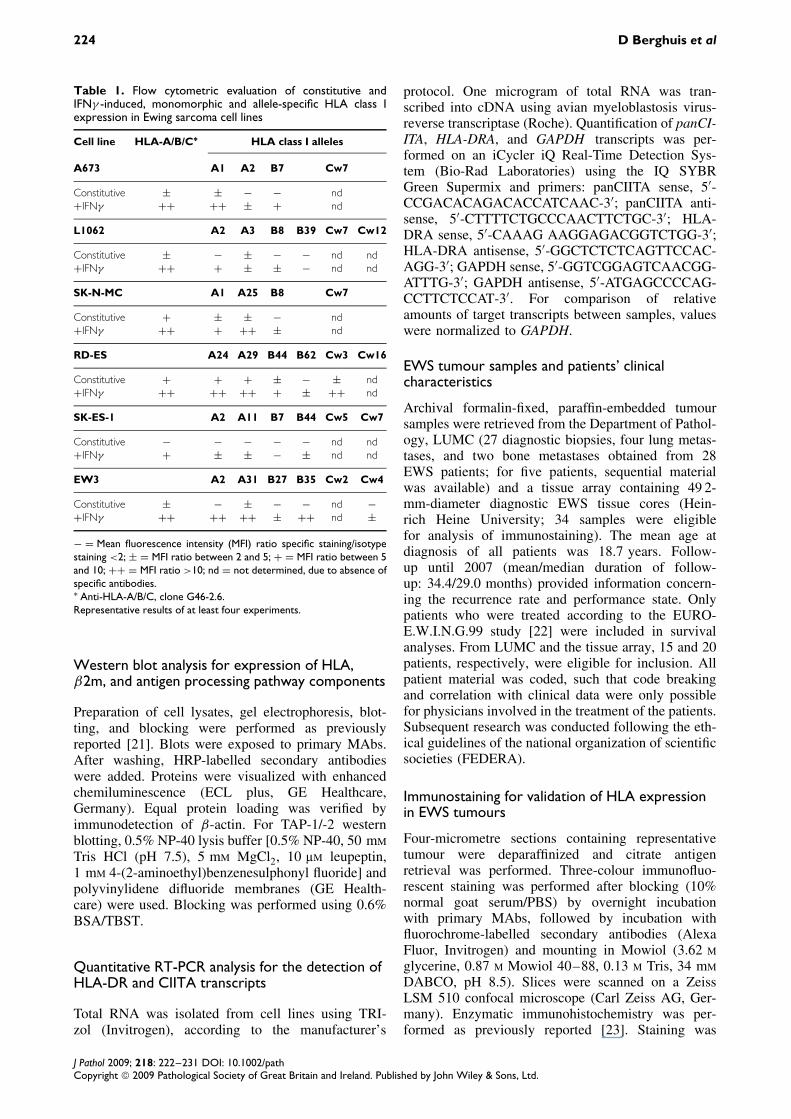
Table 1. Flow cytometric evaluation of constitutive andIFNγ -induced, monomorphic and allele-specific HLA class Iexpression in Ewing sarcoma cell lines
Cell line HLA-A/B/C∗ HLA class I alleles
A673 A1 A2 B7 Cw7
Constitutive ± ± − − nd+IFNγ ++ ++ ± + nd
L1062 A2 A3 B8 B39 Cw7 Cw12
Constitutive ± − ± − − nd nd+IFNγ ++ + ± ± − nd nd
SK-N-MC A1 A25 B8 Cw7
Constitutive + ± ± − nd+IFNγ ++ + ++ ± nd
RD-ES A24 A29 B44 B62 Cw3 Cw16
Constitutive + + + ± − ± nd+IFNγ ++ ++ ++ + ± ++ nd
SK-ES-1 A2 A11 B7 B44 Cw5 Cw7
Constitutive − − − − − nd nd+IFNγ + ± ± − ± nd nd
EW3 A2 A31 B27 B35 Cw2 Cw4
Constitutive ± − ± − − nd −+IFNγ ++ ++ ++ ± ++ nd ±
− = Mean fluorescence intensity (MFI) ratio specific staining/isotypestaining <2; ± = MFI ratio between 2 and 5; + = MFI ratio between 5and 10; ++ = MFI ratio >10; nd = not determined, due to absence ofspecific antibodies.∗ Anti-HLA-A/B/C, clone G46-2.6.Representative results of at least four experiments.
Western blot analysis for expression of HLA,β2m, and antigen processing pathway components
Preparation of cell lysates, gel electrophoresis, blot-ting, and blocking were performed as previouslyreported [21]. Blots were exposed to primary MAbs.After washing, HRP-labelled secondary antibodieswere added. Proteins were visualized with enhancedchemiluminescence (ECL plus, GE Healthcare,Germany). Equal protein loading was verified byimmunodetection of β-actin. For TAP-1/-2 westernblotting, 0.5% NP-40 lysis buffer [0.5% NP-40, 50 mM
Tris HCl (pH 7.5), 5 mM MgCl2, 10 µM leupeptin,1 mM 4-(2-aminoethyl)benzenesulphonyl fluoride] andpolyvinylidene difluoride membranes (GE Health-care) were used. Blocking was performed using 0.6%BSA/TBST.
Quantitative RT-PCR analysis for the detection ofHLA-DR and CIITA transcripts
Total RNA was isolated from cell lines using TRI-zol (Invitrogen), according to the manufacturer’s
protocol. One microgram of total RNA was tran-scribed into cDNA using avian myeloblastosis virus-reverse transcriptase (Roche). Quantification of panCI-ITA, HLA-DRA, and GAPDH transcripts was per-formed on an iCycler iQ Real-Time Detection Sys-tem (Bio-Rad Laboratories) using the IQ SYBRGreen Supermix and primers: panCIITA sense, 5′-CCGACACAGACACCATCAAC-3′; panCIITA anti-sense, 5′-CTTTTCTGCCCAACTTCTGC-3′; HLA-DRA sense, 5′-CAAAG AAGGAGACGGTCTGG-3′;HLA-DRA antisense, 5′-GGCTCTCTCAGTTCCAC-AGG-3′; GAPDH sense, 5′-GGTCGGAGTCAACGG-ATTTG-3′; GAPDH antisense, 5′-ATGAGCCCCAG-CCTTCTCCAT-3′. For comparison of relativeamounts of target transcripts between samples, valueswere normalized to GAPDH.
EWS tumour samples and patients’ clinicalcharacteristics
Archival formalin-fixed, paraffin-embedded tumoursamples were retrieved from the Department of Pathol-ogy, LUMC (27 diagnostic biopsies, four lung metas-tases, and two bone metastases obtained from 28EWS patients; for five patients, sequential materialwas available) and a tissue array containing 49 2-mm-diameter diagnostic EWS tissue cores (Hein-rich Heine University; 34 samples were eligiblefor analysis of immunostaining). The mean age atdiagnosis of all patients was 18.7 years. Follow-up until 2007 (mean/median duration of follow-up: 34.4/29.0 months) provided information concern-ing the recurrence rate and performance state. Onlypatients who were treated according to the EURO-E.W.I.N.G.99 study [22] were included in survivalanalyses. From LUMC and the tissue array, 15 and 20patients, respectively, were eligible for inclusion. Allpatient material was coded, such that code breakingand correlation with clinical data were only possiblefor physicians involved in the treatment of the patients.Subsequent research was conducted following the eth-ical guidelines of the national organization of scientificsocieties (FEDERA).
Immunostaining for validation of HLA expressionin EWS tumours
Four-micrometre sections containing representativetumour were deparaffinized and citrate antigenretrieval was performed. Three-colour immunofluo-rescent staining was performed after blocking (10%normal goat serum/PBS) by overnight incubationwith primary MAbs, followed by incubation withfluorochrome-labelled secondary antibodies (AlexaFluor, Invitrogen) and mounting in Mowiol (3.62 M
glycerine, 0.87 M Mowiol 40–88, 0.13 M Tris, 34 mM
DABCO, pH 8.5). Slices were scanned on a ZeissLSM 510 confocal microscope (Carl Zeiss AG, Ger-many). Enzymatic immunohistochemistry was per-formed as previously reported [23]. Staining was
J Pathol 2009; 218: 222–231 DOI: 10.1002/pathCopyright 2009 Pathological Society of Great Britain and Ireland. Published by John Wiley & Sons, Ltd.

HLA expression in Ewing sarcoma 225
scored semi-quantitatively by the quality control sys-tem proposed by Ruiter et al [24]. The intensity ofstaining was scored as 0, 1, 2 or 3 indicating absent,weak, clear or strong expression, respectively. Percent-ages of positive cells were scored as 0 for 0%, 1 for1–5%, 2 for 5–25%, 3 for 25–50%, 4 for 50–75%,and 5 for 75–100%. The sum of both scores wasused to identify three categories of expression: homo-geneous (total score 7–8); partial (3–6); and absent(0–2).
Statistical analysis
Statistical analyses were performed with the SPSSversion 12 software package. Mann-Whitney and chi-square or, where appropriate, Fisher’s exact tests wereused for comparison of the expression between sam-ples and associations between expression and clinico-pathological parameters. Five-year survival rates werecalculated according to the Kaplan–Meier methodusing the log-rank test. Overall survival was definedas survival until death due to disease, whereas disease-free survival was defined as the time to disease recur-rence or disease-specific death. All tests were two-sided; the significance level was set to 5%.
Results
Heterogeneous constitutive HLA class Iexpression in Ewing sarcoma cell lines
Surface expression of HLA class I antigenic deter-minants was investigated by flow cytometry in sixEWS cell lines, after staining with an anti-HLA-A/B/CMAb and allele-specific MAbs, respectively. Levelsof constitutive HLA-A/B/C surface expression var-ied among the cell lines, ranging from undetectable(SK-ES-1) to relatively high (SK-N-MC and RD-ES)(Table 1 and Figure 1A). Absence of detectable con-stitutive expression of at least one allele, most oftenwithin the HLA-B locus, was observed in all cell lines.Five cell lines exhibited complete lack of detectableconstitutive expression of either one or two loci, inall cases including the HLA-B locus (Table 1). Due tothe limited availability of HLA-C alloantigen-specificMAbs, no data were obtained regarding the expres-sion of most HLA-C alleles. Western blot analysisconfirmed the heterogeneity in constitutive HLA classI expression levels and excluded intracellular retentionof HLA class I molecules in the SK-ES-1 cell line tobe responsible for the lack of constitutive HLA classI surface expression (Figure 1B).
Invariable induction of HLA class I expression byIFNγ
In addition to the analysis of constitutive expres-sion levels, IFNγ -induced expression of HLA class Iwas investigated. Without exception, stimulation ofcell lines with IFNγ induced HLA-A/B/C expression
Figure 1. Constitutive and IFNγ -induced expression of HLAclass I, β2 microglobulin, and components of the antigenprocessing pathway in Ewing sarcoma cell lines. (A) Flowcytometric evaluation of HLA class I (anti-HLA-A/B/C, cloneG46-2.6) surface expression in unstimulated (dark grey) andIFNγ -stimulated (black) EWS cells. Representative results areshown for EWS cell lines SK-N-MC, EW3, and SK-ES-1. Similarresults were obtained for EWS cell lines A673, L1062, andRD-ES (not shown). Isotype-matched control staining is shownin light grey. (B) Western blot analysis for expression of HLA-A(HCA2 MAb), HLA-B/C (HC10 MAb), and the APP componentsTAP-1/-2, tapasin, LMP-2/-7, and β2m in unstimulated andIFNγ -stimulated EWS cell lines SK-N-MC, EW3, and SK-ES-1.Arrow: TAP-1. β-actin was used as a control for protein loading(not shown). (C) Differential IFNγ responses for the HLA-A2allele among Ewing sarcoma cell lines SK-ES-1, A673, L1062,and EW3. Mean fluorescence intensity ratio (MFI ratio) ofHLA-A2-specific staining/isotype-matched control staining
(Table 1 and Figures 1A and 1B), suggesting that theobserved absent or relatively low constitutive expres-sion levels are not due to genetic defects. Analysisof allele-specific expression supported this notion: all
J Pathol 2009; 218: 222–231 DOI: 10.1002/pathCopyright 2009 Pathological Society of Great Britain and Ireland. Published by John Wiley & Sons, Ltd.

226 D Berghuis et al
alleles were inducible by IFNγ , except for HLA-B7 inSK-ES-1 and HLA-A3/-B39 in L1062 (Table 1). Inter-estingly, differential IFNγ responses were observedfor identical alleles among the different cell lines (eglimited induction of HLA-A2 in SK-ES-1 and A673,while more robust induction of this allele in L1062and, particularly, EW3) (Figure 1C).
Simultaneous induction of antigen processingpathway components, HLA class I, and β2microglobulin by IFNγ in Ewing sarcoma cell lines
Since deficient expression of several antigen process-ing pathway (APP) components contributes to reducedHLA class I expression in various human tumours,the expression of APP components TAP-1/-2, tapasin,and LMP-2/-7 was investigated by western blot anal-ysis in three cell lines, selected on the basis ofdifferences in constitutive HLA class I expressionlevels. As shown in Figure 1B, constitutive TAP-1/-2 and tapasin expression levels were heterogeneousamong the cell lines, though congruent with thoseobserved for HLA class I. IFNγ stimulation induced
expression of these proteins, including the immuno-proteasome components LMP-2 and LMP-7, similarto HLA class I. β2m was constitutively expressed inall three cell lines and similar to the APP componentsand HLA class I, inducible by IFNγ . Together, thesedata demonstrate heterogeneous constitutive expres-sion, but IFNγ inducibility of all APP componentsand the HLA class I/β2m complex in EWS cell lines.
HLA class I expression in primary and metastaticEwing sarcoma tumour samples
To determine whether the observed heterogeneity inHLA class I expression in vitro reflected the in vivosituation, the expression of β2m, HLA-A, and HLA-B/C was evaluated in a large cohort of EWS tumoursamples by three-colour immunofluorescent stain-ing. Variation between as well as within individ-ual tumours was observed, ranging from completelack of detectable expression of both β2m and HLAclass I to homogeneous expression of these proteins(Figure 2). As shown in Table 2, 48 of 61 (79%) biop-sies exhibited aberrant HLA class I expression (definedas absent or partial expression). Seventeen biopsies
Figure 2. Heterogeneous HLA class I expression in Ewing sarcoma tumours. Representative examples of three-colourimmunofluorescent staining on formalin-fixed, paraffin-embedded EWS tumours. MAbs against CD99, β2m, and HLA class I heavychains [HLA-A (HCA2 MAb); HLA-B/C (HC10 MAb)] were used. (A + B) Diagnostic biopsy, homogeneous expression of β2mand HLA class I. (C + D) Diagnostic biopsy, focal (partial) expression of β2m and HLA class I. Stromal cells, including infiltratingleukocytes, were used as internal positive controls
J Pathol 2009; 218: 222–231 DOI: 10.1002/pathCopyright 2009 Pathological Society of Great Britain and Ireland. Published by John Wiley & Sons, Ltd.

HLA expression in Ewing sarcoma 227
Tab
le2.
Expr
essi
onof
β2
mic
rogl
obul
in,H
LAcl
ass
Ihea
vych
ains
,and
over
allH
LAcl
ass
Iin
diag
nost
ican
dm
etas
tatic
Ewin
gsa
rcom
atu
mou
rs
β2m
HL
A-A
HL
A-B
/CH
LA
clas
sI
(ove
rall)
Exp
ress
ion∗
Exp
ress
ion∗
Exp
ress
ion∗
Exp
ress
ion∗
Abs
ent
(%)
Par
tial
(%)
Ho
mo
gene
ous
(%)
Abs
ent
(%)
Par
tial
(%)
Ho
mo
gene
ous
(%)
Abs
ent
(%)
Par
tial
(%)
Ho
mo
gene
ous
(%)
Abs
ent
(%)
Par
tial
(%)
Ho
mo
gene
ous
(%)
Biop
sies
(n=
61)†
11(1
8)26
(43)
24(3
9)24
(39)
19(3
1)18
(30)
20(3
3)20
(33)
21(3
4)17
(28)
31(5
1)13
(21)
Met
asta
ses
(n=
6)#
4(6
7)2
(33)
0(0
)4
(67)
1(1
7)1
(17)
5(8
3)1
(17)
0(0
)4
(67)
2(3
3)0
(0)
∗ Exp
ress
ion
acco
rdin
gto
ref2
4.†
Biop
sies
,obt
aine
dat
diag
nosi
s#
Met
asta
ticle
sion
ssh
owed
sign
ifica
ntly
mor
eof
ten
tota
llos
sof
β2m
(p=
0.01
7,Fi
sher
’sex
act
test
)an
dH
LA-B
/Che
avy
chai
nex
pres
sion
(p=
0.02
4,Fi
sher
’sex
act
test
).
(28%) showed complete lack of detectable HLA classI. In 11 of these 17 cases (17% of all biopsies), absenceof HLA class I was accompanied by complete lack ofdetectable β2m. Within a cohort of uniformly treatedEWS patients (n = 35), neither β2m nor HLA class Iexpression was significantly associated with overallor event-free survival (data not shown). Comparedwith diagnostic biopsies, however, metastatic lesionsshowed significantly lower levels (Mann–Whitneytest, p = 0.001 and p = 0.01) and more often totalloss (Fisher’s exact test, p = 0.017 and p = 0.024) ofdetectable β2m and HLA-B/C heavy chain expression,respectively. This was due to the complete absence ofdetectable β2m and HLA class I expression in lungmetastases (n = 4). Bone metastases (n = 2), how-ever, expressed both β2m and HLA class I. This differ-ence between lung and bone metastases was observedeven within a single patient, presenting with β2m- andHLA class I-positive bone metastases 4 years afterresection of a β2m- and HLA class I-negative lungmetastasis (Table 3, UPN 1). Sequential patient mate-rial demonstrated a tendency towards decreased levelsof β2m and HLA class I expression upon disease pro-gression, particularly when relatively long periods oflatency existed between the occurrences of consecutiveevents (Table 3, UPN 2–3).
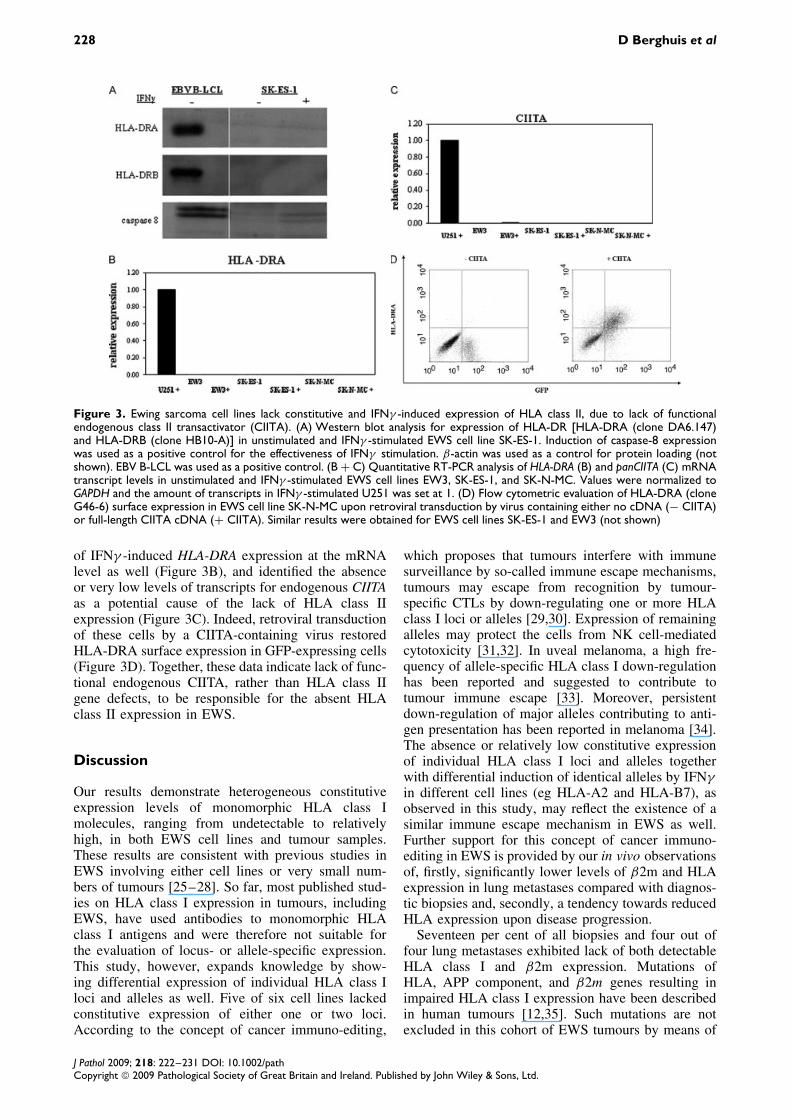
Absence of functional endogenous CIITA causeslack of HLA class II expression in Ewing sarcoma
Since HLA class II expression may contribute toimmune recognition of tumours, the expression ofHLA class II was investigated in EWS cell linesand tumours. Flow cytometric analysis showed lackof IFNγ -induced expression of HLA-DR/DP/DQ insix constitutively HLA class II-negative cell lines(data not shown). Western blot analysis confirmedthe absence of expression and excluded intracellularretention of HLA class II proteins in SK-ES-1 cells(Figure 3A). Immunohistochemical staining demon-strated lack of HLA class II expression in EWStumour samples as well (data not shown). Quantita-tive RT-PCR in three cell lines demonstrated absence
Table 3. Tendency towards decreased β2 microglobulin andoverall HLA class I expression levels upon progression of disease
UPNSample type
(years after preceding sample) β2mHLA class I
(overall)
1 Lung metastasis ° °Bone metastasis (4) ž ž2 Diagnostic biopsy ž ž
Lung metastasis (5) ° °3 Diagnostic biopsy ž žLung metastasis (4) ° °4 Diagnostic biopsy ž žBone metastasis (1) ž ž
5 Diagnostic biopsy ° °Lung metastasis (1) ° °Expression according to ref 24. ° = absent expression; ž = partialexpression; UPN = patient number.
J Pathol 2009; 218: 222–231 DOI: 10.1002/pathCopyright 2009 Pathological Society of Great Britain and Ireland. Published by John Wiley & Sons, Ltd.
228 D Berghuis et al
Figure 3. Ewing sarcoma cell lines lack constitutive and IFNγ -induced expression of HLA class II, due to lack of functionalendogenous class II transactivator (CIITA). (A) Western blot analysis for expression of HLA-DR [HLA-DRA (clone DA6.147)and HLA-DRB (clone HB10-A)] in unstimulated and IFNγ -stimulated EWS cell line SK-ES-1. Induction of caspase-8 expressionwas used as a positive control for the effectiveness of IFNγ stimulation. β-actin was used as a control for protein loading (notshown). EBV B-LCL was used as a positive control. (B + C) Quantitative RT-PCR analysis of HLA-DRA (B) and panCIITA (C) mRNAtranscript levels in unstimulated and IFNγ -stimulated EWS cell lines EW3, SK-ES-1, and SK-N-MC. Values were normalized toGAPDH and the amount of transcripts in IFNγ -stimulated U251 was set at 1. (D) Flow cytometric evaluation of HLA-DRA (cloneG46-6) surface expression in EWS cell line SK-N-MC upon retroviral transduction by virus containing either no cDNA (− CIITA)or full-length CIITA cDNA (+ CIITA). Similar results were obtained for EWS cell lines SK-ES-1 and EW3 (not shown)
of IFNγ -induced HLA-DRA expression at the mRNAlevel as well (Figure 3B), and identified the absenceor very low levels of transcripts for endogenous CIITAas a potential cause of the lack of HLA class IIexpression (Figure 3C). Indeed, retroviral transductionof these cells by a CIITA-containing virus restoredHLA-DRA surface expression in GFP-expressing cells(Figure 3D). Together, these data indicate lack of func-tional endogenous CIITA, rather than HLA class IIgene defects, to be responsible for the absent HLAclass II expression in EWS.
Discussion
Our results demonstrate heterogeneous constitutiveexpression levels of monomorphic HLA class Imolecules, ranging from undetectable to relativelyhigh, in both EWS cell lines and tumour samples.These results are consistent with previous studies inEWS involving either cell lines or very small num-bers of tumours [25–28]. So far, most published stud-ies on HLA class I expression in tumours, includingEWS, have used antibodies to monomorphic HLAclass I antigens and were therefore not suitable forthe evaluation of locus- or allele-specific expression.This study, however, expands knowledge by show-ing differential expression of individual HLA class Iloci and alleles as well. Five of six cell lines lackedconstitutive expression of either one or two loci.According to the concept of cancer immuno-editing,
which proposes that tumours interfere with immunesurveillance by so-called immune escape mechanisms,tumours may escape from recognition by tumour-specific CTLs by down-regulating one or more HLAclass I loci or alleles [29,30]. Expression of remainingalleles may protect the cells from NK cell-mediatedcytotoxicity [31,32]. In uveal melanoma, a high fre-quency of allele-specific HLA class I down-regulationhas been reported and suggested to contribute totumour immune escape [33]. Moreover, persistentdown-regulation of major alleles contributing to anti-gen presentation has been reported in melanoma [34].The absence or relatively low constitutive expressionof individual HLA class I loci and alleles togetherwith differential induction of identical alleles by IFNγin different cell lines (eg HLA-A2 and HLA-B7), asobserved in this study, may reflect the existence of asimilar immune escape mechanism in EWS as well.Further support for this concept of cancer immuno-editing in EWS is provided by our in vivo observationsof, firstly, significantly lower levels of β2m and HLAexpression in lung metastases compared with diagnos-tic biopsies and, secondly, a tendency towards reducedHLA expression upon disease progression.
Seventeen per cent of all biopsies and four out offour lung metastases exhibited lack of both detectableHLA class I and β2m expression. Mutations ofHLA, APP component, and β2m genes resulting inimpaired HLA class I expression have been describedin human tumours [12,35]. Such mutations are notexcluded in this cohort of EWS tumours by means of
J Pathol 2009; 218: 222–231 DOI: 10.1002/pathCopyright 2009 Pathological Society of Great Britain and Ireland. Published by John Wiley & Sons, Ltd.

HLA expression in Ewing sarcoma 229
gene sequencing. However, the observed intact IFNγinducibility of all APP components, β2m, and HLAclass I molecules in vitro, which can be explainedby the presence of interferon-stimulated response ele-ments in the promoters of these genes, indicates thatregulatory mechanisms are mainly responsible forabsent or low constitutive expression levels. Equiva-lent patterns of expression of APP components, β2m,and HLA class I have been reported in other humantumours [36–39]. Although we cannot exclude thatinsufficient expression of a single APP component (asdescribed by, among others, Lou et al [40]), ratherthan coordinated regulation of several APP compo-nents, restricts constitutive HLA class I expression,the observed IFNγ -induced expression indicates thatstructural aberrations are an unlikely cause for the lackof expression in EWS tumours. Support for this state-ment is provided by publications demonstrating lackof secondary genetic aberrations, acquired in addi-tion to the EWS-specific chromosomal translocations,of chromosome 6 (TAP1/-2, LMP-2/-7, tapasin, andHLA class I genes) and chromosome 15 (β2m gene)in localized and metastatic EWS tumours [41,42].
Our results demonstrate lack of (inducible) HLAclass II expression in EWS, both in vitro and in vivo,which appeared to be due to lack of endogenous CIITAexpression rather than HLA class II gene defects. Epi-genetic modifications of gene promoters, includingCIITA promoter, have been reported to influence HLAexpression in cancer [43–45]. However, pilot experi-ments in which EWS cells were pre-incubated withepigenetic modulators (Trichostatin A or 5-aza-2′-deoxycytidine) and IFNγ neither induced HLA class IInor increased HLA class I expression (data not shown).Structural CIITA gene defects seem unlikely to causethe observed lack of expression, since such defectshave seldom been described other than for patientswith specific MHC-II deficiencies [46]. Defects inthe IFNγ -signalling pathway are proven unlikely byour observation of IFNγ -induced HLA class I expres-sion. In developmental tissues, lack of inducible HLAclass II expression is assumed to reflect the silent stateof these genes during early development, creating animmune privilege for these cells to evade maternalimmune recognition. EWSs are primitive, undifferen-tiated tumours, considered to be derived from eitherneuro-ectodermal or mesenchymal stem cells [28,47].
Whether such a silent state, rather than the processesof neoplastic transformation and cancer immuno-editing, accounts for the absence of HLA class IIas well as the observed constitutive HLA class Iexpression levels in EWS remains to be established.However, irrespective of the underlying mechanism,the obtained knowledge about HLA expression inEWS may direct future immunotherapy studies usingeither (xenografted) murine or human tumour models[48,49]. Within the limitations caused by inter-speciesdifferences, EWS xenograft models using mice withdifferent degrees of immunodeficiency may furtherclarify the contribution of immune surveillance and
cancer immuno-editing to HLA phenotypes [29]. Het-erogeneity in HLA class I expression, as observed inthis study, may affect immune recognition of EWStumours by tumour-reactive T or NK cells and there-fore influence the efficacy of cellular immunothera-peutic strategies using these cytotoxic effector cells.
Acknowledgements
We would like to thank Dr B Mach (University of Geneva,Geneva, Switzerland) for provision of the vector pEBO-CIITA; Dr G Nolan (Stanford University, Stanford, California,USA) for provision of the retroviral vector pLZRS-pBMN;Professor Dr J Neefjes (Dutch Cancer Institute, Amsterdam,The Netherlands), Dr R Tampe (Institute of Biochemistry,Johann Wolfgang Goethe University, Frankfurt, Germany),Dr P van Endert (Hopital Necker, Paris, France), and Dr PCresswell (Howard Hughes Medical Institute, Yale UniversitySchool of Medicine, New Haven, CT, USA) for provision ofMAbs; and MC Verweij (Deparment of Medical Microbiologyand Center for Infectious Diseases, Leiden University MedicalCenter, Leiden, The Netherlands), G Alblas (Department ofPediatrics, Leiden University Medical Center, Leiden, TheNetherlands), Dr KBJ Scholten (Department of Pathology, VUUniversity Medical Center, Amsterdam, The Netherlands), KGvan der Ham (Department of Pathology, Leiden UniversityMedical Center, Leiden, The Netherlands), and MJ Ronkes fortechnical assistance. This study was financially supported byDutch Cancer Society Nederlandse Kankerbestrijding/KoninginWilhelmina Fonds (grant RUL 2003-2800 to ASKdeH andSJS; grant RUL 2005-3259 to DH); the European Commission(EuroBoNeT, grant No 018814), and Stichting The Quality ofLife Gala 2007.
Supporting information
Supporting information may be found in the onlineversion of this article.
References
# References 50 and 51 are cited in the supporting information tothis article.
1. van den Berg H, Kroon HM, Slaar A, Hogendoorn PCW.Incidence of biopsy-proven bone tumors in children: a report basedon the Dutch pathology registration ‘PALGA’. J Pediatr Orthop2008;28:29–35.
2. Ushigome S, Machinami R, Sorensen PH. Ewing sar-coma/primitive neuroectodermal tumour. In World Health Orga-nization Classification of Tumours. Pathology and Genetics ofTumours of Soft tissue and Bone, Fletcher CDM, Unni KK,Mertens F (eds). IARC Press: Lyon, 2002; 298–300.
3. Bernstein M, Kovar H, Paulussen M, Randall RL, Schuck A,Teot LA, et al. Ewing’s sarcoma family of tumors: currentmanagement. Oncologist 2006;11:503–519.
4. Gattinoni L, Powell DJ Jr, Rosenberg SA, Restifo NP. Adoptiveimmunotherapy for cancer: building on success. Nature RevImmunol 2006;6:383–393.
5. Melief CJ, Toes RE, Medema JP, van der Burg SH, Ossendorp F,Offringa R. Strategies for immunotherapy of cancer. Adv Immunol2000;75:235–282.
6. Malmberg KJ, Bryceson YT, Carlsten M, Andersson S, Bjork-lund A, Bjorkstrom NK, et al. NK cell-mediated targeting of
J Pathol 2009; 218: 222–231 DOI: 10.1002/pathCopyright 2009 Pathological Society of Great Britain and Ireland. Published by John Wiley & Sons, Ltd.
230 D Berghuis et al
human cancer and possibilities for new means of immunotherapy.Cancer Immunol Immunother 2008;57:1541–1552.
7. Tait BD. HLA class I expression on human cancer cells.Implications for effective immunotherapy. Hum Immunol2000;61:158–165.
8. Bjorkman PJ, Parham P. Structure, function, and diversity of classI major histocompatibility complex molecules. Annu Rev Biochem1990;59:253–288.
9. Pamer E, Cresswell P. Mechanisms of MHC class I — restrictedantigen processing. Annu Rev Immunol 1998;16:323–358.
10. Jensen PE. Recent advances in antigen processing and presenta-tion. Nature Immunol 2007;8:1041–1048.
11. van den Elsen PJ, Holling TM, Kuipers HF, van der Stoep N.Transcriptional regulation of antigen presentation. Curr OpinImmunol 2004;16:67–75.
12. Chang CC, Ferrone S. Immune selective pressure and HLA class Iantigen defects in malignant lesions. Cancer Immunol Immunother2007;56:227–236.
13. Tsukahara T, Kawaguchi S, Torigoe T, Asanuma H, Nakazawa E,Shimozawa K, et al. Prognostic significance of HLA class Iexpression in osteosarcoma defined by anti-pan HLA class Imonoclonal antibody, EMR8-5. Cancer Sci 2006;97:1374–1380.
14. Vitale M, Pelusi G, Taroni B, Gobbi G, Micheloni C, Rezzani R,et al. HLA class I antigen down-regulation in primary ovarycarcinoma lesions: association with disease stage. Clin Cancer Res2005;11:67–72.
15. Watson NF, Ramage JM, Madjd Z, Spendlove I, Ellis IO, Schole-field JH, et al. Immunosurveillance is active in colorectal cancer asdownregulation but not complete loss of MHC class I expressioncorrelates with a poor prognosis. Int J Cancer 2006;118:6–10.
16. Rodriguez T, Mendez R, Del Campo A, Aptsiauri N, Martin J,Orozco G, et al. Patterns of constitutive and IFN-gamma inducibleexpression of HLA class II molecules in human melanoma celllines. Immunogenetics 2007;59:123–133.
17. Holling TM, Schooten E, Langerak AW, van den Elsen PJ.Regulation of MHC class II expression in human T-cellmalignancies. Blood 2004;103:1438–1444.
18. Urano F, Umezawa A, Yabe H, Hong W, Yoshida K, Fujinaga K,et al. Molecular analysis of Ewing’s sarcoma: another fusiongene, EWS-E1AF, available for diagnosis. Jpn J Cancer Res1998;89:703–711.
19. Szuhai K, Ijszenga M, Tanke HJ, Rosenberg C, Hogendoorn PCW.Molecular cytogenetic characterization of four previously estab-lished and two newly established Ewing sarcoma cell lines. CancerGenet Cytogenet 2006;166:173–179.
20. Heemskerk MH, Hooijberg E, Ruizendaal JJ, van der Weide MM,Kueter E, Bakker AQ, et al. Enrichment of an antigen-specific Tcell response by retrovirally transduced human dendritic cells. CellImmunol 1999;195:10–17.
21. de Hooge ASK, Berghuis D, Santos SJ, Mooiman E, Romeo S,Kummer JA, et al. Expression of cellular FLICE inhibitoryprotein, caspase-8, and protease inhibitor-9 in Ewing sarcoma andimplications for susceptibility to cytotoxic pathways. Clin CancerRes 2007;13:206–214.
22. Juergens C, Weston C, Lewis I, Whelan J, Paulussen M, Ober-lin O, et al. Safety assessment of intensive induction with vin-cristine, ifosfamide, doxorubicin, and etoposide (VIDE) in thetreatment of Ewing tumors in the EURO-E.W.I.N.G. 99 clinicaltrial. Pediatr Blood Cancer 2006;47:22–29.
23. Bovee JVMG, van den Broek LJ, de Boer WI, Hogendoorn PCW.Expression of growth factors and their receptors in adamantinomaof long bones and the implication for its histogenesis. J Pathol1998;184:24–30.
24. Ruiter DJ, Ferrier CM, van Muijen GN, Henzen-Logmans SC,Kennedy S, Kramer MD, et al. Quality control of immunohisto-chemical evaluation of tumour-associated plasminogen activatorsand related components. European BIOMED-1 Concerted Actionon Clinical Relevance of Proteases in Tumour Invasion and Metas-tasis. Eur J Cancer 1998;34:1334–1340.
25. Borowski A, van Valen F, Ulbrecht M, Weiss EH, Blasczyk R,Jurgens H, et al. Monomorphic HLA class I-(non-A, non-B)
expression on Ewing’s tumor cell lines, modulation by TNF-alphaand IFN-gamma. Immunobiology 1999;200:1–20.
26. Mechtersheimer G, Barth T, Ludwig R, Staudter M, Moller P.Differential expression of leukocyte differentiation antigens insmall round blue cell sarcomas. Cancer 1993;71:237–248.
27. Mechtersheimer G, Staudter M, Majdic O, Dorken B, Mold-enhauer G, Moller P. Expression of HLA-A,B,C, beta 2-microglobulin (beta 2m), HLA-DR, -DP, -DQ and of HLA-D-associated invariant chain (Ii) in soft-tissue tumors. Int J Cancer1990;46:813–823.
28. Lipinski M, Braham K, Philip I, Wiels J, Philip T, Goridis C,et al. Neuroectoderm-associated antigens on Ewing’s sarcoma celllines. Cancer Res 1987;47:183–187.
29. Garcia-Lora A, Algarra I, Garrido F. MHC class I antigens,immune surveillance, and tumor immune escape. J Cell Physiol2003;195:346–355.
30. Dunn GP, Old LJ, Schreiber RD. The immunobiology of cancerimmunosurveillance and immunoediting. Immunity2004;21:137–148.
31. Storkus WJ, Alexander J, Payne JA, Dawson JR, Cresswell P.Reversal of natural killing susceptibility in target cells expressingtransfected class I HLA genes. Proc Natl Acad Sci U S A1989;86:2361–2364.
32. Wagtmann N, Rajagopalan S, Winter CC, Peruzzi M, Long EO.Killer cell inhibitory receptors specific for HLA-C and HLA-Bidentified by direct binding and by functional transfer. Immunity1995;3:801–809.
33. Hurks HM, Metzelaar-Blok JA, Mulder A, Claas FH, Jager MJ.High frequency of allele-specific down-regulation of HLA classI expression in uveal melanoma cell lines. Int J Cancer2000;85:697–702.
34. Ikeda H, Lethe B, Lehmann F, van Baren N, Baurain JF, deSmet C, et al. Characterization of an antigen that is recognizedon a melanoma showing partial HLA loss by CTL expressing anNK inhibitory receptor. Immunity 1997;6:199–208.
35. Lankat-Buttgereit B, Tampe R. The transporter associated withantigen processing: function and implications in human diseases.Physiol Rev 2002;82:187–204.
36. Wright KL, White LC, Kelly A, Beck S, Trowsdale J, Ting JP.Coordinate regulation of the human TAP1 and LMP2 genes froma shared bidirectional promoter. J Exp Med 1995;181:1459–1471.
37. Gobin SJ, van Zutphen M, Woltman AM, van den Elsen PJ.Transactivation of classical and nonclassical HLA class Igenes through the IFN-stimulated response element. J Immunol1999;163:1428–1434.
38. Romero JM, Jimenez P, Cabrera T, Cozar JM, Pedrinaci S,Tallada M, et al. Coordinated downregulation of the antigenpresentation machinery and HLA class I/beta2-microglobulincomplex is responsible for HLA-ABC loss in bladder cancer. IntJ Cancer 2005;113:605–610.
39. Johnsen A, France J, Sy MS, Harding CV. Down-regulation of thetransporter for antigen presentation, proteasome subunits, and classI major histocompatibility complex in tumor cell lines. Cancer Res1998;58:3660–3667.
40. Lou Y, Vitalis TZ, Basha G, Cai B, Chen SS, Choi KB, et al.Restoration of the expression of transporters associated withantigen processing in lung carcinoma increases tumor-specificimmune responses and survival. Cancer Res 2005;65:7926–7933.
41. Brisset S, Schleiermacher G, Peter M, Mairal A, Oberlin O,Delattre O, et al. CGH analysis of secondary genetic changes inEwing tumors: correlation with metastatic disease in a series of 43cases. Cancer Genet Cytogenet 2001;130:57–61.
42. Ferreira BI, Alonso J, Carrillo J, Acquadro F, Largo C, Suela J,et al. Array CGH and gene-expression profiling reveals distinctgenomic instability patterns associated with DNA repair andcell-cycle checkpoint pathways in Ewing’s sarcoma. Oncogene2007;27:2084–2090.
43. van den Elsen PJ, Holling TM, van der Stoep N, Boss JM.DNA methylation and expression of major histocompatibilitycomplex class I and class II transactivator genes in humandevelopmental tumor cells and in T cell malignancies. ClinImmunol 2003;109:46–52.
J Pathol 2009; 218: 222–231 DOI: 10.1002/pathCopyright 2009 Pathological Society of Great Britain and Ireland. Published by John Wiley & Sons, Ltd.
HLA expression in Ewing sarcoma 231
44. Morimoto Y, Toyota M, Satoh A, Murai M, Mita H, Suzuki H,et al. Inactivation of class II transactivator by DNA methylationand histone deacetylation associated with absence of HLA-DRinduction by interferon-gamma in haematopoietic tumour cells. BrJ Cancer 2004;90:844–852.
45. Satoh A, Toyota M, Ikeda H, Morimoto Y, Akino K, Mita H,et al. Epigenetic inactivation of class II transactivator (CIITA) isassociated with the absence of interferon-gamma-induced HLA-DR expression in colorectal and gastric cancer cells. Oncogene2004;23:8876–8886.
46. Peijnenburg A, van den Berg R, Van Eggermond MJ, Sanal O,Vossen JM, Lennon AM, et al. Defective MHC class II expressionin an MHC class II deficiency patient is caused by a novel deletionof a splice donor site in the MHC class II transactivator gene.Immunogenetics 2000;51:42–49.
47. Tirode F, Laud-Duval K, Prieur A, Delorme B, Charbord P,Delattre O. Mesenchymal stem cell features of Ewing tumors.Cancer Cell 2007;11:421–429.
48. Llombart-Bosch A, Carda C, Peydro-Olaya A, Noguera R, Perez-Bacete M, Pellin A, et al. Soft tissue Ewing’s sarcoma. Charac-terization in established cultures and xenografts with evidence ofa neuroectodermic phenotype. Cancer 1990;66:2589–2601.
49. Staege MS, Hansen G, Baersch G, Burdach S. Functional andmolecular characterization of interleukin-2 transgenic Ewingtumor cells for in vivo immunotherapy. Pediatr Blood Cancer2004;43:23–34.
50. Mulder A, Kardol M, Regan J, Buelow R, Claas F. Reactivity oftwenty-two cytotoxic human monoclonal HLA antibodies towardssoluble HLA class I in an enzyme-linked immunosorbent assay(PRA-STAT). Hum Immunol 1997;56:106–113.
51. Mehta AM, Jordanova ES, Kenter GG, Ferrone S, Fleuren G.Association of antigen processing machinery and HLA class Idefects with clinicopathological outcome in cervical carcinoma.Cancer Immunol Immunother 2008;57:197–206.
J Pathol 2009; 218: 222–231 DOI: 10.1002/pathCopyright 2009 Pathological Society of Great Britain and Ireland. Published by John Wiley & Sons, Ltd.