HIV1 gp120 N-linked glycosylation differs between plasma and leukocyte compartments
10
BioMed Central Page 1 of 10 (page number not for citation purposes) Virology Journal Open Access Research HIV-1 gp120 N-linked glycosylation differs between plasma and leukocyte compartments Yung Shwen Ho 1,4 , Ana B Abecasis 2 , Kristof Theys 2 , Koen Deforche 2 , Dominic E Dwyer 3 , Michael Charleston 4 , Anne Mieke Vandamme 2 and Nitin K Saksena* 1 Address: 1 Retroviral Genetics Laboratory, Centre for Virus Research, Westmead Millennium Institute, Westmead Hospital, University of Sydney, Westmead NSW. 2145 Sydney. Australia, 2 Clinical and Epidemiological Virology, Rega Institute for Medical Research, Leuven, Belgium, 3 Centre for Infectious Diseases and Microbiology Laboratory Services, ICPMR, Westmead Hospital, Westmead NSW 2145, Australia and 4 School of Information Technologies, University of Sydney, Camperdown NSW 2006, Australia Email: Yung Shwen Ho - [email protected]; Ana B Abecasis - [email protected]; Kristof Theys - [email protected]; Koen Deforche - [email protected]; Dominic E Dwyer - [email protected]; Michael Charleston - [email protected]; Anne Mieke Vandamme - [email protected]; Nitin K Saksena* - [email protected] * Corresponding author Abstract Background: N-linked glycosylation is a major mechanism for minimizing virus neutralizing antibody response and is present on the Human Immunodeficiency Virus (HIV) envelope glycoprotein. Although it is known that glycosylation changes can dramatically influence virus recognition by the host antibody, the actual contribution of compartmental differences in N-linked glycosylation patterns remains unclear. Methodology and Principal Findings: We amplified the env gp120 C2-V5 region and analyzed 305 clones derived from plasma and other compartments from 15 HIV-1 patients. Bioinformatics and Bayesian network analyses were used to examine N-linked glycosylation differences between compartments. We found evidence for cellspecific single amino acid changes particular to monocytes, and significant variation was found in the total number of N-linked glycosylation sites between patients. Further, significant differences in the number of glycosylation sites were observed between plasma and cellular compartments. Bayesian network analyses showed an interdependency between N-linked glycosylation sites found in our study, which may have immense functional relevance. Conclusion: Our analyses have identified single cell/compartment-specific amino acid changes and differences in N-linked glycosylation patterns between plasma and diverse blood leukocytes. Bayesian network analyses showed associations inferring alternative glycosylation pathways. We believe that these studies will provide crucial insights into the host immune response and its ability in controlling HIV replication in vivo. These findings could also have relevance in shielding and evasion of HIV-1 from neutralizing antibodies. Published: 23 January 2008 Virology Journal 2008, 5:14 doi:10.1186/1743-422X-5-14 Received: 18 December 2007 Accepted: 23 January 2008 This article is available from: http://www.virologyj.com/content/5/1/14 © 2008 Ho et al; licensee BioMed Central Ltd. This is an Open Access article distributed under the terms of the Creative Commons Attribution License (http://creativecommons.org/licenses/by/2.0 ), which permits unrestricted use, distribution, and reproduction in any medium, provided the original work is properly cited.
-
Upload
independent -
Category
Documents
-
view
2 -
download
0
Transcript of HIV1 gp120 N-linked glycosylation differs between plasma and leukocyte compartments

BioMed CentralVirology Journal
ss
Open AcceResearchHIV-1 gp120 N-linked glycosylation differs between plasma and leukocyte compartmentsYung Shwen Ho1,4, Ana B Abecasis2, Kristof Theys2, Koen Deforche2, Dominic E Dwyer3, Michael Charleston4, Anne Mieke Vandamme2 and Nitin K Saksena*1Address: 1Retroviral Genetics Laboratory, Centre for Virus Research, Westmead Millennium Institute, Westmead Hospital, University of Sydney, Westmead NSW. 2145 Sydney. Australia, 2Clinical and Epidemiological Virology, Rega Institute for Medical Research, Leuven, Belgium, 3Centre for Infectious Diseases and Microbiology Laboratory Services, ICPMR, Westmead Hospital, Westmead NSW 2145, Australia and 4School of Information Technologies, University of Sydney, Camperdown NSW 2006, Australia
Email: Yung Shwen Ho - [email protected]; Ana B Abecasis - [email protected]; Kristof Theys - [email protected]; Koen Deforche - [email protected]; Dominic E Dwyer - [email protected]; Michael Charleston - [email protected]; Anne Mieke Vandamme - [email protected]; Nitin K Saksena* - [email protected]
* Corresponding author
AbstractBackground: N-linked glycosylation is a major mechanism for minimizing virus neutralizingantibody response and is present on the Human Immunodeficiency Virus (HIV) envelopeglycoprotein. Although it is known that glycosylation changes can dramatically influence virusrecognition by the host antibody, the actual contribution of compartmental differences in N-linkedglycosylation patterns remains unclear.
Methodology and Principal Findings: We amplified the env gp120 C2-V5 region and analyzed305 clones derived from plasma and other compartments from 15 HIV-1 patients. Bioinformaticsand Bayesian network analyses were used to examine N-linked glycosylation differences betweencompartments. We found evidence for cellspecific single amino acid changes particular tomonocytes, and significant variation was found in the total number of N-linked glycosylation sitesbetween patients. Further, significant differences in the number of glycosylation sites wereobserved between plasma and cellular compartments. Bayesian network analyses showed aninterdependency between N-linked glycosylation sites found in our study, which may have immensefunctional relevance.
Conclusion: Our analyses have identified single cell/compartment-specific amino acid changes anddifferences in N-linked glycosylation patterns between plasma and diverse blood leukocytes.Bayesian network analyses showed associations inferring alternative glycosylation pathways. Webelieve that these studies will provide crucial insights into the host immune response and its abilityin controlling HIV replication in vivo. These findings could also have relevance in shielding andevasion of HIV-1 from neutralizing antibodies.
Published: 23 January 2008
Virology Journal 2008, 5:14 doi:10.1186/1743-422X-5-14
Received: 18 December 2007Accepted: 23 January 2008
This article is available from: http://www.virologyj.com/content/5/1/14
© 2008 Ho et al; licensee BioMed Central Ltd. This is an Open Access article distributed under the terms of the Creative Commons Attribution License (http://creativecommons.org/licenses/by/2.0), which permits unrestricted use, distribution, and reproduction in any medium, provided the original work is properly cited.
Page 1 of 10(page number not for citation purposes)
Virology Journal 2008, 5:14 http://www.virologyj.com/content/5/1/14
IntroductionThe HIV-1 envelope (env) gp120 region plays a crucialrole in the entry of HIV-1 into target cells through thefusion of viral envelope with the target cell membrane.Variable regions (V1-V5) in env are spaced between theconserved regions (C1-C5). Both N-linked and O-linkedglycans are present on the HIV envelope glycoprotein. O-linked glycans are present on several unidentified serineor threonine residues in env gp120, but very little isknown about their actual role in governing the viral phe-notype of both HIV and simian immunodeficiency virus(SIV) [1,2]. In contrast, N-linked glycans comprise about50% of the mass of the env gp160 [3]. These sugar moie-ties are involved in various activities such as metabolism,transport, structural maintenance of the cell and protein,protein folding, recognition of particular cell types andadhesion to other cells. The N-linked glycosylation (NLG)of viral envelope proteins, through the formation of a"glycan shield", is one of the major mechanisms forblocking or minimizing virus neutralizing antibodyresponse [4]. which promotes viral persistence andimmune evasion. This has been demonstrated in SIV[5,6], HIV-1 [4,7] influenza virus [8], hepatitis B virus [9]and the Lactate Dehydrogenase-elevating Virus [10].Despite considerable genetic variation in HIV strains, thenumber of NLG are often found to be around 25 sites inthe HIV-1 env gp120 region [11], suggesting that strongselective pressures maintain this number [4]. The HIVenvelope "glycan shield" is known to evolve in responseto host antibodies [4] and it is thought that the density ofgp120 NLG is a significant obstacle to the design of effec-tive vaccine and elicitation of humoral immuneresponses. Any alteration or positional shift of a glycosyla-tion site (commonly seen in HIV and SIV glycoproteins)can have dramatic consequences for the virus and its rec-ognition by the antibody.
Although recent studies have shown compartmentaliza-tion of HIV-1 NLG sites between viral populations inplasma and the female genital tract [12,13], the criticalissue of possible differences in NLG of HIV-1 strainsderived from cell-associated and cell-free compartmentsremains unexplored. Such differences are important tofuture drug development because the drugs used in highlyactive antiretroviral therapy (HAART) primarily targetplasma or cell-free virus. Cell-free virus has a high turno-ver rate (< 6 hours) [4] and therefore has a strong need tomaintain viral integrity through constant shielding fromhost antibodies. In contrast, cell-associated virus are keptaway from neutralizing antibodies and can remain inte-grated in the human genome indefinitely. They can pro-duce viral progeny upon activation in vivo and this acts asan impediment to the success of therapy. The integratedprovirus concealed in diverse blood leukocyte popula-tions is one strategy HIV uses to avoid immune detection.
Given the incessant virus trafficking between cellfree andcell-associated compartments, a clear determination ofdifferences of HIV populations in plasma and diverse celltypes is needed to understand critical molecular determi-nants for viral survival, turnover, evasion and adaptationin vivo. The relevance of NLG is also known for manyother viruses [14-16]. Together, these studies imply thatthe virus-producing cell type is an important factor, whichmay be crucial in viral tropism and transmissibility in vivo.
The role of single amino acid residue changes in the HIV-1 env in its adaptation to cellular compartments remainssimilarly unexplored. Given that different cellular com-partments have different immune functions in our body,we suspect that the virus populations within them are sub-jected to distinct selection pressures, as opposed to freelycirculating virus in plasma [17]. These distinct selectiveforces may further exert influence on the make-up of NLG,depending on the cell type. This evolutionary make-upmay, in turn, define biological and functional aspects ofviral variants in a given environment. Different glycosyla-tion sites have been shown to offer variable sensitivity toantibody-mediated neutralization [4], such as sites in theenv hypervariable C3 and C5 regions. The sites around thebase of the V3 loop have been consistently found to beassociated with neutralization sensitivity, especially inHIV-1 subtype B viruses [4]. As the majority (17 of 25) ofNLG sites are concentrated in the env C2-V5 region ofgp120, and given the functional relevance of glycans inHIV pathogenesis, we chose this region for the study ofHIV-1 glycosylation patterns in cell-free and cell-associ-ated compartments.
We analyzed 305 clones of HIV-1 variants derived fromplasma and diverse blood leukocytes (whole PeripheralBlood Mononuclear Cell (PBMC), CD4+ T cells, CD8+ Tcells and monocytes) from 15 HIV-1 infected patients onHAART, each displaying different levels of plasma viremiaand T cell counts. In addition to the patient specificchanges observed in our analyses, we further found evi-dence in favor of compartmental NLG differences and dis-tinct cell-specific molecular changes in the env C2-V3region of HIV-1.
ResultsPhylogenetic analysisPhylogenetic tree reconstruction using a maximum likeli-hood heuristic search algorithm showed patient-specificclustering of virus from all compartments, confirming thepatient origin of HIV-1 variants and the absence of cross-patient contamination. Further, within each patient therewas distinct clustering of HIV-1 sequences from each com-partment, confirming the purity of diverse blood leuko-cytes and plasma samples (Figure 1). This provided uswith a platform to compare our data both at the level of
Page 2 of 10(page number not for citation purposes)

Virology Journal 2008, 5:14 http://www.virologyj.com/content/5/1/14
single amino acid mutations and NLG differences acrosscell-free and cellassociated compartments.
Signature pattern analysisFrom our signature pattern analysis, we found seven sin-gle amino acid differences from the 305 HIV-1 sequencesacross five different compartments (Table 1). These posi-
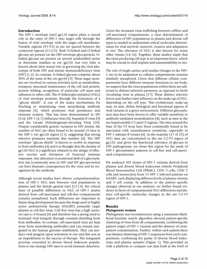
Phylogenetic tree analysis showing patient sequence purityFigure 1Phylogenetic tree analysis showing patient sequence purity. Phylogenetic analysis on the 305 HIV-1 env gp120 C2-V5 region sequences from plasma, peripheral blood mononuclear cells, CD4+ T cells, CD8+ T cells and monocytes. Using the ProML program of the PHYLIP software package, a maximum likelihood phylogenetic tree was calculated for our patient sequences. The branch lengths are scaled to distance. A single asterisk represents each sequence from our dataset. Individual sequences are not identified as it is only the broad pattern of clustering that is of interest here. Distinct clustering of patient-related sequences can be seen from the phylogenetic tree.
Page 3 of 10(page number not for citation purposes)
Virology Journal 2008, 5:14 http://www.virologyj.com/content/5/1/14
tions are referenced to the HIV-1 HXB2 prototype usingthe referencing guidelines available from the Los AlamosHIV sequence database website [18]. The columns inTable 1 categorize our data into plasma and blood leuko-cyte compartments, while the rows in Table 1 representthe amino acid differences at each of the identified sites.As shown in Table 1, CD4+ T cells, CD8+ T cells and mono-cyte-derived sequences were found with the amino acidasparagine (N) at position 279, whereas the PBMC andplasma-derived sequences were found to have asparticacid (D) at the same position. The amino acid lysine (K)was uniquely seen in monocyte-derived HIV sequences atpositions 335 and 350, whereas other compartmentsshowed arginine (R) at that position. Further amino aciddifferences across compartments were found at positions320, 336, 360 and 440 as illustrated in Table 1. Statisticalanalysis confirmed a significant association (p = 0.044) inthe observed single amino acid differences between CD4+
T cell and plasma-derived sequences at position 360.However a lost of significance was found (p = 0.22) whenwe correct for multiple comparisons.
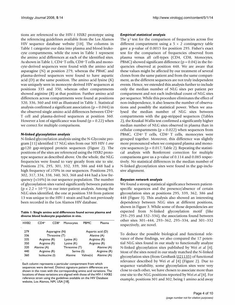
N-linked glycosylation analysisN-linked glycosylation analysis using the N-Glycosite pro-gram [11] identified 17 NLG sites from our 305 HIV-1 envgp120 gap-stripped protein sequences (Figure 2). Thepositions of the sites are referenced using the HXB2 proto-type sequence as described above. On the whole, the NLGfrequencies were found to vary greatly from site to site.Positions 276, 295, 301, 332, 339, 386 and 448 had ahigh frequency of ≥70% in our sequences. Positions 293,302, 317, 334, 338, 340, 363, 368 and 444 had a low fre-quency (<16%) in our sequence population. The numberof glycosylation sites varied significantly between patients(p < 2.2 × 10⁻¹⁶) in our inter-patient analysis. Among theNLG sites identified, the one at position 338 from patient13 was unique to the HIV-1 strain and had not previouslybeen recorded in the Los Alamos HIV database.
Empirical statistical analysisThe χ2 test for the comparison of frequencies across fivedifferent compartment using a 5 × 2 contingency tablegave a p-value of 0.0015 for position 295. Fisher's exacttest for the comparison of frequencies observed fromplasma versus all cell-types (CD4, CD8, Monocytes,PBMC) showed significant difference (p = 0.04) in the fre-quencies observed at position 448. We are aware thatthese values might be affected by our treatment of severalclones from the same patient and from the same compart-ment, as the different sequences are not truly independentevents. Hence, we extended this analysis further to includeonly the median number of NLG sites per patient percompartment and not each individual count of NLG sitesper sequence. While this procedure eliminates the effect ofnon-independence, it also lessens the number of observa-tions and possibly the statistical power. When we ana-lysed the median number of NLG sites betweencompartments with the gap-stripped sequences (Tables2), the Kruskal-Wallis test confirmed a significantly highermedian number of NLG sites observed in plasma than incellular compartments (p = 0.022) when sequences fromPBMC, CD4+ T cells, CD8+ T cells, monocytes weregrouped together. Moreover, this difference was slightlymore pronounced when we compared plasma and mono-cyte sequences (p = 0.017; Table 2). Repeating the statisti-cal analysis with Bonferroni correction for multiplecomparisons gave us a p-value of 0.114 and 0.085 respec-tively. No statistical differences in the median number ofN-linked glycosylation sites were found in the gap-inclu-sive alignment.
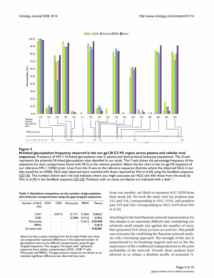
Bayesian network analysisWe found a strong statistical significance between patient-specific sequences and the presence/absence of certainglycosylation sites at positions 295, 339, 362, 386 and448 (Figure 3). This analysis also showed an interestingdependency between NLG sites at different positions,shown in Figure 3. While some of those dependencies areexpected from N-linked glycosylation motif (like293–295 and 332–334), the associations found betweenother sites 301–444, 293–362, 295–334, and 301–332respectively, are novel.
To deduce the possible biological and functional rele-vance of these findings, we also compared the 17 poten-tial NLG sites found in our study to functionally analyzeN-linked glycosylation sites published by Wei et al [4].Most of the sites noted in our study matched the N-linkedglycosylation sites (from GenBank U21135) of functionalrelevance described by Wei et al [4] (Figure 2). Due tosequence variability, some glycosylation sites were veryclose to each other, we have chosen to associate more thanone site to the NLG positions reported by Wei et al [4]. Forexample, positions 301 and 302, being 1 amino acid away
Table 1: Single amino acid differences found across plasma and diverse blood leukocyte population in vivo.
HXB2 CD4+ CD8+ Monocytes PBMC Plasma
279 Asparagine (N) Aspartic acid (D)336 Threonine (T) Alanine (A)335 Arginine (R) Lysine (K) Arginine (R)350 Arginine (R) Lysine (K) Arginine (R)320 Alanine (A) Threonine (T) Alanine (A)440 Arginine (R) Serine (S) Arginine (R)360 Isoleucine (I) Alanine Valine(v) Alanine (A)
Each column represents a particular compartment from which sequences were derived. Distinct signature pattern differences are shown in the rows with the corresponding amino acid variations. The locations of these variations are aligned with those of the HIV-1 HXB2 reference strain using the guidelines available on the HIV Database website, Los Alamos, NM, USA [18].
Page 4 of 10(page number not for citation purposes)
Virology Journal 2008, 5:14 http://www.virologyj.com/content/5/1/14
from one another, are likely to represent NLG 305N fromtheir study [4]. We took the same view for position pair332 and 334, corresponding to NLG 335N, and positionpair 339 and 340, corresponding to NLG 342N from Weiet al [4].
Searching for the best Bayesian network representation forthis dataset is an extremely difficult task considering ourrelatively small sample size against the number of varia-bles (potential NLG sites) we have account for. This pitfallwas overcome by combining the Bayesian network analy-sis with a bootstrap approach. The strength of the arcs isproportional to its bootstrap support and not to the theimportance of the conditional independencies to the jointprobability of the network. Overall, these analyses haveallowed us to obtain a detailed profile of potential N-
N-linked glycosylation frequency observed in the env gp120 C2-V5 region across plasma and cellular viral sequencesFigure 2N-linked glycosylation frequency observed in the env gp120 C2-V5 region across plasma and cellular viral sequences. Frequency of HIV-1 N-linked glycosylation sites in plasma and diverse blood leukocyte populations. The X-axis represents the potential N-linked glycosylation sites identified in our study. The Y-axis shows the percentage frequency of the sequences for each compartment found with NLG at the relevant position. Below the bar chart is the env gp120 sequence of our reference HIV-1 HXB2 strain. Lines from the X-axis to the reference sequence illustrate where the observed NLG in our data would be on HXB2. NLG sites observed were matched with those reported by Wei et al [4] using the GenBank sequence U21135. The numbers below each red oval indicates where you might associate our NLG site with those from the study by Wei et al [4] in the GenBank sequence U21135. Positions with no visual correlation are indicated with a dash '-'.
Table 2: Statistical comparison on the number of glycosylation sites between compartments using the gap-stripped sequences.
Number of NLGsites
CD4+ CD8+ Monocytes PBMC Plasma
CD4+ - 0.8713 0.1711 0.7645 0.08367CD8+ - - 0.2908 0.9132 0.0962
Monocytes - - - 0.3412 0.01696PBMC - - - - 0.0818
Grouped cells - - - - 0.02281
Above are the p-values resulting from the Kruskal-Wallis test when we compared for statistical differences in the observed number of glycosylation sites across different compartments using the gap-stripped sequences. The category "Grouped cells" represents sequences from cellular compartments (CD4+, CD8+ T cells, Monocytes and PBMC). The gap-inclusive results are not shown as no statistical significant difference was observed (see text).
Page 5 of 10(page number not for citation purposes)

Virology Journal 2008, 5:14 http://www.virologyj.com/content/5/1/14
linked glycosylation distribution and possible inter-rela-tionship between NLG sites in HIV-1 env gp120 sequencesbetween different blood leukocyte and plasma-derivedHIV-1 populations in vivo.
DiscussionIn order for HIV to be successful in evading the immunesystem, both cell-free and cell-associated forms of HIVmust adopt distinct molecular strategies to adapt and sur-vive in vivo. Such adaptation includes the modulation ofN-linked glycosylation and cell-specific single amino acidchanges in HIV. Given the importance of the envelopeglycoprotein in neutralization, pathogenesis, tropism andviral evasion, we analysed the C2-V5 region of 305 HIV-1env gp120 sequences from plasma and diverse blood leu-kocytes of 15 patients on HAART. Single amino acid dif-ferences specific to plasma and cellular compartmentswere observed. Notable was the presence of lysine (K) inmonocytes at positions 335 and 350 whereas these posi-tions have arginine (R) in the other compartments. Thissuggests that positions 335 and 350 have the greatest needto maintain a basic amino acid residue (lysine) in mono-cytes, compared with the other compartments. This differ-ence may have a role in viral adaptation to monocytes andpossibly relevant to monocyte tropism. In addition, theisoleucine at position 360 was unique to CD4+ T cells (p <
0.044). Additionally the presence of valine was unique toPBMC (and absent in plasma and the three other celltypes) at position 360. Although the monocyte compart-ment is a subset of the PBMC compartment, the majorityof PBMC sequences were found with valine (V) and themonocyte sequences with alanine (A). We believe thatalthough all the cellular compartments we analyzed werederived from the whole PBMC, the valine may be specificto a leukocyte subset, which was not analyzed in thisstudy, due to the limitation of human bleed obtainedfrom each patient. Nonetheless, this difference betweenmonocytes and PBMC supports our observation that com-partmental-specific amino acid changes in HIV-1 envgp120 are present. Our results agree with previous workon single amino acid changes in the env gene in associa-tion with viral tropism and pathogenicity [19]. A recentstudy by Clevestig et al [20], shows the V3 loop glycan(especially the sequon motif NNT) to be critical for CCR5use, which may have a direct role in HIV tropism, furthersupporting our conclusions. We believe that these singleamino acid differences could be vital to HIV and areacquired through intra-host evolution in order to success-fully adapt and thrive in different in vivo environments.
The modulation of N-linked glycosylation sites in theHIV-1 envelope has been known to facilitate viral evasionfrom the host immune system. Similar to the signaturepattern analysis, the N-linked glycosylation analysis yieldsinteresting results showing compartmental variations andpossible associations among glycosylation sites. An exam-ination of the NLG frequencies along the C2-V5 region(Figure 2) revealed that plasma virus had a similar orhigher percentage of its sequences glycosylated at theidentified sites, compared to the other compartmentalviruses. A possible explanation for this is that the differ-ences in N-linked glycosylation sites in plasma may berequired for the maintenance of infectious potential.Through the suppression of HIV-1 in CD4+ T cells andplasma in our HAART patient pool, glycosylation changein the virus may enable it to overcome the selection pres-sures from the antiretroviral regimen. Further examina-tion of the NLG sites using Bayesian networks foundunique associations between sites 301–444, 293–362,295–334, and 301–332. These associations could indicatealternative pathways for glycosylation and possible simul-taneous co-selection of glycosylation sites. Similar to thestudy of evolutionary interactions between NLG sites byPoon Art F, Y [21], we believe that these evolutionarymechanisms is important to HIV for its struggle againsthost immune selection pressure. Since all our work wasperformed directly on ex vivo-collected cells from eachpatient, our results reflect the closest possible snapshot tothe in vivo situation.
Bayesian network associations found between observed N-linked glycosylation sitesFigure 3Bayesian network associations found between observed N-linked glycosylation sites. Representation of the Bayesian network analysis results, generated as described in the methods section. Only arcs with a bootstrap support of at least 70% are presented. Associations between patient ID and glycosylation sites are dashed. Associations between glycosylation sites that can be structurally explained are colored grey. Associations between glycosylation sites that cannot be structurally explained are colored black.
�������
� ��
��� � �
���
���
���
��
��
� �
Page 6 of 10(page number not for citation purposes)

Virology Journal 2008, 5:14 http://www.virologyj.com/content/5/1/14
We examined the variation in the number of glycosylationsites among compartments (gap-free and gap-inclusive)to infer a global picture of HIV-1 in vivo. The trade-offbetween them is an important issue to consider in thisanalysis. While using the gap-inclusive alignment mightinclude unavoidable interference due to the uncertainty ofthe alignment in the hypervariable region of env, the useof the gap-stripped alignment might also introduce anoversight of the observed glycosylation due to the poten-tial removal of important glycosylation sites from thealignment. Therefore, we consider both analyses in theresults section. The results were twofold: no statistical sig-nificance in the total number of glycosylation sitesbetween plasma and different cellular compartmentsequences was found using the gap-inclusive alignment.However when we performed the gap-stripped analysis,we observed a significant difference (p = 0.022) betweenplasma and cellular compartment sequences. This is con-sistent with the notion that the number of glycosylationsites is highly conserved in HIV-1 [4]. The disparitybetween these results suggests that there is a difference inthe number of glycosylation sites between plasma virusand cellular virus in the less conserved regions of HIV-1env. Despite the loss of statistical significance when weapplied the Bonferroni correction for multiple compari-son from P = 0.022 to P = 0.115, we believe that it is stillan important finding together with the rest of our staticalanalysis. This is because by correcting for multiple com-parisons, we are also increasing the risk of making a typeII error which might lead us to not report a correlationwhen there is one. Between both gap-stripped and gap-inclusive analysis, the gap-inclusive alignment gives us theactual number of glycosylation sites, as no glycosylationsites were omitted in the evaluation. Not finding any sig-nificant differences in this gap-inclusive alignment indi-cates that possible selective pressure to removeglycosylation sites from the more conserved part of envwas compensated by the creation of new glycosylationsites in those parts of env that better tolerate substitutions,insertions and deletions.
Even though we found evidence for compartmentaliza-tion of HIV-1 across plasma and diverse blood leukocytepopulations in vivo, it is important to note that the NLGobserved in our sequences are distinctly patient-specific (p≤ 2.2 × 10⁻¹⁶). This supports our belief that the selectionof NLG sites are primarily dependent on the patient'simmune response. The choice to examine 305 sequencesfrom 15 independent patients with a range of CD4+ T cellcounts and plasma viral load in our study has allowed usto infer a balanced macroscopic perspective of HIV-1adaptation in vivo during therapy. These analyses are thefirst to provide a detailed comparison of cell-free and cel-lassociated virus, especially in individual leukocyte types.Previous studies by Hanna et al [14] and Lin et al [15] sup-
port the validity of our studies on diverse cell types andadd further credence to the functional basis of differentNLG frequencies in plasma and cell-associated virus.Together, these findings might provide a new directionand perspective into the role of glycosylation in diversecompartments in vivo and these data may have relevancein HIV immune recognition, viral adaptation, vaccinestrategies and HIV pathogenesis in general.
ConclusionThis study examined single cell/compartment-specificamino acid variations and unique differences in N-linkedglycosylation patterns between plasma and diverse bloodleukocytes. It has provided deeper insights into how HIVmay evade antibodies and maintain its pathogenic poten-tial. Bayesian analysis has shown associations that suggestpossible glycosylation pathways. We believe that theseanalyses provide useful insights into the host immuneresponse and its ability in controlling HIV replication invivo. Further, this enhance our understanding of pathoge-nous differences between cell-free and cell-associatedHIV-1. Consequently, these analyses will allow furtherbiological and functional assessment of such molecularchanges in the context of viral escape, adaptation and res-ervoir establishment in vivo. A better understanding ofdiverse N-linked glycosylation sites and their functionalrole may provide useful strategies for choosing and elimi-nating the "right" N-linked glycosylation site(s), thusfacilitating the design of more effective envelope-basedimmunogens that elicit broad neutralization antibodyresponses.
MethodsConsentThis work is carried out in accordance with the humanethics guidelines and scientific principles set out by theNational Health and Medical Research Council of Aus-tralia (NHMRC). All patients have given written consenton the study and understand that the study will be con-ducted in a manner conforming to the ethical and scien-tific principle set out by the NHMRC.
Patient selectionFifteen HIV-1 infected patients receiving HAART from theWestmead Hospital in Sydney, Australia, were enrolled inthis study after prior consent. These patients exhibited var-ying plasma viral loads and T cell counts as shown inTable 3. Patients 6, 11, 12, and 13, who were successful intherapy, had plasma viral loads below detectable levels(<50 copies/ml plasma). Patient 9, who was at anadvanced stage of therapy, had low viremia. Patients 2, 4,8, 10, 14 and 15 had varying degrees of plasma viremiaafter being on HAART for 4 weeks. Patients 1, 3, 5 and 7had high plasma viral load (>100,000 copies/ml plasma)and were resistant to antiretroviral drugs [22]. No
Page 7 of 10(page number not for citation purposes)

Virology Journal 2008, 5:14 http://www.virologyj.com/content/5/1/14
untreated patients could be recruited in this study as everyHIV patient in Australia receives treatment for their condi-tion.
Cell purification and sequence generation50 ml of blood was collected from each patient. Individ-ual cell types (CD4+ T cells, CD8+ T cells and CD14+
monocytes) were separated from PBMC using magneticbeads coated with monoclonal antibodies (Dynal, Oslo,Norway) using the procedure described and developed byPotter et al [17]. FACS analysis of separated cellular frac-tions showed an average purity of 99.6%. Proviral DNAwas extracted from PBMC and individually sorted into cel-lular fractions using the Qiagen Blood kit (Qiagen, Ger-many) as per the manufacturer's protocol. A nestedpolymerase chain reaction (PCR) was used to amplify a600 bp fragment in the C2-V5 region of env gene. Reversetranscription-PCR (RT-PCR) of HIV-1 RNA extracted fromplasma using the Qiagen RNA Extraction Kit (Qiagen,Germany) was carried out to amplify viral populationsfrom the cell-free plasma fraction [17]. Independent PCRexperiments were performed in triplicate on each sepa-rated fraction, and pooled products were used to generatecompartment-specific clones (five clones per compart-ment). To analyse cell-free and cell-associated viral popu-lations from each patient in parallel, the HIV-1populations from their whole PBMC, CD4+, CD8+ T cells,monocytes and plasma were cloned. In each case, the
major amplicon population without cloning was firstobtained to assess diversity within a patient. Followingthat, cloning and sequencing were performed as describedpreviously [17] to analyse diversity in each compartmentat the quasispecies level, using the major population fromthe same patient as a comparison. This was performed toderive a clear estimate of intrapatient genetic diversity.Reverse transcription-PCR from plasma was unsuccessfulfrom patient 6, 10 and 13 as these patients had plasmaviremia below detectable levels. PCR amplification ofCD8+ T cells was not successful for patient 6 and 9; neitherwas it successful for monocytes for patients 6, 7, 9, 14 and15 or PBMC for patients 14 and 15. A total of 305 HIV-1cloned sequences were generated from 15 patients. BLASTsearches and phylogenetic analyses (Figure 1) were doneto rule out any evidence of laboratory contaminationthrough PCR. Further, manual inspection of all sequenceswas performed to ensure that the sequenced region wasin-frame and there were no significant gene alterations(insertions, deletions and nonsense mutations) in bothmajor populations and clones. All sequences derived fromthe 15 different patients were identified as subtype B.
All 305 HIV-1 nucleotide sequences from the C2-V5region of the env gp120 region were translated into theirprotein equivalent using Transeq (EMBOSS) [23]. Fromthe protein sequences, a "gap-inclusive" alignment wascreated with a multiple sequence alignment program (inthis case CLUSTALW [24]) and verified visually. Gapsintroduced in sequences by this process correspond tohypothesized insertion or deletion (indel) events, and thealignment is therefore referred to as gap-inclusive. As fre-quent indels render the alignment very difficult, and insome cases ambiguous, we also created a "gap-stripped"alignment, by removing from the gap-inclusive alignmentall sites that contain a gap in any sequence. All alignmentsmentioned in this paper are considered gap-inclusiveunless stated otherwise. The HIV-1 HXB2 envelopesequence was used as our reference.
Phylogenetic analysisPhylogenetic reconstructions were performed to confirmthe purity of viral sequences at the level of individualpatients and each compartment analyzed. Phylogeneticanalysis was performed based on the gap-stripped aminoacid alignment using maximum likelihood on the ProMLprogram (version 3.66) of the PHYLIP package [25] withthe Jones, Taylor and Thornton model of amino acidreplacement with a constant rate of change.
Signature pattern analysisSignature pattern analysis was performed on the trans-lated protein sequences using the Viral Epidemiology Sig-nature Analysis (VESPA) software [26]. The VESPAsoftware examines single amino acid differences between
Table 3: Patients plasma viral load, CD4+ and CD8+ T cell counts.
Patient CD4+ Count/μlblood
CD8+ Count/μlblood
Plasma Viral Load(RNA c/ml)
1 200 790 > 100,0002 437 950 57303 60 900 > 100,0004 16 N/A 877005 8 172 > 100,0006 105 675 < 507 14 420 > 100,0008 302 800 602009 195 1110 1510
10 135 1647 6450011 580 870 < 5012 360 460 < 5013 1012 2806 < 5014 180 510 9770015 340 750 46000
Plasma viral load, CD4+ and CD8+ T cell counts were performed on the samples obtained from 15 patients. The patients showed varied level disease stage according to the T cell numbers and corresponding viral load presented. Patients 1,3,5,7 and 14 were at the late stage of the HIV infection with high plasma viral load (at least 100,000 copies of viral RNA per ml of plasma). Patients 4,8,10 and 15 had intermediate levels of plasma viral load. Patients 2,6,9,11,12 and 13 had low plasma viral load, indicating that they were at a earlier stage of their HIV infection.
Page 8 of 10(page number not for citation purposes)

Virology Journal 2008, 5:14 http://www.virologyj.com/content/5/1/14
groups of sequences by creating consensus sequences foreach group. In our study, the sequences were grouped intotheir original host cell types (PBMC, CD4+ T cells, CD8+ Tcells, monocytes and plasma) and compared inter-com-partmentally. Given the extensive inter-strain/inter-patient variation in our 305 sequences, the majority con-sensus parameter was used. The "no fixed rates" optionwas chosen because a fixed rate would not capture therange of diversity observed.
N-linked glycosylation analysisN-linked glycosylation analysis was performed on all 305protein sequences from the C2-V5 region of the env gp120protein to examine for NLG differences between plasmaand cell types (CD4+ T cells, CD8+ T cells, PBMC andmonocyte) virus in vivo. Our data was analysed using theprogram N-Glycosite [11], available from the Los AlamosNational Laboratory HIV Database website [27] (NM,USA). A NLG site is identified in the amino acid sequenceby the motif (or "sequon") NX[S/T] with N-Glycosite. Thesequon has to begin with an asparagine (N) followed byany amino acid except Proline. The next amino acid resi-due has to be either a threonine (T) or a serine(S). Boththe gap-stripped and gap-inclusive alignments were usedin this analysis. Through the gapstripping process, sepa-rate regions in the alignment could come together andconsequently form an unintentional NX[ST] sequon. Thiswould cause the alignment to register a false NLG site.Through careful examination, we confirmed that no glyc-osylation sites were accidentally created from the gapstripping process.
Empirical statistical analysisThe frequency of NLG sites in the sequences found in bothplasma and diverse leukocytes (PBMC, CD4+ T cells, CD8+
T cells and monocytes) was examined. The χ2 test wasused to compare the frequencies observed across five dif-ferent compartments for each NLG site identified. A 5 × 2contingency table was used to evaluate their statistical sig-nificance. Next, Fisher's exact test was used to compare thefrequencies observed from plasma versus all cell-typestogether for each NLG site. We further analysed the differ-ences in the mean and median number of NLG sites bothacross compartments and between patients using the non-parametric Kruskal-Wallis test, as the hypothesis of a nor-mal distribution for the number of glycosylation sites wasrejected by the Shapiro-Wilks test. All statistical analyseswere performed with the R package [28] (ver. 2.4.1).These statistical analyses allowed us to derive a true snap-shot of glycosylation distribution in the 305 HIV-1 envgp120 protein sequences from different blood leukocytepopulations and plasma in vivo.
Bayesian network analysisA Bayesian network describes a set of direct dependenciesthat together explain as much as possible the observedcorrelations in a dataset [29]. As there could be interde-pendencies and statistical correlation between individu-ally observed NLG sites, patient grouped sequences and/or compartment categorized sequences, we generatedBayesian networks of all 305 HIV-1 env gp120 proteinsequences using the procedure developed by Deforche etal [30].
For each patient, we created compartmental (PBMC,CD4+ T cells, CD8+ T cells, monocytes and plasma) con-sensus sequences from our clones. These five consensussequences for each patient were then used for the Bayesiannetwork analysis. This approach allowed us to remove anyfalse positive associations caused because multiple clonedsequences from the same patient of the same compart-ment are likely to be highly similar. If the consensussequences were not used, the arcs of the network could beartificially strengthened and we might overestimate of thesignificance of some associations. The most probableBayesian network is the one that maximizes the posteriorprobability of the model given the data, subject to a priordistribution of model, which we assumed was uniform.We used a simulated annealing heuristic to search in thespace of all possible Bayesian network structures [30,31].To measure the reliability of each of the arcs, a bootstrap-ping method was used in which 100 replicates of the orig-inal dataset were generated by random sampling withreplacement, and the most probable Bayesian network re-inferred. Only arcs that occurred in at least 70% of thesenetworks were considered significant for their inclusion inour result.
Competing interestsThe author(s) declare that they have no competing inter-ests.
Authors' contributionsYSH: Carried out the entire study, designed the frame-work, developed in-house bioinformatic tools for analysisand wrote the manuscript; ABA, MC, KD, KT and AMVprovided highly coordinated help with statistical analysisand Bayesian network analysis; DD provided patients andtheir enrolment for this study, and provided all the clini-cal information needed and NKS conceived of the study,and participated in its design and coordination andhelped to draft the manuscript. All authors read andapproved the final manuscript.
AcknowledgementsThe authors are thankful to all the patients for their consent to give samples and their participation. YSH is thankful to USYD for the Australian Post-graduate Award. ABA was supported by Fundação para a Ciência e Tecno-logia (Grant nr SFRH/BD/19334/2004). This work has been presented
Page 9 of 10(page number not for citation purposes)

Virology Journal 2008, 5:14 http://www.virologyj.com/content/5/1/14
Publish with BioMed Central and every scientist can read your work free of charge
"BioMed Central will be the most significant development for disseminating the results of biomedical research in our lifetime."
Sir Paul Nurse, Cancer Research UK
Your research papers will be:
available free of charge to the entire biomedical community
peer reviewed and published immediately upon acceptance
cited in PubMed and archived on PubMed Central
yours — you keep the copyright
Submit your manuscript here:http://www.biomedcentral.com/info/publishing_adv.asp
BioMedcentral
orally at the International Aids Society (IAS) 2007 conference in Sydney, Australia in the HIV diversity, tropism and compartmentalization session of the basic science track.
References1. Chackerian B, Rudensey LM, Overbaugh J: Specific N-linked and
O-linked glycosylation modifications in the envelope V1domain of simian immunodeficiency virus variants thatevolve in the host alter recognition by neutralizing antibod-ies. J Virol 1997, 71:7719-7727.
2. Huang X, Barchi JJ Jr, Lung FD, Roller PP, Nara PL, Muschik J, GarrityRR: Glycosylation affects both the three-dimensional struc-ture and antibody binding properties of the HIV-1IIIB GP120peptide RP135. Biochemistry 1997, 36:10846-10856.
3. Leonard CK, Spellman MW, Riddle L, Harris RJ, Thomas JN, GregoryTJ: Assignment of intrachain disulfide bonds and characteri-zation of potential glycosylation sites of the type 1 recom-binant human immunodeficiency virus envelopeglycoprotein (gp120) expressed in Chinese hamster ovarycells. J Biol Chem 1990, 265:10373-10382.
4. Wei X, Decker JM, Wang S, Hui H, Kappes JC, Wu X, Salazar-Gonzalez JF, Salazar MG, Kilby JM, Saag MS, et al.: Antibody neutral-ization and escape by HIV-1. Nature 2003, 422:307-312.
5. Reitter JN, Means RE, Desrosiers RC: A role for carbohydrates inimmune evasion in AIDS. Nat Med 1998, 4:679-684.
6. Pikora C, Wittish C, Desrosiers RC: Identification of two N-linked glycosylation sites within the core of the simianimmunodeficiency virus glycoprotein whose removalenhances sensitivity to soluble CD4. J Virol 2005,79:12575-12583.
7. McCaffrey RA, Saunders C, Hensel M, Stamatatos L: N-linked glyc-osylation of the V3 loop and the immunologically silent faceof gp120 protects human immunodeficiency virus type 1SF162 from neutralization by anti-gp120 and anti-gp41 anti-bodies. J Virol 2004, 78:3279-3295.
8. Skehel JJ, Stevens DJ, Daniels RS, Douglas AR, Knossow M, Wilson IA,Wiley DC: A carbohydrate side chain on hemagglutinins ofHong Kong influenza viruses inhibits recognition by a mono-clonal antibody. Proc Natl Acad Sci USA 1984, 81:1779-1783.
9. Lee J, Park JS, Moon JY, Kim KY, Moon HM: The influence of glyc-osylation on secretion, stability, and immunogenicity ofrecombinant HBV pre-S antigen synthesized in Saccharomy-ces cerevisiae. Biochem Biophys Res Commun 2003, 303:427-432.
10. Chen Z, Li K, Plagemann PG: Neuropathogenicity and sensitivityto antibody neutralization of lactate dehydrogenase-elevat-ing virus are determined by polylactosaminoglycan chains onthe primary envelope glycoprotein. Virology 2000, 266:88-98.
11. Zhang M, Gaschen B, Blay W, Foley B, Haigwood N, Kuiken C, Kor-ber B: Tracking global patterns of N-linked glycosylation sitevariation in highly variable viral glycoproteins: HIV, SIV, andHCV envelopes and influenza hemagglutinin. Glycobiology2004, 14:1229-1246.
12. Kemal KS, Foley B, Burger H, Anastos K, Minkoff H, Kitchen C, Phil-pott SM, Gao W, Robison E, Holman S, et al.: HIV-1 in genital tractand plasma of women: compartmentalization of viralsequences, coreceptor usage, and glycosylation. Proc Natl AcadSci USA 2003, 100:12972-12977.
13. Overbaugh J, Anderson RJ, Ndinya-Achola JO, Kreiss JK: Distinctbut related human immunodeficiency virus type 1 variantpopulations in genital secretions and blood. AIDS Res Hum Ret-roviruses 1996, 12:107-115.
14. Hanna SL, Pierson TC, Sanchez MD, Ahmed AA, Murtadha MM,Doms RW: N-linked glycosylation of west nile virus envelopeproteins influences particle assembly and infectivity. J Virol2005, 79:13262-13274.
15. Lin G, Simmons G, Pohlmann S, Baribaud F, Ni H, Leslie GJ, HaggartyBS, Bates P, Weissman D, Hoxie JA, Doms RW: Differential N-linked glycosylation of human immunodeficiency virus andEbola virus envelope glycoproteins modulates interactionswith DC-SIGN and DC-SIGNR. J Virol 2003, 77:1337-1346.
16. Wojczyk B, Shakin-Eshleman SH, Doms RW, Xiang ZQ, Ertl HC,Wunner WH, Spitalnik SL: Stable secretion of a soluble, oligo-meric form of rabies virus glycoprotein: influence of N-gly-can processing on secretion. Biochemistry 1995, 34:2599-2609.
17. Potter SJ, Lemey P, Achaz G, Chew CB, Vandamme AM, Dwyer DE,Saksena NK: HIV-1 compartmentalization in diverse leuko-cyte populations during antiretroviral therapy. J Leukoc Biol2004, 76:562-570.
18. Numbering Positions in HIV Relative to HXB2CG [http://www.hiv.lanl.gov/content/sequence/HIV/REVIEWS/HXB2.html]
19. Wang B, Jozwiak R, Ge YC, Saksena NK: A unique, naturallyoccurring single-amino acid mutation in HIV type 1 V3 loopcan discriminate between its cytopathicity and replication invivo and in vitro. AIDS Res Hum Retroviruses 1998, 14:1019-1021.
20. Clevestig P, Pramanik L, Leitner T, Ehrnst A: CCR5 use by humanimmunodeficiency virus type 1 is associated closely with thegp120 V3 loop N-linked glycosylation site. Journal of GeneralVirology 2006, 87:607-612.
21. Poon AFY, Lewis FI, Pond SLK, Frost SDW: Evolutionary Interac-tions between Nlinked Glycosylation Sites in the HIV-1Envelope. PLoS Computational Biology 2007, 3:e11.
22. Potter SJ, Dwyer DE, Saksena NK: Differential cellular distribu-tion of HIV-1 drug resistance in vivo: evidence for infectionof CD8+ T cells during HAART. Virology 2003, 305:339-352.
23. Rice P, Longden I, Bleasby A: EMBOSS: the European MolecularBiology Open Software Suite. Trends Genet 2000, 16:276-277.
24. Thompson JD, Higgins DG, Gibson TJ: CLUSTAL W: improvingthe sensitivity of progressive multiple sequence alignmentthrough sequence weighting, position-specific gap penaltiesand weight matrix choice. Nucleic Acids Res 1994, 22:4673-4680.
25. Felsenstein J: PHYLIP (Phylogeny Inference Package). 3.6thedition. Department of Genome Science, University of Washington,Seattle; 2005.
26. Korber B, Myers G: Signature pattern analysis: a method forassessing viral sequence relatedness. AIDS Res Hum Retroviruses1992, 8:1549-1560.
27. N-GlycoSite [http://www.hiv.lanl.gov/content/sequence/GLYCOSITE/glycosite.html]
28. Team RDc: R: A language and environment for statisticalcomputing. R foundation for Statistical Computing 2004.
29. Heckerman D: A Tutorial on Learning with Bayesian Networks MIT press;1999.
30. Deforche K, Silander T, Camacho R, Grossman Z, Soares M, Van Lae-them K, Kantor R, Moreau Y, Vandamme AM: Analysis of HIV-1pol sequences using Bayesian networks: implications fordrug resistance. Bioinformatics 2006.
31. Abecasis AB, Deforche K, Snoeck J, Bacheler LT, McKenna P, Car-valho AP, Gomes P, Camacho RJ, Vandamme AM: Protease muta-tion M89I/V is linked to therapy failure in patients infectedwith the HIV-1 non-B subtypes C, F or G. Aids 2005,19:1799-1806.
Page 10 of 10(page number not for citation purposes)
http://www.ncbi.nlm.nih.gov/entrez/query.fcgi?cmd=Retrieve&db=PubMed&dopt=Abstract&list_uids=9311856
http://www.ncbi.nlm.nih.gov/entrez/query.fcgi?cmd=Retrieve&db=PubMed&dopt=Abstract&list_uids=9311856
http://www.ncbi.nlm.nih.gov/entrez/query.fcgi?cmd=Retrieve&db=PubMed&dopt=Abstract&list_uids=9311856
http://www.ncbi.nlm.nih.gov/entrez/query.fcgi?cmd=Retrieve&db=PubMed&dopt=Abstract&list_uids=9312273
http://www.ncbi.nlm.nih.gov/entrez/query.fcgi?cmd=Retrieve&db=PubMed&dopt=Abstract&list_uids=9312273
http://www.ncbi.nlm.nih.gov/entrez/query.fcgi?cmd=Retrieve&db=PubMed&dopt=Abstract&list_uids=9312273
http://www.ncbi.nlm.nih.gov/entrez/query.fcgi?cmd=Retrieve&db=PubMed&dopt=Abstract&list_uids=2355006
http://www.ncbi.nlm.nih.gov/entrez/query.fcgi?cmd=Retrieve&db=PubMed&dopt=Abstract&list_uids=2355006
http://www.ncbi.nlm.nih.gov/entrez/query.fcgi?cmd=Retrieve&db=PubMed&dopt=Abstract&list_uids=2355006
http://www.ncbi.nlm.nih.gov/entrez/query.fcgi?cmd=Retrieve&db=PubMed&dopt=Abstract&list_uids=9623976
http://www.ncbi.nlm.nih.gov/entrez/query.fcgi?cmd=Retrieve&db=PubMed&dopt=Abstract&list_uids=9623976
http://www.ncbi.nlm.nih.gov/entrez/query.fcgi?cmd=Retrieve&db=PubMed&dopt=Abstract&list_uids=6584912
http://www.ncbi.nlm.nih.gov/entrez/query.fcgi?cmd=Retrieve&db=PubMed&dopt=Abstract&list_uids=6584912
http://www.ncbi.nlm.nih.gov/entrez/query.fcgi?cmd=Retrieve&db=PubMed&dopt=Abstract&list_uids=6584912
http://www.ncbi.nlm.nih.gov/entrez/query.fcgi?cmd=Retrieve&db=PubMed&dopt=Abstract&list_uids=8834460
http://www.ncbi.nlm.nih.gov/entrez/query.fcgi?cmd=Retrieve&db=PubMed&dopt=Abstract&list_uids=8834460
http://www.ncbi.nlm.nih.gov/entrez/query.fcgi?cmd=Retrieve&db=PubMed&dopt=Abstract&list_uids=8834460
http://www.ncbi.nlm.nih.gov/entrez/query.fcgi?cmd=Retrieve&db=PubMed&dopt=Abstract&list_uids=7873541
http://www.ncbi.nlm.nih.gov/entrez/query.fcgi?cmd=Retrieve&db=PubMed&dopt=Abstract&list_uids=7873541
http://www.ncbi.nlm.nih.gov/entrez/query.fcgi?cmd=Retrieve&db=PubMed&dopt=Abstract&list_uids=7873541
http://www.ncbi.nlm.nih.gov/entrez/query.fcgi?cmd=Retrieve&db=PubMed&dopt=Abstract&list_uids=9686649
http://www.ncbi.nlm.nih.gov/entrez/query.fcgi?cmd=Retrieve&db=PubMed&dopt=Abstract&list_uids=9686649
http://www.ncbi.nlm.nih.gov/entrez/query.fcgi?cmd=Retrieve&db=PubMed&dopt=Abstract&list_uids=9686649
http://www.ncbi.nlm.nih.gov/entrez/query.fcgi?cmd=Retrieve&db=PubMed&dopt=Abstract&list_uids=7984417
http://www.ncbi.nlm.nih.gov/entrez/query.fcgi?cmd=Retrieve&db=PubMed&dopt=Abstract&list_uids=7984417
http://www.ncbi.nlm.nih.gov/entrez/query.fcgi?cmd=Retrieve&db=PubMed&dopt=Abstract&list_uids=7984417
http://www.ncbi.nlm.nih.gov/entrez/query.fcgi?cmd=Retrieve&db=PubMed&dopt=Abstract&list_uids=1457200