
A twin-cysteine motif in the V2 region of gp120 is associated with SIV envelope trimer stabilization
Upload
independentCategory
view
0download
0
Virology 409 (2011) 163–174
Contents lists available at ScienceDirect
Virology
j ourna l homepage: www.e lsev ie r.com/ locate /yv i ro
Identification of amino acid substitutions associated with neutralization phenotypein the human immunodeficiency virus type-1 subtype C gp120
Jennifer L. Kirchherr a,b, Jennifer Hamilton a,b, Xiaozhi Lu a,b, S. Gnanakaran d, Mark Muldoon e,Marcus Daniels d, Webster Kasongo f, Victor Chalwe f, Chanda Mulenga f, Lawrence Mwananyanda f,Rosemary M. Musonda f, Xing Yuan c, David C. Montefiori c, Bette T. Korber d,g,Barton F. Haynes a,b, Feng Gao a,b,⁎a Duke Human Vaccine Institute, Duke University Medical Center, Durham, NC 27710, USAb Department of Medicine, Duke University Medical Center, Durham, NC 27710, USAc Department of Surgery, Duke University Medical Center, Durham, NC 27710, USAd Los Alamos National Laboratory, Los Alamos, NM 87545, USAe School of Mathematics, University of Manchester, Manchester, M13 9PL, UKf The Tropical Disease Research Centre, PO Box 71769 Ndola, Zambiag Santa Fe Institute, Santa Fe, NM 87501, USA
⁎ Corresponding author. 3072B MSRB II, DUMC 1030Human Vaccine Institute, Duke University Medical CenFax: +1 919 681 8992.
E-mail address: [email protected] (F. Gao).
0042-6822/$ – see front matter © 2010 Elsevier Inc. Aldoi:10.1016/j.virol.2010.09.031
a b s t r a c t
a r t i c l e i n f oArticle history:Received 13 July 2010Returned to author for revision22 August 2010Accepted 27 September 2010Available online 30 October 2010
Keywords:HIV-1EnvelopeGenetic variationNeutralizationSignature
Neutralizing antibodies (Nabs) are thought to play an important role in prevention and control of HIV-1infection and should be targeted by an AIDS vaccine. It is critical to understand how HIV-1 induces Nabs byanalyzing viral sequences in both tested viruses and sera. Neutralization susceptibility to antibodies inautologous and heterologous plasma was determined for multiple Envs (3–6) from each of 15 subtype-C-infected individuals. Heterologous neutralization was divided into two distinct groups: plasma with strong,cross-reactive neutralization (n=9) and plasmawith weak neutralization (n=6). Plasmawith cross-reactiveheterologous Nabs also more potently neutralized contemporaneous autologous viruses. Analysis of Envsequences in plasma from both groups revealed a three-amino-acid substitution pattern in the V4 region thatwas associated with greater neutralization potency and breadth. Identification of such potentialneutralization signatures may have important implications for the development of HIV-1 vaccines capableof inducing Nabs to subtype C HIV-1.
20, 106 Research Drive, Duketer, Durham, NC 27710, USA.
l rights reserved.
© 2010 Elsevier Inc. All rights reserved.
Introduction
The ability to elicit cross-reactive neutralizing antibodies (Nabs) isan important goal for HIV-1 vaccines (Burton et al., 2004; Haynes andMontefiori, 2006). HIV-1 has nine genetically related lineages(subtypes A–K), and at a minimum, at least one clade should beeffectively targeted in an HIV vaccine for the vaccine to be useful in apart of the world where a single subtype dominates the epidemic(Korber and Gnanakaran, 2009; Korber et al., 2009). Nabs capable oftargeting subtype C variants of the virus would be particularly useful,since subtype C accounts for approximately 50% of all HIV-1 infectionsworld-wide (Hemelaar et al., 2006). In addition, the HIV epidemic inlarge regions of Southern Africa and India is almost completelydominated by subtype C infections; hence in this study, we focus onsubtype C antibody responses in natural infection. Efforts to generate
cross-reactive Nabs have had limited success, and novel approachesare urgently needed (Burton et al., 2004; Desrosiers, 2004). Cross-reactive neutralizing activity that is seen with a subset of humanmonoclonal Abs (Binley et al., 2004; Lin and Nara, 2007; Moore et al.,1994; Trkola et al., 1995; Wu et al., 2010; Zwick et al., 2001) andserum samples from HIV-1-infected individuals (Binley et al., 2008;Dhillon et al., 2007; Li et al., 2007; Sather et al., 2009; Shen et al., 2009;Stamatatos et al., 2009) is evidence that improvements in immunogendesign may be possible.
Insights into how to make such improvements are being sought bystudying autologous and heterologous Nabs in HIV-1-positive serumsamples. Recent results indicate that epitopes in and around the CD4binding site of gp120 comprise key targets for broadly neutralizingHIV-1-positive sera and that other key targets that play a substantialrole remain to be identified (Binley et al., 2008; Dhillon et al., 2007; Liet al., 2007; Wu et al., 2010; Zhou et al., 2010). Studies of autologousvirus neutralization have shown that the response targets multipleregions of gp120, most notably epitopes in V1/V2 (Rong et al., 2009)and epitopes that require an interaction between C3 and V4 (Mooreet al., 2008, 2009; Rademeyer et al., 2007; Rong et al., 2009). A recent
Table 1Summary of SGAs and functional env genes from HIV-1-infected individuals.
ID Gender Age Viral load Subtype No. ofSGAs
No. offunctionalenv genes
Functionalenv genes(%)
ZM373 M 25 44,800 C 5 4 80ZM375 F 31 52,800 C 10 8 80ZM376 F 30 105,600 C 15 13 87ZM377 M 41 248,800 C 16 15 94ZM378 F 36 67,200 C 13 11 85ZM379 M 35 97,600 C 14 11 79ZM380 M 37 268,800 A/C 10 10 100ZM381 F 36 487,200 C 13 12 92ZM382 M 39 557,600 C 10 10 100ZM383 F 31 20,240 C 10 5 50ZM384 M 41 15,440 C 11 10 91ZM387 M 30 1,120 G/J 9 9 100ZM388 F 28 21,360 C 11 6 55ZM389 M 33 1,520,000 C 10 10 100ZM393 F 21 b384 C 11 2 18ZM394 F 30 197,600 C 19 12 63ZM395 F 22 776,800 C 21 20 95ZM399 M 36 68,240 C 14 14 100ZM400 M 35 103,200 C 18 15 83ZM401 M 34 84,000 C 11 9 82ZM402 F 37 3,920 C 9 6 67ZM403 F 45 48,080 C 11 10 91ZM405 F 36 13,920 C 12 9 75ZM406 M 27 104,000 C 10 10 100ZM407 M 38 283,200 D 11 9 82ZM408 F 26 42,400 C 10 8 80ZM410 M 42 143,200 C 11 6 55ZM411 F 41 61,600 C 14 6 43ZM412 F 30 260,000 C 10 7 70ZM413 F 34 155,200 C 16 14 88ZM414 F 25 213,600 C 22 14 64ZM415 M 38 64,720 C 21 18 86ZM416 M 45 80,800 C 23 23 100ZM417 M 32 96,000 C 9 9 100ZM418 F 21 532,000 C 10 8 80ZM419 F 33 433,600 C 10 1 10ZM420 F 24 286,400 C 14 13 93Total 474 377 80
164 J.L. Kirchherr et al. / Virology 409 (2011) 163–174
study identified 19 signatures in gp120 and 14 signatures in gp41associated with neutralization susceptibility of a multi-subtype panelof viruses (Kulkarni et al., 2009).Walker et al. have recently found twomAbs that bind to conformation determinants of HIV-1 Env andbroadly neutralize about two-thirds of viruses tested (Walker et al.,2009).
Previous studies of the autologous and heterologous Nabresponses in HIV-1 infection have utilized one or a limited numberof representative env genes from each individual to characterizeneutralization susceptibility (Cham et al., 2006; Derdeyn et al., 2004;Wei et al., 2003; Zhang et al., 2007). Since env is highly variable inchronic HIV-1 infection (CHI) and because minor sequence changescan affect the biological function and antigenicity of the envelopeglycoproteins (Cordonnier et al., 1989; Kalia et al., 2005; LaBrancheet al., 1995; Morris et al., 1994; Shimizu et al., 1999; Shioda et al.,1994), the study of a single env gene from each infected individualprovides only a minimal representation of viral populations in vivo.Thus, depending on the design of the study, key information aboutneutralization epitopes may be missed under these conditions.
Another limitation of previous studies is the reliance on atraditional bulk PCR methodology for Env cloning (Cham et al.,2006; Derdeyn et al., 2004; Palmer et al., 2005;Wei et al., 2003; Zhanget al., 2007). Viral sequences from the quasispecies populationobtained by bulk PCR can result in artificial recombination andresampling as well as in nucleoside misincorporation by low-fidelityTaq polymerase (Fang et al., 1998; Liu et al., 1996; Salazar-Gonzalezet al., 2008). The single-genome amplification (SGA) methodologymakes it possible to obtain bona fide viral genomes from the infectedindividual (Keele et al., 2008; Kirchherr et al., 2007; Palmer et al.,2005; Salazar-Gonzalez et al., 2008). Because viral sequences obtainedby SGA more accurately reflect what is present in vivo, they can beused to better characterize viral gene functions.
The study of multiple env genes in chronic infection in autologousand heterologous neutralization assays enabled us to explore thequestion of whether there are common neutralization signatures thatassociate with cross-reactive Nab responses among clade C viralsequences.We have utilized SGA and a novel promoter PCRmethod toexpress functional Envs in a high-throughput format (Kirchherr et al.,2007). Multiple env genes from each of 37 HIV-1-infected individualswere obtained and characterized with respect to their infectivity andtheir susceptibility to neutralization by autologous and heterologousplasma samples. After scanning the full Env for potential signaturesites, we found potential signature amino acids in the fourth variableregion (V4) of gp120 that were associated with cross-reactive Nabresponses in subtype C HIV-1-infected individuals; this region hasbeen shown to be critical for NAb susceptibility in the C subtype(Moore et al., 2008).
Results
Genetic analysis of full-length env genes
Plasma samples were collected from 50 CHI individuals in Ndola,Zambia, in 2005. A total of 474 complete env geneswere obtained from37 of 50 subjects by SGA. Negative PCR results for the remaining 13subjects were due to either low or undetectable plasma viral loads. Anaverage of thirteen env SGAs (range 5–23) were sequenced from eachof the 37 PCR-positive subjects (Table 1). Phylogenetic analysisshowed that the majority of subjects (n=34) were infected withHIV-1 subtype C (Fig. 1). Three others were infected with eithersubtype D, an A/C recombinant (subtype C in themost central region),or a G/J recombinant.
Identical or closely related env sequences were identified in 13 of37 (35%) of subjects (Table 2), suggesting frequent clonal expansionof minority viral populations in the HIV-1-infected individuals. Inthese subjects, clonally expanded Envs comprised 9–38% of the
sampled env population. Clonal expansion sequences formed a tightcluster with zero or very short branch lengths in the phylogenetictrees (Fig. 2); this tight clustering was supported by high bootstrapvalues (N85%). Nine subjects had one clonally expanded env variant(Fig. 2A–I), whereas another four subjects (ZM377, ZM405, ZM414,and ZM415) had two independent clonally expanded populations(Fig. 2J–M). Clonally expanded variants in two subjects (ZM402 andZM405) formed new lineages (Fig. 2E and K). Sequences closelyrelated to, but beginning to diverge from the clonally expandedvariants, were observed in four subjects (ZM377, ZM401, ZM414, andZM415) (Fig. 2D, J, L, and M). Recombinant env sequences were alsodetected between clonally expanded viruses and other viruses inZM394 (Fig. 2I). These latter recombinants contributed to asubstantial increase in diversity of the viral population in the subject.When recombinant sequences were excluded from the phylogenetictree, the clonally expanded viruses represented an emergence of anew viral lineage in the subject (Fig. 2I).
Functional analysis of the env quasispecies in infected individuals
A pPCR method that adds a CMV promoter to SGA amplicons wasused for high-throughput functional screening of multiple env genesfrom each infected individual (Kirchherr et al., 2007). Env-pseudo-typed viruses were generated by cotransfecting 293T cells with pPCRproducts and an Env-defective backbone plasmid (SG3Δenv).

Fig. 1. Phylogenetic tree analysis of newly characterized full-length env gene sequences. An unrooted phylogenetic tree was constructed with complete env gene sequences using theneighbor-joining method and the Kimura two-parameter model. The branch lengths are drawn to scale (the scale bar represents 0.02 nucleotide substitutions per site).
165J.L. Kirchherr et al. / Virology 409 (2011) 163–174
Infectivity was measured by luciferase activity in TZM-bl cells. Of the474 Envs examined, 377 (80%) were found to be functional (Table 1).All env genes were functional in eight subjects, whereas in a fewsubjects, only a small fraction of env genes were functional (10% and18% for ZM419 and ZM393, respectively). In three individuals(ZM373, ZM380, and ZM382), all Env-pseudotyped viruses werehighly infectious and differed in infectivity by only a few fold (Fig. 3).Notably, infectivity of Env-pseudotyped viruses from most otherindividuals was highly variable (1–2 log differences in luciferaseactivity).
Recombinant genes were found in 11 env sequences from 6individuals infected with two lineage viruses. Examination of theirfunctionality showed that 8 (73%) were biologically functional whilethe other 3 were not. ZM420.9 was not functional due to a readingframe shift caused by one nucleotide insertion. The result suggestedthat while some recombinants might gain biological advantages,others might be lethal and result in nonfunctional genes. For example,in ZM394, the recombinant viruses between the clonal expansionviruses and other viruses resulted in different levels of infectivity,ranging from nonfunctional to highly infectious (Figs. 2I and 3).However, recombinant sequences were not particularly enriched forinactive viruses relative to non-recombinant (Fisher's Exact Test,p=0.4).
Given the range of variability in Env function, it was of interest todetermine whether the level of Env infectivity in pseudovirus assayswas associated with either protein expression levels or with plasmaviral RNA load. As shown in Fig. 4, a trend towards a positivecorrelation was seen between pseudovirus infectivity and viral load(p=0.05). Protein expression in transfected 293T cells was quantifiedby Western blot for multiple env SGAs from three subjects (ZM400,ZM413, and ZM414) whose Env clones demonstrated a substantialrange in infectivity. To control for variation between independent
transfections, Env expression was standardized to Gag protein P24expression in the same cells. The sizes and processing cleavageefficiency of gp160 into gp120 and gp41 were different among testedenv genes. While all Envs showed little size variation in ZM414(except the prematurely truncated ones), they varied in both ZM400and ZM413 (Fig. 5). The cleaved gp120 was observed for nearly allexpressed Envs in ZM400, but most were uncleaved gp160 in ZM413and ZM414. Infectivity of Env pseudoviruses did not to correlate withthe level of Env expression in each individual.
These results suggested that variation in infectivity was notdetermined by expression levels or the cleavage efficiency of gp160in transfected cells. It is possible that the amount of Env in transfectedcells was above the threshold required for the production of infectiouspseudoviruses since only a small number of Env spikes are present onmature HIV-1 particles (Chertova et al., 2002; Zhu et al., 2003). Non-infectious pseudoviruses were associated with either the absence ofEnv expression or with the expression of truncated Envs; these caseswere explained by either premature stop codons or frame-shiftingdeletions by sequence analysis.
Autologous and heterologus neutralization
A checkerboard-style dataset of the neutralizing activity inautologous and heterologous plasma samples was generated byassaying plasma samples against multiple representative Env-pseu-dotyped viruses from each subject. Because of the limited supply ofplasma, it was only possible to perform neutralization assays with 60Env-pseudotyped viruses from 15 subjects (14 subtype C and 1 A/Crecombinant) using plasma samples from these 15 subjects plus oneadditional plasma sample (ZM383). Multiple env genes (range 3–6)from each subject were selected to represent the viral population asseen in a Maximum likelihood tree analysis (Fig. 6). Three or four env
Table 2Characterization of clonal expansion env sequences in HIV-1-infected individuals.
ID SGA No. Luciferaseactivity(RLU)
Total viralpopulation(%)
No. of amino aciddifferences amongenv sequences
ZM375 1 434,052 30 3, 4, 59 437,85111 610,112
ZM377 10 175,205 19 1, 2, 313 310,71014 53,926
11 354,745 19 1, 4, 512 30,76516 77,427
ZM378 7 802,968 15 39 50,041
ZM394 11 398,245 10 218 21,273
ZM395 14 327,566 10 115 192,620
ZM401 2 164,195 27 219 66,82620 161,349
ZM402 8 48,981 20 012 8,985
ZM405 25 205,738 17 444a 1,393
43 92,501 17 053 118,441
ZM408 15 118,979 20 323a 2,218
ZM411 6 92,343 14 39 203,082
ZM413 5 229,782 13 313 348,872
ZM414 1 47,219 18 0, 110 70,75423 218,27128 4,134
9 141,022 14 120 240,24325 126,764
ZM415 1 510,266 16 42 114,32026 553,315
15 496,933 10 127 297,937
ZM416 3 391,763 9 216 296,184
Control SG3Δenv 1,127
a Stop codon.
166 J.L. Kirchherr et al. / Virology 409 (2011) 163–174
genes were chosen for monophyletic viruses, whereas 1–3 env geneswere selected from each lineage in individuals with multiple Envlineages.
Neutralization results are summarized in Supplementary Table 1. Amajority of virus/plasma combinations (75%) were positive withneutralization titers of 20–3769, where approximately one-third ofthe combinations resulted in a neutralization titer N100. Variablelevels of autologous virus neutralization by contemporaneous plasma(i.e., plasma obtained at that same time point as Env) were detected inall but one subject (ZM380). The neutralization potencywas relativelyweak in most cases; however, titers N100 were detected against atleast one variant in the autologous virus quasispecies of 7 subjects(ZM378, ZM379, ZM401, ZM408, ZM413, ZM416, and ZM417). Inseveral cases, substantial differences were seen between multiple envgenes from the same subject. For example, titers of autologousneutralization against multiple env variants ranged from 24 to 598 insubject ZM408 and from 37 to 449 in subject ZM416.
All Env-pseudotyped viruses were then assayed for neutralizationsusceptibility by heterologous plasma. Plasma samples were segre-gated into low (L) and high (H) neutralization potency groups byheatmap analysis (Fig. 7). Statistically supported clustering of plasmasamples was observed, with the distinctive low-activity cluster boxedon the left. Overall, seven plasma samples had limited cross-neutralizing activity. However, the other nine plasma samplespossessed moderate to potent cross-reactive neutralization activity(Fig. 7 and Supplementary Table 1). Some plasmas (ZM408, ZM378,ZM395, and ZM379) neutralized nearly all 60 pseudoviruses from the15 infected individuals.
Neutralization signature analysis
Identification of plasma samples with potent and cross-reactiveNabs prompted us to ask if there were signature sequences presentamong the Envs associated with the potent neutralizing activity.Because each plasma sample represented one value per individual, weused only the consensus sequence of all variants within an individualfor the signature study, thereby capturing the most common aminoacid in each position. Based on the two distinct clusters shown in theheatmap (Fig. 7), we scanned each position in the alignment foramino acids associated with either strong or weak subtype C cross-reactive neutralization responses using the Maximum likelihood treecorrection method previously described (Bhattacharya et al., 2007).Because of multiple test issues (a test was done for every site in Env),we used a q-value to estimate the false discovery rate, based on thedistribution of p-values (Storey and Tibshirani, 2003).
Four signature siteswere identified: three in gp120 andone in gp41(Fig. 8). Each site had a p-value of 0.03, but a q-value of 0.27; thus it isprobable that one or more of the signatures identified were falsepositives. Nonetheless, we include them here in a hypothesis-raisingmode because they displayed the strongest trends towards signifi-cance. Intriguingly, three of the four signature sites were found in theV4 region (393G, 397G, and 413N). The V4 region, in association withthe C3 alpha-2 helical domain, is thought to contribute to patterns ofneutralization susceptibility in subtype C viruses (Gnanakaran et al.,2007; Moore et al., 2008; Rong et al., 2007) (Fig. 9). The fourthsignaturewas found at the very end of the gp41 cytoplasmdomain andconsisted of a lysine (L) in position 856. The most intriguing signaturewas position 413 N, which is part of a potential N-linked glycosylation(PNLG) site signature that was independently identified to beassociated with good serological Nab breadth in a multi-subtypestudy of envelopes isolated from individuals who had elicitedparticularly potent neutralizing antibodies versus those that hadweak responses (Gnanakaran et al., 2010). Thus, Envs associated withmore cross-reactive Nabs tended to have mutated towards an Asn (N)in position 413. They also tended to havemutations towards Gly (G) inpositions 393 and 397, although position 397 was in a highly variableregion that is difficult to align with confidence. Interestingly,autologous neutralizing activity was significantly higher(p=0.0016) in the high neutralization plasma group than in thelow-neutralization plasma group of heterologous neutralizationpotencies (Fig. 10). In other words, plasmas that possessed strongerneutralizing activity against heterologous viruses more potentlyneutralized the contemporaneous autologous virus. In addition,positions 393, 397, and 413 were found to be highly variable withinsingle individuals, indicative of recurrent immunepressure in differentinfections at these sites. Collectively, these data suggest that signaturepatterns in the V4 loop are associated with potent and cross-reactiveneutralizing antibodies against subtype C viruses.
Discussion
We characterized multiple HIV-1 env genes in each of 37individuals using SGA and pPCR methods. Sequence and functional
Fig. 2. Phylogenetic tree analysis of clonal expansion env gene sequences. Midpoint-rooted phylogenetic trees were constructed with all env sequences from each individual whoharbored clonal expansion viruses using the neighbor-joining method and the Kimura two-parameter model. Horizontal branch lengths are drawn to scale (the scale bar represents0.005 nucleotide substitutions per site); vertical separation is for clarity only. Asterisks indicate bootstrap values in which the cluster to the right is supported in 85% or morereplicates (out of 1000). Sequences with identical or nearly identical sequences are marked with black dots at the tip of the tree branches.
Fig. 3. Infectivity of Env pseudoviruses. The infectivity of 474 pseudoviruses from 37 individuals was determined in TZM-bl cells. The env genes were considered positive when theluciferase activity RLU values were 10-fold greater than those from SG3Δenv backbone control. The dotted line indicates this cutoff.
167J.L. Kirchherr et al. / Virology 409 (2011) 163–174
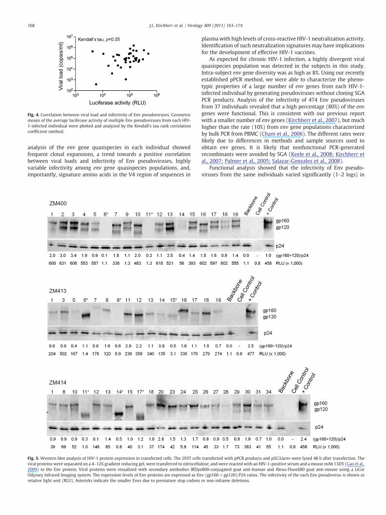
Fig. 4. Correlation between viral load and infectivity of Env pseudoviruses. Geometricmeans of the average luciferase activity of multiple Env pseudoviruses from each HIV-1-infected individual were plotted and analyzed by the Kendall's tau rank correlationcoefficient method.
168 J.L. Kirchherr et al. / Virology 409 (2011) 163–174
analysis of the env gene quasispecies in each individual showedfrequent clonal expansions, a trend towards a positive correlationbetween viral loads and infectivity of Env pseudoviruses, highlyvariable infectivity among env gene quasispecies populations, and,importantly, signature amino acids in the V4 region of sequences in
Fig. 5. Western blot analysis of HIV-1 protein expression in transfected cells. The 293T cellsviral proteins were separated on a 4–12% gradient reducing gel, were transferred to nitrocellu2009) to the Env protein. Viral proteins were visualized with secondary antibodies IRDyeOdyssey Infrared Imaging system. The expression levels of Env proteins are expressed as Enrelative light unit (RLU). Asterisks indicate the smaller Envs due to premature stop codons
plasmawith high levels of cross-reactive HIV-1 neutralization activity.Identification of such neutralization signatures may have implicationsfor the development of effective HIV-1 vaccines.
As expected for chronic HIV-1 infection, a highly divergent viralquasispecies population was detected in the subjects in this study.Intra-subject env gene diversity was as high as 8%. Using our recentlyestablished pPCR method, we were able to characterize the pheno-typic properties of a large number of env genes from each HIV-1-infected individual by generating pseudoviruses without cloning SGAPCR products. Analysis of the infectivity of 474 Env pseudovirusesfrom 37 individuals revealed that a high percentage (80%) of the envgenes were functional. This is consistent with our previous reportwith a smaller number of env genes (Kirchherr et al., 2007), but muchhigher than the rate (10%) from env gene populations characterizedby bulk PCR from PBMC (Cham et al., 2006). The different rates werelikely due to differences in methods and sample sources used toobtain env genes. It is likely that nonfunctional PCR-generatedrecombinants were avoided by SGA (Keele et al., 2008; Kirchherr etal., 2007; Palmer et al., 2005; Salazar-Gonzalez et al., 2008).
Functional analysis showed that the infectivity of Env pseudo-viruses from the same individuals varied significantly (1–2 logs) in
transfected with pPCR products and pSG3Δenv were lysed 48 h after transfection. Thelose, andwere reactedwith an HIV-1-positive serum and amousemAb 13D5 (Gao et al.,800-conjugated goat anti-human and Alexa-Fluor680 goat anti-mouse using a LiCorv (gp160+gp120):P24 ratios. The infectivity of the each Env pseudovirus is shown asor non-inframe deletions.
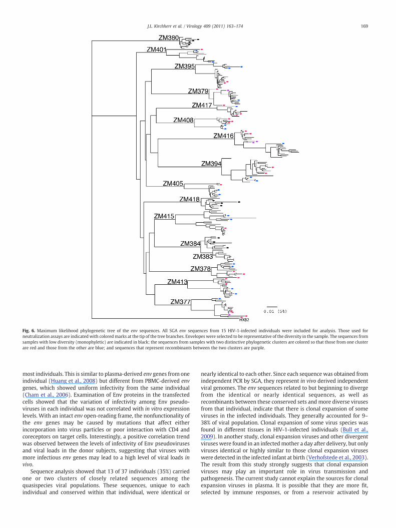
Fig. 6. Maximum likelihood phylogenetic tree of the env sequences. All SGA env sequences from 15 HIV-1-infected individuals were included for analysis. Those used forneutralization assays are indicated with coloredmarks at the tip of the tree branches. Envelopes were selected to be representative of the diversity in the sample. The sequences fromsamples with low diversity (monophyletic) are indicated in black; the sequences from samples with two distinctive phylogenetic clusters are colored so that those from one clusterare red and those from the other are blue; and sequences that represent recombinants between the two clusters are purple.
169J.L. Kirchherr et al. / Virology 409 (2011) 163–174
most individuals. This is similar to plasma-derived env genes from oneindividual (Huang et al., 2008) but different from PBMC-derived envgenes, which showed uniform infectivity from the same individual(Cham et al., 2006). Examination of Env proteins in the transfectedcells showed that the variation of infectivity among Env pseudo-viruses in each individual was not correlated with in vitro expressionlevels. With an intact env open-reading frame, the nonfunctionality ofthe env genes may be caused by mutations that affect eitherincorporation into virus particles or poor interaction with CD4 andcoreceptors on target cells. Interestingly, a positive correlation trendwas observed between the levels of infectivity of Env pseudovirusesand viral loads in the donor subjects, suggesting that viruses withmore infectious env genes may lead to a high level of viral loads invivo.
Sequence analysis showed that 13 of 37 individuals (35%) carriedone or two clusters of closely related sequences among thequasispecies viral populations. These sequences, unique to eachindividual and conserved within that individual, were identical or
nearly identical to each other. Since each sequence was obtained fromindependent PCR by SGA, they represent in vivo derived independentviral genomes. The env sequences related to but beginning to divergefrom the identical or nearly identical sequences, as well asrecombinants between these conserved sets and more diverse virusesfrom that individual, indicate that there is clonal expansion of someviruses in the infected individuals. They generally accounted for 9–38% of viral population. Clonal expansion of some virus species wasfound in different tissues in HIV-1-infected individuals (Bull et al.,2009). In another study, clonal expansion viruses and other divergentviruses were found in an infectedmother a day after delivery, but onlyviruses identical or highly similar to those clonal expansion viruseswere detected in the infected infant at birth (Verhofstede et al., 2003).The result from this study strongly suggests that clonal expansionviruses may play an important role in virus transmission andpathogenesis. The current study cannot explain the sources for clonalexpansion viruses in plasma. It is possible that they are more fit,selected by immune responses, or from a reservoir activated by
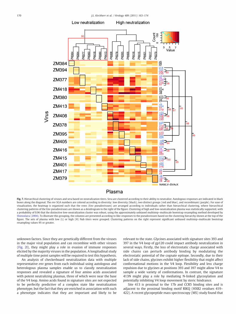
Fig. 7. Hierarchical clustering of viruses and sera based on neutralization titers. Sera are clustered according to their ability to neutralize. Autologous responses are indicated in blackboxes along the diagonal. The env SGA numbers are colored according to diversity: low diversity (black), two distinct groups (red and blue), and recombinant (purple). For ease ofvisualization, the heatmap is organized such that the rows (Env pseudoviruses) are arranged according to individuals rather than hierarchical clustering, where hierarchicalclustering patterns of the Env pseudoviruses are shown as a dendrogram to the right of the figure. Clustering of high and low-neutralization plasma was statistically supported, witha probability of 0.96 that the distinctive low-neutralization cluster was robust, using the approximately unbiased multistep–multiscale bootstrap resampling method developed byShimodaira (2004). To illustrate this grouping, the columns are presented according to like-responses to the pseudoviruses based on the clustering hierarchy shown at the top of thefigure. The sets of plasma with low (L) or high (H) Nab titers were grouped. Clustering patterns on the right represent significant unbiased multistep–multiscale bootstrapresampling values 95 or greater.
170 J.L. Kirchherr et al. / Virology 409 (2011) 163–174
unknown factors. Since they are genetically different from the virusesin the major viral population and can recombine with other viruses(Fig. 2I), they might play a role in evasion of immune responseselicited by themajority viruses in the population. A longitudinal studyof multiple time point samples will be required to test this hypothesis.
An analysis of checkerboard neutralization data with multiplerepresentative env genes from each individual using autologous andheterologous plasma samples enable us to classify neutralizationresponses and revealed a signature of four amino acids associatedwith potent neutralizing plasmas, three of which were near the baseof the V4 loop. Amino acids found in signature sites are not expectedto be perfectly predictive of a complex state like neutralizationphenotype, but the fact that they are enriched in associationwith sucha phenotype indicates that they are important and likely to be
relevant to the state. Glycines associated with signature sites 393 and397 in the V4 loop of gp120 could impact antibody neutralization inseveral ways. Firstly, the loss of electrostatic charge associated withside chains can perturb antibody binding by modulating theelectrostatic potential of the cognate epitope. Secondly, due to theirlack of side chains, glycines exhibit higher flexibility that might affectconformational motions in the V4 loop. Flexibility and less chargerepulsion due to glycines at positions 393 and 397 might allow V4 tosample a wide variety of conformations. In contrast, the signature413N might play a role by mediating N-linked glycosylation andpotentially inhibiting V4 loop movement by steric hindrance.
Site 413 is proximal to the 17b and CCR5 binding sites and isadjacent to the proximal binding motif RIKQ (HXB2 residues 419–422). A recent glycopeptide mass spectroscopy (MS) study found that
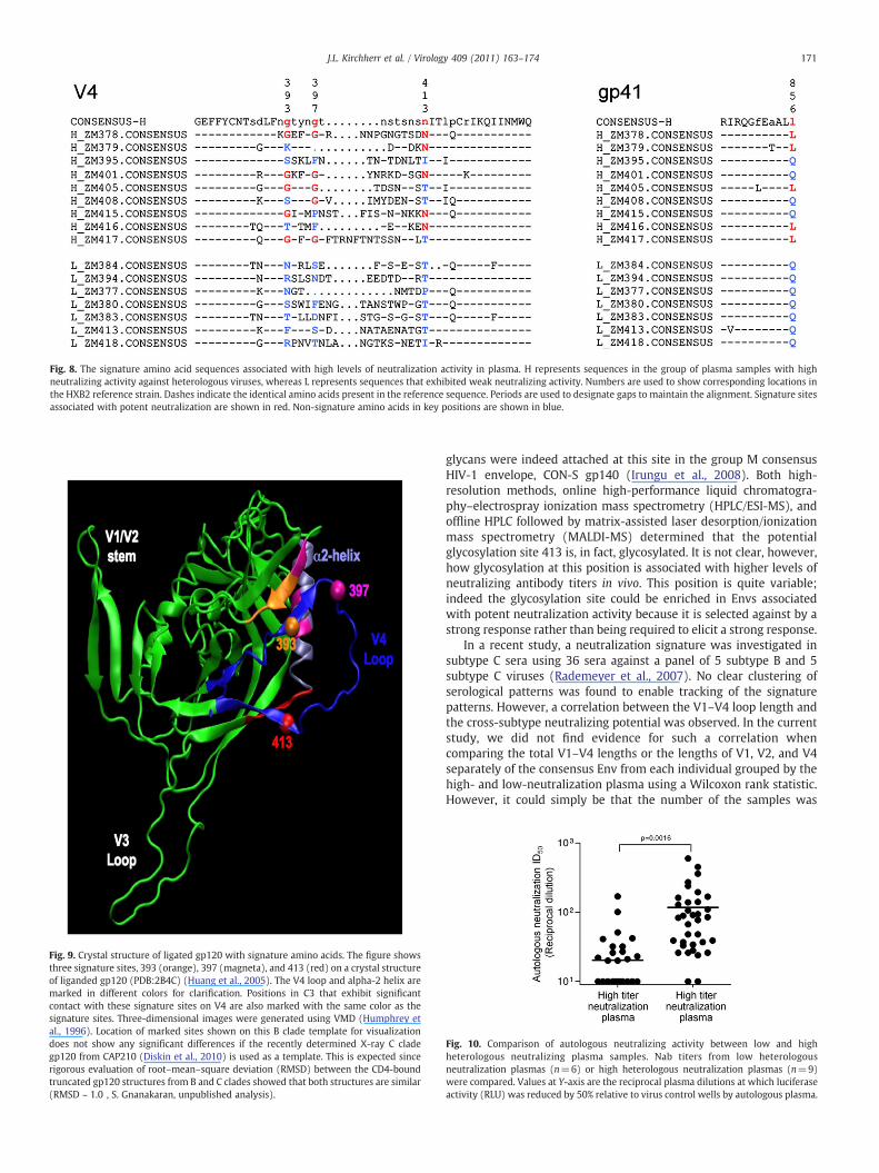
Fig. 8. The signature amino acid sequences associated with high levels of neutralization activity in plasma. H represents sequences in the group of plasma samples with highneutralizing activity against heterologous viruses, whereas L represents sequences that exhibited weak neutralizing activity. Numbers are used to show corresponding locations inthe HXB2 reference strain. Dashes indicate the identical amino acids present in the reference sequence. Periods are used to designate gaps to maintain the alignment. Signature sitesassociated with potent neutralization are shown in red. Non-signature amino acids in key positions are shown in blue.
Fig. 9. Crystal structure of ligated gp120 with signature amino acids. The figure showsthree signature sites, 393 (orange), 397 (magneta), and 413 (red) on a crystal structureof liganded gp120 (PDB:2B4C) (Huang et al., 2005). The V4 loop and alpha-2 helix aremarked in different colors for clarification. Positions in C3 that exhibit significantcontact with these signature sites on V4 are also marked with the same color as thesignature sites. Three-dimensional images were generated using VMD (Humphrey etal., 1996). Location of marked sites shown on this B clade template for visualizationdoes not show any significant differences if the recently determined X-ray C cladegp120 from CAP210 (Diskin et al., 2010) is used as a template. This is expected sincerigorous evaluation of root–mean–square deviation (RMSD) between the CD4-boundtruncated gp120 structures from B and C clades showed that both structures are similar(RMSD ~ 1.0 , S. Gnanakaran, unpublished analysis).
171J.L. Kirchherr et al. / Virology 409 (2011) 163–174
glycans were indeed attached at this site in the group M consensusHIV-1 envelope, CON-S gp140 (Irungu et al., 2008). Both high-resolution methods, online high-performance liquid chromatogra-phy–electrospray ionization mass spectrometry (HPLC/ESI-MS), andoffline HPLC followed by matrix-assisted laser desorption/ionizationmass spectrometry (MALDI-MS) determined that the potentialglycosylation site 413 is, in fact, glycosylated. It is not clear, however,how glycosylation at this position is associated with higher levels ofneutralizing antibody titers in vivo. This position is quite variable;indeed the glycosylation site could be enriched in Envs associatedwith potent neutralization activity because it is selected against by astrong response rather than being required to elicit a strong response.
In a recent study, a neutralization signature was investigated insubtype C sera using 36 sera against a panel of 5 subtype B and 5subtype C viruses (Rademeyer et al., 2007). No clear clustering ofserological patterns was found to enable tracking of the signaturepatterns. However, a correlation between the V1–V4 loop length andthe cross-subtype neutralizing potential was observed. In the currentstudy, we did not find evidence for such a correlation whencomparing the total V1–V4 lengths or the lengths of V1, V2, and V4separately of the consensus Env from each individual grouped by thehigh- and low-neutralization plasma using a Wilcoxon rank statistic.However, it could simply be that the number of the samples was
Fig. 10. Comparison of autologous neutralizing activity between low and highheterologous neutralizing plasma samples. Nab titers from low heterologousneutralization plasmas (n=6) or high heterologous neutralization plasmas (n=9)were compared. Values at Y-axis are the reciprocal plasma dilutions at which luciferaseactivity (RLU) was reduced by 50% relative to virus control wells by autologous plasma.
172 J.L. Kirchherr et al. / Virology 409 (2011) 163–174
underpowered to show this effect. The other possibility is that allsamples in the current study were from CHI individuals in whom thelength of variable loops had already become longer during theevolution of immune evasion.
Previous studies also showed that the alpha-2 helix was understrong selectionpressure (Gnanakaran et al., 2007) and this regionwasassociated with resistance to autologous neutralization of subtype Cviruses (Rong et al., 2007). The V4 region is downstream from thealpha-2 helix, and they are structurally close to each other. It was alsofound that the C3V4 region is likely responsible for inducingautologous and heterologous Nabs (Moore et al., 2008). The newlyidentified three signature amino acids were located in the vicinity ofthe previously reported region targeted byNabs. These signaturesmaybe responsible for eliciting broader andmore potent Nab responses orrepresent the escapemutations from the broadlyNabs against subtypeC viruses through similar pathways. In either case, the data suggestthat signature patterns in the V4 loop are important to consider for thedesign of a vaccine that can induce potent andbroadly reactiveNabs, atleast for subtype C, which is the most prevalent HIV-1 subtype in theworld.
Methods and materials
Amplification of HIV-1 env genes
Plasma samples were collected from 37 HIV-1-positive individualsenrolled in a study of contemporary HIV-1 strains in Ndola, Zambia.The study was approved by the ethics committee of the TropicalDisease Research Centre, the Duke University Institutional ReviewBoard, and the National Institutes of Health. No people in this studywere treated with antiretroviral drugs. The homosexual activity wasnot reported in the study population. The studied individuals in ourstudy were most likely infected through heterosexual transmission.Viral RNAwas extracted from the plasma and reverse transcribed intocDNA using Superscript III (Invitrogen; Carlsbad, CA). Multiple rev/envgenes from each individual were obtained by using single-genomeamplification (SGA), followed by the addition of a CMV promoter tothe 5′ end of the SGA products using pPCR technology as previouslydescribed (Kirchherr et al., 2007).
Single-round infection assay
pPCR products were cotransfected with an env-deficient HIV-1backbone plasmid (pSG3Δenv) into 293 T cells in a 24-well plate usingFuGENE6 transfection reagent (Roche Diagnostics; Indianapolis, IN).Briefly, pPCR DNA (150 ng) and pSG3Δenv DNA (150 ng) were mixedwith 1.2 μl of FuGENE6 (FuGENE:DNA ratio at 3 μl:1 μg) in a totalvolume of 20 μl with serum-free DMEM, incubated for 30 min andadded to 293 T cells (70% confluence) seeded 1 day earlier at 5×10⁴
per well. Forty-eight hours after transfection, supernatants wereharvested. Equal volumes of pseudovirions were added to TZM-blcells with DEAE (5 μg/ml) in a 96-well plate (200 μl). Cultures wereincubated for 48 h at 37 °C with 5% CO2. Supernatants (100 μl) frominfected TZM-bl cells were removed and 100 μl of Bright-GloLuciferase Assay substrate with buffer (Promega; Madison, WI) wasadded to the cells. Following a 2-min incubation, 100 μl of cell lysateswas added to a solid black 96-well plate. Luminescence wasmeasuredwith a Wallac 1420 Multilabel Counter (PerkinElmer: Waltham, MA).
Neutralization assay
HIV-1 neutralization was measured as a reduction in luciferaseactivity after a single-round infection of TZM-bl cells, as previouslydescribed (Li et al., 2005, 2006). Equal amounts of pseudovirions (200TCID50) were used in each reaction. Neutralization titers againstpseudoviruses were determined for 16 plasma samples (15 autolo-
gous and 1 heterologous to the tested Env pseudoviruses) and oneHIV-1-positive serum control. An amphotropic murine leukemia Envpseudovirus was also included as non-specific neutralization control.
Western blot
Forty-eight hours after cotransfection with pPCR products andpSG3Δenv, 293 T cells were lysed with 250 μl of lysis buffer (50 mMTris–HCl, 150 mM NaCl, 20 mM EDTA, 1% Triton-X100, 0.1% SDS,pH 7.4). Cell lysates were mixed with 6× denaturing sample buffer(300 mM Tris–HCl pH 6.8, 12 mM EDTA, 12% SDS, 864 mM 2-mercaptoethanol, 60% glycerol, 0.05% bromophenol blue). Sampleswere boiled for 10 min and then loaded on a NuPAGE Novex 4–12%Bis–Tris gel (Invitrogen; Carlesbad, CA). After the samples weretransferred to a nitrocellulose membrane, the membrane was blockedin PBS containing 1% casein and 0.01% NaN3 for 1 h. The blot wasreactedwith an HIV-1-positive serum (1:500) and amousemAb 13D5(1 μg/ml) to the HIV-1 Env protein. Finally, the membrane wasreacted with an IRDye800-conjugated affinity purified goat anti-human IgG antibody (Rockland Immunochemicals; Gilbertsville, PA)and an Alexa-Fluor 680-conjugated goat anti-mouse IgG antibody(Invitrogen; Carlesbad, CA). Fluorescence was detected, and thedensity of Env protein and p24 bands was determined with anOdyssey Infrared Imaging system (LiCor Biosciences; Lincoln, NE). Thelevel of Env protein in the transfected cells was expressed as folddifferences over the amount of p24 Gag antigen in the same cells.
Sequence analysis
Sequences of SGA env amplicons were obtained by cycle-sequencing and dye terminator methods with an ABI 3730xl geneticanalyzer (Applied Biosystems; Foster City, CA). Individual sequencefragments for each env SGA were assembled and edited using theSequencher program 4.7 (Gene Codes; Ann Arbor, MI). The envsequences were aligned using CLUSTAL W (Thompson et al., 1994),and Genecutter (www.hiv.lanl.gov) for obtaining codon aligned filesthat could be used for signature analysis. Phylogenetic tree wasconstructed with complete env gene sequences using the neighbor-joiningmethod (Saitou and Nei, 1987) and the Kimura two-parametermodel (Kimura, 1980). The reliability of topologies was estimated byperforming bootstrap analysis with 1000 replicates.
Hierarchical clustering analysis
The neutralizing potential of plasma (the log reciprocal dilutionID50 scores) was organized into patterns of similar neutralizingpotency when tested against the panel of envelopes used in this studyusing the Los Alamos National Lab web-based heatmap interface(http://www.hiv.lanl.gov/content/sequence/HEATMAP/heatmap.html) and the statistical package R (R Development Core Team, 2009).The test panel of Envs was resampled 10,000 times using a random-with-replacement bootstrap strategy, and this identified two robustand distinctive clusters recurring in 96% of the bootstrap samples: onegroup with little cross-neutralizing potential and another group withthe ability to cross-neutralize multiple heterologous C clade viruses(Shimodaira, 2004). These two groups were used for subsequentsignature analysis. The same strategy was used to identify statisticallyrobust clusters of the Envs with like-neutralizing susceptibility.Clusters with high bootstrap values typically only included a fewEnvs each, and these Env clusters were typically found within a singlesubject. Therefore, we decided to organize Env pseudoviruses byindividual in a heatmap figure (Fig. 7), while retaining theinformation regarding significant bootstrap clusters, shown super-imposed onto the figure.

173J.L. Kirchherr et al. / Virology 409 (2011) 163–174
Signature analysis
We first divided the envelope sequences into the two groups,based on heatmap clustering patterns that indicated whether theywere derived from an individual with plasma with a cross-reactiveneutralizing profile or a weakly neutralizing profile. Alignments usedfor signature analysis were generated with GeneCutter (www.hiv.lanl.gov) to provide codon-aligned DNA for phylogenetic analysis.Phylogenetically corrected methods were used to identify allsignature sites. Phylogenetic correction is critical because observedpatterns in data can either result from correlations imposed by theinitial historical emergence of a lineage of viruses (founder effects) or,in the case of HIV-1, be a consequence of recent biologicalinteractions. Not accounting for founder effects can lead to erroneousstatistical conclusions (Bhattacharya et al., 2007). The sequence of thevirus depends on its full evolutionary history, while causal correla-tions are manifest in correlations with recent changes. The separationof the two effects, i.e., a phylogenetic correction, enables one toestimate the impact of recent changes on the phenotype. This requiresstatistical reconstruction of genealogical relationships between theviruses and an estimate of ancestral states of the viruses. Weimplemented this through maximum likelihood phylogenies andtested for phenotypic associations with mutational patterns based onchange or stasis in a given amino acid, when compared to the mostrecent common ancestor, adapting the phylogenetically correctedFisher's Exact Methods first developed by Bhattacharya et al. (2007).We screened themutational pattern of each amino acid found at everycolumn in the alignment. To correct for multiple tests, we used a q-value to assess the false discovery rate (Storey and Tibshirani, 2003).The four strongest associations had a p-value of 0.03 and q-value of0.27, thus it is likely that at least one of the signatures identified is afalse positive, and only borderline significance was obtained overall.Consequently, these signatures should be considered in a hypothesis-raising framework as potentially interesting sites that merit furtherinvestigation.
Nucleotide sequences accession numbers
GenBank accession numbers of the env gene sequences areGU329048-GU329523.
Supplementary material related to this article can be found onlineat doi:10.1016/j.virol.2010.09.031.
Acknowledgments
This work was supported by grants from the National Institutes ofHealth/National Institute of Allergy and Infectious Diseases [CIPRA (R03AI054155) to RMM, HIVRAD PO 1 AI35351, Center for HIV/AIDS VaccineImmunology (AI067854), Duke Center for AIDS Research (AI64518)Molecular Virology Core], the Bill and Melinda Gates Foundation, and aLos Alamos National Laboratory directed research grant.
References
Bhattacharya, T., Daniels, M., Heckerman, D., Foley, B., Frahm, N., Kadie, C., Carlson, J.,Yusim, K., McMahon, B., Gaschen, B., Mallal, S., Mullins, J.I., Nickle, D.C., Herbeck, J.,Rousseau, C., Learn, G.H., Miura, T., Brander, C., Walker, B., Korber, B., 2007. Foundereffects in the assessment of HIV polymorphisms and HLA allele associations.Science 315, 1583–1586.
Binley, J.M., Wrin, T., Korber, B., Zwick, M.B., Wang, M., Chappey, C., Stiegler, G., Kunert, R.,Zolla-Pazner, S., Katinger, H., Petropoulos, C.J., Burton, D.R., 2004. Comprehensivecross-clade neutralization analysis of a panel of anti-human immunodeficiency virustype 1 monoclonal antibodies. J. Virol. 78, 13232–13252.
Binley, J.M., Lybarger, E.A., Crooks, E.T., Seaman, M.S., Gray, E., Davis, K.L., Decker, J.M.,Wycuff, D., Harris, L., Hawkins, N., Wood, B., Nathe, C., Richman, D., Tomaras, G.D.,Bibollet-Ruche, F., Robinson, J.E., Morris, L., Shaw, G.M., Montefiori, D.C., Mascola, J.R.,2008. Profiling the specificity of neutralizing antibodies in a large panel of plasmasfrom patients chronically infected with human immunodeficiency virus type 1subtypes B and C. J. Virol. 82, 11651–11668.
Bull, M.E., Learn, G.H., McElhone, S., Hitti, J., Lockhart, D., Holte, S., Dragavon, J., Coombs,R.W., Mullins, J.I., Frenkel, L.M., 2009. Monotypic human immunodeficiency virustype 1 genotypes across the uterine cervix and in blood suggest proliferation ofcells with provirus. J. Virol. 83, 6020–6028.
Burton, D.R., Desrosiers, R.C., Doms, R.W., Koff, W.C., Kwong, P.D., Moore, J.P., Nabel, G.J.,Sodroski, J., Wilson, I.A., Wyatt, R.T., 2004. HIV vaccine design and the neutralizingantibody problem. Nat. Immunol. 5, 233–236.
Cham, F., Zhang, P.F., Heyndrickx, L., Bouma, P., Zhong, P., Katinger, H., Robinson, J., vander Groen, G., Quinnan Jr., G.V., 2006. Neutralization and infectivity characteristicsof envelope glycoproteins from human immunodeficiency virus type 1 infecteddonors whose sera exhibit broadly cross-reactive neutralizing activity. Virology347, 36–51.
Chertova, E., Bess Jr., J.W., Crise, B.J., Sowder, I.R., Schaden, T.M., Hilburn, J.M., Hoxie, J.A.,Benveniste, R.E., Lifson, J.D., Henderson, L.E., Arthur, L.O., 2002. Envelopeglycoprotein incorporation, not shedding of surface envelope glycoprotein(gp120/SU), is the primary determinant of SU content of purified humanimmunodeficiency virus type 1 and simian immunodeficiency virus. J Virol 76,5315–5325.
Cordonnier, A., Montagnier, L., Emerman, M., 1989. Single amino-acid changes in HIVenvelope affect viral tropism and receptor binding. Nature 340, 571–574.
Derdeyn, C.A., Decker, J.M., Bibollet-Ruche, F., Mokili, J.L., Muldoon, M., Denham, S.A.,Heil, M.L., Kasolo, F., Musonda, R., Hahn, B.H., Shaw, G.M., Korber, B.T., Allen, S.,Hunter, E., 2004. Envelope-constrained neutralization-sensitive HIV-1 afterheterosexual transmission. Science 303, 2019–2022.
Desrosiers, R.C., 2004. Prospects for an AIDS vaccine. Nat. Med. 10, 221–223.Dhillon, A.K., Donners, H., Pantophlet, R., Johnson,W.E., Decker, J.M., Shaw, G.M., Lee, F.H.,
Richman, D.D., Doms, R.W., Vanham, G., Burton, D.R., 2007. Dissecting the neutralizingantibody specificities of broadly neutralizing sera from human immunodeficiencyvirus type 1-infected donors. J. Virol. 81, 6548–6562.
Diskin, R., Marcovecchio, P.M., Bjorkman, P.J., 2010. Structure of a clade C HIV-1 gp120bound to CD4 and CD4-induced antibody reveals anti-CD4 polyreactivity. Nat.Struct. Mol. Biol. 17, 608–613.
Fang, G., Zhu, G., Burger, H., Keithly, J.S., Weiser, B., 1998. Minimizing DNArecombination during long RT-PCR. J. Virol. Meth. 76, 139–148.
Gao, F., Scearce, R.M., Alam, S.M., Hora, B., Xia, S., Hohm, J.E., Parks, R.J., Ogburn, D.F.,Tomaras, G.D., Park, E., Lomas,W.E., Maino, V.C., Fiscus, S.A., Cohen,M.S., Moody,M.A.,Hahn, B.H., Korber, B.T., Liao, H.X., Haynes, B.F., 2009. Cross-reactive monoclonalantibodies to multiple HIV-1 subtype and SIVcpz envelope glycoproteins. Virology394, 91–98.
Gnanakaran, S., Lang, D., Daniels, M., Bhattacharya, T., Derdeyn, C.A., Korber, B., 2007.Clade-specific differences between human immunodeficiency virus type 1 clades Band C: diversity and correlations in C3-V4 regions of gp120. J. Virol. 81, 4886–4891.
Gnanakaran, S., Daniels, M.G., Bhattacharya, T., Lapedes, A.S., Sethi, A., Li, M., Tang, H.,Greene, K., Gao, H., Haynes, B.F., Cohen, M.S., Shaw, G.M., Seaman, M.S., Kumar, A.,Gao, F., Montefiori, D.C., Korber, B., 2010. Genetic signatures in the envelopeglycoproteins of HIV-1 that associate with broadly neutralizing antibodies. PLoSComput. Biol. 6, e1000955.
Haynes, B.F., Montefiori, D.C., 2006. Aiming to induce broadly reactive neutralizingantibody responses with HIV-1 vaccine candidates. Expert Rev. Vaccin. 5, 347–363.
Hemelaar, J., Gouws, E., Ghys, P.D., Osmanov, S., 2006. Global and regional distributionof HIV-1 genetic subtypes and recombinants in 2004. AIDS 20, W13–W23.
Huang, C.C., Tang, M., Zhang, M.Y., Majeed, S., Montabana, E., Stanfield, R.L., Dimitrov, D.S.,Korber, B., Sodroski, J., Wilson, I.A., Wyatt, R., Kwong, P.D., 2005. Structure of a V3-containing HIV-1 gp120 core. Science 310, 1025–1028.
Huang, W., Toma, J., Fransen, S., Stawiski, E., Reeves, J.D., Whitcomb, J.M., Parkin, N.,Petropoulos, C.J., 2008. Coreceptor tropism can be influenced by amino acidsubstitutions in the gp41 transmembrane subunit of human immunodeficiencyvirus type 1 envelope protein. J. Virol. 82, 5584–5593.
Humphrey, W., Dalke, A., Schulten, K., 1996. VMD: visual molecular dynamics. J. Mol.Graph. 14 (33–38), 27–38.
Irungu, J., Go, E.P., Zhang, Y., Dalpathado, D.S., Liao, H.X., Haynes, B.F., Desaire, H., 2008.Comparison of HPLC/ESI-FTICR MS versus MALDI-TOF/TOF MS for glycopeptideanalysis of a highly glycosylated HIV envelope glycoprotein. J. Am. Soc. MassSpectrom. 19, 1209–1220.
Kalia, V., Sarkar, S., Gupta, P., Montelaro, R.C., 2005. Antibody neutralization escapemediated by point mutations in the intracytoplasmic tail of human immunodefi-ciency virus type 1 gp41. J. Virol. 79, 2097–2107.
Keele, B.F., Giorgi, E.E., Salazar-Gonzalez, J.F., Decker, J.M., Pham, K.T., Salazar, M.G., Sun,C., Grayson, T., Wang, S., Li, H., Wei, X., Jiang, C., Kirchherr, J.L., Gao, F., Anderson, J.A.,Ping, L.H., Swanstrom, R., Tomaras, G.D., Blattner, W.A., Goepfert, P.A., Kilby, J.M.,Saag, M.S., Delwart, E.L., Busch, M.P., Cohen, M.S., Montefiori, D.C., Haynes, B.F.,Gaschen, B., Athreya, G.S., Lee, H.Y., Wood, N., Seoighe, C., Perelson, A.S.,Bhattacharya, T., Korber, B.T., Hahn, B.H., Shaw, G.M., 2008. Identification andcharacterization of transmitted and early founder virus envelopes in primary HIV-1infection. Proc. Natl Acad. Sci. USA 105, 7552–7557.
Kimura, M., 1980. A simple method for estimating evolutionary rates of basesubstitutions through comparative studies of nucleotide sequences. J. Mol. Evol.16, 111–120.
Kirchherr, J.L., Lu, X., Kasongo, W., Chalwe, V., Mwananyanda, L., Musonda, R.M., Xia, S.M.,Scearce, R.M., Liao, H.X., Montefiori, D.C., Haynes, B.F., Gao, F., 2007. High throughputfunctional analysis of HIV-1 env genes without cloning. J. Virol. Meth. 143, 104–111.
Korber, B., Gnanakaran, S., 2009. The implications of patterns in HIV diversity forneutralizing antibody induction and susceptibility. Curr. Opin. HIV AIDS 4,408–417.
Korber, B.T., Letvin, N.L., Haynes, B.F., 2009. T-cell vaccine strategies for humanimmunodeficiency virus, the virus with a thousand faces. J. Virol. 83, 8300–8314.

174 J.L. Kirchherr et al. / Virology 409 (2011) 163–174
Kulkarni, S.S., Lapedes, A., Tang, H., Gnanakaran, S., Daniels, M.G., Zhang, M.,Bhattacharya, T., Li, M., Polonis, V.R., McCutchan, F.E., Morris, L., Ellenberger, D.,Butera, S.T., Bollinger, R.C., Korber, B.T., Paranjape, R.S., Montefiori, D.C., 2009.Highly complex neutralization determinants on a monophyletic lineage of newlytransmitted subtype C HIV-1 Env clones from India. Virology 385, 505–520.
LaBranche, C.C., Sauter, M.M., Haggarty, B.S., Vance, P.J., Romano, J., Hart, T.K., Bugelski,P.J., Marsh, M., Hoxie, J.A., 1995. A single amino acid change in the cytoplasmicdomain of the simian immunodeficiency virus transmembrane molecule increasesenvelope glycoprotein expression on infected cells. J. Virol. 69, 5217–5227.
Li, M., Gao, F., Mascola, J.R., Stamatatos, L., Polonis, V.R., Koutsoukos, M., Voss, G.,Goepfert, P., Gilbert, P., Greene, K.M., Bilska, M., Kothe, D.L., Salazar-Gonzalez, J.F.,Wei, X., Decker, J.M., Hahn, B.H., Montefiori, D.C., 2005. Human immunodeficiencyvirus type 1 env clones from acute and early subtype B infections for standardizedassessments of vaccine-elicited neutralizing antibodies. J. Virol. 79, 10108–10125.
Li, M., Salazar-Gonzalez, J.F., Derdeyn, C.A., Morris, L., Williamson, C., Robinson, J.E.,Decker, J.M., Li, Y., Salazar, M.G., Polonis, V.R., Mlisana, K., Karim, S.A., Hong, K.,Greene, K.M., Bilska, M., Zhou, J., Allen, S., Chomba, E., Mulenga, J., Vwalika, C., Gao,F., Zhang, M., Korber, B.T., Hunter, E., Hahn, B.H., Montefiori, D.C., 2006. Genetic andneutralization properties of subtype C human immunodeficiency virus type 1molecular env clones from acute and early heterosexually acquired infections inSouthern Africa. J. Virol. 80, 11776–11790.
Li, Y., Migueles, S.A., Welcher, B., Svehla, K., Phogat, A., Louder, M.K., Wu, X., Shaw, G.M.,Connors, M., Wyatt, R.T., Mascola, J.R., 2007. Broad HIV-1 neutralization mediatedby CD4-binding site antibodies. Nat. Med. 13, 1032–1034.
Lin, G., Nara, P.L., 2007. Designing immunogens to elicit broadly neutralizing antibodiesto the HIV-1 envelope glycoprotein. Curr. HIV Res. 5, 514–541.
Liu, S.L., Rodrigo, A.G., Shankarappa, R., Learn, G.H., Hsu, L., Davidov, O., Zhao, L.P.,Mullins, J.I., 1996. HIV quasispecies and resampling. Science 273, 415–416.
Moore, J.P., McCutchan, F.E., Poon, S.W., Mascola, J., Liu, J., Cao, Y., Ho, D.D., 1994.Exploration of antigenic variation in gp120 from clades A through F of humanimmunodeficiency virus type 1 by using monoclonal antibodies. J. Virol. 68,8350–8364.
Moore, P.L., Gray, E.S., Choge, I.A., Ranchobe, N., Mlisana, K., Abdool Karim, S.S.,Williamson, C., Morris, L., 2008. The c3-v4 region is a major target of autologousneutralizing antibodies in human immunodeficiency virus type 1 subtype Cinfection. J. Virol. 82, 1860–1869.
Moore, P.L., Ranchobe, N., Lambson, B.E., Gray, E.S., Cave, E., Abrahams, M.R., Bandawe,G., Mlisana, K., Abdool Karim, S.S., Williamson, C., Morris, L., 2009. Limitedneutralizing antibody specificities drive neutralization escape in early HIV-1subtype C infection. PLoS Pathog. 5, e1000598.
Morris, J.F., Sternberg, E.J., Gutshall, L., Petteway Jr., S.R., Ivanoff, L.A., 1994. Effect of asingle amino acid substitution in the V3 domain of the human immunodeficiencyvirus type 1: generation of revertant viruses to overcome defects in infectivity inspecific cell types. J. Virol. 68, 8380–8385.
Palmer, S., Kearney, M., Maldarelli, F., Halvas, E.K., Bixby, C.J., Bazmi, H., Rock, D., Falloon,J., Davey Jr., R.T., Dewar, R.L., Metcalf, J.A., Hammer, S., Mellors, J.W., Coffin, J.M.,2005. Multiple, linked human immunodeficiency virus type 1 drug resistancemutations in treatment-experienced patients are missed by standard genotypeanalysis. J. Clin. Microbiol. 43, 406–413.
R Development Core Team, 2009. A Language and Environment for StatisticalComputing. R Foundation for Statistical Computing, Vienna, Austria.
Rademeyer, C., Moore, P.L., Taylor, N., Martin, D.P., Choge, I.A., Gray, E.S., Sheppard, H.W., Gray, C., Morris, L., Williamson, C., 2007. Genetic characteristics of HIV-1subtype C envelopes inducing cross-neutralizing antibodies. Virology 368,172–181.
Rong, R., Gnanakaran, S., Decker, J.M., Bibollet-Ruche, F., Taylor, J., Sfakianos, J.N., Mokili,J.L., Muldoon, M., Mulenga, J., Allen, S., Hahn, B.H., Shaw, G.M., Blackwell, J.L.,Korber, B.T., Hunter, E., Derdeyn, C.A., 2007. Unique mutational patterns in theenvelope alpha 2 amphipathic helix and acquisition of length in gp120hypervariable domains are associated with resistance to autologous neutralizationof subtype C human immunodeficiency virus type 1. J. Virol. 81, 5658–5668.
Rong, R., Li, B., Lynch, R.M., Haaland, R.E., Murphy, M.K., Mulenga, J., Allen, S.A., Pinter,A., Shaw, G.M., Hunter, E., Robinson, J.E., Gnanakaran, S., Derdeyn, C.A., 2009. Escapefrom autologous neutralizing antibodies in acute/early subtype C HIV-1 infectionrequires multiple pathways. PLoS Pathog. 5, e1000594.
Saitou, N., Nei, M., 1987. The neighbor-joining method: a new method for reconstruct-ing phylogenetic trees. Mol. Biol. Evol. 4, 406–425.
Salazar-Gonzalez, J.F., Bailes, E., Pham, K.T., Salazar, M.G., Guffey, M.B., Keele, B.F., Derdeyn,C.A., Farmer, P., Hunter, E., Allen, S., Manigart, O., Mulenga, J., Anderson, J.A.,Swanstrom, R., Haynes, B.F., Athreya, G.S., Korber, B.T., Sharp, P.M., Shaw, G.M.,Hahn, B.H., 2008. Deciphering human immunodeficiency virus type 1 transmission
and early envelope diversification by single-genome amplification and sequencing.J. Virol. 82, 3952–3970.
Sather, D.N., Armann, J., Ching, L.K., Mavrantoni, A., Sellhorn, G., Caldwell, Z., Yu, X.,Wood, B., Self, S., Kalams, S., Stamatatos, L., 2009. Factors associated with thedevelopment of cross-reactive neutralizing antibodies during human immunode-ficiency virus type 1 infection. J. Virol. 83, 757–769.
Shen, X., Parks, R.J., Montefiori, D.C., Kirchherr, J.L., Keele, B.F., Decker, J.M., Blattner, W.A.,Gao, F.,Weinhold, K.J., Hicks, C.B., Greenberg,M.L., Hahn, B.H., Shaw, G.M., Haynes, B.F.,Tomaras, G.D., 2009. In vivo gp41 antibodies targeting the 2F5 monoclonal antibodyepitope mediate human immunodeficiency virus type 1 neutralization breadth. J.Virol. 83, 3617–3625.
Shimizu, N., Haraguchi, Y., Takeuchi, Y., Soda, Y., Kanbe, K., Hoshino, H., 1999. Changesin and discrepancies between cell tropisms and coreceptor uses of humanimmunodeficiency virus type 1 induced by single point mutations at the V3 tipof the env protein. Virology 259, 324–333.
Shimodaira, H., 2004. Approximately unbiased tests of regions using multistep-multiscale bootstrap resampling. Ann. Stat. 32, 2616–2641.
Shioda, T., Oka, S., Ida, S., Nokihara, K., Toriyoshi, H., Mori, S., Takebe, Y., Kimura, S.,Shimada, K., Nagai, Y., et al., 1994. A naturally occurring single basic amino acidsubstitution in the V3 region of the human immunodeficiency virus type 1 envprotein alters the cellular host range and antigenic structure of the virus. J. Virol. 68,7689–7696.
Stamatatos, L., Morris, L., Burton, D.R., Mascola, J.R., 2009. Neutralizing antibodiesgenerated during natural HIV-1 infection: good news for an HIV-1 vaccine? Nat.Med. 15, 866–870.
Storey, J.D., Tibshirani, R., 2003. Statistical significance for genomewide studies. Proc.Natl Acad. Sci. USA 100, 9440–9445.
Thompson, J.D., Higgins, D.G., Gibson, T.J., 1994. CLUSTAL W: improving the sensitivityof progressive multiple sequence alignment through sequence weighting, position-specific gap penalties and weight matrix choice. Nucleic Acids Res. 22, 4673–4680.
Trkola, A., Pomales, A.B., Yuan, H., Korber, B., Maddon, P.J., Allaway, G.P., Katinger, H.,Barbas III, C.F., Burton, D.R., Ho, D.D., et al., 1995. Cross-clade neutralization ofprimary isolates of human immunodeficiency virus type 1 by human monoclonalantibodies and tetrameric CD4-IgG. J. Virol. 69, 6609–6617.
Verhofstede, C., Demecheleer, E., De Cabooter, N., Gaillard, P., Mwanyumba, F., Claeys,P., Chohan, V., Mandaliya, K., Temmerman, M., Plum, J., 2003. Diversity of thehuman immunodeficiency virus type 1 (HIV-1) env sequence after verticaltransmission in mother–child pairs infected with HIV-1 subtype A. J. Virol. 77,3050–3057.
Walker, L.M., Phogat, S.K., Chan-Hui, P.Y., Wagner, D., Phung, P., Goss, J.L., Wrin, T.,Simek, M.D., Fling, S., Mitcham, J.L., Lehrman, J.K., Priddy, F.H., Olsen, O.A., Frey, S.M.,Hammond, P.W., Miiro, G., Serwanga, J., Pozniak, A., McPhee, D., Manigart, O.,Mwananyanda, L., Karita, E., Inwoley, A., Jaoko, W., Dehovitz, J., Bekker, L.G.,Pitisuttithum, P., Paris, R., Allen, S., Kaminsky, S., Zamb, T., Moyle, M., Koff, W.C.,Poignard, P., Burton, D.R., 2009. Broad and potent neutralizing antibodies from anAfrican donor reveal a New HIV-1 vaccine target. Science 326, 285–289.
Wei, X., Decker, J.M., Wang, S., Hui, H., Kappes, J.C., Wu, X., Salazar-Gonzalez, J.F.,Salazar, M.G., Kilby, J.M., Saag, M.S., Komarova, N.L., Nowak, M.A., Hahn, B.H.,Kwong, P.D., Shaw, G.M., 2003. Antibody neutralization and escape by HIV-1.Nature 422, 307–312.
Wu, X., Yang, Z.-Y., Li, Y., Hogerkorp, C.-M., Schief, W.R., Seaman, M.S., Zhou, T., Schmidt,S.D., Wu, L., Xu, L., Longo, N.S., McKee, K., O'Dell, S., Louder, M.K., Wycuff, D.L., Feng,Y., Nason, M., Doria-Rose, N., Connors, M., Kwong, P.D., Roederer, M., Wyatt, R.T.,Nabel, G.J., Mascola, J.R., 2010. Rational design of envelope identifies broadlyneutralizing human monoclonal antibodies to HIV-1. Science 329, 856–861.
Zhang, P.F., Cham, F., Dong, M., Choudhary, A., Bouma, P., Zhang, Z., Shao, Y., Feng, Y.R.,Wang, L., Mathy, N., Voss, G., Broder, C.C., Quinnan Jr., G.V., 2007. Extensively cross-reactive anti-HIV-1 neutralizing antibodies induced by gp140 immunization. Proc.Natl Acad. Sci. USA 104, 10193–10198.
Zhou, T., Georgiev, I., Wu, X., Yang, Z.Y., Dai, K., Finzi, A., Do Kwon, Y., Scheid, J., Shi, W.,Xu, L., Yang, Y., Zhu, J., Nussenzweig, M.C., Sodroski, J., Shapiro, L., Nabel, G.J.,Mascola, J.R., Kwong, P.D., 2010. Structural basis for broad and potent neutraliza-tion of HIV-1 by antibody VRC01. Science 329, 811–817.
Zhu, P., Chertova, E., Bess Jr., J., Lifson, J.D., Arthur, L.O., Liu, J., Taylor, K.A., Roux, K.H.,2003. Electron tomography analysis of envelope glycoprotein trimers on HIV andsimian immunodeficiency virus virions. Proc. Natl Acad. Sci. USA 100,15812–15817.
Zwick, M.B., Labrijn, A.F., Wang, M., Spenlehauer, C., Saphire, E.O., Binley, J.M., Moore, J.P.,Stiegler, G., Katinger, H., Burton, D.R., Parren, P.W., 2001. Broadly neutralizingantibodies targeted to the membrane-proximal external region of human immuno-deficiency virus type 1 glycoprotein gp41. J. Virol. 75, 10892–10905.
Copyright © 2022 FDOKUMEN