Subcellular analysis of Ca2+ homeostasis in primary cultures of skeletal muscle myotubes

J Physiol 589.24 (2011) pp 6029–6038 6029
The
Jou
rnal
of
Phys
iolo
gy
Differential effects of Kv7 (M-) channels on synapticintegration in distinct subcellular compartments of rathippocampal pyramidal neurons
Mala M. Shah1, Michele Migliore2 and David A. Brown3
1Department of Pharmacology, School of Pharmacy, University of London, London, UK2Institute of Biophysics, National Research Council, Palermo, Italy3Department of Neuroscience, Physiology and Pharmacology, University College, London, London, UK
Non-technical summary Ion channels are pores that allow the exchange of molecules across cellmembranes. In nerve cells (neurons) of the hippocampus (a brain region involved in learning andmemory), the potassium KV7 channel is present predominantly in the cell body (the soma) andin its axon, a projection from the cell body that produces brief electrical signals known as actionpotentials. We have previously shown that axonal KV7 channels control action potential initiationand so regulate cell excitability. However, cells also receive inputs from other cells connected tothem, resulting in longer lasting electrical signals known as synaptic potentials. In this study,we show that only somatic KV7 channels influence synaptic potential shapes and summation,whereas axonal channels increase the ability of synaptic potentials to generate action potentials.Hence, axonal and somatic KV7 channels differentially contribute to information processingwithin hippocampal neurons, which may be important for processes such as cognition
Abstract The KV7/M-current is an important determinant of neuronal excitability and plays acritical role in modulating action potential firing. In this study, using a combination of electro-physiology and computational modelling, we show that these channels selectively influenceperi-somatic but not dendritic post-synaptic excitatory synaptic potential (EPSP) integration inCA1 pyramidal cells. KV7/M-channels are highly concentrated in axons. However, the competingpeptide, ankyrin G binding peptide (ABP) that disrupts axonal KV7/M-channel function, had littleeffect on somatic EPSP integration, suggesting that this effect was due to local somatic channelsonly. This interpretation was confirmed using computer simulations. Further, in accordancewith the biophysical properties of the KV7/M-current, the effect of somatic KV7/M-channelson synaptic potential summation was dependent upon the neuronal membrane potential.Somatic KV7/M-channels thus affect EPSP–spike coupling by altering EPSP integration. Inter-estingly, disruption of axonal channels enhanced EPSP–spike coupling by lowering the actionpotential threshold. Hence, somatic and axonal KV7/M-channels influence EPSP–spike couplingvia different mechanisms. This may be important for their relative contributions to physiologicalprocesses such as synaptic plasticity as well as patho-physiological conditions such as epilepsy.
(Resubmitted 19 September 2011; accepted 24 October 2011; first published online 31 October 2011)Corresponding author M. M. Shah: Department of Pharmacology, The School of Pharmacy, University of London,29–39 Brunswick Square, London WC1N 1AX, UK. Email: [email protected]
Abbreviations ABP, ankyrin G binding peptide; αEPSP, simulated EPSP waveform
C© 2011 The Authors. Journal compilation C© 2011 The Physiological Society DOI: 10.1113/jphysiol.2011.220913
) by guest on December 28, 2011jp.physoc.orgDownloaded from J Physiol (

6030 M. M. Shah and others J Physiol 589.24
Introduction
KV7 channels underlie the so-called ‘M’-current, a sub-threshold active neuronal K+ current (Brown & Passmore,2009). All five known subtypes (KV7.1–7.5) are likelyto be expressed in central neurons (Brown & Passmore,2009; Goldman et al. 2009). Although early reportsindicated that these neurons may have a somato-dendriticexpression pattern (Roche et al. 2002; Shah et al. 2002),recent reports suggest that these channels are expressedat a higher density in axons (Devaux et al. 2004; Chunget al. 2006; Pan et al. 2006). Here, they play a role inregulating the resting membrane potential and actionpotential threshold (Shah et al. 2008), and therebyinfluence post-synaptic spike firing. This may be one of thepotential mechanisms that enhance neuronal excitabilityduring benign familial neonatal convulsions (BFNC)as mutations in KV7/M-channels associated with thiscondition can result in disrupted axonal KV7 channelfunction (Chung et al. 2006).
In vivo, though, excitatory synaptic potential (EPSP)integration is an important factor in determiningaction potential generation (Debanne, 2004; Clark &Hausser, 2006). Since KV7 channels are active at rest(Shah et al. 2008), it is possible that they maycontribute to synaptic potential integration. Indeed,the summation of a train of EPSPs generated byextracellular stimulation in hippocampal CA1 pyramidsis affected by KV7/M-channel modulation (Hu et al.2007). However, since KV7/M-channels may also bepresent pre-synaptically in the hippocampus (Martireet al. 2004; Peretz et al. 2007), it is not clear whetherthis effect is pre- or post-synaptic in origin. In thisstudy, we injected EPSP waveforms (αEPSPs) in thesoma and dendrites of CA1 pyramids to investigatehow EPSP integration is affected in these neuronal sub-cellular compartments. We also used a competing peptide,ankyrin G binding peptide (ABP; Shah et al. 2008) todetermine if axonal KV7 channels contribute to somaticEPSP summation. Computer simulations were performedto corroborate our findings. We show that somatic, but notaxonal, KV7/M-channels affect synaptic integration in avoltage-dependent manner in peri-somatic locations only.Axonal KV7 channels, on the other hand, alter EPSP–spikecoupling by lowering the action potential threshold.Modulation of EPSP–spike coupling may be anothermechanism by which modification in KV7/M-channelfunction may have a substantial impact on neuronalexcitability and therefore, physiological processes suchas learning and memory as well as patho-physiologicalconditions such as epilepsy (Jentsch, 2000; Peters et al.2005; Wu et al. 2008).
Methods
Ethical approval
All experimental procedures were approved by the UKHome Office (Project licence PPL No. 70/06372) as wellas the School of Pharmacy and UCL research ethicscommittees.
Electrophysiological studies
Methods used were similar to those described pre-viously (see (Shah et al. 2008). Briefly, 5- to 6-week-oldrats were anaesthetized and perfused intracardially withice-cold modified ACSF containing (in mM): 110 cholinechloride, 2.5 KCl, 1.25 NaH2PO4, 25 NaHCO3, 0.5 CaCl2,7 MgCl2 and 10 glucose, and bubbled continuouslywith 95% O2–5% CO2 to maintain pH at 7.2.Hippocampal–entorhinal slices, 400 μm thick, wereprepared using a vibratome (Leica VT1000S). Theslices were incubated in a holding chamber for 1 h atroom temperature in external solution of the followingcomposition (in mM): 125 NaCl, 2.5 KCl, 1.25 NaH2PO4,25 NaHCO3, 2 CaCl2, 2 MgCl2 and 10 glucose, andbubbled continuously with 95% O2–5% CO2 to maintainpH at 7.2. Whole-cell current-clamp recordings wereobtained from both the soma and dendrites of CA1pyramidal neurons. For recording purposes, slices wereplaced in a chamber containing external solutionsupplemented with 0.05 mM APV, 0.01 mM CNQX,0.01 mM bicuculline and 0.001 mM CGP 55845 andmaintained at 34–36◦C. The internal pipette solutioncontained (in mM): 120 KMeSO4, 20 KCl, 10 HEPES,2 MgCl2, 0.2 EGTA, 4 Na2-ATP, 0.3 Tris-GTP and 14Tris-phosphocreatine; pH was adjusted to 7.3 with KOH.Pipettes containing this internal solution had resistancesof 8–9 M�. Whole-cell current-clamp recordings wereobtained using a bridge-mode amplifier (Axoclamp 2A),filtered at 10 kHz and sampled at 30 kHz. Series resistanceswere usually of the order of 10–15 M�. Simulated EPSPs(αEPSPs) were generated by current injection of the order:
A = (t/τ) ∗ exp(1 − (t/τ)
where A is the amplitude of the current injected and τis the rise time constant. All M-channel (KCNQ/Kv7)modulators were bath applied. All recordings wereacquired using pCLAMP 8.0 and stored on a computerfor further analysis.
Data analysis
All measurements were made using Clampfit (v8.0). Thesummation ratio was calculated as the amplitude of thefifth EPSP divided by the amplitude of the first. Group
C© 2011 The Authors. Journal compilation C© 2011 The Physiological Society
) by guest on December 28, 2011jp.physoc.orgDownloaded from J Physiol (

J Physiol 589.24 KV7/M-channels and synaptic integration 6031
data are expressed as mean ± SEM. Statistical significancewas determined using either paired or unpaired Student’st tests as appropriate. Statistical significance of P < 0.05 isindicated as ∗ in all figures.
Materials
All chemicals were obtained from Sigma (St Louis, MO,USA) apart from XE991, CNQX, CGP 55845, bicucullineand APV, which were purchased from Tocris (USA).Retigabine was provided by NeuroSearch (Ballerup,Denmark) through EU grant LSHM-CT-2004-503038.ABP and sABP was custom made by Genscript (USA).Stock solutions of bicuculline and CGP 55845 were madein DMSO and stored at −20◦C until use. These were thendissolved in the external solution such that the final DMSOconcentration was less than 0.1%. Aqueous stock solutionsof XE991, linopirdine, CNQX and APV were also kept at−20◦C until use.
Computational modelling methods
All simulations were implemented and run with theNEURON program (v7.1) (Hines & Carnevale, 1997)using the variable time step feature. The 3D reconstructionof the CA1 pyramidal neuron was one of those used in aprevious work (neuron 9068802, Migliore et al. 2005).The same standard, uniform, passive properties wereused for control conditions (τm = 28 ms, Rm = 28 k� cm2,Ra = 150 � cm). Resting potential was set at −65 mV andat 35◦C. All channel kinetic and distribution were from apreviously published model (Shah et al. 2008), and basedon the available experimental data for CA1 pyramidalneurons (reviewed in Migliore & Shepherd, 2002).Sodium and delayed rectifier potassium conductanceswere uniformly distributed throughout the dendrites,whereas an A-type potassium and a non-specific Ih
conductance were linearly increasing with distance fromthe soma. The KV7-type potassium conductance wasinserted into the soma and at a 3-fold higher densityin the axon (Shah et al. 2008). To take into account forCa2+ channels open around resting potential (reviewedin Marder & Goaillard, 2006), a low-threshold Ca2+
and Ca2+-dependent K+ conductances together witha simple Ca2+ extrusion mechanism were included atuniform density and distribution in all compartments.The simulation files are available for public downloadunder the ModelDB section (Migliore et al. 2003) ofthe Senselab database (http://senselab.med.yale.edu). Theeffects of XE991 application were modelled by deleting theKV7/M-conductance.
Results
To determine if Kv7 (M-) channels affect post-synapticEPSP kinetics and summation at the soma and dendrites,
individual EPSPs as well as 20 and 40 Hz trains of EPSPswere induced post-synaptically by injecting an alphawaveform current (Poolos et al. 2002; Shah et al. 2004)(see Methods) into either the soma or dendrite. Thesesomatic and dendritic αEPSPs had similar kinetics tothose evoked by stimulating the Schaffer collaterals inthe stratum radiatum. We chose this method to avoidcomplications due to possible pre-synaptic effects ofKV7 channels (Martire et al. 2004; Peretz et al. 2007;Luisi et al. 2009; Huang & Trussell, 2011). Experimentswere done in the presence of 1 μM TTX to preventspontaneous action potential firing in the presenceof the KV7/M-channel inhibitor, XE991 (Shah et al.2008). At −60 mV, 3 μM XE991 enhanced the amplitudeof somatic αEPSPs by 34.7 ± 11% (n = 6, P < 0.05,Fig. 1A) and lengthened the decay time constant (τ) by92.3 ± 28% (n = 6, P < 0.05, Fig. 1A). Consistent withthe M-current biophysical properties in these neurons(Shah et al. 2008), the actions of XE991 were voltagedependent with significantly smaller effects at −70 mV(% change in amplitude and τ were 23.2 ± 3.1% (n = 6)and 28.7 ± 7.4% (n = 6, P < 0.05), respectively, Fig. 1A).The KV7/M-channel enhancer, retigabine (10 μM), on theother hand, had little effect on somatic αEPSP amplitudesat either −70 mV or −60 mV (Fig. 1B). It significantlyreduced τ at −60 mV by 41.8 ± 3.8% (n = 9, P < 0.05) butnot at −70 mV (Fig. 1B). Neither retigabine nor XE991had any effect on dendritic αEPSPs (Fig. 1A and B), aswould be expected for a significantly lower dendritic KV7channel density (Yue & Yaari, 2006; Hu et al. 2007; Shahet al. 2008).
Since the KV7 modulators affected somatic αEPSP decaytime constants at −60 mV, it is likely that they may alsomodulate αEPSP integration at that voltage. Indeed, at−60 mV XE991 and retigabine significantly enhanced andreduced, respectively, the summation of 20 Hz and 40 Hztrains of somatic 5 αEPSPs (Fig. 2A and B). Removal ofTTX, in the presence of XE991, resulted in spikes duringthe somatic αEPSP train irrespective of the frequency,indicating that EPSP–spike coupling had increased (datanot shown).
Interestingly, retigabine, unlike XE991 (Fig. 2A),affected somatic αEPSP summation at −70 mV (Fig. 2B).The effects were less than that at −60 mV (Fig. 2B),consistent with the much reduced effect of the compoundon the decay time constants of single αEPSPs (Fig. 1B).Retigabine shifts the activation curve of KV7/M-current tothe left in neurons (Tatulian et al. 2001; Shah et al. 2008).Hence, in the presence of retigabine, channel openingoccurs at lower voltages and so summation is likely tobe more affected by retigabine at −70 mV than by XE991.
Neither XE911 nor retigabine altered dendritic αEPSPsummation (Fig. 2A and B), in agreement with the lackof effects of the compounds on single dendritic αEPSPsand the notion that fewer KV7 channels are located in
C© 2011 The Authors. Journal compilation C© 2011 The Physiological Society
) by guest on December 28, 2011jp.physoc.orgDownloaded from J Physiol (

6032 M. M. Shah and others J Physiol 589.24
dendrites (Yue & Yaari, 2006; Hu et al. 2007; Shah et al.2008).
To verify the interpretation of the above results, weperformed computer simulations using a model thatwe had previously found best mimicked the changes infiring patterns of CA1 neurons following KV7 channel
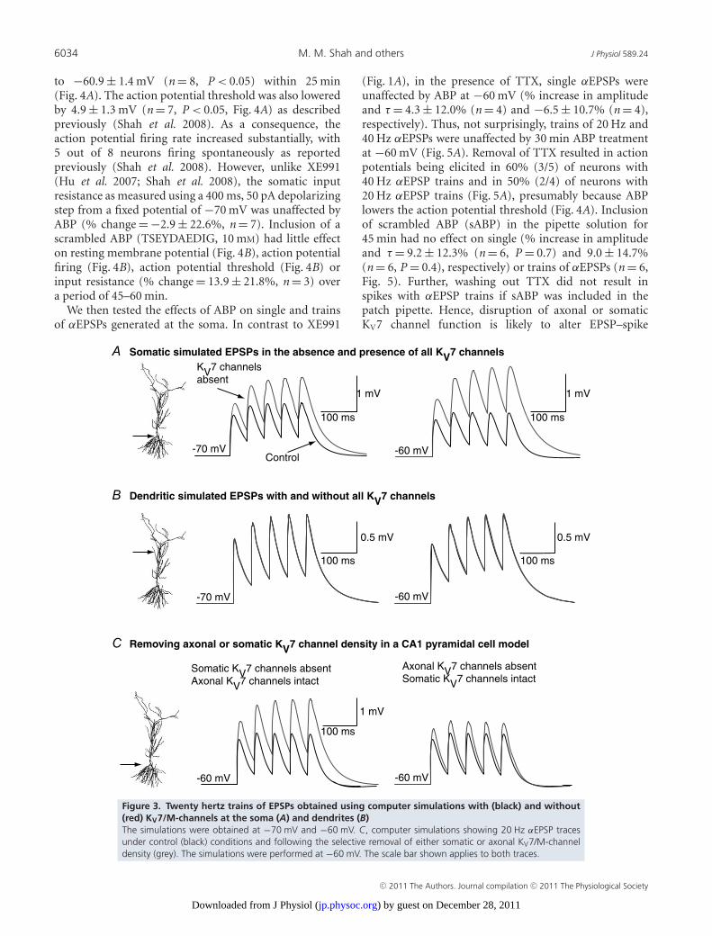
inhibition (see Shah et al. 2008). Na+ channels werenot included in these simulations as in our experimentalconditions Na+ channels had been inhibited by TTX.Compatible with the experimental results, KV7 channeldeletion throughout the neuron enhanced 20 Hz (Fig. 3A)and 40 Hz EPSP summation at the soma at −60 mV.
0.5 mV50 ms
-60 mV
XE991A
B
Soma
Retigabine
-60 mV
0.5 mV
50 ms
-60 mV
Soma
0.5 mV
-60 mV
1 mV
50 ms50 ms
3
2
1
Am
plitu
de (
mV
)
Control +XE991
**
Soma
Control +XE991
2
3
1
Am
plitu
de (
mV
)-60 mV (n=5)
-70 mV (n=6)
-60 mV (n=5)
-70 mV (n=7)
Soma,
-60
mV
Soma,
-70
mV
Den, -
60 m
V
Den, -
70 m
V
(5)
(6)
(5)(5)
3
2
1
-60 mV (n=7)
-70 mV (n=7)
Soma
Control + Retigabine
Am
plitu
de (
mV
) 3
2
1
Am
plitu
de (
mV
)
Control + Retigabine
-60 mV (n=6)
-70 mV (n=6)
*
40
20
0
-20
Soma,
-60
mV
Soma,
-70
mV
Den, -
60 m
V
Den, -
70 m
V
(6)
(6)
(9)
(7)
*
140
120
100
80
60
40
20
0
Figure 1. Example traces showing the effects of (A) the KV7/M-channel blocker XE991 (3 μM) and (B)the KV7/M-channel enhancer retigabine (10 μM) on simulated EPSP (αEPSP) shapes at the soma anddendrites when evoked at a potential of −60 mVBlack traces represent controls whereas the grey traces are those obtained following bath application of theKV7/M-channel modulator. Shown below the traces are the average (mean and SEM) somatic and dendritic αEPSPamplitudes in the absence and presence of (A) XE991 and (B) retigabine at potentials of −60 mV and −70 mV.The observations for each point are indicated in brackets next to the potential. Within each panel, the percentagechange in the decay time constant (τ ) of αEPSPs at the soma and dendrites at either −60 mV or −70 mV isillustrated in the graph on the far right. In these particular graphs, the numbers of observations are indicatedabove each bar. ∗Significance at P < 0.05.
C© 2011 The Authors. Journal compilation C© 2011 The Physiological Society
) by guest on December 28, 2011jp.physoc.orgDownloaded from J Physiol (

J Physiol 589.24 KV7/M-channels and synaptic integration 6033
The increase in summation was less than at −70 mV(Fig. 3A), consistent with the biophysical properties ofKV7 channels in hippocampal pyramidal cells (Shahet al. 2008). Dendritic EPSP summation, though, was notaffected at either frequency at −60 mV or −70 mV (Fig. 3Bfor example traces at 20 Hz). These findings support theidea that KV7 channels are principally located at thesoma/axon and affect peri-somatic post-synaptic EPSPintegration.
We next asked whether axonal or somatic KV7 channelsdifferentially affect EPSP integration. In computersimulations, removal of axonal KV7 channels but leavingsomatic channels intact, had little impact on EPSPsummation at the soma (Fig. 3C). In contrast, eliminationof somatic KV7 channels with axonal KV7 channels
still present significantly enhanced summation (Fig. 3C).These results indicate that only somatic KV7 channelsaffect peri-somatic EPSP summation.
To test if this is so experimentally, we used acompeting peptide, ABP (YIAEGESDTD), that is designedto selectively disrupt the coupling of KV7 channels toankyrin G, and thereby alter KV7 channel function inthe axon (see Shah et al. 2008). Hence, unlike XE991or retigabine, bath application of which would affectall channels within the neuron, this peptide only affectsaxonal KV7 channels. Indeed, we have previously shownthat the peptide had little effect on somatic M-currentmeasured with voltage clamp (Shah et al. 2008). Inclusionof this peptide (10 mM) in the intracellular pipettesolution caused RMP depolarization from −68 ± 0.7 mV
Soma, 20 Hz
-60 mV
1 mV0.5 mV
-60 mV
100 ms 100 ms
-60 mV-60 mV
0.5 mV100 ms
1 mV100 ms
Soma, 20 Hz
XE991A
RetigabineB
2.6
2.2
1.8
1.4
1.6
1.2
0.8
2.0
1.0
3.0
2.0
1.6
1.4
1.2
1.0
0.8
-60 mV100 ms
2 mV
100 ms
1 mV
-60 mV
Soma, 40 Hz
Soma, 40 Hz
Sum
mat
ion
Rat
io
Con XE991
*
Sum
mat
ion
Rat
io
Con XE991
*-70 mV (n=6)-60 mV (n=4)
-70 mV (n=5)-60 mV (n=4)
Sum
mat
ion
Rat
io
Con XE991
-60 mV (n=5)-70 mV (n=5)
Sum
mat
ion
Rat
io
Con Ret Con Ret Sum
mat
ion
Rat
io
Sum
mat
ion
Rat
io
-70 mV (n=7)-60 mV (n=7)
-70 mV (n=7)-60 mV (n=6)
***
*
-60 mV (n=5)-70 mV (n=6)
Con Ret
Control
XE991
Retigabine
Control
1.6
1.4
1.2
1.0
0.8
Figure 2. Representative illustrations showing trains of 20 Hz or 40 Hz somatic and dendritic αEPSPs inthe absence (black) and presence (grey) of either (A) 3 μM XE991 or (B) 10 μM retigabine at a potentialof −60 mVThe summation ratio was calculated as the amplitude of the fifth αEPSP divided by the amplitude of the first αEPSP(see Methods). Below each of the examples are graphs showing the mean and SEM of the summation ratio for theparticular frequency at potentials of either −60 mV or −70 mV. The numbers of observations for each frequencyat a particular potential are indicated in parentheses within each graph. ∗Significance at P < 0.05.
C© 2011 The Authors. Journal compilation C© 2011 The Physiological Society
) by guest on December 28, 2011jp.physoc.orgDownloaded from J Physiol (
6034 M. M. Shah and others J Physiol 589.24
to −60.9 ± 1.4 mV (n = 8, P < 0.05) within 25 min(Fig. 4A). The action potential threshold was also loweredby 4.9 ± 1.3 mV (n = 7, P < 0.05, Fig. 4A) as describedpreviously (Shah et al. 2008). As a consequence, theaction potential firing rate increased substantially, with5 out of 8 neurons firing spontaneously as reportedpreviously (Shah et al. 2008). However, unlike XE991(Hu et al. 2007; Shah et al. 2008), the somatic inputresistance as measured using a 400 ms, 50 pA depolarizingstep from a fixed potential of −70 mV was unaffected byABP (% change = −2.9 ± 22.6%, n = 7). Inclusion of ascrambled ABP (TSEYDAEDIG, 10 mM) had little effecton resting membrane potential (Fig. 4B), action potentialfiring (Fig. 4B), action potential threshold (Fig. 4B) orinput resistance (% change = 13.9 ± 21.8%, n = 3) overa period of 45–60 min.
We then tested the effects of ABP on single and trainsof αEPSPs generated at the soma. In contrast to XE991
(Fig. 1A), in the presence of TTX, single αEPSPs wereunaffected by ABP at −60 mV (% increase in amplitudeand τ = 4.3 ± 12.0% (n = 4) and −6.5 ± 10.7% (n = 4),respectively). Thus, not surprisingly, trains of 20 Hz and40 Hz αEPSPs were unaffected by 30 min ABP treatmentat −60 mV (Fig. 5A). Removal of TTX resulted in actionpotentials being elicited in 60% (3/5) of neurons with40 Hz αEPSP trains and in 50% (2/4) of neurons with20 Hz αEPSP trains (Fig. 5A), presumably because ABPlowers the action potential threshold (Fig. 4A). Inclusionof scrambled ABP (sABP) in the pipette solution for45 min had no effect on single (% increase in amplitudeand τ = 9.2 ± 12.3% (n = 6, P = 0.7) and 9.0 ± 14.7%(n = 6, P = 0.4), respectively) or trains of αEPSPs (n = 6,Fig. 5). Further, washing out TTX did not result inspikes with αEPSP trains if sABP was included in thepatch pipette. Hence, disruption of axonal or somaticKV7 channel function is likely to alter EPSP–spike
Figure 3. Twenty hertz trains of EPSPs obtained using computer simulations with (black) and without(red) KV7/M-channels at the soma (A) and dendrites (B)The simulations were obtained at −70 mV and −60 mV. C, computer simulations showing 20 Hz αEPSP tracesunder control (black) conditions and following the selective removal of either somatic or axonal KV7/M-channeldensity (grey). The simulations were performed at −60 mV. The scale bar shown applies to both traces.
C© 2011 The Authors. Journal compilation C© 2011 The Physiological Society
) by guest on December 28, 2011jp.physoc.orgDownloaded from J Physiol (

J Physiol 589.24 KV7/M-channels and synaptic integration 6035
coupling. However, only somatic KV7 channels affect EPSPintegration at the soma.
Discussion
Post-synaptic KV7/M-channels have been shown tomodulate action potential firing in a number of
different cell types (pyramidal neurons as well as inter-neurons; Brown & Passmore, 2009). Here, we show thatpost-synaptic KV7 channels also affect synaptic integrationin a location-dependent manner in CA1 pyramids. Bathapplication of KV7 channel modulators affected somaticbut not dendritic synaptic potential shapes (Fig. 1) andsummation (Fig. 2) in a voltage-dependent manner, inagreement with the reported biophysical properties of
-70 mV
-64 mV
20 mV
250 ms5 ms
20 mV
-70 mV
-150 pA
250 ms
20 mV
-68 mV -68 mV-70 mV
5 ms20 mV
ABP (YIAEGESDTD, 10 mM) A
sABP (TSEYDAEDIG, 10 mM)B
5 min 25 min
25 min
capacitative transients+150 pA
-150 pA
+150 pAcapacitative transients
10
8
6
4
2
02001501005005 min 25 min
200150100500
-54
-58
-62
-665 min 25 minAP
thre
shol
d (m
V)
AP
No.
Current (pA)
-56
-60
-64
-68
RM
P (
mV
)
*n=8
5 min25 min
n=8 n=8
-56
-60
-64
-68
RM
P (
mV
)
5 min 25 min
AP
No.
Current (pA)5 min 25 min
n=6 n=6 n=65 min25 min
5 min
76543210 -58
-54
-46
-42
-50
AP
thre
shol
d (m
V)
a cb
d e f
a cb
d e f
-70 mV
Figure 4. Effects of ABP and sABP on action potential firing and resting membrane potentialAa, Ba, Ab and Bb, examples of traces 5 min and 25 min after obtaining whole-cell patch clamp when ABP (A)or sABP (B) is included in the patch pipette solution. Ac and Bc, representative traces showing the influence ofABP and sABP on action potential threshold 5 min (black) and 25 min (grey) after breakthrough. The alterations inresting membrane potential, the number of action potentials and the action potential threshold when either ABPor sABP are present in the patch pipette are shown in Ad, Bd, Ae, Be, Af and Bf . In panels Af and Bf , the filledsquares represent the mean and standard error whereas the open squares show the individual responses.
C© 2011 The Authors. Journal compilation C© 2011 The Physiological Society
) by guest on December 28, 2011jp.physoc.orgDownloaded from J Physiol (

6036 M. M. Shah and others J Physiol 589.24
the M-current in hippocampal CA1 pyramids (Shahet al. 2008). These results were reproducible by computermodelling (Fig. 3). Hence, KV7/M-channels are likely to beprimarily located in the peri-somatic region (Yue & Yaari,2006; Hu et al. 2007; Shah et al. 2008).
In our experiments, most synaptic transmissionwas blocked. KV7/M-channels may be present onpre-synaptic terminals in the hippocampus (Martireet al. 2004; Peretz et al. 2007). Therefore, thoughpost-synaptic KV7/M-channels do not affect dendriticsynaptic potentials directly, modulation of synaptic releaseby pre-synaptic KV7/M-channels may additionally affectdendritic excitability and synaptic integration.
KV7/M-channels are likely to be present predominantlyin the axon initial segment (Devaux et al. 2004; Chunget al. 2006; Pan et al. 2006; Shah et al. 2008). However,our data suggested that only somatic channels alteredsomatic synaptic potential shapes and summation as bathapplication of XE991 and retigabine (which would affectall KV7 channels in a neuron) modified somatic EPSPsummation (Fig. 2) whereas use of the competing peptide,ABP, had little effect (Fig. 5). In these neurons, ABPmodulates only axonal KV7 channels as it lowered the
action potential threshold without changing the inputresistance (Fig. 4 and Results). In contrast, treatmentwith XE991, which would inhibit all KV7 channels,reduced the action potential threshold and increased theinput resistance (see Shah et al. 2008). Further, we havepreviously shown that bath application of XE991 afterABP has had its full effect causes little further decreasein action potential threshold (Shah et al. 2008). Thesedata thus suggest that ABP affects axonal KV7 channelsonly. In addition, the notion that somatic but not axonalKV7 channels affected summation was supported by dataobtained using computer simulations (Fig. 3C). Sincesomatic KV7 channels influence somatic input resistance(Shah et al. 2008), and variations in input resistance leadto changes in synaptic potential shapes (Magee, 2000), thismay explain why only KV7 channels located at the cell bodymodified synaptic integration. These results thereforesuggest that the effects of KV7 channels on synapticintegration are locally mediated. Since recent evidencesuggests synaptic potentials can propagate along axons(Clark & Hausser, 2006), we cannot exclude the possibilitythat axonal KV7 channels may influence shapes andsummation of synaptic potentials within these structures.
20 Hz 40 Hz
6
4
2
0
1
0
2
Sum
mat
ion
ratio
5 min 25 min 5 min 25 minSum
mat
ion
ratio
-60 mV -60 mV
2 mV100 ms
A
6
4
2
0
2 mV 2 mV50 ms 50 ms
2 mV100 ms
0
1
2
3
Sum
mat
ion
ratio
5 min 25 min
Sum
mat
ion
ratio
5 min 25 min
ABP
ABP 25 min, - TTX
control + TTX
ABP 25 min, + TTX
TruncatedActionPotential
B sABP
control + TTX
sABP 25 min, + TTX20 Hz 40 Hz
-60 mV -60 mV
n=5n=4
n=6 n=6
Figure 5. Axonal KV7 channels have littleeffect on somatic αEPSP summationA, example traces showing the effects ofincluding the ankyrin G binding peptide (ABP,10 mM) in the intracellular pipette solution on20 Hz and 40 Hz trains of αEPSPs. The traceswithin 5 min of going whole cell are deemedcontrol (see (Shah et al. 2008) and are in black.The maximal effect of ABP occurs within25 min (Shah et al. 2008) and is shown in red.TTX was present throughout. Washout of TTXresulted in spikes (AP; green trace). Generationof spikes will cause an afterdepolarizationand/or afterhyperpolarization, which will affectαEPSP summation. Indeed, in the particularexample shown, the action potential resultedin an afterdepolarization followed by anafterhyperpolarization, thereby distorting theαEPSP summation. For this reason, αEPSPsummation ratios were calculated from tracesin the which TTX was present in the externalsolution. The average (mean and SEM; filledsquares) and individual (open squares)calculated summation ratios following 5 minand 25 min of patching with pipettescontaining ABP in the presence of external TTXare shown below the traces. B, representativetraces showing the effects of scrambled ABP(sABP, 10 mM) on 20 Hz and 40 Hz trains ofαEPSPs within 5 min of going whole cell(control, black) or after 25 min (red). Allexperiments were performed in the presenceof TTX. The individual (open squares) andmean (filled squares) calculated summationratios for all cells in which sABP was included inthe pipette are shown below.
C© 2011 The Authors. Journal compilation C© 2011 The Physiological Society
) by guest on December 28, 2011jp.physoc.orgDownloaded from J Physiol (

J Physiol 589.24 KV7/M-channels and synaptic integration 6037
Indeed, recent evidence shows that KV7 channels located inimmature synaptic terminals alter inhibitory depolarizingsynaptic potential shapes and integration in the bouton,suggesting that KV7 channels exert a local effect onsynaptic integration within a particular neuronal sub-cellular compartment (Huang & Trussell, 2011).
By modulating spike threshold and the restingmembrane potential of neurons (Fig. 4; Shah et al. 2008),axonal KV7 channels can affect global cell excitabilitytoo. Thus, though ABP did not affect somatic synapticintegration per se, EPSP–spike coupling was enhanced asthe action potential threshold was reduced (Fig. 5). Thisalso raises the possibility that the manner in which KV7channels affect cell excitability may depend on the cellularcompartment in which they are expressed. Since thesechannels are modulated by a variety of neurotransmittersand intracellular signalling molecules (Delmas & Brown,2005), this may provide an exquisite mechanism by whichKV7 channels can fine-tune a cell’s intrinsic activity inresponse to changing neural network excitability andthereby influence physiological processes such as plasticityand learning as well as prevent neurological disorders suchas epilepsy (Jentsch, 2000; Peters et al. 2005; Wu et al.2008).
References
Brown DA & Passmore GM (2009). Neural KCNQ (Kv7)channels. Br J Pharmacol 156, 1185–1195.
Chung HJ, Jan YN & Jan LY (2006). Polarized axonal surfaceexpression of neuronal KCNQ channels is mediated bymultiple signals in the KCNQ2 and KCNQ3 C-terminaldomains. Proc Natl Acad Sci U S A 103, 8870–8875.
Clark B & Hausser M (2006). Neural coding: hybrid analog anddigital signalling in axons. Curr Biol 16, R585–R588.
Debanne D (2004). Information processing in the axon. NatRev 5, 304–316.
Delmas P & Brown DA (2005). Pathways modulating neuralKCNQ/M (Kv7) potassium channels. Nat Rev 6, 850–862.
Devaux JJ, Kleopa KA, Cooper EC & Scherer SS (2004).KCNQ2 is a nodal K+ channel. J Neurosci 24, 1236–1244.
Goldman AM, Glasscock E, Yoo J, Chen TT, Klassen TL &Noebels JL (2009). Arrhythmia in heart and brain: KCNQ1mutations link epilepsy and sudden unexplained death. SciTransl Med 1, 2ra6.
Hines ML & Carnevale NT (1997). The NEURON simulationenvironment. Neural Comput 9, 1179–1209.
Hu H, Vervaeke K & Storm JF (2007). M-channels (Kv7/KCNQchannels) that regulate synaptic integration, excitability, andspike pattern of CA1 pyramidal cells are located in theperisomatic region. J Neurosci 27, 1853–1867.
Huang H & Trussell LO (2011). KCNQ5 channels controlresting properties and release probability of a synapse. NatNeurosci 14, 840–847.
Jentsch TJ (2000). Neuronal KCNQ potassium channels:physiology and role in disease. Nat Rev 1, 21–30.
Luisi R, Panza E, Barrese V, Iannotti FA, Viggiano D, SecondoA, Canzoniero LM, Martire M, Annunziato L & TaglialatelaM (2009). Activation of pre-synaptic M-type K+ channelsinhibits [3H]D-aspartate release by reducing Ca2+ entrythrough P/Q-type voltage-gated Ca2+ channels. J Neurochem109, 168–181.
Magee JC (2000). Dendritic integration of excitatory synapticinput. Nat Rev 1, 181–190.
Marder E & Goaillard JM (2006). Variability, compensationand homeostasis in neuron and network function. Nat Rev 7,563–574.
Martire M, Castaldo P, D’Amico M, Preziosi P, Annunziato L &Taglialatela M (2004). M channels containing KCNQ2subunits modulate norepinephrine, aspartate, and GABArelease from hippocampal nerve terminals. J Neurosci 24,592–597.
Migliore M, Ferrante M & Ascoli GA (2005). Signalpropagation in oblique dendrites of CA1 pyramidal cells.J Neurophysiol 94, 4145–4155.
Migliore M, Morse TM, Davison AP, Marenco L, Shepherd GM& Hines ML (2003). ModelDB: making models publiclyaccessible to support computational neuroscience.Neuroinformatics 1, 135–139.
Migliore M & Shepherd GM (2002). Emerging rules for thedistributions of active dendritic conductances. Nat RevNeurosci 3, 362–370.
Pan Z, Kao T, Horvath Z, Lemos J, Sul JY, Cranstoun SD,Bennett V, Scherer SS & Cooper EC (2006). A commonankyrin-G-based mechanism retains KCNQ and NaVchannels at electrically active domains of the axon. J Neurosci26, 2599–2613.
Peretz A, Sheinin A, Yue C, Degani-Katzav N, Gibor G,Nachman R, Gopin A, Tam E, Shabat D, Yaari Y & Attali B(2007). Pre- and postsynaptic activation of M-channels by anovel opener dampens neuronal firing and transmitterrelease. J Neurophysiol 97, 283–295.
Peters HC, Hu H, Pongs O, Storm JF & Isbrandt D (2005).Conditional transgenic suppression of M channels in mousebrain reveals functions in neuronal excitability, resonanceand behavior. Nat Neurosci 8, 51–60.
Poolos NP, Migliore M & Johnston D (2002). Pharmacologicalupregulation of h-channels reduces the excitability ofpyramidal neuron dendrites. Nat Neurosci 5, 767–774.
Roche JP, Westenbroek R, Sorom AJ, Hille B, Mackie K &Shapiro MS (2002). Antibodies and a cysteine-modifyingreagent show correspondence of M current in neurons toKCNQ2 and KCNQ3 K+ channels. Br J Pharmacol 137,1173–1186.
Shah MM, Anderson AE, Leung V, Lin X & Johnston D (2004).Seizure-induced plasticity of h channels in entorhinalcortical layer III pyramidal neurons. Neuron 44,495–508.
Shah MM, Migliore M, Valencia I, Cooper EC & Brown DA(2008). Functional significance of axonal Kv7 channels inhippocampal pyramidal neurons. Proc Natl Acad Sci U S A105, 7869–7874.
Shah MM, Mistry M, Marsh SJ, Brown DA & Delmas P (2002).Molecular correlates of the M-current in cultured rathippocampal neurons. J Physiol 544, 29–37.
C© 2011 The Authors. Journal compilation C© 2011 The Physiological Society
) by guest on December 28, 2011jp.physoc.orgDownloaded from J Physiol (

6038 M. M. Shah and others J Physiol 589.24
Tatulian L, Delmas P, Abogadie FC & Brown DA (2001).Activation of expressed KCNQ potassium currents andnative neuronal M-type potassium currents by theanti-convulsant drug retigabine. J Neurosci 21, 5535–5545.
Wu WW, Chan CS, Surmeier DJ & Disterhoft JF (2008).Coupling of L-type Ca2+ channels to KV7/KCNQ channelscreates a novel, activity-dependent, homeostatic intrinsicplasticity. J Neurophysiol 100, 1897–1908.
Yue C & Yaari Y (2006). Axo-somatic and apical dendriticKv7/M channels differentially regulate the intrinsicexcitability of adult rat CA1 pyramidal cells. J Neurophysiol95, 3480–3495.
Author contributions
M.M.S. and D.A.B. designed experiments. M.M.S. performedand analysed all experiments. M.M. performed computersimulations. M.M.S. and D.A.B. wrote the manuscript. Allauthors approved the final version.
Acknowledgements
The work was supported by a Wellcome International PrizeTravel Research Fellowship (M.M.S.) and a Wellcome TrustProject Grant (WT087363MA; M.M.S., D.A.B.).
C© 2011 The Authors. Journal compilation C© 2011 The Physiological Society
) by guest on December 28, 2011jp.physoc.orgDownloaded from J Physiol (
Copyright © 2022 FDOKUMEN