Uptake and subcellular localization of tritiated dihydro-microcystin-LR in rat liver
13
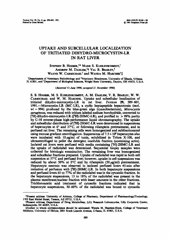
reuda~ vot . ~. rb. a ~ . se~-hot . t~t . P~ea t° c~ ttdwn. UPTAKE AND SUBCELLULAR LOCALIZATION OF TRITIATED DIHYDRO-MICROCYSTIN-LR IN RAT LIVER STSPFISN B . Hoos~t," MAIex S . Kul~n.z~vsc~T,' AxDxBw M. DAI~n .~I,~t VAL R. BBASLEY,~ WAYIaF W. CA1tiaCHA~.' and WAxDA M. TIA3Ci303iC'$ aat-0totrot tp.ao+ .oo p t99t làpm°n Pser pk 'Departments of Veterinary Pathobiology and Veterinary Big, University of Illinois, Urbane, IL 61801, and 3Departmcnt of Biological Sciences, Wright State University, Dayton, OH 45435, U .SA. (Received 13 has 1990 ; accepted 21 Novnnber 1990) S . B . Hoos>~t, M. S. KUI~.Elvscrn~T, A. M. DAHLSM, V. R . BEASLSY, W . W. CAIUUCHAEI, and W . M. Iiescl~ . Uptake and subcellular localization of tritiated dihydro-microcystin-LR in rat liver. Toxicon 29, 589-601, 1991 .-Microcystin-LR (MC-LR), a cyclic heptapeptide hepatotoxin (mol. wt = 994) produced by the blue-gnxn alga (cyanobacterium), Microcystis aeruginosa, was reduced with tritium labeled sodium borohydüde, converted to ['H]-dihydro-microcystin-LR (~H]-2HMC-LR), and purified to > 99% purity by C-18 reverse-phase high-performance liquid chromatography . The uptake and subcellular distribution of ['HJ-2HMC-LR were determined in suspensions of hepatocytes at 0° and 37°C, or following rifampicin pretreatment, and in perfused rat liver. The remaining cells were homogenized and subfractionatod using sucrose gradient centrifugation . Suspensions of 7.5 x 10⁶ hepatocytes also were incubated with 10 ~g/ml of toxin, solubilized in Triton X-100, and ultracentrifuged to pellet the detergent insoluble fraction (containing actin) . Isolated rat livers were perfused with media containing ['H]-2HMC-LR and the uptake of radiolabel was determined . Sequential biopsy samples were collected for histologic examination . The remaining liver was homogenized and subcellular fractions prepared . Uptake of radiolabel was rapid in both cell suspension at 37°C and perfused liver ; however, uptake in cell suspensions was reduced by about 50% at 0°C and by rifampicin (50 ~eg/ml) pretreatment . Hepatocyte necrosis was observed in isolated perfused livers 45 min after initiation of perfusion with ['H]-2HMC-LR . In both hepatocyte suspensions and perfusod livers 65 to 77% of the radiolabel was in the cytosolic fraction . In the hepatocyte suspensions, 13 to 18% of the radiolabel was present in the plasma membrane/nuclear fraction with lesser amounts in the other fractions . Trichloroacetic acid treatment of cytosolic fractions indicated . that in hepatocyte suspensions, 50-60% of the radiolabel was bound to cytosolic 'Present address : University of Arizona, College of Pharma4y, DepaRment of Pharmaoology/fotascology, 1703 East Mabel Street, Tureen, AZ 85721, U .SA. tPresent addn~s; Department of Drug Metabolism, Lilly Resean~ Laboratories, Lilly Corporate Center, Indianapolis, IN 46285, U.SA. $Author to whom correspondence should be addressed : Wench M . Heschek-Hock, College of Veterinary Medicine, University of Illinois, 2001 South Lincoln Avenue, Urbane, IL 61801, U.S .A . 589
Transcript of Uptake and subcellular localization of tritiated dihydro-microcystin-LR in rat liver
reuda~ vot . ~. rb. a~. se~-hot . t~t.P~ea t°c~ ttdwn.
UPTAKE AND SUBCELLULAR LOCALIZATIONOF TRITIATED DIHYDRO-MICROCYSTIN-LR
IN RAT LIVER
STSPFISN B. Hoos~t," MAIex S. Kul~n.z~vsc~T,'AxDxBw M. DAI~n.~I,~t VAL R. BBASLEY,~
WAYIaF W. CA1tiaCHA~.' and WAxDA M. TIA3Ci303iC'$
aat-0totrot tp.ao+ .oop t99t làpm°n Pser pk
'Departments ofVeterinary Pathobiology and VeterinaryBig, University of Illinois, Urbane,IL 61801, and 3Departmcnt of Biological Sciences, Wright State University, Dayton, OH 45435, U.SA.
(Received 13 has 1990; accepted 21 Novnnber 1990)
S . B . Hoos>~t, M. S. KUI~.Elvscrn~T, A. M. DAHLSM, V. R. BEASLSY, W. W.CAIUUCHAEI, and W. M. Iiescl~. Uptake and subcellular localization oftritiated dihydro-microcystin-LR in rat liver. Toxicon 29, 589-601,1991 .-Microcystin-LR (MC-LR), a cyclic heptapeptide hepatotoxin (mol.wt = 994) produced by the blue-gnxn alga (cyanobacterium), Microcystisaeruginosa, wasreduced with tritium labeled sodium borohydüde, converted to['H]-dihydro-microcystin-LR (~H]-2HMC-LR), and purified to > 99% purityby C-18 reverse-phase high-performance liquid chromatography . The uptakeand subcellular distribution of ['HJ-2HMC-LR were determined in suspensionsof hepatocytes at 0° and 37°C, or following rifampicin pretreatment, and inperfused rat liver. The remaining cells were homogenized and subfractionatodusing sucrose gradient centrifugation . Suspensions of 7.5 x 10⁶ hepatocytes alsowere incubated with 10 ~g/ml of toxin, solubilized in Triton X-100, andultracentrifuged to pellet the detergent insoluble fraction (containing actin) .Isolated rat livers were perfused with media containing ['H]-2HMC-LR andthe uptake of radiolabel was determined . Sequential biopsy samples werecollected for histologic examination. The remaining liver was homogenizedand subcellular fractions prepared . Uptake of radiolabel was rapid in both cellsuspension at 37°C and perfused liver; however, uptake in cell suspensions wasreduced by about 50% at 0°C and by rifampicin (50 ~eg/ml) pretreatment .Hepatocyte necrosis was observed in isolated perfused livers 45 min afterinitiation of perfusion with ['H]-2HMC-LR. In both hepatocyte suspensionsand perfusod livers 65 to 77% of the radiolabel was in the cytosolic fraction . Inthe hepatocyte suspensions, 13 to 18% of the radiolabel was present in theplasma membrane/nuclear fraction with lesser amounts in the other fractions.Trichloroacetic acid treatment of cytosolic fractions indicated . that inhepatocyte suspensions, 50-60% of the radiolabel was bound to cytosolic
'Present address : University of Arizona, College of Pharma4y, DepaRment of Pharmaoology/fotascology,1703 East Mabel Street, Tureen, AZ 85721, U.SA.tPresent addn~s; Department of Drug Metabolism, Lilly Resean~ Laboratories, Lilly Corporate Center,
Indianapolis, IN 46285, U.SA.$Author to whom correspondence should be addressed : Wench M. Heschek-Hock, College of Veterinary
Medicine, University of Illinois, 2001 South Lincoln Avenue, Urbane, IL 61801, U.S.A .
589

590
S. B. HOOSER et al.
protein. Studies using the perfused liver confirmed that the majority of theradiolabeled MCLR (78-88%) was bound to cytosolic protein. These datasuggest that the uptake of [3Ii]-2HMC-LR oaurs primarily by an energy-dependent transport process involving the rifampicin-sensitive hepatic bile acidcarrier and that once inside the hepatocyte, the toxin binds to a cytosolicprotein(s) .
INTRODUCTION
MICROCYSTIrr-LR (MC-LR, cyanoginosin-LR) is a cyclic, heptapeptide hepatotoxin (mol.wt = 994) produced by the blue-green alga (cyanobacterium), Microcystis aeruginosa.Toxic blooms in pondsand water reservoirs have been associated with numerous livestockdeaths and implicated epidemiologically in humanillness (Koxsr et al., 1965 ; F~LOON~t etal., 1983 ; J~cxsoN et al., 1984 ; G~LSx et al., 1987).
Micxocystins are specifically hepatotoxic. In rats and mice they cause rapid ccntrilo-baler hepatocyte necrosis, with breakdown of the sinusoidal endothelium, intrahcpatichemorrhage, and death (RUNN>~GAR and F~L.coN~t, 1982; HoaeeR et al., 1989). Primarytoxic effects in other organs are not evident. Studies using radiolabeled microcystins haveshown that they are rapidly taken up by the liver. When ['zsil-labelod MC-YM was giveni.v. to rats, 20% ofthe radioactivity waslocalized in the liver within 30 min, with no morethan 6% in any other organ (F~LOOrnm et al., 1986). Following i.p . injection ofbiosynthetically labeled ["C]-MC into mice, 70% of the radioactivity was localized in theliver after 1 min and 90% by 180min (BROOKS and CODD, 1987). Six hours afterchemically labeled ['H}-MC-LR was injected i.p . into mice, 56% of the dose waspresent inthe liver, with 10% in the carcass and 7% in the intestine (RosnvsoN et al., 1989).
Ultrastructurally, MC-LR causes hepatocyte plasma membrane invagination, blebbingand loss of microvilli prior to cell death (Hoosier et al., 1990). In cell suspension, MC-LRcauses plasma membrane blebbing in hepatocytes but not in sinusoidal endothelial orKupffer cells (Hoo~e et al., in press) . In contrast to the hepatocyte necrosis induced bymicrocystins in vivo, hepatocytes in suspension remain viable (RUNN»GAR and F~i cotveR,1982).Thereasons for hepatocyte specificity andthe mechanismof action of microcystins have
not been fully elucidated . It has been suggested that microcystin is transported intohepatocytes by a hepatocyte-specific bile acid carrier and once internalized, interacts with,or in some other way causes the aggregation of actin filaments (RUNNHOAR et al., 1981).When suspensions of isolated hepatocytes were preincubated with non-competitive inhibi-tors of hepatocyte specific bile acid carriers (rifampicin and bromosulfophthalein)(ZIECiLStt and Fhnou~, 1986), there was marked reduction in microcystin-induced plasmamembrane blebbing (RUNNEGAR et al., 1981 ; Ruxt~sa~R and FALOOIVSR, 1982). In bothcultured hepatocytes and hepatocyte suspensions, there is aggregation of actin filamentsfollowing treatment with MC-LR which corresponds with plasma membrane blebbing(EItIICSSON et al., 1989; HOae>m et al., in press) . However, a significant change in the ratioof polymerized to unpolymerized actin was not detected in microcystin-treated hepato-cytes (RUNNEGAR and FwI,coN~t, 1986; EiencssoN et al., 1989).The objectives of our studies were to : (1) determine and compare the rate and extent of
uptake of MC-LR in hepatocytes in suspension, with and without prctreatmcnt withrifampicin ; (2) determine the intracellular localization of MC-LR using subcellularfractionation of hepatocytes; (3) determine if MC-LR binds directly to hepatocyte

Localization of [6H}MC-LR in Rat Liver
S91
proteins, particularly those ofthe cytoskeleton; (4) confirm the relevance of the findings inhep~tocyte suspensions to the bt vivo situation by repeating these studies in the perfusedliver .
MATERIALS AND METHODSAnbnais
Male, 175 to 250 g, Spraguo-Dawky rate (Hunan Sprague-Dawky, Inc., Indianapolis, IN, U.S.A.) wereallowed to aodmoatize for 2 wake or loner before use. Animals had free access to a commercial laboratoryanimal radon and wafer and were maintained on a 12-hr light-dark cycle. Rats were not fasted prior to use.
ToxinMicrocystia-LR (MGLR) (approximatdy 95°k pure) from Microcystis aerttglnosa, laboratory strain PCC-
7820, was purified by the laboratory of W. W. Cewac~rwnQ . using techniques previously described (Karsriae-nsurtrar et d., 1986). The putißed toxin was dissolved in phosphate-bu$'ered saline (PBS) prior to use.Dihydromicrocyedn-LR (2HMGLR) was prepared and purified as previously described (Denier et d., 1991).
Synthesis mrdptojjrcatfat of trttban-labeled dihydronricrocystLe-LR ((jHJ-2HMC-LR)MGLR (2.Smg) was rsacted with NaH'H, (12.5 mCi, apxific activity = 14 Ci/mmole) in 70% isopropaaol
(1 .0 ml) at room temperature for 24hr, they quenched with 10'/° acetic acid (0 .5 ml, pH 4) . The reaction productwas then evaporated to dryness in wtcwo oa a rotary evaporator. Isolation of [rH]-2HMGLR was aooompliahedby loading the nasKion product in water (O .S ml) on preconditioned l g Cu camid~e (AnalYtichem Inter-national, Harbor ßty, CA, U.SA.). The cartridges were then rinsed with water (3 x 2 ml) aced l0Y° methanol(3 ml) and eluted with 100% methanol (3 x 2 ml). Final purification was by nvereo-phase high-performanceliquid chromatography (HPL,C) using an Alltuch C, s column (4.6 x 250 mm), an ieocratic mobile phase ofmethanol, and 0.05 M NaHiPO, (pH 3) (58:42) . Column elution was monitored by nhraviokt abaorbance at238~Neries, 1989).
Lrolated toxin was greater thaw 99% chemically pure, by combined HPLC, thin layer chromatography (TLC)and mare spectroscopy. It was greater than 98% radiochemically pure by HPLC andTLC with a specific activityof 5.3 ft(â/pmok (Denier, 1989). Less than l'/° of the labd was present in urine diatillste followingadministration oftoxin (Deriet, 1989). The minimal ~i°o for 2HMGLR in mice, 200 pg/kg, is approximatelytwig that for MGLR; grove and microscopic lesions induced by 2HMGLR areidentical to MGLR (Denier etd., 1991) .
Hepatocyte isolationRat hepatocytee were isolated by a previously described two-step collapenase perfusion technique
(Kuri.arisctn®r etd., 1982). Cell viability wes 85 to 90Y° as determined by trypan blue dye exclusion . The cellpellets were reswpended in Ham's F-10 medium (complete medium), containing (per 500 ml) 100ml of fetalbovine serum (FBS) and 0.3ml of insulin (100 U/ml), earl then placed on ice .
Uptake of(jHJ-2H11lC-LR boo IrepatocytesUptake offH}2HMC-LR into suspensions ofhepatocytea at 0°C, 37°C and following a 10-min preincubation
with 50 pg/ml ofrifampipn at 37°C was determined using modifications of previously described methods (F.e~roxand xr "~, 1978 ; Kuriwscnnanr er d., 1984) .
Aliquots of hepatocytes in complete medium were centrifuged at 70 xd for 2 min and the pellets woreresuspeaded is 2.5 ml of Ham'B F-10 medium without FHS but with insulin (incomplete medium) to a finalconcentration of 1.5 x 10' hepatocytea/ml, placed in capped plastic culture tubes (12 x 75mm), aced incubated ateither 0°C or 37°C under constant end-over~nd rotation at 6 rpm. For rifampicin preincnbation, rifampicin inPBS was added to the Suspension lOmin before addition of toxin. Cold 2HMGLR (37.5 I+B), premixed withfH}2HMGLR (12.5 Pg, 9.8 x ld d.p.m .), was added to PHS at 37°C so that the toxin concentration was20pg/ml. This was then added to the hepatocyte suspension resulting in a fiaei toxin concentration of 10 pg/ml(wld plus tritiated, 2 x 10' d.p .m/Pg), and a final hepatocyte oonoeatration of 7.5 x 10⁶ hepatocytee/ml in S ml ofmedium.At time 0, 20, 40 aced 60 sec and at 2, 4, 6, 8, 10, 1S and 30 min, two 100 pl aliquots ofeach suspension wen
removed, layered over 500 itl of Dow Corning S50 dlioone oilaight mineral oil (5:1) (It uni$rncnn®r, 1984), andmicroti~ged for 13 sec to separate ce1L containing toxin from free toxin in the eupervataat. The supernatant andoil fmm each tube were removed sad 100itl of 1 °/. Triton X-100 (TX-100) was used to solubilise each pellet . The

592
S. B. HOOSER et al.
100 pl pellet (in TX-100) was pLwed in 10ml LSC. All all pellets in LSC were counted on a Packard 460 Tri-Carb scintillation counter.
Following removal of the Lut two 100 pl aliquots at 30min, all suspension cultures were pLwed in an ice bathfor leas than an hour until subcelluLu fractionation was begun.
Uptake oj(3HJ-2HMC-LR neto isolated, ptr,~tred llrerTwo rats were anesthetized and the portal veins to their livers were cathetsized in a manner identical to that
used to isolate 6epatocytes, except that the Sow rate of perfusate was 25ml/min (B®ta et aL, 1988). Oncecatheterised, the livers was perfused for 15 min with compLae medium containing (per 500ml) 1.2g HEPES, pH7.35 . Sins the minimum i.p. tD,w for MC-LR in rats is 180/rg/kg (hioos~ et al., 1989), while the minimum ~,sofor 2HMC-LR is approximately two tim~ttiüt öfMGLR (Dexi~t et al., 1991), 172.4pg of toxin (two part ofcold 2HMGLR: one part [3fi]-2HMC-LR, 12.5 ltg, 9.8 x 10' d.p.m .) was added to 100 ml media (ßnal specißcradioactivity was 5.68 x 10 3 d.p.m./~. Following a 15-min perfusion with medium without toxin, the perfusatewas changed to the 100 ml of medium containing toxin and drained from the liver W allow ratirc~ilation of thetoxin. One 200 kl aliquot was removed from the contains of rocir+culating medium from the first rat lives at 0, 20,40 and 60 sec, at 2, 4, 6, 8 and 10min, and then every 5min until 120 min. Aliquots (200 kl) in 5 ml of LSCwascoveted as described above. At 120 min, the liver was perfused with 100ml ofcomplete medium without toxinand then pLuxd on is in 0.25 M sucrose (4°C) for subsequent aubcelluL~r fractionation. At 15, 30, 45 and120 min, small pieces of lives were tied off with surgical suture, removed, and pLioed in 10Y. neutral bufferodformalin for histologic examination .Sins maximum uptake of toxin oaurrod at approximately 45 min, perfi>aion of the second liver with toxin
was ended at 45 min, and lives samples for histologic examination were taken at 0, 15, 30 and 45 min.
Subcelhdm jractiorratior, ojhepatocytes midperfured /ivcrThe two paf»sed livers and the 2.8 ml of hepatocyte suspension obtained from the uptake studies described
above wse fractionated using a modifaration of the subcelluLu fractionation method of Reid (R~ andWn.i .i~~eriN. 1974). This method yields five fractions : crude nuclear (which aLw contains plums membranefragments), heavy mitochondrial, light mitachondriel (which aLtio contains lysoaomea), microeomal, and cyto-colic . Isolated hepatocytes were resuspended in coLi 0.25 M sucrose and homogenized by passage through a 26-gauge nadle 20 to 40 times . Cell disruption was verified by microscopic examination. All pellets of the variousaubcelluLa fractions were reauapended in 1 ml of PBS and aliquots were pLwed in LSC (Aquasol II, containingdetergent) for scintillation counting .
Detergent extraction ofisolated lrepatocytes following uptake of(3HJ-2HMC-LRSuspensions of hepatocytea in incomplete Ham'a F-10 medium were prepared as described above ands
"Uptake of [3H}-2HMC-LR into Hepatocytea" . Cold 2HMGLR (37.5~ and ['H}2HMC-LR (12.5~wereadded to PBS to comprise a total toxin concentration of ~pg/ml. The mixture was then added to the 2.5ml ofhepatocyte suspension so that the final toxin concentration was 10pg/ml. They were incubated at 37°C for30 min and then pLuxd on ice. The cells were pelkted by centrifugation at 70 xa for 2 min at 4°C.The supernatant was removed and saved for subsequent counting, and 10ml of lY. TX-100 extraction buffs
(IOmM EDTA,O.l M Tris-Cl, pH = 7.4) was mixed with the all pellet. This was pLiaed on ice and allowed toaolubilize for 2hr with periodic agitation. The aolubilizod cells were then centrifuged at 150,000xd for 45 min at4°C in a Beckman LS-50 ultracentrifuge (Fox et al., 1984). The detergent soluble supernatant was removed andsaved for scintillation counting. The insoluble pellet (containing actin filaments) was added to 1.5 ml of 3NHCIand warmed until dissolved. To this was added an equal amount of 3MKOH and the samples saved forscintillation counting.
7Yiclrloroacetk acid precipitation ofcyttuolk fractionsTwo hundred microlitas of 30'/0 (w/v) tricliloroaeetîc acid ('TCA) was pLuxed in 1.5 ml microfuge tubes and
thoroughly mixed with 400pl of each of the separate cytoeoFic fractions from the two isolated live psfusionaand the six isolated hepatocyte suspensions (duplicates from thra separate rata) used in uptake studies (0°C,37°C and with rifampicin pretreatment at 37°C), for a final TCAconcentration of 10X. These wero immediatelypLued on is for 1 hr, microfuged for 10 min, a~ 500It1 of aupsnatant was then pLuxd is 4.0ml of LSC. Thepellets were mixed with 500pl of LSC (Aquasol II, containing detergent) and pLuxd in a 53°C oven ovsaight tosolubilize. At the sad of this time, a small amount ofdetergent insoluble material remained. The samples wereagain microfuged for 10 min and the Aquesol soluble supernatant removed sad pLieed in 4.0ml of LSC. Theinsoluble pellet was mixed with 250pl of 3 MKOH and dissolved overnight at 53°C. Two-hundred and fiftymicrolitas (250 pl) of 3NHCIwas added for neutralisation and the 500pl sample was added to 4.5ml of LSCand detergent soluble pellet fraction for scintillation counting.

Z_e
i=â_tseixx
Localization of [jH]-MC-LR in Rat Liver
593
TIME (MINI
Fya . 1 . ICn~TCa aF urr~ ov 2HMGLR HY FI~A'hOCY'IT~ IN Sllâ~IalON .
Hepatocytes (7 .5 x 10` eells/ml) were incubated with [°H}2HMGLR (2 x lo` d.p.m./lam andanalyzed for amount of toxin uptake as described in Materials and Methods . Each pointrepresenb the mean of four replicates (two from each of two rab)tS.E.M. (/-/~ uptake at37°C ; (p-D), uptake at 37°C by rifampicân (rif}treated (50pg/ml) hepatocytes; (O-O), uptake at
0°C .
Hlatologic exmnürattonHepatocytee in sell suspension were examined unfixed for plasma membrane blabbing by light microscopy.
The samples of liver from the liver perfusion studies were fixed in 10% ~utral buffeaed formalin, embedded inglycol metbacrylate, sectioned at 2 microns, stained with hematoxylin and eosin, and exemiaed by light
pY .
RESULTS
Hepatocyte uptake of ~jHJ-2HMC-LR and morphologic effectsThe uptake of [3H]-2HMC-LR by hepatocytes in suspension at 37°C proceeded rapidly
for the first 4min then more slowly, starting to level off after 15 min (Fig.1). At 30 min,the average amount of radioactivity taken up by the 100 ~l aliquot of hepatocytes(7.5 x 10³ hepatocytes per 100 ~l) was 1,752 d.p.m . This was equivalent to an uptake of87.6 ng (88 pmole) of total toxin per 7.5 x 10³ hepatocytes.The incubation of hepatocytes at 0°C, or preincubation with rifampicin at 37°C,
markedly slowed the rate and reduood the total uptake of ~H]-2HMC-LR although it didnot completely prevent it (Fig.1). After 30 min of incubation at 0°C, uptake of['H]-2HMC-LR was reduced by 50% when compared to incubation at 37°C; preincuba-tion with rifampicin reduced uptake by 64% .
Microscopic examination of hepatocytes incubated with ['H]-2HMC-LR at 37°C for30 min revealed that almost all hepatocytes had numerous, large plasma membrane blebs.In contrast, less than 50% ofthe hepatocytes preincubated with rifampicin or incubated at0°C had multiple large plasma membrane blebs.
Uptake of ('HJ-2HMC-LR into isolatedperfused liver and morphologic effectsIn the perfused liver at 37°C, the uptake of [3H]-2HMC-LR was also rapid. In the first
experiment, there was rapid uptake of radiolabel for the fast 10 min, then uptake

394
S. B. HOOSER et al .
TIME (MIN)
Fla . 2 . R~ncs ow= urr~ ov 2HMC-LR eYr~e~ uvaA .Intact rat livers were perfused with [~H]-2HMGLR (172.4 Elg, 3.68 x 10'd.p.m./~ and analyzedfor 2HMC-LR uptake ae described in Materisle and Methods . The data are expreeeed as per antof maximum uptake in the liver at each time point. ("-"~ 45-min perfused liver, (D-p), 120-min perfuxd liver . Maximal uptake in the 43-min perftued liver was 2630 d.p.m . above
background as comparod to 930 d.p.m . in the 120-min perfueed liver.
plateaued for approximately 40 min. This was followed by a release of radioactivity fromthé liver intö the perfusiôn medium beginning at 50 min and continuing until the end ofthe experiment at 120 min (Fig. 2). At the time of maximal hepatic uptake (50 min),83.5 pg 2HMC-LR were present in this liver, which weighed 11.58 g. This was equivalentto an uptake of 7.2 hg (7.3 nmoles) of 2HMC-LR per g liver. Assuming 1 .2 x 108hepatocytes per g of liver (Dousr, 1958), this is equivalent to an uptake of 45 ng (45.6pmole) of total toxin per 7.5 x 10 5 hepatocytes as compared to 87.6 ng taken up by thisnumber of cells in suspension . The second experiment was technically superior to the firstdue to better perfusion. Uptake in this liver was even more rapid, with near maximaluptake by 4min (Fig . 2) . After 4 min, uptake virtually stopped and the radioactivity in theperfusion medium remained constant until 40 min, when hepatic radioactivity began todecrease (i .e . radioactivity in medium began to increase) . At the time of maximal uptake(40 min), 230 hg 2HMC-LR were present in this 12.62 g liver. This is equivalent to anuptake of 118 pg (119 pmoles) of total toxin per 7.5 x 10⁵ hepatocytes, which comparesfavorably to the 87.6 ng taken up by the same number of hepatocytes in suspension.In both of the perfttsed livers, microscopic lesions were seen beginning 15 min after
perfusion with [3H]-2HMC-LR was begun and became progressively more severe . Initiallesions were located periportally and were characterized by hepatocyte vacuolizationfollowed by hepatocyte disassociation and rounding . By 45 min, necrosis was observedperiportally and midzonally with hepatocyte disassociation and rounding in centrilobularregions (Fig. 3). Although necrosis was more severe at 120 min than at 45 min in the 120-min perfused liver, it was still less severe than the necrosis in the 45-min perfused liver (at45 min) .
Subcellular fractionation ofhepatocytes and perfused liver
In suspension cultures, the majority of radioactivity remained in the medium and wasnot incorporated into hepatocytes. Incubation of hepatocytes at 37°C resultod in an

Localization offH}MGLR in Rat Livcr
595
uptake of 2.5% of the [3H]-2HMC-LR from the medium. When fractionated, the largestpercentage of radioactivity was present in the cytosolic fraction, followed by the nuclear/plasma membrane fraction. In hepatocytes incubated at 37°C, 70.0 and 13.5% were foundin the cytosolic and plasma membrane/nuclear fractions, respectively. Although the totaluptake of ['H]-2HMC-LR was reduced at 0°C or with rifampicin pretreatment, thepercentages of total cellular radioactivity found in the various fractions were similar tothose found in the hepatocytes incubated at 37°C .
In the perfused liver preparations, a significantly greater percentage uptake wasobserved . Following the 45 min perfusion, 13 .1 % of the radioactivity wastaken up by theliver . When fractionated, 77.8% of the radiolabel was present in the cytosolic fractionwith 0.7 and 15.8% present in the nuclear/plasma membrane and microsomal fractions,respectively . After perfusion for 120 min, 10.5% of the radioactivity was incorporated intothe liver . When fractionated, 71 .0% of the radiolabel was in the cytosolic fraction, with7.0 and 15.7% present in the nuclear/plasma membrane and microsomal fractions,respectively (Fig . 4) .
Detergent extraction of isolated ltepatocytes following uptake of~jHJ-2HMC-LRThe detergent soluble fraction contained 97.5% of the radioactivity associated with the
cells, while only 2.5% was associated with thé insoluble pellet (predominantly actin, othercytoskeletal elements and histones) .
Trichloroacetic acid precipitation of cytosolic fractionsWhen purified ['H]-2HMC-LR was treated with TCA, 92.4% of the radioactivity
remained in the supernatant. In the cytosolic fraction ofhepatocytes incubated at 37°C for30 min, 59.1 % of the [~H]-2HMC-LR associated radioactivity was present in the precipi-tated pellet and 40.9% was in the supernatant (Fig. ~. In the cytosolic fractions ofhepatocytes preincubated with rifampicin or of those incubated at 0°C for 30 min,approximately 50% of the radioactivity was present in the precipitate and 50% remainedin the supernatant.The precipitated pellet of the cytosolic fraction of the liver perfused for 45 min
contained 88.3% of the radiolabel, while the supernatant contained 11.7%. In the liverperfused for 120 min, this was slightly reduced with 78.5% found in the precipitate and21 .5% found in the supernatant (Fig . 5) .
DISCUSSION
The uptake of ['Hj-2HMC-LR is rapid in hepatocyte suspension, plateauing after4-10 min. This could be due to either saturation of hepatocyte receptors or the establish-ment of a steady-state where uptake rate is equally balanced by toxin e®wc or degrada-tion. It is also possible that there is a rapid microcystin-induood alteration which preventsfurther uptake. Such an effect could be due to : (1) disruption of actin filaments and/ortheir plasma membrane connections; (2) an effect on the plasma membrane receptor orcarver itself; or (3) another MC-LR-intracellular protein interaction which then secon-darily affects the carrier (or receptor) . Further studies are needed to rigorously define themechanism of hepatic uptake of microcystin and to identify its cellular target site.Uptake of ['H]-2HMC-LR is similarly very rapid in the isolated perfused liver . The

S . B. HOOSER ct al.

FYo. 3 . E»cr oP 2HMGLR or1 L.1VBlt xisroL.oax >N TH8 45-~ rt~et+trl® 1 .IVlOt.(a) 0 min; control ; central vein and surrounding hepatocytea are normal ; bar = 100'un . (b) 45 min ;central vein and surrounding hepatocytes ; prominent rounding and disassociation of hepatocytesis present; bar = 100 pal. (c) 45min ; portal vein cad surrounding hepatocytes; in addition tohepatocyte rounding and disassociation, there is prominent hepatocyte necrosis characterized by
all fragmentation as well as nuclear pyknasis and karyonrhexis; bar = 100 ~cm.
é~aseéss
eo
70
60
50
40
30
20
10
0
Localization of [3H]-MC-LR in Rat Liver
597
PM/NUC
H. MIT
L. MIT
MICRO
CYiOFra. 4. Suee~L .t,vr.ale nslTemvnox oP e~,zrtivoelez» ['H]-2HMC-LR.
['H]-2HMC-LR was incubated under various conditions cad subjected to aubcellular fractionationas described in Materials and Methods (nee also legends to Figs 1 and 2) . The data are presented asthe percentage of total incorporated radioactivity found in each suboellular fraction. Each barrepresents the mean of four replicates (two from each of two rats) f S.E.M ., except for the 45-minand 120-min penfuaed livers which represent one animal for each perfusion. 1?M/NUC, plasmamembrane plus nuclear fraction; H. MTI', heavy mitochondrial fraction; L . MIT, light
mitochondrial fraction; MICRO, microsomal fraction; CYTO, cytoaolic fraction .

598
S. H. HOOSER et at.
I -
- ~ -MOLAIaD IOA7eCrTEa
11Y811 /6fUl1°II
Fia. 5 . Ea~cr oe ~r>ere~ .oaosesnc eem ox r~ soa.oaunY or 2HMGLR nv t~A70CY18crtneot rc vaACtsotva.
Liven were perf»sed or hepatocytes inwbatad in suspension under various oonditiona withfH}2HMC-LR, ~ubjectad to rubcellular fndionation, and cytosolic fractions tmated withtridiloroaatic acid as descn'bed in Materials and Methods. The data are expressed as per ant ofthe total cytwolic radioactivity subjected to TCA treatment which partitioned in the TCAsupernatant and pellet (protein fracäon~ The number ofanimals used was the same as describedfor Fig. 4. Control ~pedments ('H-MCLR) wero performed by adding a comparable amount ofradioactivity Q~i}2tII1~iGLR) to cytasolic fradiom preparod from hepatocyta incubated in the
absence of[rH}2I-IIKC-LR just before the addition of TCA
difference in amount of toxin taken up and time needed for maximal uptake in the twopreparations is presumably due to technical difficulties in the 120-min preparation. It isinteresting to note that the severity of necrosis was greater in the 45-min preparationwhich had taken up a greater amount of toxin. Additional studies are needed to confirmthese preliminary data . The release of radioactivity from the isolated liver preparationperfuaed with toxin after 40 min is likely to be due to the cellular necrosis and fragmen-tation whichwas observed histologically within 45 min of the beginning ofperfusion with[~H}-2HMGLR. Although less likely, some of the release may also be due to biliaryexcretion of the toxin. Although the main route of excretion of microcystins is in the bile(FeLCOx>ut et al., 1986 ; DeHt.>~, A. M. (1989 Ph.D . thesis. University of Illinois .), bileflow ceases within 45 min of perfusion with a toxic dose of microcystin (BBRa et al., 1988).
Incubation of hepatocytes at 0°C reduces the rate and total amount of [3H]-2HMC-LRuptake by 50%. This suggests that the uptake of ['H]-2HMC-LR into hepatocytes is mostlikely an energy-dependent process (Fh>~, 1982). l?roincubation of hepatocytes withrifampicin causes an even greater reduction in the uptake presumably through the non-oompetitive inhibition of the hepatocyte bile acid carrier that transports microcystins(ZrnaL>ut and Fit>sl~e, 198 . This is in agreement with the studies of RuNxecut andF~LCOx>at (1982), in which pretreatment of hepatocytes in suspcnaion with rifampicinprevented or greatly reduced microcystin-induced plasma membrane blebbing .The uptake of [3H]-2HMC-LR into hepatocytes at 37°C or into perfused liver was only
2.5% or up to 13.1%, respxtively, of the total in the medium. When calculated on ananogram/hepatocyte basis, however, the uptake in each system was very similar. Thevirtually identical 2HMC-LR uptake obtained in vitro, in isolated hepatocytes, and in amore in vivo relevant system, perfused liver, validates the use of isolated hepatocytes as a

Localization of [~H}-MC-LR in Rat Liver
399
physiologically relevant system for the study of micmcyatin uptake. Although the amounttaken up was very small, it is enough to induce morphologic alterations in hepatocytesand in the perfused liver which are virtually identical to those seen with MGLR. At 0°Cor with rifampicin preincubation, even though uptake is slower, morphologic changeseventually occur. Thus, it appears that a critical amount of microcystin must enter the cellbefore damage occurs but that this amount of toxin is extremely small.Although the lesions observed in livers perfused with [3H]-2HMGLR were identical to
those we and others have described with MC-LR in vivo, the distribution of the lesionswithin the hepatic lobule differod in that the earliest and moat severe changes were locatedperiportally, rather than centrilobularly (Ruxivsc~+R and F~LCOxeR, 1982 ; Hoos~t et al.,1989). Centrilobular hepatic necrosis was also observed in mice given 2HMC-LR(D~.t~ et al., 1991). Therefore the difference in lesion distribution in this study may bedue to changes in perfusion or oxygen tension in the artificial perfusion system used .
In both hepatocyte suspension and perfusod liver, the majority of radioactivity whichwas taken into the cells was found in the cytosolic fraction, and a smaller percentage waspresent in the plasma membrane/nuclear fraction . Based on these studies, it is uncertain ifthe ['H]-2HMC-LR is specifically associated with the plasma membrane or the nucleus.One might speculate that it is primarily associated with the plasma membrane bile acidcarrier because at 0°C and with rifampicin preincubation, the percentage in this fractionincrea.4es at the expense of the cytosolic fraction; this may be associated with the sloweruptake .When the cytosolic fraction from cells exposed while in suspension was treated with
trichloroacetic acid, 40--50% of the r~adiolabel was found in the supernatant, presumablyas free or degraded toxin. However, 50-GO% of the ['H]-2HMC-LR was in the precipi-tated pellet suggesting that it was bound to cytosolic protein . Even greater amounts ofradiolabel (78.5 and 88.3%) were present in the cytosolic precipitate of cells from theisolated, perfused livers. However, this may simply reflect increased cytosolic binding ofradiolabel due to the later times of fractionation (43 and 120 min) as compared to thehepatocyte suspensions (30 min) .
Since only 2.3% of the radiolabel was bound to the insoluble pellet (insoluble actin andother detergent insoluble elements) in the detergent-extraction experiments, it wouldappear unlikely that [3H]-2HMC-LR preferentially binds to actin . This is unlike phalloidinwhich directly binds to actin filaments and thus elicits its toxic effect (W~LAND, 1977).Although microcystins do not cause a change in the polymerization state of actin(Ruxxr.GeR andF~.oox~t, 1986; ERIKSaON et al., 1989), MGLR does cause morphologicalterations in the actin filaments of hepatocytes vt vitro and in vivo (ERICIC390N et al., 1989 ;Hoos$R et al., in press) . Therefore, it is possible that MC-LR(and microcystins in general)interacts with or otherwise activates a cytosolic proteins) which results in cross-linking ofactin filaments which are already polymerized, so that the polymerization state remainsconstant . Alternately, MC-LR may bind to a cytosolic protein which interacts with aprotein responsible for actin-plasma membrane binding. Either mechanism may causeactin microfilaments to detach from the plasma membrane and collapse into the interiorof the cell .The findings ofthe present study, together with previous results, support the hypothesis
that microcystins are taken up into hepatocytes by an energy-driven, hepatocyte-specificbile acid carrier system (Ruxxea~R andFALCONER, 1982; WHL~uvn et al., 1984 ; Hoos~ eral., in press). Once inside the hepatocyte, the toxin appears to bind to cytosolic proteinthat directly or indirectly causes aggregation ofpolymerized actin filaments (Hoos~t et al.,

600
S. B. HOOSER ct al.
in press), plasma membrane alterations, breakdown of the hepatic architecture, hemorr-hage and death of the animal (RUNNBCiAR and FALOON~t, 1982; Hoos>at et nl., 1989) .
Aabwwlcdganent-These studier were supported in part by the United States Army Medical Research aadDevelopment contract number DAMD 17-85-C-5241 . The viewr, opinions, and/or 5ndin8s contained in thisreport are shore of the authors and rhonld not be construed ss an of&tial Departmont of the Army position,policy, or dodsion unless so designated by other documentation.
REFERENCES
BaAa,K., Wnux,J., Cwa~acxwst, W. and DAtIHp* r"4, A. (1988) Iwlated rat liver perfusion rtudiea with cyclichepta peptide toxins of Mlcrocystir and Osclllatoria (freshwater cyanobacteria) . Toxicon Z6, 827-837.
Bacons, W. P. and t::aun, G. A. (1987) Distribution of Mlaoeystia aerygNrosa peptide toxin and interactionswith hepatic mirxosomes in mice . Pharm. Taxicol. 60, 187-191.
Dwrß~r, A. M., BFrISBY, V. R, Hwawow, K. L, Mwrswaw, K., Suzuu, M., Hwavra, C. A., Rnaurwar, K. L.and Cwa~acxws., W.W. (1991). The structure/toxicity relationship ofdehydroamino acids in microcystin-LRand nodularin, two nwnocyclic peptide hepatotoxinr from cyanobacteria. Chars. Res. Toxicol. (in press).
Douse, R (1958) The all popnlstion of liver tissue and the cytological reference base . In: Liver Fietction, ASyntpwfian o~n Approaches to the Qamttilatlvr Dcscrlption ofLiver Frmctlan, pp. 3-10 (Bawumt, RW., Ed.).Publication No.4, Americas Institute of Biological Sciences. Washington, DC .
EATON, D. L. aad ICr.nwa~r, C. D. (1978) Carrier-mediated transport of oubain in isolated hepatocytes. J.Pharmac. cxp. 71rsr. 206, 480-488.
Eanesaorv, J. E., Pww~tmeo,G. L, Mmea.uo~, J. A., Coon, G. Kwss, G. E., Nrooz~w, P. and Oaa®vn~s, S. (1989)Rapid microfllament reorganvstion induced in isolated rat hepatocytes by microcystin-LR, a cyclic peptidetoxin. Exp. Cell. Res. 1>36, 86-100.
Fwr.ootaut, I. R, Bertz~tu, A. M. and RuNruvowa, M. T. C. (1983) Evidence of liver damage by toxin from abloom of the blue-green alga, Microcystia aengbtosa. Med J. Aunt. 1, 511-514.
Fwr.cornut, I. R, BvC~Y, T. and RvNNeawa,M. T. (1986) Biological half-life, organ distribution and exactionof ~I-labelled toxic peptide from the blue-grin alga Microcystfs aenglnaw. Rust . J. Blot. Scl. 39, 17-21 .
Fox, J. E., BOYr~a, J. K., RBVNOLSS,C. C. and Prm.r.~s, D. R. (1984) Actin filament content and organiation inunstimulated platelets . J. Cell. Blot. 98,, 1985-1991.
Famr~r, M. (1982) Organotropism by carrier mediated transport . TIPS 3, 395-397.GwtBtr, F. D., HeASBSr, V. R., Cwasa~., W. W., Kz.~re, G., Hooa®e, S. B. and Ilwscr~, W. M. (1987)
Blue-green algae (Mlcrocystis ocruglnaw) hepatotoxiooms in dairy cows . J. Ann. Yet. Rrs. 48, 1415-1420 .Gaw, A. A. and Hua~, W. L. (1955) Protein fractionation on the basis of solubility in agneoua solutions of
salts and organic solvents . In: Methods in F.nzynwlogy, Vol. 1, pp.67-138 (Cot.owac[, S. and KwrLwx, N.,Eds) . New York: Academic Press, Inc.
Hoosae, S. H., Hrwn.sv, V. R., Lovm .t ., R A., Cwa~aexwr�W. W. and Hwacr~, W. M. (1989) Toxicity ofmicrocyetin-LR, a cyclic heptapeptide hepatotoxin from Microcystir aervglnasa, to rats and mix. Vet. Pathol.26, 246-252.
Hoassa, S. B., BEASIEY, V. R., BAaawt.t., E. J., CwwacxA~.,, W. W, and Flwac~~, W. M. (1990) Ultisstructuralchanges induced by microcystin-LR in rata. Yet. Pathol. 27, 9-15 .
Haa®e, S. B., BsAStBVr, V. R., WwrrB, L. L., Kurn~arrr, M. S., Cwnracwwm,, W. W. and IiAx~, W. M.(1991) Actin filament alterations in rat hepatocytea induxd fR vtvo and fx vitro by miaocystis-LR, ahepatotoxin from the blue-green alga, Microcystls aer>Sgtnosa . Yet. Pathol., 28, 259-266.
Jwcasox, A. R B., McINra3s, A., Fwr.ooNen, I. R and Ruxt~aAa, M. T. C. (1984) Clinical and pathologicalchanges in sheep experimentally poisoned by the bluo-green alga, Mlcrocystfs oerugGwsa. Yct . Pathol. 21,102-113.
Koran, H., McK~crene, P. D., GoaKwY, P. R., Roa~rtaoiv, A. and HowrQ.r., J . (1%5) Symptoms andpathology produxd by toxic Microcysrit oenglnosa NRC-1 in laboratory and domestic animale . Can. J.Comp. Afed. and Vet. Sel. 29, 221-228.
Katsmvwutnrtxv, T., Cwasactrw~, W. W. and Swavaa, E. W. (1986) Toxic peptides from freshwatercyanobactpia (blue-green algae) . I . Iwlation, purification, and characterisation of peptides from MJcrocystisaawg6tara and .lnabacnaJlos-oq+ure, Toxicoe 24, 86569.
Kut~nctn®r,M. S., Se~~.r., E., Sr.>ps, C.W., Ku
airr, T. H., S®me, F., Lam, Y. C. and Ro~AN,S. (1982) Studies on the mteroellular adhesion of rat and chicken hepatocytes. Conditions affecting xll-cellapeciacity . J. Biol. Char. 251, 3157-3164.
Kurn~wcro~r, T. B., Ktrrrl~o®r, M. S., Ito~rwN, S. and Lam, Y. C. (1984) Hinding and esdocytoeis ofglycoproteins by isolated chicken hepatocytes . Blochemirtry 23, 6437144.
Rte, E. and Wn.uwrsoN, R. (1974) Centrifugation. In : Methods Jn F~ezyn~ology, Vol. 30IXI, pp.713-734(F~enr, S. and Pwc~r, L., Eda). New York : Academic Prey .

Localisation of ['Hj-MGLR in Rat Liver
601
Roanvaox, N. A., Mtuiu, G . A ., Me~tsotv, C. F ., Dnrrt~ux, R. & and PAw, J . G . (1989) Characteri~don ofchemically tritiated mia~ocystin-LR and ib distribution in mice . Toxicaa 27, 103-1042 .
Rtrxr~üare, M . T. C . and Far.oor~, I . R (1982) The fn rtro and it vitro biological effects of the peptidehepatotoxin from the bioo-grxn alga, Microcyatia atngfiraso. S. Afi. 1. Sd. 7S, 363-366.
Ruxnma~ry M. T . and Fecc;ax®t, I. R (1986) Bffect of toxin fmm the cyanobacterium Microcyatia aenginaaon ultrastnrctural morphology and actin polymerisation in isolated hepatocyta . Toxicon 24, 109-115 .
Rvxra3cmtt, M . T ., F~u.ooeaae, I . R and Su.vmi, J . (1981) Deformation of isolated rat hepatocytes by a peptidehepatotoxin from the bh~e-green alga MiaocyaNa aenginaia. Nmaiyn-Schmied Arcb. Plmmwcol. 317,268-272.
Wt~rro, T. (1977) Modification of acdns by phallotoxim . Naturwissen. 64, 303-309.Wt~xn, T., Nor, M., Kru~e, W., Pkre~, G.,~ U. and Ktnz, G. (1984) Ideatity of hepaticmembrane transport systems for bile calta, phalloidin, and antemanide by photoa®nity labeling. Proc. Natn.Aaad. Sci. U.S..l . al, 5232-5236 .
Zmat~y K. and Fhuo~y M. (1986) Molecular aspects of cytoprotection by modified somatostatim. Klbr.Wsdtr . 64 (~ VII), 87-89 .


![[1990] Tonga LR 99 - Crown Law Tonga](https://static.fdokumen.com/doc/165x107/63221e49887d24588e0416ae/1990-tonga-lr-99-crown-law-tonga.jpg)


![Histopathological effects of [D-Leu 1]Microcystin-LR variants on liver, skeletal muscle and intestinal tract of Hypophthalmichthys molitrix (Valenciennes, 1844](https://static.fdokumen.com/doc/165x107/631cac57a1cc32504f0c98d9/histopathological-effects-of-d-leu-1microcystin-lr-variants-on-liver-skeletal.jpg)