Isolated Limb Infusion for Advanced Soft Tissue Sarcoma of the Extremity
16
7 Chapter Isolated limb infusion for advanced soft tissue sarcoma of the extremity Marc D. Moncrieff, 1,2 Hidde M. Kroon, 1 Peter C. Kam, 1,3 Paul D. Stalley, 1 Richard A. Scolyer, 1,4 and John F. Thompson 1,2 1 Sydney Melanoma Unit, Gloucester House, Royal Prince Alfred Hospital, Camperdown, NSW, Australia 2 Discipline of Surgery, The University of Sydney, Sydney, NSW, Australia 3 Discipline of Anaesthetics, The University of Sydney, Sydney, NSW, Australia 4 Discipline of Pathology, The University of Sydney, Sydney, NSW, Australia Annals of Surgical Oncology 2008;15:2749-56
-
Upload
independent -
Category
Documents
-
view
6 -
download
0
Transcript of Isolated Limb Infusion for Advanced Soft Tissue Sarcoma of the Extremity
7Chapte
r
Isolated limb infusion for advanced
soft tissue sarcoma of the extremity
Marc D. Moncrieff,1,2 Hidde M. Kroon,1 Peter C. Kam,1,3 Paul D. Stalley,1
Richard A. Scolyer,1,4 and John F. Thompson1,2
1 Sydney Melanoma Unit, Gloucester House, Royal Prince Alfred Hospital,
Camperdown, NSW, Australia
2 Discipline of Surgery, The University of Sydney, Sydney, NSW, Australia
3 Discipline of Anaesthetics, The University of Sydney, Sydney, NSW,
Australia
4 Discipline of Pathology, The University of Sydney, Sydney, NSW, Australia
Annals of Surgical Oncology 2008;15:2749-56
Abstract
Introduction: Isolated limb infusion (ILI) is a minimally invasive technique for
delivering high-dose regional chemotherapy. We report our experience with ILI for
the treatment of soft tissue sarcoma (STS).
Methods: From our prospective database, 21 patients with STS of the limb
treated with ILI between 1994 and 2007 were identified. In all patients, a high-dose
cytotoxic drug combination was used.
Results: There were 14 men, and the median age was 60 years (range, 18–85 years).
Eighteen patients (86%) had lower limb tumors. All patients had advanced local
disease. The procedure was well tolerated. Fourteen patients (67%) received ILI
before definitive surgery. The overall response rate was 90% (complete response
[CR] rate 57%, partial response rate 33%). The disease-specific overall survival
was 61.9% (median follow-up, 28 months). Only American Joint Committee on
Cancer stage was associated with overall survival. The local recurrence rate was
42%. CR and malignant fibrous histiocytoma tumor subtype were associated
with a lower local recurrence rate. A lower initial skin temperature (median 35.8
°C) was associated with a CR (p = .033). Patients who had a steep increase in
intramuscular temperature during the procedure were more likely to have a CR (p
= .055). Classification tree analysis identified patients with an initial PaO2 of ≥ 194 mmHg as being more likely to have a CR. Ultimately, the overall limb salvage rate
was 76%.
Conclusion: The outcomes after ILI are comparable to those achieved by
conventional isolated limb perfusion. ILI is a minimally invasive alternative to
isolated limb perfusion for patients with advanced STS of the extremity.
122
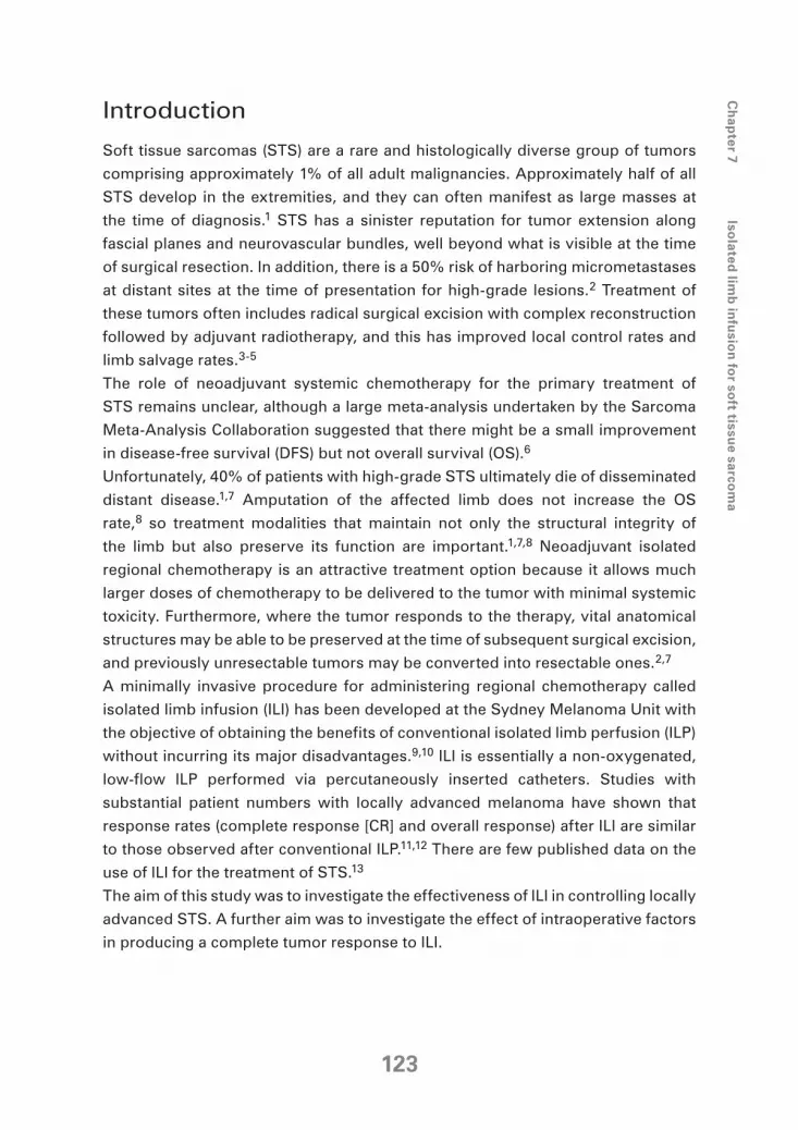
Introduction
Soft tissue sarcomas (STS) are a rare and histologically diverse group of tumors
comprising approximately 1% of all adult malignancies. Approximately half of all
STS develop in the extremities, and they can often manifest as large masses at
the time of diagnosis.1 STS has a sinister reputation for tumor extension along
fascial planes and neurovascular bundles, well beyond what is visible at the time
of surgical resection. In addition, there is a 50% risk of harboring micrometastases
at distant sites at the time of presentation for high-grade lesions.2 Treatment of
these tumors often includes radical surgical excision with complex reconstruction
followed by adjuvant radiotherapy, and this has improved local control rates and
limb salvage rates.3-5
The role of neoadjuvant systemic chemotherapy for the primary treatment of
STS remains unclear, although a large meta-analysis undertaken by the Sarcoma
Meta-Analysis Collaboration suggested that there might be a small improvement
in disease-free survival (DFS) but not overall survival (OS).6
Unfortunately, 40% of patients with high-grade STS ultimately die of disseminated
distant disease.1,7 Amputation of the affected limb does not increase the OS
rate,8 so treatment modalities that maintain not only the structural integrity of
the limb but also preserve its function are important.1,7,8 Neoadjuvant isolated
regional chemotherapy is an attractive treatment option because it allows much
larger doses of chemotherapy to be delivered to the tumor with minimal systemic
toxicity. Furthermore, where the tumor responds to the therapy, vital anatomical
structures may be able to be preserved at the time of subsequent surgical excision,
and previously unresectable tumors may be converted into resectable ones.2,7
A minimally invasive procedure for administering regional chemotherapy called
isolated limb infusion (ILI) has been developed at the Sydney Melanoma Unit with
the objective of obtaining the benefits of conventional isolated limb perfusion (ILP)
without incurring its major disadvantages.9,10 ILI is essentially a non-oxygenated,
low-flow ILP performed via percutaneously inserted catheters. Studies with
substantial patient numbers with locally advanced melanoma have shown that
response rates (complete response [CR] and overall response) after ILI are similar
to those observed after conventional ILP.11,12 There are few published data on the
use of ILI for the treatment of STS.13
The aim of this study was to investigate the effectiveness of ILI in controlling locally
advanced STS. A further aim was to investigate the effect of intraoperative factors
in producing a complete tumor response to ILI.
Ch
ap
ter 7
Is
ola
ted
limb
infu
sio
n fo
r so
ft tissu
e s
arc
om
a
123
Patients and Methods
The data were collected prospectively on all patients treated for STS with ILI
between 1994 and 2007. Patients prospectively gave informed consent for data
to be collected and used for future clinical research. Patients were referred for
ILI for two primary reasons: as neoadjuvant treatment before definitive surgical
resection, or as palliative treatment for unresectable disease. The decision about
the resectability of the extremity lesion was made as part of a multidisciplinary
team management process.
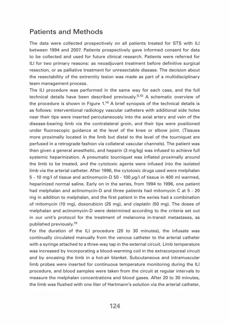
The ILI procedure was performed in the same way for each case, and the full
technical details have been described previously.9,10 A schematic overview of
the procedure is shown in Figure 1.14 A brief synopsis of the technical details is
as follows: interventional radiology vascular catheters with additional side holes
near their tips were inserted percutaneously into the axial artery and vein of the
disease-bearing limb via the contralateral groin, and their tips were positioned
under fluoroscopic guidance at the level of the knee or elbow joint. (Tissues
more proximally located in the limb but distal to the level of the tourniquet are
perfused in a retrograde fashion via collateral vascular channels). The patient was
then given a general anesthetic, and heparin (3 mg/kg) was infused to achieve full
systemic heparinization. A pneumatic tourniquet was inflated proximally around
the limb to be treated, and the cytotoxic agents were infused into the isolated
limb via the arterial catheter. After 1996, the cytotoxic drugs used were melphalan
5 - 10 mg/l of tissue and actinomycin-D 50 - 100 μg/l of tissue in 400 ml warmed,
heparinized normal saline. Early on in the series, from 1994 to 1996, one patient
had melphalan and actinomycin-D and three patients had mitomycin C at 5 - 20
mg in addition to melphalan, and the first patient in the series had a combination
of mitomycin (10 mg), doxorubicin (25 mg), and cisplatin (50 mg). The doses of
melphalan and actinomycin-D were determined according to the criteria set out
in our unit’s protocol for the treatment of melanoma in-transit metastases, as
published previously.10
For the duration of the ILI procedure (20 to 30 minutes), the infusate was
continually circulated manually from the venous catheter to the arterial catheter
with a syringe attached to a three-way tap in the external circuit. Limb temperature
was increased by incorporating a blood-warming coil in the extracorporeal circuit
and by encasing the limb in a hot-air blanket. Subcutaneous and intramuscular
limb probes were inserted for continuous temperature monitoring during the ILI
procedure, and blood samples were taken from the circuit at regular intervals to
measure the melphalan concentrations and blood gases. After 20 to 30 minutes,
the limb was flushed with one liter of Hartmann’s solution via the arterial catheter,
124
and the venous effluent was discarded. The limb tourniquet was then deflated to
restore normal limb circulation, and the catheters were removed.
Postoperatively, the serum creatine phosphokinase level was measured daily, and
limb toxicity and tumor response were assessed regularly. The scale proposed by
Wieberdink et al.15 was used to assess limb toxicity (Table 1). At routine follow-
up (usually at 6 and 12 weeks), the response of the tumors of all 21 patients
were evaluated, and CRs and partial responses (PR) were assessed by clinical
observation strictly according to the standard World Health Organization criteria.16
Figure 1: Schematic diagram (top) and photograph (bottom) demonstrating the key components
of the isolated limb infusion circuit. Note the Esmarch bandage on the foot to protect the acral
region from developing postoperative toxicity.
Ch
ap
ter 7
Is
ola
ted
limb
infu
sio
n fo
r so
ft tissu
e s
arc
om
a
125
These define a CR as the disappearance of all measurable disease determined by
two observations not < 4 weeks apart, and a PR as a ≥ 50% decrease in total tumor size determined by two observations not < 4 weeks apart and no appearance of
new lesions or progression of any lesion. In this study, the size of the cohort was
small. Patients were thus grouped into categories: those who had a CR versus
those who had a PR, no response, or disease progression. None of the patients
in this cohort received concurrent treatment between the ILI procedure and the
clinical assessment. Accordingly, any tumor response was attributed to the ILI
procedure alone. In addition to the local tumor response, the patient’s DFS and OS
were recorded. In those cases where the patient was referred from remote regions
or overseas, the general practitioner or referring surgeon looking after the patient
was contacted to determine the patient’s long-term follow-up status. All data were
collected prospectively and recorded in a computerized database.
Possible prognostic factors were tested for their influence on response rates,
local recurrence-free interval, DFS, and OS. The Fisher exact test was used for
comparison of frequency distributions, and the Mann-Whitney U-test was used
for the nonparametric variables.17 Continuous variables were assessed by
repeated-measures analysis of variance (ANOVA). Overall and DFS in addition
to local recurrence rates were analyzed by the Kaplan-Meier method.18 The
multinomial logistic regression and the Cox regression (Cox proportional hazard
model)19 analyses with the stepwise backward method were used for multivariate
analysis of response rates and survival, with various cohorts. Classification trees
(exhaustive chi-square automatic interaction detector (CHAID) method)20 were
produced to provide a multivariate analysis of the intraoperative variables by using
response type as the outcome variable. A significant difference was assumed for
a probability value of < .05. Statistical analyses were performed by GraphPad
Prism version 4.0 (San Diego, CA), SPSS version 11.0 (Chicago, IL) and AnswerTree
version 3.0 (SPSS).
Table 1: Wieberdink toxicity grading15
Grade I No visible effect
Grade II Slight erythema and/or edema
Grade III Considerable erythema and/or edema with blistering
Grade IV Extensive epidermolysis and/or obvious damage to deep tissues with a threatened
or actual compartment syndrome
Grade V Severe tissue damage necessitating amputation
126
Results
Twenty-one patients were identified from the ILI database; there were 14 men
(male:female ratio 2:1), and the average age was 60 years (range, 18 – 85 years).
Eighteen patients (86%) had disease involving the lower limb, and three had
disease of the upper limb. Fourteen patients (67%) underwent ILI as neoadjuvant
therapy before surgery, and seven patients underwent ILI for either the treatment
of inoperable recurrence or palliation. Table 2 describes the distribution of patients
according to the American Joint Committee on Cancer disease staging and the
distribution of the sarcoma subtypes. All patients except one (20 of 21) in this
study had a high-grade STS. The median follow-up time was 28 months (range, 2
– 91 months).
The disease of 12 patients (57%) had a CR to the ILI, 7 (33%) had a PR, and 2
(10%) had no response, giving an overall response rate of 90%. Neither the tumor
stage nor the tumor type (Fisher exact test) was associated with an improved or
worse response, and there was no difference in response rate between the sexes.
All of the patients who received ILI as neoadjuvant therapy before definitive
surgical resection had a positive response to the treatment. Nine (65%) of the 14
Table 2: Patient and tumor characteristics and response to ILI
Characteristics No. of patients (%)
Sex
Male 14 (67)
Female 7 (33)
Age in years, median (range) 60 (18-85)
Stage (AJCC) at ILI
I 0 (0)
II 9 (43)
III 9 (43)
IV 3 (14)
Involved limb
Lower 18 (86)
Upper 3 (14)
Soft Tissue Sarcoma Subtype
Malignant Fibrous Histiocytoma 10 (47)
Angiosarcoma 4 (19)
Fibrosarcoma 2 (10)
Others 5 (24)
Response to ILI
Complete (CR) 12 (57)
Partial (PR) 7 (33)
None 2 (10)
Ch
ap
ter 7
Is
ola
ted
limb
infu
sio
n fo
r so
ft tissu
e s
arc
om
a
127
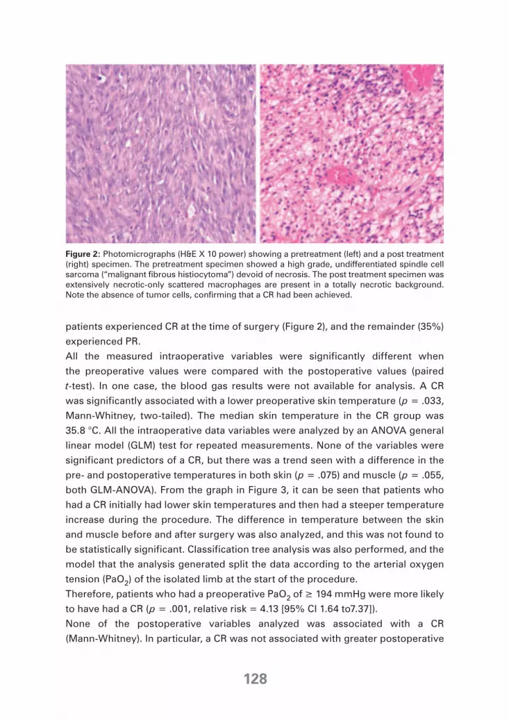
patients experienced CR at the time of surgery (Figure 2), and the remainder (35%)
experienced PR.
All the measured intraoperative variables were significantly different when
the preoperative values were compared with the postoperative values (paired
t-test). In one case, the blood gas results were not available for analysis. A CR
was significantly associated with a lower preoperative skin temperature (p = .033,
Mann-Whitney, two-tailed). The median skin temperature in the CR group was
35.8 °C. All the intraoperative data variables were analyzed by an ANOVA general
linear model (GLM) test for repeated measurements. None of the variables were
significant predictors of a CR, but there was a trend seen with a difference in the
pre- and postoperative temperatures in both skin (p = .075) and muscle (p = .055,
both GLM-ANOVA). From the graph in Figure 3, it can be seen that patients who
had a CR initially had lower skin temperatures and then had a steeper temperature
increase during the procedure. The difference in temperature between the skin
and muscle before and after surgery was also analyzed, and this was not found to
be statistically significant. Classification tree analysis was also performed, and the
model that the analysis generated split the data according to the arterial oxygen
tension (PaO2) of the isolated limb at the start of the procedure.
Therefore, patients who had a preoperative PaO2 of ≥ 194 mmHg were more likely to have had a CR (p = .001, relative risk = 4.13 [95% CI 1.64 to7.37]).
None of the postoperative variables analyzed was associated with a CR
(Mann-Whitney). In particular, a CR was not associated with greater postoperative
Figure 2: Photomicrographs (H&E X 10 power) showing a pretreatment (left) and a post treatment
(right) specimen. The pretreatment specimen showed a high grade, undifferentiated spindle cell
sarcoma (“malignant fibrous histiocytoma”) devoid of necrosis. The post treatment specimen was
extensively necrotic-only scattered macrophages are present in a totally necrotic background.
Note the absence of tumor cells, confirming that a CR had been achieved.
128
Figure 3: Graph demonstrating the
lower initial starting skin temperature
and the steeper rate of change during
the isolated limb infusion procedure
for the patients who had a complete
response compared with those who
did not.
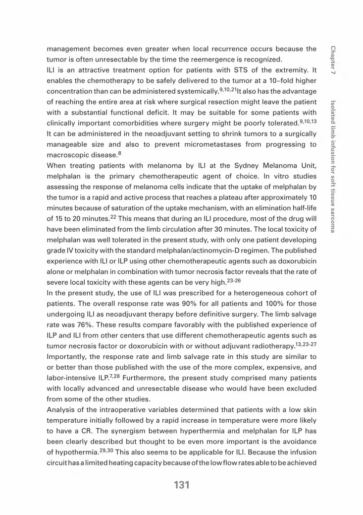
Figure 4: Kaplan-Meier curves compar-
ing local recurrence rates for patients
who had definitive surgical excision (n
= 14) with those who did not (n = 7).
Figure 5: Kaplan-Meier curves compar-
ing local recurrence rates for patients
who had a complete response to
isolated limb infusion (n = 12) with
those who did not (n = 9)
toxicity, nor was it associated with a higher maximum creatine phosphokinase
level. Three patients (14%) developed grade IV toxicity as a result of the ILI
procedure, but one of these received mitomycin C in addition to melphalan and
another received a combination of mitomycin C, cisplatin, and doxorubicin with no
melphalan. The remainder of the patients developed mild postoperative toxicity
(grades II and III).
Ch
ap
ter 7
Is
ola
ted
limb
infu
sio
n fo
r so
ft tissu
e s
arc
om
a
129

Nine patients (42%) in the total cohort developed a local recurrence during the
follow-up period, and the median local recurrence free-interval was 25 months.
Three (21%) of 14 surgical patients developed a local recurrence during follow-
up, and the patients who went on to have a completion surgical excision had a
significantly lower local recurrence rate (Kaplan-Meier, p = .013; HR = .20 [.025 to
.65], Figure 4). A CR was associated with a significantly decreased local recurrence
rate (Kaplan-Meier, p = .004; HR = 7.00 [2.09 to 47.00], Figure 5). When the tumor
subtypes were categorized into malignant fibrous histiocytoma (MFH) versus
the others, the MFHs were associated with a significantly lower chance of local
recurrence (Kaplan-Meier, p = .015; HR = 8.38 [1.41 to 24.15]) when treated with
ILI. The Cox proportional hazard model identified both CR and MFH subtypes as
statistically significant independent predictors of local recurrence rate. These two
variables were also statistically significant independent predictors of increased
DFS. Conversely, disease stage was not predictive of local recurrence or DFS.
The overall disease-specific survival was 62%. When the data were analyzed for
predictors of OS, disease stage was the only significant independent variable
(Cox proportional hazard, exp β = .106 [.013 to .868]). Thus, stage I/II disease
had significantly better OS than stage III/IV (Kaplan-Meier, p = .009; HR = 9.18
[1.63 to 29.8]). There was a trend for CR to be associated with an increased OS
(p = .071, HR = 3.32 [.89 to 18.52]) and there was also a trend for the MFH tumor
subtype to have an improved OS, but neither of these variables reached statistical
significance. There was no statistically significant increase in OS for those patients
who had surgery over those who did not. Ten patients (47%) received adjuvant
radiotherapy. Neither univariate nor multivariate analysis was able to demonstrate
a significant improvement in OS, DFS, or local recurrence rate associated with
radiotherapy.
No patient in this cohort had an amputation resulting from acute toxicity of the ILI
procedure. However, five patients (24%) ultimately had amputations for disease
progression: one patient (33%) with STS of the upper limb and four patients (22%)
with STS of the lower limb. There were trends for the amputees to have had a poor
response to the ILI (p = .082) and to have a worse OS (p = .08).
Discussion
Amputation of the limb is no longer considered standard therapy for primary STS.
Limb-sparing surgery with adjuvant radiotherapy has been shown to produce
good local control rates with no reduction in OS.1,8 However, the treatment of these
tumors becomes more difficult when the primary tumor is large, when it is abutting
or invading vital anatomic structures, or when it is high grade. The complexity of
130
management becomes even greater when local recurrence occurs because the
tumor is often unresectable by the time the reemergence is recognized.
ILI is an attractive treatment option for patients with STS of the extremity. It
enables the chemotherapy to be safely delivered to the tumor at a 10–fold higher
concentration than can be administered systemically.9,10,21It also has the advantage
of reaching the entire area at risk where surgical resection might leave the patient
with a substantial functional deficit. It may be suitable for some patients with
clinically important comorbidities where surgery might be poorly tolerated.9,10,13
It can be administered in the neoadjuvant setting to shrink tumors to a surgically
manageable size and also to prevent micrometastases from progressing to
macroscopic disease.8
When treating patients with melanoma by ILI at the Sydney Melanoma Unit,
melphalan is the primary chemotherapeutic agent of choice. In vitro studies
assessing the response of melanoma cells indicate that the uptake of melphalan by
the tumor is a rapid and active process that reaches a plateau after approximately 10
minutes because of saturation of the uptake mechanism, with an elimination half-life
of 15 to 20 minutes.22 This means that during an ILI procedure, most of the drug will
have been eliminated from the limb circulation after 30 minutes. The local toxicity of
melphalan was well tolerated in the present study, with only one patient developing
grade IV toxicity with the standard melphalan/actinomycin-D regimen. The published
experience with ILI or ILP using other chemotherapeutic agents such as doxorubicin
alone or melphalan in combination with tumor necrosis factor reveals that the rate of
severe local toxicity with these agents can be very high.23-26
In the present study, the use of ILI was prescribed for a heterogeneous cohort of
patients. The overall response rate was 90% for all patients and 100% for those
undergoing ILI as neoadjuvant therapy before definitive surgery. The limb salvage
rate was 76%. These results compare favorably with the published experience of
ILP and ILI from other centers that use different chemotherapeutic agents such as
tumor necrosis factor or doxorubicin with or without adjuvant radiotherapy.13,23-27
Importantly, the response rate and limb salvage rate in this study are similar to
or better than those published with the use of the more complex, expensive, and
labor-intensive ILP.7,28 Furthermore, the present study comprised many patients
with locally advanced and unresectable disease who would have been excluded
from some of the other studies.
Analysis of the intraoperative variables determined that patients with a low skin
temperature initially followed by a rapid increase in temperature were more likely
to have a CR. The synergism between hyperthermia and melphalan for ILP has
been clearly described but thought to be even more important is the avoidance
of hypothermia.29,30 This also seems to be applicable for ILI. Because the infusion
circuit has a limited heating capacity because of the low flow rates able to be achieved
Ch
ap
ter 7
Is
ola
ted
limb
infu
sio
n fo
r so
ft tissu
e s
arc
om
a
131

through the small-caliber catheters, it is necessary to maintain the temperature of
the limb with an external heating blanket and an overhead radiant heater as well
to reach the desired subcutaneous temperature of at least 37.8 °C in the limb by
the end of the ILI procedure when treating patients with melanoma.10,11 However,
in contrast to the melanoma studies, the STS patients that started with a cooler
limb were more likely to have a CR. Theoretically this would make sense, as these
deep-sited STSs are more likely to receive a greater delivery of chemotherapy if the
circulation to the superficial tissues is concomitantly vasoconstricted, whereas this
situation would be detrimental in the treatment of in transit melanoma metastases
situated in the dermis and superficial subcutaneous tissue planes.12 Classification
tree analysis identified a high PaO2 in the isolated limb artery at the initiation of
the ILI procedure to be associated with a CR, although this was not identified as
a statistically significant predictor by GLM-ANOVA analysis. The reasons for this
observation are not immediately apparent. In vitro studies looking at melanoma
have demonstrated that hypoxia enhances the cytotoxic effects of melphalan by a
factor of approximately 1.5, whereas the combination of hypoxia and acidosis can
increase the effect by a factor of 3.31 In the present study, a low PaO2 in the isolated
limb at the start of the procedure may have been indicative of a poor peripheral
circulation in general, suggesting atherosclerosis, which may have contributed to
an inferior delivery of cytotoxic agents to the tumor.
The overall local recurrence rate in the present study was similar to other published
studies,25-28 and the limb recurrence-free interval (LRFI) rate was greatly improved
in the group that subsequently had definitive surgical treatment. A CR was also
markedly associated with an improved LRFI, and the effect these two factors
were able to have may not only have been a result of treating main tumor bulk
but also the eradication of outlying local micrometastases that are present in a
high proportion of high-grade STS. This study was commenced before the World
Health Organization classification of tumors reclassified MFH as undifferentiated
high-grade pleomorphic sarcoma,32 and it may no longer be valid to draw
conclusions regarding this subgroup of patients. However, both the old and new
classifications describe high-grade sarcomas with a considerable amount of
cellular activity and a high cell turnover rate,32 and it is these common factors that
may be responsible for an increased uptake of the drugs resulting in the destruction
of more tumor leading to great improvement in LRFI observed in the subgroup of
patients with MFH after ILI.
As with other locoregional treatments for STS, ILI did not greatly improve OS in
this study. This was not an unexpected outcome. There was a trend for a CR to be
associated with improved OS, but this just failed to reach statistical significance
(p = .07). Where ILP has been used to treat patients with melanoma, a CR is a
statistically significant positive prognostic indicator after long-term follow-
132
up, which may reflect a more favorable tumor biology.33 Multivariate analysis
revealed that only the stage of the tumor at the initial presentation of the patient
was associated with an improved OS, and this is a finding consistent with other
treatment modalities and for other diseases where ILI was used, especially
melanoma.12,29 Unlike ILI for melanoma in-transit metastases, the rate of CR is
not related to the stage of the disease.13 The explanation for this may lie in the
inability to deliver the same concentration of cytotoxic agents systemically as
can be given locally in ILI to produce an overall response. Similarly, delivery of
high concentrations of cytotoxic drugs to the tumor to produce a CR locally may
be hampered by peripheral vascular disease in patients with otherwise favorable
early-stage disease.
In this study examining the usefulness of ILI in the treatment of advanced-stage STS,
a 13-year experience has been analyzed and presented. The stage of the disease
at presentation of the patient was the only statistically significant prognostic factor
for OS. A complete tumor response to ILI was associated with an increased LRFI
and disease-free interval. The likelihood of a complete tumor response was found
to be increased if the initial skin temperature of the affected limb was cooler and
the initial PaO2 was ≥ 194 mmHg. The limb salvage rates and duration of response after ILI were comparable to previously reported results achieved by conventional
ILP, an invasive and much more complex and costly procedure.
Ch
ap
ter 7
Is
ola
ted
limb
infu
sio
n fo
r so
ft tissu
e s
arc
om
a
133
References
1. Linehan D, Brennan M. Soft tissue tumors and melanoma. In: Holzheimer R, Mannick
J, editors. Surgical treatment: evidence-based and problem-oriented. Zuckschwerdt,
Munich 2001. Available at: http://www.ncbi.nlm.nih.gov/books/bv.fcgi?rid=surg.chap-
ter.3838.
2. Pisters PW, Ballo MT, Patel SR. Preoperative chemoradiation treatment strategies for local-
ized sarcoma. Ann Surg Oncol 2002;9:535-42.
3. Rosenberg SA, Tepper JE, Glatstein EJ, et al. The treatment of soft tissue sarcomas of the
extremities: prospective randomized evaluations of (1) limb-sparing surgery plus radia-
tion therapy compared with amputation and (2) the role of adjuvant chemotherapy. Ann
Surg 1982;196:305-15.
4. Yang JC, Chang AE, Baker AR, et al. A randomized prospective study of the benefit of
adjuvant radiation therapy in the treatment of soft tissue sarcomas of the extremity. J Clin
Oncol 1998;16:197-203.
5. O’Sullivan B, Davis AM, Turcotte R, et al. Preoperative versus postoperative radiotherapy
in soft-tissue sarcoma of the limbs: a randomized trial. Lancet 2002;359:2235-41.
6. Adjuvant chemotherapy for localised resectable soft-tissue sarcoma of adults: meta-anal-
ysis of individual data. Sarcoma meta-analysis collaboration. Lancet 1997;350:1647-54.
7. Schraffordt Koops H, Eggermont AM, Lienard D, et al. Hyperthermic isolated limb perfu-
sion for the treatment of soft tissue sarcomas. Semin Surg Oncol 1998;14:210–4.
8. Rooser B, Gustafson P, Rydholm AS. Is there no influence of local control on the rate of
metastases in high grade soft tissue sarcoma? Cancer 1990;65:1727–9.
9. Thompson JF, Waugh RC, Saw RPM, et al. Isolated limb infusion with melphalan for recur-
rent limb melanoma: a simple alternative to isolated limb perfusion. Reg Cancer Treat
1994;7:188–92.
10. Thompson JF, Kam PC, Waugh RC, et al. Isolated limb infusion with cytotoxic agents: a
simple alternative to isolated limb perfusion. Semin Surg Oncol 1998;14:238–47.
11. Lindnér P, Doubrovsky A, Kam PCA, et al. Prognostic factors after isolated limb infusion
with cytotoxic agents for melanoma. Ann Surg Oncol 2002;9:127–36.
12. Kroon HM, Moncrieff M, Kam PC, Thompson JF. Outcomes following isolated limb infu-
sion for melanoma. A 14-year experience. Ann Surg Oncol 2008;15:3003-13.
13. Hegazy MA, Kotb SZ, Sakr H, et al. Preoperative isolated limb infusion of doxorubi-
cin and external irradiation for limbthreatening soft tissue sarcomas. Ann Surg Oncol
2007;14:568–76.
14. Thompson JF, Kam PC. Isolated limb infusion for melanoma: a simple but effective alter-
native to isolated limb perfusion. J Surg Oncol 2004;1:1–3.
15. Wieberdink J, Benckhuysen C, Braat RP, et al. Dosimetry in isolation perfusion of the limbs
by assessment of perfused tissue volume and grading of toxic tissue reactions. Eur J
Cancer Clin Oncol 1982;18:905-10.
16. World Health Organization. WHO Handbook for reporting results of cancer treatments
(WHO offset publication No. 48). Geneva: World Health Organization, 1979.
17. Mann HB, Whitney DR. On a test of whether one of two random variables is stochastically
larger than the other. Ann Math Stat 1947;18:50-60.
134
18. Kaplan L, Meier P. Nonparametric estimation from incomplete observations. J Am Stat
Assoc 1985;53:457-81.
19. Cox DR. Regression models and life table. J Stat Soc 1972;34:187–220.
20. Kass GV. An exploratory technique for investigating large quantities of categorical data. J
Appl Stat 1980;29:119-27.
21. Roberts MS, Wu ZY, Siebert GA, et al. Pharmacokinetics and pharmacodynamics of
melphalan in isolated limb infusion for recurrent localized limb malignancy. Melanoma
Res 2001;11:423-31.
22. Parsons PG, Carter FB, Morrison L, et al. Mechanism of melphalan resistance develop-
ment in human melanoma cells. Cancer Res 1981;41:1525–34.
23. Nijhuis PH, Pras E, Sleijfer DT, et al. Long term results of preoperative intra-arterial doxo-
rubicin combined with neoadjuvant radiotherapy followed by extensive surgical resection
for locally advanced soft tissue sarcomas of the extremities. Radiother Oncol 1999;51:15-
9.
24. Bezwada HP, Granick MS, Long CD, et al. Soft tissue complications of intra-arterial chemo-
therapy for extremity sarcoma. Ann Plast Surg 1998;40:382–7.
25. Soulen MC, Weismann JR, Sullivan KL, et al. Intra-arterial chemotherapy with limb sparing
resection of large soft tissue sarcomas of the extremities. J Vasc Interv Radiol 1992;3:659-
63.
26. Noorda EM, Vrouenraets BC, Nieweg OE, et al. Isolated limb perfusion with TNF-α and
melphalan for unresectable soft tissue sarcoma of the extremities. Cancer 2003;7:1483-
90.
27. Lejeune FJ, Pujol N, Lienard D, et al. Limb salvage by neoadjuvant isolated perfusion with
TNF alpha and melphalan for non-resectable soft tissue sarcomas of the extremities. Eur
J Surg Oncol 2000;26:669-78.
28. Rossi CR, Mocellin S, Pilati P, et al. TNF alpha based isolated perfusion for limb threaten-
ing soft tissue sarcomas: state of the art and future trends. J Immunother 2003;26:291-
300.
29. Vrouenraets BC, Nieweg OE, Kroon BB. Thirty-five years of isolated limb perfusion for
melanoma: indications and results. Br J Surg 1996;83:1319-28.
30. Kroon BBR. Regional isolation perfusion in melanoma of the limbs: accomplishments,
unsolved problems, future. Eur J Surg Oncol 1988;14:101-10.
31. Siemann DW, Chapman M, Beikirch A. Effects of oxygenation and pH on tumor cell
response to alkylating chemotherapy. Int J Radiat Oncol Biol Phys 1991;20:287-9.
32. Fletcher C, Unni K, Mertens F, editors. So-called fibrohistiocytic tumors. In: World Health
Organization. WHO Classification of Tumors: Pathology and Genetics of Tumors of Soft
Tissue and Bone. Lyon: IARC Press, 2002:120-2.
33. Sanki A, Kam P, Thompson J. Long-term results of hyperthermic isolated limb perfusion
for melanoma: a reflection of tumor biology. Ann Surg 2007;4:591-6.
Ch
ap
ter 7
Is
ola
ted
limb
infu
sio
n fo
r so
ft tissu
e s
arc
om
a
135