Automatic Resting Tremor Assessment in Parkinson's Disease ...
Upload
khangminh22Category
view
3download
0
Citation: Raciti, L.; Pignolo, L.;
Perini, V.; Pullia, M.; Porcari, B.;
Latella, D.; Isgrò, M.; Naro, A.;
Calabrò, R.S. Improving Upper
Extremity Bradykinesia in
Parkinson’s Disease: A Randomized
Clinical Trial on the Use of
Gravity-Supporting Exoskeletons. J.
Clin. Med. 2022, 11, 2543. https://
doi.org/10.3390/jcm11092543
Academic Editor: Julien Favre
Received: 14 April 2022
Accepted: 29 April 2022
Published: 1 May 2022
Publisher’s Note: MDPI stays neutral
with regard to jurisdictional claims in
published maps and institutional affil-
iations.
Copyright: © 2022 by the authors.
Licensee MDPI, Basel, Switzerland.
This article is an open access article
distributed under the terms and
conditions of the Creative Commons
Attribution (CC BY) license (https://
creativecommons.org/licenses/by/
4.0/).
Journal of
Clinical Medicine
Article
Improving Upper Extremity Bradykinesia in Parkinson’sDisease: A Randomized Clinical Trial on the Use ofGravity-Supporting ExoskeletonsLoredana Raciti 1, Loris Pignolo 2, Valentina Perini 3, Massimo Pullia 4, Bruno Porcari 4, Desiree Latella 4,Marco Isgrò 4, Antonino Naro 5 and Rocco Salvatore Calabrò 4,*
1 GCA-Centro Spoke AO Cannizzaro, Catania, IRCCS Centro Neurolesi Bonino-Pulejo, 98124 Messina, Italy;[email protected]
2 S. Anna Institute, Research in Advanced Neurorehabilitation, 88900 Crotone, Italy; [email protected] Spoke Centre of Palermo, IRCCS Centro Neurolesi Bonino-Pulejo, 98124 Messina, Italy;
[email protected] Behavioral and Robotic Neurorehabilitation Unit, IRCCS Centro Neurolesi Bonino Pulejo,
98124 Messina, Italy; [email protected] (M.P.); [email protected] (B.P.);[email protected] (D.L.); [email protected] (M.I.)
5 Department of Clinical and Experimental Medicine, University of Messina, 98122 Messina, Italy;[email protected]
* Correspondence: [email protected]; Fax: +39-9060128950
Abstract: Hand movements are particularly impaired in patients with Parkinson’s Disease (PD),contributing to functional disability and difficulties in activities of daily living. Growing evidencehas shown that robot-assisted therapy may be considered an effective and reliable method forthe delivery of the highly repetitive training that is needed to trigger neuroplasticity, as intensive,repetitive and task-oriented training could be an ideal strategy to facilitate the relearning of motorfunction and to minimize motor deficit. The purpose of this study is to evaluate the improvementof hand function with semi-autonomous exercises using an upper extremity exoskeleton in patientswith PD. A multicenter, parallel-group, randomized clinical trial was then carried out at the IRCCSCentro Neurolesi Bonino-Pulejo (Messina, Italy). Thirty subjects with a diagnosis of PD and a Hoehn–Yahr score between 2 and 3 were enrolled in the study. Patients were 1:1 randomized into eitherthe experimental group (ERT), receiving 45 min training daily, 6 days weekly, for 8 weeks withArmeo®Spring (Volketswil, Switzerland) (a gravity-supporting device), or the control group (CPT),which was subjected to the same amount of conventional physical therapy. Motor abilities wereassessed before and after the end of the training. The main outcomes measures were the Nine-holepeg test and the motor section of the UPDRS. All patients belonging to ERT and 9 out of 15 patientsbelonging to the CPT completed the trial. ERT showed a greater improvement in the primary outcomemeasure (nine-hole peg test) than CPT. Moreover, a statistically significant improvement was foundin ERT concerning upper limb mobility, and disease burden as compared to CPT. Using an upperextremity exoskeleton (i.e., the Armeo®Spring) for semi-autonomous training in an inpatient settingis a new perspective to train patients with PD to improve their dexterity, executive function and,potentially, quality of life.
Keywords: gravity-supporting device; hand bradykinesia; Parkinson’s disease; upper-limb rehabilitation;neurodegenerative diseases
1. Introduction
Bradykinesia and hypo-akinesia are the clinical hallmarks of Parkinson’s disease (PD),mostly impairing the sequentiality and dexterity of upper extremities (UE) movements [1].Notably, the motor impairment degree in PD patients has been shown to inversely correlatewith movement velocity and directly with task difficulty [2,3]. Indeed, UE functionality
J. Clin. Med. 2022, 11, 2543. https://doi.org/10.3390/jcm11092543 https://www.mdpi.com/journal/jcm

J. Clin. Med. 2022, 11, 2543 2 of 12
impairment is usually characterized by aberrant timing and force modulation, resulting ina poor quality of hand movements [4]. These alterations contribute to the impairments ofbody functions and structures and the difficulty in activities and participation [5,6], leadingto a lack of independence and poor quality of life [7,8].
Whether motor control deficits in PD are related to the slowness of movements ratherthan to impairments in movement selection is still controversial, as the nature of bradyki-nesia is still under investigation [9,10]. Initially, some authors suggested that bradykinesiais due to an inability to generate appropriate muscle activity before and during voluntarymovements for the completion of the intended movement [11]. This was likely related toexecutive function deficit and reduced motor information-processing in PD [11], resultingin hypometric and increasingly impaired movements when the difficulty of a sequentialmotor task increases [11]. Additionally, a decreased ability in thumb–index maximumtorque generation was found in PD patients [12]. Moreover, the basal ganglia have beenrecently described to have a pivotal role in the pathogenesis of the disorder. Then, PD isconsidered the consequence of the disruption of the excitability and plasticity of the basalganglia circuits, specifically of the M1 and other non-primary motor areas [9,13].
Some therapies have proven to be effective in the improvement of motor function,enhancing the impact of non-motor symptoms and improving quality of life, even thougha poor response has been demonstrated concerning hand movement dexterity [14]. To date,only a few studies focused on intervention strategies on UE and hand movements, particularlyregarding fully adaptive, assist-as-needed exercises of the traditional rehabilitation [15].
Indeed, most of the existing data have focused on gait using either robotics [16] or otherless expensive devices to improve the different gait parameters in patients with PD [17].
Innovative technologies are widely used in many neurological diseases, includingstroke, multiple sclerosis, and spinal cord injury, with the paramount effort to increasethe active range of motion and muscle strength [18,19]. Moreover, it has been shown thatbetter outcomes could be achieved when robotics is coupled to virtual reality, resultingin a potentiation of body function and activities [20]. Such promising results depend onthe fact that conjugating robotics and virtual reality allows maximizing neural, motorand functional recovery, by providing patients with lasting, challenging, repetitive, task-oriented, motivating, salient, and intensive motor exercises; furthermore, virtual realitymay also provide patients with a potentiation of motor and cognitive functions [20–23].
There is an increasing interest in robotic rehabilitation in PD. Some studies showedthat the use of robot-assisted rehab is useful for gait training [15,24], improve freezing ofgait [25] to reduce the muscular activity requirements of PD patients [26], balance [27],and reduce the amplitude of tremors [28]. Then, the use of advanced and computerizedtechnologies could provide a better rehabilitation in patients with PD [29,30]. However, nodata are available about the use of specific robotic devices to recover arm impairment inPD patients.
The aim of this pilot study is to evaluate the efficacy of a gravity-supporting exoskele-ton apparatus (i.e., the Armeo Spring; Hocoma, Zurich, Switzerland) on hand dexterity andoverall motor functions in PD patients (experimental robotic therapy-ERT) as compared toconventional physical therapy (CPT).
2. Materials and Methods2.1. Study Design and Population
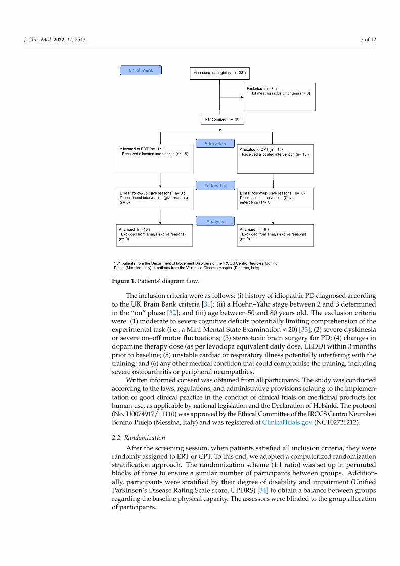
We carried out a parallel-group, single-blinded, randomized controlled trial on the PDpatients attending either the Movement Disorders Clinic of the IRCCS Centro NeurolesiBonino-Pulejo (Messina, Italy) or its Spoke Centre (Palermo, Italy) between July 2019 andMarch 2020. Indeed, the enrollment period was interrupted due to the COVID-19 pandemicand some patients abandoned the study (Figure 1).
J. Clin. Med. 2022, 11, 2543 3 of 12
J. Clin. Med. 2022, 11, x FOR PEER REVIEW 3 of 12
2. Materials and Methods
2.1. Study Design and Population
We carried out a parallel-group, single-blinded, randomized controlled trial on the
PD patients attending either the Movement Disorders Clinic of the IRCCS Centro Neu-
rolesi Bonino-Pulejo (Messina, Italy) or its Spoke Centre (Palermo, Italy) between July
2019 and March 2020. Indeed, the enrollment period was interrupted due to the COVID-
19 pandemic and some patients abandoned the study (Figure 1).
Figure 1. Patients’ diagram flow.
The inclusion criteria were as follows: (i) history of idiopathic PD diagnosed accord-
ing to the UK Brain Bank criteria [31]; (ii) a Hoehn–Yahr stage between 2 and 3 determined
in the “on” phase [32]; and (iii) age between 50 and 80 years old. The exclusion criteria
were: (1) moderate to severe cognitive deficits potentially limiting comprehension of the
experimental task (i.e., a Mini-Mental State Examination < 20) [33]; (2) severe dyskinesia
or severe on–off motor fluctuations; (3) stereotaxic brain surgery for PD; (4) changes in
dopamine therapy dose (as per levodopa equivalent daily dose, LEDD) within 3 months
prior to baseline; (5) unstable cardiac or respiratory illness potentially interfering with the
training; and (6) any other medical condition that could compromise the training, includ-
ing severe osteoarthritis or peripheral neuropathies.
Written informed consent was obtained from all participants. The study was con-
ducted according to the laws, regulations, and administrative provisions relating to the
implementation of good clinical practice in the conduct of clinical trials on medicinal
products for human use, as applicable by national legislation and the Declaration of Hel-
sinki. The protocol (No. U0074917/11110) was approved by the Ethical Committee of the
IRCCS Centro Neurolesi Bonino Pulejo (Messina, Italy) and was registered at ClinicalTri-
als.gov (NCT02721212).
2.2. Randomization
Figure 1. Patients’ diagram flow.
The inclusion criteria were as follows: (i) history of idiopathic PD diagnosed accordingto the UK Brain Bank criteria [31]; (ii) a Hoehn–Yahr stage between 2 and 3 determinedin the “on” phase [32]; and (iii) age between 50 and 80 years old. The exclusion criteriawere: (1) moderate to severe cognitive deficits potentially limiting comprehension of theexperimental task (i.e., a Mini-Mental State Examination < 20) [33]; (2) severe dyskinesiaor severe on–off motor fluctuations; (3) stereotaxic brain surgery for PD; (4) changes indopamine therapy dose (as per levodopa equivalent daily dose, LEDD) within 3 monthsprior to baseline; (5) unstable cardiac or respiratory illness potentially interfering with thetraining; and (6) any other medical condition that could compromise the training, includingsevere osteoarthritis or peripheral neuropathies.
Written informed consent was obtained from all participants. The study was conductedaccording to the laws, regulations, and administrative provisions relating to the implemen-tation of good clinical practice in the conduct of clinical trials on medicinal products forhuman use, as applicable by national legislation and the Declaration of Helsinki. The protocol(No. U0074917/11110) was approved by the Ethical Committee of the IRCCS Centro NeurolesiBonino Pulejo (Messina, Italy) and was registered at ClinicalTrials.gov (NCT02721212).
2.2. Randomization
After the screening session, when patients satisfied all inclusion criteria, they wererandomly assigned to ERT or CPT. To this end, we adopted a computerized randomizationstratification approach. The randomization scheme (1:1 ratio) was set up in permutedblocks of three to ensure a similar number of participants between groups. Addition-ally, participants were stratified by their degree of disability and impairment (UnifiedParkinson’s Disease Rating Scale score, UPDRS) [34] to obtain a balance between groupsregarding the baseline physical capacity. The assessors were blinded to the group allocationof participants.

J. Clin. Med. 2022, 11, 2543 4 of 12
2.3. Sample Size and Power Analysis
The sample size, in relation to the clinically significant changes in the primary out-come, was calculated by means of a 2-sided, 2-sample t-test, and estimated in 60 patients(30 per arm). More specifically, this sample size was required to maintain a type I error rateof 0.05 and an 80% power to detect a significant between-group difference of 5.65 s [35](including a 10% dropout rate or loss to follow-up).
2.4. Experimental Robotic Therapy
The Armeo®Spring (Figure 2) is a mechanical device (Hocoma Inc., Zurich, Switzer-land) characterized by an adaptable suspension system for the upper limb that offerssupport from the shoulder to the wrist ending with a grasping system for the hand.
J. Clin. Med. 2022, 11, x FOR PEER REVIEW 4 of 12
After the screening session, when patients satisfied all inclusion criteria, they were
randomly assigned to ERT or CPT. To this end, we adopted a computerized randomiza-
tion stratification approach. The randomization scheme (1:1 ratio) was set up in permuted
blocks of three to ensure a similar number of participants between groups. Additionally,
participants were stratified by their degree of disability and impairment (Unified Parkin-
son’s Disease Rating Scale score, UPDRS) [34] to obtain a balance between groups regard-
ing the baseline physical capacity. The assessors were blinded to the group allocation of
participants.
2.3. Sample Size and Power Analysis
The sample size, in relation to the clinically significant changes in the primary out-
come, was calculated by means of a 2-sided, 2-sample t-test, and estimated in 60 patients
(30 per arm). More specifically, this sample size was required to maintain a type I error
rate of 0.05 and an 80% power to detect a significant between-group difference of 5.65 s
[35] (including a 10% dropout rate or loss to follow-up).
2.4. Experimental Robotic Therapy
The Armeo®Spring (Figure 2) is a mechanical device (Hocoma Inc., Zurich, Switzer-
land) characterized by an adaptable suspension system for the upper limb that offers sup-
port from the shoulder to the wrist ending with a grasping system for the hand.
Figure 2. A patient with PD receiving the experimental training with the Armeo©Spring device.
System sensitivity can be adjusted depending on the patient’s condition. The system
gives information about movement parameters, such as resistance, strength, range of mo-
tion, and coordination. Based on the patient’s active mobility, the system allows to cali-
brate the working space and level of difficulty during the entire training period. The de-
vice expands any movement, allowing reinforcement and facilitation of the arm by visual
feedback (2D virtual reality) with a three-dimensional space [36].
The training was supervised by a physiotherapist expert in robot-assisted therapy.
After the device was adjusted for the patient’s arm size and angle of suspension, the work-
space and exercises were selected. Exercise difficulties were modified during the follow-
ing sessions. Each training session consisted of 45 min per session for each arm with
Armeo®Spring, 6 days per week, for 8 weeks.
2.5. Conventional Physical Therapy
Patients in the control group received the same global treatment time of the ERT.
Patients were submitted to conventional rehabilitation, such as passive- and active-as-
sisted mobilization of the upper limbs, traditional training for neuromuscular facilitation,
proprioception exercises, and reducing joints and muscles stiffness. Active exercises of
reaching and picking objects were also performed.
Figure 2. A patient with PD receiving the experimental training with the Armeo©Spring device.
System sensitivity can be adjusted depending on the patient’s condition. The systemgives information about movement parameters, such as resistance, strength, range ofmotion, and coordination. Based on the patient’s active mobility, the system allows tocalibrate the working space and level of difficulty during the entire training period. Thedevice expands any movement, allowing reinforcement and facilitation of the arm by visualfeedback (2D virtual reality) with a three-dimensional space [36].
The training was supervised by a physiotherapist expert in robot-assisted therapy.After the device was adjusted for the patient’s arm size and angle of suspension, theworkspace and exercises were selected. Exercise difficulties were modified during thefollowing sessions. Each training session consisted of 45 min per session for each arm withArmeo®Spring, 6 days per week, for 8 weeks.
2.5. Conventional Physical Therapy
Patients in the control group received the same global treatment time of the ERT.Patients were submitted to conventional rehabilitation, such as passive- and active-assistedmobilization of the upper limbs, traditional training for neuromuscular facilitation, propri-oception exercises, and reducing joints and muscles stiffness. Active exercises of reachingand picking objects were also performed.
2.6. Outcome Measures
We provided the patients with the motor section of the UPDRS and the Motricity Indexfor Upper Extremity (MI-UE) to test motor impairment, the nine-hole peg test (9HPT) [37]to objectively evaluate hand dexterity, the Fugl-Meyer Assessment for the upper extremity(FMA-UE) to test the UE motor ability to perform selective movements, the FunctionalIndependence Measure (FIM) to estimate the disability burden, and the numerical ratingscale of pain (P-NRS) to measure the range of pain intensity. The primary outcome was thechange of the 9HPT from baseline (T0) to post-treatment (T1). The secondary outcomeswere the changes of UPDRS, FMA-UE, FIM, P-NRS, and MI-UE from T0 to T1.

J. Clin. Med. 2022, 11, 2543 5 of 12
2.6.1. Primary Outcome Measure
The 9HPT test was used to assess hand dexterity, by asking the patients to take ninepegs from a container and place them into nine holes on a board and vice versa as quicklyas possible. Score was the time taken to complete the test activity [38].
2.6.2. Secondary Outcome Measures
The UPDRS is the most used screening tool to detect disease severity and motor andnon-motor complications in PD [34,38]. It consists of three subscales: part I: evaluationof mentation, behavior, and mood by an interview; part II: the activities of daily lifeby self-evaluation; part III or motor section is a clinician-scored motor evaluation; andpart IV is the evaluation of long-term levodopa complications, such as dyskinesia andmotor fluctuations.
The FMA-UE was used to evaluate upper limb motor ability to perform selectivemovements. Of the motricity scales, the most popular used scale in PD is the Fugl-MeyerMotor Assessment Scale [39,40].
The 33-item scale consists of 3 response categories (scores 0–2) for each item, with amaximum score of 66 (indicating no impairment) with sub-scores of 36 for the upper arm,10 for the wrist, 14 for the hand, and 6 for coordination and speed of movement [39,41].
The FIM measures the level of a patient’s disability burden and indicates how muchassistance is required for the individual to carry out activities of daily living. Each taskis rated on a 7-point ordinal scale that ranges from 1 = total assistance (or completedependence) to 7 = complete independence [42].
The P-NRS typically consists of a series of numbers with verbal anchors representingthe entire possible range of pain intensity. Generally, patients rate their pain from 0 to 10,from 0 to 20, or from 0 to 100. Zero represents “no pain”, whereas 10, 20, or 100 representthe opposite end of the pain continuum (e.g., “the most intense pain imaginable”, “pain asintense as it could be”, and “maximum pain”) [43].
The MI-UE can be used to assess motor impairment. MI is a feasible measure that candemonstrate the overall patients’ impairment. It is a simple, brief measure of general motorfunction that can predict mobility outcomes post-stroke [44]. All motor assessment andmotor training sessions were performed bilaterally, in the affected and unaffected side.
2.7. Statistical Analysis
Between-group T0 differences were estimated using the Mann–Whitney test. Thesignificance of the changes in each outcome measure was calculated by conducting arepeated-measure ANOVA for continuous numerical variables (using time, two levels,and group, two levels, as factors) or a Wilcoxon signed-rank test for ordinal or nominalvariables. Depending on the significance of the main interactions and effects, Bonferronicorrected pairwise comparisons using t-tests, Wilcoxon test, or Mann–Whitney test, whereappropriate, were tested. For all statistical tests, the significance level was set at α < 0.05.All the analyses included all participants for which data were available.
3. Results3.1. Baseline (T0)
Thirty-three patients were consecutively screened for study inclusion. Three failed theinclusion criteria. Therefore, 30 patients were randomized to ERT or CPT. There were nodifferences at baseline between the groups concerning age, disease duration, and LEDD(Table 1; all p > 0.2).

J. Clin. Med. 2022, 11, 2543 6 of 12
Table 1. Clinical-demographic features at baseline (T0). Data are reported as mean (SD) or median(interquartile range), and p-value of Mann–Whitney test.
ERT (n = 15) CPT (n = 9) p-Value
Age (years) 65.7 (7) 62.7 (10.1) 0.1Disease Duration (years) 5.3 (3.4) 6.2 (4.6) 0.6
LEDD (mg/day) 544 (198) 583 (191) 0.7H&Y 2 (2–3) 2 (2–3) 0.4
Legend: CPT, conventional physical therapy; ERT, experimental robotic therapy; H&Y, Hoehn–Yahr stagedetermined in the “on” phase; LEDD, levodopa equivalent daily dose.
Particularly, both groups had a Hoehn–Yahr stage between 2 and 3, which corre-sponded to an on-average moderate impairment of body functions and structure and amoderate difficulty impairment in activities and participation (i.e., present <50% of time,with intensity that interferes with day-to-day lift, occurring occasionally over last 30 days).The 9HPT, MI-UE, and FIM data were not available for three subjects belonging to the ERTgroup (Tables 2 and 3).
Table 2. Outcome measure scores at T0 (baseline) and T1 (post-treatment) in the ERT (n = 15;* n = 12) and CPT (n = 9) group. Data are reported as mean (SD) or median (interquartile range), andp-value of within-group (using t-test or Wilcoxon test) and between-group comparison (using t-testor Mann–Whitney test).
T0 T1 Within-Group Comparison Between-Group Comparison
9HPTERT 42.2 (17) 34.1 (14) 0.006
0.004CPT 35.1 (6.8) 31.4 (5.4) 0.9
UPDRS-IIIERT 28 (23–33) 21 (16–26) 0.06
0.5CPT 37 (31.5–41) 32 (23.25;40) 0.9
P-NRSERT 2.5 (0.5–3.5) 1.1 (0.3–1.8) 0.007
0.9CPT 4 (3–5) 1 (0–1.5) 0.01
MI-UEERT 72 (65–80) * 89 (83–94) * 0.04
0.0001CPT 77 (73.25–82) 82 (79.25;88.5) 0.8
FIMERT 104 (98–109) * 110 (105–115) * 0.6
0.6CPT 100 (99–103) 101 (100–106) 0.9
FMA-UEERT 48 (45–52) 53 (5–56) 0.007
0.009CPT 53 (51–55) 56 (52.5–59.5) 0.9
Legend: 9HPT, nine-hole peg test; UPDRS, Unified Parkinson’s Disease Rating Scale; P-NRS, numerical ratingscale of pain; MI-UE, Motricity Index for Upper Extremity; FIM, Functional Independence Measure; FMA-UE,Fugl-Meyer Assessment for Upper Extremity.
3.2. Post-Treatment (T1)
Six patients belonging to the CPT dropped out (Figure 1). Therefore, 15 patientsbelonging to ERT and 9 to CPT were analyzed. No adverse events were reported, apartfrom the worsening of Pisa Syndrome in one patient. The analyses showed that ERTachieved a greater improvement in 9HPT than CPT (Table 2). Furthermore, ERT showeda greater improvement in UPDRS III, MI-UE, and FMA-UE (Table 2). On the other hand,both ERT and CPT equally improved in P-NRS (Table 2). Most of the patients reported animprovement in FIM; for instance, a patient started to work at the crochet again. Whenconsidering the most affected UE (Table 3), greater changes were appreciable concerningP-NRS, MI-UE, and FIM in ERT than in CPT, whereas both groups equally improved in9HPT and FMA-UE.
J. Clin. Med. 2022, 11, 2543 7 of 12
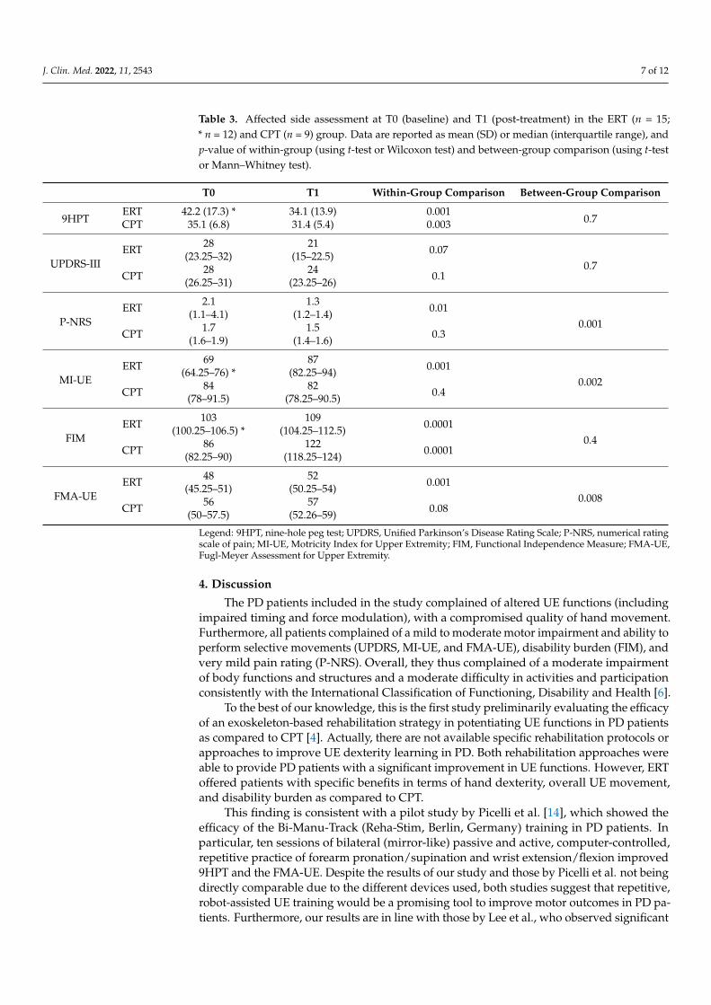
Table 3. Affected side assessment at T0 (baseline) and T1 (post-treatment) in the ERT (n = 15;* n = 12) and CPT (n = 9) group. Data are reported as mean (SD) or median (interquartile range), andp-value of within-group (using t-test or Wilcoxon test) and between-group comparison (using t-testor Mann–Whitney test).
T0 T1 Within-Group Comparison Between-Group Comparison
9HPTERT 42.2 (17.3) * 34.1 (13.9) 0.001
0.7CPT 35.1 (6.8) 31.4 (5.4) 0.003
UPDRS-IIIERT 28
(23.25–32)21
(15–22.5) 0.070.7
CPT 28(26.25–31)
24(23.25–26) 0.1
P-NRSERT 2.1
(1.1–4.1)1.3
(1.2–1.4) 0.010.001
CPT 1.7(1.6–1.9)
1.5(1.4–1.6) 0.3
MI-UEERT 69
(64.25–76) *87
(82.25–94) 0.0010.002
CPT 84(78–91.5)
82(78.25–90.5) 0.4
FIMERT 103
(100.25–106.5) *109
(104.25–112.5) 0.00010.4
CPT 86(82.25–90)
122(118.25–124) 0.0001
FMA-UEERT 48
(45.25–51)52
(50.25–54) 0.0010.008
CPT 56(50–57.5)
57(52.26–59) 0.08
Legend: 9HPT, nine-hole peg test; UPDRS, Unified Parkinson’s Disease Rating Scale; P-NRS, numerical ratingscale of pain; MI-UE, Motricity Index for Upper Extremity; FIM, Functional Independence Measure; FMA-UE,Fugl-Meyer Assessment for Upper Extremity.
4. Discussion
The PD patients included in the study complained of altered UE functions (includingimpaired timing and force modulation), with a compromised quality of hand movement.Furthermore, all patients complained of a mild to moderate motor impairment and ability toperform selective movements (UPDRS, MI-UE, and FMA-UE), disability burden (FIM), andvery mild pain rating (P-NRS). Overall, they thus complained of a moderate impairmentof body functions and structures and a moderate difficulty in activities and participationconsistently with the International Classification of Functioning, Disability and Health [6].
To the best of our knowledge, this is the first study preliminarily evaluating the efficacyof an exoskeleton-based rehabilitation strategy in potentiating UE functions in PD patientsas compared to CPT [4]. Actually, there are not available specific rehabilitation protocols orapproaches to improve UE dexterity learning in PD. Both rehabilitation approaches wereable to provide PD patients with a significant improvement in UE functions. However, ERToffered patients with specific benefits in terms of hand dexterity, overall UE movement,and disability burden as compared to CPT.
This finding is consistent with a pilot study by Picelli et al. [14], which showed theefficacy of the Bi-Manu-Track (Reha-Stim, Berlin, Germany) training in PD patients. Inparticular, ten sessions of bilateral (mirror-like) passive and active, computer-controlled,repetitive practice of forearm pronation/supination and wrist extension/flexion improved9HPT and the FMA-UE. Despite the results of our study and those by Picelli et al. not beingdirectly comparable due to the different devices used, both studies suggest that repetitive,robot-assisted UE training would be a promising tool to improve motor outcomes in PD pa-tients. Furthermore, our results are in line with those by Lee et al., who observed significant

J. Clin. Med. 2022, 11, 2543 8 of 12
improvements of UE fine and gross motor performance after several, repetitive treatmentsessions by constraint-induced movement therapy in twenty patients with PD [32].
The superiority of gravity-supporting device as compared to CPT in reducing theimpairment of, at least, some motor outcomes in PD patients is likely to depend on theintensive, repetitive, assisted-as-needed, and task-oriented motor practice provided bythe mechanical device [45]. It has been proposed that such an approach allows exploitingthe brain neural plasticity mechanisms both within affected and intact brain structuresthrough the stimulation of motor learning processes and a reshape of the “inter-hemisphericinhibition” mechanisms by means of repetitive sessions of robot-assisted therapy, as demon-strated by UE dexterity improvement in post-stroke patients [46–48]. This extends to thecase of basal ganglia impairment, where an intensive, continual, and contextual trainingallows facilitating the relearning of motor functions and minimizing motor deficit by actingon the internal regulator mechanisms of movement flow and programming [17,45,49]. Par-ticularly, the improvement in motor outcomes following ERT sessions could be consideredthe expression of a relearning process resulting from the stimulation and the activation ofthe mirror neuron system, inducing profound cortical and subcortical changes at both thecellular and synaptic levels [50]. The role of the cerebellum in motor and reinforcementlearning has been recognized. Recently, it has been shown that the cerebellum may have acompensatory and adaptive role concerning gait function recovery by favoring the precisetiming of motor actions along the gait cycle phases. This probably occurs by compensatingthe deficient internal timing clock within the basal ganglia [51]. These results have beenobtained in PD patients receiving gait training plus music (i.e., a repetitive exercise with anexternal cue). It is then hypothesizable that UL robotic rehabilitation with VR may haveboosted neural plasticity also at the cerebellar level, further improving motor recovery.
In addition, UE training benefitted from the adjunction of virtual reality treatment.This is thought to increase the motor learning of well-defined motor tasks and improvemotivation due to the feedback provided by the device itself [18–23,48–50,52–54]. Overall,these motor improvements allowed the patients to achieve a significant improvement inUE functions and disability burden. For instance, a patient was able to do a crochet workcarried out after a 25% of ERT sessions. Such activity was referred to as difficult to carryout for about two years by the patient.
Although we have not specifically investigated quality of life, it is possible that theimprovement in disability burden has also improved patients’ health-related quality of life,as suggested by other chronic diseases [55].
Strengths and Limitations
Our data suggest that UE training using a gravity-supporting device could be consid-ered a safe and effective approach for the recovery of UE motor functions in PD patients.Additionally, the rehabilitation technology is a cost-effective practice to reduce the needfor one-to-one skilled interventions. The training sessions can be performed with minimalsupervision once the therapist has set up the exercises. The attention of the patient istriggered by the visual and acoustic feedback while the exercise is performed. Moreover,the training is reproducible and essentially safe and can be easily conducted in the hos-pital setting. However, our study is limited by the small sample size (we were able toenroll only 30 patients compared to the estimated 60 because of the onset of COVID-19pandemic). This could have had relevant consequences on the results, considering that anintention-to-treat analysis was not performed. It is true that an intention-to-treat approachtends to under-estimate an effect, being thus a more conservative approach in a clinicalsuperiority trial. Nevertheless, a per-protocol approach is a reasonable analysis strategy forsensitivity analyses. Actually, a per-protocol approach is suitable when the exclusion ofpatients from the analysis due to major protocol deviations (which can of course cause atendency to wrong estimations of a treatment effect) tends to vary among the study groups.However, it is not straightforward to pre-guess the direction of a wrong estimation andthe general claim that a per-protocol analysis tends to overestimate an effect that cannot

J. Clin. Med. 2022, 11, 2543 9 of 12
be mathematically proven [56–58]. Studies with larger and homogeneous samples couldallow for a more accurate statistical analysis, including multivariate data analysis or moreadvanced tools, such as machine learning, aimed to point out predictive marker of recovery.
Another main limitation of our study is the lack of evaluation of long-term efficacy ofthe ERT (also due to COVID-19 pandemic restrictions). Nonetheless, previous studies haveshown a persistent efficacy of robotic rehabilitation after two weeks only for the 9HPT [15].Moreover, Taveggia et al. recorded a stability of motor assessment after 6 weeks from theend of treatment in stroke patients [59]; lastly, the improvement in the functional capacityoutcome measures were found at 2-month follow-up in multiple sclerosis patients [60].Last, one could criticize that the use of MI-UE is not usual in evaluating clinical impairmentin PD, compared to the FMA-UE [61]. In fact, MI-UE assesses the motor impairment in apatient who has had a stroke evaluating the ability of the patient to hold a cube againstgravity, the capacity to flex the elbow from 90◦ (so that the arm touches the shoulder) andthe capacity of the shoulder to abduct to more than 90◦ beyond the horizontal againstgravity. Rigidity in PD refers to increased muscular tone with more resistance than normalwhen the limb is passively moved, experienced by the patients as a sense of feeling stiffand uncomfortable. Then, even though the physiopathology of abnormal movements inPD are different from strokes [59], similar difficulties in achieving MI-UE goals are seen inPD patients. Then, further validation studies are needed to confirm the possibility to usethe scale even in Parkinsonism. Larger sample size RCTs are, notwithstanding, needed toaddress this concern.
5. Conclusions
Our data suggest that exoskeleton-assisted therapy, such as the Armeo©Spring, mayrepresent a safe and effective strategy for delivering a highly intensive and repetitivetraining, which is necessary to trigger the neuroplasticity mechanisms subtending UEmotor function improvement. Few studies are however available on the UE rehabilitationof patients with PD, particularly regarding technology-enhanced physical therapy, exceptfor gait training. Therefore, further investigations with larger sample sizes are needed toconfirm our results and to optimize PD-specific rehabilitation protocols.
Author Contributions: Conceptualization, R.S.C. and V.P.; methodology, A.N., V.P., R.S.C., L.R. andL.P.; software, L.P.; validation, D.L., M.I., B.P. and M.P.; formal analysis, A.N.; investigation, D.L.,M.I., B.P. and M.P.; resources, L.R.; data curation, A.N. and L.R.; writing—original draft preparation,L.R. and A.N.; writing—review and editing, A.N. and R.S.C.; visualization, D.L., M.I., B.P. andM.P.; supervision, R.S.C. and L.P. All authors have read and agreed to the published version ofthe manuscript.
Funding: This research received no external funding.
Institutional Review Board Statement: The study was conducted in accordance with the Declaration ofHelsinki and approved by the Ethics Committee of the IRCCS Centro Neurolesi Bonino Pulejo (Messina,Italy) (protocol code No. U0074917/11110) and was registered at ClinicalTrials.gov (NCT02721212).
Informed Consent Statement: Informed consent was obtained from all subjects involved in the study.
Data Availability Statement: Data could be requested on demand to the corresponding author.
Conflicts of Interest: The authors declare no conflict of interest.
References1. Ponsen, M.M.; Daffertshofer, A.; Wolters, E.C.; Beek, P.J.; Berendse, H.W. Impairment of complex upper limb motor function in de
novo Parkinson’s disease. Parkinsonism Relat. Disord. 2008, 14, 199–204. [CrossRef] [PubMed]2. Fitts, P.M. The information capacity of the human motor system in controlling the amplitude of movement. J. Exp. Psychol. 1954,
47, 381–391. [CrossRef] [PubMed]3. Sanes, J.N. Information processing deficits in Parkinson’s disease during movement. Neuropsychologia 1985, 23, 381–392. [CrossRef]4. Quinn, L.; Busse, M.; Dal Bello-Haas, V. Management of upper extremity dysfunction in people with Parkinson disease and
Huntington disease: Facilitating outcomes across the disease lifespan. J. Hand Ther. 2013, 26, 148–155. [CrossRef] [PubMed]

J. Clin. Med. 2022, 11, 2543 10 of 12
5. Rocchi, L.; Chiari, L.; Horak, F.B. Effects of deep brain stimulation and levodopa on postural sway in Parkinson’s disease.J. Neurol. Neurosurg. Psychiatry 2002, 73, 267–274. [CrossRef]
6. Raggi, A.; Leonardi, M.; Ajovalasit, D.; Carella, F.; Soliveri, P.; Albanese, A.; Romito, L. Disability and profiles of functioning ofpatients with Parkinson’s disease described with ICF classification. Int. J. Rehabil. Res. 2011, 34, 141–150. [CrossRef]
7. Diederich, N.J.; Moore, C.G.; Leurgans, S.E.; Chmura, T.A.; Goetz, C.G. Parkinson disease with old-age onset: A comparativestudy with subjects with middle-age onset. Arch. Neurol. 2003, 60, 529–533. [CrossRef]
8. Soh, S.E.; McGinley, J.L.; Watts, J.J.; Iansek, R.; Murphy, A.T.; Menz, H.B.; Huxham, F.; Morris, M.E. Determinants of health-relatedquality of life in people with Parkinson’s disease: A path analysis. Qual. Life Res. 2013, 22, 1543–1553. [CrossRef]
9. Berardelli, A.; Rothwell, J.C.; Thompson, P.D.; Hallett, M. Pathophysiology of bradykinesia in Parkinson’s disease. Brain 2001,124, 2131–2146. [CrossRef]
10. Espay, A.J.; Beaton, D.E.; Morgante, F.; Gunraj, C.A.; Lang, A.E.; Chen, R. Impairments of speed and amplitude of movement inParkinson’s disease: A pilot study. Mov. Disord. 2009, 24, 1001–1008. [CrossRef]
11. Hallett, M.; Khoshbin, S. A physiological mechanism of bradykinesia. Brain 1980, 103, 301–314. [CrossRef] [PubMed]12. Oliveira, M.A.; Rodrigues, A.M.; Caballero, R.M.; Petersen, R.D.; Shim, J.K. Strength and isometric torque control in individuals
with Parkinson’s disease. Exp. Brain Res. 2008, 184, 445–450. [CrossRef] [PubMed]13. Bologna, M.; Paparella, G.; Fasano, A.; Hallett, M.; Berardelli, A. Evolving concepts on bradykinesia. Brain 2020, 143, 727–750.
[CrossRef] [PubMed]14. Vercruysse, S.; Gilat, M.; Shine, J.M.; Heremans, E.; Lewis, S.; Nieuwboer, A. Freezing beyond gait in Parkinson’s disease: A
review of current neurobehavioral evidence. Neurosci. Biobehav. Rev. 2014, 43, 213–227. [CrossRef] [PubMed]15. Picelli, A.; Tamburin, S.; Passuello, M.; Waldner, A.; Smania, N. Robot-assisted arm training in patients with Parkinson’s disease:
A pilot study. J. Neuroeng. Rehabil. 2014, 11, 28. [CrossRef]16. Calabrò, R.S.; Cacciola, A.; Bertè, F.; Manuli, A.; Leo, A.; Bramanti, A.; Naro, A.; Milardi, D.; Bramanti, P. Robotic gait rehabilitation
and substitution devices in neurological disorders: Where are we now? Neurol. Sci. 2016, 37, 503–514. [CrossRef]17. Brognara, L.; Navarro-Flores, E.; Iachemet, L.; Serra-Catalá, N.; Cauli, O. Beneficial Effect of Foot Plantar Stimulation in Gait
Parameters in Individuals with Parkinson’s Disease. Brain Sci. 2020, 10, 69. [CrossRef]18. Mehrholz, J.; Hädrich, A.; Platz, T.; Kugler, J.; Pohl, M. Electromechanical and robot-assisted arm training for improving generic
activities of daily living, arm function, and arm muscle strength after stroke. Cochrane Database Syst. Rev. 2012, 6, CD006876.[CrossRef]
19. Bonanno, L.; Russo, M.; Bramanti, A.; Calabrò, R.S.; Marino, S. Functional connectivity in multiple sclerosis after roboticrehabilitative treatment: A case report. Medicine 2019, 98, e15047. [CrossRef]
20. Maggio, M.G.; Russo, M.; Cuzzola, M.F.; Destro, M.; Rosa, G.L.; Molonia, F.; Bramanti, P.; Lombardo, G.; Luca, R.D.; Calabrò, R.S.Virtual reality in multiple sclerosis rehabilitation: A review on cognitive and motor outcomes. J. Clin. Neurosci. 2019, 65, 106–111.[CrossRef]
21. Clark, W.E.; Sivan, M.; O’Connor, R.J. Evaluating the use of robotic and virtual reality rehabilitation technologies to improvefunction in stroke survivors: A narrative review. J. Rehabil. Assist. Technol. Eng. 2019, 6, 2055668319863557. [CrossRef]
22. Baur, K.; Schättin, A.; de Bruin, E.D.; Riener, R.; Duarte, J.E.; Wolf, P. Trends in robot-assisted and virtual reality-assistedneuromuscular therapy: A systematic review of health-related multiplayer games. J. Neuroeng. Rehabil. 2018, 15, 107. [CrossRef][PubMed]
23. Park, J.; Chung, Y. The effects of robot-assisted gait training using virtual reality and auditory stimulation on balance and gaitabilities in persons with stroke. NeuroRehabilitation 2018, 43, 227–235. [CrossRef]
24. Smania, N.; Picelli, A.; Geroin, C.; Munari, D.; Waldner, A.; Gandolfi, M. Robot-assisted gait training in patients with Parkinson’sdisease. Neurodegener. Dis. Manag. 2013, 3, 321–330. [CrossRef]
25. Capecci, M.; Pournajaf, S.; Galafate, D.; Sale, P.; Pera, D.L.; Goffredo, M.; Pandis, M.F.D.; Andrenelli, E.; Pennacchioni, M.;Ceravolo, M.G.; et al. Clinical effects of robot-assisted gait training and treadmill training for Parkinson’s disease. A randomizedcontrol. Trial. Ann. Phys. Rehabil. Med. 2019, 62, 303–312. [CrossRef]
26. Cifuentes, C.A.; Frizera, A. Human-Robot Interaction for Assisting. In Human-Robot Interaction Strategies for Walker-AssistedLocomotion; Locomotion, H., Ed.; Springer: Berlin/Heidelberg, Germany, 2016; pp. 17–31.
27. Scaletta, T.; Komada, S.; Oboe, R. Development of a human assistive robot to support hip joint movement during sit-to-standusing non-linear springs. IEEJ J. Ind. Appl. 2016, 5, 261–266. [CrossRef]
28. Huen, D.; Liu, J.; Lo, B. An integrated wearable robot for tremor suppression with context aware sensing. In Proceedings of the2016 IEEE 13th International Conference on Wearable and Implantable Body Sensor Networks (BSN), San Francisco, CA, USA,14–17 June 2016.
29. Voiculescu, I.; Cameron, S.; Zabarauskas, M.; Kozlowski, P. Towards Robot-Assisted Rehabilitation of Upper Limb dysfunction.In Advances in Robot Design and Intelligent Control: Advances in Intelligent Systems and Computing; Borangiu, T., Ed.; Springer:Berlin/Heidelberg, Germany, 2016; pp. 347–355.
30. Asakawa, T.; Sugiyama, K.; Nozaki, T.; Sameshima, T.; Kobayashi, S.; Wang, L.; Hong, Z.; Chen, S.; Li, C.; Namba, H. Can theLatest Computerized Technologies Revolutionize Conventional Assessment Tools and Therapies for a Neurological Disease? TheExample of Parkinson’s Disease. Neurol. Med.-Chir. 2019, 59, 69–78. [CrossRef]
J. Clin. Med. 2022, 11, 2543 11 of 12
31. Hughes, A.J.; Daniel, S.E.; Kilford, L.; Lees, A.J. Accuracy of clinical diagnosis of idiopathic Parkinson’s disease: A clinico-pathological study of 100 cases. J. Neurol. Neurosurg. Psychiatry 1992, 55, 181–184. [CrossRef]
32. Hoehn, M.M.; Yahr, M.D. Parkinsonism: Onset, progression and mortality. Neurology 1967, 17, 427–442. [CrossRef]33. Folstein, M.F.; Folstein, S.E.; McHugh, P.R. “Mini-mental state”. A practical method for grading the cognitive state of patients for
the clinician. J. Psychiatr. Res. 1975, 12, 189–198. [CrossRef]34. Movement Disorder Society Task Force on Rating Scales for Parkinson’s Disease. The Unified Parkinson’s Disease Rating Scale
(UPDRS): Status and recommendations. Mov. Disord. 2003, 18, 738–750. [CrossRef] [PubMed]35. Proud, E.L.; Bilney, B.; Miller, K.J.; Morris, M.E.; McGinley, J.L. Measuring Hand Dexterity in People with Parkinson’s Disease:
Reliability of Pegboard Tests. Am. J. Occup. Ther. 2019, 73, p1–p7304205050. [CrossRef] [PubMed]36. Colomer, C.; Baldoví, A.; Torromé, S.; Navarro, M.D.; Moliner, B.; Ferri, J.; Noé, E. Efficacy of Armeo® Spring during the chronic
phase of stroke. Study Mild Moderate Cases Hemiparesis Neurol. 2013, 28, 261–267. [CrossRef]37. Oxford Grice, K.; Vogel, K.A.; Le, V.; Mitchell, A.; Muniz, S.; Vollmer, M.A. Adult norms for a commercially available Nine Hole
Peg Test for finger dexterity. Am. J. Occup. Ther. 2003, 57, 570–573. [CrossRef] [PubMed]38. Raciti, L.; Nicoletti, A.; Mostile, G.; Bonomo, R.; Contrafatto, D.; Dibilio, V.; Luca, A.; Sciacca, G.; Cicero, C.E.; Vasta, R.; et al.
Validation of the UPDRS section IV for detection of motor fluctuations in Parkinson’s disease. Parkinsonism Relat. Disord. 2016,27, 98–101. [CrossRef]
39. Fugl-Meyer, A.R.; Jääskö, L.; Leyman, I.; Olsson, S.; Steglind, S. The post-stroke hemiplegic patient. 1. a method for evaluation ofphysical performance. Scand. J. Rehabil. Med. 1975, 7, 13–31. [PubMed]
40. Opara, J.; Małecki, A.; Małecka, E.; Socha, T. Motor assessment in Parkinson‘s disease. Ann. Agric. Environ. Med. 2017, 24, 411–415.[CrossRef]
41. Lee, K.S.; Lee, W.H.; Hwang, S. Modified constraint-induced movement therapy improves fine and gross motor performance ofthe upper limb in Parkinson disease. Am. J. Phys. Med. Rehabil. 2011, 90, 380–386. [CrossRef]
42. Keith, R.A.; Granger, C.V.; Hamilton, B.B.; Sherwin, F.S. The functional independence measure: A new tool for rehabilitation. Adv.Clin. Rehabil. 1987, 1, 6–18.
43. Childs, J.D.; Piva, S.R.; Fritz, J.M. Responsiveness of the numeric pain rating scale in patients with low back pain. Spine 2005,30, 1331–1334. [CrossRef]
44. Demeurisse, G.; Demol, O.; Robaye, E. Motor evaluation in vascular hemiplegia. Eur. Neurol. 1980, 19, 382–389. [CrossRef][PubMed]
45. Calabrò, R.S.; Naro, A.; Russo, M.; Leo, A.; Luca, R.D.; Balletta, T.; Buda, A.; Rosa, G.L.; Bramanti, A.; Bramanti, P. The role ofvirtual reality in improving motor performance as revealed by EEG: A randomized clinical trial. J. Neuroeng. Rehabil. 2017, 14, 53.[CrossRef] [PubMed]
46. Raciti, L.; Pizzurro, R.; Occhipinti, F.; Manuli, A.; Corallo, F.; Calabrò, R.S. A multidisciplinary advanced approach in centralpontine myelinolysis recovery: Considerations about a case report. Disabil. Rehabil. Assist. Technol. 2020, 1–12. [CrossRef][PubMed]
47. Maggio, M.G.; Torrisi, M.; Buda, A.; Luca, R.D.; Piazzitta, D.; Cannavò, A.; Leo, A.; Milardi, D.; Manuli, A.; Calabro, R.S. Effectsof robotic neurorehabilitation through lokomat plus virtual reality on cognitive function in patients with traumatic brain injury:A retrospective case-control study. Int. J. Neurosci. 2020, 130, 117–123. [CrossRef]
48. Calabrò, R.S.; Russo, M.; Naro, A.; Milardi, D.; Balletta, T.; Leo, A.; Filoni, S.; Bramanti, P. Who May Benefit from Armeo PowerTreatment? A Neurophysiological Approach to Predict Neurorehabilitation Outcomes. PM R. 2016, 8, 971–978. [CrossRef]
49. Bruni, M.F.; Melegari, C.; De Cola, M.C.; Bramanti, A.; Bramanti, P.; Calabrò, R.S. What does best evidence tell us about roboticgait rehabilitation in stroke patients: A systematic review and meta-analysis. J. Clin. Neurosci. 2018, 48, 11–17. [CrossRef]
50. Moon, S.; Huang, C.K.; Sadeghi, M.; Akinwuntan, A.E.; Devos, H. Proof-of-Concept of the Virtual Reality Comprehensive BalanceAssessment and Training for Sensory Organization of Dynamic Postural Control. Front. Bioeng. Biotechnol. 2021, 9, 678006.[CrossRef]
51. Naro, A.; Pignolo, L.; Bruschetta, D.; Calabrò, R.S. What about the role of the cerebellum in music-associated functional recovery?A secondary EEG analysis of a randomized clinical trial in patients with Parkinson disease. Parkinsonism Relat. Disord. 2022,96, 57–64. [CrossRef]
52. Straudi, S.; Benedetti, M.G.; Venturini, E.; Manca, M.; Foti, C.; Basaglia, N. Does robot-assisted gait training ameliorate gaitabnormalities in multiple sclerosis? A pilot randomized-control trial. NeuroRehabilitation 2013, 33, 555–563. [CrossRef]
53. Masiero, S.; Armani, M.; Rosati, G. Upper-limb robot-assisted therapy in rehabilitation of acute stroke patients: Focused reviewand results of new randomized controlled trial. J. Rehabil. Res. Dev. 2011, 48, 355–366. [CrossRef]
54. Maggio, M.G.; Luca, R.D.; Manuli, A.; Buda, A.; Cuzzola, M.F.; Leonardi, S.; D’Aleo, G.; Bramanti, P.; Russo, M.; Calabrò, R.S. Dopatients with multiple sclerosis benefit from semi-immersive virtual reality? A randomized clinical trial on cognitive and motoroutcomes. Appl. Neuropsychol. Adult 2022, 29, 59–65. [CrossRef]
55. López-López, D.; Pérez-Ríos, M.; Ruano-Ravina, A.; Losa-Iglesias, M.E.; Becerro-de-Bengoa-Vallejo, R.; Romero-Morales, C.;Calvo-Lobo, C.; Navarro-Flores, E. Impact of quality of life related to foot problems: A case-control study. Sci. Rep. 2021, 11,14515. [CrossRef]
56. Mazzoni, P.; Shabbott, B.; Cortés, J.C. Motor control abnormalities in Parkinson’s disease. Cold Spring Harb. Perspect. Med. 2012,2, a009282. [CrossRef]

J. Clin. Med. 2022, 11, 2543 12 of 12
57. Intention to treat analysis and per protocol analysis: Complementary information. Prescrire Int. 2012, 21, 304–306.58. Tripepi, G.; Chesnaye, N.C.; Dekker, F.W.; Zoccali, C.; Jager, K.J. Intention to treat and per protocol analysis in clinical trials.
Nephrology 2020, 25, 513–517. [CrossRef]59. Taveggia, G.; Borboni, A.; Salvi, L.; Mule, C.; Fogliaresi, S.; Villafañe, J.H.; Casale, R. Efficacy of robot-assisted rehabilitation for
the functional recovery of the upper limb in post-stroke patients: A randomized controlled study. Eur. J. Phys. Rehabil. Med. 2016,52, 767–773.
60. Gijbels, D.; Lamers, I.; Kerkhofs, L.; Alders, G.; Knippenberg, E.; Feys, P. The Armeo Spring as training tool to improve upperlimb functionality in multiple sclerosis: A pilot study. J. Neuroeng. Rehabil. 2011, 8, 5. [CrossRef]
61. Ishikuro, K.; Dougu, N.; Nukui, T.; Yamamoto, M.; Nakatsuji, Y.; Kuroda, S.; Matsushita, I.; Nishimaru, H.; Araujo, M.F.P.; Nishijo,H. Effects of Transcranial Direct Current Stimulation (tDCS) Over the Frontal Polar Area on Motor and Executive Functions inParkinson’s Disease; A Pilot Study. Front. Aging Neurosci. 2018, 10, 231. [CrossRef]
Copyright © 2022 FDOKUMEN