A Mitocentric View of Parkinson's Disease
26
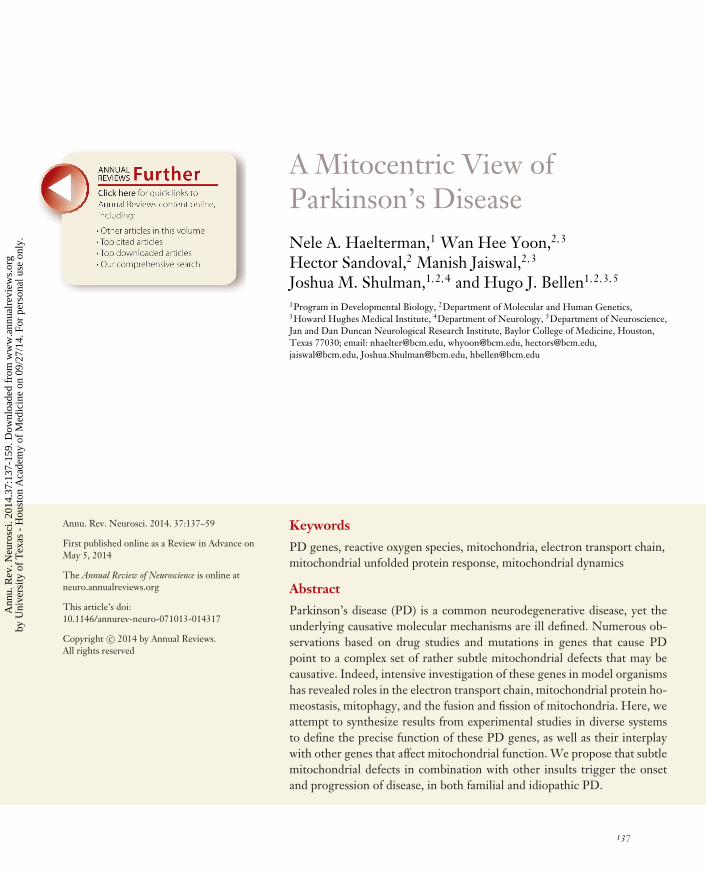
A Mitocentric View of Parkinson’s Disease Nele A. Haelterman, 1 Wan Hee Yoon, 2, 3 Hector Sandoval, 2 Manish Jaiswal, 2, 3 Joshua M. Shulman, 1, 2, 4 and Hugo J. Bellen 1, 2, 3, 5 1 Program in Developmental Biology, 2 Department of Molecular and Human Genetics, 3 Howard Hughes Medical Institute, 4 Department of Neurology, 5 Department of Neuroscience, Jan and Dan Duncan Neurological Research Institute, Baylor College of Medicine, Houston, Texas 77030; email: [email protected], [email protected], [email protected], [email protected], [email protected], [email protected] Annu. Rev. Neurosci. 2014. 37:137–59 First published online as a Review in Advance on May 5, 2014 The Annual Review of Neuroscience is online at neuro.annualreviews.org This article’s doi: 10.1146/annurev-neuro-071013-014317 Copyright c 2014 by Annual Reviews. All rights reserved Keywords PD genes, reactive oxygen species, mitochondria, electron transport chain, mitochondrial unfolded protein response, mitochondrial dynamics Abstract Parkinson’s disease (PD) is a common neurodegenerative disease, yet the underlying causative molecular mechanisms are ill defined. Numerous ob- servations based on drug studies and mutations in genes that cause PD point to a complex set of rather subtle mitochondrial defects that may be causative. Indeed, intensive investigation of these genes in model organisms has revealed roles in the electron transport chain, mitochondrial protein ho- meostasis, mitophagy, and the fusion and fission of mitochondria. Here, we attempt to synthesize results from experimental studies in diverse systems to define the precise function of these PD genes, as well as their interplay with other genes that affect mitochondrial function. We propose that subtle mitochondrial defects in combination with other insults trigger the onset and progression of disease, in both familial and idiopathic PD. 137 Annu. Rev. Neurosci. 2014.37:137-159. Downloaded from www.annualreviews.org by University of Texas - Houston Academy of Medicine on 09/27/14. For personal use only.
Transcript of A Mitocentric View of Parkinson's Disease
NE37CH08-Bellen ARI 26 May 2014 7:38
A Mitocentric View ofParkinson’s DiseaseNele A. Haelterman,1 Wan Hee Yoon,2,3
Hector Sandoval,2 Manish Jaiswal,2,3
Joshua M. Shulman,1,2,4 and Hugo J. Bellen1,2,3,5
1Program in Developmental Biology, 2Department of Molecular and Human Genetics,3Howard Hughes Medical Institute, 4Department of Neurology, 5Department of Neuroscience,Jan and Dan Duncan Neurological Research Institute, Baylor College of Medicine, Houston,Texas 77030; email: [email protected], [email protected], [email protected],[email protected], [email protected], [email protected]
Annu. Rev. Neurosci. 2014. 37:137–59
First published online as a Review in Advance onMay 5, 2014
The Annual Review of Neuroscience is online atneuro.annualreviews.org
This article’s doi:10.1146/annurev-neuro-071013-014317
Copyright c© 2014 by Annual Reviews.All rights reserved
Keywords
PD genes, reactive oxygen species, mitochondria, electron transport chain,mitochondrial unfolded protein response, mitochondrial dynamics
Abstract
Parkinson’s disease (PD) is a common neurodegenerative disease, yet theunderlying causative molecular mechanisms are ill defined. Numerous ob-servations based on drug studies and mutations in genes that cause PDpoint to a complex set of rather subtle mitochondrial defects that may becausative. Indeed, intensive investigation of these genes in model organismshas revealed roles in the electron transport chain, mitochondrial protein ho-meostasis, mitophagy, and the fusion and fission of mitochondria. Here, weattempt to synthesize results from experimental studies in diverse systemsto define the precise function of these PD genes, as well as their interplaywith other genes that affect mitochondrial function. We propose that subtlemitochondrial defects in combination with other insults trigger the onsetand progression of disease, in both familial and idiopathic PD.
137
Ann
u. R
ev. N
euro
sci.
2014
.37:
137-
159.
Dow
nloa
ded
from
ww
w.a
nnua
lrev
iew
s.or
gby
Uni
vers
ity o
f T
exas
- H
oust
on A
cade
my
of M
edic
ine
on 0
9/27
/14.
For
per
sona
l use
onl
y.
NE37CH08-Bellen ARI 26 May 2014 7:38
PD: Parkinson’sdisease
Substantia nigra(SN): pigmentedregion of the midbrain(due to melanin)containingdopaminergic neurons;participates with otherbasal ganglia nuclei inmovement control
Ubiquitin-proteasome system:the system that tagsmisfolded or damagedproteins with ubiquitinand degrades them inthe proteasome
Contents
INTRODUCTION . . . . . . . . . . . . . . . . . . . . . . . . . . . . . . . . . . . . . . . . . . . . . . . . . . . . . . . . . . . . . . . 138MITOCHONDRIAL DYSFUNCTION UNDERLIES
PARKINSON’S DISEASE . . . . . . . . . . . . . . . . . . . . . . . . . . . . . . . . . . . . . . . . . . . . . . . . . . . . . 142The Environmental Link . . . . . . . . . . . . . . . . . . . . . . . . . . . . . . . . . . . . . . . . . . . . . . . . . . . . . . . 142The Genetic Link . . . . . . . . . . . . . . . . . . . . . . . . . . . . . . . . . . . . . . . . . . . . . . . . . . . . . . . . . . . . . . 142
PROTEINS ENCODED BY PD LOCI AFFECT DIVERSEMITOCHONDRIAL FUNCTIONS . . . . . . . . . . . . . . . . . . . . . . . . . . . . . . . . . . . . . . . . . . . 143Deficits in the Electron Transport Chain . . . . . . . . . . . . . . . . . . . . . . . . . . . . . . . . . . . . . . . . 144Defects in Protein Homeostasis: The Mitochondrial Unfolded Protein Response . . 146Defects in Mitochondrial Dynamics . . . . . . . . . . . . . . . . . . . . . . . . . . . . . . . . . . . . . . . . . . . . . 147
MITOCHONDRIA AND PD: AN INTEGRATED MODEL. . . . . . . . . . . . . . . . . . . . . . 150
INTRODUCTION
Parkinson’s disease (PD) is a progressive and incurable neurodegenerative disorder affecting 1%of the older adult population. The defining motor characteristics of PD, known collectively asparkinsonism, include tremor, increased muscle tone, slow movements, and impaired gait andbalance (Fahn 2003, Lees et al. 2009). In autopsy studies, PD is characterized by the loss ofdopamine (DA) neurons in the midbrain substantia nigra (SN) in association with α-synucleinprotein aggregates, termed Lewy bodies (Goedert et al. 2013, Jellinger 2009). The SN is thesource of DA for other basal ganglia nuclei, which have an essential role in initiating and facilitatingmovement. In addition, a range of nonmotor manifestations are observed (Chaudhuri & Schapira2009), including neuropsychiatric symptoms (e.g., depression, cognitive impairment), autonomicnervous system dysfunction, sleep disturbance, pain, and constipation, which likely relate to morewidespread synuclein pathology and neurodegeneration throughout the nervous system.
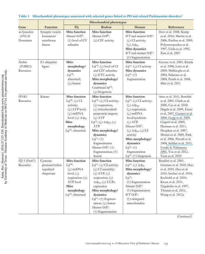
Although PD pathogenesis has been studied extensively, the primary cause(s) remain(s) elu-sive. Alterations in numerous cellular processes have been implicated, including oxidative stress(Fariello 1988), excitotoxicity (Olney et al. 1990), the ubiquitin-proteasome system (Lowe et al.1988), the endolysosomal compartment, and mitochondrial dysfunction (Schapira et al. 1989). Allthese may be directly or indirectly related to mechanisms of neurodegeneration in PD. However,mitochondrial toxins potently induce SN degeneration, and genes that have been linked to familialPD and related disorders are increasingly being shown to affect mitochondrial function (Table 1).Based on recent studies, we focus here on a mitocentric view of PD, suggesting that mitochondrialdysfunction is in fact a primary cause of PD.
Mitochondria are double-walled, dynamic, filamentous organelles that constitute the cell’s ma-jor source of adenosine triphosphate (ATP) production, the chemical energy of the cell. Withinmitochondria, ATP is produced by the citric acid cycle (Krebs cycle) in the matrix via the actionof four respiratory complexes (CI, CII, CIII, CIV) and ATP synthase in the mitochondrial innermembrane (MIM) (Figure 1). Mitochondrial mechanisms have been implicated in diverse hu-man disorders (Vafai & Mootha 2012). A severe impairment of mitochondrial function typicallyhas dramatic consequences. For instance, mutations in genes encoding subunits of the respi-ratory complexes lead to Leigh syndrome, a fatal multisystem disorder of childhood onset. Incontrast, more subtle mitochondrial impairment is associated with comparatively insidious onsetand heterogeneous clinical manifestations, including cancer, diabetes, cardiomyopathy, anemia,
138 Haelterman et al.
Ann
u. R
ev. N
euro
sci.
2014
.37:
137-
159.
Dow
nloa
ded
from
ww
w.a
nnua
lrev
iew
s.or
gby
Uni
vers
ity o
f T
exas
- H
oust
on A
cade
my
of M
edic
ine
on 0
9/27
/14.
For
per
sona
l use
onl
y.
NE37CH08-Bellen ARI 26 May 2014 7:38
Table 1 Mitochondrial phenotypes associated with selected genes linked to PD and related Parkinsonian disordersa
Mitochondrial phenotypes
Gene Function Fly Rodent Human Referencesα-Synuclein(SNCA)
Dominant
Synaptic vesicleformation/membranefusion
Mito functionMutant O/Eb:(↑) level of CVsubunits
Mito functionMutant O/Eb:(↓) CIV activity
Mito functionWT and mutant O/Ec:(↓) CI activity;(↓) �ψm
Mito dynamicsWT and mutant O/Ec:(↑) fragmentation
Devi et al. 2008, Kampet al. 2010, Martin et al.2006, Parihar et al. 2009,Polymeropoulos et al.1997, Ueda et al. 1993,Xun et al. 2007
Parkin(PARK2)
Recessive
E3 ubiquitinligase
Mitomorphology/dynamicsLof d:abnormal;(↓) fission
Mito functionLof d: (↓) level of CIand CIV subunits;(↓) ETC activity
Mito morphology/dynamics
Conditional lof d:(↓) biogenesis
Mito functionLof e: (↓) CI activityMito dynamicsLof e: (↑)fragmentation
Greene et al. 2003, Kitadaet al. 1998, Lutz et al.2009, Muftuoglu et al.2004, Palacino et al.2004, Pesah et al. 2004,Shin et al. 2011
PINK1Recessive
Kinase Mito functionLof d: (↓) CIactivity;(↓) ATP level;(↓) mtDNAlevel; (↓) �ψm
Mitomorphology
Lof d: abnormal
Mito functionLof d: (↓) CI activity;(↓) respiration;(↓) mitochondrialpreprotein import;(↓) ATP
Lof e: (↓) �ψm; (↓)ATP
Mito morphology/dynamics
Lof e: (↑)fragmentation
Mutant O/Ec: (↑)fragmentation; (↑)fission
Mito functionLof e: (↓) CI activity;(↓) �ψm;(↓) respiration;(↓) mtDNAlevel/synthesis;(↓) ATP
Mutant O/Ec:(↓) �ψm; (↓) CIactivity
Mito morphology/dynamics
Lof e: (↑)fragmentation
Lof e: (↑) biogenesis
Amo et al. 2011, Bonifatiet al. 2005, Clark et al.2006, Cui et al. 2010,Dagda et al. 2009, Exneret al. 2007, Gautier et al.2008, Gegg et al. 2009,Gispert et al. 2009,Heeman et al. 2011,Hoepken et al. 2007,Morais et al. 2009, Parket al. 2006, Piccoli et al.2008, Seibler et al. 2011,Unoki & Nakamura2001, Vos et al. 2012,Yuan et al. 2010
DJ-1 (Park7)Recessive
Cysteineprotease/redox-regulatedchaperone
Mito functionLof d:(↓) mtDNAlevel; (↓)respiration; (↓)ATP level
Mitomorphology
Lof d: abnormal
Mito functionLof e: (↓) CI activity;(↓) CI assembly;(↓) ATP; (↓)respiration; (↓)�ψm; (↓) UCPsexpression
Mito morphology/dynamics
Lof e: (↑) fragmen-tation; (↓) fusion
Mutant O/Ec:(↑) fragmentation
Mito functionLof e: (↓) �ψm
Mito morphology/dynamics
Lof e:(↑) fragmentation
Mutant O/Ec:(↑) fragmentation
WT O/Ec:(↑) elongatedmitochondria
Bonifati et al. 2003,Guzman et al. 2010, Haoet al. 2010, Heo et al.2010, Irrcher et al. 2010,Krebiehl et al. 2010,Kwon et al. 2011,Nagakubo et al. 1997,Thomas et al. 2011,Wang et al. 2012a
(Continued )
www.annualreviews.org • A Mitocentric View of Parkinson’s Disease 139
Ann
u. R
ev. N
euro
sci.
2014
.37:
137-
159.
Dow
nloa
ded
from
ww
w.a
nnua
lrev
iew
s.or
gby
Uni
vers
ity o
f T
exas
- H
oust
on A
cade
my
of M
edic
ine
on 0
9/27
/14.
For
per
sona
l use
onl
y.
NE37CH08-Bellen ARI 26 May 2014 7:38
Table 1 (Continued )
Mitochondrial phenotypes
Gene Function Fly Rodent Human ReferencesLRRK2Dominant
Kinase/GTPaseactivity
Mitomorphology
Mutant O/Eb:abnormal
Mito functionsMutant O/Ec:(↓) �ψm
Mito morphology/dynamics
WT and mutant O/Ec:(↑) fission
Mito functionLof e: (↓) �ψm;(↓) ATP; (↓) CI,CII, CIV; (↑) UCPsexpression
Mutant O/Ec:(↓) �ψm; (↑) UCPsexpression
Mito morphology/dynamics
Mutant O/Ec:(↑) fragmentation
Cherra et al. 2013,Mortiboys et al. 2010,Niu et al. 2012,Paisan-Ruız et al. 2004,Papkovskaia et al. 2012,Wang et al. 2012b
ATP13A2Recessive
LysosomalATPase
NA Mito morphologyLof e:(↑) fragmentation
Mito functionLof e: (↓) ATP;(↑) mtDNA level;(↑) oxygenconsumption rate
Mito morphologyLof e:(↑) fragmentation
Grunewald et al. 2012,Ramirez et al. 2006,Ramonet et al. 2012,Schultheis et al. 2004
FBXO7Recessive
E3 ubiquitinprotein ligasesubunit
NA NA Mito functionLof e: (↓) Parkinrecruitment tomitochondria
Burchell et al. 2013, Ilyinet al. 2000, Shojaee et al.2008
Vps35Dominant
Subunit of theretromercomplex
NA NA Mito functionLof e: (↓) delivery ofMAPL frommitochondria toperoxisomes
Braschi et al. 2010, Edgar& Polak 2000,Vilarino-Guell et al.2011
aAbbreviations: CI, complex I; CIV, complex IV; CV, complex V; ETC, electron transport chain; Gof, gain-of-function; Lof, loss-of-function; MAPL,mitochondrial-anchored protein ligase; mtDNA, mitochondrial DNA; NA, not available; O/E, overexpression; UCPs, mitochondrial uncoupling proteins;WT, wild type; �ψm, mitochondrial membrane potential.bOverexpression in vivo animal model.cOverexpression in vitro cell culture.dLoss of function in vivo animal model.eLoss of function in vitro cell culture.
Complex I (CI):large protein complex,located in the innermitochondrialmembrane, thatoxidizes nicotinamideadenine dinucleotide(NADH) to transferelectrons from NADHto ubiquinone
and neurodegeneration (Schapira 2012, Schon & Przedborski 2011). In fact, numerous studieshave documented a mitochondrial complex I (CI) deficiency in the SN of PD patients (Keeneyet al. 2006, Mizuno et al. 1989, Parker et al. 1989, Schapira et al. 1989). Moreover, a recent reportsimilarly implicated early dysregulation in the mitochondrial electron transport chain (ETC), onthe basis of transcriptional profiling of the SN from PD patients (Zheng et al. 2010).
Below, we first address the environmental and genetic evidence that links the etiology of PDto mitochondria. Next, we address how the many genes implicated in PD affect distinct mito-chondrial functions such as the ETC, the mitochondrial unfolded protein response (UPRmt), andmitochondrial dynamics. In the final section, we describe models that explain the relative vulner-ability of certain cell types, such as SN DA neurons, despite a global mitochondrial dysfunction,
140 Haelterman et al.
Ann
u. R
ev. N
euro
sci.
2014
.37:
137-
159.
Dow
nloa
ded
from
ww
w.a
nnua
lrev
iew
s.or
gby
Uni
vers
ity o
f T
exas
- H
oust
on A
cade
my
of M
edic
ine
on 0
9/27
/14.
For
per
sona
l use
onl
y.
NE37CH08-Bellen ARI 26 May 2014 7:38
P
PTrap1
Mortalin
DJ-1Fusion
Cardiolipin
ETC
ROS
Mfn
Ub
Rad6
Parkin
Ub
MM
P
Unfolded oroxidized proteins
V
Fission
Transport
Parkin
Pink1
UPS
Bioenergetics
UPRmt
Protein transport
Mito dynamics
PD locus
Protein function
α-syn
Δ MMP
α-syn
Drp1
Drp1
Mfn
Miro
Milton Kin
esi
nh
c
Microtubules
mTORC2
trc
UCPs Miro
Pink1
LRRK2
IV
II
IIIIIIIII
ATP
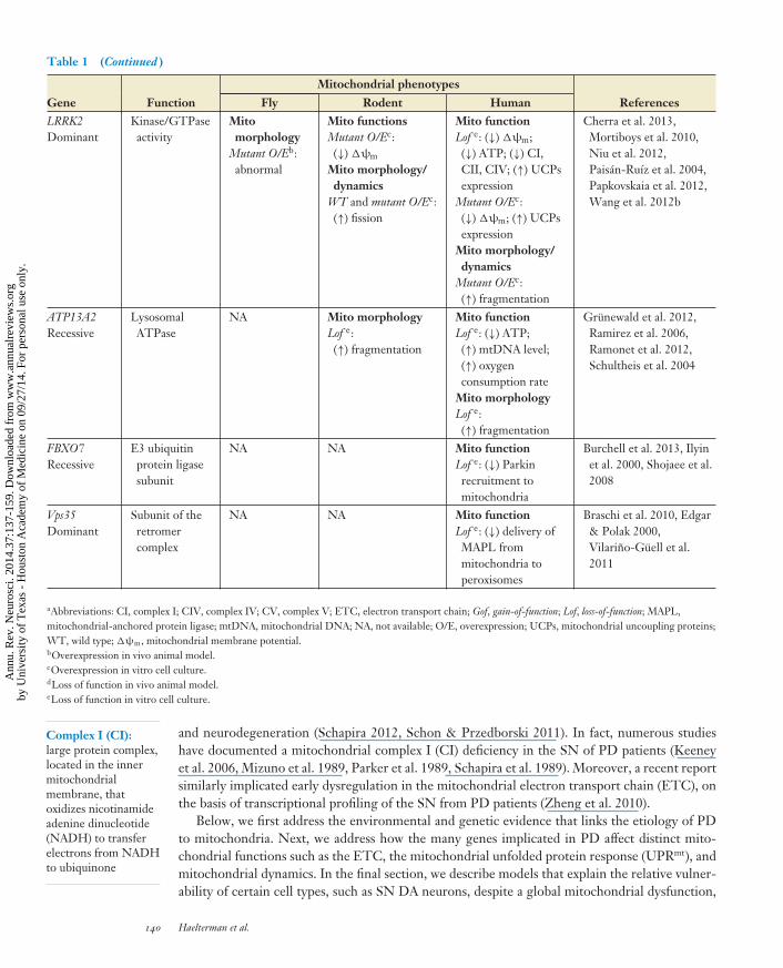
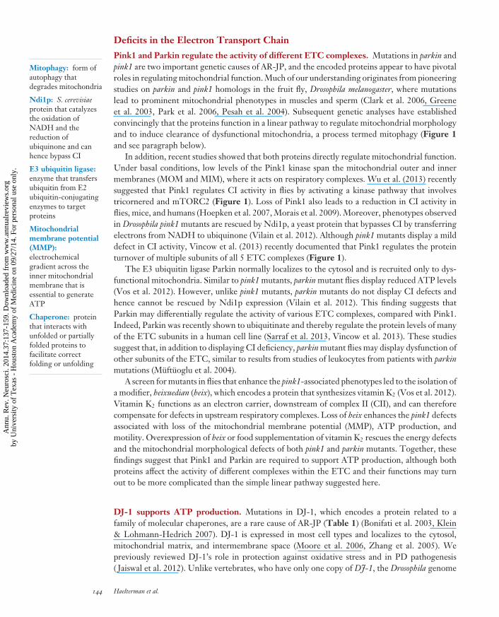
Figure 1Proteins implicated in Parkinson’s disease (PD) maintain a healthy pool of mitochondria. The effects of PD-linked proteins (thick greenline) on mitochondrial function can be divided into three different groups. Bioenergetics (yellow): Pink1 and Parkin regulate theturnover of several subunits of ETC complexes by the ubiquitin proteasome system (UPS). In addition, Pink1 supports complex Iactivity through a kinase cascade involving tricornered (trc) and mTORC2. Mitochondrially localized α-synuclein (α-syn), however,inhibits complex I. Oxidation of the mitochondrial phospholipid cardiolipin translocates this lipid from the inner to the outermitochondrial membrane where it serves as a receptor for α-synuclein. Hence, in the presence of elevated levels of reactive oxygenspecies (ROS), mitochondrial α-synuclein import is increased. LRRK2 and DJ-1 regulate the activity of uncoupling proteins (UCPs),which preserve the mitochondrial membrane potential (MMP). Both PD proteins therefore indirectly maintain ATP production.Mitochondrial unfolded protein response (UPRmt, blue): When activated, Pink1 phosphorylates the chaperone Trap1, a protein thatprotects mitochondrial proteins from ROS. In addition, Trap1 works with Hsp60 to refold imported proteins, sustaining mitochondrialprotein homeostasis. In the presence of ROS, DJ-1 is translocated into mitochondria, where it interacts with the chaperone Mortalin tosalvage oxidized proteins. Mitochondrial dynamics (orange): LRRK2 directs mitochondrial fission as it interacts with and recruits themitochondrial fission protein Drp1. DJ-1 increases mitochondrial fission by regulating Drp1 levels, although the exact mechanismthrough which the chaperone executes this function is not clear. The E3 ubiquitin ligase Parkin inhibits mitochondrial fusion throughubiquitin-mediated degradation of the fusion protein Mitofusin (Mfn). Mitochondrial transport is regulated by the Pink1/Parkinpathway. Here, Parkin-mediated degradation of the adaptor protein Miro is thought to detach a dysfunctional mitochondrion fromkinesin motor proteins, halting its transport. Finally, when a dysfunctional mitochondrion cannot be repaired, it is cleared throughmitophagy. Under basal conditions, the protein kinase Pink1 is imported into the mitochondrial intermembrane space, where it iscleaved by 2 proteases and subsequently degraded. However, upon mitigation of the MMP, Pink1 is no longer cleaved. The proteinthen phosphorylates and activates the E3 ubiquitin ligase Parkin. Parkin, along with E2 ligases such as Rad6, initiates mitophagy byubiquitinating target proteins.
www.annualreviews.org • A Mitocentric View of Parkinson’s Disease 141
Ann
u. R
ev. N
euro
sci.
2014
.37:
137-
159.
Dow
nloa
ded
from
ww
w.a
nnua
lrev
iew
s.or
gby
Uni
vers
ity o
f T
exas
- H
oust
on A
cade
my
of M
edic
ine
on 0
9/27
/14.
For
per
sona
l use
onl
y.
NE37CH08-Bellen ARI 26 May 2014 7:38
Electron transportchain (ETC): 5protein complexes thatproduce ATP via anelectrochemicalproton gradient acrossthe innermitochondrialmembrane by couplingelectron transferbetween electrondonors and acceptors
Mitochondrialunfolded proteinresponse (UPRmt):mitochondrial stressresponse to anaccumulation ofunfolded or misfoldedproteins in the matrixof mitochondria
Autosomal recessivejuvenileparkinsonism(AR-JP): familialform of early-onsetparkinsonism, usuallycharacterized atautopsy by substantianigra degeneration inthe absence of Lewybodies
and we further attempt to generalize lessons from rare familial forms of PD to the more commonsporadic disease.
MITOCHONDRIAL DYSFUNCTION UNDERLIESPARKINSON’S DISEASE
The Environmental Link
A wave of interest in a potential role for mitochondria in PD came in 1976, when investigatorsfound that drug users who took opioid analogs (MPPP) laced with 1-methyl-4-phenyl-1,2,3,6-tetrahydropyridine (MPTP) developed PD-like motor symptoms resulting from degenerationof DA neurons in the SN (Langston et al. 1983, Martinez & Greenamyre 2012). Subsequentinvestigations showed that MPTP is readily converted into MPP+, which crosses the blood brainbarrier and is selectively imported in DA neurons (Nicklas et al. 1985). MPP+ binds to CI andinhibits electron transport, reducing ATP production and increasing reactive oxygen species (ROS)levels (Nicklas et al. 1985). Together, these observations suggested that SN degeneration andsubsequent motor manifestations in PD may similarly result from mitochondrial dysfunction.This hypothesis gained further support when CI defects were documented in postmortem SNtissue from PD patients (Schapira et al. 1989).
Following on these initial findings, a variety of pesticides and toxins have been associatedwith SN degeneration in animal models (Martinez & Greenamyre 2012), and evidence fromhuman epidemiologic studies suggests that such exposure may in fact increase PD risk (Tanneret al. 2011). Several of the implicated toxins were shown to inhibit mitochondrial function,and such compounds have facilitated the development of useful animal model systems for PDstudies (Martinez & Greenamyre 2012). For instance, the widely used herbicide paraquat, whichis structurally similar to MPTP, is a potent redox cycler that accepts electrons from CI. Thisprocess converts the molecule to a free radical, which interacts with O2 to generate superoxideanions and other ROS. Similarly, the naturally occurring pesticide rotenone systemically inhibitsCI and leads to the degeneration of DA neurons in the SN. Finally, the fungicide maneb containsthe mitotoxin manganese ethylene-bisdithiocarbamate, which preferentially inhibits complex IIIof the ETC and causes proteasomal inhibition, oxidative stress, and cytoplasmic α-synucleinaggregation in DA neuron cell lines (Zhou et al. 2004).
In summary, compelling data from animal models coupled with evidence from epidemio-logic studies have led to the hypothesis that environmental exposure to mitochondrial toxins maybe an important contributor to PD in the population (Martinez & Greenamyre 2012). Largemeta-analyses suggest an approximately twofold increased risk of PD due to pesticide exposure(Priyadarshi et al. 2000). Thus additional environmental and/or genetic insults, in combinationwith aging, would likely be required to trigger PD in most affected individuals.
The Genetic Link
Rapid advances in genetics over the past two decades have transformed our thinking about PDfrom a primarily environmentally influenced disorder to one with a substantial genetic contribution(Shulman et al. 2011, Trinh & Farrer 2013). In particular, many of the genes responsible for familialforms of PD and related disorders have strong links to mitochondrial function. In 1998, mutationsin the parkin gene were discovered to cause autosomal recessive, juvenile parkinsonism (AR-JP)(Kitada et al. 1998), and subsequent work has proven parkin to be the most important cause ofPD in children and young adults (Lucking et al. 2000, Periquet et al. 2003). As detailed below,
142 Haelterman et al.
Ann
u. R
ev. N
euro
sci.
2014
.37:
137-
159.
Dow
nloa
ded
from
ww
w.a
nnua
lrev
iew
s.or
gby
Uni
vers
ity o
f T
exas
- H
oust
on A
cade
my
of M
edic
ine
on 0
9/27
/14.
For
per
sona
l use
onl
y.
NE37CH08-Bellen ARI 26 May 2014 7:38
Idiopathic PD: mostcommon form of PD,characterized bylate-onset,non-Mendelianinheritance andpathologically definedby nigral degenerationand Lewy bodies
Genome-wideassociation study:unbiased analysis ofpolymorphismsthroughout the humangenome to identifyvariants associatedwith traits at thepopulation level
studies of parkin as well as pink1 and DJ-1, which are less common causes of AR-JP (Bonifati et al.2003, 2005; Rogaeva et al. 2004; Valente et al. 2004), have strongly implicated these genes inmitochondrial function. At presentation, AR-JP is often clinically indistinguishable from typical,late-onset PD cases. As the disease progresses unusual features emerge, including prominentdystonia, diurnal fluctuations, and an overall benign course without development of characteristicPD nonmotor symptoms, suggesting a more anatomically restricted pathology. In fact, mostautopsies of PD patients who carry mutations in parkin show SN degeneration in the absenceof Lewy body pathology (Poulopoulos et al. 2012), and some have argued that AR-JP and PDshould be classified as distinct clinicopathologic entities (Doherty et al. 2013). Nevertheless, AR-JP and other Mendelian disorders with prominent parkinsonism highlight numerous genes withestablished links to mitochondrial function that appear to be similarly required for DA neurons tosurvive (Table 1). Therefore, studies of these loci will likely provide important mechanistic andperhaps therapeutic insights relevant to PD.
Although only a minority of idiopathic PD cases report a family history (10–30%), these figureslikely underestimate the genetic contributions because of the strong influence of age on diseasemanifestation. Indeed, late-onset PD is now recognized to be influenced by a large number ofgenetic susceptibility variants, including both common and rare alleles with a range of potency(Shulman et al. 2011, Trinh & Farrer 2013). Families with autosomal dominant PD have led to thediscovery of mutations or gene multiplication at the α-synuclein (SNCA) locus (Polymeropouloset al. 1997, Singleton et al. 2003), as well as mutations in LRRK2 (Paisan-Ruız et al. 2004, Zimprichet al. 2004) and VPS35 (Vilarino-Guell et al. 2011, Zimprich et al. 2011). Whereas SNCA mutationsare associated with earlier-onset and more aggressive familial disease, clinical presentations infamilies with LRRK2 and VPS35 mutations closely overlap with idiopathic PD. In addition torare, dominant mutations with high penetrance, investigators have identified additional commonsusceptibility variants at both the LRRK2 and SNCA loci (Healy et al. 2008, Maraganore et al.2006).
Table 1 highlights selected genetic causes of PD and related disorders with established connec-tions to mitochondrial biology. As expanded upon below, many such genes have been directly orindirectly associated with mitochondrial defects including abnormal mitochondria morphology,decreased ETC-activity, and altered mitochondrial membrane potential. Table 1 also highlightsa number of genes, including ATP13A2 and FBXO7, that cause recessive disorders characterizedby prominent parkinsonism along with additional neurologic features not seen in PD. We havelimited our discussion to Mendelian causes of parkinsonism in which the causal genes are clearlyestablished. However, large genome-wide association studies have recently identified numerouscommon polymorphisms with strong statistical evidence of association with PD susceptibility (Int.Parkinson’s Dis. Genomics Consort. 2011). Although these genetic loci individually have modesteffects on disease risk, they likely have a major effect on PD at the population level because theyare common. In the coming years, it will be of great interest to confirm the genes responsible forassociations at these loci and to explore their potential links to mitochondrial and/or other cellularpathways.
PROTEINS ENCODED BY PD LOCI AFFECT DIVERSEMITOCHONDRIAL FUNCTIONS
As described above, mutations in a plethora of genes cause PD (Table 1). Over the past decade,many of these genes were found to directly or indirectly affect mitochondria. Studies in differ-ent model organisms revealed their roles in mitochondrial biogenesis, physiology, stucture, anddynamics, as well as in quality control.
www.annualreviews.org • A Mitocentric View of Parkinson’s Disease 143
Ann
u. R
ev. N
euro
sci.
2014
.37:
137-
159.
Dow
nloa
ded
from
ww
w.a
nnua
lrev
iew
s.or
gby
Uni
vers
ity o
f T
exas
- H
oust
on A
cade
my
of M
edic
ine
on 0
9/27
/14.
For
per
sona
l use
onl
y.
NE37CH08-Bellen ARI 26 May 2014 7:38
Mitophagy: form ofautophagy thatdegrades mitochondria
Ndi1p: S. cerevisiaeprotein that catalyzesthe oxidation ofNADH and thereduction ofubiquinone and canhence bypass CI
E3 ubiquitin ligase:enzyme that transfersubiquitin from E2ubiquitin-conjugatingenzymes to targetproteins
Mitochondrialmembrane potential(MMP):electrochemicalgradient across theinner mitochondrialmembrane that isessential to generateATP
Chaperone: proteinthat interacts withunfolded or partiallyfolded proteins tofacilitate correctfolding or unfolding
Deficits in the Electron Transport Chain
Pink1 and Parkin regulate the activity of different ETC complexes. Mutations in parkin andpink1 are two important genetic causes of AR-JP, and the encoded proteins appear to have pivotalroles in regulating mitochondrial function. Much of our understanding originates from pioneeringstudies on parkin and pink1 homologs in the fruit fly, Drosophila melanogaster, where mutationslead to prominent mitochondrial phenotypes in muscles and sperm (Clark et al. 2006, Greeneet al. 2003, Park et al. 2006, Pesah et al. 2004). Subsequent genetic analyses have establishedconvincingly that the proteins function in a linear pathway to regulate mitochondrial morphologyand to induce clearance of dysfunctional mitochondria, a process termed mitophagy (Figure 1and see paragraph below).
In addition, recent studies showed that both proteins directly regulate mitochondrial function.Under basal conditions, low levels of the Pink1 kinase span the mitochondrial outer and innermembranes (MOM and MIM), where it acts on respiratory complexes. Wu et al. (2013) recentlysuggested that Pink1 regulates CI activity in flies by activating a kinase pathway that involvestricornered and mTORC2 (Figure 1). Loss of Pink1 also leads to a reduction in CI activity inflies, mice, and humans (Hoepken et al. 2007, Morais et al. 2009). Moreover, phenotypes observedin Drosophila pink1 mutants are rescued by Ndi1p, a yeast protein that bypasses CI by transferringelectrons from NADH to ubiquinone (Vilain et al. 2012). Although pink1 mutants display a milddefect in CI activity, Vincow et al. (2013) recently documented that Pink1 regulates the proteinturnover of multiple subunits of all 5 ETC complexes (Figure 1).
The E3 ubiquitin ligase Parkin normally localizes to the cytosol and is recruited only to dys-functional mitochondria. Similar to pink1 mutants, parkin mutant flies display reduced ATP levels(Vos et al. 2012). However, unlike pink1 mutants, parkin mutants do not display CI defects andhence cannot be rescued by Ndi1p expression (Vilain et al. 2012). This finding suggests thatParkin may differentially regulate the activity of various ETC complexes, compared with Pink1.Indeed, Parkin was recently shown to ubiquitinate and thereby regulate the protein levels of manyof the ETC subunits in a human cell line (Sarraf et al. 2013, Vincow et al. 2013). These studiessuggest that, in addition to displaying CI deficiency, parkin mutant flies may display dysfunction ofother subunits of the ETC, similar to results from studies of leukocytes from patients with parkinmutations (Muftuoglu et al. 2004).
A screen for mutants in flies that enhance the pink1-associated phenotypes led to the isolation ofa modifier, heixuedian (heix), which encodes a protein that synthesizes vitamin K2 (Vos et al. 2012).Vitamin K2 functions as an electron carrier, downstream of complex II (CII), and can thereforecompensate for defects in upstream respiratory complexes. Loss of heix enhances the pink1 defectsassociated with loss of the mitochondrial membrane potential (MMP), ATP production, andmotility. Overexpression of heix or food supplementation of vitamin K2 rescues the energy defectsand the mitochondrial morphological defects of both pink1 and parkin mutants. Together, thesefindings suggest that Pink1 and Parkin are required to support ATP production, although bothproteins affect the activity of different complexes within the ETC and their functions may turnout to be more complicated than the simple linear pathway suggested here.
DJ-1 supports ATP production. Mutations in DJ-1, which encodes a protein related to afamily of molecular chaperones, are a rare cause of AR-JP (Table 1) (Bonifati et al. 2003, Klein& Lohmann-Hedrich 2007). DJ-1 is expressed in most cell types and localizes to the cytosol,mitochondrial matrix, and intermembrane space (Moore et al. 2006, Zhang et al. 2005). Wepreviously reviewed DJ-1’s role in protection against oxidative stress and in PD pathogenesis( Jaiswal et al. 2012). Unlike vertebrates, who have only one copy of DJ-1, the Drosophila genome
144 Haelterman et al.
Ann
u. R
ev. N
euro
sci.
2014
.37:
137-
159.
Dow
nloa
ded
from
ww
w.a
nnua
lrev
iew
s.or
gby
Uni
vers
ity o
f T
exas
- H
oust
on A
cade
my
of M
edic
ine
on 0
9/27
/14.
For
per
sona
l use
onl
y.
NE37CH08-Bellen ARI 26 May 2014 7:38
Uncoupling proteins(UCPs): members ofthe family ofmitochondrial anioncarrier proteins thatdissipate the protongradient across theinner mitochondrialmembrane
Cardiolipin:mitochondria-specificphospholipid thatregulates manyprocesses such asmitochondrialdynamics, in additionto various signalingpathways
encodes two paralogs (DJ-1α and DJ-1β). When both are deleted, flies display an age-dependentdecline in mitochondrial DNA and respiration, leading to reduced ATP levels (Hao et al. 2010).Fly or human DJ-1 overexpression rescues the phenotypes of pink1 mutants, suggesting that thetwo proteins possess at least partially overlapping functions (Hao et al. 2010). In vertebrates,DJ-1 null mouse embryonic fibroblasts and DA primary neurons display reduced mitochondrialrespiration and MMP (Heo et al. 2012, Krebiehl et al. 2010). These cells also display reduced CIassembly and an increased sensitivity to oxidative stress owing to downregulation of mitochondrialuncoupling proteins (UCPs) (Guzman et al. 2010, Heo et al. 2012). In sum, data from varioussystems suggest that DJ-1 plays a role in maintaining respiratory complex stability and/or MMP,similar to the roles of other genes implicated in PD and related disorders.
α-Synuclein inhibits complex I. As introduced above, aggregation of α-synuclein comprises thedefining neuropathology of PD (Lewy bodies). Furthermore, common polymorphisms and raremutations in α-synuclein have been genetically linked to PD risk (Table 1). Although most studieshave focused on the cytoplasmic role of α-synuclein in PD pathogenesis, recent evidence alsolinks the protein to mitochondria (reviewed in Nakamura 2013). α-Synuclein contains a crypticmitochondrial targeting signal and the protein accumulates in the SN and striatal mitochondria ofPD patients, where it inhibits CI activity (Butler et al. 2012, Devi et al. 2008). Moreover, severalgroups have reported that overexpression of wild-type or mutant α-synuclein increases ROS levelsin various mammalian cell lines ( Junn & Mouradian 2002, Parihar et al. 2009). Elevated ROS levelsmay enhance mitochondrial translocation of α-synuclein, which is dependent on the phospholipidcardiolipin. Under basal conditions, cardiolipin only localizes to the MIM, but the lipid becomesmore abundant on the MOM under conditions of high oxidative stress (Cole et al. 2008). Onepossible interpretation is that aggregated or misfolded α-synuclein can inhibit CI activity, increaseROS, and thereby potentiate its own mitochondrial translocation in a vicious cycle that furtherdisrupts ETC function (Figure 1). In this context, it is intriguing that several mitochondrialtoxins have also induced α-synuclein aggregation in animal models (Martinez & Greenamyre2012).
LRRK2 affects ETC activity. Mutations in LRRK2 are a common cause of familial PD (Healyet al. 2008). The LRRK2 protein is localized primarily to the cytoplasm and membranes, includ-ing the mitochondrial membrane. It functions as a kinase, a GTPase, and a scaffolding protein(Papkovskaia et al. 2012, West et al. 2005). Several lines of evidence support a role for LRRK2in mitochondria. The MMP and total intracellular ATP levels were reduced both in cells derivedfrom skin biopsies of patients with LRRK2 mutations as well as in human cell lines expressingmutant LRRK2 (Mortiboys et al. 2010). Furthermore, LRRK2’s kinase activity regulates UCPsto maintain MMP and ATP production (Figure 1) (Papkovskaia et al. 2012). In Drosophila, over-expression of human, PD-associated mutant LRRK2 reduces life span and increases sensitivity torotenone, suggesting a potentially conserved mitochondrial role (Ng et al. 2009). Coexpression ofparkin substantially rescues these LRRK2-induced phenotypes. Moreover, activating AMP kinase,a key cellular regulator of energy metabolism, significantly improves mitochondrial function inboth LRRK2 and parkin mutant flies (Ng et al. 2012). Together, these results suggest that Parkinand LRRK2 may operate in a common pathway. Wild-type LRRK2 overexpression enhanced theviability of DA neurons in Caenorhabditis elegans exposed to rotenone (Saha et al. 2009), suggestingthat LRRK2 protects against mitochondrial damage. In conclusion, numerous studies implicatemitochondrial impairment in LRRK2-associated PD caused by LRRK2 mutations. However, ad-ditional work is needed to better define the relevant mechanisms and their relationships with otherknown LRRK2 functions (Kett & Dauer 2012).
www.annualreviews.org • A Mitocentric View of Parkinson’s Disease 145
Ann
u. R
ev. N
euro
sci.
2014
.37:
137-
159.
Dow
nloa
ded
from
ww
w.a
nnua
lrev
iew
s.or
gby
Uni
vers
ity o
f T
exas
- H
oust
on A
cade
my
of M
edic
ine
on 0
9/27
/14.
For
per
sona
l use
onl
y.
NE37CH08-Bellen ARI 26 May 2014 7:38
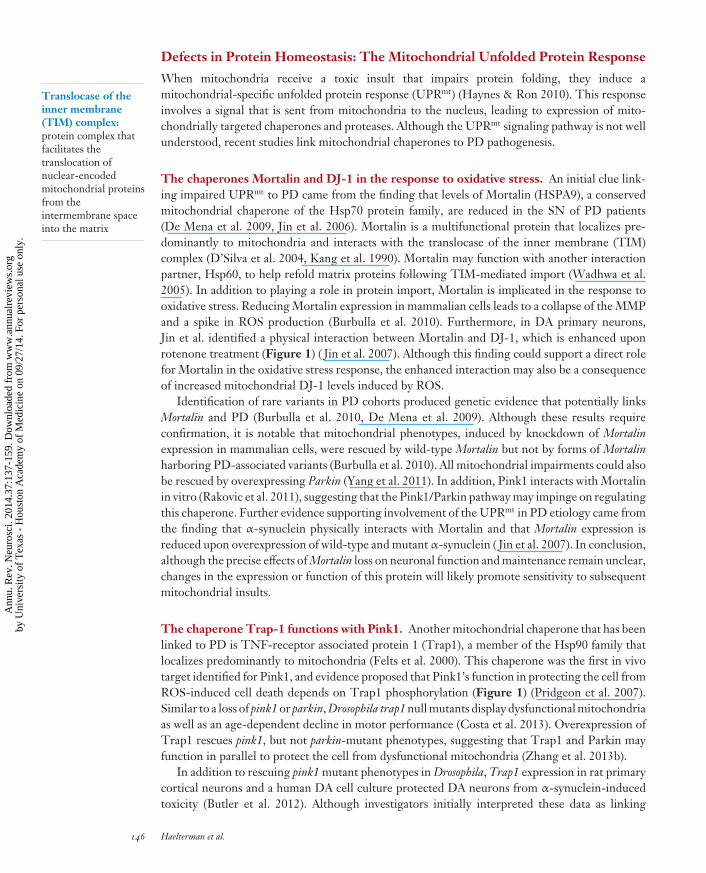
Translocase of theinner membrane(TIM) complex:protein complex thatfacilitates thetranslocation ofnuclear-encodedmitochondrial proteinsfrom theintermembrane spaceinto the matrix
Defects in Protein Homeostasis: The Mitochondrial Unfolded Protein Response
When mitochondria receive a toxic insult that impairs protein folding, they induce amitochondrial-specific unfolded protein response (UPRmt) (Haynes & Ron 2010). This responseinvolves a signal that is sent from mitochondria to the nucleus, leading to expression of mito-chondrially targeted chaperones and proteases. Although the UPRmt signaling pathway is not wellunderstood, recent studies link mitochondrial chaperones to PD pathogenesis.
The chaperones Mortalin and DJ-1 in the response to oxidative stress. An initial clue link-ing impaired UPRmt to PD came from the finding that levels of Mortalin (HSPA9), a conservedmitochondrial chaperone of the Hsp70 protein family, are reduced in the SN of PD patients(De Mena et al. 2009, Jin et al. 2006). Mortalin is a multifunctional protein that localizes pre-dominantly to mitochondria and interacts with the translocase of the inner membrane (TIM)complex (D’Silva et al. 2004, Kang et al. 1990). Mortalin may function with another interactionpartner, Hsp60, to help refold matrix proteins following TIM-mediated import (Wadhwa et al.2005). In addition to playing a role in protein import, Mortalin is implicated in the response tooxidative stress. Reducing Mortalin expression in mammalian cells leads to a collapse of the MMPand a spike in ROS production (Burbulla et al. 2010). Furthermore, in DA primary neurons,Jin et al. identified a physical interaction between Mortalin and DJ-1, which is enhanced uponrotenone treatment (Figure 1) ( Jin et al. 2007). Although this finding could support a direct rolefor Mortalin in the oxidative stress response, the enhanced interaction may also be a consequenceof increased mitochondrial DJ-1 levels induced by ROS.
Identification of rare variants in PD cohorts produced genetic evidence that potentially linksMortalin and PD (Burbulla et al. 2010, De Mena et al. 2009). Although these results requireconfirmation, it is notable that mitochondrial phenotypes, induced by knockdown of Mortalinexpression in mammalian cells, were rescued by wild-type Mortalin but not by forms of Mortalinharboring PD-associated variants (Burbulla et al. 2010). All mitochondrial impairments could alsobe rescued by overexpressing Parkin (Yang et al. 2011). In addition, Pink1 interacts with Mortalinin vitro (Rakovic et al. 2011), suggesting that the Pink1/Parkin pathway may impinge on regulatingthis chaperone. Further evidence supporting involvement of the UPRmt in PD etiology came fromthe finding that α-synuclein physically interacts with Mortalin and that Mortalin expression isreduced upon overexpression of wild-type and mutant α-synuclein ( Jin et al. 2007). In conclusion,although the precise effects of Mortalin loss on neuronal function and maintenance remain unclear,changes in the expression or function of this protein will likely promote sensitivity to subsequentmitochondrial insults.
The chaperone Trap-1 functions with Pink1. Another mitochondrial chaperone that has beenlinked to PD is TNF-receptor associated protein 1 (Trap1), a member of the Hsp90 family thatlocalizes predominantly to mitochondria (Felts et al. 2000). This chaperone was the first in vivotarget identified for Pink1, and evidence proposed that Pink1’s function in protecting the cell fromROS-induced cell death depends on Trap1 phosphorylation (Figure 1) (Pridgeon et al. 2007).Similar to a loss of pink1 or parkin, Drosophila trap1 null mutants display dysfunctional mitochondriaas well as an age-dependent decline in motor performance (Costa et al. 2013). Overexpression ofTrap1 rescues pink1, but not parkin-mutant phenotypes, suggesting that Trap1 and Parkin mayfunction in parallel to protect the cell from dysfunctional mitochondria (Zhang et al. 2013b).
In addition to rescuing pink1 mutant phenotypes in Drosophila, Trap1 expression in rat primarycortical neurons and a human DA cell culture protected DA neurons from α-synuclein-inducedtoxicity (Butler et al. 2012). Although investigators initially interpreted these data as linking
146 Haelterman et al.
Ann
u. R
ev. N
euro
sci.
2014
.37:
137-
159.
Dow
nloa
ded
from
ww
w.a
nnua
lrev
iew
s.or
gby
Uni
vers
ity o
f T
exas
- H
oust
on A
cade
my
of M
edic
ine
on 0
9/27
/14.
For
per
sona
l use
onl
y.

NE37CH08-Bellen ARI 26 May 2014 7:38
α-synuclein overexpression and mitochondrial dysfunction, further studies showed that Trap1can also induce a more general ER-mediated UPR. Thus, the observed protective effect of Trap1may, in fact, be mediated by an increased ability of the cell to cope with unfolded cytoplasmicproteins (Takemoto et al. 2011).
Mitochondrial Hsp60 may play a role in the Pink1/Parkin pathway. The third mitochondrialchaperone that may be associated with PD is mitochondrial Hsp60. This key player of the UPRmt
interacts in vitro with both Pink1 and Parkin (Davison et al. 2009, Rakovic et al. 2011). In addition,mitochondrial Hsp60 is downregulated by about 40% upon loss of Pink1 in a DA neuronal cellculture (Kim et al. 2012). Although this observation requires confirmation, Parkin and/or Pink1may interact with Hsp60 to shut down the UPRmt when a mitochondrion is deemed too damagedto save. Alternatively, if the UPRmt is extended, Hsp60 might interact with both proteins to inducemitophagy.
Taken together, because PD has been associated with mitochondrial dysfunction and a con-comitant increase in ROS, one might expect to find the UPRmt to be activated in PD model sys-tems. However, several components of the UPRmt have instead been found to be either reduced ordysfunctional in such models, and consistent observations have been made in PD patients. Further-more, experiments in mammalian cell cultures showed that the three chaperones (mitochondrialHsp60, Mortalin, and Trap1) control the gating of the mitochondrial permeability transition pore(Ghosh et al. 2010, Qu et al. 2012). Hence, their loss may sensitize a cell for apoptotic death,owing to a reduced threshold for pore opening and release of cytochrome c. Pink1-mutant micesimilarly show a lower threshold for opening of the mitochondrial transition pore and are indeedmore susceptible to toxic insults (Morais et al. 2009). Whether this observation is due to a changein the activity or levels of any of the aforementioned chaperones remains to be determined.
Defects in Mitochondrial Dynamics
As mitochondria age or are exposed to environmental toxins, they accumulate numerous mito-chondrial DNA mutations and protein insults that impair their function and may lead to cell deathif repair does not occur. It is therefore important for a cell to maintain a healthy pool of mitochon-dria. Several protective measures exist to restore or eliminate dysfunctional mitochondria. Theseprocesses include mitochondrial fusion and fission, mitochondrial trafficking, and the clearanceof dysfunctional mitochondria via mitophagy. These three processes play a critical role in mito-chondrial quality control, especially in cells that consume much energy and do not divide, suchas neuronal cells (Itoh et al. 2012, Schon & Przedborski 2011). Several genes linked to PD andrelated parkinsonian disorders have been implicated in the regulation of mitochondrial dynamics,suggesting that failure of these mechanisms may promote disease pathogenesis.
Tipping the balance toward demise: most PD loci alter fusion/fission. In response to mi-tochondrial dysfunction, fusion mixes key constituents, potentially complementing deficienciesfrom damaged proteins, DNA, and/or membrane lipids. Among the required cellular machinery,mitofusin (Mfn) and optic atrophy 1 (Opa 1) mediate fusion of the outer and inner mitochondrialmembranes, respectively. Conversely, mitochondrial fission requires dynamin related protein 1(Drp1). Loss of Mfn or Opa1 leads to small, fragmented mitochondria, whereas the loss of Drp1causes a network of large interconnected mitochondria (Chan 2012, Youle & van der Bliek 2012).Hence, a constant balance of fission and fusion is required to maintain a healthy pool of mitochon-dria. Mutations in Mfn2 lead to Charcot-Marie-Tooth type 2A (Zuchner et al. 2004), an inheritedperipheral neuropathy, and loss of Opa1 leads to autosomal dominant optic atrophy (Eiberg et al.
www.annualreviews.org • A Mitocentric View of Parkinson’s Disease 147
Ann
u. R
ev. N
euro
sci.
2014
.37:
137-
159.
Dow
nloa
ded
from
ww
w.a
nnua
lrev
iew
s.or
gby
Uni
vers
ity o
f T
exas
- H
oust
on A
cade
my
of M
edic
ine
on 0
9/27
/14.
For
per
sona
l use
onl
y.
NE37CH08-Bellen ARI 26 May 2014 7:38
1994). Loss of Drp1 causes a rare infantile mitochondrial encephalopathy, consisting of earlyneurologic failure and death (Waterham et al. 2007). Although these genes are not known to begenetically linked with PD themselves, they do show convincing interactions with established PDgenes in model organisms.
In Drosophila, mutations in pink1 or parkin lead to aberrant mitochondria that are swollenand lose their typical cristae structure (Clark et al. 2006, Greene et al. 2003, Park et al. 2006).Reducing mitochondrial fusion or increasing fission in either pink1 or parkin mutant flies restoresthe morphological defects (Deng et al. 2008, Poole et al. 2008). However, although adding onecopy of drp1 partially restores energy levels in a pink1 mutant, it fails to restore CI-activity (Liuet al. 2011, Vilain et al. 2012). Finally, whereas pink1 or parkin mutant flies are viable, removingonly a single copy of drp1 in a pink1 or parkin mutant is fatal (Poole et al. 2008). These geneticinteractions strongly suggest that the functions of Parkin and Pink1 and mediators of mitochondrialfusion/fission may be linked.
Several groups have reported that round, fragmented mitochondria appear upon overexpressionof either wild-type or mutant forms of α-synuclein, both in vitro as well as in vivo (Kamp et al.2010, Nakamura et al. 2011). The ability of α-synuclein to induce membrane fragmentation isspecific for mitochondrial membranes and appears to be independent of the fission protein Drp1(Nakamura et al. 2011). Indeed, an age-dependent decrease in both Mfn1 and Mfn2 was observedin neurons of mice overexpressing mutant α-synuclein (Xie & Chung 2012), suggesting a possiblepathological role for α-synuclein in reducing mitochondrial fusion.
Studies recently demonstrated that LRRK2 plays a role in regulating mitochondrial dynamicsin Drosophila DA neurons. Overexpression of mutant, but not wild-type, human LRRK2 resultedin enlarged mitochondria resembling those seen in pink1 or parkin mutants (Ng et al. 2012).These defects were significantly rescued by parkin coexpression, suggesting that these proteinsmay function in a common pathway (Ng et al. 2009). Skin biopsies from patients with LRRK2mutations displayed increased mitochondrial length and interconnectivity, decreased ATP levels,and MMP (Mortiboys et al. 2010). In mouse cortical primary neurons, LRRK2 also interactswith and phosphorylates the fission protein Drp1 (Niu et al. 2012). Overexpressing wild-typeand mutant LRRK2 in this system, as well as in primary DA neurons, results in recruitment ofDrp1 to the mitochondrial membrane leading to mitochondrial fragmentation and clearance (Niuet al. 2012, Wang et al. 2012b). Similar to LRRK2, mutations in DJ-1 result in increased Drp1levels and increased mitochondrial fragmentation, although the underlying mechanisms remainelusive (Wang et al. 2012a). These studies suggest that LRRK2 and DJ-1 play important rolesin regulating mitochondrial dynamics and quality control, although additional work is needed tounderstand the mechanisms (Figure 1).
In sum, altered mitochondrial fusion/fission is a feature observed in many experimental systemsrelevant to PD. However, because enhanced mitochondrial fusion and/or fission can be a responseto injury, many of the observations described here could be secondary in nature.
Mitochondrial trafficking is controlled by Pink1 and Parkin. Mitochondrial traffickingdelivers a mobile source of ATP to subcellular locations that have high energy demands, suchas the neuronal synapse (Saxton & Hollenbeck 2012, Sheng & Cai 2012). Newly formed mito-chondria travel along microtubules using kinesins and adaptor proteins that link mitochondriato the motor proteins (Saxton & Hollenbeck 2012, Sheng & Cai 2012). Specifically, in flies,Pink1 phosphorylates Miro, a rho-like GTPase that resides on the MOM, where it functionsas an adaptor to microtubule motor proteins (Figure 1) (Liu et al. 2012, Wang et al. 2011).Phosphorylation of Miro triggers its degradation via a Parkin-dependent ubiquitination pathwayand causes mitochondria to detach from cognate kinesin motors. Because Pink1 is activated upon
148 Haelterman et al.
Ann
u. R
ev. N
euro
sci.
2014
.37:
137-
159.
Dow
nloa
ded
from
ww
w.a
nnua
lrev
iew
s.or
gby
Uni
vers
ity o
f T
exas
- H
oust
on A
cade
my
of M
edic
ine
on 0
9/27
/14.
For
per
sona
l use
onl
y.
NE37CH08-Bellen ARI 26 May 2014 7:38
mitochondrial dysfunction (see next section), this action may serve as a mechanism to blockdysfunctional mitochondria from moving along the axon.
Miro and the adaptor protein Milton, which links mitochondria to the heavy chain of kinesin,form a complex with the fusion protein Mfn, which is also targeted for degradation by Parkin inboth flies and mammals (Glater et al. 2006, Glauser et al. 2011, Gorska-Andrzejak et al. 2003,Ziviani et al. 2010). In addition, genetically altering the mitochondrial fission/fusion balance in awild-type background impedes mitochondrial trafficking, leading to synapses and dendrites thatare devoid of mitochondria (Liu et al. 2012, Sheng & Cai 2012). Together, these findings suggesta tight link between regulators of mitochondrial dynamics and intracellular trafficking. Pink1 andParkin affect both processes, possibly to ensure the presence of sufficient functional mitochondriaat subcellular locations with energy-intensive demands. Because LRRK2 and DJ-1 each regulateDrp1 levels, they may indirectly regulate mitochondrial trafficking as well (Niu et al. 2012, Wanget al. 2012a).
Impaired clearance of dysfunctional mitochondria in PD. When mitochondria can no longerbe repaired, they are cleared through a form of autophagy known as mitophagy (Narendra et al.2010). For degradation, mitochondria initially undergo fragmentation, allowing autophagosomesto engulf them. The protein machinery that mediates fission and fusion is essential for this process,and genetic manipulations that inhibit fission or enhance fusion interfere with mitophagy (Gomeset al. 2011).
Under normal conditions, the ubiquitously expressed Pink1 spans both the MOM and theMIM ( Jin et al. 2010, Weihofen et al. 2009). Within the MIM, it is cleaved by two proteins[mitochondrial processing peptidase (Greene et al. 2012) and presenilin-associated rhomboid-likeprotease ( Jin et al. 2010)], after which it is thought to be degraded (Matsuda et al. 2013). Underbasal conditions, Pink1 protein levels are therefore kept low. Once a mitochondrion becomesdysfunctional and loses its MMP, the import of Pink1 into the MIM is blocked, and it is no longercleaved. Pink1 therefore quickly accumulates on the MOM, where it initiates mitophagy ( Jin &Youle 2012, Youle & Narendra 2011). It does so by recruiting and activating the E3 ubiquitinligase Parkin, a process that requires Pink1’s kinase activity (Kim et al. 2008). However, whetherPink1 acts directly on Parkin or whether it indirectly recruits Parkin to the MOM is still underdebate (Iguchi et al. 2013, Kim et al. 2008, Narendra et al. 2010). In our opinion, the data aremost consistent with a model where Pink1 phosphorylates several proteins, one of which wouldsubsequently recruit Parkin. One potential candidate would be the Pink1-target MFN2, which isrequired to recruit Parkin to damaged mitochondria (Chen & Dorn 2013).
Upon activation, Parkin functions with E2 ubiquitin-conjugating enzymes, such as the recentlyidentified Rad6, to ubiquitinate its targets (Haddad et al. 2013). Over the past few years, investi-gators have identified many substrates for parkin-dependent ubiquitination, including numerousmitochondrial proteins (Figure 1) (Sarraf et al. 2013). Overall, activation of the Pink/Parkin path-way halts the trafficking of a dysfunctional mitochondrion, initiates its fragmentation, and leadsto its subsequent degradation. In addition, the Pink1/Parkin pathway appears to support mito-chondrial biogenesis. Shin et al. recently found that Parkin-activation leads to the degradation ofParis, which represses the transcription of a key regulator of mitochondrial biogenesis, PGC1-α(Shin et al. 2011).
Because most of the mitophagy-related research has focused on Pink1 and Parkin, the questionarises whether impaired mitophagy is a general theme in the pathogenesis of PD. A recent studyin mice found that overexpressing wild-type or mutant α-synuclein induces mitophagy (Sampaio-Marques et al. 2012). Similarly, overexpressing mutant LRRK2 in mouse cortical neurons resultsin autophagic degradation of mitochondria (Cherra et al. 2013). In conclusion, multiple aspects
www.annualreviews.org • A Mitocentric View of Parkinson’s Disease 149
Ann
u. R
ev. N
euro
sci.
2014
.37:
137-
159.
Dow
nloa
ded
from
ww
w.a
nnua
lrev
iew
s.or
gby
Uni
vers
ity o
f T
exas
- H
oust
on A
cade
my
of M
edic
ine
on 0
9/27
/14.
For
per
sona
l use
onl
y.
NE37CH08-Bellen ARI 26 May 2014 7:38
of mitochondrial dynamics appear to be affected to various extents in mutants for PD-related loci(Table 1).
MITOCHONDRIA AND PD: AN INTEGRATED MODEL
As summarized in this review, numerous genes associated with familial forms of PD and relateddisorders with prominent parkinsonism can be linked to mitochondrial mechanisms. In addition,mitochondrial toxins cause SN degeneration in both humans and various other model systems.These data support models in which mitochondria play a central role in DA neuronal loss inPD. It is remarkable that even in cases of AR-JP caused by mutations in parkin or pink1, wheremitochondrial dysfunction seems to be the primary insult, the overall effect on mitochondrialfunction appears subtle. This conclusion is based in part on studies of model organisms, whereloss of homologous genes causes a mild reduction of CI activity and does not significantly impactviability (Morais et al. 2009, Vilain et al. 2012). A similar partial CI deficiency has been docu-mented in postmortem SN tissue from PD patients (Bindoff et al. 1991; Parker et al. 1989, 2008;Schapira et al. 1989). In humans, mutations that more severely affect ETC function are associatedwith correspondingly more aggressive disease. For instance, Leigh syndrome, a mitochondrialencephalopathy, has an infantile onset, affects multiple organ systems, and is often fatal (Vafai &Mootha 2012). Consistent with these observations, mutations in Leigh syndrome–associated genehomologs in mice or flies are lethal [e.g., Sco2 (Porcelli et al. 2010, Yang et al. 2010), LRPPRC(Ruzzenente et al. 2012), C8ORF38 (Zhang et al. 2013a), SURF1 (Agostino et al. 2003, Zordanet al. 2006), among others].
Although the overall extent of mitochondrial dysfunction may be modest, one important lessonfrom studies of Mendelian forms of PD and related parkinsonian disorders is that the responsiblegenetic lesions appear to have distributed effects on several core features of mitochondrial biology,including (a) the ETC, (b) the UPRmt, and (c) mitochondrial dynamics. As shown in Figure 2, wepropose that simultaneous disruptions in these core systems interact and overwhelm the capacityof the mitochondria to compensate for additional insults, promoting a vicious cycle that may ulti-mately result in cell death. However, given the near-universal requirement of mitochondria in alleukaryotic cells, this model does not explain the selective vulnerability of certain neuronal subtypesin PD, such as DA neurons in the SN. Therefore, apart from affecting these core mitochondrialprocesses (a, b, and c), an additional insult (“second hit”) is likely required, tipping the balancetoward severe dysfunction in distinct cellular contexts. In the case of DA neurons, elevated endoge-nous ROS levels may fulfill the second-hit requirement (see sidebar, Why Are DA Neurons MoreSusceptible To Stress Than Are Other Neurons?). For example, in an individual with juvenile
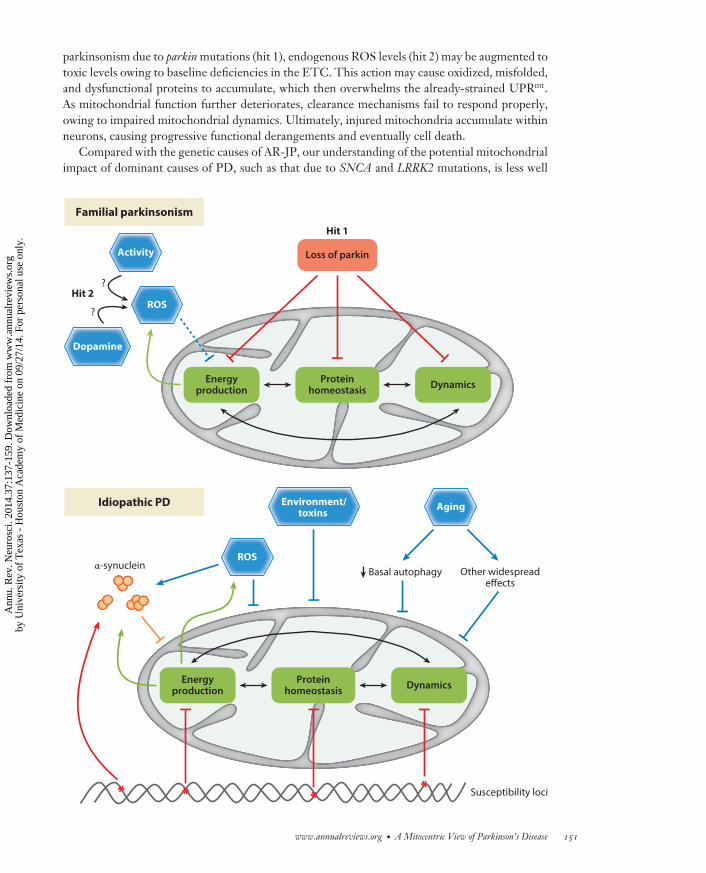
−−−−−−−−−−−−−−−−−−−−−−−−−−−−−−−−−−−−−−−−−−−−−−−−−−−−−−−−−−−−−−−−−−−−−−−−→Figure 2The multiple hit model of Parkinson’s disease (PD). Top panel: In familial parkinsonism, for example due toloss of function of Parkin, several key aspects of mitochondrial homeostasis are mildly affected, includingenergy production, protein folding (UPRmt), or dynamics (fission and fusion). These defects interact andamplify one another, but a second hit, such as elevated endogenous reactive oxygen species (ROS) or cellularactivity, is likely required to trigger neuronal dysfunction and loss in certain vulnerable cell types, such as inDA neurons. Bottom panel: In idiopathic PD, multiple hits, including both common and rare genomicvariations at diverse susceptibility loci, may in combination cause similar, subtle, and distributed defects inmitochondrial function. α-Synuclein pathology, ROS, potential environmental factors, and the widespreadcellular effects of aging further degrade mitochondrial activity. In a potential feedback mechanism,mitochondrial dysfunction may also promote α-synuclein aggregation, which in turn may further amplifymitochondrial defects, ultimately leading to neuronal dysfunction and cell death.
150 Haelterman et al.
Ann
u. R
ev. N
euro
sci.
2014
.37:
137-
159.
Dow
nloa
ded
from
ww
w.a
nnua
lrev
iew
s.or
gby
Uni
vers
ity o
f T
exas
- H
oust
on A
cade
my
of M
edic
ine
on 0
9/27
/14.
For
per
sona
l use
onl
y.
NE37CH08-Bellen ARI 26 May 2014 7:38
parkinsonism due to parkin mutations (hit 1), endogenous ROS levels (hit 2) may be augmented totoxic levels owing to baseline deficiencies in the ETC. This action may cause oxidized, misfolded,and dysfunctional proteins to accumulate, which then overwhelms the already-strained UPRmt.As mitochondrial function further deteriorates, clearance mechanisms fail to respond properly,owing to impaired mitochondrial dynamics. Ultimately, injured mitochondria accumulate withinneurons, causing progressive functional derangements and eventually cell death.
Compared with the genetic causes of AR-JP, our understanding of the potential mitochondrialimpact of dominant causes of PD, such as that due to SNCA and LRRK2 mutations, is less well
Familial parkinsonism
Energyproduction
Proteinhomeostasis Dynamics
ROS
Activity
Hit 2?
Dopamine
?
Idiopathic PD
Energyproduction
Proteinhomeostasis Dynamics
α-synuclein
** ******
ROS
Susceptibility loci
Basal autophagy
Environment/toxins
Other widespreadeffects
Aging
Loss of parkin
Hit 1
www.annualreviews.org • A Mitocentric View of Parkinson’s Disease 151
Ann
u. R
ev. N
euro
sci.
2014
.37:
137-
159.
Dow
nloa
ded
from
ww
w.a
nnua
lrev
iew
s.or
gby
Uni
vers
ity o
f T
exas
- H
oust
on A
cade
my
of M
edic
ine
on 0
9/27
/14.
For
per
sona
l use
onl
y.
NE37CH08-Bellen ARI 26 May 2014 7:38
WHY ARE DA NEURONS MORE SUSCEPTIBLE TO STRESS THAN ARE OTHERNEURONS?
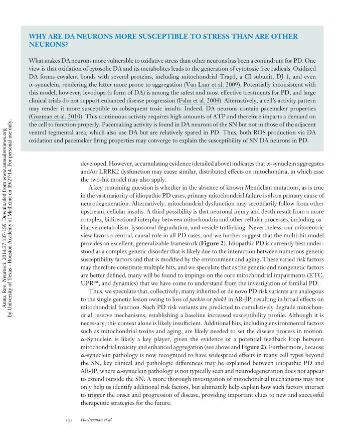
What makes DA neurons more vulnerable to oxidative stress than other neurons has been a conundrum for PD. Oneview is that oxidation of cytosolic DA and its metabolites leads to the generation of cytotoxic free radicals. OxidizedDA forms covalent bonds with several proteins, including mitochondrial Trap1, a CI subunit, DJ-1, and evenα-synuclein, rendering the latter more prone to aggregation (Van Laar et al. 2009). Potentially inconsistent withthis model, however, levodopa (a form of DA) is among the safest and most effective treatments for PD, and largeclinical trials do not support enhanced disease progression (Fahn et al. 2004). Alternatively, a cell’s activity patternmay render it more susceptible to subsequent toxic insults. Indeed, DA neurons contain pacemaker properties(Guzman et al. 2010). This continuous activity requires high amounts of ATP and therefore imparts a demand onthe cell to function properly. Pacemaking activity is found in DA neurons of the SN but not in those of the adjacentventral tegmental area, which also use DA but are relatively spared in PD. Thus, both ROS production via DAoxidation and pacemaker firing properties may converge to explain the susceptibility of SN DA neurons in PD.
developed. However, accumulating evidence (detailed above) indicates that α-synuclein aggregatesand/or LRRK2 dysfunction may cause similar, distributed effects on mitochondria, in which casethe two-hit model may also apply.
A key remaining question is whether in the absence of known Mendelian mutations, as is truein the vast majority of idiopathic PD cases, primary mitochondrial failure is also a primary cause ofneurodegeneration. Alternatively, mitochondrial dysfunction may secondarily follow from otherupstream, cellular insults. A third possibility is that neuronal injury and death result from a morecomplex, bidirectional interplay between mitochondria and other cellular processes, including ox-idative metabolism, lysosomal degradation, and vesicle trafficking. Nevertheless, our mitocentricview favors a central, causal role in all PD cases, and we further suggest that the multi-hit modelprovides an excellent, generalizable framework (Figure 2). Idiopathic PD is currently best under-stood as a complex genetic disorder that is likely due to the interaction between numerous geneticsusceptibility factors and that is modified by the environment and aging. These varied risk factorsmay therefore constitute multiple hits, and we speculate that as the genetic and nongenetic factorsare better defined, many will be found to impinge on the core mitochondrial impairments (ETC,UPRmt, and dynamics) that we have come to understand from the investigation of familial PD.
Thus, we speculate that, collectively, many inherited or de novo PD risk variants are analogousto the single genetic lesion owing to loss of parkin or pink1 in AR-JP, resulting in broad effects onmitochondrial function. Such PD risk variants are predicted to cumulatively degrade mitochon-drial reserve mechanisms, establishing a baseline increased susceptibility profile. Although it isnecessary, this context alone is likely insufficient. Additional hits, including environmental factorssuch as mitochondrial toxins and aging, are likely needed to set the disease process in motion.α-Synuclein is likely a key player, given the evidence of a potential feedback loop betweenmitochondrial toxicity and enhanced aggregation (see above and Figure 2). Furthermore, becauseα-synuclein pathology is now recognized to have widespread effects in many cell types beyondthe SN, key clinical and pathologic differences may be explained between idiopathic PD andAR-JP, where α-synuclein pathology is not typically seen and neurodegeneration does not appearto extend outside the SN. A more thorough investigation of mitochondrial mechanisms may notonly help us identify additional risk factors, but ultimately help explain how such factors interactto trigger the onset and progression of disease, providing important clues to new and successfultherapeutic strategies for the future.
152 Haelterman et al.
Ann
u. R
ev. N
euro
sci.
2014
.37:
137-
159.
Dow
nloa
ded
from
ww
w.a
nnua
lrev
iew
s.or
gby
Uni
vers
ity o
f T
exas
- H
oust
on A
cade
my
of M
edic
ine
on 0
9/27
/14.
For
per
sona
l use
onl
y.
NE37CH08-Bellen ARI 26 May 2014 7:38
DISCLOSURE STATEMENT
The authors are not aware of any affiliations, memberships, funding, or financial holdings thatmight be perceived as affecting the objectivity of this review.
LITERATURE CITED
Agostino A, Invernizzi F, Tiveron C, Fagiolari G, Prelle A, et al. 2003. Constitutive knockout of Surf1 isassociated with high embryonic lethality, mitochondrial disease and cytochrome c oxidase deficiency inmice. Hum. Mol. Genet. 12:399–413
Amo T, Sato S, Saiki S, Wolf AM, Toyomizu M, et al. 2011. Mitochondrial membrane potential decreasecaused by loss of PINK1 is not due to proton leak, but to respiratory chain defects. Neurobiol. Dis.41:111–18
Bindoff LA, Birch-Machin MA, Cartlidge NE, Parker WD Jr, Turnbull DM. 1991. Respiratory chain abnor-malities in skeletal muscle from patients with Parkinson’s disease. J. Neurol. Sci. 104:203–8
Bonifati V, Rizzu P, van Baren MJ, Schaap O, Breedveld GJ, et al. 2003. Mutations in the DJ-1 gene associatedwith autosomal recessive early-onset parkinsonism. Science 299:256–59
Bonifati V, Rohe CF, Breedveld GJ, Fabrizio E, De Mari M, et al. 2005. Early-onset parkinsonism associatedwith PINK1 mutations: frequency, genotypes, and phenotypes. Neurology 65:87–95
Braschi E, Goyon V, Zunino R, Mohanty A, Xu L, McBride HM. 2010. Vps35 mediates vesicle transportbetween the mitochondria and peroxisomes. Curr. Biol. 20:1310–15
Burbulla L, Schelling C, Kato H, Rapaport D, Woitalla D, et al. 2010. Dissecting the role of the mitochondrialchaperone mortalin in Parkinson’s disease: functional impact of disease-related variants on mitochondrialhomeostasis. Hum. Mol. Genet. 19:4437–52
Burchell VS, Nelson DE, Sanchez-Martinez A, Delgado-Camprubi M, Ivatt RM, et al. 2013. The Parkinson’sdisease-linked proteins Fbxo7 and Parkin interact to mediate mitophagy. Nat. Neurosci. 16:1257–65
Butler EK, Voigt A, Lutz AK, Toegel JP, Gerhardt E, et al. 2012. The mitochondrial chaperone proteinTRAP1 mitigates α-synuclein toxicity. PLoS Genet. 8:e1002488
Chan DC. 2012. Fusion and fission: interlinked processes critical for mitochondrial health. Annu. Rev. Genet.46:265–87
Chaudhuri KR, Schapira AH. 2009. Non-motor symptoms of Parkinson’s disease: dopaminergic pathophys-iology and treatment. Lancet Neurol. 8:464–74
Chen Y, Dorn GW 2nd. 2013. PINK1-phosphorylated mitofusin 2 is a Parkin receptor for culling damagedmitochondria. Science 340:471–75
Cherra SJ III, Steer E, Gusdon AM, Kiselyov K, Chu CT. 2013. Mutant LRRK2 elicits calcium imbalanceand depletion of dendritic mitochondria in neurons. Am. J. Pathol. 182:474–84
Clark IE, Dodson MW, Jiang C, Cao JH, Huh JR, et al. 2006. Drosophila pink1 is required for mitochondrialfunction and interacts genetically with parkin. Nature 441:1162–66
Cole NB, Dieuliis D, Leo P, Mitchell DC, Nussbaum RL. 2008. Mitochondrial translocation of α-synucleinis promoted by intracellular acidification. Exp. Cell Res. 314:2076–89
Costa AC, Loh SH, Martins LM. 2013. Drosophila Trap1 protects against mitochondrial dysfunction in aPINK1/parkin model of Parkinson’s disease. Cell Death Dis. 4:e467
Cui M, Tang X, Christian WV, Yoon Y, Tieu K. 2010. Perturbations in mitochondrial dynamics inducedby human mutant PINK1 can be rescued by the mitochondrial division inhibitor mdivi-1. J. Biol. Chem.285:11740–52
D’Silva P, Liu Q, Walter W, Craig E. 2004. Regulated interactions of mtHsp70 with Tim44 at the transloconin the mitochondrial inner membrane. Nat. Struct. Mol. Biol. 11:1084–91
Dagda RK, Cherra SJ 3rd, Kulich SM, Tandon A, Park D, Chu CT. 2009. Loss of PINK1 function promotesmitophagy through effects on oxidative stress and mitochondrial fission. J. Biol. Chem. 284:13843–55
Davison E, Pennington K, Hung C-C, Peng J, Rafiq R, et al. 2009. Proteomic analysis of increased Parkinexpression and its interactants provides evidence for a role in modulation of mitochondrial function.Proteomics 9:4284–97
www.annualreviews.org • A Mitocentric View of Parkinson’s Disease 153
Ann
u. R
ev. N
euro
sci.
2014
.37:
137-
159.
Dow
nloa
ded
from
ww
w.a
nnua
lrev
iew
s.or
gby
Uni
vers
ity o
f T
exas
- H
oust
on A
cade
my
of M
edic
ine
on 0
9/27
/14.
For
per
sona
l use
onl
y.
NE37CH08-Bellen ARI 26 May 2014 7:38
De Mena L, Coto E, Sanchez-Ferrero E, Ribacoba R, Guisasola LM, et al. 2009. Mutational screening of themortalin gene (HSPA9) in Parkinson’s disease. J. Neural Transm. 116:1289–93
Deng H, Dodson MW, Huang H, Guo M. 2008. The Parkinson’s disease genes pink1 and parkin promotemitochondrial fission and/or inhibit fusion in Drosophila. Proc. Natl. Acad. Sci. USA 105:14503–8
Devi L, Raghavendran V, Prabhu BM, Avadhani NG, Anandatheerthavarada HK. 2008. Mitochondrial importand accumulation of alpha-synuclein impair complex I in human dopaminergic neuronal cultures andParkinson disease brain. J. Biol. Chem. 283:9089–100
Doherty KM, Silveira-Moriyama L, Parkkinen L, Healy DG, Farrell M, et al. 2013. Parkin disease: a clinico-pathologic entity? JAMA Neurol. 70:571–79
Edgar AJ, Polak JM. 2000. Human homologues of yeast vacuolar protein sorting 29 and 35. Biochem. Biophys.Res. Commun. 277:622–30
Eiberg H, Kjer B, Kjer P, Rosenberg T. 1994. Dominant optic atrophy (OPA1) mapped to chromosome 3qregion. I. Linkage analysis. Hum. Mol. Genet. 3:977–80
Exner N, Treske B, Paquet D, Holmstrom K, Schiesling C, et al. 2007. Loss-of-function of human PINK1results in mitochondrial pathology and can be rescued by parkin. J. Neurosci. 27:12413–18
Fahn S. 2003. Description of Parkinson’s disease as a clinical syndrome. Ann. N. Y. Acad. Sci. 991:1–14Fahn S, Oakes D, Shoulson I, Kieburtz K, Rudolph A, et al. 2004. Levodopa and the progression of Parkinson’s
disease. N. Engl. J. Med. 351:2498–508Fariello RG. 1988. Experimental support for the implication of oxidative stress in the genesis of parkinsonian
syndromes. Funct. Neurol. 3:407–12Felts SJ, Owen BA, Nguyen P, Trepel J, Donner DB, Toft DO. 2000. The hsp90-related protein TRAP1 is
a mitochondrial protein with distinct functional properties. J. Biol. Chem. 275:3305–12Gautier CA, Kitada T, Shen J. 2008. Loss of PINK1 causes mitochondrial functional defects and increased
sensitivity to oxidative stress. Proc. Natl. Acad. Sci. USA 105:11364–69Gegg ME, Cooper JM, Schapira AH, Taanman JW. 2009. Silencing of PINK1 expression affects mitochondrial
DNA and oxidative phosphorylation in dopaminergic cells. PLoS ONE 4:e4756Ghosh JC, Siegelin MD, Dohi T, Altieri DC. 2010. Heat shock protein 60 regulation of the mitochondrial
permeability transition pore in tumor cells. Cancer Res. 70:8988–93Gispert S, Ricciardi F, Kurz A, Azizov M, Hoepken HH, et al. 2009. Parkinson phenotype in aged PINK1-
deficient mice is accompanied by progressive mitochondrial dysfunction in absence of neurodegeneration.PLoS ONE 4:e5777
Glater EE, Megeath LJ, Stowers RS, Schwarz TL. 2006. Axonal transport of mitochondria requires miltonto recruit kinesin heavy chain and is light chain independent. J. Cell Biol. 173:545–57
Glauser L, Sonnay S, Stafa K, Moore DJ. 2011. Parkin promotes the ubiquitination and degradation of themitochondrial fusion factor mitofusin 1. J. Neurochem. 118:636–45
Goedert M, Spillantini MG, Del Tredici K, Braak H. 2013. 100 years of Lewy pathology. Nat. Rev. Neurol.9:13–24
Gomes LC, Di Benedetto G, Scorrano L. 2011. During autophagy mitochondria elongate, are spared fromdegradation and sustain cell viability. Nat. Cell Biol. 13:589–98
Gorska-Andrzejak J, Stowers RS, Borycz J, Kostyleva R, Schwarz TL, Meinertzhagen IA. 2003. Mitochon-dria are redistributed in Drosophila photoreceptors lacking milton, a kinesin-associated protein. J. Comp.Neurol. 463:372–88
Greene AW, Grenier K, Aguileta MA, Muise S, Farazifard R, et al. 2012. Mitochondrial processing peptidaseregulates PINK1 processing, import and Parkin recruitment. EMBO Rep. 13:378–85
Greene JC, Whitworth AJ, Kuo I, Andrews LA, Feany MB, Pallanck LJ. 2003. Mitochondrial pathology andapoptotic muscle degeneration in Drosophila parkin mutants. Proc. Natl. Acad. Sci. USA 100:4078–83
Grunewald A, Arns B, Seibler P, Rakovic A, Munchau A, et al. 2012. ATP13A2 mutations impair mitochondrialfunction in fibroblasts from patients with Kufor-Rakeb syndrome. Neurobiol. Aging 33:1843.e1–7
Guzman JN, Sanchez-Padilla J, Wokosin D, Kondapalli J, Ilijic E, et al. 2010. Oxidant stress evoked bypacemaking in dopaminergic neurons is attenuated by DJ-1. Nature 468:696–700
Haddad DM, Vilain S, Vos M, Esposito G, Matta S, et al. 2013. Mutations in the intellectual disability geneUbe2a cause neuronal dysfunction and impair Parkin-dependent mitophagy. Mol. Cell 50:831–43
154 Haelterman et al.
Ann
u. R
ev. N
euro
sci.
2014
.37:
137-
159.
Dow
nloa
ded
from
ww
w.a
nnua
lrev
iew
s.or
gby
Uni
vers
ity o
f T
exas
- H
oust
on A
cade
my
of M
edic
ine
on 0
9/27
/14.
For
per
sona
l use
onl
y.
NE37CH08-Bellen ARI 26 May 2014 7:38
Hao L-Y, Giasson BI, Bonini NM. 2010. DJ-1 is critical for mitochondrial function and rescues PINK1 lossof function. Proc. Natl. Acad. Sci. USA 107:9747–52
Haynes C, Ron D. 2010. The mitochondrial UPR – protecting organelle protein homeostasis. J. Cell Sci.123:3849–55
Healy DG, Falchi M, O’Sullivan SS, Bonifati V, Durr A, et al. 2008. Phenotype, genotype, and worldwidegenetic penetrance of LRRK2-associated Parkinson’s disease: a case-control study. Lancet Neurol. 7:583–90
Heeman B, Van den Haute C, Aelvoet SA, Valsecchi F, Rodenburg RJ, et al. 2011. Depletion of PINK1 affectsmitochondrial metabolism, calcium homeostasis and energy maintenance. J. Cell Sci. 124:1115–25
Heo JY, Park JH, Kim SJ, Seo KS, Han JS, et al. 2012. DJ-1 null dopaminergic neuronal cells exhibit defectsin mitochondrial function and structure: involvement of mitochondrial complex I assembly. PLoS ONE7:e32629
Hoepken HH, Gispert S, Morales B, Wingerter O, Del Turco D, et al. 2007. Mitochondrial dysfunction,peroxidation damage and changes in glutathione metabolism in PARK6. Neurobiol. Dis. 25:401–11
Iguchi M, Kujuro Y, Okatsu K, Koyano F, Kosako H, et al. 2013. Parkin-catalyzed ubiquitin-ester transfer istriggered by PINK1-dependent phosphorylation. J. Biol. Chem. 288:22019–32
Ilyin GP, Rialland M, Pigeon C, Guguen-Guillouzo C. 2000. cDNA cloning and expression analysis of newmembers of the mammalian F-box protein family. Genomics 67:40–47
Int. Parkinson’s Dis. Genomics Consort. (IPDGC), Wellcome Trust Case Control Consort. 2 (WTCCC2).2011. A two-stage meta-analysis identifies several new loci for Parkinson’s disease. PLoS Genet. 7:e1002142
Irrcher I, Aleyasin H, Seifert EL, Hewitt SJ, Chhabra S, et al. 2010. Loss of the Parkinson’s disease-linkedgene DJ-1 perturbs mitochondrial dynamics. Hum. Mol. Genet. 19:3734–46
Itoh K, Nakamura K, Iijima M, Sesaki H. 2012. Mitochondrial dynamics in neurodegeneration. Trends CellBiol. 23:64–71
Jaiswal M, Sandoval H, Zhang K, Bayat V, Bellen HJ. 2012. Probing mechanisms that underlie humanneurodegenerative diseases in Drosophila. Annu. Rev. Genet. 46:371–96
Jellinger KA. 2009. A critical evaluation of current staging of α-synuclein pathology in Lewy body disorders.Biochim. Biophys. Acta 1792:730–40
Jin J, Hulette C, Wang Y, Zhang T, Pan C, et al. 2006. Proteomic identification of a stress protein, mor-talin/mthsp70/GRP75: relevance to Parkinson disease. Mol. Cell. Proteomics 5:1193–204
Jin J, Li G, Davis J, Zhu D, Wang Y, et al. 2007. Identification of novel proteins associated with bothα-synuclein and DJ-1. Mol. Cell. Proteomics 6:845–59
Jin SM, Lazarou M, Wang C, Kane LA, Narendra DP, Youle RJ. 2010. Mitochondrial membrane potentialregulates PINK1 import and proteolytic destabilization by PARL. J. Cell Biol. 191:933–42
Jin SM, Youle RJ. 2012. PINK1- and Parkin-mediated mitophagy at a glance. J. Cell Sci. 125:795–99Junn E, Mouradian MM. 2002. Human α-synuclein over-expression increases intracellular reactive oxygen
species levels and susceptibility to dopamine. Neurosci. Lett. 320:146–50Kamp F, Exner N, Lutz AK, Wender N, Hegermann J, et al. 2010. Inhibition of mitochondrial fusion by
α-synuclein is rescued by PINK1, Parkin and DJ-1. EMBO J. 29:3571–89Kang P, Ostermann J, Shilling J, Neupert W, Craig E, Pfanner N. 1990. Requirement for hsp70 in the
mitochondrial matrix for translocation and folding of precursor proteins. Nature 348:137–43Keeney PM, Xie J, Capaldi RA, Bennett JP Jr. 2006. Parkinson’s disease brain mitochondrial complex I has
oxidatively damaged subunits and is functionally impaired and misassembled. J. Neurosci. 26:5256–64Kett LR, Dauer WT. 2012. Leucine-rich repeat kinase 2 for beginners: six key questions. Cold Spring Harb.
Perspect. Med. 2:a009407Kim K-H, Song K, Yoon S-H, Shehzad O, Kim Y-S, Son J. 2012. Rescue of PINK1 protein null-specific
mitochondrial complex IV deficits by ginsenoside Re activation of nitric oxide signaling. J. Biol. Chem.287:44109–20
Kim Y, Park J, Kim S, Song S, Kwon SK, et al. 2008. PINK1 controls mitochondrial localization of Parkinthrough direct phosphorylation. Biochem. Biophys. Res. Commun. 377:975–80
Kitada T, Asakawa S, Hattori N, Matsumine H, Yamamura Y, et al. 1998. Mutations in the parkin gene causeautosomal recessive juvenile parkinsonism. Nature 392:605–8
Klein C, Lohmann-Hedrich K. 2007. Impact of recent genetic findings in Parkinson’s disease. Curr. Opin.Neurol. 20:453–64
www.annualreviews.org • A Mitocentric View of Parkinson’s Disease 155
Ann
u. R
ev. N
euro
sci.
2014
.37:
137-
159.
Dow
nloa
ded
from
ww
w.a
nnua
lrev
iew
s.or
gby
Uni
vers
ity o
f T
exas
- H
oust
on A
cade
my
of M
edic
ine
on 0
9/27
/14.
For
per
sona
l use
onl
y.
NE37CH08-Bellen ARI 26 May 2014 7:38
Krebiehl G, Ruckerbauer S, Burbulla LF, Kieper N, Maurer B, et al. 2010. Reduced basal autophagy andimpaired mitochondrial dynamics due to loss of Parkinson’s disease-associated protein DJ-1. PLoS ONE5:e9367
Kwon HJ, Heo JY, Shim JH, Park JH, Seo KS, et al. 2011. DJ-1 mediates paraquat-induced dopaminergicneuronal cell death. Toxicol. Lett. 202:85–92
Langston JW, Ballard P, Tetrud JW, Irwin I. 1983. Chronic Parkinsonism in humans due to a product ofmeperidine-analog synthesis. Science 219:979–80
Lees AJ, Hardy J, Revesz T. 2009. Parkinson’s disease. Lancet 373:2055–66Liu S, Sawada T, Lee S, Yu W, Silverio G, et al. 2012. Parkinson’s disease-associated kinase PINK1 regulates
Miro protein level and axonal transport of mitochondria. PLoS Genet. 8:e1002537Liu W, Acin-Perez R, Geghman KD, Manfredi G, Lu B, Li C. 2011. Pink1 regulates the oxidative phosphor-
ylation machinery via mitochondrial fission. Proc. Natl. Acad. Sci. USA 108:12920–24Lowe J, Blanchard A, Morrell K, Lennox G, Reynolds L, et al. 1988. Ubiquitin is a common factor in
intermediate filament inclusion bodies of diverse type in man, including those of Parkinson’s disease,Pick’s disease, and Alzheimer’s disease, as well as Rosenthal fibres in cerebellar astrocytomas, cytoplasmicbodies in muscle, and Mallory bodies in alcoholic liver disease. J. Pathol. 155:9–15
Lucking CB, Durr A, Bonifati V, Vaughan J, De Michele G, et al. 2000. Association between early-onsetParkinson’s disease and mutations in the parkin gene. N. Engl. J. Med. 342:1560–67
Lutz AK, Exner N, Fett ME, Schlehe JS, Kloos K, et al. 2009. Loss of parkin or PINK1 function increasesDrp1-dependent mitochondrial fragmentation. J. Biol. Chem. 284:22938–51
Maraganore DM, de Andrade M, Elbaz A, Farrer MJ, Ioannidis JP, et al. 2006. Collaborative analysis ofα-synuclein gene promoter variability and Parkinson disease. JAMA 296:661–70
Martin LJ, Pan Y, Price AC, Sterling W, Copeland NG, et al. 2006. Parkinson’s disease alpha-synucleintransgenic mice develop neuronal mitochondrial degeneration and cell death. J. Neurosci. 26:41–50
Martinez TN, Greenamyre JT. 2012. Toxin models of mitochondrial dysfunction in Parkinson’s disease.Antioxid. Redox Signal. 16:920–34
Matsuda S, Kitagishi Y, Kobayashi M. 2013. Function and characteristics of PINK1 in mitochondria. Oxid.Med. Cell. Longev. 2013:601587
Mizuno Y, Ohta S, Tanaka M, Takamiya S, Suzuki K, et al. 1989. Deficiencies in complex I subunits of therespiratory chain in Parkinson’s disease. Biochem. Biophys. Res. Commun. 163:1450–55
Moore DJ, Dawson VL, Dawson TM. 2006. Lessons from Drosophila models of DJ-1 deficiency. Sci. AgingKnowl. Environ. 2006:pe2
Morais VA, Verstreken P, Roethig A, Smet J, Snellinx A, et al. 2009. Parkinson’s disease mutations in PINK1result in decreased Complex I activity and deficient synaptic function. EMBO Mol. Med. 1:99–111
Mortiboys H, Johansen KK, Aasly JO, Bandmann O. 2010. Mitochondrial impairment in patients withParkinson disease with the G2019S mutation in LRRK2. Neurology 75:2017–20
Muftuoglu M, Elibol B, Dalmizrak O, Ercan A, Kulaksiz G, et al. 2004. Mitochondrial complex I and IVactivities in leukocytes from patients with parkin mutations. Mov. Disord. 19:544–48
Nagakubo D, Taira T, Kitaura H, Ikeda M, Tamai K, et al. 1997. DJ-1, a novel oncogene which transformsmouse NIH3T3 cells in cooperation with ras. Biochem. Biophys. Res. Commun. 231:509–13
Nakamura K. 2013. α-Synuclein and mitochondria: partners in crime? Neurotherapeutics 10:391–99Nakamura K, Nemani VM, Azarbal F, Skibinski G, Levy JM, et al. 2011. Direct membrane association drives
mitochondrial fission by the Parkinson disease-associated protein α-synuclein. J. Biol. Chem. 286:20710–26
Narendra DP, Jin SM, Tanaka A, Suen DF, Gautier CA, et al. 2010. PINK1 is selectively stabilized on impairedmitochondria to activate Parkin. PLoS Biol. 8:e1000298
Ng CH, Guan MS, Koh C, Ouyang X, Yu F, et al. 2012. AMP kinase activation mitigates dopaminergicdysfunction and mitochondrial abnormalities in Drosophila models of Parkinson’s disease. J. Neurosci.32:14311–17
Ng CH, Mok SZS, Koh C, Ouyang X, Fivaz ML, et al. 2009. Parkin protects against LRRK2 G2019Smutant-induced dopaminergic neurodegeneration in Drosophila. J. Neurosci. 29:11257–62
156 Haelterman et al.
Ann
u. R
ev. N
euro
sci.
2014
.37:
137-
159.
Dow
nloa
ded
from
ww
w.a
nnua
lrev
iew
s.or
gby
Uni
vers
ity o
f T
exas
- H
oust
on A
cade
my
of M
edic
ine
on 0
9/27
/14.
For
per
sona
l use
onl
y.
NE37CH08-Bellen ARI 26 May 2014 7:38
Nicklas WJ, Vyas I, Heikkila RE. 1985. Inhibition of NADH-linked oxidation in brain mitochon-dria by 1-methyl-4-phenyl-pyridine, a metabolite of the neurotoxin, 1-methyl-4-phenyl-1,2,5,6-tetrahydropyridine. Life Sci. 36:2503–8
Niu J, Yu M, Wang C, Xu Z. 2012. Leucine-rich repeat kinase 2 disturbs mitochondrial dynamics via Dynamin-like protein. J. Neurochem. 122:650–58
Olney JW, Zorumski CF, Stewart GR, Price MT, Wang GJ, Labruyere J. 1990. Excitotoxicity of L-dopa and6-OH-dopa: implications for Parkinson’s and Huntington’s diseases. Exp. Neurol. 108:269–72
Paisan-Ruız C, Jain S, Evans EW, Gilks WP, Simon J, et al. 2004. Cloning of the gene containing mutationsthat cause PARK8-linked Parkinson’s disease. Neuron 44:595–600
Palacino JJ, Sagi D, Goldberg MS, Krauss S, Motz C, et al. 2004. Mitochondrial dysfunction and oxidativedamage in parkin-deficient mice. J. Biol. Chem. 279:18614–22
Papkovskaia TD, Chau KY, Inesta-Vaquera F, Papkovsky DB, Healy DG, et al. 2012. G2019S leucine-richrepeat kinase 2 causes uncoupling protein-mediated mitochondrial depolarization. Hum. Mol. Genet.21:4201–13
Parihar MS, Parihar A, Fujita M, Hashimoto M, Ghafourifar P. 2009. Alpha-synuclein overexpression andaggregation exacerbates impairment of mitochondrial functions by augmenting oxidative stress in humanneuroblastoma cells. Int. J. Biochem. Cell Biol. 41:2015–24
Park J, Lee SB, Lee S, Kim Y, Song S, et al. 2006. Mitochondrial dysfunction in Drosophila PINK1 mutants iscomplemented by parkin. Nature 441:1157–61
Parker WD Jr, Boyson SJ, Parks JK. 1989. Abnormalities of the electron transport chain in idiopathicParkinson’s disease. Ann. Neurol. 26:719–23
Parker WD Jr, Parks JK, Swerdlow RH. 2008. Complex I deficiency in Parkinson’s disease frontal cortex.Brain Res. 1189:215–18
Periquet M, Latouche M, Lohmann E, Rawal N, De Michele G, et al. 2003. Parkin mutations are frequent inpatients with isolated early-onset parkinsonism. Brain 126:1271–78
Pesah Y, Pham T, Burgess H, Middlebrooks B, Verstreken P, et al. 2004. Drosophila parkin mutants havedecreased mass and cell size and increased sensitivity to oxygen radical stress. Development 131:2183–94
Piccoli C, Sardanelli A, Scrima R, Ripoli M, Quarato G, et al. 2008. Mitochondrial respiratory dysfunction infamiliar parkinsonism associated with PINK1 mutation. Neurochem. Res. 33:2565–74
Polymeropoulos MH, Lavedan C, Leroy E, Ide SE, Dehejia A, et al. 1997. Mutation in the α-synuclein geneidentified in families with Parkinson’s disease. Science 276:2045–47
Poole AC, Thomas RE, Andrews LA, McBride HM, Whitworth AJ, Pallanck LJ. 2008. The PINK1/Parkinpathway regulates mitochondrial morphology. Proc. Natl. Acad. Sci. USA 105:1638–43
Porcelli D, Oliva M, Duchi S, Latorre D, Cavaliere V, et al. 2010. Genetic, functional and evolutionarycharacterization of scox, the Drosophila melanogaster ortholog of the human SCO1 gene. Mitochondrion10:433–48
Poulopoulos M, Levy OA, Alcalay RN. 2012. The neuropathology of genetic Parkinson’s disease. Mov. Disord.27:831–42
Pridgeon JW, Olzmann JA, Chin L-S, Li L. 2007. PINK1 protects against oxidative stress by phosphorylatingmitochondrial chaperone TRAP1. PLoS Biol. 5:e172
Priyadarshi A, Khuder SA, Schaub EA, Shrivastava S. 2000. A meta-analysis of Parkinson’s disease and exposureto pesticides. Neurotoxicology 21:435–40
Qu M, Zhou Z, Chen C, Li M, Pei L, et al. 2012. Inhibition of mitochondrial permeability transition poreopening is involved in the protective effects of mortalin overexpression against beta-amyloid-inducedapoptosis in SH-SY5Y cells. Neurosci. Res. 72:94–102
Rakovic A, Grunewald A, Voges L, Hofmann S, Orolicki S, et al. 2011. PINK1-interacting proteins: proteomicanalysis of overexpressed PINK1. Park. Dis. 2011:153979
Ramirez A, Heimbach A, Grundemann J, Stiller B, Hampshire D, et al. 2006. Hereditary parkinsonism withdementia is caused by mutations in ATP13A2, encoding a lysosomal type 5 P-type ATPase. Nat. Genet.38:1184–91
Ramonet D, Podhajska A, Stafa K, Sonnay S, Trancikova A, et al. 2012. PARK9-associated ATP13A2 localizesto intracellular acidic vesicles and regulates cation homeostasis and neuronal integrity. Hum. Mol. Genet.21:1725–43
www.annualreviews.org • A Mitocentric View of Parkinson’s Disease 157
Ann
u. R
ev. N
euro
sci.
2014
.37:
137-
159.
Dow
nloa
ded
from
ww
w.a
nnua
lrev
iew
s.or
gby
Uni
vers
ity o
f T
exas
- H
oust
on A
cade
my
of M
edic
ine
on 0
9/27
/14.
For
per
sona
l use
onl
y.
NE37CH08-Bellen ARI 26 May 2014 7:38
Rogaeva E, Johnson J, Lang AE, Gulick C, Gwinn-Hardy K, et al. 2004. Analysis of the PINK1 gene in alarge cohort of cases with Parkinson disease. Arch. Neurol. 61:1898–904
Ruzzenente B, Metodiev MD, Wredenberg A, Bratic A, Park CB, et al. 2012. LRPPRC is necessary forpolyadenylation and coordination of translation of mitochondrial mRNAs. EMBO J. 31:443–56
Saha S, Guillily MD, Ferree A, Lanceta J, Chan D, et al. 2009. LRRK2 modulates vulnerability to mitochondrialdysfunction in Caenorhabditis elegans. J. Neurosci. 29:9210–18
Sampaio-Marques B, Felgueiras C, Silva A, Rodrigues M, Tenreiro S, et al. 2012. SNCA (α-synuclein)-inducedtoxicity in yeast cells is dependent on sirtuin 2 (Sir2)-mediated mitophagy. Autophagy 8:1494–509
Sarraf SA, Raman M, Guarani-Pereira V, Sowa ME, Huttlin EL, et al. 2013. Landscape of the PARKIN-dependent ubiquitylome in response to mitochondrial depolarization. Nature 496:372–76
Saxton WM, Hollenbeck PJ. 2012. The axonal transport of mitochondria. J. Cell Sci. 125:2095–104Schapira AHV. 2012. Mitochondrial diseases. Lancet 379:1825–34Schapira AH, Cooper JM, Dexter D, Jenner P, Clark JB, Marsden CD. 1989. Mitochondrial complex I
deficiency in Parkinson’s disease. Lancet 1:1269Schon EA, Przedborski S. 2011. Mitochondria: the next (neurode)generation. Neuron 70:1033–53Schultheis PJ, Hagen TT, O’Toole KK, Tachibana A, Burke CR, et al. 2004. Characterization of the P5
subfamily of P-type transport ATPases in mice. Biochem. Biophys. Res. Commun. 323:731–38Seibler P, Graziotto J, Jeong H, Simunovic F, Klein C, Krainc D. 2011. Mitochondrial Parkin recruitment is
impaired in neurons derived from mutant PINK1 induced pluripotent stem cells. J. Neurosci. 31:5970–76Sheng Z-H, Cai Q. 2012. Mitochondrial transport in neurons: impact on synaptic homeostasis and neurode-
generation. Nat. Rev. Neurosci. 13:77–93Shin JH, Ko HS, Kang H, Lee Y, Lee YI, et al. 2011. PARIS (ZNF746) repression of PGC-1α contributes
to neurodegeneration in Parkinson’s disease. Cell 144:689–702Shojaee S, Sina F, Banihosseini SS, Kazemi MH, Kalhor R, et al. 2008. Genome-wide linkage analysis of a
Parkinsonian-pyramidal syndrome pedigree by 500 K SNP arrays. Am. J. Hum. Genet. 82:1375–84Shulman JM, De Jager PL, Feany MB. 2011. Parkinson’s disease: genetics and pathogenesis. Annu. Rev. Pathol.
Mech. Dis. 6:193–222Singleton AB, Farrer M, Johnson J, Singleton A, Hague S, et al. 2003. α-Synuclein locus triplication causes
Parkinson’s disease. Science 302:841Takemoto K, Miyata S, Takamura H, Katayama T, Tohyama M. 2011. Mitochondrial TRAP1 regulates the
unfolded protein response in the endoplasmic reticulum. Neurochem. Int. 58:880–87Tanner CM, Kamel F, Ross GW, Hoppin JA, Goldman SM, et al. 2011. Rotenone, paraquat, and Parkinson’s
disease. Environ. Health Perspect. 119:866–72Thomas KJ, McCoy MK, Blackinton J, Beilina A, van der Brug M, et al. 2011. DJ-1 acts in parallel to the
PINK1/parkin pathway to control mitochondrial function and autophagy. Hum. Mol. Genet. 20:40–50Trinh J, Farrer M. 2013. Advances in the genetics of Parkinson disease. Nat. Rev. Neurol. 9:445–54Ueda K, Fukushima H, Masliah E, Xia Y, Iwai A, et al. 1993. Molecular cloning of cDNA encoding an
unrecognized component of amyloid in Alzheimer disease. Proc. Natl. Acad. Sci. USA 90:11282–86Unoki M, Nakamura Y. 2001. Growth-suppressive effects of BPOZ and EGR2, two genes involved in the
PTEN signaling pathway. Oncogene 20:4457–65Vafai SB, Mootha VK. 2012. Mitochondrial disorders as windows into an ancient organelle. Nature 491:374–83Valente EM, Salvi S, Ialongo T, Marongiu R, Elia AE, et al. 2004. PINK1 mutations are associated with
sporadic early-onset parkinsonism. Ann. Neurol. 56:336–41Van Laar V, Mishizen A, Cascio M, Hastings TG. 2009. Proteomic identification of dopamine-conjugated
proteins from isolated rat brain mitochondria and SH-SY5Y cells. Neurobiol. Dis. 34:487–500Vilain S, Esposito G, Haddad D, Schaap O, Dobreva MP, et al. 2012. The yeast complex I equivalent NADH
dehydrogenase rescues pink1 mutants. PLoS Genet. 8:e1002456Vilarino-Guell C, Wider C, Ross OA, Dachsel JC, Kachergus JM, et al. 2011. VPS35 mutations in Parkinson
disease. Am. J. Hum. Genet. 89:162–67Vincow ES, Merrihew G, Thomas RE, Shulman NJ, Beyer RP, et al. 2013. The PINK1-Parkin pathway
promotes both mitophagy and selective respiratory chain turnover in vivo. Proc. Natl. Acad. Sci. USA110:6400–5
158 Haelterman et al.
Ann
u. R
ev. N
euro
sci.
2014
.37:
137-
159.
Dow
nloa
ded
from
ww
w.a
nnua
lrev
iew
s.or
gby
Uni
vers
ity o
f T
exas
- H
oust
on A
cade
my
of M
edic
ine
on 0
9/27
/14.
For
per
sona
l use
onl
y.
NE37CH08-Bellen ARI 26 May 2014 7:38
Vos M, Esposito G, Edirisinghe JN, Vilain S, Haddad DM, et al. 2012. Vitamin K2 is a mitochondrial electroncarrier that rescues pink1 deficiency. Science 336:1306–10
Wadhwa R, Takano S, Kaur K, Aida S, Yaguchi T, et al. 2005. Identification and characterization of molecularinteractions between mortalin/mtHsp70 and HSP60. Biochem. J. 391:185–90
Wang X, Petrie TG, Liu Y, Liu J, Fujioka H, Zhu X. 2012a. Parkinson’s disease-associated DJ-1 mutationsimpair mitochondrial dynamics and cause mitochondrial dysfunction. J. Neurochem. 121:830–39
Wang X, Winter D, Ashrafi G, Schlehe J, Wong YL, et al. 2011. PINK1 and Parkin target Miro for phos-phorylation and degradation to arrest mitochondrial motility. Cell 147:893–906
Wang X, Yan MH, Fujioka H, Liu J, Wilson-Delfosse A, et al. 2012b. LRRK2 regulates mitochondrialdynamics and function through direct interaction with DLP1. Hum. Mol. Genet. 21:1931–44
Waterham HR, Koster J, van Roermund CW, Mooyer PA, Wanders RJ, Leonard JV. 2007. A lethal defectof mitochondrial and peroxisomal fission. N. Engl. J. Med. 356:1736–41
Weihofen A, Thomas KJ, Ostaszewski BL, Cookson MR, Selkoe DJ. 2009. Pink1 forms a multiprotein complexwith Miro and Milton, linking Pink1 function to mitochondrial trafficking. Biochemistry 48:2045–52
West AB, Moore DJ, Biskup S, Bugayenko A, Smith WW, et al. 2005. Parkinson’s disease-associated mutationsin leucine-rich repeat kinase 2 augment kinase activity. Proc. Natl. Acad. Sci. USA 102:16842–47
Wu Z, Sawada T, Shiba K, Liu S, Kanao T, et al. 2013. Tricornered/NDR kinase signaling mediates PINK1-directed mitochondrial quality control and tissue maintenance. Genes Dev. 27:157–62
Xie W, Chung KK. 2012. Alpha-synuclein impairs normal dynamics of mitochondria in cell and animal modelsof Parkinson’s disease. J. Neurochem. 122:404–14
Xun Z, Sowell RA, Kaufman TC, Clemmer DE. 2007. Lifetime proteomic profiling of an A30P alpha-synucleinDrosophila model of Parkinson’s disease. J. Proteome Res. 6:3729–38
Yang H, Brosel S, Acin-Perez R, Slavkovich V, Nishino I, et al. 2010. Analysis of mouse models of cytochromec oxidase deficiency owing to mutations in Sco2. Hum. Mol. Genet. 19:170–80
Yang H, Zhou X, Liu X, Yang L, Chen Q, et al. 2011. Mitochondrial dysfunction induced by knockdown ofmortalin is rescued by Parkin. Biochem. Biophys. Res. Commun. 410:114–20
Youle RJ, Narendra DP. 2011. Mechanisms of mitophagy. Nat. Rev. Mol. Cell Biol. 12:9–14Youle RJ, van der Bliek AM. 2012. Mitochondrial fission, fusion, and stress. Science 337:1062–65Yuan XL, Guo JF, Shi ZH, Xiao ZQ, Yan XX, et al. 2010. R492X mutation in PTEN-induced putative kinase
1 induced cellular mitochondrial dysfunction and oxidative stress. Brain Res. 1351:229–37Zhang K, Li Z, Jaiswal M, Bayat V, Xiong B, et al. 2013a. The C8ORF38 homologue Sicily is a cytosolic
chaperone for a mitochondrial complex I subunit. J. Cell Biol. 200:807–20Zhang L, Karsten P, Hamm S, Pogson JH, Muller-Rischart AK, et al. 2013b. TRAP1 rescues PINK1 loss-
of-function phenotypes. Hum. Mol. Genet. 22:2829–41Zhang L, Shimoji M, Thomas B, Moore DJ, Yu SW, et al. 2005. Mitochondrial localization of the Parkinson’s
disease related protein DJ-1: implications for pathogenesis. Hum. Mol. Genet. 14:2063–73Zheng B, Liao Z, Locascio JJ, Lesniak KA, Roderick SS, et al. 2010. PGC-1α, a potential therapeutic target
for early intervention in Parkinson’s disease. Sci. Transl. Med. 2:52ra73Zhou Y, Shie FS, Piccardo P, Montine TJ, Zhang J. 2004. Proteasomal inhibition induced by manganese
ethylene-bis-dithiocarbamate: relevance to Parkinson’s disease. Neuroscience 128:281–91Zimprich A, Benet-Pages A, Struhal W, Graf E, Eck SH, et al. 2011. A mutation in VPS35, encoding a subunit
of the retromer complex, causes late-onset Parkinson disease. Am. J. Hum. Genet. 89:168–75Zimprich A, Biskup S, Leitner P, Lichtner P, Farrer M, et al. 2004. Mutations in LRRK2 cause autosomal-
dominant parkinsonism with pleomorphic pathology. Neuron 44:601–7Ziviani E, Tao RN, Whitworth AJ. 2010. Drosophila Parkin requires PINK1 for mitochondrial translocation
and ubiquitinates mitofusin. Proc. Natl. Acad. Sci. USA 107:5018–23Zordan MA, Cisotto P, Benna C, Agostino A, Rizzo G, et al. 2006. Post-transcriptional silencing and functional
characterization of the Drosophila melanogaster homolog of human Surf1. Genetics 172:229–41Zuchner S, Mersiyanova IV, Muglia M, Bissar-Tadmouri N, Rochelle J, et al. 2004. Mutations in the mito-
chondrial GTPase mitofusin 2 cause Charcot-Marie-Tooth neuropathy type 2A. Nat. Genet. 36:449–51
www.annualreviews.org • A Mitocentric View of Parkinson’s Disease 159
Ann
u. R
ev. N
euro
sci.
2014
.37:
137-
159.
Dow
nloa
ded
from
ww
w.a
nnua
lrev
iew
s.or
gby
Uni
vers
ity o
f T
exas
- H
oust
on A
cade
my
of M
edic
ine
on 0
9/27
/14.
For
per
sona
l use
onl
y.
NE37-FrontMatter ARI 23 June 2014 16:8
Annual Review ofNeuroscience
Volume 37, 2014Contents
Embodied Cognition and Mirror Neurons: A Critical AssessmentAlfonso Caramazza, Stefano Anzellotti, Lukas Strnad, and Angelika Lingnau � � � � � � � � � � � 1
Translational Control in Synaptic Plasticity and Cognitive DysfunctionShelly A. Buffington, Wei Huang, and Mauro Costa-Mattioli � � � � � � � � � � � � � � � � � � � � � � � � � � � �17
The Perirhinal CortexWendy A. Suzuki and Yuji Naya � � � � � � � � � � � � � � � � � � � � � � � � � � � � � � � � � � � � � � � � � � � � � � � � � � � � � � � � � � � �39
Autophagy and Its Normal and Pathogenic States in the BrainAi Yamamoto and Zhenyu Yue � � � � � � � � � � � � � � � � � � � � � � � � � � � � � � � � � � � � � � � � � � � � � � � � � � � � � � � � � � � � � �55
Apolipoprotein E in Alzheimer’s Disease: An UpdateJin-Tai Yu, Lan Tan, and John Hardy � � � � � � � � � � � � � � � � � � � � � � � � � � � � � � � � � � � � � � � � � � � � � � � � � � � � �79
Function and Dysfunction of Hypocretin/Orexin: An Energetics Pointof ViewXiao-Bing Gao and Tamas Horvath � � � � � � � � � � � � � � � � � � � � � � � � � � � � � � � � � � � � � � � � � � � � � � � � � � � � � � 101
Reassessing Models of Basal Ganglia Function and DysfunctionAlexandra B. Nelson and Anatol C. Kreitzer � � � � � � � � � � � � � � � � � � � � � � � � � � � � � � � � � � � � � � � � � � � � � 117
A Mitocentric View of Parkinson’s DiseaseNele A. Haelterman, Wan Hee Yoon, Hector Sandoval, Manish Jaiswal,
Joshua M. Shulman, and Hugo J. Bellen � � � � � � � � � � � � � � � � � � � � � � � � � � � � � � � � � � � � � � � � � � � � � � � 137
Coupling Mechanism and Significance of the BOLD Signal:A Status ReportElizabeth M.C. Hillman � � � � � � � � � � � � � � � � � � � � � � � � � � � � � � � � � � � � � � � � � � � � � � � � � � � � � � � � � � � � � � � � � � � 161
Cortical Control of Whisker MovementCarl C.H. Petersen � � � � � � � � � � � � � � � � � � � � � � � � � � � � � � � � � � � � � � � � � � � � � � � � � � � � � � � � � � � � � � � � � � � � � � � � � 183
Neural Coding of Uncertainty and ProbabilityWei Ji Ma and Mehrdad Jazayeri � � � � � � � � � � � � � � � � � � � � � � � � � � � � � � � � � � � � � � � � � � � � � � � � � � � � � � � � 205
Neural Tube DefectsNicholas D.E. Greene and Andrew J. Copp � � � � � � � � � � � � � � � � � � � � � � � � � � � � � � � � � � � � � � � � � � � � � � � 221
Functions and Dysfunctions of Adult Hippocampal NeurogenesisKimberly M. Christian, Hongjun Song, and Guo-li Ming � � � � � � � � � � � � � � � � � � � � � � � � � � � � � � � 243
Emotion and Decision Making: Multiple Modulatory Neural CircuitsElizabeth A. Phelps, Karolina M. Lempert, and Peter Sokol-Hessner � � � � � � � � � � � � � � � � � � � 263
v
Ann
u. R
ev. N
euro
sci.
2014
.37:
137-
159.
Dow
nloa
ded
from
ww
w.a
nnua
lrev
iew
s.or
gby
Uni
vers
ity o
f T
exas
- H
oust
on A
cade
my
of M
edic
ine
on 0
9/27
/14.
For
per
sona
l use
onl
y.
NE37-FrontMatter ARI 23 June 2014 16:8
Basal Ganglia Circuits for Reward Value–Guided BehaviorOkihide Hikosaka, Hyoung F. Kim, Masaharu Yasuda, and Shinya Yamamoto � � � � � � � 289
Motion-Detecting Circuits in Flies: Coming into ViewMarion Silies, Daryl M. Gohl, and Thomas R. Clandinin � � � � � � � � � � � � � � � � � � � � � � � � � � � � � � � 307
Neuromodulation of Circuits with Variable Parameters: SingleNeurons and Small Circuits Reveal Principles of State-Dependentand Robust NeuromodulationEve Marder, Timothy O’Leary, and Sonal Shruti � � � � � � � � � � � � � � � � � � � � � � � � � � � � � � � � � � � � � � � 329
The Neurobiology of Language Beyond Single WordsPeter Hagoort and Peter Indefrey � � � � � � � � � � � � � � � � � � � � � � � � � � � � � � � � � � � � � � � � � � � � � � � � � � � � � � � � � � 347
Coding and Transformations in the Olfactory SystemNaoshige Uchida, Cindy Poo, and Rafi Haddad � � � � � � � � � � � � � � � � � � � � � � � � � � � � � � � � � � � � � � � � � � � 363
Chemogenetic Tools to Interrogate Brain FunctionsScott M. Sternson and Bryan L. Roth � � � � � � � � � � � � � � � � � � � � � � � � � � � � � � � � � � � � � � � � � � � � � � � � � � � � � 387
Meta-Analysis in Human Neuroimaging: Computational Modeling ofLarge-Scale DatabasesPeter T. Fox, Jack L. Lancaster, Angela R. Laird, and Simon B. Eickhoff � � � � � � � � � � � � � 409
Decoding Neural Representational Spaces Using MultivariatePattern AnalysisJames V. Haxby, Andrew C. Connolly, and J. Swaroop Guntupalli � � � � � � � � � � � � � � � � � � � � � 435
Measuring Consciousness in Severely Damaged BrainsOlivia Gosseries, Haibo Di, Steven Laureys, and Melanie Boly � � � � � � � � � � � � � � � � � � � � � � � � � � 457
Generating Human Neurons In Vitro and Using Them to UnderstandNeuropsychiatric DiseaseSergiu P. Pasca, Georgia Panagiotakos, and Ricardo E. Dolmetsch � � � � � � � � � � � � � � � � � � � � � � 479
Neuropeptidergic Control of Sleep and WakefulnessConstance Richter, Ian G. Woods, and Alexander F. Schier � � � � � � � � � � � � � � � � � � � � � � � � � � � � � � 503
Indexes
Cumulative Index of Contributing Authors, Volumes 28–37 � � � � � � � � � � � � � � � � � � � � � � � � � � � 533
Cumulative Index of Article Titles, Volumes 28–37 � � � � � � � � � � � � � � � � � � � � � � � � � � � � � � � � � � � � � 537
Errata
An online log of corrections to Annual Review of Neuroscience articles may be found athttp://www.annualreviews.org/errata/neuro
vi Contents
Ann
u. R
ev. N
euro
sci.
2014
.37:
137-
159.
Dow
nloa
ded
from
ww
w.a
nnua
lrev
iew
s.or
gby
Uni
vers
ity o
f T
exas
- H
oust
on A
cade
my
of M
edic
ine
on 0
9/27
/14.
For
per
sona
l use
onl
y.
AnnuAl Reviewsit’s about time. Your time. it’s time well spent.
AnnuAl Reviews | Connect with Our expertsTel: 800.523.8635 (us/can) | Tel: 650.493.4400 | Fax: 650.424.0910 | Email: [email protected]
New From Annual Reviews:
Annual Review of Statistics and Its ApplicationVolume 1 • Online January 2014 • http://statistics.annualreviews.org
Editor: Stephen E. Fienberg, Carnegie Mellon UniversityAssociate Editors: Nancy Reid, University of Toronto
Stephen M. Stigler, University of ChicagoThe Annual Review of Statistics and Its Application aims to inform statisticians and quantitative methodologists, as well as all scientists and users of statistics about major methodological advances and the computational tools that allow for their implementation. It will include developments in the field of statistics, including theoretical statistical underpinnings of new methodology, as well as developments in specific application domains such as biostatistics and bioinformatics, economics, machine learning, psychology, sociology, and aspects of the physical sciences.
Complimentary online access to the first volume will be available until January 2015. table of contents:•What Is Statistics? Stephen E. Fienberg•A Systematic Statistical Approach to Evaluating Evidence
from Observational Studies, David Madigan, Paul E. Stang, Jesse A. Berlin, Martijn Schuemie, J. Marc Overhage, Marc A. Suchard, Bill Dumouchel, Abraham G. Hartzema, Patrick B. Ryan
•The Role of Statistics in the Discovery of a Higgs Boson, David A. van Dyk
•Brain Imaging Analysis, F. DuBois Bowman•Statistics and Climate, Peter Guttorp•Climate Simulators and Climate Projections,
Jonathan Rougier, Michael Goldstein•Probabilistic Forecasting, Tilmann Gneiting,
Matthias Katzfuss•Bayesian Computational Tools, Christian P. Robert•Bayesian Computation Via Markov Chain Monte Carlo,
Radu V. Craiu, Jeffrey S. Rosenthal•Build, Compute, Critique, Repeat: Data Analysis with Latent
Variable Models, David M. Blei•Structured Regularizers for High-Dimensional Problems:
Statistical and Computational Issues, Martin J. Wainwright
•High-Dimensional Statistics with a View Toward Applications in Biology, Peter Bühlmann, Markus Kalisch, Lukas Meier
•Next-Generation Statistical Genetics: Modeling, Penalization, and Optimization in High-Dimensional Data, Kenneth Lange, Jeanette C. Papp, Janet S. Sinsheimer, Eric M. Sobel
•Breaking Bad: Two Decades of Life-Course Data Analysis in Criminology, Developmental Psychology, and Beyond, Elena A. Erosheva, Ross L. Matsueda, Donatello Telesca
•Event History Analysis, Niels Keiding•StatisticalEvaluationofForensicDNAProfileEvidence,
Christopher D. Steele, David J. Balding•Using League Table Rankings in Public Policy Formation:
Statistical Issues, Harvey Goldstein•Statistical Ecology, Ruth King•Estimating the Number of Species in Microbial Diversity
Studies, John Bunge, Amy Willis, Fiona Walsh•Dynamic Treatment Regimes, Bibhas Chakraborty,
Susan A. Murphy•Statistics and Related Topics in Single-Molecule Biophysics,
Hong Qian, S.C. Kou•Statistics and Quantitative Risk Management for Banking
and Insurance, Paul Embrechts, Marius Hofert
Access this and all other Annual Reviews journals via your institution at www.annualreviews.org.
Ann
u. R
ev. N
euro
sci.
2014
.37:
137-
159.
Dow
nloa
ded
from
ww
w.a
nnua
lrev
iew
s.or
gby
Uni
vers
ity o
f T
exas
- H
oust
on A
cade
my
of M
edic
ine
on 0
9/27
/14.
For
per
sona
l use
onl
y.