The Role of Inflammation in Retinal Neurodegeneration and ...

Parkinson’s disease-linked mutations in VPS35induce dopaminergic neurodegeneration
Elpida Tsika1, Liliane Glauser1, Roger Moser1, Aris Fiser1, Guillaume Daniel1, Una-Marie
Sheerin3, Andrew Lees4, Juan C. Troncoso5, Patrick A. Lewis3,6, Rina Bandopadhyay7,
Bernard L. Schneider2 and Darren J. Moore1,8,∗
1Laboratory of Molecular Neurodegenerative Research, 2Neurodegenerative Disease Laboratory, Brain Mind Institute,
School of Life Sciences, Ecole Polytechnique Federale de Lausanne (EPFL), 1015 Lausanne, Switzerland, 3Department
of Molecular Neuroscience, 4Queen Square Brain Bank for Neurological Disorders, University College London Institute of
Neurology, London WC1N 3BG, UK, 5Department of Pathology, Johns Hopkins University School of Medicine, Baltimore,
MD 21205, USA, 6School of Pharmacy, University of Reading, Reading RG6 6AP, UK, 7Reta Lila Weston Institute of
Neurological Studies, University College London Institute of Neurology, London WC1N 1PJ, UK and 8Center for
Neurodegenerative Science, Van Andel Institute, Grand Rapids, MI 49503, USA
Received March 11, 2014; Revised April 10, 2014; Accepted April 14, 2014
Mutations in the vacuolar protein sorting 35 homolog (VPS35) gene at the PARK17 locus, encoding a key com-ponent of the retromer complex, were recently identified as a new cause of late-onset, autosomal dominantParkinson’s disease (PD). Here we explore the pathogenic consequences of PD-associated mutations inVPS35 using a number of model systems. VPS35 exhibits a broad neuronal distribution throughout the rodentbrain, including within thenigrostriatal dopaminergic pathway. In thehuman brain, VPS35 protein levels and dis-tribution are similar in tissues from control and PD subjects, and VPS35 is not associated with Lewy body path-ology. The common D620N missense mutation in VPS35 does not compromise its protein stability or localizationto endosomal and lysosomal vesicles, or the vesicular sorting of the retromer cargo, sortilin, SorLA and cation-independent mannose 6-phosphate receptor, in rodent primaryneurons or patient-derived humanfibroblasts. Inyeast we show that PD-linked VPS35 mutations are functional and can normally complement VPS35 null pheno-types suggesting that they do not result in a loss-of-function. In rat primary cortical cultures the overexpressionof human VPS35 induces neuronal cell death and increases neuronal vulnerability to PD-relevant cellular stress.In a novel viral-mediated gene transfer rat model, the expression of D620N VPS35 induces the marked degener-ation of substantia nigra dopaminergic neurons and axonal pathology, a cardinal pathological hallmark of PD.Collectively, thesestudies establish thatdominantVPS35 mutations lead toneurodegeneration inPDconsistentwith a gain-of-function mechanism, and support a key role for VPS35 in the development of PD.
INTRODUCTION
Parkinson’s disease (PD) is a common progressive neurodegen-erative movement disorder (1,2). The motor deficits of PD resultfrom the relatively selective degeneration of dopaminergicneurons of the substantia nigra pars compacta. PD is character-ized neuropathologically by the appearance of Lewy bodies insurviving dopaminergic neurons that are enriched for fibrillara-synuclein (3). While the clinical and pathologic features ofPD are well-defined, the underlying cause of the disease
remains enigmatic. In 5–10% of cases, PD is inherited in a famil-ial manner and disease-causing mutations have been identified inat least eight genes (4,5).
Mutations in the VPS35 gene cause late-onset, autosomal dom-inant familial PD (6,7). A single missense mutation, Asp620Asn(D620N), was originally shown to segregate with PD in Swissand Austrian families, and has been identified in a number ofPD subjects and families worldwide (6–8). Additional rareVPS35 variants (i.e. P316S, R524W, I560T, H599R andM607V) may also be linked to PD although their pathogenicity
∗To whom correspondence should be addressed at: Van Andel Institute, 333 Bostwick Ave NE, Grand Rapids, MI 49503, USA. Tel: +1 6162345346;Email: [email protected]
# The Author 2014. Published by Oxford University Press.This is an Open Access article distributed under the terms of the Creative Commons Attribution License (http://creativecommons.org/licenses/by/ .0/),which permits unrestricted reuse, distribution, and reproduction in any medium, provided the original work is properly cited.
Human Molecular Genetics, 2014, Vol. 23, No. 17 4621–4638doi:10.1093/hmg/ddu178Advance Access published on April 15, 2014
4
by guest on April 12, 2016
http://hmg.oxfordjournals.org/
Dow
nloaded from

Figure 1. Cellular distribution and levels of endogenous VPS35 in normal and pathological mammalian brain. (A) Subcellular fractionation of endogenous VPS35 inmouse cerebral cortex. VPS35 is enriched in the microsomal (P3), synaptosomal (LP1) and synaptic vesicle (LP2) membrane fractions. Dynamin 1, TIM23,a-synuclein and synaptophysin serve as markers for microsomes, mitochondria, synaptic vesicle cytosolic and synaptosomal/synaptic vesicle membranes, respect-ively. Molecular mass is indicated in kDa. (B) Immunolabeling of endogenous VPS35 in the rat brain. VPS35 is detected in (i) pyramidal neurons of cortical layer III,(ii) pyramidal neurons of the hippocampus (CA1 region), (iii) ventral midbrain, (iv) brainstem (superior olivary complex), (v) Purkinje neurons in the cerebellum(granule cell layer, gcl; molecular layer, ml), (vi) deep cerebellar nuclei, and (vii) a sagittal section of rat brain (cerebral cortex, Ctx; hippocampal formation, Hip;
4622 Human Molecular Genetics, 2014, Vol. 23, No. 17
by guest on April 12, 2016
http://hmg.oxfordjournals.org/
Dow
nloaded from

remains unclear. VPS35 mutations are the second most commoncause of late-onset familial PD after LRRK2 mutations (9). Theneuropathological features of VPS35-linked PD are not yetknown since no mutation carriers have so far come to autopsy al-though clinical and neuroimaging data suggest a classical diseasespectrum similar to idiopathic PD (6,7). The mechanism by whichdominantly inherited mutations in VPS35 precipitate PD is notknown.
Human VPS35 encodes a 796 amino acid protein that forms ahorseshoe-shaped a-helical solenoid (10,11). VPS35 is a keysubunit of the retromer complex involved in the retrieval andsorting of transmembrane proteins from endosomes to the trans-Golgi network (11,12). VPS35 interacts with VPS26A andVPS29 to form a trimeric cargo recognition subcomplex that se-lectively recognizes and binds to transmembrane cargo proteins(13). This trimeric subcomplex associates with a sorting nexindimer required for its recruitment to endosomes (13). Retromersubstrates include intracellular receptors, such as the cation-inde-pendent mannose 6-phosphate receptor (CI-M6PR), sortilin andSorLA (11). The D620 residue of VPS35 is highly conservedfrom yeast to humans suggesting a potentially important function(6). It isconceivable that the familial D620N mutationmaydisruptthe proper retromer-dependent trafficking of cargo proteins.
Aside from human genetic data (9), there are limited studies todate exploring a role for VPS35 in PD. Here, we comprehensive-ly explore the pathogenic effects of PD-linked VPS35 mutationsby exploiting numerous model systems, including Saccharo-myces cerevisiae, patient-derived fibroblasts, primary neuronalcultures and viral-mediated gene transfer in rodents. Our datademonstrate that PD-linked VPS35 mutations induce neuronaldegeneration most likely through a gain-of-function mechanismand provide support for an important contribution of VPS35 tothe development of PD.
RESULTS
Distribution and levels of VPS35 in the normaland pathological mammalian brain
To begin to understand how familial VPS35 mutations precipi-tate neurodegeneration in PD, we investigated the normaldistribution of endogenous VPS35 in the mammalian brain. Sub-cellular fractionation of mouse cerebral cortex reveals an enrich-ment of VPS35 in microsomal vesicles (P3) and at lower levels incrude synaptosomes (LP1) and synaptic vesicle membranes(LP2) (Fig. 1A). Within the rat brain, VPS35 is broadly distrib-uted to multiple neuronal populations including those within thecerebral cortex, hippocampal formation, ventral midbrain,
brainstem and cerebellum (Fig. 1B). VPS35 is not particularlyenriched within neurons of the nigrostriatal dopaminergicpathway, which selectively degenerate in PD (Fig. 1B).However, confocal microscopic analyses reveal localization ofVPS35 to intracellular punctate structures within dopaminergicneurons from rat primary midbrain cultures (Fig. 1C) or the intactrat substantia nigra (Fig. 1D), consistent with the localizationof VPS35 to multiple vesicular compartments. Collectively,VPS35 is selectively localized to neuronal vesicular compart-ments throughout the rodent brain, including substantia nigradopaminergic neurons that selectively degenerate in PD.
To explore the relationship of VPS35 with PD, the distributionand protein levels of VPS35 were assessed in postmortem humanbrain tissue from normal control and PD/dementia with Lewybodies (DLB) subjects (refer to Table 1). VPS35 localizes equiva-lently to pyramidal neurons throughout all layers of the cingulatecortex of control and idiopathic PD/DLB subjects (Fig. 1E). Thesteady-state levels of VPS35 are not significantly different incaudate putamen extracts from a series of control and idiopathicPD/DLB subjects (Fig. 1F, refer to Table 1) or in frontal cortexextracts from an independent series of control, idiopathic PD orG2019S LRRK2-linked PD subjects (Fig. 1G, refer to Table 2).VPS35 fails to co-localize with a-synuclein-positive LB path-ology within neurons located throughout the cingulate cortex ofidiopathic PD/DLB cases but localizes normally to intracellularvesicles within LB-positive and LB-negative neurons fromcontrol and PD/DLB brains (Fig. 1H). Our data demonstratethat the neuronal distribution and levels of VPS35 protein arenot altered in the brains of PD or DLB subjects. PD-associatedmutations in the dominant gene product, LRRK2 (G2019S), donot alter VPS35 levels within the human brain.
D620n VPS35 exhibits normal stability, vesicularlocalization and sorting of retromer cargo
To explore the putative pathogenic effects of dominant familialPD mutations in VPS35, we generated lentiviral vectors expres-sing V5-tagged human VPS35 harboring the common D620Nmutation or wild-type (WT) protein. The D620N mutationdoes not influence the steady-state levels of human VPS35protein exogenously expressed in rat primary cortical neurons(Fig. 2A). Furthermore, the D620N mutation fails to significantlyalter the vesicular localization of human VPS35 (Fig. 2B–C).WT and D620N variants of VPS35 display a similar degree ofco-localization with multiple vesicular or membranous compart-ments, including early (Rab5), late (Rab7) and recycling (Rab9)endosomes, lysosomes (LAMP1) and the trans-Golgi network
cerebellum, Crb; deep cerebellar nuclei, DCN; caudate putamen, CPu; substantia nigra, SN. (C) Confocal microscopy analysis of rat primary midbrain cultures immu-nolabeled with VPS35 and the dopaminergic marker, tyrosine hydroxylase (TH). Nuclei are labeled with DAPI. VPS35 localizes to punctate intracellular vesicularstructures within the soma and neuritic processes of TH-positive dopaminergic neurons. Scale bar: 10 mm. (D) Co-localization of endogenous VPS35 withTH-positive dopaminergic neurons in the substantia nigra pars compacta of adult rats. Scale bar: 10 mm. (E) Immunolabeling of endogenous VPS35 in the humancingulate cortex of control (1) and PD/DLB (3) subjects. Scale bar: 200 mm. High-magnification images of pyramidal neurons from cortical layer III are shown cor-responding to the boxed area from control (2) and PD/DLB (4) brains. Scale bar: 50 mm. (F–G) Western blot analysis of soluble extracts from F human caudateputamenof controland idiopathic PD/DLBsubjects, and G humanfrontal cortexof control, idiopathicPD (PD) or G2019SLRRK2-linked PD subjects,with antibodiesto VPS35, and actin orb-tubulin as protein loading controls. Densitometric analysis of VPS35 normalized to actin orb-tubulin levels for individual subjects are shownexpressed as a percent of the mean of control subjects. Horizontal bars represent mean+SEM (n ¼ 4–5 subjects/group) for each subject group. Subjects withoutVPS35 expression in F were excluded from the analysis (control ¼ 4 from 5; PD ¼ 5 from 7). ns, non-significant by one-way analysis of variance (ANOVA) withDunnett’s post hoc test. (H) Confocal microscopic analysis of VPS35 co-localization with Lewy bodies labeled with phospho-Ser129-a-synuclein in corticallayer III neurons from a PD/DLB subject. Correlation coefficients (Rcoloc) and cytofluorograms indicate a lack of co-localization of VPS35 with Lewy bodies.Scale bars: 20 mm (top panels) or 5 mm (bottom panels).
Human Molecular Genetics, 2014, Vol. 23, No. 17 4623
by guest on April 12, 2016
http://hmg.oxfordjournals.org/
Dow
nloaded from
(Giantin/GOLGB1 and Golgin/GOLGA4) in primary corticalneurons (Fig. 2B–C). We do not observe any clear differencesin the subcellular distribution of VPS35-positive endosomaland lysosomal vesicles between the WT and D620N variants(Fig. 2B). In general, the D620N mutation does not compromisethe protein stability or vesicular localization of VPS35.
To determine whether the D620N mutation interferes with thevesicular sorting of retromer cargo proteins, we assessed the ves-icular localization of sortilin, SorLA and CI-M6PR. Thesteady-state levels of sortilin and SorLA in cortical neuronsare not altered by the overexpression of human WT or D620NVPS35 (Fig. 3A). Furthermore, the localization of endogenoussortilin and SorLA to endosomes (Rab5, Rab7 and Rab9), lyso-somes (LAMP1) and the Golgi network (GM130) in cortical
neurons are not significantly altered by the overexpression ofhuman WT or D620N VPS35 (Fig. 3B–C). The expression ofa third retromer cargo, CI-M6PR, could not be detected in cor-tical neurons. Patient-derived skin fibroblasts were next usedto determine the effects of the D620N mutation on endogenousVPS35. Primary fibroblasts derived from a single PD subject har-boring the D620N mutation reveal normal steady-state levels ofendogenous VPS35 compared with WT control fibroblasts bywestern blot analysis (Fig. 4A). The localization of the retromercargo, CI-M6PR, to endosomes (Rab5, Rab7 or Rab9), lyso-somes (LAMP1) or the trans-Golgi network (Giantin) is not sig-nificantly altered in D620N mutant fibroblasts compared withWT control cells (Fig. 4B–C). The expression of additional ret-romer cargo, sortilin and SorLA, could not be detected inprimary fibroblasts. Our data indicate that the D620N mutationin VPS35 does not adversely influence the vesicular sorting ofretromer cargo proteins (i.e. sortilin, SorLA and CI-M6PR) inprimary neurons or patient-derived fibroblasts.
Familial PD mutations in VPS35 do not causea loss-of-function in yeast
To determine whether familial PD mutations in human VPS35 actthrough a gain-of-function or a loss-of-function mechanism, weemployed the baker’s yeast S. cerevisiae for functional comple-mentation studies. Yeast contains a highly conserved orthologof human VPS35 of 944 amino acids with �54% protein similar-ity (14). To initially identify phenotypes in yeast resulting fromVPS35 loss-of-function, haploid yeast cells harboring a deletionof the endogenous VPS35 gene (Dvps35) were evaluated forgrowth fitness on a number of carbon sources as well as sensitivityto heavy metal exposure, based on phenotypic data deposited inthe Saccharomyces Genome Database (www.yeastgenome.org).VPS35 null yeast exhibit increased resistance to growth onmedia containing nickel (Ni2+), increased sensitivity tocadmium (Cd2+) but no difference to manganese (Mn2+) com-pared with a WT yeast strain (Fig. 5A–D). VPS35 null yeastfurther display normal growth on media containing fermentablecarbon sources (dextrose orgalactose)but reduced growth on non-fermentable carbon sources (glycerol and ethanol) compared withWT yeast (Fig. 5A–D). Non-fermentable carbon sources requiremitochondrial oxidative phosphorylation for their utilization sug-gesting that deletion of VPS35 in yeast may impair mitochondrialrespiration.
The overexpression of V5-tagged human VPS35 variants[WT, D620N and P316S, a putative pathogenic mutation identi-fied in a US family (6)] from a low-copy galactose-induciblevector (p416GAL1) is unable to robustly complement thegrowth phenotypes of Dvps35 yeast on media containing Ni2+
or Cd2+ (Fig. 5F). However, we observe a rather modestrescue of the Cd2+-induced growth deficit inDvps35 yeast by ex-pression of D620N VPS35, suggesting a partial complementa-tion compared with WT or P316S VPS35 (Fig. 5F). Notably,the inducible overexpression of human VPS35 variants in WTor Dvps35 yeast does not generally influence cell growth andtherefore VPS35 variants are not intrinsically toxic to yeast(Fig. 5F). Our data suggest that human VPS35 lacks functionalconservation with yeast VPS35, at least in regulating the suscep-tibility to heavy metal exposure.
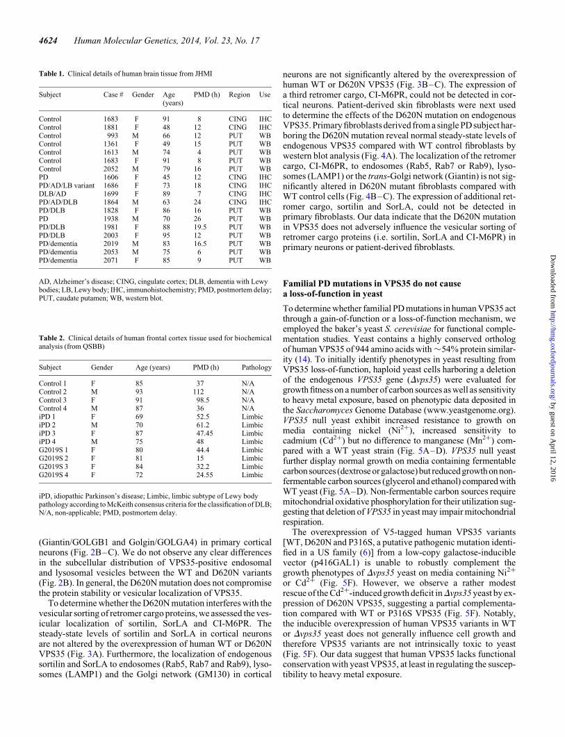
Table 1. Clinical details of human brain tissue from JHMI
Subject Case # Gender Age(years)
PMD (h) Region Use
Control 1683 F 91 8 CING IHCControl 1881 F 48 12 CING IHCControl 993 M 66 12 PUT WBControl 1361 F 49 15 PUT WBControl 1613 M 74 4 PUT WBControl 1683 F 91 8 PUT WBControl 2052 M 79 16 PUT WBPD 1606 F 45 12 CING IHCPD/AD/LB variant 1686 F 73 18 CING IHCDLB/AD 1699 F 89 7 CING IHCPD/AD/DLB 1864 M 63 24 CING IHCPD/DLB 1828 F 86 16 PUT WBPD 1938 M 70 26 PUT WBPD/DLB 1981 F 88 19.5 PUT WBPD/DLB 2003 F 95 12 PUT WBPD/dementia 2019 M 83 16.5 PUT WBPD/dementia 2053 M 75 6 PUT WBPD/dementia 2071 F 85 9 PUT WB
AD, Alzheimer’s disease; CING, cingulate cortex; DLB, dementia with Lewybodies; LB, Lewy body; IHC, immunohistochemistry; PMD, postmortem delay;PUT, caudate putamen; WB, western blot.
Table 2. Clinical details of human frontal cortex tissue used for biochemicalanalysis (from QSBB)
Subject Gender Age (years) PMD (h) Pathology
Control 1 F 85 37 N/AControl 2 M 93 112 N/AControl 3 F 91 98.5 N/AControl 4 M 87 36 N/AiPD 1 F 69 52.5 LimbiciPD 2 M 70 61.2 LimbiciPD 3 F 87 47.45 LimbiciPD 4 M 75 48 LimbicG2019S 1 F 80 44.4 LimbicG2019S 2 F 81 15 LimbicG2019S 3 F 84 32.2 LimbicG2019S 4 F 72 24.55 Limbic
iPD, idiopathic Parkinson’s disease; Limbic, limbic subtype of Lewy bodypathology according to McKeith consensus criteria for the classification of DLB;N/A, non-applicable; PMD, postmortem delay.
4624 Human Molecular Genetics, 2014, Vol. 23, No. 17
by guest on April 12, 2016
http://hmg.oxfordjournals.org/
Dow
nloaded from

As an alternative strategy, mutations analogous to the familialPD variants, D686N (D620N in hVPS35) and P299S (P316S inhVPS35), were introduced into yeast VPS35 in low-copygalactose-inducible expression vectors to assess the functionaleffects of these analogous PD-like mutations. The D620 andP316 residues in VPS35 are highly conserved from yeast tohumans (Fig. 5E). The overexpression of HA-tagged yeast
VPS35 variants (WT, D686N and P299S) are able to equivalentlyand robustly rescue the growth phenotype of Dvps35 yeast onmedia containing Ni2+ or Cd2+, whereas the overexpression ofyeast VPS35 variants does not influence the normal growth ofWT or Dvps35 yeast strains (Fig. 5F). Western blot analysisreveals equivalent levels of expression of human and yeastVPS35 variants in yeast following galactose induction
Figure 2. Normal steady-state levels and vesicular localization of VPS35D620N in cortical neurons. (A) Western blot analysis of soluble extracts from primary corticalneurons infected with lentiviral vectors expressing V5-tagged human VPS35 (WT or D620N) or control virus with anti-V5 or b-tubulin antibodies. Densitometricanalysis of human VPS35 normalized to b-tubulin levels indicates the equivalent expression of WT and D620N variants (mean+SEM, n ¼ 4 experiments). n.s.,non-significant by unpaired, two-tailed Student’s t-test. (B) Representative confocal microscopic images of primary cortical neurons co-labeled for WT or D620Nhuman VPS35 (V5) and RFP-Rab5, GFP-Rab7, RFP-LAMP1 or trans-Golgi protein Giantin, and DAPI. Inset indicates enlarged boxed area in merged images. Cyto-fluorograms and correlation coefficients (Rcoloc, mean+SEM, n ≥ 5 neurons) indicate the degree of co-localization of fluorescence signals for V5 and each marker.Scale bars: 10 mm. (C) Graph showing co-localization coefficients (mean+SEM, n ≥ 5 neurons/group) of WT or D620N VPS35 with each vesicular marker in cor-tical neurons. n.s., non-significant by unpaired, two-tailed Student’s t-test as indicated.
Human Molecular Genetics, 2014, Vol. 23, No. 17 4625
by guest on April 12, 2016
http://hmg.oxfordjournals.org/
Dow
nloaded from

Figure 3. Protein levels and vesicular sorting of the retromer cargo sortilin and SorLA are not altered by VPS35D620N expression in primary cortical neurons. (A) Westernblot analysis of soluble extracts from primary cortical neurons infected with lentiviral vectors expressing V5-tagged human VPS35 (WT or D620N) or a control virus,with antibodies to sortilin, SorLA and V5 or b-tubulin as a protein loading control. Graphs indicate densitometric analysis of sortilin or SorLA normalized tob-tubulinlevels and expressed as percent of the VPS35WT condition (mean+SEM, n ¼ 4 experiments). (B) Representative confocal microscopic images of primary corticalneurons co-labeled for human VPS35 (V5), sortilin or SorLA and each vesicular marker (GFP-Rab7 or RFP-LAMP1), and DAPI. Inset indicates enlarged boxed areain merged images. Cytofluorograms and correlation coefficients (Rcoloc, mean+SEM, n ≥ 7 neurons) indicate the degree of co-localization of fluorescence signalsfor sortilin or SorLA and each vesicular marker. Scale bars: 10 mm. (C) Graph showing co-localization coefficients (mean+SEM, n ≥ 7 neurons/group) of sortilin(upper) or SorLA (lower) with each vesicular marker (RFP-Rab5, GFP-Rab7, GFP-Rab9, RFP-LAMP1 or Golgi protein GM130) in cortical neurons expressinghuman VPS35 (WT or D620N) or a control vector. n.s., non-significant by one-way ANOVA with Newman–Keuls post hoc analysis.
4626 Human Molecular Genetics, 2014, Vol. 23, No. 17
by guest on April 12, 2016
http://hmg.oxfordjournals.org/
Dow
nloaded from

Figure 4. Normal VPS35 Levels and Vesicular Sorting of the Retromer Cargo CI-M6PR in Primary Human Fibroblasts Derived from a D620N Mutant PD Subject.(A) Westernblotanalysisof1%Triton-soluble (T-sol.) and Triton-insoluble (T-insol.) fractions ofprimaryfibroblasts derived froma Parkinson’s diseasesubject harboringthe D620N VPS35 mutation (PD) and a healthy control (Ctl). Blots are probed with antibodies to VPS35 and b-tubulin as a protein loading control. Molecular massis indicated in kDa. (B) Representative confocal microscopic images and cytofluorograms of human primary fibroblasts (control or D620N) co-labeled forcation-independent mannose-6-phosphate receptor (CI-M6PR) and vesicular markers (RFP-Rab5, GFP-Rab7, GFP-Rab9, RFP-LAMP1 or trans-Golgi proteinGiantin). Enlarged boxed areas of merged images are shown. Scale bar: 10 mm. (C) Graph indicates the co-localization coefficients (mean+SEM, n ≥ 5 cells) ofCI-M6PR with each vesicular marker in primary fibroblasts from control or D620N PD subjects. n.s., non-significant by unpaired, two-tailed Student’s t-test, as indicated.
Human Molecular Genetics, 2014, Vol. 23, No. 17 4627
by guest on April 12, 2016
http://hmg.oxfordjournals.org/
Dow
nloaded from

Figure 5. Analogous PD-like mutations in yeast VPS35 can functionally complement growth phenotypes in VPS35 null yeast cells. (A–D) Haploid yeast cells(BY4741, MATa), either wild-type (WT) or with a deletion of VPS35 (vps35D), were spotted onto YP(Dex) rich media containing different concentrations of(A) nickel (Ni2+), (C) cadmium (Cd2+) or (D) manganese (Mn2+), or YP media containing (B) non-fermentable carbon sources, glycerol (Gly) or ethanol(EtOH), and grown for 2–3 days at 308C. Shown are 5-fold serial dilutions (from top to bottom) starting with equal numbers of cells. (E) Protein sequence alignmentof VPS35 in the region encompassing the human P316 and D620 residues across several model species, indicating the high conservation of these two residues. (F) WT
4628 Human Molecular Genetics, 2014, Vol. 23, No. 17
by guest on April 12, 2016
http://hmg.oxfordjournals.org/
Dow
nloaded from
(Fig. 5G). Collectively, our data indicate that analogous PD-likemutations in yeast VPS35 (P299S and D686N), that are highlyconserved with human VPS35 (P316S and D620N) (Fig. 5E),are fully functional compared with the WT protein, suggestingthat these dominantly inherited mutations do not act through aloss-of-function mechanism. Our data support instead a gain-of-function mechanism for the pathogenic actions of familialPD mutations in VPS35.
Human VPS35 induces neuronal cell death and increasesneuronal vulnerability to stress
To explore the putative pathogenic effects of familial PD muta-tions in VPS35, human VPS35 variants (WT and D620N) wereoverexpressed in neuronal cultures to model gain-of-functioneffects. We evaluated the viability of rat primary corticalneurons following infection with lentiviral vectors expressinghuman VPS35 variants or GFP as a negative control. Lentiviralinfection of cortical cultures at days-in vitro (DIV) 3 results in theefficient and long-term transduction of the majority of corticalneurons as indicated by labeling of cultures for V5-taggedVPS35 or GFP (Fig. 6A). The overexpression of WT orD620N VPS35 in cortical neurons up to DIV 14 significantlyincreases apoptotic neuronal cell death (Fig. 6B) and reducesneuronal viability (Fig. 6C) compared to infection with controllentiviral vectors (empty or GFP). Furthermore, the transientoverexpression of human WT or D620N VPS35 in corticalneurons significantly impairs neurite outgrowth (Fig. 6D–E).Notably, no significant differences are observed between WTand D620N variants upon these VPS35-dependent neuronal phe-notypes.
To further explore and compare the impact of human WT andD620N VPS35 on neuronal vulnerability, we assessed the contri-bution of VPS35 to neuronal cell death induced by various cellu-lar toxins or stressors implicated in the pathogenesis of PD (5).The lentiviral-mediated overexpression of human WT orD620N VPS35 significantly sensitizes to neuronal cell deathinduced by exposure of primary cortical cultures to the mito-chondrial Complex-I inhibitors, MPP+ (250 mM) and rotenone(500 nM), and to oxidative stress induced by hydrogen peroxide(25 mM) (Fig. 6F). Human VPS35 variants also increase thevulnerability of cortical neurons to proteosomal inhibitioninduced by MG132 (10 mM) although to a smaller extent(Fig. 6G). VPS35 overexpression fails to increase neuronal vul-nerability to the autophagy/lysosomal inhibitor, bafilomycin A1(60 nM), or to disruption of ER-Golgi transport induced by bre-feldin A (250 mM) (Fig. 6G), thereby supporting a specific inter-action of VPS35 with neuronal susceptibility to mitochondrialtoxins, oxidative stress and proteasomal inhibition. No signifi-cant differences are observed between the WT and D620N var-iants of VPS35 in their capacity to increase neuronalvulnerability to toxins thereby arguing against a loss-of-functionmechanism for familial PD mutations. Taken together, our data
demonstrate that the overexpression of human VPS35 inducesneuronal cell death, impaired neurite outgrowth and increasesneuronal vulnerability to cellular stress most likely through again-of-function mechanism. However, pathogenic phenotypesinduced in primary neuronal culture models do not obviouslypermit the discrimination between the effects of WT andD620N VPS35, an observation that is similar for dominantlyinherited mutations in a-synuclein (15).
Viral-mediated expression of D620N VPS35 inducesdopaminergic neurodegeneration in rats
To explore the pathogenic effects of dominant VPS35 mutationsin vivo, human WT and D620N VPS35 were overexpressed insubstantia nigra dopaminergic neurons of rats to develop ananimal model of VPS35-associated PD. Adeno-associated viralvectors (AAV2/6) expressing V5-tagged human VPS35 (WT orD620N) were delivered to the substantia nigra pars compacta ofadult rats by unilateral stereotactic injection. At 12 weeks postin-jection of virus, we observe robust, widespread and equivalent ex-pression of WT and D620N VPS35 throughout the injectedsubstantia nigra with substantial co-localization with tyrosinehydroxylase (TH)-positive dopaminergic neurons (Fig. 7A–B).To assess whether human WT or D620N VPS35 induces dopa-minergic neurodegeneration, we quantified the number ofTH-positive dopaminergic and total Nissl-positive neurons inthe substantia nigra pars compacta of injected animals using un-biased stereological methodology. The expression of D620NVPS35 produces a significant loss of nigral dopaminergicneurons (32.02+8.6% loss) in the injected nigra comparedwith a control virus (5.9+3.2% loss) (Fig. 7C). The expressionof WT VPS35 induces an intermediate level of dopaminergicneuronal loss (24.6+7.4% loss) in the injected nigra comparedwith the control condition (Fig. 7C). A parallel loss of Nissl-positive neurons is observed in the injected substantia nigra rela-tive to the non-injected nigra with each virus (D620N, 23.23+8.2%; WT, 17.93+5.1%; control, 6.5+1.1%) indicating dopa-minergic neurodegeneration rather than a loss of TH expression ordopaminergic phenotype (Fig. 7C). A corresponding loss ofTH-positive dopaminergic nerve terminals (D620N, 14.6+4.4%; WT, 9.1+5.5%; control, 5.3+1.3%) is also observedin the ipsilateral striatum compared with the contralateral striatumof rats injected with each virus (Fig. 7D–E). To determinewhether VPS35-induced dopaminergic neuronal loss culminatesin motoric deficits in rats, we employed the cylinder test tomeasure contralateral forelimb use which is controlled by the ip-silateral substantia nigra (16). However, we observe no significantimpairment ofcontralateral forelimbuse inducedby the ipsilateralexpression of human WT or D620N VPS35 at 7, 9 and 11 weekspostinjection of virus (Fig. 7F).
To confirm the equivalent expression of human VPS35 var-iants in injected animals, we conducted western blot analysisof ventral midbrain extracts at 12 weeks postinjection. Human
or vps35D yeast strains were transformed with galactose-inducible low-copy expression vectors containing HA-tagged yeast VPS35 (WT, D686N or P299S) orV5-tagged human VPS35 (WT, D620N or P316S) variants, or with an empty vector (p416GAL1). Equivalent numbers of cells were spotted as five-fold serial dilutions(from left to right) onto complete synthetic media lacking uracil (CSM) and containing galactose as the sole carbon source with or without additional Ni2+ (2.5 mM) orCd2+ (12 mM), as indicated. (G) Western blot analysis of soluble extracts from WT yeast cells transformed with yeast or human VPS35 variants following growth onCSM-URA media containing galactose to induce VPS35 expression. VPS35 is detected using anti-HA or anti-V5 antibodies with an antibody to 3-phosphoglyceratekinase (PGK) used as a protein loading control.
Human Molecular Genetics, 2014, Vol. 23, No. 17 4629
by guest on April 12, 2016
http://hmg.oxfordjournals.org/
Dow
nloaded from

V5-tagged WT and D620N VPS35 are detected at equivalentlevels in the injected ventral midbrain of rats (Fig. 7G–H). Anantibody that detects both rat and human VPS35 fails to demon-strate the overexpression of human VPS35 relative to endogen-ous VPS35 in ventral midbrain extracts suggesting either adilution effect of the transgene in this brain region or a potentiallowering of endogenous VPS35 levels in response to humanVPS35 expression (Fig. 7G). Western blot analysis further indi-cates a reduction of TH levels in the ventral midbrain induced byWT and D620N VPS35 expression (Fig. 7G–H), which corre-lates with the observed dopaminergic neuronal loss (Fig. 7C).Consistent with our data in neuronal cultures (Fig. 3A), we donot observe alterations in the steady-state levels of sortilin or
SorLA resulting from the expression of human VPS35 variantsin the ventral midbrain of rats (Fig. 7G–H). It was not possibleto reliably detect CI-M6PR in these ventral midbrain extractswith currently available antibodies. Instead, the lysosomal pro-tease cathepsin D, a soluble ligand of CI-M6PR (17), wasassessed. However, the expression of human VPS35 variantsdoes not alter the steady-state levels or processing of cathepsinD in the ventral midbrain (Fig. 7G–H).
The neuropathological spectrum of PD brains harboringVPS35 mutations is uncertain since no mutation carriers haveyet come to autopsy (9). To determine the pathological conse-quences of VPS35 mutations in vivo, we assessed the distributionof a number of classical markers of neurodegeneration in the
Figure 6. Overexpression of human VPS35 induces neuronal cell death, impairs neurite outgrowth and increases neuronal vulnerability to cellular stress. (A) Rep-resentative immunofluorescent images of primary cortical neurons at DIV 14 infected with GFP or VPS35WT lentivirus (green) and co-labeling of apoptotic (TUNEL-positive, red) or total (DAPI-positive, cyan) nuclei to assess neuronal cell death. Scale bar: 50 mm. (B) Cortical neurons infected with lentiviral vectors expressing GFPor human VPS35 variants were assessed for apoptotic cell death by quantifying the number of TUNEL-positive neurons as a percent of total neurons (DAPI-positivenuclei) for each condition (mean+SEM, n ¼ 5 independent cultures). (C) Neuronal viability was assessed in cortical cultures infected with lentiviral vectors at DIV14 by counting the average number of neurons (DAPI-positive nuclei) for each condition, and expressed as a percent of an empty control virus (mean+SEM, n ¼ 5independent cultures). (D) Representative fluorescent microscopic images of cortical neurons at DIV 7 co-labeled with V5-tagged human VPS35 (WT or D620N) andGFP. GFP images were pseudocolored with ICA to identify neuronal soma (arrowhead) and axonal processes (arrows) for neurite length measurements. Scale bar:200 mm. (E) Quantitation of axonal process length from GFP-positive neurons expressing human VPS35 (WT or D620N) compared with control neurons (emptyvector). Bars represent neurite length (mean+SEM, n ¼ 5 independent cultures) from 170–189 neurons. (F) Primary cortical neurons infected with lentiviralvectors expressing human VPS35 (WT or D620N) or GFP were treated at DIV 14 with cellular toxins (MPP+, rotenone or H2O2) for 24 h and assessed by alamarBluecell viability assay. Bars represent cell viability expressed as a percent of untreated cultures for each condition (mean+SEM, n ≥ 3 independent cultures). (G) Cor-tical neurons infected with lentiviral vectors as in F were treated with cellular toxins (MG132, BrefeldinA or Bafilomycin A1) for 24 h prior to cell viability assays. Cellviability relative to untreated cultures is shown (mean+SEM, n ≥ 3 independent cultures). Data were analyzed by one-way ANOVA with Newman–Keuls post hocanalysis, as indicated (∗P,0.05, ∗∗P,0.01 or ∗∗∗P,0.001). n.s., non-significant.
4630 Human Molecular Genetics, 2014, Vol. 23, No. 17
by guest on April 12, 2016
http://hmg.oxfordjournals.org/
Dow
nloaded from

substantia nigra pars compacta of rats expressing WT or D620NVPS35. At 12 weeks postinjection, we do not observe alterationsin the distribution of total or phosphorylated (Ser129)a-synuclein (Fig. 8), a major component of Lewy body path-ology in PD and other a-synucleinopathies (3,18), followinghuman VPS35 expression. Similarly, we do not detect changesin total (Tau5) or hyperphosphorylated (PHF-1) forms of themicrotubule-associated protein tau (Fig. 8), a protein involvedin neurodegenerative tauopathies (19) and implicated in PD(20,21). Alterations in phosphorylated neurofilaments (SMI31and SMI312), a marker of axonal processes and terminals, orp62/sequestosome 1, a marker of autophagic/lysosomalvacuoles, are also not detected in the injected substantia nigra(Fig. 8). The expression of human WT and D620N VPS35results in the accumulation of spherical inclusions containingamyloid precursor protein (APP) within axonal processes(Fig. 8), a sensitive marker of axonal damage (22). Gliosis typic-ally accompanies neuronal degeneration in PD and other neuro-degenerative diseases. However, astrogliosis (GFAP) andmicrogliosis (Iba1) are not generally observed in the substantianigra of rats at 12 weeks postinjection of virus (Fig. 8).
The accumulation of APP-positive axonal inclusions suggeststhat human VPS35 expression may also promote axonal degener-ation. Substantia nigra sections from injected rats at 12 weekswere stained with silver which specifically labels components ofdegenerating neurons, including lysosomes, axonal processesand their terminals (23,24). The expression of D620N VPS35induces a significant increase in the appearance of silver-positivedegenerating axonal processes and nerve terminals comparedwith WT VPS35 or a control virus (Fig. 9). Collectively, ourdata reveal the marked degeneration of substantia nigra dopamin-ergic neurons and axonal pathology induced by the expression ofthe familial D620N mutation of VPS35 in the adult rat brain,which recapitulates a cardinal pathological hallmark of PD.
DISCUSSION
Mutations in the VPS35 gene cause late-onset, autosomal domin-ant PD with the D620N mutation representing a common cause ofVPS35-linked disease (6–9). The mechanism(s) through whichdominant VPS35 mutations precipitate neurodegeneration in PDare not known, and the neuropathological consequences ofVPS35 mutations in PD subjects are poorly defined at present.Here, we present multiple lines of evidence demonstrating thatthe dominant D620N mutation in VPS35 is capable of inducingdopaminergic neuronal degeneration most likely through again-of-function mechanism. First, familial mutations do notcompromise VPS35 protein stability or its normal localizationto endosomes and lysosomes. Furthermore, the vesicular sortingof retromer cargo, sortilin, SorLA and CI-M6PR, which dependon VPS35 as a key component of the retromer substrate recogni-tion subcomplex (11), is not altered by mutations in VPS35.Second, the equivalent PD-like mutation in yeast VPS35(D686N) is fully functional compared with the WT yeastprotein in complementing growth phenotypes in VPS35 nullyeast. The D620 residue and the VPS35 protein sequence ingeneral are highly conserved throughout evolution. Therefore,our data suggest that the D620N mutation does not result in aloss-of-function. Third, the lentiviral-mediated overexpression
of human VPS35 variants impairs neuronal integrity and viability,and increases the vulnerability of neurons to PD-relevant cellularstressors, including mitochondrial Complex-I inhibition, oxida-tive stress and proteasomal inhibition (5). Fourth, the AAV-mediated expression of human D620N VPS35 in the substantianigra of adult rats induces marked dopaminergic neuronal degen-eration with an intermediate effect of human WT VPS35 com-pared with a control virus. Therefore, the D620N mutation inVPS35 recapitulates a cardinal pathological hallmark of PD in anovel animal model. Taken together, our data demonstrate thatthe PD-associated dominant D620N mutation in VPS35 is ableto induce neuronal degeneration most likely through a gain-of-function mechanism, thereby establishing an important contribu-tion of VPS35 to the development of PD.
The distinct absence of truncation, rearrangement or deletionmutations in VPS35 identified so far across multiple PD cohortsworldwide, together with the dominant inheritance pattern ofthe common D620N mutation, suggests that mutations are notlikely to act through a loss-of-function mechanism. Thus,VPS35 mutations most likely operate through a gain-of-functionmechanism. Consistent with such a mechanism, the D620N muta-tion does not compromise the steady-state levels of VPS35 and itsnormal localization to early and late endosomes and lysosomeswithincorticalneurons, suggesting the absence of major structuralperturbations toVPS35.Arecent studysuggestsa redistributionofearly and late endosomes containing D620N VPS35 to the peri-nuclear region in A431 cells (25). However, our data do not indi-cate a redistribution of D620N VPS35-positive vesicles in corticalneurons. We further demonstrate that the ectopic expressionof D620N VPS35 in rodent cortical neurons or endogenousD620N VPS35 in human fibroblasts does not influence thelevels or vesicular sorting of well-known retromer cargo proteins,such as sortilin, SorLA and CI-M6PR. Thus, the D620N mutationdoes not apparently influence the overall substrate recognition orbinding of retromer cargo to VPS35. A recent study suggests thatthe expression of D620N VPS35 causes a defect in the vesiculartrafficking and processing of the lysosomal protease cathepsinD, a soluble ligand of CI-M6PR (25). Whether the defective traf-ficking of cathepsin D induced by D620N VPS35 also occurs inneurons and promotes neuronal degeneration in PD remainsunclear. However, we failed to observe alterations in the levelsand processing of cathepsin D in the ventral midbrain of ratsexpressing human D620N VPS35. Our findings suggest that fa-milial mutations in VPS35 do not lead to an overall disruptionof VPS35 protein structure and retromer-dependent proteinsorting in neurons, implying that mutations may instead exertsubtle or selective effects on specific retromer cargo and/or in spe-cific cell types.
Recent studies have suggested that the D620N mutation mightcompromise neuroprotective effects mediated by human VPS35overexpression, consistent with a loss-of-function mechanism.WT VPS35 was recently shown to protect against neuronal tox-icity induced by the mitochondrial Complex-I inhibitor, MPP+
(26), as well as against G2019S LRRK2 overexpression orRAB7L1 deficiency (27), two models relevant to familial PD. Inthis scenario, VPS35 haploinsufficiency would be sufficient toinduce neurodegeneration related to PD. While the homozygousdeletion of VPS35 in mice results in early embryonic lethality,VPS35 haploinsufficiency has been reported to enhance Alzhei-mer’s disease (AD)-like neuropathology in a mouse model of
Human Molecular Genetics, 2014, Vol. 23, No. 17 4631
by guest on April 12, 2016
http://hmg.oxfordjournals.org/
Dow
nloaded from

Figure 7. Dopaminergic neuronal degeneration induced by AAV2/6-mediated expression of D620N VPS35 in the substantia nigra of adult rats. (A) Photomicrographsshowing immunofluorescent co-labeling of substantia nigra with anti-V5 and anti-TH antibodies indicating the equivalent expression of V5-tagged human VPS35WT
and VPS35D620N proteins within dopaminergic neurons at 12 weeks following stereotactic injection of AAV2/6 vectors. Scale bar: 200 mm. (B) Representative photo-micrographs of anti-V5 and anti-TH immunohistochemical analysis in adjacent sections of rat substantia nigra at 12 weeks following the stereotactic injection ofAAV2/6 vectors expressing human VPS35WT and VPS35D620N or a control vector. V5 labeling in the substantia nigra is accompanied by a decrease in TH
4632 Human Molecular Genetics, 2014, Vol. 23, No. 17
by guest on April 12, 2016
http://hmg.oxfordjournals.org/
Dow
nloaded from

AD (28). Whether PD-related neuropathology or motor deficitsoccur in mice heterozygous for VPS35 is not yet known (28).We employed yeast as a model to demonstrate that mutationsequivalent to D620N in a highly conserved yeast ortholog ofVPS35 (i.e. D686N) can normally complement growth pheno-types inDvps35 yeast thereby indicating that the D � N substitu-tion at this highly conserved residue does not compromise VPS35function. In contrast to prior studies (26,27), we demonstrate thatthe overexpression of human WT VPS35 in primary corticalneurons is detrimental rather than neuroprotective and leads toneuronal cell death and impaired neurite outgrowth. Moreover,VPS35 overexpression dramatically increased neuronal vulner-ability to PD-relevant cellular stressors, including the mitochon-drial Complex-I inhibitors MPP+ and rotenone, rather thaneliciting neuroprotection as previously suggested (26). Impor-tantly, the D620N mutation did not impair VPS35-induced neur-onal toxicity or increased vulnerability which would beinconsistent with a loss-of-function mechanism. In our neuronalculture model we were not able to discern obvious differencesbetween WT and D620N VPS35 in their capacity to induce neur-onal damage. In contrast, the viral-mediated overexpression ofhuman D620N VPS35 in the rat substantia nigra induced dopa-minergic neuronal degeneration and axonal damage, whereasWT VPS35 produced an intermediate level of neuropathology.Therefore, the pathological effects of the D620N mutation canbe unmasked given the proper cellular context, and supports again-of-function mechanism for the actions of PD-associatedVPS35 mutations.
We have developed a novel animal model of VPS35-associatedPD that recapitulates one of the cardinal pathological features ofdisease, i.e. nigral dopaminergic neuronal loss. The degenerationof dopaminergic neurons in adult rats induced by D620N VPS35was pronounced at 12 weeks after viral delivery. Whether thismodel recapitulates the progressive nature of neuronal loss inPD is not yet clear. It will be important in future studies to deter-mine whether varying the dosage or time course of transgene ex-pression would be sufficient to further enhance nigral neuronaldegeneration, and potentially exacerbate differences betweenWT and D620N VPS35. The expression of D620N VPS35 inthe rat substantia nigra does not recapitulate other aspects ofPD-related neuropathology such as the formation of a-synu-clein-positive Lewy pathology, tau-positive neurofibrillarytangles or gliosis. However, we do not yet know the full repertoireof pathological inclusions or aggregates in brains from PD sub-jects with VPS35 mutations. Our in vivo studies formally provethat the dominant D620N mutation inVPS35can induce dopamin-ergic neuronal loss, a key pathological feature of familial and
idiopathic PD. This new rodent model of VPS35-associated PDwill prove useful for uncovering the molecular mechanisms ofVPS35-dependent neurodegeneration and for the identificationof disease-modifying therapeutic agents.
MATERIALS AND METHODS
Animals
Rats were maintained in a pathogen-free barrier facility andexposed to a 12 h light/dark cycle with food and water providedad libitum. Pregnant female Sprague-Dawley rats were obtainedfrom Charles River Laboratories (L’Arbresle Cedex, France)and resulting P1 rats were used for preparation of primary neur-onal cultures. All animal experiments were approved by theSCAV (Service de la consommation et des affaires veterinaires)in the Canton de Vaud, Switzerland (Animal authorization No.2572), and conducted in strict accordance with the EuropeanUnion directive (2010/63/EU) for the care and use of laboratoryanimals.
Expression plasmids and antibodies
A pLenti6/V5/DEST mammalian expression plasmid contain-ing full-length human VPS35 with a C-terminal V5 tag wasobtained from Addgene [#21691, (29)]. D620N and P316Smutations were introduced by PCR-mediated site-directed mu-tagenesis and sequenced to confirm their integrity. V5-taggedVPS35 cDNAs (WT, D620N or P316S) were amplified byPCR from a pLenti6-VPS35 vector, and cloned into a directionalpENTR/D-TOPO entry vector (Invitrogen), sequenced toconfirm their integrity, and subjected to recombination with aGateway-compatible lentiviral vector, pSIN-PGK-WHV (30)(kindly provided by Dr. Nicole Deglon, CHUV, Switzerland).As plasmid controls, pcDNA3.1 (Invitrogen), pEGFP-N1 (Clon-tech) and pDsRed-Max-N1 (Addgene #21718) were employed.Expression plasmids containing human RFP-Rab5A (#14437),human GFP-Rab7A (#12605), human GFP-Rab9A (#12663)and rat LAMP1-RFP (#1817) were obtained from Addgene.
The following primary antibodies were used: rabbit VPS35(ABT48, Millipore), mouse anti-V5 and anti-V5-peroxidase(Invitrogen), mouse anti-b-tubulin (clone TUB 2.1, Sigma-Aldrich), rabbit anti-tyrosine hydroxylase (TH, NB300-109,Novus Biologicals), mouse anti-TH (clone TH-2, Sigma-Aldrich), mouse anti-HA-peroxidase (clone 12CA5, Roche),mouse anti-PGK (clone 22C5D8, Invitrogen), rabbit anti-Dynamin-1 (PA1-660, Thermo Scientific), mouse anti-TIM23
immunostaining. Scale bar: 1 mm. (C) Stereological analysis of TH-positive dopaminergic or total Nissl-positive neurons in the substantia nigra expressed as percentcell loss relative to the contralateral uninjected nigra. Bars represent mean+SEM (n ¼ 6 animals/group). ∗P,0.05 by one-way ANOVA with Dunnett’s post hocanalysis. n.s., non-significant. (D) Representative photomicrographs of immunostaining for TH-positive nerve terminal in the striatum at 12 weeks following AAV2/6delivery. Scale bar: 1 mm. (E) Quantitation of optical density of TH immunostaining in the striatum. Data are expressed as percent loss of TH-positive fibers relative tothe contralateral uninjected side. Bars represent the mean+SEM (n ≥ 4 animals/group). Data were assessed by one-way ANOVA with Dunnett’s post hoc analysis.(F) Cylinder asymmetry test performed at 7, 9 and 11 weeks postsurgery assessing forelimb contacts with the cylinder wall over a 5 min period. Forelimb use contra-lateral to the injected nigra is expressed as a percent of total forelimb contacts (left + right) on the cylinder wall. Dashed line indicates the expected forelimb usageunder normal conditions. Bars represent mean+SEM (n ¼ 8 animals/group). Data were assessed by one-way ANOVA with Newman–Keuls post hoc analysis. (G)Western blot analysis of soluble extracts from ventral midbrain at 12 weeks following stereotactic injection of rats with AAV2/6 vectors, separated into ipsilateral (+)and contralateral (2) hemispheres. Blots were probed with antibodies to V5, VPS35, TH, sortilin, SorLA and cathepsin D, with b-tubulin as a loading control. Mo-lecular masses are indicated in kDa. (H) Densitometric analysis of V5, TH, sortilin, SorLA or cathepsin D levels normalized to b-tubulin levels on blots containingventral midbrain extracts. Bars represent mean+SEM (n ¼ 3–4 animals/group). Data were analyzed by one-way ANOVA with Newman–Keuls post hoc analysisfor multiple comparisons, or by unpaired, two-tailed Student’s t-test for pair-wise comparisons, as indicated. n.s., non-significant.
Human Molecular Genetics, 2014, Vol. 23, No. 17 4633
by guest on April 12, 2016
http://hmg.oxfordjournals.org/
Dow
nloaded from

Figure 8. AAV2/6-mediated expression of human VPS35 in the rat substantia nigra induces axonal pathology. Immunohistochemical analysis of substantia nigra at 12weeks following the stereotactic injection of AAV2/6 vectors expressing human VPS35WT and VPS35D620N or a control vector. Antibodies detecting totala-synuclein, a-synuclein phosphorylated at Ser129 (P-a-synuclein), total tau (Tau5), tau phosphorylated at Ser396 and Ser404 (PHF-1), APP isoforms, phosphory-lated neurofilament H (SMI31), phosphorylated neurofilament M/H (SMI312), autophagy substrate p62/sequestosome 1, GFAP and Iba1 were employed. Represen-tativephotomicrographsare taken from the injected substantia nigra as indicated in the schematicdiagram.Scale bar: 50 mm. Macroscopic imagesof GFAPand Iba1 inthe injected and non-injected substantia nigra are shown for comparison. Boxes indicate the region used for high-power images of the injected substantia nigra. Scalebar: 50 mm.
4634 Human Molecular Genetics, 2014, Vol. 23, No. 17
by guest on April 12, 2016
http://hmg.oxfordjournals.org/
Dow
nloaded from

(clone 32, BD Biosciences), mouse anti-a-synuclein (clone 42,BD Biosciences), mouse anti-pSer129 a-synuclein (Wako),mouse anti-GM130 (clone 35, BD Biosciences), rabbit anti-Giantin (ab24586, Abcam), rabbit anti-tGolgin-1/GOLGA4(kindly provided by Dr Mickey Marks, University of Pennsylva-nia), rabbit anti-sortilin (ab16640, Abcam), rabbit anti-SorLA(ab16642, Abcam), mouse anti-CI-M6PR (clone 2G11,Abcam), rabbit anti-cathepsin D (Santa Cruz Biotech), rabbitanti-Iba1 (Wako), rabbit anti-GFAP (Z0334, Dako), mouse anti-phosphorylated neurofilament H (clone SMI-31, Covance),mouse anti-phosphorylated neurofilament M/H (clone SMI-312,Covance), mouse anti-synaptophysin 1 (clone 7.2, SynapticSystems), guinea pig anti-p62 (GP62, Progen Biotech), mouseanti-APP (clone 22C11, Millipore), mouse anti-Tau (cloneTAU-5, Invitrogen), mouse anti-PHF-1 (kindly provided by DrPeter Davies, Albert Einstein College of Medicine). For bright-field microscopy, biotinylated secondary antibodies used weregoat anti-mouse, anti-rabbit or anti-guinea pig (Vector Laborator-ies) followed by ABC reagent and DAB Peroxidase Substrate(Vector Laboratories). For fluorescence confocal imaging, sec-ondary antibodies used were: AlexaFluor-488, -546, or -633goat anti-mouse IgG (H + L), and AlexaFluor-488, -546 or-633 goat anti-rabbit IgG (H + L) (Life Technologies). Second-ary antibodies used were: HRP- or biotin-conjugated mousemonoclonal anti-rabbit IgG, light chain-specific (Jackson Immu-noresearch), goat anti-mouse IgG, light chain-specific (JacksonImmunoresearch) and goat polyclonal anti-guinea pig IgG, H +L (Abcam).
Yeast growth assays
Human VPS35 variants (WT, D620N or P316S) were amplifiedby PCR from a pLenti6-VPS35 vector (Addgene #21691), andyeast VPS35 variants (WT, D686N or P299S) were amplified
by PCR from a pBG1805-VPS35 vector containing a C-terminalHA tag (Thermo Scientific). Blunt-end PCR products werecloned into a directional pENTR/D-TOPO entry vector,sequenced to confirm their integrity, and subjected to recombin-ation with the Gateway-compatible vector, p416GAL1-ccdB(CEN6, URA3) obtained from Addgene (31). Yeast haploidWT parental or vps35D strains on a BY4741 genetic background(MATa, his3D1, leu2D0, met15D0, ura3D0) were obtained fromThermo Scientific. Yeast cells were transformed with plasmidsusing a standard lithium acetate procedure. Yeast cells trans-formed with p416GAL1 expression plasmids were routinelygrown in complete synthetic media lacking uracil (CSM-URA)with 2% dextrose. Equivalent yeast cells were serially diluted(5-fold) and spotted onto plates containing solid media (YP orCSM-URA) with 2% dextrose, galactose, glycerol or ethanolas the sole carbon source. Cells were grown at 308C for 2–3days before imaging.
Lentivirus production
Lentiviral vectors were produced as described (32) inHEK-293T cells using a third generation packaging system.Lentivirus was purified by centrifugation and resuspended in1× phosphate buffered saline pH 7.4 and 0.5% bovine serumalbumin buffer. Viral titer was determined using the HIV-1p24 antigen ELISA kit (Zeptometrix Corp, Buffalo, USA). Ap24 of 66 ng/ml was routinely used for infecting primary neuron-al cultures at a density of 400 000 cells in 3 ml media.
Cell culture and transfection
HEK-293T cells were grown in Dulbecco’s modified Eagle’smedia (DMEM) supplemented with 10% fetal bovine serum(FBS) and addition of penicillin/streptomycin in a humidified
Figure 9. Nigral axonal degeneration induced by AAV2/6-mediated expression of D620N VPS35. Representative photomicrographs of substantia nigra sections pro-cessedwith Gallyas silver stain, from rats injected with AAV2/6vectors expressinghumanVPS35WT and VPS35D620N or a control vector, to reveal degenerating axonsand nerve terminals (indicated in black). Scale bar: 50 mm. Images were subjected to deconvolution, color separation and filtering (circularity index ≤0.6 to removenuclei/soma) using ImageJ to isolate black particles representative of neuritic processes and terminals for quantitation. Examples of particles analyzed are shownpseudocolored in red. Data are expressed as the number of black particles (silver-positive neurites/terminals) and bars represent the mean+SEM (n ¼ 6 animals/group). ∗∗P,0.01 or ∗∗∗P,0.001 by one-way ANOVA with Tukey’s post hoc analysis as indicated. n.s., non-significant.
Human Molecular Genetics, 2014, Vol. 23, No. 17 4635
by guest on April 12, 2016
http://hmg.oxfordjournals.org/
Dow
nloaded from
378C incubator with 5% CO2. Primary human fibroblasts weremaintained in DMEM supplemented with 10% FBS, 2 mML-glutamine and penicillin/streptomycin. Expression plasmidswere transfected into cells using XtremeGENE HP DNA trans-fection reagent (Roche Applied Science) or Lipofectamine2000 (Life Technologies) according to manufacturer’s instruc-tions. Biochemical or immunocytochemical analysis was per-formed at 48–72 h post-transfection.
Human primary fibroblasts
Fibroblasts were generated from a 6 mm skin punch biopsy takenunder local anesthetic following local ethics board approval andinformed consent from the patient. Biopsies were dissected into1 mm pieces and cultured in 5 cm2 petri dishes in media untilfibroblasts grew out from the explants. When fibroblastsreached confluency they were transferred to larger culturevessels for expansion. Fibroblasts were generated from asingle normal control individual and a PD subject harboring ac.1858G.A, p.Asp620Asn mutation in the VPS35 gene, asdescribed (33).
Immunocytochemistry
Primary neurons or fibroblasts were fixed in 4% paraformalde-hyde (PFA) and processed for immunocytochemistry as previ-ously described (32). Images were captured with a Zeiss LSM700 laser scanning confocal microscope (Carl Zeiss AG) witha Plan-Apochromat 63×/1.40 oil objective. Acquired imagesfrom single z-plane (0.1-mm thick) were subjected to deconvolu-tion with HuygensPro software (Scientific Volume Imaging).Co-localization coefficients (Rcoloc) of deconvolved multi-channel images were calculated using NIH ImageJ software aspreviously described (32).
Neuronal viability and neurite length assays
Primary cortical neuronal cultures were prepared from P1Sprague-Dawley rats as previously described (32,34). For assess-ment of neurite length, cultures were co-transfected at DIV 3 withV5-tagged human VPS35 and GFP plasmids at a 10:1 DNA molarratio (5 mg total DNA per 35 mm dish) using Lipofectamine 2000reagent (Invitrogen). Cultures were fixed with 4% PFA at DIV 7and immunostained with mouse anti-V5 antibody (Invitrogen),and anti-mouse IgG-AlexaFluor-633 antibody (Invitrogen).Images were acquired using an EVOS fluorescence digitalmicroscope (Advanced Microscopy Group). ImageJ softwarewas used to measure the length of the longest neurite fromcontrol (GFP-positive) and VPS35-expressing (GFP-/V5-posi-tive) neurons. Sampling was performed randomly with ≥30neurons measured per condition for each experiment from 4–6 in-dependent cultures/experiments. All measurements were per-formed by investigators blinded to experimental condition.TUNEL labeling was performed as previously described (32) toassess apoptotic cell death in cortical neurons infected with lenti-viral vectors expressing V5-tagged VPS35 or GFP at DIV 3 andfixed with 4% PFA at DIV 14. Cell viability was also assessed fol-lowing treatment of lentiviral-infected cortical neurons seeded in96-well plates with cellular toxins for 24 h using the alamarBlueCell Viability assay (Invitrogen).
Subcellular fractionation
Cerebral cortex from adult mice (C57Bl/6J) was subjected tosubcellular fractionation as previously described (32). The fol-lowing fractions were obtained: total homogenate (H) andnuclear/whole cell pellet (P1), soluble cytosolic (S1, S2 andS3), heavy membrane (P2), light membrane/microsomal (P3),synaptosomal membrane (LP1) and cytosolic (LS1), synapticvesicle-enriched membrane (LP2) and cytosolic (LS2).
Human brain tissue
For immunohistochemical analysis, postmortem human braintissue was obtained through the brain donation program of theMorris K. Udall Parkinson’s Disease Research Center of Excel-lence and the Alzheimer’s Disease Research Center at JohnsHopkins Medical Institutions (JHMI) in compliance with localInstitutional Review Board and HIPAA regulations. Formalin-fixed, paraffin-embedded tissue sections (10-mm thick) contain-ing cingulate cortex tissue from two normal control and four PD/DLB brains was obtained as indicated in Table 1 and describedpreviously (34). For biochemical analysis, caudate putamentissue derived from five normal control and seven PD/DLB sub-jects was obtained from JHMI (Table 1), and frontal cortex tissuederived from four normal control and eight PD subjects wasobtained from the archive at Queen Square Brain Bank(QSBB) as previously reported (35) and indicated in Table 2.Flash-frozen tissues were homogenized with (10% w/v) lysisbuffer [20 mM Tris–HCl pH 7.4, 150 mM NaCl, Complete pro-tease inhibitor and phosphatase inhibitor cocktails (RocheApplied Science)] and equal proteins were resolved by 3–8%Tris–acetate sodium dodecyl sulphate-polyacrylamide gel elec-trophoresis (SDS-PAGE) (Invitrogen). Blots were probed withanti-VPS35, anti-actin or anti-b-tubulin antibodies.
Recombinant AAV2/6 virus production
V5-tagged human VPS35 (WT or D620N) cDNAs were ampli-fied by PCR from a pLenti6-VPS35 vector and cloned into apAAV-mPGK-MCS vector (16), modified from the serotype 2pAAV-CMV-MCS vector (Stratagene, La Jolla, CA, USA).The same backbone plasmid was used to produce the non-expressing control virus. Recombinant AAV2 genome packagedin the AAV serotype six capsid was produced and titrated asdescribed previously (16,36). Viruses were diluted to a final con-centration of 2.5 × 10⁹ transducing units (TU) per ml.
Stereotactic surgeries
Female adult Sprague-Dawley rats (Charles River Laboratories,France) weighing 180–200 g received a viral titer of 5 × 10⁶
TUs (in a 2 ml volume), unilaterally, at coordinates correspond-ing to the rat substantia nigra, as previously described (36).Animals were sacrificed at 12 weeks postsurgery.
Biochemical analysis of cell culture and tissues
Cells or brains were homogenized with lysis buffer containing1% Triton X-100, 50 mM Tris–HCl pH 7.5, 150 mM NaCl,5% glycerol, 1 mM EDTA, Complete protease inhibitors
4636 Human Molecular Genetics, 2014, Vol. 23, No. 17
by guest on April 12, 2016
http://hmg.oxfordjournals.org/
Dow
nloaded from

(Roche Applied Sciences) and phosphatase inhibitor cocktails 2and 3 (Sigma). Rat tissues for biochemical analysis were har-vested at 12 weeks after viral delivery (n ¼ 4 animals/group).Ventral midbrains from injected and non-injected hemisphereswere rapidly dissected and subjected to mechanical homogen-ization with (10% w/v) lysis buffer. Cell and tissue lysateswere centrifuged at 100 000g for 30 min at 48C. To obtain the in-soluble fraction, Triton X-100-insoluble pellets from fibroblastswere solubilized in buffer containing 2% SDS, 50 mM Tris–HClpH 7.4 and Complete protease inhibitors (Roche). Equivalentproteins as determined by BCA assay (Pierce Biotechnology)were resolved by SDS-PAGE and subjected to western blot ana-lysis with appropriate primary and secondary antibodies. Pro-teins were visualized by enhanced chemiluminescence (GEHealthcare) and digital images were captured using a chemilu-minescent Image Analysis system (LAS-4000, FujiFilm).Acquired images were subjected to densitometric quantitationusing NIH ImageJ software. The relative optical density valuesfor each protein band were normalized to the correspondinglevels of b-tubulin, actin or PGK proteins to control for proteinloading.
Immunohistochemistry
Animals were deeply anesthetized and transcardially perfusedwith saline solution followed by 4% PFA in 0.1 M phosphatebuffer (pH 7.3). Brains were immersed in 30% sucrose solutionbefore sectioning using a cryostat (Leica, Jung Frigocut 2800N).Immunohistochemistry on rat and human brain tissue withvarious antibodies was conducted as described previously(34,37). Silver staining was performed using the FD NeuroSilverkit II following the manufacturer’s protocol (FD Neurotechnol-ogies, Ellicott City, MD). Images were captured with the lightmicroscopes AX70 and Slide Scanner VS120-L100 (Olympus)or a Zeiss LSM 700 Laser Scanning confocal microscope (CarlZeiss AG).
Cylinder test
The forelimb usage of rats was assessed by placing each animalinto a transparent cylinder and recording the exploratory activityfor 5 min. The contact of either left or right forelimbs with thecylinder wall, while the animals were in a vertical posture, tosupport body weight or to initiate lateral stepping movementswas scored in a manner blinded to experimental group. Forelimbuse asymmetry was quantified by calculating the instances whenthe impaired (contralateral) forelimb was used as a percentage oftotal number of both limb usages on the cylinder wall.
Stereological quantitation of neurons
Unbiased stereological estimation of the number of dopamin-ergic neurons was performed using the optical fractionatorprobe of the StereoInvestigator software (MicroBrightFieldBiosciences), as described (37,38). Every fifth serial coronalsection of 40 mm thickness, covering the entire substantianigra region was immunostained with rabbit anti-TH antibodyand counterstained with cresyl violet stain. Sampling was per-formed in a systematic random manner using a grid of 220 ×240 mm squares covering the substantia nigra overlayed oneach section and applying an optical dissector consisting of a
75 × 75 × 20 mm square cuboid. All analyses were performedby investigators blinded to each condition.
The optical density of TH-immunoreactive nerve terminalswas quantified throughout the striatum as a measure of dopamin-ergic innervation. The rostral and caudal limits of the striatumused for analysis were 2.00 to 20.50 mm relative to bregma,and every 10th serial section within this region was examined.Following immunolabeling with anti-TH antibody, sectionswere scanned at 4× magnification using a virtual slide micro-scope scanning system (VS110, Olympus) and quantifiedusing NIH ImageJ software.
ACKNOWLEDGEMENTS
We thank members of the EPFL Histology Core Facility andBioImaging and Optics Facility for assistance with histologicaltechniques, confocal microscopy and image processing/quanti-tation. We thank Vivianne Padrun and Fabienne Pidoux for tech-nical assistance with AAV production.
Conflict of Interest statement. None declared.
FUNDING
This work was supported by funding from the Michael J. FoxFoundation for Parkinson′s Research (D.J.M.), the Ecole Poly-technique Federale de Lausanne (D.J.M.), the Johns Hopkins Uni-versity Morris K. Udall Parkinson′s Disease Research Center ofExcellence (NINDS P50NS38377; J.C.T.) and the JHU Alzheim-er′s Disease Research Center (NIA P50AG05146; J.C.T.). R.B. isfunded by the Reta Lila Weston Institute. P.A.L. is a Parkinson’sUK research fellow (grant F1002).
REFERENCES
1. Lang, A.E. and Lozano, A.M. (1998) Parkinson’s disease—first of two parts.N. Engl. J. Med., 339, 1044–1053.
2. Lang, A.E. and Lozano, A.M. (1998) Parkinson’s disease. Second of twoparts. N. Engl. J. Med., 339, 1130–1143.
3. Spillantini, M.G., Schmidt, M.L., Lee, V.M., Trojanowski, J.Q., Jakes, R.and Goedert, M. (1997) Alpha-synuclein in Lewy bodies. Nature, 388,839–840.
4. Gasser, T. (2009) Mendelian forms of Parkinson’s disease. Biochim.Biophys. Acta, 1792, 587–596.
5. Moore, D.J., West, A.B., Dawson, V.L. and Dawson, T.M. (2005) Molecularpathophysiology of Parkinson’s disease. Annu. Rev. Neurosci., 28, 57–87.
6. Vilarino-Guell, C., Wider, C., Ross, O.A., Dachsel, J.C., Kachergus, J.M.,Lincoln, S.J., Soto-Ortolaza, A.I., Cobb, S.A., Wilhoite, G.J., Bacon, J.A.et al. (2011) VPS35 mutations in Parkinson disease. Amer. J. Hum. Genet.,89, 162–167.
7. Zimprich, A., Benet-Pages, A., Struhal, W., Graf, E., Eck, S.H., Offman,M.N., Haubenberger, D., Spielberger, S., Schulte, E.C., Lichtner, P. et al.(2011) A mutation in VPS35, encoding a subunit of the retromer complex,causes late-onset Parkinson disease. Amer. J. Hum. Genet., 89, 168–175.
8. Sharma, M., Ioannidis, J.P., Aasly, J.O., Annesi, G., Brice, A., Bertram, L.,Bozi, M., Barcikowska, M., Crosiers, D., Clarke, C.E. et al. (2012) Amulti-centre clinico-genetic analysis of the VPS35 gene in Parkinson diseaseindicates reduced penetrance for disease-associated variants. J. Med. Genet.,49, 721–726.
9. Deng, H., Gao, K. and Jankovic, J. (2013) The VPS35 gene and Parkinson’sdisease. Mov. Disord., 28, 569–575.
10. Seaman, M.N., McCaffery, J.M. and Emr, S.D. (1998) A membrane coatcomplex essential for endosome-to-Golgi retrograde transport in yeast.J. Cell Biol., 142, 665–681.
Human Molecular Genetics, 2014, Vol. 23, No. 17 4637
by guest on April 12, 2016
http://hmg.oxfordjournals.org/
Dow
nloaded from

11. Bonifacino, J.S. and Hurley, J.H. (2008) Retromer. Curr. Opin. Cell Biol.,20, 427–436.
12. Seaman, M.N. (2005) Recycle your receptors with retromer. Trends CellBiol., 15, 68–75.
13. Hierro, A., Rojas, A.L., Rojas, R., Murthy, N., Effantin, G., Kajava, A.V.,Steven, A.C., Bonifacino, J.S. and Hurley, J.H. (2007) Functional architectureof the retromer cargo-recognition complex. Nature, 449, 1063–1067.
14. Paravicini, G., Horazdovsky, B.F. and Emr, S.D. (1992) Alternativepathways for the sorting of soluble vacuolar proteins in yeast: a vps35 nullmutant missorts and secretes only a subset of vacuolar hydrolases. Mol. Biol.Cell, 3, 415–427.
15. Waxman, E.A. and Giasson, B.I. (2009) Molecular mechanisms of alpha-synuclein neurodegeneration. Biochim. Biophys. Acta, 1792, 616–624.
16. Dusonchet, J., Bensadoun, J.C., Schneider, B.L. and Aebischer, P. (2009)Targeted overexpression of the parkin substrate Pael-R in the nigrostriatalsystem of adult rats to model Parkinson’s disease. Neurobiol. Dis., 35,32–41.
17. Gieselmann, V., Hasilik, A. and von Figura, K. (1985) Processing of humancathepsin D in lysosomes in vitro. J. Biol. Chem., 260, 3215–3220.
18. Fujiwara, H., Hasegawa, M., Dohmae, N., Kawashima, A., Masliah, E.,Goldberg, M.S., Shen, J., Takio, K. and Iwatsubo, T. (2002) Alpha-synucleinis phosphorylated in synucleinopathy lesions. Nat. Cell Biol., 4, 160–164.
19. Lee, V.M., Goedert, M. and Trojanowski, J.Q. (2001) Neurodegenerativetauopathies. Annu. Rev. Neurosci., 24, 1121–1159.
20. Simon-Sanchez, J., Schulte, C., Bras, J.M., Sharma, M., Gibbs, J.R., Berg,D., Paisan-Ruiz, C., Lichtner,P., Scholz, S.W.,Hernandez,D.G. et al. (2009)Genome-wide association study reveals genetic risk underlying Parkinson’sdisease. Nat. Genet., 41, 1308–1312.
21. Satake, W., Nakabayashi, Y., Mizuta, I., Hirota, Y., Ito, C., Kubo, M.,Kawaguchi, T., Tsunoda, T., Watanabe, M., Takeda, A. et al. (2009)Genome-wide association study identifies common variants at four loci asgenetic risk factors for Parkinson’s disease. Nat. Genet., 41, 1303–1307.
22. Coleman, M. (2005) Axon degeneration mechanisms: commonality amiddiversity. Nat. Rev. Neurosci., 6, 889–898.
23. Lee, M.K., Stirling, W., Xu, Y., Xu, X., Qui, D., Mandir, A.S., Dawson, T.M.,Copeland, N.G., Jenkins, N.A. and Price, D.L. (2002) Human alpha-synuclein-harboring familial Parkinson’s disease-linked Ala-53 � Thrmutation causes neurodegenerative disease with alpha-synucleinaggregation in transgenic mice. Proc. Natl. Acad. Sci. U. S. A., 99,8968–8973.
24. Lo Bianco, C., Ridet, J.L., Schneider, B.L., Deglon, N. and Aebischer, P.(2002) Alpha-synucleinopathy and selective dopaminergic neuron loss ina rat lentiviral-based model of Parkinson’s disease. Proc. Natl. Acad. Sci.U. S. A., 99, 10813–10818.
25. Follett, J., Norwood, S.J., Hamilton, N.A., Mohan, M., Kovtun, O., Tay, S.,Zhe, Y., Wood, S.A., Mellick, G.D., Silburn, P.A. et al. (2013) The Vps35D620N mutation linked to Parkinson’s disease disrupts the cargo sortingfunction of retromer. Traffic, 15, 230–244.
26. Bi, F., Li, F., Huang, C. and Zhou, H. (2013) Pathogenic mutation inVPS35 impairs its protection against MPP(+) cytotoxicity. Int. J. Biol. Sci.,9, 149–155.
27. MacLeod, D.A., Rhinn, H., Kuwahara, T., Zolin, A., Di Paolo, G., McCabe,B.D., Marder, K.S., Honig, L.S., Clark, L.N., Small, S.A. et al. (2013)RAB7L1 interacts with LRRK2 to modify intraneuronal protein sorting andParkinson’s disease risk. Neuron, 77, 425–439.
28. Wen, L., Tang, F.L., Hong, Y., Luo, S.W., Wang, C.L., He, W., Shen, C.,Jung, J.U., Xiong, F., Lee, D.H. et al. (2011) VPS35 haploinsufficiencyincreases Alzheimer’s disease neuropathology. J. Cell Biol., 195,765–779.
29. Scott, K.L., Kabbarah, O., Liang, M.C., Ivanova, E., Anagnostou, V., Wu, J.,Dhakal, S., Wu, M., Chen, S., Feinberg, T. et al. (2009) GOLPH3 modulatesmTOR signalling and rapamycin sensitivity in cancer. Nature, 459,1085–1090.
30. Deglon, N., Tseng, J.L., Bensadoun, J.C., Zurn, A.D., Arsenijevic, Y.,Pereira de Almeida, L., Zufferey, R., Trono, D. and Aebischer, P. (2000)Self-inactivating lentiviral vectors with enhanced transgene expression aspotential gene transfer system in Parkinson’s disease. Hum Gene Therapy,11, 179–190.
31. Alberti, S., Gitler, A.D. and Lindquist, S. (2007) A suite of Gateway cloningvectors for high-throughput genetic analysis in Saccharomyces cerevisiae.Yeast, 24, 913–919.
32. Stafa, K., Trancikova, A., Webber, P.J., Glauser, L., West, A.B. andMoore, D.J. (2012) GTPase activity and neuronal toxicity of Parkinson’sdisease-associated LRRK2 is regulated by ArfGAP1. PLoS Genet., 8,e1002526.
33. Sheerin, U.M., Charlesworth, G., Bras, J., Guerreiro,R., Bhatia, K., Foltynie,T., Limousin, P., Silveira-Moriyama, L., Lees, A. and Wood, N. (2012)Screening for VPS35 mutations in Parkinson’s disease. Neurobiol. Aging,33, 838.e1–838.e5.
34. Ramonet, D., Podhajska, A., Stafa, K., Sonnay, S., Trancikova, A., Tsika, E.,Pletnikova, O., Troncoso, J.C., Glauser, L. and Moore, D.J. (2012)PARK9-associated ATP13A2 localizes to intracellular acidic vesicles andregulates cation homeostasis and neuronal integrity. Hum. Mol. Genet., 21,1725–1743.
35. Trancikova, A., Mamais, A., Webber, P.J., Stafa, K., Tsika, E., Glauser, L.,West, A.B., Bandopadhyay, R. and Moore, D.J. (2012) Phosphorylation of4E-BP1 in the mammalian brain is not altered by LRRK2 expression orpathogenic mutations. PloS One, 7, e47784.
36. Low, K., Aebischer, P. and Schneider, B.L. (2013) Direct andretrograde transduction of nigral neurons with AAV6, 8, and 9 andintraneuronal persistence of viral particles. Hum. Gene Therapy, 24,613–629.
37. Ramonet, D., Daher, J.P., Lin, B.M., Stafa, K., Kim, J., Banerjee, R.,Westerlund, M., Pletnikova, O., Glauser, L., Yang, L. et al. (2011)Dopaminergic neuronal loss, reduced neurite complexity and autophagicabnormalities in transgenic mice expressing G2019S mutant LRRK2. PLoSOne, 6, e18568.
38. Daher, J.P., Pletnikova, O., Biskup, S., Musso, A., Gellhaar, S., Galter, D.,Troncoso, J.C., Lee, M.K., Dawson, T.M., Dawson, V.L. et al. (2012)Neurodegenerative phenotypes in an A53 T alpha-synuclein transgenicmouse model are independent of LRRK2. Hum. Mol. Genet., 21,2420–2431.
4638 Human Molecular Genetics, 2014, Vol. 23, No. 17
by guest on April 12, 2016
http://hmg.oxfordjournals.org/
Dow
nloaded from
Copyright © 2022 FDOKUMEN