Freezing of gait in Parkinson's disease is associated with ...
11
BRAIN A JOURNAL OF NEUROLOGY Freezing of gait in Parkinson’s disease is associated with functional decoupling between the cognitive control network and the basal ganglia James M. Shine, 1 Elie Matar, 1 Philip B. Ward, 2 Michael J. Frank, 3 Ahmed A. Moustafa, 4 Mark Pearson, 1 Sharon L. Naismith 1 and Simon J. G. Lewis 1 1 Parkinson’s Disease Research Clinic, Brain and Mind Research Institute, The University of Sydney, NSW, Australia 2 School of Psychiatry, University of New South Wales, Sydney and Schizophrenia Research Unit, South Western Sydney Local Health District, Australia 3 Department of Cognitive, Linguistic and Psychological Sciences, Brown Institute for Brain Science, Brown University, Providence, RI 02912, USA 4 School of Social Sciences and Psychology and Marcs Institute for Brain and Behaviour, University of Western Sydney, Sydney, New South Wales, Australia Correspondence to: A/Prof Simon J. G. Lewis, Parkinson’s Disease Research Clinic, Brain and Mind Research Institute, The University of Sydney, NSW, Australia E-mail: [email protected] Recent neuroimaging evidence has led to the proposal that freezing of gait in Parkinson’s disease is due to dysfunctional interactions between frontoparietal cortical regions and subcortical structures, such as the striatum. However, to date, no study has employed task-based functional connectivity analyses to explore this hypothesis. In this study, we used a data-driven multivariate approach to explore the impaired communication between distributed neuronal networks in 10 patients with Parkinson’s disease and freezing of gait, and 10 matched patients with no clinical history of freezing behaviour. Patients performed a virtual reality gait task on two separate occasions (once ON and once OFF their regular dopaminergic medication) while functional magnetic resonance imaging data were collected. Group-level independent component analysis was used to extract the subject-specific time courses associated with five well-known neuronal networks: the motor network, the right- and left cognitive control networks, the ventral attention network and the basal ganglia network. We subsequently analysed both the activation and connectivity of these neuronal networks between the two groups with respect to dopaminergic state and cog- nitive load while performing the virtual reality gait task. During task performance, all patients used the left cognitive control network and the ventral attention network and in addition, showed increased connectivity between the bilateral cognitive control networks. However, patients with freezing demonstrated functional decoupling between the basal ganglia network and the cognitive control network in each hemisphere. This decoupling was also associated with paroxysmal motor arrests. These results support the hypothesis that freezing behaviour in Parkinson’s disease is because of impaired communication between complimentary yet competing neural networks. Keywords: Parkinson’s disease; freezing; functional magnetic resonance imaging; basal ganglia; cognitive control Abbreviation: CCN = cognitive control network doi:10.1093/brain/awt272 Brain 2013: 136; 3671–3681 | 3671 Received June 6, 2013. Revised July 21, 2013. Accepted August 2, 2013. Advance Access publication October 18, 2013 ß Crown copyright 2013. Downloaded from https://academic.oup.com/brain/article/136/12/3671/442330 by guest on 01 June 2022
-
Upload
khangminh22 -
Category
Documents
-
view
0 -
download
0
Transcript of Freezing of gait in Parkinson's disease is associated with ...
BRAINA JOURNAL OF NEUROLOGY
Freezing of gait in Parkinson’s disease isassociated with functional decoupling between thecognitive control network and the basal gangliaJames M. Shine,1 Elie Matar,1 Philip B. Ward,2 Michael J. Frank,3 Ahmed A. Moustafa,4
Mark Pearson,1 Sharon L. Naismith1 and Simon J. G. Lewis1
1 Parkinson’s Disease Research Clinic, Brain and Mind Research Institute, The University of Sydney, NSW, Australia
2 School of Psychiatry, University of New South Wales, Sydney and Schizophrenia Research Unit, South Western Sydney Local Health District,
Australia
3 Department of Cognitive, Linguistic and Psychological Sciences, Brown Institute for Brain Science, Brown University, Providence, RI 02912, USA
4 School of Social Sciences and Psychology and Marcs Institute for Brain and Behaviour, University of Western Sydney, Sydney, New South Wales,
Australia
Correspondence to: A/Prof Simon J. G. Lewis,
Parkinson’s Disease Research Clinic,
Brain and Mind Research Institute,
The University of Sydney,
NSW, Australia
E-mail: [email protected]
Recent neuroimaging evidence has led to the proposal that freezing of gait in Parkinson’s disease is due to dysfunctional
interactions between frontoparietal cortical regions and subcortical structures, such as the striatum. However, to date, no study
has employed task-based functional connectivity analyses to explore this hypothesis. In this study, we used a data-driven
multivariate approach to explore the impaired communication between distributed neuronal networks in 10 patients with
Parkinson’s disease and freezing of gait, and 10 matched patients with no clinical history of freezing behaviour. Patients
performed a virtual reality gait task on two separate occasions (once ON and once OFF their regular dopaminergic medication)
while functional magnetic resonance imaging data were collected. Group-level independent component analysis was used to
extract the subject-specific time courses associated with five well-known neuronal networks: the motor network, the right- and
left cognitive control networks, the ventral attention network and the basal ganglia network. We subsequently analysed both the
activation and connectivity of these neuronal networks between the two groups with respect to dopaminergic state and cog-
nitive load while performing the virtual reality gait task. During task performance, all patients used the left cognitive control
network and the ventral attention network and in addition, showed increased connectivity between the bilateral cognitive
control networks. However, patients with freezing demonstrated functional decoupling between the basal ganglia network
and the cognitive control network in each hemisphere. This decoupling was also associated with paroxysmal motor arrests.
These results support the hypothesis that freezing behaviour in Parkinson’s disease is because of impaired communication
between complimentary yet competing neural networks.
Keywords: Parkinson’s disease; freezing; functional magnetic resonance imaging; basal ganglia; cognitive control
Abbreviation: CCN = cognitive control network
doi:10.1093/brain/awt272 Brain 2013: 136; 3671–3681 | 3671
Received June 6, 2013. Revised July 21, 2013. Accepted August 2, 2013. Advance Access publication October 18, 2013
� Crown copyright 2013.
Dow
nloaded from https://academ
ic.oup.com/brain/article/136/12/3671/442330 by guest on 01 June 2022
IntroductionFreezing of gait is a symptom of advanced Parkinson’s disease in
which an individual suffers from a paroxysmal breakdown in their
normal footstep pattern while walking (Giladi et al., 1992, 2000;
Menon and Uddin, 2010). Similar freezing behaviour has been
shown across a number of non-gait tasks (Nieuwboer et al.,
2009; Vercruysse et al., 2012), suggesting that freezing of gait
may exist as a motor manifestation of a global dysfunction in the
concurrent processing of information across neuronal networks
(Lewis and Barker, 2009; Nutt et al., 2011; Shine et al., 2011c,
2013c). In support of this concept, freezing behaviour has been
shown to be strongly related to impairments in dual-task perform-
ance (Browner and Giladi, 2010; Spildooren et al., 2010; Snijders
et al., 2010; Nutt et al., 2011; Shine et al., 2011c) and attentional
set shifting (Amboni et al., 2008; Naismith et al., 2010; Friston,
2011; Shine et al., 2013e), both of which depend on parallel
processing across multiple levels of the CNS.
In keeping with the association between impaired executive
functions and freezing of gait, neuroimaging studies have shown
that freezing is related to dysfunction within frontoparietal regions
of the cortex (Bartels and Leenders, 2008), known to subserve
cognitive and executive functions (Wager et al., 2004; Cole and
Schneider, 2007). For example, although all patients with
Parkinson’s disease are able to recruit the dorsolateral prefrontal
cortex and the posterior parietal cortex during the dual-processing
of cognitive and motor tasks, patients with clinical freezing of gait
show decreased activity in the pre-supplementary motor area and
the anterior insula (Shine et al., 2013d). This suggests a possible
impairment in the ability to effectively ‘shift’ between competing
attentional networks (Lewis and Barker, 2009; Menon and Uddin,
2010; Nutt et al., 2011; Shine et al., 2011c).
Alternatively, these functional abnormalities could reflect im-
paired interactions within habitual (‘internal’) drivers based in the
basal ganglia leading to an over-reliance on goal-directed (‘exter-
nal’) neural networks acting at a cortical level to complete nor-
mally automatic tasks, which they are not well suited to
accomplish (Hallett, 2008; Browner and Giladi, 2010; Spildooren
et al., 2010; Nutt et al., 2011; Shine et al., 2011c). Indeed, recent
neuroimaging studies have shown that episodes of freezing elicited
during the performance of a virtual reality task are associated with
activity in frontoparietal regions with concomitant impairments in
important subcortical structures, such as the caudate nucleus, the
globus pallidus, the subthalamic nucleus and the mesencephalic
locomotor region (Shine et al., 2013c, d). These results highlight
the key role of subcortical activity in the pathophysiology of freez-
ing of gait, which may reflect an inability to effectively ‘update’
motor sets during ongoing motor task performance (Chee et al.,
2009). Alternatively, freezing behaviour may also be because of
increased inhibitory output from the subthalamic nucleus in the
presence of cognitive and motor conflict (Cavanagh and Frank,
2013; Shine et al., 2013c). Furthermore, it has been suggested
that during freezing, the output structures of the basal ganglia
may be involved in pathological oscillatory activity associated
with decreased inhibitory tone from an underactive striatum
(Lewis and Barker, 2009; Shine et al., 2013c), leading to
overcompensation in the mesencephalic locomotor region
(Bartels and Leenders, 2008; Snijders et al., 2010).
However, it is not clear from these previous experiments whether
the relative differences in blood oxygen level-dependent response
observed in each region was related to differences in the functional
connection strength between the regions per se. For example, two
brain regions may be relatively dissociated based on the average
blood oxygen level-dependent response during the performance of
a task but there may be no discernible functional relationship be-
tween the two areas. One way to avoid this issue is through use of
functional connectivity analyses, which allow for the measurement
of ‘functional coupling’ between distinct neural regions (Friston,
2011). Although these analyses have generally been performed
during the ‘resting state’, they can also be modified to explore
the co-ordination between neuronal networks during the perform-
ance of behavioural tasks (Spreng et al., 2010; Fornito et al., 2012).
In this study, we sought to determine whether motor arrests
elicited during the performance of a virtual reality task were asso-
ciated with impaired task-based functional connectivity between
the large-scale neuronal networks that are likely to be involved in
the phenomenon of freezing. Our previous analyses have sug-
gested that freezing would be associated with the executive net-
works becoming functionally decoupled from the basal ganglia
nuclei, leading to impaired efficiency of neuronal information pro-
cessing. Thus this analysis explored the motor and basal ganglia
networks as well as the networks subserving executive control,
such as the ‘cognitive control network’ (CCN), which includes
the dorsolateral prefrontal cortex and the posterior parietal
cortex (Wager et al., 2004; Cole and Schneider, 2007); and the
‘ventral attention network’, which includes the anterior cingulate
and anterior insula (Menon and Uddin, 2010).
To achieve this aim, we performed functional connectivity ana-
lyses on blood oxygen level-dependent data collected during the
performance of a virtual reality task that is able to elicit freezing
behaviour in Parkinson’s disease (Shine et al., 2013c). The task
was performed on two separate occasions: once ON and once
OFF regular dopaminergic medication, to determine whether a
relative lack of dopamine exacerbated freezing behaviour along
with underlying neuronal network impairments. In addition, we
also manipulated cognitive load in the virtual reality environment
in an attempt to determine whether there was an additive effect
between medication state and cognitive load. Together, these ex-
periments were designed to allow for the assessment of the
degree to which impairments in the functional coupling between
neuronal networks are associated with the pathophysiological
mechanisms underlying freezing of gait.
Materials and methods
Patient detailsTable 1 shows the demographic details of the patients in the study. All 20
patients were assessed twice (with an average of 4 weeks between ses-
sions): once in their ON state and once in their clinically defined
OFF state, having withdrawn from dopaminergic medication over-
night (minimum time OFF medication = 18h; average time OFF
3672 | Brain 2013: 136; 3671–3681 J. M. Shine et al.
Dow
nloaded from https://academ
ic.oup.com/brain/article/136/12/3671/442330 by guest on 01 June 2022
medication = 22.5 � 3.1 h). Although some effects from dopamine agon-
ist medications may still be evident, the protocol was similar between
groups and any lingering dopaminergic effects would likely decrease po-
tential differences between the two states, such that the findings obtained
will represent a conservative estimate of dopaminergic effects. In addition,
this procedure is commonly used in the cognitive literature, revealing
robust effects (Cools et al., 2001), and patients were randomly assigned
to perform either their ON or OFF assessment first (Cools et al., 2001;
Frank et al., 2004; Moustafa et al., 2008).
All patients were screened before entry through their response to
item three of the Freezing of Gait Questionnaire [‘Do you feel that
your feet get glued to the floor while walking, making a turn or when
trying to initiate walking (freezing)?’] (Giladi et al., 2000), which has
previously been shown to be a reliable screening tool for patients with
freezing of gait (Giladi et al., 2009). To confirm the presence of clinical
freezing of gait, patients performed a brief series of timed up-and-go
trials where they were required to make tight 180� turns to the left
and right, immediately before scanning during the OFF state session.
Patients were deemed to suffer from freezing of gait if they displayed
one or more episodes of foot movement cessation during this brief
assessment (Schaafsma et al., 2003; Shine et al., 2012). All patients in
the freezer group showed worse freezing behaviour in the OFF state
and no patient in the ‘non-freezer’ group displayed either self-reported
or clinical freezing behaviour. Previously, 14 of the 20 patients re-
ported here (10 non-freezers and four freezers) have been included
in separate functional MRI studies exploring the freezing phenomenon
(Shine et al., 2013c, f), however this experiment used both the ON
and OFF medication state and a novel analytic technique.
Virtual reality gait paradigmIn each session, a single 10-min task was performed in the scanner,
which consisted of a modified stop-signal task that was implemented
in a virtual reality environment (Naismith and Lewis, 2010). The pa-
tients were positioned in the magnetic resonance scanner so that they
could clearly view a screen on which the virtual reality task was
displayed while their feet rested on a pair of magnetic resonance-
compatible foot pedals (Shine et al., 2013c). They were able to navi-
gate a first-person view of the corridor by the using the pedals that
were fixed to a board at the base of the MRI scanner. As in previous
experiments (Shine et al., 2013c, d), walking and stopping in the
virtual reality environment was initiated by cue words that were
displayed on the screen. These cue words were arranged into alter-
nating blocks that carried ‘low’ or ‘high’ cognitive load. In the low
cognitive load blocks, patients were instructed to respond to ‘WALK’
cues that were displayed in green text, hereafter referred to as ‘direct’
cues. Direct cues to stop walking during these low cognitive load
blocks were signalled by a ‘STOP’ cue that appeared in red text.
Task difficulty was manipulated by introducing inter-leaved blocks of
high cognitive load, which contained ‘indirect’ cues for walking and
stopping. This counterbalancing resulted in an equal division through-
out the experiment responding to high or low cognitive load cues for
equal amounts of time during the 10-min trial. These indirect cues
used colour-word pairings based upon a modified version of the
Stroop task (Treisman and Fearnley, 1969), in which the direct
‘WALK’ and ‘STOP’ cues were replaced with the presentation of
either congruent (e.g. ‘RED’ written in red) or incongruent (e.g.
‘BLUE’ written in red) colour word-pairings (i.e. ‘indirect’ cues).
Before the experiment, patients were taught that a congruent
colour-word pairing either represented a cue to ‘WALK’ or ‘STOP’.
Conditions were randomly counterbalanced across patients, such
that the congruent colour-word pairings represented ‘WALK’ for half
of the group and ‘STOP’ for the remaining patients. Conditions were
also randomly counterbalanced between sessions to ensure a lack of
systematic bias associated with the performance of a specific rule in
either the ON or OFF state. Before scanning, all participants were
trained on the paradigm until they demonstrated that they understood
the rules (495% correct response to cue presentations during 2 min of
practice). There were 10 blocks in the experiment (five each of both
low and high cognitive load), so that each patient responded to an
equal number of direct and indirect cues.
As a primary outcome measure, we explored paroxysmal episodes of
normal footstep cessation despite the intention to walk. Based on pre-
vious methodology (Naismith and Lewis, 2010; Shine et al., 2013d),
we identified all occasions when a patient suffered a motor arrest,
which was defined as a period in time when a patient suffered from
a between-footstep latency longer than twice their modal footstep
latency. Using this method, any epoch greater than a threshold of
twice the modal footstep latency were defined as a motor arrest.
This measure of freezing in the virtual reality paradigm has recently
been correlated with the frequency and duration of actual clinical
freezing of gait events suffered by patients while performing timed
up-and-go walking tasks (Shine et al., 2013b). As in previous studies,
the cessation of the motor arrest was defined by the re-initiation of
the normal walking pattern using the footpedals.
A mixed repeated-measures ANOVA was used to assess for differ-
ences in the number of motor arrests between the two groups with
respect to medication state (Medication) and between the two differ-
ent experimental blocks (Load), as well as for the interaction between
the two elements of the experiment (Medication � Load). Post hoc
t-tests were used for interpretation of the direction of significance at
each level of the model. Where the data were deemed non-paramet-
ric, Kruskal-Wallis tests were used for group differences and Mann-
Whitney U tests were used for post hoc comparisons.
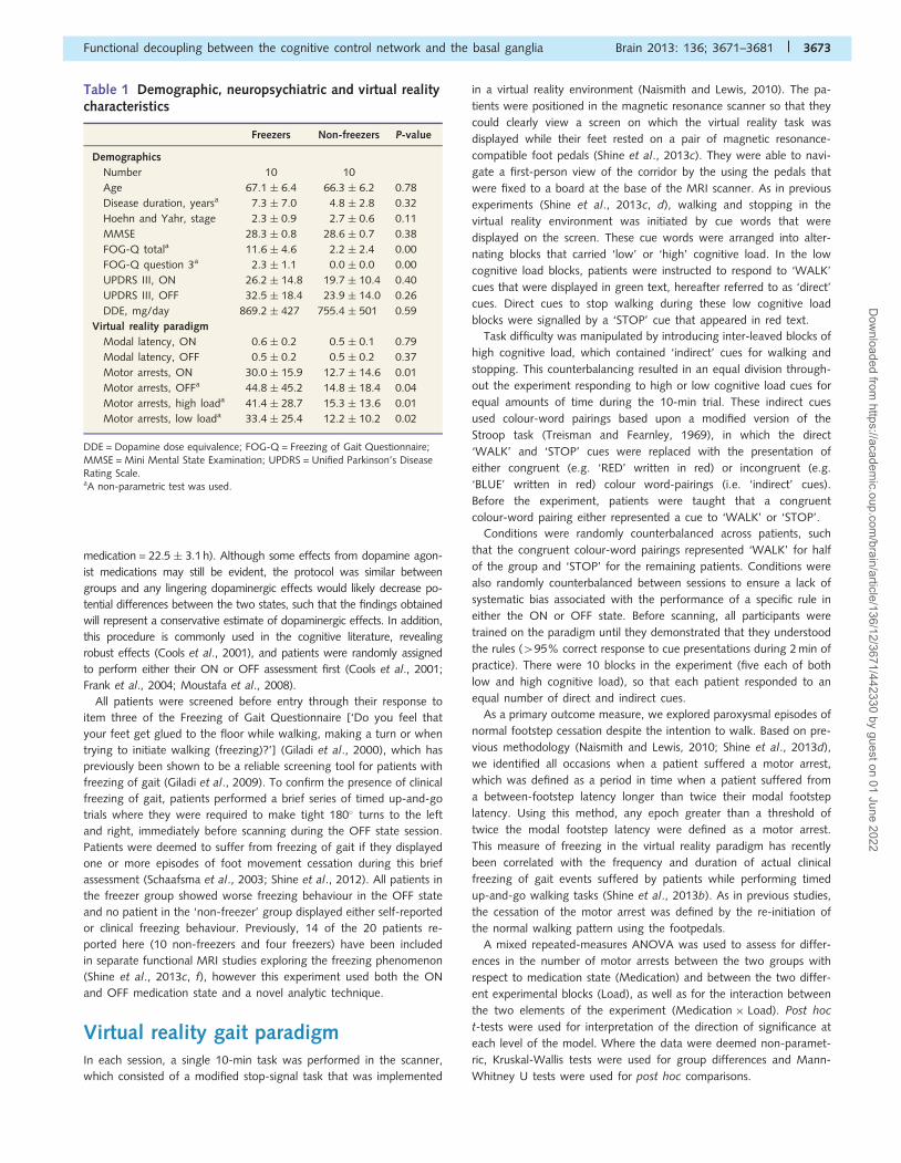
Table 1 Demographic, neuropsychiatric and virtual realitycharacteristics
Freezers Non-freezers P-value
Demographics
Number 10 10
Age 67.1 � 6.4 66.3 � 6.2 0.78
Disease duration, yearsa 7.3 � 7.0 4.8 � 2.8 0.32
Hoehn and Yahr, stage 2.3 � 0.9 2.7 � 0.6 0.11
MMSE 28.3 � 0.8 28.6 � 0.7 0.38
FOG-Q totala 11.6 � 4.6 2.2 � 2.4 0.00
FOG-Q question 3a 2.3 � 1.1 0.0 � 0.0 0.00
UPDRS III, ON 26.2 � 14.8 19.7 � 10.4 0.40
UPDRS III, OFF 32.5 � 18.4 23.9 � 14.0 0.26
DDE, mg/day 869.2 � 427 755.4 � 501 0.59
Virtual reality paradigm
Modal latency, ON 0.6 � 0.2 0.5 � 0.1 0.79
Modal latency, OFF 0.5 � 0.2 0.5 � 0.2 0.37
Motor arrests, ON 30.0 � 15.9 12.7 � 14.6 0.01
Motor arrests, OFFa 44.8 � 45.2 14.8 � 18.4 0.04
Motor arrests, high loada 41.4 � 28.7 15.3 � 13.6 0.01
Motor arrests, low loada 33.4 � 25.4 12.2 � 10.2 0.02
DDE = Dopamine dose equivalence; FOG-Q = Freezing of Gait Questionnaire;MMSE = Mini Mental State Examination; UPDRS = Unified Parkinson’s DiseaseRating Scale.aA non-parametric test was used.
Functional decoupling between the cognitive control network and the basal ganglia Brain 2013: 136; 3671–3681 | 3673
Dow
nloaded from https://academ
ic.oup.com/brain/article/136/12/3671/442330 by guest on 01 June 2022
To ensure that the presence of motor arrests was not due to global
impairments on the virtual reality task, we calculated the number of
‘effective stops’, defined as instances in which patients performed two
or fewer stops following a STOP cue. Depending on the individual
cadence of each subject, WALK cues often appeared mid-step thus
precluding direct measurements of response times to WALK cues. Thus
an indirect measurement of response time was calculated by taking the
average duration of the longest footstep that occurred within three
steps of either a high or low cognitive cue presentation, which was
taken to reflect the amount of time taken to effectively respond to a
WALK cue. These outcomes measures were compared across group,
medication state and cognitive load and were also correlated with
the number of motor arrests across the virtual reality task. Where
appropriate, non-parametric tests were used to evaluate statistical
significance.
Neuroimaging analysis
Image acquisition
Imaging was conducted on a General Electric 3 T MRI (General
Electric). T2*-weighted echo planar functional images were acquired
in sequential order with repetition time = 3 s, echo time = 32 ms, flip
angle = 90�, 32 axial slices covering the whole brain, field of
view = 220 mm, and raw voxel size = 3.9 � 3.9 � 4 mm thick. High-
resolution 3D T1-weighted, anatomical images (voxel size
0.4 � 0.4 � 0.9 mm) were obtained for co-registration with functional
data.
Image preprocessing
Statistical parametric mapping software (SPM8, Wellcome Trust Centre
for Neuroimaging, London, UK, http://www.fil.ion.ucl.ac.uk/spm/soft
ware/) was used for image preprocessing, according to a standard
pipeline: (i) scans were slice-time corrected to the median (21st) slice
in each repetition time; (ii) scans were then realigned to create a mean
realigned image and measures of 6� of rigid head movements were
calculated for later use in the correction of minor head movements;
(iii) images were normalized to the echo planar image template; and
(iv) scans were then smoothed using an 8-mm full-width at half-
maximum isotropic Gaussian kernel.
Independent component analysis
Preprocessed images were then imported into an independent compo-
nent analysis procedure using the GIFT toolbox (http://mialab.mrn.
org/software) in SPM8. Briefly, independent component analysis is a
data-driven approach that searches for maximally independent clusters
of voxels within the brain that co-vary together in reliable temporal
relationships. In this study, we used the InfoMax algorithm to extract
20 maximally independent components for the entire group of 20
patients with Parkinson’s disease. The 20 extracted components
were then spatially sorted at the group level using a set of predefined
regions of interest, the co-ordinates of which reproduced hubs of well-
known neural networks (Fig. 1). Co-ordinates for the masks were
taken from previous imaging studies (Fox et al., 2005; Spreng et al.,
2010) and were defined in MNI space (Table 2). Specifically, we used:
a midline precentral gyrus mask (8 mm sphere centred around 0, �31,
67) to extract the motor network; two separate dorsolateral prefrontal
cortex masks (10 mm spheres centred around �45, 11, 34 and 45, 11,
34) to extract a frontoparietal CCN for each hemisphere; a right
anterior insula mask (8 mm sphere centred around 32, 20, �2) to
extract the ventral attention network; and a right sided caudate
mask (8 mm sphere centred around 10, 14, 10) to extract the basal
ganglia network.
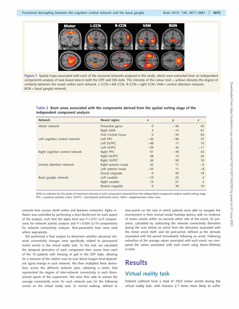
Task-based functional connectivity
Each component extracted from the independent component analysis
was also associated with a unique time course (with one numeric value
per repetition time) that represented the average blood oxygen level-
dependent signal across the entire component. Using the Functional
Network Connectivity toolbox (http://mialab.mrn.org/software), the
time courses for the five component networks of interest (motor net-
work, left CCN, right CCN, ventral attention network and basal gang-
lia network) were subjected to a number of further preprocessing
steps, including de-trending and interpolation, to remove spurious
low frequency noise from the data (Friston, 2011). In addition, the
time courses were also temporally filtered using a high-pass filter
(0.009 Hz), further removing low-frequency noise from the data. The
time course from each network was then extracted for each patient.
Using SPM8 software, task regressors modelling the onset and offset
of high and low cognitive load blocks were convolved with the
haemodynamic response function, creating a new variable that repre-
sented the expected blood oxygen level-dependent response in each
experimental block. The five network time courses for each patient
were then multiplied against these convolved task regressors, leading
to a score that reflected the specific ‘activity’ in each network during
blocks of both high and low cognitive load (Fig. 2). After this step, a
series of Pearson’s product-moment correlations were computed for
each patient and were converted into a Z-score using Fisher’s r-to-Z
transformation.
To calculate network activity, the time course for each of the five
networks was correlated against the convolved task regressor for each
of the four experimental combinations (i.e. high and low cognitive
load in each medication state). To calculate network connectivity,
each of the network time courses was correlated with the other four
networks in each of the four experimental combinations, allowing for
an estimation of task-based functional connectivity across the different
modes of the experiment.
To determine whether either of these measures changed with re-
spect to freezing behaviour, we calculated a correlation between the
frequency of motor arrests in each experimental block and the relevant
network activity and network connectivity Z-score. To allow for cal-
culations relevant to each outcome measure, the frequency of motor
arrests was pooled across each experimental manipulation. The Dunn
and Clark statistic (Dunn and Clark, 1969) was used to test for stat-
istical significance between the two aspects of each mode of the
experiment within each group. Finally, the Z-score associated with
this statistic was compared between the two groups to determine
whether the two groups showed significantly different network modu-
lations with respect to the number of motor arrests.
In each analysis, the effect of medication state was calculated by
contrasting the average Z-score representing the network association
with both cognitive load states in the OFF state with the correspond-
ing Z-scores from the ON state. To assess the effect of cognitive load,
a similar comparison was made, however, the data were pooled with
respect to cognitive load, rather than medication state. To assess for
the presence of differences in the interaction between medication state
and cognitive load, difference scores were calculated that represented
the difference between high and low cognitive load in the OFF state
with respect to the ON state. A series of t-tests were performed on
these variables to determine whether activity within and/or between
any of the networks was associated with a specific combination of the
experimental variables. Subsequently, a series of independent-sample
t-tests were used to assess potential group differences in any of the
3674 | Brain 2013: 136; 3671–3681 J. M. Shine et al.
Dow
nloaded from https://academ
ic.oup.com/brain/article/136/12/3671/442330 by guest on 01 June 2022
network time courses (both within and between networks). Alpha in-
flation was controlled by performing a strict Bonferroni for each aspect
of the analysis, such that the alpha level was P = 0.01 (�/5 compari-
sons) for network activity analyses and P = 0.005 (�/10 comparisons)
for network connectivity analyses. Non-parametric tests were used
where appropriate.
We performed a final analysis to determine whether abnormal net-
work connectivity changes were specifically related to paroxysmal
motor arrests in the virtual reality task. To this end, we calculated
the temporal derivative of each component time course from each
of the 10 patients with freezing of gait in the OFF state, allowing
for a measure of the relative scan-to-scan blood oxygen level-depend-
ent signal change in each network. We then multiplied these deriva-
tives across the different network pairs, obtaining a metric that
represented the degree of inter-network connectivity in each three-
second epoch of the experiment. We were then able to extract the
average connectivity score for each network pair for the following
events on the virtual reality task: (i) normal walking, defined as
time-points on the task in which patients were able to navigate the
environment in their normal modal footstep latency with no evidence
of motor arrests within six seconds either side of the event; (ii) pre-
arrest, calculated by subtracting the network connectivity derivative
during the scan before an arrest from the derivative associated with
the motor arrest itself; and (iii) post-arrest, defined as the derivate
associated with the period immediately following an arrest. Following
extraction of the average values associated with each event, we com-
pared the values associated with each event using Mann-Whitney
U-tests.
Results
Virtual reality taskPatients suffered from a total of 1023 motor arrests during the
virtual reality task, with freezers 2.7 times more likely to suffer
Table 2 Brain areas associated with the components derived from the spatial sorting stage of theindependent component analysis
Network Neural region x y z
Motor network Precentral gyrus 0 �36 63
Right SMA 3 �13 61
Post Central Gyrus 0 �54 63
Left cognitive control network Left PPC �42 �58 37
Left DLPFC �48 11 10
Left VLPFC �39 44 �11
Right cognitive control network Right PPC 51 �49 43
Right DLPFC 48 14 25
Right VLPFC 36 50 10
Ventral attention network Right anterior insula 42 11 �5
Left anterior insula �42 11 �8
Dorsal cingulate 0 50 19
Basal ganglia network Left caudate �15 23 4
Right caudate 13 21 3
Rostral cingulate 0 39 10
MNI co-ordinates for the peaks of maximum intensity in each component extracted from the independent component analysis spatial sorting stage.PPC = posterior parietal cortex; DLPFC = dorsolateral prefrontal cortex; SMA = supplementary motor area.
Figure 1 Spatial maps associated with each of the neuronal networks analysed in this study, which were extracted from an independent
components analysis of task-based data in both the OFF and ON state. The intensity of the colour (red! yellow) denotes the degree of
similarity between the voxels within each network. L-CCN = left CCN; R-CCN = right CCN; VAN = ventral attention network;
BGN = basal ganglia network.
Functional decoupling between the cognitive control network and the basal ganglia Brain 2013: 136; 3671–3681 | 3675
Dow
nloaded from https://academ
ic.oup.com/brain/article/136/12/3671/442330 by guest on 01 June 2022
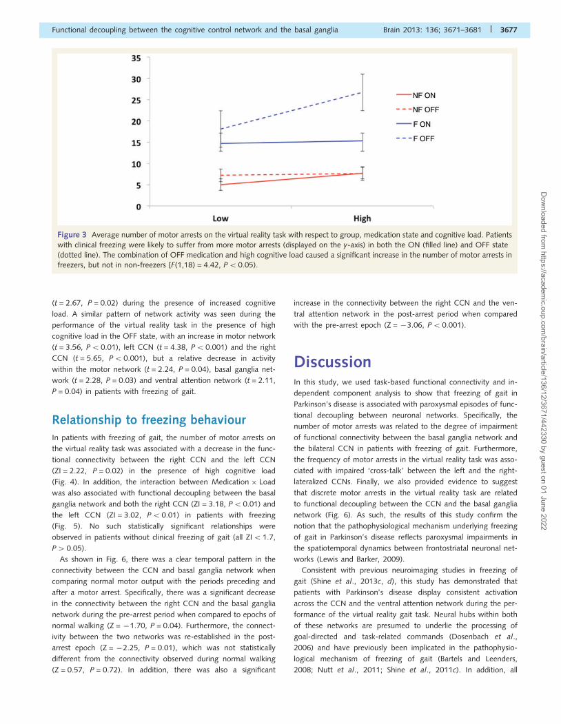
from freezing events across the entire experiment [Group effect;
F(1,18) = 4.42, P50.05; Fig. 3]. The Group �Medication effect
was not significant [F(1,18) = 1.00, P40.05], however, freezers
were more likely to suffer from motor arrests during both the OFF
state (t = 2.24, P = 0.04) and the ON state (t = 2.86, P = 0.02)
than non-freezers. In addition, The Group � Load effect was sig-
nificant [F(1,18) = 7.86, P5 0.01], as freezers were more likely to
suffer from motor arrests in both high cognitive load (t = 2.83,
P = 0.01) and low cognitive load (t = 2.67, P = 0.02) blocks
when compared with non-freezers. In keeping with previous stu-
dies (Shine et al., 2013d), freezers were also more likely to spend
a greater proportion of time ‘frozen’ during the experiment,
‘freezing’ for an average of 9.6 � 9% compared to 5.4 � 6% in
non-freezers (t = 2.20, P50.05).
Despite these differences, there were no significant differences
in the modal footstep latency either between groups, between
blocks or between medication states [F(1,18) = 0.347, P5 0.05],
suggesting that the motor arrests were due to paroxysmal freezing
events rather than general motor differences between the two
groups and their medication states. There were no significant dif-
ferences with respect to the number of effective responses to
STOP signals between the two groups [F(3,16) = 2.44, P = 0.08],
although there was a trend for increased ineffective stops in the
freezing group. In addition, there were no significant differences in
the average response times to cognitive cues in either group in
both the ON and OFF states [F(3,16) = 0.23, P = 0.88]. Similarly
neither group was more likely to suffer from motor arrests when
responding to congruent versus incongruent cues (t = 1.7,
P = 0.10). Finally, none of these measures were significantly
correlated with the number of motor arrests experienced during
the virtual reality gait task (r5 |0.445|, P40.05).
Independent component analysisSpatial maps for each of the selected components are presented in
Fig. 1 and the voxel co-ordinates used to select each network map
are presented in Table 2. There were no significant differences
between the two groups when analysing the voxel patterns of
the selected components (analysed at P50.05 corrected for
multiple comparisons using family-wise error; cluster size410).
Network activity and connectivityWhile performing the virtual reality task, all patients regularly dis-
played activity within the left CCN (t = 3.39, P = 0.003) and the
ventral attention network (t = 3.82, P = 0.001). In addition, all pa-
tients showed an increase in the connectivity between the left
CCN and the right CCN (t = 5.43, P50.001). In the OFF state,
all patients showed a decrease in the activity within the basal
ganglia network (t = 2.95, P = 0.009), however, the increased
connectivity between the left CCN and the right CCN remained
intact (t = 5.08, P5 0.001).
In the presence of high cognitive load, there was an increased
recruitment of the motor network (t = 3.69, P5 0.01), the left
CCN (t = 4.76, P5 0.001) and the right CCN (t = 5.23,
P50.001), along with an increased connectivity between the
motor network and the left CCN (t = 3.52, P50.01). However,
freezers were unable to sustain similar levels of activity in the
motor network (t = 2.45, P = 0.03) or basal ganglia network
Figure 2 Graphical depiction of the experimental analysis. After extracting the time courses from each of the five network components,
we then multiplied each time course with the convolved regressor representing either low or high cognitive load, leading to the creation of
individual correlation coefficients representing the strength of network activation in each experimental block (orange and blue lines). To
measure network connectivity, we then correlated each time course with one another, leading to a correlation coefficient representing the
strength of association of each network pair in each block (green line). Finally, individual network activation and connectivity scores are
correlated with the number of motor arrests in each block, leading to the calculation of network modulation.
3676 | Brain 2013: 136; 3671–3681 J. M. Shine et al.
Dow
nloaded from https://academ
ic.oup.com/brain/article/136/12/3671/442330 by guest on 01 June 2022
(t = 2.67, P = 0.02) during the presence of increased cognitive
load. A similar pattern of network activity was seen during the
performance of the virtual reality task in the presence of high
cognitive load in the OFF state, with an increase in motor network
(t = 3.56, P5 0.01), left CCN (t = 4.38, P5 0.001) and the right
CCN (t = 5.65, P50.001), but a relative decrease in activity
within the motor network (t = 2.24, P = 0.04), basal ganglia net-
work (t = 2.28, P = 0.03) and ventral attention network (t = 2.11,
P = 0.04) in patients with freezing of gait.
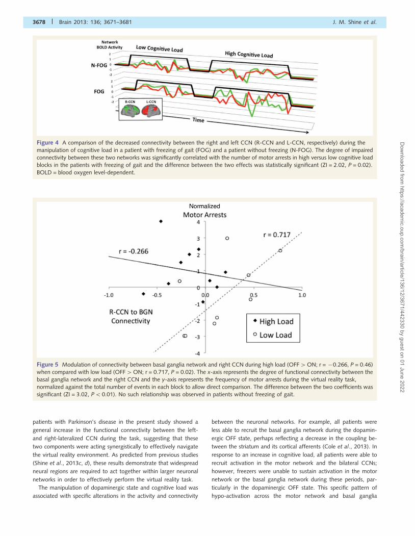
Relationship to freezing behaviourIn patients with freezing of gait, the number of motor arrests on
the virtual reality task was associated with a decrease in the func-
tional connectivity between the right CCN and the left CCN
(ZI = 2.22, P = 0.02) in the presence of high cognitive load
(Fig. 4). In addition, the interaction between Medication � Load
was also associated with functional decoupling between the basal
ganglia network and both the right CCN (ZI = 3.18, P50.01) and
the left CCN (ZI = 3.02, P50.01) in patients with freezing
(Fig. 5). No such statistically significant relationships were
observed in patients without clinical freezing of gait (all ZI51.7,
P4 0.05).
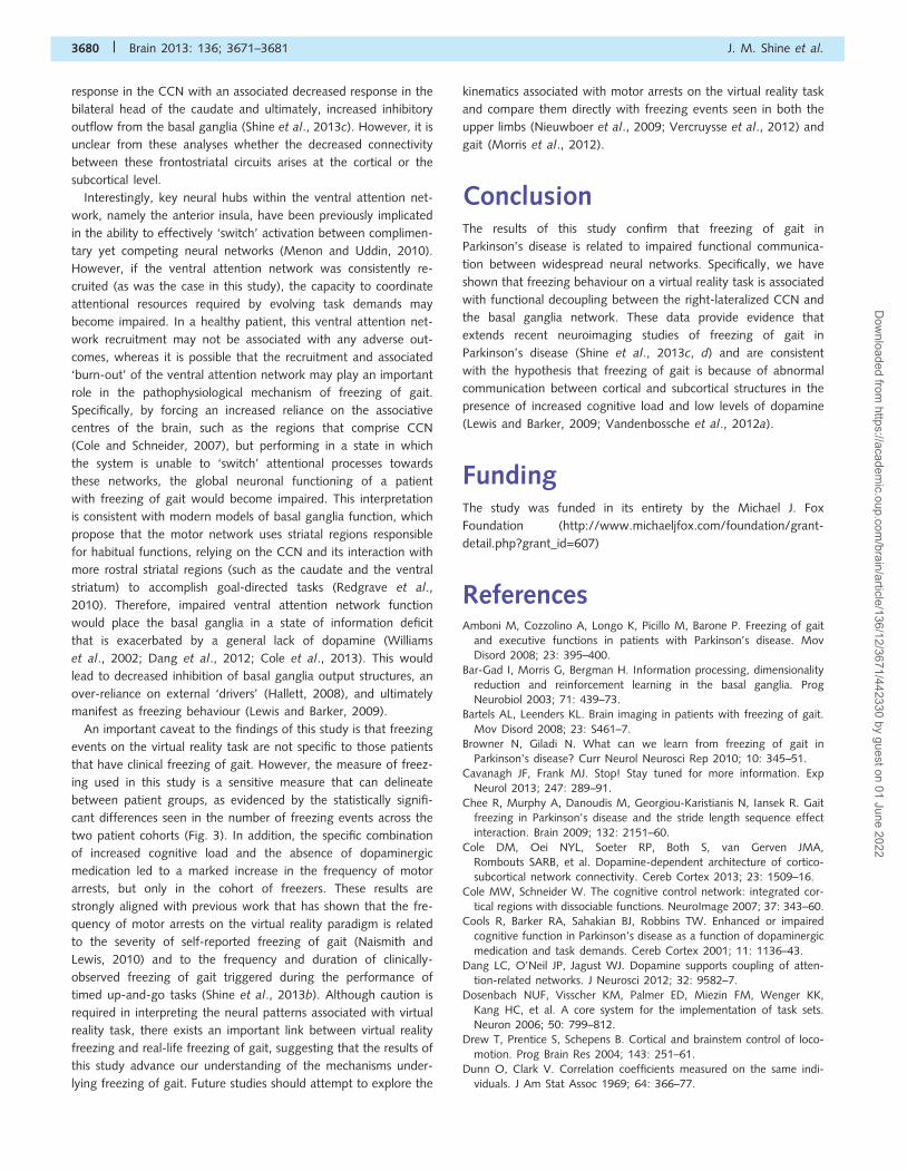
As shown in Fig. 6, there was a clear temporal pattern in the
connectivity between the CCN and basal ganglia network when
comparing normal motor output with the periods preceding and
after a motor arrest. Specifically, there was a significant decrease
in the connectivity between the right CCN and the basal ganglia
network during the pre-arrest period when compared to epochs of
normal walking (Z = �1.70, P = 0.04). Furthermore, the connect-
ivity between the two networks was re-established in the post-
arrest epoch (Z = �2.25, P = 0.01), which was not statistically
different from the connectivity observed during normal walking
(Z = 0.57, P = 0.72). In addition, there was also a significant
increase in the connectivity between the right CCN and the ven-
tral attention network in the post-arrest period when compared
with the pre-arrest epoch (Z = �3.06, P5 0.001).
DiscussionIn this study, we used task-based functional connectivity and in-
dependent component analysis to show that freezing of gait in
Parkinson’s disease is associated with paroxysmal episodes of func-
tional decoupling between neuronal networks. Specifically, the
number of motor arrests was related to the degree of impairment
of functional connectivity between the basal ganglia network and
the bilateral CCN in patients with freezing of gait. Furthermore,
the frequency of motor arrests in the virtual reality task was asso-
ciated with impaired ‘cross-talk’ between the left and the right-
lateralized CCNs. Finally, we also provided evidence to suggest
that discrete motor arrests in the virtual reality task are related
to functional decoupling between the CCN and the basal ganglia
network (Fig. 6). As such, the results of this study confirm the
notion that the pathophysiological mechanism underlying freezing
of gait in Parkinson’s disease reflects paroxysmal impairments in
the spatiotemporal dynamics between frontostriatal neuronal net-
works (Lewis and Barker, 2009).
Consistent with previous neuroimaging studies in freezing of
gait (Shine et al., 2013c, d), this study has demonstrated that
patients with Parkinson’s disease display consistent activation
across the CCN and the ventral attention network during the per-
formance of the virtual reality gait task. Neural hubs within both
of these networks are presumed to underlie the processing of
goal-directed and task-related commands (Dosenbach et al.,
2006) and have previously been implicated in the pathophysio-
logical mechanism of freezing of gait (Bartels and Leenders,
2008; Nutt et al., 2011; Shine et al., 2011c). In addition, all
Figure 3 Average number of motor arrests on the virtual reality task with respect to group, medication state and cognitive load. Patients
with clinical freezing were likely to suffer from more motor arrests (displayed on the y-axis) in both the ON (filled line) and OFF state
(dotted line). The combination of OFF medication and high cognitive load caused a significant increase in the number of motor arrests in
freezers, but not in non-freezers [F(1,18) = 4.42, P50.05).
Functional decoupling between the cognitive control network and the basal ganglia Brain 2013: 136; 3671–3681 | 3677
Dow
nloaded from https://academ
ic.oup.com/brain/article/136/12/3671/442330 by guest on 01 June 2022
patients with Parkinson’s disease in the present study showed a
general increase in the functional connectivity between the left-
and right-lateralized CCN during the task, suggesting that these
two components were acting synergistically to effectively navigate
the virtual reality environment. As predicted from previous studies
(Shine et al., 2013c, d), these results demonstrate that widespread
neural regions are required to act together within larger neuronal
networks in order to effectively perform the virtual reality task.
The manipulation of dopaminergic state and cognitive load was
associated with specific alterations in the activity and connectivity
between the neuronal networks. For example, all patients were
less able to recruit the basal ganglia network during the dopamin-
ergic OFF state, perhaps reflecting a decrease in the coupling be-
tween the striatum and its cortical afferents (Cole et al., 2013). In
response to an increase in cognitive load, all patients were able to
recruit activation in the motor network and the bilateral CCNs;
however, freezers were unable to sustain activation in the motor
network or the basal ganglia network during these periods, par-
ticularly in the dopaminergic OFF state. This specific pattern of
hypo-activation across the motor network and basal ganglia
Figure 5 Modulation of connectivity between basal ganglia network and right CCN during high load (OFF4ON; r = �0.266, P = 0.46)
when compared with low load (OFF4ON; r = 0.717, P = 0.02). The x-axis represents the degree of functional connectivity between the
basal ganglia network and the right CCN and the y-axis represents the frequency of motor arrests during the virtual reality task,
normalized against the total number of events in each block to allow direct comparison. The difference between the two coefficients was
significant (ZI = 3.02, P50.01). No such relationship was observed in patients without freezing of gait.
Figure 4 A comparison of the decreased connectivity between the right and left CCN (R-CCN and L-CCN, respectively) during the
manipulation of cognitive load in a patient with freezing of gait (FOG) and a patient without freezing (N-FOG). The degree of impaired
connectivity between these two networks was significantly correlated with the number of motor arrests in high versus low cognitive load
blocks in the patients with freezing of gait and the difference between the two effects was statistically significant (ZI = 2.02, P = 0.02).
BOLD = blood oxygen level-dependent.
3678 | Brain 2013: 136; 3671–3681 J. M. Shine et al.
Dow
nloaded from https://academ
ic.oup.com/brain/article/136/12/3671/442330 by guest on 01 June 2022
network may have predisposed susceptible patients to an
increased frequency of motor arrests, which were substantially
higher in the OFF state when performing the high cognitive
load blocks of the experiment (Fig. 3). This raises the possibly
that the decreased connectivity between these networks repre-
sents a predisposing factor for the increased freezing seen
during the high cognitive load blocks in the OFF state, possibly
reflecting impairments in the effective ‘switching’ between neur-
onal networks (Menon and Uddin, 2010; Shine et al., 2013e).
The frequency of motor arrests was also strongly correlated with
functional decoupling between key neuronal networks during the
performance of the virtual reality task. In the presence of high
cognitive load, the number of motor arrests was significantly cor-
related with the degree of impaired connectivity between the left
and right CCN, possibly reflecting impaired ‘cross-talk’ between
the two hemispheres (Fig. 4) (Lewis and Barker, 2009) which may
be due in part to impaired efferent connectivity with the pedun-
culopontine nucleus, particularly in the right hemisphere (Fling
et al., 2013). During blocks of high cognitive load in the dopa-
minergic OFF state, the frequency of motor arrests suffered
by patients with clinical freezing of gait was predictive of the
degree of impaired connectivity between the basal ganglia net-
work and both the left and the right CCN (Fig. 5).
Paroxysmal motor arrests on the virtual reality task were asso-
ciated with impaired connectivity between the CCN and the basal
ganglia network, which was re-established on the breaking of a
motor arrest (Fig. 6). In addition, the breaking of a motor arrest
was also associated with increased connectivity between the CCN
and the ventral attention network. Together, these results suggest
that freezing of gait is due to functional decoupling within the
frontostriatal networks responsible for the mediation of goal-
directed and flexible behaviours (Spreng et al., 2010), an
interpretation which is consistent with recent behavioural evidence
showing that freezing of gait is related to impairments in the pro-
cessing of conflict-related signals (Vandenbossche et al., 2011,
2012b). Indeed, a strong prediction from these lines of research
is that the dissociation of frontoparietal cortical regions responsible
for conscious goal-directed behaviour and the subcortical struc-
tures responsible for habitual behaviour would manifest peripher-
ally as an episode of freezing (Vandenbossche et al., 2012a).
The results of this study bring together a number of disparate
findings across the unique (but not necessarily exclusive) hypoth-
eses that have been used to explain the pathophysiology of freez-
ing of gait (Nutt et al., 2011; Shine et al., 2011c). One major
difficulty in aligning these models has been the elucidation of a
mechanism that can link the provocation of freezing of gait (which
is often associated with the completion of cortically mediated
tasks, such as dual-tasking) with the manifestation of freezing of
gait (which is likely to be because of the interruption of neuronal
activity within the brainstem and spinal cord structures controlling
gait) (Drew et al., 2004; Lemon, 2008). The results of this study
suggest that the functional decoupling of goal-directed frontostria-
tal systems is a likely mechanism responsible for the triggering of
episodes of freezing of gait. This impaired coupling would lead to
the loss of inhibitory influence over the output structures of the
basal ganglia (Lewis and Barker, 2009; Shine et al., 2013c), po-
tentially leading to overwhelming inhibition in regions of the mes-
encephalic locomotor region that control locomotion, such as the
glutamatergic nuclei of the pedunculopontine nucleus (Pahapill
and Lozano, 2000). Interestingly, recent structural neuroimaging
studies have shown a relative lack of white matter connectivity
between the pedunculopontine nucleus and the cerebellum
(Schweder et al., 2010; Fling et al., 2013), along with the thal-
amus and frontal cortex (Fling et al., 2013), possibly placing the
mesencephalic locomotor region (of which the pedunculopontine
nucleus is a contributing region) at an increased risk of hyper-
polarization by GABAergic input from the globus pallidus. This
activity would therefore ultimately manifest as abnormal neuronal
afferent signals travelling to the spinal cord, and poorly coordi-
nated muscle firing patterns; ‘trembling in place’ (Jacobs et al.,
2009; Nutt et al., 2011).
Another possible interpretation of the impaired intra-network
connectivity in this study is that the freezing phenomenon is a
manifestation of a global decrease in the information processing
capacity of the brain. Theoretically, this effect would likely occur
within neural circuits that perform a more domain-general func-
tion, such as switching activation between neural networks (Seeley
et al., 2007; Nelson et al., 2010) or represent the attempted re-
duction of high-dimensional activity from the corticothalamic
system by the basal ganglia nuclei (Bar-Gad et al., 2003). In this
manner, the functional decoupling between key members of the
‘cognitive’ frontostriatal loop in this experiment may reflect a
breakdown in information processing within these circuits, which
is supported by the recent finding of impaired executive network
connectivity in patients with freezing of gait during the resting
state (Tessitore et al., 2012). Such an interpretation would be
consistent with the results of a recent event-related functional
MRI study, which showed that motor arrests on the virtual reality
task were associated with increased blood oxygen level-dependent
Figure 6 The period preceding a motor arrest in the virtual
reality task was associated with a significant decrease in the
connectivity between the right CCN (R-CCN) and the basal
ganglia network (BGN) when comparing normal walking with
the epoch preceding a motor arrest (pre-arrest; P5 0.05). This
impairment in connectivity was subsequently restored in the
epoch following a motor arrest (post-arrest; P50.08), which
was normalized to the levels of connectivity seen during normal
walking (P = 0.68). These results suggest that individual motor
arrests on the virtual reality task are related to a functional
decoupling within frontostriatal networks.
Functional decoupling between the cognitive control network and the basal ganglia Brain 2013: 136; 3671–3681 | 3679
Dow
nloaded from https://academ
ic.oup.com/brain/article/136/12/3671/442330 by guest on 01 June 2022
response in the CCN with an associated decreased response in the
bilateral head of the caudate and ultimately, increased inhibitory
outflow from the basal ganglia (Shine et al., 2013c). However, it is
unclear from these analyses whether the decreased connectivity
between these frontostriatal circuits arises at the cortical or the
subcortical level.
Interestingly, key neural hubs within the ventral attention net-
work, namely the anterior insula, have been previously implicated
in the ability to effectively ‘switch’ activation between complimen-
tary yet competing neural networks (Menon and Uddin, 2010).
However, if the ventral attention network was consistently re-
cruited (as was the case in this study), the capacity to coordinate
attentional resources required by evolving task demands may
become impaired. In a healthy patient, this ventral attention net-
work recruitment may not be associated with any adverse out-
comes, whereas it is possible that the recruitment and associated
‘burn-out’ of the ventral attention network may play an important
role in the pathophysiological mechanism of freezing of gait.
Specifically, by forcing an increased reliance on the associative
centres of the brain, such as the regions that comprise CCN
(Cole and Schneider, 2007), but performing in a state in which
the system is unable to ‘switch’ attentional processes towards
these networks, the global neuronal functioning of a patient
with freezing of gait would become impaired. This interpretation
is consistent with modern models of basal ganglia function, which
propose that the motor network uses striatal regions responsible
for habitual functions, relying on the CCN and its interaction with
more rostral striatal regions (such as the caudate and the ventral
striatum) to accomplish goal-directed tasks (Redgrave et al.,
2010). Therefore, impaired ventral attention network function
would place the basal ganglia in a state of information deficit
that is exacerbated by a general lack of dopamine (Williams
et al., 2002; Dang et al., 2012; Cole et al., 2013). This would
lead to decreased inhibition of basal ganglia output structures, an
over-reliance on external ‘drivers’ (Hallett, 2008), and ultimately
manifest as freezing behaviour (Lewis and Barker, 2009).
An important caveat to the findings of this study is that freezing
events on the virtual reality task are not specific to those patients
that have clinical freezing of gait. However, the measure of freez-
ing used in this study is a sensitive measure that can delineate
between patient groups, as evidenced by the statistically signifi-
cant differences seen in the number of freezing events across the
two patient cohorts (Fig. 3). In addition, the specific combination
of increased cognitive load and the absence of dopaminergic
medication led to a marked increase in the frequency of motor
arrests, but only in the cohort of freezers. These results are
strongly aligned with previous work that has shown that the fre-
quency of motor arrests on the virtual reality paradigm is related
to the severity of self-reported freezing of gait (Naismith and
Lewis, 2010) and to the frequency and duration of clinically-
observed freezing of gait triggered during the performance of
timed up-and-go tasks (Shine et al., 2013b). Although caution is
required in interpreting the neural patterns associated with virtual
reality task, there exists an important link between virtual reality
freezing and real-life freezing of gait, suggesting that the results of
this study advance our understanding of the mechanisms under-
lying freezing of gait. Future studies should attempt to explore the
kinematics associated with motor arrests on the virtual reality task
and compare them directly with freezing events seen in both the
upper limbs (Nieuwboer et al., 2009; Vercruysse et al., 2012) and
gait (Morris et al., 2012).
ConclusionThe results of this study confirm that freezing of gait in
Parkinson’s disease is related to impaired functional communica-
tion between widespread neural networks. Specifically, we have
shown that freezing behaviour on a virtual reality task is associated
with functional decoupling between the right-lateralized CCN and
the basal ganglia network. These data provide evidence that
extends recent neuroimaging studies of freezing of gait in
Parkinson’s disease (Shine et al., 2013c, d) and are consistent
with the hypothesis that freezing of gait is because of abnormal
communication between cortical and subcortical structures in the
presence of increased cognitive load and low levels of dopamine
(Lewis and Barker, 2009; Vandenbossche et al., 2012a).
FundingThe study was funded in its entirety by the Michael J. Fox
Foundation (http://www.michaeljfox.com/foundation/grant-
detail.php?grant_id=607)
ReferencesAmboni M, Cozzolino A, Longo K, Picillo M, Barone P. Freezing of gait
and executive functions in patients with Parkinson’s disease. Mov
Disord 2008; 23: 395–400.
Bar-Gad I, Morris G, Bergman H. Information processing, dimensionality
reduction and reinforcement learning in the basal ganglia. Prog
Neurobiol 2003; 71: 439–73.Bartels AL, Leenders KL. Brain imaging in patients with freezing of gait.
Mov Disord 2008; 23: S461–7.Browner N, Giladi N. What can we learn from freezing of gait in
Parkinson’s disease? Curr Neurol Neurosci Rep 2010; 10: 345–51.
Cavanagh JF, Frank MJ. Stop! Stay tuned for more information. Exp
Neurol 2013; 247: 289–91.
Chee R, Murphy A, Danoudis M, Georgiou-Karistianis N, Iansek R. Gait
freezing in Parkinson’s disease and the stride length sequence effect
interaction. Brain 2009; 132: 2151–60.
Cole DM, Oei NYL, Soeter RP, Both S, van Gerven JMA,
Rombouts SARB, et al. Dopamine-dependent architecture of cortico-
subcortical network connectivity. Cereb Cortex 2013; 23: 1509–16.
Cole MW, Schneider W. The cognitive control network: integrated cor-
tical regions with dissociable functions. NeuroImage 2007; 37: 343–60.Cools R, Barker RA, Sahakian BJ, Robbins TW. Enhanced or impaired
cognitive function in Parkinson’s disease as a function of dopaminergic
medication and task demands. Cereb Cortex 2001; 11: 1136–43.Dang LC, O’Neil JP, Jagust WJ. Dopamine supports coupling of atten-
tion-related networks. J Neurosci 2012; 32: 9582–7.
Dosenbach NUF, Visscher KM, Palmer ED, Miezin FM, Wenger KK,
Kang HC, et al. A core system for the implementation of task sets.
Neuron 2006; 50: 799–812.
Drew T, Prentice S, Schepens B. Cortical and brainstem control of loco-
motion. Prog Brain Res 2004; 143: 251–61.
Dunn O, Clark V. Correlation coefficients measured on the same indi-
viduals. J Am Stat Assoc 1969; 64: 366–77.
3680 | Brain 2013: 136; 3671–3681 J. M. Shine et al.
Dow
nloaded from https://academ
ic.oup.com/brain/article/136/12/3671/442330 by guest on 01 June 2022

Fling BW, Cohen RG, Mancini M, Nutt JG, Fair DA, Horak FB.Asymmetric pedunculopontine network connectivity in parkinsonian
patients with freezing of gait. Brain 2013; 136 (Pt 8): 2405–18.
Fornito A, Harrison BJ, Zalesky A. Competitive and cooperative dynamics
of large-scale brain functional networks supporting recollection. ProcNatl Acad Sci USA 2012; 109: 12788–93.
Fox MD, Snyder AZ, Vincent JL, Corbetta M, Van Essen DC, Raichle ME.
The human brain is intrinsically organized into dynamic, anticorrelated
functional networks. Proc Natl Acad Sci USA 2005; 102: 9673–8.Frank MJ, Seeberger LC, O’Reilly RC. By carrot or by stick: cognitive
reinforcement learning in parkinsonism. Science 2004; 306: 1904–43.
Friston KJ. Functional and effective connectivity: a review. Brain Connect2011; 1: 13–36.
Giladi N, McMahon D, Przedborski S, Flaster E, Guillory S, Kostic V, et al.
Motor blocks in Parkinson’s disease. Neurology 1992; 42: 333–9.
Giladi N, Shabtai H, Simon E, Biran S, Tal J, Korczyn A. Construction offreezing of gait questionnaire for patients with Parkinsonism.
Parkinsonism Relat Disord 2000; 6: 165–70.
Giladi N, Tal J, Azulay T, Rascol O, Brooks DJ, Melamed E, et al.
Validation of the freezing of gait questionnaire in patients withParkinson’s disease. Mov Disord 2009; 24: 655–61.
Hallett M. The intrinsic and extrinsic aspects of freezing of gait. Mov
Disord 2008; 23 (Suppl 2): S439–43.
Jacobs JV, Nutt JG, Carlson-Kuhta P, Stephens M, Horak FB. Knee trem-bling during freezing of gait represents multiple anticipatory postural
adjustments. Exp Neurol 2009; 215: 334–41.
Lemon RN. Descending pathways in motor control. Annu Rev Neurosci2008; 31: 195–218.
Lewis SJG, Barker RA. A pathophysiological model of freezing of gait in
Parkinson’s disease. Parkinsonism Relat Disord 2009; 15: 333–8.
Menon V, Uddin LQ. Saliency, switching, attention and control: a net-work model of insula function. Brain Struct Funct 2010; 214: 655–67.
Morris TR, Cho C, Dilda V, Shine JM, Naismith S, Lewis SJG, et al. A
comparison of clinical and objective measures of freezing of gait in
Parkinson’s disease. Parkinsonism Relat Disord 2012; 18: 572–7.Moustafa AA, Sherman SJ, Frank MJ. A dopaminergic basis for working
memory, learning and attentional shifting in Parkinsonism.
Neuropsychologia 2008; 46: 3144–56.Naismith SL, Lewis SJG. A novel paradigm for modelling freezing of gait
in Parkinson’s disease. J Clin Neurosci 2010; 17: 984–7.
Naismith SL, Shine JM, Lewis SJG. The specific contributions of set-
shifting to freezing of gait in Parkinson’s disease. Mov Disord 2010;25: 1000–4.
Nelson SM, Dosenbach NUF, Cohen AL, Wheeler ME, Schlaggar BL,
Petersen SE. Role of the anterior insula in task-level control and
focal attention. Brain Struct Funct 2010; 214: 669–80.Nieuwboer A, Vercruysse S, Feys P, Levin O, Spildooren J, Swinnen S.
Upper limb movement interruptions are correlated to freezing of gait
in Parkinson’s disease. Eur J Neurosci 2009; 29: 1422–30.
Nutt JG, Bloem BR, Giladi N, Hallett M, Horak FB, Nieuwboer A.Freezing of gait: moving forward on a mysterious clinical phenom-
enon. Lancet Neurol 2011; 10: 734–44.
Pahapill PA, Lozano AM. The pedunculopontine nucleus and Parkinson’sdisease. Brain 2000; 123 (Pt 9): 1767–83.
Redgrave P, Rodriguez M, Smith Y, Rodriguez-Oroz MC, Lehericy S,
Bergman H, et al. Goal-directed and habitual control in the basal
ganglia: implications for Parkinson’s disease2010; 11: 760–72.Schaafsma JD, Balash Y, Gurevich T, Bartels AL, Hausdorff JM, Giladi N.
Characterization of freezing of gait subtypes and the response of each
to levodopa in Parkinson’s disease. Eur J Neurol 2003; 10: 391–8.
Schweder PM, Hansen PC, Green AL, Quaghebeur G, Stein J, Aziz T.Connectivity of the pedunculopontine nucleus in parkinsonian freezing
of gait. NeuroReport 2010; 21: 914–16.
Seeley WW, Menon V, Schatzberg AF, Keller J, Glover GH, Kenna H,et al. Dissociable intrinsic connectivity networks for salience processing
and executive control. J Neurosci 2007; 27: 2349–56.
Shine JM, Matar E, Bolitho SJ, Dilda V, Morris TR. Modeling freezing of
gait in Parkinson’s disease with a virtual reality paradigm. Gait Posture2013b; 38: 104–8.
Shine JM, Matar E, Ward PB, Bolitho SJ, Gilat M, Pearson M, et al.
Exploring the cortical and subcortical functional magnetic resonance
imaging changes associated with freezing in Parkinson’s disease.Brain 2013c; 136: 1204–15.
Shine JM, Matar E, Ward PB, Bolitho SJ, Pearson M, Naismith SL, et al.
Differential neural activation patterns in patients with Parkinson’s dis-ease and freezing of gait in response to concurrent cognitive and
motor load. PLoS One 2013d; 8: e52602.
Shine JM, Moore ST, Bolitho SJ, Morris TR, Dilda V, Naismith SL, et al.
Assessing the utility of freezing of gait questionnaires in Parkinson’sdisease. Parkinsonism Relat Disord 2012; 18: 25–9.
Shine JM, Naismith SL, Lewis SJG. The pathophysiological mechanisms
underlying freezing of gait in Parkinson’s disease. J Clin Neurosci
2011c; 18: 1154–7.Shine JM, Naismith SL, Palavra NC, Lewis SJG, Moore ST, Dilda V, et al.
Attentional set-shifting deficits correlate with the severity of freezing
of gait in Parkinson’s disease. Parkinsonism Relat Disord 2013e; 19:
388–90.Shine JM, Ward PB, Naismith SL, Pearson M, Lewis SJG. Utilising func-
tional MRI (fMRI) to explore the freezing phenomenon in Parkinson’s
disease. J Clin Neurosci 2011b; 18: 807–10.Snijders AH, Leunissen I, Bakker M, Overeem S, Helmich RC, Bloem BR,
et al. Gait-related cerebral alterations in patients with Parkinson’s
disease with freezing of gait. Brain 2010; 134: 59–72.
Spildooren J, Vercruysse S, Desloovere K. Freezing of gait in Parkinson’sdisease: the impact of dual-tasking and turning. Movement 2010; 25:
2563–70.
Spreng RN, Stevens WD, Chamberlain JP, Gilmore AW, Schacter DL.
Default network activity, coupled with the frontoparietal control net-work, supports goal-directed cognition. NeuroImage 2010; 53:
303–17.
Tessitore A, Amboni M, Esposito F, Russo A, Picillo M. Resting-statebrain connectivity in patients with Parkinson’s disease and freezing
of gait. Parkinsonism Relat Disord 2012; 18: 781–7.
Treisman A, Fearnley S. The Stroop Test: selective attention to colours
and words. Nature 1969; 222: 437–9.Vandenbossche J, Deroost N, Soetens E, Coomans D, Spildooren J,
Vercruysse S, et al. Freezing of gait in Parkinson’s disease: disturbances
in automaticity and control. Front Hum Neurosci 2012a; 6: 356.
Vandenbossche J, Deroost N, Soetens E, Spildooren J, Vercruysse S,Nieuwboer A, et al. Freezing of gait in Parkinson disease is associated
with impaired conflict resolution. Neurorehabil Neural Repair 2011; 25:
765–73.
Vandenbossche J, Deroost N, Soetens E, Zeischka P, Spildooren J,Vercruysse S, et al. Conflict and freezing of gait in Parkinson’s disease:
support for a response control deficit. Neuroscience 2012b; 206:
144–54.Vercruysse S, Spildooren J, Heremans E, Vandenbossche J, Wenderoth N,
Swinnen SP, et al. Abnormalities and cue dependence of rhythmical
upper-limb movements in Parkinson patients with freezing of gait.
Neurorehabil Neural Repair 2012; 26: 636–45.Wager TD, Jonides J, Reading S. Neuroimaging studies of shifting atten-
tion: a meta-analysis. NeuroImage 2004; 22: 1679–93.
Williams D, Tijssen M, Van Bruggen G, Bosch A, Insola A, Di Lazzaro V,
et al. Dopamine-dependent changes in the functional connectivity be-tween basal ganglia and cerebral cortex in humans. Brain 2002; 125:
1558–69.
Functional decoupling between the cognitive control network and the basal ganglia Brain 2013: 136; 3671–3681 | 3681
Dow
nloaded from https://academ
ic.oup.com/brain/article/136/12/3671/442330 by guest on 01 June 2022