Chronic systemic pesticide exposure reproduces features of Parkinson's disease
6
nature neuroscience • volume 3 no 12 • december 2000 1301 articles Parkinson’s disease is a late-onset, progressive motor disease marked by selective degeneration of dopaminergic neurons of the substantia nigra and formation of fibrillar cytoplasmic inclu- sions, known as Lewy bodies, which contain ubiquitin and α-synuclein 1 . Rare cases of familial PD have been linked to muta- tions in α-synuclein or parkin 2–4 , but the cause of the more commonly encountered sporadic disease is unknown, and the role of genetics in these cases is uncertain 5 . Post mortem studies strongly implicate oxidative damage and mitochondrial impair- ment in the pathogenesis of PD 6 . Epidemiological studies have suggested that pesticide exposure is associated with an increased risk of developing PD 7–9 . After the pro-toxin N-methyl-4-phenyl-1,2,3,6-tetrahy- dropyridine (MPTP) was reported to produce in humans an acute parkinsonian syndrome that is virtually indistinguishable from idiopathic PD 10 , its metabolite, 1-methyl-4-pyridinium (MPP + ), was found to be a mitochondrial poison that inhibits mitochondrial respiration at complex I of the electron transport chain 11,12 . The selectivity of MPP + for dopaminergic neurons is due to the fact that it is an excellent substrate for the dopamine transporter, and is thereby accumulated preferentially in cells that transport dopamine 13 . Following recognition of MPTP’s tox- icity and its mechanism of action, several laboratories reported a selective defect in complex I of the electron transport chain in PD 14–22 . This defect seems to be systemic, affecting not only the brain, but also peripheral tissues such as platelets. An accurate in vivo experimental model of PD should repro- duce the progressive, selective nigrostriatal dopaminergic degen- eration and Lewy body formation seen in PD, test the relevance of the systemic complex I defect, and explain the potential involve- ment of pesticide exposure in development of parkinsonism. Unfortunately, no current animal model incorporates all of these features. The MPTP model causes selective nigrostriatal degen- eration by inhibiting complex I, but, unlike PD, MPTP does not cause a systemic complex I defect. Instead, the inhibition is high- ly selective for dopaminergic neurons. Moreover, MPTP does not typically produce cytoplasmic inclusions that closely resemble Lewy bodies 23 . Transgenic mice expressing the pathogenic human α-synuclein mutation develop modest dopaminergic pathology (although the specificity of this degeneration is not detailed), and many neurons contain small cytoplasmic inclusions that are gran- ular rather than fibrillar 24 . To develop a more accurate in vivo model of PD, we exposed rats chronically, continuously and systemically to the common pesticide, rotenone. A naturally occurring compound derived from the roots of certain plant species, rotenone is commonly used as an insecticide in vegetable gardens, and is also used to kill or sample fish populations in lakes and reservoirs. It is wide- ly believed to be a safe, natural alternative to synthetic pesticides. Rotenone is also a well characterized, high-affinity, specific inhibitor of complex I, one of the five enzyme complexes of the inner mitochondrial membrane involved in oxidative phospho- rylation. Because it is extremely hydrophobic, rotenone crosses biological membranes easily, and it does not depend on the dopamine transporter for access to the cytoplasm. Therefore, rotenone—unlike MPTP—is well-suited to produce a systemic inhibition of complex I. RESULTS Sprague-Dawley and Lewis rats were infused continuously with rotenone by a jugular vein cannula attached to a subcutaneous osmotic minipump. Lewis rats showed less variability and more consistent lesions than Sprague-Dawley rats 25 and were used exclu- sively after completion of pilot studies. Initially, rotenone was infused at doses ranging from 1 to 12 mg/kg per day for various lengths of time. High doses of rotenone for short periods of time produced systemic (cardiovascular) toxicity and non-specific brain lesions, as reported by others 26 (M.F. Beal and J. Shulz, personal Chronic systemic pesticide exposure reproduces features of Parkinson’s disease Ranjita Betarbet, Todd B. Sherer, Gillian MacKenzie, Monica Garcia-Osuna, Alexander V. Panov and J. Timothy Greenamyre Department of Neurology, Emory University, 1639 Pierce Drive, WMB 6000, Atlanta, Georgia 30322, USA The first two authors contributed equally to this work Correspondence should be addressed to J.T.G. ([email protected]) The cause of Parkinson’s disease (PD) is unknown, but epidemiological studies suggest an association with pesticides and other environmental toxins, and biochemical studies implicate a sys- temic defect in mitochondrial complex I. We report that chronic, systemic inhibition of complex I by the lipophilic pesticide, rotenone, causes highly selective nigrostriatal dopaminergic degeneration that is associated behaviorally with hypokinesia and rigidity. Nigral neurons in rotenone-treated rats accumulate fibrillar cytoplasmic inclusions that contain ubiquitin and α-synuclein. These results indi- cate that chronic exposure to a common pesticide can reproduce the anatomical, neurochemical, behavioral and neuropathological features of PD. © 2000 Nature America Inc. • http://neurosci.nature.com © 2000 Nature America Inc. • http://neurosci.nature.com
-
Upload
independent -
Category
Documents
-
view
2 -
download
0
Transcript of Chronic systemic pesticide exposure reproduces features of Parkinson's disease
nature neuroscience • volume 3 no 12 • december 2000 1301
articles
Parkinson’s disease is a late-onset, progressive motor diseasemarked by selective degeneration of dopaminergic neurons ofthe substantia nigra and formation of fibrillar cytoplasmic inclu-sions, known as Lewy bodies, which contain ubiquitin and α-synuclein1. Rare cases of familial PD have been linked to muta-tions in α-synuclein or parkin2–4, but the cause of the morecommonly encountered sporadic disease is unknown, and therole of genetics in these cases is uncertain5. Post mortem studiesstrongly implicate oxidative damage and mitochondrial impair-ment in the pathogenesis of PD6. Epidemiological studies havesuggested that pesticide exposure is associated with an increasedrisk of developing PD7–9.
After the pro-toxin N-methyl-4-phenyl-1,2,3,6-tetrahy-dropyridine (MPTP) was reported to produce in humans anacute parkinsonian syndrome that is virtually indistinguishablefrom idiopathic PD10, its metabolite, 1-methyl-4-pyridinium(MPP+), was found to be a mitochondrial poison that inhibitsmitochondrial respiration at complex I of the electron transportchain11,12. The selectivity of MPP+ for dopaminergic neurons isdue to the fact that it is an excellent substrate for the dopaminetransporter, and is thereby accumulated preferentially in cellsthat transport dopamine13. Following recognition of MPTP’s tox-icity and its mechanism of action, several laboratories reported aselective defect in complex I of the electron transport chain inPD14–22. This defect seems to be systemic, affecting not only thebrain, but also peripheral tissues such as platelets.
An accurate in vivo experimental model of PD should repro-duce the progressive, selective nigrostriatal dopaminergic degen-eration and Lewy body formation seen in PD, test the relevanceof the systemic complex I defect, and explain the potential involve-ment of pesticide exposure in development of parkinsonism.Unfortunately, no current animal model incorporates all of thesefeatures. The MPTP model causes selective nigrostriatal degen-eration by inhibiting complex I, but, unlike PD, MPTP does not
cause a systemic complex I defect. Instead, the inhibition is high-ly selective for dopaminergic neurons. Moreover, MPTP does nottypically produce cytoplasmic inclusions that closely resembleLewy bodies23. Transgenic mice expressing the pathogenic humanα-synuclein mutation develop modest dopaminergic pathology(although the specificity of this degeneration is not detailed), andmany neurons contain small cytoplasmic inclusions that are gran-ular rather than fibrillar24.
To develop a more accurate in vivo model of PD, we exposedrats chronically, continuously and systemically to the commonpesticide, rotenone. A naturally occurring compound derivedfrom the roots of certain plant species, rotenone is commonlyused as an insecticide in vegetable gardens, and is also used tokill or sample fish populations in lakes and reservoirs. It is wide-ly believed to be a safe, natural alternative to synthetic pesticides.Rotenone is also a well characterized, high-affinity, specificinhibitor of complex I, one of the five enzyme complexes of theinner mitochondrial membrane involved in oxidative phospho-rylation. Because it is extremely hydrophobic, rotenone crossesbiological membranes easily, and it does not depend on thedopamine transporter for access to the cytoplasm. Therefore,rotenone—unlike MPTP—is well-suited to produce a systemicinhibition of complex I.
RESULTSSprague-Dawley and Lewis rats were infused continuously withrotenone by a jugular vein cannula attached to a subcutaneousosmotic minipump. Lewis rats showed less variability and moreconsistent lesions than Sprague-Dawley rats25 and were used exclu-sively after completion of pilot studies. Initially, rotenone wasinfused at doses ranging from 1 to 12 mg/kg per day for variouslengths of time. High doses of rotenone for short periods of timeproduced systemic (cardiovascular) toxicity and non-specific brainlesions, as reported by others26 (M.F. Beal and J. Shulz, personal
Chronic systemic pesticide exposure reproduces features of Parkinson’s disease
Ranjita Betarbet, Todd B. Sherer, Gillian MacKenzie, Monica Garcia-Osuna, Alexander V. Panovand J. Timothy Greenamyre
Department of Neurology, Emory University, 1639 Pierce Drive, WMB 6000, Atlanta, Georgia 30322, USA
The first two authors contributed equally to this work
Correspondence should be addressed to J.T.G. ([email protected])
The cause of Parkinson’s disease (PD) is unknown, but epidemiological studies suggest anassociation with pesticides and other environmental toxins, and biochemical studies implicate a sys-temic defect in mitochondrial complex I. We report that chronic, systemic inhibition of complex I bythe lipophilic pesticide, rotenone, causes highly selective nigrostriatal dopaminergic degenerationthat is associated behaviorally with hypokinesia and rigidity. Nigral neurons in rotenone-treated ratsaccumulate fibrillar cytoplasmic inclusions that contain ubiquitin and α-synuclein. These results indi-cate that chronic exposure to a common pesticide can reproduce the anatomical, neurochemical,behavioral and neuropathological features of PD.
© 2000 Nature America Inc. • http://neurosci.nature.com©
200
0 N
atu
re A
mer
ica
Inc.
• h
ttp
://n
euro
sci.n
atu
re.c
om
1302 nature neuroscience • volume 3 no 12 • december 2000
communication). Downward titration of rotenone dosing result-ed in less systemic illness and highly specific dopaminergic degen-eration. The optimal dose for inducing the pathology of PD wasdetermined to be 2–3 mg/kg per day in Lewis rats, and animalswere euthanized after 7 days to more than 5 weeks of continuoustreatment. In the study presented here, 25 rats were infused withrotenone in this dose range, and 12 demonstrated clear nigrostri-atal dopaminergic lesions; no vehicle-treated rats had lesions.
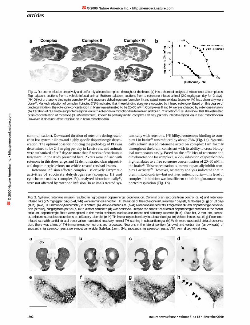
Rotenone infusion affected complex I selectively. Enzymaticactivities of succinate dehydrogenase (complex II) andcytochrome oxidase (complex IV), analyzed histochemically27,were not affected by rotenone infusion. In animals treated sys-
temically with rotenone, [3H]dihydrorotenone binding to com-plex I in brain28 was reduced by about 75% (Fig. 1a). Systemi-cally administered rotenone acted on complex I uniformlythroughout the brain, consistent with its ability to cross biolog-ical membranes easily. Based on the affinities of rotenone anddihydrorotenone for complex I, a 75% inhibition of specific bind-ing translates to a free rotenone concentration of 20–30 nM inthe brain28. This concentration is known to partially inhibit com-plex I activity29. However, oximetry analysis indicated that inbrain mitochondria—but not liver mitochondria—this level ofcomplex I inhibition was insufficient to inhibit glutamate-sup-ported respiration (Fig. 1b).
articles
Fig. 2. Systemic rotenone infusion resulted in nigrostriatal dopaminergic degeneration. Coronal brain sections from control (a, e) and rotenone-infused rats (2.5 mg/kg per day; (b–d, f–h) were immunostained for TH. Duration of the rotenone infusion was 7 days (b, f), 36 days (c, g) or 33 days(d, h). (a–d) TH immunocytochemistry in striatum. (a) Vehicle-infused rat. (b–d) Rotenone-infused rats. Progressive striatal dopaminergic denerva-tion (arrows), ranging from partial (b, c) to almost complete (d) was observed. Despite the almost total loss of dopaminergic terminals in the motorstriatum, dopaminergic fibers were spared in the medial striatum, nucleus accumbens and olfactory tubercle (b–d). Scale bar, 2 mm. ctx, cortex; st, striatum; na, nucleus accumbens; ot, olfactory tubercle. (e–h) TH immunocytochemistry in substantia nigra. (e) Vehicle-infused rat. (f, g) Rotenone-infused rats with partial striatal denervation maintained relatively normal TH staining in substantia nigra. (h) With more substantial striatal denerva-tion, there was a loss of TH-immunoreactive neurons and processes. Neurons in the lateral portion (arrows) and ventral tier (arrowheads) ofsubstantia nigra pars compacta were most vulnerable. Scale bar, 1 mm. SNc, substantia nigra pars compacta; VTA, ventral tegmental area.
Fig. 1. Rotenone infusion selectively and uniformly affected complex I throughout the brain. (a) Histochemical analysis of mitochondrial complexes.Top, adjacent sections from a vehicle-infused animal. Bottom, adjacent sections from a rotenone-infused animal (2.0 mg/kg per day for 2 days).[3H]Dihydrorotenone binding to complex I28 and succinate dehydrogenase (complex II) and cytochrome oxidase (complex IV) histochemistry weredone27. Marked reduction of complex I binding (73%) indicated that these binding sites were occupied by infused rotenone. Based on this degree ofbinding inhibition, the rotenone concentration in brain was estimated to be 20–30 nM27. Complexes II and IV were unchanged by rotenone infusion.(b) Titration of glutamate-supported respiration with rotenone in mitochondria from liver and brain. Oximetry41,42 studies show that the estimatedbrain concentration of rotenone (30 nM maximum), known to partially inhibit complex I activity, partially inhibits respiration in liver mitochondria.However, it does not affect respiration in brain mitochondria.
a
a
e
b c d
f g h
b
© 2000 Nature America Inc. • http://neurosci.nature.com©
200
0 N
atu
re A
mer
ica
Inc.
• h
ttp
://n
euro
sci.n
atu
re.c
om
nature neuroscience • volume 3 no 12 • december 2000 1303
Selective nigrostriatal dopaminergic degenerationSystemic partial inhibition of complex I resulted in progressivenigrostriatal dopaminergic degeneration. Immunocytochemistryof phenotypic markers for dopaminergic neurons (tyrosinehydroxylase, TH; dopamine transporter, DAT; vesicularmonoamine transporter type 2, VMAT2) all gave identical results,indicating dopaminergic degeneration. Depending on the doseand duration of rotenone exposure, animals demonstrated vary-ing degrees of striatal dopaminergic denervation (Fig. 2b–d).Lesions typically began focally in the central or dorsolateral por-tion of the anterior striatum, as evidenced by a complete loss ofphenotypic markers, and spread to involve most of the motorstriatum. Even when striatal TH depletion was almost complete,there was relative sparing of dopaminergic fibers in medial aspectsof striatum, nucleus accumbens and olfactory tubercle (Fig. 2d),areas that are relatively spared in idiopathic PD30.
There was also evidence of dopaminergic lesions in the cell bod-ies of the substantia nigra pars compacta. Animals with partialreductions in TH staining in striatum had dopaminergic neuronsin substantia nigra that looked relatively normal (Fig. 2f and g);however, in animals with near complete striatal denervation, therewere obvious reductions in TH-stained cells in substantia nigra(Fig. 2h). These results suggest that striatal nerve terminals wereaffected earlier and more severely by rotenone than nigral cell bod-ies. Similarly, in brains of dying patients with PD, there is a moreprofound loss of dopamine in striatum compared to substantianigra31. Neurons in the lateral portion and ventral tier of the sub-stantia nigra pars compacta were most vulnerable to systemicrotenone infusion (Fig. 2h). This pattern of vulnerability matchesthe pattern in idiopathic PD. Despite the loss of TH staining in
articles
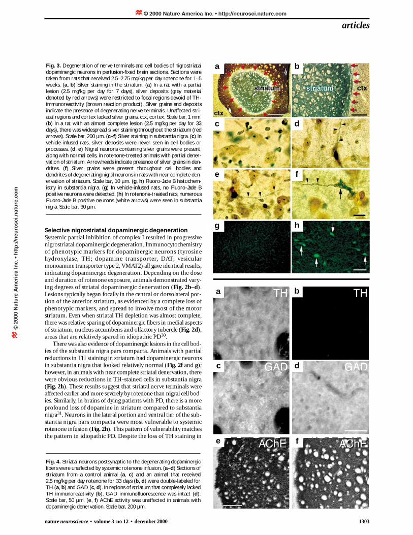
Fig. 3. Degeneration of nerve terminals and cell bodies of nigrostriataldopaminergic neurons in perfusion-fixed brain sections. Sections weretaken from rats that received 2.5–2.75 mg/kg per day rotenone for 1–5weeks. (a, b) Silver staining in the striatum. (a) In a rat with a partiallesion (2.5 mg/kg per day for 7 days), silver deposits (gray materialdenoted by red arrows) were restricted to focal regions devoid of TH-immunoreactivity (brown reaction product). Silver grains and depositsindicate the presence of degenerating nerve terminals. Unaffected stri-atal regions and cortex lacked silver grains. ctx, cortex. Scale bar, 1 mm.(b) In a rat with an almost complete lesion (2.5 mg/kg per day for 33days), there was widespread silver staining throughout the striatum (redarrows). Scale bar, 200 µm. (c–f) Silver staining in substantia nigra. (c) Invehicle-infused rats, silver deposits were never seen in cell bodies orprocesses. (d, e) Nigral neurons containing silver grains were present,along with normal cells, in rotenone-treated animals with partial dener-vation of striatum. Arrowheads indicate presence of silver grains in den-drites. (f) Silver grains were present throughout cell bodies anddendrites of degenerating nigral neurons in rats with near complete den-ervation of striatum. Scale bar, 10 µm. (g, h) Fluoro-Jade B histochem-istry in substantia nigra. (g) In vehicle-infused rats, no Fluoro-Jade Bpositive neurons were detected. (h) In rotenone-treated rats, numerousFluoro-Jade B positive neurons (white arrows) were seen in substantianigra. Scale bar, 30 µm.
Fig. 4. Striatal neurons postsynaptic to the degenerating dopaminergicfibers were unaffected by systemic rotenone infusion. (a–d) Sections ofstriatum from a control animal (a, c) and an animal that received 2.5 mg/kg per day rotenone for 33 days (b, d) were double-labeled forTH (a, b) and GAD (c, d). In regions of striatum that completely lackedTH immunoreactivity (b), GAD immunofluorescence was intact (d).Scale bar, 50 µm. (e, f) AChE activity was unaffected in animals withdopaminergic denervation. Scale bar, 200 µm.
a b
c d
e f
g h
a b
c d
e f
© 2000 Nature America Inc. • http://neurosci.nature.com©
200
0 N
atu
re A
mer
ica
Inc.
• h
ttp
://n
euro
sci.n
atu
re.c
om
1304 nature neuroscience • volume 3 no 12 • december 2000
substantia nigra, dopaminergic neurons of the ventral tegmentalarea (VTA) were relatively spared, as they are in PD. Moreover, justas in idiopathic PD, noradrenergic neurons of the locus ceruleuswere mildly to moderately affected (data not shown).
To confirm that the loss of staining of dopaminergic neuronsand nerve terminals was due to degeneration rather than reducedexpression of phenotypic markers by surviving cells, silver stain-ing was done alone and in combination with TH immunostain-ing. In the striatum, regions of degeneration densely stained withsilver corresponded to areas where there was loss of TH staining(Fig. 3a and b). In the substantia nigra, there was also clear degen-eration. Although animals treated with vehicle never showednigral degeneration (Fig. 3c), animals with partial loss of striataldopamine terminals had many nigral neurons with silver depositsin their cell bodies or dendrites, interspersed with neurons thatlooked normal (Fig. 3d and e). Thus, although nigral TH stain-ing was relatively normal in animals with partial denervation ofstriatum, silver staining revealed degenerative changes. Animalswith severe dopaminergic denervation of striatum had extensivesilver deposition in substantia nigra corresponding to the loss ofphenotypic markers in this region (Fig. 3f). Silver staining alsoconfirmed the progressive, retrograde nature of dopaminergicdegeneration in the rotenone model. In an animal that survivedfor two weeks beyond the end of rotenone infusion, there was nolonger silver staining in the region of striatum depleted ofdopaminergic markers. Presumably, this was due to previousphagocytosis of degenerating neuronal elements. However, inthis animal, there was massive silver deposition in substantia
nigra (data not shown). Silver staining also showed that therewas no consistent neuronal degeneration elsewhere in the brainsof rotenone-treated animals. For example, there was no silverstaining in the cortex in Fig. 3b. Although we never saw silverstaining in control animals, there was the possibility that silverdeposition could reflect protein aggregation in the absence offrank degeneration. Therefore, the neuronal degeneration wasconfirmed in rotenone-infused animals with Fluoro-Jade B his-tochemistry. Fluoro-Jade B is a fluorescein derivative that selec-tively stains neurons undergoing degeneration32. Vehicle-infusedanimals had no Fluoro-Jade B-positive neurons in substantianigra (Fig. 3g); however, there were numerous Fluoro-Jade B-positive neurons in substantia nigra of rotenone-infused animals(Fig. 3h). Together, silver staining and Fluoro-Jade B histo-chemistry conclusively demonstrated selective degeneration ofnigrostriatal dopamine neurons in rotenone-infused animals.
Despite the profound loss of presynaptic dopaminergic nerveterminals in striatum, postsynaptic neuronal elements in stria-tum remained intact. Nissl staining, though relatively insensitivein striatum, did not show obvious lesions of striatal neurons (datanot shown). More than 90% of striatal neurons use γ-aminobu-tyric acid as their transmitter, and they can be identified withimmunocytochemistry for glutamic acid decarboxylase (GAD).In striatal regions lacking dopaminergic terminals, there was noloss of GAD immunoreactivity (Fig. 4a–d). Thus, the predomi-nant cell type in striatum seemed to be intact in rotenone-treatedanimals. This is in direct contrast to what occurs when complexII—rather than complex I—is inhibited systemically. Animalstreated systemically with the complex II inhibitor, 3-nitropropionicacid, display selective degeneration of striatal neurons with relativesparing of the nigrostriatal dopamine system33. An additionalsmaller population of striatal neurons is cholinergic, and can beassessed with acetylcholinesterase (AChE) staining34. Striatal AChEstaining was similar in vehicle- and rotenone-treated animals (Fig. 4e and f). These results further support the nigrostriataldopaminergic selectivity of rotenone-induced neurodegeneration.
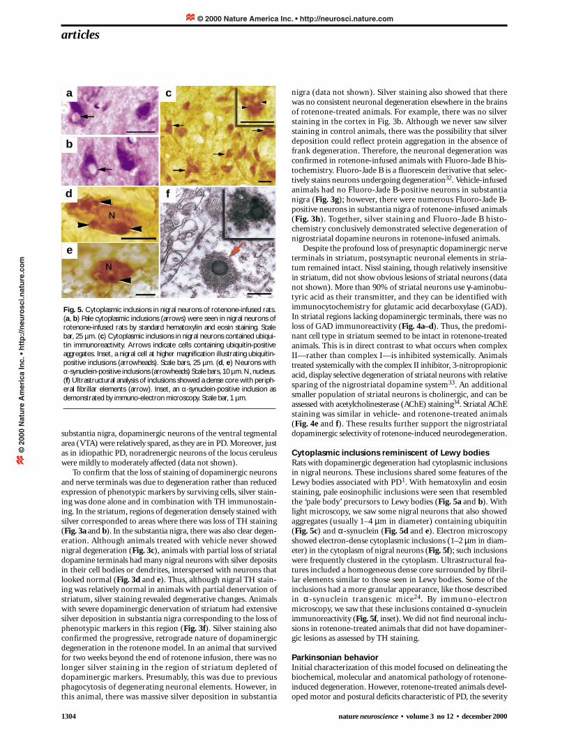
Cytoplasmic inclusions reminiscent of Lewy bodiesRats with dopaminergic degeneration had cytoplasmic inclusionsin nigral neurons. These inclusions shared some features of theLewy bodies associated with PD1. With hematoxylin and eosinstaining, pale eosinophilic inclusions were seen that resembledthe ‘pale body’ precursors to Lewy bodies (Fig. 5a and b). Withlight microscopy, we saw some nigral neurons that also showedaggregates (usually 1–4 µm in diameter) containing ubiquitin(Fig. 5c) and α-synuclein (Fig. 5d and e). Electron microscopyshowed electron-dense cytoplasmic inclusions (1–2 µm in diam-eter) in the cytoplasm of nigral neurons (Fig. 5f); such inclusionswere frequently clustered in the cytoplasm. Ultrastructural fea-tures included a homogeneous dense core surrounded by fibril-lar elements similar to those seen in Lewy bodies. Some of theinclusions had a more granular appearance, like those describedin α-synuclein transgenic mice24. By immuno-electronmicroscopy, we saw that these inclusions contained α-synucleinimmunoreactivity (Fig. 5f, inset). We did not find neuronal inclu-sions in rotenone-treated animals that did not have dopaminer-gic lesions as assessed by TH staining.
Parkinsonian behaviorInitial characterization of this model focused on delineating thebiochemical, molecular and anatomical pathology of rotenone-induced degeneration. However, rotenone-treated animals devel-oped motor and postural deficits characteristic of PD, the severity
articles
Fig. 5. Cytoplasmic inclusions in nigral neurons of rotenone-infused rats.(a, b) Pale cytoplasmic inclusions (arrows) were seen in nigral neurons ofrotenone-infused rats by standard hematoxylin and eosin staining. Scalebar, 25 µm. (c) Cytoplasmic inclusions in nigral neurons contained ubiqui-tin immunoreactivity. Arrows indicate cells containing ubiquitin-positiveaggregates. Inset, a nigral cell at higher magnification illustrating ubiquitin-positive inclusions (arrowheads). Scale bars, 25 µm. (d, e) Neurons with α-synuclein-positive inclusions (arrowheads) Scale bars, 10 µm. N, nucleus.(f) Ultrastructural analysis of inclusions showed a dense core with periph-eral fibrillar elements (arrow). Inset, an α-synuclein-positive inclusion asdemonstrated by immuno-electron microscopy. Scale bar, 1 µm.
a
b
c
d
e
f
© 2000 Nature America Inc. • http://neurosci.nature.com©
200
0 N
atu
re A
mer
ica
Inc.
• h
ttp
://n
euro
sci.n
atu
re.c
om
nature neuroscience • volume 3 no 12 • december 2000 1305
of which depended on the extent of lesions. All animals with adopaminergic lesion became hypokinetic and had unsteadymovement and hunched posture, even after termination of therotenone infusion. Seven animals developed severe rigidity, andthree animals had shaking of one or more paws that was remi-niscent of rest tremor. Preliminary experiments in two animalsindicate that the motor deficits were ameliorated by the dopamineagonist, apomorphine (data not shown).
DISCUSSIONTaken together, our results indicate that a systemic partial defectin complex I is enough to reproduce the behavioral, anatomical,neurochemical and neuropathological features of PD. The find-ing that rotenone affected complex I uniformly throughout thebrain (Fig. 1a), coupled with the resultant highly selective neu-rodegeneration of the nigrostriatal dopaminergic system, sug-gested that this population of neurons had an intrinsic sensitivityto complex I defects. Because the brain rotenone concentration(20–30 nM) was enough to partially inhibit complex I, but toolow to significantly impair respiration of brain mitochondria, itseemed that a bioenergetic defect with ATP depletion could notexplain the neurodegneration. Instead, oxidative damage mighthave been involved. Inhibition of complex I by rotenone stimu-lates production of reactive oxygen species35,36. Moreover, wehave found that culture of neural cells with 5 nM rotenoneinduces, over a period of weeks, progressive oxidative damage toproteins and DNA, and sensitizes cells to subsequent oxidativestressors; eventually, it also begins to induce release of cytochromec from mitochondria to the cytoplasm (Sherer et al., Soc. Neu-rosci. Abstr. 26, 280.21, 2000). This mechanism could also explainthe cytoplasmic inclusions found in nigral neurons of rotenone-treated rats, because both oxidative damage and cytochrome cenhance α-synuclein aggregation37,38.
Rotenone seems to have little toxicity when administered oral-ly (T. B. S. and J. T. G., unpublished results); however, our resultshighlight the possibility that environmental toxins, including pes-ticides that inhibit mitochondrial function, may contribute to thepathogenesis of PD. Many other naturally occurring compoundsand synthetic pesticides are potent inhibitors of complex I39. Indi-viduals are likely to be variably exposed to numerous natural orsynthetic complex I inhibitors through diet, drinking water orother environmental factors. Such exposures, combined withgenetic differences in complex I function14–22, or inter-individ-ual differences in the ability to metabolize xenobiotics7, mayunderlie most cases of typical idiopathic PD.
METHODSAnimal surgery. We used male Sprague Dawley and Lewis rats(300–350 g; approximately 2 months old) for this study. Alzet osmot-ic mini pumps (models 2ML4 or 2ML1) were filled with rotenone, dis-solved in equal volumes of dimethylsulfoxide (DMSO) andpolyethylene glycol (PEG). Pumps were attached to Tygon microbore(0.58 mm i.d.) tubing (Fisher Scientific, Pittsburgh, Pennsylvania) andplaced in sterile 0.9% saline at 37°C for at least 4 h. Ketamine (75 mg/kg) and Rompum (10 mg/kg) were injected intramuscularlyfor anesthetic. Alzet osmotic mini pumps were implanted under theskin on the back of each animal, and the right jugular vein was can-nulated. Pumps were exchanged after 28 days. Control rats receivedDMSO:PEG (1:1) only. Following surgery, rats were monitored forbehavior, weight loss and overall health. In the instances whenrotenone-treated rats began to lose weight, diet was supplementedwith oral administration of Nutrical (Evsco Pharmaceuticals, Buena,New Jersey). Subcutaneous lactated Ringer’s injection USP solution(Baxter Healthcare Corporation, Deerfield, Illinois) was given whenrats showed signs of dehydration.
Mitochondrial respiration. Oxygen consumption by rat brain and livermitochondria was measured polargraphically as described40 using anInstech minichamber (Instech Laboratories, Plymouth Meeting, Penn-sylvania) equipped with a magnetic stirrer and oxygen electrode (YellowSpring Instrument Company, Yellow Springs, Ohio) connected to a chartrecorder. For maximum State 3 respiration activity, the following medi-um was used: 125 mM KCl, 10 mM MOPS, 2 mM MgCl2, 2 mMKH2PO4, 1 mM EGTA, 0.7 mM CaCl2, 20 mM glucose, 8 units of hex-okinase, and 20 mM glutamate plus 2 mM malate. Brain mitochondriawere isolated by the method of Sims41 without BSA, because BSA bindsrotenone nonspecifically.
Immunocytochemistry. We used paraformaldehyde-fixed brains. For sin-gle immunolabeling studies, 40-µm sections were incubated in primaryantibody for 24 h, followed by 1 h incubation with biotinylated secondaryantibody. The avidin-biotin complex method was used to detect the anti-gen signal (ABC elite kit, Vector laboratories, Burlingame, California) and3,3´-diaminobenzidine tetrachloride (DAB) was used to visualize the finalproduct. The primary antibodies used were monoclonal mouse antibodyagainst TH (1:2000; Chemicon, Temecula, California), polyclonal rabbitantibody against ubiquitin (1:1000; Dako Corporation, Carpinteria, Cal-ifornia) and polyclonal rabbit antibody against α-synuclein (1:400). Sec-ondary antibodies used were biotinylated goat anti-mouseimmunoglobulin G (IgG) or biotinylated goat anti-rabbit IgG (1:200; Jack-son Immunoresearch Labs, West Grove, Pennsylvania). Double labelingfor TH and GAD was performed by immunofluorescence. Sections weresimultaneously incubated with antibodies against TH (1:2000; Chemicon)and GAD (1:2000; polyclonal rabbit antibody; Chemicon) for 24 h. Texasred-conjugated goat anti-mouse IgG was used to detect TH-positive cellsand fluorescein isothiocyanate-conjugated goat anti-rabbit IgG was used todetect GAD- expressing cells (1:200; Jackson Immunoresearch Labs). Forcontrols, one or both primary antibodies were omitted. We examinedimmunostained sections using bright-field microscopy or conventionalfluorescence microscopy. We captured images on a Leitz microscope (Leica,Wetzlar, Germany) linked to an image analysis system (Imaging Research,St. Catharines, Ontario, Canada) with selective filter sets to visualize FITCand Texas red separately, as well as simultaneously. For final output, imageswere processed using Adobe Photoshop 5.0 software.
Detection of neuronal degeneration. Both silver staining and Fluoro-Jade B histochemistry were used to detect degenerating neurons. The FDNeurosilver kit (FD NeuroTechnologies, Ellicott City, Maryland) staineddegenerating neuronal elements in paraformaldehyde-fixed brain tissuefrom control and rotenone-infused rats. Silver staining was done accord-ing to the manufacturer’s protocol. Following silver staining, sectionswere mounted on Superfrost plus slides (Fisher Scientific, Pittsburgh,Pennsylvania), air-dried, cleared with xylene, and coverslipped with Per-mount. Sections were examined using bright-field microscopy. TH-immunostained sections were also processed for silver staining todemonstrate selective neurodegeneration. Fluoro-Jade B histochemistrywas done on paraformaldehyde-fixed brain sections from control androtenone-infused rats according to the manufacturer’s protocol (Histo-Chem, Jefferson, Arkansas).
Electron microscopy. Paraformaldehyde-fixed brain sections from fourrats were processed for standard electron microscopy. The tissue waspost-fixed in osmium tetroxide and embedded in epon. Ultra-thin sec-tions were stained with uranyl acetate and lead citrate and examined witha Hitachi (Mountain View, California) electron microscope. Forimmuno-electron microscopy, sections stained for α-synuclein usingDAB as chromophore were fixed with 0.5% glutaraldehyde and processedfor routine electron microscopy as described.
ACKNOWLEDGEMENTSWe thank P. Piccardo and B. Ghetti for donation of the α-synuclein antibody,
which was generated with support from the Indiana Alzheimer’s Disease Center
(P30AG10133). We also thank M. DeLong and A. Levey for reading this manu-
script. This work was supported by NIH grants to JTG (NS38399, NS33779, and
AG14648).
articles
© 2000 Nature America Inc. • http://neurosci.nature.com©
200
0 N
atu
re A
mer
ica
Inc.
• h
ttp
://n
euro
sci.n
atu
re.c
om

1306 nature neuroscience • volume 3 no 12 • december 2000
RECEIVED 7 SEPTEMBER; ACCEPTED 25 OCTOBER 2000
1. Baba, M. et al. Aggregation of alpha-synuclein in Lewy bodies of sporadicParkinson’s disease and dementia with Lewy bodies. Am. J. Pathol. 152, 879–884(1998).
2. Kruger, R. et al. Ala30Pro mutation in the gene encoding alpha-synuclein inParkinson’s disease. Nat. Genet. 18, 106–108 (1998).
3. Polymeropoulos, M. H. et al. Mutation in the alpha-synuclein gene identified infamilies with Parkinson’s disease. Science 276, 2045–2047 (1997).
4. Kitada, T. et al. Mutations in the parkin gene cause autosomal recessive juvenileparkinsonism. Nature 392, 605–608 (1998).
5. Tanner, C. M. et al. Parkinson disease in twins: an etiologic study. JAMA 281,341–346 (1999).
6. Dexter, D. T. et al. Basal lipid peroxidation in substantia nigra is increased inParkinson’s disease. J. Neurochem. 52, 381–389 (1989).
7. Menegon, A., Board, P. G., Blackburn, A. C., Mellick, G. D. & Le Couteur, D. G.Parkinson’s disease, pesticides, and glutathione transferase polymorphisms.Lancet 352, 1344–1346 (1998).
8. Butterfield, P. G., Valanis, B. G., Spencer, P. S., Lindeman, C. A. & Nutt, J. G.Environmental antecedents of young-onset Parkinson’s disease. Neurology 43,1150–1158 (1993).
9. Gorell, J. M., Johnson, C. C., Rybicki, B. A., Peterson, E. L. & Richardson, R. J.The risk of Parkinson’s disease with exposure to pesticides, farming, well water,and rural living. Neurology 50, 1346–1350 (1998).
10. Langston, J. W., Ballard, P. A., Tetrud, J. W. & Irwin, I. Chronic parkinsonism inhumans due to a product of meridine-analog synthesis. Science 219, 979–980(1983).
11. Nicklas, W. J. & Heikkila, R. E. Inhibition of NADH-linked oxidation in brainmitochondria by 1-methyl-4-phenylpyridine, a metabolite of the neurotoxin, 1-methyl-4-phenyl-1,2,3,6-tetrahydropyridine. Life Sci. 36, 2503–2508 (1985).
12. Tipton, K. F. & Singer, T. P. Advances in our understanding of the mechanisms ofthe neurotoxicity of MPTP and related compounds. J. Neurochem. 61,1191–1206 (1993).
13. Tolwani, R. J., Jakowec, M. W., Petzinger, G. M., Green, S. & Waggie, K.Experimental models of Parkinson’s disease: insights from many models. Lab.Anim. Sci. 49, 363–371 (1999).
14. Parker, W. D., Boyson, S. J. & Parks, J. K. Abnormalities of the electron transportchain in idiopathic Parkinson’s disease. Ann. Neurol. 26, 719–723 (1989).
15. Bindhoff, L. A., Birch-Machin, M., Cartlidge, N. E., Parker, W. D. Jr. & Turnbull,D. M. Mitochondrial function in Parkinson’s disease. Lancet 2, 49 (1989).
16. Schapira, A.H. et al. Mitochondrial complex I deficiency in Parkinson’s disease.J. Neurochem. 54, 823–827 (1990).
17. Shoffner, J. M., Watts, R. L., Juncos, J. L., Torroni, A. & Wallace, D. C.Mitochondrial oxidative-phosphorylation defects in Parkinson’s disease. Ann.Neurol. 30, 332–339 (1991).
18. Mann, V. M. et al. Brain, skeletal-muscle and platelet homogenatemitochondrial function in Parkinson’s disease. Brain 115, 333–342 (1992).
19. Cardellach, F. et al. Mitochondrial respiratory-chain activity in skeletal musclefrom patients with Parkinsons disease. Neurology 43, 2258–2262 (1993).
20. Blin, O. et al. Mitochondrial respiratory failure in skeletal muscle from patientswith Parkinson’s disease and multiple system atrophy. J. Neurol. Sci. 125, 95–101(1994).
21. Swerdlow, R. H. et al. Matrilineal inheritance of complex I dysfunction in amultigenerational Parkinson’s disease family. Ann. Neurol. 44, 873–881 (1998).
22. Mizuno, Y. et al. Mitochondrial dysfunction in Parkinson’s disease. Ann.Neurol. 44 (3 Suppl. 1), S99–109 (1998).
23. Forno, L. S., DeLanney, L. E., Irwin, I. & Langston, J. W. Electron microscopy ofLewy bodies in the amygdala-parahippocampal region. Comparison withinclusions bodies in the MPTP-treated squirrel monkey. Adv. Neurol. 69,217–228 (1996).
24. Masliah, E. et al. Dopaminergic loss and inclusion body formation in α-synuclein mice: implications for neurodegenerative disorders. Science 287,1265–1269 (2000).
25. Ouary, S. et al. Major strain differences in response to chronic systemicadministration of the mitochondrial toxin 3-nitropropionic acid in rats:implications for neuroprotection studies. Neuroscience 97, 521–530 (2000).
26. Ferrante, R. J., Schluz, J. B., Kowall, N. W. & Beal, M. F. Systemic administrationof rotenone produces selective damage in the striatum and globus pallidus, butnot the substantia nigra. Brain Res. 753, 157–162 (1997).
27. Porter, R. H. P., Greene, J. G., Higgins, D. S. Jr. & Greenamyre, J. T. Polysynapticregulation of glutamate receptors and mitochondrial enzyme activities in thebasal ganglia of rats with unilateral dopamine depletion. J. Neurosci. 14,7192–7199 (1994).
28. Higgins, D. S. Jr. & Greenamyre, J. T. [3H] Dihydrorotenone binding to NADH:Ubiquinone reductase (complex I) of the electron transport chain: anautoradiographic study. J. Neurosci. 16, 3807–3816 (1996).
29. Davey, G. P. & Clark, J. B. Threshold effects and control of oxidativephosphorylation in nonsynaptic rat brain mitochondria. J. Neurochem. 66,1617–1624 (1996).
30. Miller, G. W. et al. Immunochemical analysis of dopamine transporter proteinin Parkinson’s disease. Ann. Neurol. 41, 530–539 (1997).
31. Hornykiewicz, O. Dopamine (3-hydroxytyramine) and brain function.Pharmacol. Rev. 18, 925–965 (1966).
32. Schmued, L. C., Albertson, C. & Slikker, W. Jr. Fluoro-Jade: a novelfluorochrome for the sensitive and reliable histochemical localization ofneuronal degeneration. Brain Res. 751, 37–46 (1997).
33. Beal, M. F. et al. Neurochemical and histologic characterization of striatalexcitotoxic lesions produced by the mitochondrial toxin 3-nitropropionic acid.J. Neurosci. 13, 4181–4192 (1993).
34. Hedreen, J. C., Bacon, S. J. & Price, D. L. A modified histochemical technique tovisualize acetylcholinesterase-containing axons. J. Histochem. Cytochem. 33,134–140 (1985).
35. Hensley, K. et al. Interaction of alpha-phenyl-N-tert-butyl nitrone andalternative electron acceptors with complex I indicates a substrate reductionsite upstream from the rotenone binding site. J. Neurochem. 71, 2549–2557(1998).
36. Seaton, T. A., Cooper, J. M. & Schapira, A. H. Free radical scavengers protectdopaminergic cell lines from apoptosis induced by complex I inhibitors. BrainRes. 777, 110–118 (1997).
37. Souza, J. M., Giasson, B. I., Chen, Q., Lee, V. M.-Y & Ischiropoulos, H.Dityrosine cross-linking promotes formation of stable α–synuclein polymers.J. Biol. Chem. 275, 18344–18349 (2000).
38. Hashimoto, M., Takeda, A., Hsu, L. J., Takenouchi, T. & Masliah, E. Role ofcytochrome c as a stimulator of α–synuclein aggregation in Lewy body disease.J. Biol. Chem. 274, 28849–28852 (2000).
39. Degli Esposti, M. Inhibitors of NADH-ubiquinone reductase: an overview.Biochim. Biophys. Acta 1364, 222–235 (1998).
40. Panov, A. V. & Scaduto, R. C. Jr. Substrate specific effects of calcium onmetabolism of rat heart mitochondria. Am. J. Physiol. 270, H1398–H1406(1996).
41. Sims, N. R. Rapid isolation of metabolically active mitochondria from rat brainand subregions using Percoll density gradient centrifugation. J. Neurochem. 55,698–707 (1990).
articles
© 2000 Nature America Inc. • http://neurosci.nature.com©
200
0 N
atu
re A
mer
ica
Inc.
• h
ttp
://n
euro
sci.n
atu
re.c
om