Lower Extremity Review
64
-
Upload
khangminh22 -
Category
Documents
-
view
3 -
download
0
Transcript of Lower Extremity Review


grace and gordon
drcomfort.comFollow us: @drcomfort Dr.ComfortIndividual results may vary. Neither DJO Global, Inc. nor any of its subsidiaries dispense medical advice. The contents of this sheet do not constitute medical, legal, or any other type of professional advice. Rather, please consult your healthcare professional for information on the courses of treatment, if any, which may be appropriate for you.
T 800.556.557210300 North Enterprise Drive I Mequon, WI 53092 I U.S.A.drcomfort.com I Copyright © 2019 by DJO, LLC I MKT00-8422 Rev A
meetA reliable workout team that helps improve mobility, provides the stability you need, and the lasting comfort to keep going.
OM19-MKT00-8422-DRC-Gordon-Grace-2Page-Spread-LER.indd All Pages 3/5/19 1:38 PM

STAY IN THE GAME WITHSPORTMAXX™ ORTHOTICS
Footmaxx has a line of orthotics specifically for different sports. From baseball to skiing,
your patients will be ready to go.
Footmaxx.com F | L
KEEP YOUR PATIENTS OFF THE BENCHCall us today to learn more
1.800.779.3668 Footmaxx
LER Sportmaxx Ad.indd 1 4/4/19 3:11 PM

VOLUME 11 NUMBER 3 LERMAGAZINE .COM
The views and opinions expressed here are those of the authors and do not necessarily reflect the official policy or position of Lower Extremity Review.
COVER
34 HOW MOUNTAIN BIKING IS RESHAPING THE LANDSCAPE OF CYCLING INJURY
Differences in equipment, terrain, riding style, and other risk factors mean different types and prevalence of injuries when riding a mountain bike, compared to a road bike.
By Michael Reeder, DO, and Brent Alumbaugh, MS
GUEST EDITORIAL
9 WE ARE THE HEART OF NATIONAL BIOMECHANICS DAY
The International and American Societies of Biomechanics were founded in 1973 and 1977, respectively. Coincident with the expansion of Biomechanics during the following decades, fitness and health …
By Paul DeVita, PhD
CALL FOR MANUSCRIPTS
10 HERE’S HOW TO SUBMIT YOUR MANUSCRIPT
FEATURES
19 BONE STIMULATION REPRESENTS NEW FRONTIER FOR OPTIMIZING PATIENT OUTCOMES
Limited research into electromagnetic and ultrasound bone stimulation devices complicates efforts to determine efficacy. For the majority of people, lower-limb fractures and fractures in other parts of the body heal well and reliably. However, some people experience complications that…
By Nicole Wetsman
27 MITIGATING THE OPIOID CRISIS WITH PAIN MANAGEMENT REGIMENS IN A MULTIMODAL APPROACH
For help limiting the number of opioid tablets you prescribe—and to still treat postop pain successfully—turn to practice guidelines, evidence from the literature about local and …
By ROBERT G. SMITH, DPM, MSC, RPH, CPED, CPRS
43 TAPE USE TO PREVENT BLISTERS: DOES IT REALLY DO WHAT WE THINK IT DOES?
Taping is a mainstay of preventive foot blister management in athletes and active people. Its use is based on the premise that rubbing causes blisters, and that tape protects the skin from this rubbing and/or provides thermal insulation from the heat generated by rubbing. This commentary highlights…
By Rebecca Rushton, BSc(Pod)
51 THE THERAPEUTIC POWER OF ELASTIC BANDS
Here’s why resistance therapy with these low-tech tools is gaining prominence—for youth soccer players, geriatric patients, and everyone in between. Elastic resistance bands are increasingly popular in physical therapy, as the modality is being used…
By Keith Loria
FROM THE LITERATURE13 • Benefits of a Foot Orthosis During
a Step-descent Task on Kinematics, Kinetics, and Activation of Muscle
• Concurrent Changes in Eccentric Hamstring Strength and Knee-joint Kinematics
• Delayed Charcot Diagnosis Increases Costs, Hospital Days, Amputations
• AFOs for Stroke: Early Use Does Not Limit Long-Term Benefit
AD INDEX
57 GET CONTACT INFO FOR ALL OF OUR ADVERTISERS
INDUSTRY SNAPSHOT
58 PRODUCTS, ASSOCIATION NEWS & MARKET UPDATES
CROSSWORD PUZZLE
62 TEST YOUR KNOWLEDGE OF INFORMATION FROM THIS ISSUE
March 2019contents

New!
© 2019 PediFix, Inc. LER419
Yes! Please send me:☐1 Free Sample☐12 Roll Clinic Pack (1˝ x 1.5 yd), $48.00 ☐12 Roll Clinic Pack (2˝ x 1.5 yd), $72.00
Your Name__________________________________________________________________________
Practice Name ______________________________________________________________________
Shipping Address ___________________________________________________________________
City ________________________________________State ______ Zip ________________________
Phone _______________________________________________________________________________
Fax __________________________________________________________________________________
Email _______________________________________________________________________________
In our practice, we see approximately ___________ (#) patients each week.
My favorite supplier is ___________________________________________________________
I prefer: ☐ to Dispense ☐ to Prescribe ☐Patient Direct OrderMail to: PediFix, Dept. LER419, 301 Fields Lane, Brewster, NY 10509
Fax to: 845-277-2851
To order, get a free sample* or more information, mention code LER419
Call: 1-800-424-5561
Fax: 845-277-2851
E-mail: [email protected]
Return this Coupon to: PediFix, 301 Fields Lane, Dept. LER419, Brewster, NY 10509
Effective. Economical. Easy.
Please provide all information requested. Allow 2 weeks for delivery.
®Medical Footcare
A new, better alternative to paper and cloth tape wound dressings• Shields Blisters, Corns & Irritations
• Reduces Friction & Skin Damage
• Gently Secures Bandages & Dressings
• Helps Improve the Appearance of Scars
• Creates an Occlusive Barrier for FasterPenetration of Topical Wart Treatments
• More!
Safe & Easy Release for Painless Removal Without Skin Damage
Thin, Self-adhering, Waterproof, Reusable, Hypoallergenic
Save skin with Sorespot® Silicone Tape — the new skin protectant and topical bandage option
Skin Saver!
Medical Footcare®
*This offer is for healthcare professionals only. Limit one free sample per customer.
4 Ways SoreSpot® Silicone Tape Will Help Soothe & Heal Your PatientsFoot Care — Skin Care — Wound Care — Scar Care
New 12 Roll Clinic Pack
19108-Silicone Tape Full-Page Ad LER419.indd 1 4/9/19 3:23 PM

PUBLISHER AND CHIEF EXECUTIVE OFFICERRichard Dubin [email protected] | 518.221.4042
EDITORIAL STAFFEditorJanice T. Radak | [email protected]
Senior editor - PediatricsEmily Delzell | [email protected]
Senior editorKaren Hentry |[email protected]
Associate editorLaura Fonda Hochnadel | [email protected]
New products editorRikki Lee Travolta | [email protected]
Consulting editorJohn Baranowski | [email protected]
Operations coordinatorMelissa Rosenthal-Dubin | [email protected]
Graphic design & website developmentAnthony Palmeri | PopStart Web [email protected]
Audience developmentChristopher Wees | Media Automation, Inc.
Our Mission:Lower Extremity Review informs healthcare practitioners on current developments in the diagnosis, treatment, and prevention of lower extremity injuries. LER encourages a collaborative multidisciplinary clinical approach with an emphasis on functional outcomes and evidence-based medicine.
Opinions expressed are those of the authors and do not necessarily represent those of Lower Extremity Review, LLC.
LER is published monthly, except for a combined November/December issue and an additional special issue in December, by Lower Extremity Review, LLC. Copyright ©2019 Lower Extremity Review, LLC. All rights reserved. The publication may not be reproduced in any fashion, including electronically, in part or whole, without written consent. LER is a registered trademark of Lower Extremity Review.
Subscriptions may be obtained for $38 domestic and $72 international by writing to: LER, PO Box 390418 Minneapolis, MN 55439-0418. POSTMASTER: Please send address changes to LER, PO Box 390418, Minneapolis, MN 55439-0418.
LOWER EXTREMITY REVIEW41 State St. • Suite 604-16 • Albany, NY 12207518.452.6898
Craig R. Bottoni, MD Chief, Sports Medicine Service Orthopaedic Surgery Service Tripler Army Medical Center Honolulu, Hawaii
Jonathan Chang, MD Orthopaedic Surgery University of Southern California School of Medicine Alhambra, California
Robert Conenello, DPM Orangetown Podiatry Clinical Director, NJ Special Olympics NYPD Honorary Surgeon Greater New York City Area, New York
Sarah Curran, PhD, FCPodMed Professor, Podiatric Medicine & Rehabilitation Cardiff Metropolitan University Cardiff, United Kingdom
Stefania Fatone, PhD, BPO Professor, Physical Medicine & Rehabilitation Northwestern University Chicago, Illinois
Timothy E. Hewett, PhD Director, Biomechanics Laboratories & Sports Medicine Research Center Mayo Clinic Minneapolis, Minnesota
Geza Kogler, PhD, CO Director, Clinical Biomechanics Laboratory School of Biological Sciences Georgia Institute of Technology Atlanta, Georgia
Robert S. Lin, CPO Managing Partner, Adaptive Prosthetics & Orthotics Residency Coordinator, New England Orthotic and Prosthetic Systems, LLC Wethersfield, Connecticut
Antonio Robustelli, CSCS Sports Performance Consultant Applied Sport Scientist/Technologist Strength & Conditioning Specialist Salerno, Italy
Jarrod Shapiro, MD Vice Chair, Department of Podiatric Medicine, Surgery & Biomechanics Associate Professor of Podiatric Medicine, Surgery & Biomechanics Western University of Health Sciences ACFAOM Liaison Pomona, California
Bruce E. Williams, DPM Medical Director Go4-D Chicago, Illinois
INFORMATION FOR AUTHORS LER encourages a collaborative multidisciplinary clinical approach to the care of the lower extremity with an emphasis on functional outcomes using evidence-based medicine. We welcome manuscripts (1000-2000 words) that cross the clinical spectrum, including podiatry, orthopedics, physical medicine and rehabilitation, biomechanics, obesity, wound management, physical and occupational therapy, athletic training, orthotics and prosthetics, and pedorthics.
See detailed Author Guidelines at lermagazine.com – click the Editorial tab on the homepage.
ELECTRONIC SUBMISSIONS Please attach manuscript as an MS Word file or plain text. Tables may be included in the main document, but figures should be submitted as separate jpg attachments. Send to: [email protected]
lermagazine.com 3.19 7
EDITORIAL ADVISORY BOARD

Call nowfor a free
Temporal SpatialGait AnalysisInterpretation
Guide!610.449.4879protoKinetics.com/[email protected]

The International and American Societies of
Biomechanics were founded in 1973 and 1977,
respectively. Coincident with the expansion of
Biomechanics during the following decades, fit-
ness and health have moved out from under the
realm of medicine and are rapidly overtaking
the realm of the personal—the personal lifestyle
to be more exact (think yoga, yogurt, and yoga
pants). It is no coincidence that this trend has
mirrored the increasing role of Biomechanics in
our everyday lives as the evolving science has
allowed us to see that sitting is the new smoking
and that movement is our best option to main-
tain or get back to health.
And Biomechanics, the study of human
movement, of how bones, tendons, ligaments,
even cell components all work together, will
inform every step of that journey. The world
of Biomechanics today encompasses a host of
activities that are necessary to keep life moving
smoothly, but also to improve sport perfor-
mance, enhance rehabilitation, correct gait,
stabilize balance… all the pieces and parts of
maintaining a healthy active lifestyle, no matter
what age.
The good news is, biomechanists across the
globe are using today’s latest technology to help
improve patient outcomes and keep athletes
safe. But imagine if more young people were
aware of the field before they got to college?
How much faster would the knowledge base
spread if middle- and high-school students were
made aware of what Biomechanics has to offer?
That was the stimulus behind the first
National Biomechanics Day (NBD) in 2016
(aka, “The Early Year”). It began as a US-based
initiative to expand the impact of Biomechanics
on peoples’ lives. By 2017 (aka, “The Expansion
Year,”), NBD had grown into an international
celebration of Biomechanics. As of this date, 22
countries were expected to participate in NBD
2019. We think that, by introducing Biomechan-
ics to high school students, a population largely
under-exposed to Biomechanics science, we will
be seeding larger numbers of future biome-
chanists and a larger biomechanical impact.
Indeed, because Biomechanics education exists
nearly entirely at universities, any exposure to
younger people would bring new people to the
field. Although it is too early to know if we will
be successful in the long term, we certainly have
created a foundation for success. From 2016 to
2018, NBD excited more than 20,000 young
people and 800 teachers about Biomechanics
science, application, and careers. “That’s a lotta
kids,” as I like to say.
Still, why has NBD become such an as-
tounding success so quickly? Is there some facet
about NBD that entices biomechanists to not
only join the initiative but to thrive in it and to
enjoy it so fully? Certainly, many Biomechanics
labs around the world conduct outreach services
to show children and community leaders their
science. Actually, many university departments
beyond those housing Biomechanics programs
conduct social outreach to the benefit of both
the department and community. The partici-
pating departments benefit by increasing the
popularity of their fields and their particular
programs, and families benefit by learning
about potential career opportunities for their
children. These outreach events are wonderful
for all involved. Yet, these demonstrations and
events lack one element that is central to the
NBD mission. NBD celebrates the broad field
of Biomechanics beyond any individual lab,
program, or university. National Biomechanics
Day is enjoyed around the world because it is
a unifying platform upon which we coalesce
into one grand celebration of our science. NBD
enables biomechanists to unite into a single
synchronized, worldwide Biomechanics party.
It is not any individual person, it is all of us…
together: we all do it together and together
we are the Heart of NBD. The outcome of our
efforts is described by a single phrase, “Through
NBD, there are more smiles on more faces, in
more Biomechanics labs than on any other day.”
Pretty cool.
Our expansion to more biomechanists and
more high schoolers is paralleled by our expan-
sion to more sponsors and supporters. We have
33 sponsors who provide financial assistance
and promotional advertisements. For example,
LER has published two articles on NBD and
numerous photographs showing NBD events
around the world. Our sponsors include many
Biomechanics societies, instrument manufactur-
ers, and commercial entities using Biomechanics
to create their products and services. There is
also an important idea we emphasize about our
sponsors: They are not simply NBD sponsors,
they sponsor nearly all events and activities that
are Biomechanics. They sponsor Biomechanics
societies, Biomechanics conferences, Biome-
chanics outreach events, and, not to be neglect-
ed, they hold their own Biomechanics events.
They are us. Each individual company cannot
sponsor everything Biomechanics, but many of
them sponsor multiple Biomechanics entities.
NBD is extremely grateful to all its sponsors and
to all sponsors of Biomechanics. Without them,
we could not conduct the science of Biomechan-
lermagazine.com 3.19 9
GUEST EditorialWe Are The Heart of National Biomechanics Day
Continued on page 10
By Paul DeVita, PhD

ics as it is conducted now.
NBD and all NBDers are excited about
the future of Biomechanics. We have from the
start promoted the idea that Biomechanics
will be the breakthrough science of the 21st
century. We explained this position last year,1
but to briefly restate: First, Biomechanics has
great, yet untapped value for society; it can help
many people in many ways. While we know
this, it has yet to make a major, commercial
breakthrough that is fully within the public
consciousness. Second, we also propose that
while Biomechanics is largely unknown outside
of Biomechanics, it is on the verge of great
popularity. Many people know vaguely about 3D
motion capture as the basis for video games and
movies. Biomechanics is seen throughout social
media and television, including regular shows
but also commercials. It is seen in publications
like LER, which increase the awareness of our
science. People are also becoming aware of its
application for improving sport performance.
Still, it remains more unknown than known.
Because Biomechanics will have a major impact
on peoples’ lives and because it is on the verge
of society’s awareness, we propose, through our
efforts, that Biomechanics will be the break-
through science of this century.
We encourage all biomechanists in all Bio-
mechanics fields to join National Biomechanics
Day and help us expand the many beneficial
outcomes of the practice of Biomechanics.
Paul DeVita is one of two Leroy T. Walker
Distinguished Professors or Kinesiology and
Director, Biomechanics Laboratory, Department
of Kinesiology, East Carolina University, Green-
ville, North Carolina. To learn more about NBD,
contact him at [email protected].
Reference
1. DeVita P. Why National Biomechanics Day?
J. Biomech. 2018;71:1-3.
Continued from page 9
CALL FOR MANUSCRIPTSThe Editors of Lower Extremity Review
want to highlight the work of thoughtful,
innovative practitioners who have solved
their patients’ vexing problems. We are
seeking reports of your most intriguing
cases in the following areas:
• Gait analysis
• Drop Foot
• Ankle-foot orthotics
Before you begin to write, query the
Editors about your proposed topic (email is
fine). Doing so ensures that your manu-
script will conform to the mission of the
publication and that the topic does not
duplicate an article already accepted for
publication. Furthermore, a query often
allows the Editors and the publication’s
advisors to make recommendations for
improving the utility of the manuscript for
readers.
Case reports should be no more than 1500 words (not including references, legends, and author biographies). Photos (≤4) are encouraged. Case reports can in-clude a literature review as is appropriate for the topic. (Please note that for HIPPA compliance, photos should be de-identified before sending.)
Manuscripts must be original and not under consideration for publication else-where. Any prior publication of material must be explained in a cover letter.
All authors must be medical professionals in good standing. Students will be consid-ered as first author only when the byline includes a fully licensed professional.
Manuscripts are submitted with the under-standing that they will be reviewed; that revisions of content might be requested; and that the editorial staff will undertake editing, as necessary, aimed at improving
clarity and conciseness and applying con-
formity to style. Authors will have the op-
portunity to review and approve the edited
version of their work before publication.
The Editors reserve the right to reject any
unsolicited or solicited article that does not
meet with editorial approval, including ap-
proval denied following requested revision.
Electronic Submission
Please attach the manuscript as a Mic-
rosoft Word document or plain text file.
Photos, tables, and figures can be embed-
ded in the document, although submission
of individual files is preferred. Figures not
embedded in the main Word document
should be submitted as .jpg files.
Please send queries and submissions to:
We look forward to hearing from you!
Figure 1. NBD 2019 sponsors
10 3.19 lermagazine.com


Benefits of a Foot Orthosis During a Step-descent Task on Kinematics, Kinetics, and Activation of Muscle By Sarah Curran, PhD, FCPodMed
Foot orthoses are considered to work
by controlling internal rotation of
the tibia and eversion of the cal-
caneus, and have been reported to
be effective during weightbearing ac-
tivities such as walking and running.
As a result, they have been shown to
be effective in managing a range of
musculoskeletal conditions—not the
least of which is patellofemoral pain.
Everyday life and activities
place a variety of demands on the
lower extremities; an example is
stair-climbing—in particular, the
stair descent, which can place
increased loading on the patella.
Moreover, the mechanism for
progressing and stepping down a
step uses a toe–toe–heel pattern of
contact, and foot orthoses that incor-
porate forefoot posting can therefore
influence pronation of the foot and
the proximal musculoskeletal chain
of the lower extremities.
This present study set out to
explore the influence of 3 types of
foot orthoses on kinematics, kinet-
ics, and activation of muscle during
a step-down task. Sixteen healthy
volunteers (mean age, 25.7 years)
performed a step-down task from a
height of 20 cm in 3 conditions of
orthosis: a control flat insole, a 5° rearfoot medial wedge, and a 5° rear-
foot and forefoot medial wedge, all while wearing standardized footwear
(Dr Comfort Winner Plus).
The findings showed that both types of wedged foot orthoses
(rearfoot and forefoot) improved kinematics within the foot (metatarsal to
calcaneal rotation), the ankle, and the hip; while knee adduction moment
was increased, the internal rotation moment was reduced, compared to
what was observed with the control insole. Activity of the abductor hallucis
muscle was reduced with the use of the 5° rearfoot medial wedge and a 5°
rearfoot and forefoot medial wedge, compared to the control insole. Only
the 5° rearfoot medial wedge showed a reduction in activity for the tibialis
anterior muscle.
These findings offer further insight into the use of foot orthoses on
musculoskeletal conditions—in particular, patellofemoral pain—through
the role of changing kinematics (internal rotation) and kinetics of the lower
extremities that creates stability. Furthermore, this is the first study to show
the influence of abductor hallucis and tibialis anterior muscles, which had
a reduction in activity with both sets of medial wedged foot orthoses during
the step-down maneuver. This reduction in activity could be due to arch
support and a wedge that acts as a mechanical barrier to minimize foot
pronation; alternatively, it could indicate improved control of foot position
and support from the foot orthosis.
These findings might be of use clinically to patients with patellofem-
oral pain, given the demands placed on the lower limb and knee during
step descent. Although this study explored only the impact of a single step,
further assessment on continuous and cumulative loading of step descent
would provide further insight. From a clinical perspective—particularly
when a patient presents with patellofemoral pain—the clinician should
seek assessment of stair descent in terms of pain and function.
Sarah Curran, PhD, FCPodMed, is Professor of Podiatric Medicine and
Rehabilitation at Cardiff Metropolitan University, Cardiff, Wales.
Source: Bonifácio D, Richards J, Selfe J, Curran S, Trede R. Influence
and benefits of foot orthoses on kinematics, kinetics and muscle activation
during step descent task. Gait Posture. 2018;65:106-111.
Concurrent Changes in Eccentric Hamstring Strength and Knee-joint Kinematics By Matt Greig, MPhil, PhD
Hamstring strain incidence in soccer increases during the latter stages
of match-play, consistent with experimental studies that report reduced
eccentric hamstring strength during soccer-specific fatigue.
The biarticular function of the hamstring is such that fatigue-in-
duced changes in strength are also likely to have an impact on knee-joint
stabilization, with the knee a common site of severe injury in soccer. The
aim of this study was to consider concurrent changes in eccentric ham-
string strength and knee-joint kinematics, with isokinetic testing speed
matched to the kinematic demands of the task. In selecting an appropri-
ate functional task, jump landings and hopping tasks are commonly used
because they replicate the mechanism of knee ligamentous injury.
From the LITERATURE
Continued on page 14
lermagazine.com 3.19 13
Tibialis anterior muscle diagram. Originally in Gray’s Anatonmy.

COMPREFLEX® LITE
FOR MANAGEMENT OF CVI & LYMPHEDEMA
20–50mmHg of gradient, short-stretch compression
Billable for ulcers under Code A6545-AW
Easily worn under standard footwear
Soft, conforming Breathe-O-Prene® material creates a comfortable fit.
1.800.322.7744 | sigvarisusa.comSIGVARIS and COMPREFLEX are registered trademarks of SIGVARIS, CH-9014 St.Gallen/Switzerland,
in many countries worldwide. Breathe-O-Prene is a registered trademark of Accumed Innovative Technologies, LLC. All Rights Reserved. © Copyright 2019 SIGVARIS, Inc.
HomeCareMagazine_0119.indd 1 1/18/19 3:22 PM
Premium Materials at Economic Pricing
For more information call 800-342-2602
or visit us at jmsplastics.com!
Now AvailableJMS Cotton Stockinettes
Continued from page 13
Figure 1. Time history of changes in knee varus at touchdown. * Significantly different to
pre-exercise.
2468
1012141618
0 15 30 45 60 75 90 105
Knee
varu
s (º)
Time (min)
Inversion Eversion Neutral
* * *
Figure. Time history of changes in knee varus at touchdown.
* Significantly different to pre-exercise. Used with permission from Elsevier Ltd. All rights reserved.
To investigate the influence of soccer-specific fatigue on concurrent
changes in knee-joint kinematics and hamstring strength, 10 male profes-
sional soccer players completed reactive inversion, eversion, and neutral
hop tasks at 15-minute intervals during a soccer-specific treadmill proto-
col. Knee-joint kinematics in frontal and sagittal planes were calculated
every 15 minutes. In a separate trial, players completed maximal eccentric
knee flexions at 160° s-1 (reflecting average knee angular velocity in the
functional task) at 15-minute intervals, quantifying peak torque.
All trials were characterized by knee flexion and varus at touch-
down. Knee flexion of approximately 30° was maintained throughout the
exercise protocol, but a main effect for task highlighted greater flexion
at touchdown in the inversion trial (P = .01). This task-dependent
observation in the degree of knee flexion at landing most likely indicates
the greater mechanical challenge imposed by this task, with inversion
movements incurring a greater risk of ligamentous injury.
Knee varus at touchdown (Figure) was sensitive to exercise dura-
tion, with approximately 4° greater malalignment elicited over the final
15 minutes of the protocol (P ≤ .05). Although knee flexion at touchdown
serves as a protective mechanism, varus displacement of the knee might
be a risk factor for ligamentous injury. Genu varum in these elite male
players is in marked contrast to the knee valgus commonly observed in
female players, and has been identified as a risk factor for patellofemo-
ral pain syndrome, deterioration of the articular cartilage in the medial
tibiofemoral compartment of the knee, and osteoarthritis.
Peak eccentric hamstring strength was significantly (P = .05)
reduced throughout the second half of the trial, compared to pre-exercise
values. This reduced strength might provide the mechanism for increased
varus displacement because hamstring musculature acts to stabilize the
knee joint during such functional tasks. Conversely, the knee in varus
would increase tension on the biceps femoris, creating an increased risk
of hamstring strain injury with fatigue—a finding that would support
epidemiological observations.
Coincident impairment of eccentric hamstring strength and
increased knee varus at touchdown predisposes the player to injury—
again, supporting epidemiological observations. The association between
14 3.19 lermagazine.com

Continued on page 16
lermagazine.com 3.19 15
strength and joint kinematics implicated in injury risk provides clinical
merit for isokinetic assessments, provided that the isokinetic assessment
has functional relevance to the demands of the sport or the mechanism of
injury (or both). In this study, functional relevance was aligned to speed-
matched analyses of strength and knee-joint motion.
Matt Greig, MPhil, PhD, is Associate Head for Sports Therapy, and
Sports Injuries Research Group Lead at Edge Hill University, Ormskirk,
Lancashire, England. His research focuses on the physical (biomechanical
and physiological) response to soccer-specific activity, with implications for
performance enhancement and injury prevention.
Source: Greig M. Concurrent changes in eccentric hamstring strength and
knee joint kinematics induced by soccer-specific fatigue. Phys Ther Sport.
2019:37:21-26.
Delayed Charcot Diagnosis Increases Costs, Hospital Days, AmputationsBy Jonathan M. Labovitz, DPM, FACFAS, CHCQM
Early recognition and treatment of Charcot neuroarthropathy has been
long considered paramount to optimizing clinical outcomes and deliver-
ing high-quality care. Without this rapid response to control the devastat-
ing effects of Charcot, patients are left with significant deformities;
increased risk of ulcers, infection, and amputations; a reduced quality of
life that often fails to improve; and a higher 5-year mortality rate.
Regrettably, delayed diagnosis occurs frequently due to similar clinical
presentations as many of the other complications of diabetes mellitus.
iStockphoto.com #480156920
Our study looked at public discharge records from acute care hospitals in
California and compared those with a timely diagnosis and those with a
delayed diagnosis of Charcot neuroarthropathy. Our hypothesis was that
a delayed diagnosis of Charcot neuroarthropathy increases cost of care
and healthcare utilization.
We identified 4,363 patients with Charcot neuroarthropathy as one
of the top five discharge diagnoses that were discharged from California
hospitals from 2009-2012. A delayed diagnosis was observed in 13.2% of
the patients. The costs of the acute care stay were 10.8% greater in these
patients and length of stay (LOS) was 0.8 days longer with a delayed
diagnosis, both of which were significantly different from patients with
a diagnosis at the time of admission (P < .05). In addition, there was
a moderate correlation for cost and LOS for both the diagnosis and the
delayed diagnosis groups.
When evaluating potential diagnoses often mistaken for Charcot
neuroarthropathy, only diabetic foot infection resulted in significantly
greater costs despite a significantly higher prevalence of diabetic foot
Data presented as % or mean ± standard deviation. Abbreviations: AMA, against medical advice; LOS, length of stay; SNF, skilled nursing facility.* v<.05 • † p < .01Reprinted with permission from The Journal of Foot and Ankle Surgery, the official journal of the American College of Foot and Ankle Surgeons, published by Elsevier, Inc.
Total acute care cost and usage of healthcare services
Variable Group (%) Cost ($) LOS (days)
Diagnosed Delayed Diagnosis
p Value Diagnosed Delayed Diagnosis p Value Diagnosed Delayed Diagnosis
p Value
Total Disposition 86.3 13.2 $18,347 ± $21,091 $20,334 ± $23,200 .038* 6.6 ± 9.0 7.4 ± 8.8 .042*
Acute care 2.6 2.4 .889 $20,217 ± $27,771 $32,352 ± $25,797 .126 9.5 ± 14.9 10.8 ± 12.9 .757
Discharged to home 48.7 44.4 .06 $15,776 ± $20,548 $16,983 ± $24,355 .39 5.2 ± 8.6 6.2 ± 9.3 .085
Died 0.3 1.2 .011* $30,900 ± $23,731 $47,710 ± $30,503 .184 10.2 ± 8.2 12.4 ± 9.0 .587
Home health service 22.2 23.9 .363 $18,720 ± $18,019 $18,877 ± $15,152 .923 7.6 ± 8.2 7.4 ± 6.4 .785
Left AMA 1.1 1.6 .296 $10,132 ± $7,824 $9,450 ± $10,943 .875 4.6 ± 4.6 4.6 ± 7.1 1
Other hospital care 2.7 3.8 .18 $25,932 ± $29,256 $34,752 ± $28,535 .162 8.4 ± 11.7 12.5 ± 13.3 .148
Residential care/SNF 21.8 22.4 .746 $22,784 ± $21,976 $24,163 ± $14,182 .508 8.0 ± 8.1 8.5 ± 7.3 .509
Change in level of care services needed at discharge versus before admission
More services 42.0 43.5 .527 $15,323 ± $16,941 $17,456 ± $24,826 .085 4.8 ± 6.6 6.1 ± 9.2 .006†
Fewer services 9.3 10.4 .402 $20,616 ± $20,011 $22,075 ± $m21,733 .607 6.6 ± 6.5 7.1 ± 7.0 .586
No change 48.7 46.1 .264 $23,918 ± $36,969 $25,814 ± $19,704 .413 15.4 ± 19.1 14.4 ± 10.5 .403

ulcers, infections, and lower-extremity amputations in the delayed
diagnosis group. A significantly greater number of chronic conditions and
number of procedures performed during the hospital stay also occurred
when there was a delayed diagnosis. Additional procedures strongly
correlated with increased acute care costs, with each supplementary pro-
cedure resulting in additional costs of $4,800 and an increase in LOS by
1.7 days. Potentially most concerning was the 2.8% increase in the total
number of lower-extremity amputations that occurred when there was
a delay in the diagnosis. This resulted in an incremental cost increase
of $7,201 (30.4%) and LOS increased 2.6 days (31.6%) compared to a
timely diagnosis.
Because Charcot neuroarthropathy often poses a diagnostic chal-
lenge due to the clinical presentation similarities with other diabetes-re-
lated complications, a delayed diagnosis often occurs. Despite likely un-
derestimating the total costs to the healthcare system by relying only on
acute care costs, we quantified the costs and resource utilization during
the hospital stay to begin evaluating the consequence of a delayed diag-
nosis in a value-based care system. Over a 4-year period in California, a
delayed diagnosis of Charcot neuroarthropathy resulted in an additional
spending of $1.15 million and 462 additional bed days. As we move to a
value-based system, it is critical we see the impact of delayed diagnosis in
this challenging population has potentially significant negative impact on
cost as well as quality of care.
Source: Labovitz JM, Shapiro JM, Satterfield VK, Smith NT. Excess cost
and healthcare resources associated with delayed diagnosis of Charcot
foot. J Foot Ankle Surg. 2018;57(5):952-956.
AFOs for Stroke: Early Use Does Not Limit Long-Term Benefit
iStockphoto.com #992705780
Stroke often impairs walking, particularly by altering foot clearance
during the swing phase. Activity of the tibialis anterior (TA) plays an
important role in foot clearance; clinicians often prescribe an ankle-foot
orthosis (AFO) for post-stroke patients to help with foot clearance. But
Continued from page 15
LER-Mar2019-half.indd 2 2/27/2019 8:18:14 AM
16 3.19 lermagazine.com

some commentators worry that providing an AFO too soon after stroke
could in fact impede recovery of foot clearance. To explore this concern,
researchers from the Netherlands looked at early and delayed use of
AFOs in stroke patients to determine whether:
• use of an AFO for 26 weeks affects TA activity
• early or delayed timing of AFO use matters
• AFOs affect TA activity within a single measurement.
This randomized controlled trial analyzed 26 unilateral hemiparetic
patients: 16 assigned to receive the AFO 1 week post-stroke (15 were an-
alyzed) and 17 assigned to receive the AFO 8 weeks post-stroke (11 were
analyzed). In study Weeks 1, 9, 17, and 26, electromyography was used to
measure TA activity.
The researchers found that, compared with walking without an
AFO, use of an AFO significantly reduced TA activity levels during the
swing phase (P = .041). Throughout the 26-week period, they saw no
changes in TA activity without an AFO, neither within groups (P =
.420 [early] and P = 0.282 [delayed]) nor between groups (P = .987).
Furthermore, at the end of 26 weeks, the researcher found no differences
in TA activity between both groups in the swing phase, with (P = .207)
or without an AFO (P = .310).
Use of an AFO for 26 weeks did not affect muscle activity, regard-
less of when the AFO was provided (Week 1 or Week 8). The researchers
did not detect negative effects on activity of the TA with long-term use of
an AFO early after stroke.
To explain their results, the researchers cited studies suggesting that
the possible negative effects of an AFO on muscle activity in a single gait
cycle might be counteracted by the fact that an AFO improves walking in
general. They proposed that an increase in the amount of walking (steps
taken) offsets a decrease in EMG during a single step. They also note that
this work builds on that of others showing:
• positive effects of AFOs on ankle kinematics early after stroke
• no differences in the effects of early or delayed AFO provision on
pelvis, hip, and knee kinematics
• beneficial effects of AFOs on functional levels
• no differences with respect to balance and mobility between early
and delayed provision of AFOs; early provision showed favorable
outcomes in the first 11 to 13 weeks, possibly resulting in earlier
independent and safe walking.
The authors conclude: “For clinical practice, this means that clinicians,
together with the patient, can decide when to start AFO treatment based
on personal priorities and preferences. Early AFO provision is expected to
provide beneficial effects on a functional level in the short-term, without
negatively affecting muscle activity of the TA in the long-term.”
Source: Nikamp C, Buurke J, Schaake L, Van der Palen J, Rietman
J, Hermens H. Effect of long-term use of ankle-foot orthoses on tibialis
anterior muscle electromyography in patients with sub-acute stroke: a
randomized controlled trial. J Rehabil Med. 2019;51(1):11–17.
The Revolutionary Push® ortho AFOFrom the full line of Push Premium Braces
BraceLab.com | 888.235.8221Embrace Functional Freedom
Recreate Natural GaitHelp patients walk safely and efficiently
My husband [is] driving, walking, and back at work. None of this would have been possible without the Push [AFO]. I hope clinicians will open their minds to something that looks different but works the way this does. I can't thank you enough for what you've given us.
— Rebecca R., Patient’s Wife ”
“
lermagazine.com 3.19 17


Limited research into electromagnetic and ultrasound bone stimulation devices complicates efforts to determine efficacy.
By Nicole Wetsman
For the majority of people, lower-limb fractures
and fractures in other parts of the body heal
well and reliably. However, some people expe-
rience complications that may cause significant
harm. Strategies that might help avoid these
complications are appealing to clinicians.
“There’s a lot of interest in trying to find
ways to further optimize patient recovery,” said
Jason Busse, DC, PhD, an associate professor at
McMaster University.
One major complication is nonunion,
which occurs when a broken bone fails to knit
back together. Anywhere from 10% to 20% of
tibial shaft fractures, for example, result in non-
union (Figure 1).1 It can be caused by a variety of
factors, ranging from poor nutrition to old age.
One strategy to treat nonunion has been
the use of bone stimulators, which strap to the
injury location and send pulses of ultrasonic or
electromagnetic energy onto the fracture with
the aim of facilitating healing. Pre-clinical and
model research shows that there are mecha-
nisms by which these devices could encourage
bone healing. However, the clinical data on their
efficacy is mixed, and the studies that have been
conducted have been inconsistent—making it
challenging to form a clear picture of their effi-
cacy and to make treatment recommendations.
Electromagnetic bone stimulators come
in various forms (Figure 2).2 Direct current stim-
ulators are surgically implanted at the fracture
site and therefore require an invasive procedure
to use. While these devices don’t require the
patient to take any action to trigger stimula-
tion, the surgical process comes with risks of
infection.
Other types of electromagnetic devices
are placed on the skin above the fracture site.
Capacitive coupling devices place electrodes on
either side of the limb, forming an electrical field
around the break. Inductive coupling, on the
other hand, places one or two coils directly on
the fracture site. Ultrasound stimulators, which
are non-invasive, are placed directly on the skin
above the fracture site.
“They are very useful,” said Ardeshir Bayat,
PhD, principal investigator in the Division of
Musculoskeletal & Dermatological Sciences at
the University of Manchester. “The problem is
that there are too many modalities in ways to
deliver treatment, and people are very confused
about which tool is the best for which purpose.”
Mechanisms of actionIn bone and in other tissues, mechanical stimu-
lation and movement trigger cellular pathways
that promote healing. Electromagnetic and
ultrasound work in a similar way, said Regis
O’Keefe, MD, PhD, chair of the department of
orthopaedic surgery at Washington University
School of Medicine. “When cells respond to the
stimulation, they activate various signals and
signaling pathways,” he said. “It’s likely these
Continued on page 21
lermagazine.com 3.19 19
Bone Stimulation Represents New Frontier for Optimizing Patient Outcomes
Figure 1. An old fracture with nonunion of the fracture fragments. Image use is per Creative Commons Share-Alike Unported license.
“There’s a lot of interest in trying to find ways to further
optimize patient recovery.”


lermagazine.com 3.19 21
Continued from page 19
mechanisms work through pathways that are
analogous to mechanical stimulation.”
The protein insulin-like growth factor 2,
for example, is key for bone tissue formation,
and exposing skeletal tissue to electromagnetic
fields increases levels of both the protein and
the protein’s RNA. In one study, published in the
journal Bioelectrochemistry and Bioenergetics,
30 minutes of exposure to magnetic fields
significantly increased levels of IGF-II in human
osteoblasts, which synthesize bone. The protein
was elevated following magnetic field exposure
in rodent models of fractures as well.3
Pulsed electromagnetic fields increase the
expression of bone morphogenic proteins—im-
portant growth factors in the generation of bone
and cartilage—as has been demonstrated in
chick embryos, rat models, and human cells.
They also upregulate transforming growth factor
beta 1, another protein that assists in cell growth
and proliferation.4
In a February 2019 study, researchers
exposed bone-derived human stem cells to
static magnetic fields of varying strengths for 2
weeks and found that the groups of cells under
the magnetic fields had higher expression of
proteins that would promote the differentia-
tion of the stem cells into bone. The team also
examined the effect of static magnetic fields on
rodents with bone defects and found that the
group in the magnetic field condition had more
new bone formation than the control group.5
Stimulation by ultrasound also increases
the expression of growth factors, like insulin
growth factors and transforming growth factors.
In addition, in laboratory studies, ultrasound
stimulation has been shown to improve the in-
tegration of calcium ions into bone. It improves
the formation of callus, which forms across a
fracture preceding full bone repair in rodent
models, likely by improving production of cells
integral to that process.6
However, despite the cell line and rodent
model research into the mechanisms at play
during bone stimulation, there are still gaps in
the scientific understanding of the process. “We
have some ideas, but we don’t really know,”
Bayat said. “Fundamentally, we don’t know the
molecular mechanisms in detail.”
In the clinicIn the United States, both electromagnetic
and ultrasound bone stimulation devices
are approved for use by the Food and Drug
Administration. According to a market analysis,
sales of these devices are increasing, particularly
pulse electromagnetic field stimulators.7 It’s hard
to say how many physicians are actually using
them for treatment, said Busse, though some
surveys have been conducted.
“In prior surveys we’ve done among
surgeons, we really find there are a substantial
group of surgeons advocating for these products,
and a substantial group advocating not. It
almost breaks down 50-50,” he said. In a survey
of Canadian physicians, published in 2004, or-
thopedic surgeons using these devices preferred
electromagnetic and ultrasound stimulation
equally.8
It’s still unclear which group—advocates
or detractors—has the stronger case, as clinical
evidence for the efficacy of these devices is not
only limited, but what is available is mixed.
A 2016 meta-analysis9 of electrical stimu-
lators examined 15 trials that included a total of
1,247 patients. The analysis showed moderate
evidence that the devices led to reduced pain
and reduced rates of fracture nonunion—ac-
cording to the study authors, the data showed
that the devices could prevent 1 nonunion for
every 7 patients treated. It did not find that the
devices have an effect on patient function.
Other reviews of research on these stim-
ulating devices were inconclusive, and a 2011
Cochrane review found that the devices did not
significantly improve union rates. “Though the
available evidence suggests that electromagnet-
ic field stimulation may offer some benefit in
the treatment of delayed union and nonunion
of long bone fractures, it is inconclusive and
Figure 2. The three methods of administering electric stimulation are shown in this diagram. (a) Direct current (DC): A cathode is implanted at the fracture site which is attached to either a subcutaneous power source or an external power source to generate an electric field at the fracture site. (b) Capacitive coupling (CC): Two capacitive coupled electrodes are situated on the skin on either sides of the fracture site. An external power source is then attached to the electrodes, which induces an electric field at the fracture site. (c) Inductive coupling (IC): An electromagnetic current carrying coil is placed on the skin overlying the fracture site, which is attached to an external power source. The coil generates a magnetic field, which induces an electrical field at the fracture site.2 Use is per the Creative Commons Attribution License.
“When cells respond to the stimulation,
they activate various signals and signaling
pathways.” Continued on page 22

insufficient to inform current practice,” wrote the
authors of the Cochrane review.10
Some individual studies of ultrasound de-
vices find evidence that they can assist in bone
healing and functional outcomes. However,
a systematic review of 26 trials conducted by
Busse and published in 2017 determined that
they did not improve patient outcomes. Of the
studies that the authors classified as having a
low risk of bias, the devices did not reduce days
to weight bearing. Overall results that included
studies that had a risk of bias found that the
devices might have a slight reduction in pain
and radiographic healing, as well as a return to
weight bearing.11
“Evidence from higher quality studies sug-
gests no effect on patient-reported outcomes,”
Busse said. “The issue seems to be settled.”
According to Busse, although ultrasound
devices seem to no longer be worth pursuing,
there’s still potential in electromagnetic devices.
“We’re kind of in a situation where we have a
lot of small studies, a number at risk of bias,
and with the vast majority focusing on surrogate
outcomes,” he said. More research is needed
before that determination can be made.
The small number of well-designed studies
and an inconsistency in research methods has
made drawing overarching conclusions about
the devices difficult, researchers said.
Most of the research that has been done
to date on both types of devices looks at the
final scans and radiographs for evidence of
efficacy—not patient results—which makes the
studies less useful, Busse said. “Patients don’t
really care about that outcome; they care about
pain and function,” he said. “We have very little
information on those outcomes.”
Bayat agreed that the varied outcome mea-
sures complicate efforts to draw conclusions. “If
someone talks about bone healing, they might
not also talk about functional outcomes. That
makes it difficult to navigate,” he said.
Researchers also have little understand-
ing of how the devices compare to each other.
“There aren’t many trials that don’t just look at
the same device,” Bayat said. “The regime for
currents can be different from device to device.
How can you judge which is better? One of
the fundamental things we don’t understand is
which is the optimal modality of delivery.” That
might, he said, be one reason some studies don’t
see a result with the treatment.
That argument is less convincing to Busse,
however. “There is always the argument that if
you deliver it in a different way at a different
intensity, it might work better,” he said.
O’Keefe said that there might be other
reasons why both reviews and individual studies
show limited efficacy, and why the devices might
not spur healing in every patient: for example,
the mechanisms likely rely on an intact and
robust periosteum, the layer of tissue that
surrounds bone and contains cells necessary
for regrowth (Figure 3). “If you have a situation
where you don’t have progenitor cell population
that can respond, from aging or other disease,
you might not see as much of an effect,” he said.
Devices that deliver the ultrasound or
electromagnetic currents are commercial prod-
ucts, which adds another layer of complication
to research: many studies are, at least in part,
funded by device manufacturers, who have a
financial stake in the findings and seek greater
control of the messaging around the data.
Busse came across those challenges first-
hand while working with a device company on a
study of ultrasound devices. “We wanted to en-
roll 500 patients and follow them for a year,” he
said. The team wanted to look at functionality as
the primary outcome measure, but the company
wanted to look at radiographic outcomes. The
company also wanted to see unblinded data
from an intermediary point in the study and
then decided to pull the plug on the trial when
the results appeared not to support the efficacy
of the device. Busse’s team continued the study
with its remaining funding and began to put
together a paper with negative results. Out
of more than a dozen subgroup analyses, the
company wanted them to stress the three that
showed some benefits from their devices.
Studies have found that research spon-
sored by companies leads to more favorable
results than research funded by other means.12
O’Keefe said that those conflicts of interest
pose a big challenge in this and other areas of
research. “Conflicts of interest influence the way
that data is presented, and the interpretation of
that data,” he said.
Those conflicts will be important to note as
research on electromagnetic stimulation moves
forward, Busse said. “The evidence we do have
Continued on page 24
Continued from page 21
Figure 3. The periosteum forms the outer surface of bone, and the endosteum lines the medullary cavity. Illustration from Anatomy & Physiology, Connexions Website. http://cnx.org/content/col11496/1.6/, Jun 19, 2013. Image use is per Creative Commons 3.0 Share-Alike Unported license.
“Evidence from higher quality studies
suggests no effect on patient-reported
outcomes.”
22 3.19 lermagazine.com


suggests a potential there, but there really needs
to be a large trial done to understand if it does
have a benefit. Whether or not a company will
choose to do that study, I don’t know. There
are lessons to be learned from our ultrasound
experience.”
Despite the challenges and lack of clarity
around efficacy, Bayat thinks that clinicians and
researchers should continue to consider bone
stimulators a promising treatment approach.
“There is a lot of evidence to suggest that there
is a role for them, and it should spur more
research,” he said.
Nicole Wetsman is a freelance writer based in
New York, New York.
References
1. Hernandez RK, Do TP, Critchlow CW, et al.
Patient-related risk factors for fracture-healing
complications in the United Kingdom Gen-
eral Practice Research Database. Acta Orthop.
2012;83:653-660.
2. Griffin M, Bayat A. Electrical Stimulation in
Bone Healing: Critical Analysis by Evaluating
Levels of Evidence. Eplasty. 2011;11:e34.
3. Ryaby J, Fitzsimmons R, Khin NA, et al.
The role of insulin-like growth factor II in
magnetic field regulation of bone formation.
Bioelectrochem Bioenerg. 1994;35:87:91.
4. Aaron RK, Boyan BD, Ciombor DM, et al.
Stimulation of Growth Factor Synthesis by
Electric and Electromagnetic Fields. Clin. Or-
thop. Relat. Res. 2004;419:30–37.
5. He Y, Yu L, Liu J, et al. Enhanced osteogenic
differentiation of human bone-derived mes-
enchymal stem cells in 3-dimensional printed
porous titanium scaffolds by static magnetic
field through up-regulating Smad4. FASEB J.
2019; doi: 10.1096/fj.201802195R
6. Hannouche D, Petite H, Sedel L. Current
trends in the enhancement of fracture heal-
ing. J Bone Joint Surg Br. 2001;83(2):157-64.
7. Victoria C, Petrisor B, Drew B, et al. Bone
stimulation for fracture healing: What’s all the
fuss? Indian J Orthop. 2009; 4:117–120.
8. Busse JW, Bhandari M. Therapeutic ul-
trasound and fracture healing: a survey of
beliefs and practices. Arch Phys Med Rehabil.
2004;85:1653-6.
9. Aleem IS, Aleem I, Evaniew N et al. Efficacy
of Electrical Stimulators for Bone Healing:
A Meta-Analysis of Randomized Sham-Con-
trolled Trials. Sci Rep. 2016; 19;6:31724.
10. Griffin XL, Costa ML, Parsons N, et al.
Electromagnetic field stimulation for treating
delayed union or non-union of long bone
fractures in adults. Cochrane Database Syst
Rev. 2014:CD008471.
11. Schandelmaier S, Kaushal A, Lytvyn L, et
al. Low intensity pulsed ultrasound for bone
healing: systematic review of randomized
controlled trials. BMJ. 2017;356:j656.
12. Lundh A, Lexchin J, Mintzes B et al. In-
dustry sponsorship and research outcome.
Cochrane Database Syst Rev. 2017; doi:
10.1002/14651858.MR000033.pub3.
Continued from page 22
24 3.19 lermagazine.com

THE IDEAL CHOICE FOR MANAGING:SprainsStrainsContusionsAbrasions
Unless otherwise indicated, all trademarks are owned by or licensed to Ferris. © 2013, Ferris Mfg. Corp., Fort Worth, TX 76106 USA MKL-619, REV-1, 0913
Ferris Mfg. Corp. 5133 Northeast Parkway, Fort Worth, TX 76106 USA 1 817-900-1301 www.polymem.eu
Burns
With PolyMem, finger and toe injuries don’t have to slow you down.
®
NEW, BIGGER sizes available – ideal for large toes!


For help limiting the number of opioid tablets you prescribe—and to still treat postop pain successfully—turn to practice guidelines, evidence from the literature about local and regional anesthesia techniques, and constraints of the law.
By Robert G. Smith, DPM, MSc, RPh,
CPed, CPRS
Opioid abuse is among the most consequential
and preventable public health threats in the
United States1; many of these analgesic medi-
cations are associated with a high likelihood of
physical dependence and a relatively high risk of
addiction. This is a critical problem that needs
to be addressed by all health-care professions.
To manage patients’ pain after invasive
procedures, all practicing clinicians prescribe
medication on occasion; the central theme of
this article is responsible opioid pain manage-
ment by lower-extremity specialists. I’ll describe
prescription opioid strategies for use in alleviat-
ing lower-extremity pain, as well as how modern
clinical-based evidence has led to ethical pre-
scribing standards and legal regulations aimed
at alleviating the widespread crisis we face. My
goals? To have lower-extremity practices estab-
lish 1) procedures to better control and limit
opioid prescriptions and 2) opioid monitoring
strategies to recognize and reduce the risk of
aberrant opioid misuse and abuse,2 and develop
analgesic regimens to treat pain.*
Opioids or non-opioids? Or both?Analgesic opioid therapy has been the corner-
stone of pharmacotherapeutic management of
acute and chronic pain. Ideally, opioid analge-
sics are prescribed by balancing beneficial and
adverse effects. Although often overlooked as a
source of opioid medications, podiatric and or-
thopedic surgical interventions are often painful
postoperatively; therefore, these specialists are
frequent opioid prescribers.5
No single opioid analgesic is perfect, and
no single agent can treat all types of pain.5,8 The
underlying rationale for combination analgesic
strategies involves the availability of individual
agents that induce analgesia through sepa-
rate or overlapping mechanisms or that have
separate adverse effect profiles. The basic goal
of a combination strategy is to amplify desired
effects while decreasing, or at least not equally
increasing, the undesired effects of individual
agents.5, 8
Many treatments are available to manage
pain. Some non-opioid therapies are likely to be
as effective as, or more effective than, opioids,
and, potentially, carry less risk when used
appropriately. Any meaningful effort to improve
pain management will require a basic culture
shift in the nation’s approach to mandating
pain-related education for all health-care profes-
sionals who provide care to people with pain.
Prescribing guidelines may be most effec-
tive when accompanied by education; therefore,
an evidence-based national approach to pain
education that addresses pharmacotherapeutic
and nonpharmacotherapeutic treatments and
includes materials on opioid prescribing, is
needed.2,5,9,10 The appropriate combination of
agents, including opioids and adjunctive medi-
Continued on page 29
lermagazine.com 3.19 27
iStockphoto.com #900309188
A REVIEW FOR LOWER-EXTREMITY SPECIALISTS
Mitigating the Opioid Crisis with Pain Management Regimens in a Multimodal Approach
*See “For lower-extremity specialists: Key points in opioid analgesic prescribing,” page 29.2,3 Elsewhere,2, 4-7 the author has discussed [within the scope of lower-extremity pain management] the pharmacology, pharmacodynamics, and pharmacokinetics of opioid analgesics, as well as opioid drug–drug interactions and adverse effects.

CONVENIENTEFFICIENTPORTABLE
Finally... an effective and affordable scanning solution
for foot orthotics.
The Levy® iPad scanner offers clinicians a quick, easy and accurate scanning method for foot orthotics.
1-800-564-LEVY (5389)LevyandRappel.com339 10th StreetSaddle Brook, NJ 07663
The intuitive software gives you the power, precision and control to include modifications and select the device you prescribe from our complete catalog of products.
lermagazine.com 3.19 29
cations, can be seen as rational pharmacother-
apy, providing a stable therapeutic platform on
which treatment changes can be based:
• Reassessing the pain score and level
of function, regularly reassessing the
patient, and combining this effort with
corroborative support from family and other
knowledgeable third parties; doing so will
help document the rationale for continuing
or modifying the therapeutic trial.
• Regularly assessing the “4 As” of pain
medicine: routine assessment of Analgesia,
Activity, Adverse effects, and Aberrant
behavior.5,9,10
• Periodically reviewing the pain diagnosis
and comorbid conditions, including
addictive disorders and any underlying
illness. The findings of diagnostic tests
change with time; in the pain and addiction
continuum, it is not uncommon for a patient
to move from a dominance of one disorder
to another. As a result, the treatment focus
might need to change over time.
From the literature: Strategies to reduce the need for opioidsPublished clinical-based evidence has described
the effects of employing local anesthetic prod-
ucts to reduce postoperative pain and reduce the
need for opioid analgesics:
Osteotomy. Kim et al. investigated 30 consecu-
tive patients who underwent bilateral proximal
osteotomy for correction of a hallux valgus
deformity.11 Each patient acted as his (her) own
control: 1 foot received local infiltration of a test
solution made with ropivacaine, morphine, ke-
torolac, and epinephrine; the other foot received
the same amount of normal saline. A visual pain
analogue scale was used to assess pain intensity
4 hours after surgical intervention and through-
out the night of the first postoperative day.
Finding: The difference in visual analogue
scale values between left and right sides was
most notable 8 hours after the operation, then
gradually decreased through the first and second
postoperative days. The investigators concluded
that the local multidrug cocktail was easy to
perform, safe, and effective in reducing pain
and enhancing patient satisfaction after hallux
valgus surgery.
Talar and calcaneal fracture repair. Luiten
et al.12 investigated their hypothesis that a
continuous peripheral-nerve block would reduce
the pain score more effectively than systemic
analgesics, improve recovery, and reduce length
of stay. They retrospectively analyzed 3 years of
data from patients who underwent open reduc-
tion and internal fixation of talar or calcaneal
fracture and received 1) intravenous opioid,
patient-controlled analgesia or 2) continuous
peripheral-nerve block.
Finding: On the first postoperative day,
the patient-controlled analgesia group (which
had unrestricted access to opioids) required
approximately 30-fold more opioids than the
continuous peripheral-nerve block group (which
had to request additional opioids).
Mitchell-Kramer osteotomy. Gadek and Liszka13
evaluated the influence of local anesthetic infil-
tration before hallux valgus surgery on postop-
erative pain and the need for analgesics. Their
study group comprised 134 patients who under-
went chevron or mini-invasive Mitchell-Kramer
osteotomy of the first distal metatarsal. Each
patient was randomized to receive 7 mL of local
anesthetic (4 mL of bupivacaine, 0.25%, and 3
mL of lidocaine, 2%) or normal saline 15 min-
utes prior to skin incision. Each patient’s level of
pain was assessed by the visual analogue scale
at Hours 2, 4, 8, 12, 16, and 24, and again 72
hours after release of the tourniquet.
Finding: The authors concluded that pre-
Continued from page 27
Continued on page 30
FOR LOWER-EXTREMITY SPECIALISTS
KEY POINTS IN OPIOID ANALGESIC PRESCRIBING2,3 It is critical that lower-extremity specialists first, understand the underlying issues of opioid prescribing and, second, exercise sound clinical judgment in identifying patients who might have, or develop, physical or psychological dependence on these drugs. Such understanding includes:
• knowing that opioid analgesics should be prescribed by balancing their beneficial and adverse effects2
• managing pain while minimizing abuse potential through careful procedural techniques and use of alternative therapies, by limiting prescriptions to appropriate quantities when opioids are deemed necessary, and by altering the attitudes of patients and physicians2
• providing pain management responsibly in an error-free environment, adhering to state and federal regulations (such as Centers for Disease Control and Prevention guidelines that help providers manage pain effectively amid the opioid crisis)3
• awareness that providers are not insulated from medical errors, medication misfortune, or ethical and legal responsibilities when prescribing opioid medication.
“Opioid abuse is among the most consequential and preventable public
health threats in the United States1 ”
iStockphoto.com #936318564

emptive local anesthetic infiltration significantly
decreased pain during the first 24 hours after
hallux valgus surgery.
Foot and ankle surgery. Gupta et al14 investi-
gated the number of narcotic tablets taken by
opioid-naïve patients undergoing outpatient
foot and ankle surgery with regional anesthesia.
Their patient population was 84 patients who
underwent outpatient surgery using spinal
blockade and a long-acting popliteal anesthetic
block.
Patients were given 40 or 60 narcotic
tablets, a 3-day supply of ibuprofen, deep-vein
thrombosis prophylaxis, and an antiemetic. A
survey completed at postop Days 3, 7, 14, and
56 documented whether they were still taking
narcotics, how many pills they consumed,
whether refills were obtained, pain level, and
their reason for stopping opioids, if that was
what they had done.
Finding: Patients consumed a mean of
22.5 tablets (95% confidence interval, 18-27
tablets). Those who received regional anesthe-
sia reported a progressively lower pain score
with low narcotic use, for as long as 56 days
postoperatively.
Foot and ankle surgery. Saini et al15 conducted
a prospective investigation to evaluate utilization
patterns and prescribing guidelines for opioid
consumption after foot and ankle surgery. Their
study population included patients undergoing
outpatient orthopedic foot and ankle proce-
dures who met inclusion criteria. The following
prospective information was collected: patient
demographics, preoperative health history,
patient-reported outcomes, anesthesia type,
procedure type, and opioid prescription and
consumption details. The morphine milligram
equivalent dosage was calculated for each
prescription, then converted to the equivalent of
a 5-mg oxycodone “pill.”
Univariate analyses were performed to
identify variables with a statistically robust
association with opioid consumption, for inclu-
sion in a multivariable linear regression model.
A stepwise backward regression was then
performed to identify independent predictors
of opioid consumption. Postoperative opioid
utilization was reported for 988 patients (mean
age, 49 years).
Finding: Overall, patients consumed a me-
dian of 20 pills; the median number prescribed
was 40. The study revealed that these patients
were overprescribed narcotic medication in an
amount nearly twice what they consumed.
Postop analgesia protocol. Boffeli and Gorman16
recently described creation of a postoperative
pain protocol using a multimodal approach that
involves various medications used in the preop-
erative setting to tackle difficult lower-extremity
pain pathways.
Finding: One year after adopting the pain
protocol, the authors had reduced the amount
of opioids prescribed postoperatively by 30% by
adding non-steroidal anti-inflammatory agents
to the protocol—without an incident of delayed
union or nonunion.
Rescue therapy. Suzuki and Birnbaum17 assert-
ed that opioid medication may have a role as
rescue medications, but only as a last resort for
neuropathic pain.
Reducing opioid use: Challenging, but doableMultimodal analgesia for lower-extremity sur-
gery is widely practiced as a means of reducing
the use of opioids and opioid-related adverse
effects. A multimodal approach is likely to
produce analgesia superior to an opioid-based
approach because multimodal analgesic agents
target a variety of pain pathways. Furthermore,
many non-opioid multimodal agents are inex-
pensive and offer benefit by reducing consump-
tion, and, therefore, adverse effects of opioids.
Lower-extremity providers face the
challenging task of setting appropriate proto-
cols when balancing pain relief and regulatory
guidelines. A clinical evidence base has revealed
that the use of regional local anesthesia tech-
niques during lower-extremity procedures has
decreased consumption of opioids. Using a com-
bination of the clinical evidence base that I’ve
reviewed here, clinical practice guidelines (see
“Suggested reading on pain management using
opioids,” page 33), and state- and federally-leg-
islated opioid limitations, the lower-extremity
clinician can begin to reduce the number of
opioid tablets prescribed to treat acute, chronic,
and postsurgical pain.
Robert G. Smith is in private podiatry practice at
Shoe String Podiatry in Ormond Beach, Florida.
He has been an advisor to the US Food and
Drug Administration and the US Drug Enforce-
ment Administration regarding the rescheduling
of hydrocodone combinations. He has delivered
presentations on the appropriate prescribing of
opioids to avoid drug–drug interactions, adverse
effects of opioids, and avoidance of opioid abuse
disorder at meetings of state, national, and
international professional organizations in 2017
and 2018.
References
1. Manchikanti L, Sanapati J, Benyamin RM, et
al. Reframing the prevention strategies of the
opioid crisis: focusing on prescription opioids,
fentanyl, and heroin epidemic. Pain Physician.
2018;21(4):309-326.
2. Smith RG. Opioid prescribing: podiatric
implications. Podiatry Management.
Continued from page 29
Continued on page 32
“Some non-opioid therapies are likely to be as effective as, or more effective than, opioids, and, potentially, carry less risk when used
appropriately.”
iStockphoto.com #155132832
30 3.19 lermagazine.com


Visit our website or give us a call for more information on how your patients could benefit from these products. Surestep.net | 877.462.0711
You already trust Surestep for your patients who have low tone. But we also have a variety of high tone solutions:
• Big Shot SMO• Indy 2 Stage• Pullover AFO• Hinged AFO
• TLSO• DCO• DeRotation Straps
Ask about our high tone
solutions
Indy 2 Stage
TLSO
2018;37(5):161-169. http://www.podiatrym.
com/cme/CME618.pdf. Accessed January 20,
2019.
3. Centers for Disease Control and Prevention.
CDC guideline for prescribing opioids for
chronic pain - United States, 2016. MMWR
Recomm Rep. 2016;65(1):1-49. .
4. Smith RG. A review of opioid analgesics fre-
quently prescribed by podiatric physicians. J
Am Podiatr Med Assoc. 2006;96(4):367-373.
5. Smith RG. Using clinical-based evidence as
the sextant to prescribe and navigate through
the opioid crisis. Foot and Ankle Quarterly.
2018;29(3):143-157.
6. Smith RG. Drug interactions and opioids:
what you should know. Podiatry Today.
2018:31(4). www.podiatrytoday.com/drug-in-
teractions-and-opioids-what-you-should-know.
Accessed January 20, 2019.
7. Smith RG. Opioid prescribing and the opi-
oid crisis in the lower extremity. Advance
Research on the Foot and Ankle: ARFA-106.
2018;2018(1):1-7.
8. Raffa RB, Clark-Vetri R, Tallarida RJ, et al.
Combination strategies for pain management.
Expert Opin Pharmacother. 2003;4(10):1697-
1708.
9. Bonnie RJ, Ford MA, Phillips JK, editors;
National Academies of Sciences, Engineering,
and Medicine. Consensus Study Report: Pain
Management and the Opioid Epidemic: Bal-
ancing Societal and Individual Benefits and
Risks of Prescription Opioid Use. Washington,
DC: The National Academies Press; July 12,
2017. https://www.nap.edu/catalog/24781/
pain-management-and-the-opioid-epidem-
ic-balancing-societal-and-individual. Accessed
January 20, 2019.
10. Passik SD, Weinreb HJ. Managing chronic
nonmalignant pain: overcoming obstacles to
the use of opioids. Adv Ther. 2000;17(2):70-
83.
11. Kim BS, Shim DS, Lee JW, et al. Compar-
ison of multi-drug injection versus placebo
after hallux valgus surgery. Foot Ankle Int.
2011;32(9):856-860.
12. Luiten WE, Schepers T, Luitse JS, et al.
Comparison of continuous nerve block versus
patient-controlled analgesia for postoperative
pain and outcome after talar and calcaneal
fractures. Foot Ankle Int. 2014;35(11):1116-
1121.
13. Gdek A, Liszka H. Preemptive local anesthetic
infiltration in hallux valgus one-day surgery.
Przegl Lek 2015;72(1):16-19.
14. Gupta A, Kumar K, Roberts MM, et al. Pain
management after outpatient foot and ankle
surgery. Foot Ankle Int. 2018;39(2):149-154.
15. Saini S, McDonald EL, Shakked R, et al. Pro-
spective evaluation of utilization patterns and
prescribing guidelines of opioid consumption
following orthopedic foot and ankle surgery.
Foot Ankle Int. 2018;39(11):1257-1265.
16. Boffeli T, Gorman C. A guide to postoper-
ative pain management. Podiatry Today.
2018;31(9):48-54.
17. Suzuki K, Birnbaum Z. Pain management and
wound care patients: key principles. Podiatry
Today. 2018;31(12):36-41.
Continued from page 30
32 3.19 lermagazine.com

lermagazine.com 3.19 33
SUGGESTED READING ON PAIN MANAGEMENT USING OPIOIDS
This listing of guidelines for using opioids to manage acute and chronic pain, developed by professional organizations and government agencies, is certainly not
exhaustive and does not necessarily represent the views of the author or Lower Extremity Review.
Guideline for Prescribing Opioids for Chronic Pain – United States 2016
Centers for Disease Control and Prevention: https://www.cdc.gov/mmwr/volumes/65/rr/rr6501e1.htm?CDC_AA_refVal=https%3A%2F%2Fwww.cdc.
gov%2Fmmwr%2Fvolumes%2F65%2Frr%2Frr6501e1er.htm
Opioid Prescribing: Acute and Postoperative Pain Management
American Association of Oral and Maxillofacial Surgeons: https://www.aaoms.org/docs/govt_affairs/advocacy_white_papers/opioid_prescribing.pdf
Opioid-Prescribing Guidelines for Common Surgical Procedures: An Expert Panel Consensus
Opioids after Surgery Workgroup of the American College of Surgeons: www.journalacs.org/article/S1072-7515(18)31129-3/fulltext
Oregon Acute Opioid Prescribing Guidelines
Public Health Division, Oregon Health Authority: https://www.oregon.gov/oha/PH/PREVENTIONWELLNESS/SUBSTANCEUSE/OPIOIDS/Documents/Acute-
Prescribing-Guidelines.pdf
Postoperative Opioid Prescribing Guidelines: Background for Surgeons
City of Philadelphia Department of Public Health: www.phila.gov/media/20181219135343/OpioidGuidelines-BackgroundforSurgeons_12_14.pdf

By MICHAEL REEDER, DO, AND BRENT ALUMBAUGH, MS
Over the past several decades, mountain biking has
become remarkably popular as a competitive and recre-
ational activity. Mountain biking is popular international-
ly and can be observed in numerous competitive events;
the most popular are the annual World Cup Series,
Olympic Games, and World Championships. The largest
increase in number of participants, however, has been in
recreational mountain biking. The Outdoor Foundation1
recently reported mountain biking as one of the most
popular outdoor activities in the United States, with
approximately 8.32 million mountain bikers in 2015 (see
“Why so much interest in mountain biking?,” page 40).
Equipment and Technology Has ChangedRecently, there has been a change in bike sales in the
United States: More mountain bikes are now sold than
road bikes.2 More recently, electrical bikes, or e-bikes,
have become popular, including electrical-assist moun-
tain bikes, which might make mountain biking a feasible
option for more of the population. The price of mountain
bikes ranges from a few hundred dollars for an entry-lev-
el bike to more than $10,000 for high-end bikes.
Bicycle manufacturers have improved mountain
bike technology since the first models appeared on the
market in the early 1970’s. There are now different types
of mountain bikes that vary significantly, depending
on the inclination of rider and the intended use. As the
technology of mountain bikes has improved, it’s become
easier for riders to attempt more technical terrain and
ride at higher speeds. Compared to road bikes, mountain
bikes are designed for stability and maneuverability in
technical terrain; they generally have larger tires, lower
bottom brackets, shorter chain stays, and lower head-
tube angles (see annotated image, page 37).
Furthermore, improvements in protective apparel
have made mountain biking safer; however, these im-
provements have likely led to greater rider confidence—
leading, somewhat paradoxically, to higher speeds and
more risk-taking behavior. Despite remarkable improve-
ments in bike technology and the use of protective equip-
ment and clothing, the risk of injury to all parts of the
body is ever-present in mountain biking, as we describe
in detail in this article.
Getting a Handle on Injury in Mountain BikingThe nature of injury rates in mountain biking is not
well-defined. Mountain bikers include competitive and
recreational riders, ranging from adolescents to older
How Mountain Biking Is Reshaping the Landscape of Cycling InjuryDifferences in equipment, terrain, riding style, and other risk factors mean different types and prevalence of injuries when riding a mountain bike, compared to a road bike. Be prepared to see those differences in your practice.
34 3.19 lermagazine.com

adults—all of whom have different preferences in riding
style. Most older studies of mountain biking injuries are
limited to competition and mountain bike parks, making
the injury rate outside of those settings hard to define,
especially when injuries go unreported.
A study by Gaulrapp et al3 of more than 3800
mountain bikers, most of whom were noncompetitive
recreational bikers, cited a very low rate of significant
injury: 1 injury for every 1000 hours of riding, with 75%
of injuries considered minor. In contrast, Kronisch
et al4 looked at competitive mountain bikers and cited an
injury rate of 3.7 injuries for every 1000 hours of racing
in cross-country biking and 43.4 injuries for every 1000
hours in downhill racing. Similarly, a prospective study
by Becker et al5 of downhill mountain biking injuries
seen in competition is in agreement with Kronisch’s find-
ings,4 showing an increased incidence of injuries: 16.8
injuries for every 1000 hours of downhill racing cycling.
Nelson and Mckenzie6 illustrated the importance
of the age of the rider as it relates to traumatic brain
injury. Reporting on more than 200,000 patients who
suffered a mountain-biking injury and were treated in
an emergency department between 1994 and 2007, the
researchers showed that patients 14 to 19 years of age
sustained the greatest percentage of traumatic brain in-
juries. Similarly, 2 studies7,8 from Canada illustrated the
preponderance of head and neck injury in young male
recreational riders.
What’s behind this preponderance of adolescent
male victims? The popularity and continued develop-
ment of mountain bike parks at ski resorts likely has
contributed to the demographic phenomenon. As resorts
expand ski-lift access and develop terrain that is more
technical, riders are able to spend more time riding
downhill at high speed. During competitive downhill
races, mountain bikers attain speeds approaching 60
mph over uneven, often rocky, terrain.
There is more: In the United States, competitive
high school mountain bike leagues are increasingly pop-
ular, drawing more and more participants. These leagues
are overseen by the National Interscholastic Cycling As-
sociation (NICA; www.nationalmtb.org/), which has de-
veloped appropriate rider safety guidelines. In response
to concerns over safety, NICA, in conjunction with the
University of Utah, recently initiated a study comparing
injury reports among various high school sports and
cycling (www.nationalmtb.org/nica-safety-study/).
A Look at the Scope of Mountain Biking InjuryAs the number of off-road cyclists increases, so has the
number of cycling-related injuries.9 Surveys of mountain
bikers demonstrate that injury is common, but most are
minor and involve soft tissue. The nature of mountain
biking lends itself to injuries to skin, with the most
common types being soft-tissue abrasion, laceration, and
contusion.3,5 After soft-tissue injury, mountain bikers
experience the following injuries in order of prevalence:
fracture of the clavicle, fracture of the ribs, fracture of the
distal radius, and fracture of the scaphoid.5 What follows
Continued on page 37
lermagazine.com 3.19 35
“The risk of injury to all parts of the body is ever-present in
mountain biking”

Meet the world’s first mechatronic stance and swing phase control orthosis system (SSCO®), which controls both the stance and swing phase with microprocessor sensor technology. For patients with neurological conditions affecting their legs, C-Brace supports the user during the entire gait cycle and adapts to everyday situations in real-time. It’s time to step into the future.
• Request a C-Brace Trial at professionals.ottobockus.com/c-brace
The new C-Brace®Step into the future.
3/19
©20
19 O
ttobo
ck H
ealth
Car
e, L
P, A
ll rig
hts
rese
rved
.
lermagazine.com 3.19 37
Continued from page 35
is a review of mountain biking injury by body
region.
Head and face. Along with soft-tissue injury
and fractures, older literature suggested that
mountain bike riders have a relatively higher
associated rate of maxillofacial trauma com-
pared to road cyclists.10 Gassner10 and Chow11
proposed that conventional cycling helmets were
inadequate for mountain bikers; they recom-
mended adding face guards to mountain bike
helmets secondary to the high rate of maxillofa-
cial trauma.11 Perhaps in response to this recom-
mendation, there has been an increase in hel-
mets that provide greater protection, as shown
in a study by Becker et al,5 which demonstrated
that the great majority of competitive downhill
mountain bikers wear a full-face helmet.
Spine. High speeds and technical terrain
associated with downhill racing increase the
risk of acute spinal injury. As noted, the greatest
occurrence of spinal injury is observed among
younger male riders. Similarly, McLean et al12
reported on spinal injury in mountain bikers
and showed that, principally, young males are at
risk. They recommended mandatory universal
use of protective equipment and appropriate
rider training.
With regard to overuse injury of the spine,
the nature of riding leads to cyclists remaining
in a flexed position on the bike, which can lead
to neck and back pain; such complaints are
frequent in road cyclists. However, the geometry
of a mountain bike allows a more upright riding
position and changes in riding terrain often
require different body positions, which may
decrease the incidence of chronic neck and back
pain in mountain biking, compared to what is
seen in road cycling.
Abdomen. Spleen and liver injuries are the
most frequently reported abdominal injuries;9
some have been associated with using bar ends
on handlebars that allow for additional riding
positions. Nehoda13 reported the relationship
between an increase in liver injury and bar ends
on handlebars in the late 1990’s, which led to
a decrease in the use of bar ends. Recently,
however, there has been a resurgence of shorter
attachments to the handlebars because of per-
ceived improved comfort.
Skin and soft tissue. Skin and soft-tissue
injury is common in mountain biking. Trau-
matic lacerations and abrasions are common
and, likely, underreported. It is probable that
many soft-tissue injuries from mountain biking
are taken care of at home and, therefore, not
reported. For injuries requiring evaluation by a
medical provider, initial management includes
copious irrigation and tetanus immunization if
needed. Proper anesthesia is essential to allow
full wound assessment for foreign bodies and
deeper injuries (to tendons and joints) as well as
appropriate irrigation and debridement.
The anterior lower leg of mountain bikers
frequently sustains soft-tissue trauma, including
injuries from plant undergrowth, teeth of the
chain ring, and sharp edges of flat pedals. Most
such injuries heal well with appropriate wound
care, which might include aggressive irrigation
and debridement. However, these injuries might
also require more significant intervention, as
noted by Patel,14 who reported significant pretib-
ial lacerations from the chain ring when using
quick-release pedals—lacerations that required
debridement under anesthesia and delayed
wound closure or skin grafting.
Trauma in mountain biking can result
in injury to the soft-tissue bursa from a direct
blow, resulting in fluid collection and swelling.
A similar injury to the bursa involves a shearing
force that separates superficial fascia from deep
fascia, resulting in a collection of fluid, known
as a Morel-Lavallée lesion.15 This lesion is most
often seen at the olecranon bursa, the greater
trochanter of the hip, or one of the knee bursa.
Although many of these injuries are treated
with ice and compression, management for a
small percentage of patients includes timely
aspiration.15
The upper extremity. Ulnar and median neu-
ropathies are the most common overuse prob-
lems of the upper extremity in all cyclists. This
problem is related to direct pressure on nerves
Continued on page 38
TABLE: LOWER-EXTREMITY OVERUSE AND ACCIDENTAL INJURIES IN MOUNTAIN BIKING
Foot numbness
Foot pain
• Metatarsalgia • Morton’s neuroma • Plantar fasciitis • Sesamoiditis
Iliotibial band syndrome
Knee pain
Other
• Bilateral tarsal tunnel syndrome • Lisfranc fracture

38 3.19 lermagazine.com
at the wrist on the handlebar. Ulnar neuropathy,
or cyclist’s palsy, causes injury to the ulnar nerve
at Guyon’s canal and can produce numbness
and tingling of the fourth and fifth fingers.
Median nerve injury or irritation also occurs
and can present with numbness or tingling of
the thumb, index finger, and middle finger or
with other signs and symptoms of carpal tunnel
syndrome. Initial treatment of ulnar neuropathy
and median nerve injury includes padded gloves
and handlebars, improving rider posture, proper
bike fit, night splints, and other appropriate
strength and stretching therapy for these specific
nerve-compression syndromes.
After such soft-tissue injuries as abrasion
and laceration, fractures of the upper extremity
are the most common reported injuries among
mountain bikers, with fracture of the clavicle
most common. Bush16 performed a study in
the area of the Whistler Mountain Bike Park
in British Columbia, Canada, and reported that
scaphoid, distal radius, and metacarpal fractures
were the most common fractures of the hand,
particularly from landing on an outstretched
arm.
The lower extremity. The 3 principal areas
of contact between cyclist and bike are the
handlebars, saddle, and foot–pedal interface.
That foot–pedal link, which is the connection
to the drive train of the bike and relates to the
kinematics of cycling, is one of the contributors
to overuse injury in cycling (Table, page 37).
Although there is an abundance of literature re-
lated to shoe wear and orthoses in running and
walking, information about shoes and orthoses
in cycling is limited. Specific to mountain biking,
it is more challenging to make observations or
give general recommendations about footwear
because riders use different types of shoes and
flat or clipless pedals.
Knee pain is a common complaint among
cyclists, reported by 20% to 27% of mountain
bikers.17 During an average weekend ride, a
cyclist can flex and extend the knee 15,000
times. With that amount of repetitive motion,
even small inefficiencies can contribute to knee
pain—particularly, anterior pain. The cycling
literature on knee pain generally suggests that
pain occurs due to:
• a sudden increase in mileage and intensity
• impaired hip gluteal and hip abductor
function
• seat position too low or too far forward
• poor patellar mechanics.18
In a systematic review of the literature
regarding knee pain in cyclists, Bini and Bini19
attempted to synthesize previous studies, suggest-
ing that cyclists with knee pain have increased
medial projection of the knee and abnormal
muscular activation. They noted, however, that
research in this area is limited and true biome-
chanical understanding of this injury is lacking.
Interestingly, they concluded that there is little
evidence, based on their review of studies, for
a relationship between saddle height and knee
pain, which conflicts with the theory that decreas-
ing saddle height might increase patellofemoral
pain and pressure, causing anterior knee pain.
Iliotibial band syndrome (ITBS) is a
common overuse injury at the knee in cycling.
Although forces at the iliotibial band at the
knee in a cyclist are markedly less than forces
seen in runners, bike fit and the high amount
of repetition of cycling in the impingement zone
likely play a role in ITBS in cycling. It has been
suggested that a seat set too high and training
errors are the main causes of ITBS in cycling.20
Mountain bikers may present with foot
pain or numbness (or both); this can be related
to type of shoe or pedal, location of the cleats,
and a high amount of repetitive movement.
Traditional cycling shoes with cleats might be
too narrow in the forefoot for mountain biking,
causing compression of the metatarsal phalan-
geal joints and interdigital nerves. Common
diagnoses of foot pain in this group include
metatarsalgia, plantar fasciitis, sesamoiditis, and
Morton’s neuroma (i.e., of an intermetatarsal
plantar nerve).
Initial treatment of foot pain and numb-
ness in a cyclist can include adjustment of
shoe wear, modifying the cleat position (often,
positioning is too far forward), and adding
appropriate footbeds. If the rider uses clipless
pedals, adjusting the cleat might help because
a cleat that is too far forward can increase 1)
pressure on the forefoot and sesamoids of the
first metatarsal phalangeal joint and 2) stress on
the plantar fascia.
Introduction of newer carbon-soled shoes,
which are stiffer than older plastic models,
might decrease some of these foot problems
by distributing forces differently. Conventional
solutions, such as footbeds or orthoses, can be
challenging with many traditional (i.e., narrow)
cycling shoes, where there is a need to be
space-conscious. In addition, there is less re-
search and guidance available to clinicians and
Continued on page 40
Continued from page 37
WHY SO MUCH INTEREST IN MOUNTAIN BIKING?The increase in popularity of mountain biking is likely due to a number of contributing factors, including:
• improvements in mountain bike technology
• accessibility to more and more trails
• growing knowledge of associated benefits of cardiovascular exercise with cycling.
Although mountain bikers may have similar reasons for participating in the activity, they are still a highly heterogeneous group, with a wide range of disciplines—ranging from recreational cross-country riding to extreme downhill and free-riding.
Medical providers often encourage mountain biking as a lifetime activity that can improve health. There are potential health benefits to mountain biking compared to road biking because of augmented use of the upper body and increased cognitive effort and decision-making. Although there are clearly health benefits, risk exists for acute and chronic overuse injury in this sport, as this article describes, given the nature of the terrain and the technical nature of the sport. It has been challenging to weigh the risks versus the benefits of mountain biking, however, because, first, there are likely many unreported injuries and, second, it is difficult to study the heterogeneous population that participates in this sport.

Embrace the future of podiatry and learn more about 3D printing at 1.888.353.4643
GO 4-D presents
3D PRINTEDCUSTOM ORTHOTICS
Go 4-D is the North American healthcare distributor of FitStation powered by HP and manufacturer of 3D printed custom orthotics. Recommended by the NFL Medical Committee
go4-d.com
Our 3D printed custom orthotics don’t have the thickness and bulkiness seen in traditional manufacturing.
The unique biomechanical lattice-design of our 3D printed custom orthotic has segmental stiffness and fl exibility that is impossible to achieve with traditional manufacturing methods.
Utilizing the scanning and data power of FitStation powered by HP
A powerful foot scanning and data platform that is revolutionizing the orthotic industry.

40 3.19 lermagazine.com
therapists about appropriate use of orthoses and
wedges in mountain biking. Older studies that
looked at performance have evaluated wedges
and orthoses in cycling and have reported im-
provements in power, kinematics, and changes
in forces at the knee;21 however, those studies
are limited and there is a general lack of direc-
tion for providers concerning footwear solutions
for foot and ankle pain in mountain biking.
Making a decision to improve comfort and,
perhaps, safety, many recreational cyclists prefer
flat pedals. They have adopted a stiffer, moun-
tain bike-specific shoe with a wider toe box and
sturdy construction, which might offer improved
protection of the foot from direct trauma.
Last, unusual foot injuries can occur in
mountain biking:
• Lisfranc fracture. A case report described a
fracture dislocation of the tarsometatarsal
joint in a mountain biker. The mechanism
of injury was axial loading of the foot while
attempting to prevent a fall at a high rate
of speed.22
• Bilateral tarsal tunnel syndrome. This
uncommon problem was reported in a
cyclist, caused by a marked increase in
cycling intensity, leading to symptoms of
nerve impingement.23
Risk of Injury Is InherentMountain biking has evolved into a popular out-
door activity with health benefits. However, given
the nature of the terrain and the demographics of
mountain bikers, there remains associated risk to
this sport. Medical providers should be aware of
common associated injuries and some of the dis-
tinctive differences in mountain biking, compared
to other cycling activities.
Michael Reeder, DO, board-certified in emergency
medicine, is Director of the Monfort Family Hu-
man Performance Lab, Colorado Mesa University,
Grand Junction, Colorado. He is Director of
Medical Education for Cycling CME, a provider of
continuing medical education for physicians, PAs,
and other health-care providers (see page 62). Dr.
Reeder holds a Certificate of Added Qualifications
Fellowship in Primary Care Sports Medicine
Brent Alumbaugh, MS, is Clinical Coor-
dinator and Exercise Physiologist at Monfort
Family Human Performance Lab, Colorado Mesa
University.
References
1. Outdoor Foundation. (2017). Out-
door recreation Participation Topline
Report 2017. Available at https://
outdoorindustry.org/wp-content/up-
loads/2017/04/2017-Topline-Report_FINAL.
2. The NPD Group. U.S. Retail Tracking Ser-
vice, Independent Bicycle Dealers, Dollar
Sales, January-December 2017.
3. Gaulrapp H, Weber A, Rosemeyer B. Inju-
ries in mountain biking. Knee Surg Sports
Traumatol Arthrosc. 2001;9(1):48-53.
4. Kronisch RL, Pfeiffer RP, Chow TK. Acute
injuries in cross-country and downhill off-
road bicycle racing. Med Sci Sports Exerc.
1996;28(11):1351-1355.
5. Becker J, Runer A, Neunhäuserer D, Frick
N, Resch H, Moroder P. A prospective study
of downhill mountain biking injuries. Br J
Continued from page 38
IMPROVE OUTCOMES
SPECIFICALLY TAILORED FOR O&PMOST AFFORDABLE GAIT ANALYSIS
TRUE OBJECTIVE MEASURES FOR REIMBURSMENT
FOR 27 YEARS NOW IN 54 COUNTRIES WORLDWIDE GOLD STANDARD
888-482-2362
www.gaitrite.com
Minimal Training - Easiest to use
Quick Set up under 75 seconds
Leave in place or stores easily
Mullple Reporlng Oplons
No Calibralon needed
w w w . g a i t r i t e . c o m
®GAITRite BASIC

Sports Med. 2013; 47(7):458-462.
6. Nelson NG, McKenzie LB. Mountain bik-
ing-related injuries treated in emergency
departments in the Unites States, 1994-2007.
Am J Sports Med. 2011;39(2):404-409.
7. Dodwell ER, Kwon BK, Hughes B, et al. Spi-
nal column and spinal cord injuries in moun-
tain bikers: a 13-year review. Am J Sports
Med. 2010;38(8):1647-1652.
8. Ashwell Z, McKay MP, Brubacher JR, Gareau
A. The epidemiology of mountain bike park
injuries at the Whistler Bike Park, British
Columbia (BC), Canada. Wilderness Environ
Med. 2012;23(2):140-145.
9. Kim PT, Jangra D, Ritchie AH, et al. Moun-
tain biking injuries requiring trauma center
admission: a 10-year regional trauma system
experience. J Trauma. 2006;60(2):312-318.
10. Gassner RJ, Hackl W, Tuli T, Fink C, Wald-
hart E. Differential profile of facial injuries
among mountain bikers compared to bicy-
clists. J Trauma. 1999;47(1):50-54.
11. Chow TK, Corbett SW, Farstad DJ. Do con-
ventional bicycle helmets provide adequate
protection in mountain biking. Wilderness
Environ Med. 1995;6(4):385-390.
12. McLean NJ, Kim PW, Brubacher J, Kel-
ly-Smith C, Simons R. Spine injuries in
mountain bikers: the Vancouver experience.
CJEM. 2006;8(3 Suppl).
13. Nehoda H, Hochleitner BW. Subcap-
sular liver haematomas caused by bar
ends in mountain-bike crashes. Lancet.
1998;351(9099):342.
14. Patel ND. Mountain bike injuries and clipless
pedals: a review of three cases. Br J Sports
Med. 2004;38(3):340-341.
15. Khodaee M, Deu RS, Mathern S, Bravman
JT. Morel-Lavallée lesion in sports. Curr
Sports Med Rep. 2016;15(6):417-422.
16. Bush K, Meredith S, Demsey D. Acute hand
and wrist injuries sustained during recre-
ational mountain biking: a prospective study.
Hand (N Y). 2013;8(4):397-400.
17. Campbell ML, Lebec MT. Etiology and in-
tervention for common overuse syndromes
associated with mountain biking. Ann Sports
Med Research. 2015;2(3):1022.
18. Silberman MR. Bicycling injuries. Curr Sports
Med Rep. 2013;12(5):337-345.
19. Bini RR, Flores Bini A. Potential factors
associated with knee pain in cyclists: a sys-
tematic review. Open Access J Sports Med.
2018;9:99-106.
20. Farrell KC, Reisinger KD, Tillman MD. Force
and repetition in cycling: possible implica-
tions for iliotibial band friction syndrome.
Knee. 2003;10(1):103-109.
21. FitzGibbon S, Vicenzino B, Sisto SA. Inter-
vention at the foot-shoe-pedal interface in
competitive cyclists. Int J Sports Phys Ther.
2016;11(4):637-650.
22. Callaghan MJ, Jane MJ. Fracture dislo-
cation of the tarsometatarsal (Lisfranc’s)
joint by a mountain biker. Phys Ther Sport.
2000;1(1):15-18.
23. Paolasso I, Granata G, Erra C, Coraci D,
Padua L. Bilateral tarsal tunnel syndrome re-
lated to intense cycling activity: proposal of a
multimodal diagnostic approach. Neurol Sci.
2015;36(10):1921-1923.
lermagazine.com 3.19 41

Footmaxx.com
Acquire3D™ from Footmaxx• Scan/view from an iPad
• Send orders quickly and easily
• Save scans locally, upload to cloud later
• No internet connection needed to scan
1.800.779.3668Download Acquire3DFree on the App Store
™
FAI/LER Acquire Ad.indd 1 4/12/19 11:32 AM

By Rebecca Rushton, Bsc(Pod)
Taping is one of the favoured modalities for
preventing blisters in athletic and active patients.
Runners, hikers, tennis players, golfers, and oth-
er athletes frequently use either rigid or flexible
tape, for either treatment or prophylaxis. Taping
is definitely the favoured preventative option at
aid stations of running/walking events as well. At
a guess, taping could make up 80%-90% of all
preventative interventions for blisters.
But how does it work?
Effectiveness of Preventive Blister Taping?Both Brennan1 in 2002 and Richie2 in 2010
state that the scientific evidence behind the use
of adhesive tape for blister prevention is lacking.
“Athletes, athletic trainers and other sports
medicine specialists have anecdotally used these
products with varying degrees of success.” 1
Since then, Lipman et al have published
two papers on the use of paper tape in ultramar-
athon runners. The first in 20143 found it to not
be protective against blisters, while the second, in
2016,4 found it was protective against blisters.
Mechanism of Action?While there is little evidence that taping prevents
foot blisters, there is also little discussion about
how taping might accomplish this.
Most dialogue in both medical and main-
stream literature seems to assume that tape
prevents blisters by protecting the skin from
rubbing. But is rubbing on the skin required for
blister development?
What Causes BlistersBlisters form when shear stress exceeds the shear
strength of the skin, resulting in a separation
within the stratum spinosum of the epidermis.
This was established 40-60 years ago with the
work of Naylor,5 Sulzberger et al,6 Akers and Sul-
zberger 7 and Comaish,8 among others. Consider
this explanation of the mechanics of shear during
gait from Richie:2
“Shear forces are applied to the human
foot during walking and running because of the
mechanics of foot alignment during contact and
propulsion. The foot approaches the ground at a
tangential angle (not a purely vertical angle) and
then pushes off in a similar tangential direction.
The foot [bones] must skid to a stop and then
push into the ground to propel forward. The
skidding will occur in both an anterior-posterior
and medial-lateral direction, depending on the
activity and demands of the sport.”
The force of friction acts in the opposite
direction to bone movement (see Figure 1). It
acts to keep the skin surface stationary (for
longer than the bones). During gait, because
internal tissue layers are structurally connected,
soft tissues between skin and bone undergo
shear distortion. During a shear distortion,
these connected tissue layers move parallel to
one another. If shear occurs to excess, it results
in a “mechanical fatigue” within the layer of
the stratum spinosum of the epidermis.5, 7-9
Presumably this is the layer of cells with the least
resistance to shear. These microscopic tears are
the initial blister injury. The higher the shear
magnitude, the fewer repetitions required to
cause a blister.5,6,8
Rubbing Not RequiredIn spite of experimental blister studies using
rubbing to produce blisters,5-8,10 and in spite of
real-life blister situations often involving rubbing,
skin surface rubbing is not necessary to produce
skin damage such as blisters.3,11,12 In fact, shear
distortions peak in a state of static friction –
the state in which there is no relative motion
between two surfaces. As soon as the transition
Continued on page 44
Tape Use to Prevent Blisters: Does It Really Do What We Think It Does?
lermagazine.com 3.19 43
Figure 1. The opposing horizontal forces creating soft tissue shear.
Taping is a mainstay of preventive foot blister management in athletes and active people. Its use is based on the premise that rubbing causes blisters, and that tape protects the skin from this rubbing and/or provides thermal insulation from the heat generated by rubbing. This commentary highlights research that casts doubt on these assumptions and discusses alternative mechanisms of action for preventive blister taping.

to kinetic friction occurs (relative motion between
two surfaces), shear distortions reduce, as seen
in Figure 2. Veijgen et al found static friction
coefficients to be 18% higher than dynamic
friction coefficients.13 This phenomenon can
easily be observed when pushing a heavy piece
of furniture across the floor – the force required
to get the furniture moving is much higher than
that required to keep it moving.14
An easy demonstration of shear on the
human body:
Step 1: The tip of the index finger is pressed
against the back of the opposing hand.
Step 2: Wobbling the finger back and forth
while keeping it in contact with the same patch of
skin results in skin stretching. This is shear, and
it is the true cause of blisters.
Shear might seem to involve rubbing, but it
does not. Notice in the above demonstration that
the fingertip has not moved relative to the skin
of the back of the hand. But the skin on the back
of the hand has moved relative to the underlying
bone – it has undergone shear distortion. Blister
causation is a “duck under water” scenario – be-
low the surface is where the action is occurring!
Blister causation is about stretching (shear), not
rubbing.
Thankfully, the tissues of the feet can tol-
erate a high amount of shear. While blisters will
only form when shear is excessive and repetitive,
the blister-causing threshold will vary between
individuals. This was evident in research by:
• Naylor in 1955 on 19 volunteer British
medical students and doctors.5 He showed
blistering occurred between 27 and 138
repetitions. The middle of the shin was used
in this experiment.
• Sulzberger et al in 1966 conducted on 54
American army personnel.6 Some soldiers
blistered in 3 minutes and others still had
not blistered even after 50 minutes. The
frictional force repetitions were applied to
the palmar skin.
• Hashmi et al in 2013 on 30 volunteers at
the University of Salford.10 They found that
blister onset ranged from between 4 and
32 minutes. The frictional force repetitions
were applied to the back of the heel.
How Might Taping Prevent Blisters?Personal experience and research3 shows a
blister can occur underneath the tape applied to
prevent it. But divergent personal experience and
research4 also shows taping can prevent blisters.
This is why many practitioners use taping on
patients.
With an understanding of what causes
blisters, the following are four potential hypothe-
ses for how preventive taping works:
1. Thermal insulation
Rubbing any two surfaces together causes the
production of heat, and it is no different for the
skin.15-17 Assuming heat is a causative factor in
blister formation, perhaps tape applied to the
skin provides thermal insulation to lessen the
effect of frictional heat. Although plausible,
research to date does not support the theory that
local surface heat generated by rubbing is a sig-
nificant factor in blister formation, as explained
below.
If heat transfer from the surface of the stra-
tum corneum to the stratum spinosum (see Fig-
ure 3) were a major factor in blister development,
one would expect faster rubbing to increase
blister occurrence. Naylor found this not to be
the case.5 In his experimental blister research, he
compared blister rates at two rubbing speeds –
the faster rubbing speed did not cause blisters to
form any more quickly than the slower rubbing
speed. Separately, he compared rubbing with 2
materials that differed in thermoconductivity. No
difference in blistering rate was found.
Hashmi et al produced experimental blisters
at the posterior heel of 30 subjects with an inter-
mittent rubbing force.10 The median temperature
increase at the blister site at the time of blister
formation was 5.1°C (range: 1.7 – 9.9°C) and did
not exceed 35°C.
Li et al demonstrated that rubbing causes
a moderate increase in skin temperature, and
that this warmer area of skin increases sweat
production, which increases skin friction.17 This
perspiration/friction mechanism
[rubbing heat → more perspiration → higher
friction level → blister]
differs from the burn mechanism
[rubbing → heat → burn → blister]
and this distinction should be recognized.
Besides, remembering that the initial blister
injury is within the stratum spinosum of the
epidermis, if heat transfer from the skin surface
to the stratum spinosum were a major factor
in blister development, one would expect the
Continued on page 47
Continued from page 43
44 3.19 lermagazine.com
Figure 2. Shear will peak in a state of static friction (no rubbing). A state of kinetic friction (rubbing) brings about reduced shear strain.

A GAIT STABILIZING ORTHOSIS FOR ADULTS The Surestep Stabilizer is a device that provides mediolateral
stability, as well as stabilizing the foot/ankle in the sagittal plane, facilitating clearance during swing phase for patients with
dropfoot. With the carbon fiber insert on the posterior strut, the Stabilizer helps to bring the foot up as the leg swings across,
but also helps to assists with deceleration of the foot after heel strike. This makes for a much more normal, natural gait.
Visit our website for resources including our fall risk assessment and measurement order form.
STABILIZER
Stability
17530 Dugdale Drive | South Bend, IN 46635 | 877.462.0711 | surestep.net
Prevent Falls & Gain
D E V E LO P E D B Y
T H E

EXPLOREM O R E
Footmaxx.com F | L
Soft and flexible, yet tough on impact,
the super shock-absorbing midlayer
XRD® Technology is lightweight, thin,
and engineered for repeated impact.
Breathable top cover helps
eliminate odor and is anti-microbial
Call us for more information1.800.779.3668
FAI/LER Ad.indd 1 4/4/19 1:39 PM

thicker stratum corneum of the plantar surface to
afford a level of blister protection. The reality is,
a thick corneum is one of the requisites for blister
formation.5-7
Heat is not a requirement for blister for-
mation. Although friction blisters on the feet are
more common in warmer temperatures, blisters
can occur in cold temperatures too.18,19 Griffin et
al18 produced experimental blisters on chilled,
warmed, and “normal” temperature skin and
noted:
• blisters formed more quickly when initial
skin temperature was higher
• blisters took longer to form on chilled skin
(14°C) compared to normal (30°C ) and
warmed skin (46°C )
• when the initial skin temperature was
low, the skin temperature at the time of
blistering was also lower.
Overall, Knape’s review summarizes
it nicely: “Skin temperature appears to be
a minor factor in blister formation. Blisters
form somewhat more rapidly when the
skin temperature is higher but blisters also
occur when the skin temperature is low. In
experimental rubbing studies, local heat is
produced and skin temperatures have been
reported between 41 degrees Celsius and 50
degrees Celsius. However, friction blisters do not
resemble second-degree thermal burns either
clinically or histologically.”20
2. Protection from rubbing
By the admission of sports medicine practitioners
and athletes alike, the most common
understanding of how taping prevents blisters
is “by protecting the skin from rubbing.” But
as explained earlier, rubbing on the skin is not
necessary to cause a blister, and blisters can
occur under tape.3
What does rubbing do? It removes cells
from the skin surface, exposing progressively
deeper and deeper layers. This is not a blister.
This is an abrasion.
So tape does not prevent blisters by
stopping rubbing. Taping protects the skin from
abrasion. But this is abrasion prevention, not
blister prevention.
Consider another blister prevention
strategy: that of lubricants. Lubricants facilitate
rubbing of the sock against the skin to prevent
blisters by way of reducing friction levels (more
below).
3. Reducing friction levels
In-shoe conditions result in high-friction levels.
This is partly by design as materials are chosen
to provide traction for the foot,2,11 and partly
due to the high density of sweat glands and
enclosed environment, because there is minimal
evaporation of moisture when shod.21
It is plausible that tape reduces shear by
lowering friction levels. By replacing the higher
friction skin-sock interface with a lower friction
tape-sock interface, earlier rubbing movement
occurs between the two surfaces. This earlier
state of dynamic friction means shear peak will
be lower, potentially providing blister prevention.
Zhang and Mak articulate this idea well,
stating: “The injurious effects of friction on the
skin and the underlying tissues can be divided
into two classes, those without slip and those
with slip. The former may rupture the epidermis
and occlude blood and interstitial fluid-flows by
stretching or compressing the skin. The latter
adds an abrasion to this damage.”22
Understanding this, Polliack and Scheinberg
gathered a number of blister dressings and tested
their friction properties, observing coefficients of
Continued from page 44
Continued on page 48
EXPLOREM O R E
Footmaxx.com F | L
Soft and flexible, yet tough on impact,
the super shock-absorbing midlayer
XRD® Technology is lightweight, thin,
and engineered for repeated impact.
Breathable top cover helps
eliminate odor and is anti-microbial
Call us for more information1.800.779.3668
FAI/LER Ad.indd 1 4/4/19 1:39 PM
lermagazine.com 3.19 47
Epidermis
Dermis
Dermis
Stratum Basale
Stratum Spinosum
Stratum Granulosum
Stratum Corneum
Hypodermis
Figure 3. Layers of the epidermis. Medical illustration by Chyna LaPorte for LER.
Table 1. Laboratory product comparisons using a custom-made friction-measurement apparatus 23
Product Manufacturer Average CoF† Difference, %‡ Thickness, mm No. of tests
Bursatek bandage Advanced Wound Systems, Newport, OR 0.57 – 6 3
Dr Scholl’s Moleskin Plus Schering-Plough Corp, Kenilworth, NJ 0.69 +21 31 3
Moleskin PPR Inc, Brooklyn, NY 0.94 +64 26 3
Band-Aid Johnson & Johnson, New Brunswick, NJ 1.01 +77 22 3
Band-Aid Plastic Johnson & Johnson 1.03 +80 18 3
2nd Skin Blister Pads Spenco Medical Corp, Waco, TX 1.04 +82 35 3
New-Skin Medtech, Jackson, WY 1.05 +84 9 4
Nexcare Comfort 3M Health Care, St Paul, MN 1.08 +89 35 3
Dr Scholl’s Blister Treatment Schering-Plough Corp 1.20 +110 32 3
Blister Block (Compeed) Johnson & Johnson 1.37 +139 40 3
Tegaderm 3M Health Care 1.54 +169 1.5 3
†CoF indicates coefficient of friction. 237-g normal applied load to end probe. ‡Compared with the Bursatek device. Copyright ©2016 Wilderness Medical Society. Published by Elsevier Inc. Used with permission.

48 3.19 lermagazine.com
friction ranging from 0.57 to 1.54 (see Table).23
Other in-shoe coefficient of friction data can be found in Carlson,14
including insights into the effect of moisture.
Unfortunately, few of the adhesive tapes used for the prevention of
blisters have had their friction properties tested in any research – only
Moleskin from these charts. What’s more surprising is that we could find
no coefficient of friction data available from the manufacturers for these
tapes! Perhaps this is because most tapes used in blister prevention are
not specifically made for blister prevention. This represents an area for
improving our understanding of this modality’s mechanism of action.
4. Spreading shear load
Cushioning spreads vertical load over a larger area to reduce peak pres-
sure. Does tape spread the tangential shear load over a larger area of skin
to reduce peak shear in a similar way?24
Could it be that without adhesive tape applied, shear peaks at a
certain discrete location? And could it be that with adhesive tape applied
to a wider area of skin, shear is dispersed over that wider area, reducing
the peak? In other words, by spreading the shear load, shear per unit area
of skin may be lessened. If this is the case, a stiffer tape would exert this
effect better than a compliant tape.
ConclusionFor a modality that is arguably the most common blister prevention
strategy used, it is odd that we know so little about how it works. Once a
mechanism is established, more could be understood about which tapes
are best in certain conditions and for certain anatomical regions. Specifics
about application technique could also be identified. The mechanism of
preventing blisters is an area that deserves much more research.
Rebecca Rushton, BSc(Pod), is a podiatrist in private practice at Esperance
Podiatry in Perth, Australia. She is the owner of the foot blister website
blisterprevention.com.au and BlisterPod, which sells blister products.
References
1. Brennan FH. Managing blisters in competitive athletes. Curr Sports
Med Rep. 2002;1(6):319-322.
2. Richie D. How to manage friction blisters. Podiatry Today.
2010;23(6):42-48.
3. Lipman G, Ellis M, Lewis E, et al. A Prospective Randomized Blister
Prevention Trial Assessing Paper Tape in Endurance Distances (Pre-
TAPED). Wilderness Environ Med. 2014;25(4):457-61.
4. Lipman GS, Sharp LJ, Christensen M, et al. Paper Tape Prevents
Foot Blisters: A Randomized Prevention Trial Assessing Paper Tape
in Endurance Distances II (Pre-TAPED II). Clin J Sport Med. 2016
Sep;26(5):362-8.
5. Naylor P. Experimental friction blisters. British J Dermatol.
1955;67:327–342.
6. Sulzberger M, Cortese T, Fishman L, Wiley H. Studies on blisters
Continued from page 47

lermagazine.com 3.19 49
produced by friction: I. Results of linear rubbing and twisting technics.
J Invest Dermatol. 1966;47(5):456-465.
7. Akers W, Sulzberger M. The Friction Blister. Mil Med. 1972;137:1-7.
8. Comaish JS. Epidermal Fatigue as a Cause of Friction Blisters. Lancet.
1973;Jan:81-83.
9. Cortese T, Griffin T, Layton L, Hutsell T; Experimental friction blisters
in macaque monkeys. J Invest Dermatol. 1969;53(2):172-177.
10. Hashmi F, Richards B, Forghany S, Hatton A, Nester C. The formation
of friction blisters on the foot: the development of a laboratory-based
blister creation model. Skin Res Technol. 2013;19(1)479-489.
11. Carlson J. Functional limitations from pain caused by repetitive loading
on the skin: A review and discussion for practitioners, with new data
for limiting friction loads. J Prosthet Orthot. 2006;18(4):93-103.
12. Carlson J. The mechanics of soft tissue damage: removing the “teeth”
from the “rub”. The Academy TODAY. 2011;7.1 (Feb):A5-7.
13. Veijgen N, Van der Heide E, Masen M. A multivariable model for pre-
dicting the frictional behaviour and hydration of the human skin. Skin
Res Technol. 2013;19(3):330-338.
14. Carlson J. The friction factor. OrthoKinetic Review 2001;1.7(Nov-
Dec):1-3.
15. Akers WA. Sulzberger on friction blistering. Internat J Dermatol.
1977;16:369-72.
16. El-Shimi AF. In vivo skin friction measurements. J Soc Cosmetic
Chemists 1977;28(Feb):37-51.
17. Li W, Pang Q, Jiang Y, Zhai Z, Zhou Z. Study of physiological parame-
ters and comfort sensations during friction contacts of the human skin.
Tribological Letters. 2012;8:1-12.
18. Griffin C, Cortese T, Layton L, Sulzberger M. Inverse time and tem-
perature relationship in experimental friction blisters. J Invest Derma-
tol. 1969;52:384-401.
19. Knapik J, Reynolds K, Staab J, Vogel JA, Jones B: Injuries associated
with strenuous road marching. Mil Med. 1992;157:64–67.
20. Knapik J, Reynolds K, Duplantis K, Jones B. Friction blisters – patho-
physiology, prevention and treatment. Sports Med. 1995;20(3):136-147.
21. Deng F, Gong T, Jin H, Du S. Research on factors influencing tempera-
ture and relative humidity inside of shoes. College of Resources and
Environments, Shanxi University of Science and Technology. 2009.
Available at https://www.aaqtic.org.ar/congresos/china2009/down-
load/2-6/2-218.pdf. Accessed March 10, 2019.
22. Zhang M, Mak A. In vivo friction properties of human skin. Prosthet
Orthot Internat. 1999;23:135-141.
23. Polliack A, Scheinberg S. A New Technology of Reducing Shear and
Friction Forces on the Skin: Implications for Blister Care in the Wilder-
ness Setting. Wilderness Environ Med. 2006;17:109-119.
24. Carlson J, personal communication, 2013.

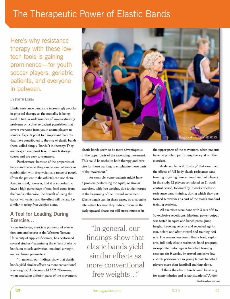
Here’s why resistance therapy with these low-tech tools is gaining prominence—for youth soccer players, geriatric patients, and everyone in between.By Keith Loria
Elastic resistance bands are increasingly popular
in physical therapy as the modality is being
used to treat a wide number of lower-extremity
problems on a diverse patient population that
covers everyone from youth sports players to
seniors. Experts point to 3 important features
that have contributed to the rise of elastic bands
(here, called simply “bands”) in therapy: They
are inexpensive; don’t take up much storage
space; and are easy to transport.
Furthermore, because of the properties of
bands and because they can be used alone or in
combination with free weights, a range of people
(from the patient to the athlete) can use them.
Keep in mind, however, that it is important to
have a high percentage of total load come from
the bands; otherwise, the benefit of using the
bands will vanish and the effect will instead be
similar to using free weights alone.
A Tool for Loading During Exercise…Vidar Andersen, associate professor of educa-
tion, arts and sports at the Western Norway
University of Applied Sciences, has performed
several studies1,2 examining the effects of elastic
bands on muscle activation, maximal strength,
and explosive parameters.
“In general, our findings show that elastic
bands yield similar effects as more conventional
free weights,” Andersen told LER. “However,
when analyzing different parts of the movement,
elastic bands seem to be more advantageous
in the upper parts of the ascending movement.
This could be useful in both therapy and exer-
cise for those wanting to emphasize these parts
of the movement.”
For example, some patients might have
a problem performing the squat, or similar
exercises, with free weights, due to high torque
at the beginning of the upward movement.
Elastic bands can, in these cases, be a valuable
alternative because they reduce torque in the
early upward phase but still stress muscles in
the upper parts of the movement, when patients
have no problem performing the squat or other
exercises.
Andersen led a 2018 study3 that examined
the effects of full-body elastic resistance band
training in young female team handball players.
In the study, 12 players completed an 11-week
control period, followed by 9 weeks of elastic
resistance band training, during which they per-
formed 6 exercises as part of the team’s standard
training sessions.
All exercises were done with 3 sets of 6 to
10 explosive repetitions. Maximal power output
was tested in squat and bench press, jump
height, throwing velocity and repeated agility
run, before and after control and training peri-
ods. The researchers found that a brief, explo-
sive, full-body elastic resistance band program,
incorporated into regular handball training
sessions for 9 weeks, improved explosive low-
er-limb performance in young female handball
players more than handball training alone.
“I think the elastic bands could be strong
for many injuries and rehab situations,” Ander-
Continued on page 52
The Therapeutic Power of Elastic Bands
lermagazine.com 3.19 51
“In general, our findings show that elastic bands yield similar effects as
more conventional free weights…”
iStockPhoto.com # 482982350

sen said. “The elastic bands don’t create a great
torque when going from a descending to an
ascending movement, but more gently increase
the torque as the bands are stretched. Further,
we have performed several interventions with
the elderly performing explosive training and
our experience is that in these situations, the
elastic bands are very useful.”
…and for UnloadingAnother important aspect of the utility of bands
is that they can be used to unload. For example,
if a patient isn’t able to perform a chair rise or
squat because of pain or lack of muscle strength,
bands can be used to reduce some of the body
weight, thus assisting the person in performing
the exercise.
“The research on this topic has increased
in the recent years and I believe the interest
among experts has increased simultaneously,”
Andersen said. “For me, elastic bands have
become more prevalent and will be in the years
to come.
“However, I don’t think we should regard
elastic bands as a replacement for free weights,
but more as an alternative or variation in situ-
ations where the properties of the elastic bands
could be useful.”
A Means to Boost Functional PerformanceEduardo Lusa Cadore, PhD, professor at the
physical education school of the Federal Univer-
sity of Rio Grande do Sul, Porto Alegre, Brazil,
led a study4 that provided evidence that resis-
tance training using elastic bands is an easy,
effective tool for inducing marked functional
performance in older people.
“Specifically, we observed marked improve-
ments in tests that included the ability of sit-to-
stand, walking, dynamic balance, upper-body
muscular endurance and flexibility,” Cadore
explained to LER. “This is an important finding
since elastic bands are easy to carry and may be
used in any space, including at home.”
Cadore described how bands can be used
to treat femoroacetabular impingement (hip
impingement syndrome). For example, patients
can hold the bands at the ankles and perform
exercises for various hip muscles, in an isomet-
ric or dynamic manner (if the syndrome allows
them to exercise dynamically). This approach
has the potential to prevent or delay the need
for surgery and can be used to rehabilitate the
hips postoperatively.
“These bands help, because it is a kind of
resistance that can be used to train, as any kind
of resistance,” Cadore said. “Of course, [a band
has] some upper threshold of resistance, and
one wouldn’t use it for training a competitive
weightlifter. Nevertheless, for people searching
for health and neuromuscular function improve-
ments, it can be used.”
Cadore foresees bands becoming even
more prevalent because the number of studies
using resistance training with elastic bands in
exercise-intervention models is growing.5,6 In
addition, the number of studies validating and
quantifying loads—in each elastic band color
(ie, each color denoting a progressive increase in
resistance), with different initial lengths, and in
various lengths of elastic deformation—is also
increasing. He noted that this is an important
matter because, to prescribe resistance training
using elastic bands, exercise professionals need
to know how to quantify the intensity of the
tool.
“Another important aspect of these bands
is that the workload is always progressive when
bands are deformed,” Cadore said. “In some
joints, [the band] won’t respect the biomechan-
ical characteristic of a muscle group. For exam-
ple, during a knee extension, the internal torque
produced by the knee extensors increases from
90° of knee flexion to full extension, whereas
the band provides progressive resistance. So,
the use of the band in this movement is not
impeditive, but this issue must be taken into
consideration.”
Bands Have Utility in Older PatientsMaurício Krause, PhD, professor in the
department of physiology at the Institute of
Basic Health Sciences, Federal University of
Rio Grande do Sul, recently led a study on
uncomplicated band exercise training for elderly
people.7
“The age-related loss of muscle mass and
function arise from an imbalance between pro-
tein synthesis and protein degradation, resulting
in muscle atrophy and consequent increases in
morbidity and mortality, and can be accelerated
by a combination of factors, which include a
reduced physical activity level and a suboptimal
nutritional profile,” Krause said. “Concerns with
the impact on the healthcare system due to
increased economic and social costs associated
with possible loss of independence with aging
have brought attention to alternative exercise
Continued from page 51
iStockPhoto.com # 621232042
52 3.19 lermagazine.com
Continued on page 55


interventions as an effective way of delaying
the negative effects of aging on functional and
metabolic parameters.”7,8
In this way, physical activity—in particular,
resistance exercise—is an established counter-
measure to attenuate the decline in lean body
mass that occurs with aging. In fact, Krause
said, resistance exercise training can induce
several benefits in older people, including:
• increased muscle strength and power
• improved insulin sensitivity
• enhanced mitochondrial function
• reduced inflammation
• muscle-mass growth.
Krause and colleagues’ study7 sought to
determine the effects of 12 weeks of resistance
exercise training, plus protein supplementation
on body composition, markers of muscle atro-
phy and hypertrophy, and heat-shock response
(a molecular pathway involved in cellular pro-
tection, having an important anti-inflammatory
role) in 38 healthy, older (mean age, 63.5 ± 4.4
years) adults.
“Our group demonstrated that 12-weeks of
traditional [resistance exercise], using profes-
sional gym equipment, in diabetic elderly people
is an effective tool to improve muscle function,
metabolism, and immune system functional-
ity,” Krause said. “However, the use of specific
exercise equipment is not always available.
So, we tested a nontraditional, body weight-
based, elastic-band resistance training to test if
this type of exercise would also be effective to
improve muscle metabolism, body composition,
and muscle function in healthy elderly people.”
To test that hypothesis, the researchers
designed a 12-week training protocol using
only body weight and elastic bands as sources
of resistance. The training was conducted in a
gymnasium on 3 nonconsecutive days a week
by qualified sport and exercise scientists; each
session lasted 45 to 60 minutes and consisted
of a warm-up period, body weight- and elastic
band-based resistance exercise training, and
a cool-down period. Primary exercises used
throughout the program included squats, lunges,
hip abduction, shoulder press, latissimus dorsi
pull-down, bicep curls, calf raises, pushups, tri-
ceps dips, and lumbopelvic stabilization exercises.
“Among the findings were that body weight/
elastic bands-based resistance exercise training
increases muscle mass and function in healthy
elderly people, independently of protein sup-
plementation,” Krause said. “This indicates that
exercise training, using body weight and elastic
bands, is an efficient way to maintain and even
increase muscle mass in elderly people, especially
at the lower limbs.”
Participants in the exercise training also
saw their muscle function-testing scores improve.
When protein supplementation was added to
their diet, additional gains were detected, includ-
ing lower fat mass and changes in skeletal-muscle
signaling (eg, protein synthesis pathways and the
heat-shock response).
State of the Art: Uncomplicated, Under SupervisionKrause summed it up: “The use of elastic
bands and body weight as resistance exercise is
Continued from page 52
54 3.19 lermagazine.com
SIZE RUN: Widths D-9E ; Sizes 5-17
Mesh Walking ShoesBreathableDurable
1.888.937.2747www.apisfootwear.com
13026 Forest Centre Court, Louisville, KY 40223
2239 Tyler Avenue, South El Monte, CA 91733
9704 As Shown
receiving more attention from exercise experts
because [bands offer] an uncomplicated way
to exercise at any place—home, work, outside,
etc. However, it is important that people receive
an appropriate orientation and supervision on
how to use [bands] correctly, how to choose the
individual load, and how they should progress
with the training.”
Keith Loria is a Washington, DC-based freelance
writer.
References
1. Andersen V, Fimland MS, Kolnes MK, et al.
Elastic bands in combination with free weights
in strength training: neuromuscular effects. J
Strength Cond Res. 2015;29(10):2932-2940.
2. Saeterbakken AH, Andersen V, Kolnes MK,
et al. Effects of replacing free weights with
elastic band resistance in squats on trunk
muscle activation. J Strength Cond Res.
2014;28(11):3056-3062.
3. Andersen V, Fimland MS, Cumming
KT, et al. Explosive resistance training
using elastic bands in young female team
handball players. Sports Med Int Open.
2018;2(6):E171-E178.
4. Cadore EL, Pinto RS, Bottaro M, et al.
Strength and endurance training prescrip-
tion in healthy and frail elderly. Aging Dis.
2014;5(3):183-195.
5. Iversen VM, Mork PJ, Vasseljen O, et al. Mul-
tiple-joint exercises using elastic resistance
bands vs. conventional resistance-training
equipment: A cross-over study. Eur J Sport Sci.
2017;17(8):973-982.
6. Kubo T, Hirayama K, Nakamura N, et al.
Effect of accommodating elastic bands on
mechanical power output during back squats.
Sports (Basel). 2018;6(4):pii:E151.
7. Krause M, Crognale D, Cogan K, et al. The
effects of a combined bodyweight-based and
elastic bands resistance training, with or with-
out protein supplementation, on muscle mass,
signaling and heat shock response in healthy
older people. Exp Gerontol. 2019;115:104-113.
8. de Lemos Muller CH, Rech A, Botton CE, et
al. Heat-induced extracellular HSP72 release
is blunted in elderly diabetic people compared
with healthy middle-aged and older adults, but
it is partially restored by resistance training.
Exp Gerontol. 2018;111:180-187.
LATEST RESEARCH…Resistance Exercise to Forestall Cognitive Decline?“As a preventative tool [for Alzheimer’s disease], resistance exercise improves memory, attention, spatial awareness, reaction time, planning, and information processing. Improvements in cognitive performance following resistance exercise and training may be mediated by peripheral elevations in … physiological biomarkers (ie, neural and vascular).”
Source: Marston KJ, Brown BM, Rainey-Smith SR, Peiffer JJ. Resistance exercise-induced responses in physiological factors linked with cognitive health. J Alzheimers Dis. 2019:68(1):39-64.
lermagazine.com 3.19 55

Is something from your business?ABC could be the final piece to unlock your success.
Choose ABC for your facility accreditation TODAY
• Improve your business
• Save money
• 94% satisfaction rate
To learn more, call ABC at 703.836.7114 or check out more information at abcop.org.

lermagazine.com 3.19 57
ad index
8sole 31267/368-8165 8soloe.com
ABC 56703/836-7114 abcop.org
Allard USA 12 888/678-6548 allardusa.com
Apex/OHI back cover800/323-0024 apexfoot.com
Apis 54888/937-2747 apisfootwear.com
BraceLab 17888/235-8221 bracelab.com
Cascade DAFO 16800/848-7332 cascadedafo.com
CMTA 53800/606-2682 cmtausa.org
Comfort Products 24, 33800/822-7500 comfortoandp.com
CustomComposite 20866/273-2230 cc-mfg.com
Darco 18800/999-8866 darcointernational.com
Dr. Comfort inside front cover, 3877/728-3450 drcomfort.com
Ferris Mfg. 25800/765-9636 polymem.com
Footmaxx 4, 42, 46800/779-3668 footmaxx.com
GAITRite 40888/482-2362 gaitrite.com
Go 4-D 39800/353-4643 go4-d.com
Hapad 41800/232-9427 hapad.com
Hersco Ortho Labs 11800/301-8275 hersco.com
JMMR 55800/236-5166 blountsdisease.net
JMS Plastics Supply 14800/342/2602 jmsplastics.com
Kinetic Research 49800/919-3668 kineticresearch.com
Levy & Rappel 28800/564-LEVY (5389) levyandrappel.com
MD Orthopaedics 23877/766-7384 mdorthopaedics.com
Northwest Podiatric Laboratory 26800/675-1766 nwpodiatric.com
Ortho-Rite inside back cover800/473-6682 ortho-rite.com
Ottobock 36800/328-4058 ottobockus.com
Pedifix 6800/424-5561 pedifix.com
Pedlite 48219/756-0901 pedlite.com
ProtoKinetics 8610/449-4879 protokinetics.com
Sigvaris 14800/322-7744 sigvarisusa.com
Surestep 32, 45877/462-0711 surestep.net
Tencate 50805/482-1722 tencatecomposites.com
TECHNOLOGY
FABRICATION
ANKLE FOOT
KNEE
PEDIATRIC REHABILITATION
Positive Outcomes for Successful Practitioners
resourceguide 2017
PROSTHETICS
LERRG17-Ankle 1 12/20/16 3:02 PM Page 1
LER07-17V64_LER 7/21/17 11:12 AM Page 1
LER04-17_LER 4/18/17 5:53 PM Page 1
LER06-17_LER 6/19/17 3:33 PM Page 1
LER02-16_LER 2/9/16 4:20 PM Page 1
LER10-16_LER 10/13/16 5:35 PM Page 1
Please Support our Advertisers…Because of them, we are able to provide you with this
unique, informative and invaluable magazine!
lermagazine.com

THE X BRACE ADDED TO MEDI-DYNE PAIN RELIEF SOLUTIONS
Medi-Dyne Healthcare Products has acquired The X Brace. Developed by Joshua P. Eldridge, DC, DACBSP, for relief of heel pain due to plantar fasciitis and Sever’s disease, The X Brace is an easy-to-use foot and heel support product. It provides a comfortable, low-profile, pain-relieving solution for previously hard-to-treat circumstances, such as when wearing flip flops or sandals, or when walking barefoot. Ac-cording to Medi-Dyne, The X Brace has been used effectively by athletes and other sufferers worldwide. Medi-Dyne products, including The X Brace, are made in the US.
Medi-Dyne Healthcare Products800/810-1740medi-dyne.com
UNISA CREATES RECIPE FOR REALISTIC FOOT ULCERSPodiatrists at the University of South Australia (UniSA), Adelaide, have blended icing sugar, chicken stock, and flexible resin to create real-istic-looking foot ulcers as part of a world-first podiatric training initiative. Helen Banwell, DPM, and Ryan Causby, DPM, concocted the mixture, which is being added to 3D-printed feet and is designed to mimic infected and non-infected diabetic foot wounds.
The addition of lifelike ulcers and effects is added by UniSA’s podiatry team and can en-compass anything from dry gangrene to oozing pus. Banwell said the 3D foot models, which
are made from thermoplastic polyurethane, will play a part in teaching fourth-year podiatry students how to treat and manage high-risk foot conditions.
“The most effective way to manage these conditions is to medically remove dead or damaged skin to expose the healthy skin un-derneath and encourage healing,” said Banwell. “The 3D foot models—and the mock injuries with which we enhance them—enable us to provide a realistic but safe learning tool for stu-dents to practice their scalpel skills, before they begin clinical placements, and all without the stress or anxiety of treating a real patient.”
To support the training, ulcer debridement and management videos are being developed.
UniSA is also trialing 3D-printed baby legs in podiatry pediatric teaching sessions to allow students hands-on practice for clubfoot casting.
CMS LAUNCHES “WHAT’S COVERED” APP TO GUIDE BENEFICIARIESThe Centers for Medicare & Medicaid Ser-vices (CMS) launched a new app that gives consumers direct access on a mobile device to some of the most-used content on Medicare.gov. The new “What’s Covered” app lets people with Original Medicare, caregivers, and others quickly see whether Medicare covers a specific medical item or service. The app also provides beneficiaries with access to Blue Button 2.0, an
application programming interface, created by the US Digital Service, that enables beneficia-ries with different needs to grant access to de-velopers who can help them monitor for drug conflicts, refill prescriptions, and track progress toward desired healthcare outcomes.
CMS launched the eMedicare initiative in 2018 to empower beneficiaries with cost and quality information. Other tools in the eMedi-care suite include:
• Enhanced interactive online decision
support to help beneficiaries better
understand and evaluate their Medicare
coverage options and costs between
Medicare and Medicare Advantage.
• A new online service that lets people see
how different coverage choices will affect
their estimated out-of-pocket costs.
• New price transparency tools that let
consumers compare the national average
costs of certain procedures between
settings, so people can see what they’ll
pay for procedures done in a hospital
outpatient department versus an
ambulatory surgical center.
• A new webchat option in the Medicare
Plan Finder.
The What’s Covered app is available for free in Google Play (play.google.com/store/apps/details?id=gov.medicare.coverage) and the Apple App Store (itunes.apple.com/us/app/whats-covered/id1444143600?mt=8).
WOUND HEALING CLINICAL & TRANSLATIONAL RESEARCH TEAM RECEIVES GRANT TO JOIN DIABETIC FOOT CONSORTIUMThe clinical and translational research team at the University of Miami (UM) Miller School of Medicine’s Department of Dermatology and Cutaneous Surgery received a 4-year, $1.7
INDUSTRY SNAPSH TNoteworthy products, association news, and market updates
58 3.19 lermagazine.com
Photograph of 3D-printed feet, some enhanced with foot ulcers, courtesy of UniSa.

million grant to become 1 of 6 Clinical Re-search Units (CRU) in the US to help improve outcomes for patients with diabetic foot ulcers. This team approach to research is part of the National Institute of Diabetes and Digestive and Kidney Diseases’ Diabetic Foot Consor-tium (DFC) for future clinical study of selected biomarkers.
The objective of the DFC is to establish an organizational framework and infrastructure that will serve as a platform for integrated multidisciplinary experimental clinical and translational research. The UM-CRU will, in conjunction with DFC, develop the research design and study protocol, and establish proto-cols for participant recruitment and follow-up, data collection, quality control, interim data and safety monitoring, final data analysis and interpretation, and publication of results. In addition, UM-CRU and DFC will facilitate procurement, storage, and shipment of the bio-materials (tissue samples, swabs, wound fluid, blood, serum, and/or urine) as a part of the future biomarker studies. It will also establish and maintain a clinical database that will allow overall data analyses of the DFC.
PIEDRO PEDIATRIC ORTHOPEDIC FOOTWEAR
Cascade Orthopedic Supply has expanded its product line with the addition of Piedro Pediat-ric Orthopedic Footwear. The new line of foot-wear features a wider and deeper toe box to ensure a precision fit with orthotic devices. The double stitching and high abrasion-resistant toe cap provide increased durability for children. Piedro Pediatric Orthopedic Footwear features an adjustable depth and a trimmable sole, and is available with either lace or Velcro closures. Color options for the new shoe line include
pink, blue, and black to match any taste. Shoe sizes include Toddler (4–9), Youth (10–13, 1–3), and Adolescent (4–7).
Cascade Orthopedic Supply800/888-0865cascade-usa.com
ICEBREAKER HIKE LIGHT CREW
Suitable for adventure and hiking, Icebreaker’s men’s Hike Light Crew sock is designed to be an ideal sock for year-round comfort on the trail. The sock has double up cushioning for comfort. It is made from breathable, tempera-ture-regulating merino wool (60%); nylon (37%) for durability; and Lycra (3%). Addi-tional design features include Achilles support, reinforced heel and toe, instep support, and a comfort cuff. The overall goal of the adven-ture sock is to keep the wearer’s feet happy to ensure great journeys. The socks come in a twisted-heather color and are available in small, medium, large, and extra-large sizes. The company offers a lifetime guarantee on all of its socks.
Icebreaker877/827-6816icebreaker.com
SIGVARIS IS NOW SIGVARIS GROUP SIGVARIS, Peachtree, Georgia, has changed its corporate name and logo from SIGVARIS to SIGVARIS GROUP. SIGVARIS, a 155-year-
old Swiss company, manufactures medical compression socks and hosiery with a wide assortment of inelastic wraps and lymphedema garments. SIGVARIS GROUP will use this opportunity to build a customer-centric plat-form for innovation and growth, according to a company press release. In the new brand ar-chitecture, products will have their own unique brands to bring more clarity and understanding to product positioning and to differentiate the organization from product brands.
ORTHOTIC STRAP KITS
JMS Plastics Supply has introduced orthotic straps as a new market offering. The exclusive straps are made from reinforced PVC that is welded together with high frequency to make them strong and durable. The straps feature hook-and-loop closures and are available in kits or sold individually. Each kit contains one PVC Chafe, one strap, and one pad. Kits are offered in nine colors and four widths. JMS Plastics’ orthotic straps can also be ordered to feature any company logo.
JMS Plastics Supply800/342-2602jmsplastics.com
INDUSTRY SNAPSHOT
lermagazine.com 3.19 59

INDUSTRY SNAPSHOT
REVISTIM WEARABLE DC STIMULATOR
Prizm Medical has introduced the ReviStim DC Stimulator, which is described as the world’s first wearable DC stimulation device. The unit’s copyrighted software delivers DC electrotherapeutic energy uniformly over the entire treatment area through hydrogel stick-on electrodes or Prizm’s proprietary Sigma gar-ment electrodes. Sigma garments are soft and comfortable, and they are available in the form of socks, sleeves, and gloves. The ease of use of the products, coupled with the compact size of the ReviStim, allows for nighttime delivery of electrotherapeutic treatment, thereby in-creasing the number of treatments per day and allowing for improved patient compliance.
Prizm Medical770/622-0933woundstim.com
CMS INCLUDES 7 OTS KNEE ORTHOSES IN 2021 COMPETITIVE BIDDING PROGRAMThe Centers for Medicare & Medicaid Ser-vices (CMS) announced that the Durable Medical Equipment, Prosthetics, Orthotics, and Supplies (DMEPOS) Competitive Bid-ding Program for dates of service beginning January 1, 2021, will include 7 off-the-shelf (OTS) knee orthoses. The affected codes are L1812, L1830, L1833, L1836, L1850, L1851, and L1852. According to CMS, the law requires that OTS orthotics or braces are included in the DMEPOS competitive bidding program, specifically, “orthotics which require minimal
self-adjustment for appropriate use and do not require expertise in trimming, bending, mold-ing, assembling, or customizing to fit to the individual.”
To access the DMEPOS Competitive Bidding Round 2021 Fact Sheet, visit gov/newsroom/fact-sheets/dmepos-competitive-bid-ding-round-2021.
INSIGHTFUL PRODUCTS’ MASTED KNEE BRACE
The Masted Knee Brace (MKB) is the newest design offered by Insightful Products. The MKB was developed primarily to address the common problem of distal migration. This knee orthosis is said to ensure good knee and brace-joint alignment. It has adjustable flexion and extension stops at 15-degree increments and the foot section offers full range of motion for the foot/ankle complex. The posterior calf and thigh sections allow for easy donning. This orthotic solution is indicated for conical limb shapes where suspension is a challenge. Ac-cording to Insightful Products, the MKB is ap-propriate for rehabilitation, post-op, ACL, or to control recurvatum. It can be configured with an integrated Step-Smart Brace for those with foot drop. Videos on the company’s website of-fer more information. HCPCS code L1832.
Insightful Products207/885-0414insightfulproducts.com
$11 MILLION NIH GRANT CREATES NEW CENTER FOR MUSCULOSKELETAL RESEARCH Clemson University, South Carolina, received an $11 million grant from the National In-stitutes of Health’s Center for Biomedical Research Excellence to launch a new research center that will bring together scientists from across the state to change the way musculo-skeletal disorders are diagnosed, treated, and studied. Led by bioengineers at Clemson, the South Carolina Center for Translational Research Improving Musculoskeletal Health combines orthopedics and other clinical exper-tise from the Greenville Health System and the Medical University of South Carolina with computer scientists, computational engineers, biophysicists, and other experts to better understand musculoskeletal disorders and to design and evaluate new devices, interventions, and drug therapies.
RENOWNED ORTHOPEDIC SURGEON TO LEAD FOOT AND ANKLE DIVISION AT NYU LANGONE HEALTHJohn G. Kennedy, MD, renowned for his work in the surgical treat-ment of foot and ankle injuries, has been ap-pointed the new chief of the Foot and Ankle Division in the De-partment of Orthopedic Surgery, and Director of the Foot and Ankle Center at NYU Langone Health. He joined NYU Langone in January, where he holds the title of professor of ortho-pedic surgery at NYU School of Medicine.
Kennedy brings to NYU Langone a spe-cial interest in articular cartilage injury and joint preservation. He has been a leader in minimally invasive, arthroscopic treatments of cartilage injuries of the ankle and smaller joints of the foot. An award-winning researcher, Ken-nedy and his team collaborate on National In-stitutes of Health (NIH) research into cartilage
60 3.19 lermagazine.com

INDUSTRY SNAPSHOT
lermagazine.com 3.19 61
regeneration and work with several interna-tional universities, investigating basic science and clinical outcomes in cartilage regenera-tions, tendon healing, and bone growth. These collaborations have resulted in more than 200 publications in medical journals and more than 500 podium presentations worldwide.
Kennedy is an alumnus of the Royal Col-lege of Surgeons in Dublin, Ireland. In 2002, he started the first subspecialty foot and ankle sports injury clinic in New York City. Most recently, Kennedy spent the past 17 years as an assistant professor of orthopedic surgery at Weill Cornell Medical School, and as an assis-tant attending physician at Hospital for Special Surgery, both of which are located in New York City. In addition, Kennedy serves as co-presi-dent of the International Society on Cartilage Repair of the Ankle, which last year established the International Consensus Meeting on Car-tilage Repair of the Ankle to standardize ankle cartilage treatment algorithms.
SAMUEL HUBBARD WOMEN’S CITY ZIPPER BOOT
Slide in. Zip up. Step out. The City Zipper from Samuel Hubbard is designed to wow the wearer’s feet. Comfort engineering is at the heart of how Samuel Hubbard designs and builds its shoes. Meant to be the go-to-boot in any women’s closet, the women’s City Zipper is fabricated from premium European sourced,
tumbled leather soft uppers, and buttery-soft glove leather sock linings for comfort and breathability. The boot features an easy-glide, extra-light EVA custom-designed outsole, and incorporates removable Poron insoles and heel support. The boots are available in size 5½ to 11, including half sizes and medium width. Samuel Hubbard’s footwear is handcrafted in Portugal.
Samuel Hubbard844/482-4800samuelhubbard.com
CORE-SPUN PATTERNED AFO SOCKS BY SMARTKNIT
Knit-Rite has long been a manufacturer of SmartKnit Seamless Socks made for adults and children who use AFOs. SmartKnit Seam-less Socks are soft and comfortable, giving the feeling of a cool second layer of skin between the wearer and the AFO. Knit-Rite now offers Core-Spun Patterned AFO Socks by Smart-Knit. The children’s AFO Socks are available in 2 patterns: thin line (white with grey and purple striping), cheery (black sock with bright blue, doughnut-shaped spots), and halo (3-pack of socks with the same pattern but in 3 different color combinations—white with soft yellow striping, grey with hunter green striping, and black with bright blue striping). For adults, Knit-Rite offers a classic diamond pattern for a professional look.
Knit-Rite800/821-3094knitrite.com
APTA, ACSM FORMALIZE NEW PARTNERSHIPThe American Physical Therapy Association (APTA) has partnered with the American College of Sports Medicine (ACSM), under the APTA Partnerships program, which seeks to enhance relationships and develop mutually beneficial partnerships with other organiza-tions that share common goals. The program allows APTA to extend its reach in advancing the physical therapy profession.
In signing this memorandum of understanding, APTA and ACSM seek to build on existing collaboration to advance their missions and the health of communities and individuals through movement, physical activity, and exercise. APTA and ACSM also hope to work jointly on advocacy efforts as well as to incorporate elements and expand participation of both groups in each other’s events and initiatives.
SPS PROMOTES WEGER TO VP AND GENERAL MANAGERHanger, Inc., Austin, Texas, announced that Regina Weger has been promoted to VP and general manager of its subsidiary SPS, Alpharetta, Georgia. Weger has been with SPS for over 20 years, having started her ca-reer within the customer service function and taking on additional roles and responsibilities throughout the years. For the past 2 years she served as VP of SPS sales and marketing. We-ger will continue reporting to Jay Wendt, pres-ident of Hanger’s Product & Services business segment, in her new role as a member of the Products & Services leadership team.
“This is a well-deserved promotion for Regina, as she has been functioning in a gen-eral manager capacity for us, taking on several additional leadership responsibilities on top of her role as vice president of sales and market-ing,” said Wendt.

ACROSS1 More of these bikes sold than road bikes5 Resort often offers gym facilities7 Common name for diagnostic images pro-
duced by electromagnetic waves9 Dieters fixate on this measurement, abbr.11 In good physical condition12 SSN, for one13 With chamomile, it might alleviate anxiety14 Hits the ground16 Fluid-filled sac is a toe problem for marathon
runners17 ____ manual20 Anatomical sac in the knee21 Almost an eternity23 Often describes timing of a physical exam24 Severe, as pain26 Here are found phalanxes27 Triathlon competitor30 Surgical procedure, abbr.31 __ Capitan, beloved by climbers32 Take it by mouth, abbr34 West Coast city that holds annual marathon35 One type of bone stimulation
DOWN1 Relating to jaws and face; frequent type of
mountain bike injury2 Land of the ACA3 Trials4 Pen point5 Most common type of injury associated with
mountain biking accidents6 The “A” in PAD8 Nutritionist’s intake measure, for short9 Places for experiments10 They are being used to improve bone fracture
healing15 Problem when a bone fails to knit back
together18 Area19 One source of skin Ca, en Espanol22 Made better25 Admit wrongdoing28 Inclined29 Neither’s partner33 Copper symbol
Italy7-Day Road Bike Tour/CME
Tuscany Region of Italy
June 1-8, 2019
5 cycling days: 30-50 miles each day
10 AMA-PRA Category 1 Credits
colorado3-day Mountain Bike CME Grand Junction, Colorado
October 9-12, 2019
3 cycling days: 25-35 miles each day
10 AMA-PRA Category 1 Credits; 10 AOA 1-A Credits
Unique CME Experience for Physicians, PA-C’s, and Other Medical
Professionals Who Love to Bike.
www.cyclingcme.com
How Well Did You Read This Issue?
Test your knowledge of information from this issue of Lower Extremity Review and the world in general with our new crossword puzzle feature. The answer box can be found online at lermagazine.com
Crossword by Myles Mellor
With over 12,000 crosswords published internationally, Myles Mellor is one of the top crossword
writers in the world. His work includes crosswords, diamond crosswords, syndicated puzzles, cryp-
tograms, diagramless crosswords, word search, sudokus, anagrams, and word games published on
mobile devices and e-readers. www.themecrosswords.com
62 3.19 lermagazine.com

© OrthoRite ad campaign v2.psd @ 25% (CMYK/8)

5LCBSS5LCBSS
TM
190328_SafeStep-LER-Basis Slip On 2019-outlined.indd 1 2019-04-02 4:04:42 PM