DNA Damage, Somatic Aneuploidy, and Malignant Sarcoma Susceptibility in Muscular Dystrophies
Upload
independentCategory
view
1download
0
Hindawi Publishing CorporationSarcomaVolume 2012, Article ID 541650, 10 pagesdoi:10.1155/2012/541650
Clinical Study
Prognostic Impacts of Hypoxic Markers in Soft Tissue Sarcoma
Eivind Smeland,1, 2 Thomas K. Kilvaer,3 Sveinung Sorbye,1, 4 Andrej Valkov,1, 4
Sigve Andersen,1, 2 Roy M. Bremnes,1, 2 Lill-Tove Busund,3, 4 and Tom Donnem1, 2
1 Institute of Clinical Medicine, University of Tromso, 9037 Tromso, Norway2 Department of Oncology, University Hospital of North Norway, 9038 Tromso, Norway3 Institute of Medical Biology, University of Tromso, 9037 Tromso, Norway4 Department of Pathology, University Hospital of North Norway, 9038 Tromso, Norway
Correspondence should be addressed to Eivind Smeland, [email protected]
Received 24 October 2011; Accepted 1 December 2011
Academic Editor: Alberto Pappo
Copyright © 2012 Eivind Smeland et al. This is an open access article distributed under the Creative Commons AttributionLicense, which permits unrestricted use, distribution, and reproduction in any medium, provided the original work is properlycited.
Background. We aimed to explore the prognostic impact of the hypoxia-induced factors (HIFαs) 1 and 2, the metabolic HIF-regulated glucose transporter GLUT-1, and carbonic anhydrase IX (CAIX) in non-gastrointestinal stromal tumor soft tissuesarcomas (non-GIST STS). Methods. Duplicate cores with viable tumor tissue from 206 patients with non-GIST STS were obtainedand tissue microarrays were constructed. Immunohistochemistry (IHC) was used to evaluate expression of hypoxic markers.Results. In univariate analyses, GLUT-1 (P < 0.001) and HIF-2α (P = 0.032) expression correlated significantly with a poordisease-specific survival (DSS). In the multivariate analysis, however, only high expression of GLUT-1 (HR 1.7, CI 95% 1.1–2.7,P = 0.021) was a significant independent prognostic indicator of poor DSS. Conclusion. GLUT-1 is a significant independentnegative prognostic factor in non-GIST STS.
1. Background
Adaptation of tumor cells to hypoxia is critical for tumor sur-vival and progression [1]. Tumor hypoxia leads to resistanceto radiotherapy and chemotherapy and is associated with anincreased metastatic potential [2, 3]. In soft tissue sarcomas(STS) hypoxia has been found to be related to reduceddisease-free survival [4] and increased cell proliferation [5].
Principally there are three methods of measuringhypoxia, directly in vivo through microelectrodes (e.g.,Eppendorf pO2 Histograph), visualization of nitroimidazolecompounds by PET, or in tumor tissue by studying endoge-nous markers of hypoxia [6]. HIF-1, GLUT-1, and CAIXhave been proposed as endogenous immunohistochemical(IHC) markers of hypoxia [7, 8] although this is a matter ofcontroversy [9, 10].
Since Semenza et al. discovered HIF-1α in 1992, thehypoxia inducible factors (HIFs) have been identified askey regulators of genes involved in hypoxic responses[11]. Except for differences in distribution, abundance, andexpression in response to prolonged hypoxia, the regulatory
characteristics are quite similar among HIFs [12–15]. IHCassessments in human cancer biopsies have found elevatedlevels of HIF-1α and/or HIF-2α protein in the majority ofprimary human cancers and their metastases [16]. Further-more, clinical data shows that increased levels of HIF-1α andHIF-2α are associated with higher patient mortality in manyhuman cancers [17].
Cancer cells prefer glycolysis with or without hypoxia[18]. The HIF-regulated glucose transporter GLUT-1 facil-itates increased influx of glucose and is upregulated inhypoxic conditions. GLUT-1 is overexpressed in severaltumors [19, 20], and increased expression of GLUT-1appears to be correlated with a poor prognosis in a varietyof tumors [21–23]. CAIX mediates the extracellular trappingof acidity. Together with GLUT-1, it is among the mostcritical molecules, for maintaining ATP levels with stableintracellular pH, needed for cancer cell survival [24–26].Hypoxia-related markers are inadequately explored in STS,and the data are diverging. Hence, further knowledge on themolecular mechanisms in this tumor entity is warranted.

2 Sarcoma
As the prognosis of patients with STS is still unsat-isfactory [27], new therapeutic targets are highly desired.The remarkable history of imatinib in CD 117 positiveGIST patients is inspiring [28]. The evaluation of potentialprognostic molecular markers may help to identify bothpromising targets and patients in need of adjuvant treatment.As a consequence, it is important to enhance our knowledgeabout pivotal molecular markers with inherent and diversesignificant prognostic relevance for tumor progression andsurvival.
We have previously reported on the prognostic impact ofvarious angiogenic factors in sarcoma [29]. Herein, using ahigh-throughput TMA technique, we explore the prognosticimpact of markers associated with hypoxia (HIF1α, HIF2α)and related metabolic markers (GLUT-1, CAIX) in non-GIST STS.
2. Materials and Methods
2.1. Patients and Clinical Samples. Primary tumor tissuesfrom anonymized patients diagnosed with non-GIST STS atthe University Hospital of North Norway and the Hospitalsof Arkhangelsk county, Russia, from 1973 through 2006,were collected. In total 496 patients were registered fromthe hospital databases. Of these 290 patients were excludedfrom the study because of: missing clinical data (n = 86),inadequate paraffin-embedded fixed tissue blocks (n = 161),or metastasis at the time of diagnosis (n = 43). Thus 206patients were included in this study.
This report includes followup data as of September 2009.The median follow-up was 37.6 (range 0.1–391.7) months.Complete demographic and clinical data were collected ret-rospectively. Formalin-fixed and paraffin-embedded tumorspecimens were obtained from the archives of the Depart-ments of Pathology at the University Hospital of NorthNorway and the Hospitals of Arkhangelsk county, Russia.The tumors were graded according to the French FederationNationale des centres de Lutte Contre le Cancer (FNCLCC)system and histologically subtyped according to the WorldHealth Organization guidelines [30, 31]. Wide resectionmargins were defined as wide local resection with freemicroscopic margins or amputation of the affected limb ororgan. Nonwide resection margins were defined as marginalor intralesional resection margins, or no surgery.
2.2. Microarray Construction. All sarcomas were histologi-cally reviewed by two trained pathologists (S. Sorbye andA. Valkov), and the most representative areas of tumorcells (neoplastic mesenchymal cells) were carefully selectedand marked on the hematoxylin and eosin (H/E) slideand sampled for the tissue microarray (TMA) blocks. TheTMAs were assembled using a tissue-arraying instrument(Beecher Instruments, Silver Springs, MD). The detailedmethodology has been previously reported [32]. Briefly, weused a 0.6 mm diameter stylet, and the study specimens wereroutinely sampled with duplicate cores from different areasof neoplastic tissue. Normal soft tissues were used as stainingcontrols.
To include all core samples, 12 TMA blocks were con-structed. Multiple 4 μm sections were cut with a Micronmicrotome (HM355S) and stained by specific antibodies forIHC analysis.
2.3. Immunohistochemistry. All applied antibodies had beensubjected to in-house validation by the manufacturer forIHC on paraffin-embedded material. All sections were de-paraffinised with xylene and rehydrated with ethanol.The 4 μm sections, containing tissue cores, were subjectedto the following antibodies: HIF1α (mouse monoclonal,NB100-131, Novus Biologicals,1 : 3500), HIF2α (rabbit poly-clonal, ab199, Abcam,1 : 40), GLUT-1 (mouse monoclonal,AB40084, Abcam,1 : 500), and CAIX (rabbit polyclonal,ab15086, Abcam,1 : 200).
CAIX, GLUT-1, and the HIFs were stained using theVentana Benchmark XT (Ventana Medical Systems Inc.),procedure ultraview DAB. Antigen retrieval was done auto-matic by Cell Conditioning Solution (CC1) mild (30 min forCAIX and HIFs and 1 hour for GLUT-1).
The primary antibody was visualized by adding a sec-ondary antibody conjugated with Biotin, followed by anAvidin/Biotin/Peroxydase complex (Vectastain ABC Elite kitfrom Vector Laboratories). Finally, all slides were counter-stained with hematoxylin to visualize the nuclei.
2.4. Scoring of Immunohistochemistry. The ARIOL imagingsystem (Genetix, San Jose, CA) was used to scan the slidesof antibody staining of the TMAs. The slides were loaded inthe automated slide loader (Applied Imaging SL 50), and thespecimens were scanned at low resolution (1.25x) and highresolution (20x) using the Olympus BX 61 microscope withan automated platform (Prior). Representative and viabletissue sections were scored manually and semiquantitativelyon the computer screen. HIF-1α showed in most cases cyto-plasmic staining or cytoplasmic and nuclear staining. ForHIF-2α nuclear, or nuclear and weak cytoplasmic stainingwas recorded. Although it is suggested that nuclear HIF is theactive form, it is synthesized and degraded in the cytoplasm.Hence, there may be some redistribution explaining bothnuclear and cytoplasmic staining. However, the overallexpression indicates upregulation of the pathway [16, 33].GLUT-1 and CAIX antibodies usually recognize membrane-bound proteins [34–36], at least on cells of epithelial origin.But in our study, GLUT-1 showed cytoplasmic staining,and in a few cases both cytoplasmic and nuclear staining.For CAIX cytoplasmic staining was evaluated (Figure 1).Whether this is due to true differences between sarcomasand epithelial tumors, or that sarcoma cells with scantycytoplasm render the identification of membrane stainingdifficult on the background of a strong cytoplasmic reactivity,remains unclear [37–39].
The dominant staining intensity was scored as: 0 =negative; 1 = weak; 2 = intermediate; 3 = strong. All sampleswere anonymized and independently scored by two trainedpathologists (A. Valkov and S. Sorbye). When assessing avariable for a given core, the observers were blinded to thescores of the other variables and to outcome.

Sarcoma 3
CAIX
Neg
ativ
e
GLUT-1 HIF-1α HIF-2α
Scor
e 1
Scor
e 2
Scor
e 3
Figure 1: Immunohistochemical analysis in non-GIST STS representing negative, and score 1–3 of CAIX, GLUT-1, HIF-1α, and HIF-2α.non-GIST STS: non-gastrointestinal stromal tumor soft-tissue sarcomas, CAIX: carbonic anhydrase IX, GLUT-1: glucose transporter-1, andHIF-1/2α: hypoxia induced factor 1/2α.
In case of disagreement the slides were reexamined,and consensus was reached by the observers. Mean scorefor duplicate cores from each individual was calculatedseparately. High expression was defined as: = 3 for HIF1α;≥2.5 for HIF2α; ≥2 for CAIX; ≥1 for GLUT-1 (Figure 1).
2.5. Statistical Methods. All statistical analyses were doneusing the statistical package SPSS (Chicago, IL), version15. The IHC scores from each observer were compared forinterobserver reliability by use of a two-way random effectmodel with absolute agreement definition. The intraclasscorrelation coefficient (reliability coefficient) was obtainedfrom these results. The Chi-square and Fishers Exact testswere used to examine the association between molecularmarker expression and various clinicopathological param-eters. Fisher Exact test was used when there was a 2 × 2table, and the sample size was small (less than 5 in a givencell). Otherwise chi-square was used. We consider r > 0.2 aspotentially relevant and due to multiple testing significant Pvalue was set at <0.01 in correlation analyses.
Univariate analyses were done using the Kaplan-Meiermethod, and statistical significance between survival curveswas assessed by the log rank test. The significance level used
for log rank test was P < 0.05. DSS was determined from thedate of diagnosis to the time of cancer-related death. To assessthe independent value of different pretreatment variableson survival, in the presence of other variables, multivariateanalyses were carried out using the Cox proportional hazardsmodel. Only variables of significant value from the univariateanalyses were entered into the Cox regression analyses.Probability for stepwise entry and removal was set at .05 and.10, respectively.
3. Ethical Clearance
The National Data Inspection Board and The Regional(Northern Norway) Committee for Research Ethicsapproved the study. The committee classified the projectas retrospective nontherapeutic, bio- and genetechnologyscience on already registered data and archived tumormaterial and hence specifically waived the need for consent.
4. Results
4.1. Clinicopathological Variables. The clinicopathologicalvariables are summarized in Table 1. The median age was

4 Sarcoma
Table 1: Prognostic relevance of clinicopathological variables for disease-specific survival in 206 non-gastrointestinal stromal tumor soft-tissue sarcomas (univariate analyses, log rank test, multivariate analyses, Cox proportional hazards model).
CharacteristicsUnivariate analyses Multivariate analyses∗
Patients(n)
Patients(%)
Median survival(months)
5-year survival(%)
P HR 95% CI P
Age 0.030 0.121#
≤ 20 years 17 8 41 47 1.000
21–60 years 89 43 NR 61 0.728 0.281–1.889 0.515
>60 years 100 49 52 46 1.222 0.445–3.356 0.698
Gender 0.265
Male 89 43 NR 55
Female 117 57 75 51
Patient nationality 0.014
Norwegian 140 68 NR 58 1.000 0.142
Russian 66 32 39 42 1.453 0.882–2.393
Histological entity 0.003 0.086#
Pleomorphicsarcoma
54 26 54 48 1.000
Leiomyosarcoma 48 23 89 64 0.595 0.327–1.082 0.089
Liposarcoma 32 16 NR 71 0.411 0.169–0.999 0.050
Fibrosarcoma 16 8 123 56 0.773 0.330–1.808 0.552
Angiosarcoma 10 5 10 30 1.124 0.386–3.276 0.830
Rhabdomyosarcoma 12 6 41 50 0.419 0.156–1.126 0.085
MPNST 9 4 NR 56 0.544 0.162–1.824 0.324
Synovial sarcoma 13 6 31 28 1.257 0.571–2.767 0.570
Sarcoma NOS 12 6 9 25 1.640 0.707–3.805 0.249
Tumor localization 0.805
Extremities 79 38 123 56
Trunk 40 19 44 49
Retroperitoneum 30 15 57 47
Head/neck 16 8 27 47
Visceral 41 20 75 57
Tumor size 0.019 0.061#
≤5 cm 60 29 NR 66 1.000
5–10 cm 77 37 62 51 1.478 0.825–2.646 0.189
>10 cm 67 33 37 44 2.072 1.129–3.801 0.019
Missing 2 1
Malignancy grade <0.001 <0.001#
1 56 27 NR 78 1.000
2 82 40 62 51 2.761 1.352–5.639 0.005
3 68 33 22 34 4.642 2.219–9.710 <0.001
Tumor depth 0.002
Superficial 16 8 NR 93 1.000
Deep 190 92 59 50 7.658 1.043–56.215 0.045
Surgery <0.001
Yes 194 94 123 56 1.000
No 12 6 4 0 16.689 5.776–48.218 <0.001
Resection margins <0.001
Wide 97 47 NR 66 1.000
Nonwide/no surgery 109 53 36 41 1.894 1.214–2.955 0.005

Sarcoma 5
Table 1: Continued.
CharacteristicsUnivariate analyses Multivariate analyses∗
Patients(n)
Patients(%)
Median survival(months)
5-year survival(%)
P HR 95% CI P
Chemotherapy 0.641
No 166 81 91 54
Yes 40 19 41 47
Radiotherapy 0.224
No 141 69 127 55
Yes 65 32 52 49
NR: not reached; MPNST: malignant peripheral nerve sheath tumor; NOS: not otherwise specified∗Only significant variables from the univariate analyses were entered into the multivariate analyses#Overall significance as prognostic factor.
60 (range 0–91) years, 57% were female, 140 patients wereNorwegian, and 66 Russian. The non-GIST STSs comprised206 tumors including angiosarcoma (n = 10), fibrosarcoma(n = 16), leiomyosarcoma (n = 48), liposarcoma (n =32), undifferentiated pleomorphic sarcoma (n = 54), neu-rofibrosarcoma/malignant peripheral nerve sheath tumor(MPNST, n = 9), rhabdomyosarcoma (n = 12), synovialsarcoma (n = 13), and unspecified sarcoma (n = 12). Thetumor origins were distributed as follows: 38% extremities,19% trunk, 15% retroperitoneal, 8% head/neck, and 20%visceral.
4.2. Interobserver Variability. Interobserver scoring agree-ment was tested for GLUT-1. The intraclass correlation co-efficient (r) was 0.88 (P < 0.001), indicating good repro-ducibility between the investigators.
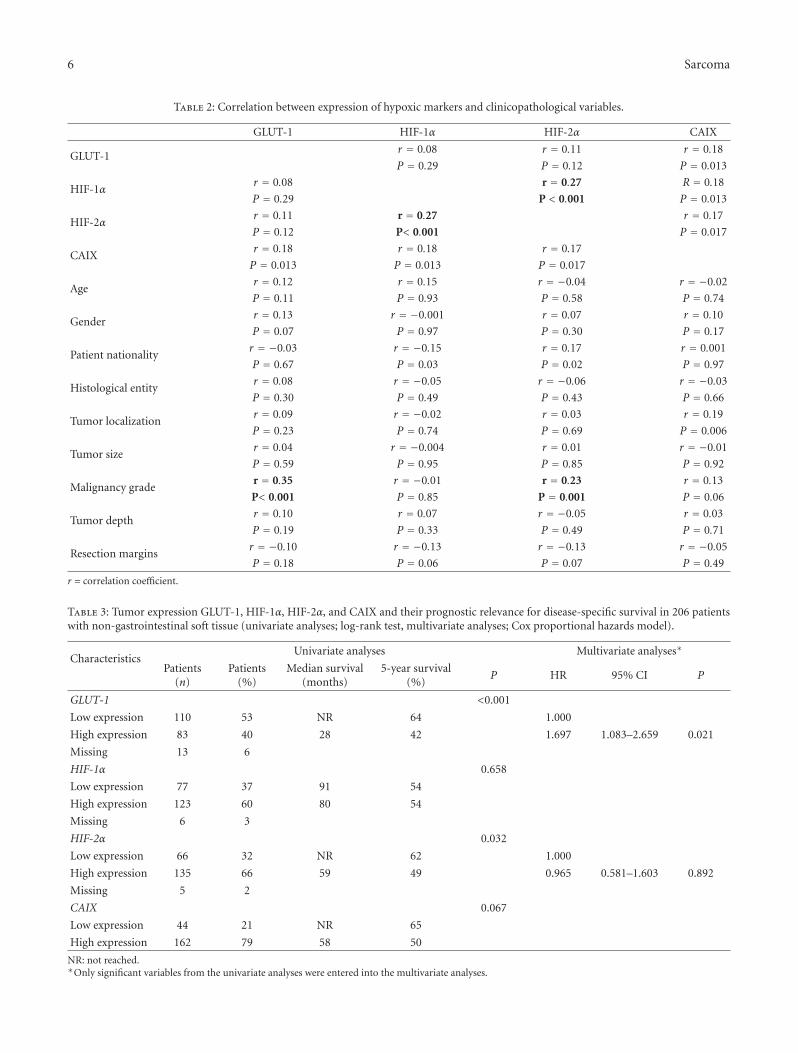
4.3. Expression of Hypoxia-Related Markers and Their Cor-relations. None of the markers correlated significantly withage, gender, histological subgroup, tumor depth, or tumorsize. High GLUT-1 expression was significantly associated(r = 0.35, P < 0.001) with a high histological grade (highexpression: grade I 21.8%, grade II 39.5%, grade III 66.1%).The same significant association (r = 0.23, P = 0.001)was also found for high HIF-2α expression and histologicalgrade (high expression: grade I 49.1%, grade II 71.3%, GradeIII 77.3%). Futhermore, there was a significant correlationbetween HIF-1α and HIF-2α (r = 0.27, P < 0.001), but noother significant association was found among the markers(Table 2).
4.4. Univariate Analyses. Table 1 summarizes the prognosticimpact of the clinicopathological variables. Age (P = 0.030),patient nationality (P = 0.014), histological entity (P =0.003), tumor size (P = 0.019), malignancy grade (P <0.001), tumor depth (P = 0.002), surgery (P < 0.001), andsurgical margins (P < 0.001) were significant prognosticindicators for DSS.
Among the examined molecular markers, high tumor cellGLUT-1 expression (P < 0.001) and HIF-2α expression (P =0.021) correlated significantly with a poor DSS (Table 3 andFigure 2).
Subgroup analyses with respect to histology and resectionmargins were done for all markers, but due to a lownumber of cases many results only tended to be significant.However, GLUT1 became a significant prognostic marker inpleomorphic sarcomas (P = 0.02). The prognostic impactof GLUT1 was not statistically significant in patients withnonwide resection margins, but significant in patients withwide resection margins (P = 0.001).
4.5. Multivariate Cox Proportional Hazards Analyses. Resultsfrom the multivariate analysis are presented in Tables 1 and 3.High malignancy grade (P < 0.001), deep tumor depth (P =0.045), no surgery (P < 0.001), nonwide resection margins(P < 0.001), and high GLUT-1 expression were significantindependent negative prognostic indicators of DSS.
5. Discussion
In this large-scale non-GIST STSs TMA analysis, we inves-tigated the prognostic impact of HIF-1α, HIF-2α, andthe metabolic HIF-regulated GLUT-1 and CAIX. Interest-ingly, high GLUT-1 expression is an independent negativeprognostic factor in non-GIST STSs, while high HIF-2αexpression is significantly associated with a poor prognosisin univariate analyses.
GLUT-1 is extensively expressed in several tumors [19,20]. Furthermore, GLUT-1 has been correlated with a dismalprognosis in different cancer types, such as ovarial cancer[21], nonsmall cell lung cancer (NSCLC) [23], and colorectalcancer [22]. With respect to sarcoma, Ahrens et al. reportedimmunohistochemical GLUT-1 expression in 247 soft tissueand bone neoplasms [37]. GLUT-1 expression was seen ina wide variety of both benign and malignant mesenchymaltumors although they did not assess the prognostic impactof these markers.
Also in sarcomas has the function of GLUT-1 been relatedto glucose metabolism. In a prospective evaluation usinga [18F]fluorodeoxyglucose positron emission tomography(FDG-PET), Tateishi and coworkers observed that GLUT-1 expression and enhanced glucose metabolism were asso-ciated with tumour grade in bone and soft tissue sarco-mas [39]. GLUT-1-positive tumors had significantly higher
6 Sarcoma
Table 2: Correlation between expression of hypoxic markers and clinicopathological variables.
GLUT-1 HIF-1α HIF-2α CAIX
GLUT-1r = 0.08 r = 0.11 r = 0.18
P = 0.29 P = 0.12 P = 0.013
HIF-1αr = 0.08 r = 0.27 R = 0.18
P = 0.29 P < 0.001 P = 0.013
HIF-2αr = 0.11 r = 0.27 r = 0.17
P = 0.12 P< 0.001 P = 0.017
CAIXr = 0.18 r = 0.18 r = 0.17
P = 0.013 P = 0.013 P = 0.017
Ager = 0.12 r = 0.15 r = −0.04 r = −0.02
P = 0.11 P = 0.93 P = 0.58 P = 0.74
Genderr = 0.13 r = −0.001 r = 0.07 r = 0.10
P = 0.07 P = 0.97 P = 0.30 P = 0.17
Patient nationalityr = −0.03 r = −0.15 r = 0.17 r = 0.001
P = 0.67 P = 0.03 P = 0.02 P = 0.97
Histological entityr = 0.08 r = −0.05 r = −0.06 r = −0.03
P = 0.30 P = 0.49 P = 0.43 P = 0.66
Tumor localizationr = 0.09 r = −0.02 r = 0.03 r = 0.19
P = 0.23 P = 0.74 P = 0.69 P = 0.006
Tumor sizer = 0.04 r = −0.004 r = 0.01 r = −0.01
P = 0.59 P = 0.95 P = 0.85 P = 0.92
Malignancy grader = 0.35 r = −0.01 r = 0.23 r = 0.13
P< 0.001 P = 0.85 P = 0.001 P = 0.06
Tumor depthr = 0.10 r = 0.07 r = −0.05 r = 0.03
P = 0.19 P = 0.33 P = 0.49 P = 0.71
Resection marginsr = −0.10 r = −0.13 r = −0.13 r = −0.05
P = 0.18 P = 0.06 P = 0.07 P = 0.49
r = correlation coefficient.
Table 3: Tumor expression GLUT-1, HIF-1α, HIF-2α, and CAIX and their prognostic relevance for disease-specific survival in 206 patientswith non-gastrointestinal soft tissue (univariate analyses; log-rank test, multivariate analyses; Cox proportional hazards model).
CharacteristicsUnivariate analyses Multivariate analyses∗
Patients(n)
Patients(%)
Median survival(months)
5-year survival(%)
P HR 95% CI P
GLUT-1 <0.001
Low expression 110 53 NR 64 1.000
High expression 83 40 28 42 1.697 1.083–2.659 0.021
Missing 13 6
HIF-1α 0.658
Low expression 77 37 91 54
High expression 123 60 80 54
Missing 6 3
HIF-2α 0.032
Low expression 66 32 NR 62 1.000
High expression 135 66 59 49 0.965 0.581–1.603 0.892
Missing 5 2
CAIX 0.067
Low expression 44 21 NR 65
High expression 162 79 58 50
NR: not reached.∗Only significant variables from the univariate analyses were entered into the multivariate analyses.

Sarcoma 7
0.8
0.6
0.4
0.2
120100806040200
0
1
Survival (months)
Dis
ease
-spe
cifi
c su
rviv
al
Low, n = 110
High, n = 83
P < 0.001
GLUT-1
(a)
120100806040200
Survival (months)
0.8
0.6
0.4
0.2
0
1
Dis
ease
-spe
cifi
c su
rviv
al
Low, n = 77
High, n = 123
P = 0.658
HIF1-α
(b)
120100806040200
Survival (months)
0.8
0.6
0.4
0.2
0
1
Dis
ease
-spe
cifi
c su
rviv
al
Low, n = 66
High, n = 135
P = 0.032
HIF2-α
(c)
CAIX
120100806040200
Survival (months)
0.8
0.6
0.4
0.2
0
1
Dis
ease
-spe
cifi
c su
rviv
al
Low, n = 44
High, n = 162
P = 0.067
(d)
Figure 2: DSS curves according to GLUT-1 (a), HIF-1α (b), HIF-2α (c) and CAIX (d) expression in non-GIST STS. DSS: disease-specificsurvival, GLUT-1: glucose transporter-1, HIF-1/2α: hypoxia induced factor 1/2α, CAIX: carbonic anhydrase IX, and non-GIST STS: non-gastrointestinal stromal tumor soft tissue sarcomas.
mean and maximal standardized uptake values (SUVs) thanthe GLUT-1-negative tumors. Likewise, Nagamatsu et al.reported the use of FDG-PET for diagnosis of uterinesarcomas [40]. They detected GLUT-1 expression scores to besignificantly higher in sarcomas and endometrial cancer thanin leiomyomas and concluded that immunohistochemicalexamination of GLUT-1 confirmed the high FDG uptake inleiomyosarcoma patients.
The prognostic impact of GLUT-1 has been documentedby Endo and coworkers [38]. They reported GLUT-1 expres-sion in 22 patients with bone sarcomas and 45 with STS.They found GLUT-1 overexpression in as much as 83% of thepatients. The patients with GLUT-1 overexpression showedsignificantly worse OS compared with those without (P =0.029). Possibly due to the small number of cases, GLUT-1did not appear as an independent prognostic factor in theirstudy. Herein we document for the first time that GLUT-1expression is an independent indicator of poor prognosis innon-GIST STSs.
Overexpression of HIF-2α was associated with reducedsurvival in the univariate analysis. To our knowledge, thisis the first report to examine HIF-2α in sarcoma patients.
These results are, however, comparable with findings in othertumors. Koukourakis and colleagues found that HIF-2α andCAIX were associated with radiotherapy failure in head andneck cancer patients [33]. Likewise, HIF-1α and HIF-2α werehighly expressed in metastatic gastric cancers and correlatedsignificantly with clinical stage [41].
In our study, HIF-1α overexpression was not associatedwith inferior survival, which is in contrast to the findings byShintani et al. [42]. They reported IHC expression of HIF-1αin 49 specimens of STS and found strong and moderate HIF-1α expression to be independently associated with a shortersurvival.
For the prognostic impact of HIF-2α, several reports areconsistent with our results. Yoshimura et al. examined HIF-1α and HIF-2α expression in 87 resected colorectal carcino-mas [43]. HIF-1α (45%) was more frequently expressed thanHIF-2α (30%), but clinicopathological variables representingtumor aggressiveness correlated more often with HIF-2α,than HIF-1α. In lung cancer, Giatromanolaki and coworkers[44] found tumor HIF-2α-expression, not HIF-1α, to beindependently associated with survival. Furthermore, as IHCis a “snapshot” of the tissue metabolism, involved molecules

8 Sarcoma
may not be expressed at high level simultaneously. HIFs arealso known to be rapidly degraded [7] and differentiallyexpressed during prolonged hypoxia [45, 46]. Other tran-scription factors may also be involved, hence blurring theclear-cut image we have of hypoxia pathways [47, 48].
In our study, CAIX tended towards a negative prognosticimpact in the univariate analysis. In central chondrosarcoma,the only sarcoma in which CAIX has been studied, Boeuf etal. reported CAIX reactivity to be a grade-independent pre-dictor of poor metastasis-free survival [49]. The results fromother malignancies are divergent. Woelber et al. found CAIXto be upregulated in ovarian cancer [50]. Serum concentra-tion of CAIX showed, however, no significant changes duringfirst-line therapy, and there was no association betweenserum CAIX and progression-free or overall survival. In renalcell carcinoma, CAIX is strongly expressed and associatedwith clinical outcome, but not as an independent prognosticmarker [51]. An independent prognostic impact of CAIXwas, however, observed by Lie et al. in NSCLC [52]. Moreintriguing is the data from Eckert et al. in oral squamouscell carcinoma [53], where patients with low coexpression ofHIF-1α/CAIX indicated a good prognosis, whereas patientswith increased HIF-1α and low CAIX expression had around5-fold increased risk of tumor-related death (P = 0.042).
As this is a retrospective study, none of the tumors wereavailable for in vivo analysis. Hence, no firm conclusionsregarding these factors association to actual hypoxia itself canbe drawn. In vivo hypoxia can only be measured directly withelectrodes on tumors available for such instrumentation,indirectly by PET using nitroimidazole compounds or newerspecific tracers like F-FAZA (Fluorine-fluoroazomycin arabi-noside) or F-EF5 (Fluorine 2-(2-nitro-1H-imidazol-1-yl)-N-(2,2,3,3,3-pentafluoropropyl)-acetamide) [54]. In addition,it is the hypoxia in vivo that is clinically important sinceabsolute hypoxia initiates when the tumor blood flow isterminated under surgery. It also has to be kept in mind thatit usually takes hours until the tissues are fixed in formalin[55]. In this study we have instead explored the impact ofhypoxia indirectly by evaluating proposed hypoxic markersdue to their up regulation by hypoxia [8], although their roleis controversial [10].
It is possible to question the heterogeneity of the patientpopulation, regarding origin, histology, and treatment. How-ever, this is the limitation for almost all studies on seldommalignancies such as STS. Furthermore, GLUT-1 had thesame tendency in all subgroups in our material.
Another potential limitation with TMA is the eventuallack of homogeneous expression that is easily identified inlarger tissue sections. Hence, to confine the impact of thisissue, we used duplicate cores which were selected to be asrepresentative as possible. Up to 95% correlation has beendemonstrated when comparing tumour cell assessment induplicate 0.6 mm cores versus the whole slide [56].
In cancer therapy there is an increasing focus on person-alized therapy. This shift is associated with the introductionof novel cytotoxic agents and molecular targeted drugs.Inhibitors of HIF-1 and CAIX have been developed and arecurrently under examination [57–59]. Increased knowledgeabout hypoxia-associated markers and metabolic markers in
non-GIST-STS will be vital in identifying different tumor cellphenotypes as candidates for specific molecular targeting.
Though further studies are needed, GLUT-1 appears as apotentially relevant prognostic factor in routine examinationin non-GIST-STS. The identification of independent prog-nostic markers in STS and other malignancies is vital for thefuture development of new molecular targeted drugs.
Conflict of Interests
The authors declare that there is no conflict of interests.
Authors’ Contribution
All authors participated in the study design, result inter-pretation and in the writing. E. Smeland, T. Kilvear, A.Valkov and S. Sorbye contributed in making the clinical anddemographic database. S. Sorbye and A. Valkov scored thecores. E. Smeland T. Kilvear and T. Donnem did the statisticalanalysis. E. Smeland drafted the paper. All authors read andapproved the final paper.
Disclosure
The funders had no role in study design, data collection andanalysis, decision to publish, or preparation of the paper.
Acknowledgments
The authors would like to thank The Norwegian Cancer Soci-ety, The Northern Norway Health Authority, the NorwegianNational Network of Childhood Cancer, and The NorwegianSarcoma Group for supporting this study. They would alsolike to thank Professor Helge Stalsberg for great support inthe initial work with the database.
References
[1] J. Pouyssegur, F. Dayan, and N. M. Mazure, “Hypoxiasignalling in cancer and approaches to enforce tumourregression,” Nature, vol. 441, no. 7092, pp. 437–443, 2006.
[2] R. E. Airley, R. M. Phillips, A. E. Evans et al., “Hypoxia-reg-ulated glucose transporter Glut-1 may influence chemosen-sitivity to some alkylating agents: results of EORTC (FirstTranslational Award) study of the relevance of tumour hypoxiato the outcome of chemotherapy in human tumour-derivedxenografts,” International Journal of Oncology, vol. 26, no. 6,pp. 1477–1484, 2005.
[3] M. Hockel, K. Schlenger, B. Aral, M. Mitze, U. Schaffer, and P.Vaupel, “Association between tumor hypoxia and malignantprogression in advanced cancer of the uterine cervix,” CancerResearch, vol. 56, no. 19, pp. 4509–4515, 1996.
[4] D. M. Brizel, S. P. Scully, J. M. Harrelson et al., “Tumor ox-ygenation predicts for the likelihood of distant metastases inhuman soft tissue sarcoma,” Cancer Research, vol. 56, no. 5,pp. 941–943, 1996.
[5] M. Nordsmark, M. Hoyer, J. Keller, O. S. Nielsen, O. M.Jensen, and J. Overgaard, “The relationship between tumoroxygenation and cell proliferation in human soft tissue

Sarcoma 9
sarcomas,” International Journal of Radiation Oncology BiologyPhysics, vol. 35, no. 4, pp. 701–708, 1996.
[6] L. S. Mortensen, S. Buus, M. Nordsmark et al., “Identifyinghypoxia in human tumors: a correlation study between 18F-FMISO PET and the Eppendorf oxygen-sensitive electrode,”Acta Oncologica, vol. 49, no. 7, pp. 934–940, 2010.
[7] S. Kaluz, M. Kaluzova, S. Y. Liao, M. Lerman, and E.J. Stanbridge, “Transcriptional control of the tumor- andhypoxia-marker carbonic anhydrase 9: a one transcriptionfactor (HIF-1) show?” Biochimica et Biophysica Acta, Reviewson Cancer, vol. 1795, no. 2, pp. 162–172, 2009.
[8] D. Vordermark and J. M. Brown, “Endogenous markers oftumor hypoxia: predictors of clinical radiation resistance?”Strahlentherapie und Onkologie, vol. 179, no. 12, pp. 801–811,2003.
[9] B. Jankovic, C. Aquino-Parsons, J. A. Raleigh et al., “Compar-ison between pimonidazole binding, oxygen electrode mea-surements, and expression of endogenous hypoxia markersin cancer of the uterine cervix,” Cytometry Part B, ClinicalCytometry, vol. 70, no. 2, pp. 45–55, 2006.
[10] A. Mayer, M. Hockel, and P. Vaupel, “Endogenous hypoxiamarkers: case not proven!,” Advances in Experimental Medicineand Biology, vol. 614, pp. 127–136, 2008.
[11] G. L. Semenza and G. L. Wang, “A nuclear factor inducedby hypoxia via de novo protein synthesis binds to thehuman erythropoietin gene enhancer at a site required fortranscriptional activation,” Molecular and Cellular Biology, vol.12, no. 12, pp. 5447–5454, 1992.
[12] H. Tian, S. L. McKnight, and D. W. Russell, “Endothelial PASdomain protein 1 (EPAS1), a transcription factor selectivelyexpressed in endothelial cells,” Genes and Development, vol. 11,no. 1, pp. 72–82, 1997.
[13] M. Heidbreder, F. Frohlich, O. Johren, A. Dendorfer, F. Qadri,and P. Dominiak, “Hypoxia rapidly activates HIF-3alphamRNA expression,” The FASEB Journal, vol. 17, no. 11, pp.1541–1543, 2003.
[14] Y. Z. Gu, S. M. Moran, J. B. Hogenesch, L. Wartman, and C.A. Bradfield, “Molecular characterization and chromosomallocalization of a third α- class hypoxia inducible factorsubunit, HIF3α,” Gene Expression, vol. 7, no. 3, pp. 205–213,1998.
[15] M. Ema, S. Taya, N. Yokotani, K. Sogawa, Y. Matsuda, andY. Fujii-Kuriyama, “A novel bHLH-PAS factor with closesequence similarity to hypoxia-inducible factor 1α regulatesthe VEGF expression and is potentially involved in lung andvascular development,” Proceedings of the National Academy ofSciences of the United States of America, vol. 94, no. 9, pp. 4273–4278, 1997.
[16] H. Zhong, A. M. de Marzo, E. Laughner et al., “Overexpressionof hypoxia-inducible factor 1α in common human cancers andtheir metastases,” Cancer Research, vol. 59, no. 22, pp. 5830–5835, 1999.
[17] G. L. Semenza, “Defining the role of hypoxia-inducible factor1 in cancer biology and therapeutics,” Oncogene, vol. 29, no. 5,pp. 625–634, 2010.
[18] O. Warburg, “On the origin of cancer cells,” Science, vol. 123,no. 3191, pp. 309–314, 1956.
[19] M. Kunkel, T. E. Reichert, P. Benz et al., “Overexpressionof Glut-1 and increased glucose metabolism in tumors areassociated with a poor prognosis in patients with oralsquamous cell carcinoma,” Cancer, vol. 97, no. 4, pp. 1015–1024, 2003.
[20] M. Younes, L. V. Lechago, J. R. Somoano, M. Mosharaf, and J.Lechago, “Wide expression of the human erythrocyte glucose
transporter Glut1 in human cancers,” Cancer Research, vol. 56,no. 5, pp. 1164–1167, 1996.
[21] G. Cantuaria, A. Fagotti, G. Ferrandina et al., “GLUT-1 ex-pression in ovarian carcinoma: association with survival andresponse to chemotherapy,” Cancer, vol. 92, no. 5, pp. 1144–1150, 2001.
[22] R. S. Haber, A. Rathan, K. R. Weiser et al., “GLUT1 glucosetransporter expression in colorectal carcinoma: a marker forpoor prognosis,” Cancer, vol. 83, no. 1, pp. 34–40, 1998.
[23] M. Younes, R. W. Brown, M. Stephenson, M. Gondo, and P. T.Cagle, “Overexpression of Glut1 and Glut3 in stage I nonsmallcell lung carcinoma is associated with poor survival,” Cancer,vol. 80, no. 6, pp. 1046–1051, 1997.
[24] M. C. Brahimi-Horn, J. Chiche, and J. Pouyssegur, “Hypoxiaand cancer,” Journal of Molecular Medicine, vol. 85, no. 12, pp.1301–1307, 2007.
[25] J. Chiche, K. Ilc, J. Laferriere et al., “Hypoxia-induciblecarbonic anhydrase IX and XII promote tumor cell growthby counteracting acidosis through the regulation of theintracellular pH,” Cancer Research, vol. 69, no. 1, pp. 358–368,2009.
[26] G. L. Semenza, “Regulation of cancer cell metabolism byhypoxia-inducible factor 1,” Seminars in Cancer Biology, vol.19, no. 1, pp. 12–16, 2009.
[27] A. Jemal, R. Siegel, E. Ward, Y. Hao, J. Xu, and M. J. Thun,“Cancer statistics, 2009,” CA Cancer Journal for Clinicians, vol.59, no. 4, pp. 225–249, 2009.
[28] J. Y. Blay, M. von Mehren, and M. E. Blackstein, “Perspectiveon updated treatment guidelines for patients with gastroin-testinal stromal tumors,” Cancer, vol. 116, no. 22, pp. 5126–5137, 2010.
[29] T. K. Kilvaer, A. Valkov, S. Sorbye et al., “Profiling of VEGFsand VEGFRs as prognostic factors in soft tissue sarcoma:VEGFR-3 is an independent predictor of poor prognosis,”PLoS One, vol. 5, no. 12, Article ID e15368, 2010.
[30] C. D. M. Fletcher, “The evolving classification of soft tissuetumours: an update based on the new WHO classification,”Histopathology, vol. 48, no. 1, pp. 3–12, 2006.
[31] L. Guillou, J. M. Coindre, F. Bonichon et al., “Comparativestudy of the National Cancer Institute and French Federationof Cancer Centers Sarcoma Group grading systems in apopulation of 410 adult patients with soft tissue sarcoma,”Journal of Clinical Oncology, vol. 15, no. 1, pp. 350–362, 1997.
[32] T. Donnem, S. Al-Saad, K. Al-Shibli et al., “Inverse prognosticimpact of angiogenic marker expression in tumor cells versusstromal cells in non-small cell lung cancer,” Clinical CancerResearch, vol. 13, no. 22, pp. 6649–6657, 2007.
[33] M. I. Koukourakis, S. M. Bentzen, A. Giatromanolaki etal., “Endogenous markers of two separate hypoxia responsepathways (hypoxia inducible factor 2 alpha and carbonicanhydrase 9) are associated with radiotherapy failure in headand neck cancer patients recruited in the CHART randomizedtrial,” Journal of Clinical Oncology, vol. 24, no. 5, pp. 727–735,2006.
[34] Y. S. Choi, S. J. Kim, and D. S. Kim, “Glucose transporter-1expression in squamous cell carcinoma of the tongue,” CancerResearch and Treatment, vol. 29, pp. 109–115, 2007.
[35] I. H. Ozbudak, K. Shilo, F. Tavora et al., “Glucose transporter-1 in pulmonary neuroendocrine carcinomas: expression andsurvival analysis,” Modern Pathology, vol. 22, no. 5, pp. 633–638, 2009.
[36] L. Woelber, K. Kress, J. F. Kersten et al., “Carbonic anhydraseIX in tumor tissue and sera of patients with primary cervicalcancer,” BMC Cancer, vol. 11, article 12, 2011.

10 Sarcoma
[37] W. A. Ahrens, R. V. Ridenour III, B. L. Caron, D. V. Miller,and A. L. Folpe, “GLUT-1 expression in mesenchymal tumors:an immunohistochemical study of 247 soft tissue and boneneoplasms,” Human Pathology, vol. 39, no. 10, pp. 1519–1526,2008.
[38] M. Endo, U. Tateishi, K. Seki et al., “Prognostic implicationsof glucose transporter protein-1 (Glut-1) overexpression inbone and soft-tissue sarcomas,” Japanese Journal of ClinicalOncology, vol. 37, no. 12, pp. 955–960, 2007.
[39] U. Tateishi, U. Yamaguchi, K. Seki, T. Terauchi, Y. Arai,and T. Hasegawa, “Glut-1 expression and enhanced glu-cose metabolism are associated with tumour grade in boneand soft tissue sarcomas: a prospective evaluation by [18F]fluorodeoxyglucose positron emission tomography,” EuropeanJournal of Nuclear Medicine and Molecular Imaging, vol. 33, no.6, pp. 683–691, 2006.
[40] A. Nagamatsu, N. Umesaki, L. Li, and T. Tanaka, “Use of18F-fluorodeoxyglucose positron emission tomography fordiagnosis of uterine sarcomas,” Oncology Reports, vol. 23, no.4, pp. 1069–1076, 2010.
[41] Y. Wang, Z. Li, H. Zhang et al., “HIF-1α and HIF-2α correlatewith migration and invasion in gastric cancer,” Cancer Biologyand Therapy, vol. 10, no. 4, pp. 376–382, 2010.
[42] K. Shintani, A. Matsumine, K. Kusuzaki et al., “Expression ofhypoxia-inducible factor (HIF)-1α as a biomarker of outcomein soft-tissue sarcomas,” Virchows Archiv, vol. 449, no. 6, pp.673–681, 2006.
[43] H. Yoshimura, D. K. Dhar, H. Kohno et al., “Prognosticimpact of hypoxia-inducible factors 1α and 2α in colorectalcancer patients: correlation with tumor angiogenesis andcyclooxygenase-2 expression,” Clinical Cancer Research, vol.10, no. 24, pp. 8554–8560, 2004.
[44] A. Giatromanolaki, M. I. Koukourakis, E. Sivridis et al.,“Relation of hypoxia inducible factor 1α and 2α in operablenon-small cell lung cancer to angiogenic/molecular profile oftumours and survival,” British Journal of Cancer, vol. 85, no. 6,pp. 881–890, 2001.
[45] Q. F. Li, X. R. Wang, Y. W. Yang, and H. Lin, “Hypoxiaupregulates hypoxia inducible factor (HIF)-3α expression inlung epithelial cells: characterization and comparison withHIF-1α,” Cell Research, vol. 16, no. 6, pp. 548–558, 2006.
[46] T. Uchida, F. Rossignol, M. A. Matthay et al., “Pro-longed hypoxia differentially regulates hypoxia-inducible fac-tor (HIF)-1α and HIF-2α expression in lung epithelial cells:implication of natural antisense HIF-1α,” Journal of BiologicalChemistry, vol. 279, no. 15, pp. 14871–14878, 2004.
[47] H. Ishibashi, K. Nakagawa, M. Onimaru et al., “Sp1 decoytransfected to carcinoma cells suppresses the expressionof vascular endothelial growth factor, transforming growthfactor/β1, and tissue factor and also cell growth and invasionactivities,” Cancer Research, vol. 60, no. 22, pp. 6531–6536,2000.
[48] N. Pore, A. K. Gupta, G. J. Cerniglia et al., “Nelfinavir down-regulates hypoxia-inducible factor 1α and VEGF expressionand increases tumor oxygenation: implications for radiother-apy,” Cancer Research, vol. 66, no. 18, pp. 9252–9259, 2006.
[49] S. Boeuf, J. V. M. G. Bovee, B. Lehner, P. C. W. Hogendoorn,and W. Richter, “Correlation of hypoxic signalling to histolog-ical grade and outcome in cartilage tumours,” Histopathology,vol. 56, no. 5, pp. 641–651, 2010.
[50] L. Woelber, V. Mueller, C. Eulenburg et al., “Serum carbonicanhydrase IX during first-line therapy of ovarian cancer,”Gynecologic Oncology, vol. 117, no. 2, pp. 183–188, 2010.
[51] B. C. Leibovich, Y. Sheinin, C. M. Lohse et al., “Carbonicanhydrase IX is not an independent predictor of outcome forpatients with clear cell renal cell carcinoma,” Journal of ClinicalOncology, vol. 25, no. 30, pp. 4757–4764, 2007.
[52] M. Lie, N. M. Mazure, V. Hofman et al., “High levels ofcarbonic anhydrase IX in tumour tissue and plasma arebiomarkers of poor prognostic in patients with non-small celllung cancer,” British Journal of Cancer, vol. 102, no. 11, pp.1627–1635, 2010.
[53] A. W. Eckert, M. H. W. Lautner, A. Schutze et al., “Co-expression of Hif1α and CAIX is associated with poorprognosis in oral squamous cell carcinoma patients,” Journalof Oral Pathology and Medicine, vol. 39, no. 4, pp. 313–317,2010.
[54] S. E. Lapi, T. F. Voller, and M. J. Welch, “Positron emissiontomography imaging of hypoxia,” PET Clinics, vol. 4, pp. 39–47, 2009.
[55] Sauter, Tissue Microarrays, Springer, 2010.[56] O. P. Kallioniemi, U. Wagner, J. Kononen, and G. Sauter,
“Tissue microarray technology for high-throughput molecu-lar profiling of cancer,” Human Molecular Genetics, vol. 10, no.7, pp. 657–662, 2001.
[57] Z. G. Dikmen, G. C. Gellert, P. Dogan et al., “In vivo andin vitro effects of a HIF-1α inhibitor, RX-0047,” Journal ofCellular Biochemistry, vol. 104, no. 3, pp. 985–994, 2008.
[58] G. L. Semenza, “Evaluation of HIF-1 inhibitors as anticanceragents,” Drug Discovery Today, vol. 12, no. 19-20, pp. 853–859,2007.
[59] A. Thiry, J. M. Dogne, B. Masereel, and C. T. Supuran, “Tar-geting tumor-associated carbonic anhydrase IX in cancertherapy,” Trends in Pharmacological Sciences, vol. 27, no. 11,pp. 566–573, 2006.
Copyright © 2022 FDOKUMEN