Prognostic correlations with the microbiome of breast cancer ...
Upload
khangminh22Category
view
0download
0
Oncogene (2018) 37:5269–5280https://doi.org/10.1038/s41388-018-0288-y
ARTICLE
Immune checkpoints PVR and PVRL2 are prognostic markers in AMLand their blockade represents a new therapeutic option
Hauke Stamm1● Felix Klingler1 ● Eva-Maria Grossjohann1
● Jana Muschhammer1 ● Eik Vettorazzi 2●
Michael Heuser3 ● Ulrike Mock4 ● Felicitas Thol3 ● Gabi Vohwinkel1 ● Emily Latuske1 ● Carsten Bokemeyer1 ●
Roman Kischel5 ● Cedric Dos Santos6 ● Sabine Stienen5● Matthias Friedrich5
● Michael Lutteropp5● Dirk Nagorsen7
●
Jasmin Wellbrock1 ● Walter Fiedler1
Received: 19 October 2017 / Revised: 8 March 2018 / Accepted: 2 April 2018 / Published online: 31 May 2018© The Author(s) 2018. This article is published with open access
AbstractImmune checkpoints are promising targets in cancer therapy. Recently, poliovirus receptor (PVR) and poliovirus receptor-related 2 (PVRL2) have been identified as novel immune checkpoints. In this investigation we show that acute myeloidleukemia (AML) cell lines and AML patient samples highly express the T-cell immunoreceptor with Ig and ITIM domains(TIGIT) ligands PVR and PVRL2. Using two independent patient cohorts, we could demonstrate that high PVR and PVRL2expression correlates with poor outcome in AML. We show for the first time that antibody blockade of PVR or PVRL2 onAML cell lines or primary AML cells or TIGIT blockade on immune cells increases the anti-leukemic effects mediated byPBMCs or purified CD3+ cells in vitro. The cytolytic activity of the BiTE® antibody construct AMG 330 against leukemic cellscould be further enhanced by blockade of the TIGIT-PVR/PVRL2 axis. This increased immune reactivity is paralleled byaugmented secretion of Granzyme B by immune cells. Employing CRISPR/Cas9-mediated knockout of PVR and PVRL2 inMV4-11 cells, the cytotoxic effects of antibody blockade could be recapitulated in vitro. In NSG mice reconstituted withhuman T cells and transplanted with either MV4-11 PVR/PVRL2 knockout or wildtype cells, prolonged survival was observedfor the knockout cells. This survival benefit could be further extended by treating the mice with AMG 330. Therefore, targetingthe TIGIT-PVR/PVRL2 axis with blocking antibodies might represent a promising future therapeutic option in AML.
Introduction
Escape of neoplastic cells from immune destruction hasrecently been added to the list of hallmarks of cancer [1].But, effector lymphocytes may acquire an exhausted phe-notype during the course of the disease, preventing efficienttumor rejection [2, 3].
Inhibition of T-cell activation is accomplished by severalreceptor/ligand systems involved in checkpoint control ofT-cell effector functions such as CTLA-4/CD80 and CD86
These authors contributed equally: Jasmin Wellbrock and WalterFiedler.
* Walter [email protected]
1 Department of Oncology, Hematology and Bone MarrowTransplantation with Section Pneumology, Hubertus WaldUniversity Cancer Center, University Medical Center Hamburg-Eppendorf, Hamburg, Germany
2 Department of Medical Biometry and Epidemiology, UniversityMedical Center Hamburg-Eppendorf, Hamburg, Germany
3 Hematology, Hemostasis, Oncology and Stem Cell
Transplantation, Hannover Medical School, Hannover, Germany4 Research Department Cell and Gene Therapy, Clinic for Stem Cell
Transplantation, University Medical Center Hamburg-Eppendorf,Hamburg, Germany
5 Amgen Research (Munich) GmbH, Munich, Germany6 Oncology Biomarkers and Early Clinical Development, Amgen,
San Francisco, CA, USA7 Global Clinical Development, Amgen, Thousand Oaks, CA, USA
Electronic supplementary material The online version of this article(https://doi.org/10.1038/s41388-018-0288-y) contains supplementarymaterial, which is available to authorized users.
1234
5678
90();,:
1234567890();,:

or PD-1/PD-L1 and PD-L2. Recently, therapeutic anti-bodies have been developed that inhibit these checkpointsresulting in reactivation of a cytotoxic phenotype. Clinicaltrials showed that CTLA-4 blocking antibodies ipilimumabor tremelimumab induced prolonged remissions in patientswith malignant melanoma [4]. Antibodies against PD-1such as pembrolizumab and nivolumab showed clinicalactivity in different tumor types including melanoma,Hodgkin's disease, renal, bladder and lung cancer [5, 6].Currently, much effort is being directed toward the identi-fication of novel immune checkpoint inhibitors [7].
A second class of immunotherapeutic agents are thebispecific T-cell engagers (BiTE®). BiTE® antibodies pos-sess binding sites for CD3 on T cells and for tumor anti-gens, bringing neoplastic cells and T cells in close contact toinduce their cytolytic action. Blinatumomab, a CD19/CD3BiTE®, is the most advanced member in this class, and it isFDA and EMA approved for the treatment of acute lym-phoblastic leukemia (ALL) [8]. For the treatment of acutemyeloid leukemia (AML), AMG 330, a CD33/CD3 BiTE®antibody construct, has shown preclinical activity and iscurrently undergoing phase 1 clinical testing(NCT02520427) [9, 10]. Combining both approaches,tumor cell killing by T cells in the presence of BiTE®antibody constructs, as well as blockade of checkpointmolecules may result in enhanced therapeutic efficacy.
In the present investigation, we explored the therapeuticpotential of inhibition of the novel immune regulatorspoliovirus receptor (PVR, CD155, Tage 4) and poliovirusreceptor-related 2 (PVRL2, CD112, Nectin-2, PRR2), which
bind to the CD28 family member T cell immunoreceptorwith Ig and ITIM domains (TIGIT). TIGIT is a type Itransmembrane protein with an Ig variable extracellulardomain expressed on activated and memory T cells, reg-ulatory T cells, as well as NK and NKT cells [11, 12]. Uponligand interaction, TIGIT suppresses the immune responsethrough its cytosolic immunoglobulin tail tyrosine (ITT)-like phosphorylation motif and immunoreceptor tyrosine-based inhibitory motif (ITIM) [13, 14]. PVR has beeninitially described as the poliovirus binding site and waslinked to blood cells being an extraneural site for poliovirusreplication [15, 16]. PVR is overexpressed by some tumorentities including melanoma, glioblastoma, colorectal andpancreatic carcinoma [17–20].
In our study, we analyzed the expression of TIGITligands PVR and PVRL2 on AML cell lines and patientsamples and exploited the potential of this axis for thetreatment of AML. For the first time, we show that blockingthe TIGIT-PVR/PVRL2 axis augments T-cell mediated lysisof AML cells and additionally enhances the cytotoxic effectsof the CD33/CD3 BiTE® antibody construct AMG 330.
Results
TIGIT ligands PVR and PVRL2 are highly expressedon AML cells
The flow cytometric analysis revealed expression ofPVR and PVRL2 on AML cell lines, all being CD33
Fig. 1 PVR and PVRL2 are highly expressed on AML cell lines andprimary CD33+ AML blasts. PVR and PVRL2 protein expression, asdepicted by the percentage of CD33+ cells as well as median
fluorescence intensity as the measure of expression intensity on AMLcell lines (n= 9; a, b) and CD33+ AML blasts from untreated patients(n= 17; c, d). Black dashes represent the median
5270 H. Stamm et al.

positive (n= 9), and the majority of CD33+ AML blastsfrom untreated AML patients (n= 17; Fig. 1); see repre-sentative FACS plots in Supplemental Fig. 1). Furthermore,we could show that the majority of leukemia-initiating cells,defined as being CD34+/CD38−, expressed PVR andPVRL2 or at least one of the immune checkpoint ligands(mean expression value 77%, range 51–93%, n= 10). Theexpression of the corresponding receptor TIGIT wasdetected on 13.6 ± 3.7% of naïve CD3+ T cells from per-ipheral blood mononuclear cells of healthy donors (HD-PBMCs; n= 4, data not shown).
Blockade of the TIGIT-PVR/PVRL2 axis significantlyaugments T-cell mediated lysis of AML cells alone orin combination with the BiTE® antibody constructAMG 330
The therapeutic potential of blocking the novel immunecheckpoint molecules PVR and PVRL2 on leukemic blastswas examined in in vitro cytotoxicity assays. The specificlysis of AML cells by HD-PBMCs in the presence orabsence of blocking PVR/PVRL2 antibodies is depicted asthe mean fold change of dead target cells normalized to thecontrol without blocking antibodies for the cell lines TF-1,Molm-13 and Kasumi-1 (Fig. 2a–c). Since in MV4-11 cellsonly, a cytotoxic effect of the PVR and PVRL2 antibodiescould be observed in the absence of HD-PBMCs (see
Supplemental Fig. 2), we repeated the cytotoxicity assaysusing MV4-11 and TF-1 cells in the presence of a blockinganti-TIGIT antibody to replace the PVR/PVRL2 antibodies.Cell kill could be significantly augmented in both tested celllines, indicating the importance of the TIGIT-PVR/PVRL2axis as immune checkpoints (Fig. 2d, e).
Further, we analyzed whether the T-cell mediated cyto-toxicity exerted by AMG 330 could be enhanced byblocking PVR and PVRL2 or TIGIT. TF-1, Molm-13 orKasumi-1 cells were incubated with HD-PBMCs or purifiedT cells for 24 h with or without 0.1 ng/mL AMG 330 in thepresence or absence of PVR and PVRL2 blocking anti-bodies. Additionally, the killing of the cell lines MV4-11and TF-1 was also investigated in the presence or absenceof the anti-TIGIT antibody. As shown in Fig. 3a, thecytotoxic effect of AMG 330 was significantly increased bythe administration of PVR or PVRL2 blocking antibodiesusing TF-1 cells (Fig. 3a). Significantly enhanced killing incomparison to the control could be detected after blockingPVR, PVRL2 or both antigens for the cell line Molm-13,although only moderate differences could be observedbetween all treatment arms (Fig. 3b). For Kasumi-1, onlythe PVR blocking antibody was able to induce a noteworthyenhancement of the cell lysis mediated by AMG 330 (Fig.3c). The additional effect of blocking PVRL2 wasneglectable in spite of high expression of PVRL2 on the cellline (Figs. 1, 3c). Employing the anti-TIGIT antibody, the
Fig. 2 Blocking of the TIGIT-PVR/PVRL2 axis increases the lysis ofAML cell lines. HD-PBMC-mediated lysis, with subject to theblocking of PVR and PVRLs on AML cell lines TF-1 (a, n= 3),Molm-13 (b, n= 6), Kasumi-1 (c, n= 3), was measured after 24 h.The effect of blocking the receptor TIGIT on effector cells was
examined for the cell lines TF-1 (d, n= 5) and MV4-11 (e, n= 3).Results are depicted as the mean ± SD fold changes (FC) of dead targetcells, relative to the control without blocking antibodies. Measure-ments were performed in technical triplicates and for statistical ana-lysis Mann–Whitney U-tests were performed (#p ≤ 0.05; *p ≤ 0.001)
Immune checkpoints PVR and PVRL2 are prognostic markers in AML and their blockade represents a new. . . 5271

effects of AMG 330 were augmented in a comparablemanner (Fig. 3d, e). To exclude the effects of antibodydependent cellular cytotoxicity (ADCC), control experi-ments were performed. These included the use of an irre-levant antibody on c-kit (CD117) positive Kasumi-1 cells,and utilizing purified T cells as effectors (Supplemental Fig.S3 and S4 and Fig. 3f). Both controls emphasize the inde-pendence of ADCC in this context. To further investigatethe T-cell mediated killing of target cells, we measuredGranzyme B concentrations in the supernatants after incu-bation of TF-1 and MV4-11 with HD-PBMCs and anti-bodies against PVR, PVRL2, their combination or TIGIT.A significant increase in Granzyme B concentration wasdetected by the addition of blocking antibodies with orwithout AMG 330 (Fig. 4a–f, for a time course employingTF-1 cells see Supplemental Fig. S5).
Blockade of PVR and PVRL2 significantly augmentscytotoxicity against primary AML blasts
The effect of a PVR and PVRL2 blockade on the lysis ofprimary AML blasts from patient samples with high blastcontent was examined. Increased lysis of blasts due to thecombined blocking of PVR and PVRL2 in four of the tenanalyzed patient samples could be measured. The anti-
leukemic effect of the BiTE® antibody construct AMG 330could be enhanced in five out of nine CD33-expressingpatient samples (Fig. 5a–f; see Supplemental Fig. S6 fornon-responders).
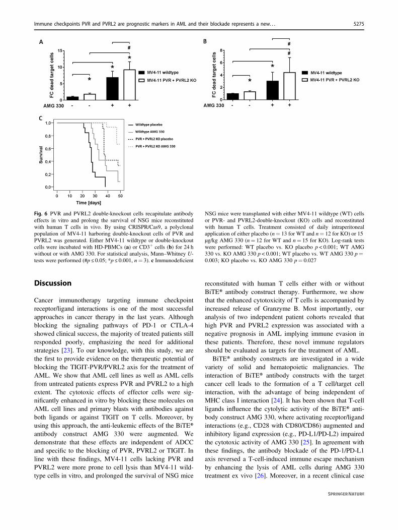
MV4-11 PVR and PVRL2 double-knockout cellsrecapitulate antibody effects
To further strengthen our results, we generated PVR andPVRL2 double-knockouts for the cell line MV4-11 usingCRISPR/Cas9. Cells harboring the double-knockout ofPVR and PVRL2 on protein level were sorted by FACS(Supplemental Fig. S7). Furthermore, we analyzed theCRISPR/Cas9-induced genetic modifications resulting inthe protein knockout in a number of single clones (Sup-plemental Fig. S8). The PVR and PVRL2 double-knockoutcells were compared to their wildtype counterparts incytotoxicity assays. In this setting, significantly enhancedkilling of the double-knockout cells either by HD-PBMCsor purified T cells could be detected (Fig. 6a, b). This effectalso resulted in higher secretion of Granzyme B during thekill of the double-knockout cells vs. the wildtype cells (datanot shown). Addition of AMG 330 led again to a furthersignificant increased cell lysis of the PVR and PVRL2double-knockout cells compared to MV4-11 wildtype cells,
Fig. 3 T-cell mediated lysis of the BiTE® antibody construct AMG330 is significantly enhanced by additional administration of PVRand PVRL2 or TIGIT blocking antibodies. TF-1 (a, n= 3), Molm-13(b, n= 6), Kasumi-1 (c, n= 6) cells were incubated with HD-PBMCsand AMG 330 in the presence or absence of blocking antibodiesagainst PVR or PVRL2. Blocking the receptor TIGIT on immune cellsshowed similar results for the cell line TF-1 (d, n= 5) and MV4-11 (e,n= 3). Results are depicted as the mean ± SD fold change (FC) of
dead target cells, relative to the control without blocking antibodies.Lysis is mediated via CD3+ cells, as comparing HD-PBMCs andpurified CD3+ cells from the same donor showed comparable resultsusing the cell line TF-1 (f, n= 2). Results are depicted as the mean ±SD of dead target cells of two independent experiments. Measure-ments were performed in technical triplicates, and for statistical ana-lysis Mann–Whitney U-tests were performed (#p ≤ 0.05; *p ≤ 0.001)
5272 H. Stamm et al.

indicating an augmented effect of combining both treatmentapproaches (Fig. 6a, b).
Knockout of PVR and PVRL2 results in prolongedsurvival of NSG mice reconstituted with humanT cells
To test the therapeutic impact of PVR and PVRL2 in vivo,in a blinded fashion, MV4-11 PVR and PVRL2 double-knockout cells or their wildtype counterpart were trans-planted intravenously into immunodeficient NSG mice.Five days later, all mice were intraperitoneally injected withenriched and activated T cells from one blood donor. Micereceived daily placebo or 15 µg/kg AMG 330. As shown inFig. 6c, mice transplanted with the MV4-11 PVR andPVRL2 double-knockout cells had a significant survivalbenefit, compared to mice transplanted with MV4-11 wild-type cells, either in the placebo or AMG 330 arm (mediansurvival of 37 vs. 27 days and 33 days vs. not reachedduring the 50 day observation period, respectively). Of note,MV4-11 wildtype cells had no growth advantage, comparedto the MV4-11 double-knockout cells in vitro (Supple-mental Fig. S9). Most importantly, in a preceding experi-ment with NSG mice not reconstituted with human T cells,no survival difference between the double-knockout and
wildtype group was detectable, indicating no difference ofAML engraftment and leukemia development in vivo (29 ±3 vs. 29 ± 2 days, n= 4 and n= 3, respectively).
Clinical significance: high expression of PVR andPVRL2 confers a negative prognosis to AML patients
To determine the expression of PVR and PVRL2, cDNAsamples from 139 newly diagnosed AML patients enrolledinto the AMLSG 07-04 study of the German-Austrian StudyGroup (NCT00151242) were analyzed by RT-qPCR (cohortA [21]). Patient characteristics are summarized in Supple-mental Table S1. PVR and PVRL2 expression was found in94% and 95% of AML patients, respectively. The level ofgene expression and baseline clinical characteristics (age,karyotype and FLT3 mutation status) on event-free survival(EFS), relapse-free survival (RFS) and overall survival (OS)was investigated in a multivariate Cox proportional hazardsmodel, implementing a stepwise removal of insignificantterms. Due to the low number of cases displaying a favor-able karyotype, patients with intermediate and favorablekaryotype were combined. High PVR expression as well asan unfavorable karyotype were identified as independentnegative prognostic markers for EFS, RFS and OS,respectively (see Table 1 for hazard ratios and p-values).
Fig. 4 Blocking of the TIGIT-PVR/PVRL2 axis results in increasedlevels of Granzyme B secretion of immune cells. TF-1 (a–d) andMV4-11 (e, f) target cells were mixed with HD-PBMCs and incubatedwith blocking antibodies against PVR and PVRL2 or TIGIT in thepresence or absence of the BiTE® antibody construct AMG 330. After
24 h, supernatants were harvested and human Granzyme B con-centration was measured using ELISA. Results are depicted as themean ± SD Granzyme B concentration of at least three independentexperiments. For statistical analysis paired t-tests were performed (#p ≤ 0.05)
Immune checkpoints PVR and PVRL2 are prognostic markers in AML and their blockade represents a new. . . 5273

Due to a high correlation between PVR and PVRL2(Pearson’s rho= 0.827, p < 0.001), PVRL2 was removedduring the stepwise process. Nevertheless, if PVR wasexcluded from the multivariate cox regression, high PVRL2expression represented a negative prognostic marker forRFS (p= 0.017), and had a borderline significantnegative impact on OS (p= 0.087; see Table 1 for hazardratios).
As a validation cohort, the microarray-based geneexpression data published by Verhaak et al. was used(cohort B [22]). The distribution of low vs. high PVRexpressors was relatively equal in FAB type M2, M4 andM5, whereas it was significantly lower in AMLM1 subgroup (Fisher’s exact test p= 0.01), while forPVRL2, no significant difference was observed in the var-ious FAB subgroups (Fisher’s test p= 0.114; SupplementalTable S2). Due to the low number of 20 cases in total, FABsubtypes M0, M3, M6 and M7 were excluded from thesubgroup analysis.
In addition to PVR and PVRL2, we also analyzed theexpression of CTLA-4 ligands CD80 and CD86 as well asPD-1 ligands PD-L1 and PD-L2. Clinical data of 290 AMLpatients were available enabling the analysis of the prog-nostic impact of gene expression of immune checkpointmolecules on OS (Supplemental Table S1 for patient
characteristics). The patient cohort was divided into low vs.high expressors for the Kaplan–Meier survival analysis. Theprognostic impact of PVR and PVRL2 could be confirmed,since PVR as well as PVRL2 high expressors displayed asignificantly poorer OS, compared to the low expressiongroups (p= 0.003 and p= 0.032, respectively; Fig. 7).Interestingly, the CTLA-4 ligands CD80 and CD86 as wellas the PD-1 ligand PD-L1 had no impact on the patient’soutcome, while PD-L2 expression was not detectable in theAML patients (Supplemental Fig. S10).
Furthermore, OS was analysed in a multivariate Coxproportional hazards model including the immune check-point ligands CD80, CD86, PD-L1, PVR, and PVRL2, andthe baseline parameters age, karyotype and FLT3 muta-tional status. After a stepwise removal of insignificantterms, only high expression of PVR and a FLT3 mutationremained as independent prognostic survival markers (forPVR: HR 3.39 (95% CI 1.45–7.94), p= 0.005; for FLT3:HR 1.37 (95% CI 1.01–1.85), p= 0.045).
Of note, as no protein expression data was available forcohort A and B, we validated the correlation between thePVR and PVRL2 mRNA measured by RT-qPCR expres-sion measured by flow cytometry in a set of pAML samples(n= 17; Pearson Rho 0.456, p= 0.066 for PVR and Pear-son Rho, p= 0.009 for PVRL2).
Fig. 5 Combined blocking of PVR and PVRL2 on primary AMLblasts increases the cytotoxic effects of HD-PBMCs. Mononuclearcells containing at least 75% blasts from bone marrow aspirates of tendifferent, newly diagnosed AML patients were stained with CMFDA(CellTracker™), mixed with HD-PBMCs as effector cells, and incu-bated for 72 h. Blocking of PVR and PVRL2 alone could increase the
specific lysis of primary blasts in four (a, b, c, f) of the ten analyzedpatients, and augment the anti-leukemic effect of the BiTE® antibodyconstruct AMG 330 in five of nine patients (a–e). The sample ofpatient (f) was CD33 negative, and therefore excluded from the AMG330 experiment. Results are depicted as the mean of technical tripli-cates ± SD of dead target cells
5274 H. Stamm et al.
Discussion
Cancer immunotherapy targeting immune checkpointreceptor/ligand interactions is one of the most successfulapproaches in cancer therapy in the last years. Althoughblocking the signaling pathways of PD-1 or CTLA-4showed clinical success, the majority of treated patients stillresponded poorly, emphasizing the need for additionalstrategies [23]. To our knowledge, with this study, we arethe first to provide evidence on the therapeutic potential ofblocking the TIGIT-PVR/PVRL2 axis for the treatment ofAML. We show that AML cell lines as well as AML cellsfrom untreated patients express PVR and PVRL2 to a highextent. The cytotoxic effects of effector cells were sig-nificantly enhanced in vitro by blocking these molecules onAML cell lines and primary blasts with antibodies againstboth ligands or against TIGIT on T cells. Moreover, byusing this approach, the anti-leukemic effects of the BiTE®antibody construct AMG 330 were augmented. Wedemonstrate that these effects are independent of ADCCand specific to the blocking of PVR, PVRL2 or TIGIT. Inline with these findings, MV4-11 cells lacking PVR andPVRL2 were more prone to cell lysis than MV4-11 wild-type cells in vitro, and prolonged the survival of NSG mice
reconstituted with human T cells either with or withoutBiTE® antibody construct therapy. Furthermore, we showthat the enhanced cytotoxicity of T cells is accompanied byincreased release of Granzyme B. Most importantly, ouranalysis of two independent patient cohorts revealed thathigh PVR and PVRL2 expression was associated with anegative prognosis in AML implying immune evasion inthese patients. Therefore, these novel immune regulatorsshould be evaluated as targets for the treatment of AML.
BiTE® antibody constructs are investigated in a widevariety of solid and hematopoietic malignancies. Theinteraction of BiTE® antibody constructs with the targetcancer cell leads to the formation of a T cell/target cellinteraction, with the advantage of being independent ofMHC class I interaction [24]. It has been shown that T-cellligands influence the cytolytic activity of the BiTE® anti-body construct AMG 330, where activating receptor/ligandinteractions (e.g., CD28 with CD80/CD86) augmented andinhibitory ligand expression (e.g., PD-L1/PD-L2) impairedthe cytotoxic activity of AMG 330 [25]. In agreement withthese findings, the antibody blockade of the PD-1/PD-L1axis reversed a T-cell-induced immune escape mechanismby enhancing the lysis of AML cells during AMG 330treatment ex vivo [26]. Moreover, in a recent clinical case
Fig. 6 PVR and PVRL2 double-knockout cells recapitulate antibodyeffects in vitro and prolong the survival of NSG mice reconstitutedwith human T cells in vivo. By using CRISPR/Cas9, a polyclonalpopulation of MV4-11 harboring double-knockout cells of PVR andPVRL2 was generated. Either MV4-11 wildtype or double-knockoutcells were incubated with HD-PBMCs (a) or CD3+ cells (b) for 24 hwithout or with AMG 330. For statistical analysis, Mann–Whitney U-tests were performed (#p ≤ 0.05; *p ≤ 0.001, n= 3). c Immunodeficient
NSG mice were transplanted with either MV4-11 wildtype (WT) cellsor PVR- and PVRL2-double-knockout (KO) cells and reconstitutedwith human T cells. Treatment consisted of daily intraperitonealapplication of either placebo (n= 13 for WT and n= 12 for KO) or 15µg/kg AMG 330 (n= 12 for WT and n= 15 for KO). Log-rank testswere performed: WT placebo vs. KO placebo p < 0.001; WT AMG330 vs. KO AMG 330 p < 0.001; WT placebo vs. WT AMG 330 p=0.003; KO placebo vs. KO AMG 330 p= 0.027
Immune checkpoints PVR and PVRL2 are prognostic markers in AML and their blockade represents a new. . . 5275

report, the possibility of resistance against the CD19/CD3BiTE® antibody construct blinatumomab due to increasedPD-L1 expression was described [27]. Considering theresults of our study, we identified another importantimmune regulatory escape mechanism in AML, whichserves as an attractive target for monotherapy as well as forcombined administration with AMG 330.
In this report we show a high protein expression of theTIGIT ligands PVR and PVRL2 on AML cell lines and onmost of the analysed CD33+ primary AML blasts. PVR andPVRL2 are physiologically expressed on hematopoieticcells and might be further upregulated by cancer cells as amechanism of immune escape, whereas in AML, PD-L1 isnot constitutively expressed, but upregulated by treatmentwith hypomethylating agents or during BiTE® antibodyconstruct therapy [26, 28–30]. Two clinical studies havebeen recently presented in abstract form combining nivo-lumab with azacytidine or intensive chemotherapy, respec-tively. Here, additional immunotherapy targeting PD-1resulted in only moderate improvement of the expectedresponse rates [31, 32]. This favors the selection of PVRand PVRL2 as therapeutic targets over other immunecheckpoints.
The exact mechanism by which PVR and PVRL2 exerttheir immunosuppressive function is still subject of debate.Both are also ligands to DNAX accessory molecule-1
(DNAM-1, CD226), which has immune activating proper-ties for NK cells and T cells, promoting tumor cell lysis[33–35]. In analogy to the co-stimulatory and co-inhibitoryreceptor pair CD28 and CTLA-4, respectively, sharing theligands CD80 and CD86, competitive binding of PVR toDNAM-1 or TIGIT has been proposed [36]. PVR andPVRL2 bind to the activating receptor DNAM-1 with lowaffinity and to the suppressing receptor TIGIT with highaffinity. Additionally, CD96 has been identified on immunecells as a negative regulator of immune response competingwith DNAM-1 for the common ligand PVR [37]. Never-theless, there is accumulating evidence for the immunolo-gical importance of CD8+ effector function regulated byTIGIT. Johnston et al. showed that anti-viral and anti-tumoreffector functions of CD8+ T cells are negatively regulatedby TIGIT [38]. In our study, we describe that PVR andPVRL2 are important immune regulators for the immunesurveillance of AML and that antibody blockade of thesemolecules augments anti-leukemic effects of cytotoxic Tlymphocytes (CTL). Our findings are in line with a recentreport in which melanoma cells expressing PVR controlanti-melanoma CTL responses via the interaction betweenTIGIT and PVR in the effector phase by a suppression ofcytokine release from melanoma-specific CTL [18].
Our results highlight the importance of the TIGIT-PVR/PVRL2 axis for the prognosis of AML patients understandard chemotherapy, implying that immune effects arealso operational in this setting, since a high PVR andPVRL2 expression correlated with shorter overall survivalin two independent patient cohorts. These results arestrongly supported by a recent report stating that TIGITcontributes to functional T-cell impairment and that highTIGIT expression on T cells from AML patients correlateswith poor clinical outcome [39]. Interestingly, a numberof additional studies report a prognostic relevance forPVR in several tumor entities. Although no detectableimmunohistological staining for PVR was found in normaltissue, PVR expression was elevated in some primarytumors including colorectal, prostate, renal and pancreaticcarcinoma, melanoma as well as glioblastoma [17–20]. Insupport of our findings regarding the clinical significancefor PVR and PVRL2 expression in AML, in pancreaticcancer patients, a high PVR expression also represented anindependent prognostic factor for overall survival [20].
TIGIT is described as the dominant negative receptorwith high affinity for PVR and low affinity for PVRL2 [11].To our knowledge, all published studies concentrate on theimpact of PVR on cancer outcome, whereas our findingshighlight the equal importance of PVRL2 in this setting.This statement is espoused by the recent discovery of thenovel inhibitory checkpoint receptor on human T cells,CD112R, which binds PVRL2. The authors show thatdisruption of this interaction enhances the human T-cell
Table 1 Multivariate analysis of cohort A for event-free survival(EFS), relapse- free survival (RFS), and overall survival (OS)
Variable Hazard ratio 95% CI p-value
Including PVR
EFS
PVR expression 1.68 1.14–2.47 0.009
Karyotype 3.05 1.99–4.66 <0.001
RFS
PVR expression 2.18 1.40–3.40 0.001
Karyotype 2.35 0.40–3.95 0.001
OS
PVR expression 1.52 1.04–2.23 0.032
Karyotype 2.63 1.63–4.24 <0.001
Excluding PVR
EFS
PVRL2 expression 1.16 0.95–1.42 0.150
Karyotype 2.95 1.92–4.55 <0.001
RFS
PVRL2 expression 1.30 1.05–1.61 0.017
Karyotype 2.19 1.29–3.72 0.004
OS
PVRL2 expression 1.17 0.98–1.40 0.087
Karyotype 2.52 0.95–3.10 <0.001
CI confidence interval
5276 H. Stamm et al.

response [40]. As a third inhibitory receptor competing withDNAM-1 for ligands, the immune suppressive features ofPVR and PVRL2 outweigh the immune stimulating prop-erties. Moreover, in immune cells with exhausted pheno-types DNAM-1 is downregulated and might therefore notbe able to display its role as immune activator [39, 41, 42].Interestingly, the high expression of TIGIT on CD8+ T cellsfrom AML patients observed by Kong and colleagues wasinversely correlated to low DNAM-1 expression levels [39].These findings might explain why immune stimulatoryproperties predominate upon blockade of PVR and PVRL2,as shown in our study.
Our preclinical findings indicate that the disruption of theTIGIT-PVR/PVRL2 axis might be of therapeutic value inpatients. In this regard, a phase I and a phase I/II clinicalstudy investigating the safety and tolerability of a TIGITblocking antibody for the treatment of advanced andmetastatic solid tumors is recruiting patients(NCT02913313 and NCT02794571). In addition, studiesshow that the inhibition of an immune response in cancer isoften mediated via several receptors. In line with this, thecombined administration of nivolumab and ipilimumab topatients with advanced melanoma resulted in a significantlyprolonged RFS, compared to ipilimumab alone [43]. Inmultiple preclinical cancer models including AML, TIGITexpression was strongly associated with expression of otherco-inhibitory molecules such as PD-1, Tim-3 and Lag-3,indicating the potential need for targeting multiple check-points in AML treatment [44, 45]. As shown in our study,the blocking of PVR and PVRL2 combined with otherpromising immunological approaches like BiTE® antibodyconstruct therapies might be beneficial for cancer treatment[46, 47].
Materials and methods
Cell lines and cell culture
The AML cell lines in this study were recently verifiedusing the multiplex human cell line authentication test(Multiplexion), and were tested for mycoplasma con-tamination using MycoAlert (Lonza). For culture conditionsfor AML cell lines, see Supplemental Methods.
Patients and healthy donor samples
To analyze the clinical significance of PVR and PVRL2expression in AML, two AML patient cohorts were studied.Cohort A comprised bone marrow and peripheral bloodmononuclear cell (PBMCs) samples from 139 leukemicpatients with newly diagnosed AML being treated withinthe AMLSG 07-04 study of the German-Austrian StudyGroup [21]. The second, independent patient cohort Bcomprised 290 AML patients of whom microarray-basedgene expression data was published (GEO accessionGSE6891 [22]).
Primary AML cells for in vitro experiments were isolatedfrom bone marrow or PB using density gradient cen-trifugation. PBMCs were derived from healthy humancytomegalovirus seronegative, anonymous donors obtainedfrom buffy coats kindly provided by the blood bank of theUniversity Medical Center Hamburg-Eppendorf (Germany).T cells were isolated from PBMCs of a healthy donor usingthe Pan T Cell Isolation Kit and activated and enrichedusing the T Cell Activation/Expansion Kit (both MiltenyiBiotec) in RPMI 1640+ 10% FBS supplemented with 20units/mL rIL2 (R&D Systems).
Fig. 7 Impact of PVR andPVRL2 expression on clinicaloutcome. Microarray-based geneexpression data of 290 AMLpatients (cohort B) were dividedinto low and high expressors andanalyzed for OS. Highexpression of either PVR orPVRL2 correlated significantlywith a shortened overall survival
Immune checkpoints PVR and PVRL2 are prognostic markers in AML and their blockade represents a new. . . 5277

Flow cytometry
Expression of PVR, PVRL2 and TIGIT was measured inflow cytometry using a BD FACSCalibur (BD Bios-ciences). A detailed protocol is provided in the Supple-mental Methods section.
Cytotoxicity assay
AML target cells were stained with 140 nM CellTracker™Green CMFDA Dye (ThermoFisher) and mixed in a ratio of1:6 with HD-PBMCs or CD3+ T cells at 1 × 10⁶ cells/mL.Two hundred microliter of cell suspension was plated intriplicates in 96-well plates in corresponding cell culturemedium and incubated with or without 4 µg/mL blockinganti-PVR (clone D171, NeoMarkers), 25 µL/mL blockinganti-PVRL2 (clone L14 [35]), or 50 µg/mL blocking anti-TIGIT (Clone #A15153G, Biolegend) antibodies in thepresence or absence of the BiTE® antibody construct AMG330 (AMGEN Inc.) at 0.1 ng/mL. Assessment of specificlysis of the target cells was performed after 24 h of incu-bation by measuring the 7AAD (BD Biosciences) stainingof gated CMFDA positive cells in flow cytometry. If notindicated differently, experiments were performed at leastthree times. To measure the lysis of AML blasts of de novoAML patients, the mononuclear cell fraction was isolatedfrom bone marrow aspirates of newly diagnosed AMLpatients using density centrifugation. Only mononuclearcell fractions with more than 75% blasts were used. Themononuclear cells were stained with 205 nM CellTracker™Green and the assay was performed as described above.Specific lysis was measured after an incubation timeof 72 h.
Granzyme B ELISA
For determining immune cell activation, Granzyme Bconcentration in supernatants of cytotoxicity assayswas measured using the human Granzyme B DuoSetELISA, according to manufacturer’s instructions.
Generation of knockout cell lines using CRISPR/Cas9technique
The PVR and PVRL2 double-knockout cell line MV4-11was generated stepwise using CRISPR/Cas9 deliveredby non-integrating lentiviral vectors (NILV). Detailsabout the design, sequence of guide RNAs and thecloning protocol are provided in the SupplementalMethods section. Target cell lines were transducedwith vector-containing supernatant and sorting ofPVR/PVRL2 negative cells was performed by flow cyto-metry (FACS-Arialllu (BD Bioscience)). For analysis of
CRISPR/Cas9-induced genomic alterations see Supple-mental Methods.
AML xenograft model
Either 2 × 10⁵ MV4-11 wildtype or PVR and PVRL2double-knockout cells were injected intravenously intofemale 8-weeks-old NSG mice. Mice were reconstitutedwith 2 × 10⁷ enriched and activated T cells from a healthydonor 5 days later. Starting from day 8, mice were treateddaily with PBS (placebo) or 15 µg/kg AMG 330 by intra-peritoneal injection.
Reverse transcription quantitative real-time PCR(RT-qPCR)
A detailed protocol of the RT-qPCR and data analysis forsamples of cohort A is provided in the SupplementalMethods section. For primer sequences, see SupplementalTable S3.
Statistics
All statistical analyses and graphical representations weredone with SPSS 21 (SPSS) or GraphPad Prism® (GraphPadSoftware). In vitro analyses were compared using theMann–Whitney U-test, a paired t-test, or Pearson correla-tion. To identify those gene expressions with independentsignificant predictive power, gene expressions were enteredsimultaneously into the same multivariable Cox model, andbackwards selection was applied. Kaplan–Meier survivalcurves were calculated for different categories and com-pared with log-rank tests. See Supplemental Methods formore detailed information. P-values of ≤0.05 were con-sidered as statistically significant.
Study approval
The AMLSG 07-04 study of the German-Austrian StudyGroup (NCT00151242 [21]) was approved by the ethicscommittees of each study site and was conducted inaccordance with the Austrian and German drug develop-ment regulations and the Declaration of Helsinki. Collectionof samples from these patients was also approved by theEthics Committee of the University of Ulm (148/10). Pri-mary AML cells for in vitro experiments were obtainedafter patient’s informed consent and approval of the studyby the local ethics committee (PV3469, Ethik Kommissionder Ärztekammer Hamburg).
All animal experimental procedures in this study complywith the German Animal Welfare Act and the EuropeanGuideline EU 2010/63 and have been approved by the localauthorities.
5278 H. Stamm et al.

Acknowledgements We gratefully thank Dr. Daniela Pende (IRCCSAOU San Martino-IST, Genoa, Italy) for scientific discussion andprovision of the anti-PVRL2 L14 antibody. We gratefully acknowl-edge Prof. E. Tolosa of the Institute of Immunology, UniversityMedical Center Hamburg-Eppendorf, Germany, for fruitful scientificdiscussions and technical support. We are grateful to Prof. C. Hagel,Institute of Neuropathology, University Medical Center Hamburg-Eppendorf, Germany, for scientific discussions. We would like tothank Dr. Malte Mohme for the scientific discussions and technicalsupport. We would like to thank the members of the Center forTransfusion Medicine for providing buffy coats from healthy donors,the members of the Flow Cytometry UCCH Core Facility for sortingof the CRISPR/Cas9-generated double-knockout cells and the mem-bers of the Animal Facility for technical assistance and managing themouse colonies, all located at the University Medical Center Ham-burg-Eppendorf, Germany. The authors thank Prof. R. Schlenk (Uni-versity Hospital Ulm, Germany) and all participating study centers forproviding clinical data on study patients. This work was funded in partby the Lieselotte Beutel Stiftung and by AMGEN Inc.
Author contribution HS designed the study, performed experiments,analyzed and interpreted data, wrote and approved the manuscript; FKperformed experiments, analyzed and interpreted data, criticallyrevised and approved the manuscript; EV performed statistical ana-lysis, critically revised and approved the manuscript; MH and FTprovided patient samples and clinical data, critically revised andapproved the manuscript; UM and EL provided scientific and technicalsupport, critically revised and approved the manuscript; E-MG, JMand GV performed experiments and approved the manuscript; CB, SSand ML critically revised and approved the manuscript; RK and CDSprovided scientific support and reagents, critically revised andapproved the manuscript; MF and DN provided scientific support,critically revised and approved the manuscript; JW designed andsupervised the study, performed experiments and statistical analysis,analyzed and interpreted data, wrote and approved the manuscript; WFdesigned and supervised the study, interpreted data, wrote andapproved the manuscript.
Compliance with ethical standards
Conflict of interest HS, FK, JW, and WF: Inventorship on patents heldby Amgen Research (Munich) GmbH / Amgen Inc.; RK, SS and MF:Employment by Amgen Research (Munich) GmbH, stock ownershipof Amgen Inc., inventorship on patents held by Amgen Research(Munich) GmbH / Amgen Inc.; CDS and DN: Amgen Employment,stock ownership of Amgen Inc.; WF: Travel grant, advisory board andresearch funding by Amgen Inc., travel grant and advisory board byTEVA GmbH, advisory board: Ariad/Incyte Inc., travel grant byGilead Inc. and Jazz. GmbH, research funding by Pfizer Inc.; HS:Travel grant Astellas GmbH. The remaining authors declare that theyhave no conflict of interest.
Open Access This article is licensed under a Creative CommonsAttribution 4.0 International License, which permits use, sharing,adaptation, distribution and reproduction in any medium or format, aslong as you give appropriate credit to the original author(s) and thesource, provide a link to the Creative Commons license, and indicate ifchanges were made. The images or other third party material in thisarticle are included in the article’s Creative Commons license, unlessindicated otherwise in a credit line to the material. If material is notincluded in the article’s Creative Commons license and your intendeduse is not permitted by statutory regulation or exceeds the permitteduse, you will need to obtain permission directly from the copyrightholder. To view a copy of this license, visit http://creativecommons.org/licenses/by/4.0/.
References
1. Hanahan D, Weinberg RA. Hallmarks of cancer: the next gen-eration. Cell Elsevier Inc. 2011;144:646–74.
2. Barber DL, Wherry EJ, Masopust D, Zhu B, Allison JP, SharpeAH, et al. Restoring function in exhausted CD8 T cells duringchronic viral infection. Nature. 2006;439:682–7.
3. Ahmadzadeh M, Johnson La, Heemskerk B, Wunderlich JR,Dudley ME, White DE, et al. Tumor antigen-specific CD8 T cellsinfiltrating the tumor express high levels of PD-1 and are func-tionally impaired. Blood. 2009;114:1537–44.
4. Hodi FS, O’Day SJ, McDermott DF, Weber RW, Sosman JA,Haanen JB, et al. Improved survival with ipilimumab inpatients with metastatic melanoma. N Engl J Med.2010;363:711–23.
5. Brahmer JR, Tykodi SS, Chow LQM, Hwu W-J, Topalian SL,Hwu P, et al. Safety and activity of anti-PD-L1 antibody inpatients with advanced cancer. N Engl J Med. 2012;366:2455–65.
6. Topalian SL, Hodi FS, Brahmer JR, Gettinger SN, Smith DC,McDermott DF, et al. Safety, activity, and immune correlates ofanti-PD-1 antibody in cancer. N Engl J Med. 2012;366:2443–54.
7. Le Mercier I, Chen W, Lines JL, Day M, Li J, Sergent P, et al.VISTA regulates the development of protective antitumorimmunity. Cancer Res. 2014;74:1933–44.
8. Zugmaier G, Klinger M, Schmidt M, Subklewe M. Clinicaloverview of anti-CD19 BiTE® and ex vivo data from anti-CD33BiTE® as examples for retargeting T cells in hematologic malig-nancies. Mol Immunol Elsevier Ltd. 2015;67:58–66.
9. Friedrich M, Henn A, Raum T, Bajtus M, Matthes K, Hendrich L,et al. Preclinical characterization of AMG 330, a CD3/CD33-Bispecific T-cell-engaging antibody with potential for treatment ofacute myelogenous leukemia. Mol Cancer Ther.2014;13:1549–57.
10. Krupka C, Kufer P, Kischel R, Zugmaier G, Bogeholz J,Kohnke T, et al. CD33 target validation and sustained depletion ofAML blasts in long-term cultures by the bispecific T-cell-engaging antibody AMG 330. Blood. 2014;123:356–65.
11. Stanietsky N, Simic H, Arapovic J, Toporik A, Levy O, Novik A,et al. The interaction of TIGIT with PVR and PVRL2 inhibitshuman NK cell cytotoxicity. Proc Natl Acad Sci.2009;106:17858–63.
12. Yu X, Harden K, C Gonzalez L, Francesco M, Chiang E, Irving B,et al. The surface protein TIGIT suppresses T cell activation bypromoting the generation of mature immunoregulatory dendriticcells. Nat Immunol. 2009;10:48–57.
13. Li M, Xia P, Du Y, Liu S, Huang G, Chen J, et al. T-cellimmunoglobulin and ITIM domain (TIGIT) receptor/poliovirusreceptor (PVR) ligand engagement suppresses interferon-γ pro-duction of natural killer cells via β-arrestin 2-mediated negativesignaling. J Biol Chem. 2014;289:17647–57.
14. Liu S, Zhang H, Li M, Hu D, Li C, Ge B, et al. Recruitment ofGrb2 and SHIP1 by the ITT-like motif of TIGIT suppressesgranule polarization and cytotoxicity of NK cells. Cell DeathDiffer. 2013;20:456–64.
15. Nobis P, Zibirre R, Meyer G, Kuhne J, Warnecke G, Koch G.Production of a monoclonal antibody against an epitope on HeLacells that is the functional poliovirus binding site. J Gen Virol.1985;66:2563–9.
16. Freistadt MS, Fleit HB, Wimmer E. Poliovirus receptor on humanblood cells: a possible extraneural site of poliovirus replication.Virology. 1993;195:798–803.
17. Sloan KE, Eustace BK, Stewart JK, Zehetmeier C, Torella C,Simeone M, et al. CD155/PVR plays a key role in cell motilityduring tumor cell invasion and migration. BMC Cancer.2004;4:73.
Immune checkpoints PVR and PVRL2 are prognostic markers in AML and their blockade represents a new. . . 5279

18. Inozume T, Yaguchi T, Furuta J, Harada K, Kawakami Y, Shi-mada S. Melanoma cells control antimelanoma CTL responses viainteraction between TIGIT and CD155 in the effector phase. JInvest Dermatol Elsevier Inc. 2016;136:255–63.
19. Masson D, Jarry A, Baury B, Blanchardie P, Laboisse C, Lus-tenberger P, et al. Overexpression of the CD155 gene in humancolorectal carcinoma. Gut. 2001;49:236–40.
20. Nishiwada S, Sho M, Yasuda S, Shimada K, Yamato I, Akahori T,et al. Clinical significance of CD155 expression in human pan-creatic cancer. Anticancer Res. 2015;35:2287–97.
21. Schlenk R, Döhner K, Krauter J, Gaidzik V, Paschka P, HeuserM, et al. All-trans retinoic acid improves outcome in youngeradult patients with nucleophosmin-1 mutated acute myeloid leu-kemia—results of the AMLSG 07-04 randomized treatment trial.ASH Annu Meet Expos. 2011;118:80.
22. Verhaak RGW, Wouters BJ, Erpelinck CAJ, Abbas S, BeverlooHB, Lugthart S, et al. Prediction of molecular subtypes in acutemyeloid leukemia based on gene expression profiling. Haemato-logica. 2009;94:131–4.
23. Callahan MK, Wolchok JD. At the bedside: CTLA-4- and PD-1-blocking antibodies in cancer immunotherapy. J Leukoc Biol.2013;94:41–53.
24. Offner S, Hofmeister R, Romaniuk A, Kufer P, Baeuerle PA.Induction of regular cytolytic T cell synapses by bispecific single-chain antibody constructs on MHC class I-negative tumor cells.Mol Immunol. 2006;43:763–71.
25. Laszlo GS, Gudgeon CJ, Harrington KH, Walter RB. T-cellligands modulate the cytolytic activity of the CD33/CD3 BiTEantibody construct, AMG 330. Blood Cancer J Nat Publ Group.2015;5:e340.
26. Krupka C, Kufer P, Kischel R, Zugmaier G, Lichtenegger FS,Köhnke T, et al. Blockade of the PD-1/PD-L1 axis augments lysisof AML cells by the CD33/CD3 BiTE antibody construct AMG330: reversing a T-cell-induced immune escape mechanism.Leukemia. 2015;30:484–91.
27. Köhnke T, Krupka C, Tischer J, Knösel T, Subklewe M. Increaseof PD-L1 expressing B-precursor ALL cells in a patient resistantto the CD19/CD3-bispecific T cell engager antibody blinatumo-mab. J Hematol Oncol. 2015;8:111.
28. Yang H, Bueso-Ramos C, DiNardo C, Estecio MR, Davanlou M,Geng Q-R, et al. Expression of PD-L1, PD-L2, PD-1 and CTLA4in myelodysplastic syndromes is enhanced by treatment withhypomethylating agents. Leukemia. Nat Publ Group.2014;28:1280–8.
29. Lopez M, Aoubala M, Jordier F, Isnardon D, Gomez S, DubreuilP. The human poliovirus receptor related 2 protein is a newhematopoietic/endothelial homophilic adhesion molecule. Blood.1998;92:4602–11.
30. Gerhardt T, Ley K. Monocyte trafficking across the vessel wall.Cardiovasc Res. 2015;107:321–30.
31. Daver N, Garcia-Manero G, Basu S, Cortes JE, Ravandi F, Jab-bour EJ, et al. Nivolumab (Nivo) with azacytidine (AZA) inpatients (pts) with relapsed acute myeloid leukemia (AML) orfrontline elderly AML. Blood. 2017;130:1345.
32. Ravandi F, Daver N, Garcia-Manero G, Benton CB, ThompsonPA, Borthakur G, et al. Phase 2 study of combination of cytar-abine, idarubicin, and nivolumab for initial therapy of patients
with newly diagnosed acute myeloid leukemia. Blood.2017;130:815.
33. Pende D, Bottino C, Castriconi R, Cantoni C, Marcenaro S,Rivera P, et al. PVR (CD155) and Nectin-2 (CD112) as ligands ofthe human DNAM-1 (CD226) activating receptor: involvement intumor cell lysis. Mol Immunol. 2005;42:463–9.
34. Shibuya A, Campbell D, Hannum C, Yssel H, Franz-Bacon K,McClanahan T, et al. DNAM-1, a novel adhesion moleculeinvolved in the cytolytic function of T lymphocytes. Immunity.1996;4:573–81.
35. Bottino C, Castriconi R, Pende D, Rivera P, Nanni M, CarnemollaB, et al. Identification of PVR (CD155) and nectin-2 (CD112) ascell surface ligands for the human DNAM-1 (CD226) activatingmolecule. J Exp Med. 2003;198:557–67.
36. Levin SD, Taft DW, Brandt CS, Bucher C, Howard ED, Chad-wick EM, et al. Vstm3 is a member of the CD28 family and animportant modulator of T-cell function. Eur J Immunol.2011;41:902–15.
37. Chan CJ, Martinet L, Gilfillan S, Souza-Fonseca-Guimaraes F,Chow MT, Town L, et al. The receptors CD96 and CD226 opposeeach other in the regulation of natural killer cell functions. NatImmunol. 2014;15:431–8.
38. Johnston RJ, Comps-Agrar L, Hackney J, Yu X, Huseni M, YangY, et al. The immunoreceptor TIGIT regulates antitumor andantiviral CD8(+) T cell effector function. Cancer Cell ElsevierInc. 2014;26:923–37.
39. Kong Y, Zhu L, Schell TD, Zhang J, Claxton DF, Ehmann WC,et al. T-cell immunoglobulin and ITIM domain (TIGIT) associateswith CD8+T-cell exhaustion and poor clinical outcome in AMLpatients. Clin Cancer Res. 2016;22:3057–66.
40. Zhu Y, Paniccia A, Schulick AC, Chen W, Koenig MR, Byers JT,et al. Identification of CD112R as a novel checkpoint for humanT cells. J Exp Med. 2016;213:167–76.
41. Carlsten M, Norell H, Bryceson YT, Poschke I, Schedvins K,Ljunggren H-G, et al. Primary human tumor cells expressingCD155 impair tumor targeting by down-regulating DNAM-1 onNK cells. J Immunol. 2009;183:4921–30.
42. Cella M, Presti R, Vermi W, Lavender K, Turnbull E,Ochsenbauer-Jambor C, et al. Loss of DNAM-1 contributes toCD8+ T-cell exhaustion in chronic HIV-1 infection. Eur JImmunol. 2010;40:949–54.
43. Larkin J, Chiarion-Sileni V, Gonzalez R, Grob JJ, Cowey CL, LaoCD, et al. Combined nivolumab and ipilimumab or monotherapyin untreated melanoma. N Engl J Med. 2015;373:23–34.
44. Zhou Q, Munger ME, Veenstra RG, Weigel BJ, Hirashima M,Munn DH, et al. Coexpression of Tim-3 and PD-1 identifies aCD8+T-cell exhaustion phenotype in mice with disseminatedacute myelogenous leukemia. Blood. 2011;117:4501–10.
45. Kurtulus S, Sakuishi K, Zhang H, Joller N, Tan D, Smyth M, et al.Mechanisms of TIGIT-driven immune suppression in cancer. JImmunother Cancer BioMed Cent Ltd. 2014;2:O13.
46. Morse MA, Lyerly HK. Checkpoint blockade in combination withcancer vaccines. Vaccine. 2015;33:7377–85.
47. Dao T, Pankov D, Scott A, Korontsvit T, Zakhaleva V, Xu Y,et al. Therapeutic bispecific T-cell engager antibody targeting theintracellular oncoprotein WT1. Nat Biotechnol Nat Publ Group.2015;33:1079–86.
5280 H. Stamm et al.
Copyright © 2022 FDOKUMEN