Disease Liability Prediction from Large Scale Genotyping Data Using Classifiers with a Reject Option
Upload
independentCategory
view
4download
0
The combined actions of NK and T lymphocytes are necessary to reject an
EGFP1 mesenchymal tumor through mechanisms dependent on NKG2D and IFNcAinhoa Arina1, Oihana Murillo1, Sandra Herv�as-Stubbs1, Arantza Azpilikueta1, Juan Dubrot1, Inigo Tirapu1,Eduardo Huarte1, Carlos Alfaro1, Jose L. P�erez-Gracia1, Gloria Gonz�alez-Aseguinolaza1, Pablo Sarobe1,Juan J. Lasarte
1, Amanda Jamieson
2, Jes�us Prieto1, David H. Raulet
2and Ignacio Melero
1*
1Gene Therapy Unit, Centro de Investigaci�on M�edica Aplicada, University of Navarra, Pamplona, Spain2Department of Molecular and Cell Biology and Cancer Research Laboratory, University of California, Berkeley, CA 94720-3200
Better understanding of the mechanisms that mediate spontaneousimmune rejections ought to be important in the quest for improve-ments in immunotherapy of cancer. A set of intraperitonealtumors of mesenchymal origin that had been chemically inducedin ubiquitously expressing EGFP transgenic mice provided amodel in which both T and NK cells were absolutely required fortumor rejection. Tumor cells were traceable because of being fluo-rescent and readily grafted in RAG1
2/2immunodeficient mice,
whereas they were rejected in a majority of syngeneic C57BL/6and EGFP-transgenic mice. Tumor-cell clones with the highestEGFP expression tended to be rejected, but a direct involvementof EGFP as the antigen recognized for the immune rejections wasruled out. Rejections were absolutely dependent on NK cells aswell as on CD4
1and CD8
1T lymphocytes according to selective
depletion studies. Furthermore, CD81 and CD41 T lymphocytesas well as NK cells were detected in the inflammatory infiltratethat mediates tumor rejection along with some DC. The effects ofIFNc, produced at the tumor site by T and NK lymphocytes, wereonly required at the malignant cell level and were necessary fortumor eradication. NK recognition of tumor cells was mediated bythe NKG2D-activating receptor and blocking its function in vivopartially interfered with rejection. Therefore, complete rejectionof these mesenchymal tumors requires a concerted set of activitiesincluding direct tumor-cell destruction and IFNc production thatare mediated by both NK and T cells.' 2007 Wiley-Liss, Inc.
Key words: NK cell; NKG2D; CD81 T cell; CD41 T cell; IFNc
NK cells have been found to be capable of killing tumor cellswith neither specific recognition nor memory.1,2 Several lines ofevidence in mouse transplantable tumors suggest a role for NKcells in tumor control.2–5 In addition, effects mediated by NK cellshave been shown in various experimental immunotherapies thatinclude approaches based on cytokines,6 costimulatory molecules,7
dendritic cells,8 and immunostimulating monoclonal antibodies.9
The involvement of NK cells in therapy is most prominent whentumor cells are transfected to express IL-12,6 IL-15,10 IL-21,11
CD7012 or CD80.7 Treatment with anti-CD137 agonistic mAbalso relies on the function of NK cells.13,14 In addition, intravenousadoptive transfer of autologous lymphokine-activated lympho-cytes that include NK cells have been used in clinical trials formelanoma and other tumors with some objective benefit.15 Therole of NK cells in prophylaxis of malignancies as suggested bythe immunosurveillance hypothesis is a matter of debate, but it hasgained experimental support in the recent past.16,17
The cytolytic mechanisms of NK cells are activated by surfacereceptors that interact with molecules detected on the surface oftarget cells.5 Many of such receptors have been identified in thehuman and in the mouse system.5 NKG2D is a molecule expressedon NK and T cells that controls this cytolytic function both inmurine and human NK cells.18 It interacts with MHC class I-likemolecules induced under stressful conditions for the target cell. Infact, gene transfection of these stress molecules such as H60or Rae1 to experimental tumors leads to tumor regression andeffective vaccination against untransfected variants.19 Uponengagement, NKG2D activates the DAP-10 and, in some in-stances, DAP-12 adaptor molecules to turn on activation signaling
pathways.18 Other authors have gathered evidence in cancerpatients and mice to sustain that chronic exposure to NKG2Dligands downregulates NKG2D20 and desensitizes NK cells fortheir immunosurveillance mission.21,22 Expression of NKG2Dligands in tumor cells may be the result of the activation of theDNA damage response,23 an event that occurs early in tumorigen-esis, and may involve epigenetic changes regulated by histone de-acetylation.24
NK cytotoxicity has been proposed as the result of the balanceof the action of sets of activatory receptors, such as NKG2D, andinhibitory receptors tightly controlling this function.5 Mouse NKcells preferentially lyse allogeneic target cells since the membersof the Ly49 set of clonally distributed NK-inhibitory receptors failto recognize autologous MHC class I molecules, not expressed onallogeneic cells, that would otherwise downregulate cytotoxicity.The system is believed to be set in such a fashion that each singlefunctional NK cell would express at least one inhibitory receptorrecognizing a self MHC class I allele or become anergic duringontogeny.25 Recent provocative experimentation has also involvedmouse ‘‘memory’’ NK cell subpopulations in mediating contacthypersensitivity by selective recognition of certain haptens throughstill undefined molecular mechanisms.26
Immune effects of T cells on mouse tumors have been studiedin more detail. However, the precise train of events that leads tooptimal priming and activation of the immune system against themalignancy is still incompletely understood.27 There is circum-stantial evidence for the involvement of dendritic cells presumablycross-presenting antigens from malignant cells and the functionsof other lymphocytes such as NK,5 NKT28 and suppressor T cells29
are also crucial for the final outcome of the antitumor immuneresponse in some instances. Tumor recognition by effector T cellsimplies tumor infiltration and recognition of tumor antigens pre-sented on MHC class I molecules. Th1 cells can also be helpful byrecognizing antigens on MHC class II molecules expressed bythe stromal cellular component of the tumor.30 A role for NK cellsin the in vitro T-cell priming against mouse tumor cells with arequirement for IL-12 and IL-18 has also been reported.31
A number of research lines have demonstrated a reciprocalregulation of NK and DC.32 DC contribute to NK activation by
Grant sponsor: CICYT; Grant numbers: SAF02/0373, SAF05/03131;Grant sponsor: Gobierno de Navarra (Departamentos de Salud y Educa-ci�on); Grant sponsor: Redes Tem�aticas de Investigaci�on Cooperativa FIS;Grant numbers: C03/10 and C03/02; Grant sponsor: FIS; Grant number:01/1310; Grant sponsors: UTE-project CIMA, Instituto de Salud Carlos III(BEFI), Spanish Ministry of Education.*Correspondence to: Centro de Investigaci�on M�edica Aplicada and
Clinica Universitaria, University of Navarra, Avda, Pio XII, 55, 31008Pamplona, Spain. Fax:134-948194717. E-mail: [email protected] 4 July 2006; Accepted after revision 16 March 2007DOI 10.1002/ijc.22795Published online 22 May 2007 in Wiley InterScience (www.interscience.
wiley.com).
Abbreviations: CTLs, cytolytic T lymphocytes; DC, dendritic cells;EGFP, enhanced green fluorescent protein; IFN, interferon; IL-, Interleu-kin-; LN, lymph node; 3-MCA, 3-methylcholantrene; NK, natural killer.
Int. J. Cancer: 121, 1282–1295 (2007)' 2007 Wiley-Liss, Inc.
Publication of the International Union Against Cancer

paracrine secretion of IL-128,33 and possibly other cytokines,while NK cells can both promote DC maturation for T cell pri-ming or the elimination of DC by cytolytic mechanisms.34,35 Theinteraction of NK cells and DC could be very important to initiateand sustain antitumor T cell responses, in as much as DC areknown sources of IL-2 and IL-15, that are well studied T and NKactivators.36 Recent intravital microscopy experiments support theidea of this relationship showing close interactions of DC and NKcell pairs that reside in lymph nodes.37 Indirectly, early tumor-celldebris produced by NK-mediated tumor lysis is a potential sourceof antigen for DC-mediated crosspresentation. There is evidencethat tumor-cell apoptotic bodies38 or peptides chaperoned by heatshock proteins39 can be sources of antigen transferred from themalignant cell into the professional APC. Other investigationshave shown that arrival of tumor cells to LN is an important eventfor T-cell immunization40 either by direct priming of CTL precur-sors or through intralymph-node crosspresentation by DC subsetsresident in the LN.41,42 IFNg production by NK cells at the sentrylymph node seems to be critical for the recruitment of T cells inhigh numbers into the responding node, as well as for Th1 differ-entiation.43 NK cells also crosstalk to DC reaching lymph nodesto upgrade their T-cell-activating functions.36
In this study, we describe a mouse transplantable tumor obtainedin EGFP transgenic mice in pure C57BL/6 background. Thistumor was found to be under a rejecting immune pressure that wasmediated in part by NK cells activated through the NKG2D recep-tor. Experiments in this tumor system clearly show that only whenthe concurrent orchestrated activities of NK and T cells are pres-ent, can rejection of the tumor take place.
Materials and methods
GIPT tumors obtention
About 8 3 8 mm2 spongostan (Codman, Piscataway, NJ) plugswere soaked in a 20 mg/ml 3-MCA (Sigma-Aldrich, St. Louis,MO) suspension in corn oil for �2 min before being sewn into thepancreas of 15 female, EGFP-transgenic mice in order to obtaingreen fluorescent tumors. Mice were palpated and weighed weeklystarting approximately 2 months postimplantation of the 3-MCAplugs, as described.44 All of these mice developed intraperitonealtumors in the period of time between 1.5–3 months following sur-gery. Tumor explants were obtained after animal sacrifice, mincedto 1–2 mm cubes and plated in 6-well plates. A cell line derivedfrom such tumor explants (GIPT) was chosen for in vivo experi-mentation, and cloned by limiting dilution for the obtention of theGIPThigh and GIPTlow variants. The GIPTrelapse cell line was deri-ved from an explanted GIPThigh tumor that grew in a C57BL/6mouse. Sections of tumors stained with H&E were analyzed undervisible and U.V. light. Cell culture was performed in RPMI 1640medium supplemented with 100 U/ml penicillin, 100 lg/mlstreptomycin, 2 mM L-glutamine and 10% fetal bovine serum(Invitrogen, Carlsbad, CA).
Mice and in vivo experiments
C57BL/6 mice were obtained from Harlan (Barcelona, Spain)and were used between 6 and 14 weeks of age. RAG12/2 micefrom The Jackson Laboratory (Bar Harbor, ME) were bred in ouranimal facility under pathogen-free conditions. EGFP-transgenicmice45 were a generous gift from M. Okabe (Genome InformationResearch Center, Osaka University, Japan). Those transgenic micewere backcrossed 10 times to C57BL/6 mice and then brother-to-sister crossed to achieve homozygous transmission of the trans-gene. Mutant mice deficient in IFNg receptor were obtained fromthe Jackson Laboratory, and CD1-deficient mice were kindlyprovided by Van Kaer and coworkers.46 OT-1 TCR-transgenicmice, expressing a TCR recognizing the dominant H-2b-restrictedOVA epitope (SIINFEKL)47 were kindly supplied by F. Carbone(Melbourne, Australia). All the transgenic strains were in C57BL/6background and were bred in our animal facility. Experimentswere performed in agreement with our institution’s Committee on
Animal Research and Ethics guidelines (study approval number003/02). For in vivo tumor-growth experiments, 5 3 105 GIPT,GIPThigh, GIPTlow or GIPTrelapse cells were injected subcutane-ously in the right flank of C57BL/6, RAG12/2, EGFP transgenic,CD12/2 or IFNgR2/2 mice. Tumor growth was monitored twicea week by measuring two perpendicular diameters using a Verniercalliper. For NK1.11, CD41 and CD81 T-cell depletion experi-ments and in vivo IFNg neutralization, NK1.1, CD4, CD8 andIFNg-specific rat anti-mouse antibodies were obtained as asciticfluid of the PK136, GK1-5, 53.6.72 and R4-6A2 hybridomas(ATCC), respectively, in nude mice (Harlan). In all cases, anti-bodies were administered at 100 ll/dose at days 21/0, 7 and 14(day 0 being the day of tumor inoculation) except for anti-NK1.1,which was admistered as 150 ll/dose at days 0, 2, 7, 11, 14, 17and 21. Depletion of NK cells was also accomplished by theadministration of a rabbit anti-asialo GM1 antiserum (Wako,Neuss, Germany) at days 0, 7 and 14 (Fig. 3a) or with a singledose per mouse at days 0, 14 or 18 as indicated in Figure 3c.MI-6 anti-NKG2D antibody48 was purified from ascitic fluid byaffinity chromatography in sepharose protein-G columns (Phar-macia Biotech, Uppsala, Sweden), according to the manufac-turer’s instructions. For in vivo NKG2D-blocking experiments,200 lg of purified MI-6 mAb or 200 ll of ascitic fluid were i.p.administered at day 21 and then every 3–4 days for 3 weeks.Hydrodynamic injections of a reported plasmid encoding mouseFlt3-L were given with a volume of 100 ml/kg using a 27-gaugeneedle at a rate of 0.4 ml/s as previously described.49
NK and CTL cultures and cytotoxicity assays
Activated NK cells used as effectors were prepared fromC57BL/6 splenocytes cultured in the presence of 6,000 IU/mlhuman IL-2 (Chiron, CA) for 6 days before being used in cytotox-icity assays, as described.50 Otherwise, they were magneticallypurified from the spleens of mice treated with polyI:C (200 lg,i.p.) 18 hr before or injected with human IL-2 (2 3 105 IU/dose)at days 24 and 22. In vivo activated or resting C57BL/6 NK cellswere positively selected from spleen cells by using DX5 mAb-coupled magnetic beads with the corresponding column system(AutoMACS, Miltenyi Biotec, Germany). CTL from OT-1 micewere obtained as described.51 Briefly, splenocytes of OT-1 trans-genic mice were stimulated in vitro with the synthetic SIINFEKLpeptide for 5 days before being used as effectors. The 51Cr releaseassays were performed using 51Cr-labeled targets, as previouslydescribed.52 EL-4 tumor targets were used as negative controlfor the killing (data not shown). When stated, purified MI-6 anti-NKG2D (eBioscience, San Diego, CA) and control Rat IgG Abswere used at a final concentration of 20 lg/ml and incubated witheffector cells for 30 min before adding targets. Anti-H-2Kb/Db-blocking antibody (28-8-6, BD Pharmingen) and control mouseIgG2a were used at 10 lg/ml. Target cells were preincubated for30 min before being added to the effectors, and the same concen-tration of Ab was maintained during the assay. In some experi-ments, target cells were preincubated with 1,000 IU/ml ofrecombinant murine IFNg (Peprotech, Rocky Hill, NJ) for 36–48 hr before being used for the 51Cr release assay. For the in vitrokilling experiment shown in Figure 5a, 4 3 105 GIPThigh cellswere seeded in 6-well plates and 16 hr later C57BL/6-IL-2-activated NK cells from C57BL/6 mice were added at a 40:1 E:Tratio. Fluorescence microscopy pictures showing target progres-sive detachment and death were taken at the indicated time points.
Immunization against EGFP
To assess the immunogenicity of EGFP protein in C57BL/6background, 6–8-week old females were immunized by differentprotocols with no or very little success. The computer programsdeveloped by Parker et al. (http://bimas.dcrt.nih.gov/molbio/hla_bind) and F. Borras laboratory, the latter based on the datapublished by H.G. Rammensee, (http://www.syfpeithi.de/scripts/MHCServer.dll) were consulted for the selection of potentialMHC-I-binding peptides. The 8 EGFP-encoded peptides with the
1283NK AND T CELL ORCHESTRATED TUMOR REJECTION

highest predicted binding affinities for H-2Db and Kb were synthe-tized as in Ref. 53. The selected peptides, (YNSHNVYIM, GVV-PILVEL, DTLVNRIEL, AQASNSAVD, TAAGITLGM for Db
and EGDATYGKL, KLPVPWPTL, KFICTTGKL for Kb, respec-tively), were first evaluated for their ability to stabilize the expres-sion of MHC-I molecules on the surface of TAP-deficient cell lineRMA-S. The 3 peptides with the highest binding to RMA-S(GVVPILVEL, DTLVNRIEL, KLPVPWPTL) were used to immu-nize 3 C57BL/6 mice/peptide by s.c. injection at the base of thetail and footpads with a mixture containing 50 nmol of eachpeptide plus 50 nmol of helper peptide PADRE54 emulsified incomplete Freund’s adjuvant. Fourteen days later, animals weresacrificed and their LN and spleens were removed. After 2 roundsof in vitro peptide restimulation, CTL activity was tested in astandard 51Cr release assay using EL-4 pulsed with each peptide
with no detectable cytotoxicity as a result. Other protocols ofimmunization (s.c. injection of mature/immature DC or total sple-nocytes obtained from EGFP transgenic mice, and recombinantEGFP protein) were also unsuccessful in producing an anti-EGFPresponse, as measured by cytotoxic activity or IFNg production(data not shown).
Immunofluorescence and flow cytometry
Indirect and direct immunofluorescence and flow cytometrywere performed as described.55 The mAbs and reagents used wereanti-CD107a-FITC, antiCD4-FITC, antiCD8-PE, antiNK1.1-PE,antiCD11c-APC, antiCD45.2-PerCP, antiIFNg-PE, purified anti-mouse CD119 (IFNgR a chain), anti-DX5-biotin, anti-CD11c-biotin, anti-H-2Kb/H-2Db-biotin and their respective isotype-
FIGURE 1 – 3-MCA induction of mesenchymal intraperitoneal tumors from EGFP transgenic mice in C57BL/6 background that are rejectedin immunocompetent mice. (a) Picture of the dissected intraperitoneal cavity of an EGFP-transgenic female mouse showing a 2-cm diameter sin-gle tumor nodule. (b) Two magnifications of a microscopy field (3200 and 3400) from tumor samples under visible and UV light. (c) FACSanalysis of a cell line named GIPT derived from the EGFP transgenic tumor showing high intensity of fluorescence (solid line) when comparedwith nonfluorescent Panc02 cells (dotted line). The inset shows the aspect of the cell line in culture by UV microscopy. (d) In vivo grafting ofGIPT cells injected subcutaneously into the flank of the indicated mouse strains in pure C57BL/6 background, including EGFP transgenic mice,shown as the follow-up of the mean diameter of the tumor of 5 mice per group. The fraction on top of each line shows the relative number ofmice developing progressive lethal tumors at the end of the experiment. This experiment is representative of 2 separately performed. (e) Limit-ing dilution of GIPT original explanted tumor-cell line rendered cells with different levels of expression of EGFP as detected by FACS, whichwere named GIPThigh and GIPTlow. (f) H&E staining of sections from tumors derived from subcutaneous inoculation of GIPThigh and GIPTlow
cells in RAG12/2 mice.
1284 ARINA ET AL.

matched control antibodies (BD Biosciences, San Jose, CA);streptavidin-cy-chrome, -PE or -APC-cy7 (BD); anti-CD3-PE-cy7and -APC, anti-NK1.1-biotin (eBioscience); anti-rat Ig-biotin(SouthernBiotech, Birmingham, AL), goat anti-rat IgG-FITC(Caltag, Burlingame, CA); anti-Rae1-APC and purified anti-Mult1(R&D, Minneapolis, MN). For ex vivo tumor analysis, minced tu-mor fragments were digested at 37�C for 15 min with a mix con-taining collagenase D (Roche, Basel, Switzerland) 400 Mandl/mland DNase I (Roche) 10 mg/ml, and passed through a 70-lm cellstrainer. Blocking of Fc receptors was ensured by incubating cellswith anti-CD16/32 mAb (BD). To perform FACS analysis on GIP-Thigh cells obtained from growing tumors in C57BL/6, and in anal-ysis of tumor infiltrates, TOPRO-3 (Invitrogen) or 7-AAD (BD)were added to discard dead cells. Expression of H-2Kb/H-2Db wasassessed in the FL2 channel in TOPRO-32, CD45.22 and EGFP1
cells. For detection of intracellular IFNg in tumor infiltrating lym-phocytes, CD45.21 cells were FACS-sorted from tumor infiltratesand were then incubated for 4 hr with Brefeldin-A (Sigma) inRPMI 10% FBS. After surface staining, cells were fixed andpermeabilized with Cytofix/Cytoperm (BD Pharmingen). Finally,intracellular cytokine staining was performed. A FACScaliburflow cytometer and Cell Quest software, or a FACSAria and DiVasoftware (BD Biosciences), were used to collect and analyze thedata.
GIPThigh cells detection in the LN
In order to follow tumor-cell migration to the lymphoid tissue,53 105 GIPThigh cells were injected s.c. in the flanks and footpadsof 3 C57BL/6 mice. After 7 hr, mice were sacrificed and theirLN and spleens analyzed by flow cytometry. Tumor cells wereeasily detectable using forward and side-scatter electronic gatingand taking advantage of their high FL-1 fluorescence intensity.
In vitro production of IFNc by NK and T cells
In the experiments to determine IFNg production by NK cells,GIPThigh cells were seeded at 3 3 104 cells/well in 48-well plates.After overnight incubation, 3 3 105 RAG12/2 splenocytes/wellwere added. Control wells received either targets or effectors onlyor C57BL/6 splenocytes with ConA 10 lg/ml (positive control).Recombinant human IL-2 at 0, 100, 250, 1000 and 6000 IU/mland murine IL-12 (Peprotech) at 60 ng/ml were added to somewells, as indicated. All conditions were tested in triplicate.Forty-eight-hours supernatants were collected and analysed forIFNg production using ELISA (BD PharMingen), according tothe manufacturer’s instructions. When testing IFNg production byT cells, 2 3 105 irradiated GIPThigh cells and 8 3 105 splenocytesfrom mice that had rejected GIPThigh tumors or control na€ıve micewere plated per well in serum-free HL-1 medium (Biowhittaker,
FIGURE 2 – Heterogeneous im-munogenicity of clonal variants ofthe GIPT tumor. (a) Observation ofthe in vivo grafting of the GIPTclonal variants by s.c. injectionof tumor cells in 5 mice/groupthat are individually represented.GIPThigh tumors progressed inRAG12/2 mice but were rejectedin C57BL/6 after transient growth.The inset shows a tumor thatrelapsed in a parallel experimentwith C57BL/6 mice after a3-week control of its progression.GIPTlow cells progress both inRAG12/2 and in C57BL/6. A cellline, named GIPTrelapse wasderived from the GIPThigh pro-gressing tumor. GIPTrelapse showedsimilar fluorescence intensity toGIPThigh (inset) and gave rise tofast growing tumors in RAG12/2
(n 5 3) and in wild type C57BL/6(n 5 4). Similar results wereobtained in three independentexperiments. (b) Survival of micethat had rejected GIPThigh tumors�9 months prior to the experimentwhen they were subcutaneouslychallenged with GIPTlow (n 5 6)and GIPTrelapse (n 5 6), in compar-ison with gender and age matchedcontrol na€ıve mice (n 5 6 and n 55, respectively).
1285NK AND T CELL ORCHESTRATED TUMOR REJECTION

Walkersville, MD). Where indicated, those splenocytes were mag-netically depleted of CD41, CD81 T cells or both, using mouseCD4 and CD8 magnetic beads (Miltenyi) and AutoMACS tech-nology according to the manufacturer’s instructions. In this case,72-hr supernatants were collected.
NK degranulation and EGFP1 material uptake by DC
For the NK in vitro degranulation and fluorescent materialuptake by DC experiments, 2–3 3 105 GIPThigh cells were seededin 6-well plates. C57BL/6 splenocytes (20–40 3 106) per wellwere added, and after 24-hr, NK/CD8 T-cell degranulation was
measured as percentage of cells positive for the surface expressionof CD107a granular membrane protein.56 This experiment wasread by electronically gating on the DX51 and CD81 populations.Similarly, uptake of fluorescent material by DC was detected at24 hr as EGFP1 cells inside the CD11c1 gated cell population. Inthis case, splenocytes were obtained from mice injected with aplasmid encoding for human Flt3-L When working with purifiedpopulations (Fig. 7b), DC, NK and T cells were consecutivelyobtained from splenocytes of mice equally treated, using CD11c-,DX5- and CD90-specific microbeads (Miltenyi) and AutoMACS.Purity of fractions was checked by FACS. The remaining negativefraction was also kept. DC: GIPThigh ratio was 5:1 and this experi-
FIGURE 3 – Depletion of NK, CD4 or CD8 lymphocytes leads to tumor progression of GIPT tumors in immunocompetent syngeneic mice. (a)Depletions with anti-asialoGM1 antiserum, anti-CD8 mAb and anti-CD4 mAb or their combinations in mice injected with GIPThigh (left) orGIPTlow (right) cells. Data represent the mean diameter of the tumors with 5 mice/group. This experiment has been repeated 3 times withcomparable results. Next to each graph the fraction of mice developing lethal tumors is depicted. (b) GIPThigh tumor cells were inoculatedin RAG12/2 mice and the mean diameter of 5 mice/group was followed. In some groups (triangles), mice received i.v. injections of 60 3 106
splenocytes from healthy C57BL/6 mice to reconstitute their immune system at the indicated time points. The group represented by emptycircles received 60 3 106 spleen cells/mouse from donor C57BL/6 mice, which had rejected GIPThigh tumors 1 month before becoming donorsfor adoptive transfer. (c) Effect of NK cell depletion at different time points (days 0, 4 and 8 after tumor-cell injection) in the rejection ofGIPThigh tumors in C57BL/6 mice. The fraction of mice developing progressive tumors is depicted for each group. Pooled data of 2 experimentsseparately performed are presented. (d) Individual follow-up of tumor mean diameters from mice injected s.c with 5 3 105 GIPThigh cells thatwere subsequently injected with depleting anti-NK1.1 mAb admistered i.p. at days 0, 2, 7, 11, 14, 17 and 21 (right panel) compared to untreatedcontrol mice (left panel). The fraction of mice developing progressive lethal tumors is provided.
1286 ARINA ET AL.

mental condition gave the maximum of EGFP intake by DC. Theeffect of the presence of NK and/or T cells was studied by addingcells of each population to the coculture (the same numberas DCs) and matching the total number of cells/well with the nega-tive fraction.
Statistical analysis
We analyzed the effect of CD4 and/or CD8 T cells depletion onIFNg production by mice splenocytes by nonparametric Kruskal-Wallis test. Subsequent 2 by 2 comparisons were performed withMann-Whitney U test with the correction of Bonferoni. Significantdifferences in tumor grafting were determined by the Fisher exacttest. P values less than 0.05 were considered significant. Allstatistical analysis was carried out with SPSS v14 (SPSS).
Results
An EGFP1 cell line from a methylcholantrene-inducedmesenchymal tumor that is rejected in syngeneic mice
EGFP transgenic mice under the b-actin promoter express thisprotein in most if not all organs during development and adultlife.45 To generate EGFP1 tumors, 15 of such mice in pureC57BL/6 background were surgically implanted with gauze plugssaturated with 3-MCA that were sewn to the peritoneum coveringthe pancreatic tissue. Seven tumors were excised from euthanizedmice and explanted to give rise to continuously growing tumorcell lines. A tumor-cell line, named GIPT for green intraperitonealtumor, was derived from the ball-shaped intraperitoneal tumorshown in Figure 1a, with histological features of fibrosarcomaand whose cells were brightly fluorescent under UV microscopy(Fig. 1b). Focal immunostaining with vimentin and lack of stain-ing for cytokeratins and calretinin further supported the notionthat GIPT tumor was indeed a fusocellular mesenchymal tumor(data not shown). Minced pieces of explanted tumor were seededin tissue culture and gave rise to intensely fluorescent plastic-adherent cells that proliferated very rapidly (Fig. 1c). About 5 3105 cells of this tumor-cell line were injected s.c. in 5 RAG12/2
mice that developed fast progressing tumors, whereas thosetumors were rejected in every case (5 of 5) in EGFP transgenicmice and in 4 of 5 C57BL/6 mice, after a transient phase of palpa-ble tumor nodules (Fig. 1d). These results indicate that GIPT cellswere sufficiently immunogenic to be rejected after transientgrowth in syngeneic animals. Tumor rejections in ubiquitouslyexpressing EGFP transgenic mice that should be tolerant to EGFPas an autologous antigen, indicated that EGFP is not the relevantrejection antigen.
Clonally heterogeneous behavior of GIPT cells with regardto immunogenicity that is unrelated to EGFP expression
In a series of grafting experiments, we noticed some heteroge-neous behavior with some cases of tumor progression in immuno-competent mice. To homogenize the phenotype of the GIPT cellline, clones were obtained by limiting dilution and analyzed forEGFP expression. Several tumor subcultures were tested. Twoclones with characteristically different fluorescence intensitieswere chosen and named GIPThigh and GIPTlow (Fig. 1e). Thehistopathological aspect of the malignancies derived from theinoculation of GIPThigh and GIPTlow was different. Although GIP-Thigh maintained the fusocellular organization in bundles, GIPTlow
was much less differentiated showing a higher number of mitosesand more prominent vascularization (Fig. 1f).
GIPThigh cells showed rapid progression as s.c. solid tumorsin RAG12/2 but were rejected after transient growth in C57BL/6 mice (Fig. 2a) as well as in most EGFP transgenic mice (datanot shown). In contrast, GIPTlow progressed in immunocompe-tent and immunodeficient mice (Fig. 2a). An extensive series ofexperiments confirmed these results. In a minority of cases,GIPThigh cells were able to progress after 2–3-week delay in tu-mor growth in syngeneic mice (inset in Fig. 2a upper right). Acell line derived from one of such escape tumors was namedGIPTrelapse. It showed similar fluorescence intensity when com-pared with the original GIPThigh cells (inset in Fig. 2a lowerleft), although it was not rejected at all in C57BL/6 (Fig. 2a) inspite of comparable levels of EGFP and MHC class I molecules(data not shown).
We wondered whether EGFP could be a relevant tumor rejec-tion antigen. However, published evidence in the gene therapyliterature clearly showed that EGFP was virtually invisible forCTLs in H-2b mice,57,58 while there was potential recognition byCTLs of a 9mer peptide presented by H-2Kd (HYLSTQSAL).59 Inaddition, as detailed in Materials and methods section, we synthe-sized 8 peptides predicted by algorithms to fit motifs for theH-2Kb and H-2Db molecules. All of these stabilized class I expres-sion in RMA-S binding assays, but none was recognized by CTLsfrom mice that had rejected GIPThigh tumors (data not shown).Moreover, several attempts to immunize mice with peptides in ad-juvant, pulsed onto DC, DC from EGFP transgenic mice orrepeated injection of a recombinant adenovirus encoding EGFPproduced negative results (Huarte et al., unpublished observa-tions). These observations in conjunction with the fact that GIPTtumors showed similar grafting kinetics in EGFP transgenic mice(that should be tolerant for EGFP because of thymic expression ofthe transgene) (Fig. 1d), indicate that in fact, EGFP is not detected
FIGURE 4 – IFNg is required for GIPThigh rejection whereas expression of IFNgR by the host mouse or the presence of NKT cells are dispen-sable. (a) Tumor growth of GIPThigh tumors in C57BL/6 mice treated with a mAb neutralizing IFNg or a control mAb. Inset: expression ofIFNgR by GIPThigh cell line observed by FACS (solid line). Dashed line represents isotype matched control. (b) Tumor growth of GIPThigh cellsin mice of the indicated mutated mouse strains in C57BL/6 background. For these experiments, the fraction of mice developing lethal tumors ineach group is provided.
1287NK AND T CELL ORCHESTRATED TUMOR REJECTION

as a tumor rejection antigen in this system. In addition, micethat had rejected GIPThigh showed no in vivo cytotoxicity againstsplenocytes from EGFP transgenic mice that express MHCclass II, class Ia and Ib antigen presenting molecules (data notshown).
Nonetheless, other cellular antigens generated during mutagenictumorigenesis could account for the immune-mediated rejections.These antigens were shown to induce some degree of memory.In fact, mice that had rejected GIPThigh tumors became immune totumor challenge with GIPTlow cells in 80% of cases and showed aclear delay to lethal progression if challenged with the GIPTrelapse
variant, indicating that the tumor-cell lines shared antigens re-
cognizable by the immune system of these preimmunized micebut not by control na€ıve mice (Fig. 2b).
In addition, tumor-cell doses were important for immune rejec-tion. A dose of 2 3 106 GIPThigh cells/animal gave rise to lethaltumors in approximately half of the mice, whereas all the micedeveloped lethal tumors when the dose was increased to 10 3 106
cells/animal (data not shown). This indicates that the observedimmune rejection can be overiden by high numbers of malignantcells in the original inoculum.
As a whole, these data indicate that GIPT tumor growth in vivois hampered by the immune system by means of recognition ofantigens other than EGFP.
FIGURE 5 – GIPT tumor cells arehighly sensitive to NK cell cytoly-tic activity in vitro. Exposure toIFNg decreases sensitivity to NKlymphocytes but increases suscep-tibility to lysis by activated CTLvia upregulation of surface MHC-I.(a) Inverted microscopy pictures(3200) under UV light of cocul-tures of IL-2 activated NK fromC57BL/6 mice and GIPThigh cells(40:1 ratio) at the marked timepoints. (b) 5-hr standard 51Crrelease assay using as effectors IL-2-activated NK cells and GIPThigh
or GIPTlow cells as targets. Emptysymbols indicate that GIPThigh andGIPTlow cells were preincubatedwith murine rIFNg 1000 IU/ml for48 hr. (c) Experiment as in B per-formed with GIPThigh as targetcells in which a blocking anti-MHC-I mAb or an irrelevantcontrol antibody were preincu-bated with the target cells andmaintained during the assay. (d) H-2Kb/ H-2Db expression of GIPThigh
and GIPTlow cells before (lightline) and after (heavy line) 36 hrtreatment with rIFNg 1000 IU/ml.Isotype-matched control antibodyis depicted in dashed line. (e) 5-hr51Cr release assay using as effec-tors specific CTL obtained fromOT-1 transgenic mice and GIPThigh
and GIPTlow pulsed and unpulsedwith SIINFEKL peptide as targets.When indicated, GIPThigh andGIPTlow cells were preincubatedwith rIFNg 1000 IU/ml for 36 hr.(f) FACS analysis of direct immu-nofluorescence staining with anti-H-2Kb/ H-2Db gated on CD45-
EGFPbright cells on cell suspensionsfrom excised GIPThigh tumors ondays 4 and 9 after tumor-cell inoc-ulation. The mean fluorescence in-tensity (MFI) 6 SD of 4 mice/timepoint is provided within the dotplot.
1288 ARINA ET AL.

GIPT tumors are rejected or delayed by the combinedactivities of NK, CD81 T cells and CD41 T cells
Selective depletion of lymphocyte subsets with antibodies wasused to study the cellular requirements of the phenomena thatlead to GIPThigh rejection and the delay in the in vivo growth ofGIPTlow tumors. Mice depleted of NK cells with anti-asialo GM1antiserum showed fast in vivo progressing courses of their tumornodules both when injected subcutaneously with GIPThigh orGIPTlow cells. These experiments show that NK cells are criticallyinvolved in the process of rejection or in the growth retardation ofGIPTlow (Fig. 3a). A similar effect was observed when CD4 orCD8 lymphocytes were eliminated in vivo. The rate of growthafter T-lymphocyte depletion was as fast as that achieved by NKcell depletion indicating that depletion of NK cells did not leavebehind any observable antitumor effects mediated by the innateimmune system, as also indicated by combined depletions(Fig. 3a). Reconstitution of RAG12/2 mice, which have normalNK cells, with healthy C57BL/6 splenocytes was not able torestore rejection even if these ‘‘empty’’ mice were reconstituted asearly as 4 days after tumor-cell injection, indicating that criticalsteps for the rejection process take place soon after tumor chal-lenge. However, if reconstitution was carried out with spleen cellsfrom C57BL/6 mice that had previously rejected GIPThigh cells,the tumors were rejected in 4 out of 4 cases (Fig. 3b). This set ofresults shows that the integrated function of NK and T cells isrequired for the rejection of this tumor model through mechanismsthat needed an operational cellular immune system before the ma-lignant tissue is established. However, as can be seen in Figure 3c,
depletion of NK cells as late as 8 days after inoculation of GIP-Thigh cells resulted in tumor progression in 4/5 mice, which sup-port a role for NK lymphocytes also at the effector phase of theimmune response.
AsialoGM1 can be expressed on macrophages60 and activatedcytotoxic T lymphocytes61; therefore the conclusions of depletionwith anti-asialoGM1 antisera are potentially misguiding. To fur-ther support the critical involvement of NK cells, depletions wereinduced by repeated injections of an anti-NK1.1 mAb that alsoprevented rejection in the majority of cases (Fig. 3d).
IFNc is required for GIPThigh rejection acting on IFNcRon tumor cells but not on host mouse cells
A monoclonal antibody that neutralizes in vivo the bioactivityof IFNg was able to completely eliminate the spontaneous rejec-tion of GIPThigh tumors in C57BL/6 mice in all cases (Fig. 4a).Two potential nonmutually exclusive sites of action for this block-ing antibody could be envisaged: the malignant cell shown toexpress IFNgR on its surface (Fig. 4a inset) or IFNgR as expressedon cells of the host mouse. Interestingly, GIPThigh tumor cellsgrafted onto IFNgR2/2 mice failed to progress as they did inRAG12/2 and NK depleted mice (Fig. 4b). This observationshowed that the critical site of IFNg action was the tumor cell.However, in vitro culture of GIPThigh cells with up to 1000 IU/mlof IFNg did not cause apoptosis or decrease the proliferation ofthe cell line (data not shown).
FIGURE 6 – GIPT tumor cellsreach draining lymph node wheninjected as cell suspensions andinduce degranulation and IFNgproduction by splenic NK cells invitro. (a) FACS analysis of cellsuspensions from pooled inguinalLN of 3 mice injected s.c. into the2 flanks with GIPThigh cells. Thegated highly fluorescent cells arethe GIPThigh cells detected in theLN. (b) Degranulation of spleenNK cells in 24-hr coculture withGIPThigh cells at a 10:1 ratio. NKwere gated as DX51 cells anddegranulation inside that gate wasmeasured by the acquisition of sur-face CD107a. The upper left histo-gram shows NK cells without GIP-Thigh cells and the upper right his-togram shows NK in coculturewith them. The lower panels showthe lack of CD107a surface stain-ing of the CD81 cells gated on thesame cultures. (c) IFNg concentra-tion in the 48 hr supernatants ofcultures of RAG12/2 splenocyteswith or without GIPThigh cells at a10:1 ratio. When indicated in thefigure, hIL-2 (0-6000 IU/ml) and/or mIL-12 (60 ng/ml) were addedto the cells for the duration of theexperiment.
1289NK AND T CELL ORCHESTRATED TUMOR REJECTION

GIPThigh tumors were also completely rejected in CD12/2 micethat are deficient in NKT cells (Fig. 4b). Therefore CD1 as an anti-gen-presenting molecule and NKT cells are dispensable for theobserved rejections, although they could still be performingimmune activities that in their absence are taken over by NK cellsor other subpopulations of T cells.
GIPT cells are sensitive to NK-mediated cytolysis in vitro, butIFNc decreases NK cell sensitivity through inductionof MHC-I expression on tumor target cells
IL-2-activated NK cells from the spleen of C57BL/6 mice canrapidly (in less than 2 hr) achieve destruction of fluorescent GIP-Thigh cells when seeded at 40:1 ratio in flat bottom culture dishesas observed by UV microscopy (Fig. 5a). These data were con-firmed in standard 5 hr 51Cr release assays that showed that IL-2-activated NK cells can lyse GIPT cells in vitro (Fig. 5b). In theseassays, similar levels of tumor lysis were observed for GIPThigh
and GIPTlow cells. However, IFNg pretreatment of target cellsclearly decreases sensitivity to cytolysis by activated NK cells(Figs. 5b and 5c). IFNg readily induced MHC class I expressionon GIPThigh and GIPTlow tumor cells (Fig. 5d) and these mole-cules are known to protect targets from NK lysis. Indeed, we couldobserve that the inhibition of NK cytolysis attained by incubationof the target cell with IFNg can be partly reverted by a blockingmAb recognizing H2-Kb and H2-Db (Fig. 5c).
In contrast, IFNg-mediated increases of MHC-I expression aug-mented the sensitivity of GIPThigh and GIPTlow cells to lysis byantigen-specific CTLs. This observation was made by usingactivated TCR transgenic OT1 CD81 cells against GIPT tumorspreincubated with the synthetic SIINFEKL cognate peptide pre-sented by H-2Kb as a surrogate antigen (Fig. 5e). Our data showthe ability of NK cells to destroy these tumors and the double-edged-sword effect of IFNg: protecting target cells from NK lysiswhile at the same time sensitizing them for the attack of CTLs.
When explanting GIPThigh-derived tumors from C57BL/6 miceon days 4 and 9 after tumor-cell inoculation, it was observed thatthere was a marked increase in the surface levels of MHC class Imolecules as assessed by immunofluorescence staining on gatedviable EGFPbright cells from the explants (Fig. 5f). This resultshows that MHC class I upregulation also takes place in vivo.
Injected GIPThigh cells reach draining lymph nodes
When tumors are induced by inoculation of cell suspensions,some cells can find their way to local LN. The bright fluorescentnature of GIPThigh cells was ideally suited to track these events. Infact, GIPThigh cells reached inguinal draining LN when injectedunder the flank skin (Fig. 6a) or the popliteal LN when injectedinto the footpad (data not shown) within the first 7 hr. EGFP cellsare no longer detectable in LN after 48 hr (data not shown). Earlyarrival of tumor cells to LN could potentially be involved in seriesof immune-promoting events.
GIPThigh cells induce degranulation and IFNc secretionfrom NK cells
We found that DX51 NK cells obtained from lymphoid tissuedegranulate upon interaction with GIPThigh cells, as detected bymeasuring the induction of surface expression of the granulemolecule CD107a on NK cells, but not on CD81 T lymphocytes(Fig. 6b). Incubation of NK cells with GIPThigh cells induced therelease of IFNg in synergy with IL-2 and IL-12 (Fig. 6c). Theeffect of IL-2 and IL-12 on the GIPThigh-induced production ofIFNg by NK cells is relevant because conceivably these 2 cyto-kines are produced during a cellular immune response in vivo.
EGFP-transfer from GIPThigh cells to dendritic cells takesplace in vitro and in vivo
We found in spleen cell cultures from mice enriched on DC bytreatment with Flt3-L that the addition of GIPThigh cells resultedin some fluorescent transfer into the CD11chigh gated dendritic
cells (Fig. 7a). It was conceivable that NK cells could play a roleproviding malignant cell debris for DC antigen upload. However,the addition of purified NK cells to cocultures of GIPThigh cellsand purified splenic DC did not increase, or even slightly lessened,the transfer of fluorescent material into DC (Fig. 7b). As can beseen, addition of T cells to the cocultures did not increase EGFPtransfer either. Moreover, CD11chigh DC present inside tumor
FIGURE 7 – Transfer of EGFP from tumor cells to DC takes place invitro and in vivo. (a) FACS analysis of the green fluorescence inCD11c1 gated cells from the spleen of mice that had been enriched inCD11c cells by hydrodynamic gene transduction of the liver with aplasmid encoding for mouse Flt3-L. Those splenocytes were culturedwithout (left) and with GIPThigh cells (right) for 24 hr before FACSdata acquisition. (b) Quantitative analysis of DC associated fluores-cence as in (a), in 24-h cocultures set-up adding immunomagneticallysorted CD11chigh (DC), DX5 (NK), CD901(T) and/or the remainingnegative fraction from splenocytes taken from mice treated as in (a).Results are presented as the percentage of EGFP1 cells within theCD11chigh gate in each coculture. (c) Percentage of green fluorescentDC analyzed on gated CD11high cells in cell suspensions from spleenand tumor nodules of mice s.c. grafted with GIPThigh cells 8 daysbefore. A representative histogram is shown and the mean (%) 6 SDof 3 mice is provided for each case.
1290 ARINA ET AL.

nodules developed in syngeneic mice showed uptake of fluores-cent material, indicating that this phenomenon also takes placein vivo (Fig. 7c). Under these conditions, fluorescent material isnot found inside DC from draining lymph nodes or spleen (Fig. 7cand data not shown). Furthermore, in vivo NK depletion eitherwith anti-asialoGM1 or anti-NK1.1 did not significantly affect theintake of fluorescent material by DC inside the tumor (data notshown).
Active NK and T cells in the tumor rejecting infiltrate:recognition of tumor cells by CD4 and CD8 T lymphocytes
The involvement of NK cells at the effector phase of tumorrejections was suspected from the data on NK depletion performed4 and 8 days after tumor inoculation (Fig. 3c). GIPThigh tumorsapproximately on day 7 start to lose volume. When explanted,they show a peritumoral infiltrate mainly composed of mononu-clear cells with scattered eosinophils (Fig. 8a). Disaggregatedtumor tissue revealed the presence of CD32 NK1.11 NK cells aswell as CD31CD8a1 T cells (Fig. 8b) and CD31CD41 T cells. Aminority (2.5% 6 1.14%) of CD11chigh DC (Fig. 7c) and NKT
cells (Fig. 8b) was also observed. Recognition of tumor antigensin vitro by both CD81 and CD41 T cells was documented byassessing the production of IFNg by splenocytes from mice thathad rejected GIPThigh tumors incubated with irradiated GIPThigh
cells (Fig. 8c). Depletion of either CD4 or CD8 cells from spleno-cytes decreased IFNg production while simultaneous depletioncompletely abolished the specific IFNg output into the culturesupernatant. Na€ıve mice were used as a control. Similar resultswere obtained by adding blocking anti-CD4 or anti-CD8 mAbs tothe cultures (data not shown). In spite of the contribution of CD81
T cells to tumor rejection and IFNg production, our extensiveattempts to in vitro restimulate tumor-specific CTLs active inchromium release assays from mice that had rejected GIPThigh
tumors rendered negative results (data not shown).
To address whether NK and T cells become activated in vivo asto produce IFNg, experiments of intracelullar staining of this cyto-kine were performed on leukocytes isolated from GIPThigh tumorsexplanted on day 8 after tumor-cell inoculation. As shown inFigure 9, both NK (CD32 NK1.11) and T (CD31 NK1.12) cellswere synthesizing IFNg without any need for in vitro restimula-tion, while their splenic counterparts were not. The percentage of
FIGURE 8 – Abundance of NKand T cells in the infiltrate of GIP-Thigh tumors. (a) Light microscopyimage of H&E-stained 10 dayGIPThigh tumors showing a peri-tumoral mononuclear leukocyteinfiltrate (left). Eosinophils werealso dtected with higher magnifica-tion (right). (b) Immunofluores-cence staining of GIPThigh tumorsexplanted at days 7–10. Tumor anddead cells (7-AAD-stained) weregated out. Lymphocytes were dou-ble-stained with combinations ofanti-CD4-FITC, CD8-PE, NK1.1-PE and CD3-PE-CY7 as indicated.Percentages of NK1.11CD32,CD31CD41 and CD31CD81 cellsgiven as the mean 6 SD of 8 tumorinfiltrates and a representative dotplot for each population is shown.(c) IFNg concentration in the cul-ture supernatants of 8 3 105 spleencells and 2 3 105 irradiated GIP-Thigh cells per well (each conditionwas tested in triplicate). Data repre-sent mean 6 SD from individualmice that had rejected GIPThigh
tumors 2–5 months prior to theexperiment. A control nayve mouseis also shown. Where indicatedCD4 or/and CD8 cells were immu-nomagnetically depleted from totalsplenocytes. Differences betweendepleted and nondepleted condi-tions were found significant (p <0.05) according to Mann-WhitneyU test. ND, not detectable.
1291NK AND T CELL ORCHESTRATED TUMOR REJECTION
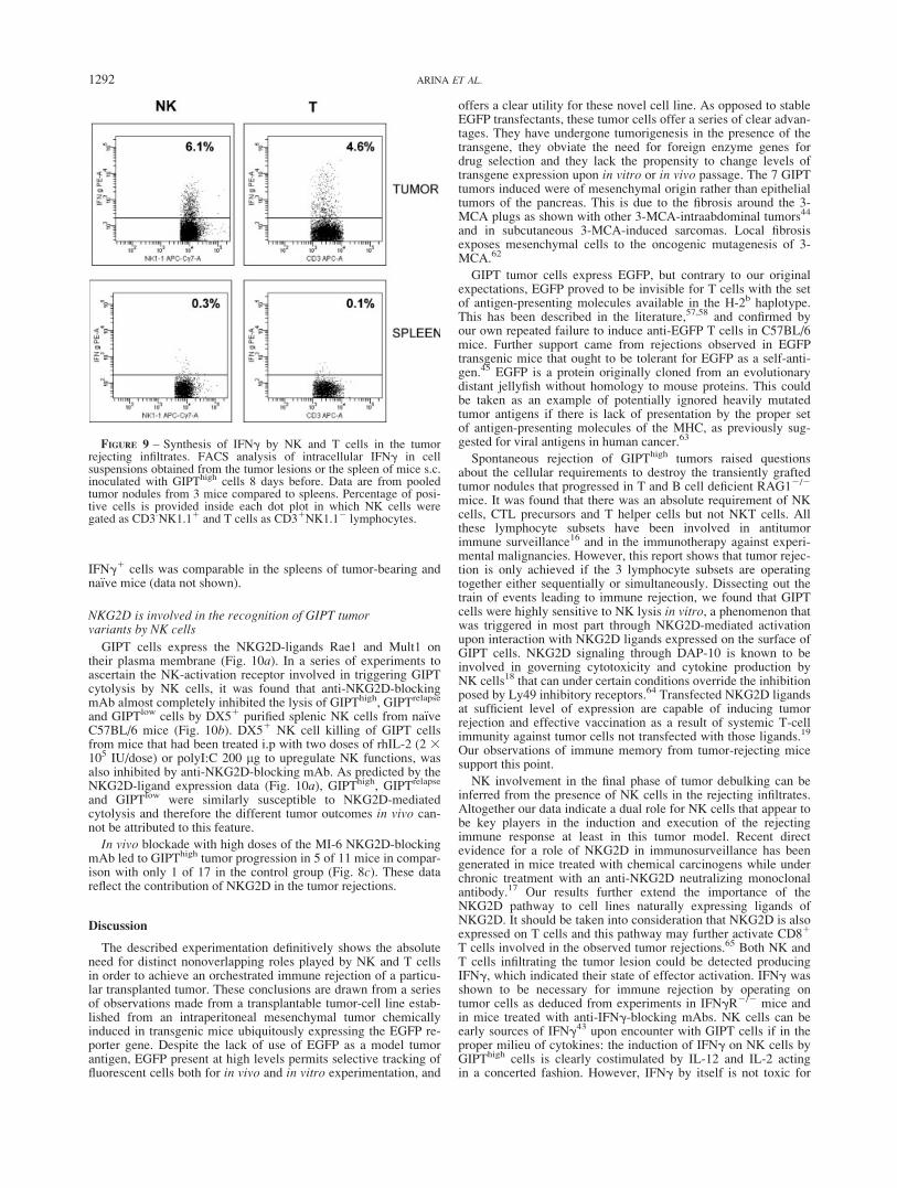
IFNg1 cells was comparable in the spleens of tumor-bearing andna€ıve mice (data not shown).
NKG2D is involved in the recognition of GIPT tumorvariants by NK cells
GIPT cells express the NKG2D-ligands Rae1 and Mult1 ontheir plasma membrane (Fig. 10a). In a series of experiments toascertain the NK-activation receptor involved in triggering GIPTcytolysis by NK cells, it was found that anti-NKG2D-blockingmAb almost completely inhibited the lysis of GIPThigh, GIPTrelapse
and GIPTlow cells by DX51 purified splenic NK cells from na€ıveC57BL/6 mice (Fig. 10b). DX51 NK cell killing of GIPT cellsfrom mice that had been treated i.p with two doses of rhIL-2 (2 3105 IU/dose) or polyI:C 200 lg to upregulate NK functions, wasalso inhibited by anti-NKG2D-blocking mAb. As predicted by theNKG2D-ligand expression data (Fig. 10a), GIPThigh, GIPTrelapse
and GIPTlow were similarly susceptible to NKG2D-mediatedcytolysis and therefore the different tumor outcomes in vivo can-not be attributed to this feature.
In vivo blockade with high doses of the MI-6 NKG2D-blockingmAb led to GIPThigh tumor progression in 5 of 11 mice in compar-ison with only 1 of 17 in the control group (Fig. 8c). These datareflect the contribution of NKG2D in the tumor rejections.
Discussion
The described experimentation definitively shows the absoluteneed for distinct nonoverlapping roles played by NK and T cellsin order to achieve an orchestrated immune rejection of a particu-lar transplanted tumor. These conclusions are drawn from a seriesof observations made from a transplantable tumor-cell line estab-lished from an intraperitoneal mesenchymal tumor chemicallyinduced in transgenic mice ubiquitously expressing the EGFP re-porter gene. Despite the lack of use of EGFP as a model tumorantigen, EGFP present at high levels permits selective tracking offluorescent cells both for in vivo and in vitro experimentation, and
offers a clear utility for these novel cell line. As opposed to stableEGFP transfectants, these tumor cells offer a series of clear advan-tages. They have undergone tumorigenesis in the presence of thetransgene, they obviate the need for foreign enzyme genes fordrug selection and they lack the propensity to change levels oftransgene expression upon in vitro or in vivo passage. The 7 GIPTtumors induced were of mesenchymal origin rather than epithelialtumors of the pancreas. This is due to the fibrosis around the 3-MCA plugs as shown with other 3-MCA-intraabdominal tumors44
and in subcutaneous 3-MCA-induced sarcomas. Local fibrosisexposes mesenchymal cells to the oncogenic mutagenesis of 3-MCA.62
GIPT tumor cells express EGFP, but contrary to our originalexpectations, EGFP proved to be invisible for T cells with the setof antigen-presenting molecules available in the H-2b haplotype.This has been described in the literature,57,58 and confirmed byour own repeated failure to induce anti-EGFP T cells in C57BL/6mice. Further support came from rejections observed in EGFPtransgenic mice that ought to be tolerant for EGFP as a self-anti-gen.45 EGFP is a protein originally cloned from an evolutionarydistant jellyfish without homology to mouse proteins. This couldbe taken as an example of potentially ignored heavily mutatedtumor antigens if there is lack of presentation by the proper setof antigen-presenting molecules of the MHC, as previously sug-gested for viral antigens in human cancer.63
Spontaneous rejection of GIPThigh tumors raised questionsabout the cellular requirements to destroy the transiently graftedtumor nodules that progressed in T and B cell deficient RAG12/2
mice. It was found that there was an absolute requirement of NKcells, CTL precursors and T helper cells but not NKT cells. Allthese lymphocyte subsets have been involved in antitumorimmune surveillance16 and in the immunotherapy against experi-mental malignancies. However, this report shows that tumor rejec-tion is only achieved if the 3 lymphocyte subsets are operatingtogether either sequentially or simultaneously. Dissecting out thetrain of events leading to immune rejection, we found that GIPTcells were highly sensitive to NK lysis in vitro, a phenomenon thatwas triggered in most part through NKG2D-mediated activationupon interaction with NKG2D ligands expressed on the surface ofGIPT cells. NKG2D signaling through DAP-10 is known to beinvolved in governing cytotoxicity and cytokine production byNK cells18 that can under certain conditions override the inhibitionposed by Ly49 inhibitory receptors.64 Transfected NKG2D ligandsat sufficient level of expression are capable of inducing tumorrejection and effective vaccination as a result of systemic T-cellimmunity against tumor cells not transfected with those ligands.19
Our observations of immune memory from tumor-rejecting micesupport this point.
NK involvement in the final phase of tumor debulking can beinferred from the presence of NK cells in the rejecting infiltrates.Altogether our data indicate a dual role for NK cells that appear tobe key players in the induction and execution of the rejectingimmune response at least in this tumor model. Recent directevidence for a role of NKG2D in immunosurveillance has beengenerated in mice treated with chemical carcinogens while underchronic treatment with an anti-NKG2D neutralizing monoclonalantibody.17 Our results further extend the importance of theNKG2D pathway to cell lines naturally expressing ligands ofNKG2D. It should be taken into consideration that NKG2D is alsoexpressed on T cells and this pathway may further activate CD81
T cells involved in the observed tumor rejections.65 Both NK andT cells infiltrating the tumor lesion could be detected producingIFNg, which indicated their state of effector activation. IFNg wasshown to be necessary for immune rejection by operating ontumor cells as deduced from experiments in IFNgR2/2 mice andin mice treated with anti-IFNg-blocking mAbs. NK cells can beearly sources of IFNg43 upon encounter with GIPT cells if in theproper milieu of cytokines: the induction of IFNg on NK cells byGIPThigh cells is clearly costimulated by IL-12 and IL-2 actingin a concerted fashion. However, IFNg by itself is not toxic for
FIGURE 9 – Synthesis of IFNg by NK and T cells in the tumorrejecting infiltrates. FACS analysis of intracellular IFNg in cellsuspensions obtained from the tumor lesions or the spleen of mice s.c.inoculated with GIPThigh cells 8 days before. Data are from pooledtumor nodules from 3 mice compared to spleens. Percentage of posi-tive cells is provided inside each dot plot in which NK cells weregated as CD3-NK1.11 and T cells as CD31NK1.12 lymphocytes.
1292 ARINA ET AL.

GIPThigh cells in culture. This suggests that IFNg acting on tumorcells elicits indirect mechanisms of immune destruction that areoperational in the in vivo setting. The role of NK cells in initiatingand shaping the early immune response has been shown byMartin-Fontecha et al.,43 who clearly demonstrated an involve-ment of NK cells in the differentiation of Th0 to Th1 cells as wellas in the dramatic increase in the number of lymphocytes insidethe responding lymphoid tissue.66 IFNg secreted by NK cells is atleast one of the factors involved in those effects.66 In our system,the IFNgR is needed on tumor cells but not on cells of the hostmouse. Our data are fully consistent with the proposed role ofIFNg in immunosurveillance, shown to operate on the nascent ma-lignant cells,67 but not on the immune system cells.
Taking advantage of the fluorescence of GIPT cells, we showthat DC acquire fluorescent material from GIPT cells both in vivoand in vitro. Surprisingly, NK killing of tumor cells was dispensa-ble for this in vitro transfer of biological material from tumor cellsto splenic DC, as previously shown by other authors with humanDC and viable tumor cells.68 In this study, we show thatCD11chigh cells can be detected at the tumor infiltrate carryingEGFP fluorescence uptaken from GIPThigh cells. Furthermore,in vivo depletion of NK cells did not impair the uptake of EGFPby DC, thus confirming that generation of tumor-cell debris byNK-mediated cytotoxicity was not required for these DC loadingevents (data not shown). The intense fluorescence of tumor cells
was also exploited to demonstrate that GIPThigh cells reach LNsoon after inoculation of tumor cells suspensions as previously de-scribed for other tumor cells.40
The final execution of tumor rejection, at least in syngeneicmice, is most likely mediated by T cells and NK cells, which arealso located in the inflammatory infiltrate of the about-to-berejected tumor. The contribution of NK cells is clearly neededduring the effector phase, since NK depletion as late as on day 8results in lethal tumor growth. Resting NK cells are capable todegranulate in vitro in the presence of GIPThigh cells while CD81
cells are not. This suggests an involvement of NK cells at earlypoints of time in the orchestration of tumor rejection. The in vivorole of CD81 CTL could include direct tumor-cell destructionenhanced by IFNg that upregulates MHC-I on tumor cells. Therole of NK cells in favoring the priming of CTLs has been previ-ously observed in vitro.31 CD41 cells could act as helpers for CD8immunity but could also be important for inflicting local damageon the malignant tissue when activated by stromal cells expressingMHC class II,30 as recently observed in other tumor models.69 Inaddition, we can detect eosinophils at the tumor infiltrates andtheir role is being currently explored, since implication of eosino-phils in tumor rejections has been described for tumors undergoinginterleukin-4-based immunotherapy.70
This study supports the notion that in vivo rejection of mesen-chymal tumors requires the combined action of NK and T cells
FIGURE 10 – NKG2D role in NKcell killing of GIPThigh cells and intumor rejection. (a) FACS analysishistograms showing surface ex-pression of the NKG2D ligandsRae1 and Mult1 on the 3 GIPT clo-nal variants. (b) 5-hr 51Cr releaseassays with DX5 magnetic bead-purified NK cells from the spleensof C57BL/6 mice either naive orpreinjected with of IL-2 or PolyI:Cas indicated. NK cells were con-fronted with GIPThigh, GIPTrelapse
or GIPTlow cells at 100:1 effector:-target ratio, in the presence of con-trol antibody (white bars) or ablocking mAb against NKG2D(MI-6) at 20 lg/ml (black bars). (c)Individual tumor diameter follow-up of mice not treated (N 5 11) ortreated with control rat IgG (N 56) or MI-6 mAb (N 5 11), on day21 before GIPThigh tumor inocula-tion and after that every 3–4 daysduring 3 weeks. The relative num-ber of mice with progressing lethaltumors is shown as a fraction in thegraph (p 5 0.022 using a Fisherexact test).
1293NK AND T CELL ORCHESTRATED TUMOR REJECTION

and is dependent on NKG2D ligand recognition and production ofIFNg. The use of NK cells in cancer immunotherapy has receiveda boost as a result of studies into haploidentical bone marrowtransplantation71 and adoptive transfer of activated NK cells.72
Knowledge showing direct involvement of NK cells in orchestrat-ing the induction of a curative immune response in our systemoffers grounds for protocols of gene transfection of NKG2D-ligands before adoptively infusing NK cells. NK cells operatingby their direct activity on tumor cells, their crosstalk with DC and/or their interplay with T lymphocytes all need to be consideredwhen designing immunotherapy strategies for cancer.
Acknowledgements
Critical reading and scientific discussion and advice byDr. M.D. Lozano, Dr. M. Bustos, Mrs. C. Miqueo and Mrs. A.Borja-Lopetegi are acknowledged, as well as technical assistancefrom Mrs. Izaskun Gabari, Mrs. M.D. Mart�ınez and Mrs. MartaRuiz. We also thank Drs. Okabe and Carbone for their generousgifts of mutant mice. Excellent animal facility assistance by Mr.Juan Percaz and Mr. Javier Guill�en is also acknowledged.
A.A. receives a scholarship from Instituto de Salud Carlos III(BEFI) and O.M. is supported by the Spanish Ministry ofEducation.
References
1. Diefenbach A, Raulet DH. The innate immune response to tumors andits role in the induction of T-cell immunity. Immunol Rev 2002;188:9–21.
2. Wallace ME, Smyth MJ. The role of natural killer cells in tumor con-trol-effectors and regulators of adaptive immunity. Springer SeminImmunopathol 2005;27:49–64.
3. Kim S, Iizuka K, Aguila HL, Weissman IL, Yokoyama WM. In vivonatural killer cell activities revealed by natural killer cell-deficientmice. Proc Natl Acad Sci USA 2000;97:2731–6.
4. Scott P, Trinchieri G. The role of natural killer cells in host-parasiteinteractions. Curr Opin Immunol 1995;7:34–40.
5. Lanier LL. NK cell recognition. Annu Rev Immunol 2005;23:225–74.
6. Trinchieri G. Interleukin-12: a cytokine at the interface of inflamma-tion and immunity. Adv Immunol 1998;70:83–243.
7. Wu TC, Huang AY, Jaffee EM, Levitsky HI, Pardoll DM. A reassess-ment of the role of B7-1 expression in tumor rejection. J Exp Med1995;182:1415–21.
8. Rodriguez-Calvillo M, Duarte M, Tirapu I, Berraondo P, MazzoliniG, Qian C, Prieto J, Melero I. Upregulation of natural killer cells func-tions underlies the efficacy of intratumorally injected dendritic cellsengineered to produce interleukin-12. Exp Hematol 2002;30:195–204.
9. Murillo O, Arina A, Tirapu I, Alfaro C, Mazzolini G, Palencia B,Lopez-Diaz De Cerio A, Prieto J, Bendandi M, Melero I. Potentiationof therapeutic immune responses against malignancies with monoclo-nal antibodies. Clin Cancer Res 2003;9:5454–64.
10. Meazza R, Lollini PL, Nanni P, De Giovanni C, Gaggero A, ComesA, Cilli M, Di Carlo E, Ferrini S, Musiani P. Gene transfer of a secre-table form of IL-15 in murine adenocarcinoma cells: effects on tumor-igenicity, metastatic potential and immune response. Int J Cancer2000;87:574–81.
11. Wang G, Tschoi M, Spolski R, Lou Y, Ozaki K, Feng C, Kim G,Leonard WJ, Hwu P. In vivo antitumor activity of interleukin 21mediated by natural killer cells. Cancer Res 2003;63:9016–22.
12. Kelly JM, Darcy PK, Markby JL, Godfrey DI, Takeda K, Yagita H,Smyth MJ. Induction of tumor-specific T cell memory by NK cell-mediated tumor rejection. Nat Immunol 2002;3:83–90.
13. Melero I, Johnston JV, Shufford WW, Mittler RS, Chen L. NK1.1cells express 4-1BB (CDw137) costimulatory molecule and arerequired for tumor immunity elicited by anti-4-1BB monoclonal anti-bodies. Cell Immunol 1998;190:167–72.
14. Wilcox RA, Tamada K, Strome SE, Chen L. Signaling through NKcell-associated CD137 promotes both helper function for CD81 cyto-lytic T cells and responsiveness to IL-2 but not cytolytic activity.J Immunol 2002;169:4230–6.
15. Rosenberg SA, Lotze MT, Muul LM, Chang AE, Avis FP, Leitman S,Linehan WM, Robertson CN, Lee RE, Rubin JT, Seipp CA, SimpsonCG, White DE. A progress report on the treatment of 157 patients withadvanced cancer using lymphokine-activated killer cells and interleu-kin-2 or high-dose interleukin-2 alone. N Engl J Med 1987;316:889–97.
16. Dunn GP, Old LJ, Schreiber RD. The immunobiology of cancerimmunosurveillance and immunoediting. Immunity 2004;21:137–48.
17. Smyth MJ, Swann J, Cretney E, Zerafa N, Yokoyama WM, HayakawaY. NKG2D function protects the host from tumor initiation. J ExpMed 2005;202:583–8.
18. Raulet DH. Roles of the NKG2D immunoreceptor and its ligands. NatRev Immunol 2003;3:781–90.
19. Diefenbach A, Jensen ER, Jamieson AM, Raulet DH. Rae1 and H60ligands of the NKG2D receptor stimulate tumour immunity. Nature2001;413:165–71.
20. Oppenheim DE, Roberts SJ, Clarke SL, Filler R, Lewis JM, TigelaarRE, Girardi M, Hayday AC. Sustained localized expression of ligandfor the activating NKG2D receptor impairs natural cytotoxicity invivo and reduces tumor immunosurveillance. Nat Immunol 2005;6:928–37.
21. Groh V, Wu J, Yee C, Spies T. Tumour-derived soluble MIC ligandsimpair expression of NKG2D and T-cell activation. Nature 2002;419:734–8.
22. Wiemann K, Mittrucker HW, Feger U, Welte SA, Yokoyama WM,Spies T, Rammensee HG, Steinle A. Systemic NKG2D down-regula-tion impairs NK and CD8 T cell responses in vivo. J Immunol2005;175:720–9.
23. Gasser S, Orsulic S, Brown EJ, Raulet DH. The DNA damage path-way regulates innate immune system ligands of the NKG2D receptor.Nature 2005;436:1186–90.
24. Armeanu S, Bitzer M, Lauer UM, Venturelli S, Pathil A, Krusch M,Kaiser S, Jobst J, Smirnow I, Wagner A, Steinle A, Salih HR. Naturalkiller cell-mediated lysis of hepatoma cells via specific induction ofNKG2D ligands by the histone deacetylase inhibitor sodiumvalproate. Cancer Res 2005;65:6321–9.
25. Fernandez NC, Treiner E, Vance RE, Jamieson AM, Lemieux S,Raulet DH. A subset of natural killer cells achieves self-tolerancewithout expressing inhibitory receptors specific for self-MHC mole-cules. Blood 2005;105:4416–23.
26. O’Leary J G, Goodarzi M, Drayton DL, von Andrian UH. T cell- andB cell-independent adaptive immunity mediated by natural killercells. Nat Immunol 2006;7:507–16.
27. Sandel MH, Dadabayev AR, Menon AG, Morreau H, Melief CJ,Offringa R, van der Burg SH, Janssen-van Rhijn CM, Ensink NG,Tollenaar RA, van de Velde CJ, et al. Prognostic value of tumor-infiltrating dendritic cells in colorectal cancer: role of maturation statusand intratumoral localization. Clin Cancer Res 2005;11:2576–82.
28. Kawano T, Cui J, Koezuka Y, Toura I, Kaneko Y, Motoki K, Ueno H,Nakagawa R, Sato H, Kondo E, Koseki H, Taniguchi M. CD1d-restricted and TCR-mediated activation of valpha14 NKT cells byglycosylceramides. Science 1997;278:1626–9.
29. Casares N, Arribillaga L, Sarobe P, Dotor J, Lopez-Diaz de Cerio A,Melero I, Prieto J, Borras-Cuesta F, Lasarte JJ. CD41/CD251 regula-tory cells inhibit activation of tumor-primed CD41 T cells with IFN-gamma-dependent antiangiogenic activity, as well as long-lastingtumor immunity elicited by peptide vaccination. J Immunol 2003;171:5931–9.
30. Corthay A, Skovseth DK, Lundin KU, Rosjo E, Omholt H, HofgaardPO, Haraldsen G, Bogen B. Primary antitumor immune responsemediated by CD41 T cells. Immunity 2005;22:371–83.
31. Tanaka F, Hashimoto W, Okamura H, Robbins PD, Lotze MT, TaharaH. Rapid generation of potent and tumor-specific cytotoxic T lympho-cytes by interleukin 18 using dendritic cells and natural killer cells.Cancer Res 2000;60:4838–44.
32. Fernandez NC, Lozier A, Flament C, Ricciardi-Castagnoli P, BelletD, Suter M, Perricaudet M, Tursz T, Maraskovsky E, Zitvogel L.Dendritic cells directly trigger NK cell functions: cross-talk relevant ininnate anti-tumor immune responses in vivo. Nat Med 1999;5:405–11.
33. Borg C, Jalil A, Laderach D, Maruyama K, Wakasugi H, Charrier S,Ryffel B, Cambi A, Figdor C, Vainchenker W, Galy A, Caignard A,et al. NK cell activation by dendritic cells (DCs) requires the forma-tion of a synapse leading to IL-12 polarization in DCs. Blood2004;104:3267–75.
34. Gerosa F, Baldani-Guerra B, Nisii C, Marchesini V, Carra G,Trinchieri G. Reciprocal activating interaction between natural killercells and dendritic cells. J Exp Med 2002;195:327–33.
35. Ferlazzo G, Tsang ML, Moretta L, Melioli G, Steinman RM, Munz C.Human dendritic cells activate resting natural killer (NK) cells andare recognized via the NKp30 receptor by activated NK cells. J ExpMed 2002;195:343–51.
36. Moretta A. Natural killer cells and dendritic cells: rendezvous inabused tissues. Nat Rev Immunol 2002;2:957–64.
37. Bajenoff M, Breart B, Huang AY, Qi H, Cazareth J, Braud VM,Germain RN, Glaichenhaus N. Natural killer cell behavior in lymphnodes revealed by static and real-time imaging. J Exp Med 2006;203:619–31.
1294 ARINA ET AL.
38. Larsson M, Fonteneau JF, Bhardwaj N. Dendritic cells resurrect anti-gens from dead cells. Trends Immunol 2001;22:141–8.
39. Binder RJ, Srivastava PK. Peptides chaperoned by heat-shock proteinsare a necessary and sufficient source of antigen in the cross-primingof CD81 T cells. Nat Immunol 2005;6:593–9.
40. Ochsenbein AF, Klenerman P, Karrer U, Ludewig B, Pericin M,Hengartner H, Zinkernagel RM. Immune surveillance against a solidtumor fails because of immunological ignorance. Proc Natl Acad SciUSA 1999;96:2233–8.
41. Allan RS, Smith CM, Belz GT, van Lint AL, Wakim LM, Heath WR,Carbone FR. Epidermal viral immunity induced by CD8g1 dendriticcells but not by Langerhans cells. Science 2003;301:1925–8.
42. Huarte E, Tirapu I, Arina A, Vera M, Alfaro C, Murillo O, PalenciaB, Busto V, Marin V, Mazzolini G, Melero I. Intratumoural adminis-tration of dendritic cells: hostile environment and help by gene ther-apy. Expert Opin Biol Ther 2005;5:7–22.
43. Martin-Fontecha A, Thomsen LL, Brett S, Gerard C, Lipp M,Lanzavecchia A, Sallusto F. Induced recruitment of NK cells tolymph nodes provides IFN-gamma for T(H)1 priming. Nat Immunol2004;5:1260–5.
44. Corbett TH, Roberts BJ, Leopold WR, Peckham JC, Wilkoff LJ,Griswold DP, Jr, Schabel FM, Jr. Induction and chemotherapeuticresponse of two transplantable ductal adenocarcinomas of the pan-creas in C57BL/6 mice. Cancer Res 1984;44:717–26.
45. Okabe M, Ikawa M, Kominami K, Nakanishi T, Nishimune Y. �Greenmice� as a source of ubiquitous green cells. FEBS Lett 1997;407:313–9.
46. Mendiratta SK, Martin WD, Hong S, Boesteanu A, Joyce S, Van KaerL. CD1d1 mutant mice are deficient in natural T cells that promptlyproduce IL-4. Immunity 1997;6:469–77.
47. Hogquist KA, Jameson SC, Heath WR, Howard JL, Bevan MJ,Carbone FR. T cell receptor antagonist peptides induce positive selec-tion. Cell 1994;76:17–27.
48. Jamieson AM, Diefenbach A, McMahon CW, Xiong N, Carlyle JR,Raulet DH. The role of the NKG2D immunoreceptor in immune cellactivation and natural killing. Immunity 2002;17:19–29.
49. He Y, Pimenov AA, Nayak JV, Plowey J, Falo LD, Jr, Huang L. Intra-venous injection of naked DNA encoding secreted flt3 ligand dramati-cally increases the number of dendritic cells and natural killer cells invivo. Hum Gene Ther 2000;11:547–54.
50. Basse PH, Whiteside TL, Herberman RB. Use of activated naturalkiller cells for tumor immunotherapy in mouse and human. MethodsMol Biol 2000;121:81–94.
51. Gough M, Crittenden M, Thanarajasingam U, Sanchez-Perez L,Thompson J, Jevremovic D, Vile R. Gene therapy to manipulateeffector T cell trafficking to tumors for immunotherapy. J Immunol2005;174:5766–73.
52. Perez-Villar JJ, Melero I, Navarro F, Carretero M, Bellon T, Llano M,Colonna M, Geraghty DE, Lopez-Botet M. The CD94/NKG2-A in-hibitory receptor complex is involved in natural killer cell-mediatedrecognition of cells expressing HLA-G1. J Immunol 1997;158:5736–43.
53. Casares N, Lasarte JJ, de Cerio AL, Sarobe P, Ruiz M, Melero I,Prieto J, Borras-Cuesta F. Immunization with a tumor-associated CTLepitope plus a tumor-related or unrelated Th1 helper peptide elicitsprotective CTL immunity. Eur J Immunol 2001;31:1780–9.
54. Alexander J, Sidney J, Southwood S, Ruppert J, Oseroff C, MaewalA, Snoke K, Serra HM, Kubo RT, Sette A, Grey HM. Development ofhigh potency universal DR-restricted helper epitopes by modificationof high affinity DR-blocking peptides. Immunity 1994;1:751–61.
55. Rodriguez-Calvillo M, Gabari I, Duarte M, Mazzolini G, Rifon J,Rocha E, Prieto J, Melero I. Thrombopenic purpura induced by amonoclonal antibody directed to a 35-kilodalton surface protein (p35)
expressed on murine platelets and endothelial cells. Exp Hematol2001;29:589–95.
56. Alter G, Malenfant JM, Altfeld M. CD107a as a functional marker forthe identification of natural killer cell activity. J Immunol Methods2004;294:15–22.
57. Denaro M, Oldmixon B, Patience C, Andersson G, Down J. EGFP-transduced EL-4 cells from tumors in C57BL/6 mice. Gene Ther2001;8:1814–5.
58. Skelton D, Satake N, Kohn DB. The enhanced green fluorescent pro-tein (eGFP) is minimally immunogenic in C57BL/6 mice. Gene Ther2001;8:1813–4.
59. Gambotto A, Dworacki G, Cicinnati V, Kenniston T, Steitz J, TutingT, Robbins PD, DeLeo AB. Immunogenicity of enhanced green fluo-rescent protein (EGFP) in BALB/c mice: identification of an H2-Kd-restricted CTL epitope. Gene Ther 2000;7:2036–40.
60. Mercurio AM, Schwarting GA, Robbins PW. Glycolipids of themouse peritoneal macrophage. Alterations in amount and surfaceexposure of specific glycolipid species occur in response to inflamma-tion and tumoricidal activation. J Exp Med 1984;160:1114–25.
61. Suttles J, Schwarting GA, Stout RD. Flow cytometric analysis revealsthe presence of asialo GM1 on the surface membrane of alloimmunecytotoxic T lymphocytes. J Immunol 1986;136:1586–91.
62. Qin Z, Kim HJ, Hemme J, Blankenstein T. Inhibition of methylcho-lanthrene-induced carcinogenesis by an interferon gamma receptor-dependent foreign body reaction. J Exp Med 2002;195:1479–90.
63. Ellis JR, Keating PJ, Baird J, Hounsell EF, Renouf DV, Rowe M, Hop-kins D, Duggan-Keen MF, Bartholomew JS, Young LS, Stern PL. Theassociation of an HPV16 oncogene variant with HLA-B7 has implica-tions for vaccine design in cervical cancer. Nat Med 1995;1:464–70.
64. Regunathan J, Chen Y, Wang D, Malarkannan S. NKG2D receptor-mediated NK cell function is regulated by inhibitory Ly49 receptors.Blood 2005;105:233–40.
65. Ehrlich LI, Ogasawara K, Hamerman JA, Takaki R, Zingoni A,Allison JP, Lanier LL. Engagement of NKG2D by cognate ligand orantibody alone is insufficient to mediate costimulation of human andmouse CD81 T cells. J Immunol 2005;174:1922–31.
66. Martin-Fontecha A, Sebastiani S, Hopken UE, Uguccioni M, Lipp M,Lanzavecchia A, Sallusto F. Regulation of dendritic cell migration tothe draining lymph node: impact on T lymphocyte traffic and priming.J Exp Med 2003;198:615–21.
67. Shankaran V, Ikeda H, Bruce AT, White JM, Swanson PE, Old LJ,Schreiber RD. IFNgamma and lymphocytes prevent primary tumourdevelopment and shape tumour immunogenicity. Nature 2001;410:1107–11.
68. Russo V, Zhou D, Sartirana C, Rovere P, Villa A, Rossini S, TraversariC, Bordignon C. Acquisition of intact allogeneic human leukocyte anti-gen molecules by human dendritic cells. Blood 2000; 95:3473–7.
69. Hung K, Hayashi R, Lafond-Walker A, Lowenstein C, Pardoll D,Levitsky H. The central role of CD4(1) T cells in the antitumorimmune response. J Exp Med 1998;188:2357–68.
70. Tepper RI, Coffman RL, Leder P. An eosinophil-dependentmechanism for the antitumor effect of interleukin-4. Science 1992;257:548–51.
71. Ruggeri L, Capanni M, Urbani E, Perruccio K, Shlomchik WD, TostiA, Posati S, Rogaia D, Frassoni F, Aversa F, Martelli MF, Velardi A.Effectiveness of donor natural killer cell alloreactivity in mismatchedhematopoietic transplants. Science 2002;295:2097–100.
72. Miller JS, Soignier Y, Panoskaltsis-Mortari A, McNearney SA, YunGH, Fautsch SK, McKenna D, Le C, Defor TE, Burns LJ, Orchard PJ,Blazar BR, et al. Successful adoptive transfer and in vivo expansionof human haploidentical NK cells in patients with cancer. Blood2005; 105:3051–7.
1295NK AND T CELL ORCHESTRATED TUMOR REJECTION
Copyright © 2022 FDOKUMEN