Beggar Thy Neighbour: British Imports during the Inter-War Years and the effect of the 1932 tariff

Neuron Glia Biologyhttp://journals.cambridge.org/NGB
Additional services for Neuron Glia Biology:
Email alerts: Click hereSubscriptions: Click hereCommercial reprints: Click hereTerms of use : Click here
The effects of LNAME on neuronal NOS and SOD1 expression in the DRG–spinal cord network of axotomised Thy 1.2 eGFP mice
Matthew J. G. Bradman, Richard Morris, Anne McArdle, Malcolm J. Jackson and Thimmasettappa Thippeswamy
Neuron Glia Biology / Volume 7 / Issue 24 / May 2011, pp 129 141DOI: 10.1017/S1740925X12000051, Published online: 22 May 2012
Link to this article: http://journals.cambridge.org/abstract_S1740925X12000051
How to cite this article:Matthew J. G. Bradman, Richard Morris, Anne McArdle, Malcolm J. Jackson and Thimmasettappa Thippeswamy (2011). The effects of LNAME on neuronal NOS and SOD1 expression in the DRG–spinal cord network of axotomised Thy 1.2 eGFP mice. Neuron Glia Biology, 7, pp 129141 doi:10.1017/S1740925X12000051
Request Permissions : Click here
Downloaded from http://journals.cambridge.org/NGB, IP address: 138.253.100.121 on 22 Apr 2013
The effects of L-NAME on neuronalNOS and SOD1 expression in theDRG–spinal cord network of axotomisedThy 1.2 eGFP mice
matthew j. g. bradman, richard morris, anne mcardle, malcolm j. jackson
and thimmasettappa thippeswamy
Nitric oxide (NO) plays an important role in pathophysiology of the nervous system. Copper/zinc superoxide dismutase(SOD1) reacts with superoxide, which is also a substrate for NO, to provide antioxidative protection. NO production isgreatly altered following nerve injury, therefore we hypothesised that SOD1 and NO may be involved in modulatingaxotomy responses in dorsal root ganglion (DRG)–spinal network. To investigate this interaction, adult Thy1.2 enhancedmembrane-bound green fluorescent protein (eGFP) mice underwent sciatic nerve axotomy and received NG-nitro-,L-arginine methylester (L-NAME) or vehicle 7–9 days later. L4–L6 spinal cord and DRG were harvested for immuno-histochemical analyses. Effect of injury was confirmed by axotomy markers; small proline-rich repeat protein 1A(SPRR1A) was restricted to ipsilateral neuropathology, while Thy1.2 eGFP revealed also contralateral crossover effects.L-NAME, but not axotomy, increased neuronal NO synthase (nNOS) and SOD1 immunoreactive neurons, with nocolocalisation, in a lamina-dependent manner in the dorsal horn of the spinal cord. Axotomy and/or L-NAME had noeffect on total nNOS+ and SOD1+ neurons in DRG. However, L-NAME altered SOD1 expression in subsets of axotomisedDRG neurons. These findings provide evidence for differential distribution of SOD1 and its modulation by NO, which mayinteract to regulate axotomy-induced changes in DRG–spinal network.
Keywords: Nitric oxide signalling, nerve injury, neuroprotection, NOS inhibition, superoxide
I N T R O D U C T I O N
The superoxide dismutases (SOD) comprise a group ofenzymes capable of reacting preferentially with superoxideradicals to provide antioxidative protection (Imlay et al.,1988; Imlay and Linn, 1988) to cells and tissues in whichthey are produced (Orr and Sohal, 1994). Cu/ZnSOD, thefirst intracellular isoform to be discovered, is also known asSOD1, and historically as SOD3 in humans (Perry et al.,2010). In the central nervous system, SOD1 has been reportedin astrocytes, primarily the cytoplasm and processes, and to alesser extent in motor neurons and other neurons (Noacket al., 1998; Lindenau et al., 2000). A study of immaturerats, from P2 to P7, reported SOD1 in neuronal cell bodiesand processes, as well as in glia and the eppendymal cells ofthe central canal of the spinal cord (Rogerio et al., 2005).
SOD1 is essential for normal function of the nervoussystem. In human, SOD1 mutation causes motor neurondegeneration as in familial amyotrophic lateral sclerosis(Deng et al., 1993). However, little is known about the roleof normal SOD1 in sensory pathways following peripheralnerve injury. Rosenfeld et al. (1997) reported no changes in
SOD1, but increased MnSOD (SOD2), 12 days post-axotomyin adult rats. In neonates, increased SOD1 protein in spinalcord homogenates was found following axotomy comparedto unlesioned controls, but no change in SOD1 or SOD2expression patterns were seen up to 7 days post-axotomy(Rogerio et al., 2005).
The biochemistry of superoxide is closely linked to themost studied reactive oxygen species, nitric oxide (NO),which is produced from the substrate L-arginine by NOsynthase (Stamler et al., 1992; Moncada and Higgs, 2006).Since recognition of NO as a signalling molecule, three iso-forms of NO synthase have been described, namely endo-thelial, inducible and neuronal NO synthase (eNOS, iNOSand nNOS, respectively). Following axotomy, nNOS isgreatly increased in rat dorsal root ganglion (DRG) neuronsby 7–8 days (Verge et al., 1992; Zhang et al., 1993;Thippeswamy et al., 2001). Neuronal NOS is also altered incentral terminals of DRG neurons in the dorsal horn of thespinal cord, and in neuronal somata in laminae (L) I, II andVI, and also L X surrounding the central canal of rats andmice following axotomy (Keilhoff et al., 2004; Freire et al.,2009). This suggests that NO released in the DRG–spinalcord network might play an important role in sensory proces-sing. However, the distribution of nNOS and SOD withrespect to peripheral axotomy is not fully understood. Thereis evidence to suggest that NOS functionality may beSOD-dependent (Hobbs et al., 1994; Schmidt et al., 1996)
Corresponding author:Thimmasettappa ThippeswamyEmail: [email protected]
129
Neuron Glia Biology, 2011, 7(2–4), 129–141. # Cambridge University Press, 2012doi:10.1017/S1740925X12000051

and that their relative level may determine the formation ofperoxynitrite or nitrotyrosine (Beckman et al., 1993; Huieand Padmaja, 1993; Wink and Mitchell, 1998).
In this study, we examined the changes in distribution ofSOD1 and nNOS in DRG–spinal cord network following per-ipheral axotomy in transgenic mice in which membrane-linked enhanced membrane-bound green fluorescent protein(eGFP) driven by Thy1.2 promoter in a subset of DRGneurons and central terminals. eGFP combined with anaxotomy marker, small proline-rich repeat protein 1A(SPRR1A) immunoreactivity (Bonilla et al., 2002; Starkeyet al., 2009), enabled identification of central terminals of axo-tomised DRG neurons. Axotomised mice were treated withthe NOS inhibitor, LNG-nitroarginine methyl ester(L-NAME) or vehicle, which was administered to coincidewith previously reported peaks in NO production by 7–8days post-injury (Verge et al., 1992; Thippeswamy et al.,2001, 2007; Cheng et al., 2007; Bradman et al., 2010), tounderstand whether altered NO levels affect SOD1 distri-bution. Furthermore, the response of nNOS to axotomy hasbeen contentious within the literature (Callsen-Cencic et al.,1999; Cheng et al., 2007; Bradman et al., 2010), possiblybecause of the great degree of anatomical and physiologicalspecificity between neurons which respond as heterogenouspopulations within the dorsal horn (Maxwell et al., 2007). Inview of this, the present study is focused on the effects ofaxotomy and NO modulation within spinal cord regions,defined by denervation and DRG central input, to understandSOD1–NO interaction.
O B J E C T I V E
The objective of this study was to examine the effects of sciaticnerve axotomy and/or NOS inhibition on DRG–spinal cordnetwork in a transgenic mouse that was known to produceenhanced GFP (eGFP) driven by Thy1.2 promoter.Superoxide and NO play important roles in signal transduc-tion in the nervous system. Based on the review of the litera-ture, including our own, it was known that axotomy inducesnNOS upregulation in rat DRG (�45%) and to a lesserextent in wild-type C57Bl/6 mouse DRG (�23%). In Thy1.2 eGFP transgenic mouse (with C57Bl/6 background), theeGFP marks a subset of DRG neurons and their central term-inals, and is known to be useful in studying the effects ofaxotomy. The hypothesis was that axotomy and/orL-NAME alter the expression of SOD1 and nNOS in DRG–spinal cord network, and eGFP would serve as readout foraxotomy-induced changes. Axotomised mice were treatedwith the NOS inhibitor, L-NAME or vehicle to coincidewith previously reported peaks in nNOS upregulation at 7–9days post-axotomy. Immunohistochemical analyses, to under-stand whether axotomy or L-NAME affects SOD1/nNOSexpression, revealed some unexpected results which are dis-cussed in this study.
M E T H O D S
AnimalsAll animal procedures, breeding and maintenance took placeunder the UK Home Office licensing authority. Transgenic
mice expressing eGFP under the control of the Thy1.2 promo-ter were generated in the laboratory of Professor Arber(University of Basel, Switzerland) and maintained on aC57Bl/6 strain genetic background. Thy1 (CD90) has beenwell characterised since its recognition as a neuronal glyco-protein (Morris and Grosveld, 1989). The particular Thy1.2reporter used in this study was selected for its ability toreveal bilateral effects of axotomy in the spinal cord, as pre-viously demonstrated (Belle et al., 2007).
Surgery – axotomyEight adult males were anaesthetised (isoflurane gas with O2
and N2O) and were given an antimicrobial (5 mg kg21 enro-floxacin, Baytril 2.5%) and buprenorphine analgesic(Temgesic, 0.5 mg kg21) prior to surgery. All surgical pro-cedures were performed under strict aseptic conditions asdescribed, for rat, previously (Thippeswamy et al., 2001,2007). Briefly, the left sciatic nerve was exposed at the mid-thigh and a short (�5 mm) piece of the nerve was removedto prevent re-apposition of transected nerve. The woundwas closed using standard procedures and coated withOpSite (Nephew and Nephew). Animals were weighed andmonitored post-operatively on a daily basis. On the 7th daypost-surgery, four mice were treated with L-NAME (Sigma,50 mg kg21, i.p. twice daily for 2.5 days) to coincide withreported increases in NO (Verge et al., 1992; Cheng et al.,2007; Thippeswamy et al., 2001, 2007), while the rest received0.9% w/v saline as a vehicle control. Three hours after the lastdose all animals were deeply anaesthetised by an overdose ofsodium pentobarbitone (60 mg kg21). The mice were perfusedtranscardially with 4% paraformaldehyde (PFA) inphosphate-buffered saline (PBS) as described previously(Thippeswamy et al., 2007).
ImmunohistochemistryL4–L6 spinal cord and DRG were dissected out immediately,and post-fixed in PFA for up to 4 h. They were cryo-protectedin 25% sucrose solution in PBS for a minimum of 12 h. Tissueswere gelatine embedded, and rapidly frozen by immersion inisopentane cooled by liquid nitrogen. Tissue blocks werestored at –808C until further use. Sections were cut at10 mm using a cryostat (Leica, UK) and mounted ontochrome alum gelatine-coated glass slides. Sections werewashed three times in 0.1 M PBS before treatment with 10%donkey serum (DS) to block non-specific binding. Primaryantibodies were applied (anti-SOD1 rabbit polyclonal,Stressgen, 1:500; anti-nNOS sheep polyclonal, gift ofDr. P. C. Emson, 1:4000; anti-SPRR1A, gift of Dr. M. Sivula,1:2000) overnight in a PBS-based solution containing 0.1%Triton, 2.5% DS and 0.25% sodium azide. Negative controlscomprised adjacent sections treated with diluting solutionwithout primary antibodies. These were processed in parallelwith those used for primary antibodies.
Following several further washes in PBS, appropriateanti-species secondary antibodies were applied for 1 h.Fluorochrome-conjugated or biotinylated secondary anti-bodies followed by fluorochrome-conjugated streptavidin,where appropriate, were used to reveal antibody binding(1:400 anti-rabbit Cy3; anti-sheep biotinylated and strepta-vidin FITC/marina blue, Jackson ImmunoResearch). Finalwashing was in PBS several times, then once in distilled
130 matthew j. g. bradman et al.

water to prevent salt precipitates. Sections were covered withVectashield soft-mount (Vector Labs, UK) and slides werecoverslipped. Digital photomicrographs were acquired usinga Nikon TE2000-S microscope and Hamamatsu C4742–95camera via IPLab software. Images from vehicle control andL-NAME-treated samples were captured at equal scales andexposure times without digital enhancement; subsequent pro-cessing and quantification were carried out in ImageJ(Rasband, 1997) as described below.
QuantificationConsidering variation between different strains of mice in seg-mentation of the spinal cord and topography of hindlimbinnervation (Rigaud et al., 2008), instead of assuming L4/L5/L6, the region of greatest denervation was selected fromthe lumbar spinal cord of each animal using the followingthree criteria: endogenous eGFP, IB4 staining and SPRR1Aimmunoreactivity (Bonilla et al., 2002; Starkey et al., 2009).Every 20th section (in steps of 200 mm from 10 mm sections)was examined from throughout the lumbar enlargement ofthe spinal cord of all animals, and the mediolateral breadthof the denervation gap assessed for each section. As previouslyreported (Belle et al., 2007), the presence of Thy1.2 eGFP cor-responds closely with the IB4 lectin binding population ofsmall-diameter afferents, and shows an equivalent band inlamina II. Axotomy causes an equivalent ipsilateral loss ofeGFP following the lamina-specific withdrawal of afferentterminals (Belle et al., 2007). Furthermore, SPRR1A immu-nostaining was performed on adjacent sections to confirmthe extent of the injury (Fig. 1, Table 1). Thus spinal cordsections were selected for quantification specifically fromthe region of peak denervation in each individual, correspond-ing to the widest extent of the loss of eGFP terminals fromlamina II.
Neuronal cell bodies showing nNOS or SOD1-like im-munoreactivity were counted manually and blindly fromdorsal horn. All stained cell bodies (ipsi- and contralateral)were counted from three sections (peak denervation) fromeach of four animals in each treatment group; cell bodiesvisible per section were averaged by animal to prevent pseudo-sampling. Cell bodies from the dorsal horn were assigned toeither denervated or intact regions on the ipsilateral side,and on the contralateral side to cross-over affected or intactregions, as well as to different laminae (LI, LII, LIII or LIVand deeper). These regions are summarised in Table 1.From dorsal horn, totals of 1073 SOD1+ cell bodies and 669nNOS+ cell bodies were counted.
To quantify SOD1 staining within the central canal,where manual counts of cell bodies could not be clearlymade, region of interests (ROIs) were drawn freehand onImageJ to select only the lumen and surrounding cellswithout including grey matter. The mean staining intensity,modal staining intensity, variability of intensity (by standarddeviation) and ROI area were compared between L-NAMEand vehicle groups by Student’s t-test (independentsamples).
In DRG, the effect of axotomy was confirmed in all gangliaby SPRR1A staining. DRG neurons were counted as pre-viously described (Thippeswamy et al., 2005b, 2007).Manual blinded counts of all neurons with visible nucleiwere taken from three sections each from four animals pergroup and, as with spinal cord the counts were pooled
animal by animal to prevent pseudosampling. SOD1, nNOSand GFP images from separate channels were stacked socells were identifiable according to all possible combinationsof markers. To assess effects of NO in axotomised DRG, ipsi-lateral neurons only were assigned to categories as follows:SOD1 only, eGFP only, nNOS only, SOD1 + eGFP, SOD1 +nNOS, eGFP + nNOS, SOD1 + eGFP + nNOS and unstained.Total counts for each marker were calculated from thesecategories, for example; total SOD1+ cells¼ (SOD1 only) +(SOD1 + GFP) + (SOD1 + nNOS) + (SOD1 + GFP + nNOS),and expressed as a proportion of total neurons.
Given the total prevalence of each marker, a null hypoth-esis for random colocalisation rates of SOD1 + GFP andSOD1 + nNOS was generated as a mathematical likelihoodof colocalisation predicted from the known incidence ofeach individual marker. For example, predicted colocalisationSOD1 + nNOS incidence ¼ (total proportion SOD1)×(totalproportion nNOS). This predicted rate was expected to beobserved only if the molecules under test were randomlyassorted between neurons. Any convergence/divergence inobserved expression patterns towards/from predicted randomassortment was assessed by paired t-test, in which the pre-dicted value for each animal was compared with the observedvalue, to a confidence level of 95%.
SOD1+ neurons in DRG were further assessed according tocross-sectional area. The two-category approach (Lawson,1979) was considered to subdivide the neuronal populationto provide the minimum computed number of normal distri-butions contributing towards the observed histogram of cross-sectional area. All SOD1+ neurons counted were furthercategorised into small and large neurons based on cut-offpoint of 400 mm2 cell area (Lawson, 1979), inferred duringcounting from the diameter and shape of neurons on cali-brated images.
The effects of axotomy and L-NAME on cell counts wereassessed by means of one-way analysis of variance(ANOVA) coupled with downstream least significant differ-ence (LSD) using software (SPSS 17, IBM). Where appropri-ate, paired t-tests were applied to compare ipsi- andcontralateral sides in order to control for inter-animal vari-ance in staining intensity.
R E S U L T S
SPRR1A versus Thy1.2 eGFP asan axotomy markerIn the dorsal horn, strong SPRR1A immunoreactivity wasevident in ipsilateral dorsal laminae which was coincidedwith the loss of eGFP from lamina II (Fig. 1A,B; Table 1)confirming the effect of axotomy. The greatest extent ofSPRR1A within dorsal horn of ipsilateral side coincidedwith the broadest axotomy-induced gap in eGFP. However,there was also loss of eGFP in contralateral laminae butSPRR1A immunoreactivity was absent confirming the latteras a reliable marker of nerve injury. SPRR1A was alsostrongly expressed in ipsilateral ventral motor neuronswhich provided further evidence for axotomy and alsoserved as a marker for the denervated segment of thespinal cord. In ipsilateral L4 and L5 DRG, a clear majorityof neurons were strongly SPRR1A+ with an eccentric
effects of l-name on neuronal nos and sod1 expression 131

Fig. 1. Axotomy markers in spinal cord and DRG. (A,B) Schematic representation of cross section of spinal grey matter (A) showing different laminae (modifiedfrom original by Rexed, 1952). Shaded area in A represents distribution of eGFP in Thy 1.2 transgenic mice (green in B). A gap that appears in LII (indicated bydual arrow in A,B) represents the region of denervation effect. Axotomy marker SPRR1A immunostaining (red in B) confirms ipsilateral side of the spinal cord andreveals the extent of neuropathology. Contralateral side also shows the loss of eGFP, representing cross-over effects of axotomy. SPRR1A did not mark thecontralateral side in B. (C) Contra- and ipsilateral DRG, from vehicle- and L-NAME-treated mice, stained with SPRR1A (red) and nNOS (blue). Axotomydecreases the number of eGFP+ neurons in DRG (green in ii and iv), which gives rise to the gap in the lamina II of the dorsal horn (B) where neuronalpathology is revealed by SPRR1A. Note the lack of eGFP in SPRR1A+ neurons. Scale bar 100 mm.
Table 1. Summary of the extent of anatomical resolution employed in the spinal cord with respect to eGFP and SPRR1A immunohistochemistry
Experimental regionssummary �
Axotomystatus�
Contralateral Ipsilateral
Thy1.2status�
eGFP present zone eGFP absent zone eGFP absent zone eGFP presentzone
Dorsal horn of spinal cordAnatomical regions
(after Rexed, 1952)LI Cross-over unaffected Cross-over affected M SPRR1A +++ SPRR1A ++
LII eGFP ++ eGFP – ve I eGFP – ve SPRR1A +++ eGFP ++SPRR1A +
LIII Likely to be minimallyaffected by cross-over response
Presumed affectedby cross-overresponse
D SPRR1A ++ SPRR1A +
LIV anddeeper
L Presumed to be variably affected byaxotomy; SPRR1A +/2
INE
Tabulated regions correlate with anatomical organisation: dorsal to ventral; ipsilateral and contralateral (see also Fig. 1A,B). Axotomy effects, in terms ofThy1.2 eGFP and SPRR1A intensity, are shown where fluorescence intensity was detected. SPRR1A was not detected in contralateral side.
132 matthew j. g. bradman et al.
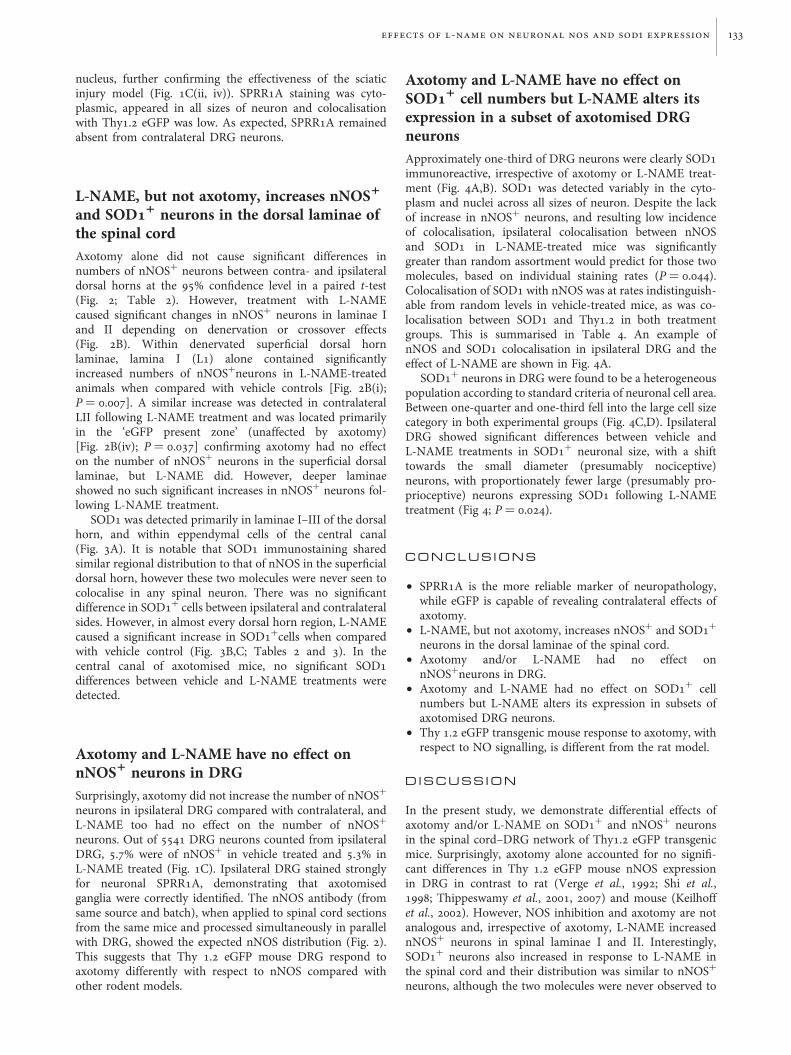
nucleus, further confirming the effectiveness of the sciaticinjury model (Fig. 1C(ii, iv)). SPRR1A staining was cyto-plasmic, appeared in all sizes of neuron and colocalisationwith Thy1.2 eGFP was low. As expected, SPRR1A remainedabsent from contralateral DRG neurons.
L-NAME, but not axotomy, increases nNOS1
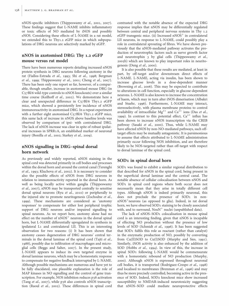
and SOD11 neurons in the dorsal laminae ofthe spinal cordAxotomy alone did not cause significant differences innumbers of nNOS+ neurons between contra- and ipsilateraldorsal horns at the 95% confidence level in a paired t-test(Fig. 2; Table 2). However, treatment with L-NAMEcaused significant changes in nNOS+ neurons in laminae Iand II depending on denervation or crossover effects(Fig. 2B). Within denervated superficial dorsal hornlaminae, lamina I (L1) alone contained significantlyincreased numbers of nNOS+neurons in L-NAME-treatedanimals when compared with vehicle controls [Fig. 2B(i);P ¼ 0.007]. A similar increase was detected in contralateralLII following L-NAME treatment and was located primarilyin the ‘eGFP present zone’ (unaffected by axotomy)[Fig. 2B(iv); P ¼ 0.037] confirming axotomy had no effecton the number of nNOS+ neurons in the superficial dorsallaminae, but L-NAME did. However, deeper laminaeshowed no such significant increases in nNOS+ neurons fol-lowing L-NAME treatment.
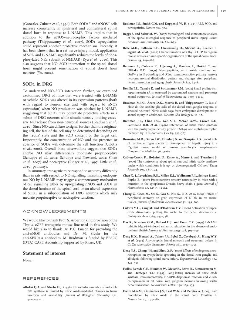
SOD1 was detected primarily in laminae I–III of the dorsalhorn, and within eppendymal cells of the central canal(Fig. 3A). It is notable that SOD1 immunostaining sharedsimilar regional distribution to that of nNOS in the superficialdorsal horn, however these two molecules were never seen tocolocalise in any spinal neuron. There was no significantdifference in SOD1+ cells between ipsilateral and contralateralsides. However, in almost every dorsal horn region, L-NAMEcaused a significant increase in SOD1+cells when comparedwith vehicle control (Fig. 3B,C; Tables 2 and 3). In thecentral canal of axotomised mice, no significant SOD1differences between vehicle and L-NAME treatments weredetected.
Axotomy and L-NAME have no effect onnNOS1 neurons in DRGSurprisingly, axotomy did not increase the number of nNOS+
neurons in ipsilateral DRG compared with contralateral, andL-NAME too had no effect on the number of nNOS+
neurons. Out of 5541 DRG neurons counted from ipsilateralDRG, 5.7% were of nNOS+ in vehicle treated and 5.3% inL-NAME treated (Fig. 1C). Ipsilateral DRG stained stronglyfor neuronal SPRR1A, demonstrating that axotomisedganglia were correctly identified. The nNOS antibody (fromsame source and batch), when applied to spinal cord sectionsfrom the same mice and processed simultaneously in parallelwith DRG, showed the expected nNOS distribution (Fig. 2).This suggests that Thy 1.2 eGFP mouse DRG respond toaxotomy differently with respect to nNOS compared withother rodent models.
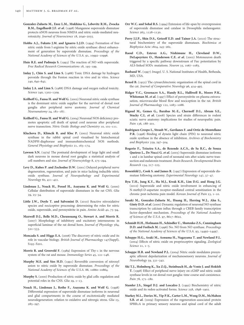
Axotomy and L-NAME have no effect onSOD11 cell numbers but L-NAME alters itsexpression in a subset of axotomised DRGneuronsApproximately one-third of DRG neurons were clearly SOD1immunoreactive, irrespective of axotomy or L-NAME treat-ment (Fig. 4A,B). SOD1 was detected variably in the cyto-plasm and nuclei across all sizes of neuron. Despite the lackof increase in nNOS+ neurons, and resulting low incidenceof colocalisation, ipsilateral colocalisation between nNOSand SOD1 in L-NAME-treated mice was significantlygreater than random assortment would predict for those twomolecules, based on individual staining rates (P ¼ 0.044).Colocalisation of SOD1 with nNOS was at rates indistinguish-able from random levels in vehicle-treated mice, as was co-localisation between SOD1 and Thy1.2 in both treatmentgroups. This is summarised in Table 4. An example ofnNOS and SOD1 colocalisation in ipsilateral DRG and theeffect of L-NAME are shown in Fig. 4A.
SOD1+ neurons in DRG were found to be a heterogeneouspopulation according to standard criteria of neuronal cell area.Between one-quarter and one-third fell into the large cell sizecategory in both experimental groups (Fig. 4C,D). IpsilateralDRG showed significant differences between vehicle andL-NAME treatments in SOD1+ neuronal size, with a shifttowards the small diameter (presumably nociceptive)neurons, with proportionately fewer large (presumably pro-prioceptive) neurons expressing SOD1 following L-NAMEtreatment (Fig 4; P ¼ 0.024).
C O N C L U S I O N S
† SPRR1A is the more reliable marker of neuropathology,while eGFP is capable of revealing contralateral effects ofaxotomy.
† L-NAME, but not axotomy, increases nNOS+ and SOD1+
neurons in the dorsal laminae of the spinal cord.† Axotomy and/or L-NAME had no effect on
nNOS+neurons in DRG.† Axotomy and L-NAME had no effect on SOD1+ cell
numbers but L-NAME alters its expression in subsets ofaxotomised DRG neurons.
† Thy 1.2 eGFP transgenic mouse response to axotomy, withrespect to NO signalling, is different from the rat model.
D I S C U S S I O N
In the present study, we demonstrate differential effects ofaxotomy and/or L-NAME on SOD1+ and nNOS+ neuronsin the spinal cord–DRG network of Thy1.2 eGFP transgenicmice. Surprisingly, axotomy alone accounted for no signifi-cant differences in Thy 1.2 eGFP mouse nNOS expressionin DRG in contrast to rat (Verge et al., 1992; Shi et al.,1998; Thippeswamy et al., 2001, 2007) and mouse (Keilhoffet al., 2002). However, NOS inhibition and axotomy are notanalogous and, irrespective of axotomy, L-NAME increasednNOS+ neurons in spinal laminae I and II. Interestingly,SOD1+ neurons also increased in response to L-NAME inthe spinal cord and their distribution was similar to nNOS+
neurons, although the two molecules were never observed to
effects of l-name on neuronal nos and sod1 expression 133

Fig. 2. nNOS in spinal dorsal horn and its modulation by L-NAME. (A) nNOS immunohistochemistry. Transverse sections of L4 spinal dorsal horn from Thy1.2 eGFP mouse following sciatic nerve axotomy (9 dpo) and treatment with L-NAME (iii, iv) or vehicle control (i, ii). Green: endogenous eGFP. Red: nNOS.L-NAME causes upregulation of nNOS in both axotomised and intact dorsal horn in a region-dependent manner. Increased numbers of nNOS+ cell bodieswere detected in L-NAME-treated animals (examples are marked with arrowheads in iii and iv); when compared with similar regions in the vehicle-treatedcontrol animals (i, ii). Scale bar 100 mm. (B) Graphs show mean cell counts of nNOS+ cell bodies from ‘eGFP absent zone’ (represents both ipsilateral‘denervated zone’ and the contralateral ‘cross-over affected zone’) and ‘eGFP present zone’ of the dorsal horn lamina I and II (9 dpo) from L-NAME- andvehicle-treated mice. Error bars, SEM. In lamina I, the denervated area [eGFP absent zone (i), ipsi] showed a significant increase in nNOS+ cells followingL-NAME treatment (P ¼ 0.007), while the ‘eGFP present zone’ (ii) showed no such effect. Overall, in ‘eGFP present zone’ of lamina I (ii) there was decreasednNOS+ neurons in both ipsi- and contra-lateral side when compared with ‘eGFP absent zone’ of the same lamina (i) and ‘eGFP present zone’ of lamina II(iv). In lamina II, the denervated area [eGFP absent zone (iii), ipsi] showed no difference in nNOS+ cells, while in the ‘eGFP present zone’ contralaterally (iv,contra), a significant difference was detected between the control and L-NAME-treated groups (P ¼ 0.037).
134 matthew j. g. bradman et al.

Table 2. Mean SOD1+ and nNOS+ cell body counts from the region of peak denervation at the full extent of anatomical resolution available, and in combined regions
Antigen Treatment Laterality Value Counted regions (cells per 10 mm) Derived counts (combined regions, cells per 10 mm)
L I L II L III L IV
GFP1ve
GFP2ve
GFP1ve
GFP2ve
GFP1ve
GFP2ve
GFP1ve
GFP2ve
LItotal
LIItotal
LIIItotal
LIV ontotal
GFP1 LIto LIII
GFP2 LIto LIII
GFP1total
GFP2total
Total/10 mmsection
SOD1 Vehicle Contra Mean 2.5 1.75 1.5 0.75 1.58 0.92 3.33 1.75 4.25 2.25 2.5 5.08 5.58 3.42 8.92 5.17 14.08SEM 0.3 0.14 0.3 0.12 0.21 0.2 0.65 0.16 0.25 0.37 0.36 0.79 0.72 0.35 1.35 0.37 1.63
Ipsi Mean 1.92 2.17 1.92 0.58 1.5 0.5 2.08 1.67 4.08 2.5 2 3.75 5.33 3.25 7.42 4.92 12.33SEM 0.32 0.29 0.49 0.16 0.25 0.1 0.31 0.16 0.52 0.63 0.3 0.35 0.98 0.4 1.09 0.43 1.36
L-NAME Contra Mean 4.33 2.75 5.67 3.58 4.58 2.17 5.67 4.33 7.08 9.25 6.75 10 14.58 8.5 20.25 12.83 33.08SEM 0.62 0.16 1.13 0.75 0.78 0.3 0.6 0.57 0.78 1.77 1.01 1.05 2.53 1.19 2.82 1.67 4.15
Ipsi Mean 3.5 2.83 5.08 4.25 3 2.75 4.08 4.42 6.33 9.33 5.75 8.5 11.58 9.83 15.67 14.25 29.92SEM 0.17 0.39 0.63 0.55 0.39 0.4 0.52 0.45 0.3 1.13 0.73 0.96 1.02 1.24 1.33 1.65 2.89
nNOS Vehicle Contra Mean 0.5 1.5 2.75 1.92 1.5 1 1.58 0.33 2 4.67 2.5 1.92 4.75 4.42 6.33 4.75 11.08SEM 0.12 0.05 0.25 0.14 0.12 0.11 0.21 0.08 0.14 0.37 0.17 0.16 0.28 0.2 0.41 0.16 0.46
Ipsi Mean 0.17 1 2.17 2.33 1.33 1 2.5 0.83 1.17 4.5 2.33 3.33 3.67 4.33 6.17 5.17 11.33SEM 0.05 0.14 0.29 0.3 0.14 0.14 0.2 0.18 0.17 0.57 0.14 0.3 0.35 0.26 0.53 0.32 0.72
L-NAME Contra Mean 0.5 2.75 5.58 3 1.83 1.42 1 0.5 3.25 8.58 3.25 1.5 7.92 7.17 8.92 7.67 16.58SEM 0.17 0.24 0.64 0.14 0.05 0.28 0.14 0.05 0.28 0.71 0.29 0.18 0.76 0.42 0.87 0.47 1.31
Ipsi Mean 0.33 3 4.33 3.25 1.83 1.5 2 0.5 3.33 7.58 3.33 2.5 6.5 7.75 8.5 8.25 16.75SEM 0.14 0.42 0.64 0.39 0.34 0.32 0.34 0.1 0.55 0.94 0.62 0.3 0.83 1 0.63 1.02 1.61
Values are cell bodies visible per 10 mm section, expressed as means of 4 animals; actual numbers of cells counted were therefore three times higher per animal and 12 times higher per group (see Methods) in every region.Note: eGFP+/- denotes presence/absence from L II as defined in Table 1 and Fig.1B.
ef
fe
ct
so
fl
-n
am
eo
nn
eu
ro
na
ln
os
an
ds
od
1e
xp
re
ss
io
n135

colocalise in any single neuron. In contrast, in the DRG,higher numbers of small diameter neurons contained SOD1following axotomy and L-NAME treatment.
NOS inhibitors and rodent models of axotomyWe have previously tested, in rats, the effects of various NOSinhibitors including nNOS-specific inhibitors such as 7-nitroindazole, 1,2-trifluoromethylphenyl imidazole or domi-nant negative viral vectors targeting either nNOS or sGC inboth in vivo and in vitro models of nerve injury(Thippeswamy et al., 2001, 2005a, 2005b, 2007). In ratmodels of nerve injury, we and others (Verge et al., 1992;Shi et al., 1998; Bergman et al., 1999) have shown a significantincrease in the number of nNOS+ neurons in DRG at 7–8days following injury to their axons. In subsequent years,the role of iNOS and eNOS in nerve injury models began toemerge (Levy et al., 2001; Sunico et al., 2008). We have very
Fig. 3. SOD1 distribution in spinal cord and its modulation by L-NAME. (A) Mouse spinal cord dorsal horn ipsilateral to sciatic nerve axotomy (9 dpo) fromL-NAME-treated mouse, double stained for SOD1 and nNOS. Red: SOD1, in the superficial laminae (i) and eppendymal cells of the central canal (ii). Green:nNOS. Note: nNOS staining in this case was originally identified with anti-sheep biotinylated and streptavidin Marina blue. Images were captured as blackand white but later pseudo coloured with green to contrast with red. Therefore, eGFP in the dorsal horn is not shown. Scale bars 100 mm. (B) Representativeimages showing SOD1 immunoreactivity (red) and eGFP in the dorsal horn following sciatic nerve axotomy (9 dpo). Irrespective of axotomy, there areincreased numbers of SOD+ cells in L-NAME-treated groups. See graph C and Tables 2 and 3 for lamina-specific details of cell counts. Scale bar 100 mm. (C)Graph summarises regional quantification of SOD1+ cells from dorsal horn of the spinal cord (see also Tables 2 and 3). Counts from all regions were groupedby lamina and by the presence or absence of eGFP (axotomy effect ipsilaterally/cross-over effect contralaterally) and subjected to statistical analysis byANOVA. No differences were detected between contra and ipsilateral sides, however L-NAME caused significant increases in SOD1+ cell counts at the 95%confidence level in most groups (see Table 3 for P values).
Table 3. P values (vehicle versus L-NAME) for SOD1 quantification, byregion, from the dorsal horn of the spinal cord (Fig. 3)
Anatomical regions Comparison by ANOVA
Lamina GFP-definedregion
Contralateral Ipsilateral
vehicle versusL-NAME
vehicle versusL-NAME
I Full width DH 0.041 NSDII Full width DH 0.024 0.027III Full width DH 0.023 0.041IV and deeper Full width DH 0.032 0.038L1–LIII Present 0.030 NSDL1–LIII Absent 0.040 0.011Full depth DH Present 0.024 NSDFull depth DH Absent 0.024 0.008Full depth DH Full width DH 0.015 0.022
Note: NSD ¼ no significant difference at 95% confidence level.
136 matthew j. g. bradman et al.

recently demonstrated the presence of iNOS-producingimmune cells such as CD68+ macrophages in axotomisedDRG (Bradman et al., 2010). L-NAME is considered as ageneral NOS inhibitor as it targets all three known NOSs to
prevent NO production. In view of this, we recently testedthe effects of L-NAME also in a rat model of axotomywhere L-NAME did not cause degenerative changes in ratDRG neurons (Bradman et al., 2010) as opposed to
Fig. 4. Effects of axotomy and/or L-NAME on SOD1 in DRG. (A) SOD1 immunoreactivity (blue in i, ii, iv and v) was localised mainly in neuronal cytoplasmand nuclei of DRG neurons, irrespective of neuronal subtypes. (iii) and (vi) SOD1 (red) and nNOS (green) in ipsilateral DRG. SOD1 colocalisation with nNOS(yellow) was observed in larger diameter neurons of ipsilateral DRG in vehicle-treated mouse (iii), while in L-NAME-treated mouse it was restricted tosmall-medium sized neurons (vi). Scale bar 100 mm. (B) SOD1 immunoreactive neurons expressed as a percentage of total neuron number irrespective ofDRG neuronal subtypes, i.e. small diameter (,400 mm) and large diameter (.400 mm) neurons. No significant differences were found between the treatmentgroups in the proportion of neurons expressing SOD1. Neither was significant difference detected within treatment groups comparing ipsilateral withcontralateral sides by ANOVA or in a paired t-test. Error bars SEM. (C) An example for DRG neurons phenotypes. SOD1+ neuron (blue) with respect toneuronal size. S (red): small diameter (,400 mm) neurons. L (green): large diameter (.400 mm) neurons. (D) Quantification of SOD1+ neurons based onsize criteria as illustrated in (C). Asterisk indicates significant shift in SOD1 expression towards small diameter neurons (P ¼ 0.024) in L-NAME-treatedipsilateral DRG when compared with vehicle-treated group. Contralateral changes are below significance at 95% confidence.
Table 4. Summary of SOD1 staining in DRG neurons from axotomised and L-NAME or vehicle treated mice at 9 day post-axotomy. Immunoreactivitywas quantified in terms of SOD1+ percentages of all neurons stained, sizes of stained neurons, and colocalisation with nNOS and eGFP; see Fig. 4 graphs
and sample images. Note: H0 ¼ hypothetical random assortment (see Methods); Obs. ¼ observed colocalisation
Treatment Laterality Total quantified SOD1 immunoreactive SOD1 and nNOS SOD1 and GFP
% of total (SEM) % of SOD1 (SEM) % of total neurons (SEM)
Small < 400 mm2 Large > 400 mm2 H0 Obs. H0 Obs.
Observed counts
Vehicle Contra 1217 41.7 (1.2) 67.1 (3.6) 33.1 (3.6) Ipsilateral onlyIpsi 1342 33.9 (1.8) 64.4 (4.3) 35.2 (4.3) 1.5 (0.1) 2 (0.3) 6.3 (0.5) 5.2 (0.4)
L-NAME Contra 1535 33.7 (2.0) 74.6 (3.0) 26.2 (3.0) Ipsilateral onlyIpsi 1447 31.6 (1.2) 74.7 (2.3) 25.5 (2.3) ∗1.4 (0.2) ∗2.5 (0.3) 6.3 (0.5) 4.1 (0.5)
∗Predicted random colocalisation rate was significantly lower than observed colocalisation rate for SOD1 with nNOS in L-NAME treated ipsilateral DRG,P ¼ 0.044 by paired t-test. Following NO modulation by L-NAME, SOD1 was found to colocalise with nNOS significantly more frequently than atrandom.
effects of l-name on neuronal nos and sod1 expression 137
nNOS-specific inhibitors (Thippeswamy et al., 2001, 2007).These findings suggest that L-NAME inhibits inflammatoryor toxic effects of NO mediated by iNOS and possiblyeNOS. Considering these effects of L-NAME in a rat model,we extended this to Thy1.2 eGFP mice in which subpopu-lations of DRG neurons are selectively marked by eGFP.
nNOS in axotomised DRG: Thy 1.2 eGFPmouse versus rat modelThere have been numerous reports detailing increased nNOSprotein synthesis in DRG neurons following axotomy in therat (Fiallos-Estrada et al., 1993; Shi et al., 1998; Bergmanet al., 1999; Thippeswamy et al., 2001; Cheng et al., 2007).There has been only one report so far, however, of a compar-able, though smaller, increase in axotomised mouse DRG (inC57Bl/6 wild-type controls to nNOS knockouts) over a similartime course (Keilhoff et al., 2002). We demonstrate here aclear and unexpected difference in C57Bl/6 Thy1.2 eGFPmice, which showed a persistently low incidence of nNOSimmunoreactivity in axotomised DRG. In a repeat experimentwith a further eight axotomised C57Bl/6 Thy1.2 eGFP mice,this same lack of increase in nNOS above baseline levels wasobserved by comparison of ipsi- with contralateral DRG.The lack of nNOS increase was clear in spite of robust ipsilat-eral increases in SPRR1A, an established marker of neuronalinjury (Bonilla et al., 2002; Starkey et al., 2009).
nNOS signalling in DRG–spinal dorsalhorn networkAs previously and widely reported, nNOS staining in thespinal cord was detected primarily in cell bodies and processeswithin the dorsal horn and around the central canal (Terenghiet al., 1993; Kluchova et al., 2002). It is necessary to consideralso the possible effects of nNOS from DRG neurons inrelation to immunoreactivity reported in the dorsal horn. Aswell as being locally active within ganglia (Thippeswamyet al., 2007), nNOS may be transported centrally to sensitisedorsal spinal neurons (Ryu et al., 2010) or peripherally tothe injured site to promote neuritogenesis (Zochodne et al.,1999). These mechanisms are considered as ‘axotomyresponses’ to compensate for either lost peripheral trophicsupport of DRG neurons and/or impaired signalling tospinal neurons. As we report here, axotomy alone had noeffect on the number of nNOS+ neurons in the dorsal spinalhorn, but L-NAME differentially upregulated its synthesis inipsilateral L1 and contralateral LII. This is an interestingobservation for two reasons: (i) It has been shown thataxotomy causes degeneration of DRG neurons’ central ter-minals in the dorsal laminae of the spinal cord (Tajti et al.,1988), possibly due to infiltration of macrophages and micro-glial cells (Beggs and Salter, 2007). In the present study,L-NAME appears to induce its own targeted enzyme indorsal laminae neurons, which may be a homeostatic responseto compensate for negative feedback interrupted by L-NAME.Although possible mechanisms are numerous and have yet tobe fully elucidated, one plausible explanation is the role ofMAP kinases in NO signalling and the control of gene tran-scription. For example, NOS inhibition reduces p38 activation(Tang et al., 2007), while p38 also controls nNOS transcrip-tion (Raoul et al., 2002). These differences in spinal cord
contrasted with the notable absence of the expected DRGresponse implies that nNOS may be differentially regulatedbetween central and peripheral nervous systems in Thy 1.2eGFP transgenic mice. (ii) Increased nNOS+ in contralateralLII neurons, in response to L-NAME, could possibly play arole in contralateral sprouting of fibres. We have shown pre-viously that the nNOS-mediated pathway activates the pro-duction of neurotrophic factors such as nerve growth factorand neurotrophin 3 by glial cells (Thippeswamy et al.,2005b) which are known to play important roles in neurito-genesis (Deng et al., 2000).
It is also possible that these results are mediated, at least inpart, by off-target and/or downstream direct effects ofL-NAME. L-NAME, acting via insulin, has been shown toincrease glucose levels in a dose-dependent manner(Browning et al., 2006). This may be expected to contributeto alterations in cell function, especially in glucose-dependentneurons. L-NAME is also known to increase the concentrationof haem, which may in turn alter iNOS dimerisation (Albakriand Stuehr, 1996). Furthermore, L-NAME may interact,stereoselectively, with plasma membrane proteins to controlavailability of intracellular Mg2+ and Ca2+ ions (Das et al.,1999). In contrast to this potential effect, Ca2+ influx hasbeen shown to increase nNOS transcription via the CREBpathway (Sasaki et al., 2000). Thus, while L-NAME mayhave affected nNOS by non-NO-mediated pathways, such off-target effects may be mutually antagonistic. It is parsimoniousto assume that effects attributed to L-NAME administrationprimarily occur following NOS inhibition, and are thereforelikely to be NOS-targeted rather than off-target with respectto dorsal laminae of the spinal cord.
SOD1 in spinal dorsal hornSOD1 was found to exhibit a similar regional distribution tothat described for nNOS in the spinal cord, being present inthe superficial dorsal laminae and the central canal. Thenotable absence of cellular colocalisation between nNOS andSOD1 in spinal cord regions where both occur does notnecessarily mean that they arise in totally different celltypes. Although nNOS is indeed primarily neuronal, thisdoes not preclude the presence of SOD1 in non-nNOS+neurons (as opposed to glia). Indeed, in rat dorsalhorn, we have observed SOD1 staining to be closely associatedwith, and to surround, NeuN+ nuclei (unpublished data).
The lack of nNOS–SOD1 colocalisation in mouse spinalcord is an interesting finding, given that nNOS is incapableof effecting NO production without the presence of lowlevels of SOD (Schmidt et al., 1996). It has been suggestedthat SOD1 fulfils this role as reactant (rather than catalyst)in the enzymatic production of NO, possibly by convertingfrom Cu(II)SOD to Cu(I)SOD (Murphy and Sies, 1991).Similarly, iNOS activity is also enhanced by the addition ofSOD (Hobbs et al., 1994). In view of this, the increase inspinal SOD1 following L-NAME would be commensuratewith a homeostatic rebound of NO production (Murphy,2000). Although nNOS is expressed throughout neuronalcell bodies, it is transported (Rodriguez-Crespo et al., 1998)and localised to membranes (Brenman et al., 1996) and maythus be more precisely controlled, becoming active in the pres-ence of SOD. Indeed, SOD2 knockout mice show increasedsusceptibility to NMDAR-induced neurotoxicity suggestingthat nNOS–SOD could mediate neuroprotective effects
138 matthew j. g. bradman et al.
(Gonzalez-Zulueta et al., 1998). Both SOD1+ and nNOS+ cellsincrease consistently in ipsilateral and contralateral spinaldorsal horn in response to L-NAME. This implies that inaddition to the nNOS–neurotrophic factors mediatedpathway (Thippeswamy et al., 2007), SOD1 upregulationcould represent another protective mechanism. Recently, ithas been shown that in a rat nerve injury model, applicationof SOD and L-NAME significantly reduces the levels of phos-phorylated NR1 subunit of NMDAR (Ryu et al., 2010). Thisalso suggests that NO–SOD interaction at the spinal dorsalhorn might prevent sensitisation of spinal dorsal hornneurons (Yu, 2002).
SOD1 in DRGTo understand NO–SOD interaction further, we examinedaxotomised DRG of mice that were treated with L-NAMEor vehicle. SOD1 was altered in its expression patterns (bothwith regard to neuron size and with regard to nNOSexpression) when NO production was blocked by L-NAME,suggesting that SOD1 may potentiate protective effects in asubset of DRG neurons while simultaneously limiting exces-sive NO release from non-neuronal sources (Bradman et al.,2010). Since NO can diffuse to signal further than the originat-ing cell, the fate of the cell may be determined depending onthe ‘redox’ state and the SOD content of the target cell.Importantly, the concentration of NO and the presence orabsence of SOD1 will determine the cell function (Culottaet al., 2006). Overall these observations suggest that SOD1and/or NO may differentially modulate proprioceptive(Schuppe et al., 2004; Schuppe and Newland, 2004; Chenet al., 2007) and nociceptive (Ridger et al., 1997; Little et al.,2010) pathways.
In summary, transgenic mice respond to axotomy differentlythan in rats with respect to NO signalling. Inhibiting endogen-ous NO by L-NAME may trigger a compensatory mechanismof cell signalling either by upregulating nNOS and SOD1 inthe dorsal laminae of the spinal cord or an altered expressionof SOD1 in a subpopulation of DRG neurons which maymediate proprioceptive or nociceptive function.
A C K N O W L E D G E M E N T S
We would like to thank Prof. S. Arber for kind provision of theThy1.2 eGFP transgenic mouse line used in this study. Wewould like also to thank Dr. P.C. Emson for providing theanti-nNOS antibodies and Dr. M. Sivula for theanti-SPRR1A antibodies. M. Bradman is funded by BBSRC(DTA) CASE studentship supported by Pfizer, UK.
Statement of interestNone.
R E F E R E N C E S
Albakri Q.A. and Stuehr D.J. (1996) Intracellular assembly of inducibleNO synthase is limited by nitric oxide-mediated changes in hemeinsertion and availability. Journal of Biological Chemistry 271,5414–5421.
Beckman J.S., Smith C.M. and Koppenol W. H. (1993) ALS, SOD, andperoxynitrite. Nature 364, 584.
Beggs S. and Salter M. W. (2007) Stereological and somatotopic analysisof the spinal microglial response to peripheral nerve injury. Brain,Behavior, and Immunity 21, 624–633.
Belle M.D., Pattison E.F., Cheunsuang O., Stewart A., Kramer I.,Sigrist M. et al. (2007) Characterization of a thy1.2 GFP transgenicmouse reveals a tissue-specific organization of the spinal dorsal horn.Genesis 45, 679–688.
Bergman E., Carlsson K., Liljeborg A., Manders E., Hokfelt T. andUlfhake B.D. (1999) Neuropeptides, nitric oxide synthase andGAP-43 in B4-binding and RT97 immunoreactive primary sensoryneurons: normal distribution pattern and changes after peripheralnerve transection and aging. Brain Research 832, 63–83.
Bonilla I.E., Tanabe K. and Strittmatter S.M. (2002) Small proline-richrepeat protein 1A is expressed by axotomized neurons and promotesaxonal outgrowth. Journal of Neuroscience 22, 1303–1315.
Bradman M.J.G., Arora D.K., Morris R. and Thippeswamy T. (2010)How do the satellite glia cells of the dorsal root ganglia respond tostressed neurons? Nitric oxide saga from embryonic development toaxonal injury in adulthood. Neuron Glia Biology 6, 11–17.
Brenman J.E., Chao D.S., Gee S.H., McGee A.W., Craven S.E.,Santillano D.R. et al. (1996) Interaction of nitric oxide synthasewith the postsynaptic density protein PSD-95 and alphal-syntrophinmediated by PDZ domains. Cell 84, 757–767.
Browning M.D., Garyu J.W., Dumler J.S. and Scorpio D.G. (2006) Roleof reactive nitrogen species in development of hepatic injury in aC57bl/6 mouse model of human granulocytic anaplasmosis.Comparative Medicine 56, 55–62.
Callsen-Cencic P., Hoheisel U., Kaske A., Mense S. and Tenschert S.(1999) The controversy about spinal neuronal nitric oxide synthase:under which conditions is it up- or downregulated? Cell and TissueResearch 295, 183–194.
Chen X.-J., Levedakou E.N., Millen K.J., Wollmann R.L., Soliven B. andPopko B. (2007) Proprioceptive sensory neuropathy in mice with amutation in the cytoplasmic Dynein heavy chain 1 gene. Journal ofNeuroscience 27, 14515–14524.
Cheng C., Chen M., Shi S., Gao S., Niu S., Li X. et al. (2007) Effect ofperipheral axotomy on gene expression of NIDD in rat neuraltissues. Journal of Molecular Neuroscience 32, 199–206.
Culotta V.C., Yang M. and O’Halloran T.V. (2006) Activation of super-oxide dismutases: putting the metal to the pedal. Biochimica etBiophysica Acta 1763, 747–758.
Das R., Kravtsov G.M., Ballard H.J. and Kwan C.Y. (1999) L-NAMEinhibits Mg(2+)-induced rat aortic relaxation in the absence of endo-thelium. British Journal of Pharmacology 128, 493–499.
Deng H.X., Hentati A., Tainer J.A., Iqbal Z., Cayabyab A., Hung W.Y.et al. (1993) Amyotrophic lateral sclerosis and structural defects inCu,Zn superoxide dismutase. Science 261, 1047–1051.
Deng Y.S., Zhong J.H. and Zhou X.F. (2000) Effects of endogenous neu-rotrophins on sympathetic sprouting in the dorsal root ganglia andallodynia following spinal nerve injury. Experimental Neurology 164,344–350.
Fiallos-Estrada C.E., Kummer W., Mayer B., Bravo R., Zimmermann M.and Herdegen T.D. (1993) Long-lasting increase of nitric oxidesynthase immunoreactivity, NADPH-diaphorase reaction and c-JUNco-expression in rat dorsal root ganglion neurons following sciaticnerve transection. Neuroscience Letters 150, 169–173.
Freire M.A.M., Guimaraes J.S., Leal W.G. and Pereira A. (2009) Painmodulation by nitric oxide in the spinal cord. Frontiers inNeuroscience 3, 175–181.
effects of l-name on neuronal nos and sod1 expression 139
Gonzalez-Zulueta M., Ensz L.M., Mukhina G., Lebovitz R.M., ZwackaR.M., Engelhardt J.F. et al. (1998) Manganese superoxide dismutaseprotects nNOS neurons from NMDA and nitric oxide-mediated neu-rotoxicity. Journal of Neuroscience 18, 2040–2055.
Hobbs A.J., Fukuto J.M. and Ignarro L.J.D. (1994) Formation of freenitric oxide from l-arginine by nitric oxide synthase: direct enhance-ment of generation by superoxide dismutase. Proceedings of theNational Academy of Sciences of the U.S.A. 91, 10992–10996.
Huie R.E. and Padmaja S. (1993) The reaction of NO with superoxide.Free Radical Research Communications 18, 195–199.
Imlay J., Chin S. and Linn S. (1988) Toxic DNA damage by hydrogenperoxide through the Fenton reaction in vivo and in vitro. Science240, 640–642.
Imlay J.A. and Linn S. (1988) DNA damage and oxygen radical toxicity.Science 240, 1302–1309.
Keilhoff G., Fansa H. and Wolf G. (2002) Neuronal nitric oxide synthaseis the dominant nitric oxide supplier for the survival of dorsal rootganglia after peripheral nerve axotomy. Journal of ChemicalNeuroanatomy 24, 181–187.
Keilhoff G., Fansa H. and Wolf G. (2004) Neuronal NOS deficiency pro-motes apoptotic cell death of spinal cord neurons after peripheralnerve transection. Nitric Oxide: Biology and Chemistry 10, 101–111.
Kluchova D., Klimcik R. and Kloc P. (2002) Neuronal nitric oxidesynthase in the rabbit spinal cord visualised by histochemicalNADPH-diaphorase and immunohistochemical NOS methods.General Physiology and Biophysics 21, 163–174.
Lawson S.N. (1979) The postnatal development of large light and smalldark neurons in mouse dorsal root ganglia: a statistical analysis ofcell numbers and size. Journal of Neurocytology 8, 275–294.
Levy D., Kubes P. and Zochodne D.W. (2001) Delayed peripheral nervedegeneration, regeneration, and pain in mice lacking inducible nitricoxide synthase. Journal of Neuropathology and ExperimentalNeurology 60, 411–421.
Lindenau J., Noack H., Possel H., Asayama K. and Wolf G. (2000)Cellular distribution of superoxide dismutases in the rat CNS. Glia29, 25–34.
Little J.W., Doyle T. and Salvemini D. (2010) Reactive nitroxidativespecies and nociceptive processing: determining the roles for nitricoxide, superoxide, and peroxynitrite in pain. Amino Acids 42, 75–94.
Maxwell D.J., Belle M.D., Cheunsuang O., Stewart A. and Morris R.(2007) Morphology of inhibitory and excitatory interneurons insuperficial laminae of the rat dorsal horn. Journal of Physiology 584,521–533.
Moncada S. and Higgs E.A. (2006) The discovery of nitric oxide and itsrole in vascular biology. British Journal of Pharmacology 147(Suppl),S193–S201.
Morris R. and Grosveld F. (1989) Expression of Thy-1 in the nervoussystem of the rat and mouse. Immunology Series 45, 121–148.
Murphy M.E. and Sies H.D. (1991) Reversible conversion of nitroxylanion to nitric oxide by superoxide dismutase. Proceedings of theNational Academy of Sciences of the U.S.A. 88, 10860–10864.
Murphy S. (2000) Production of nitric oxide by glial cells: regulation andpotential roles in the CNS. Glia 29, 1–13.
Noack H., Lindenau J., Rothe F., Asayama K. and Wolf G. (1998)Differential expression of superoxide dismutase isoforms in neuronaland glial compartments in the course of excitotoxically mediatedneurodegeneration: relation to oxidative and nitrergic stress. Glia 23,285–297.
Orr W.C. and Sohal R.S. (1994) Extension of life-span by overexpressionof superoxide dismutase and catalase in Drosophila melanogaster.Science 263, 1128–1130.
Perry J.J.P., Shin D.S., Getzoff E.D. and Tainer J.A. (2010) The struc-tural biochemistry of the superoxide dismutases. Biochimica etBiophysica Acta 1804, 245–262.
Raoul C.D., Estevez A.G., Nishimune H., Cleveland D.W.,DeLapeyriere O., Henderson C.E. et al. (2002) Motoneuron deathtriggered by a specific pathway downstream of Fas: potentiation byALS-linked SOD1 mutations. Neuron 35, 1067–1083.
Rasband W. (1997) ImageJ. U. S. National Institutes of Health, Bethesda,MD, USA.
Rexed B. (1952) The cytoarchitectonic organization of the spinal cord inthe cat. Journal of Comparative Neurology 96, 414–495.
Ridger V.C., Greenacre S.A., Handy R.L., Halliwell B., Moore P.K.,Whiteman M. et al. (1997) Effect of peroxynitrite on plasma extrava-sation, microvascular blood flow and nociception in the rat. BritishJournal of Pharmacology 122, 1083–1088.
Rigaud M., Gemes G., Barabas M.-E., Chernoff D.I., Abram S.E.,Stucky C.L. et al. (2008) Species and strain differences in rodentsciatic nerve anatomy: implications for studies of neuropathic pain.Pain 136, 188–201.
Rodriguez-Crespo I., Straub W., Gavilanes F. and Ortiz de MontellanoP.R. (1998) Binding of dynein light chain (PIN) to neuronal nitricoxide synthase in the absence of inhibition. Archives of Biochemistryand Biophysics 359, 297–304.
Rogerio F., Teixeira S.A., de Rezende A.C.S., de Sa R.C., de SouzaQueiroz L., De Nucci G. et al. (2005) Superoxide dismutase isoforms1 and 2 in lumbar spinal cord of neonatal rats after sciatic nerve tran-section and melatonin treatment. Brain Research. Developmental BrainResearch 154, 217–225.
Rosenfeld J., Cook S. and James R. (1997) Expression of superoxide dis-mutase following axotomy. Experimental Neurology 147, 37–47.
Ryu T.H., Jung K.Y., Ha M.J., Kwak K.H., Lim D.G. and Hong J.G.(2010) Superoxide and nitric oxide involvement in enhancing ofN-methyl-D-aspartate receptor-mediated central sensitization in thechronic post-ischemia pain model. Korean Journal of Pain 23, 1–10.
Sasaki M., Gonzalez-Zulueta M., Huang H., Herring W.J., Ahn S.,Ginty D.D. et al. (2000) Dynamic regulation of neuronal NO synthasetranscription by calcium influx through a CREB family transcriptionfactor-dependent mechanism. Proceedings of the National Academyof Sciences of the U.S.A. 97, 8617–8622.
Schmidt H.H., Hofmann H., Schindler U., Shutenko Z.S., CunninghamD.D. and Feelisch M. (1996) No. NO from NO synthase. Proceedingsof the National Academy of Sciences of the U.S.A. 93, 14492–14497.
Schuppe H.G., Araki M., Aonuma H., Nagayama T. and Newland P.L.(2004) Effects of nitric oxide on proprioceptive signaling. ZoologicalScience 21, 1–5.
Schuppe H.R. and Newland P.L. (2004) Nitric oxide modulates presyn-aptic afferent depolarization of mechanosensory neurons. Journal ofNeurobiology 59, 331–342.
Shi T.J., Holmberg K., Xu Z.Q., Steinbusch H., de Vente J. and HokfeltT. (1998) Effect of peripheral nerve injury on cGMP and nitric oxidesynthase levels in rat dorsal root ganglia: time course and coexistence.Pain 78, 171–180.
Stamler J.S., Singel D.J. and Loscalzo J. (1992) Biochemistry of nitricoxide and its redox-activated forms. Science 258, 1898–1902.
Starkey M.L., Davies M., Yip P.K., Carter L.M., Wong D.M., McMahonS.B. et al. (2009) Expression of the regeneration-associated proteinSPRR1A in primary sensory neurons and spinal cord of the adult
140 matthew j. g. bradman et al.

mouse following peripheral and central injury. Journal of ComparativeNeurology 513, 51–68.
Sunico C.R., Portillo F., Gonzalez-Forero D., Kasparov S. andMoreno-Lupez B. (2008) Evidence for a detrimental role of nitricoxide synthesized by endothelial nitric oxide synthase after peripheralnerve injury. Neuroscience 157, 40–51.
Tajti J., Fischer J., Knyiher-Csillik E. and Csillik B. (1988)Transganglionic regulation and fine structural localization of lectin-reactive carbohydrate epitopes in primary sensory neurons of therat. Histochemistry 88, 213–218.
Tang Q., Svensson C.I., Fitzsimmons B., Webb M., Yaksh T.L. and HuaX.-Y. (2007) Inhibition of spinal constitutive NOS-2 by 1400Wattenuates tissue injury and inflammation-induced hyperalgesia andspinal p38 activation. European Journal of Neuroscience 25, 2964–2972.
Terenghi G., Riveros-Moreno V., Hudson L.D., Ibrahim N.B. andPolak J.M. (1993) Immunohistochemistry of nitric oxide synthasedemonstrates immunoreactive neurons in spinal cord and dorsalroot ganglia of man and rat. Journal of the Neurological Sciences 118,34–37.
Thippeswamy T., Haddley K., Corness J.D., Howard M.R., McKay J.S.,Beaucourt S.M. et al. (2007) NO-cGMP mediated galanin expressionin NGF-deprived or axotomized sensory neurons. Journal ofNeurochemistry 100, 790–801.
Thippeswamy T., Jain R.K., Mumtaz N. and Morris R. (2001) Inhibitionof neuronal nitric oxide synthase results in neurodegenerative changesin the axotomised dorsal root ganglion neurons: evidence for a neuro-protective role of nitric oxide in vivo. Neuroscience Research 40, 37–44.
Thippeswamy T., McKay J.S., Morris R., Quinn J., Wong L.-F. andMurphy D. (2005a) Glial-mediated neuroprotection: evidence forthe protective role of the NO-cGMP pathway via neuron-glial com-munication in the peripheral nervous system. Glia 49, 197–210.
Thippeswamy T., McKay J.S., Quinn J. and Morris R. (2005b) Eithernitric oxide or nerve growth factor is required for dorsal root ganglionneurons to survive during embryonic and neonatal development.Brain Research. Developmental Brain Research 154, 153–164.
Verge V.M., Xu Z., Xu X.J., Wiesenfeld H.Z. and Hokfelt T. (1992)Marked increase in nitric oxide synthase mRNA in rat dorsal rootganglia after peripheral axotomy: in situ hybridization and functionalstudies. Proceedings of the National Academy of Sciences of the U.S.A.89, 11617–11621.
Wink D.A. and Mitchell J.B. (1998) Chemical biology of nitric oxide:insights into regulatory, cytotoxic, and cytoprotective mechanisms ofnitric oxide. Free Radical Biology and Medicine 25, 434–456.
Yu W.H.A. (2002) Spatial and temporal correlation of nitric oxidesynthase expression with CuZn-superoxide dismutase reduction inmotor neurons following axotomy. Annals of the New York Academyof Sciences 962, 111–121.
Zhang X., Verge V., Wiesenfeld-Hallin Z., Ju G., Bredt D., Synder S.H.et al. (1993) Nitric oxide synthase-like immunoreactivity in lumbardorsal root ganglia and spinal cord of rat and monkey and effect ofperipheral axotomy. Journal of Comparative Neurology 335, 563–575.
Zochodne D.W., Levy D., Zwiers H., Sun H., Rubin I., Cheng C. andLauritzen M. (1999) Evidence for nitric oxide and nitric oxidesynthase activity in proximal stumps of transected peripheral nerves.Neuroscience 91, 1515–1527.
A U T H O R S ’ A D D R E S S
Faculty of Health and Life Sciences, Institute of Ageing andChronic Disease, University of Liverpool, Liverpool, UK
Correspondence should be addressed to:Thimmasettappa ThippeswamyFaculty of Health and Life SciencesUniversity of LiverpoolRoom no. 4.3064th Floor, UCD Duncan BuildingDaulby Street, Liverpool L69 3GA, UKphone: 00 44 151 706 4048email: [email protected]
effects of l-name on neuronal nos and sod1 expression 141
Copyright © 2022 FDOKUMEN