EGFP-tagged core and linker histones diffuse via distinct mechanisms within living cells
11
EGFP-Tagged Core and Linker Histones Diffuse via Distinct Mechanisms within Living Cells Dipanjan Bhattacharya,* Aprotim Mazumder,* S. Annie Miriam,* and G. V. Shivashankar* y *National Center for Biological Sciences, TIFR, Bangalore-560065, India; and y Raman Research Institute, Bangalore, India ABSTRACT The effect of chromatin organization on EGFP-tagged histone protein dynamics within the cell nucleus has been probed using fluorescence correlation and recovery measurements on single living HeLa cells. Our studies reveal that free fraction of core-particle histones exist as multimers within the cell nucleus whereas the linker histones exist in monomeric forms. The multimeric state of core histones is found to be invariant across mammalian and polytene chromosomes and this is ATP dependent. In contrast, the dynamics of the linker histones exhibits two distinct diffusion timescales corresponding to its transient binding and unbinding to chromatin governed by the tail domain residues. Under conditions of chromatin condensation induced by apoptosis, the free multimeric fraction of core histones is found to become immobile, while the monomeric linker histone mobility is partially reduced. In addition, we observe differences in nuclear colocalization of linker and core particle histones. These results are validated through Brownian dynamics simulation of core and linker histone mobility. Our findings provide a framework to understand the coupling between the state of chromatin assembly and histone protein dynamics that is central to accessing regulatory sites on the genome. INTRODUCTION Core and linker histone proteins form the major class of nuclear proteins that condense the genome into a highly organized chromatin assembly within living cells (1). DNA wraps around core histones that exist in an octameric struc- ture, with two dimers of H2A-H2B and a tetramer of H3-H4, called the nucleosome (2). The linker histones then clamp the entry and exit sites of DNA around the core histone octamer (3). The array of nucleosomes is repeated, forming the chro- matin structure. The DNA-histone interactions are remod- eled during cellular processes such as genome duplication, gene expression, and DNA repair (4). For this histone pro- teins undergo a variety of epigenetic modifications on their unstructured tail residues leading to the tightening, loosen- ing, or removal of nucleosomes by chromatin remodeling machines (5). The dynamic removal and reassembly of his- tones and their variants are considered essential in the maintenance of epigenetic memory (6). The functional roles of core and linker histones are found to be distinct arising due to their differential nature of DNA association. Although the core histones with their epigenetic modifications play a central role in regulating access to DNA (7), the linker his- tones are thought to participate in maintaining the dynamic higher order chromatin structure (8). Although some of the linker histone variants play a role in somatic development (9), the implicit roles of linker histones remain unclear (10). Because histones are found to be regulators of cellular pro- cesses, understanding their in vivo dynamics in the context of chromatin organization and nuclear architecture has there- fore become important. Recent progress in fluorescence-based live-cell monitor- ing techniques (11) and modifying histone proteins with GFP fusion proteins (12) have provided new means to study their mobility within the nucleus. These studies have revealed that the core and linker histones that package the genome are highly dynamic within living cells. Core histones are found to exchange with a t 1/2 ; 130 min (13) whereas the linker histones have a t 1/2 ; minutes (14,15). Most core histones are found to be associated with the chromatin and are main- tained in position through mitosis. Among the free fraction of core histones, the H2A-H2B dimer exchanges more rapidly than the H3-H4 tetramer (13), suggesting that the bound H3-H4 may provide stable epigenetic markers (16). It is also found that replication independent dynamic exchange of the core histones is deposited to transcriptionally active sites (17). The exchange of H2A and H2B is found to be mediated by processes that are ATP dependent (18). In contrast, the mobility of linker histones and their subtypes is dependent on their tail residues (19) and affected by the phosphorylation of these residues (20). Despite the existing information on histone mobility within the nucleus, the un- derlying mechanisms of their diffusion remain unclear. In this article, we explore quantitatively the nature of core and linker histone protein diffusion and its coupling to local chromatin assembly within living cells. The hindrance to histone protein mobility offered by the heterogeneous chro- matin assembly is first established. We then show that the core histones exist in multimeric states and that their mo- bility is invariant across organisms. In contrast the linker his- tone mobility shows two distinct timescales, one arising due to normal diffusion and the other due to its interactions with chromatin. Colocalization experiments of histones reveal spatial heterogeneity in their enrichment within living cells. Functional perturbations to chromatin, such as ATP deple- tion and cell death, suggest differential roles for core and Submitted December 13, 2005, and accepted for publication June 9, 2006. Address reprint requests to G. V. Shivashankar, E-mail: [email protected]. Ó 2006 by the Biophysical Society 0006-3495/06/09/2326/11 $2.00 doi: 10.1529/biophysj.105.079343 2326 Biophysical Journal Volume 91 September 2006 2326–2336
Transcript of EGFP-tagged core and linker histones diffuse via distinct mechanisms within living cells
EGFP-Tagged Core and Linker Histones Diffuse via Distinct Mechanismswithin Living Cells
Dipanjan Bhattacharya,* Aprotim Mazumder,* S. Annie Miriam,* and G. V. Shivashankar*y
*National Center for Biological Sciences, TIFR, Bangalore-560065, India; and yRaman Research Institute, Bangalore, India
ABSTRACT The effect of chromatin organization on EGFP-tagged histone protein dynamics within the cell nucleus has beenprobed using fluorescence correlation and recoverymeasurements on single livingHeLa cells. Our studies reveal that free fractionof core-particle histones exist as multimers within the cell nucleus whereas the linker histones exist in monomeric forms. Themultimeric state of corehistones is found tobe invariant acrossmammalianandpolytenechromosomesand this isATPdependent.In contrast, the dynamics of the linker histones exhibits two distinct diffusion timescales corresponding to its transient binding andunbinding to chromatin governed by the tail domain residues. Under conditions of chromatin condensation induced by apoptosis,the free multimeric fraction of core histones is found to become immobile, while the monomeric linker histone mobility is partiallyreduced. In addition, we observe differences in nuclear colocalization of linker and core particle histones. These results arevalidated through Brownian dynamics simulation of core and linker histone mobility. Our findings provide a framework tounderstand the coupling between the state of chromatin assembly and histone protein dynamics that is central to accessingregulatory sites on the genome.
INTRODUCTION
Core and linker histone proteins form the major class of
nuclear proteins that condense the genome into a highly
organized chromatin assembly within living cells (1). DNA
wraps around core histones that exist in an octameric struc-
ture, with two dimers of H2A-H2B and a tetramer of H3-H4,
called the nucleosome (2). The linker histones then clamp the
entry and exit sites of DNA around the core histone octamer
(3). The array of nucleosomes is repeated, forming the chro-
matin structure. The DNA-histone interactions are remod-
eled during cellular processes such as genome duplication,
gene expression, and DNA repair (4). For this histone pro-
teins undergo a variety of epigenetic modifications on their
unstructured tail residues leading to the tightening, loosen-
ing, or removal of nucleosomes by chromatin remodeling
machines (5). The dynamic removal and reassembly of his-
tones and their variants are considered essential in the
maintenance of epigenetic memory (6). The functional roles
of core and linker histones are found to be distinct arising
due to their differential nature of DNA association. Although
the core histones with their epigenetic modifications play a
central role in regulating access to DNA (7), the linker his-
tones are thought to participate in maintaining the dynamic
higher order chromatin structure (8). Although some of the
linker histone variants play a role in somatic development
(9), the implicit roles of linker histones remain unclear (10).
Because histones are found to be regulators of cellular pro-
cesses, understanding their in vivo dynamics in the context
of chromatin organization and nuclear architecture has there-
fore become important.
Recent progress in fluorescence-based live-cell monitor-
ing techniques (11) and modifying histone proteins with GFP
fusion proteins (12) have provided new means to study their
mobility within the nucleus. These studies have revealed that
the core and linker histones that package the genome are
highly dynamic within living cells. Core histones are found
to exchange with a t1/2 ; 130 min (13) whereas the linker
histones have a t1/2 ; minutes (14,15). Most core histones
are found to be associated with the chromatin and are main-
tained in position through mitosis. Among the free fraction
of core histones, the H2A-H2B dimer exchanges more
rapidly than the H3-H4 tetramer (13), suggesting that the
bound H3-H4 may provide stable epigenetic markers (16). It
is also found that replication independent dynamic exchange
of the core histones is deposited to transcriptionally active
sites (17). The exchange of H2A and H2B is found to be
mediated by processes that are ATP dependent (18). In
contrast, the mobility of linker histones and their subtypes is
dependent on their tail residues (19) and affected by the
phosphorylation of these residues (20). Despite the existing
information on histone mobility within the nucleus, the un-
derlying mechanisms of their diffusion remain unclear.
In this article, we explore quantitatively the nature of core
and linker histone protein diffusion and its coupling to local
chromatin assembly within living cells. The hindrance to
histone protein mobility offered by the heterogeneous chro-
matin assembly is first established. We then show that the
core histones exist in multimeric states and that their mo-
bility is invariant across organisms. In contrast the linker his-
tone mobility shows two distinct timescales, one arising due
to normal diffusion and the other due to its interactions with
chromatin. Colocalization experiments of histones reveal
spatial heterogeneity in their enrichment within living cells.
Functional perturbations to chromatin, such as ATP deple-
tion and cell death, suggest differential roles for core andSubmitted December 13, 2005, and accepted for publication June 9, 2006.
Address reprint requests to G. V. Shivashankar, E-mail: [email protected].
� 2006 by the Biophysical Society
0006-3495/06/09/2326/11 $2.00 doi: 10.1529/biophysj.105.079343
2326 Biophysical Journal Volume 91 September 2006 2326–2336
linker histone dynamics. The results are then validated
through detailed numerical simulation to deduce the under-
lying diffusive mechanisms of core and linker histone pro-
tein dynamics.
MATERIALS AND METHODS
Plasmid construction and cell culture
The H2B-EGFP expressing pBOS plasmid was gifted to us by Kanda et al.
(12). Human H4-EGFP plasmid was made in two steps. The gene was first
amplified from HeLa genomic DNA using primers (X60483, H4-fwd,
59GGGGTACCATGTCTGGCC GCGG 39 and H4-rev, 59 CGGGATCCC-
CACCGAAACCGTAGAG 39), was digested and ligated into pBOS-H2B-
EGFP using KpnI and BamHI restriction sites. Then the BamHI-EGFP-NotIfragment from the resulting plasmid was replaced with a BglII-EGFP-NotI
fragment from pEGFP N1 (Clontech, Palo Alto, CA) plasmid, to generate a
23 amino acid residue linker between H4 and EGFP, driven by an elongation
factor (EF) 1a promoter. Human H1.1 gene, driven by CMV promoter,
was amplified from HeLa genomic DNA using primers (X57130; H1.
1-fwd, 59 GGGGGATCCATGTCTGAAACAGTGCCTC 39 and H1.1-rev,
59 AAAACCGGTTTCTTGGGTGCCGCTTTC 39) and cloned into pEGFP
N1 vector using BamHI and AgeI restriction sites. Human H1.2 and H1.4
genes were amplified from HeLa genomic DNA using primers (X57129;
H1.2-fwd, 59 GGGGTACCATGTCC GAGACTGCTCCTG 39; H1.2-rev,
59CGGGATCCCGTTTCTTCTTGGGCG39 and M60748; H1.4-fwd,
59GGGGTACCATGTCCGAGACTGCGCC39; H1.4-rev, 59CGGGATCC-
TTTTTCTTGGCTGCCGC 39) and cloned into pEGFP N1 vector using
KpnI and BamHI restriction sites. Human H1.5 gene was also amplified from
HeLa genomic DNA using primers (X83509; H1.5-fwd, 59GGGGTACC-
ATGTCGGAAACCGC39; H1.5-rev, 59CGGGATCCT TCTTTTTGGCA-
GCC39) and ligated into pEGFP N1 vector at BamHI site on the 39 end and a
blunt end on the 59 end. The tailless H1.1 was constructed by screening for
the core domains of the proteins using the Conserved Domain Search
database (http://www.ncbi.nlm.nih.gov/structure/cdd/wrpsb.cgi) and then
amplifying the core domains alone. The tailless H1.1 gene was amplified
from pH1.1-EGFP N1 by using primers (tailless H1.1-fwd, 59GGGGGA-
TCCATGTCCGTGTCAGAGC39; tailless H1.1-rev, 59AAAACCGGTTT-
GGTTTCCACGGAGG 39) and cloned into pEGFP N1 using AgeI and
BamHI restriction sites.
HeLa cells were cultured in Dubelco’s minimum eagle medium (DMEM)
(Gibco, Paisley, UK) supplemented with 10% fetal bovine serum (FBS)
(Gibco) and penicillin-streptomycin (Gibco), in a 5% CO2 incubator. HeLa
wild-type cells were transfected with 200 ng of DNA using lipofectamine
2000 (Invitrogen, Carlsbad, CA). Stable cell lines expressing H2B-EGFP
were generated using selection by blasticidin. ATP depletion was realized
by incubating cells for 1 h with 6 mM 2-deoxy-D-glucose and 10 mM
sodium azide in Medium1 buffer at 37�C.To flow sort the chromatin samples, HeLa cells were collected in M1
buffer (Tris-HCl 50 mM, MgCl2 100 mM, NH4Cl 100 mM, 4% PEG 3350,
pH 7.5) lysed by mechanical shearing with a 23-gauge needle and
sonication. Then the fluorescently labeled chromatin was sorted out using
a Becton-Dickinson (Franklin Lakes, NJ) FACSvantage flow sorter. The
sorted chromatin was allowed to settle on poly-d-lysine (Sigma, St. Louis,
MO) coated coverslips, and the tetramethylrhodamine (TMR)-dextran was
externally added to the sample for fluorescence correlation spectroscopy
(FCS) experiments.
Salivary gland cells (with polytene chromosomes), derived from the
Drosophila larvae (transgenic flies bearing histone C-terminal H2B fused to
EGFP), were used. The salivary glands, from the third instar larvae, were
dissected in Ringers medium (or medium containing 600 mM NaCl for
control experiments) using standard protocols. In brief, under an inspection
microscope, the middle portion of the larvae is held using one micro-needle.
Another pair of fine-tipped forceps is used to pull out the mouth region of the
larvae. The salivary glands are dissected from this open preparation. The
glands are then transferred onto a clean microscope No. 1 coverslip in the
Ringer’s medium and sealed using another coverslip. Care is taken to ensure
that the cells are intact during the sample preparation procedure and the
samples are stable for microscopic observations.
Fluorescence microscopy and FRAP experiments
All imaging experimentswere done inMedium1buffer (150mMNaCl, 5mM
KCl, 1mMCaCl2, 1mMMgCl2, 20mMHEPES, pH7.3), supplementedwith
1% glucose, except in ATP depletion experiments. A Zeiss Confocor (model-
LSM510-Meta/Confocor2) fluorescence microscope equipped with fluores-
cence correlation spectroscopy was used in our experiments. For imaging,
fluorescence recovery after photobleaching (FRAP) and FCS experiments,
we used a C-Apochromat 403/ 1.2 N.A water corrected objective. Confocal
images (5123 512 pixels, 12 bit images, pinhole aperture size;1 airy units)
were acquired. EGFP and its fusion proteins were excited with the 488-nm
line of an argon-ion laser (Lasos, Jena, Germany) and the emission collected
with a 500–530 nm bandpass filter. Dextrans conjugated to tetramethyl
rhodamine (TMR-dextrans (Molecular Probes, Eugene,OR))was excitedwith
a543-nmhelium-neon laser line, and imagedwith a longpass560-nmemission
filter, whereas a 560–615 nm bandpass filter was used in FCS studies.
Fluorescence correlation spectroscopy
In FCS experiments, the correlation of the time course of intensity
fluctuations of the fluorescence signal I(t) was measured. From the intensity
time series the autocorrelation function, GðtÞ ¼ ðÆIðt1tÞ3 IðtÞæ� ÆI2ðtÞæ=ÆI2ðtÞæÞ was calculated, where t is the correlation time. The data was
collected for a period of 10-s intervals and averaged over 10 runs to get the
autocorrelation function and the corresponding fits. All FCS data is an
average of more than 20 sets. The pinhole size was kept at 70 mm for 488-nm
laser line (confocal diameter of ;300 nm) and 78 mm for 543-nm laser line
(confocal diameter of ;360 nm). In our experiments, appropriate laser
power of 488-nm line (argon-ion laser, Lasos) was used to avoid artefacts
arising due to photobleaching and to ensure high counts per particle.
The following functions were used to fit the experimentally obtained
autocorrelation curves. For unhindered three-dimensional (3D) diffusion:
GðtÞ ¼ 1� A1A3 expð�t=CÞ1� A
� �
1
N
� �1
11 t
tD
� �h i3 11 ð1=s2Þ t
tD
� �h i1=2264
375
0B@
1CA: (1)
Here N is the number concentration of the fluorescent species in the
confocal volume, tD is the diffusion timescale, such that diffusion constant
D¼v2/4tD,wherev is theXYspreadof theconfocal spot, and s is the structure
parameter; A andC are the triplet fraction and triplet timescales, respectively.
However, for molecules like TMR-dextrans, activated notch, or the histone
core particles, diffusion inside the cell nucleus cannot be adequately described
by this model. To understand these results, we used the modified autocor-
relation function with an anomalous subdiffusion term b, which is a useful
parameter to describe the underlying heterogeneity of the matrix (26,28,29).
GðtÞ ¼ 1� A1A3 expð�t=CÞ1� A
� �
1
N
� �1
11 t
tD
� �b� �
3 11 t
s2tD
� �b� �½
26664
37775
0BBB@
1CCCA: (2)
Histone Dynamics within the Nucleus 2327
Biophysical Journal 91(6) 2326–2336

The data obtained for the linker histone dynamics was fitted to a sum of
two diffusing species. For this, we used the maximum entropy method,
MEMFCS (35), used in FCS experiments, to fit our data to independently
verify our assumption of using sum of two diffusing species to describe the
dynamics of linker histones.
where, N1 is the inverse of the total number of bright molecules (EGFP
tagged linker histones) in the confocal volume and N2 corresponds to the
fraction of the species having correlation timescale tD1.
Numerical simulation of Brownian diffusion
Labview software from National Instruments (Austin, TX) is used to
simulate Brownian diffusion of particles. Here the particles undergo 3D
random walk with equal probability in each direction. A particle crossing the
confocal volume, located around (0,0,0) position with a Gaussian profile,
IðtÞ ¼ I0ð1=ffiffiffiffiffiffiffiffiffiffiffi2ps2
pÞ expð½�ðx21y21z2Þ=2s2�Þ; contribute to the intensity
(fluorescence) signal, depending upon its coordinate in the profile. We then
calculate the total intensity in each time trace for N such particles. From the
intensity signal, we calculate the autocorrelation function GðtÞ ¼ðÆIðt1tÞ:IðtÞæ� ÆI2ðtÞæ=ÆI2ðtÞæÞ and averaged over a number of traces.
RESULTS
Heterogeneity in chromatin organization isreflected in translational diffusion of moleculeswithin the cell nucleus
Heterogeneity in chromatin assembly is the prime determi-
nant of diffusion inside the cell nucleus (21). To correlate the
size and function of the molecules with the chromatin ar-
chitecture we have monitored, using FCS, the diffusion of
differently sized noninteracting particles (EGFP and TMR-
dextrans) inside the cell nucleus. Fig. 1 shows fluorescence
images of HeLa cells expressing EGFP and HeLa cells in-
corporated with TMR-dextrans. Typical FCS curves associ-
ated with differently sized molecules are shown in Fig. 1. A
small (RH; 1.5 nm), noninteracting particle such as EGFP is
insensitive to the architecture and a 3D unhindered diffusion
(Eq. 1) is sufficient to explain its diffusion behavior withD¼26.4 6 2.7 mm2/s. Notably, EGFP diffusion either in the
nucleus or in the cytoplasm show similar viscosity, in line
with previous findings (22). Next, 10 kDa TMR-dextran (RH
; 3 nm) was incorporated into HeLa cells by a hypotonic
shock to the cells (23). However, the diffusion data for a
large noninteracting molecule like TMR-dextran, could not
be adequately described by simple 3D unhindered diffusion.
The data was fitted to Eq. 2 and the fit parameter b (mean
value of the anomalous diffusion factor) was fixed at;0.67,
which reflects the average chromatin mesh size. Here the
diffusion timescale was D ¼ 19.8 6 4.9 mm2/s.
To determine if the chromatin assembly primarily defines
the anomalous factor b, we studied TMR-dextrans diffusion
through sorted chromosomes from HeLa cells stably trans-
fected with H2B-EGFP. When data were fitted with b as a
free parameter, the histograms of the anomalous diffusion
factor inside the cell nucleus and in the sorted chromosomes
practically overlap; see Fig. 1 (inset), indicating that diffu-
sion inside cells is primarily governed by the local chromatin
architecture. For a small molecule like EGFP, when b is
maintained as a free-fitting parameter, it attains a value close
to one reflecting unhindered 3D diffusion.
GðtÞ ¼ 1� A1A3 expð�t=CÞ1� A
� �ðN1 � N2Þ
1
11 t
tD1
� �h i3 11 ð1=s2Þ t
tD1
� �h i½264
375
0B@
1CA
0B@
1 ðN13ð1� N2ÞÞ1
11 t
tD2
� �h i3 11 ð1=s2Þ t
tD2
� �h i½264
375
0B@
1CA1CA;
ð3Þ
FIGURE 1 Heterogeneity in chromatin organization and its reflection on
the translation diffusion of molecules within the cell nucleus. The fluo-
rescence images of (i) EGFP-transfected and (ii) TMR-D incorporated HeLa
cell nucleus. (Scale bar, 5 mm) and their associated autocorrelation function
curves for EGFP (q), and TMR-D (d) in the HeLa cell nucleus. (Inset)Probability histogram of b-factor for TMR-D inside the cell nucleus ()), in
the buffer (s), and in the sorted chromosome (w).
2328 Bhattacharya et al.
Biophysical Journal 91(6) 2326–2336
Core histone dynamics
Core histone mobility is sensitive to chromatin assembly andexists in a multimeric state
Histone proteins condense the DNA into a highly organized
chromatin assembly. We explored the diffusion of the free
fraction of core histone proteins (H2B) to assess their dy-
namic interaction with the chromatin assembly as well as
other core histones. The mobility of core histone H2B tagged
with EGFP (H2B-EGFP (12)) within the cell nucleus was
measured using FCS and the associated correlation curves
are shown in Fig. 2 a. The typical correlation timescale is
tD ¼ 830.5 6 232 ms (the corresponding diffusion constant
D ¼ 7.3 6 1.9 mm2/s) under normal physiological condi-
tions. The standard deviation in the timescales reflects the
heterogeneity in the chromatin assembly. Interestingly, the
mean correlation timescale for H2B-EGFP is much higher
than what is expected from its molecular size (;41 kDa),
suggesting that it exists in a multimeric state. To test this, we
measured the correlation timescale of H2B-EGFP in the
cytoplasm, in transiently transfected HeLa cells overexpress-
ing H2B-EGFP. The correlation timescale of H2B-EGFP in
the cytoplasm tD ¼ 268 6 65.2 ms (with b ¼ 0.67) and the
corresponding diffusion constant D ¼ 22.2 6 5.4 mm2/s
were indicative of a monomeric state. It is likely that after the
core histones translocate to the nucleus they multimerize.
Similar results were obtained for another core histone
H4-EGFP (Supplementary Material).
Core histone mobility is conserved between mammalianand polytene cells
To test whether the core histone mobility is a generic feature,
we measured H2B mobility in the polytene chromosomes of
salivary glands derived from transgenic H2B-EGFP Dro-sophila lines (24). In this system as well, H2B undergoes
hindered diffusion with b; 0.67. Here, two distinct regimes
in the distribution of correlation timescales t1¼ 3686 68 ms
and t2¼ 8636 217mswith the associated diffusion constants
D1 ¼ 15.8 6 3.2 mm2/s and D2 ¼ 6.8 6 1.3 mm2/s were
observed (Fig. 2 b). The magnitude of these timescales is
consistent with those observed in HeLa cells. Thus t1 repre-
sents the monomeric fraction of H2B-EGFP whereas t2represents the multimeric H2B-EGFP. The larger presence of
monomeric fraction of core histones in the polytene system, in
comparison to HeLa cell nucleus, is perhaps due to the nature
of chromatin organization inDrosophila salivary gland cells.
Spatial heterogeneity of chromatin architecture determinescore histone mobility
We use salt-induced disassembly of nucleosomes to alter the
heterogeneity of chromatin architecture. Upon addition of
600 mM NaCl (25), the chromatin architecture is affected,
which is reflected in the change of b-value. Here the
correlation curve fits perfectly with single species normal 3D
unhindered diffusion with a single mean correlation time
scale of 301 6 104 ms with the corresponding D ¼ 20.9 6
6.9 mm2/s (Fig. 2 b). The lower correlation time of H2B-
EGFP upon addition of 600 mM NaCl is possibly due to the
dissociation of both free and bound multimeric H2B-EGFP
into monomers. With the addition of salt, the change in
b-value from 0.67 to 1 and the decrease in the standard
deviation of correlation timescale suggest that diffusion of
core histone proteins is dependent on the spatial heteroge-
neity of chromatin architecture within the cell nucleus.
FIGURE 2 Core histone (H2B-EGFP) dynamics suggests their multi-
meric state. (a) Autocorrelation curves of EGFP in the cell nucleus and H2B-
EGFP in the cytoplasm and in the nucleus. (Inset) Mean diffusion constants
for the above cases. (b) Fluorescence images of the Drosophila salivary
gland cell nucleus (polytene chromosomes) and HeLa nucleus and the
associated autocorrelation curves of H2B-EGFP proteins in HeLa nucleus
(w), salivary gland cell nucleus ()), and salivary gland cell nucleus with
600 mM NaCl (d). (Inset) Probability histograms of the diffusion
correlation timescales for H2B-EGFP in HeLa nucleus and salivary gland
cell nucleus and comparison of the probability histograms with H2B-EGFP
diffusion in salivary gland cells in 600 mM NaCl concentration.
Histone Dynamics within the Nucleus 2329
Biophysical Journal 91(6) 2326–2336
Linker histone dynamics
Linker histone diffusion is distinct from core histones andreveals two timescales
Linker histones bind at the entry and exit sites of DNA on the
nucleosome to form a stable higher order chromatin structure
and this interaction has been suggested to be highly dynamic
(14,15). We find that the autocorrelation behavior of the
linker histone protein H1.1-EGFP, measured using FCS, is
significantly different from that of the core histone proteins
Fig. 3 a. Here the FCS curves do not fit with single species
3D unhindered diffusion or with the anomalous diffusion.
Clearly there is a second distinct timescale in the autocor-
relation function, which may be attributed to the dynamic
interactions of the linker histones with the chromatin fiber,
consistent with earlier FRAP studies (14,15). To obtain the
underlying diffusion timescales, we have fitted the data with
a two species diffusion model described in the Methods
section. The fits to the data show two distinct timescales
(tD1 ¼ 298.3 6 58.8 ms) with diffusion constant D1 ¼ 19.5
6 3.5 mm2/s commensurate with 3D diffusion, and (tD2 ¼26.5 6 12.8 ms) D2 ¼ 0.3 6 0.1 mm2/s possibly arising due
to H1.1-EGFP interaction with DNA.
Tail residues determine the interaction timescales of linkerhistone diffusion
To gain more insight into the origin of the second timescale
(t2), the amino acid residues (1–40 and 121–216) corre-
sponding to the tail sequence of H1.1 were deleted. Deleting
FIGURE 3 Linker histone shows distinct interaction timescales inside the cell nucleus. (a) Fluorescence images of H1.1-EGFP and tailless H1.1-EGFP
transfected HeLa nucleus (scale bar, 5 mm) and the associated autocorrelation function curves for H1.1-EGFP (w), tailless H1.1-EGFP (s). (Inset)
Fluorescence recovery curves for tailless H1.1-EGFP, H1.1-EGFP, EGFP, and core histones, H2B-EGFP and H4-EGFP. (b) Different subtypes of H1 proteins
show distinct interaction timescales. Autocorrelation curves for different H1 subtypes, H1.1, H1.2, H1.4, H1.5 inside the HeLa cell nucleus. (Inset) Normalized
FRAP data for different subtypes of H1 proteins, H1.1, H1.2, H1.4, and H1.5. (c) Probability histograms of different subtypes of H1 proteins H1.1-EGFP,
H1.2-EGFP, H1.4-EGFP, H1.5-EGFP. (Inset to panel c (i)) Mean and standard deviations of the fractions of the noninteracting species in different
H1 subtypes. (Inset to panel c (ii)) Autocorrelation function curves for H1.5-EGFP, in the cytoplasm and inside the cell nucleus.
2330 Bhattacharya et al.
Biophysical Journal 91(6) 2326–2336
the tail residues of H1.1-EGFP histones abolished the second
diffusion timescale and the resultant FCS curves (Fig. 3 a) fitwell with single species unhindered 3D diffusion with D ¼20.2 6 5.2 mm2/s. H1.1 tails could both interact with DNA
as well as with adjacent histones on the chromatin assembly
suggesting that these interactions are the source of the second
timescale (t2) (19). FRAP experiments presented in inset to
Fig. 3 a for H1.1-EGFP, tailless H1.1-EGFP in comparison
with free EGFP, H4-EGFP, and H2B-EGFP strongly support
our conclusions. To test the origin of the second timescale
(t2) of the linker histones, we measured the correlation
timescale of strongly interacting H1.5-EGFP in the cyto-
plasm in transiently transfected cells overexpressing H1.5-
EGFP. FCS curves (Fig. 3 c, inset) showed a single species
subdiffusive autocorrelation behavior with mean correlation
timescale 376.6 6 132.4 ms (with Æbæ ¼ 0.72 reflecting the
cytoplasmic anomalous diffusion factor). This indicates that
the second timescale (t2) of the linker histones within the
nucleus arises primarily due to its interaction with the
chromatin assembly.
Linker histone subtypes show distinct changes in theirmobility and localization
To see if the various linker histone subtypes show similar
diffusion characteristics, we repeated our investigations on
linker histone subtypes (H1.2, H1.4, and H1.5). These his-
tones also showed two distinct timescales of diffusion where
the mean second timescale (t2) corresponded to 28 ms, 34
ms, 34 ms for H1.2, H1.4, H1.5, respectively (Fig. 3, b and
c). A fit of the interaction timescale (t2) with the normal
distribution shows that the peak values of the distribution
function is statistically distinct for H1.1, and H1.2 compared
to H1.4 and H1.5 (Fig. 3 c). The C-terminal tails of H1.1 and
H1.2 are smaller, with less number of interacting residues,
than that of H1.4 and H1.5. This is possibly reflected in the
shorter diffusion timescale commensurate with their tail
lengths that define the extent of interactions. The data on the
fraction of interacting linker histone subtypes is shown in the
inset to Fig. 3 c.
To outline the differences in mobility between different
linker histone subtypes, both FRAP and FCS experiments
were performed within the cell nucleus. In FRAP experi-
ments, the bleaching cross section was;3 mm and the mean
recovery curve is an average of the FRAP from 10 different
cells. The FRAP data shows that the fluorescence recovery of
the linker histones are markedly faster than the core particles
(H2B and H4). This is in line with previous observations in
the literature (13–15) and is characteristic of the dynamic
nature of interactions associated with linker histones. A com-
parison of the FRAP data between different subtypes of
H1 proteins shows that the H1.1, H1.2, H1.4, H1.5 recover
to about 96%, 87%, 83%, 83% in a time duration of 160 s.
In contrast, core histones H2B and H4 recover to,20% over
a 30-min timescale. The colocalization experiments between
the various linker histone subtypes, show differences in their
spatial localization within the cell nucleus, possibly a reflec-
tion of their functional roles (Supplementary Material).
Functional perturbations such as ATP depletionand cell death differentially affect core and linkerhistone dynamics
Alterations in core and linker histone diffusion duringATP depletion
To probe whether the multimeric state of the H2B is ATP
dependent, we carried out FCS measurements on H2B-EGFP
in ATP-depleted cells. Fig. 4 a shows the distribution of
correlation timescale in H2B-EGFP cells under normal and
ATP-depleted conditions. Upon ATP depletion, there is a
significant decrease in both the mean correlation timescale
tD ¼ 533.5 6 248.3 ms (D ¼ 12.7 6 5.3 mm2/s) and its
distribution reflecting the emergence of a fast timescale. This
could be possibly due to an enriched monomeric fraction
upon ATP depletion, suggesting that the maintenance of the
multimeric state is an energy-dependent process. Similar re-
sults are obtained for another core histone particle, H4-EGFP
(Supplementary Material). To check if the linker histone dif-
fusion is ATP dependent, FCS measurements were carried
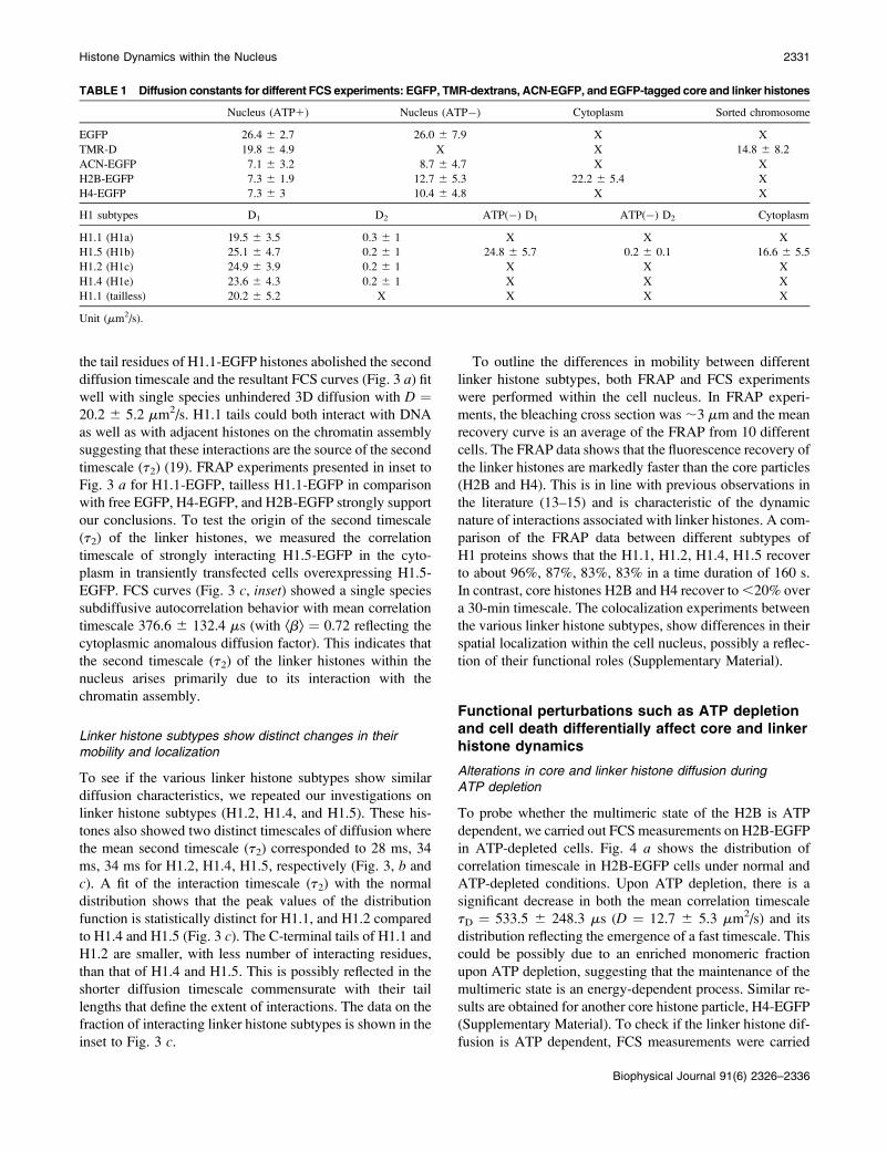
TABLE 1 Diffusion constants for different FCS experiments: EGFP, TMR-dextrans, ACN-EGFP, and EGFP-tagged core and linker histones
Nucleus (ATP1) Nucleus (ATP�) Cytoplasm Sorted chromosome
EGFP 26.4 6 2.7 26.0 6 7.9 X X
TMR-D 19.8 6 4.9 X X 14.8 6 8.2
ACN-EGFP 7.1 6 3.2 8.7 6 4.7 X X
H2B-EGFP 7.3 6 1.9 12.7 6 5.3 22.2 6 5.4 X
H4-EGFP 7.3 6 3 10.4 6 4.8 X X
H1 subtypes D1 D2 ATP(�) D1 ATP(�) D2 Cytoplasm
H1.1 (H1a) 19.5 6 3.5 0.3 6 1 X X X
H1.5 (H1b) 25.1 6 4.7 0.2 6 1 24.8 6 5.7 0.2 6 0.1 16.6 6 5.5
H1.2 (H1c) 24.9 6 3.9 0.2 6 1 X X X
H1.4 (H1e) 23.6 6 4.3 0.2 6 1 X X X
H1.1 (tailless) 20.2 6 5.2 X X X X
Unit (mm2/s).
Histone Dynamics within the Nucleus 2331
Biophysical Journal 91(6) 2326–2336
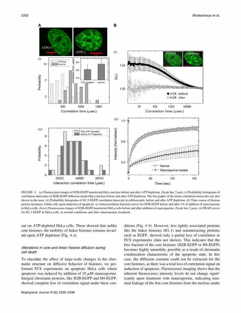
out on ATP-depleted HeLa cells. These showed that unlike
core histones, the mobility of linker histones remains invari-
ant upon ATP depletion (Fig. 4 a).
Alterations in core and linker histone diffusion duringcell death
To elucidate the effect of large-scale changes in the chro-
matin structure on diffusive behavior of histones, we per-
formed FCS experiments on apoptotic HeLa cells where
apoptosis was induced by addition of 10 mM staurosporine.
Integral chromatin proteins, like H2B-EGFP and H4-EGFP,
showed complete loss of correlation signal under these con-
ditions (Fig. 4 b). However, less tightly associated proteins
like the linker histones (H1.1) and noninteracting proteins
such as EGFP, showed only a partial loss of correlation in
FCS experiments (data not shown). This indicates that the
free fraction of the core histones (H2B-EGFP or H4-EGFP)
becomes highly immobile, possibly as a result of chromatin
condensation characteristic of the apoptotic state. In this
case, the diffusion constant could not be extracted for the
core histones, as there was a total loss of correlation signal on
induction of apoptosis. Fluorescence imaging shows that the
inherent fluorescence intensity levels do not change signif-
icantly upon treatment with staurosporine, indicating min-
imal leakage of the free core histones from the nucleus under
FIGURE 4 (a) Fluorescence images of H2B-EGFP transfected HeLa nucleus before and after ATP depletion. (Scale bar, 5 mm). (i) Probability histograms of
correlation timescales of H2B-EGFP diffusion inside HeLa nucleus before and after ATP depletion. The bar graphs of the mean correlation timescales are also
shown in the inset. (ii) Probability histograms of H1.5-EGFP correlation timescale in milliseconds, before and after ATP depletion. (b) Time course of histone
protein dynamics within cells upon induction of apoptosis. (i) Autocorrelation function curves for H2B-EGFP before and after 4 h of addition of staurosporine
in HeLa cells. (Inset) Fluorescence images of H2B-EGFP transfected HeLa cells before and after addition of staurosporine. (Scale bar, 5 mm). (ii) FRAP curves
for H1.1-EGFP in HeLa cells, in normal conditions and after staurosporine treatment.
2332 Bhattacharya et al.
Biophysical Journal 91(6) 2326–2336

these conditions. The fluorescence confocal image (Fig. 4 b,inset) shows punctated bright structures upon induction of
apoptosis, indicating the heterogeneity in chromatin com-
paction. Further to elucidate changes in linker histone mo-
bility in apoptotic cells, FRAP experiments were carried out
on HeLa cells expressing strongly interacting H1.5-EGFP
under normal and apoptotic conditions. FRAP recovery is
significantly slower under conditions of apoptosis relative to
normal cells (Fig. 4 b).These findings using a combination of FCS and FRAP
techniques on both linker and core histones, suggest that core
histones, are more adversely affected by induction of apo-
ptosis in comparison with those of linker histones or a free
molecule that interact less with the chromatin. The measured
diffusion constants from all the experiments are presented in
Table 1.
Numerical simulation reveals possiblemechanism of core and linker histone diffusion
Core histone mobility
The FCS experiments show that the core histone mobility
deviates from single species normal 3D diffusion but un-
dergoes subdiffusive transport. To understand the origins of
this subdiffusion, numerical simulations (26) were carried
out that incorporated all the relevant experimental details.
In the simulation, Brownian particles are made to undergo
normal 3D diffusion through a homogenous mesh that
mimicked the chromatin assembly. Each of these particles
is made to undergo 10,000 random steps in 3D such that
at any given instant an average of at least one molecule
crosses the confocal detection. Such a detection, in the sim-
ulation, is realized by imposing a Gaussian excitation profile
with the width (s) of the profile defined by 50 units. Equiv-
alent to the FCS experiment, the Brownian particle crossing
through this Gaussian profile contributes to the intensity
variation in the time series and therefore the autocorrelation
curve may be calculated. Particles starting from any random
site diffuse in 3D with step size of 10 units through the mesh.
In each random step, if the particle encounters the mesh
coordinates, a time delay is introduced by reversing the par-
ticle to its previous coordinates. The parameters in the sim-
ulation are the step size (dX), transmission probability, the
confocal volume, and the mesh size (Supplementary Mate-
rial). Here the transmission probability is defined as the
probability of crossing the barrier as the particle reaches the
mesh structure. By keeping the step size (10 units), confocal
volume (s ¼ 50 units), and transmission probability (5%)
fixed, we vary the mesh size to visualize the mechanism of
core histone diffusion. Mesh sizes were chosen to be 0 (no
mesh), 10 and 50 units. In addition, to mimic the dynamic
nature of the chromatin assembly, we introduce 20% var-
iability in the mean value of the mesh size. As expected, the
autocorrelation function (ACF) of a Brownian particle with
mesh size (0 units) undergoes normal unhindered 3D dif-
fusion. But as the mesh size is varied, the effect of con-
finement starts being reflected in the particle undergoing
subdiffusive transport (Fig. 5 a). In the inset, we plot the
mean square displacement of the particle as a function of
time for an average of 100 particles with step size 10 units,
diffusing through a mesh of size 0, 11, 12, and 15units. The
effect of confinement is maximum when the mesh size is
comparable to the step size (ÆDX2æ } tb, where b, 1). As the
mesh size is increased relative to the step size, the particle
again undergoes normal 3D unhindered diffusion (where b
FIGURE 5 Numerical simulation study of core and linker histone
dynamics. (a) Autocorrelation function generated by numerical simulation
for single species of 3D diffusion with confined mesh structure with varying
sizes (m ¼ 0, 10, 50). (Inset (i)) Typical single particle trajectory of 2D
random walk generated within a confined mesh structure. (Inset (ii)) Mean
square displacement versus time for different mesh sizes (m¼ 0, 11, 12, 15).
(b) Autocorrelation function generated by numerical simulation for 3D
diffusion with randomly distributed interacting sites (N¼ 0, 1500, 2000, and
3000). (Inset) Typical single particle trajectory of 2D random walk.
Histone Dynamics within the Nucleus 2333
Biophysical Journal 91(6) 2326–2336
approaches 1). Taken together with our experimental find-
ings, these simulations suggest that as the size of a particle
becomes comparable to the mesh size its diffusion tends to
be confined within the local chromatin mesh, leading to sub-
diffusive transport.
Linker histone mobility
To understand the microscopic origins of the two distinct
timescales obtained in the case of linker histones, we carried
out a similar numerical simulation on the linker histones
(26). Each of these particles is made to undergo 40,000
random steps, with step size of 4 units in 3D such that at
any given instant, on average at least one molecule crosses
the confocal detection with width (s ¼ 30 units). In this sim-
ulation, the linker histone proteins are treated as Brownian
particles that undergo both 3D random diffusion and interact
with dynamic pinning sites depicting chromatin binding
sites. To mimic the environment experienced by the linker
histones, we incorporated the presence of binding sites in the
simulation. Here we assume a single particle diffusing in an
environment with a variable number of interacting sites (N¼0, 1500, 2000, 3000) in the confocal volume, where the
particle binds with a mean residence time of 4000 units with
a Gaussian distribution of width 40%. The results of this
simulation are plotted in Fig. 5 b, showing the presence of
distinct second timescale in the autocorrelation curve. In ad-
dition, when the number of interacting sites was varied and
all the other parameters fixed, a clear increase in the second
timescale was observed. By comparing the simulation with
the experimental results, the maximum number of binding
sites within the confocal volume is found to be ;2000.
DISCUSSIONS
Implications of core histone dynamics
Histone protein dynamics within the cell nucleus has become
increasingly important in the context of chromatin remod-
eling, epigenetic states, and various genetic processes (27).
Our results suggest that core histones are in a multimeric
form whereas the linker histones are in monomeric form
within the cell nucleus. The diffusion of multimeric core his-
tones is sensitive to the architecture of the chromatin assem-
bly. Previous measurements suggested that the reconstituted
nucleosomes, with a hydrodynamic diameter of 10 nm that
diffused through a polymer mesh, underwent anamolous
diffusion (28). The chromatin architecture within the cell
nucleus is reminiscent of a dynamic polymer matrix char-
acterized by a mesh size. Molecules smaller than ;3 nm
hydrodynamic radius (EGFP, RH ; 1.7 nm) undergo un-
hindered diffusion whereas larger molecules are confined by
the mesh. The physical origins of anomalous diffusion arise
due to such confinement, where additional time is spent dur-
ing each random Brownian step. In such cases, the mean
square displacement is not proportional to time but propor-
tional to tb, where b (b, 1) is a measure of the confinement
(29). This multimerization process is found to be ATP
dependent because upon ATP depletion the multimeric core
histones (H2B-EGFP) become monomeric, outlined in the
schematic in Fig. 6. Interestingly, the depletion of ATP leads
to the enrichment of monomeric, unbound core histones
within the nucleus. This illustrates the existence of a dy-
namic equilibrium between the chromatin-bound and free
fraction of core histones that is tuned by energy-dependent
processes. A similar-sized molecule, such as a transcription
cofactor activated notch, also undergoes confined diffusion
within the nucleus (Supplementary Material). The core
histone mobility was invariant across organisms, whether in
mammalian cell lines or polytene chromosomes, suggesting
similar evolutionarily conserved local chromatin architecture
in these two systems. Because the histone proteins are
released from the chromatin assembly either in transcription
or replication, their ordered repacking is crucial to maintain
epigenetic states. The possible existence of histones in their
multimeric form and their local confined diffusion within the
cell nucleus suggests higher fidelity of dynamic exchange of
histones to maintain epigenetic memory (6).
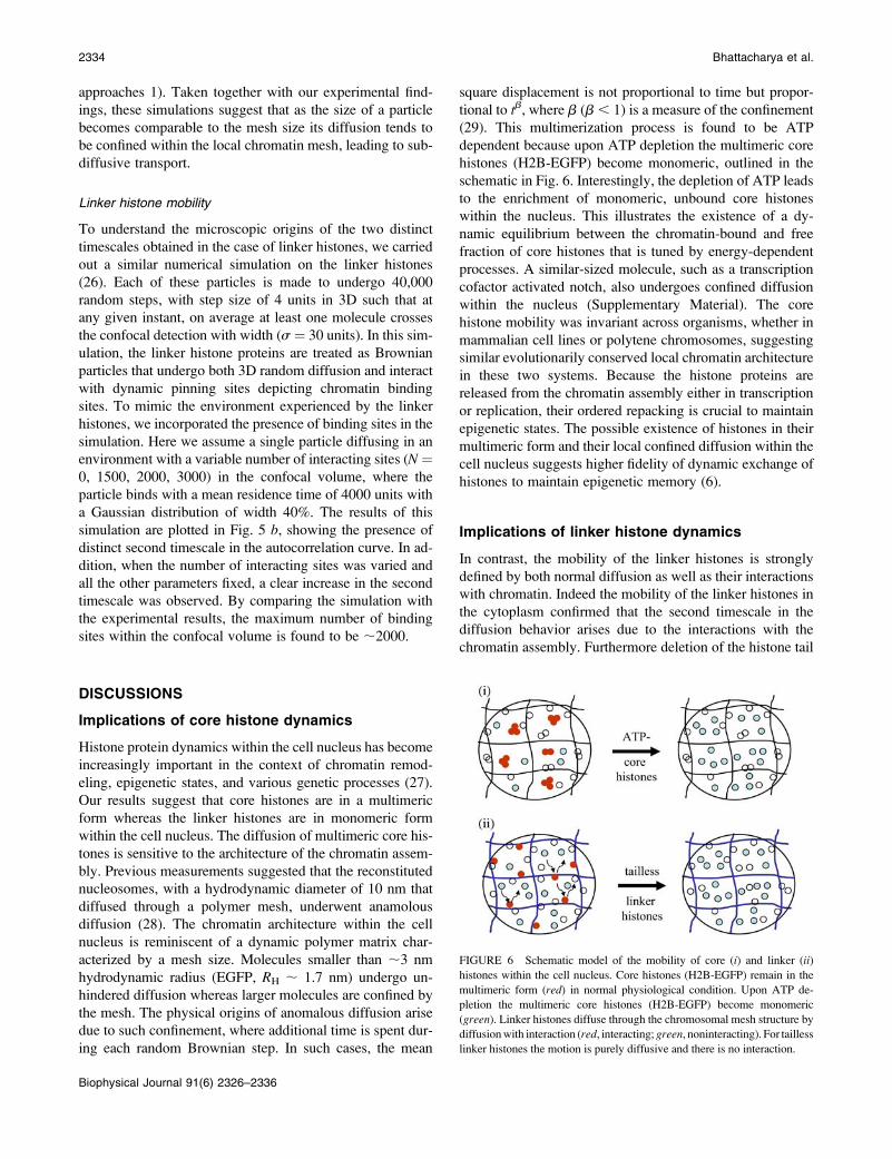
Implications of linker histone dynamics
In contrast, the mobility of the linker histones is strongly
defined by both normal diffusion as well as their interactions
with chromatin. Indeed the mobility of the linker histones in
the cytoplasm confirmed that the second timescale in the
diffusion behavior arises due to the interactions with the
chromatin assembly. Furthermore deletion of the histone tail
FIGURE 6 Schematic model of the mobility of core (i) and linker (ii)
histones within the cell nucleus. Core histones (H2B-EGFP) remain in the
multimeric form (red) in normal physiological condition. Upon ATP de-
pletion the multimeric core histones (H2B-EGFP) become monomeric
(green). Linker histones diffuse through the chromosomal mesh structure by
diffusionwith interaction (red, interacting;green, noninteracting). For tailless
linker histones the motion is purely diffusive and there is no interaction.
2334 Bhattacharya et al.
Biophysical Journal 91(6) 2326–2336
residues on H1.1 establishes that the tail residues are in-
volved in these interactions (19) (Fig. 6). In the case of linker
histones, the unhindered diffusion timescale and the inter-
action timescale are insensitive to ATP depletion suggesting
that interactions are primarily governed by energy-indepen-
dent molecular processes. The various subtypes of linker his-
tones are found to undergo similar diffusive processes where
the minor variations in the interaction timescales could pos-
sibly correspond to differential tail lengths of the histones.
This interaction timescale ;30 ms, measured as the mean
correlation timescale, may suggest a mechanism to introduce
dynamic local conformational fluctuations in chromatin as-
sembly. Here, the DNA at entry and exit points on the nucle-
osome can be continuously modulated by the binding and
unbinding of the linker histones thus providing a mechanism
by which regulatory proteins may spontaneously access
specific DNA sequences (30,31).
The mobility of histone proteins that are differentially
associated with chromatin is also differentially affected upon
induction of apoptosis. The highly interacting core histones
become completely immobile in apoptotic nuclei, while the
mobility of less interacting linker histones or noninteracting
protein is only partially affected. The simulation results ade-
quately describe our experimental findings that the nanoscale
chromatin architecture defines the dynamics of core and
linker histones within the cell nucleus. The differential bind-
ing timescales of core and linker histones, in addition to other
chromatin binding proteins (such as high mobility group
proteins (HMG) (32) and heterochromatin binding proteins
HP1 (33)), perhaps provides an appropriate substrate for
dynamic self-organization of regulatory information on DNA
within living cells (34). Work in progress in our lab, using
single molecule microscopy, is currently addressing the im-
portance of histone dynamics in the context of their epi-
genetic states and their role in transcription control.
We thank Satyajit Mayor, Madan Rao, Apurva Sarin, and K. Vijay
Raghavan for useful discussions and NCBS for the imaging facility funded
by the Wellcome Trust. We also thank Yamuna Krishnan for critical reading
of the manuscript.
REFERENCES
1. Spector, D. L. 2003. The dynamics of chromosome organization andgene regulation. Annu. Rev. Biochem. 72:573–608.
2. Schalch, T., S. Duda, D. F. Sargent, and T. J. Richmond. 2005. X-raystructure of a tetranucleosome and its implications for the chromatinfibre. Nature. 436:138–142.
3. Zlatanova, J., P. Caiafa, and K. van Holde. 2000. Linker histonebinding and displacement: versatile mechanism for transcriptionalregulation. FASEB J. 14:1697–1704.
4. Cosgrove, M. S., J. D. Boeke, and C. Wolberger. 2004. Regulatednucleosome mobility and the histone code. Nat. Struct. Mol. Biol.11:1037–1043.
5. Jenuwein, T., and C. D. Allis. 2001. Translating the histone code.Science. 293:1074–1080.
6. Ahmad, K., and S. Henikoff. 2002. Epigenetic consequences ofnucleosome dynamics. Cell. 111:281–284.
7. Khorasanizadeh, S. 2004. The nucleosome: from genomic organizationto genomic regulation. Cell. 116:259–272.
8. Hansen, J. C. 2002. Conformational dynamics of the chromatin fiber insolution: determinants, mechanisms, and functions. Annu. Rev.Biophys. Biomol. Struct. 31:361–392.
9. Becker, M., A. Becker, F. Miyara, Z. Han, M. Kihara, D. T. Brown,G. L. Hager, K. Latham, E. Y. Adashi, and T. Misteli. 2005. Differential in-vivo binding dynamics of somatic and oocyte-specific linker histones inoocytes and during ES cell nuclear transfer.Mol. Biol. Cell. 16:3887–3895.
10. Bustin, M., F. Catez, and J. H. Lim. 2005. The dynamics of histone H1function in chromatin. Mol. Cell. 17:617–620.
11. Gasser, S. M. 2002. Visualizing chromatin dynamics in interphasenuclei. Science. 296:1412–1416.
12. Kanda, T., K. F. Sullivan, and G. M. Wahi. 1998. Histone-GFP fusionprotein enables sensitive analysis of chromosome dynamics in livingmammalian cells. Curr. Biol. 8:377–385.
13. Kimura, H., and P. Cook. 2001. Kinetics of core histones in livinghuman cells: little exchange of H3 & H4 and some rapid exchange ofH2B. J. Cell Biol. 153:1341–1353.
14. Misteli, T., A. Gunjan, R. Hock, M. Bustin, and D. T. Brown. 2000.Dynamic binding of histone H1 to chromatin in living cells. Nature.408:877–881.
15. Lever, M. A., J. P. Th’ng, X. Sun, and M. J. Hendzel. 2000. Rapidexchange of histone H1.1 on chromatin in the living human cells.Nature. 408:873–876.
16. Turner, B. M. 2002. Cellular memory and the histone code. Cell.111:285–291.
17. Thiriet, C., and J. J. Hayes. 2005. Replication-independent core histonedynamics at transcriptionally active loci in vivo. Genes Dev. 19:677–682.
18. Bruno, M., A. Flaus, C. Stockdale, C. Rencurel, H. Ferreira, and T. O.Hughes. 2003. Histone H2A/H2B dimer exchange by ATP-dependentchromatin remodeling activities. Mol. Cell. 12:1599–1606.
19. Th’ng, J. P., R. Sung, M. Ye, and M. J. Hendzel. 2005. H1 familyhistones in the nucleus: control of binding and localization by thec-terminal domain. J. Biol. Chem. 280:27809–27814.
20. Contreras, A., and R. E. Herrera. 2003. The dynamic mobility of histoneH1 is regulated by cyclin/CDK phosphorylation. Mol. Cell. Biol. 23:8626–8636.
21. Marshall, W. F. 2002. Order and disorder in the nucleus. Curr. Biol.12:185–192.
22. Chen, Y., J. D. Muller, Q. Q. Ruan, and E. Gratton. 2002. Molecularbrightness characterization of EGFP in vivo by fluorescence fluctuationspectroscopy. Biophys. J. 82:133–144.
23. Koberna, K., D. Stanek, J. Malinsky, M. Eltsov, A. Pliss, V. Ctrnacta,S. Cermanova, and I. Raska. 1999. Nuclear organization studied withthe help of a hypotonic shift: its use permits hydrophilic molecules toenter into living cells. Chromosoma. 108:325–335.
24. Pardue, M. L. 1994. Looking at Polytene Chromosomes. Methods CellBiol. 44:333–351.
25. Mangenot, S., A. Leforestier, P. Vachette, D. Durand, and F. Livolant.2002. Salt induced conformation and interaction changes of nucleo-some core particles. Biophys. J. 82:345–356.
26. Weiss, M., M. Elsner, F. Kartberg, and T. Nilsson. 2004. Anomaloussubdiffusion is a measure for cytoplasmic crowding in living cells.Biophys. J. 87:3518–3524.
27. Kimura, H. 2005. Histone dynamics in living cells revealed byphotobleaching. DNA Repair. 4:939–950.
28. Mangenot, S., S. Keller, and J. Radler. 2003. Transport of nucleosomecore particles in semidilute DNA solutions. Biophys. J. 85:1817–1825.
29. Weidemann, T., M. Wachsmuth, T. A. Knoch, G. Muller, W. Waldeck,and J. Langowski. 2003. Counting nucleosome in living cells with acombination of fluorescence correlation spectroscopy and confocalimaging. J. Mol. Biol. 334:229–240.
30. Tomschik, M., H. Zheng, K. van Holde, J. Zlatanova, and S. H. Leuba.2005. Fast, long-range, reversible conformational fluctuations in
Histone Dynamics within the Nucleus 2335
Biophysical Journal 91(6) 2326–2336

nucleosomes revealed by single-pair fluorescence resonance energytransfer. Proc. Natl. Acad. Sci. USA. 102:3278–3283.
31. Li, G., M. Levitus, C. Bustamante, and J. Widom. 2005. Rapidspontaneous accessibility of nucleosomal DNA. Nat. Struct. Mol. Biol.12:46–53.
32. Phair, R. D., P. Scaffidi, C. Elbi, J. Vecerova, A. Dey, K. Ozato, D. T.Brown, G. Hager, M. Bustin, and T. Misteli. 2004. Global nature ofdynamic protein-chromatin interactions in vivo: three-dimensionalgenome scanning and dynamic interaction networks of chromatinproteins. Mol. Cell. Biol. 24:6393–6402.
33. Schmiedeberg, L., K. Weisshart, S. Diekmann, G. Meyer zu Hoerste,and P. Hemmerich. 2004. High and low-mobility populations ofHP1 in heterochromatin of mammalian cells. Mol. Biol. Cell. 15:2819–2833.
34. Gorski, S., and T. Misteli. 2005. Systems biology in the cell nucleus.J. Cell Sci. 118:4083–4092.
35. Periasamy, N., and A. S. Verkman. 1998. Analysis of fluorophorediffusion by continuous distribution of diffusion coefficients: applica-tion to photo-bleaching measurements of multicomponent and anom-alous diffusion. Biophys. J. 75:557–567.
2336 Bhattacharya et al.
Biophysical Journal 91(6) 2326–2336