Linker histone H1 represses recombination at the ribosomal DNA locus in the budding yeast...
14
Linker histone H1 represses recombination at the ribosomal DNA locus in the budding yeast Saccharomyces cerevisiae Chonghua Li, John E. Mueller, Megan Elfline and Mary Bryk* Department of Biochemistry and Biophysics, Texas A&M University, College Station, TX, USA. Summary Several epigenetic phenomena occur at ribosomal DNA loci in eukaryotic cells, including the silencing of Pol I and Pol II transcribed genes, silencing of repli- cation origins and repression of recombination. In Saccharomyces cerevisiae, studies focusing on the silencing of Pol II transcription and genetic recombi- nation at the ribosomal DNA locus (rDNA) have pro- vided insight into the mechanisms through which chromatin and chromatin-associated factors regulate gene expression and chromosome stability. The core histones, H2A, H2B, H3 and H4, the fundamental building blocks of chromatin, have been shown to regulate silent chromatin at the rDNA; however, the function of the linker histone H1 has not been well characterized. Here, we show that S. cerevisiae histone H1 represses recombination at the rDNA without affecting Pol II gene silencing. The most highly studied repressor of recombination at the rDNA is the Silent information regulator protein Sir2. We find that cells lacking histone H1 do not exhibit a premature-ageing phenotype nor do they accumulate the rDNA recombination intermediates and products that are found in cells lacking Sir2. These results suggest that histone H1 represses recombination at the rDNA by a mechanism that is independent of the recombination pathways regulated by Sir2. Introduction The fundamental unit of eukaryotic chromatin is the nucleosome, consisting of DNA wrapped around an octamer of histones containing two copies of each of the four core histones, H2A, H2B, H3 and H4. Neighbouring nucleosomes are separated by a segment of DNA that can interact with the linker histone allowing further com- paction of chromatin (reviewed in Luger, 2006). In eukary- otic cells, the core histones H2A, H2B, H3 and H4 are highly conserved, while the linker histone, histone H1, is less well conserved (Wells and Brown, 1991). Saccharomyces cerevisiae histone H1, encoded by the HHO1 gene, is unusual, having two globular domains instead of the one found in linker histones from other eukaryotes (Landsman, 1996; Ono et al., 2003; Ali et al., 2004). A number of observations are consistent with HHO1 encoding a linker histone. First, there is a relatively high degree of sequence identity and/or structural similarity when comparing S. cerevisiae histone H1 to linker histones in higher eukaryotes (Landsman, 1996; Ushinsky et al., 1997; Ono et al., 2003; Ali et al., 2004). Second, studies have shown that histone H1 localizes to the nucleus, copurifies with core histones, and protects DNA located at the entry and exit point of a nucleosome from micrococcal nuclease cleavage (Ushinsky et al., 1997; Patterton et al., 1998; Freidkin and Katcoff, 2001). Consistent with each globular domain being functional, S. cerevisiae histone H1 has been shown to interact simultaneously with two four-way junctions that may have a structure similar to the point on the nucleosome where DNA enters and exits (Schafer et al., 2005). This result supports a model where a single histone H1 protein can bridge two nucleosomes and facilitate the compaction of chromatin. These properties of the S. cerevisiae H1 mirror those of linker histones from higher eukaryotes (reviewed in Bustin et al., 2005), supporting its role as a bona fide linker histone. The in vivo functions of linker histones in higher eukary- otes have been difficult to assess owing to the large number of linker histone variants. Early studies showed that deletion of any one or two of the eight known genes encoding H1 variants in mice causes no obvious mutant phenotypes (Fan et al., 2001). Although, age-dependent differences in silencing of a human b-globin transgene have been observed in mice lacking a single H1 gene (Fan et al., 2001; Alami et al., 2003). More extensive mutant analyses have demonstrated an essential function for linker histones in mammals. Mice lacking three specific H1 variants have an embryonic lethal phenotype, with total histone H1 levels reduced to ~50% of normal, severe developmental defects, altered chromatin structure, and Accepted 18 December, 2007. *For correspondence. E-mail bryk@ tamu.edu; Tel. (+1) 979 862 2294; Fax (+1) 979 845 9274. Molecular Microbiology (2008) 67(4), 906–919 doi:10.1111/j.1365-2958.2007.06101.x First published online 7 January 2008 © 2008 The Authors Journal compilation © 2008 Blackwell Publishing Ltd
Transcript of Linker histone H1 represses recombination at the ribosomal DNA locus in the budding yeast...

Linker histone H1 represses recombination at the ribosomalDNA locus in the budding yeast Saccharomyces cerevisiae
Chonghua Li, John E. Mueller, Megan Elfline andMary Bryk*Department of Biochemistry and Biophysics, TexasA&M University, College Station, TX, USA.
Summary
Several epigenetic phenomena occur at ribosomalDNA loci in eukaryotic cells, including the silencing ofPol I and Pol II transcribed genes, silencing of repli-cation origins and repression of recombination. InSaccharomyces cerevisiae, studies focusing on thesilencing of Pol II transcription and genetic recombi-nation at the ribosomal DNA locus (rDNA) have pro-vided insight into the mechanisms through whichchromatin and chromatin-associated factors regulategene expression and chromosome stability. The corehistones, H2A, H2B, H3 and H4, the fundamentalbuilding blocks of chromatin, have been shown toregulate silent chromatin at the rDNA; however,the function of the linker histone H1 has not beenwell characterized. Here, we show that S. cerevisiaehistone H1 represses recombination at the rDNAwithout affecting Pol II gene silencing. The mosthighly studied repressor of recombination at therDNA is the Silent information regulator protein Sir2.We find that cells lacking histone H1 do not exhibit apremature-ageing phenotype nor do they accumulatethe rDNA recombination intermediates and productsthat are found in cells lacking Sir2. These resultssuggest that histone H1 represses recombination atthe rDNA by a mechanism that is independent of therecombination pathways regulated by Sir2.
Introduction
The fundamental unit of eukaryotic chromatin is thenucleosome, consisting of DNA wrapped around anoctamer of histones containing two copies of each of thefour core histones, H2A, H2B, H3 and H4. Neighbouringnucleosomes are separated by a segment of DNA that
can interact with the linker histone allowing further com-paction of chromatin (reviewed in Luger, 2006). In eukary-otic cells, the core histones H2A, H2B, H3 and H4 arehighly conserved, while the linker histone, histone H1, isless well conserved (Wells and Brown, 1991).
Saccharomyces cerevisiae histone H1, encoded by theHHO1 gene, is unusual, having two globular domainsinstead of the one found in linker histones from othereukaryotes (Landsman, 1996; Ono et al., 2003; Ali et al.,2004). A number of observations are consistent withHHO1 encoding a linker histone. First, there is a relativelyhigh degree of sequence identity and/or structuralsimilarity when comparing S. cerevisiae histone H1 tolinker histones in higher eukaryotes (Landsman, 1996;Ushinsky et al., 1997; Ono et al., 2003; Ali et al., 2004).Second, studies have shown that histone H1 localizes tothe nucleus, copurifies with core histones, and protectsDNA located at the entry and exit point of a nucleosomefrom micrococcal nuclease cleavage (Ushinsky et al.,1997; Patterton et al., 1998; Freidkin and Katcoff, 2001).Consistent with each globular domain being functional,S. cerevisiae histone H1 has been shown to interactsimultaneously with two four-way junctions that may havea structure similar to the point on the nucleosome whereDNA enters and exits (Schafer et al., 2005). This resultsupports a model where a single histone H1 protein canbridge two nucleosomes and facilitate the compaction ofchromatin. These properties of the S. cerevisiae H1 mirrorthose of linker histones from higher eukaryotes (reviewedin Bustin et al., 2005), supporting its role as a bona fidelinker histone.
The in vivo functions of linker histones in higher eukary-otes have been difficult to assess owing to the largenumber of linker histone variants. Early studies showedthat deletion of any one or two of the eight known genesencoding H1 variants in mice causes no obvious mutantphenotypes (Fan et al., 2001). Although, age-dependentdifferences in silencing of a human b-globin transgenehave been observed in mice lacking a single H1 gene(Fan et al., 2001; Alami et al., 2003). More extensivemutant analyses have demonstrated an essential functionfor linker histones in mammals. Mice lacking three specificH1 variants have an embryonic lethal phenotype, withtotal histone H1 levels reduced to ~50% of normal, severedevelopmental defects, altered chromatin structure, and
Accepted 18 December, 2007. *For correspondence. E-mail [email protected]; Tel. (+1) 979 862 2294; Fax (+1) 979 845 9274.
Molecular Microbiology (2008) 67(4), 906–919 � doi:10.1111/j.1365-2958.2007.06101.xFirst published online 7 January 2008
© 2008 The AuthorsJournal compilation © 2008 Blackwell Publishing Ltd

either increased or decreased expression of a smallnumber of genes (Fan et al., 2003; 2005).
In contrast to mammalian cells, in several unicellulareukaryotes, including S. cerevisiae, A. nidulans andT. thermophila, histone H1 is not essential for viability(Shen and Gorovsky, 1996; Escher and Schaffner, 1997;Ushinsky et al., 1997; Ramon et al., 2000). Althoughhistone H1 has been shown to associate with chromatinin S. cerevisiae, cells lacking or overexpressing HHO1exhibit wild-type growth characteristics (Escher andSchaffner, 1997; Ushinsky et al., 1997; Patterton et al.,1998; Freidkin and Katcoff, 2001; Downs et al., 2003;Veron et al. 2006). Microarray analyses have revealedthat expression of very few genes is altered in cellslacking HHO1 (Freidkin and Katcoff, 2001; Hellauer et al.,2001). Moreover, defects in gene silencing at the silentmating-type loci or telomeres, phenotypes often associ-ated with mutations that alter chromatin function, havenot been observed in cells lacking histone H1 (Escherand Schaffner, 1997; Patterton et al., 1998; Veron et al.,2006). Evidence supporting a role for S. cerevisiaehistone H1 in chromatin function has been provided bytwo reports. First, overexpression studies have indicatedthat histone H1 regulates chromatin barriers locatedbetween euchromatin and heterochromatin-like domains(Veron et al., 2006). Second, S. cerevisiae cells thatlack histone H1 exhibit increased tolerance to methyl-methane-sulphonate and other related phenotypes, sup-porting a role for histone H1 in genetic recombinationand/or DNA repair (Downs et al., 2003).
RNA polymerase II (Pol II) transcription and geneticrecombination are repressed at the ribosomal DNA locusin S. cerevisiae (reviewed in Moazed, 2001; Ruscheet al., 2003). The maintenance of silent chromatin at therDNA requires several types of factors with known roles inchromatin function. Sir2, an NAD-dependent histonedeacetylase, and Ubp10, a histone H2B deubiquitylatingenzyme, are required for the repression of Pol II tran-scription and recombination at the rDNA (Gottlieb andEsposito, 1989; Bryk et al., 1997; Smith and Boeke, 1997;Smith et al., 1998; Straight et al., 1999; Garcia and Pillus,2002; Kobayashi et al., 2004; Gardner et al., 2005; Emreet al., 2005; Calzari et al., 2006; Cubizolles et al., 2006).Sir2 has been shown to associate with several regions ofthe rDNA and deacetylate histones H3 and H4 throughoutthe rDNA (Bryk et al., 2002; Buck et al., 2002; Huang andMoazed, 2003; Li et al., 2006). A second class of rDNAsilencing factors, including Set1, a histone H3 lysine 4methyltransferase, silences Pol II gene transcription butnot recombination at the rDNA (Briggs et al., 2001; Bryket al., 2002; Krogan et al., 2002; Mueller et al., 2006).Members of a third class of rDNA silencing factors includ-ing Sgs1 and Hpr1, repress recombination at the rDNAwithout affecting Pol II gene silencing (Sinclair et al.,
1997; Bryk et al., 2001; McVey et al., 2001; Merker andKlein, 2002).
Considering the role of histone H1 in compaction ofchromatin and its function at barrier elements, we asked ifhistone H1 regulates silent chromatin at the rDNA. Ourdata show that histone H1 behaves similarly to Sgs1 andHpr1, repressing recombination at the rDNA while havingno effect on Pol II gene silencing. The results of geneticand molecular experiments suggest that histone H1represses recombination at the rDNA through a mecha-nism that is largely independent of Sir2. For example, inhho1D cells, extrachromosomal rDNA circles (ERCs), aproduct of the Sir2 regulated recombination pathway, donot accumulate (reviewed in Rothstein and Gangloff,1999; Kobayashi, 2006). Based on our findings, wepropose that histone H1 acts independently of Sir2 toprevent recombination events at the rDNA that if allowedto occur would contribute to genomic instability.
Results
The ribosomal DNA locus on chromosome XII inS. cerevisiae contains ~150–200 tandem direct repeats ofthe ribosomal rRNA genes. The rDNA is located in thenucleolus, a substructure near the periphery of thenucleus, where transcription and processing of the ribo-somal RNA (rRNA) occurs (Warner, 1990). Each 9.1 kbrDNA repeat contains a 35S rRNA gene that is transcribedby RNA polymerase I, and a 5S rRNA gene that is tran-scribed by RNA polymerase III, which is located in themiddle of the non-transcribed spacer (NTS), splitting it intoNTS1 and NTS2 (Fig. 1A). Considering the high levels ofPol I and Pol III transcription that occur at the rDNA, it issomewhat contradictory that this locus exhibits character-istics of silent chromatin. However, silencing at the rDNAmaintains genome stability and cell longevity throughthe repression of Pol II transcription and homologousrecombination.
Histone H1 does not regulate transcriptionalsilencing at the rDNA
Previous work has shown that histone H1 does not regu-late transcriptional silencing at the silent mating-type lociand telomeres in S. cerevisiae (Escher and Schaffner,1997; Patterton et al., 1998; Veron et al., 2006). However,a function for histone H1 at the rDNA has been suggestedby the results of chromatin immunoprecipitation (ChIP)experiments showing that H1 associates with the rDNA(Freidkin and Katcoff, 2001; Downs et al., 2003). To deter-mine if histone H1 is required for the silencing of Pol IIgenes at the rDNA, we analysed RNA from wild-type andhho1D cells that each contain a single copy of a geneti-cally marked Ty1 element, Ty1his3AI, located in NTS1
Histone H1 represses rDNA recombination 907
© 2008 The AuthorsJournal compilation © 2008 Blackwell Publishing Ltd, Molecular Microbiology, 67, 906–919

(-236) or NTS2 (-272 or -234) of a single rDNA repeat(Fig. 1). Ty1 elements are transcribed by Pol II and thelevel of mRNA from a Ty1his3AI element integrated at therDNA is a sensitive indicator of Pol II gene silencing (Bryket al., 1997). An RNA probe complementary to the his3portion of Ty1his3AI was used to detect Ty1his3AI mRNA(Fig. 1B, Ty1his3AI ). As a control for loading, we mea-sured the level of PYK1 transcript (PYK1). In the leftmostblot of Fig. 1B, analysis of the ratio of Ty1his3AI-236:PYK1 mRNA showed that the level of Ty1his3AI-236mRNA in the hho1D cells was similar to the level in wild-type cells, indicating that Pol II gene silencing at the rDNAdoes not require histone H1. As a positive control, wemeasured Ty1his3AI-236 mRNA in cells lacking thehistone methyltransferase Set1 that is required for tran-scriptional silencing at the rDNA and telomeres (Briggset al., 2001; Bryk et al., 2002; Krogan et al., 2002; Muelleret al., 2006). Consistent with the requirement for Set1 inrDNA silencing, we observed a significant increase inTy1his3AI-236 mRNA in set1D cells (Fig. 1B). We analy-
sed pairs of wild-type and hho1D cells with Ty1his3AIelements in NTS2 and found that silencing of theseTy1his3AI elements was not affected in cells lackinghistone H1 (Fig. 1B, -272 and -234 lanes).
Most S. cerevisiae strains contain about 30 Ty1 ele-ments at different locations throughout the genome.Although expression of the Ty1his3AI element in the rDNAwas not affected by deletion of HHO1, we checked thelevel of RNA from the genomic Ty1 elements using aprobe that hybridizes to all Ty1 mRNA. We did not detecta significant difference in the level of total Ty1 mRNA inwild-type and hho1D cells (Fig. 1B, total Ty1). Likewise,we observed similar levels of Ty1his3AI mRNA from theTy1his3AI-242 element located outside the rDNA on chro-mosome XII in wild-type and hho1D cells (Fig. 1B, -242lanes). In summary, our results indicate that histone H1 isnot required for the silencing of Pol II-transcribed genesintegrated in the rDNA NTS.
To determine if histone H1 is required for silencing ofPol II genes inserted in the 35S rRNA gene, we used a
Fig. 1. Cells lacking histone H1 do notexhibit transcriptional silencing defect.A. The ribosomal DNA locus on chromosomeXII is comprised of ~150–200 copies of therDNA repeat. Two rDNA repeats are shown inthe enlargement with each containing anintergenic spacer (NTS) and the Pol Itranscribed 35S rRNA gene (35S rRNA). TheNTS is divided into NTS1 and NTS2 by thePol III transcribed 5S rRNA gene (5S).Stacked triangles, replication fork barrier(RFB); open circle, origin of replication (ARS);bent arrows with numbers, location of theTy1his3AI elements.B. Northern blot analysis of total RNA isolatedfrom wild-type, hho1D and set1D cells.Strand-specific probes were used to measurethe steady-state mRNA levels of Ty1his3AI(top panel), total Ty1 (middle panel) or PYK1(bottom panel). The average ratio ofTy1his3AI/PYK1 mRNA and total Ty1/PYK1mRNA for each strain analysed afternormalization to the wild-type strain is shownbelow the top and middle panel respectively.The normalized values of the averageratio � standard error for Ty1his3AI-236/PYK1mRNA were: hho1D : WT, 0.9 � 0.1, n = 16;set1D : WT, 2.4 � 0.2, n = 5; and for totalTy1/PYK1 mRNA were: hho1D : WT,1.3 � 0.1, n = 14; set1D : WT, 1.0 � 0.2,n = 3. Average hho1D : WT forTy1his3AI-272/PYK1 mRNA was 0.9 � 0.04,n = 4; and for total Ty1/PYK1 mRNA was0.7 � 0.1, n = 4. Average hho1D : WT forTy1his3AI-234/PYK1 mRNA was 1.0 � 0.1,n = 4; and for total Ty1/PYK1 mRNA was0.8 � 0.1, n = 4. Average hho1D : WT forTy1his3AI-242/PYK1 mRNA was 0.8 � 0.1,n = 4; and for total Ty1/PYK1 mRNA was0.8 � 0.1, n = 4. Total RNA from wild-typecells lacking the Ty1his3AI element (none)was analysed to provide a measure ofnon-specific binding of the radiolabelledprobe.
908 C. Li, J. E. Mueller, M. Elfline and M. Bryk �
© 2008 The AuthorsJournal compilation © 2008 Blackwell Publishing Ltd, Molecular Microbiology, 67, 906–919
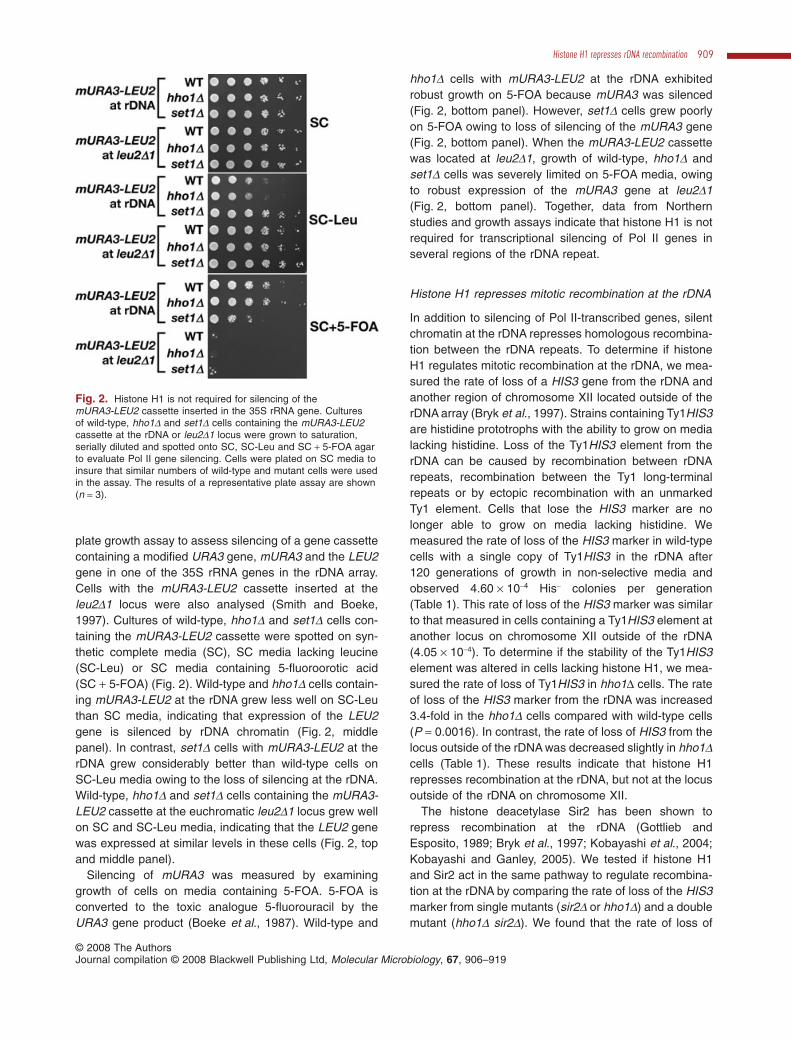
plate growth assay to assess silencing of a gene cassettecontaining a modified URA3 gene, mURA3 and the LEU2gene in one of the 35S rRNA genes in the rDNA array.Cells with the mURA3-LEU2 cassette inserted at theleu2D1 locus were also analysed (Smith and Boeke,1997). Cultures of wild-type, hho1D and set1D cells con-taining the mURA3-LEU2 cassette were spotted on syn-thetic complete media (SC), SC media lacking leucine(SC-Leu) or SC media containing 5-fluoroorotic acid(SC + 5-FOA) (Fig. 2). Wild-type and hho1D cells contain-ing mURA3-LEU2 at the rDNA grew less well on SC-Leuthan SC media, indicating that expression of the LEU2gene is silenced by rDNA chromatin (Fig. 2, middlepanel). In contrast, set1D cells with mURA3-LEU2 at therDNA grew considerably better than wild-type cells onSC-Leu media owing to the loss of silencing at the rDNA.Wild-type, hho1D and set1D cells containing the mURA3-LEU2 cassette at the euchromatic leu2D1 locus grew wellon SC and SC-Leu media, indicating that the LEU2 genewas expressed at similar levels in these cells (Fig. 2, topand middle panel).
Silencing of mURA3 was measured by examininggrowth of cells on media containing 5-FOA. 5-FOA isconverted to the toxic analogue 5-fluorouracil by theURA3 gene product (Boeke et al., 1987). Wild-type and
hho1D cells with mURA3-LEU2 at the rDNA exhibitedrobust growth on 5-FOA because mURA3 was silenced(Fig. 2, bottom panel). However, set1D cells grew poorlyon 5-FOA owing to loss of silencing of the mURA3 gene(Fig. 2, bottom panel). When the mURA3-LEU2 cassettewas located at leu2D1, growth of wild-type, hho1D andset1D cells was severely limited on 5-FOA media, owingto robust expression of the mURA3 gene at leu2D1(Fig. 2, bottom panel). Together, data from Northernstudies and growth assays indicate that histone H1 is notrequired for transcriptional silencing of Pol II genes inseveral regions of the rDNA repeat.
Histone H1 represses mitotic recombination at the rDNA
In addition to silencing of Pol II-transcribed genes, silentchromatin at the rDNA represses homologous recombina-tion between the rDNA repeats. To determine if histoneH1 regulates mitotic recombination at the rDNA, we mea-sured the rate of loss of a HIS3 gene from the rDNA andanother region of chromosome XII located outside of therDNA array (Bryk et al., 1997). Strains containing Ty1HIS3are histidine prototrophs with the ability to grow on medialacking histidine. Loss of the Ty1HIS3 element from therDNA can be caused by recombination between rDNArepeats, recombination between the Ty1 long-terminalrepeats or by ectopic recombination with an unmarkedTy1 element. Cells that lose the HIS3 marker are nolonger able to grow on media lacking histidine. Wemeasured the rate of loss of the HIS3 marker in wild-typecells with a single copy of Ty1HIS3 in the rDNA after120 generations of growth in non-selective media andobserved 4.60 ¥ 10-4 His- colonies per generation(Table 1). This rate of loss of the HIS3 marker was similarto that measured in cells containing a Ty1HIS3 element atanother locus on chromosome XII outside of the rDNA(4.05 ¥ 10-4). To determine if the stability of the Ty1HIS3element was altered in cells lacking histone H1, we mea-sured the rate of loss of Ty1HIS3 in hho1D cells. The rateof loss of the HIS3 marker from the rDNA was increased3.4-fold in the hho1D cells compared with wild-type cells(P = 0.0016). In contrast, the rate of loss of HIS3 from thelocus outside of the rDNA was decreased slightly in hho1Dcells (Table 1). These results indicate that histone H1represses recombination at the rDNA, but not at the locusoutside of the rDNA on chromosome XII.
The histone deacetylase Sir2 has been shown torepress recombination at the rDNA (Gottlieb andEsposito, 1989; Bryk et al., 1997; Kobayashi et al., 2004;Kobayashi and Ganley, 2005). We tested if histone H1and Sir2 act in the same pathway to regulate recombina-tion at the rDNA by comparing the rate of loss of the HIS3marker from single mutants (sir2D or hho1D) and a doublemutant (hho1D sir2D). We found that the rate of loss of
Fig. 2. Histone H1 is not required for silencing of themURA3-LEU2 cassette inserted in the 35S rRNA gene. Culturesof wild-type, hho1D and set1D cells containing the mURA3-LEU2cassette at the rDNA or leu2D1 locus were grown to saturation,serially diluted and spotted onto SC, SC-Leu and SC + 5-FOA agarto evaluate Pol II gene silencing. Cells were plated on SC media toinsure that similar numbers of wild-type and mutant cells were usedin the assay. The results of a representative plate assay are shown(n = 3).
Histone H1 represses rDNA recombination 909
© 2008 The AuthorsJournal compilation © 2008 Blackwell Publishing Ltd, Molecular Microbiology, 67, 906–919

Ty1HIS3 element from the rDNA in sir2D cells was4.19 ¥ 10-3, which was 9.1-fold higher than the rate inwild-type cells (P = 0.0005), and the rate of loss in thehho1D sir2D double mutant was 12.6-fold higher (P =0.0025) (Table 1). Loss of Ty1HIS3 from the rDNA in thehho1D sir2D double mutant was approximately equal tothe sum of the rates observed for the two single mutants,suggesting that Sir2 and histone H1 act independently toregulate recombination at the rDNA.
We also addressed possible genetic interactionsbetween histone H1 and Fob1, the rDNA replication forkbarrier protein that has been shown to recruit the Sir2-containing RENT complex to NTS1 (Huang and Moazed,2003). Consistent with previous work (Kobayashi andHoriuchi, 1996), the rate of marker loss from the rDNAwas lower in fob1D cells than in wild-type cells (Table 1).In hho1D fob1D double mutants recombination rates wererestored to wild-type levels, suggesting that histone H1and Fob1 function antagonistically to regulate recombina-tion at the rDNA.
Histone H1 associates with the NTS andthe 35S regions of the rDNA locus
The results of experiments examining recombination(Table 1) suggest that histone H1 and Sir2 function inindependent pathways to regulate the mitotic stability ofthe rDNA array. To determine if histone H1 is required forthe function of Sir2 at the rDNA NTS and if Sir2 affects theassociation of histone H1 with the rDNA, we performed aseries of ChIP experiments. First, we examined the asso-ciation of Sir2 with the rDNA in cells lacking histone H1.ChIPs were performed on wild-type, hho1D and sir2D cellsusing antisera specific for Sir2, and a slot blot containingthe immunoprecipitated and input DNA was analysedafter hybridization with a radiolabelled probe to the rDNA
NTS. The analysis showed that the association of Sir2with the rDNA NTS is similar in wild-type and hho1D cells(Fig. 3A). To assess the function of Sir2 at the rDNA inthese cells, we measured the level of acetylated histoneH3 at the rDNA NTS. The results showed that the level ofK9-K14 acetylated histone H3 at the rDNA NTS is lower inwild-type cells than in sir2D cells (Fig. 3B), reflecting thehistone deacetylase activity of Sir2. We found that thelevel of K9-K14-acetylated H3 at the rDNA NTS in hho1Dcells was similar to the level in wild-type cells, suggestingthat histone H1 does not affect the histone deacetylasefunction of Sir2 at the rDNA.
Next, we asked if Sir2 affects the ability of histone H1to associate with the rDNA. For these ChIPs, we utilizedcells containing a myc-tagged version of histone H1 andcells containing untagged histone H1 to obtain a mea-surement of background signal. After immunoprecipita-tion, we analysed immunoprecipitated and input DNAs byquantitative real-time PCR using 14 sets of primers thatspan the NTS and the 5′ end of the 35S rRNA gene(Fig. 3C). Consistent with earlier work (Freidkin andKatcoff, 2001), we detected histone H1 at the 35S rRNAgene. In addition, we found that histone H1 associateswith the NTS region of the rDNA repeat (Fig. 3C, blackbars). In cells lacking Sir2, we detected reduced levels ofhistone H1 in several regions of the NTS, with the largesteffect (approximately twofold) between the RFB and 5SrRNA gene. Interestingly, the level of histone H3 is alsodecreased in this region in sir2D cells (Li et al., 2006),which may reflect loss of nucleosomes from, or changesin the stability or positioning of nucleosomes, at the rDNANTS (Fritze et al., 1997; Bryk et al., 2002; Li et al., 2006).We also examined the association of histone H1 withthe actively transcribed RPS16A gene and an intergenicregion on chromosome VIII in wild-type and sir2D cells.We found that the association of histone H1 with these
Table 1 Mitotic stability of Ty1HIS3 elements.
Strain (locationa) Relevant genotype Loss HIS3/genb, average(�SE; n) Loss relative to wild type
MBY2038 (in rDNA) wild type 4.60 (�0.55; 6) X 10-4 –MBY2039c (in rDNA) hho1D 1.58 (�0.20; 12d) X 10-3 3.4MBY2296 (in rDNA) sir2D 4.19 (�0.36; 3) X 10-3 9.1MBY2141 (in rDNA) hho1D sir2D 5.80 (�0.75; 2e) X10-3 12.6MBY2146 (in rDNA) fob1D 2.07 (�0.35; 3) X 10-4 0.45MBY2144 (in rDNA) hho1D fob1D 5.62 (�0.55; 3) X10-4 1.2MBY1447 (chr XII) wild type 4.05 (�3.06; 2e) X 10-4 –MBY2238f (chr XII) hho1D 3.33 (�1.28; 3) X 10-4 0.82
a. Location of Ty1HIS3: in rDNA, in NTS1 of a single rDNA repeat at position 460482; chr XII, in YLR460C at position 1060536.b. Determined after 120 generations (gen) of growth in non-selective YPADT broth. Mixed cultures of isogenic His- and His+ cells were analysedto verify that His+ cells did not have a growth advantage. The mixed cultures contained a fraction of 787/1987 (0.467) His- cells prior to growth inYPADT broth and 541/1223 (0.442) His- cells after 120 generations of growth in YPADT.c. Includes data from two additional hho1D strains, MBY2040 and MBY2041.d. Includes data from three isogenic hho1D isolates.e. Average (� range) determined from two independent experiments.f. Includes data from a second hho1D strain, MBY2239.
910 C. Li, J. E. Mueller, M. Elfline and M. Bryk �
© 2008 The AuthorsJournal compilation © 2008 Blackwell Publishing Ltd, Molecular Microbiology, 67, 906–919

loci was similar in wild-type and sir2D cells (Fig. 3C),suggesting that the association of histone H1 with chro-matin is not regulated by Pol II activity. In summary, thesedata show that in cells lacking Sir2, histone H1 maintainsthe ability to interact with the NTS and the 5′ portion ofthe 35S rRNA gene, albeit at somewhat reduced levels. Itis important to note that a significant amount of histoneH1 remains at the rDNA in sir2D cells, a finding that isconsistent with the higher level of mitotic recombinationobserved at the rDNA in the hho1D sir2D double mutant in
comparison to the level in either single mutant (hho1D orsir2D).
Extrachromosomal rDNA circles do not accumulatein hho1D cells
We wanted to determine if histone H1 acts in the samerecombination pathway as Sir2 or through a differentmechanism, as suggested by the additive effect on recom-bination that was observed in the hho1D sir2D double
Fig. 3. Histone H1 associates with therDNA.A. ChIPs were performed on wild-type(MBY1653), hho1D (MBY1961) and sir2D(MBY2074) cells using antisera thatrecognizes Sir2 and analysed by slot blotusing a radiolabelled probe to the rDNA NTS.Data from sir2D cells provide a measure ofbackground signal. Open triangle representsserial dilution of input DNA to verify thathybridization signal is linear with respect tothe amount of DNA applied to the blot. Thecorrected average ratio of %IP (�range,n = 2) of Sir2 at the rDNA NTS for hho1D:wildtype cells was 1.2 (�0.1).B. ChIP analysis measuring the level ofK9-K14 acetylated histone H3 at the rDNANTS in wild-type, hho1D and sir2D cells(same strains as in A). No ab, samples towhich no antibody was added. Other labels asin (A). The corrected ratio of %IP (�range,n = 2) of K9-K14 acetylated histone H3 at theNTS for hho1D:wild type cells, 1.10 (�0.16);sir2D : wild type, 5.01 (�0.46).C. Schematic of the region of rDNA analysedby ChIP for H1 association. Numbereddashes represent the location of PCRproducts used for analysis of the ChIP. Otherlabels are as in Fig. 1A. Bar graph shows theanalysis of the ChIP using antimyc antiseraand extracts from wild-type (MBY2191, filledbars) and sir2D (MBY2290, hatched bars)cells expressing myc-tagged histone H1.Extracts from cells with an untagged versionof H1 (MBY1198) were evaluated to provide ameasurement of background signal (openbars). Input DNA and immunoprecipitatedDNA were analysed by quantitative real-timePCR using primer pairs that span the NTSand the first 1344 bp of the 35S rRNA gene,the RPS16A gene and an intergenic region onchromosome VIII. Values of the average %IP(� range) for two independent experimentsare shown on the bar graph.
Histone H1 represses rDNA recombination 911
© 2008 The AuthorsJournal compilation © 2008 Blackwell Publishing Ltd, Molecular Microbiology, 67, 906–919

mutant (Table 1). ERCs, episomes containing one or morerDNA repeats, accumulate to high levels in sir2 mutantsowing to the failure to repress recombination at the rDNA.Because hho1D cells had a high level of recombination atthe rDNA, we asked if these cells also accumulate ERCs.To measure ERCs, two-dimensional (2D) gel electrophore-sis in the presence of chloroquine was performed. Chloro-quine is a DNA intercalating agent that induces positivesupercoiling in circular DNA and allows separation of cir-cular molecules, such as ERCs, from linear moleculesduring 2D gel electrophoresis. We found that the level ofERCs in hho1D cells was equivalent to the level in wild-typecells (Fig. 4A). In contrast, the level of ERCs in sir2D cellswas almost sevenfold higher than the level in wild-typecells. In summary, despite an increase in mitotic recombi-nation at the rDNA, cells lacking histone H1 do not accu-mulate ERCs. From this result, we conclude that reciprocalintrachromosomal recombination events that are proposedto generate ERCs are not increased in hho1D cells.
The accumulation of ERCs due to high levels of recom-bination at the rDNA is associated with shortened lifespanin several S. cerevisiae mutants (Sinclair et al., 1997)(reviewed in Piper, 2006). Lifespan in S. cerevisiae isdefined as the number of times a mother cell divides andproduces a daughter cell. A previous study that measuredthe lifespan of cells of the W303 genetic background, acommon laboratory strain of S. cerevisiae, reported thatcells lacking histone H1 have a shortened lifespan (Downset al., 2003). Thus, we expected that our cells lackinghistone H1 would contain a high level of ERCs, yet theresults presented in Fig. 4A did not agree with that predic-tion. The yeast strains used in our study are from a differentgenetic background, S288C, another common laboratorystrain of S. cerevisiae. Because we did not detect anaccumulation of ERCs, we measured the average lifespanin our hho1D cells to address the possibility that the short-ened lifespan of hho1D mutants is a strain-dependentphenotype. Different phenotypes in W303 and S288Cstrains with the same mutant allele have been observedpreviously (for example, see Rogowska-Wrzesinska et al.,2001). We analysed lifespan in wild-type, hho1D and sir2Dcells of the S288C genetic background (Fig. 4B). Theaverage lifespan of our wild-type cells was 31.4 genera-tions and that of hho1D cells was 29.8 generations. Thedifference in lifespan between wild-type and hho1D cellswas not significant (P = 0.55). As expected, sir2D cells hada shorter lifespan than wild-type cells, with an average of10.9 generations. These data indicate that histone H1 doesnot regulate lifespan in S288C cells.
Cells lacking histone H1 form fewer Holliday junctions
Increased reciprocal recombination occurs at the rDNA incells lacking Sir2. These recombination events involve the
formation of Holliday junction intermediates that can bevisualized using 2D gel electrophoresis in the presence ofethidium bromide (Fig. 5). A schematic of the expectedmigration of rDNA molecules is shown in the left panel ofFig. 5 adjacent to the analysis of DNA from wild-type,hho1D and sir2D cells. We found that the average relativeintensity of recombination intermediates in hho1D cellswas significantly lower than in wild-type cells, indicating
Fig. 4. Analysis of ERCs and lifespan in hho1D cells.A. Analysis of rDNA and ERCs isolated from wild-type (MBY2038),hho1D (MBY2039) and sir2D (MBY2296) cells. Two-dimensional gelelectrophoresis in the presence of chloroquine was used toseparate circular DNA molecules from linear molecules. ERCs thatmigrate as arcs below the linear rDNA (diagonal streak) areindicated with arrows. The average ratio (�range) of ERCDNA:linear rDNA after normalization to the wild-type strain forhho1D cells was 1.16 (�0.03; n = 2) and for sir2D cells was 6.91(�1.86; n = 2).B. Lifespan analysis was performed on wild-type (BY4742), hho1D(12125) and sir2D (13738) cells of the S288C genetic background.The average lifespan (�SE) for wild-type cells was 31.4 (�1.8;n = 51) with a maximum lifespan of 52; for hho1D cells was 29.8(�1.5; n = 49) with a maximum lifespan of 47; and for sir2D cellswas 10.9 (�0.4; n = 51) with a maximum lifespan of 17. Thelifespan of hho1D cells was not significantly different from that ofwild-type cells (P = 0.55). The lifespan of sir2D cells wassignificantly different from that of wild-type cells (P = 2 ¥ 10-6).
912 C. Li, J. E. Mueller, M. Elfline and M. Bryk �
© 2008 The AuthorsJournal compilation © 2008 Blackwell Publishing Ltd, Molecular Microbiology, 67, 906–919

that cells lacking histone H1 form fewer Holliday junctions.As shown previously (Kobayashi et al., 2004), the levelsof Holliday junctions formed in wild-type and sir2D cellswere similar. These findings add further support to ourmodel that histone H1 functions at the rDNA by repressinga recombination pathway that is independent of Sir2.
Discussion
In S. cerevisiae, rDNA chromatin is associated with silenc-ing proteins as well as hypoacetylated and hypome-thylated histones that contribute to the repression ofPol II transcription and homologous recombination events(Straight et al., 1999; Bryk et al., 2002; Buck et al., 2002;Huang and Moazed, 2003; Kobayashi et al., 2004; Huanget al., 2006; Li et al., 2006). Recent work suggests thatsilencing at the rDNA requires the stable association ofcohesin and condensin proteins to promote the properalignment and pairing of the replicated sister chromatids(Laloraya et al., 2000; Kobayashi et al., 2004; Machinet al., 2004; Kobayashi and Ganley, 2005; Huang et al.,2006; Johzuka et al., 2006). In the absence of Sir2, theassociation of proteins required for cohesion is decreasedat the rDNA, and it is hypothesized that unequal sisterchromatid recombination causes the overproduction ofERCs, whose accumulation is linked to a shortenedlifespan in S. cerevisiae, coupled with changes in thelength of the rDNA array (reviewed in Kobayashi, 2006).Our studies on histone H1 highlight the existence ofanother rDNA silencing pathway that represses recombi-nation at the rDNA but does not involve the accumulationof ERCs or cause premature ageing.
Histone H1, Sir2 and ERCs
We investigated the role of the histone H1 in silencing atthe rDNA. Our results indicate that histone H1 repressesmitotic recombination but is not required for silencing ofPol II-transcribed genes at the rDNA (Table 1; Figs 1 and2). We determined that the rate of marker loss from therDNA in an hho1D sir2D double mutant is equal to the sumof the levels observed in the two single mutants, suggest-ing that histone H1 regulates recombination at the rDNAthrough a Sir2-independent pathway. This conclusion issupported by the results of our ageing studies that indi-cate that hho1D cells of the S288C background do notexhibit a shortened lifespan (Fig. 4B). Moreover, datafrom ChIP experiments revealed that cells lacking histoneH1 maintain wild-type levels of Sir2 and low levels of K9,K14-acetylated histone H3 at the rDNA NTS (Fig. 3). Pre-vious analyses of chromatin structure in cells lackinghistone H1 did not identify changes in nucleosome posi-tioning at several regions of the genome (Puig et al.,1999; Freidkin and Katcoff, 2001). Likewise, we did notobserve differences in the accessibility of rDNA chromatinfrom wild-type and hho1D cells to MNase (data notshown). Based on our findings, we conclude that histoneH1 regulates silent chromatin at the rDNA in a mannerthat does not involve Sir2 or nucleosome positioning.
Histone H1 can be classified with Hpr1 and Sgs1 as anrDNA silencing protein that represses mitotic recombina-tion at the rDNA without affecting Pol II gene silencing. Incontrast to several rDNA-silencing mutants, hho1D cellsdo not accumulate ERCs (Fig. 4A). Merker and Kleinshowed that cells lacking Hpr1, a protein that regulatesrecombination at the rDNA does so without an increase ofERCs (Merker and Klein, 2002). As increased recombina-tion at the rDNA in hho1D and hpr1D mutants is notassociated with the accumulation of ERCs, one possibilityis that histone H1 and Hpr1 regulate the same recombi-nation mechanism at the rDNA.
Regulation of recombination by histone H1
To obtain information about the effect of histone H1 on themitotic stability of chromosomes, we measured the rate ofloss of a marker from the rDNA and another locus onchromosome XII in cells lacking histone H1. Loss of theHIS3 marker from a Ty1HIS3 element could occur byrecombination between repeated sequences at each endof the Ty1HIS3 element or by a non-reciprocal gene con-version event with one of the 30 unmarked Ty1 elementsin the S. cerevisiae genome. For the Ty1HIS3 in therDNA, intrachromosomal recombination or interchromo-somal (unequal sister chromatid exchange during or afterreplication of rDNA) between rDNA repeats could alsocontribute to the loss of the HIS3 marker. Our findings,
Fig. 5. Cells lacking histone H1 form fewer Holliday junctionintermediates. Analysis of recombination and replicationintermediates from wild-type (MBY2038), hho1D (MBY2039) andsir2D (MBY1238) cells using 2D gel electrophoresis. Left panel,schematic of 2D gel results. Large filled circle, 1N linear rDNA;Y-arc, replicating branched DNA; RFB, stalled replication forks atthe replication fork barrier; X spike, Holliday junction intermediates.Relative levels of Holliday junction intermediates were calculated bynormalizing the signal in the X spike to that in the Y arc (Kobayashiet al., 2004). The normalized values of the average hho1D : WTratio � SE was 0.76 � 0.08, n = 4, P = 0.02. The normalized valuesof the average sir2D : WT ratio � SE was 1.16 � 0.33, n = 3,P = 0.66.
Histone H1 represses rDNA recombination 913
© 2008 The AuthorsJournal compilation © 2008 Blackwell Publishing Ltd, Molecular Microbiology, 67, 906–919

comparing mitotic stability in wild-type and hho1D cells,indicate that there is not a significant difference in therates of loss of the HIS3 marker from outside of the rDNA,suggesting that histone H1 does not regulate marker lossrecombination events at the locus outside of the rDNA.However, when the marked Ty1HIS3 element is located inthe rDNA array we observed a 3.4-fold increase in the rateof marker loss in cells lacking histone H1.
Our data suggest that histone H1 plays a direct rolein regulating recombination at the rDNA. Consistent withprevious reports (Freidkin and Katcoff, 2001; Downset al., 2003), we detect histone H1 at the rDNA at levelsthat are equal to or higher than levels observed at theactively transcribed Pol II gene RPS16A and an intergenicregion on chromosome VIII (Fig. 3C). In further support ofa direct role for histone H1 at the rDNA, in affinity purifi-cation experiments performed to identify proteins involvedin repressing recombination at the rDNA, histone H1copurified with the RFB-interacting protein Fob1 (Huanget al., 2006). Our recombination data (Table 1) suggestthat the hyper-recombinogenic activities of Fob1 areoffset by the action of histone H1, an interaction that islikely to promote the mitotic stability of the rDNA.
The rDNA is an unusual locus in S. cerevisiae; it isa highly transcribed region containing ~150–200 directrepeats, sequences that induce hyper-recombination, andsilent chromatin. We expect that the factors governingrecombination and the recombination events themselvesare more numerous and complicated at the rDNA than atsingle-copy loci. Evidence suggests that several types ofrecombination events can occur within the rDNA, includ-ing gene conversion events, equal and unequal sisterchromatid exchanges and single-strand annealing events(Gangloff et al., 1996; Paques and Haber, 1999; Krauset al., 2001; Prado et al., 2003; Kobayashi, 2006). At therDNA, intrastrand recombination events that involve across-over are predicted to give rise to ERCs (Defossezet al., 1999; Rothstein and Gangloff, 1999; Kobayashiet al., 2004; Kobayashi, 2006). Given that we do notdetect an increase in the level of ERCs and that weobserve fewer Holliday junction intermediates in cellslacking histone H1 (Fig. 4A and 5), we conclude thathistone H1 does not regulate intrachromosomal recombi-nation events that give rise to ERCs. This conclusion issupported by the finding that we do not detect an increasein Pol II transcription at the rDNA or a loss of Sir2 from therDNA, two events that are associated with the accumula-tion of ERCs (reviewed in Kobayashi, 2006).
We hypothesize that in cells lacking histone H1, there isan increase in the formation of lesions that are repaired byrecombination pathways that are associated with markerloss yet do not produce ERCs. Examples of such re-combination events are shown in Fig. 6. Gene con-version events between repeated Ty1 sequences or rDNA
sequences that are resolved in a non-reciprocal mannerwould result in marker loss without the generation ofERCs (Fig. 6A). Likewise, single-strand annealing eventsused to repair a double-strand break in the rDNA couldresult in loss of a marker during resection of the brokenstrand prior to repair of the break (Fig. 6B). Additionally,break-induced replication using an unequal rDNA repeatas template, such that the marker is not replicated in thenewly made DNA, could also lead to marker loss withoutthe production of ERCs (Fig. 6C, pathway on right).Ongoing and future experiments that combine the hho1Dmutation with mutations in factors required for specificrecombination pathways, such as RAD1, which isrequired for the single-strand annealing mechanism,should provide insight into the specific recombinationpathways that are regulated by histone H1.
Experimental procedures
Media
Yeast media were prepared according to Rose et al. (1990).YPADT consists of YPD media supplemented with 20 mg l-1
L-tryptophan and 20 mg l-1 adenine sulphate.
Yeast strains
Saccharomyces cerevisiae strains used in this study areshown in Table 2. MBY1198, MBY1308, MBY1311, MBY1314and MBY1317 have been described previously (Bryk et al.,2002). BY4742, BY4742 hho1D::kan r and BY4742 sir2D::kan r
were purchased from Open Biosystems, Huntsville, AL. TheHHO1 gene was deleted and replaced with the LEU2 genefrom pRS405 or KANMX4 from pRS400 (Christianson et al.,1992; Brachmann et al., 1998) by PCR-mediated gene dis-ruption (Baudin et al., 1993). The sir2D hho1D double mutantwas made by a genetic cross. The his3AI gene was replacedwith the HIS3 gene by transformation of yeast cells with a0.83 kb Cla1 fragment from BJC38 (Curcio and Garfinkel,1992). His+ transformants were verified by PCR amplificationof genomic DNA. Histone H1 was tagged at the C-terminuswith the myc epitope using the PCR-mediated tagging vector,pMPY-3xMYC (Schneider et al., 1995). We determined thatthe tagged HHO1-myc allele behaved similarly to a wild-typeHHO1 allele by measuring the rate of marker loss from therDNA in wild-type (MBY1445, 6.3 ¥ 10-4) and the HHO1-myccells (MBY2191, 7.9 ¥ 10-4). All strains containing gene dele-tions or insertions were checked by restriction digest of PCRamplified genomic DNA and genetic crosses to ensureMendelian segregation of markers.
Northern blot analysis
Total RNA was prepared according to Bryk et al. (1997), withblotting and hybridization performed as described (Swansonet al., 1991). Ty1his3AI, total Ty1 and PYK1 transcripts weredetected with strand specific 32P-labelled riboprobes (Curcioand Garfinkel, 1992). Northern blots were quantified on a
914 C. Li, J. E. Mueller, M. Elfline and M. Bryk �
© 2008 The AuthorsJournal compilation © 2008 Blackwell Publishing Ltd, Molecular Microbiology, 67, 906–919

Molecular Dynamics (Sunnyvale, CA) Storm 860 phospho-rimager using ImageQuant software.
Plate assay for expression of the LEU2 and mURA3
Wild-type, hho1D and set1D cultures were grown tosaturation. Ten-fold serial dilutions of each culture were madein sterile water and 5 ml of each dilution was spotted onto SC,SC-Leu and SC + 5-FOA agar. Plates were photographedafter 3 days of incubation at 30°C.
Mitotic stability of Ty1HIS3 elements
Mitotic stability was measured according to (Bryk et al., 1997)with minor modifications. His+ strains were grown overnight at30°C in SC-His medium and counted prior to inoculation of10 ml of YPADT media with equal numbers of cells. Cells ofthe His- auxotroph, MBY1653 with Ty1his3AI-236 (pregrownin YPADT) and the Ty1HIS3-236 derivative MBY2038 weremixed in 10 ml of YPADT to analyse the fraction of His- cellsbefore and after 120 generations of growth. Two-tailedStudent’s t-tests were performed to determine the significanceof the data.
Analysis of extrachromosomal rDNA circles
DNA was prepared according to Wu and Gilbert (1995)from logarithmically growing cells (2 ¥ 107cells ml-1). Two-dimensional gel electrophoresis in the presence of chloro-quine was performed according to Sinclair and Guarente(1997) and Calzari et al. (2006). After electrophoresis, DNAwas transferred to nylon membrane and blots were hybridizedto a 5.4 kb probe (to the rDNA NTS and part of the 35S rRNAgene) labelled with 32P-dATP by random priming (Ausubelet al., 1988). ERCs and ribosomal DNA were visualized andquantified using a Molecular Dynamics (Sunnyvale, CA)Storm 860 phosphorimager with ImageQuant software.
Lifespan analysis
Lifespan analyses were preformed as described previously(Kennedy et al., 1994). For statistical analysis, lifespan datafrom hho1D and sir2D cells were compared with data fromwild-type cells using the Mann–Whitney U-test.
Analysis of Holliday junction intermediates
DNA was prepared from logarithmically growing cultures asdescribed (Wu and Gilbert, 1995). DNA was cleaved with
Fig. 6. Recombination models to account for marker loss events that do not generate ERCs.A. Intrachromosomal repair of a double-strand break (DSB) by synthesis-dependent strand annealing (SDSA). SDSA is consistent with anintrachromosomal break repair process that leads to marker loss without the generation of ERCs because Holliday junctions and cross-overproducts, which generate ERCs, are not formed. 1, schematic representation of eight rDNA repeats. Thick lines, 35S rRNA gene; thin lines,NTS sequences; vertical hash mark, marker gene. In 2, 3 and 4, only the rDNA repeats involved in the repair event are shown. 2, a DSB isgenerated near the marker gene followed by exonucleolytic removal of marker sequences. 3, the 3′ end of a broken strand invades thehomologous sequences of another rDNA repeat to initiate repair synthesis (thin line with arrowhead). 4 and 5, the newly synthesized strand isreleased and serves as a template for synthesis of the opposite strand and repair of the DSB.B. Repair of a DSB by the single-stranded annealing (SSA) pathway. 1, the top and bottom strands of two rDNA repeats are shown with halfarrows representing the 3′ ends. Other labels as in (A). 2, a DSB is generated near the marker gene. 3, 5′ exonucleolytic degradation resultsin removal of the marker gene from the top strand and the generation of substantial 3′ extensions on the top and bottom strands. 4 and 5,homologous pairing and nucleolytic processing leads to repair of the DSB, loss of the marker gene and a decrease in the number of rDNArepeats. (C) Break-induced replication (BIR). 1, the top and bottom strands of three rDNA repeats on two sister chromatids are shown. Labels,as in (A) and (B). 2, a DSB is generated near the marker gene on one chromatid. 3, 5′ exonucleolytic processing leads to loss of thechromosomal fragment. 4a, if sister chromatids are equally aligned, invasion and annealing of a 3′ end from the broken duplex with thehomologous sister chromatid leads to the initiation of repair synthesis. 5a, completion of repair maintains the marker gene on both chromatids.4b and 5b, if sister chromatids are unequally aligned the repair process leads to an unequal sister chromatid exchange and loss of the markergene from the broken chromatid.
Histone H1 represses rDNA recombination 915
© 2008 The AuthorsJournal compilation © 2008 Blackwell Publishing Ltd, Molecular Microbiology, 67, 906–919

BglII producing a 4577 bp fragment containing the NTSregion of the rDNA with the RFB in the middle. Cleaved DNAfrom wild-type, hho1D and sir2D cells was separated at1.25 V cm-1 for 24 h through 0.4% (w/v) agarose gels in1¥ Tris-Borate-EDTA (TBE) buffer (pH 8.0) (Ausubel et al.,1988). Following electrophoresis, gels were equilibrated in 1¥TBE buffer containing 5 mg ml-1 ethidium bromide. Individuallanes were excised from the gel and sealed with moltenagarose in 0.9% (w/v) agarose gels containing 5 mg ml-1
ethidium bromide. DNA molecules were separated throughthe second dimension in 1¥ TBE buffer containing 5 mg ml-1
ethidium bromide at 4 V cm-1 for 18 h. Separated DNA wastransferred to a nylon membrane and hybridized to a 32P-labelled 4.2 kb rDNA probe. Holliday junction intermediates,Y-shaped replication forks and linear ribosomal DNA werevisualized and quantified as described for the ERC gels.
Chromatin immunoprecipitation analysis
ChIPs were performed as described (Li et al., 2006). Antiseraused were: 20 ml of a-Sir2 (sc-6666, Santa Cruz Biotechnol-ogy, CA); 10 ml of a-K9-K14-acetylated histone H3 (06-599,Upstate Biotechnology, NY); or 10 ml of a-myc (sc-789, Santa
Cruz Biotechnology, CA). Quantitative real-time PCR analy-sis was performed according to Li et al. (2006). The per centimmunoprecipitation (%IP) was calculated by dividing thesignal from immunoprecipitated chromatin by the signal frominput DNA. For analysis of the Sir2 ChIPs (Fig. 3A), thevalue of %IP from sir2D cells was subtracted to correct forbackground. For analysis of the a-K9-K14-acetylatedhistone H3 ChIP experiments (Fig. 3B), the value of %IPfrom no-antibody samples was subtracted to correct forbackground. Slot blot analysis were performed as described(Mueller et al., 2006). NTS sequences were detected byhybridization with a 32P-labelled probe to the rDNA NTS. Blotswere quantified on a Molecular Dynamics (Sunnyvale, CA)Storm 860 phosphorimager. Primer sequences are availableupon request.
Statistical analysis
Student t-tests were performed using a two-tailed analysis.
Acknowledgements
We gratefully acknowledge Jim Haber for helpful discus-sions. We thank Brian Kennedy, Lindsay Fox and Lydia
Table 2 Yeast strains.
Strain Genotype
MBY21 MATa ura3-167 his3D200 GAL+
MBY221 MATa ura3-167 his3D200 GAL+ Ty1his3AI-234 leu2::hisGMBY228 MATa ura3-167 his3D200 GAL+ Ty1his3AI-242 leu2::hisGMBY320 MATa ura3-167 his3D200 GAL+ Ty1his3AI-272 leu2::hisGMBY1198 MATa ade2D::hisG his3D200 leu2D0 met15D0 trp1D63 ura3D0 Ty1his3AI-236 Ty1ade2AI-515MBY1238 MBY1198 sir2D::KANMX4MBY1308 MATa his3D200 leu2D1 ura3-167 trp1D63 RDN1::mURA3-LEU2MBY1311 MBY1308 set1D::TRP1MBY1314 MATa his3D200 ura3-167 trp1D63 leu2D1::mURA3-LEU2MBY1317 MBY1314 set1D::TRP1MBY1445 MATa ade2D::hisG his3D200 leu2D0 met15D0 trp1D63 ura3D0 Ty1HIS3-236 Ty1ade2AI-515MBY1447 MATa ura3-167 Ty1HIS3-242MBY1653 MATa his3D200 leu2D0 met15D0 trp1D63 ura3D0 Ty1his3AI-236 Ty1pTEF1b kanAI-2910MBY1961 MBY1653 hho1D8::LEU2MBY1962 MBY1653 hho1D18::LEU2MBY2038 MATa his3D200 leu2D0 met15D0 trp1D63 ura3D0 Ty1HIS3-236 Ty1pTEF1b kanAI-2910MBY2039 MBY2038 hho1D8::LEU2MBY2040 MBY2038 hho1D18::LEU2MBY2041 MBY2038 hho1D19::LEU2MBY2074 MBY1653 sir2D::LEU2MBY2141 MAT his3D200 leu2D0 met15D0 trp1D63 ura3D0 hho1D8::LEU2 sir2D::KANMX4 Ty1HIS3-236MBY2144 MBY2038 hho1D::LEU2 fob1D::KANMX4MBY2146 MBY2038 fob1D::KANMX4MBY2156 MBY1653 set1D::TRP1MBY2191 MBY1198 HHO1-C-myc3MBY2238 MATa his3D200 ura3D0 trp1D63 Ty1HIS3-242 hho1D::LEU2 Ty1pTEF1b kanAI-2910MBY2239 MATa his3D200 ura3–167 Ty1HIS3-242 hho1D::LEU2 Ty1pTEF1b kanAI-2910MBY2290 MBY1198 HHO1-C-myc3 sir2D::KANMX4MBY2296 MBY1445 sir2D::LEU2MBY2343 MBY221 hho1D::LEU2MBY2345 MBY228 hho1D::LEU2MBY2347 MBY320 hho1D::LEU2MBY2353 MBY1308 hho1D::KANMX4MBY2354 MBY1314 hho1D::KANMX4BY4742 MATa his3D1 leu2D0 lys2D0 ura3D013738 BY4742 sir2D::kan r
12125 BY4742 hho1D::kan r
Underlined a in the genotype of strains MBY2038 and MBY2239 represents the genotype of this specific yeast strain.
916 C. Li, J. E. Mueller, M. Elfline and M. Bryk �
© 2008 The AuthorsJournal compilation © 2008 Blackwell Publishing Ltd, Molecular Microbiology, 67, 906–919

Bogomolnaya for advice on lifespan analysis. These studieswere supported by a grant to M.B. from the National Institutesof Health (GM-070930).
References
Alami, R., Fan, Y., Pack, S., Sonbuchner, T.M., Besse, A.,Lin, Q., et al. (2003) Mammalian linker-histone subtypesdifferentially affect gene expression in vivo. Proc Natl AcadSci USA 100: 5920–5925.
Ali, T., Coles, P., Stevens, T.J., Stott, K., and Thomas, J.O.(2004) Two homologous domains of similar structure butdifferent stability in the yeast linker histone, Hho1p. J MolBiol 338: 139–148.
Ausubel, F.M., Brent, R., Kingston, R.E., Moore, D.D.,Seidman, J.G., Smith, J.A., and Struhl, K. (1988) CurrentProtocols in Molecular Biology. New York: Greene Publish-ing Associates and John Wiley & Sons.
Baudin, A., Ozier-Kalogeropoulos, O., Denouel, A., Lacroute,F., and Cullin, C. (1993) A simple and efficient method fordirect gene deletion in Saccharomyces cerevisiae. NucleicAcids Res 21: 3329–3330.
Boeke, J.D., Trueheart, J., Natsoulis, G., and Fink, G.R.(1987) 5-fluoroorotic acid as a selective agent in yeastmolecular genetics. Methods Enzymol 154: 164–175.
Brachmann, C.B., Davies, A., Cost, G.J., Caputo, E., Li, J.,Hieter, P., and Boeke, J.D. (1998) Designer deletionstrains derived from Saccharomyces cerevisiae S288C: auseful set of strains and plasmids for PCR-mediated genedisruption and other applications. Yeast 14: 115–132.
Briggs, S.D., Bryk, M., Strahl, B.D., Cheung, W.L., Davie,J.K., Dent, S.Y., et al. (2001) Histone H3 lysine 4 methy-lation is mediated by Set1 and required for cell growth andrDNA silencing in Saccharomyces cerevisiae. Genes Dev15: 3286–3295.
Bryk, M., Banerjee, M., Murphy, M., Knudsen, K.E., Garfinkel,D.J., and Curcio, M.J. (1997) Transcriptional silencing ofTy1 elements in the RDN1 locus of yeast. Genes Dev 11:255–269.
Bryk, M., Banerjee, M., Conte, D., Jr and Curcio, M.J. (2001)The Sgs1 helicase of Saccharomyces cerevisiae inhibitsretrotransposition of Ty1 multimeric arrays. Mol Cell Biol21: 5374–5388.
Bryk, M., Briggs, S.D., Strahl, B.D., Curcio, M.J., Allis,C.D., and Winston, F. (2002) Evidence that Set1, afactor required for methylation of histone H3, regulatesrDNA silencing in S. cerevisiae by a Sir2-independentmechanism. Curr Biol 12: 165–170.
Buck, S.W., Sandmeier, J.J., and Smith, J.S. (2002) RNApolymerase I propagates unidirectional spreading of rDNAsilent chromatin. Cell 111: 1003–1014.
Bustin, M., Catez, F., and Lim, J.H. (2005) The dynamics ofhistone H1 function in chromatin. Mol Cell 17: 617–620.
Calzari, L., Orlandi, I., Alberghina, L., and Vai, M. (2006)The histone deubiquitinating enzyme Ubp10 is involved inrDNA locus control in Saccharomyces cerevisiae byaffecting Sir2p association. Genetics 174: 2249–2254.
Christianson, T.W., Sikorski, R.S., Dante, M., Shero, J.H.,and Hieter, P. (1992) Multifunctional yeast high-copy-number shuttle vectors. Gene 110: 119–122.
Cubizolles, F., Martino, F., Perrod, S., and Gasser, S.M.
(2006) A homotrimer-heterotrimer switch in Sir2 structuredifferentiates rDNA and telomeric silencing. Mol Cell 21:825–836.
Curcio, M.J., and Garfinkel, D.J. (1992) Posttranslationalcontrol of Ty1 retrotransposition occurs at the level ofprotein processing. Mol Cell Biol 12: 2813–2825.
Defossez, P.A., Prusty, R., Kaeberlein, M., Lin, S.J.,Ferrigno, P., Silver, P.A., et al. (1999) Elimination of re-plication block protein Fob1 extends the life span of yeastmother cells. Mol Cell 3: 447–455.
Downs, J.A., Kosmidou, E., Morgan, A., and Jackson, S.P.(2003) Suppression of homologous recombination by theSaccharomyces cerevisiae linker histone. Mol Cell 11:1685–1692.
Emre, N.C., Ingvarsdottir, K., Wyce, A., Wood, A., Krogan,N.J., Henry, K.W., et al. (2005) Maintenance of low histoneubiquitylation by Ubp10 correlates with telomere–proximalSir2 association and gene silencing. Mol Cell 17: 585–594.
Escher, D., and Schaffner, W. (1997) Gene activation at adistance and telomeric silencing are not affected by yeasthistone H1. Mol Gen Genet 256: 456–461.
Fan, Y., Sirotkin, A., Russell, R.G., Ayala, J., and Skoultchi,A.I. (2001) Individual somatic H1 subtypes are dispensablefor mouse development even in mice lacking the H1(0)replacement subtype. Mol Cell Biol 21: 7933–7943.
Fan, Y., Nikitina, T., Morin-Kensicki, E.M., Zhao, J.,Magnuson, T.R., Woodcock, C.L., and Skoultchi, A.I.(2003) H1 linker histones are essential for mouse develop-ment and affect nucleosome spacing in vivo. Mol Cell Biol23: 4559–4572.
Fan, Y., Nikitina, T., Zhao, J., Fleury, T.J., Bhattacharyya, R.,Bouhassira, E.E., et al. (2005) Histone H1 depletion inmammals alters global chromatin structure but causes spe-cific changes in gene regulation. Cell 123: 1199–1212.
Freidkin, I., and Katcoff, D.J. (2001) Specific distribution ofthe Saccharomyces cerevisiae linker histone homologHHO1p in the chromatin. Nucleic Acids Res 29: 4043–4051.
Fritze, C.E., Verschueren, K., Strich, R., and EastonEsposito, R. (1997) Direct evidence for SIR2 modulation ofchromatin structure in yeast rDNA. EMBO J 16: 6495–6509.
Gangloff, S., Zou, H., and Rothstein, R. (1996) Gene conver-sion plays the major role in controlling the stability of largetandem repeats in yeast. EMBO J 15: 1715–1725.
Garcia, S.N., and Pillus, L. (2002) A unique class of condi-tional sir2 mutants displays distinct silencing defects inSaccharomyces cerevisiae. Genetics 162: 721–736.
Gardner, R.G., Nelson, Z.W., and Gottschling, D.E. (2005)Ubp10/Dot4p regulates the persistence of ubiquitinatedhistone H2B: distinct roles in telomeric silencing andgeneral chromatin. Mol Cell Biol 25: 6123–6139.
Gottlieb, S., and Esposito, R.E. (1989) A new role for a yeasttranscriptional silencer gene, SIR2, in regulation of recom-bination in ribosomal DNA. Cell 56: 771–776.
Hellauer, K., Sirard, E., and Turcotte, B. (2001) Decreasedexpression of specific genes in yeast cells lacking histoneH1. J Biol Chem 276: 13587–13592.
Huang, J., and Moazed, D. (2003) Association of the RENTcomplex with nontranscribed and coding regions of rDNAand a regional requirement for the replication fork block
Histone H1 represses rDNA recombination 917
© 2008 The AuthorsJournal compilation © 2008 Blackwell Publishing Ltd, Molecular Microbiology, 67, 906–919

protein Fob1 in rDNA silencing. Genes Dev 17: 2162–2176.
Huang, J., Brito, I.L., Villen, J., Gygi, S.P., Amon, A., andMoazed, D. (2006) Inhibition of homologous recombinationby a cohesin-associated clamp complex recruited to therDNA recombination enhancer. Genes Dev 20: 2887–2901.
Johzuka, K., Terasawa, M., Ogawa, H., Ogawa, T., andHoriuchi, T. (2006) Condensin loaded onto the replicationfork barrier site in the rRNA gene repeats during S phase ina FOB1-dependent fashion to prevent contraction of a longrepetitive array in Saccharomyces cerevisiae. Mol Cell Biol26: 2226–2236.
Kennedy, B.K., Austriaco, N.R., Jr and Guarente, L. (1994)Daughter cells of Saccharomyces cerevisiae from oldmothers display a reduced life span. J Cell Biol 127: 1985–1993.
Kobayashi, T. (2006) Strategies to maintain the stability of theribosomal RNA gene repeats – collaboration of recombina-tion, cohesion, and condensation. Genes Genet Syst 81:155–161.
Kobayashi, T., and Ganley, A.R. (2005) Recombination regu-lation by transcription-induced cohesin dissociation inrDNA repeats. Science 309: 1581–1584.
Kobayashi, T., and Horiuchi, T. (1996) A yeast gene product,Fob1 protein, required for both replication fork blockingand recombinational hotspot activities. Genes Cells 1:465–474.
Kobayashi, T., Horiuchi, T., Tongaonkar, P., Vu, L., andNomura, M. (2004) SIR2 regulates recombination betweendifferent rDNA repeats, but not recombination within indi-vidual rRNA genes in yeast. Cell 117: 441–453.
Kraus, E., Leung, W.Y., and Haber, J.E. (2001) Break-induced replication: a review and an example in buddingyeast. Proc Natl Acad Sci USA 98: 8255–8262.
Krogan, N.J., Dover, J., Khorrami, S., Greenblatt, J.F.,Schneider, J., Johnston, M., and Shilatifard, A. (2002)COMPASS, a histone H3 (Lysine 4) methyltransferaserequired for telomeric silencing of gene expression. J BiolChem 277: 10753–10755.
Laloraya, S., Guacci, V., and Koshland, D. (2000) Chromo-somal addresses of the cohesin component Mcd1p. J CellBiol 151: 1047–1056.
Landsman, D. (1996) Histone H1 in Saccharomyces cerevi-siae: a double mystery solved? Trends Biochem Sci 21:287–288.
Li, C., Mueller, J.E., and Bryk, M. (2006) Sir2 Repressesendogenous polymerase ii transcription units in the riboso-mal DNA nontranscribed spacer. Mol Biol Cell 17: 3848–3859.
Luger, K. (2006) Dynamic nucleosomes. Chromosome Res14: 5–16.
Machin, F., Paschos, K., Jarmuz, A., Torres-Rosell, J., Pade,C., and Aragon, L. (2004) Condensin regulates rDNAsilencing by modulating nucleolar Sir2p. Curr Biol 14:125–130.
McVey, M., Kaeberlein, M., Tissenbaum, H.A., and Guarente,L. (2001) The short life span of Saccharomyces cerevisiaesgs1 and srs2 mutants is a composite of normal agingprocesses and mitotic arrest due to defective recombina-tion. Genetics 157: 1531–1542.
Merker, R.J., and Klein, H.L. (2002) hpr1Delta affects riboso-mal DNA recombination and cell life span in Saccharomy-ces cerevisiae. Mol Cell Biol 22: 421–429.
Moazed, D. (2001) Common themes in mechanisms of genesilencing. Mol Cell 8: 489–498.
Mueller, J.E., Canze, M., and Bryk, M. (2006) The require-ments for COMPASS and Paf1 in transcriptional silencingand methylation of histone H3 in Saccharomycescerevisiae. Genetics 173: 557–567.
Ono, K., Kusano, O., Shimotakahara, S., Shimizu, M.,Yamazaki, T., and Shindo, H. (2003) The linker histonehomolog Hho1p from Saccharomyces cerevisiae repre-sents a winged helix-turn-helix fold as determined by NMRspectroscopy. Nucleic Acids Res 31: 7199–7207.
Paques, F., and Haber, J.E. (1999) Multiple pathways ofrecombination induced by double-strand breaks in Saccha-romyces cerevisiae. Microbiol Mol Biol Rev 63: 349–404.
Patterton, H.G., Landel, C.C., Landsman, D., Peterson, C.L.,and Simpson, R.T. (1998) The biochemical and phenotypiccharacterization of Hho1p, the putative linker histone H1 ofSaccharomyces cerevisiae. J Biol Chem 273: 7268–7276.
Piper, P.W. (2006) Long-lived yeast as a model for ageingresearch. Yeast 23: 215–226.
Prado, F., Cortes-Ledesma, F., Huertas, P., and Aguilera, A.(2003) Mitotic recombination in Saccharomyces cerevi-siae. Curr Genet 42: 185–198.
Puig, S., Matallana, E., and Perez-Ortin, J.E. (1999) Stochas-tic nucleosome positioning in a yeast chromatin region isnot dependent on histone H1. Curr Microbiol 39: 168–172.
Ramon, A., Muro-Pastor, M.I., Scazzocchio, C., and Gonza-lez, R. (2000) Deletion of the unique gene encoding atypical histone H1 has no apparent phenotype in Aspergil-lus nidulans. Mol Microbiol 35: 223–233.
Rogowska-Wrzesinska, A., Larsen, P.M., Blomberg, A.,Gorg, A., Roepstorff, P., Norbeck, J., and Fey, S.J. (2001)Comparison of the proteomes of three yeast wild typestrains: CEN.PK2, FY1679 and W303. Comp FunctGenomics 2: 207–225.
Rose, M.D., Winston, F., and Hieter, P. (1990) Methods inYeast Genetics: A Laboratory Course Manual. Plainview,NY: Cold Spring Harbor Laboratory Press.
Rothstein, R., and Gangloff, S. (1999) The shuffling of amortal coil. Nat Genet 22: 4–6.
Rusche, L.N., Kirchmaier, A.L., and Rine, J. (2003) Theestablishment, inheritance, and function of silenced chro-matin in Saccharomyces cerevisiae. Annu Rev Biochem72: 481–516.
Schafer, G., Smith, E.M., and Patterton, H.G. (2005) TheSaccharomyces cerevisiae linker histone Hho1p, with twoglobular domains, can simultaneously bind to two four-wayjunction DNA molecules. Biochemistry 44: 16766–16775.
Schneider, B.L., Seufert, W., Steiner, B., Yang, Q.H., andFutcher, A.B. (1995) Use of polymerase chain reactionepitope tagging for protein tagging in Saccharomycescerevisiae. Yeast 11: 1265–1274.
Shen, X., and Gorovsky, M.A. (1996) Linker histone H1 regu-lates specific gene expression but not global transcriptionin vivo. Cell 86: 475–483.
Sinclair, D.A., and Guarente, L. (1997) ExtrachromosomalrDNA circles – a cause of aging in yeast. Cell 91: 1033–1042.
918 C. Li, J. E. Mueller, M. Elfline and M. Bryk �
© 2008 The AuthorsJournal compilation © 2008 Blackwell Publishing Ltd, Molecular Microbiology, 67, 906–919

Sinclair, D.A., Mills, K., and Guarente, L. (1997) Acceleratedaging and nucleolar fragmentation in yeast sgs1 mutants.Science 277: 1313–1316.
Smith, J.S., and Boeke, J.D. (1997) An unusual form oftranscriptional silencing in yeast ribosomal DNA. GenesDev 11: 241–254.
Smith, J.S., Brachmann, C.B., Pillus, L., and Boeke, J.D.(1998) Distribution of a limited Sir2 protein pool regulatesthe strength of yeast rDNA silencing and is modulated bySir4p. Genetics 149: 1205–1219.
Straight, A.F., Shou, W., Dowd, G.J., Turck, C.W., Deshaies,R.J., Johnson, A.D., and Moazed, D. (1999) Net1, a Sir2-associated nucleolar protein required for rDNA silencingand nucleolar integrity. Cell 97: 245–256.
Swanson, M.S., Malone, E.A., and Winston, F. (1991) SPT5,an essential gene important for normal transcription in Sac-charomyces cerevisiae, encodes an acidic nuclear protein
with a carboxy-terminal repeat. Mol Cell Biol 11: 3009–3019.
Ushinsky, S.C., Bussey, H., Ahmed, A.A., Wang, Y., Friesen,J., Williams, B.A., and Storms, R.K. (1997) Histone H1 inSaccharomyces cerevisiae. Yeast 13: 151–161.
Veron, M., and Zou, Y., Yu, Q., Bi, X., Selmi, A., Gilson, E.,and Defossez, P.A. (2006) Histone H1 of Saccharomycescerevisiae inhibits transcriptional silencing. Genetics 173:579–587.
Warner, J.R. (1990) The nucleolus and ribosome formation.Curr Opin Cell Biol 2: 521–527.
Wells, D., and Brown, D. (1991) Histone and histone genecompilation and alignment update. Nucl Acids Res 19(Suppl.): 2173–2188.
Wu, J.R., and Gilbert, D.M. (1995) Rapid DNA preparation for2D gel analysis of replication intermediates. Nucleic AcidsRes 23: 3997–3998.
Histone H1 represses rDNA recombination 919
© 2008 The AuthorsJournal compilation © 2008 Blackwell Publishing Ltd, Molecular Microbiology, 67, 906–919