
Smad6 Represses Dlx3 Transcriptional Activity through Inhibition of DNA Binding

The transcription factor DREAM represses A20 and mediatesinflammation
Chinnaswamy Tiruppathi1, Dheeraj Soni1, Dong-Mei Wang1, Jiaping Xue1, Vandana Singh1,Prabhakar B. Thippegowda1, Bopaiah P. Cheppudira1, Rakesh K. Mishra1, Auditi DebRoy1,Zhijian Qian2, Kurt Bachmaier1, Youyang Zhao1, John W. Christman1, Stephen M. Vogel1,Averil Ma3, and Asrar B. Malik1
1Department of Pharmacology and the Center for Lung and Vascular Biology, University ofIllinois, Chicago, IL, USA
2Department of Hematology/Oncology, College of Medicine, University of Illinois, Chicago, IL,USA
3Department of Medicine, School of Medicine, University of California at San Francisco, SanFrancisco, CA, USA
Abstract
Here we show that the transcription-repressor DREAM binds to the A20 promoter to repress the
expression of A20, the deubiquitinase suppressing inflammatory NF-κB signaling. DREAM-
deficient (Dream−/−) mice displayed persistent and unchecked A20 expression in response to
endotoxin. DREAM functioned by transcriptionally repressing A20 through binding to
downstream regulatory elements (DREs). In contrast, USF1 binding to the DRE-associated E-box
domain activated A20 expression in response to inflammatory stimuli. These studies define the
critical opposing functions of DREAM and USF1 in inhibiting and inducing A20 expression,
respectively, and thereby the strength of NF-κB signaling. Targeting of DREAM to induce USF1-
mediated A20 expression is therefore a potential anti-inflammatory strategy in diseases such as
acute lung injury associated with unconstrained NF-κB activity.
The transcriptional repressor downstream regulatory element antagonist modulator
(DREAM) is a Ca2+-binding protein family member containing 4 Ca2+ binding motifs (“EF-
hands”) that interact as a tetramer with downstream regulatory element (DRE) to inhibit
transcription1. Ca2+ signaling has been linked to DREAM activation because decreased
intracellular Ca2+ concentration increases the binding affinity of DREAM to DRE, and
thereby to repress transcriptionally the target genes1. DREAM binding to DRE was reversed
Users may view, print, copy, download and text and data- mine the content in such documents, for the purposes of academic research,subject always to the full Conditions of use: http://www.nature.com/authors/editorial_policies/license.html#terms
Address for correspondence: Chinnaswamy Tiruppathi, Department of Pharmacology (M/C868), College of Medicine, University ofIllinois, 835 South Wolcott Ave, Chicago, IL 60612; Telephone # 312-355-0249; Fax # 312-996-1225; [email protected].
AUTHORS CONTRIBUTIONS:C.T., D.S., J.W.C., A.M. and A.B.M. designed the research; C.T., D.S., D.W., J.X., V.S., P.B.T., B.P.C., R.K.M., A.D., Z. Q., K.B.,Y.Z., and S. M.V. did the experiments; C.T. analyzed data; C.T. and A.B.M wrote the manuscript.
COMPETEING FINANCIAL INTERESTS:The authors declare no competing financial interests.
NIH Public AccessAuthor ManuscriptNat Immunol. Author manuscript; available in PMC 2014 September 01.
Published in final edited form as:Nat Immunol. 2014 March ; 15(3): 239–247. doi:10.1038/ni.2823.
NIH
-PA
Author M
anuscriptN
IH-P
A A
uthor Manuscript
NIH
-PA
Author M
anuscript

by cAMP activation of protein kinase A (PKA) through phosphorylation of the DREAM-
interacting protein α-CREM, which blocked the binding of DREAM to DRE2,3. DREAM is
involved in sensing pain4,5, a hallmark of inflammation. DREAM is expressed in pain
sensing areas of the spinal cord in association with κ-opiate receptors4,5 but it is also present
in immune cells such as T and B lymphocytes6,7 where its function is not understood.
Transgenic mice expressing a dominant-active DREAM mutant showed markedly reduced
production of the cytokines IL-2, IL-4, and IFN-γ, increased B cell numbers, and decreased
IgG production6,7. As activation of the transcription factor NF-κB may regulate some of
these responses, we surmised that DREAM is involved in the mechanism of inflammation
through its ability to control NF-κB signaling. In addition to the transcription repressor
function of DREAM, the transcription factor upstream stimulatory factor 1 (USF1), which
binds to the E-box domain associated with DREs on the A20 promoter, is also involved in
A20 (TNFAIP3) gene transcription initiation8. An important question therefore arises
whether DREAM and USF1 function cooperatively to coordinate A20 transcription, and
thus the magnitude of pro-inflammatory NF-κB signaling.
NF-κB is composed of dimers of 5 proteins, p50, p52, p65 (RelA), RelB and c-Rel, that
exist in inactive form in the cytoplasm bound to 3 inhibitory proteins, IκBα, IκBβ and
IκBε9–11. Activation of NF-κB in the classical pathway requires activation of IκB-kinase
(IKK) complex, containing the kinases IKKα and IKKβ and the regulatory protein IKKγ9.
Activated IKKs phosphorylate IκBα and IκBβ, which leads to their proteolytic degradation
and frees NF-κB dimer to translocate to the nucleus to induce expression of multiple target
genes. Signaling via Toll-like receptors (TLRs), interleukin-1 receptor (IL-1R), tumor
necrosis factor receptor (TNFR), and G protein-coupled receptors all induce activation of
IKK resulting in NF-κB activity9–12. The identification of feedback checks on NF-κB
activation has been of great interest as possible drug targets. A key downregulator of NF-κB
is the deubiquitinase A2013, first identified as an anti-apoptotic protein in human umbilical
vein endothelial cells (HUVECs)14. NF-κB within hours of activation in response to TNF or
LPS induced A20 expression15–17. A20 in turn inhibited the functions of TNF receptor-
associated factors 2, 5, and 6 (TRAF2, TRAF5, TRAF6), receptor interacting proteins 1 and
2 (RIP1 and RIP2), and IKKγ upstream of IKKs by editing ubiquitin chains on these
proteins essential for IKK activation15–21. The ovarian tumor (OTU) domain of A20
mediates deubiquitylation of K63-linked polyubquitylated proteins and C-terminal zinc
finger domain of A20 possesses ubiquitin ligase activity, which mediates K48-linked
polyubiquitylation of target proteins to induce its proteasomal degradation and terminate
NF-κB signaling19,20. Tnfaip3 (A20)-deficient mice displayed spontaneous inflammation
and cachexia and died prematurely22. Targeted cardiac over-expression of A20 improved
outcomes by suppressing inflammation in the mouse model of myocardial infarction23. A20
over-expression was also protective in the atherosclerosis mouse model whereas A20 haplo-
insufficiency resulted in severe atherosclerosis24. These studies underscore the importance
of A20 in restricting inflammation. However, the transcriptional mechanisms of A20
expression are poorly understood. Therefore, to gain insight into the transcriptional
mechanisms of A20 expression, we analyzed the A20 promoter and observed the presence
of DREAM binding DRE elements both upstream and downstream of the transcription start
site (TSS) in intron-1 of human and mouse A20 genes. In addition, the E-box domain was
Tiruppathi et al. Page 2
Nat Immunol. Author manuscript; available in PMC 2014 September 01.
NIH
-PA
Author M
anuscriptN
IH-P
A A
uthor Manuscript
NIH
-PA
Author M
anuscript

shown to be an integral component of DREs. Further, we observed that both basal and
endotoxin-induced A20 expression in endothelial cells and macrophages was markedly
augmented in Kcnip3−/− (hereafter referred to as Dream−/−) mice, which in turn prevented
the activation of NF-κB signaling. Production of inflammatory cytokines IL-6, MCP-1, and
TNF, lung neutrophil (PMN) sequestration, and ICAM-1 expression were also suppressed in
response to endotoxin and survival in Dream−/− mice was greatly enhanced compared to
wild type.
RESULTS
DREAM mediates inflammatory lung injury and mortality
We first determined the expression of ICAM-1, pathological changes in lungs, and lung
myeloperoxidase (MPO) activity (an indicator of PMN sequestration) at different times in
wild-type (WT) and Dream−/− mice after i.p. lipopolysaccharide (LPS, 10 mg/kg). LPS
induced severe lung injury and sequestration of PMNs in lungs and increased ICAM-1
protein expression in a time-dependent manner, whereas these responses were significantly
reduced in Dream−/− mouse lungs (Fig. 1a–c). To quantify changes in lung vascular
permeability, an index of inflammatory injury, we measured pulmonary microvessel
filtration coefficient (Kf,c). LPS significantly increased Kf,c in WT lungs whereas DREAM
deletion abrogated the response (Fig. 1d). We also observed marked reduction in the number
of PMNs and MPO activity in bronchoalveloar lavage fluid (BALF) from Dream−/− mice
compared with WT (Fig. 1e). In addition, BALF concentrations of pro-inflammatory
mediators IL-6, MCP-1, and TNF from Dream−/− mice in response to LPS were reduced
compared with WT (Fig. 1f). In survival studies, 90% of WT mice died within 6 days of
LPS administration whereas only 50% of Dream−/− mice died during the same period, and
thereafter there were no further deaths (Fig. 1g). To validate the above findings in a severe
model of sepsis, we used cecal ligation and puncture (CLP) to induce polymicrobial sepsis
in age, sex, and weight matched WT and Dream−/− mice. In these studies, 100% mortality
was seen in WT mice within 36 h of CLP whereas only 20% of Dream−/− mice died within
the same period (Fig. 1h); 50% of Dream−/− mice were alive 3 days after CLP and 40%
remained alive more than 2 weeks after CLP (Fig. 1h).
To determine whether DREAM deficiency in hematopoietic cells as opposed to non-
hematopoietic cells such as endothelial cells (which comprise ~50% of the total lung cell
population25) and epithelial cells was responsible for its anti-inflammatory function
uncovered in Dream−/− mice, we transplanted WT-mouse bone marrow (BM) cells into
lethally irradiated Dream−/− mice26. These chimeric (WT-BM→Dream−/−) mice were used
for experiments 6 weeks after transplantation. Male specific Sry gene analysis using DNA
isolated from recipient mice blood cells showed highly efficient reconstitution of WT bone
marrow (Supplemental Fig. 1a). On challenging WT, Dream−/−, or WT-BM→Dream−/−
mice similarly with LPS, we observed that PMN sequestration in lungs, presence of
chemokines and cytokines (MCP-1, IL-6, and TNF) in BALF, and expression of ICAM-1 in
lungs of WT-BM→Dream−/− mice were not significantly different from Dream−/− mice
(Supplemental Fig. 1b–d,f). Serum concentrations of MCP-1, IL-6 and TNF after LPS
challenge in WT-BM→Dream−/− mice were not significantly different from WT mice
Tiruppathi et al. Page 3
Nat Immunol. Author manuscript; available in PMC 2014 September 01.
NIH
-PA
Author M
anuscriptN
IH-P
A A
uthor Manuscript
NIH
-PA
Author M
anuscript

(Supplemental Fig. 1e). However, serum MCP-1 concentration did not increase after LPS in
Dream−/− mice relative to WT mice (Supplemental Fig. 1e), suggesting that unlike the
changes in serum concentrations of IL-6, and TNF the primary source of MCP-1 is
hematopoietic cells; this finding is consistent with MCP-1 as being primarily generated by
hematopoietic cells27. Mortality in WT-BM →Dream−/− mice resembled that of Dream−/−
mice (Supplemental Fig. 1f). These results together suggest that DREAM signaling in
hematopoietic cells was not responsible for the full-blown inflammatory lung injury
response.
DREAM and USF1 coordinate A20 transcription
We observed that LPS-induced inflammatory responses were attenuated in Dream−/− mice;
however, the mechanism for the attenuation of inflammation in Dream−/− mice is unknown.
Since DREAM represses target genes by binding to DRE elements, we asked whether
DREAM represses the anti-inflammatory deubiquitnase A20 resulting in inflammation.
Thus, we analyzed the 5′-regulatory region of the hA20 gene and identified the presence of
DREAM binding “DRE” (GTCA sequence) sites downstream of the TSS in intron-1 and 3
additional DRE sites upstream of TSS (Fig. 2a). one of these sites, DRE3, had an
overlapping E-box sequence (Fig. 2a).
Using chromatin immunoprecipitation (ChIP) to determine DREAM binding to DREs of
A20 promoter in human lung microvessel endothelial cells (HLMVECs), we observed that
basal binding of DREAM to DRE3 and DRE4 in hA20 promoter was significantly elevated
(Fig. 2b–e) consistent with DREAM’s transcription repressing function. In contrast to
DREAM binding to DRE3 and DRE4, DREAM did not bind to DRE1 and DRE2. LPS or
TNF challenge induced uncoupling of DREAM from DRE3 and DRE4, which persisted for
90 min, but DREAM binding cycled back within 180 min (Fig. 2d,e), indicating a reversible
event. Thus, DREAM’s function in transcriptionally suppressing hA20 expression involves
reduction in binding of DREAM to DRE3 and DRE4 followed by time-dependent restored
binding to the same domain.
Next to address the possible coordinating role of the overlapping E-box sequence associated
with DRE3 in regulating A20 transcription (Fig. 2a), we studied the transcription factor
USF1, the dominant E-box binding protein involved in A20 gene transcription initiation8.
USF1 binding to DRE3-E-box increased within 90 min in response to LPS or TNF (this
increase was opposite to the reduced DREAM binding during this period), and as with
DREAM binding, USF1 binding returned to baseline within 120 after stimulation (Fig. 2f).
To investigate whether USF1 was essential for regulating A20 transcription8, we next
silenced USF1 expression in HUVECs and measured A20 expression. USF1 knockdown
prevented TNF-induced A20 expression (Fig. 2g). These results show that DREAM
functioned basally to repress A20 transcription, but in response to inflammatory stimuli
DREAM dissociated from DRE, and USF1 bound to DRE3-E-box to signal A20
transcription.
To address whether DREAM also represses the mouse A20 (Tnfaip3) gene, we analyzed
mA20 promoter sequence and observed that mA20 promoter had “DRE” site downstream of
TSS in intron-1 and 2 additional DRE sites upstream of TSS (Fig. 3a). Similar to hA20 gene,
Tiruppathi et al. Page 4
Nat Immunol. Author manuscript; available in PMC 2014 September 01.
NIH
-PA
Author M
anuscriptN
IH-P
A A
uthor Manuscript
NIH
-PA
Author M
anuscript

the mA20 gene DRE2 has an overlapping E-box sequence (Fig. 3a). We observed that
basally DREAM bound primarily to the DRE3 domain of mA20 promoter, and to a lesser
extent to DRE2 (Fig. 3b–d). As expected DREAM binding was not seen in Dream−/− cells
(Fig. 3b–d). DREAM binding to DRE2 and DRE3 decreased upon LPS challenge in a time-
dependent manner in WT-macrophages as in the human cells above, and returned to baseline
by 90 min (Fig. 3c,d). We also observed a positive correlation between the amount of
DREAM protein in the nucleus and DREAM binding to the DREs (Supplemental Fig. 2A).
Since DRE2 in the mA20 promoter overlaps with E-box, we determined interaction between
DRE2-E-box and USF1. USF1 binding to DRE2-E-box increased maximally within 90 min
after LPS challenge (Fig. 3e) similar to results above in human cells showing temporal
USF1 binding to hDRE3-E-box (Fig. 2f). USF1 binding to DRE2-E-box was seen basally in
Dream−/− macrophages (Fig. 3e) indicating a role of USF1 binding in mediating the
persistent A20 transcription in the absence of the repressive effect of DREAM. USF1
binding to DRE-E-box increased in Dream−/− macrophages until peaking at 90 min post-
LPS to the same level as in WT cells (Fig. 3e), indicating USF1 continued to bind to the
A20 promoter in the absence of DREAM binding. These findings collectively demonstrate
that similar to hA20, DREAM represses mA20 transcription by binding to DRE elements
whereas USF1 binding to the DRE-associated E-Box domain in response to inflammatory
stimuli promotes mA20 transcription. Thus, these results analyzing the A20 promoter
describe a model for the regulation of A20 transcription by the coordinated actions of
DREAM and USF1 (Supplemental Fig. 2B).
DREAM modulates A20 expression in inflammation
We observed that DREAM protein was expressed in variety of cells involved in
inflammation, including lung endothelial cells (LECs), PMNs, and bone marrow derived
macrophages (BMDMs) of mice (Fig. 4a–c). There was ~3-fold increased expression of A20
protein in LECs, PMNs, and BMDMs from Dream−/− mice compared with WT mice (Fig.
4a–c) consistent with DREAM’s role in suppressing A20 transcription in these cells (as
described in Fig. 2 and Fig. 3). Since LPS-induced acute lung injury was markedly reduced
in Dream−/− (Fig. 1), we next investigated the possibility that augmented A20 expression in
these mice was responsible for the reduced lung injury response. LPS challenge induced 3–4
fold greater A20 protein expression in lungs of Dream−/− mice compared to WT (Fig. 4d). It
was shown previously that DREAM represses c-Fos expression by binding to DRE element
in the Fos promoter1. As a positive control, we determined LPS induced c-Fos expression in
WT and Dream−/− mice. We observed that c-Fos protein expression in response to LPS was
increased in lungs of Dream−/− mice compared to WT mice (Supplemental Fig. 3),
indicating that DREAM deficiency augments the expression of DREAM target genes.
A20 knockdown restores inflammation in Dream−/− mice
To address the causal role of augmented A20 expression seen in Dream−/− mice (Fig. 4a–c)
in mediating the markedly reduced inflammatory lung injury response (described in Fig. 1),
we silenced A20 expression in lung vascular endothelial cells in vivo using a liposome-
mediated delivery, which targets lung endothelial cells28,29, of small interference RNA
(siRNA). Here we studied DREAM signaling in endothelial cells since DREAM’s role in
these cells was shown to be essential for inflammatory lung injury (Supplemental Fig. 1). At
Tiruppathi et al. Page 5
Nat Immunol. Author manuscript; available in PMC 2014 September 01.
NIH
-PA
Author M
anuscriptN
IH-P
A A
uthor Manuscript
NIH
-PA
Author M
anuscript

48 h after siRNA delivery, we observed >80% reduction in A20 protein in A20-siRNA
injected Dream−/− mice compared to control Dream−/− mice or control-siRNA (Sc-siRNA)
injected Dream−/− mice (Fig. 4e). ICAM-1 expression induced by LPS challenge of A20-
siRNA treated Dream−/− mice was significantly increased compared to control Dream−/−
mice or Sc-siRNA-treated Dream−/− mice (Fig. 4f,g). Also PMN sequestration (assessed by
MPO activity) was increased in lungs of A20-siRNA treated Dream−/− mice compared with
control Dream−/− mice or Sc-siRNA treated Dream−/− mice (Fig. 4h). Thus, the
upregulated A20 expression in lung endothelial cells seen in Dream−/− mice was required
for mitigating inflammatory lung injury.
DREAM promotes TAK1-mediated NF-κB activation
We next addressed the mechanisms by which the DREAM-induced inhibition of A20
expression mediated inflammatory lung injury. As A20 cleaves K63-linked polyubiquitin
chains in TRAF2 and TRAF6 to prevent TAK1 kinase activity15–21, and the subsequent
activation of NF-κB15–21, we focused on TNF-induced activation of both TAK1 and
downstream IKK in LECs obtained from WT and Dream−/− mice. We observed time-
dependent TNF-induced phosphorylation of IKKβ in WT-LECs, but this effect was
suppressed in Dream−/−-LECs (Fig. 5a). We next investigated the role of DREAM in
mediating expression of IκBα based on the concept that NF-κB signaling is required for
IκBα expression, and IκBα in a negative feedback manner inhibits NF-κB activation9,10.
We observed that basal expression of IκBα was significantly reduced in LECs from
Dream−/− mice compared to WT mice (Fig. 5b). TNF challenge produced time-dependent
increases in IκBα transcript (Fig. 5c) and protein (Fig. 5b) in WT-LECs. These responses
were abrogated in Dream−/−-LECs (Fig. 5b,c). To address whether TAK1 activation was
also suppressed in Dream−/−-LECs, we treated LECs from WT and Dream−/− mice with
TNF, and measured phosphorylation of TAK130. Here we observed time-dependent
phosphorylation of TAK1 in WT-LECs but not in Dream−/−-LECs in response to TNF (Fig.
5d). To determine whether the suppressed activation of IKK in Dream−/−-LECs was the
result of enhanced A20 expression per se, we performed a rescue experiment in which WT-
DREAM or mut-DREAM (unable to bind DNA) was expressed in Dream−/−-LECs. We
observed that expression of WT-DREAM, but not of mut-DREAM, in Dream−/−-LECs
restored IKK activation in response to TNF (Fig. 5e).
Since TAK1 lies upstream of JNK and p38 signaling30, we validated the role of DREAM in
regulating TAK1 activation by also assessing the MAP kinases JNK and p38 activation.
TNF-induced phosphorylation of both JNK and p38 was markedly reduced in LECs from
Dream−/− mice compared with WT mice (Fig. 6a,b). To address whether reduced
phosphorylation of these kinases was the result of increased A20 expression seen in
Dream−/− mice, we silenced A20 and measured TNF-induced phosphorylation of p38. Here
TNF-induced p38 phosphorylation was restored in A20 knockdown in Dream−/− LECs (Fig.
6c). In further support of these findings, we observed markedly reduced transcript
expression of MCP-1 and ICAM-1 in Dream−/− LECs in response to TNF challenge (Fig.
6d); in contrast, A20 expression was augmented in Dream−/− LECs (Fig. 6e).
Tiruppathi et al. Page 6
Nat Immunol. Author manuscript; available in PMC 2014 September 01.
NIH
-PA
Author M
anuscriptN
IH-P
A A
uthor Manuscript
NIH
-PA
Author M
anuscript

Next we studied the role of DREAM in regulating endotoxin-induced NF-κB signaling. A20
protein expression was increased to a greater extent in bone marrow-derived macrophages
(BMDMs) from Dream−/− mice compared to WT (Fig. 6f). To address the functional
relevance of enhanced A20 expression in the endotoxin response, we studied activation of
TAK1 and IKK in macrophages from Dream−/− and WT mice following LPS challenge. As
in the above studies, the LPS-induced activation of TAK1 and IKK were both markedly
reduced and delayed in Dream−/− cells compared with WT (Supplemental Fig. 4a,b). These
results support the notion that augmented A20 expression restricts TAK1-mediated IKK and
MAPK activation in Dream−/− cells.
DREAM regulates A20 targets mediating NF-κB signaling
We then set out to determine the consequence of DREAM-induced downregulation of A20
in mediating activation of NF-κB (as shown in Fig. 1). For this, we evaluated the expression
of A20 targets in lungs of Dream−/− and WT mice. Expression of TRAFs (TRAF2 and
TRAF6) (Fig. 7a), RIPs (RIP1 and RIP2) (Fig. 7a), IκBα (Fig. 7a), and IKKγ (Fig. 7b)
were suppressed in Dream−/− mouse lungs compared to WT. IKKα and IKKβ were
however unaffected (Fig. 7b). Next we determined the expression of NF-κB proteins in
lungs of Dream−/− and WT mice. p65-RelA expression was not different between Dream−/−
and WT mouse lungs (Fig. 7c), whereas NF-κB1, NF-κB2, RelB, and c-Rel protein
expression was suppressed (Fig. 7c). Also the expression levels of RIPs, TRAFs, IKKγ,
IκBα, and NF-κB proteins (NF-κB1, NF-κB2, RelB, and c-Rel) were reduced in Dream−/−-
LECs compared to WT-LECs (Fig. 7d). Next, we measured mRNA expression of these NF-
κB signaling components by qRT-PCR. Expression of mRNA for RIP2 and TRAF2 was
significantly reduced in lungs of Dream−/− mice (Supplemental Fig. 5), whereas mRNA
expression of RIP1, TRAF6, NEMO, NF-κB1, NF-κB2, RelB, and c-Rel was not altered in
lungs of Dream−/− mice compared to WT mice (Supplemental Fig. 5).
To address whether decreased expression of NF-κB signaling components seen in Dream−/−
mice is the result of A20, we performed rescue experiments in which WT-DREAM or DNA-
binding defective mutant DREAM (mut-DREAM) was ectopically expressed in LECs of
Dream−/− mice. In this study, WT-DREAM but not mut-DREAM expression suppressed
A20 as well as c-Fos (another DREAM regulated protein1) expression in Dream−/−-LECs
(Fig. 8). Expression of WT-DREAM (but not mut-DREAM) restored the expression of A20
targets and NF-κB signaling components except RIP2 in LECs of Dream−/− mice (Fig. 8).
These findings together demonstrate that DREAM-mediated suppression of A20 expression
was responsible for activating NF-κB signaling and the NF-κB target genes responsible for
inflammatory lung injury. Based on the present results, we propose a model (Supplemental
Fig. 6) for the mechanism of DREAM regulation of A20 expression and thereby the
inflammatory NF-κB signaling pathways.
DISCUSSION
The present results demonstrate the crucial pro-inflammatory function of the transcription
repressor DREAM and its interaction with the transcription activator USF1 in the
mechanism of A20 expression and the subsequent tuning of NF-κB activity. DREAM has
Tiruppathi et al. Page 7
Nat Immunol. Author manuscript; available in PMC 2014 September 01.
NIH
-PA
Author M
anuscriptN
IH-P
A A
uthor Manuscript
NIH
-PA
Author M
anuscript

been shown to be important in the spinal cord in mediating the sensation of pain4,5. Mice
lacking DREAM (Dream−/−) had increased prodynorphin mRNA and dynorphin A peptides
in the spinal cord and reduced pain sensation4. Here we demonstrated that Dream−/− mice
failed to develop inflammatory lung injury in response to sepsis as the result of USF1-
mediated expression of the deubiquitinase A20, and thereby the downstream inhibition of
TAK1-mediated NF-κB activity and signaling.
DREAM was shown to bind constitutively to DRE3 and DRE4 in the hA20 promoter.
DREAM binding decreased for 90 min on LPS or TNF exposure, but returned to baseline
within 180 min. The cyclic nature of the DREAM binding response was mirrored by binding
of the A20 transcription activator USF1 to the DRE3-E-box; that is, USF1 binding
functioned to induce A20 transcription. A similar pattern emerged in the mA20 promoter,
suggesting a well conserved mechanism of coordinated DREAM-USF1 regulation of A20
transcription. The reciprocal function of DREAM and USF1 in regulating A20 expression is
consistent with the notion that USF1 binding to the E-box sequence on the A20 promoter is
important for mediating A20 gene transcription initiation8.
We observed in Dream−/− mice, in which USF1 binding to the A20 promoter remained
intact, that endotoxin resulted in reduced ICAM-1 expression and PMN sequestration in
lungs and normal lung vascular barrier function as compared to WT mice. The DREAM-
deleted mice also showed markedly reduced generation of NF-κB-transcribed pro-
inflammatory mediators IL-6, MCP-1, and TNF and displayed enhanced survival in a CLP
model of severe polymicrobial sepsis, results consistent with augmented A20 expression and
decreased NF-κB activation in these mice. Therefore, inactivation of DREAM signaling had
an indispensible anti-inflammatory function.
As DREAM expressed in hematopoietic6,7 as well as endothelial cells may be essential in
the mechanism of inflammatory lung injury, we addressed whether DREAM deficiency was
responsible for DREAM’s pro-inflammatory role uncovered in Dream−/− mice. In chimeric
mice in which WT-mice bone marrow cells were transplanted into Dream−/− mice, we
observed that PMN sequestration in lungs, lung production of MCP-1, IL-6, and TNF and
lung vascular ICAM-1 expression were similar to Dream−/− mice. These findings rule out
the primary role of DREAM expression in hematopoietic cells in the mechanism of the
inflammatory lung injury response. They are however consistent with results showing that
selective expression of the degradation-resistant form of NF-κB in the vascular endothelium
prevented inflammation in mice31. Our results suggest that DREAM’s pro-inflammatory
role identified in Dream−/− mice is likely the result of endothelial cell-expressed DREAM.
Since inflammatory lung injury was markedly reduced in Dream−/− mice, we investigated
whether the augmented A20 expression was responsible for mediating the response. We
silenced A20 expression in lung vascular endothelial cells using liposome-mediated delivery
of A20-siRNA28,29. ICAM-1 expression and lung PMN sequestration induced by LPS
challenge of A20-siRNA treated Dream−/− mice were significantly increased compared to
control Dream−/− mice. Thus, A20 expression in LECs of Dream−/− mice was required and
sufficient to re-establish the inflammatory lung injury response in these mice.
Tiruppathi et al. Page 8
Nat Immunol. Author manuscript; available in PMC 2014 September 01.
NIH
-PA
Author M
anuscriptN
IH-P
A A
uthor Manuscript
NIH
-PA
Author M
anuscript

Based on studies using LECs from Dream−/− mice, we demonstrated that the persistent A20
expression in these mice interfered with phosphorylation of TAK1, and thereby downstream
activation of IKKβ and NF-κB. To test the functional relevance of this finding, we carried
out a crucial rescue experiment in which WT-DREAM or mut-DREAM (unable to bind
DRE) was expressed in Dream−/− -LECs. In this study, expression of WT-DREAM, but not
of mut-DREAM, in Dream−/− cells restored IKK activation in response to TNF. These
studies demonstrate a key role of DREAM and its relationship with USF1 described above
in regulating the activation of TAK1-mediated NF-κB signaling.
Because DREAM and USF1 function through modulating A20 expression, we also
determined expression of multiple constituents of the NF-κB signaling pathway that as a
consequence might be altered by A20 expression. We observed that expression of p65/RelA,
IKKα, and IKKβ was similar between WT and Dream−/− mice. A likely explanation of this
finding is that these factors are not transcriptionally regulated by NF-κB32. We found
however that the expression of other NF-κB signaling components TRAFs (TRAF2 and
TRAF6), RIPs (RIP1 and RIP2), IκBα, IKKγ, NF-κB1, NF-κB2, RelB, c-Rel were all
downregulated in Dream−/− mice. mRNA expression of TRAF2 and RIP2 was significantly
reduced in Dream−/− mice compared with WT mice. Until now transcription mechanisms of
TRAF2 expression have not been identified. Our promoter analysis revealed the presence of
multiple binding sites for the transcription factor AP1 in both human and mouse TRAF2
genes. It is known that NF-κB signaling mediates transcription of both the human and
mouse RIP2 genes33. A20 restricts the activation of IKK and MAPK (MAPK signaling is
essential for AP1 activation34) by blocking TAK1 function15–21; therefore, the enhanced
A20 expression seen in Dream−/− cells likely prevented transcription of TRAF2 and RIP2
by this mechanism. A20 activity also decreased the expression of NF-κB signaling
components through proteosomal pathway, which involves A20-mediated deubiquitylation
of K63-linked ubiquitin chains followed by ubiquitylation of K48-linked ubiquitin chains on
the target molecules20. Thus, it is possible that decreased expression of NF-κB signaling
components such as TRAF6 and RIP1 may be the result of constitutive A20-mediated
proteosomal degradation of these molecules in Dream−/− cells. Our findings collectively
support a key role of enhanced A20 expression mediated by USF1 in Dream−/− mice in
inhibiting TAK1-mediated signaling. These findings suggest that the reciprocal relationship
between DREAM and USF1 functions as a rheostat regulating A20 expression, and thus
enables fine-tuning of NF-κB signaling. The anti-inflammatory function of DREAM
deletion described here suggests that targeting DREAM is a potentially useful therapeutic
strategy in inflammatory diseases such as acute lung injury.
ONLINE METHODS
Antibodies and other reagents
Polyclonal antibodies generated against DREAM, ICAM-1, USF1, TRAF2, TRAF6, NF-
κB1, NF-κB2, RelB, c-Rel, and IκBα were obtained from Santa Cruz Biotechnology, Inc.
Anti-A20 mouse monoclonal antibody (mAb; 59A426) was from Calbiochem. c-Fos
polyclonal antibody (pAb), phospho-TAK1 (Thr184/187) pAb, TAK1 rabbit mAb (D94D7),
phospho-p38 MAPK (Thr/Tyr182) mAb (28B10), p38 MAPK pAb, phospho-SAPK/JNK
Tiruppathi et al. Page 9
Nat Immunol. Author manuscript; available in PMC 2014 September 01.
NIH
-PA
Author M
anuscriptN
IH-P
A A
uthor Manuscript
NIH
-PA
Author M
anuscript

(Thr183/Tyr185) rabbit mAb (81E11), SAPK/JNK rabbit mAb (56G8), IKKα pAb,
phospho-IKKα/β (Ser176/180) pAb, IKKβ pAb, phospho-IκBα (Ser32) rabbit mAb
(14D4), RIP1 pAb, and RIP2 pAb were from Cell Signaling. IKKγ mAb (clone 72C627)
and DREAM mAb (clone 40A5) were obtained from Upstate. p65/RelA pAb was from
Chemicon. Control siRNA was from Qiagen and hUSF1-siRNA (SMARTpool, cat #
L003617) was from Dharmacon. siRNA transfection reagent was obtained from Santa Cruz
Biotechnology, Inc. Lipids (dimethyldioctadecylammonium bromide and chlolesterol) for
liposome preparation and anti-β-actin mAb (clone AC-15) were purchased from Sigma.
PCR primers were custom-synthesized from Integrated DNA Technologies.
Mice
DREAM knockout (Dream−/−) mice4 generated using C57BL/6 background strain was
obtained from Dr. Josef Penninger’s laboratory (Vienna, Austria). Dream−/− mice were
backcrossed into a C57BL/6J background for 8 generations. Age matched Dream+/+ (WT)
and Dream−/− mice littermates were used for all experiments. All mice were housed in the
University of Illinois Animal Care Facility in accordance with institutional guidelines and
guidelines of the US National Institute of Health. Veterinary care of these animals and
related animal experiments was approved by the University of Illinois Animal Resources
Center.
Generation of bone marrow chimeras
Lethal irradiation of Dream−/− mice was performed as described26. At 3 h following
irradiation, mice were transplanted with 1 × 10⁷ isolated Dream+/+ (WT) bone marrow cells
through tail-vein injection26. The bone marrow reconstitution was assessed at 3 weeks after
transplantation by Sry (male specific) gene presence in recipient mice blood cells by
qualitative PCR35. Mice were used for experiments 6 weeks after bone marrow
transplantation.
Lung injury in mice
Age- and weight-matched Dream+/+ (WT) and Dream−/− mice received a single dose (10
mg/kg) of LPS (ultrapure E. coli 0111:B4, InvivoGen) intraperitoneally. For histology, 5-μm
paraffin-embedded sections prepared from the lungs were stained with hematoxylin and
eosin. For MPO assay, lungs were perfused with PBS to remove all blood then used to
measure MPO activity36. Lung microvascular permeability Kf,c was measured using isolated
lung preparations as described37,38. BALF collection and the inflammatory cells in the
BALF were measured as described39. Cytokines in the BALF and serum were measured by
ELISA (eBioscience). Polymicrobial sepsis was induced by CLP using a 18-gauge needle as
described40. For survival studies, mice were monitored 4 times daily up to 2 days and then
twice daily for two weeks.
In vivo A20 knockdown in mice
mA20 target SMARTpool siRNA (Target sequences: AGAGACAUGCCUCGAACUA;
GCUGUGAAGAUACGAGAGA; UGUUACUGCCUCUGCGAA;
GCACCUAAGCCAACGAGUA) was from Dharmacon. Control siRNA (Sc-siRNA; target
Tiruppathi et al. Page 10
Nat Immunol. Author manuscript; available in PMC 2014 September 01.
NIH
-PA
Author M
anuscriptN
IH-P
A A
uthor Manuscript
NIH
-PA
Author M
anuscript

sequence:CAGGGTATCGACGATTACAAA) was obtained from Qiagen. Cationic
liposome was prepared using dimethyldioctadecylammonium bromide and chlolesterol (1:1
molar ratio) as we described28,29. The liposome and siRNA (Sc-siRNA or A20 target
SMARTpool siRNA) was mixed (lipid 8 moles:1 μg siRNA) and the mixture (1 mg
siRNA/kg body weight) was intravenously (i.v.) injected in mice28,29. At 48 after siRNA
delivery, the animals were used for experiments.
Endothelial cells and BMDMs
LECs from age-matched wild type (WT) and Dream−/− mice were isolated using anti-
platelet endothelial cell adhesion molecule (PECAM)-1 mAb38. After isolation the cells
were placed in culture and again affinity purified using anti-ICAM-2 mAb. LECs were
characterized by their cobblestone morphology, Dil-Ac-LDL uptake, and VE-cadherin
expression38. Human vascular endothelial cells from umbilical vein and lung microvessel
were grown as described by us12,41. BMDMs from WT and Dream−/− mice were generated
by culture of bone marrow cells as described previously16.
Immunoblotting
Lungs harvested were homogenized in lysis buffer (50 mM Tris-HCl, pH 7.5, 150 mM
NaCl, 1 mM EGTA, 1% Triton X-100, 0.25% sodium deoxycholate, 0.1% SDS, and
protease inhibitor mixture). The homogenate was centrifuged (14,000g at 4°C for 10 min)
and clear supernatant was used for immunoblot. BMDMs stimulated with LPS or endothelial
cells stimulated with TNF were lysed with lysis buffer containing phosphatase inhibitor
mixture12. The lysate was centrifuged and clear supernatant was used for immunoblot12.
Promoter analysis
Consensus binding sites for transcription factors and repressor elements in the human and
mouse A20 genes 5′-regulatory region and intron-1 were analyzed using Genomatix
Software (Germany).
ChIP assay
CHIP assay was performed as described42. The CHIP-PCR primers used are: hA20 DRE1
forward 5′-GTCCATGGAGCGTCGCC-3′, reverse 5′-GGGTCGCTGCCCAACAT-3′;
hDRE2, forward 5′-GTCTGGGTTTTGAAGTGCTGG-3′, reverse 5′-
TGCAACGCTTGGCTCCAAAA-3′; hA20 DRE3, forward 5′-
CCCGGGGCGGGGCGAGGGAGTTTCTC-3′, reverse 5′-
ACTTTCCAAAGTCACGTGACTCTCTGGGTC-3′; hA20 DRE4, forward, 5′-
GCTGGGAGTTGAGGTCACTGCTGCAGAGGT, reverse 5′-
CTTCTGCAAGGTCTACGTGG-3′; mA20 DRE1, forward, 5′-
TGCACTGCATCCAACCTGAA-3′, reverse 5′-AAATCGCGGTGATGGGAACT-3′;
mA20 DRE2, forward, 5′-GTTCCCATCACCGCGATTTC-3′, reverse, 5′-
GGAGCATCGCTCACCTCTTG-3′; mA20 DRE3, forward, 5′-
AGAGGTGAGCGATGCTCCG-3′, reverse, 5′-CGACCACACGACCTAGGAAC-3′. The
relative sample DNA-protein interaction was calculated with the following formula:
, where ΔCtx = Ct input − Ct sample and ΔCtb = Ct input − Ct control Ab.
Tiruppathi et al. Page 11
Nat Immunol. Author manuscript; available in PMC 2014 September 01.
NIH
-PA
Author M
anuscriptN
IH-P
A A
uthor Manuscript
NIH
-PA
Author M
anuscript

Quantitative real-time PCR (QRT-PCR)
Total RNA from lung tissue or LECs was isolated and reverse-transcribed (RT) using oligo-
dt primers with SuperScript reverse transcriptase (Invitrogen). The obtained cDNA was
mixed with SYBR Green PCR Master mix (AB Applied biosystems) and gene specific
primers for PCR. The quantitative PCR was carried out utilizing ABI Prism 7000. GAPDH
expression was used as internal control. The following primers were used: mIκBα: forward
(F): 5′-AACCTGCAGCAGACTCCAC-3′, reverse (R): 5′-
GACACGTGTGGCCATTGTAG; mA20: F 5′-CAGTGGGAAGGGACACAACT-3′, R 5′-
GCAGTGGCAGAAACTTCCTC-3′; GAPDH: F 5′-ACCCAGAAGACTGTGGATGG-3′,
R 5′-CACATTGGGGGTAGGAACAC-3′; mNF-κB1: F 5′-
CATCCCGGAGTCACGAAATC-3′, R 5′-GCACAATCTTTAGGGCCATTTT-3′; mNF-
κB2: F 5′-CGTTCATAAACAGTATGCCATTGTG-3′, R 5′-
CCCACGCTTGCGTTTCAG-3′; mRelB: F 5′-GCTGGGAATTGACCCCTACA-3′, R 5′-
CATGTCGACCTCCTGATGGTT-3′; mc-Rel: F 5′-CCAGGGCAAGCTGAACCTTA-3′,
R 5′-GTGGGTGATGTGGCAATCC-3′; mIKKγ/NEMO: F
GAGTAAAGGAGGCTGGGGAG-3′, R 5′-GGAGTATTTGCAGGAGCAGC-3′;
mTRAF2: F 5′-CATTCCTGCTCAGTGTGGTG-3′, R 5′-
GTCCCAATGATGGATGCACT-3′; mTRAF6: F 5′-GCAAACAGCCTTTATTTGGG-3′,
R 5′-AAGCCTGCATCATCAAATCC-3′; mRIP1: F 5′-
CTCCAACACACCACTTTTGG-3′, R 5′-ACTTGCTGTCATCTAGCGGG-3′; mRIP2: F
5′-CAGCTGGGATGGTATCGTTT-‘3, R 5′-ACTCTGGATCCACTGTTGGG-‘3;
mMCP-1: F 5′-AAGCCAGCTCTCTCTTCCTC-‘3, R 5′-
CCTCTCTCTTTGAGCTTGGTG-‘3; mICAM-1: F 5′-AAAGATCACATGGGTCGAGG-
‘3, R 5′-AAAGTAGGTGGGGAGGTGCT-‘3; mA20: F 5′-
CAGTGGGAAGGGACACAACT-‘3, R 5′-GCAGTGGCAGAAACTTCCTC-‘3.
Preparation and expression of DREAM constructs
Plasmids encoding human WT-DREAM and DNA-binding defective mutant (mut-DREAM)
were custom prepared by GenScript. WT or mut-DREAM construct cloned into pCMV-
SPORT6 vector was used for experiments. In DNA-binding defective mut-DREAM, alanine
was substituted for arginine at 98, lysine at 101, lysine at 115, lysine at 166, lysine at 168,
lysine at 178, lysine at 184, lysine at 221, and lysine at 224 (R98A-L101A-L115A-L166A-
L168A-L178A-L184A-L221A-L224A). Mouse lung endothelial cells grown to ~70%
confluence were transfected with WT-DREAM or mut-DREAM construct12,41. Plasmid
DNA (1 μg/ml) was transfected using SuperFect transfection reagent (QIAGEN). At 48 h
after transfection, the cells were used for experiments. WT-DREAM or mut-DREAM was
ectopically expressed in HEK-293 cells. Nuclear extracts prepared from WT-DREAM or
mut-DREAM expressing cells were used for electrophoretic mobility shift assay to
determine DREAM binding to hA20 DRE4 sequence. We observed that WT-DREAM but
not mut-DREAM interacted with hA20 DRE4 sequence.
Tiruppathi et al. Page 12
Nat Immunol. Author manuscript; available in PMC 2014 September 01.
NIH
-PA
Author M
anuscriptN
IH-P
A A
uthor Manuscript
NIH
-PA
Author M
anuscript

Statistical analysis
Data were analyzed by unpaired two-tailed Student’s t test and log-rank test. Experimental
values were reported as the mean ± s.d. or mean ± s.e.m. Difference in mean values were
considered significant at p ≥ 0.05.
Supplementary Material
Refer to Web version on PubMed Central for supplementary material.
Acknowledgments
This work was supported by the National Institute of Health grant P01 HL077806. The authors wish to thank Dr.Josef M. Penninger (IMBA, Institute of Molecular Biotechnology of the Austrian Academy of Sciences, Vienna,Austria) for the DREAM knockout mice and Mr. YuBin Wu (Department of Pharmacology, University of Illinois)for help with lung endothelial cell isolation and culture.
References
1. Carrion AM, Link WA, Ledo F, Mellstrom B, Naranjo JR. Dream is a Ca2+-regulatedtranscriptional repressor. Nature. 1999; 398:80–84. [PubMed: 10078534]
2. Carrion AM, Mellstrom B, Naranjo JR. Protein Kinase A-dependent derepression of the humanprodynorphin gene via differential binding to an intragenic silencer element. Mol Cell Biol. 1998;18:6921–6929. [PubMed: 9819380]
3. Ledo F, Carrion AM, Link WA, Mellistrom B, Naranjo JR. DREAM-αCREM interaction vialeucine-charged domains derepresses downstream regulatory element-dependent transcription. MolCell Biol. 20:9120–9126. [PubMed: 11094064]
4. Cheng HY, et al. DREAM is a critical transcriptional repressor for pain modulation. Cell. 2002;108:31–43. [PubMed: 11792319]
5. Iadarola MJ, Brady LS, Draisci G, Dubner R. Enhancement of dynorphin gene expression in spinalcord following experimental inflammation: stimulus specificity, behavioral parameters and opioidreceptor binding. Pain. 1988; 35:313–326. [PubMed: 2906426]
6. Savignac M, et al. Transcriptional repressor DREAM regulates T-lyphocyte proliferation andcytokine gene expression. EMBO J. 2005; 24:3555–3564. [PubMed: 16177826]
7. Savignac M, et al. Increased B cell proliferation and reduced Ig production in DREAM transgenicmice. J Immunol. 2010; 185:7527–7536. [PubMed: 21059893]
8. Amir-Zilberstein L, Dikstein R. Interplay between E-box and NF-kappaB in regulation of A20 geneby DRB sensitivity-inducing factor (DSIF). J Biol Chem. 2008; 283:1317–1323. [PubMed:17962196]
9. Hayden MS, Ghosh S. Shared Principles of NF-κB signaling. Cell. 2008; 132:344–362. [PubMed:18267068]
10. Bonizzi G, Karin M. The two NF-κB activation pathways and their role in innate and adaptiveimmunity. Trends Immunol. 2004; 25:280–288. [PubMed: 15145317]
11. Hoffman A, Baltimore D. Circuitry of nuclear factor κB signaling. Immunol Rev. 2006; 210:171–186. [PubMed: 16623771]
12. Bair AM, et al. Ca2+ Entry via TRPC Channels is Necessary for Thrombin-Induced NF-κBActivation in Endothelial Cells Through AMP-Activated Protein Kinase and Protein Kinase Cδ. JBiol Chem. 2009; 284:563–574. [PubMed: 18990707]
13. Werner SL, et al. Encoding NF-κB temporal control in response to TNF: distinct roles for thenegative regulators IκBα and A20. Genes Dev. 2008; 22:2093–2101. [PubMed: 18676814]
14. Opipari AW Jr, Hu HM, Yabkowitz R, Dixit VM. The A20 zinc finger protein protects cells fromtumor necrosis factor cytotoxicity. J Biol Chem. 1992; 267:12424–12427. [PubMed: 1618749]
Tiruppathi et al. Page 13
Nat Immunol. Author manuscript; available in PMC 2014 September 01.
NIH
-PA
Author M
anuscriptN
IH-P
A A
uthor Manuscript
NIH
-PA
Author M
anuscript
15. Song HY, Rothe M, Goeddel DV. The tumor necrosis factor-inducible zinc finger protein A20interacts with TRAF1/TRAF2 and inhibits NF-κB activation. Proc Natl Acad Sci USA. 1996;93:6721–6725. [PubMed: 8692885]
16. Wertz IE, et al. De-ubiquitination and ubiquitin ligase domains of A20 downregulate NF-κBsignalling. Nature. 2004; 430:694–699. [PubMed: 15258597]
17. Boone DL, et al. The ubiquitin-modifying enzyme A20 is required for termination of Toll-likereceptor responses. Nat Immunol. 2004; 10:1052–1060. [PubMed: 15334086]
18. Coornaert B, Carpentier I, Beyaert R. A20:Central gatekeeper in inflammation and immunity. JBiol Chem. 2009; 284:8217–8221. [PubMed: 19008218]
19. Hymowitz SG, Wertz IE. A20: from ubquitin editing to tumor suppression. Nat Rev Cancer. 2010;10:332–341. [PubMed: 20383180]
20. Harhaj EW, Dixit VM. Regulation of NF-κB by deubiquitinases. Immunol Rev. 2012; 246:107–124. [PubMed: 22435550]
21. Skaug B, et al. Direct, noncatalytic mechanism of IKK inhibition by A20. Mol Cell. 2011; 44:559–571. [PubMed: 22099304]
22. Lee EG, et al. Failure to regulate TNF-induced NF-κB and cell death responses in A20-deficientmice. Science. 2000; 289:2350–2354. [PubMed: 11009421]
23. Li HL, et al. Targeted cardiac overexpression of A20 improves left ventricular performance andreduces compensatory hypertrophy after myocardial infarction. Circulation. 2007; 115:1885–1894.[PubMed: 17389268]
24. Wolfrum S, Teupser D, Tan M, Chen KY, Breslow JL. The protective effect of A20 onatherosclerosis in apolipoprotein E-deficient mice is associated with reduced expression of NF-κBtarget genes. Proc Natl Acad Sci U S A. 2007; 104:18601–18606. [PubMed: 18006655]
25. Haies DM, Gil J, Weibel ER. Morphometric study of rat lung cells I Numerical and dimensionalcharacteristics of parenchymal population. Am Rev Respir Dis. 1981; 123:533–541. [PubMed:7015935]
26. Zhao YY, et al. Endothelial cell-restricted disruption of FoxM1 impairs endothelial repairfollowing LPS-induced vascular injury. J Clin Invest. 2006; 116:2333–2343. [PubMed: 16955137]
27. Deshmane SL, Kremlev S, Amini S, Sawaya BE. Monocyte chemoattractant protein-1 (MCP-1):An overview. J Interferon Cytokine Res. 2009; 29:313–326. [PubMed: 19441883]
28. Zhou MY, et al. In Vivo expression of neutrophil inhibitory factor via gene transfer prevents LPS-induced lung neutrophil infiltration and injury by β2 integrin-dependent mechanism. J Clin Invest.1998; 101:2427–2437. [PubMed: 9616214]
29. Broman MT, et al. Cdc42 regulates adherens junction stability and endothelial permeability byinducing α-catenin interaction with the vascular endothelial cadherin complex. Circ Res. 2006;98:73–80. [PubMed: 16322481]
30. Sato S, et al. Essential function for the kinase TAK1 in innate and adaptive immune responses. NatImmunol. 2005; 6:1087–1095. [PubMed: 16186825]
31. Ye X, et al. Divergent roles of endothelial NF-κB in multiple organ injury and bacterial clearancein mouse models of sepsis. J Exp Med. 2008; 205:1303–1315. [PubMed: 18474628]
32. Ghosh S, Hayden MS. New regulators of NF-κB in inflammation. Nat Immunol Rev. 2008; 8:837–848.
33. Yin X, Krikorian P, Logan T. Induction of RIP-2 kinase by proinflammatory cytokines is mediatedvia NF-κB signaling pathways and involves a novel feed-forward regulatory mechanism. Mol CellBiochem. 2010; 333:251–259. [PubMed: 19693652]
34. Sakurai H. Targeting of TAK1 in inflammatory disorders and cancer. Trends Pharmacol Sci. 2012;33:522–530. [PubMed: 22795313]
35. Rey S, et al. Synergistic effect of HIF-1alpha gene therapy and HIF-1-activated bone marrow-derived angiogenic cells in a mouse model of limb ischemia. Proc Natl Acad Sci U S A. 2009;106:20399–20404. [PubMed: 19948968]
36. Hickey MJ, et al. Inducible nitric oxide synthase (iNOS) in endotoxemia: chimeric mice revealdifferent cellular sources in various tissues. FASEB J. 2002; 16:1141–1143. [PubMed: 12039841]
Tiruppathi et al. Page 14
Nat Immunol. Author manuscript; available in PMC 2014 September 01.
NIH
-PA
Author M
anuscriptN
IH-P
A A
uthor Manuscript
NIH
-PA
Author M
anuscript

37. Vogel SM, et al. Abrogation of thrombin-induced increase in pulmonary microvascularpermeability in proteinase activated receptor-1(PAR-1−/−) knockout mice. Physiol Genomics.2000; 4:137–145. [PubMed: 11120874]
38. Tiruppathi C, et al. Impairment of store-operated Ca2+ entry in TRPC4−/− mice interferes withincrease in lung microvascular permeability. Circ Res. 2002; 91:70–76. [PubMed: 12114324]
39. Wang YL, et al. Innate immune function of the aherens junction protein p120-catenin inendothelial response to endotoxin. J Immunol. 2011; 186:3180–3187. [PubMed: 21278343]
40. Rittirsch D, Huber-Lang MS, Flierl MA, Ward PA. Immuno design of experimental sepsis by cecalligation and puncture. Nat Protoc. 2009; 4:31–36. [PubMed: 19131954]
41. Paria BC, et al. Ca2+ influx-induced by PAR-1 activates a feed forward mechanism of TRPC1expression via NF-κB activation in endothelial cells. J Biol Chem. 2006; 281:20715–20727.[PubMed: 16709572]
42. Gatta R, Mantovani R. Single nucleosome ChIPs identify an extensive switch of acetyl marks oncell cycle promoters. Cell Cycle. 2010; 9:2149–59. [PubMed: 20505338]
Tiruppathi et al. Page 15
Nat Immunol. Author manuscript; available in PMC 2014 September 01.
NIH
-PA
Author M
anuscriptN
IH-P
A A
uthor Manuscript
NIH
-PA
Author M
anuscript

Figure 1. Genetic deletion of DREAM prevents endotoxin-induced lung inflammatory injury and sepsis-induced mortalityDream+/+ and Dream−/− mice (n = 6 per group) were challenged with LPS (10 mg/kg, i.p.) and lungs were removed at the
indicated times. a, ICAM-1 protein expression in lungs was determined by immunoblot (IB). Immunoblots from 6 experiments
were quantified and values normalized to β-actin (bottom panel). b, Haematoxylin and eosin staining of lung sections (bar = 100
μm). c, Reduction in lung tissue MPO activity in Dream−/− mice. d, Pulmonary microvessel filtration coefficient (Kf,c)
determined in lungs from Dream+/+ and Dream−/− mice. e, After LPS challenge, inflammatory cells present in BALF was
measured. Total number of PMNs (middle panel) and MPO activity (right panel) in BALF. f, Cytokines (IL-6, MCP-1, and
TNF) in BALF were measured. **p< 0.01, ***p<0.001, different from LPS-injected Dream−/− mice. Data are representative of
six experiments (a, f; mean ± s.d.; c,d,e, mean ± s.e.m; unpaired two-tailed Student’s t-test). g, Survival of Dream+/+ and
Dream−/− mice after LPS challenge (10 mg/kg, i.p.). Age and weight matched male Dream+/+ and Dream−/− mice were
followed for 12 days after LPS administration (there was no mortality after day 6 in both groups). n = 20 in each group. ***p<
0.001, vs. Dream+/+ mice using the log-rank test. h, Survival after CLP. Age and weight matched male Dream+/+ and
Dream−/− mice were challenged with CLP and observed for two weeks. n = 12 in each group. ***p< 0.001, vs. Dream+/+ mice
using the log-rank test.
Tiruppathi et al. Page 16
Nat Immunol. Author manuscript; available in PMC 2014 September 01.
NIH
-PA
Author M
anuscriptN
IH-P
A A
uthor Manuscript
NIH
-PA
Author M
anuscript

Figure 2. DREAM and USF1 coordinate A20 transcriptiona, DREAM binding DRE elements and other transcription factors binding sites in human (h) A20 gene. hA20 gene contains 4
DRE elements. DRE3 overlaps with E-box sequence. b–e, CHIP assay used to assess interaction of DREAM with hA20 gene
DREs in HLMVECs. HLMVECs challenged with LPS (1 μg/ml) or TNF (500 units/ml) for indicated times. Data are pooled
from four experiments (mean ± s.d.). LPS or TNF challenge produced similar responses. d, ***p< 0.001, control cells (0 min)
vs. 30, 60 or 90 min; *p< 0.05, 0 vs. 120 min. e, ***p< 0.001, 0 min vs. 30 or 60 min; **p< 0.01, 0 vs. 90 min (unpaired two-
tailed Student’s t-test). f, LPS- or TNF-induced association and dissociation of USF1 with DRE3 element was determined by
CHIP. Data are from four experiments (mean ± s.d.). ***p< 0.001, 0 vs. 60 min; ***p< 0.001, 0 vs. 90 min (unpaired two-tailed
Student’s t-test). g, USF1 knockdown suppressed A20 expression in endothelial cells. HUVECs transfected with control Sc-
siRNA or USF1-siRNA (top panels) were used to determine USF1 protein expression (top panels) or A20 protein induction in
response to TNF (bottom panels). Data are representative of three experiments. Immunoblots were quantified and values (mean
± s.e.m) normalized to β-actin (right panel). ***p< 0.001, control vs. TNF stimulated; *p< 0.001, Sc-siRNRA treated vs. USF1-
siRNA treated (unpaired two-tailed Student’s t-test). b–e, DREAM binding to DRE sites was normalized to input and then LPS-
or TNF-induced DREAM association or dissociation with DRE sites was expressed relative to basal values. f, in response to
LPS or TNF challenge, USF1 binding fold-increase over basal with hDRE3 was shown.
Tiruppathi et al. Page 17
Nat Immunol. Author manuscript; available in PMC 2014 September 01.
NIH
-PA
Author M
anuscriptN
IH-P
A A
uthor Manuscript
NIH
-PA
Author M
anuscript

Figure 3. Analysis of DREAM and USF1 regulation of A20 transcription in Dream−/− micea, Representation of DREAM binding DRE elements and other transcription factors binding sites in mouse (m) A20 gene. DRE2
sequence overlaps with E-box sequence in mA20 promoter. b–d, BMDMs from Dream+/+ or Dream−/− mice challenged with
LPS (1 μg/ml) for indicated time periods were used for CHIP assay. Results are from four experiments (mean ± s.d.). No
significant DREAM binding observed in Dream−/− BMDMs. c, ***p< 0.001, control cells (0 min) vs. 30, 60 or 90 min in
Dream+/+ BMDMs. d, ***p< 0.001, 0 min vs. 30, or 60 in Dream+/+ BMDMs. e, BMDMs from Dream+/+ or Dream−/− mice
challenged with LPS (1 μg/ml) for indicated times were used for CHIP assay to determine USF1 binding to DRE2. Results are
from four experiments (mean ± s.d.). ***p< 0.001, 0 vs. 30, 60, or 90 min either Dream+/+ or Dream−/−. ***p< 0.001,
Dream+/+ vs. Dream−/− at 0, 30, 60, or 120 min (unpaired two-tailed Student’s t-test). b–d, DREAM binding to DRE sites was
normalized to input and then LPS-induced DREAM association or dissociation with DRE sites was expressed relative to basal
values. e, in response to LPS challenge, USF1 binding fold-increase over basal with mDRE2 was shown.
Tiruppathi et al. Page 18
Nat Immunol. Author manuscript; available in PMC 2014 September 01.
NIH
-PA
Author M
anuscriptN
IH-P
A A
uthor Manuscript
NIH
-PA
Author M
anuscript
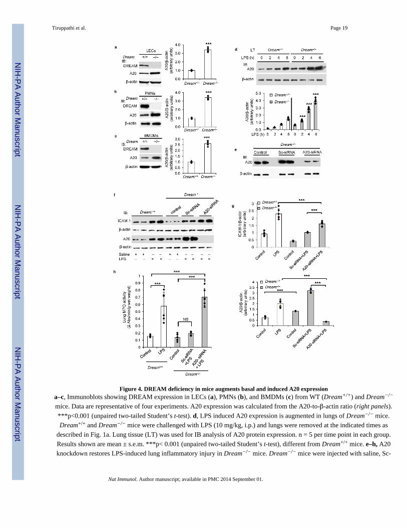
Figure 4. DREAM deficiency in mice augments basal and induced A20 expressiona–c, Immunoblots showing DREAM expression in LECs (a), PMNs (b), and BMDMs (c) from WT (Dream+/+) and Dream−/−
mice. Data are representative of four experiments. A20 expression was calculated from the A20-to-β-actin ratio (right panels).
***p<0.001 (unpaired two-tailed Student’s t-test). d, LPS induced A20 expression is augmented in lungs of Dream−/− mice.
Dream+/+ and Dream−/− mice were challenged with LPS (10 mg/kg, i.p.) and lungs were removed at the indicated times as
described in Fig. 1a. Lung tissue (LT) was used for IB analysis of A20 protein expression. n = 5 per time point in each group.
Results shown are mean ± s.e.m. ***p< 0.001 (unpaired two-tailed Student’s t-test), different from Dream+/+ mice. e–h, A20
knockdown restores LPS-induced lung inflammatory injury in Dream−/− mice. Dream−/− mice were injected with saline, Sc-
Tiruppathi et al. Page 19
Nat Immunol. Author manuscript; available in PMC 2014 September 01.
NIH
-PA
Author M
anuscriptN
IH-P
A A
uthor Manuscript
NIH
-PA
Author M
anuscript

siRNA, or A20-siRNA. At 48 h after injection, lungs harvested were used to determine A20 protein expression by IB (e). At 48
h after injection with saline, Sc-siRNA, or A20-siRNA injected Dream−/− mice were challenged with saline or LPS (10 mg/kg,
i.p.). At 6 h after saline or LPS challenge, lungs harvested were used to determine ICAM-1 and A20 expression by IB (f and g)
or MPO activity (h). In this experiment, Dream+/+ mice were also used as a positive control to study the LPS effect. Data are
representative of six experiments. N= 6 in each group; Values are mean ± s.e.m. ***p< 0.001 (unpaired two-tailed Student’s t-
test), different from respective control groups.
Tiruppathi et al. Page 20
Nat Immunol. Author manuscript; available in PMC 2014 September 01.
NIH
-PA
Author M
anuscriptN
IH-P
A A
uthor Manuscript
NIH
-PA
Author M
anuscript

Figure 5. DREAM deletion attenuates TNF-induced TAK1 and IKK activation in lung endothelial cellsa, Dream+/+ or Dream−/− LECs treated with TNF (1000 U/ml) for different time intervals were used to measure IKKβ
phosphorylation. Top, representative immunoblots (IBs) from four experiments. Bottom, the ratio of phospho-IKKβ to total-
IKKβ. b, Top panel: TNF-induced IκBα protein expression was measured in LECs from Dream+/+ and Dream−/− mice. c,Quantitative RT-PCR was used to determine TNF-induced IκBα mRNA expression in LECs from Dream+/+ or Dream−/− mice.
d, Dream+/+ or Dream−/− LECs treated with TNF (1000 U/ml) for different time intervals were used to measure TAK1
phosphorylation at Thr-184/187. Top, representative IBs from four experiments. Bottom, quantitative results, the band intensity
measured by densitometry and the ratio of phospho-TAK1 to total TAK1 was calculated. In a–d, results are mean ± s.d. of 4
experiments. ***p<0.001 (unpaired two-tailed Student’s t-test). e, Dream−/− LECs were transfected with vector, WT-DREAM,
Tiruppathi et al. Page 21
Nat Immunol. Author manuscript; available in PMC 2014 September 01.
NIH
-PA
Author M
anuscriptN
IH-P
A A
uthor Manuscript
NIH
-PA
Author M
anuscript

or DNA-binding defective mutant (mut-DREAM). At 48 h after transfection, cells exposed to TNF were immunoblotted with
anti-phopho-IκBα, anti-IκBα, anti-DREAM, or anti-β-actin. Representative IBs from two separate experiments are shown. In
the middle, the ratio of phopho-IκBα to total IκBα is shown. f and g, top panels:
Tiruppathi et al. Page 22
Nat Immunol. Author manuscript; available in PMC 2014 September 01.
NIH
-PA
Author M
anuscriptN
IH-P
A A
uthor Manuscript
NIH
-PA
Author M
anuscript

Figure 6. DREAM deletion attenuates TNF-induced JNK and p38 MAPK activation in lung endothelial cellsTNF-induced phosphorylation of JNK (a) or p38 (b) was measured in LECs from Dream+/+ and Dream−/− mice. Bottom
panels, the ratio of phospho-JNK to total-JNK or phospho-p38 to total-p38. In a, b, results are mean ± s.d.; ***p< 0.001
(unpaired two-tailed Student’s t-test). c, LECs from WT or Dream−/− mice were transfected with Sc-siRNA or A20-siRNA. At
48 h after transfection, cells were used to measure A20 protein expression (top) or p38 phosphorylation after stimulation with
TNF (bottom). Results shown are representative of three experiments. The ratio of phopho-p38 to total-p38 is indicated. d,Dream+/+ or Dream−/− LECs treated with TNF (1000 U/ml) for different time intervals were used to determine mRNA
expression for MCP-1, ICAM-1, and A20 by qRT-PCR. Results shown are mean ± s.e.m. of 4 experiments.
***p<0.001(unpaired two-tailed Student’s t-test). e, BMDMs in culture from Dream+/+ and Dream−/− mice exposed to LPS
(100 ng/ml) for different time intervals were used to determine A20 protein expression. Data shown are representative of three
experiments. Immunoblots were quantified (right panel). *p<0.05; **p<0.01; ***p<0.001 (unpaired two-tailed Student’s t-test).
Tiruppathi et al. Page 23
Nat Immunol. Author manuscript; available in PMC 2014 September 01.
NIH
-PA
Author M
anuscriptN
IH-P
A A
uthor Manuscript
NIH
-PA
Author M
anuscript

Figure 7. DREAM differentially regulates the expression of NF-κB signaling components and target genesLung tissue (LT) from Dream+/+ and Dream−/− mice was used to determine the expression of TRAFs (TRAF2 and TRAF6),
RIPs (RIP1 and RIP2), and IκBα (a), IKKs (IKKα, IKKβ, IKKγ) (b), and NF-κB proteins (p65/RelA, NF-κB1/p50, NF-κB2/
p52, RelB, and c-Rel) (c). Representative IBs are shown in a–c. Quantitative comparisons between Dream+/+ and Dream−/− are
shown. n = 5 mice per group (mean ± s.d.). ***p<0.001 (unpaired two-tailed Student’s t-test). In d, LECs from Dream+/+ and
Dream−/− mice were used for immunoblot analysis to determine the expression of NF-κB signaling components as above.
Results are representative of three experiments.
Tiruppathi et al. Page 24
Nat Immunol. Author manuscript; available in PMC 2014 September 01.
NIH
-PA
Author M
anuscriptN
IH-P
A A
uthor Manuscript
NIH
-PA
Author M
anuscript

Figure 8. WT-DREAM expression restores NF-κB signaling components level in DREAM deficient LECsDream−/− LECs were transfected with empty vector (mock), WT-DREAM, or mut-DREAM (DNA-binding defective mutant).
At 48 h after transfection, cells were used for IB analysis. Results are representative of at least two independent experiments.
Tiruppathi et al. Page 25
Nat Immunol. Author manuscript; available in PMC 2014 September 01.
NIH
-PA
Author M
anuscriptN
IH-P
A A
uthor Manuscript
NIH
-PA
Author M
anuscript
Copyright © 2022 FDOKUMEN