Smad6 Represses Dlx3 Transcriptional Activity through Inhibition of DNA Binding
12
Smad6 Represses Dlx3 Transcriptional Activity through Inhibition of DNA Binding * Received for publication, March 30, 2006, and in revised form, May 8, 2006 Published, JBC Papers in Press, May 10, 2006, DOI 10.1074/jbc.M603049200 Kathie A. Berghorn 1 , Patricia A. Clark-Campbell 1 , Li Han 1 , Michael McGrattan, Robert S. Weiss, and Mark S. Roberson 2 From the Department of Biomedical Sciences, Cornell University, Ithaca, New York 14853 Dlx3 (Distal-less 3) is a homeobox-containing transcription factor required for normal placental development in mice. Here we demonstrate that Dlx3 interacts with Smad6, a member of a larger family of transcriptional regulators generally thought to regulate transforming growth factor /bone morphogenetic protein signaling. Immunocytochemical and immunoprecipita- tion studies demonstrate overlapping nuclear localization and physical interaction between Dlx3 and Smad6 in human chori- ocarcinoma cells and in differentiated trophoblasts from human placenta. In vitro protein interaction studies mapped the Smad6 interaction domain within Dlx3 to residues 80 –163, a region of Dlx3 that includes a portion of the homeodomain. Dlx3 and Dlx4 share homology within this region, and Dlx4 was also found to bind Smad6. Using the Esx1 gene promoter as a model for a Dlx3-responsive gene, studies demonstrate two near con- sensus Dlx3 binding sites within the proximal 2.3 kb of the tran- scription start site. Interestingly, binding of Dlx3 to one of these two sites was inhibited by interaction with Smad6. Consistent with this result, expression of an Esx1 promoter luciferase reporter was increased by overexpression of Dlx3; this effect was reversed with co-expression of Smad6. Further, small interference RNA- mediated knockdown of endogenous Smad6 increased Dlx3-de- pendent expression of the Esx1 gene promoter. Thus, Smad6 appears to functionally interact with Dlx3, altering the ability of Dlx3 to bind target gene promoters. Smad6 appears to play a mod- ulatory role in the regulation of Dlx3-dependent gene transcription within placental trophoblasts. The Distal-less family of transcriptional regulators includes six members in mammals, arrayed in pairs and aligned with the hox gene clusters along different chromosomes (1, 2). Dlx3 is tandemly arrayed with Dlx4 on human chromosome 17 and is involved in developmental determination of multiple tissues, including the first and second branchial arches, teeth, bone, and multiple epithelia, including the skin, mammary gland primor- dia, and the placenta (3). The relationship between conver- gently transcribed pairs of Dlx family members and specific hox gene clusters has suggested that, although independent of hox gene expression patterns, these homeodomain-containing transcription factors are clearly involved in important aspects of developmental morphogenesis (reviewed in Ref. 2). The importance of Dlx3 during development and in the adult arises from several different observations of disease states. Mutations in Dlx3 are believed to be causally related to tricho-dento osse- ous syndrome, a genetic disorder manifested by taurodontism, hair abnormalities, and increased bone density in the cranium (4 – 6). The defect in Dlx3 leading to tricho-dento osseous syn- drome appears to be associated with a four-nucleotide deletion just downstream of the homeodomain, resulting in a premature truncation of the protein. Amelogenesis imperfecta with taur- odontism has similar characteristics as tricho-dento osseous syndrome and has also been associated with mutations with Dlx3 in some families investigated, albeit distinct from the four- nucleotide deletion/mutation described above (7). Amelogen- esis imperfecta is an autosomal dominant trait leading to dental enamel defects and enlarged pulp chambers and has been asso- ciated with a two-nucleotide deletion within the homeodomain of Dlx3. This deletion again results in a frameshift and prema- ture truncation of Dlx3 in the carboxyl terminus, primarily downstream of the homeobox. In addition to tricho-dento osseous syndrome and amelogenesis imperfecta, Dlx3/Dlx4 have been identified in the gene interval thought to be involved in some forms of craniofacial abnormalities, including cleft pal- ate (8). The putative involvement of Dlx3 in the occurrence of cleft palate is also supported by the murine model deficient in endothelin-A receptor (the endothelin-A receptor), which results in cleft palate and hypoplasia of the mandible (9 –11). In this model, Dlx3 expression is thought to be dependent upon endothelin-1 through a G q /G 11 -dependent mechanism. In the G q /G 11 -deficient mouse, Dlx3, among other factors, is specifically down-regulated, supporting the speculation of the importance of Dlx3 in cranio-facial morphogenesis (12). Thus, the role and importance of Dlx3 in morphogenic aspects of development and in epithelial differentiated function is rather far reaching. The Dlx3 null mouse dies in utero by embryonic day 10 due to putative placental failure (13). This was associated with a failure in the development/morphogenesis of the placental labyrinth compartment of the murine placenta. Further, genetic loss of Dlx3 was correlated with reduced expression of an additional homeobox factor, Esx1, suggesting that Dlx3 may be an impor- tant transcriptional regulator of Esx1 promoter activity. Studies from our laboratory identified Dlx3 as a cell type-specific tran- scriptional activator in placental trophoblasts. Dlx3 binds to * This work was supported by NICHD, National Institutes of Health, Grant R01 HD 39354 (to M. S. R.). The costs of publication of this article were defrayed in part by the payment of page charges. This article must therefore be hereby marked “advertisement” in accordance with 18 U.S.C. Section 1734 solely to indicate this fact. 1 These authors contributed equally to this work. 2 To whom correspondence should be addressed: T3-004d Veterinary Research Tower, Dept. of Biomedical Sciences, Cornell University, Ithaca, NY 14853. Tel.: 607-253-3469; Fax: 607-253-3851; E-mail: [email protected]. THE JOURNAL OF BIOLOGICAL CHEMISTRY VOL. 281, NO. 29, pp. 20357–20367, July 21, 2006 © 2006 by The American Society for Biochemistry and Molecular Biology, Inc. Printed in the U.S.A. JULY 21, 2006 • VOLUME 281 • NUMBER 29 JOURNAL OF BIOLOGICAL CHEMISTRY 20357 by guest on July 17, 2016 http://www.jbc.org/ Downloaded from
-
Upload
independent -
Category
Documents
-
view
4 -
download
0
Transcript of Smad6 Represses Dlx3 Transcriptional Activity through Inhibition of DNA Binding
Smad6 Represses Dlx3 Transcriptional Activity throughInhibition of DNA Binding*
Received for publication, March 30, 2006, and in revised form, May 8, 2006 Published, JBC Papers in Press, May 10, 2006, DOI 10.1074/jbc.M603049200
Kathie A. Berghorn1, Patricia A. Clark-Campbell1, Li Han1, Michael McGrattan, Robert S. Weiss,and Mark S. Roberson2
From the Department of Biomedical Sciences, Cornell University, Ithaca, New York 14853
Dlx3 (Distal-less 3) is a homeobox-containing transcriptionfactor required for normal placental development inmice. Herewe demonstrate that Dlx3 interacts with Smad6, a member of alarger family of transcriptional regulators generally thought toregulate transforming growth factor �/bone morphogeneticprotein signaling. Immunocytochemical and immunoprecipita-tion studies demonstrate overlapping nuclear localization andphysical interaction between Dlx3 and Smad6 in human chori-ocarcinoma cells and indifferentiated trophoblasts fromhumanplacenta. In vitro protein interaction studiesmapped the Smad6interaction domain within Dlx3 to residues 80–163, a region ofDlx3 that includes a portion of the homeodomain. Dlx3 andDlx4 share homology within this region, and Dlx4 was alsofound to bind Smad6. Using the Esx1 gene promoter as a modelfor a Dlx3-responsive gene, studies demonstrate two near con-sensus Dlx3 binding sites within the proximal 2.3 kb of the tran-scription start site. Interestingly, binding of Dlx3 to one of thesetwo sites was inhibited by interaction with Smad6. Consistentwith this result, expressionof anEsx1promoter luciferase reporterwas increased by overexpression of Dlx3; this effect was reversedwith co-expression of Smad6. Further, small interference RNA-mediated knockdown of endogenous Smad6 increased Dlx3-de-pendent expression of the Esx1 gene promoter. Thus, Smad6appears to functionally interact with Dlx3, altering the ability ofDlx3 to bind target gene promoters. Smad6 appears to play amod-ulatoryrole intheregulationofDlx3-dependentgenetranscriptionwithin placental trophoblasts.
The Distal-less family of transcriptional regulators includessix members in mammals, arrayed in pairs and aligned with thehox gene clusters along different chromosomes (1, 2). Dlx3 istandemly arrayed with Dlx4 on human chromosome 17 and isinvolved in developmental determination of multiple tissues,including the first and second branchial arches, teeth, bone, andmultiple epithelia, including the skin, mammary gland primor-dia, and the placenta (3). The relationship between conver-gently transcribed pairs of Dlx familymembers and specific hox
gene clusters has suggested that, although independent of hoxgene expression patterns, these homeodomain-containingtranscription factors are clearly involved in important aspectsof developmental morphogenesis (reviewed in Ref. 2). Theimportance of Dlx3 during development and in the adult arisesfrom several different observations of disease states. Mutationsin Dlx3 are believed to be causally related to tricho-dento osse-ous syndrome, a genetic disorder manifested by taurodontism,hair abnormalities, and increased bone density in the cranium(4–6). The defect in Dlx3 leading to tricho-dento osseous syn-drome appears to be associated with a four-nucleotide deletionjust downstreamof the homeodomain, resulting in a prematuretruncation of the protein. Amelogenesis imperfecta with taur-odontism has similar characteristics as tricho-dento osseoussyndrome and has also been associated with mutations withDlx3 in some families investigated, albeit distinct from the four-nucleotide deletion/mutation described above (7). Amelogen-esis imperfecta is an autosomal dominant trait leading to dentalenamel defects and enlarged pulp chambers and has been asso-ciatedwith a two-nucleotide deletionwithin the homeodomainof Dlx3. This deletion again results in a frameshift and prema-ture truncation of Dlx3 in the carboxyl terminus, primarilydownstream of the homeobox. In addition to tricho-dentoosseous syndrome and amelogenesis imperfecta, Dlx3/Dlx4have been identified in the gene interval thought to be involvedin some forms of craniofacial abnormalities, including cleft pal-ate (8). The putative involvement of Dlx3 in the occurrence ofcleft palate is also supported by the murine model deficient inendothelin-A receptor (the endothelin-A receptor), whichresults in cleft palate and hypoplasia of themandible (9–11). Inthis model, Dlx3 expression is thought to be dependent uponendothelin-1 through a G�q/G�11-dependent mechanism. Inthe G�q/G�11-deficient mouse, Dlx3, among other factors, isspecifically down-regulated, supporting the speculation of theimportance of Dlx3 in cranio-facial morphogenesis (12). Thus,the role and importance of Dlx3 in morphogenic aspects ofdevelopment and in epithelial differentiated function is ratherfar reaching.TheDlx3nullmouse dies in uteroby embryonic day 10 due to
putative placental failure (13). This was associatedwith a failurein the development/morphogenesis of the placental labyrinthcompartment of the murine placenta. Further, genetic loss ofDlx3 was correlated with reduced expression of an additionalhomeobox factor, Esx1, suggesting that Dlx3 may be an impor-tant transcriptional regulator of Esx1 promoter activity. Studiesfrom our laboratory identified Dlx3 as a cell type-specific tran-scriptional activator in placental trophoblasts. Dlx3 binds to
* This work was supported by NICHD, National Institutes of Health, Grant R01HD 39354 (to M. S. R.). The costs of publication of this article were defrayedin part by the payment of page charges. This article must therefore behereby marked “advertisement” in accordance with 18 U.S.C. Section 1734solely to indicate this fact.
1 These authors contributed equally to this work.2 To whom correspondence should be addressed: T3-004d Veterinary Research
Tower, Dept. of Biomedical Sciences, Cornell University, Ithaca, NY 14853. Tel.:607-253-3469; Fax: 607-253-3851; E-mail: [email protected].
THE JOURNAL OF BIOLOGICAL CHEMISTRY VOL. 281, NO. 29, pp. 20357–20367, July 21, 2006© 2006 by The American Society for Biochemistry and Molecular Biology, Inc. Printed in the U.S.A.
JULY 21, 2006 • VOLUME 281 • NUMBER 29 JOURNAL OF BIOLOGICAL CHEMISTRY 20357
by guest on July 17, 2016http://w
ww
.jbc.org/D
ownloaded from
and transactivates the promoter for the glycoprotein hormone� subunit gene via a cis-acting element required for full basalactivity of this gene (14). The glycoprotein hormone � subunitis a subunit of the heterodimeric glycoprotein hormone, chori-onic gonadotropin (CG).3 Trophoblast-derived CG has beenidentified in primates and equine and appears to play a criticalrole in the maintenance of early pregnancy in women, provid-ing early gonadotropic support to the corpus luteum andmain-tenance of progesterone production (15–17). Both for the caseof the � subunit promoter and regulation of Esx1 in the Dlx3null mouse, Dlx3 appears to function as a putative transcrip-tional activator. However, it has also been proposed that Dlx3can serve as a negative regulator of gene transcription inamphibian models (2). This apparent activation/repressioncapability may be due to variable heterodimeric partners ofDlx3 (as proposed in Ref. 2), dependent upon cell context andphysiological state. This observation was the impetus for us toexamine potential binding partners ofDlx3 in the context of thehuman placenta. The present studies identify Smad6 as a bind-ing partner for Dlx3 using a yeast two-hybrid screen of a humanterm placental cDNA library. Dlx3 and Smad6 are co-localizedin the nucleus of cells of trophoblast origin, including cytotro-phoblasts and syncytial trophoblasts from fully differentiatedhuman term placenta. Interaction between Dlx3 and Smad6alters the DNA binding properties of Dlx3 such that Smad6serves as a negative regulator of Dlx3-dependent gene tran-scription of the Esx1 promoter.
MATERIALS AND METHODS
Plasmids and cDNAs—All plasmids used in these studieswere prepared by two cycles through cesium chloride usingstandard protocols. Expression vector for humanDlx3was gen-erously provided by Dr. Maria Morasso (National Institutes ofHealth, Bethesda,MD). A series of deletionmutants of theDlx3cDNA were constructed by PCR. To facilitate cloning into thepKH3 vector (generously provided by Dr. Jun-Lin Guan,Cornell University, Ithaca, NY), EcoRI and ClaI restriction siteswere added to the forward and reverse primers, respectively.The forward primers used in these reactions were as follows:forward 1, 5�-TCAGGAATTCAAATGAGCGGCTCCTTCG-ATCGC-3�, forward 40, 5�-TCAGGAATTCAACTGGGCTA-TTACAGCGCTCCTCAG-3�, forward 80, 5�-TCAGGAATT-CAATACTCGCCCAAGTCGGAATATACC-3�, forward 121,5�-TCAGGAATTCAAATGGTGAACGGCAAGCCCAAAA-AG-3�, and forward 195, 5�-TCAGGAATTCAACTGGAACA-CAGCCCCAACAACAGT-3�. The reverseprimersused in thesereactions were as follows: reverse 128, 5�-GTACATCGATCAC-GGCTTTCGGACCTTCTTGGGCTTCCC-3�; reverse 163, 5�-GTACATCGATCAAGCTAGCTCGGCGCGCTCAGGCAA-3�, reverse 202, 5�-GTACATCGATCAACTGTATTGGGACT-GTGCTCCAG-3�, and reverse 287, 5�-GTACATCGATTC-AGTACACAGCCCCAGGGTT-3�. PCRproductswere cloned
initially into the pGEM T Easy vector (Promega Corp., Madi-son, WI). Once verified by nucleotide sequence analysis, frag-ments were subcloned into the pKH3 vector for use in studies.Smad6 expression plasmid was a gift from Dr. Ali H. Brivanlou(The Rockefeller University, NewYork, NY). Smad4 expressionvector was a gift from Dr. Colin Clay (Colorado State Univer-sity, Fort Collins, CO). The humanDlx4 cDNAwas obtained byPCR from RNA isolated from JEG3 cells using the followingprimers: 5�-TCAGGAATTCAAATGACCTCTTTACCCTG-TCCC-3� and 5�-GTACATCGATCACATCATCTGAGGCA-GTGC-3�. The resultingDlx4 cDNAwas cloned into pKH3 andverified by nucleotide sequence analysis. The Esx1–2.3kb pro-moterwas obtained by PCRusingmouse genomicDNAand thefollowing primers: 5� primer (5�-GGTACCAGCACCGAGC-TATCACAACCATCA-3�) and 3� primer (5�-GCTAGCTAC-CAGCTGCTTCTCCCGTA-3�). To facilitate cloning, KpnIandNheI restriction enzyme sites were engineered at the end ofthe 5� primer and 3� primer, respectively. The PCR productswere cloned into pGEM T-Easy vector. After KpnI and NheIdigestion, the promoter fragment was subcloned into a lucif-erase reporter vector. The fidelity of the construct was con-firmed by nucleotide sequence analysis. PCR-based site-di-rected mutagenesis was used to disrupt the distal Dlx3 bindingsite within the Esx1 luciferase reporter. This mutation substi-tuted a Not-1 restriction site for the near consensus Dlx3 bind-ing site. The mutation was confirmed using nucleotidesequence analysis. The human � subunit gene promoter lucif-erase reporter has been reported previously (14).Yeast Two-hybrid Screen—To investigate novel protein-pro-
tein interactions, full-length Dlx3 served as the bait proteinwith a human termplacental cDNA library serving as the target.The bait, human Dlx3 cDNA, was cloned into the vectorpGBKT7 and transformed in the yeast strain AH109. A pre-transformed human term placental Matchmaker cDNA librarywas in yeast strain Y187 (BD Biosciences/Clontech, Palo AltoCA). The bait and library plasmids were expressed as GAL4fusion proteins. 3-Amino-1,2,4-triazole was titrated (5–35mM)using the bait strain to control background yeast growth. Aconcentration of 12 mM 3-amino-1,2,4-triazole was used in thelibrary screen. The bait and library strains were mated with anefficiency of �4%. The bait strain required Leu� syntheticdropout (SD) minimal medium, and the library strain requiredTrp� SD minimal medium. Mating was carried out in YPDAmedia containing 0.003% adenine hemisulfate. Following mat-ing of the bait strain with the human placental library, yeastswere initially plated on intermediate stringency SD media(His�/Leu�/Trp�) plates. When colonies were of sufficientsize, colonies were replica-plated on high stringency SDmedium (Ade�/His�/Leu�/Trp�/X-�-gal) plates to verify thattheymaintained the correct phenotype. A colony filter lift assaywas performed to access�-galactosidase activity to identify andrank the strength of potential interactions. Once identified,yeast plasmids were isolated using disruption with glass beads,and plasmids were rescued/purified using theQiagenMiniprepreagents and a spin column (Qiagen Inc., Valencia, CA). Iden-tity of the rescued plasmidswas verified by nucleotide sequenceanalysis. The interaction between Dlx3 and target genes wasexamined using a reconstitution assay, where both plasmids
3 The abbreviations used are: CG, chorionic gonadotropin; PBS, phosphate-buffered saline; KPBS, potassium phosphate-buffered saline; siRNA, smallinterference RNA; EMSA, electrophoretic mobility shift assay; TKDP, tro-phoblast Kunitz domain protein; TGF�, transforming growth factor �; BMP,bone morphogenetic protein; JRE, junctional regulatory element; E3, ubiq-uitin-protein isopeptide ligase; X-�-gal, X-�-galactosidase.
Dlx3 and Smad6 in Placenta
20358 JOURNAL OF BIOLOGICAL CHEMISTRY VOLUME 281 • NUMBER 29 • JULY 21, 2006
by guest on July 17, 2016http://w
ww
.jbc.org/D
ownloaded from
were co-transformed into the AH109 yeast strain and plated onhigh stringency SD medium.Preparation of JEG3 Cell Nuclear Extracts—Subconfluent
JEG3 cells were used for the preparation of nuclear extracts.Cells were washed twice with ice-cold Dulbecco’s phosphate-buffered saline (PBS; Invitrogen). Cells were collected by scrap-ing in ice-cold PBS supplemented with a 1:1000 dilution of pro-tease inhibitor mixture (referred to as protease inhibitormixture; Sigma), 5 mM benzamidine, and 0.2 mM phenylmeth-ylsulfonyl fluoride. Cells were pelleted by centrifugation andresuspended in a hypotonic buffer consisting of 120 mM potas-sium chloride, 30 mM sodium chloride, 30 mM Hepes (pH 8.0),0.3 M sucrose, protease inhibitor mixture, 5 mM benzamidine,and 0.2 mM phenylmethylsulfonyl fluoride and allowed to swellfor 15 min on ice. Cells were lysed by Dounce homogenizing,and nuclei were isolated by layering the broken cell lysate over asucrose cushion (0.9 M sucrose) followed by centrifugation at2000� g for 30min at 4 °C. The nuclear pellet was resuspendedin a buffer containing 10 mM Tris (pH 7.5), 50 mM NaCl, 5%glycerol, 1 mM EDTA, protease inhibitor mixture, 5 mM benza-midine, and 0.2 mM phenylmethylsulfonyl fluoride. Additionalsodium chloride was added to a final concentration of 450 mM,and nuclear proteins were extracted with constant rocking at4 °C for 30 min. Nuclear extracts were clarified by centrifuga-tion (85,000 � g for 60min), and the nuclear extract was storedin aliquots at �80 °C until later use. Protein concentrations ofthe nuclear extracts were determined by Bradford assay.Immunoprecipitation from JEG3 Nuclear Extracts andWest-
ern Blotting Analysis—JEG3 cell nuclear extracts (200 �g) weresuspended in 1ml of 0.1%Triton X buffer (50mMTris (pH 7.6),50 mM sodium chloride, 0.1% Triton X, protease inhibitor mix-ture, 5 mM benzamidine, and 0.2 mM phenylmethylsulfonyl flu-oride). To preclear the nuclear extracts, protein A/G-agarose(Santa Cruz Biotechnology, Inc., Santa Cruz, CA) was added toeach suspension and allowed to mix for 1 h at 4 °C with gentlerocking. Following centrifugation (1200 � g for 1 min) toremove protein A/G-agarose, antibodies were added at the fol-lowing dilutions: normal rabbit serum at 1:1000; Dlx3 antibodyat 1:1000; Smad6 antibody (SantaCruz Biotechnology) at 1:100.Following 2 h of gentle rocking at 4 °C, protein A/G-agarose(Santa Cruz Biotechnology) was added and allowed to mix foran additional 2 h. Complexes were then washed four times with0.1% Triton X buffer. Samples were then suspended in an equalvolume of 2� SDS loading buffer (100 mM Tris (pH 6.8), 4%SDS, 20% glycerol, and 200mM dithiothreitol). Protein sampleswere boiled for 3min and chilled for 5min on ice. Proteins wereresolved by SDS-polyacrylamide gel electrophoresis and trans-ferred to polyvinylidine difluoride membranes by electro-blotting. Membranes were blocked with nonfat dried milk (5%)in Tris-buffered saline (10 mM Tris (pH 7.6), 150 mM sodiumchloride) containing 0.1%Tween 20 (TBST). ForWestern blots,the Dlx3 antibody was used at 1:5000 in TBST, 5% nonfat driedmilk. The reciprocal Western blot from immunoprecipitations(IPs) using the Smad6 antibody was not possible, since the IPheavy chain IgG blocked visualization of Smad6 on Westernblot due to similar molecular size. In other Western blot stud-ies, the Smad6 antibody was used at 1:500, and the actin anti-body (Santa Cruz Biotechnology) was used at 1:1000 dilution.
Proteins bands were visualized by chemiluminescence reagents(PerkinElmer Life Sciences).Immunocytochemistry—JEG3 cells were cultured on glass
slides or coverslips for 24 h, rinsed one time with potassiumphosphate-buffered saline (KPBS), and then fixed for 20 minwith 4% paraformaldehyde. Slides or coverslips were thenstored in 70% ethanol until used. Prior to use, slides were rinsedin KPBS seven times over 1 h. JEG3 cells were incubated withprimary antibody (Smad6 at 1:100; Dlx3 at 1:500) overnight at4 °C. Slides were again rinsed with KPBS, followed by incuba-tion with a fluorescence-conjugated secondary antibody (Alexa594, Molecular Probes, Inc., Eugene OR; and Cy2, JacksonImmunoresearch Laboratories, Westgrove, PA) in KPBS-Tri-ton X at 37 °C for 2 h. Cells were rinsed in KPBS, dehydratedthrough a graded series of ethanol, and cleared with xylene,and coverslips were attached with Krystalon (EM Science,Gibbstown, NJ).Samples of human term placenta (derived from elective cae-
sarian section) were obtained from Cayuga Medical Center(Ithaca, NY) under the guidelines and approval of the CornellUniversity and the Cayuga Medical Center Committees on theUse of Human Subjects in Research. Samples were collected,fixed with 4% paraformaldehyde for 48 h, and transferred to70% ethanol until processing. Tissues were paraffin-embedded,and 5-�m sections were obtained. Immunocytochemistry wasperformed as previously described (14), except that fluores-cence-conjugated secondary antibodies were used as describedabove.Recombinant Proteins and Immunoprecipitation Analysis—
Recombinant Smad6, Smad4, Dlx4, Dlx3, and deletions of Dlx3were prepared using a coupled transcription and translationWheat Germ Extract System (Promega Corp., Madison, WI)following the prescribed protocol. Proteins were radioactivelylabeled using [35S]methionine (1000 Ci/mmol at 10 mCi/ml;Amersham Biosciences). A portion (10%) of each recombinantproteinwas saved for input analysis. Protein combinationswereadded at a 1:1 (by volume) mixture to a 0.1% Triton X bufferalong with appropriate antibody at specified concentrations(Dlx3 at 1:1000; Smad6 (Santa Cruz Biotechnology) at 1:100;and Smad4 (Santa Cruz Biotechnology) at 1:500). Following 2 hof gentle rocking at 4 °C, protein A/G-agarose (Santa Cruz Bio-technology) was added and allowed tomix for an additional 2 h.Complexes were then washed four times with 0.1% Triton Xbuffer. Proteins were resolved by SDS-polyacrylamide gel elec-trophoresis, and the gel was fixed in 25% methanol and 15%glacial acetic acid for 1 h with gentle rocking at room tempera-ture. The gel was then washed three times in 40% isopropylalcohol solution and dried, and bands were visualized byautoradiography.Electrophoretic Mobility Shift Assay—Electrophoretic mobility
shift assays (EMSAs) were carried out as described previously (14,18) using the indicated antibodies. Reactions (withoutprobe)weremaintained at room temperature for 30min followed by the addi-tion of 32P-labeled oligonucleotide Dlx3 binding site probes (thejunctional regulatory element (JRE) from the glycoprotein hor-mone� subunit promoter (14) and twoputativeDlx3binding sitesidentified within the Esx1 promoter). The binding reactions weremaintained anadditional 30minand then resolvedonnativepoly-
Dlx3 and Smad6 in Placenta
JULY 21, 2006 • VOLUME 281 • NUMBER 29 JOURNAL OF BIOLOGICAL CHEMISTRY 20359
by guest on July 17, 2016http://w
ww
.jbc.org/D
ownloaded from

acrylamide gels. Todeterminewhether Smad6 could displace pre-bound Dlx3 in EMSA, binding reactions containing Dlx3 alonewere allowed to incubate with probe for 30 min to reach equilib-
rium. Smad6 protein was then added,and the reactions were incubated foran additional 30min. These reactionswere compared with binding reac-tions where both Dlx3 and Smad6were added together and incubatedfor the full 60min. Following electro-phoresis, the gels were dried, andDNA-protein complexes were visual-ized by autoradiography. All DNAbinding studies were conducted atleast three times with similar results.The nucleotide sequences for probeswere as follows (only one strand indi-cated): JRE, 5�-ACGTCATGGTAA-TTACACCAAG-3�; distal bindingsite, 5�-ACAAGGAGCTAATTTA-CTTCCT-3�; proximal binding site,5�-TTAGGGCTCTAATTCAGAC-TCT-3�.Cell Culture and Transient Trans-
fection Studies—JEG3 cells were cul-tured in monolayer using Dulbecco’smodified Eagle’s medium supple-mented with fetal bovine serum(10%). Before transfection studies,cells were split to 35-mm dishes, andsubconfluent cultures were used.JEG3 cells were transiently co-trans-fected with an Esx1 promoter lucifer-ase reporter construct, Dlx3 expres-sionvectorat aconstantdose (2.0�g),and increasing doses of Smad6expression vector using lipofection(FuGENE 6 transfection reagent;
Roche Applied Science). All transfections were carried out with aconstant amount of DNA by supplementing reactions with theparent vector pKH3. Luciferase activity was determined after 24 hof transfection using reagents from the Luciferase Reporter AssaySystem (Promega Corp.), and luciferase activity was standardizedby total cell protein amount (luciferase/1.0�g) as determined by aBradford assay. All transfection studies were conducted in tripli-cate on at least three separate occasions with similar results. Dataare shown asmeans (n� 3)� S.E. of a representative experiment.Preparation of Stable Cell Lines Expressing Small Interference
RNAs (siRNAs)—The mammalian expression vector, pSUPER-retro-neo (OligoEngine, Seattle, WA) was used for preparationof retrovirus containing specific siRNAs and expression of siR-NAs in JEG3 cells following viral infection. Each gene-specificinsert targeted a 19-nucleotide sequence within the humanSmad6 mRNA. The siRNA sequences were as follows: Smad6-#1, 5�-CACATTGTCTTACACTGAA; Smad6-#2, 5�-TCAA-GGTGTTCGACTTCGA; Smad6-#3, 5�-GCCACTGGATCT-GTCCGAT. The plasmids were referred to as Smad6 siRNA#1,siRNA#2, and siRNA#3. A control siRNA vector (also prepared
in pSUPER-retro-neo) was constructed using a 19-nucleotidesequence (5�-TTCTCCGAACGTGTCACGT) putatively with-
FIGURE 1. Dlx3 and Smad6 interact in yeast two hybrid and co-localize in placental trophoblasts. A, yeasttwo-hybrid screen of a human term placental library was carried out using full-length Dlx3 as bait. Followingplasmid rescue, Gal4 DNA binding domain-Dlx3 bait was co-transformed along with Gal4 activation domain-Smad6 into the AH109 yeast strain and plated on (His�, Leu�, Trp�) media as described under “Materials andMethods.” B, JEG3 cells were plated on glass slides and fixed as described under “Materials and Methods.”Double-labeled immunocytochemistry was used to determine localization of Dlx3 (green) or Smad6 (red ). Themerged images demonstrate overlapping expression patterns. Human term placenta was obtained, fixed, andsectioned as described under “Materials and Methods.” Similar double-labeled immunocytochemistry wasused to localize Dlx3 and Smad6. The white arrowhead identifies nuclei in syncytial trophoblasts, whereas theblack arrowhead identifies nuclei in cytotrophoblasts. Bar, 20 �m.
FIGURE 2. Dlx3 and Smad6 interact in the JEG3 choriocarcinoma cellmodel. A, JEG3 cell nuclear extracts were used in IP studies. IPs included theuse of normal rabbit serum (NRS) or antisera (ab) directed against Dlx3 orSmad6. Recombinant Dlx3 (rDlx3; without hemagglutinin epitope tag) andIPs were resolved by SDS-PAGE, and Western blots for Dlx3 were carried out(IB Dlx3). Molecular size standards (MW ) are depicted to the left. B, recombi-nant Dlx3, Smad6, and Smad4 were produced using wheat germ lysates in acoupled transcription/translation reaction containing [35S]methionine(Input). Dlx3 and either Smad6 or Smad4 were combined, followed by IP withthe Dlx3 antibody (Dlx3 IP). The input and IPs were resolved by SDS-PAGE, thegels were fixed and dried, and autoradiography was used to visualize bands.
Dlx3 and Smad6 in Placenta
20360 JOURNAL OF BIOLOGICAL CHEMISTRY VOLUME 281 • NUMBER 29 • JULY 21, 2006
by guest on July 17, 2016http://w
ww
.jbc.org/D
ownloaded from
out significant similarity to anymammalian gene sequence andtherefore served as an appropriate negative control (Oligo-Engine, Seattle,WA). The forward and reverse strands of oligo-nucleotides containing the siRNAs and nonsense sequence alsocontained BglII and HindIII sites at the 5�-end of the forwardand reverse oligonucleotides, respectively. The oligonucleo-tides were annealed and inserted into the pSUPER-retro-neovector after digestion of the vector with BglII and HindIII.These siRNAs were a self-contained hairpin loop for the dou-ble-stranded siRNA. All siRNA sequences were confirmed bynucleotide sequencing.Cell Culture, Transfection, and Retroviral Infection of siRNAs—
HEK293 Phoenix Ampho packaging cells (American Type Cul-ture Collection; Manassas, VA) were cultured in Dulbecco’smodified Eagle’s medium containing 10% fetal bovine serumand were transfected with the pSUPER-retro-neo siRNA plas-mids using FuGENE 6 transfection reagent (RocheApplied Sci-ence). Forty-eight hours following transfection, the culturemedia containing the retrovirus for individual siRNAs and con-trol siRNA were filtered through a 0.45-�m filter, and the viral
supernatant was used for infectionof JEG3 cells in the presence of 8�g/ml polybrene. JEG3 cells wereexposed to the retrovirus overnight,and cells were then washed withfresh medium and allowed torecover for 24 h. Following infec-tion, stable cell lines were selectedusing neomycin at 500 �g/ml for 7days (time until untransfected cellsall died), and then cultures weremaintained in medium containing500 �g/ml neomycin. Transienttransfections of the siRNA cell lineswere carried out as described above.Statistical Analysis—Luciferase
data were subjected to analysis ofvariance, and differences betweentreatments were determined usingTukey’s Studentized Range Test.Probability of less than 0.05 (p �0.05) was considered statisticallysignificant.
RESULTS
Dlx3 and Smad6 FunctionallyInteract in a Yeast Two-hybridScreen—Full-length human Dlx3was used as a bait protein in a yeasttwo-hybrid screen of a human termplacental library. The screenincluded coverage of �3.4 � 106independent clones. Once colonieswere identified from the originalscreen and library plasmids wererescued, plasmids were retrans-formed in a reconstitution assaywith the bait vector into the AH109
yeast strain. The library plasmid resulting in the most robustinteraction (as measured by �-galactosidase activity) with Dlx3was a cDNA containing the entire coding region of Smad6.Transformation of the Smad6 library vector alone did not sup-port yeast growth on high stringency SDmedium (Ade�/His�/Leu�/Trp�/X-�-gal) plates in the presence of 12 mM 3-amino-1,2,4-triazole. Transformation of Dlx3 bait vector minimallysupported yeast growth under the same conditions. Co-trans-formation of Dlx3 bait and Smad6 library plasmid resulted inrescue of yeast growth (Fig. 1A).Dlx3 and Smad6 Are Co-localized in the Nucleus in Chorio-
carcinoma Cells and in Human Placental Trophoblasts—Ini-tially, studies focused on examining localization of Dlx3 andSmad6 proteins in cells of trophoblast origin. Dlx3 and Smad6were expressed and localized primarily in the nuclear compart-ment in JEG3 cells (Fig. 1B), a choriocarcinoma cell line of tro-phoblast origin. Consistent with this observation, Dlx3 andSmad6 were nuclear localized to both cytotrophoblast and syn-cytial trophoblast within microvilli of fully differentiated termhuman placenta. Localization of Dlx3 in term placenta pro-
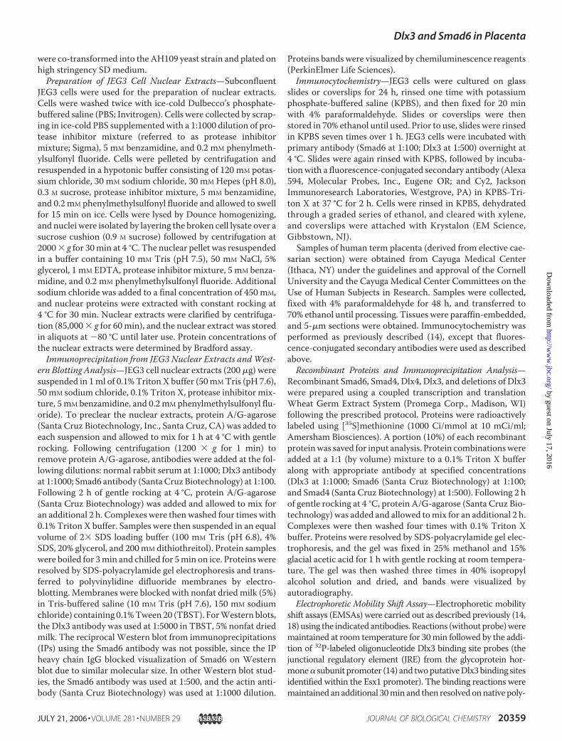
FIGURE 3. Deletion mutants of Dlx3 reveal interaction interface with Smad6. A, a series of Dlx3 deletions(characterized in schematic form) were prepared using PCR. The numbers represent amino acid designationsfor the termini of wild type Dlx3 and each mutant. B, wild type Dlx3, Smad6, and the Dlx3 deletion series wereprepared as recombinant proteins as described in the legend to Fig. 2B (Input) and subjected to IP studies usingthe Smad6 antibody (Smad6 IP), recombinant Smad6, and the combination of deletion mutants indicated. Theinput and IPs were resolved by SDS-PAGE, gels were fixed, and bands were visualized by autoradiography.*, bands consistent with protein-protein interactions.
Dlx3 and Smad6 in Placenta
JULY 21, 2006 • VOLUME 281 • NUMBER 29 JOURNAL OF BIOLOGICAL CHEMISTRY 20361
by guest on July 17, 2016http://w
ww
.jbc.org/D
ownloaded from
vided additional insight into the expression pattern of Dlx3during gestation in primates. We have previously shown Dlx3in placental trophoblasts in human placenta obtained at 8weeks gestation during peak production of humanCG (14). Thecurrent studies support the conclusion that Dlx3 expressionmay be maintained within trophoblast cell populations untilterm in the human placenta.Since both Dlx3 and Smad6 were expressed endogenously in
the JEG3 choriocarcinoma cell model, we sought to use thismodel for subsequent analyses. We have used JEG3 cells previ-ously for molecular analysis of gene regulatory processesrelated to glycoprotein hormone and Dlx3 gene promoterexpression (14, 18, 19). IP studies using nuclear extracts fromJEG3 cells and Smad6 antibody revealed that Dlx3 and Smad6interact in mammalian cells (Fig. 2A). The reciprocal studyusing Dlx3 antibody to IP Smad6 was not possible due to themolecular size of Smad6 and interference with the IgG heavychain used in the IP studies. However, this constraint was over-comewith preparation of Dlx3 and Smad6 as recombinant pro-teins labeled with [35S]methionine. IP of recombinant Dlx3 andSmad6 with the Dlx3 antibody revealed specific associationwith Smad6 but not Smad4 (Fig. 2B). Thus, the original inter-action defined in the yeast system was supported by IP stud-ies in choriocarcinoma cells that endogenously express thesetwo proteins and in vitro using recombinant Dlx3 and Smad6proteins.
Structure/Function Analysis of theDlx3/Smad6 Interaction Interface—To further understand the mecha-nism of the Dlx3/Smad6 interaction,we constructed a series of deletionmutants ofDlx3 (Fig. 3A). The ration-ale for these mutations was predi-cated on the existing understandingof important domains within Dlx3defined by others (20, 21), centeringupon the homeodomain (residues130–189; Fig. 3A), the centrallylocated DNA binding domain. Dlx3deletion mutants were prepared invitro along with full-length Dlx3 andSmad6. Smad6 IP was then used todetermine the domains sufficient tosupport interaction with Dlx3. Ini-tially, we identified the importance ofDlx3-(1–202),whichboundSmad6atlevels similar to Dlx3-(1–287) (Fig.3B). Dlx3-(121–287) bound Smad6as well, suggesting that Smad6 inter-action domain required at least a por-tion of the homeodomain (Fig. 3B).Based upon these results, we pre-dicted that Dlx3-(121–202) mutantwould also bind Smad6. This wasnot readily interpretable in ourstudies, since putative degradationfragments/products of these recom-
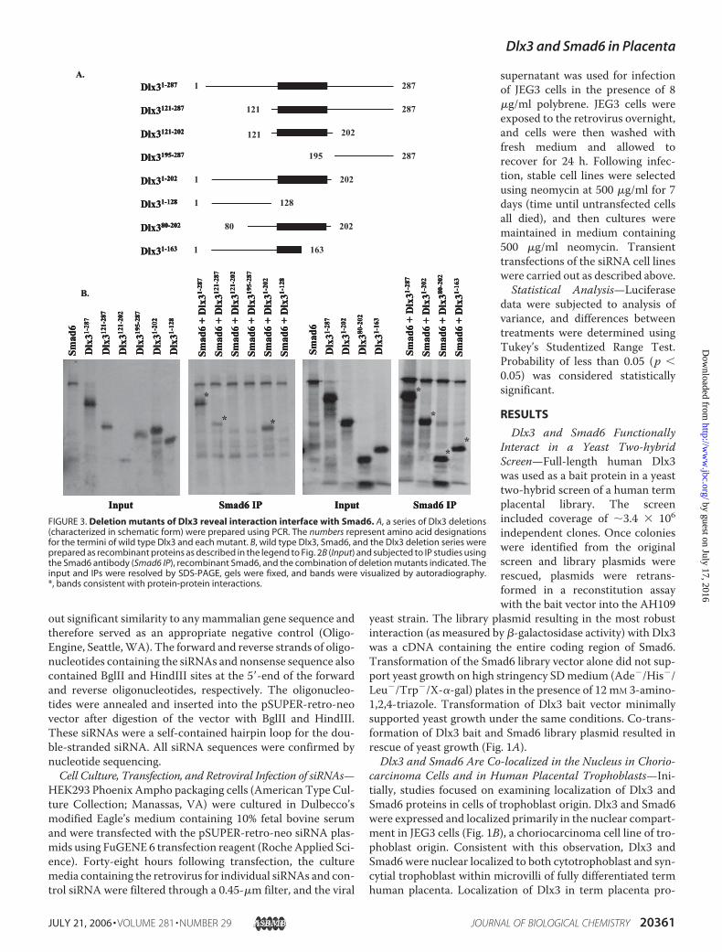
binant deletion mutants were of similar molecular size as theDlx3-(121–287) mutant. Subsequent Dlx3 deletion mutantsdefined a region of Dlx3-(80–163) that was sufficient to bindSmad6 in vitro (Fig. 3B). These studies again supported theconclusion that at least the amino-terminal portion of thehomeodomain was sufficient for interaction with Smad6 invitro. Alignment of Dlx3 with Dlx4 (another member of theDistal-less family expressed in placenta (22)) revealed �37%amino acid conservation within this region (residues 80–163;Fig. 4A), with higher levels of homology particularly clusteredwithin the homeodomain, supporting the prediction that Dlx4may also bind Smad6. Based upon in vitro studies, recombinantfull-length Dlx4 bound Smad6 similarly to Dlx3-(1–287) (Fig.4B). Dlx3-(195–287), previously shown not to bind Smad6 (Fig.3B), was used in these studies as a negative control.Dlx3 Binds to the 5�-Flanking Sequence of the Esx1 Promoter—
To begin to examine the functional significance of the Dlx3/Smad6 interaction on gene transcription, we cloned 2.3 kilo-bases of the 5�-flanking sequence of the Esx1 promoter. Thereport of the Dlx3 null mouse provided evidence that the loss ofDlx3 in vivowas correlated with a loss of Esx1 mRNA inmouseplacenta (13). Taking advantage of this observation, we identi-fied two near consensus Dlx3 binding sites within the 2.3-kbpromoter fragment. The distal site was located at �2135 andthe more proximal site at �585 relative to the transcriptionstart site of the Esx1 promoter, as previously defined (23). Atboth of these two sites, the central core of the binding site
FIGURE 4. Dlx4 binds Smad6. A, alignment of Dlx3 and Dlx4 between residue 80 and 198 reveals regions ofconserved sequence within the Smad6 interaction domain (residues 80 –163). The consensus amino acidsequence is listed in boldface type below the alignment. B, wild type Dlx3-(1–287), Dlx3-(195–287), Smad6, andDlx4 were prepared as recombinant proteins as described in the legend to Fig. 2 (Input) and subjected to IPstudies using the Smad6 antibody (Smad6 IP) and recombinant Smad6 and combinations of Dlx3 or Dlx4 asindicated. The input and IPs were resolved by SDS-PAGE, gels were fixed, and bands were visualized by auto-radiography. *, bands consistent with protein-protein interactions.
Dlx3 and Smad6 in Placenta
20362 JOURNAL OF BIOLOGICAL CHEMISTRY VOLUME 281 • NUMBER 29 • JULY 21, 2006
by guest on July 17, 2016http://w
ww
.jbc.org/D
ownloaded from
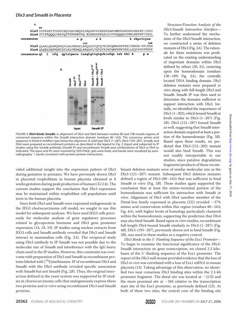
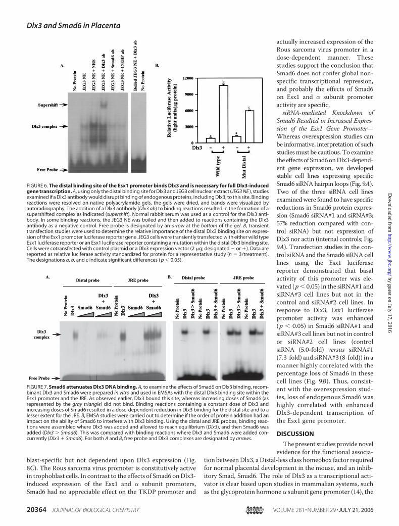
(TAATT)was conserved; however, the two nucleotides presenton the 3� termini of this central core were not conserved (Fig.5A). These nucleotides varied from the consensus Dlx3 bindingsite (24) we defined in the human glycoprotein hormone � sub-unit promoter (JRE) (14). Using EMSAs, we compared theDNAbinding complexes formed when using the distal (�2135) andproximal (�585) putativeDlx3 binding sites from the Esx1 pro-moter and nuclear extracts from JEG3 cells (Fig. 5B). As a pos-itive control, the JRE from the � subunit promoter was used.Fig. 5B depicts direct comparison of complexes formed with allthree of these binding sites. The distal site was more similar tothe JRE binding complex electrophoretic mobility when com-pared with the proximal site. When using recombinant Dlx3 inEMSA (Fig. 5B), all three sites were sufficient to bind Dlx3;however, clear differences in the relative binding of Dlx3 forthese binding sites were revealed. The recombinant Dlx3 wasslightly larger in molecular size due to the presence of the threehemagglutinin epitopes engineered into the recombinant pro-tein. The rank order of relative binding for recombinant Dlx3was the JRE (highest) � the distal site � the proximal site (low-est). We focused our attention on the distal Dlx3 binding sitewithin the Esx1 gene promoter. EMSA studies using nuclearextracts and antiserum against Dlx3 provided direct evidencethat endogenous Dlx3 can bind to this site (Fig. 6A). The addi-tion of the Dlx3 antibody to EMSA binding reaction resulted insupershifted complexes that were not evident using preim-
mune normal rabbit serum. The addition of the Smad6 or theCCAAT/enhancer-binding protein antiserum to these bindingreactions did not appreciably alter complex formation inEMSA, suggesting that Smad6 may not directly participate inDNA binding with Dlx3 and that Dlx3 antibody interactionswere specific. To determine the importance of this site toDlx3-induced Esx1 gene transcription, the distal site wasmutagenized to a NotI restriction site and examined using aluciferase reporter gene approach in transient transfectionstudies (Fig. 6B). The wild type Esx1 promoter was stronglyinduced byDlx3. Themutation in the distal putativeDlx3 bind-ing site reduced Dlx3-induced Esx1 expression by �50% (p �0.05), suggesting that this site was important for promoteractivity induced by Dlx3.We then sought to determine the impact of Smad6 on Dlx3
DNA binding. Reconstitution EMSAs using recombinant Dlx3and Smad6 demonstrated that the Dlx3/Smad6 interactioneffectively reduced/blocked association of Dlx3 with the distalDlx3 binding site within the Esx1 promoter (Fig. 7A). In thisexperiment, Smad6 alone did not form a complex with the dis-tal Dlx3 binding site.When Smad6was titrated into the bindingreactions containing Dlx3, the Dlx3 binding complex wasdiminished in a dose-dependent manner. Using the JRE asprobe, a similar titration of Smad6 protein reduced Dlx3 bind-ing, albeit to a lesser extent compared with the distal site of theEsx1 promoter. Studies then focused on determining if pre-bound Dlx3 could be displaced by Smad6 in EMSA bindingreactions (Fig. 7B). Binding reactions compared the addition ofDlx3 concurrent with Smad6 and reactions whereDlx3 bindingwas allowed to reach equilibrium and then Smad6 was added.These studies revealed that for the distal probe, Smad6 com-peted for Dlx3 binding regardless of the order of protein addi-tion. Similar levels of competition were not observed using theJRE (stronger relative binding), suggesting that Smad6 cancompete forDlx3 binding, particularly on gene targets that haverelatively weaker Dlx3 binding sites, like those characterizedwithin the Esx1 gene promoter.To assess the functional consequences of the Dlx3/Smad6
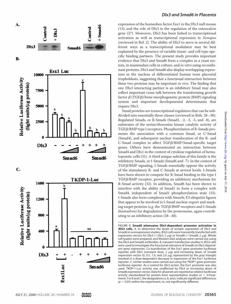
interaction on the Esx1 promoter, we again used the Esx1 pro-moter-luciferase reporter construct (Fig. 8). Based upon thebinding studies described above, our predictionwas that Smad6overexpression would probably repress expression of the Esx1promoter induced by Dlx3. Using transient transfection inJEG3 cells, overexpression of Dlx3 and Smad6 resulted inincreased levels of ectopically expressed Dlx3 and Smad6 (Fig.8A). Further, Dlx3 overexpression increased Esx1 luciferaseactivity (p � 0.05; Fig. 8B). Titration of Smad6 into this systemresulted in reduced basal activity of the Esx1 reporter gene (p�0.05). Consistent with our prediction, co-transfection withincreasing doses of Smad6 expression vector along with Dlx3resulted in amarked inhibition (p� 0.05) ofDlx3-inducedEsx1promoter activity. Dlx3-induced activation of the � subunitgene promoter was also reduced by overexpression of Smad6(data not shown). In an effort to determine the specificity ofSmad6 action on gene transcription in general, we examinedthe effects of Smad6 on the trophoblast Kunitz domain protein1 (TKDP1) (Fig. 8C ) and Rous sarcoma virus promoters (datanot shown). TKDP1 promoter expression (25, 26) is tropho-
FIGURE 5. Two putative Dlx3 binding sites are present within the Esx1promoter. A, the JRE (a consensus Dlx3 binding site) from the glycoproteinhormone � subunit promoter was compared with the distal (position �2135)and proximal (position �585) binding sites from the Esx1 promoter. The cen-tral core of the binding site is underlined. B, EMSAs were used to comparebinding complexes formed on the JRE and the distal and proximal Dlx3 bind-ing sites within the Esx1 promoter using JEG3 cell nuclear extract and recom-binant Dlx3. Equal amounts of nuclear extracts or recombinant Dlx3 wereadded to each binding reaction. Recombinant Dlx3 in these studies wasepitope-tagged with three copies of the hemagglutinin epitope (3�HA-Dlx3).Binding reactions were resolved on native polyacrylamide gels, the gels weredried, and bands were visualized by autoradiography. Free probe is desig-nated by an arrow at the bottom of the gel.
Dlx3 and Smad6 in Placenta
JULY 21, 2006 • VOLUME 281 • NUMBER 29 JOURNAL OF BIOLOGICAL CHEMISTRY 20363
by guest on July 17, 2016http://w
ww
.jbc.org/D
ownloaded from
blast-specific but not dependent upon Dlx3 expression (Fig.8C). The Rous sarcoma virus promoter is constitutively activein trophoblast cells. In contrast to the effects of Smad6 onDlx3-induced expression of the Esx1 and � subunit promoters,Smad6 had no appreciable effect on the TKDP promoter and
actually increased expression of theRous sarcoma virus promoter in adose-dependent manner. Thesestudies support the conclusion thatSmad6 does not confer global non-specific transcriptional repression,and probably the effects of Smad6on Esx1 and � subunit promoteractivity are specific.siRNA-mediated Knockdown of
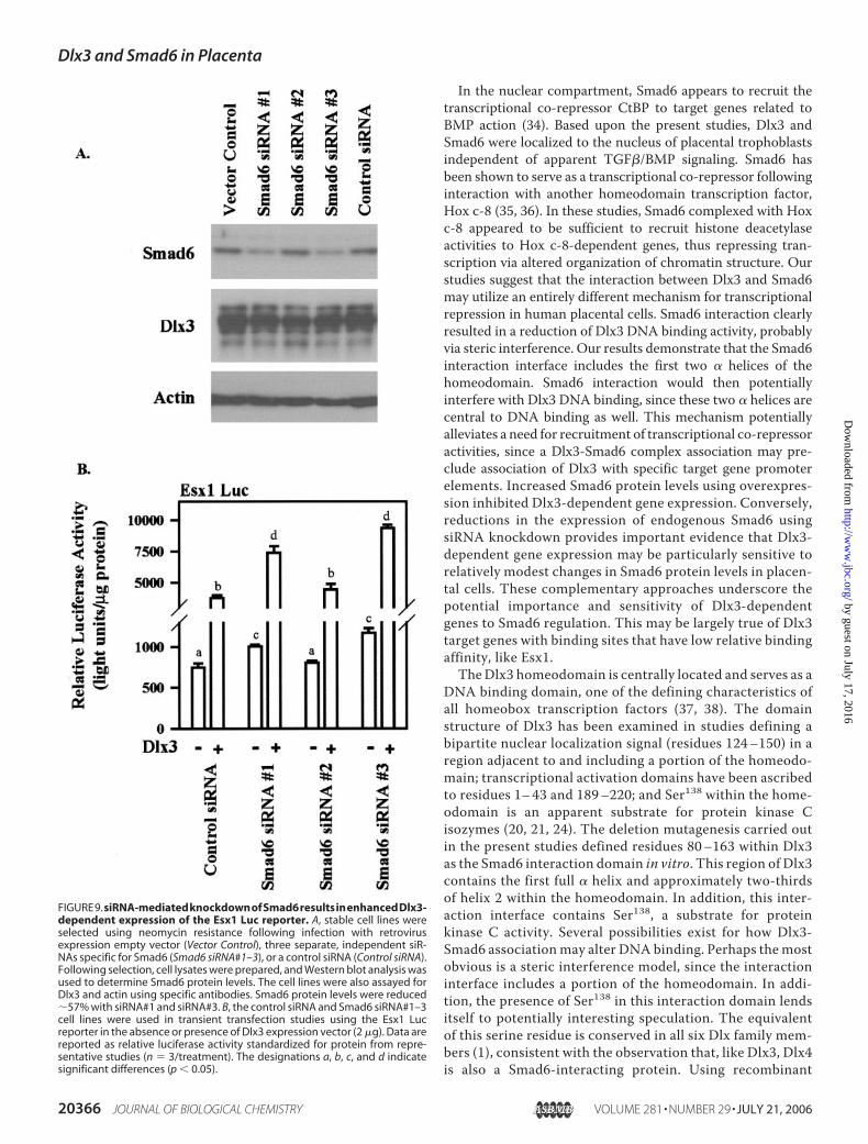
Smad6 Resulted in Increased Expres-sion of the Esx1 Gene Promoter—Whereas overexpression studies canbe informative, interpretation of suchstudiesmust be cautious. To examinethe effects of Smad6onDlx3-depend-ent gene expression, we developedstable cell lines expressing specificSmad6 siRNAhairpin loops (Fig. 9A).Two of the three siRNA cell linesexaminedwere found to have specificreductions in Smad6 protein expres-sion (Smad6 siRNA#1 and siRNA#3;57% reduction compared with con-trol siRNA) but not expression ofDlx3 nor actin (internal controls; Fig.9A). Transfection studies in the con-trol siRNA and the Smad6 siRNA celllines using the Esx1 luciferasereporter demonstrated that basalactivity of this promoter was ele-vated (p� 0.05) in the siRNA#1 andsiRNA#3 cell lines but not in thecontrol and siRNA#2 cell lines. Inresponse to Dlx3, Esx1 luciferasepromoter activity was enhanced(p � 0.05) in Smad6 siRNA#1 andsiRNA#3 cell lines but not in controlor siRNA#2 cell lines (controlsiRNA (5.0-fold) versus siRNA#1(7.3-fold) and siRNA#3 (8-fold)) in amanner highly correlated with thepercentage loss of Smad6 in thesecell lines (Fig. 9B). Thus, consist-ent with the overexpression stud-ies, loss of endogenous Smad6 washighly correlated with enhancedDlx3-dependent transcription ofthe Esx1 gene promoter.
DISCUSSION
The present studies provide novelevidence for the functional associa-
tion betweenDlx3, aDistal-less class homeobox factor requiredfor normal placental development in the mouse, and an inhib-itory Smad, Smad6. The role of Dlx3 as a transcriptional acti-vator is clear based upon studies in mammalian systems, suchas the glycoprotein hormone � subunit gene promoter (14), the
FIGURE 6. The distal binding site of the Esx1 promoter binds Dlx3 and is necessary for full Dlx3-inducedgene transcription. A, using only the distal binding site for Dlx3 and JEG3 cell nuclear extract (JEG3 NE ), studiesexamined if a Dlx3 antibody would disrupt binding of endogenous proteins, including Dlx3, to this site. Bindingreactions were resolved on native polyacrylamide gels, the gels were dried, and bands were visualized byautoradiography. The addition of a Dlx3 antibody (Dlx3 ab) to binding reactions resulted in the formation of asupershifted complex as indicated (supershift). Normal rabbit serum was used as a control for the Dlx3 anti-body. In some binding reactions, the JEG3 NE was boiled and then added to reactions containing the Dlx3antibody as a negative control. Free probe is designated by an arrow at the bottom of the gel. B, transienttransfection studies were used to determine the relative importance of the distal Dlx3 binding site on expres-sion of the Esx1 promoter luciferase reporter gene. JEG3 cells were transiently transfected with either wild typeEsx1 luciferase reporter or an Esx1 luciferase reporter containing a mutation within the distal Dlx3 binding site.Cells were cotransfected with control plasmid or a Dlx3 expression vector (2 �g; designated � or ). Data arereported as relative luciferase activity standardized for protein for a representative study (n � 3/treatment).The designations a, b, and c indicate significant differences ( p � 0.05).
FIGURE 7. Smad6 attenuates Dlx3 DNA binding. A, to examine the effects of Smad6 on Dlx3 binding, recom-binant Dlx3 and Smad6 were prepared in vitro and used in EMSAs with the distal Dlx3 binding site within theEsx1 promoter and the JRE. As observed earlier, Dlx3 bound this site, whereas increasing doses of Smad6 (asrepresented by the gray triangle) did not bind. Binding reactions containing a constant dose of Dlx3 andincreasing doses of Smad6 resulted in a dose-dependent reduction in Dlx3 binding for the distal site and to alesser extent for the JRE. B, EMSA studies were carried out to determine if the order of protein addition had animpact on the ability of Smad6 to interfere with Dlx3 binding. Using the distal and JRE probes, binding reac-tions were assembled where Dlx3 was added and allowed to reach equilibrium (Dlx3), and then Smad6 wasadded (Dlx3 � Smad6). This was compared with binding reactions where Dlx3 and Smad6 were added con-currently (Dlx3 Smad6). For both A and B, free probe and Dlx3 complexes are designated by arrows.
Dlx3 and Smad6 in Placenta
20364 JOURNAL OF BIOLOGICAL CHEMISTRY VOLUME 281 • NUMBER 29 • JULY 21, 2006
by guest on July 17, 2016http://w
ww
.jbc.org/D
ownloaded from
expression of the homeobox factor Esx1 in the Dlx3 null mouse(13), and the role of Dlx3 in the regulation of the osteocalcingene (27). Moreover, Dlx3 has been linked to transcriptionalactivation as well as transcriptional repression in Xenopus(reviewed in Ref. 2). The ability of Dlx3 to serve in several dif-ferent ways as a transcriptional modulator may be bestexplained by the presence of variable tissue- and cell type-spe-cific binding partners. The present study provides importantevidence that Dlx3 and Smad6 form a complex in a yeast sys-tem, in mammalian cells in culture, and in vitro using recombi-nant proteins. Dlx3 and Smad6 also display overlapping expres-sion in the nucleus of differentiated human term placentaltrophoblasts, suggesting that a functional interaction betweenthese two proteins may be important in vivo. The finding thatone Dlx3 interacting partner is an inhibitory Smad may alsoreflect important cross-talk between the transforming growthfactor � (TGF�)/bone morphogenetic protein (BMP) signalingsystem and important developmental determinants thatrequire Dlx3.Smad proteins are transcriptional regulators that can be sub-
divided into essentially three classes (reviewed in Refs. 28–30).Regulated Smads, or R-Smads (Smad1, -2, -3, -5, and -8), aresubstrates of the serine/threonine kinase catalytic activity ofTGF�/BMP type I receptors. Phosphorylation of R-Smads pro-motes the association with a common Smad, or C-Smad(Smad4), and subsequent nuclear translocation of the R- andC-Smad complex to affect TGF�/BMP/Smad-specific targetgenes. Others have demonstrated an interaction betweenSmad4 and Dlx1 in the context of cytokine regulation of hema-topoietic cells (31). A third unique subclass of this family is theinhibitory Smads, or I-Smads (Smad6 and -7). In the context ofTGF�/BMP signaling, I-Smads essentially oppose the activityof the stimulatory R- and C-Smads at several levels. I-Smadshave been shown to compete for R-Smad binding to the type ITGF�/BMP receptor, providing an inhibitory mechanism forR-Smad activity (32). In addition, Smad6 has been shown tointerfere with the ability of Smad1 to form a complex withSmad4, independent of Smad1 phosphorylation state (33).I-Smads also form complexes with Smurfs, E3 ubiquitin ligasesthat appear to be involved in I-Smad nuclear export and mark-ing target proteins (e.g. the TGF�/BMP receptors and I-Smadsthemselves) for degradation by the proteosome, again contrib-uting to an inhibitory action (28–30).
FIGURE 8. Smad6 attenuates Dlx3-dependent promoter activation inJEG3 cells. A, to determine the levels of ectopic expression of Dlx3 andSmad6 in overexpression studies, JEG3 cells were transiently transfected withexpression vectors for Dlx3 (Dlx3; 2 �g) or Smad6 (Smad6; 2 �g). Wholecell lysates were prepared, and Western blot analyses were carried out usingthe Dlx3 and Smad6 antibodies. B, transient transfection studies in JEG3 cellswere used to investigate the functional relevance of Smad6 on Dlx3-depend-ent gene expression. Co-transfection of the Esx1 gene promoter-luciferasereporter with Dlx3 (constant dose, 2 �g) and increasing doses of Smad6expression vector (0, 0.5, 1.0, and 2.0 �g; represented by the gray triangle)resulted in a dose-dependent decrease in expression of the Esx1 luciferasereporter. C, similar studies were carried out using the TKDP1 gene promoterluciferase reporter. As a control for Dlx3 action, the Esx1 promoter was alsoused. TKDP-1-Luc activity was unaffected by Dlx3 or increasing doses ofSmad6 expression vector. Data for all panels are reported as relative luciferaseactivity standardized for protein from representative studies (n � 3/treat-ment). For B and C, the designations a, b, and c indicate significant differences(p � 0.05) within the experiment; ns, not significantly different.
Dlx3 and Smad6 in Placenta
JULY 21, 2006 • VOLUME 281 • NUMBER 29 JOURNAL OF BIOLOGICAL CHEMISTRY 20365
by guest on July 17, 2016http://w
ww
.jbc.org/D
ownloaded from
In the nuclear compartment, Smad6 appears to recruit thetranscriptional co-repressor CtBP to target genes related toBMP action (34). Based upon the present studies, Dlx3 andSmad6 were localized to the nucleus of placental trophoblastsindependent of apparent TGF�/BMP signaling. Smad6 hasbeen shown to serve as a transcriptional co-repressor followinginteraction with another homeodomain transcription factor,Hox c-8 (35, 36). In these studies, Smad6 complexed with Hoxc-8 appeared to be sufficient to recruit histone deacetylaseactivities to Hox c-8-dependent genes, thus repressing tran-scription via altered organization of chromatin structure. Ourstudies suggest that the interaction between Dlx3 and Smad6may utilize an entirely different mechanism for transcriptionalrepression in human placental cells. Smad6 interaction clearlyresulted in a reduction of Dlx3 DNA binding activity, probablyvia steric interference. Our results demonstrate that the Smad6interaction interface includes the first two � helices of thehomeodomain. Smad6 interaction would then potentiallyinterfere with Dlx3 DNA binding, since these two � helices arecentral to DNA binding as well. This mechanism potentiallyalleviates a need for recruitment of transcriptional co-repressoractivities, since a Dlx3-Smad6 complex association may pre-clude association of Dlx3 with specific target gene promoterelements. Increased Smad6 protein levels using overexpres-sion inhibited Dlx3-dependent gene expression. Conversely,reductions in the expression of endogenous Smad6 usingsiRNA knockdown provides important evidence that Dlx3-dependent gene expression may be particularly sensitive torelatively modest changes in Smad6 protein levels in placen-tal cells. These complementary approaches underscore thepotential importance and sensitivity of Dlx3-dependentgenes to Smad6 regulation. This may be largely true of Dlx3target genes with binding sites that have low relative bindingaffinity, like Esx1.TheDlx3 homeodomain is centrally located and serves as a
DNA binding domain, one of the defining characteristics ofall homeobox transcription factors (37, 38). The domainstructure of Dlx3 has been examined in studies defining abipartite nuclear localization signal (residues 124–150) in aregion adjacent to and including a portion of the homeodo-main; transcriptional activation domains have been ascribedto residues 1–43 and 189–220; and Ser138 within the home-odomain is an apparent substrate for protein kinase Cisozymes (20, 21, 24). The deletion mutagenesis carried outin the present studies defined residues 80–163 within Dlx3as the Smad6 interaction domain in vitro. This region of Dlx3contains the first full � helix and approximately two-thirdsof helix 2 within the homeodomain. In addition, this inter-action interface contains Ser138, a substrate for proteinkinase C activity. Several possibilities exist for how Dlx3-Smad6 associationmay alter DNA binding. Perhaps themostobvious is a steric interference model, since the interactioninterface includes a portion of the homeodomain. In addi-tion, the presence of Ser138 in this interaction domain lendsitself to potentially interesting speculation. The equivalentof this serine residue is conserved in all six Dlx family mem-bers (1), consistent with the observation that, like Dlx3, Dlx4is also a Smad6-interacting protein. Using recombinant
FIGURE9.siRNA-mediatedknockdownofSmad6resultsinenhancedDlx3-dependent expression of the Esx1 Luc reporter. A, stable cell lines wereselected using neomycin resistance following infection with retrovirusexpression empty vector (Vector Control), three separate, independent siR-NAs specific for Smad6 (Smad6 siRNA#1–3), or a control siRNA (Control siRNA).Following selection, cell lysates were prepared, and Western blot analysis wasused to determine Smad6 protein levels. The cell lines were also assayed forDlx3 and actin using specific antibodies. Smad6 protein levels were reduced�57% with siRNA#1 and siRNA#3. B, the control siRNA and Smad6 siRNA#1–3cell lines were used in transient transfection studies using the Esx1 Lucreporter in the absence or presence of Dlx3 expression vector (2 �g). Data arereported as relative luciferase activity standardized for protein from repre-sentative studies (n � 3/treatment). The designations a, b, c, and d indicatesignificant differences (p � 0.05).
Dlx3 and Smad6 in Placenta
20366 JOURNAL OF BIOLOGICAL CHEMISTRY VOLUME 281 • NUMBER 29 • JULY 21, 2006
by guest on July 17, 2016http://w
ww
.jbc.org/D
ownloaded from
Dlx3, Morasso and co-workers (21) demonstrated that phos-phorylation at Ser138 by protein kinase C (most strongly byprotein kinase C�) resulted in partial inhibition of Dlx3DNA binding, consistent with the potential effects of Smad6on DNA binding. This supports speculation that phospho-rylation of Dlx3 at Ser138 may alter or increase Smad6 bind-ing, leading to reduced DNA binding at Dlx3 target genes.Studies are currently under way to address this possibility.R- and C-Smads appear to be constitutively expressed in
most cell types, whereas I-Smads are subject to regulation by anumber of growth factors, such as epidermal growth factor,TGF�, and BMPs. In many cases, the effects of these growthfactors are mediated through R- and C-Smad-dependent tran-scriptional mechanisms. This has led to a hypothesis implicat-ing an intracellular negative feedback loop, whereby positiveTGF�/BMP signals are modulated over time by accumulationof induced I-Smads (reviewed in Ref. 40). Interestingly, Dlx3expression patterns during Xenopus development depends inpart upon BMP signaling gradients (41, 42). Inhibition of thesegradients using the BMP receptor antagonist chordin resultedin a dose-sensitive inhibition of Dlx3, Dlx5, and Dlx6 mRNAexpression. Dlx3 expression in mouse keratinocytes was alsoreported to be subject to regulation by BMPs (43) in a mannerpotentially coordinated with I-Smad expression. This supportsspeculation that not only might BMP-regulated Smad6 expres-sion serve as a negative feedback mechanism controlling theduration of Smad signaling, but increased BMP-dependentSmad6 expression may lead to important modulation of Dlx3-dependent gene expression. BMPs have been shown to regulateearly embryogenesis during the preimplantation period, suchthat these types of ligands are probably present and importantduring placental morphogenesis (39).
Acknowledgments—We thank Drs. Maria Morasso, Jun-Lin Guan,Ali Brivanlou, and Colin Clay for generously providing usefulreagents during the course of these studies. We especially thank Dr.Satish Devapatla and the staff at the CayugaMedical Center (Ithaca,NY) for providing important help in tissue collections and Dr. MariaMorasso and members of the Roberson laboratory for helpful com-ments in the preparation of the manuscript.
REFERENCES1. Bendall, A. J., and Abate-Shen, C. (2000) Gene (Amst.) 247, 17–312. Beanan, M. J., and Sargent, T. D. (2000) Dev. Dyn. 218, 545–5533. Morasso, M. I., Mahon, K. A., and Sargent, T. D. (1995) Proc. Natl. Acad.
Sci. U. S. A. 92, 3968–39724. Price, J. A., Bowden, D. W., Wright, J. T., Pettenati, M. J., and Hart, T. C.
(1998) Hum. Mol. Genet. 7, 563–5695. Price, J. A., Wright, J. T., Kula, K., Bowden, D. W., and Hart, T. C. (1998)
J. Med. Genet. 35, 825–8286. Haldeman, R. J., Cooper, L. F., Hart, T. C., Phillips, C., Boyd, C., Lester,
G. E., and Wright, J. T. (2004) Bone 35, 988–9977. Dong, J., Amor, D., Aldred, M. J., Gu, T., Escamilla, M., and MacDougall,
M. (2005) Am. J. Med. Genet. A 133, 138–1418. Juriloff, D. M., Harris, M. J., and Brown, C. J. (2001)Mamm. Genome 12,
426–435
9. Clouthier, D. E., Williams, S. C., Yanagisawa, H., Wieduwilt, M., Richard-son, J. A., and Yanagisawa, M. (2000) Dev. Biol. 217, 10–24
10. Clouthier, D. E., Williams, S. C., Hammer, R. E., Richardson, J. A., andYanagisawa, M. (2003) Dev. Biol. 261, 506–519
11. Clouthier, D. E., and Schilling, T. F. (2004) Birth Defects Res. C Embryol.Today 72, 190–199
12. Ivey, K., Tyson, B., Ukidwe, P., McFadden, D. G., Levi, G., Olson, E. N.,Srivastava, D., and Wilkie, T. M. (2003) Dev. Biol. 255, 230–237
13. Morasso, M. I., Grinberg, A., Robinson, G., Sargent, T. D., and Mahon,K. A. (1999) Proc. Natl. Acad. Sci. U. S. A. 96, 162–167
14. Roberson, M. S., Meermann, S., Morasso, M. I., Mulvaney-Musa, J. M.,and Zhang, T. (2001) J. Biol. Chem. 276, 10016–10024
15. France, J. T., Keelan, J., Song, L., Liddell, H., Zanderigo, A., and Knox, B.(1996) Aust. N. Z. J. Obstet. Gynaecol. 36, 325–330
16. Acevedo, H. F. (2002) J. Exp. Ther. Oncol. 2, 133–14517. Keay, S. D., Vatish, M., Karteris, E., Hillhouse, E. W., and Randeva, H. S.
(2004) Br. J. Obstet. Gynaecol. 111, 1218–122818. Holland, M. P., Bliss, S. P., Berghorn, K. A., and Roberson, M. S. (2004)
Endocrinology 145, 1096–110519. Roberson,M. S., Ban, M., Zhang, T., andMulvaney, J. M. (2000)Mol. Cell.
Biol. 20, 3331–334420. Bryan, J. T., and Morasso, M. I. (2000) J. Cell Sci. 113, 4013–402321. Park, G. T., Denning, M. F., and Morasso, M. I. (2001) FEBS Lett. 496,
60–6522. Quinn, L. M., Johnson, B. V., Nicholl, J., Sutherland, G. R., and Kalionis, B.
(1997) Gene (Amst.) 187, 55–6123. Li, Y., Lemaire, P., and Behringer, R. R. (1997) Dev. Biol. 188, 85–9524. Feledy, J. A., Morasso, M. I., Jang, S. I., and Sargent, T. D. (1999) Nucleic
Acids Res. 27, 764–77025. MacLean, J. A., Roberts, R. M., and Green, J. A. (2004) Biol. Reprod. 71,
455–46326. MacLean, J. A., Chakrabarty, A., Xie, S., Bixby, J. A., Roberts, R. M., and
Green, J. A. (2003)Mol. Reprod. Dev. 65, 30–4027. Hassan, M. Q., Javed, A., Morasso, M. I., Karlin, J., Montecino, M., van
Wijnen, A. J., Stein, G. S., Stein, J. L., and Lian, J. B. (2004)Mol. Cell. Biol.24, 9248–9261
28. Ten, D. P.,Miyazono, K., andHeldin, C.H. (2000)Trends Biochem. Sci. 25,64–70
29. Ten, D. P., and Hill, C. S. (2004) Trends Biochem. Sci. 29, 265–27330. Chen, D., Zhao,M., andMundy, G. R. (2004)Growth Factors 22, 233–24131. Chiba, S., Takeshita, K., Imai, Y., Kumano, K., Kurokawa, M., Masuda, S.,
Shimizu, K., Nakamura, S., Ruddle, F. H., and Hirai, H. (2003) Proc. Natl.Acad. Sci. U. S. A. 100, 15577–15582
32. Shi, Y., and Massague, J. (2003) Cell 113, 685–70033. Hata, A., Lagna, G., Massague, J., and Hemmati-Brivanlou, A. (1998)
Genes Dev. 12, 186–19734. Lin, X., Liang, Y. Y., Sun, B., Liang,M., Shi, Y., Brunicardi, F. C., Shi, Y., and
Feng, X. H. (2003)Mol. Cell. Biol. 23, 9081–909335. Bai, S., Shi, X., Yang, X., and Cao, X. (2000) J. Biol. Chem. 275, 8267–827036. Bai, S., and Cao, X. (2002) J. Biol. Chem. 277, 4176–418237. Gehring, W. J. (1993) Gene (Amst.) 135, 215–22138. Wright, C. V., Cho, K.W., Oliver, G., andDe Robertis, E.M. (1989)Trends
Biochem. Sci. 14, 52–5639. Paria, B. C., Ma,W., Tan, J., Raja, S., Das, S. K., Dey, S. K., and Hogan, B. L.
(2001) Proc. Natl. Acad. Sci. U. S. A. 98, 1047–105240. Miyazono, K. (2002) Cytokine Growth Factor Rev. 13, 7–941. Feledy, J. A., Beanan, M. J., Sandoval, J. J., Goodrich, J. S., Lim, J. H.,
Matsuo-Takasaki,M., Sato, S.M., and Sargent, T. D. (1999)Dev. Biol. 212,455–464
42. Luo, T., Matsuo-Takasaki, M., Lim, J. H., and Sargent, T. D. (2001) Int. J.Dev. Biol. 45, 681–684
43. Park, G. T., and Morasso, M. I. (2002) Nucleic Acids Res. 30, 515–522
Dlx3 and Smad6 in Placenta
JULY 21, 2006 • VOLUME 281 • NUMBER 29 JOURNAL OF BIOLOGICAL CHEMISTRY 20367
by guest on July 17, 2016http://w
ww
.jbc.org/D
ownloaded from
Weiss and Mark S. RobersonKathie A. Berghorn, Patricia A. Clark-Campbell, Li Han, Michael McGrattan, Robert S.
BindingSmad6 Represses Dlx3 Transcriptional Activity through Inhibition of DNA
doi: 10.1074/jbc.M603049200 originally published online May 10, 20062006, 281:20357-20367.J. Biol. Chem.
10.1074/jbc.M603049200Access the most updated version of this article at doi:
Alerts:
When a correction for this article is posted•
When this article is cited•
to choose from all of JBC's e-mail alertsClick here
http://www.jbc.org/content/281/29/20357.full.html#ref-list-1
This article cites 42 references, 17 of which can be accessed free at
by guest on July 17, 2016http://w
ww
.jbc.org/D
ownloaded from