Menin Interacts with the AP1 Transcription Factor JunD and Represses JunD-Activated Transcription
10
Cell, Vol. 96, 143–152, January 8, 1999, Copyright 1999 by Cell Press Menin Interacts with the AP1 Transcription Factor JunD and Represses JunD-Activated Transcription (36%) (Debelenko et al., 1997; Heppner et al., 1997; Zhu- ang et al., 1997). Germline and somatic mutations in MEN1 occur throughout the predicted protein coding Sunita K. Agarwal,* ‡ Siradanahalli C. Guru, ² Christina Heppner,* Michael R. Erdos, ² Regina M. Collins,* Sylvia Y. Park,* Suraj Saggar,* Settara C. Chandrasekharappa, ² Francis S. Collins, ² region with no obvious hot spots or correlation between genotype and phenotype (Agarwal et al., 1997). The ma- Allen M. Spiegel,* Stephen J. Marx,* and A. Lee Burns* * Metabolic Diseases Branch jority of mutations predict premature protein truncation due to either nonsense or frame-shifting type mutations, National Institute of Diabetes and Digestive and Kidney Diseases but missense mutations have also been identified in about 30% of cases. The frequent protein truncation National Institutes of Health Bethesda, Maryland 20892 mutations likely cause gene inactivation, consistent with the first hit to a tumor suppressor gene (Fearon, 1998). ² Genetics and Molecular Biology Branch National Human Genome Research Institute Few other clues about the function of menin can be deciphered from the predicted amino acid sequence or National Institutes of Health Bethesda, Maryland 20892 the pattern of mutations. However, menin recently was found to localize to the nucleus, with two independent nuclear localization signals (Guru et al., 1998). To begin to ascertain the molecular function of menin, Summary we attempted to identify menin-interacting proteins. A yeast two-hybrid screen was performed with full-length MEN1 is a tumor suppressor gene that encodes a 610 menin fused to the Gal4 DNA-binding domain (Gal4DBD). amino acid nuclear protein (menin) of previously un- We have detected the AP1 (activator protein 1) transcrip- known function. Using a yeast two-hybrid screen with tion factor JunD as an interacting partner of menin. menin as the bait, we have identified the transcription factor JunD as a direct menin-interacting partner. Results and Discussion Menin did not interact directly with other Jun and Fos family members. The menin–JunD interaction was Yeast Two-Hybrid Screen confirmed in vitro and in vivo. Menin repressed tran- An adult human brain cDNA library was screened with scriptional activation mediated by JunD fused to the menin as the bait by sequential transformation. From Gal4 DNA-binding domain from a Gal4 responsive re- approximately 3.9 3 10 7 yeast transformants screened, porter, or by JunD from an AP1-responsive reporter. ten were true positives because both the selection on Several naturally occurring and clustered MEN1 mis- -His media and also their ability to express b-galactosi- sense mutations disrupted menin interaction with dase depended on their interaction with menin. Four of JunD. These observations suggest that menin’s tumor these positives were the AP1 transcription factor JunD, suppressor function involves direct binding to JunD encoded by 2 different clones named pGAD10-JD1 and and inhibition of JunD activated transcription. pGAD10-JD2 (Table 1); four identical positive clones were chaperonin 10, and two were homologous to ribo- Introduction somal RNA. pGAD10-JD1 was represented by 3 identical clones encoding amino acids (aa) 8 to 340 of JunD, while Multiple endocrine neoplasia type 1 (MEN1) is an au- pGAD10-JD2 was represented by one clone encoding tosomal dominant disorder associated mainly with tu- aa 1 to 306 (full-length JunD is 347 aa). pGAD10-JD2 mors of parathyroid, enteropancreatic neuroendocrine also had sequence from the JunD 59 UTR (120 bp) up- tissue, and anterior pituitary (Marx, 1998). Recently, we stream from the JunD initiation codon ATG, that was in- identified the MEN1 gene by positional cloning (Chand- frame with the Gal4 transcriptional activation domain rasekharappa et al., 1997). MEN1 encodes a 67 kDa (Gal4AD) and JunD, resulting in the insertion of 40 aa protein, menin (Chandrasekharappa et al., 1997). Germ- between the two proteins. The JD1 insert lacks part of line mutations in the MEN1 gene cause familial MEN1 the first transactivation domain of JunD, but retains all and sporadic MEN1 (Agarwal et al., 1997). Tumors in the other known essential functional domains (Abate et affected individuals have lost the wild-type MEN1 allele al., 1991). The JD1 clone was used in most studies. at chromosome 11q13 (Larsson et al., 1988), suggesting Full-length mouse JunD fused to the Gal4AD was also that MEN1 is a tumor suppressor gene (Fearon, 1998). positive for interaction with mouse or human menin Loss of heterozygosity (LOH) at 11q13 is found not only fused to Gal4DBD, and Gal4DBD-mouse menin inter- in MEN1 tumors, but also in sporadic endocrine tumors acted with pGAD10-JD1 or pGAD10-JD2 (Table 1). of patients without MEN1 (Friedman et al., 1989). So- matic MEN1 mutations are the most common mutations in several types of non-MEN1 tumors, including parathy- Menin–JunD Interaction In Vitro roid adenomas (MEN1 mutation in 21%), gastrinomas In vitro–translated (IVT) menin was tested for binding (33%), insulinomas (17%), and bronchial carcinoids with glutathione-agarose beads coupled with glutathi- one S-transferase (GST) alone or GST–JD1 (JunD insert from pGAD10-JD1 fused in-frame at the C terminus of ‡ To whom correspondence should be addressed (e-mail: sunitaa@ amb.niddk.nih.gov). GST). Menin was specifically associated with GST–JD1,
-
Upload
independent -
Category
Documents
-
view
4 -
download
0
Transcript of Menin Interacts with the AP1 Transcription Factor JunD and Represses JunD-Activated Transcription

Cell, Vol. 96, 143–152, January 8, 1999, Copyright 1999 by Cell Press
Menin Interacts with the AP1 Transcription FactorJunD and Represses JunD-Activated Transcription
(36%) (Debelenko et al., 1997; Heppner et al., 1997; Zhu-ang et al., 1997). Germline and somatic mutations inMEN1 occur throughout the predicted protein coding
Sunita K. Agarwal,*‡ Siradanahalli C. Guru,†Christina Heppner,* Michael R. Erdos,†Regina M. Collins,* Sylvia Y. Park,* Suraj Saggar,*Settara C. Chandrasekharappa,† Francis S. Collins,† region with no obvious hot spots or correlation between
genotype and phenotype (Agarwal et al., 1997). The ma-Allen M. Spiegel,* Stephen J. Marx,* and A. Lee Burns**Metabolic Diseases Branch jority of mutations predict premature protein truncation
due to either nonsense or frame-shifting type mutations,National Institute of Diabetesand Digestive and Kidney Diseases but missense mutations have also been identified in
about 30% of cases. The frequent protein truncationNational Institutes of HealthBethesda, Maryland 20892 mutations likely cause gene inactivation, consistent with
the first hit to a tumor suppressor gene (Fearon, 1998).†Genetics and Molecular Biology BranchNational Human Genome Research Institute Few other clues about the function of menin can be
deciphered from the predicted amino acid sequence orNational Institutes of HealthBethesda, Maryland 20892 the pattern of mutations. However, menin recently was
found to localize to the nucleus, with two independentnuclear localization signals (Guru et al., 1998).
To begin to ascertain the molecular function of menin,Summarywe attempted to identify menin-interacting proteins. Ayeast two-hybrid screen was performed with full-lengthMEN1 is a tumor suppressor gene that encodes a 610menin fused to the Gal4 DNA-binding domain (Gal4DBD).amino acid nuclear protein (menin) of previously un-We have detected the AP1 (activator protein 1) transcrip-known function. Using a yeast two-hybrid screen withtion factor JunD as an interacting partner of menin.menin as the bait, we have identified the transcription
factor JunD as a direct menin-interacting partner.Results and DiscussionMenin did not interact directly with other Jun and Fos
family members. The menin–JunD interaction wasYeast Two-Hybrid Screenconfirmed in vitro and in vivo. Menin repressed tran-An adult human brain cDNA library was screened withscriptional activation mediated by JunD fused to themenin as the bait by sequential transformation. FromGal4 DNA-binding domain from a Gal4 responsive re-approximately 3.9 3 107 yeast transformants screened,porter, or by JunD from an AP1-responsive reporter.ten were true positives because both the selection onSeveral naturally occurring and clustered MEN1 mis--His media and also their ability to express b-galactosi-sense mutations disrupted menin interaction withdase depended on their interaction with menin. Four ofJunD. These observations suggest that menin’s tumorthese positives were the AP1 transcription factor JunD,suppressor function involves direct binding to JunDencoded by 2 different clones named pGAD10-JD1 andand inhibition of JunD activated transcription.pGAD10-JD2 (Table 1); four identical positive cloneswere chaperonin 10, and two were homologous to ribo-Introductionsomal RNA. pGAD10-JD1 was represented by 3 identicalclones encoding amino acids (aa) 8 to 340 of JunD, whileMultiple endocrine neoplasia type 1 (MEN1) is an au-pGAD10-JD2 was represented by one clone encodingtosomal dominant disorder associated mainly with tu-aa 1 to 306 (full-length JunD is 347 aa). pGAD10-JD2mors of parathyroid, enteropancreatic neuroendocrinealso had sequence from the JunD 59 UTR (120 bp) up-tissue, and anterior pituitary (Marx, 1998). Recently, westream from the JunD initiation codon ATG, that was in-identified the MEN1 gene by positional cloning (Chand-frame with the Gal4 transcriptional activation domainrasekharappa et al., 1997). MEN1 encodes a 67 kDa(Gal4AD) and JunD, resulting in the insertion of 40 aaprotein, menin (Chandrasekharappa et al., 1997). Germ-between the two proteins. The JD1 insert lacks part ofline mutations in the MEN1 gene cause familial MEN1the first transactivation domain of JunD, but retains alland sporadic MEN1 (Agarwal et al., 1997). Tumors inthe other known essential functional domains (Abate etaffected individuals have lost the wild-type MEN1 alleleal., 1991). The JD1 clone was used in most studies.at chromosome 11q13 (Larsson et al., 1988), suggesting
Full-length mouse JunD fused to the Gal4AD was alsothat MEN1 is a tumor suppressor gene (Fearon, 1998).positive for interaction with mouse or human meninLoss of heterozygosity (LOH) at 11q13 is found not onlyfused to Gal4DBD, and Gal4DBD-mouse menin inter-in MEN1 tumors, but also in sporadic endocrine tumorsacted with pGAD10-JD1 or pGAD10-JD2 (Table 1).of patients without MEN1 (Friedman et al., 1989). So-
matic MEN1 mutations are the most common mutationsin several types of non-MEN1 tumors, including parathy- Menin–JunD Interaction In Vitroroid adenomas (MEN1 mutation in 21%), gastrinomas In vitro–translated (IVT) menin was tested for binding(33%), insulinomas (17%), and bronchial carcinoids with glutathione-agarose beads coupled with glutathi-
one S-transferase (GST) alone or GST–JD1 (JunD insertfrom pGAD10-JD1 fused in-frame at the C terminus of‡ To whom correspondence should be addressed (e-mail: sunitaa@
amb.niddk.nih.gov). GST). Menin was specifically associated with GST–JD1,

Cell144
Table 1. Interaction of Menin with JunD in the Yeast Two-Hybrid System
b-GalactosidaseGal4DBD Plasmid Gal4AD Plasmid Colony Assay
pAS2-1–menin pGAD10–JD1 (JunD aa 8–340) (111)pAS2-1–menin pGAD10–JD2 (JunD aa 1–306) (111)pAS2-1–menin pACT2–mouse JunD (111)pAS2-1–mouse menin pGAD10–JD1 (JunD aa 8–340) (111)pAS2-1–mouse menin pACT2–mouse JunD (111)pAS2-1–menin pACT2 (2)pAS2-1–menin pGAD10 (2)pAS2-1–mouse menin pACT2 (2)pAS2-1–mouse menin pGAD10 (2)pAS2-1 pGAD10–JD1 (JunD aa 8–340) (2)pAS2-1 pGAD10–JD2 (JunD aa 1–306) (2)pAS2-1 pACT2–mouse JunD (2)
Yeast cells (Y190) cotransformed with pairs of Gal4DBD fusions in pAS2-1 and Gal4AD fusions in pACT2 (or pGAD10) were selected on -Trp,-Leu, -His medium containing 25 mM 3AT. Colonies growing in the selection medium were tested for the expression of b-galactosidase bythe filter lift assay as described by the manufacturer (Clontech).(111) indicates strong positive interaction, while (2) indicates no interaction. pAS2-1 expresses Gal4DBD, while pACT2 and pGAD10 expressGal4AD.
but not with GST alone (Figure 1A). Similar GST binding significant reporter activity. Similar results were ob-tained in 293wt, Cos7, F9, or NTera2 cells, and with pFR,experiments were done with nuclear lysates of untrans-a Gal4 reporter plasmid with luciferase as reporter (datafected 293T cells or 293T cells transfected with plasmidsnot shown). Thus, menin can interact with JunD in vivoexpressing menin or mouse JunD. GST–JD1 retainednot only in yeast but also in mammalian cells.menin from menin-overexpressing cells (Figure 1B).
Likewise, GST–menin retained JunD from JunD overex-Interacting Regions of Menin and JunDpressing cells (Figure 1C); GST–JD1 also retained meninTo determine which region(s) in menin is sufficient forfrom untransfected cells (Figure 1D).binding to JunD, we generated N- or C-terminal dele-tions of menin in the Gal4DBD vector. These deletionIn Vivo Association of Menin and JunDmutants were tested for interaction with JunD in theby Immunoprecipitationyeast two-hybrid system. Deletion of 162 aa at the CTo characterize further the in vivo association of meninterminus of menin M(del 449–610) did not abrogateand JunD, we examined whether the two proteins couldmenin interaction with pGAD10-JD1, while the deletionbe coimmunoprecipitated with anti-menin antibodiesof 40 aa from the N terminus of menin completely abol-from untransfected or transfected 293wt cells. Nuclearished menin interaction with pGAD10-JD1 (Figure 2C).extracts of 293wt cells cotransfected with plasmids ex-This indicates that the C terminus of menin is dispens-pressing mouse JunD and menin, when immunoprecipi-able for menin–JunD interaction while the N terminus oftated with two different anti-menin antibodies (AEA,menin is essential. The menin deletion protein M(delSQV), also were enriched for JunD (Figure 1E). In addi-323–610) did not interact with pGAD10-JD1, indicatingtion, the same result was obtained from immunoprecipi-that a separate central domain of menin may also betation of nuclear extracts of untransfected 293wt cellsrequired. All the Gal4DBD–menin fusion constructs ex-(Figure 1E). Results using anti-JunD antibodies for coim-pressed proteins of the predicted size (Figure 2D).munoprecipitation were variable (not shown).
Several menin deletion constructs were also testedby GST pull-down assays. Cell lysates were prepared
Mammalian Two-Hybrid Analysis from 293T cells transfected with M(del 572–610), M(delBecause the mammalian two-hybrid (m2h) assay is per- 278–477), or M(del 1–277). The binding experiments wereformed in mammalian cells, proteins are more likely to carried out with GST–JD1 as above. M(del 572–610) pro-be appropriately modified posttranslationally, and the tein bound to GST–JD1, whereas the others did notresults are therefore more likely to represent biologically (Figure 2A and 2B).significant interactions. Full-length menin fused to the Since pGAD10-JD2 was missing 41 aa from the CGal4DBD in the m2h vector pM was cotransfected into terminus of JunD, we deleted more of the C terminusHeLa cells with JD1 (JunD) insert fused to the VP16AD of JunD to find out the minimal region necessary for(VP16 transcriptional activation domain, derived from menin–JunD interaction. A construct containing only thethe VP16 protein of the herpes simplex virus) in the N-terminal 70 aa of JunD fused to the Gal4AD was suffi-m2h vector pVP16, and a reporter plasmid pG5CAT. cient for a strong interaction with menin in the yeastMenin–JunD interaction resulted in the production of two-hybrid system (Figure 2E). The deletion of 120 aahigh levels of CAT (Figure 1F). Control experiments, from the N terminus of the JD1 (JunD) insert completelywhich included cotransfecting a Gal4 reporter plasmid, abolished JunD–menin interaction. This fusion constructtogether with menin or mouse JunD plasmid alone, pM- expressed a protein of the predicted size in yeast (Figuremenin or pVP16-JD1 alone, pM-menin along with the 2F). Therefore, menin requires the N terminus but not
the C terminus of JunD for interaction.pVP16, pVP16-JD1 along with pM, did not show any

Menin Interacts with JunD145
Figure 1. Menin Interacts with JunD In Vitro and In Vivo
Menin–JunD interaction in vitro by GST pull-down assay (A–D). Menin binding was detected with KC27 antibody and JunD binding wasdetected with anti-JunD antibody (sc-74).(A) GST or GST–JD1 was incubated with in vitro–translated (IVT) menin. Input represents 20% of the IVT menin incubated with GST or GST–JD1.(B) GST or GST–JD1 were incubated with nuclear lysate of 293T cells transfected with pCMVsportMenin (menin expression construct). Inputrepresents 5% of total nuclear lysate used in binding experiments.(C) Similar analysis with GST–menin and nuclear lysate of 293T cells transfected with pcDNA 3.1 mouse JunD (JunD expression construct).Input represents 5% of total nuclear lysate used in each binding experiment.(D) GST or GST–JD1 were incubated with nuclear lysate of untransfected 293T cells. Input represents 20% of total nuclear lysate used inbinding experiments.(E) Menin-JunD interaction in vivo by coimmunoprecipitation analysis. Nuclear lysates from untransfected 293wt cells, or 293wt cells cotrans-fected with plasmids expressing mouse JunD and human menin were immunoprecipitated with indicated antibodies. The input lane correspondsto 30 mg of nuclear lysate from transfected 293wt cells. The membranes were probed with an anti-JunD antibody (sc-74). The arrow pointsto the JunD protein that precipitates with the anti-JunD antibody and coprecipitates with the anti-menin antibodies AEA or SQV but not withthe unrelated control antibody. The difference between the efficiency of AEA and SQV in coprecipitating JunD correlates with their avidity formenin (data not shown). The broad band above the JunD protein corresponds to IgG recognized by the secondary antibody.(F) Menin-JunD interaction in vivo using the mammalian two-hybrid system. HeLa cells were cotransfected with 0.5 mg of reporter plasmidpG5CAT and 0.5 mg of each plasmid in the combination indicated, and the amount of CAT produced was detected by CAT ELISA. RelativeCAT activity from a representative experiment performed in triplicate is shown (with mean and standard error).
Menin Binding with Jun and Fos Family Members (12-O-tetradecanoylphorbol213-acetate [TPA] respon-sive element) (Angel and Herrlich, 1994).The AP1 transcription factor family consists of Jun mem-
bers (c-Jun, which is the cellular counterpart of the v-Jun Since JunD belongs to the AP1 family, we testedwhether menin interacted with other known membersoncogene, JunB, and JunD) and Fos members (c-Fos,
FosB, Fra1, and Fra2) (Rahmsdorf, 1996; Karin et al., of this family. We also tested whether menin could ho-modimerize. The interaction assay was performed in1997). These are the so-called basic leucine-zipper
(bZIP) transcription factors that dimerize at their C-ter- the yeast two-hybrid system, with menin fused to theGal4DBD, and JunB, c-Jun, c-Fos, Fra1, Fra2, or meninminal leucine zipper domains in synergy with their adja-
cent basic domains binding to DNA. They regulate tran- fused to the Gal4AD. None of the other AP1 proteinsinteracted with menin, and menin did not homodimerize.scription from promoters by binding to a consensus TRE

Cell146
Figure 2. Deletion Effects on Menin-JunD Interaction in GST Pull-Down Assays and in the Yeast Two-Hybrid System
(A) Schematic diagram of menin with the locations of the two putative nuclear localization signals, NLS1 (aa 479–497) and NLS2 (aa 588–608).Thick lines represent MEN1 coding region present in each construct and the dashed lines represent the deleted region. The binding ability of eachof the menin deletion proteins to GST–JD1 is represented by (111) for strong binding, (1) for weak binding, and (2) for no detectable binding.(B) Cell lysates of 293T cells expressing menin deletion M(del 278–477), M(del 1–277), or M(del 572–610) were incubated with either GST orGST–JD1 on glutathione-agarose beads. The three myc-tagged menin deletion proteins are indicated by arrows. For M(del 572–610), onlytransfected protein is detected since endogenous menin is not myc tagged. Note that in the lanes with the other two deletions, endogenousmenin at 67 kDa is also detected since an anti-menin antibody is used for detection. GST alone did not bind endogenous menin or the menindeletion proteins. GST–JD1 retained endogenous menin (67 kDa) or M(del 572–610) (63 kDa), whereas menin deletion proteins, 52 kDa M(del278–477) and 41 kDa M(del 1–277) did not bind to GST–JD1. Input (5% of amount used for binding) is shown for comparison.Mapping of the menin–JunD interacting region as tested by the yeast two-hybrid system (C–F).(C) Diagram of Gal4DBD fusion constructs of menin deletions. Interaction of Gal4DBD-menin (and menin deletions) with Gal4AD-JunD fusionprotein (pGAD10JD1) or Gal4AD alone (pGAD10) were determined by b-galactosidase filter lift assay of yeast reporter strain Y190 cotransformedwith the indicated plasmid pairs. Strong positive interaction is indicated by (111) and no interaction by (2). No interaction was observed forGAL4DBD-menin deletion proteins with Gal4AD alone.(D) Western blot analysis of proteins cloned as fusions with the Gal4DBD (menin deletions), probed with anti-Gal4DBD antibody. Preparationof yeast extracts and Western blotting is described in Humphrey et al., 1997. Approximate expected sizes of the Gal4DBD fusion proteins inkDa: M(del 503–610) 5 80, M(del 477–610) 5 75, M(del 449–610) 5 70, M(del 323–610) 5 58, and M(del 1–40) 5 90. Some weaker intensitybands of unknown origin are also seen.(E) Schematic diagram of JunD. Solid boxes denote the three transactivation regions (TA) in JunD (aa 2–9, 95–108, and 132–146), cross-hatched area (aa 262–289) indicates the basic domain “b”, and the hatched box shows the leucine zipper (ZIP) region (aa 290–325). Belowthe structure of JunD are Gal4AD fusion constructs of JunD deletions. Interaction with Gal4DBD-menin fusion protein (pAS2-1 menin) or Gal4DBDalone (pAS2-1) was determined as described in (C). No interaction was observed for GAL4AD-JunD deletion proteins with Gal4DBD alone.(F) Western blot analysis of Gal4AD fusions [JD1 or JD1 (del 1–120)] probed with anti-Gal4AD antibody. Approximate expected sizes of the Gal4ADfusion proteins in kDa: JD1 (JunD aa 8–340) 5 60, and JD1 (del 1–120) 5 50. The signal seen for JD1 is weaker than JD1(del 1–120) because yeastcells expressing JunD and menin fusion proteins together result in very small colonies, which does not allow efficient protein extraction.
Menin Interacts with JunD147
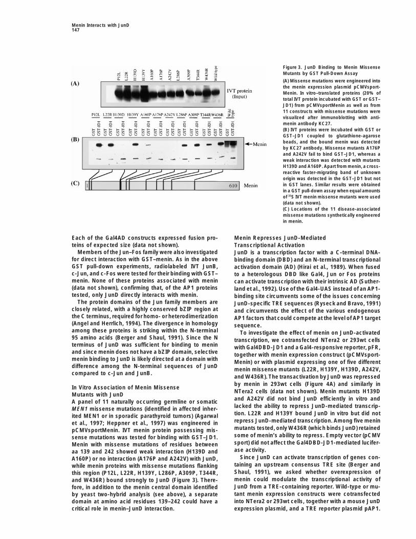
Figure 3. JunD Binding to Menin MissenseMutants by GST Pull-Down Assay
(A) Missense mutations were engineered intothe menin expression plasmid pCMVsport-Menin. In vitro–translated proteins (20% oftotal IVT protein incubated with GST or GST–JD1) from pCMVsportMenin as well as from11 constructs with missense mutations werevisualized after immunoblotting with anti-menin antibody KC27.(B) IVT proteins were incubated with GST orGST–JD1 coupled to glutathione-agarosebeads, and the bound menin was detectedby KC27 antibody. Missense mutants A176Pand A242V fail to bind GST–JD1, whereas aweak interaction was detected with mutantsH139D and A160P. Apart from menin, a cross-reactive faster-migrating band of unknownorigin was detected in the GST–JD1 but notin GST lanes. Similar results were obtainedin a GST pull-down assay when equal amountsof 35S IVT menin missense mutants were used(data not shown).(C) Locations of the 11 disease-associatedmissense mutations synthetically engineeredin menin.
Each of the Gal4AD constructs expressed fusion pro- Menin Represses JunD-Mediatedteins of expected size (data not shown). Transcriptional Activation
Members of the Jun–Fos family were also investigated JunD is a transcription factor with a C-terminal DNA-for direct interaction with GST–menin. As in the above binding domain (DBD) and an N-terminal transcriptionalGST pull-down experiments, radiolabeled IVT JunB, activation domain (AD) (Hirai et al., 1989). When fusedc-Jun, and c-Fos were tested for their binding with GST– to a heterologous DBD like Gal4, Jun or Fos proteinsmenin. None of these proteins associated with menin can activate transcription with their intrinsic AD (Suther-(data not shown), confirming that, of the AP1 proteins land et al., 1992). Use of the Gal4-UAS instead of an AP1-tested, only JunD directly interacts with menin. binding site circumvents some of the issues concerning
The protein domains of the Jun family members are JunD-specific TRE sequences (Ryseck and Bravo, 1991)closely related, with a highly conserved bZIP region at and circumvents the effect of the various endogenousthe C terminus, required for homo- or heterodimerization AP1 factors that could compete at the level of AP1 target(Angel and Herrlich, 1994). The divergence in homology sequence.among these proteins is striking within the N-terminal To investigate the effect of menin on JunD-activated95 amino acids (Berger and Shaul, 1991). Since the N transcription, we cotransfected NTera2 or 293wt cellsterminus of JunD was sufficient for binding to menin with Gal4DBD-JD1 and a Gal4-responsive reporter, pFR,and since menin does not have a bZIP domain, selective together with menin expression construct (pCMVsport-menin binding to JunD is likely directed at a domain with Menin) or with plasmid expressing one of five differentdifference among the N-terminal sequences of JunD menin missense mutants (L22R, H139Y, H139D, A242V,compared to c-Jun and JunB.
and W436R). The transactivation by JunD was repressedby menin in 293wt cells (Figure 4A) and similarly in
In Vitro Association of Menin MissenseNTera2 cells (data not shown). Menin mutants H139DMutants with JunDand A242V did not bind JunD efficiently in vitro andA panel of 11 naturally occurring germline or somaticlacked the ability to repress JunD-mediated transcrip-MEN1 missense mutations (identified in affected inher-tion. L22R and H139Y bound JunD in vitro but did notited MEN1 or in sporadic parathyroid tumors) (Agarwalrepress JunD-mediated transcription. Among five meninet al., 1997; Heppner et al., 1997) was engineered inmutants tested, only W436R (which binds JunD) retainedpCMVsportMenin. IVT menin protein possessing mis-some of menin’s ability to repress. Empty vector (pCMVsense mutations was tested for binding with GST–JD1.sport) did not affect the Gal4DBD-JD1-mediated lucifer-Menin with missense mutations of residues betweenase activity.aa 139 and 242 showed weak interaction (H139D and
Since JunD can activate transcription of genes con-A160P) or no interaction (A176P and A242V) with JunD,taining an upstream consensus TRE site (Berger andwhile menin proteins with missense mutations flankingShaul, 1991), we asked whether overexpression ofthis region (P12L, L22R, H139Y, L286P, A309P, T344R,menin could modulate the transcriptional activity ofand W436R) bound strongly to JunD (Figure 3). There-JunD from a TRE-containing reporter. Wild-type or mu-fore, in addition to the menin central domain identifiedtant menin expression constructs were cotransfectedby yeast two-hybrid analysis (see above), a separateinto NTera2 or 293wt cells, together with a mouse JunDdomain at amino acid residues 139–242 could have a
critical role in menin–JunD interaction. expression plasmid, and a TRE reporter plasmid pAP1.

Cell148
Figure 4. Menin Represses JunD-ActivatedTranscription in Mammalian Cells
Results are shown as relative luciferase activ-ity from a representative experiment per-formed in triplicate in each cell line, with meanand standard error. In vitro JunD binding isindicated by (1) or (2) for menin and meninmutants. Since the expression level of themenin mutants was not the same as wild-type menin (as per Western blot analysis), a2-fold amount of menin mutant plasmid wastransfected.(A) Menin represses transactivation by aGal4DBD-JD1 fusion protein. The Gal4 re-porter plasmid pFR (0.5 mg) was cotrans-fected into 293wt cells with Gal4DBD alone orGal4DBD-JD1 plasmid, together with emptyvector or 2 mg of pCMVsportMenin or 4 mg ofmenin mutant expression plasmid indicated.Empty vector had no effect on Gal4DBD-JD1-induced transcription (data not shown). Mini-mal reporter activity was observed withGal4DBD alone (data not shown).(B) Menin represses transactivation by JunDfrom a TRE-containing reporter plasmid. NTera2cells were transiently transfected with 0.5 mgof the reporter plasmid pAP1, 0.5 mg ofpcDNA3.1 mouse JunD, together with emptyvector or 2 mg of pCMVsportMenin or 4 mg ofmenin mutant expression plasmid indicated.Empty vector had no effect on JunD-inducedtranscription (data not shown). Minimal re-porter activity was observed with pCMVsport-Menin alone and pAP1 (data not shown). Lev-els of expression of menin and JunD in thisexperiment are shown below the diagram ofthe reporter. Equal amounts (30 mg) of totalcell lysates were analyzed by Western blotanalysis using anti-menin antibody (SQV) oranti-JunD antibody. The order of samples onthe blot corresponds to the order in whichthe data are presented in the graph above.
Expression plasmids transfected are, lane 1: JunD alone; JunD was cotransfected with menin or menin mutants in lanes 2 to 7: menin, L22R,H139Y, H139D, A242V, W436R, respectively; lane 8: menin alone. The faster migrating band in the anti-JunD blot corresponds to an alternateform of JunD generated due to an internal initiation of translation (Okazaki et al., 1998).(C) Menin represses JunD-induced transcription, but augments c-Jun-induced transcription. 293wt cells were transiently transfected with 0.5mg of the reporter plasmid pAP1, 0.5 mg of Jun expression plasmid indicated (mouse JunD, mouse-c-Jun, JunD:c-Jun chimera, or c-Jun:JunDchimera), together with empty vector or 2 mg of pCMVsportMenin. Empty vector had no effect on transcription activated by any of the Junexpression plasmids (data not shown). Note logarithmic scale.
Similar to the results obtained above with the Gal4 re- menin and JunD expression plasmids when each wastransfected alone or in combination with other expres-porter, the transactivation by JunD was repressed effec-
tively by menin in both cell lines. Again, irrespective of sion plasmids (Figure 4B).To evaluate if the repressive effect of menin is specificthe ability to bind JunD, the menin mutants, L22R,
H139Y, H139D, and A242V had no effect or resulted in for transcription induced by JunD, we tested the effectof menin on c-Jun-mediated transcriptional activationmarginal augmentation of JunD-mediated transcription.
Only the menin mutant W436R maintained a modest (c-Jun is an AP1 member that does not bind directly tomenin). 293wt cells were cotransfected with the samerepressive effect on JunD activity. Figure 4B shows re-
sults obtained in NTera2 cells. JunD activity was not AP1 reporter plasmid as above, c-Jun expression plas-mid alone or c-Jun expression plasmid together withrepressed when the menin expression construct was
substituted by empty vector (pCMVsport) (data not menin expression plasmid. Menin did not inhibit tran-scription stimulated by c-Jun, but rather augmentedshown). In identical experiments using the same lucifer-
ase reporter without TRE sequences upstream of the transcription activated by c-Jun about 14-fold (Figure4C). The effect of menin on transcriptional activationminimal TATA, JunD did not activate transcription (data
not shown). Also, overexpression of menin or menin was next tested on Jun chimeras. The chimeric proteinswere more potent activators of transcription. In the chi-mutants neither activated nor repressed the transcrip-
tional activity obtained when the reporter vectors alone mera containing the N terminus from JunD [Jun(D1C)],menin repressed transcription about 14-fold; while inwere transfected into 293wt or NTera2 cells (data not
shown). Similar levels of protein were achieved from the chimera where the JunD N terminus was substituted

Menin Interacts with JunD149
Figure 5. Menin–JunD Interaction Involvesthe N Terminus and Midregion of Menin, andRequires the N Terminus of JunD
Dotted lines show regions in menin or JunDthat could be deleted without disruptingmenin–JunD binding. Solid lines show the re-gions that disrupt menin–JunD binding afterdeletion or via missense mutation. Numbersbelow the dotted and solid lines are aminoacid numbers. Solid double-headed arrowsshow the regions in menin (aa 1–40, aa 323–428) and JunD (aa 8–70) required for menin–JunD interaction, deduced from deletion anal-yses. Dashes with double-headed arrow showthe region in menin (aa 139–242) where mis-sense mutations disrupt binding with JunD.
by the N terminus of c-Jun [Jun(C1D)], menin aug- seen when menin was cotransfected with c-Jun. Alterna-tively, menin–JunD interaction could block activator(s)mented transcription about 11-fold (Figure 4C). Empty
vector pCMVsport did not affect the transcriptional ac- from interacting with the transcription complex.tivity of any of the Jun proteins (data not shown).
Biological Significance of Menin’s Repressionof JunD-Activated TranscriptionMechanisms for Menin’s Repression
of JunD-Activated Transcription In spite of structural similarities among the Jun proteins,JunD exhibits distinct biological properties. Unlike c-JunIn theory, menin could repress JunD-mediated tran-
scriptional activation by disturbing the complexing of and JunB, which are classified as immediate-earlygenes, the expression of JunD is generally constitutiveJunD with other members of the AP1 family on specific
DNA sequences. The C terminus of JunD, containing its (primarily expressed in quiescent cells) and is only mar-ginally affected by growth factors and external stimuliDNA-binding region and the bZIP dimerization region
appears not to be essential for interacting with menin (Pfarr et al., 1994). The N-terminal regions of c-Jun orJunB, but not that of JunD, have been reported to inhibit(Figure 5), and menin does not possess a bZIP dimeriza-
tion domain. Therefore, the interaction of menin with estrogen receptor activity (Doucas et al., 1991), interferewith the function of other nuclear receptors (reviewedJunD seems unlikely to interfere with JunD DNA-binding
ability or JunD homo- or heterodimerization. in Pfahl, 1993), and suppress trans-activating capacityof the tissue-specific transcription factor MyoD (Li etSeveral other types of protein:protein interactions
have been identified for the AP1 family of proteins. AP1- al., 1992). High levels of c-Jun have been shown totransform avian cells or established cell lines from ro-mediated transcription can also be modulated by inter-
action of the Jun proteins with transcriptional activators dents, while JunB only poorly transforms such cells, andJunD does not transform them at all (Castellazzi et al.,or repressors. JDP2 negatively regulates AP1 activity by
heterodimerizing with c-Jun or JunD, thereby competing 1991).Several studies using different approaches have sug-with unidentified activators for c-Jun or JunD binding
(Aronheim et al., 1997). The following proteins have been gested anti-mitogenic effects of JunD. In T antigen-transformed 3T3T cells, antisense JunD is mitogenicidentified that interact with the N-terminal region of Jun
family members: c-Jun d region inhibitory factor, Jun while antisense JunB or antisense c-Jun are anti-mito-genic (Wang et al., 1996). Overexpression of JunD inkinases, TBP, and JAB1 (Baichwal and Tjian, 1990; Smeal
et al., 1992; Franklin et al., 1995; Claret et al., 1996). It NIH3T3 cells slows growth and partially suppresses rastransformation, effects opposite to c-Jun (Pfarr et al.,is thus possible that menin could modify the effects
of other cellular factors that influence JunD-mediated 1994; Mechta et al., 1997). The difference in transforma-tion ability maps to the amino terminus (Alani et al.,transcription.
The above considerations suggest several possible 1991).The inhibitory effect of JunD on cell growth is a prop-explanations for menin to repress JunD-mediated tran-
scriptional activation. Menin could itself be a repressor erty unique from that of the other AP1 proteins. Sincemenin is a tumor suppressor and JunD also can sup-or it could recruit a repressor protein to JunD. High level
expression of menin could result in sequestering and press growth, our observation of menin’s repressive ef-fect on JunD-mediated transcriptional activation seemstitration of the putative transcriptional inhibitory compo-
nent. This may explain the augmentation of transcription paradoxical. The identification of specific target genes

Cell150
(sc-605), Fra2 (sc-604), Gal4DBD (sc-510), Gal4AD (sc-1663) werepreferentially regulated by the menin–JunD complex,from Santa Cruz Biotechnology, Inc. (Santa Cruz, CA).and their subsequent effect on cell growth and transfor-
mation, may be needed to resolve this issue. AlthoughCell Culture and Transfections
inactivation of JunD has so far not been implicated in HEK-293wt, HEK-293T, NTera2, HeLa, F9, and Cos7 (ATCC, Rock-the pathogenesis of any tumors, it is now plausible that ville, MD) cells were grown in DMEM supplemented with 10% fetalthe tumor suppressor-like properties that have been calf serum and 2 mM glutamine, 100 mg penicillin-streptomycin per
ml at 5% CO2. Transient transfection was carried out with Lipofec-observed for JunD are due to the functional synergytamine (Life Technologies, Inc., Gaithersburg, MD) for 293T cellsbetween the suppressive effects of menin and JunD.and Superfect for all other cell lines (Qiagen, Valencia, CA) as perConversely, JunD may provide menin with its tumor sup-the manufacturer.
pressor function. For reporter assays, cells were seeded 24 hr prior to transfectionConsistent with the idea of menin–JunD interaction in 6-well plates. Equal amount of DNA was maintained in each trans-
being essential for the tumor suppressor function of fection by correcting with pcDNA3.1. Cells were harvested 48 hrafter transfection. CAT (chloramphenicol acetyltransferase) proteinmenin, some naturally occurring MEN1 missense muta-was detected by CAT ELISA (Boerhinger Mannheim, Indianapolis,tions in a limited region of menin disrupt menin–JunDIN), and the luciferase activity was measured by luciferase assayinteraction. No genotype–phenotype correlation can bekit (Promega). Preparation of cell lysates and reporter assays were
attributed to any of these missense mutations. The loca- done as per the manufacturer’s protocols. Assay results were cor-tion of the mutations studied showed a relation with rected for equal amount of protein. All transfections were performedwhether a menin protein with a particular missense mu- at least three times in duplicate or triplicate with independent DNA
preparations.tation could bind directly to JunD. The observation ofnormal menin–JunD binding for disease-associated mu-
Yeast Two-Hybrid Systemtations that fall outside the region of aa 139–242 sug-All experiments were performed in the yeast reporter strain Y190.gests that disruption of JunD binding may not be theThe yeast vector pAS2-1 is a Gal4 DNA-binding domain (Gal4DBD)
feature common to all MEN1 mutations. Irrespective of encoding vector that allows fusion of desired protein with thethe ability to bind JunD, all of the five menin mutants C-terminal part of Gal4DBD. The yeast vector pACT2 is a Gal4 activa-tested were deficient in repressing or resulted in mar- tion domain (Gal4AD) encoding vector, that allows fusion of desired
protein to the C-terminal part of Gal4AD. The yeast two-hybrid (y2h)ginal augmentation of JunD-mediated transcription. Thelibrary screen and protocols were as recommended (Clontech). Themodest suppressive effect observed with W436R indi-protein coding region of MEN1 cDNA was amplified from pCMVcates that even some loss of repression of JunD activitysportMenin (Chandrasekharappa et al., 1997) and cloned into the
may be enough to cause MEN1. Alternatively, W436R vectors pAS2-1 and pACT2. pAS2-1–menin or pACT2–menin didcould be a rare benign MEN1 polymorphism. not have any self-activating property nor did they interact with the
This report establishes a role for the tumor suppressor protein lamin or Gal4AD or Gal4DBD. pAS2-1-menin was used asa bait to screen a y2h adult human brain cDNA library (in pGAD10,protein menin in a known molecular pathway. The pre-with inserts fused at the C terminus of Gal4AD) (Clontech) by se-cise mechanism by which menin represses JunD-acti-quential transformation.vated transcription remains to be elucidated. Further
Various deletions from the N and C terminus of menin, M(del 503–investigation is required to understand transcriptional 610), M(del 477–610), M(del 323–620), M(del 1–40), and M(del 1–177)regulation mediated by the menin–JunD interaction in were cloned as PCR-amplified fragments into pAS2-1. M(del 477–normal physiology and in neoplasia. 610) was subcloned from EGFP-5 construct (Guru et al., 1998) into
pAS2-1. Full-length mouse menin was PCR amplified from themouse MEN1 cDNA clone (Guru et al., unpublished) and ligated intoExperimental ProcedurespAS2-1.
The constructs, JunD(del 105–347) and JD1(del 1–120), were gen-Reagentserated by subcloning from pGAD10-JD2 and pGAD10-JD1, respec-Matchmaker two-hybrid system II and the Mammalian Matchmakertively, into pACT2. JunD(del 99–347) and JunD(del 71–347) weretwo-hybrid assay kit were purchased from Clontech Labs, Inc. (Palocloned as PCR fragments amplified from human genomic DNA intoAlto, CA). DNA fragments for most plasmid constructs were gener-pACT2.ated by PCR amplification using Pfu polymerase (Stratagene, La
Protein coding region of each of the Jun–Fos family membersJolla, CA) and primers designed with appropriate restriction sites.were PCR amplified and cloned in-frame with the Gal4AD intoDetails of plasmid constructs and all primer sequences are availablepACT2. Mouse JunD was amplified from the plasmid pKS mouseon the Cell web site (http://www.cell.com/cgi/content/full/96/1/143/JunD, mouse JunB was amplified from RNA of NIH3T3 cells; c-Jun,DC1) or on request from the authors. Plasmid constructs were se-c-Fos, Fra1, and Fra2 were amplified from human leukocyte Mara-quenced entirely for the inserts on at least one strand by the Taqthon cDNA (Clontech).Dideoxy Terminator Cycle sequencing method (Applied Biosystems,
Each of the above fragments corresponding to protein codingFoster City, CA) using an Applied Biosystems model 377 or 373regions of Jun and Fos family members was also subcloned intoautomated sequencer, or by manual radioactive sequencing usingplasmid vectors pcDNA3.11 (Invitrogen, Carlsbad, CA) or pCI (Pro-g-33P end-labeled primer and the AmpliCycle sequencing kit (Perkinmega, Madison, WI) for expression in mammalian cells and for inElmer, Branchburg, NJ). Mouse JunD clone (pKS mouse JunD) wasvitro transcription and translation.used with permission from Rodrigo Bravo.
Protein concentrations in all cell and nuclear extracts were de-termined with a protein assay kit (Bio-Rad laboratories, Hercules, Mammalian Two-Hybrid System
MEN1 protein coding region was subcloned from pACT2–menin intoCA). Western blots were developed by enhanced chemilumines-cence (ECL). mammalian two-hybrid vector pM (named pM-menin). JD1 insert
was subcloned from pGAD10-JD1 into pM and pVP16 (named pM-JD1/Gal4DBD-JD1 and pVP16-JD1, respectively). The vectors pMAntibodies
All anti-menin polyclonal antibodies were generated against syn- and pVP16 allow fusions of desired proteins with the C-terminalpart of Gal4DBD and VP16AD, respectively.thetic peptides and then affinity purified (Guru et al., 1998). The
antibody used as a negative control for coimmunoprecipitations For confirming menin–JunD interaction, cells (293wt, Cos7, F9,NTera2, or HeLa) were transfected with 0.5 mg of the Gal4 reporterwas raised against BRCA1 peptide (M. E., unpublished). Antibodies
against JunD (sc-74), JunB (sc-73), c-Jun (H-79), c-Fos (sc-52), Fra1 plasmid pFR (Stratagene) (5 copies of the Gal4 UAS upstream of a

Menin Interacts with JunD151
minimal TATA and luciferase reporter gene), or pG5CAT (Clontech) and the precipitation was allowed to occur for 2 hr. Beads werewashed extensively using wash buffer. Beads were then boiled in(5 copies of the Gal4 UAS [upstream activating sequence], the E1b
promoter and CAT as the reporter), together with 0.5 mg of the SDS sample buffer for 5 min, and the proteins were separated on10% SDS-PAGE gels and detected by Western blotting.expression plasmids pM-menin and pVP16-JD1.
pM-menin and pVP16-JD1 were also transfected alone or in com-bination with the vectors pM and pVP16, to determine autonomous Construction of Jun Chimerasactivation of the Gal4 reporters pFR or pG5CAT by the expression Regions were exchanged between mouse JunD and mouse c-junplasmids, and also to determine the basal transcriptional potential cDNA using the AccI restriction site conserved in both genes, atof each plasmid. CAT ELISA or Luciferase assays were performed amino acid 182 of JunD and corresponding amino acid residue 16848 hr after transfection depending on which reporter plasmid was of c-Jun (Metivier et al., 1993). PCR-amplified fragments corre-used in the experiment. sponding to the protein coding regions of mouse JunD and c-jun
were used to generate the chimeric inserts, which were then sub-Mutagenesis cloned in the vector pcDNA3.1(1). Jun(D1C) contains JunD aaEleven different MEN1 missense mutations—P12L, L22R, H139D, 1–182:c-Jun aa 169–334. Jun(C1D) contains c-Jun aa 1–168:JunDH139Y, A160P, A176P, A242V, L286P, A309P, T344R, and W436R aa 183–341.(Agarwal et al., 1997; Heppner et al., 1997)—were engineered intothe MEN1 sequence in the plasmid pCMVsportMenin by primer- Effect of Menin on JunD Transactivationmediated mutagenesis using QuickChange mutagenesis kit (Stra- The effect of menin and menin mutants on JunD-induced transcrip-tagene) or recombinant PCR (Higuchi, 1990). All synthetic mutations tion was studied in 293wt and N-Tera2 cells. For determining theand PCR-amplified regions were confirmed by sequencing. effect of menin on transcription activated by JunD fused to the
Gal4DBD, cells were transfected with 0.5 mg of Gal4 reporter plasmidIn Vitro Transcription and Translation pFR, 0.5 mg of Gal4DBD-JD1 alone or together with 2 mg ofAll the in vitro translation reactions were carried out using the TNT pCMVsportMenin or 4 mg of a menin mutant expression plasmid orcoupled reticulocyte lysate system (Promega). Two micrograms of empty vector pCMVsport. For determining the effect of menin onDNA were used in the presence of complete amino acid mix with AP1 reporter-mediated activation by JunD, cells were transfectedeither SP6 or T7 RNA Polymerase as per the manufacturer’s pro- with 0.5 mg reporter plasmid pAP1 (Stratagene) (which contains 7tocol. copies of the AP1 consensus TRE “TGACTAA” upstream of a mini-
mal TATA and luciferase reporter gene), 0.5 mg pcDNA3.1 mouseJunD (mouse JunD expression plasmid) alone or together with 2 mgGST Pull-Down Assaysof pCMVsportMenin or 4 mg of a menin mutant expression plasmidPlasmid constructs of GST–menin and GST–JD1 (GST fused to theor empty vector pCMVsport. Since the expression level of the meninJunD insert from pGAD10-JD1, human JunD aa 8–340) were gener-mutants was not the same as wild-type menin (as per Western blotated in pGEX-1lT (allows fusion of desired protein at the C terminusanalysis), we transfected with a larger amount of menin mutantof GST) (Pharmacia-Biotech, Piscataway, NJ). The fusion proteinsplasmid. Expression plasmids of menin or menin mutants were co-were purified as described in the manufacturer’s protocol excepttransfected with reporter vector to determine their effect on reporterthat 10% glycerol and 1% Triton X-100 were added before son-activity.ication.
To assess the effect of menin on c-Jun and Jun-chimeras, 293wtpcDNA3.1(2)menin plasmid has full-length menin in pcDNA3.1(2)cells were transfected with 0.5 mg reporter plasmid pAP1, 0.5 mgMyc-His vector (Guru et al., 1998). Deletion constructs M(del 572–of the Jun expression plasmid [mouse JunD, mouse c-Jun,610), M(del 1–277), and M(del 278–477) were generated by takingJun(D1C) or Jun(C1D)] alone or together with 2 mg of pCMVsport-advantage of convenient restriction sites in pcDNA3.1(2)menin.Menin or empty vector pCMVsport.293T cells were transfected with plasmid DNA expressing either
full-length menin or menin deletions. Twenty four or 48 hr aftertransfection, cells were washed in PBS and lysed either as whole Acknowledgmentscells or after fractionating nuclei (Guru et al., 1998) in lysis buffer(50 mM Tris HCl [pH 8.0], 100 mM NaCl, 0.3 mM DTT, 10 mM MgCl2, We thank Dr. George Poy for DNA sequencing and Mary Beth Kester10% glycerol and 0.1% NP-40, 100 mg/ml AEBSF [4-(2-Aminoethyl)- for technical assistance. We also thank Dr. Rodrigo Bravo for per-Benzenesulfonyl fluoride hydrochloride], and 1 mg/ml aprotinin/leu- mission to use the mouse JunD plasmid, and Dr. Kevin Gardner forpeptin). sharing reagents. C. H. was supported by a grant from The Fritz
GST–menin, GST–JD1, or GST alone (2–5 mg) was attached to Thyssen Stiftung (Germany).glutathione-agarose beads and incubated with IVT protein (10 ml),total cell lysate, or nuclear extracts (200 mg protein) in binding buffer Received August 17, 1998; revised November 10, 1998.(lysis buffer containing 2% BSA). The mixture was incubated byshaking for 2 hr at 48C. The glutathione-agarose beads were washed Referencesthree times in binding buffer, resuspended in 20 ml of SDS samplebuffer, and boiled for 5 min, and the proteins were separated on Abate, C., Luk, D., and Curran, T. (1991). Transcriptional regulation10%–20% gradient gels by SDS-PAGE. Proteins were detected us- by Fos and Jun in vitro: interaction among multiple activator anding standard Western blotting techniques with appropriate primary regulatory domains. Mol. Cell. Biol. 11, 3624–3632.antibody (KC27 for detecting menin, anti-myc antibody for myc- Agarwal, S.K., Kester, M.B., Debelenko, L.V., Heppner, C., Emmert-tagged menin, sc-74 for JunD) and secondary antibody (horseradish Buck, M.R., Skarulis, M.C., Doppman, J.L., Kim, Y.S., Lubensky, I.A.,peroxidase-conjugated anti-rabbit antibody). Zhuang, Z., et al. (1997). Germline mutations of the MEN1 gene in
familial multiple endocrine neoplasia type 1 and related states. Hum.Immunoprecipitation (IP) Mol. Genet. 6, 1169–1175.Nuclear extracts from 293wt cells were prepared as described (Sam-
Alani, R., Brown, P., Binetruy, B., Dosaka, H., Rosenberg, R.K.,uels et al., 1974). Protease inhibitors (Boehringer Mannheim) wereAngel, P., Karin, M., and Birrer, M.J. (1991). The transactivatingadded to all buffers used for preparation of nuclear extracts and IP.domain of the c-Jun proto-oncoprotein is required for cotransforma-Prior to IP, nuclear extracts of untransfected cells (480 mg) ortion of rat embryo cells. Mol. Cell. Biol. 11, 6286–6295.transfected cells (300 mg) were diluted 1:1 with wash buffer (20 mMAngel, P.E., and Herrlich, P.A. (1994). General structure of AP1 sub-Tris HCl [pH 8.00], 137 mM NaCl, 10% glycerol, 1 mM NaF, 1 mMunits and characteristics of the Jun proteins. In The Fos and JunNa3VO4 and 0.1% Igepal) and incubated on ice for 15 min to ensureFamily of Transcription Factors, P.E. Angel and P.A. Herrlich, eds.complete solubilization of proteins. Next, the nuclear extracts were(Boca Raton, FL: CRC Press), pp. 3–14.precleared using magnetic beads coated with a sheep anti-rabbit
antibody (Dynal, Oslo, Norway) and then incubated with the respec- Aronheim, A., Zandi, E., Hennemann, H., Elledge, S.J., and Karin,M. (1997). Isolation of an AP-1 repressor by a novel method fortive antibodies (5 mg) overnight at 48C. Beads were added again

Cell152
detecting protein-protein interactions. Mol. Cell. Biol. 17, 3094– Metivier, C., Piu, F., Pfarr, C.M., Yaniv, M., Loiseau, L., and Caste-llazzi, M. (1993). In vitro transforming capacities of mouse c-jun:junD3102.chimeric genes. Oncogene 8, 2311–2315.Baichwal, V.R., and Tjian, R. (1990). Control of c-Jun activity by
interaction of a cell-specific inhibitor with regulatory domain delta: Okazaki, S., Ito, T., Ui, M., Watanabe, T., Yoshimatsu, K., and Iba,H. (1998). Two proteins translated by alternative usage of initiationdifferences between v- and c-Jun. Cell 63, 815–825.codons in mRNA encoding a JunD transcriptional regulator. Bio-Berger, I., and Shaul, Y. (1991). Structure and function of humanchem. Biophys. Res. Commun. 250, 347–353.Jun-D. Oncogene 6, 561–566.Pfahl, M. (1993). Nuclear receptor/AP-1 interaction. Endocr. Rev.Castellazzi, M., Spyrou, G., La Vista, N., Dangy, J.P., Piu, F., Yaniv,14, 651–658.M., and Brun, G. (1991). Overexpression of c-jun, junB, or junD
affects cell growth differently. Proc. Natl. Acad. Sci. USA 88, 8890– Pfarr, C.M., Mechta, F., Spyrou, G., Lallemand, D., Carillo, S., andYaniv, M. (1994). Mouse JunD negatively regulates fibroblast growth8894.and antagonizes transformation by ras. Cell 76, 747–760.Chandrasekharappa, S.C., Guru, S.C., Manickam, P., Olufemi, S.E.,
Collins, F.S., Emmert-Buck, M.R., Debelenko, L.V., Zhuang, Z., Lu- Rahmsdorf, H.J. (1996). Jun: transcription factor and oncoprotein.J. Mol. Med. 74, 725–747.bensky, I.A., Liotta, L.A., et al. (1997). Positional cloning of the gene
for multiple endocrine neoplasia-type 1. Science 276, 404–407. Ryseck, R.P., and Bravo, R. (1991). c-JUN, JUN B, and JUN D differin their binding affinities to AP-1 and CRE consensus sequences:Claret, F.X., Hibi, M., Dhut, S., Toda, T., and Karin, M. (1996). A new
group of conserved coactivators that increase the specificity of AP-1 effect of FOS proteins. Oncogene 6, 533–542.transcription factors. Nature 383, 453–457. Samuels, H.H., Tasi, J.S., Casanova, J., and Stanley, F. (1974). Thy-
roid hormone action: in vitro characterization of solubilized nuclearDebelenko, L.V., Brambilla, E., Agarwal, S.K., Swalwell, J.I., Kester,M.B., Lubensky, I.A., Zhuang, Z., Guru, S.C., Manickam, P., Olufemi, receptors from rat liver and cultured GH1 cells. J. Clin. Invest. 54,
853–865.S.E., et al. (1997). Identification of MEN1 gene mutations in sporadiccarcinoid tumors of the lung. Hum. Mol. Genet. 6, 2285–2290. Smeal, T., Binetruy, B., Mercola, D., Grover-Bardwick, A., Heidecker,
G., Rapp, U.R., and Karin, M. (1992). Oncoprotein-mediated signal-Doucas, V., Spyrou, G., and Yaniv, M. (1991). Unregulated expres-sion of c-Jun or c-Fos proteins but not Jun D inhibits oestrogen ing cascade stimulates c-Jun activity by phosphorylation of serines
63 and 73. Mol. Cell. Biol. 12, 3507–3513.receptor activity in human breast cancer derived cells. EMBO J. 10,2237–2245. Sutherland, J.A., Cook, A., Bannister, A.J., and Kouzarides, T. (1992).
Conserved motifs in Fos and Jun define a new class of activationFearon, E.R. (1998). Tumor suppressor genes. In The Genetic Basisof Human Cancer, B. Vogelstein and K.W. Kinzler, eds. (New York: domain. Genes Dev. 6, 1810–1819.McGraw Hill), pp. 229–236. Wang, H., Xie, Z., and Scott, R.E. (1996). Differentiation modulates
the balance of positive and negative Jun/AP-1 DNA-binding activi-Franklin, C.C., McCulloch, A.V., and Kraft, A.S. (1995). In vitro associ-ation between the Jun protein family and the general transcription ties to regulate cellular proliferative potential: different effects in
nontransformed and transformed cells. J. Cell. Biol. 135, 1151–1162.factors, TBP and TFIIB. Biochem. J. 305, 967–974.
Friedman, E., Sakaguchi, K., Bale, A.E., Falchetti, A., Streeten, E., Zhuang, Z., Vortmeyer, A.O., Pack, S., Huang, S., Pham, T.A., Wang,C., Park, W.S., Agarwal, S.K., Debelenko, L.V., Kester, M., et al.Zimering, M.B., Weinstein, L.S., McBride, W.O., Nakamura, Y.,
Brandi, M.L., et al. (1989). Clonality of parathyroid tumors in familial (1997). Somatic mutations of the MEN1 tumor suppressor gene insporadic gastrinomas and insulinomas. Cancer Res. 57, 4682–4686.multiple endocrine neoplasia type 1. N. Engl. J. Med. 321, 213–218.
Guru, S.C., Goldsmith, P.K., Burns, A.L., Marx, S.J., Spiegel, A.M.,Collins, F.S., and Chandrasekharappa, S.C. (1998). Menin, the prod-uct of the MEN1 gene, is a nuclear protein. Proc. Natl. Acad. Sci.USA 95, 1630–1634.
Heppner, C., Kester, M.B., Agarwal, S.K., Debelenko, L.V., Emmert-Buck, M.R., Guru, S.C., Manickam, P., Olufemi, S.E., Skarulis, M.C.,Doppman, J.L., et al. (1997). Somatic mutation of the MEN1 genein parathyroid tumours. Nat. Genet. 16, 375–378.
Higuchi, R. (1990). Recombinant PCR. In PCR Protocols: A Guideto Methods and Applications, M.A. Innis, D.H. Gelfand, J.J. Sninsky,and T.J. White, eds. (New York: Academic Press, Inc.), pp. 177–183.
Hirai, S.I., Ryseck, R.P., Mechta, F., Bravo, R., and Yaniv, M. (1989).Characterization of JunD: a new member of the jun proto-oncogenefamily. EMBO J. 8, 1433–1439.
Humphrey, J.S., Salim, A., Erdos, M.R., Collins, F.S., Brody, L.C.,and Klausner, R.D. (1997). Human BRCA1 inhibits growth in yeast:potential use in diagnostic testing. Proc. Natl. Acad. Sci. USA 94,5820–5825.
Karin, M., Liu, Z., and Zandi, E. (1997). AP-1 function and regulation.Curr. Opin. Cell. Biol. 9, 240–246.
Larsson, C., Skogseid, B., Oberg, K., Nakamura, Y., and Nordensk-jold, M. (1988). Multiple endocrine neoplasia type 1 gene maps tochromosome 11 and is lost in insulinoma. Nature 332, 85–87.
Li, L., Chambard, J.C., Karin, M., and Olson, E.N. (1992). Fos andJun repress transcriptional activation by myogenin and MyoD: theamino terminus of Jun can mediate repression. Genes Dev. 6,676–689.
Marx, S.J. (1998). Multiple endocrine neoplasia type 1. In The Ge-netic Basis of Human Cancer, B. Vogelstein and K.W. Kinzler, eds.(New York: McGraw Hill), pp. 489–506.
Mechta, F., Lallemand, D., Pfarr, C.M., and Yaniv, M. (1997). Trans-formation by ras modifies AP1 composition and activity. Oncogene14, 837–847.