Brachypodium as an emerging model for cereal–pathogen interactions

EUKARYOTIC CELL, Feb. 2005, p. 443–454 Vol. 4, No. 21535-9778/05/$08.00�0 doi:10.1128/EC.4.2.443–454.2005Copyright © 2005, American Society for Microbiology. All Rights Reserved.
Activation of an AP1-Like Transcription Factor of the Maize PathogenCochliobolus heterostrophus in Response to Oxidative
Stress and Plant SignalsSophie Lev,1 Ruthi Hadar,1 Paolo Amedeo,2† Scott E. Baker,2‡ O. C. Yoder,2§
and Benjamin A. Horwitz1*Department of Biology, Technion–Israel Institute of Technology, Haifa, Israel,1 and
Torrey Mesa Research Institute, San Diego, California2
Received 27 September 2004/Accepted 3 December 2004
Redox sensing is a ubiquitous mechanism regulating cellular activity. Fungal pathogens face reactive oxygenspecies produced by the host plant’s oxidative burst in addition to endogenous reactive oxygen species pro-duced during aerobic metabolism. An array of preformed and induced detoxifying enzymes, including super-oxide dismutase, catalases, and peroxidases, could allow fungi to infect plants despite the oxidative burst. Weisolated a gene (CHAP1) encoding a redox-regulated transcription factor in Cochliobolus heterostrophus, a fun-gal pathogen of maize. CHAP1 is a bZIP protein that possesses two cysteine-rich domains structurally and func-tionally related to Saccharomyces cerevisiae YAP1. Deletion of CHAP1 in C. heterostrophus resulted in decreasedresistance to oxidative stress caused by hydrogen peroxide and menadione, but the virulence of chap1 mutantswas unaffected. Upon activation by oxidizing agents or plant signals, a green fluorescent protein (GFP)-CHAP1fusion protein became localized in the nucleus. Expression of genes encoding antioxidant proteins was inducedin the wild type but not in chap1 mutants. Activation of CHAP1 occurred from the earliest stage of plant in-fection, in conidial germ tubes on the leaf surface, and persisted during infection. Late in the course of in-fection, after extensive necrotic lesions were formed, GFP-CHAP1 redistributed to the cytosol in hyphae growingon the leaf surface. Localization of CHAP1 to the nucleus may, through changes in the redox state of the cell,provide a mechanism linking extracellular cues to transcriptional regulation during the plant-pathogen interaction.
Aerobic organisms are exposed to reactive oxygen speciesformed by incomplete reduction of molecular oxygen duringrespiration and other metabolic processes. Fungal pathogensof plants must, in addition, deal with the oxidative burst gene-rated by the host upon exposure to pathogens (45). In some in-teractions, the plant hypersensitive response is associated withblockage of the infection process (30). In contrast, some necro-trophic fungi may be insensitive to the hypersensitive responseand even exploit plant cell death resulting from this response(20, 41).
It has been proposed that preformed and inducible detoxi-fying enzymes, including superoxide dismutase, catalases, andperoxidases, contribute to fungal reactive oxygen species tol-erance (19, 23). The redox state of cells subjected to oxidativestress is altered, leading to activation of three major oxidant sca-venging systems: glutathione, thioredoxin, and NADPH. Glu-tathione and thioredoxins are kept in their reduced state byNADPH and used as cofactors by a number of enzymes invarious detoxification reactions. Altered redox potential alsoaffects the sulfhydryl groups of cysteine residues. These groupsare oxidized to form intra- or intermolecular disulfide bonds orproduce protein-glutathione mixed disulfides. Oxidation of re-dox-sensitive proteins, in the normally reductive intracellular
environment, can result in either gain or loss of function, pro-viding a mechanism for redox signaling (9). Many mammalianproteins involved in signal transduction have critical thiols, andtheir redox-dependent modifications contribute directly to geneexpression (12, 16).
The Saccharomyces cerevisiae transcription factor YAP1 wasidentified as a functional homolog of mammalian AP-1 (acti-vator protein 1) on the basis of its ability to bind an AP-1recognition element. Upon exposure to oxidative stress, YAP1becomes localized to the nucleus, where it induces the expres-sion of antioxidant proteins (8, 10, 11, 25, 27). YAP1 containsa basic leucine zipper (bZIP) domain similar to that of Jun,which is a component of mammalian AP-1 transcription factorcomplexes. Two cysteine-rich domains, carboxy terminal (c-CRD) and amino terminal (n-CRD), are critical for YAP1-mediated resistance to oxidative stress and for appropriatesubcellular YAP1 localization. The activity of YAP1 is con-trolled by redox-sensitive nuclear export: redox signals causeformation of intramolecular disulfide bonds, resulting in changesin protein conformation that mask its nuclear export sequence(8, 10, 11, 25, 27). S. cerevisiae YAP2, a homolog of YAP1, isalso activated during oxidative stress, inducing genes encodingproducts that help to stabilize and fold proteins in an oxidativeenvironment (7).
Homologs of YAP1 have been found in Candida albicans(CAP1) (2), Schizosaccharomyces pombe (PAP1) (42), and Kluy-veromyces lactis (KLYAP1) (5). Genetic analyses indicate thatthese proteins are involved in oxidative stress responses andcadmium resistance, as found for YAP1. CAP1 and PAP1, likeYAP1, are involved in multidrug resistance (1, 2, 5, 34, 40, 42,
* Corresponding author. Mailing address: Department of Biology,Technion, Haifa 32000, Israel. Phone: 972 4 8293976. Fax: 972 48225153. E-mail: [email protected].
† Present address: Institute for Genomic Research, Rockville, Md.‡ Present address: Pacific Northwest National Laboratory, Richland,
Wash.§ Present address: Diversa Corporation, San Diego, Calif.
443
on Decem
ber 17, 2015 by guesthttp://ec.asm
.org/D
ownloaded from

44). All fungal YAP1-like proteins are distinctive due to char-acteristic residues present in their basic DNA-binding domains(Fig. 1) (15). YAP1, YAP2, CAP1, PAP1, KLYAP1, and twouncharacterized genes from Neurospora crassa and Aspergillusnidulans constitute a subgroup within the YAP family of tran-scription factors that possess a conserved c-CRD.
A number of YAP1-regulated genes have been identified,including GSH1, encoding �-glutamylcysteine synthetase, cat-alyzing the first, rate-limiting step of glutathione synthesis (38,46); GSH2, encoding glutathione synthetase, the second en-zyme involved in glutathione biosynthesis (39); GLR1, encod-ing glutathione reductase (21); TRX2, encoding thioredoxin 2(26, 33); TRR1, encoding thioredoxin reductase (28); FLR1, en-coding a multidrug resistance transporter belonging to the majorfacilitator superfamily (1, 34); and YCF1, encoding an ABC trans-porter essential for cadmium tolerance (44). Numerous otherYAP1-activated genes have been identified with microarray hy-bridization, genetic screens of target genes with a translationalfusion library, and high-density membrane hybridization (13, 18).
Cochliobolus heterostrophus is a foliar pathogen of maize, re-sponsible for southern corn leaf blight (47). The conidia of thepathogen germinate and form appressoria; emerging penetra-tion pegs then breach the leaf surface and start invasive growthinside the plant tissue. Spreading hyphae cause necrotic lesions,and mycelium growing on the surface of damaged plant tissue
produces abundant conidia. We cloned a gene encoding theC. heterostrophus homolog of YAP1, designated CHAP1 (Co-chliobolus heterostrophus AP-1-like), and studied its functionwith deletion mutants. We also isolated C. heterostrophus ho-mologs of yeast YAP1 target genes and verified their regula-tion by CHAP1 during oxidative stress. Finally, we followedrelocalization of a green fluorescent protein (GFP)-CHAP1fusion protein into the nucleus upon exposure to oxidizingagents and during plant infection.
MATERIALS AND METHODS
Identification of CHAP1. The degenerate oligonucleotide GCICKTTGAGCIGCICKRTTYTGRGC (where I is inosine, K is G/T, R is A/G, and Y is C/T) wasend labeled and used to screen a cDNA library constructed in pADGAL4 (Strat-agene) from a pool of RNA from fungus grown on synthetic media and infectedplants. Positive clones were transferred from phages to Escherichia coli by in vivoexcision and screened again. One of the second-round positive clones showedlimited homology to the AP-1-like protein of Neurospora crassa, and this se-quence was used to search the assembled 5� coverage of the C. heterostrophusgenome (TMRI/Syngenta, San Diego, Calif.).
Fungal transformation vectors. Vectors for replacement of CHAP1 by selec-tion markers conferring resistance to hygromycin or bialaphos were constructedby ligation of upstream and downstream flanking regions to the relevant resis-tance cassette. Protoplasts of C. heterostrophus strain C4 (MAT1-2 Tox1�, ATCC48331) were transformed (31).
The following fragments were assembled to create the CHAP1 disruptionvector conferring hygromycin resistance: a left 616-bp EagI-XhoI flanking regionincluding the 5� untranslated region and part of the CHAP1 coding region;
FIG. 1. Structural organization of CHAP1. A. Upper map, organization of the gene: E1 to E4 indicate exons; I1 to I3 indicate introns. Lowermap, predicted protein: bZIP, basic leucine zipper domain; c-CRD, C-terminal cysteine-rich domain; arrows indicate cysteine residues C394 andC418 belonging to the n-CRD (N-terminal cysteine-rich domain). For full details of the gene structure and domains, see GenBank accession no.AY486156. B. Alignment of bZIP and c-CRD domains of the YAP proteins. Consensus: uppercase letters indicate identity, lowercase lettersindicate 50% or more similar residues. Residues characteristic of the YAP protein family are underlined. Abbreviations: Ch (CHAP1), Coch-liobolus heterostrophus; An (AP-1-like protein, EAA62093), Aspergillus nidulans; Nc (AP-1-like protein, CAB91681), Neurospora crassa; Sp (Pap1,NP_593662), Schizosaccharomyces pombe; Ca (Cap1, EAL02784), Candida albicans; Sc (Yap1, NP_013707), Saccharomyces cerevisiae.
444 LEV ET AL. EUKARYOT. CELL
on Decem
ber 17, 2015 by guesthttp://ec.asm
.org/D
ownloaded from
SalI-XbaI hygromycin resistance cassette from the vector pUC-ATPH (31);798-bp NheI-HindIII right flanking region, including part of the coding regionand 3� untranslated region; and NotI-HindIII pBluescript cloning vector.
The GFP-CHAP1 transformation vector was constructed by joining the fol-lowing fragments: 1.5-kb CHAP1 KpnI-NcoI promoter generated by PCR withprimers chapp-sKpnI (cagggtaCCTCGTTGTCGCCAGTGTCC [uppercaseletters indicate the gene homology region, and lowercase letters indicate thenonhomologous region with introduced restriction site]) and chapp-asNcoI(gaggccatggTGCGAAAGCTGGGGTGAAT); NcoI-NdeI GFP coding se-quence (GFP GenBank accession number U84737); 2.4-kb CHAP1 NdeI-BglIIgenomic sequence, which includes coding region and part of 3�untranslatedregion generated by PCR with primers chap-sNdeI (AGCTTTCcatATGGCCGGCACAGGCAACGA) and chap-asBglII (ggaagaTCTGATAGCCAAGCACGCC); BamHI-XbaI hygromycin resistance cassette, and XbaI-KpnI pUC57-6D6vector. The CHAP1 disruption vector conferring glufosinate (bialaphos) resis-tance was constructed from the following fragments: 955-bp EagI-PstI left flank-ing region including the 5� untranslated region and part of the CHAP1 codingregion; PstI-XbaI phosphinothricin acetyltransferase encoding gene (BAR) un-der regulation of the Aspergillus nidulans trpC promoter; 798-bp NheI-HindIIIright flanking region including part of the CHAP1 coding sequence and 3� un-translated region; and the NotI-HindIII pBluescript cloning vector (Stratagene).Hygromycin B (used at 50 �g/ml) was from Calbiochem, and bialaphos (used at100 �g/ml, in minimal medium) was from Duchefa.
Reverse transcription-PCR with primers gfp-f (GGGATCACTCACGGCATGGA) and chap-s (CCAGTCAGATGAGAACAATTATCCAA) was applied todetect the expression of the GFP-CHAP1 coding sequence. Expression of thenative CHAP1 copy was detected with primers chap-m (ACCCCAGCTTTCGCAATGGC) and chap-s. The sequence corresponding to the primer chap-m ispresent in the native CHAP1 copy but not in GFP-CHAP1 (see Fig. 6).
Transformant verification. Disruption of CHAP1 was confirmed by PCR withgenomic DNA as the template. The following primer pairs were used for veri-fication of fungal lines transformed with the CHAP1 disruption vector carryingthe hygromycin phosphotransferase gene: chap5 (TATTCGTCCGCCCTCCGCACG) and chap3 (CGTCTGATAGCCAAGCACGCC), chap5–ptrp (GGTCGTTCACTTACCTTGCTTG), and chap3–ttrp (GGTGTTCAGGATCTCGATAAG). To verify CHAP1 disruption in lines transformed with the vector carryingthe phosphinothricin acetyltransferase gene (BAR), we used the followingprimer pairs (see Fig. 8): chap5–bar-f (CGGCCGTCTGCACCATCG) andchap3–bar-r (GGTACCGGCAGGCTGAAGTC).
Nucleic acid isolation and reverse transcription-PCR. Crude genomic DNAfor PCR analysis was isolated from mycelium ground in liquid nitrogen (14).RNA was isolated from mycelia and leaf tissue ground in liquid nitrogen by amodified phenol-sodium dodecyl sulfate method (4) and purified by lithiumacetate precipitation. For reverse transcription-PCR, 4 �g of total RNA wastreated with RNase-free DNase (Qiagen 79254) in restriction enzyme buffer 4(New England Biolabs) for 20 min at 30°C in a total volume of 10 �l andincubated 15 min at 65°C for DNase inactivation; 2 �l of the treated RNA wasused as the template for reverse transcription and amplification (Access reversetranscription-PCR kit A1250; Promega).
Cloning of CHAP1 target genes. The following primers were designed forPCR amplification of putative CHAP1 target genes: ChGLR1, 2071-s, GCCGAGACCAAGGAGTGTGATT, and 2071-as, CCGTGGGCTGCTTGTGCTCT;ChGSH1, 1353-s, CCCCATATCCGCTTCCCAAC, and 1353-as, CCAGACGAGGTTTGTTGCCC; ChTRXL, 4383-s, GCAAAATCATCGCCGTTACCTC,and 4383-as, TATCGCTTCGCCTTCACGTTG; ChTRX1, 4350-s, CCACTCTTCACCCCACGTTTCT, and 4350-as, CATGATTGCGGGAACGGTCC;ChTRR1, 4742-s, CGGCATCGCAGCAGGTGGTC, and 4742-as, GGGTTAGAGCGGTATTCGGGA; and ChGSH2, 4210-s, CCCAGTCTTTTCCCACGCTCT, and 4210-as, CCCTCCTCGCTCTGGTCGCC. PCRs were performed withC. heterostrophus genomic DNA as a template.
Assay of sensitivity to oxidizing agents, preparation of plant extracts, and hostinfection. Hydrogen peroxide (0.5 to 2 mM) and menadione (2-methyl-1,4-naph-thoquinone, Fluka) (20 to 50 �M, from a 100 mM ethanol stock) were incorpo-rated into complete medium plates. Plates were inoculated with 1, 2, and 3 �l ofconidial suspension (�50 spores/�l) and incubated for 2 days at 30°C in darkness.
To follow GFP-CHAP1 relocalization, hydrogen peroxide (50 mM), menadi-one (1 mM prepared from a 100 mM stock in ethanol), diethyl maleate (10 mM),and maize and Arabidopsis thaliana plant extracts (25 to 100%) were applied toconidia germinated on glass slides. GFP-CHAP1 subcellular location was ob-served by confocal microscopy after 10 min of incubation. Plant extracts wereprepared by grinding leaves in liquid nitrogen; leaf powder was defrosted imme-diately before use and mixed with water (unless otherwise indicated) to achievethe required percentage (100% is 1:1, wt/vol). The suspension was centrifuged at
14,000 rpm for 2 min, and the supernatant was used immediately. Hydrogenperoxide in maize extract was assayed with PeroXOquant (Pierce). To furtherfractionate the CHAP1 activating component, the 100% maize extract washeated for 5 min at 95°C and centrifuged 5 min to remove denatured proteins.The resulting supernatant was tested directly or further separated by phasepartition against ethyl acetate. To test the activity of this organic extract, asample was dried under vacuum and redissolved in water.
For CHAP1 activation in culture, fungal strains were grown in liquid completemedium with shaking (230 rpm) for 48 h at 30°C. Hydrogen peroxide was addedto a final concentration of 20 mM, and samples were collected 30 and 60 minlater. For induction by maize leaf extract, mycelium was filtered and resuspendedin 25% plant extract. Samples were collected 1 h later.
To assay virulence, the first leaves of maize plants (Jubilee, 2 weeks old) wereinoculated with drops of conidial suspension containing 0.05% Tween 80. Plantswere incubated in moist chambers at 30°C with continuous illumination.
Microscopy. Confocal microscopy was performed as described (29). For DAPI(4�,6�-diamidino-2-phenylindole) staining, germinated spores adhering to glassslides were fixed (6% paraformaldehyde in 100 mM piperazine ethanesulfonicacid (PIPES buffer, pH 6.5) for 20 min and then washed twice with Tris-bufferedsaline (TBS: 10 mM Tris, pH 7.5, 200 mM NaCl). Spores and germ tubes werestained with DAPI solution (1 �g/ml in TBS) for 5 min and washed twice with TBS.
Yeast YAP1 complementation. S. cerevisiae �yap1 strain BY4742-YML007Wand the strain from which it was derived, BY4742 (MAT� his3�1 leu2�0 lys2�0ura3�0), were obtained from Euroscarf (Frankfurt, Germany). The full-lengthCHAP1 cDNA (1.8 kb) was amplified with primers chap-m and chap-s and clonedinto pTZ57R/T (Fermentas). The insert was reexcised with KpnI and SphI restric-tion enzymes, cloned into the yeast expression vector pYES2 (Invitrogen), andtransformed into BY4742-YML007W and BY4742 strains, and colonies were se-lected on synthetic complete medium lacking uracil. As a control, these strains werealso transformed with empty pYES2 vector. To induce CHAP1 expression undercontrol of the GAL1 promoter, transformed yeast cells were grown on syntheticcomplete medium without uracil and with 1% galactose and 1% raffinose as acarbon source instead of glucose. About 2,000 yeast cells in a 10-�l volume wereplated on peroxide-containing plates (see Fig. 2) and grown for 2 days at 30°C.
Nucleotide sequence accession number. The C. heterostrophus CHAP1 se-quence has been deposited in GenBank with accession number AY486156.
RESULTS
Identification of an AP-1 homolog in C. heterostrophus. AcDNA library was screened with a degenerate oligonucleotidecorresponding to a region of basic amino acids that is the mostconserved motif in known fungal AP-1-like proteins. One ofthe resulting clones, designated CHAP1, had limited homology
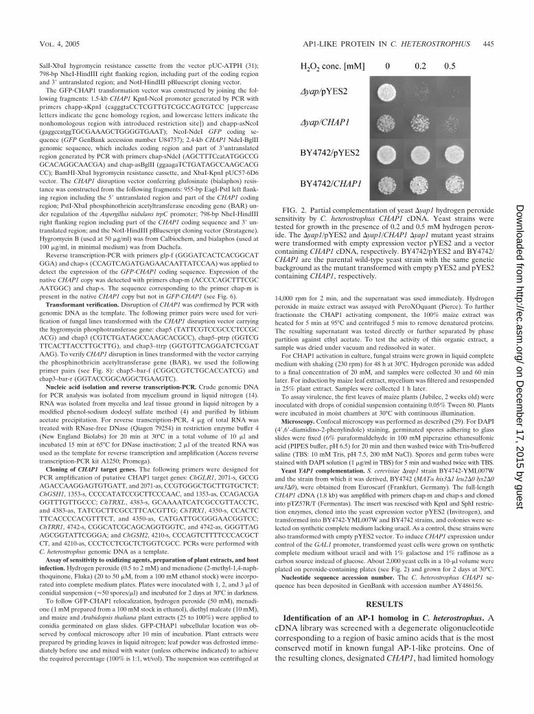
FIG. 2. Partial complementation of yeast �yap1 hydrogen peroxidesensitivity by C. heterostrophus CHAP1 cDNA. Yeast strains weretested for growth in the presence of 0.2 and 0.5 mM hydrogen perox-ide. The �yap1/pYES2 and �yap1/CHAP1 �yap1 mutant yeast strainswere transformed with empty expression vector pYES2 and a vectorcontaining CHAP1 cDNA, respectively. BY4742/pYES2 and BY4742/CHAP1 are the parental wild-type yeast strain with the same geneticbackground as the mutant transformed with empty pYES2 and pYES2containing CHAP1, respectively.
VOL. 4, 2005 AP1-LIKE PROTEIN IN C. HETEROSTROPHUS 445
on Decem
ber 17, 2015 by guesthttp://ec.asm
.org/D
ownloaded from

to the AP-1-like protein of Neurospora crassa (3), and thissequence was used to search the C. heterostrophus genome.The genomic sequence of CHAP1 has a predicted 1,976-bpcoding region. A full-length cDNA clone was obtained by re-verse transcription-PCR. The cDNA sequence confirmed thelocations of three introns (53, 102, and 51 bp) and encodes aprotein of 590 amino acids. The predicted CHAP1 protein hasa basic leucine zipper domain with residues characteristic ofthe YAP1-like protein family: Q171, Q176, A178, and F179(15) and two cysteine-rich domains, c-CRD and n-CRD (Fig.1) (10, 43). The homology of CHAP1 to other YAP proteins islow outside the bZIP and c-CRD regions. The BLAST searchof the C. heterostrophus genome with the amino acid sequencecorresponding to the bZIP domain of CHAP1 revealed thatthere are at least two additional proteins sharing the residuesthat are a signature of the YAP protein family.
Complementation of a yeast yap1 mutant. To determinewhether CHAP1 could replace yeast YAP1 in a deletion mu-tant, we transformed a �yap1 strain with the expression vectorpYES2 harboring a full-length CHAP1 cDNA clone. In addi-tion, pYES2-CHAP1 was transformed into the parental wild-type yeast strain BY4742. As a control, the �yap1 and BY4742strains were transformed with empty pYES2 vector. All thestrains were grown on galactose medium to activate the GAL4promoter of the expression vector. In the absence of oxidizingagents, yeast strains harboring CHAP1 were less viable thanthose transformed with empty vector (Fig. 2). In the presenceof 0.2 mM hydrogen peroxide, growth of the �yap1 mutant wasalmost completely inhibited, whereas growth of the�yap1/CHAP1 strain was only slightly affected compared to theBY4742/CHAP1 strain. At 0.5 mM hydrogen peroxide thegrowth of the �yap1/CHAP1 strain was further inhibited, where-
FIG. 3. Sensitivity of the wild-type and chap1-deleted mutant to oxidizing agents. Hydrogen peroxide (B) and menadione (C) were incorporatedinto complete medium at the indicated concentrations (A is complete medium alone). Plates were inoculated with, from left to right in each panel,3, 2, and 1 �l of conidial suspension (�50 spores/�l) and grown for 3 days at 30°C.
446 LEV ET AL. EUKARYOT. CELL
on Decem
ber 17, 2015 by guesthttp://ec.asm
.org/D
ownloaded from

as growth of the BY4742/CHAP1 and BY4742/pYES2 strainswas unaffected (Fig. 2). These findings indicate that CHAP1 isable to complement yeast �yap1, at least partially. Neither thetoxic effect of CHAP1 expression nor complementation could
be observed when the strains were grown on glucose instead ofgalactose (data not shown).
Deletion of CHAP1. To investigate the function of CHAP1,the gene was deleted by double crossover integration, replac-
FIG. 4. Induction of putative CHAP1 target genes in response to oxidative stress and plant extract in wild-type and chap1 deletion strains. Geneabbreviations: ChTRX1 and ChTRX2, thioredoxins 1 and 2, respectively; ChGSH1, �-glutamylcysteine synthetase; ChGSH2, glutathione synthetaselarge chain; ChTRR1, thioredoxin reductase; ChGLR1, glutathione reductase. RNA loading was verified by ethidium bromide staining. Sizemarkers (in kilobases) at the left indicate the rRNA bands. C, control prior to treatment; 0.5 h and 1 h indicate the time after addition of hydrogenperoxide or leaf extract.
VOL. 4, 2005 AP1-LIKE PROTEIN IN C. HETEROSTROPHUS 447
on Decem
ber 17, 2015 by guesthttp://ec.asm
.org/D
ownloaded from

ing the entire coding region with an expression cassette con-ferring resistance to hygromycin B. The disruption constructwas transformed into C. heterostrophus. More than 20 hygro-mycin-resistant transformants were obtained, and homologousintegration and loss of the wild-type copy were confirmed byPCR amplification with primer pairs chap5 and chap3; chap5and ptrp; and chap3 and ttrp (see Materials and Methods).Deletion mutants were indistinguishable from the wild typewith respect to colony morphology, sporulation, and conidialgermination. Two independently isolated transformants wereselected for further characterization.
Drops of conidial suspension of the wild-type and �chap1strains were inoculated on complete medium plates containingdifferent concentrations of hydrogen peroxide. Conidia of boththe mutant and the wild type began to germinate at the sametime on plates containing 0.5, 1, or 2 mM hydrogen peroxide(not shown). Elongation of the germ tubes of the mutant, how-ever, was inhibited shortly after germination. Subsequent colonygrowth was markedly inhibited in the mutant (Fig. 3A and B).
We examined the resistance of a �chap1 mutant to anotheroxidizing agent, the superoxide-generating compound menadi-one (2-methyl-1,4-naphthoquinone). Growth of the wild typeon complete medium with 20 or 50 �M menadione was inhib-ited less than that of a �chap mutant (Fig. 3C).
CHAP1 target genes. S. cerevisiae YAP1 is activated in re-sponse to oxidative stress and then regulates the expression ofgenes involved in detoxification. We cloned C. heterostrophus ho-mologs of several YAP1-dependent genes in order to investigatewhether their expression is regulated by CHAP1 in response tooxidative stress. Genes encoding the following proteins were iden-tified, with the indicated similarity to their yeast homologs, in theC. heterostrophus genome: thioredoxin reductase (ChTRR1, 77%),�-glutamylcysteine synthetase (ChGSH1, 65%), glutathione re-ductase (ChGLR1, 69%), glutathione synthetase (ChGSH2, 70%),and two thioredoxins (ChTRX1 and ChTRX2). ChTRX2 is high-ly similar to both thioredoxins of S. cerevisiae (78% to TRX1and 80% to TRX2), in contrast to ChTRX1, which is only 43%similar to both TRX1 and TRX2. There is 42% similaritybetween the two C. heterostrophus thioredoxins.
We used hydrogen peroxide (20 mM) as an oxidizing agentto activate CHAP1 in mycelium grown in liquid culture; sam-ples were collected 30 and 60 min after addition of hydrogenperoxide. RNA blot analyses were performed with probes cor-responding to putative CHAP1 target genes. All the genestested except ChTRX1 are upregulated in response to hydro-gen peroxide, and their expression depends on CHAP1 (Fig.4). This implies that the same mechanism of oxidative stressdefense functions in S. cerevisiae and in C. heterostrophus andthat structurally similar members of the YAP family are re-sponsible for it. The thioredoxin gene ChTRX1 was upregu-lated upon exposure to hydrogen peroxide in a CHAP1-inde-pendent manner. Its activation was delayed in comparison tothat of other genes tested and occurred to a lesser extent (Fig.4). Perhaps an additional redox state sensor is involved inChTRX1 activation, as in S. cerevisiae, whose two thioredoxinsare differentially regulated (17).
Despite the increased sensitivity of the chap1 deletion mu-tants to oxidizing agents, as well as the loss of induction of a setof target genes, inoculation of maize leaves by drops of conidialsuspension (30 and 60 conidia per drop) of the wild-type and
mutant strains resulted in similar disease symptoms. The av-erage lesion size was the same within the variability for the wildtype and the mutant (Fig. 5).
Nuclear localization of the GFP-CHAP1 fusion protein uponactivation. To investigate the mode of CHAP1 activation, wefollowed the intracellular response of CHAP1 to oxidizingconditions. A construct was designed encoding an N-terminalfusion of GFP to the CHAP1 coding sequence under regulationof the CHAP1 promoter (Fig. 6A). Fungal transformation wasperformed, and integration of the CHAP1 promoter-GFP-CHAP1 sequence was confirmed by PCR with genomic DNAas the template (data not shown) and by reverse transcription-PCR (Fig. 6B). An endogenous CHAP1 was transcribed in thetransformants as well as the fused GFP-CHAP1 coding se-quence, indicating that the construct was not integrated intothe CHAP1 coding region but elsewhere in the genome.
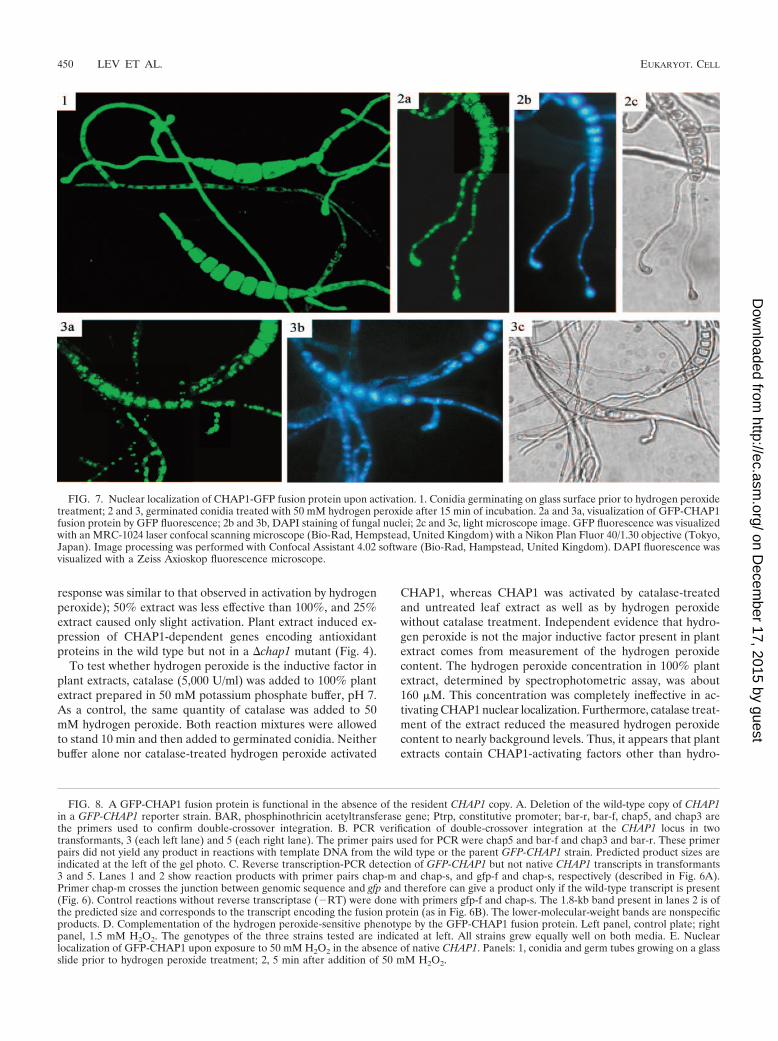
Conidia of GFP-CHAP1 transformants were placed on glassslides and allowed to germinate (4 h at 30°C). Germ tubes andconidia were uniformly fluorescent during germination andappressorium formation on glass slides (Fig. 7, panel 1). Hy-
FIG. 5. Disease symptoms on maize leaves caused by wild-type and�chap1 fungal strains. A. First leaves of 2-week-old seedlings wereinoculated with drops containing 30 and 60 spores, and the plants wereincubated at 30°C for 2 days. B. The bar graph compares average lesionsize. Bars indicate standard deviation, and each sample includes 20 to28 lesion spots.
448 LEV ET AL. EUKARYOT. CELL
on Decem
ber 17, 2015 by guesthttp://ec.asm
.org/D
ownloaded from

drogen peroxide (50 mM) was added, and conidia were ob-served by confocal microscopy 15 min later. Fluorescence wasrestricted to subcellular compartments in treated conidia andgerm tubes. GFP fluorescence and DAPI staining colocalizedto the same compartment, the nucleus (Fig. 7). Upon additionof hydrogen peroxide, bubbling was observed almost immedi-ately, indicating catalase activity. Bubbling and CHAP1 acti-vation occur almost simultaneously, perhaps a manifestation oftwo strategies to cope with stress caused by hydrogen peroxide.Catalase activity makes the effective concentration of hydrogenperoxide available at the surface of the fungal cell unstable.Consistent and widespread activation of CHAP1 was observedonly upon application of at least 15 mM hydrogen peroxide.About 10-fold lower hydrogen peroxide concentrations wereinhibitory when conidia were plated on solid medium (Fig. 3).It is likely that conidia germinating on agar plates containinghydrogen peroxide encounter a continuous supply of H2O2, incontrast to the mycelium growing on a glass slide to which 20-to 30-�l drops of H2O2-containing solution were added. It seemsthat catalase secreted from the growing hyphae was sufficient topartially degrade H2O2 in the first moments after addition.
To test the function of the GFP-CHAP1 fusion protein inthe absence of the native protein, we disrupted the endogenousCHAP1 copy in a GFP-CHAP1-expressing strain with resis-
tance to bialaphos as the selection marker. Double-crossoverintegration was confirmed by PCR on genomic DNA of thetransformants, and reverse transcription-PCR confirmed theabsence of wild-type CHAP1 transcripts in these bialaphos-resistant lines (Fig. 8A and B). The growth of these transfor-mants (GFP-CHAP1 �chap1) on plates containing 1.5 mMhydrogen peroxide was indistinguishable from that of theparental strain expressing both GFP-CHAP1 and wild-typeCHAP1 (Fig. 8D). CHAP1 activation by hydrogen peroxidewas tested with a GFP-CHAP1 �chap1 transformant (Fig. 8E),and similar results were obtained, indicating that the fusionprotein becomes localized to the nucleus under oxidative stressin the absence of a wild-type copy.
Activation of CHAP1 by oxidizing agents and plant extracts.CHAP1 activation (nuclear localization) was examined in re-sponse to other oxidizing agents: the superoxide-generatingcompound menadione (2-methyl-1,4-naphthoquinone) and theglutathione-depleting compound diethyl maleate. Both mena-dione (1 mM) and diethyl maleate (10 mM) caused rapidCHAP1 activation in treated hyphae at a rate comparable tothe response to hydrogen peroxide. Maize leaf extract (25, 50,and 100%) was added to conidia germinated on glass slides.Within a few minutes after application of the 100% extract,nearly all GFP fluorescence relocalized into nuclei (the rate of
FIG. 6. Construction of strains expressing the GFP-CHAP1 fusion protein. A. Construct map. GFP, GFP gene; Ptrp, constitutive promoter;HPH, hygromycin phosphotransferase gene; chap-m, chap-s, and gfp-f, primers used for transformant verification. Upper drawing: transformant;lower drawing: wild type. B. Confirmation of CHAP1-GFP expression by reverse transcription-PCR. The following reactions were performed withRNA of the wild type and transformants gfp-chap1 and gfp-chap2. Lanes 1, gfp-f and chap-s; lanes 2, chap-m and chap-s. Control reactions withoutreverse transcriptase (RT) were performed with primers gfp-f and chap-s for transformants and chap-m and chap-s for the wild type. Thepredicted product size, 1.8 kb, is indicated at the left of the gel; note that this is smaller than the size indicated in A, 1,976 bp, due to splicing of introns.
VOL. 4, 2005 AP1-LIKE PROTEIN IN C. HETEROSTROPHUS 449
on Decem
ber 17, 2015 by guesthttp://ec.asm
.org/D
ownloaded from
response was similar to that observed in activation by hydrogenperoxide); 50% extract was less effective than 100%, and 25%extract caused only slight activation. Plant extract induced ex-pression of CHAP1-dependent genes encoding antioxidantproteins in the wild type but not in a �chap1 mutant (Fig. 4).
To test whether hydrogen peroxide is the inductive factor inplant extracts, catalase (5,000 U/ml) was added to 100% plantextract prepared in 50 mM potassium phosphate buffer, pH 7.As a control, the same quantity of catalase was added to 50mM hydrogen peroxide. Both reaction mixtures were allowedto stand 10 min and then added to germinated conidia. Neitherbuffer alone nor catalase-treated hydrogen peroxide activated
CHAP1, whereas CHAP1 was activated by catalase-treatedand untreated leaf extract as well as by hydrogen peroxidewithout catalase treatment. Independent evidence that hydro-gen peroxide is not the major inductive factor present in plantextract comes from measurement of the hydrogen peroxidecontent. The hydrogen peroxide concentration in 100% plantextract, determined by spectrophotometric assay, was about160 �M. This concentration was completely ineffective in ac-tivating CHAP1 nuclear localization. Furthermore, catalase treat-ment of the extract reduced the measured hydrogen peroxidecontent to nearly background levels. Thus, it appears that plantextracts contain CHAP1-activating factors other than hydro-
FIG. 7. Nuclear localization of CHAP1-GFP fusion protein upon activation. 1. Conidia germinating on glass surface prior to hydrogen peroxidetreatment; 2 and 3, germinated conidia treated with 50 mM hydrogen peroxide after 15 min of incubation. 2a and 3a, visualization of GFP-CHAP1fusion protein by GFP fluorescence; 2b and 3b, DAPI staining of fungal nuclei; 2c and 3c, light microscope image. GFP fluorescence was visualizedwith an MRC-1024 laser confocal scanning microscope (Bio-Rad, Hempstead, United Kingdom) with a Nikon Plan Fluor 40/1.30 objective (Tokyo,Japan). Image processing was performed with Confocal Assistant 4.02 software (Bio-Rad, Hampstead, United Kingdom). DAPI fluorescence wasvisualized with a Zeiss Axioskop fluorescence microscope.
FIG. 8. A GFP-CHAP1 fusion protein is functional in the absence of the resident CHAP1 copy. A. Deletion of the wild-type copy of CHAP1in a GFP-CHAP1 reporter strain. BAR, phosphinothricin acetyltransferase gene; Ptrp, constitutive promoter; bar-r, bar-f, chap5, and chap3 arethe primers used to confirm double-crossover integration. B. PCR verification of double-crossover integration at the CHAP1 locus in twotransformants, 3 (each left lane) and 5 (each right lane). The primer pairs used for PCR were chap5 and bar-f and chap3 and bar-r. These primerpairs did not yield any product in reactions with template DNA from the wild type or the parent GFP-CHAP1 strain. Predicted product sizes areindicated at the left of the gel photo. C. Reverse transcription-PCR detection of GFP-CHAP1 but not native CHAP1 transcripts in transformants3 and 5. Lanes 1 and 2 show reaction products with primer pairs chap-m and chap-s, and gfp-f and chap-s, respectively (described in Fig. 6A).Primer chap-m crosses the junction between genomic sequence and gfp and therefore can give a product only if the wild-type transcript is present(Fig. 6). Control reactions without reverse transcriptase (RT) were done with primers gfp-f and chap-s. The 1.8-kb band present in lanes 2 is ofthe predicted size and corresponds to the transcript encoding the fusion protein (as in Fig. 6B). The lower-molecular-weight bands are nonspecificproducts. D. Complementation of the hydrogen peroxide-sensitive phenotype by the GFP-CHAP1 fusion protein. Left panel, control plate; rightpanel, 1.5 mM H2O2. The genotypes of the three strains tested are indicated at left. All strains grew equally well on both media. E. Nuclearlocalization of GFP-CHAP1 upon exposure to 50 mM H2O2 in the absence of native CHAP1. Panels: 1, conidia and germ tubes growing on a glassslide prior to hydrogen peroxide treatment; 2, 5 min after addition of 50 mM H2O2.
450 LEV ET AL. EUKARYOT. CELL
on Decem
ber 17, 2015 by guesthttp://ec.asm
.org/D
ownloaded from

VOL. 4, 2005 AP1-LIKE PROTEIN IN C. HETEROSTROPHUS 451
on Decem
ber 17, 2015 by guesthttp://ec.asm
.org/D
ownloaded from

gen peroxide, the presence of which does not depend on activeplant defense. To determine whether the CHAP1-activatingsubstance is specific to maize, we tested a similar extract pre-pared from Arabidopsis thaliana leaves. It caused a responsecomparable to that of maize extract. Apparently, the activa-tor(s) of CHAP1 is not plant species specific.
Initial characterization of active components in the plantextract. To provide an initial characterization of the activecomponents in the plant extract, we used nuclear localizationof GFP-CHAP1 for activity-guided fractionation. First, we fil-tered the extract through an ultrafiltration membrane with acutoff of 10,000 Da. The active component was able to crossthe membrane. When the extract was heated for 5 min at 95°Cand centrifuged at 14,000 rpm for 2 min to precipitate dena-tured proteins, the resulting supernatant contained the activa-tor. The supernatant after heat treatment and centrifugation wasthen extracted with an equal volume of ethyl acetate. Upon sep-
aration of the phases by centrifugation, the active compound(s)partitioned into the organic phase. No detectable active com-ponents were extracted from the leaf powder by chloroform-methanol, while ethanol did efficiently extract the activator(s).
Activation of CHAP1 during plant infection. Two-week-oldmaize plants were inoculated with suspensions of conidia fromGFP-CHAP1 transformants. Germ tubes emerged from coni-dia within an hour, and the fusion protein began migrating intothe nuclei of the germ tubes on the leaf surface prior to pene-tration. This migration was specific to the plant surface, as germtubes on glass slides remained uniformly fluorescent. Duringthe subsequent 2 to 3 h, the germ tubes elongated and CHAP1continued to be localized in nuclei. After about 3 h, the hyphaltips formed appressoria, and CHAP1 became localized to theirnuclei. During hyphal penetration of the plant cuticle andepidermis and development inside the leaf, CHAP1 remainedin the hyphal nuclei. After about 24 h, when well-developed
FIG. 9. Subcellular localization of CHAP1 during plant infection. Numbers indicate time after inoculation: 1 h, conidia start germination,CHAP1 is activated in emerging germ tubes, and germ tubes elongate (2.5 h) and form appressoria (3.5 h); 24 h and 26 h, extensive mycelialnetwork interlaces plant tissue, necrotic lesion is formed at the site of inoculation, fluorescence is localized in the nucleus in most of the hyphae;30 h-a, hyphae found in the outermost layers of the infected leaf: GFP fluorescence is distributed in the cytosol; 30 h-b, hyphae found inside theplant tissue: in these hyphae CHAP1 is still activated. Confocal image acquisition was done as described for Fig. 7; the autofluorescence of the leaf,contributed primarily by chlorophyll, was detected with the Cy5-optimized channel.
452 LEV ET AL. EUKARYOT. CELL
on Decem
ber 17, 2015 by guesthttp://ec.asm
.org/D
ownloaded from

necrotic lesions were visible at inoculation sites, bundles ofuniformly fluorescent hyphae appeared. During further pro-gression of the disease, the pattern of CHAP1 inactivation (re-appearance in the cytoplasm) became more discernible: hy-phae on leaf surfaces were uniformly fluorescent, whereasinside the leaf CHAP1 was localized in the nuclei (Fig. 9).
DISCUSSION
Redox regulation is a ubiquitous mechanism for controllingcellular activity (16). Direct modification of transcription fac-tors or signal transduction by redox-sensing proteins altersgene expression in response to an oxidizing environment. Weisolated a C. heterostrophus homolog of the yeast YAP1 tran-scription factor. CHAP1 has the same structural features asYAP1, including a bZIP domain and two cysteine-rich regions.An oxidizing environment causes activation of CHAP1 and itsrelocalization into the nucleus. It then regulates a set of genesencoding antioxidant proteins. We found that this set overlapsthe set of YAP1-dependent genes of S. cerevisiae.
Despite the marked homology of both AP-1-like proteins intheir DNA-binding domains, no YAP1 recognition sequencecould be identified in the promoters of the CHAP1-dependentgenes tested. Nevertheless, CHAP1 could partially comple-ment the hydrogen peroxide-sensitive phenotype of a yeastyap1 mutant. It appears that there is sufficient structural ho-mology to allow at least partial replacement of YAP1 functionin S. cerevisiae. It is possible that in C. heterostrophus, CHAP1forms a heterodimer with another bZIP protein and the re-sulting complex has a different DNA binding specificity, as wasshown for the mammalian bZIP protein family (24). Expres-sion of CHAP1 under the strong GAL4 promoter in S. cerevi-siae led to reduced growth. The reason for this toxicity is notclear, but it was independent of oxidative stress and of thepresence of the resident yeast YAP1 copy (Fig. 2). CHAP1overexpression might have allowed binding to nonspecific se-quences, interfering with metabolism and growth.
Activation of CHAP1 nuclear relocalization during plantinfection might be induced by penetration of the activator intothe cell directly or through membrane channels. Another routeof activation could be interaction of the activating substancewith the extracellular domains of membrane receptor proteins,followed by transduction of the redox signal into the cell bythiol-disulfide exchange (6, 16, 36). Several lines of evidenceindicate that existing molecules other than reactive oxygenspecies are involved in CHAP1 activation on the plant. First,plant extracts prepared from maize leaves that have never beenin contact with the pathogen caused intense CHAP1 activation.As the extract was made from quickly frozen plant tissue, it isunlikely that any wounding or defense responses were acti-vated during its preparation. Second, pretreatment of the plantextract with catalase did not affect its activating property. Theactivator(s) is not plant species specific and activates CHAP1as rapidly as hydrogen peroxide does.
Our initial fractionation experiments revealed that the activecomponent is heat stable and has a relatively low molecularweight. The active molecule is sufficiently polar to dissolve inwater or in ethanol, and it partitions from water into ethylacetate. In view of the recent report (35) that indole-3-aceticacid activates downstream targets of the yeast CHAP1 ortholog,
YAP1, indole-3-acetic acid might be a candidate inducer. Wetherefore tested whether indole-3-acetic acid activates CHAP1nuclear relocalization. Up to the highest concentration tested,500 �M, indole-3-acetic acid did not activate CHAP1. Meth-ylglyoxal, a toxic by-product of glycolyis, was recently shown toactivate yeast YAP1 (32). Methylglyoxal could be producedintracellularly in the fungus and also by the plant; its activity asa plant-derived inducer can be tested with CHAP1 nuclear re-localization as an assay. Oxidizing agents, cadmium, indole-3-acetic acid, and methylglyoxal, are unrelated compounds, all ofwhich activate yeast YAP1. This raises the possibility that YAP1,characterized as a redox-responsive transcription factor, mayhave a more general sensory role than initially thought. In C. het-erostrophus, it seems likely that a novel plant compound isresponsible for the induction of CHAP1 relocalization, andits full chemical characterization is currently in progress.
Activation of CHAP1 on the plant (Fig. 9) suggests a role ininfection. Nevertheless, deletion of CHAP1 had no significanteffect on virulence. Apparently, other C. heterostrophus YAP-like proteins are not able to compensate for the defect causedby CHAP1 disruption: transcription of the CHAP1-dependentantioxidant genes is not induced in absence of CHAP1. Theability of the fungus to overcome the absence of CHAP1 dur-ing plant infection might therefore result from alternative pro-tective systems that compensate for the loss of CHAP1. InS. cerevisiae, glutathione and catalase provide overlapping de-fenses against hydrogen peroxide (22). The genome of C. het-erostrophus encodes three catalase-encoding (CAT) genes(37). Mutants deficient in CAT3 had enhanced sensitivity tohydrogen peroxide compared with the wild type and with mu-tants deficient in CAT1, CAT2, or both. Nevertheless, all thecatalase-deficient mutants had normal virulence on maize.
One interpretation of normal virulence in both CHAP1 mu-tants (this study) and catalase mutants (37) is that either ofthese distinct mechanisms of defense against oxidative stress issufficient. Another possibility is that C. heterostrophus does notencounter damaging levels of reactive oxygen species duringinfection under the conditions that have been tested. CHAP1relocalization was induced before penetration into the leaf andcertainly before massive death of host cells. This suggests thatthe activation of CHAP1 may act as an “early warning” system,relaying a signal for impending exposure to oxidative stress.The antioxidant transcriptional program would be induced andready, even if this stress does not actually occur. Some defensemechanisms, particularly against as universal a challenge asoxidative stress, may have evolved before specialization offungi to their hosts. The homologs of CHAP1 as well as of thecatalase genes might have essential roles in the virulence ofother plant pathogens.
Transcriptional regulation mediated by nuclear localizationis well known in animals and plants and in S. cerevisiae. Nuclearrelocalization of CHAP1 at the earliest stages of infectionis among the most rapid known molecular consequences ofgrowth of a plant pathogen on its host. Further study shoulduncover both the protective and signaling roles of redox-sen-sitive transcription factors in fungus-plant interactions.
ACKNOWLEDGMENTS
We are grateful to Jonathan Gressel for critical reading of themanuscript. We are grateful to Torrey Mesa Research Institute/Syn-
VOL. 4, 2005 AP1-LIKE PROTEIN IN C. HETEROSTROPHUS 453
on Decem
ber 17, 2015 by guesthttp://ec.asm
.org/D
ownloaded from

genta for access to the Cochliobolus sequence database (sequenced byCelera Genomics for TMRI/Syngenta).
This work was supported in part by ISF grant 233002 from the IsraelAcademy of Sciences.
REFERENCES
1. Alarco, A. M., I. Balan, D. Talibi, N. Mainville, and M. Raymond. 1997.AP1-mediated multidrug resistance in Saccharomyces cerevisiae requiresFLR1 encoding a transporter of the major facilitator superfamily. J. Biol.Chem. 272:19304–19313.
2. Alarco, A. M., and M. Raymond. 1999. The bZip transcription factor Cap1pis involved in multidrug resistance and oxidative stress response in Candidaalbicans. J. Bacteriol. 181:700–708.
3. Altschul, S., W. Gish, W. Miller, E. Myers, and D. Lipman. 1990. Basic localalignment tool. J. Mol. Biol. 215:403–410.
4. Ausubel, F. M., R. Brent, R. E. Kinston, D. D. Moore, J. A. Smith, J. G.Seidman, and K. Struhl. 1987. Current protocols in molecular biology.Wiley-Interscience, New York, N.Y.
5. Billard, P., H. Dumond, and M. Bolotin-Fukuhara. 1997. Characterization ofan AP-1-like transcription factor that mediates an oxidative stress responsein Kluyveromyces lactis. Mol. Gen. Genet. 257:62–70.
6. Ciriolo, M. R., M. Paci, M. Sette, A. De Martino, A. Bozzi, and G. Rotilio.1993. Transduction of reducing power across the plasma membrane by re-duced glutathione. A 1H-NMR spin-echo study of intact human erythro-cytes. Eur. J. Biochem. 215:711–718.
7. Cohen, B. A., Y. Pilpel, R. D. Mitra, and G. M. Church. 2002. Discriminationbetween paralogs using microarray analysis: application to the Yap1p andYap2p transcriptional networks. Mol. Biol. Cell 13:1608–1614.
8. Coleman, S. T., E. A. Epping, S. M. Steggerda, and W. S. Moye-Rowley. 1999.Yap1p activates gene transcription in an oxidant-specific fashion. Mol. Cell.Biol. 19:8302–8313.
9. Danon, A. 2002. Redox reactions of regulatory proteins: do kinetics promotespecificity? Trends Biochem. Sci. 27:197–203.
10. Delaunay, A., A. D. Isnard, and M. B. Toledano. 2000. H2O2 sensing throughoxidation of the Yap1 transcription factor. EMBO J. 19:5157–5166.
11. Delaunay, A., D. Pflieger, M. B. Barrault, J. Vinh, and M. B. Toledano. 2002.A thiol peroxidase is an H2O2 receptor and redox-transducer in gene acti-vation. Cell 111:471–481.
12. Dickinson, D. A., and H. J. Forman. 2002. Cellular glutathione and thiolsmetabolism. Biochem. Pharmacol. 64:1019–1026.
13. Dumond, H., N. Danielou, M. Pinto, and M. Bolotin-Fukuhara. 2000. Alarge-scale study of Yap1p-dependent genes in normal aerobic and H2O2-stress conditions: the role of Yap1p in cell proliferation control in yeast. Mol.Microbiol. 36:830–845.
14. Edwards, K., C. Johnstone, and C. Thompson. 1991. A simple and rapidmethod for the preparation of plant genomic DNA for PCR analysis. NucleicAcids Res. 19:1349.
15. Fernandes, L., C. Rodrigues-Pousada, and K. Struhl. 1997. Yap, a novelfamily of eight bZIP proteins in Saccharomyces cerevisiae with distinct bio-logical functions. Mol. Cell. Biol. 17:6982–6993.
16. Filomeni, G., G. Rotilio, and M. R. Ciriolo. 2002. Cell signalling and theglutathione redox system. Biochem. Pharmacol. 64:1057–1064.
17. Garrido, E. O., and C. M. Grant. 2002. Role of thioredoxins in the responseof Saccharomyces cerevisiae to oxidative stress induced by hydroperoxides.Mol. Microbiol. 43:993–1003.
18. Gasch, A. P., P. T. Spellman, C. M. Kao, O. Carmel-Harel, M. B. Eisen, G.Storz, D. Botstein, and P. O. Brown. 2000. Genomic expression programs inthe response of yeast cells to environmental changes. Mol. Biol. Cell 11:4241–4257.
19. Gil-ad, N. L., N. Bar-Nun, T. Noy, and A. M. Mayer. 2000. Enzymes ofBotrytis cinerea capable of breaking down hydrogen peroxide. FEMS Micro-biol. Lett. 190:121–126.
20. Govrin, E. M., and A. Levine. 2000. The hypersensitive response facilitatesplant infection by the necrotrophic pathogen Botrytis cinerea. Curr. Biol.10:751–757.
21. Grant, C. M., F. H. Maciver, and I. W. Dawes. 1996. Stationary-phaseinduction of GLR1 expression is mediated by the yAP-1 transcriptionalregulatory protein in the yeast Saccharomyces cerevisiae. Mol. Microbiol.22:739–746.
22. Grant, C. M., G. Perrone, and I. W. Dawes. 1998. Glutathione and catalaseprovide overlapping defenses for protection against hydrogen peroxide in theyeast Saccharomyces cerevisiae. Biochem. Biophys. Res. Commun. 253:893–898.
23. Kawasaki, L., and J. Aguirre. 2001. Multiple catalase genes are differentiallyregulated in Aspergillus nidulans. J. Bacteriol. 183:1434–1440.
24. Kerppola, T. K., and T. Curran. 1994. A conserved region adjacent to thebasic domain is required for recognition of an extended DNA binding site byMaf/Nrl family proteins. Oncogene 9:3149–3158.
25. Kuge, S., M. Arita, A. Murayama, K. Maeta, S. Izawa, Y. Inoue, and A.Nomoto. 2001. Regulation of the yeast Yap1p nuclear export signal is me-diated by redox signal-induced reversible disulfide bond formation. Mol.Cell. Biol. 21:6139–6150.
26. Kuge, S., and N. Jones. 1994. YAP1 dependent activation of TRX2 isessential for the response of Saccharomyces cerevisiae to oxidative stress byhydroperoxides. EMBO J. 13:655–664.
27. Kuge, S., N. Jones, and A. Nomoto. 1997. Regulation of yAP-1 nuclearlocalization in response to oxidative stress. EMBO J. 16:1710–1720.
28. Lee, J., C. Godon, G. Lagniel, D. Spector, J. Garin, J. Labarre, and M. B.Toledano. 1999. Yap1 and Skn7 control two specialized oxidative stressresponse regulons in yeast. J. Biol. Chem. 274:16040–16046.
29. Lev, S., and B. A. Horwitz. 2003. A mitogen-activated protein kinase pathwaymodulates the expression of two cellulase genes in Cochliobolus heterostro-phus during plant infection. Plant Cell 15:835–844.
30. Levine, A., R. I. Pennell, M. E. Alvarez, R. Palmer, and C. Lamb. 1996.Calcium-mediated apoptosis in a plant hypersensitive disease resistance re-sponse. Curr. Biol. 6:427–437.
31. Lu, S., L. Lyngholm, G. Yang, C. Bronson, O. C. Yoder, and B. G. Turgeon.1994. Tagged mutations at the Tox1 locus of Cochliobolus heterostrophus byrestriction enzyme-mediated integration. Proc. Natl. Acad. Sci. USA 91:12649–12653.
32. Maeta, K., S. Izawa, S. Okazaki, S. Kuge, and Y. Inoue. 2004. Activity of theYap1 transcription factor in Saccharomyces cerevisiae is modulated by meth-ylglyoxal, a metabolite derived from glycolysis. Mol. Cell. Biol. 24:8753–8764.
33. Morgan, B. A., G. R. Banks, W. M. Toone, D. Raitt, S. Kuge, and L. H.Johnston. 1997. The Skn7 response regulator controls gene expression in theoxidative stress response of the budding yeast Saccharomyces cerevisiae.EMBO J. 16:1035–1044.
34. Nguyen, D. T., A. M. Alarco, and M. Raymond. 2001. Multiple Yap1p-binding sites mediate induction of the yeast major facilitator FLR1 gene inresponse to drugs, oxidants, and alkylating agents. J. Biol. Chem. 276:1138–1145.
35. Prusty, R., P. Grisafi, and G. R. Fink. 2004. The plant hormone indoleaceticacid induces invasive growth in Saccharomyces cerevisiae. Proc. Natl. Acad.Sci. USA 101:4153–4157.
36. Reglinski, J., S. Hoey, W. E. Smith, and R. D. Sturrock. 1988. Cellularresponse to oxidative stress at sulfhydryl group receptor sites on the eryth-rocyte membrane. J. Biol. Chem. 263:12360–12366.
37. Robbertse, B., O. C. Yoder, A. Nguyen, C. L. Schoch, and B. G. Turgeon.2003. Deletion of all Cochliobolus heterostrophus monofunctional catalase-encoding genes reveals a role for one in sensitivity to oxidative stress butnone with a role in virulence. Mol. Plant-Microbe Interact. 16:1013–1021.
38. Stephen, D. W., S. L. Rivers, and D. J. Jamieson. 1995. The role of the YAP1and YAP2 genes in the regulation of the adaptive oxidative stress responsesof Saccharomyces cerevisiae. Mol. Microbiol. 16:415–423.
39. Sugiyama, K., S. Izawa, and Y. Inoue. 2000. The Yap1p-dependent inductionof glutathione synthesis in heat shock response of Saccharomyces cerevisiae.J. Biol. Chem. 275:15535–15540.
40. Tenreiro, S., A. R. Fernandes, and I. Sa-Correia. 2001. Transcriptionalactivation of FLR1 gene during Saccharomyces cerevisiae adaptation togrowth with benomyl: role of Yap1p and Pdr3p. Biochem. Biophys. Res.Commun. 280:216–222.
41. Tiedemann, A. V. 1997. Evidence for a primary role of active oxygen speciesin induction of host cell death during infection of bean leaves with Botrytiscinerea. Physiol. Mol. Plant Pathol. 50:151–166.
42. Toone, W. M., S. Kuge, M. Samuels, B. A. Morgan, T. Toda, and N. Jones.1998. Regulation of the fission yeast transcription factor Pap1 by oxidativestress: requirement for the nuclear export factor Crm1 (exportin) and thestress-activated MAP kinase Sty1/Spc1. Genes Dev. 12:1453–1463.
43. Toone, W. M., B. A. Morgan, and N. Jones. 2001. Redox control of AP-1-likefactors in yeast and beyond. Oncogene 20:2336–2346.
44. Wemmie, J. A., M. S. Szczypka, D. J. Thiele, and W. S. Moye-Rowley. 1994.Cadmium tolerance mediated by the yeast AP-1 protein requires the pres-ence of an ATP-binding cassette transporter-encoding gene, YCF1. J. Biol.Chem. 269:32592–32597.
45. Wojtaszek, P. 1997. Oxidative burst: an early plant response to pathogeninfection. Biochem. J. 322:681–692.
46. Wu, A. L., and W. S. Moye-Rowley. 1994. GSH1, which encodes gamma-glutamylcysteine synthetase, is a target gene for yAP-1 transcriptional reg-ulation. Mol. Cell. Biol. 14:5832–5839.
47. Yoder, O. C. 1988. Cochliobolus heterostrophus, cause of southern corn leafblight. Adv. Plant Pathol. 6:93–112.
454 LEV ET AL. EUKARYOT. CELL
on Decem
ber 17, 2015 by guesthttp://ec.asm
.org/D
ownloaded from
Copyright © 2022 FDOKUMEN