Microretroreflector-Sedimentation Immunoassays for Pathogen Detection
7
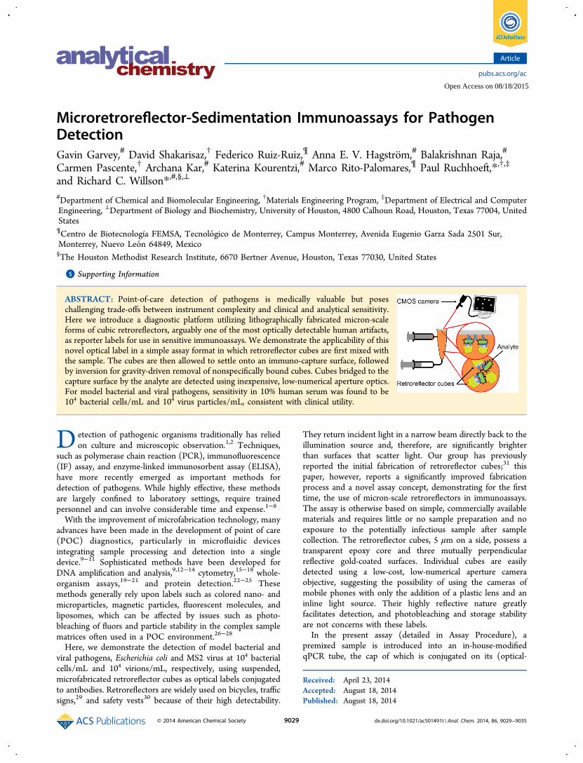
Microretroreflector-Sedimentation Immunoassays for Pathogen Detection Gavin Garvey, # David Shakarisaz, † Federico Ruiz-Ruiz, ¶ Anna E. V. Hagströ m, # Balakrishnan Raja, # Carmen Pascente, † Archana Kar, # Katerina Kourentzi, # Marco Rito-Palomares, ¶ Paul Ruchhoeft,* ,†,‡ and Richard C. Willson* ,#,§,⊥ # Department of Chemical and Biomolecular Engineering, † Materials Engineering Program, ‡ Department of Electrical and Computer Engineering, ⊥ Department of Biology and Biochemistry, University of Houston, 4800 Calhoun Road, Houston, Texas 77004, United States ¶ Centro de Biotecnología FEMSA, Tecnoló gico de Monterrey, Campus Monterrey, Avenida Eugenio Garza Sada 2501 Sur, Monterrey, Nuevo Leó n 64849, Mexico § The Houston Methodist Research Institute, 6670 Bertner Avenue, Houston, Texas 77030, United States * S Supporting Information ABSTRACT: Point-of-care detection of pathogens is medically valuable but poses challenging trade-offs between instrument complexity and clinical and analytical sensitivity. Here we introduce a diagnostic platform utilizing lithographically fabricated micron-scale forms of cubic retroreflectors, arguably one of the most optically detectable human artifacts, as reporter labels for use in sensitive immunoassays. We demonstrate the applicability of this novel optical label in a simple assay format in which retroreflector cubes are first mixed with the sample. The cubes are then allowed to settle onto an immuno-capture surface, followed by inversion for gravity-driven removal of nonspecifically bound cubes. Cubes bridged to the capture surface by the analyte are detected using inexpensive, low-numerical aperture optics. For model bacterial and viral pathogens, sensitivity in 10% human serum was found to be 10 4 bacterial cells/mL and 10 4 virus particles/mL, consistent with clinical utility. D etection of pathogenic organisms traditionally has relied on culture and microscopic observation. 1,2 Techniques, such as polymerase chain reaction (PCR), immunofluorescence (IF) assay, and enzyme-linked immunosorbent assay (ELISA), have more recently emerged as important methods for detection of pathogens. While highly effective, these methods are largely confined to laboratory settings, require trained personnel and can involve considerable time and expense. 1−8 With the improvement of microfabrication technology, many advances have been made in the development of point of care (POC) diagnostics, particularly in microfluidic devices integrating sample processing and detection into a single device. 9−11 Sophisticated methods have been developed for DNA amplification and analysis, 9,12−14 cytometry, 15−18 whole- organism assays, 19−21 and protein detection. 22−25 These methods generally rely upon labels such as colored nano- and microparticles, magnetic particles, fluorescent molecules, and liposomes, which can be affected by issues such as photo- bleaching of fluors and particle stability in the complex sample matrices often used in a POC environment. 26−28 Here, we demonstrate the detection of model bacterial and viral pathogens, Escherichia coli and MS2 virus at 10 4 bacterial cells/mL and 10 4 virions/mL, respectively, using suspended, microfabricated retroreflector cubes as optical labels conjugated to antibodies. Retroreflectors are widely used on bicycles, traffic signs, 29 and safety vests 30 because of their high detectability. They return incident light in a narrow beam directly back to the illumination source and, therefore, are significantly brighter than surfaces that scatter light. Our group has previously reported the initial fabrication of retroreflector cubes; 31 this paper, however, reports a significantly improved fabrication process and a novel assay concept, demonstrating for the first time, the use of micron-scale retroreflectors in immunoassays. The assay is otherwise based on simple, commercially available materials and requires little or no sample preparation and no exposure to the potentially infectious sample after sample collection. The retroreflector cubes, 5 μm on a side, possess a transparent epoxy core and three mutually perpendicular reflective gold-coated surfaces. Individual cubes are easily detected using a low-cost, low-numerical aperture camera objective, suggesting the possibility of using the cameras of mobile phones with only the addition of a plastic lens and an inline light source. Their highly reflective nature greatly facilitates detection, and photobleaching and storage stability are not concerns with these labels. In the present assay (detailed in Assay Procedure), a premixed sample is introduced into an in-house-modified qPCR tube, the cap of which is conjugated on its (optical- Received: April 23, 2014 Accepted: August 18, 2014 Published: August 18, 2014 Article pubs.acs.org/ac © 2014 American Chemical Society 9029 dx.doi.org/10.1021/ac501491t | Anal. Chem. 2014, 86, 9029−9035 Open Access on 08/18/2015
-
Upload
independent -
Category
Documents
-
view
0 -
download
0
Transcript of Microretroreflector-Sedimentation Immunoassays for Pathogen Detection
Microretroreflector-Sedimentation Immunoassays for PathogenDetectionGavin Garvey,# David Shakarisaz,† Federico Ruiz-Ruiz,¶ Anna E. V. Hagstrom,# Balakrishnan Raja,#
Carmen Pascente,† Archana Kar,# Katerina Kourentzi,# Marco Rito-Palomares,¶ Paul Ruchhoeft,*,†,‡
and Richard C. Willson*,#,§,⊥
#Department of Chemical and Biomolecular Engineering, †Materials Engineering Program, ‡Department of Electrical and ComputerEngineering, ⊥Department of Biology and Biochemistry, University of Houston, 4800 Calhoun Road, Houston, Texas 77004, UnitedStates¶Centro de Biotecnología FEMSA, Tecnologico de Monterrey, Campus Monterrey, Avenida Eugenio Garza Sada 2501 Sur,Monterrey, Nuevo Leon 64849, Mexico§The Houston Methodist Research Institute, 6670 Bertner Avenue, Houston, Texas 77030, United States
*S Supporting Information
ABSTRACT: Point-of-care detection of pathogens is medically valuable but poseschallenging trade-offs between instrument complexity and clinical and analytical sensitivity.Here we introduce a diagnostic platform utilizing lithographically fabricated micron-scaleforms of cubic retroreflectors, arguably one of the most optically detectable human artifacts,as reporter labels for use in sensitive immunoassays. We demonstrate the applicability of thisnovel optical label in a simple assay format in which retroreflector cubes are first mixed withthe sample. The cubes are then allowed to settle onto an immuno-capture surface, followedby inversion for gravity-driven removal of nonspecifically bound cubes. Cubes bridged to thecapture surface by the analyte are detected using inexpensive, low-numerical aperture optics.For model bacterial and viral pathogens, sensitivity in 10% human serum was found to be104 bacterial cells/mL and 104 virus particles/mL, consistent with clinical utility.
Detection of pathogenic organisms traditionally has reliedon culture and microscopic observation.1,2 Techniques,
such as polymerase chain reaction (PCR), immunofluorescence(IF) assay, and enzyme-linked immunosorbent assay (ELISA),have more recently emerged as important methods fordetection of pathogens. While highly effective, these methodsare largely confined to laboratory settings, require trainedpersonnel and can involve considerable time and expense.1−8
With the improvement of microfabrication technology, manyadvances have been made in the development of point of care(POC) diagnostics, particularly in microfluidic devicesintegrating sample processing and detection into a singledevice.9−11 Sophisticated methods have been developed forDNA amplification and analysis,9,12−14 cytometry,15−18 whole-organism assays,19−21 and protein detection.22−25 Thesemethods generally rely upon labels such as colored nano- andmicroparticles, magnetic particles, fluorescent molecules, andliposomes, which can be affected by issues such as photo-bleaching of fluors and particle stability in the complex samplematrices often used in a POC environment.26−28
Here, we demonstrate the detection of model bacterial andviral pathogens, Escherichia coli and MS2 virus at 104 bacterialcells/mL and 104 virions/mL, respectively, using suspended,microfabricated retroreflector cubes as optical labels conjugatedto antibodies. Retroreflectors are widely used on bicycles, trafficsigns,29 and safety vests30 because of their high detectability.
They return incident light in a narrow beam directly back to theillumination source and, therefore, are significantly brighterthan surfaces that scatter light. Our group has previouslyreported the initial fabrication of retroreflector cubes;31 thispaper, however, reports a significantly improved fabricationprocess and a novel assay concept, demonstrating for the firsttime, the use of micron-scale retroreflectors in immunoassays.The assay is otherwise based on simple, commercially availablematerials and requires little or no sample preparation and noexposure to the potentially infectious sample after samplecollection. The retroreflector cubes, 5 μm on a side, possess atransparent epoxy core and three mutually perpendicularreflective gold-coated surfaces. Individual cubes are easilydetected using a low-cost, low-numerical aperture cameraobjective, suggesting the possibility of using the cameras ofmobile phones with only the addition of a plastic lens and aninline light source. Their highly reflective nature greatlyfacilitates detection, and photobleaching and storage stabilityare not concerns with these labels.In the present assay (detailed in Assay Procedure), a
premixed sample is introduced into an in-house-modifiedqPCR tube, the cap of which is conjugated on its (optical-
Received: April 23, 2014Accepted: August 18, 2014Published: August 18, 2014
Article
pubs.acs.org/ac
© 2014 American Chemical Society 9029 dx.doi.org/10.1021/ac501491t | Anal. Chem. 2014, 86, 9029−9035
Open Access on 08/18/2015

quality) inner surface with antibodies to the target. The tubesare inverted to allow the dense cubes to settle; any analytepresent is captured and bridges the antibody-modified cubeonto the antibody-coated cap inner surface. The test vessel isthen turned upright after applying a fluidic force discriminationstep by pulsing on a vortex mixer to remove weakly boundcubes, and bound retroreflectors are counted using a lownumerical aperture camera with inline illumination.
■ MATERIALS AND METHODS
Reagents. Optical qPCR tubes (8× strip, catalog no.401428) and separate caps (8× strip, catalog no. 401425) wereobtained from Agilent Technologies (Santa Clara, CA). Rabbitpolyclonal anti-E. coli antibodies were obtained from Fitzgerald(Acton, MA). Rabbit polyclonal anti-MS2 antibodies wereobtained from Tetracore (Rockville, MD). E. coli (strainMB1457), and bacteriophage MS2 (strain 15597-B1) wereobtained from the American Type Culture Collection(Manassas, VA). Phosphate buffered saline (PBS) tablets (10mM Phosphate, 2.7 mM potassium chloride, 140 mM sodiumchloride, pH 7.4) were purchased from Bioline (Taunton, MA).Anonymized human serum was obtained from the Gulf CoastRegional Blood Center (Houston, TX). Fluorescein isothio-cyanate (FITC) was obtained from Pierce (Rockford, IL). 1-Ethyl-3-(3-(dimethylamino)propyl) carbodiimide hydrochlor-ide (EDC), N-hydroxysuccinimide (NHS), bovine serumalbumin (BSA), Tween-20, hydroxylamine, 6-mercapto-1-hexanol, dimethyl sulfoxide (DMSO), anhydrous chromiumtrioxide, 96.7% sulfuric acid, dithiobis(succinimidyl propionate)(DSP), and sodium cyanoborohydride were purchased fromSigma-Aldrich (St. Louis, MO).Fabrication of Polypropylene Test Vessel and Prep-
aration of Polypropylene Observation Window. Adetailed description of the modification of the qPCR tubetest vessels can be found in the Supporting Information(section S1). The final design was optimized to prevent thepresence of air bubbles, which can sweep away bound particlesby surface tension.23,32
The as-purchased, optical-quality qPCR tube caps, with 3mm diameter, transparent observation windows, are notoptimized for protein adsorption; antibodies, therefore, werecovalently attached by the following method. The caps werefirst cleaned by bath sonication in anhydrous ethanol for 15min, and then immersed for 1 min in 29:42:29 (w/w/w)concentrated sulfuric acid, distilled water and chromiumtrioxide at 70 °C.33,34 After oxidation, the carboxylated capswere washed extensively with deionized water purified with aMilli-Q system (Millipore, Billerica, MA), and anhydrousethanol, and then dried under a stream of nitrogen.Activation of Carboxylated Observation Window and
Immobilization of Antibodies. The carboxylated qPCR tubecaps were activated by incubation with 3 mM EDC and 5 mMNHS in 0.1 M MES buffer, pH 6.0 for 15 min at roomtemperature, then washed with PBS. Rabbit anti-E. coliantibodies (1 mg/mL in 10 mM PBS, pH 7.4) or rabbit anti-MS2 antibodies (1 mg/mL in 10 mM PBS, pH 7.4) werecovalently attached to the caps by spotting 10 μL of antibodysolution on the activated observation window and incubating at4 °C, overnight. The tube caps were then washed with PBST(10 mM PBS, pH 7.4, 0.1% Tween-20), and passivated byincubation with a 2% BSA, 100 mM hydroxylamine solution for3 h. The conjugated caps were then washed with PBS and
passivated by adsorption of 2% BSA for 3 h at roomtemperature.
Fabrication of Retroreflector Cubes and Immobiliza-tion of Antibodies. Retroreflector cubes were fabricated usinga modification of a process previously reported31 (detailedprocess and schematic shown in section S2, Figure S2,Supporting Information). Briefly, a 5 μm thick layer of SU-85 photoresist was deposited onto a copper-coated silicon waferby spin-coating at 2000 rpm for 1 min. The coated wafer wasbaked on a hot plate at 95 °C for 3 min, cross-linked viaexposure to 254 nm UV irradiation, then baked once more tocure the epoxy-based resist. A 200 nm layer of copper was thendeposited onto the wafer by thermal evaporation followed byspin-coating a 70 nm layer of polystyrene.An array of squares defining individual cubic retroreflectors
was next printed using an in-house ion beam proximitylithography tool, and the polystyrene developed in a toluenebath. The copper layer under the polystyrene was isotropicallyetched using a copper etchant and the pattern then transferredinto the SU-8 layer using an oxygen reactive ion etchingprocess. Citric acid solution was used to etch the final copperlayer, leaving an undercut. A 10 nm titanium layer and a 100nm gold layer were then evaporated at an angle relative thesurface normal to coat only three of the optically transparentSU-8 surfaces. The cubes, possessing three gold sides and threeSU-8 sides, were released by immersion in citric acid solution toremove the remaining copper and subsequently recovered bycentrifugation at 5000 × g for 5 min. Following wash andresuspension steps, the cubes were diluted to 1.5 × 10⁷/mL inPBS and stored at 4 °C for subsequent experiments. It shouldbe noted that water is a weak nucleophile whose reaction withepoxides proceeds at appreciable rates under highly acidicconditions (<pH 4) or at elevated temperatures (>70 °C).35 Itwas, therefore, imperative either to functionalize the cubesimmediately, or to store the particles at low temperatures andneutral pH to retard hydrolysis.To preferentially functionalize the free SU-8 sides of the
cubes, the particles were incubated with 10 mM glycine in 100mM PBS at pH 9.0 for 1 h to allow the covalent attachment ofprimary amines to the exposed epoxy groups on the cubes,36
leaving behind carboxyl moieties. The glycine-functionalizedcubes were then incubated for 1 h with 100 mM 6-mercapto-1-hexanol in 20:80 (v/v) DMSO in PBS to form a hydroxyl-terminated self-assembled monolayer on the exposed goldsurfaces (Figure 1). The self-assembled monolayer provides adegree of passivation by reducing protein adsorption to thegold surfaces. The derivatized cubes were washed with PBS,pelleted, and then resuspended in 1 mL PBS.Carboxyl-functionalized cubes were incubated with EDC/
NHS as described in Activation of Carboxylated ObservationWindow and Immobilization of Antibodies section. Theactivated cubes were pelleted and resuspended in 400 μL of0.25 mg/mL antibody solution for overnight incubation at 4 °Cwith constant mixing by inversion. The antibody concentrationused was chosen to avoid cube aggregation during conjugationsince conjugated cubes were observed to aggregate at offeredantibody concentrations of 25 μg/mL, but showed minimalaggregation at concentrations of greater than 125 μg/mL(section S2, Figure S3, Supporting Information). The cubeswere then pelleted and resuspended in 2% BSA, 100 mMhydroxylamine for passivation at room temperature for 3 h. Thecubes were then washed with PBST, resuspended in 1 mL PBSand stored at 4 °C.
Analytical Chemistry Article
dx.doi.org/10.1021/ac501491t | Anal. Chem. 2014, 86, 9029−90359030
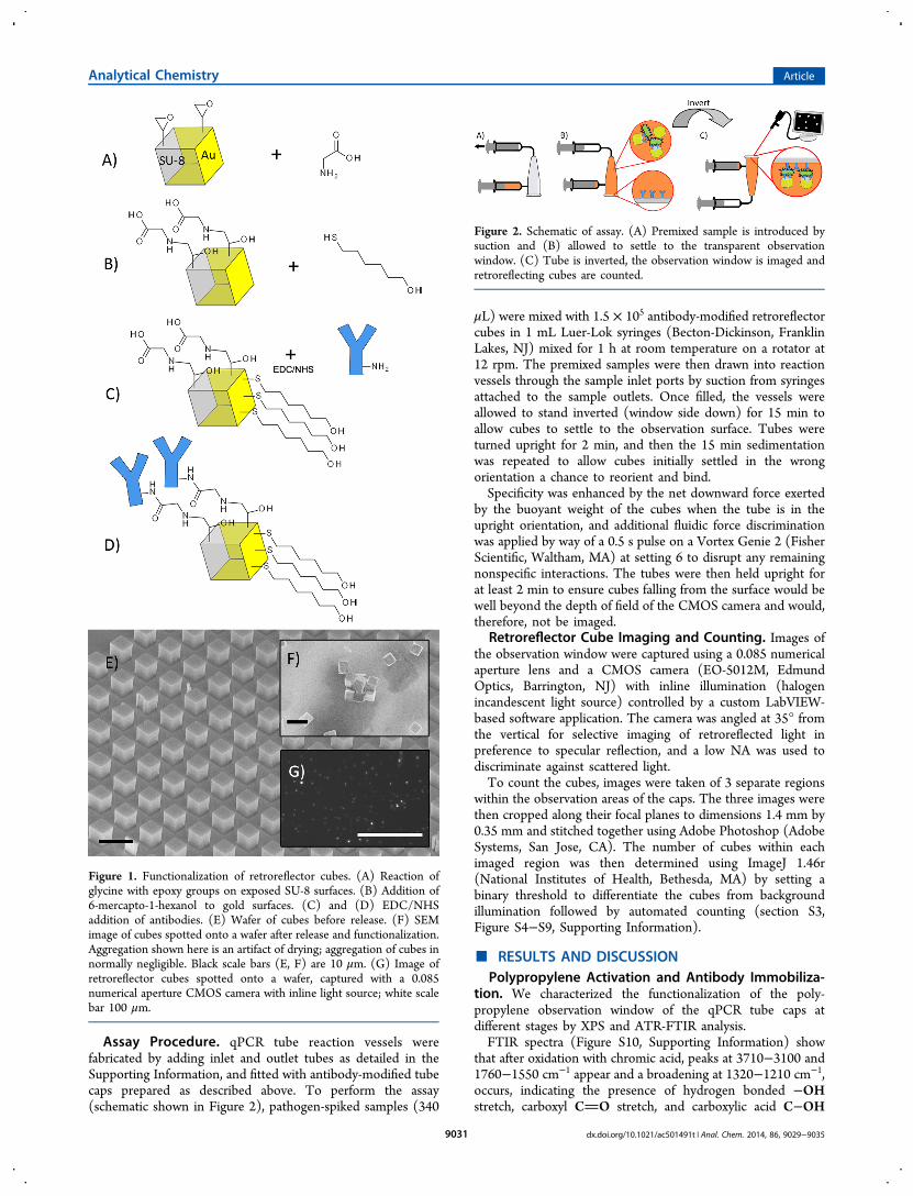
Assay Procedure. qPCR tube reaction vessels werefabricated by adding inlet and outlet tubes as detailed in theSupporting Information, and fitted with antibody-modified tubecaps prepared as described above. To perform the assay(schematic shown in Figure 2), pathogen-spiked samples (340
μL) were mixed with 1.5 × 10⁵ antibody-modified retroreflectorcubes in 1 mL Luer-Lok syringes (Becton-Dickinson, FranklinLakes, NJ) mixed for 1 h at room temperature on a rotator at12 rpm. The premixed samples were then drawn into reactionvessels through the sample inlet ports by suction from syringesattached to the sample outlets. Once filled, the vessels wereallowed to stand inverted (window side down) for 15 min toallow cubes to settle to the observation surface. Tubes wereturned upright for 2 min, and then the 15 min sedimentationwas repeated to allow cubes initially settled in the wrongorientation a chance to reorient and bind.Specificity was enhanced by the net downward force exerted
by the buoyant weight of the cubes when the tube is in theupright orientation, and additional fluidic force discriminationwas applied by way of a 0.5 s pulse on a Vortex Genie 2 (FisherScientific, Waltham, MA) at setting 6 to disrupt any remainingnonspecific interactions. The tubes were then held upright forat least 2 min to ensure cubes falling from the surface would bewell beyond the depth of field of the CMOS camera and would,therefore, not be imaged.
Retroreflector Cube Imaging and Counting. Images ofthe observation window were captured using a 0.085 numericalaperture lens and a CMOS camera (EO-5012M, EdmundOptics, Barrington, NJ) with inline illumination (halogenincandescent light source) controlled by a custom LabVIEW-based software application. The camera was angled at 35° fromthe vertical for selective imaging of retroreflected light inpreference to specular reflection, and a low NA was used todiscriminate against scattered light.To count the cubes, images were taken of 3 separate regions
within the observation areas of the caps. The three images werethen cropped along their focal planes to dimensions 1.4 mm by0.35 mm and stitched together using Adobe Photoshop (AdobeSystems, San Jose, CA). The number of cubes within eachimaged region was then determined using ImageJ 1.46r(National Institutes of Health, Bethesda, MA) by setting abinary threshold to differentiate the cubes from backgroundillumination followed by automated counting (section S3,Figure S4−S9, Supporting Information).
■ RESULTS AND DISCUSSIONPolypropylene Activation and Antibody Immobiliza-
tion. We characterized the functionalization of the poly-propylene observation window of the qPCR tube caps atdifferent stages by XPS and ATR-FTIR analysis.FTIR spectra (Figure S10, Supporting Information) show
that after oxidation with chromic acid, peaks at 3710−3100 and1760−1550 cm−1 appear and a broadening at 1320−1210 cm−1,occurs, indicating the presence of hydrogen bonded −OHstretch, carboxyl CO stretch, and carboxylic acid C−OH
Figure 1. Functionalization of retroreflector cubes. (A) Reaction ofglycine with epoxy groups on exposed SU-8 surfaces. (B) Addition of6-mercapto-1-hexanol to gold surfaces. (C) and (D) EDC/NHSaddition of antibodies. (E) Wafer of cubes before release. (F) SEMimage of cubes spotted onto a wafer after release and functionalization.Aggregation shown here is an artifact of drying; aggregation of cubes innormally negligible. Black scale bars (E, F) are 10 μm. (G) Image ofretroreflector cubes spotted onto a wafer, captured with a 0.085numerical aperture CMOS camera with inline light source; white scalebar 100 μm.
Figure 2. Schematic of assay. (A) Premixed sample is introduced bysuction and (B) allowed to settle to the transparent observationwindow. (C) Tube is inverted, the observation window is imaged andretroreflecting cubes are counted.
Analytical Chemistry Article
dx.doi.org/10.1021/ac501491t | Anal. Chem. 2014, 86, 9029−90359031
stretch, respectively. After conjugation to antibodies, the broad−OH stretch peak at 3600−3000 cm−1 remains, and a shiftedpeak at 1670 cm−1 indicates the presence of amide CO.34,37−39 These chemical functionalities were not seen on theuntreated material.XPS survey scans (Figure S11, Supporting Information)
show that atomic oxygen on the surface of the polypropylenematerial increased from 0.67% to 14.9% after reaction with thechromic acid solution. High resolution C1s scans of theoxidized sample were fitted with four Gaussian peaksrepresenting the bonds C−C/C−H at 284.6 eV, C−CO2 at286.0 eV, C−O at 287.2 eV, and CO2 at 288.6 eV indicatingthe presence of carboxyl and other oxygenated carbon moietieson the surface after treatment.34,39,40
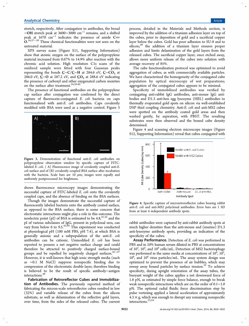
The presence of functional antibodies on the polypropylenecap surface after conjugation was confirmed by the directcapture of fluorescein-conjugated E. coli bacteria on capsfunctionalized with anti-E. coli antibodies. Caps covalentlymodified with BSA were used as a negative control. Figure 3
shows fluorescence microscopy images demonstrating thesuccessful capture of FITC-labeled E. coli onto the covalentlycoupled caps, and the absence of binding on the BSA surfaces.Though the images demonstrate the successful capture of
fluorescently labeled bacteria onto the antibody coated surface,as opposed to the BSA surface, there is some concern thatelectrostatic interactions might play a role in this outcome. Theisoelectric point (pI) of BSA is estimated to be 4.8,41,42 and thepI of various subclasses of IgG, present in polyclonal sera, canvary from below 6 to 9.5.43,44 This experiment was conductedat physiological pH (100 mM PBS, pH 7.4), at which BSA isgenerally anionic and a subpopulation of the anti-E. coliantibodies can be cationic. Unmodified E. coli has beenreported to possess a net negative surface charge and couldtherefore be attracted to positively charged surface-boundgroups and be repelled by negatively charged surfaces.45−47
However, it is well-known that high ionic strength media (suchas ∼0.1 M NaCl) suppress nonspecific binding due tocompression of the electrostatic double layer, therefore bindingis believed to be the result of specific antibody−antigeninteractions.48
Fabrication of Retroreflector Cubes and Immobiliza-tion of Antibodies. The previously reported method offabricating the micron-scale retroreflector cubes resulted in low(32%) and variable release of the cubes from the siliconsubstrate, as well as delamination of the reflective gold layers,over time, from the sides of the released cubes. The current
process, detailed in the Materials and Methods section, isimproved by the addition of a titanium adhesion layer on top ofthe cubes, prior to deposition of gold and a sacrificial copperlayer below the cubes. Gold has poor adhesion to SU-8 and tosilicon;49 the addition of a titanium layer ensures properadhesion and limits delamination of the gold layers from thereleased cubes. The sacrificial copper layer, once etched away,allows more uniform release of the cubes into solution withaverage recovery of 85%.The cube functionalization protocol was optimized to avoid
aggregation of cubes; as with commercially available particles.We have characterized the homogeneity of the conjugated cubepopulation by optical microscopy of wet preparations;aggregation of the conjugated cubes appears to be minimal.Specificity of immobilized antibodies was verified by
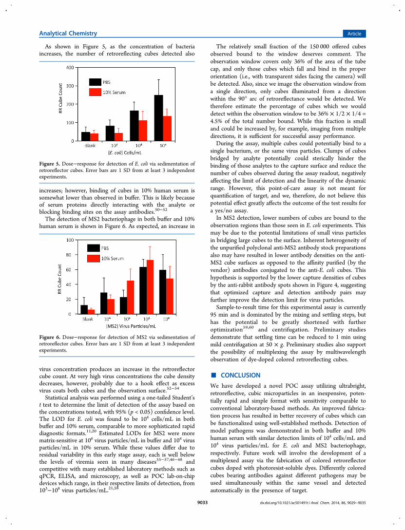
conjugating anti-rabbit IgG antibodies, anti-mouse IgG anti-bodies and D1.3 anti-hen egg lysozyme (HEL) antibodies tothermally evaporated gold spots on silicon via well-establishedDSP thiol coupling chemistry. Anti-E. coli and anti-MS2 cubeswere spotted on the antibody coated gold areas and thenwashed gently, by aspiration, with PBST. The resultingsubstrates were then observed and the bound cube densitydetermined.Figure 4 and scanning electron microscope images (Figure
S12, Supporting Information) reveal that cubes conjugated with
rabbit antibodies were captured by anti-rabbit antibody spots atmuch higher densities than the anti-mouse and (murine) D1.3anti-lysozyme antibody spots, providing an indication of thespecificity of the cubes.
Assay Performance. Detection of E. coli was performed inPBS and in 10% human serum diluted in PBS at concentrationsof 103, 104, and 106 cells/mL. Detection of MS2 bacteriophagewas performed in the same media at concentrations of 104, 105,106, and 108 virus particles/mL. The assay system design wasoptimized to prevent the presence of air bubbles, which maysweep away bound particles by surface tension.32 To achievespecificity, during upright orientation of the assay tubes, thebuoyant weight of the cubes applies a net downward force of1.8 pN, as estimated by simple force balance, enough to breakweak nonspecific interactions which are on the order of 0.1−1.0pN. The optional radial fluidic force discrimination step bypulse vortexing applied a lateral acceleration of approximately4.3 × g, which was enough to disrupt any remaining nonspecificinteractions.23,24
Figure 3. Demonstration of functional anti-E. coli antibodies onpolypropylene observation window by specific capture of FITC-labeled E. coli. ( A) Fluorescence image of covalently coupled anti-E.coli surface and of (B) covalently coupled BSA surface after incubationwith the bacteria. Scale bars are 10 μm; images were equally anduniformly postprocessed for brightness.
Figure 4. Specific capture of microretroreflector cubes bearing rabbitanti-E. coli and anti-MS2 polyclonal antibodies. Error bars are 1 SDfrom at least 4 independent antibody spots.
Analytical Chemistry Article
dx.doi.org/10.1021/ac501491t | Anal. Chem. 2014, 86, 9029−90359032
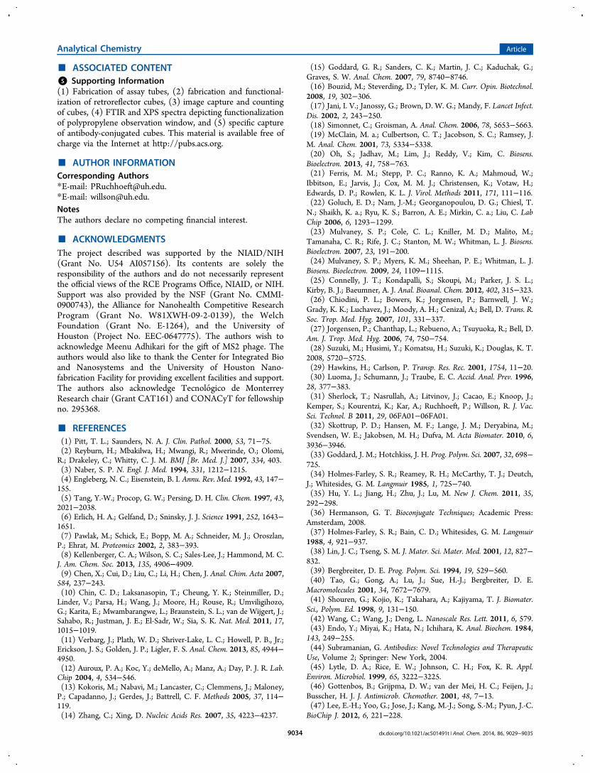
As shown in Figure 5, as the concentration of bacteriaincreases, the number of retroreflecting cubes detected also
increases; however, binding of cubes in 10% human serum issomewhat lower than observed in buffer. This is likely becauseof serum proteins directly interacting with the analyte orblocking binding sites on the assay antibodies.50−52
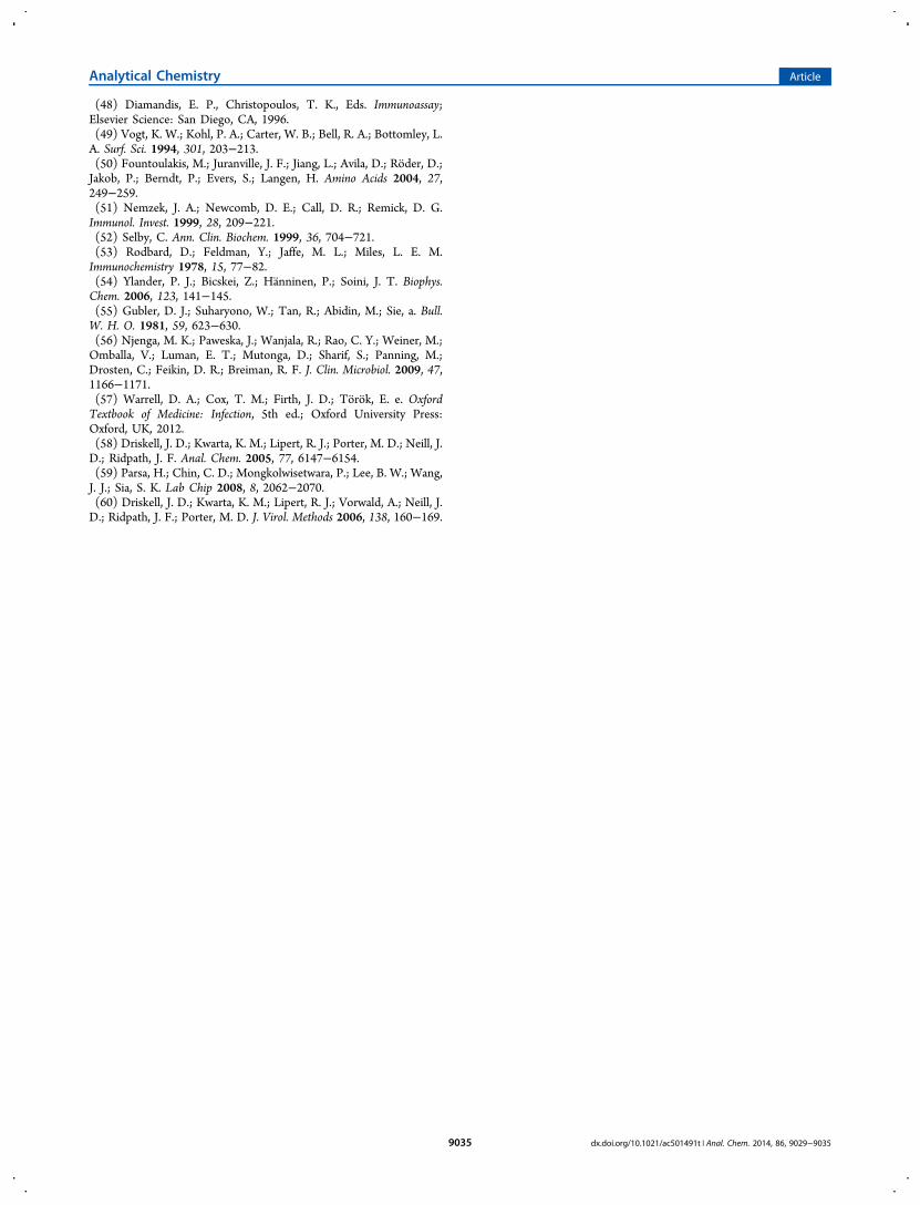
The detection of MS2 bacteriophage in both buffer and 10%human serum is shown in Figure 6. As expected, an increase in
virus concentration produces an increase in the retroreflectorcube count. At very high virus concentrations the cube densitydecreases, however, probably due to a hook effect as excessvirus coats both cubes and the observation surface.52−54
Statistical analysis was performed using a one-tailed Student’st test to determine the limit of detection of the assay based onthe concentrations tested, with 95% (p < 0.05) confidence level.The LOD for E. coli was found to be 104 cells/mL in bothbuffer and 10% serum, comparable to more sophisticated rapiddiagnostic formats.11,20 Estimated LODs for MS2 were morematrix-sensitive at 106 virus particles/mL in buffer and 104 virusparticles/mL in 10% serum. While these values differ due toresidual variability in this early stage assay, each is well belowthe levels of viremia seen in many diseases55−57,46−48 andcompetitive with many established laboratory methods such asqPCR, ELISA, and microscopy, as well as POC lab-on-chipdevices which range, in their respective limits of detection, from103−108 virus particles/mL.21,58
The relatively small fraction of the 150 000 offered cubesobserved bound to the window deserves comment. Theobservation window covers only 36% of the area of the tubecap, and only those cubes which fall and bind in the properorientation (i.e., with transparent sides facing the camera) willbe detected. Also, since we image the observation window froma single direction, only cubes illuminated from a directionwithin the 90° arc of retroreflectance would be detected. Wetherefore estimate the percentage of cubes which we woulddetect within the observation window to be 36% × 1/2 × 1/4 =4.5% of the total number bound. While this fraction is smalland could be increased by, for example, imaging from multipledirections, it is sufficient for successful assay performance.During the assay, multiple cubes could potentially bind to a
single bacterium, or the same virus particles. Clumps of cubesbridged by analyte potentially could sterically hinder thebinding of those analytes to the capture surface and reduce thenumber of cubes observed during the assay readout, negativelyaffecting the limit of detection and the linearity of the dynamicrange. However, this point-of-care assay is not meant forquantification of target, and we, therefore, do not believe thispotential effect greatly affects the outcome of the test results fora yes/no assay.In MS2 detection, lower numbers of cubes are bound to the
observation regions than those seen in E. coli experiments. Thismay be due to the potential limitations of small virus particlesin bridging large cubes to the surface. Inherent heterogeneity ofthe unpurified polyclonal anti-MS2 antibody stock preparationsalso may have resulted in lower antibody densities on the anti-MS2 cube surfaces as opposed to the affinity purified (by thevendor) antibodies conjugated to the anti-E. coli cubes. Thishypothesis is supported by the lower capture densities of cubesby the anti-rabbit antibody spots shown in Figure 4, suggestingthat optimized capture and detection antibody pairs mayfurther improve the detection limit for virus particles.Sample-to-result time for this experimental assay is currently
95 min and is dominated by the mixing and settling steps, buthas the potential to be greatly shortened with furtheroptimization59,60 and centrifugation. Preliminary studiesdemonstrate that settling time can be reduced to 1 min usingmild centrifugation at 50 × g. Preliminary studies also supportthe possibility of multiplexing the assay by multiwavelengthobservation of dye-doped colored retroreflecting cubes.
■ CONCLUSION
We have developed a novel POC assay utilizing ultrabright,retroreflective, cubic microparticles in an inexpensive, poten-tially rapid and simple format with sensitivity comparable toconventional laboratory-based methods. An improved fabrica-tion process has resulted in better recovery of cubes which canbe functionalized using well-established methods. Detection ofmodel pathogens was demonstrated in both buffer and 10%human serum with similar detection limits of 104 cells/mL and104 virus particles/mL for E. coli and MS2 bacteriophage,respectively. Future work will involve the development of amultiplexed assay via the fabrication of colored retroreflectorcubes doped with photoresist-soluble dyes. Differently coloredcubes bearing antibodies against different pathogens may beused simultaneously within the same vessel and detectedautomatically in the presence of target.
Figure 5. Dose−response for detection of E. coli via sedimentation ofretroreflector cubes. Error bars are 1 SD from at least 3 independentexperiments.
Figure 6. Dose−response for detection of MS2 via sedimentation ofretroreflector cubes. Error bars are 1 SD from at least 3 independentexperiments.
Analytical Chemistry Article
dx.doi.org/10.1021/ac501491t | Anal. Chem. 2014, 86, 9029−90359033
■ ASSOCIATED CONTENT*S Supporting Information(1) Fabrication of assay tubes, (2) fabrication and functional-ization of retroreflector cubes, (3) image capture and countingof cubes, (4) FTIR and XPS spectra depicting functionalizationof polypropylene observation window, and (5) specific captureof antibody-conjugated cubes. This material is available free ofcharge via the Internet at http://pubs.acs.org.
■ AUTHOR INFORMATIONCorresponding Authors*E-mail: [email protected].*E-mail: [email protected] authors declare no competing financial interest.
■ ACKNOWLEDGMENTSThe project described was supported by the NIAID/NIH(Grant No. U54 AI057156). Its contents are solely theresponsibility of the authors and do not necessarily representthe official views of the RCE Programs Office, NIAID, or NIH.Support was also provided by the NSF (Grant No. CMMI-0900743), the Alliance for Nanohealth Competitive ResearchProgram (Grant No. W81XWH-09-2-0139), the WelchFoundation (Grant No. E-1264), and the University ofHouston (Project No. EEC-0647775). The authors wish toacknowledge Meenu Adhikari for the gift of MS2 phage. Theauthors would also like to thank the Center for Integrated Bioand Nanosystems and the University of Houston Nano-fabrication Facility for providing excellent facilities and support.The authors also acknowledge Tecnologico de MonterreyResearch chair (Grant CAT161) and CONACyT for fellowshipno. 295368.
■ REFERENCES(1) Pitt, T. L.; Saunders, N. A. J. Clin. Pathol. 2000, 53, 71−75.(2) Reyburn, H.; Mbakilwa, H.; Mwangi, R.; Mwerinde, O.; Olomi,R.; Drakeley, C.; Whitty, C. J. M. BMJ [Br. Med. J.] 2007, 334, 403.(3) Naber, S. P. N. Engl. J. Med. 1994, 331, 1212−1215.(4) Engleberg, N. C.; Eisenstein, B. I. Annu. Rev. Med. 1992, 43, 147−155.(5) Tang, Y.-W.; Procop, G. W.; Persing, D. H. Clin. Chem. 1997, 43,2021−2038.(6) Erlich, H. A.; Gelfand, D.; Sninsky, J. J. Science 1991, 252, 1643−1651.(7) Pawlak, M.; Schick, E.; Bopp, M. A.; Schneider, M. J.; Oroszlan,P.; Ehrat, M. Proteomics 2002, 2, 383−393.(8) Kellenberger, C. A.; Wilson, S. C.; Sales-Lee, J.; Hammond, M. C.J. Am. Chem. Soc. 2013, 135, 4906−4909.(9) Chen, X.; Cui, D.; Liu, C.; Li, H.; Chen, J. Anal. Chim. Acta 2007,584, 237−243.(10) Chin, C. D.; Laksanasopin, T.; Cheung, Y. K.; Steinmiller, D.;Linder, V.; Parsa, H.; Wang, J.; Moore, H.; Rouse, R.; Umviligihozo,G.; Karita, E.; Mwambarangwe, L.; Braunstein, S. L.; van de Wijgert, J.;Sahabo, R.; Justman, J. E.; El-Sadr, W.; Sia, S. K. Nat. Med. 2011, 17,1015−1019.(11) Verbarg, J.; Plath, W. D.; Shriver-Lake, L. C.; Howell, P. B., Jr.;Erickson, J. S.; Golden, J. P.; Ligler, F. S. Anal. Chem. 2013, 85, 4944−4950.(12) Auroux, P. A.; Koc, Y.; deMello, A.; Manz, A.; Day, P. J. R. Lab.Chip 2004, 4, 534−546.(13) Kokoris, M.; Nabavi, M.; Lancaster, C.; Clemmens, J.; Maloney,P.; Capadanno, J.; Gerdes, J.; Battrell, C. F. Methods 2005, 37, 114−119.(14) Zhang, C.; Xing, D. Nucleic Acids Res. 2007, 35, 4223−4237.
(15) Goddard, G. R.; Sanders, C. K.; Martin, J. C.; Kaduchak, G.;Graves, S. W. Anal. Chem. 2007, 79, 8740−8746.(16) Bouzid, M.; Steverding, D.; Tyler, K. M. Curr. Opin. Biotechnol.2008, 19, 302−306.(17) Jani, I. V.; Janossy, G.; Brown, D. W. G.; Mandy, F. Lancet Infect.Dis. 2002, 2, 243−250.(18) Simonnet, C.; Groisman, A. Anal. Chem. 2006, 78, 5653−5663.(19) McClain, M. a.; Culbertson, C. T.; Jacobson, S. C.; Ramsey, J.M. Anal. Chem. 2001, 73, 5334−5338.(20) Oh, S.; Jadhav, M.; Lim, J.; Reddy, V.; Kim, C. Biosens.Bioelectron. 2013, 41, 758−763.(21) Ferris, M. M.; Stepp, P. C.; Ranno, K. A.; Mahmoud, W.;Ibbitson, E.; Jarvis, J.; Cox, M. M. J.; Christensen, K.; Votaw, H.;Edwards, D. P.; Rowlen, K. L. J. Virol. Methods 2011, 171, 111−116.(22) Goluch, E. D.; Nam, J.-M.; Georganopoulou, D. G.; Chiesl, T.N.; Shaikh, K. a.; Ryu, K. S.; Barron, A. E.; Mirkin, C. a.; Liu, C. LabChip 2006, 6, 1293−1299.(23) Mulvaney, S. P.; Cole, C. L.; Kniller, M. D.; Malito, M.;Tamanaha, C. R.; Rife, J. C.; Stanton, M. W.; Whitman, L. J. Biosens.Bioelectron. 2007, 23, 191−200.(24) Mulvaney, S. P.; Myers, K. M.; Sheehan, P. E.; Whitman, L. J.Biosens. Bioelectron. 2009, 24, 1109−1115.(25) Connelly, J. T.; Kondapalli, S.; Skoupi, M.; Parker, J. S. L.;Kirby, B. J.; Baeumner, A. J. Anal. Bioanal. Chem. 2012, 402, 315−323.(26) Chiodini, P. L.; Bowers, K.; Jorgensen, P.; Barnwell, J. W.;Grady, K. K.; Luchavez, J.; Moody, A. H.; Cenizal, A.; Bell, D. Trans. R.Soc. Trop. Med. Hyg. 2007, 101, 331−337.(27) Jorgensen, P.; Chanthap, L.; Rebueno, A.; Tsuyuoka, R.; Bell, D.Am. J. Trop. Med. Hyg. 2006, 74, 750−754.(28) Suzuki, M.; Husimi, Y.; Komatsu, H.; Suzuki, K.; Douglas, K. T.2008, 5720−5725.(29) Hawkins, H.; Carlson, P. Transp. Res. Rec. 2001, 1754, 11−20.(30) Luoma, J.; Schumann, J.; Traube, E. C. Accid. Anal. Prev. 1996,28, 377−383.(31) Sherlock, T.; Nasrullah, A.; Litvinov, J.; Cacao, E.; Knoop, J.;Kemper, S.; Kourentzi, K.; Kar, A.; Ruchhoeft, P.; Willson, R. J. Vac.Sci. Technol. B 2011, 29, 06FA01−06FA01.(32) Skottrup, P. D.; Hansen, M. F.; Lange, J. M.; Deryabina, M.;Svendsen, W. E.; Jakobsen, M. H.; Dufva, M. Acta Biomater. 2010, 6,3936−3946.(33) Goddard, J. M.; Hotchkiss, J. H. Prog. Polym. Sci. 2007, 32, 698−725.(34) Holmes-Farley, S. R.; Reamey, R. H.; McCarthy, T. J.; Deutch,J.; Whitesides, G. M. Langmuir 1985, 1, 725−740.(35) Hu, Y. L.; Jiang, H.; Zhu, J.; Lu, M. New J. Chem. 2011, 35,292−298.(36) Hermanson, G. T. Bioconjugate Techniques; Academic Press:Amsterdam, 2008.(37) Holmes-Farley, S. R.; Bain, C. D.; Whitesides, G. M. Langmuir1988, 4, 921−937.(38) Lin, J. C.; Tseng, S. M. J. Mater. Sci. Mater. Med. 2001, 12, 827−832.(39) Bergbreiter, D. E. Prog. Polym. Sci. 1994, 19, 529−560.(40) Tao, G.; Gong, A.; Lu, J.; Sue, H.-J.; Bergbreiter, D. E.Macromolecules 2001, 34, 7672−7679.(41) Shouren, G.; Kojio, K.; Takahara, A.; Kajiyama, T. J. Biomater.Sci., Polym. Ed. 1998, 9, 131−150.(42) Wang, C.; Wang, J.; Deng, L. Nanoscale Res. Lett. 2011, 6, 579.(43) Endo, Y.; Miyai, K.; Hata, N.; Ichihara, K. Anal. Biochem. 1984,143, 249−255.(44) Subramanian, G. Antibodies: Novel Technologies and TherapeuticUse, Volume 2; Springer: New York, 2004.(45) Lytle, D. A.; Rice, E. W.; Johnson, C. H.; Fox, K. R. Appl.Environ. Microbiol. 1999, 65, 3222−3225.(46) Gottenbos, B.; Grijpma, D. W.; van der Mei, H. C.; Feijen, J.;Busscher, H. J. J. Antimicrob. Chemother. 2001, 48, 7−13.(47) Lee, E.-H.; Yoo, G.; Jose, J.; Kang, M.-J.; Song, S.-M.; Pyun, J.-C.BioChip J. 2012, 6, 221−228.
Analytical Chemistry Article
dx.doi.org/10.1021/ac501491t | Anal. Chem. 2014, 86, 9029−90359034
(48) Diamandis, E. P., Christopoulos, T. K., Eds. Immunoassay;Elsevier Science: San Diego, CA, 1996.(49) Vogt, K. W.; Kohl, P. A.; Carter, W. B.; Bell, R. A.; Bottomley, L.A. Surf. Sci. 1994, 301, 203−213.(50) Fountoulakis, M.; Juranville, J. F.; Jiang, L.; Avila, D.; Roder, D.;Jakob, P.; Berndt, P.; Evers, S.; Langen, H. Amino Acids 2004, 27,249−259.(51) Nemzek, J. A.; Newcomb, D. E.; Call, D. R.; Remick, D. G.Immunol. Invest. 1999, 28, 209−221.(52) Selby, C. Ann. Clin. Biochem. 1999, 36, 704−721.(53) Rodbard, D.; Feldman, Y.; Jaffe, M. L.; Miles, L. E. M.Immunochemistry 1978, 15, 77−82.(54) Ylander, P. J.; Bicskei, Z.; Hanninen, P.; Soini, J. T. Biophys.Chem. 2006, 123, 141−145.(55) Gubler, D. J.; Suharyono, W.; Tan, R.; Abidin, M.; Sie, a. Bull.W. H. O. 1981, 59, 623−630.(56) Njenga, M. K.; Paweska, J.; Wanjala, R.; Rao, C. Y.; Weiner, M.;Omballa, V.; Luman, E. T.; Mutonga, D.; Sharif, S.; Panning, M.;Drosten, C.; Feikin, D. R.; Breiman, R. F. J. Clin. Microbiol. 2009, 47,1166−1171.(57) Warrell, D. A.; Cox, T. M.; Firth, J. D.; Torok, E. e. OxfordTextbook of Medicine: Infection, 5th ed.; Oxford University Press:Oxford, UK, 2012.(58) Driskell, J. D.; Kwarta, K. M.; Lipert, R. J.; Porter, M. D.; Neill, J.D.; Ridpath, J. F. Anal. Chem. 2005, 77, 6147−6154.(59) Parsa, H.; Chin, C. D.; Mongkolwisetwara, P.; Lee, B. W.; Wang,J. J.; Sia, S. K. Lab Chip 2008, 8, 2062−2070.(60) Driskell, J. D.; Kwarta, K. M.; Lipert, R. J.; Vorwald, A.; Neill, J.D.; Ridpath, J. F.; Porter, M. D. J. Virol. Methods 2006, 138, 160−169.
Analytical Chemistry Article
dx.doi.org/10.1021/ac501491t | Anal. Chem. 2014, 86, 9029−90359035