Host–Pathogen Interaction in Leishmaniasis: Immune ... - MDPI
37
Citation: Yasmin, H.; Adhikary, A.; Al-Ahdal, M.N.; Roy, S.; Kishore, U. Host–Pathogen Interaction in Leishmaniasis: Immune Response and Vaccination Strategies. Immuno 2022, 2, 218–254. https://doi.org/ 10.3390/immuno2010015 Academic Editors: Alan G. Goodman and Juan Bautista De Sanctis Received: 30 November 2021 Accepted: 22 February 2022 Published: 9 March 2022 Publisher’s Note: MDPI stays neutral with regard to jurisdictional claims in published maps and institutional affil- iations. Copyright: © 2022 by the authors. Licensee MDPI, Basel, Switzerland. This article is an open access article distributed under the terms and conditions of the Creative Commons Attribution (CC BY) license (https:// creativecommons.org/licenses/by/ 4.0/). Review Host–Pathogen Interaction in Leishmaniasis: Immune Response and Vaccination Strategies Hadida Yasmin 1 , Anureeta Adhikary 1 , Mohammed N. Al-Ahdal 2 , Syamal Roy 3 and Uday Kishore 4, * 1 Immunology and Cell Biology Laboratory, Department of Zoology, Cooch Behar Panchanan Barma University, Cooch Behar 736101, West Bengal, India; [email protected] (H.Y.); [email protected] (A.A.) 2 Department of Infection and Immunity, King Faisal Specialist Hospital and Research Centre, Riyadh 11211, Saudi Arabia; [email protected] 3 Council for Scientific and Industrial Research-Indian Institute of Chemical Biology, Kolkata 700032, West Bengal, India; [email protected] 4 Biosciences, College of Health, Medicine and Life Sciences, Brunel University London, Uxbridge UB8 3PH, UK * Correspondence: [email protected] or [email protected] Abstract: Leishmaniasis is a zoonotic and vector-borne infectious disease that is caused by the genus Leishmania belonging to the trypanosomatid family. The protozoan parasite has a digenetic life cycle involving a mammalian host and an insect vector. Leishmaniasisis is a worldwide public health problem falling under the neglected tropical disease category, with over 90 endemic countries, and approximately 1 million new cases and 20,000 deaths annually. Leishmania infection can progress toward the development of species–specific pathologic disorders, ranging in severity from self-healing cutaneous lesions to disseminating muco-cutaneous and fatal visceral manifestations. The severity and the outcome of leishmaniasis is determined by the parasite’s antigenic epitope characteristics, the vector physiology, and most importantly, the immune response and immune status of the host. This review examines the nature of host–pathogen interaction in leishmaniasis, innate and adaptive immune responses, and various strategies that have been employed for vaccine development. Keywords: Leishmania; sand fly; macrophage; innate immunity; Th1/Th2; vaccine 1. Introduction Leishmaniasis is a protozoan parasitic disease caused by over 20 Leishmania species; it is among the major neglected vector-borne tropical diseases with outbreak and consid- erable mortality. Leishmania is a dimorphic obligate intracellular protozoan parasite with two distinct and alternating developmental stages: the promastigote form, which is the flagellated and motile stage residing in the midgut of Sandfly vectors, and the amastigote, non-motile form that resides within the mononuclear phagocytes in the mammalian host. All species of Leishmania are transmitted by the female Phlebotomine Sandflies, which infect a range of mammals including humans, rodents and canids [1]. There are over 90 sandfly species that are known to transmit Leishmania parasites. Leishmaniasis has three main forms: visceral, cutaneous and mucocutaneous. Nearly 95% cases of visceral leishmaniasis (VL), also known as kala-azar, if left untreated, become fatal. Visceral, cutaneous (CL) or mucocutaneous leishmaniasis (MCL) are endemic in Al- geria and countries in East Africa, where outbreaks of VL occur frequently [2]. In addition, Post-kala-azar dermal leishmaniasis (PKDL) also exists, mainly in East Africa and the Indian subcontinent. PKDL is a dermal manifestation of VL, developing in 5–10% of kala-azar pa- tients after 2–3 years of treatment who are also considered a potential source of Leishmania in- fection. As estimated by the WHO, between 50,000 and 90,000 new cases of VL and between 600,000 and 1 million new cases of CL occur worldwide annually, where different species of Leishmania cause varied clinical manifestations, from self-healing cutaneous lesions to Immuno 2022, 2, 218–254. https://doi.org/10.3390/immuno2010015 https://www.mdpi.com/journal/immuno
-
Upload
khangminh22 -
Category
Documents
-
view
1 -
download
0
Transcript of Host–Pathogen Interaction in Leishmaniasis: Immune ... - MDPI

�����������������
Citation: Yasmin, H.; Adhikary, A.;
Al-Ahdal, M.N.; Roy, S.; Kishore, U.
Host–Pathogen Interaction in
Leishmaniasis: Immune Response
and Vaccination Strategies. Immuno
2022, 2, 218–254. https://doi.org/
10.3390/immuno2010015
Academic Editors: Alan G. Goodman
and Juan Bautista De Sanctis
Received: 30 November 2021
Accepted: 22 February 2022
Published: 9 March 2022
Publisher’s Note: MDPI stays neutral
with regard to jurisdictional claims in
published maps and institutional affil-
iations.
Copyright: © 2022 by the authors.
Licensee MDPI, Basel, Switzerland.
This article is an open access article
distributed under the terms and
conditions of the Creative Commons
Attribution (CC BY) license (https://
creativecommons.org/licenses/by/
4.0/).
Review
Host–Pathogen Interaction in Leishmaniasis: ImmuneResponse and Vaccination StrategiesHadida Yasmin 1, Anureeta Adhikary 1, Mohammed N. Al-Ahdal 2 , Syamal Roy 3 and Uday Kishore 4,*
1 Immunology and Cell Biology Laboratory, Department of Zoology, Cooch Behar Panchanan Barma University,Cooch Behar 736101, West Bengal, India; [email protected] (H.Y.); [email protected] (A.A.)
2 Department of Infection and Immunity, King Faisal Specialist Hospital and Research Centre, Riyadh 11211,Saudi Arabia; [email protected]
3 Council for Scientific and Industrial Research-Indian Institute of Chemical Biology, Kolkata 700032,West Bengal, India; [email protected]
4 Biosciences, College of Health, Medicine and Life Sciences, Brunel University London,Uxbridge UB8 3PH, UK
* Correspondence: [email protected] or [email protected]
Abstract: Leishmaniasis is a zoonotic and vector-borne infectious disease that is caused by the genusLeishmania belonging to the trypanosomatid family. The protozoan parasite has a digenetic life cycleinvolving a mammalian host and an insect vector. Leishmaniasisis is a worldwide public healthproblem falling under the neglected tropical disease category, with over 90 endemic countries, andapproximately 1 million new cases and 20,000 deaths annually. Leishmania infection can progresstoward the development of species–specific pathologic disorders, ranging in severity from self-healingcutaneous lesions to disseminating muco-cutaneous and fatal visceral manifestations. The severityand the outcome of leishmaniasis is determined by the parasite’s antigenic epitope characteristics,the vector physiology, and most importantly, the immune response and immune status of the host.This review examines the nature of host–pathogen interaction in leishmaniasis, innate and adaptiveimmune responses, and various strategies that have been employed for vaccine development.
Keywords: Leishmania; sand fly; macrophage; innate immunity; Th1/Th2; vaccine
1. Introduction
Leishmaniasis is a protozoan parasitic disease caused by over 20 Leishmania species;it is among the major neglected vector-borne tropical diseases with outbreak and consid-erable mortality. Leishmania is a dimorphic obligate intracellular protozoan parasite withtwo distinct and alternating developmental stages: the promastigote form, which is theflagellated and motile stage residing in the midgut of Sandfly vectors, and the amastigote,non-motile form that resides within the mononuclear phagocytes in the mammalian host.All species of Leishmania are transmitted by the female Phlebotomine Sandflies, which infecta range of mammals including humans, rodents and canids [1]. There are over 90 sandflyspecies that are known to transmit Leishmania parasites.
Leishmaniasis has three main forms: visceral, cutaneous and mucocutaneous. Nearly95% cases of visceral leishmaniasis (VL), also known as kala-azar, if left untreated, becomefatal. Visceral, cutaneous (CL) or mucocutaneous leishmaniasis (MCL) are endemic in Al-geria and countries in East Africa, where outbreaks of VL occur frequently [2]. In addition,Post-kala-azar dermal leishmaniasis (PKDL) also exists, mainly in East Africa and the Indiansubcontinent. PKDL is a dermal manifestation of VL, developing in 5–10% of kala-azar pa-tients after 2–3 years of treatment who are also considered a potential source of Leishmania in-fection. As estimated by the WHO, between 50,000 and 90,000 new cases of VL and between600,000 and 1 million new cases of CL occur worldwide annually, where different speciesof Leishmania cause varied clinical manifestations, from self-healing cutaneous lesions to
Immuno 2022, 2, 218–254. https://doi.org/10.3390/immuno2010015 https://www.mdpi.com/journal/immuno

Immuno 2022, 2 219
life-threatening VL (https://www.who.int/news-room/fact-sheets/detail/leishmaniasis(accessed on 30 November 2021)). VL has a broad range of clinical manifestations, pri-marily affecting the lymphoreticular system, leading to lymphadenopathy, hepatomegalyand splenomegaly, pancytopenia, hypergammaglobulinaemia, and renal disease [3]. CLrepresents a small localised erythematous nodule developing at the site of the sandfly bite,which gradually develops into an ulcerative condition, rarely penetrating into the subcu-taneous tissue [4]. MCL is a highly disfiguring and life-threatening condition occurringin patients with a history of CL with mucosal involvement after 1–5 years of healing [5,6].Hematogenous or lymphatic dissemination of the parasite in MCL affects the mucosalsurfaces of the mouth, nose, and pharynx.
Leishmaniasis can be subclinical (inapparent), localized (skin lesions), and/or dissemi-nated (cutaneous, mucosal, or visceral). There are several factors that determine the clinicalpresentation and outcome of the disease, such as the parasite and host factors, inflammatoryresponses, parasite-escape mechanisms as well as the host immunocompetence [7,8]. Thisreview will focus on the mechanisms of leishmanial pathogenesis, host immune responsesagainst the parasite, and the current status of the vaccine development.
2. Life Cycle of Leishmania
Leishmania transmission occurs primarily via the bite of infected female sandfliesbelonging to the genus Phlebotomous (Old World) or Lutzomyia (New World). Leishmaniaspecies have their individual characteristics but they share a similar digenetic life involvinga mammalian host and an insect vector. However, in the case of VL in India, and CL causedby Leishmania tropica, instead of sandflies, human beings are incidental hosts of infection(human anthroponosis), and other mammals act as reservoir hosts. Through blood transfu-sion, sharing needles and due to transplacental spread or organ transplantation, VL canbe initiated by amastigotes [9,10]. The chances of transmitting infection by sub-clinicallyinfected humans are feeble [11]. Canids, especially dogs, act as a reservoir of the parasite inmost cases; cats, rats, hares, opossums, foxes and other wild animals may also serve as syl-vatic reservoirs [12–16]. Environmental factors such as climate change involving alterationsin temperature, deforestation, natural disasters and poor socio-economic factors, also con-tribute to the spread of this disease. Due to geographical overlap, malaria–VL co-infectionsoccur in large populations; it is common in East African countries where malaria and VLare co-endemic [17,18]. Drug resistance, organ transplantation and immunosuppression byHIV-1 contribute to the spread of Leishmaniasis [19]. VL patients co-infected with HIV-1may also be important reservoirs for sustained Leishmania transmission [20].
Leishmania parasite exists in two forms: Promastigote, a flagellated form that is foundin sandflies, and Amastigote, a non-flagellated tissue form that can survive within thephagosomes of macrophage in mammalian hosts. An infected female sandfly (Phlebotomusand Lutzomyia sp.) injects the promastigotes while biting a mammalian host which thenenter into the phagocytic cells. Sandflies are relatively weak, prefer moist and dark areasto rest and become active during evening and thus, prefer the bloodmeal at or afterdusk. Within the phagocytic cells, promastigotes transform into round non-flagellatedamastigotes. The replicative amastigotes then enter into the sandfly while feeding on aninfected host.
2.1. Sandfly-Specific Stages of the Parasite
Amastigotes transform into infective metacyclic promastigotes inside the sandflygut [21], which at a subsequent bloodmeal, are regurgitated [22] and injected back into themammalian skin to complete the life cycle (Figure 1). This is the only established route ofLeishmanial infection [23]; the amastigote forms are present in the skin and are not usuallyfound in the peripheral circulation.

Immuno 2022, 2 220Immuno 2022, 2, FOR PEER REVIEW 4
Figure 1. Digenetic life cycle of Leishmania: (A) Inside the sandfly, amastigotes undergo several non-infective stages before they finally differentiate into metacyclic promastigotes; (a) amastigotes enter into the sandfly (vector), while it bites an infected mammal (host), (b) amastigotes transform into replicative procyclic promastigotes (flagellated form) inside the fly abdominal midgut, (c) Procyclic stage transforms into an elongated nectomonad promastigote that attaches to the microvilli of the abdominal midgut through its flagella, (d) nectomonad then differentiates into a replicative form (leptomonad promastigote), and migrates towards the thoracic midgut, (e) leptomonad pro-mastigotes can differentiate into either heptomonad promastigotes, or (f) metacyclic promastigotes. The haptomonad promastigotes can attach to the stomodeal valve, (g) during the second blood meal, a new replicative stage, known as retroleptomonad promastigote, exists due to reverse meta-cyclogenesis, where they multiply rapidly and differentiate into metacyclic promastogotes enhanc-ing sandfly infectivity. (B) The metacyclic promastigote belongs to the infective stage that is trans-mitted to the mammalian host when an infected sandfly bites; (a) metacyclic promastigotes are taken up by phagocytic cells such as macrophages and neutrophils present in the skin, (b) promastigotes are internalised into phagosomes (later, it transforms into a parasitophorus vacuole), (c) pro-mastigotes change into amastigotes inside the parasitophorus vacuole and proliferate, (d) amastigotes burst out from the phagocytes, (e) amastigotes can enter into another life cycle inside the sandfly when taken with the blood meal, or can re-infect fresh phagocytes.
3. Host Immune Response There are several important factors that contribute to a successful transmissible in-
fection, such as the species of the parasite, the vector combination, PSG production, and the host immune response against the parasite. The first crucial event is the initial contact and stable interaction of highly polarized and motile promastigotes with mammalian host cells for their efficient phagocytosis. Macrophages act as the primary host cells for Leish-mania; however, monocytes, dendritic cells (DCs), and neutrophils are also infected and have important roles in the immunopathology of Leishmaniasis.
Figure 1. Digenetic life cycle of Leishmania: (A) Inside the sandfly, amastigotes undergo several non-infective stages before they finally differentiate into metacyclic promastigotes; (a) amastigotes enterinto the sandfly (vector), while it bites an infected mammal (host), (b) amastigotes transform intoreplicative procyclic promastigotes (flagellated form) inside the fly abdominal midgut, (c) Procyclicstage transforms into an elongated nectomonad promastigote that attaches to the microvilli of theabdominal midgut through its flagella, (d) nectomonad then differentiates into a replicative form(leptomonad promastigote), and migrates towards the thoracic midgut, (e) leptomonad promastig-otes can differentiate into either heptomonad promastigotes, or (f) metacyclic promastigotes. Thehaptomonad promastigotes can attach to the stomodeal valve, (g) during the second blood meal,a new replicative stage, known as retroleptomonad promastigote, exists due to reverse metacyclo-genesis, where they multiply rapidly and differentiate into metacyclic promastogotes enhancingsandfly infectivity. (B) The metacyclic promastigote belongs to the infective stage that is transmittedto the mammalian host when an infected sandfly bites; (a) metacyclic promastigotes are taken upby phagocytic cells such as macrophages and neutrophils present in the skin, (b) promastigotes areinternalised into phagosomes (later, it transforms into a parasitophorus vacuole), (c) promastigoteschange into amastigotes inside the parasitophorus vacuole and proliferate, (d) amastigotes burst outfrom the phagocytes, (e) amastigotes can enter into another life cycle inside the sandfly when takenwith the blood meal, or can re-infect fresh phagocytes.
The amastigotes, while inside the sandfly, undergoe transformation via several non-infective stages; first, it becomes a procyclic promastigote, which after few days, differ-entiates into a strong, elongated motile nectomonad promastigote; this is followed by itstransformation into shorter replicative forms, known as leptomonad promastigotes [24],allowing flies to regurgitate parasites into the skin before it takes the second blood meal.The infection is amplified in the vector’s gut [24]; sometimes, a small number of the nec-tomonad/leptomonad promastigotes also differentiate into haptomonad promastigotes [25].Finally, they develop into infective metacyclic promastigotes (metacyclogenesis). Thesepromastigotes, while inside the sandfly gut, secrete filamentous proteophosphoglycan(fPPG), which condenses into the promastigote secretory gel (PSG), helping in its trans-
Immuno 2022, 2 221
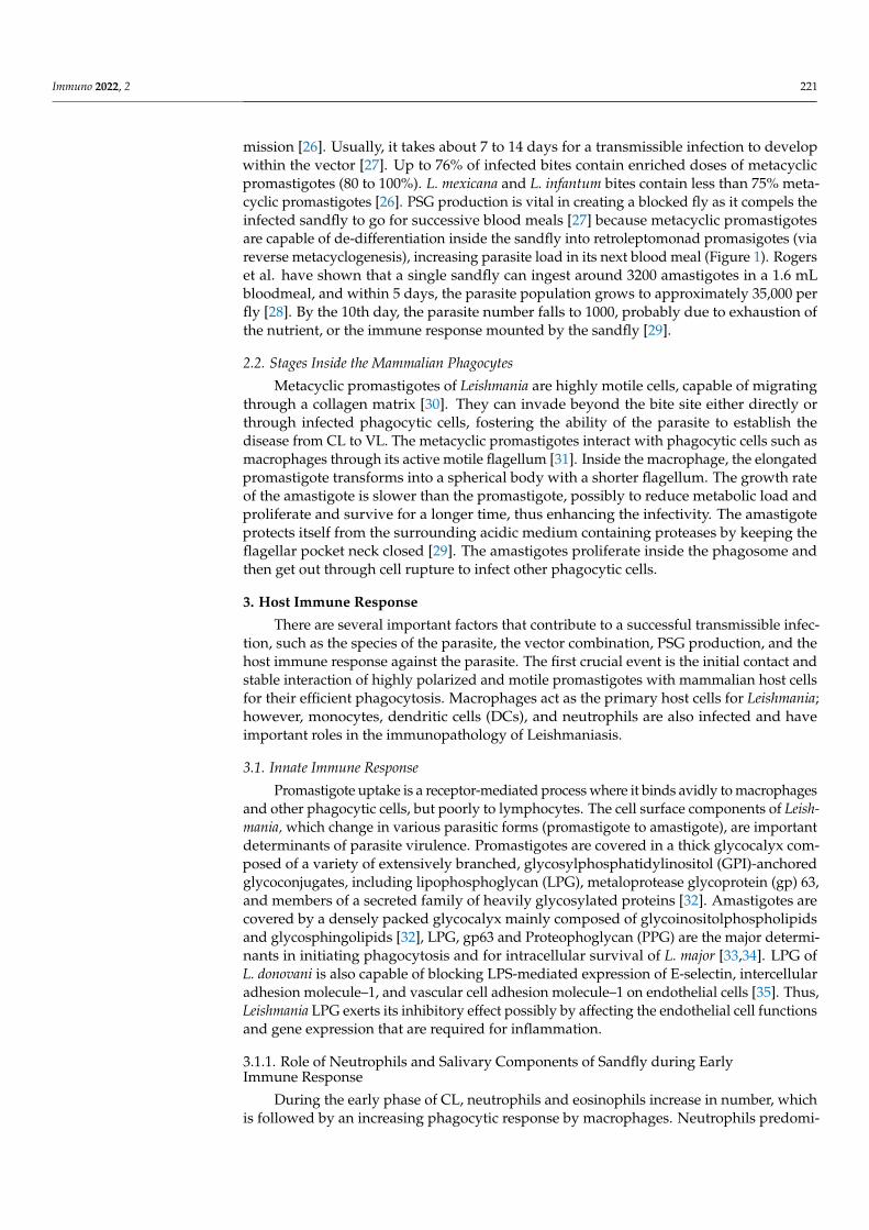
mission [26]. Usually, it takes about 7 to 14 days for a transmissible infection to developwithin the vector [27]. Up to 76% of infected bites contain enriched doses of metacyclicpromastigotes (80 to 100%). L. mexicana and L. infantum bites contain less than 75% meta-cyclic promastigotes [26]. PSG production is vital in creating a blocked fly as it compels theinfected sandfly to go for successive blood meals [27] because metacyclic promastigotesare capable of de-differentiation inside the sandfly into retroleptomonad promasigotes (viareverse metacyclogenesis), increasing parasite load in its next blood meal (Figure 1). Rogerset al. have shown that a single sandfly can ingest around 3200 amastigotes in a 1.6 mLbloodmeal, and within 5 days, the parasite population grows to approximately 35,000 perfly [28]. By the 10th day, the parasite number falls to 1000, probably due to exhaustion ofthe nutrient, or the immune response mounted by the sandfly [29].
2.2. Stages Inside the Mammalian Phagocytes
Metacyclic promastigotes of Leishmania are highly motile cells, capable of migratingthrough a collagen matrix [30]. They can invade beyond the bite site either directly orthrough infected phagocytic cells, fostering the ability of the parasite to establish thedisease from CL to VL. The metacyclic promastigotes interact with phagocytic cells such asmacrophages through its active motile flagellum [31]. Inside the macrophage, the elongatedpromastigote transforms into a spherical body with a shorter flagellum. The growth rateof the amastigote is slower than the promastigote, possibly to reduce metabolic load andproliferate and survive for a longer time, thus enhancing the infectivity. The amastigoteprotects itself from the surrounding acidic medium containing proteases by keeping theflagellar pocket neck closed [29]. The amastigotes proliferate inside the phagosome andthen get out through cell rupture to infect other phagocytic cells.
3. Host Immune Response
There are several important factors that contribute to a successful transmissible infec-tion, such as the species of the parasite, the vector combination, PSG production, and thehost immune response against the parasite. The first crucial event is the initial contact andstable interaction of highly polarized and motile promastigotes with mammalian host cellsfor their efficient phagocytosis. Macrophages act as the primary host cells for Leishmania;however, monocytes, dendritic cells (DCs), and neutrophils are also infected and haveimportant roles in the immunopathology of Leishmaniasis.
3.1. Innate Immune Response
Promastigote uptake is a receptor-mediated process where it binds avidly to macrophagesand other phagocytic cells, but poorly to lymphocytes. The cell surface components of Leish-mania, which change in various parasitic forms (promastigote to amastigote), are importantdeterminants of parasite virulence. Promastigotes are covered in a thick glycocalyx com-posed of a variety of extensively branched, glycosylphosphatidylinositol (GPI)-anchoredglycoconjugates, including lipophosphoglycan (LPG), metaloprotease glycoprotein (gp) 63,and members of a secreted family of heavily glycosylated proteins [32]. Amastigotes arecovered by a densely packed glycocalyx mainly composed of glycoinositolphospholipidsand glycosphingolipids [32], LPG, gp63 and Proteophoglycan (PPG) are the major determi-nants in initiating phagocytosis and for intracellular survival of L. major [33,34]. LPG ofL. donovani is also capable of blocking LPS-mediated expression of E-selectin, intercellularadhesion molecule–1, and vascular cell adhesion molecule–1 on endothelial cells [35]. Thus,Leishmania LPG exerts its inhibitory effect possibly by affecting the endothelial cell functionsand gene expression that are required for inflammation.
3.1.1. Role of Neutrophils and Salivary Components of Sandfly during EarlyImmune Response
During the early phase of CL, neutrophils and eosinophils increase in number, whichis followed by an increasing phagocytic response by macrophages. Neutrophils predomi-

Immuno 2022, 2 222
nate at the inoculation site within first few hours of infection, they take up promastigotes,and serve as a vehicle for subsequent infection of monocyte-derived or tissue-residentmacrophages [36]. Neutrophils and eosinophils possess leishmanicidal activity that re-strains parasite progression [37]. Neutrophils could enhance killing of L. braziliensis ininfected macrophages through upregulation of TNF-α and ROS [38]. Activated neutrophilsare capable of killing promastigotes via the neutrophil trap (NET) [39] and also throughoxygen metabolites generated during phagocytosis-induced respiratory burst [40]. In NETassociated with promastigotes, DNA, elastase, and histones are detected; histones likelyexhibit the leishmanicidal activity [41].
While injecting the promastigotes into the host skin, the sandfly also salivates anddelivers certain salivary proteins that are chemotactic and inducer of inflammatory responsefacilitating Leishmania infection [42]. The saliva also contains vector gut microbiota thatactivates inflammasome-derived IL-1β production and aids in neutrophil recruitment [43].The saliva of Lutzomyia longipalpis contains a female-specific secreted endonuclease calledLundep capable of hydrolysing DNA, thus negating the effect of neutrophil NETosis,in order to protect the promastigotes [44]. A 45 kDa neutrophil chemotactic salivaryprotein acts through a G-protein-coupled receptor, which enhances lesion pathology andincreases the parasite burden in mice upon co-infection with Leishmania parasites [45]. Co-infection with helminths such as Filaria may modulate the immune response to Phlebotomusduboscqi (Pd) saliva; repeated exposure to Pd saliva polarizes human monocyte functiontowards a tolerized phenotype while co-infection with filaria favors a Leishmania-promotingTh2/regulatory immune response [46].
Neutrophils appear to restrain parasite progression [36] by secreting cytokines andchemokines, releasing its granular contents, triggering pattern recognition signals, andby interacting directly with other inflammatory and resident cells [47–49]. Leishmaniapromastigotes release a soluble chemotactic factor (LCF), as confirmed in the supernatantsof L. major, L. aethiopica, and L. donovani parasites cultures, which aids in neutrophil recruit-ment. This induces IL-8 secretion by neutrophils, thus, amplifying neutrophil recruitment.Often, gut microbes present in the saliva of the sandfly are egested into host skin along withLeishmania parasites. The egested microbes trigger the inflammasome to produce signifi-cantly higher levels of IL-1β compared to levels induced by parasites alone. Removing gutmicrobiota, or blocking IL-1β before transmission, abolishes neutrophil recruitment andimpairs Leishmania dissemination [43]. IL-17A-producing Innate Lymphoid Cells (ILCs),which are RORγt+, are involved in the microbiota-driven immunopathology in CL. Pateintsinfected with L. braziliensis were found often with dominant Staphylococcus dysbiosis, andthis probably can influence IL-17 synthesis. The lesion size in mice infected with L. majorwas also found to be increased due to Staphylococcus colonization [50]. Thus, investigatingthe mechanistic aspects of immunomodulation due to sandfly microbiota can offer insightinto the immunopathology of early infection in Leishmaniasis.
There is also evidence to suggest the survival of Leishmania promastigotes followingtheir rapid uptake by neutrophils. Under an experimental condition, inhibiting neutrophilicinfiltration increased host resistance to infection [49]. L. major and L. donovani promastigoteswere observed to be surviving in human neutrophils which correlated with a lack ofrespiratory burst. Inhibition of IP-10 (gamma interferon-inducible protein 10) productionby neutrophils in presence of Leishmania can inhibit recruitment and activation of NK cellsand Th1 cells (Figure 2). Thus, by infecting neutrophils, promastigotes find an escapefrom neutrophil-mediated defense mechanisms [51]. IL-17 seems important for neutrophilrecruitment during the development of Leishmania-induced human and murine lesions.CD4+ T cells and neutrophils produce increased amounts of IL-17 in L. major infectedBALB/c mice [52,53]. However, promastigotes also somewhat contribute to programmedcell death of neutrophils [50], which are cleared via phagocytosis by macrophages andDCs. Infected neutrophils often deliver viable promastigotes to macrophages during theirphagocytosis [54,55].

Immuno 2022, 2 223
Immuno 2022, 2, FOR PEER REVIEW 6
(ILCs), which are RORγt+, are involved in the microbiota-driven immunopathology in CL. Pateints infected with L. braziliensis were found often with dominant Staphylococcus dysbiosis, and this probably can influence IL-17 synthesis. The lesion size in mice infected with L. major was also found to be increased due to Staphylococcus colonization [50]. Thus, investigating the mechanistic aspects of immunomodulation due to sandfly microbiota can offer insight into the immunopathology of early infection in Leishmaniasis.
There is also evidence to suggest the survival of Leishmania promastigotes following their rapid uptake by neutrophils. Under an experimental condition, inhibiting neutro-philic infiltration increased host resistance to infection [49]. L. major and L. donovani pro-mastigotes were observed to be surviving in human neutrophils which correlated with a lack of respiratory burst. Inhibition of IP-10 (gamma interferon-inducible protein 10) pro-duction by neutrophils in presence of Leishmania can inhibit recruitment and activation of NK cells and Th1 cells (Figure 2). Thus, by infecting neutrophils, promastigotes find an escape from neutrophil-mediated defense mechanisms [51]. IL-17 seems important for neutrophil recruitment during the development of Leishmania-induced human and mu-rine lesions. CD4+ T cells and neutrophils produce increased amounts of IL-17 in L. major infected BALB/c mice [52,53]. However, promastigotes also somewhat contribute to pro-grammed cell death of neutrophils [50], which are cleared via phagocytosis by macro-phages and DCs. Infected neutrophils often deliver viable promastigotes to macrophages during their phagocytosis [54,55].
Figure 2. Early immune response triggered by Leishmania: (a) within minutes of infection, pro-mastigotes are rapidly taken up by phagocytic cells, including neutrophils and macrophages, (b) promastigotes are injected into the host skin along with certain vector-derived salivary proteins as well as vector salivary microbiota, which exhibit neutrophil chemotactic properties and trigger in-flammasome formation. IL-8 secretion by neutrophils also amplifies further neutrophil recruitment. Monocyte and eosinophil infiltration also occur, (c) neutrophils and eosinophils possibly show leish-manicidal activity that controls parasite proliferation. Activated neutrophils are capable of killing promastigotes via NET and ROS generation, (d) Leishmania is capable of accelerating neutrophil apoptosis, which are then phagocytosed by macrophages and dendritic cells along with the amastigotes, (e) Amastigotes released from apoptotic neutrophils can infect uninfected macro-phages where they are internalized into phagosomes, divide many times by binary fission, eventu-ally getting released by rupturing the macrophages, (f) activated macrophages and dendritic cells
Figure 2. Early immune response triggered by Leishmania: (a) within minutes of infection, promastig-otes are rapidly taken up by phagocytic cells, including neutrophils and macrophages, (b) promastig-otes are injected into the host skin along with certain vector-derived salivary proteins as well as vectorsalivary microbiota, which exhibit neutrophil chemotactic properties and trigger inflammasomeformation. IL-8 secretion by neutrophils also amplifies further neutrophil recruitment. Monocyteand eosinophil infiltration also occur, (c) neutrophils and eosinophils possibly show leishmanicidalactivity that controls parasite proliferation. Activated neutrophils are capable of killing promastigotesvia NET and ROS generation, (d) Leishmania is capable of accelerating neutrophil apoptosis, which arethen phagocytosed by macrophages and dendritic cells along with the amastigotes, (e) Amastigotesreleased from apoptotic neutrophils can infect uninfected macrophages where they are internalizedinto phagosomes, divide many times by binary fission, eventually getting released by rupturing themacrophages, (f) activated macrophages and dendritic cells present the processed parasitic antigensto T cells for adaptive immune response, (g) IP-10 secretion by neutrophils inhibits recruitment andactivation of NK cells and Th1 cells.
3.1.2. Macrophage as a Cellular Host of Leishmania
In leishmaniasis, amongst the myeloid host cells, macrophages play quite a significantrole: one, as a replicative niche during the acute phase of infection, and two, as anti-leishmanial effector, immunoregulatory, and permissive host cells for long-term survival ofthe parasite [36,56]. During CL, infected macrophages remain near the sandfly bite. Otherforms of leishmaniasis develop when infected macrophages migrate away from the sandflybite site; VL involves dissemination throughout the body, especially to the liver and spleenvia infected macrophges.
Parasitophorous Vacuoles (PV) as a Safe Haven for Leishmania
Uptake of promastigotes by macrophage involves complement receptors and PAMPssuch as LPG or gp63 [33,57]. Among the Leishmania promastigote surface molecules,glycoinositol phospholipids (GIPLs) are most abundant and are actively expressed onpromastigotes as well as amastigotes [58–60]. The promastigotes bind to the complementreceptor 1 (CR1), CR3, fibronectin receptor, and the mannose-fucose receptor on the surfaceof macrophages (Figure 3) [61]. Various opsonins such as C3b/iC3b, mannan-bindinglectin (MBL), and galectins can bind LPG, gp63, and PPG present on the surface of L. majorpromastigotes [33,34,62–64]. TLR-2 and TLR-9 play opposite roles in host response toLeishmania infection; TLR-2 is involved in parasite survival in macrophages upon activationby LPG, whereas TLR-9 seems to promote a host-protective response [65]. The best studied

Immuno 2022, 2 224
Leishmania protease is gp63, which is capable of activating host phosphatases such as PTP1B,PTP-PEST, TCPTP, and SH-1. Upon activation, SHP-1 downregulates the macrophageresponse to IFN-γ by interacting with JAK-2, which further modulates TLR and lectin-mediated pathways [66]. However, Leishmania phosphatases are equally relevant forthe development of the parasite and its survival. LmPRL-1, one of the phosphatasesthat promotes virulence during macrophage infection, is secreted within exosomes andlocalized in the PV membrane. During metacyclogenesis, LmPRL-1 is constantly expressedby promastigotes that seems to help the intracellular replication of the parasite in primarymouse macrophages [66]. LPG supports survival of Leishmania within human peripheralblood macrophages by suppressing oxidative burst [67]. Murine peritoneal macrophages,stimulated with L. braziliensis LPG, show higher TNF-α, IL-1β, IL-6 and NO productionthan L. infantum. Furthermore, L. braziliensis LPG activates NF-κB [68]. Induction of TGF-β by murine peritoneal macrophages following infection with L. amazonensis has beenreported. TGF-β treated mice develop large, non-healing lesions, thus overcoming theinherent resistance to leishmaniasis in C57BL/6 (Th1) mice [69].
Once the contact has been made between the promastigote and the macrophage, it isinternalized inside the phagosome. The phagosome surrounding the ingested promastigotefuses with lysosomes and endosomes modify both the membrane and the lumen of thephagosome [70]. These changes transform phagosome into a hydrolytic compartmentwith a privileged environment for Leishmania, termed as parasitophorous vacuoles (PV),an endocytic organelle. The actin-based flagellar motility plays an important role in thephagocytic uptake, taking the promastigote smoothly towards the cell centre inside a PV.The flagellum exhibits active beating, reorienting itself towards the plasma membraneand even protruding out of the membrane. At the end of the intracellular flagellar os-cillation, they are drawn close to the host cell nucleus as observed, in real time and athigh-resolution time-lapse microscopy, during the encounter between hamster-derivedvirulent metacyclic-enriched L. donovani promastigotes and primary bone-marrow-derivedmacrophages (BMMs) [71]. This continuous flagellar movement leads to plasma membranedamage, favouring lysosomal exocytosis. The flagellum also acts as a sensory organ beingoften in contact with the PV membrane to sense the longevity of the macrophages by as-sessing its metabolites [72,73]. This allows the amastigotes to decide whether to proliferateinside the macrophage or not.
Leishmania species not only survive inside the PV but also multiply without beingdegraded by the lysosomal enzymes [74]. Phagosomes containing promastigotes poorlyinteract with endosomes as well as lysosomes and delay the recruitment of LAMP-1. Inser-tion of LPG into the phagosome membrane destabilizes its lipid microdomains hamperingthe microbicidal activity in J774 macrophages [75]. This event leads to the exclusion ofthe membrane fusion regulator, synaptotagmin V (Syt V), which dampens recruitment ofthe V-ATPase and interferes with the phagosome acidification [76]. Hamster peritonealmacrophages infected with L. donovani promastigotes show a decrease in the lysosomalenzymes such as p-galactosidase, N-acetyl-β-D-glucosaminidase, and α-mannosidase [77].The pinocytic rate of macrophages increases following Leishmania infection, resulting inthe expression of leishmanial antigens on their surfaces [70]. PV of mouse bone marrowderived macrophage infected with L. amazonensis WHOM/BR/75 Josefa strain consistedof host plasma membrane, phagosomal organelles, and parasite-derived [78] proteases,phosphatases, and cytosolic expression of Rab7, LAMP1, and LAMP2 under highly acidicpH [79,80]. Promastigote transforms into amastigote inside the PV and its intracellularmetabolism requires a neutral pH. An increase in temperature and a decrease in pH isbelieved to trigger differentiation of promastigote into amastigote [81]. In the case ofL. amazonensis, iron uptake and generation of hydrogen peroxide are the major inducersof parasite differentiation [82,83]. The densely packed glycocalyx comprising glycoinos-itolphospholipids and glycosphingolipids protects the amastigote from the acidic pH aslow as 4.0 [84]. Autophagosomes have also been shown to fuse with Leishmania PVs [85].Two distinct types of PVs have been observed inside the infected macrophages, one with

Immuno 2022, 2 225
multiple amastigotes, as in L. amazonensis, whereas in the case of L. major, PV can have asingle amastigote parasite [86,87].
Immuno 2022, 2, FOR PEER REVIEW 11
Figure 3. Macrophage as the final destination of Leishmania: Once promastigote interacts with the macrophage, it gets internalized inside the phagosome. The phagosome fuses with lysosomes and endosomes; modifications within the membrane and the lumen of the phagosome transforms it into a hydrolytic compartment with privileged environment for Leishmania, termed as parasitophorous vacuoles (PV). Inside PV, promastigotes differentiate into amastigotes; (a) Uptake of promastigotes by macrophages is a receptor-mediated phagocytosis, where the PAMPs of the Leishmania parasite, LPG and gp63, interact with complement receptors (CR1, CR3), Fc receptor, fibronectin receptor, mannose receptor, TLR-2, and TLR-9. Leishmania can fix C3, and induce its subsequent cleavage to iC3b. The MBL-MASP complex can also bind to the surface of promastigotes likely via mannose residues, enhancing parasite uptake by macrophages; (b) Leishmania lack in heme synthesis machin-ery; hence, it induces infected macrophages to become hyperactive and triggers hemophagocytosis that takes place at a higher rate for the supply of heme; LHR1 helps in the transport of iron; (c) Leishmania activates iron regulatory proteins to stimulate expression of transferrin receptors for iron uptake. Transferrin, taken up by Leishmania-infected macrophages, is delivered to PV. Transferrin endocytosed by amastigotes is degraded within them, since the availability of transferrin promotes amastigote multiplication; (d) Nramp1, a membrane protein present on late endosomes and lyso-somes of macrophages, fuses with PV and translocates Fe2+ from the vesicles into the cytosol. Amastigotes seek continuous iron sources to compete with the host; (e) Ferric iron reductase (LFR1) and ferrous iron transporter (LITI) are mediators for iron acquisition and their expression in Leish-mania is upregulated while inside the PV to compete with the host for iron. LFR1 reduces the insol-uble Fe3+ to soluble Fe2+ and L1T1 transports Fe2+ inside the PV; (f) Lysosomal vesicles fuse with the PV containing the parasite, but Leishmania survives without being degraded by the lysosomal en-zymes. The delay in the recruitment of LAMP-1 induced by Leishmania also dampens the interactive activity of phagosomes with the lysosomal vesicles inside macrophages. Macrophage activates ROS generation for killing promastigotes and amastigotes. However, during Leishmania infection, ROS generation is significantly reduced via upregulation of Heme-oxygenase-1 (HO-1); (g) Lipid drop-lets (LDs) rich in triacylglycerols and sterol esters from ER are released into the cytoplasm and are taken up by PV. Since Leishmania cannot synthesize cholesterol, host LDs, low density lipoproteins (LDL), high density lipoproteins (HDL) and lipoprotein lipase compensate for its energy and lipid requirements; (h) Th1, Th2, Th17 and Treg cells play an important role in leishmaniasis. Impairment of CD8+ T cell proliferation/function as well as anergy facilitates survival of Leishmania.
3.1.3. Dual Role of Complement
Figure 3. Macrophage as the final destination of Leishmania: Once promastigote interacts with themacrophage, it gets internalized inside the phagosome. The phagosome fuses with lysosomes andendosomes; modifications within the membrane and the lumen of the phagosome transforms it intoa hydrolytic compartment with privileged environment for Leishmania, termed as parasitophorousvacuoles (PV). Inside PV, promastigotes differentiate into amastigotes; (a) Uptake of promastigotes bymacrophages is a receptor-mediated phagocytosis, where the PAMPs of the Leishmania parasite, LPGand gp63, interact with complement receptors (CR1, CR3), Fc receptor, fibronectin receptor, mannosereceptor, TLR-2, and TLR-9. Leishmania can fix C3, and induce its subsequent cleavage to iC3b.The MBL-MASP complex can also bind to the surface of promastigotes likely via mannose residues,enhancing parasite uptake by macrophages; (b) Leishmania lack in heme synthesis machinery; hence, itinduces infected macrophages to become hyperactive and triggers hemophagocytosis that takes placeat a higher rate for the supply of heme; LHR1 helps in the transport of iron; (c) Leishmania activates ironregulatory proteins to stimulate expression of transferrin receptors for iron uptake. Transferrin, takenup by Leishmania-infected macrophages, is delivered to PV. Transferrin endocytosed by amastigotesis degraded within them, since the availability of transferrin promotes amastigote multiplication;(d) Nramp1, a membrane protein present on late endosomes and lysosomes of macrophages, fuseswith PV and translocates Fe2+ from the vesicles into the cytosol. Amastigotes seek continuous ironsources to compete with the host; (e) Ferric iron reductase (LFR1) and ferrous iron transporter (LITI)are mediators for iron acquisition and their expression in Leishmania is upregulated while inside the PVto compete with the host for iron. LFR1 reduces the insoluble Fe3+ to soluble Fe2+ and L1T1 transportsFe2+ inside the PV; (f) Lysosomal vesicles fuse with the PV containing the parasite, but Leishmaniasurvives without being degraded by the lysosomal enzymes. The delay in the recruitment of LAMP-1induced by Leishmania also dampens the interactive activity of phagosomes with the lysosomalvesicles inside macrophages. Macrophage activates ROS generation for killing promastigotes andamastigotes. However, during Leishmania infection, ROS generation is significantly reduced viaupregulation of Heme-oxygenase-1 (HO-1); (g) Lipid droplets (LDs) rich in triacylglycerols and sterolesters from ER are released into the cytoplasm and are taken up by PV. Since Leishmania cannotsynthesize cholesterol, host LDs, low density lipoproteins (LDL), high density lipoproteins (HDL)and lipoprotein lipase compensate for its energy and lipid requirements; (h) Th1, Th2, Th17 and Tregcells play an important role in leishmaniasis. Impairment of CD8+ T cell proliferation/function aswell as anergy facilitates survival of Leishmania.

Immuno 2022, 2 226
Upon infection of mouse macrophages with Leishmania, ROS generation is significantlyreduced, through the upregulation of antioxidant enzyme, Heme-oxygenase-1 (HO-1). Ananti-inflammatory condition is created due to the release of carbon monoxide (CO) byHO-1 during heme degradation. CO then inhibits TLR-4 association with MyD88 and TRIF.This further inactivates NF-κB- and IRF3-dependent proinflammatory cytokine production,making the environment conducive for the parasite [88]. Nitric Oxide Synthase (iNOS,NOS2) induced NO production is important for the elimination of Leishmania [36]. iNOSgene knock-out mice become more susceptible to L. major infection; macrophages derivedfrom iNOS deficient mice are unable to eliminate L. major in vitro [89,90].
Leishmania-Induced Hemophagocytosis and Iron Uptake
During erythropoiesis, red pulp macrophages in the spleen, Kupffer cells in the liverand central nurse macrophages in the bone marrow ensure a coordinated metabolism ofiron. Macrophages can recycle up to 95% of the iron found in the body while maintainingerythropoiesis through hemophagocytosis, a process where erythrocytes and leukocytesare consumed (phagocytosed) in the bone marrow, liver, or spleen by macrophages orhistiocytes. Spleen and liver macrophages phagocytose senescent or injured blood cells,providing continuous delivery of recycled iron under steady-state conditions and duringanaemic stress frequency [91]. Trypanosomatid parasites are incapable of producing heme,hence, they infect macrophages for heme acquisition. Following infection (e.g., Salmonella,Mycobacterium, Babesia, and Leishmania), hemophagocytosis by macrophages occurs at ahigh rate [91–95]. Hematophagocytosis has been observed in the bone marrow of infectedVL patients recovering from amphotericin B and sodium stibogluconate treatment [96–101].
Hemophagocytosis by heavily infected macrophages in the spleen is the possible causeof anemia in VL. IFN-γ and TNF-α are capable of inducing hemophagocytosis associatedwith infection by Salmonella, Epstein-Barr virus, lymphocytic choriomeningitic virus, cy-tomegalovirus and Trypanosoma brucei [102–106]. L. donovani infected macrophages showsignificant level of hemophagocytosis; uninfected macrophages rarely show hemophago-cytosis, suggesting that Leishmania is capable of modulating macrophage function di-rectly [107]. Phagocytosis of RBCs also allows nutrient availability for the pathogen, help-ing in their growth and survival. Down-regulation of Signal regulatory protein α (SIRPα)was observed in L. donovani-infected RAW264.7 cells, as well as in MGCs in the spleen ofmice infected with L. donovani [107]. CD47-SIRPα signaling is the known mechanism forlimiting hemophagocytosis, which recognises RBCs and gives the phagocytosis-inhibitorysignals. L. donovani infection thus induces the downregulation of SIRPα in macrophagesand disrupts the CD47-SIRPα interaction resulting in hemophagocytosis.
Slc11a1 (formerly Nramp1; Natural resistance-associated macrophage protein) is amembrane protein of late endosomes and lysosomes of macrophages, which translocatesFe2+ from the vesicles into the cytosol [108]. This is immediately recruited to the membraneof microbe-containing phagosomes so that Leishmania amastigotes need to compete withthe host for iron. This is recognized as a host susceptibility gene for infections withL. donovani and L. infantum [109]. Nramp1 is downregulated by ubiquitin-proteasomedegradation pathway in case of Leishmania infection [110]. Hepcidin, the iron-regulatorypeptide hormone, induces the signalling for downregulation of Nramp1. Thus, whenNramp1 degradation is blocked using proteasome inhibitor, or by transcriptional agonistof hepcidin, it leads to depletion of phagolysosomal iron pool [110]. Leishmania can alsoinduce the host cell to internalize iron. L. donovani activates iron regulatory proteins (IRPs),which further stimulate expression of transferrin receptors [111]. PVs of L. amazonensisinfected macrophages fuse with transferrin-containing endosomes to maintain a constantsupply of iron for the proliferating amastigotes [112]. There are three mediators for ironacquisition that are upregulated in Leishmaniasis; ferric iron reductase (LFR1), ferrousiron transporter (LITI) and heme transporter (LHR1). LFR1 reduces the insoluble Fe3+ tosoluble Fe2+ and L1T1 transports Fe2+ inside the PV. Leishmania expresses LHR1 and LIT1to uptake iron within macrophages (Figure 3) [81]. Macrophages infected with L. donovani

Immuno 2022, 2 227
promasigotes as well as amastigotes show inhibition of NAD(P)H oxidase assembly at thephagosome membrane [113].
Lipid Uptake by Leishmania
For an intracellular parasite like Leishmania, lipid metabolism is of considerable impor-tance to fulfil its need of lipids. A significant increase in cholesterol content and moderatedecrease in Phospholipid (PL) content has been observed during the differentiation ofpromastigotes into amastigotes. A decline in the membrane and serum cholesterol lev-els [114] in the VL patient was found to be inversely proportional to the parasitic loadin the spleen [115,116]. An increase in Phosphatidylserine (PS) in amastigotes may alsosuggest its role in the pathogenicity of leishmaniasis [117]. Leishmania can degrade CerPCho(sphingomyelin) from host cells possibly to enhance its virulence [118]. Lipid droplets (LD),rich in triacylglycerols and sterol esters, are derived from the accumulation of newly formedlipids within ER. These LDs bud off from ER and are released into the cytoplasm [119].Leishmania cannot synthesize cholesterol, and thus, host LDs, low density lipoproteins(LDL), high density lipoproteins (HDL) and lipoprotein lipase, compensate for its energyand lipid requirement. Leishmania is capable of capturing cholesterol from LDL through itsspecific LDL binding sites. L. amazonensis has both HDL and LDL binding sites and canscavenge cholesterol from plasma. Host cholesterol has been identified on the membraneextensions of L. amazonensis [120]. In L. donovani, COX-2-mediated PGE2 release is involvedin downregulation of microbiocidal activity of macrophages [121]. Leishmania vector salivacan modulate eicosanoids metabolism and LD formation in the host cells [122,123]. TheLD formation in macrophages infected with L. major has also been documented; LDs areobserved inside PV as well as in the parasite cytoplasm. [124].
Cytokine Induced Macrophage Activation
Macrophages are indispensable for parasite survival, replication and differentiation;at the same time, they are also the major effector cells responsible for elimination ofthe parasites. For Leishmania, the macrophage is the final host cell for its proliferation.There are two functionally distinct macrophage phenotypes: M1, a classically activatedpro-inflammatory subtype with microbicidal properties, and M2 that is an alternativelyactivated anti-inflammatory subtype that is associated with the resolution of inflammation.Th1 cell-mediated production of IFN-γ, TNF-α, and GM-CSF polarizes macrophages to M1phenotype, whereas Th2 cell-mediated production of IL-4, IL-10, IL-13, TGF-β, and M-CSFpolarizes macrophages to M2 phenotype [125]. M1 macrophages are characterized by ahigh production of pro-inflammatory cytokines such as TNF-α, IL-1β, IL-6, IL-12, IL-18,IL-23, and Type 1 IFN [126]. Restriction of parasite growth has been associated with theinduction of robust Th1 responses and activation of iNOS-expressing M1 macrophage [127].During Leishmania infection, mTOR (mammalian target of rapamycin) pathway plays animportant role in regulating M2 polarization. It increases expression of M2 markers such asarginase-1, IL-10, TGF-β, CD206, and CD163, while causing reduction in the expressionof M1 macrophage parameters such as ROS, NO, iNOS, NOX-1, IL-12, IL-1β, and TNF-α.This M2 polarization induced by Leishmania aids in the survival of parasite inside thehost [128]. Reciprocal changes in histone lysine methylation/demethylation of M (LPS+ IFN-γ)/M(IL-10) genes is one of the factors that direct macrophage polarization for itssuccessful establishment within the host [129].
L. donovani infection stimulates TNF-α production by macrophages but abolishesIL-1β generation in vitro and in vivo [130–133]; in the case of infection with L major, IL-1βgeneration by macrophages is enhanced [134]. Expression of TNF-α in response to LPS isalso inhibited in L. donovani-infected macrophages [130]. Cells of the mouse pouch exudaterecruited in response to Leishmania infection seem to express RANTES, MIP-1α, MIP-1β,MIP-2, IP-10, MCP-1 and TCA-3 [135], suggesting recruitment of a mixed immune cellpopulation including neutrophils, monocytes, macrophages, and eosinophils.

Immuno 2022, 2 228
Active leishmaniasis in humans is associated with an increased level of serum IL-10as well as increased IL-10 mRNA expression in tissue lesions [136,137]. IL-10 productionrepresents a shift from a pro-inflammatory and protective immune response to a regulatoryimmune response, causing uncontrolled disease progression. Infected macrophages areactivated by IFN-γ and TNF-α to kill intracellular amastigotes via the L-arginine nitricoxide pathway [138,139]. Leishmania-specific lymphoproliferative unresponsiveness andlower production of IFN-γ upon Leishmania antigen stimulation lead to susceptivity to-wards development of clinical VL [140,141]. Th1 polarization leads to immunosuppressiveresponse [137]; thus, negating IFN-γ can be vital for Leishmania survival.
3.1.3. Dual Role of Complement
The complement system acts as a first line of host immune defense that is activated bya proteolytic cascade to eliminate the invading pathogens through the membrane attackcomplex (MAC) formation and opsonisation. Complement receptors are the primarymediators of parasitic adhesion; in the presence of complement, Leishmania can bindefficiently with macrophages. Complement plays a diabolically opposite role: complement-mediated lysis by MAC leads to the elimination of parasites, whereas opsonization byC3b/iC3b enhances phagocytic activity that aids in the internalization and eventual survivalof the parasite. Opsonization with C3 leads to enhancement of binding and phagocytosisof L. major promastigotes by macrophages. L. enrietti and L. tropica activate the complementalternative pathway leading to iC3b deposition on its surface, and hence, enhanced uptakeof promastigotes via complement receptors [142].
The cutaneous species of Leishmania (L. major, L. mexicana, L. mexicana amazonensis,L. braziliensis guyanens, L. enriettii and L. tropica) are susceptible to lysis by normal serumthrough activation of the alternative pathway on the promastigote surface [142,143]. Forvisceral strains such as L. chagasi promastigotes, complement seems to be a vehicle to escapefrom the inoculation site to viscera [144]. Complement plays an important role in controllingthe CL lesions caused by L. amazonensis. Complement-dependent adhesion of Leishmaniais mediated by leukocyte integrin Mac-1 and CR1 [145]. Both metacyclic and logarithmic-phase promastigotes of human L. major are capable of binding to Mac-1 or CR1. Mac-1is also the predominant receptor mediating the internalization of complement-opsonizedmetacyclic L. major promastigotes by monocyte-derived macrophages [146]. Promastigotesof L. donovani activate the complement classical pathway when opsonized by host naturalantibodies and deposit C3 on the parasite surface [146]. C3-coated promastigotes bind toRBC as well as mononuclear phagocytes through CR1 and CR3 [147], leading to enhancedphagocytosis. Promastigotes of L. braziliensis can bind to the MBL–MASP complex likelyvia mannose, activate the complement lectin pathway, and thus, enhance parasite uptakeby phagocytes [148]. The binding of MBL, CL-11, ficolin -1, and ficolin-3, but not ficolin-2,was observed on the surface of live metacyclic promastigotes, highlighting the possiblerole MBL and ficolins play in the initial innate immune response to L. infantum [149]. Thecomplement regulatory protein, factor H, from human serum and factor H-like proteinsfrom dog serum, were found to bind to L. infantum and inactivate C3b in the presence offactor I [150].
3.1.4. Role of Dendritic Cell
DCs are capable of recognizing PAMPs expressed by Leishmania via PRRs leading totheir activation, which can greatly influence innate and adaptive immune responses inorder to promote parasite eradication. During infection with L. braziliensis, TLR-2 deficiency(TLR2−/− mice) resulted in activation of DC with increased IL-12 p40 production anddecreased IL-10 production. This induced reduction in lesion size in TLR2−/− mice, withlimiting L. braziliensis infection. Deficiency of MyD88 inhibited IL-12 p40 by Leishmania-infected DCs, which resulted in lower levels of DC activation, impairing the protectiveimmunity [151]. In the case of L. mexicana, TLR-2 of monocyte derived DCs interacts withLPG to enhance MHC class II and CD86 expression [152]. DC/TLR-9 interaction enhances

Immuno 2022, 2 229
neutrophil recruitment during L. infantum infection in mice [153]. Bone marrow-derivedDCs via intracellular TLR-9 interaction produce IL-12, which further enhances cytotoxicityof NK cells and IFN-γ expression [154].
Cell surface DC-SIGN (Dendritic Cell-Specific Intercellular adhesion molecule-3-Grabbing Non-integrin) mediates efficient internalization of L. mexicana promastigotesin vitro [155]. DCs stimulated with excreted-secreted antigens (ESA) of L. major and L. dono-vani show reduction in the expression of DC-SIGN [155]. Parasitic uptake by DCs leadsto their enhanced migration to lymph node for antigen presentation to T cells. Leishmaniahave evolved strategies to interfere with DC migration, by inhibiting expression of CCR7through IL-10 production [156]. In animal models, CCR2 gene knock-out [157] as well asdeficiency in CCL19 and CCL21 [156] caused reduced DC migration to secondary lymphoidorgans for antigen presentation.
DCs produce IL-12, and promote Th1 differentiation, characterized by IFN-γ pro-duction by Th1 cells, which leads to parasite elimination. Epidermal Langerhans cells(LCs), observed near the inoculation site, internalise L. major amastigotes more than pro-mastigotes. The parasite uptake sets off protective Th1 immunity, which is associatedwith upregulation of MHC-class I and class-II antigens, costimulatory molecules such asCD40, CD54, CD80, and CD86, and IL-12 p40 production [158]. However, infection withL. amazonensis can alter DC functions, favouring parasite survival. Amastigote infection isincapable of inducing CD40-dependent IL-12 production [159]. L. infantum infected DCsexpress less costimulatory molecules and higher levels of IL-10 [160]. Thus, Leishmaniais also capable of downregulating Th1 mediated adaptive immune response as one of itspathogenic mechanisms.
3.2. Adaptive Immune Response
The course of Leishmania infection and its resolution greatly depends on efficient estab-lishment of cell mediated immune response. T cell response correlates with recovery fromhuman leishmaniasis as well as resistance to it. CD4+ T cell response seems to correlate withlesion development, while activation of CD4+ and/or CD8+ T cells appears to be essentialfor the healing process [161]. Th1, Th2, Th17, and Treg subsets play important roles inleishmaniasis [162,163]. There is some evidence of antibody-mediated protection; however,cell-mediated immune response is principally instrumental in providing protection againstleishmaniasis [164–166].
3.2.1. Immunomodulation by T Helper SubtypesTh1 and Th2 Polarization
The Th1 immune response not only plays a critical role in protecting the host againstprimary infection but also provides lifelong immunity to reinfection [167,168]. A protectiveimmune response against CL due to L. major, L. mexicana, or L. amazonensis, as well as VLcaused by L. donovani or L. infantum, relies on the establishment of the pro-inflammatory Tcell profile [163]. CD4+ Th1 cells induce macrophage-driven proinflammatory responsewhich secrete TNF-α, IL-1β, IL-6, IL-12, IL-18, and IL-23 cytokines, induce ROS genera-tion, and enhance phagocytosis [126,169]; together, they are very effective in generatingprotective immunity against L. donovani [170]. In murine experimental CL, protectiveimmunity appears to be dependent on IFN-γ producing CD4+ Th1 and CD8+ Tc1 cells.Whether CD4+ and CD8+ T-cell responses are involved in resistance and/or cure of VLdepends on their efficient memory responses, influenced by IFN-γ, IL-2, and TNF-α.CD4+TNF-α+IFN-γ+, CD4+IL-2+TNF-α+IFN-γ+, CD4+TNF-α+, and CD4+IFN-γ+ T cellsincrease throughout the treatment. CD8+IL-2+TNF-α+IFN-γ+ and CD8+TNF-α+IFN-γ Tcell counts also increase, contributing to the healing process [171]. Th2 response is cor-related with an anti-inflammatory phenotype producing IL-4, IL-13, IL-10, and TGF-βcytokines. IL-4, IL-10, and TGF-β modulate Th1 responses by dampening macrophageactivation, and hence, aggravate the disease [172,173]. Th2 response can also induce IL-21-mediated downregulation of iNOS, TNF-α, and TLR-4, allowing the Leishmania parasite to
Immuno 2022, 2 230
proliferate [174,175]. Poudel et al. (2020) have shown that IL-4 promotes CL pathology bypromoting Th2 immune response as well as via pathogenic CD8+ T cell responses [176]. IL-10 is an important regulatory cytokine and its sources have been identified as CD4+/CD25+
T cells (Th2), CD4+/CD25−/FoxP3+ regulatory T cells (Tregs) and CD4+/CD25−/FoxP3−
T cells (Th1) [177–179]. IL-10 is capable of inhibiting phagocytosis, and thus, it contributesto the growth and spread of Leishmania [180,181]. IL-10 acts as an immunosuppressivefactor in VL and facilitates the spread of Leishmania parasites. IL-10 secretion by T cells caninfluence immune activation at the early stage of infection, which renders BALB/c micesusceptible to an uncontrolled L. major infection [182]. IL-27 was found to have pleiotropiceffects on Th1, Th2, and Th17 cells during L. major infection; it exhibits a regulatory rolein balancing between protective immunity and pathogenesis during leishmaniasis. Thus,Leishmania infection is characterized by a mixed Th1/Th2 response, where a dominant Th1response provides resistance, whereas a dominant Th2 response confers susceptibility.
T Regulatory and T17 Cells
T regulatory cells (Tregs) play a fundamental role in the infection and persistence ofLeishmania. In the case of L. donovani and L. major infection, Tregs lead to disease exacerba-tion and prevent immune-mediated parasite clearance and disease reactivation [183,184].Tregs are also beneficial in resolving a hyper-inflammatory state and lead to disease re-mediation in case of L. amazonensis [185]. In the murine model of CL, Tregs act as amajor suppressor of T effector cells. There is an accumulation of IL-10 producing Tregs(CD4+CD25+FoxP3+) at the CL lesions as well as in VL patients with persistence of parasiteload and reactivation of the pathology [137,184]. Tregs isolated from bone marrow produceIL-10 and inhibit effector T cell activation. Drug unresponsive patients exhibit higher levelsof IL-10, indicating its role in immunosuppression in VL patients [186]. Anderson et al.have suggested CD4+CD25−Foxp3−Th1 cells as the dominant player in IL-10–mediatedimmune suppression in chronic CL induced by L. major; in the absence of Foxp3+ cells,expansion of antigen-induced CD4+ T effector cells takes place optimally [179,187]. PBMCsfrom CL patients, when challenged with Leishmania antigens, show upregulation of theactivation markers CD25 and CD69 and expression of costimulatory molecule, CD86 [188].Adoptive transfer of Tregs from naive mice can halt disease progression, reduce the parasiteburden, and downregulate production of IL-10, IL-13, IL-17, and IFN-γ in a mouse modelof chronic L. (Viannia) panamensis infection. Impaired Treg cell response during L. (Viannia)panamensis infection has also been reported [189]. However, the Treg-mediated immunesuppression among human patients still needs validation.
Th17 cells can play a role in balancing the pro- and anti-inflammatory responses inexperimental models as well as in patients with Leishmania infection [162,163] Elevatedlevels of IL-17, suggesting activation of Th17 cells, have been observed in patients with CLand MCL, due to L. major [53,190], L. braziliensis [191], L. tropica [192], L. panamensis [191],L. guyanensis, L. amazonensis, and L. naiffi [193] infections. IL-17 also acts as a crucial modu-lator of adaptive immunity against Leishmania by initiating neutrophil recruitment [162]. Atthe early phase of VL infection, a strong IL-17 response was observed which progressivelyreduced to basal level during chronic VL, due to suppression of Th17 cell proliferation byTregs (CD4+CD25+ T cells). TGF-β and IL-35, derived from CD4+CD25+ T cells, are the keymediators for the downregulation of IL-17 during chronic VL [194]. A recent experiment inBALB/c mice with L. donovani infection showed that low levels of Th17 cytokines, IL-17,IL-22 and IL-23, and elevated levels of IL-6, IL-1β and TGF-β were associated with activeinfection. Amphotericin B treatment restored production of IL-17 and IL-22, suggestingTh17 cytokines are possibly associated with protection against VL infection [195].
3.2.2. CD8 T Cells
In human CL, CD8+ T cells are capable of aggravating disease pathogenesis as well pro-viding protection to the host [196,197]. Joshi et al. have shown impairment of CD8+ T cellfunction facilitating survival of L. donovani; chronic infection in the mouse model leads to
Immuno 2022, 2 231
proliferation of defective/anergic CD8+ T cells [198]. The expressions of negative regulatorsof T cell activation, Cytotoxic T lymphocytes antigen 4 (CTLA-4) and programmed deathprotein 1 (PD1) are elevated in VL patients, together with upregulated IL-10 mRNA expres-sion, CD94, CD158a, and CD158b [199]. T cells from VL patients stained more positive forFas and Annexin-V in comparison to post-treatment or healthy controls [200]. CD8+ T cellssecreting IL-10 have been noted in PKDL as well as with L. guanyensis infection [201,202].In the lesions from PKDL patients, CTLA-4 mRNA expression was higher in the case ofpre-treatment compared to post-treatment or controls [203]. B7-H1 (290-amino-acid type Itransmembrane glycoprotein belonging to B7-CD28 family) preferentially co-stimulatesIL-10 production in resting T cells; PD-1/B7-H1 interaction mediates the inhibition ofactivated T cell response [204]. Blockade of B7-H1 thus is possibly a way to enhance T cellresponse and inhibit L. donovani infection [198–206].
3.2.3. Defects in Antigen Presentation to T Cells
Leishmania infection-induced defects in antigen presentation has been observed. Itinhibits induction of MHC class I and II, as observed in case of L. donovani [131], or bringsabout defects in peptide loading while expressing normal level of MHC class II in L. ama-zonensis infection [207,208]. Defective antigen loading has also been observed in L. majorinfected macrophages [209]. Reduction of MHC class I-restricted antigen presentation uponinfection with L donovani parasites has been observed in murine studies. [131,210]
Subsequent to the binding of T-cell receptor (TCR) to the MHC II-peptide complexon the antigen presenting cells (APCs), binding of CD28 or CD40L on T cells to cos-timulatory molecules such as those of the B7 family or CD40 provides a key elementfor the exquisite control of T cell activation. Leishmania can interfere with macrophagecostimulatory signals. L. donovani infection blocks LPS-mediated B7-1 expression in in-fected macrophages [211,212]. In the case of infection with L. amazonensis, disruption ofCD40/CD40L ligation results in enhanced susceptibility [213] through inhibition of iNOSexpression [213,214] and IL-12 production [215] by infected macrophages. Macrophages in-fected with L. major also show defects in CD40 signalling in a p38-dependent manner [216].Interaction between CD28 on T cells and B7 molecule is a major costimulatory signal forT cell activation on murine peritoneal macrophages; its expression is also decreased onthe surface of L. donovani-infected BALB/c macrophages [212]. L. chagasi infection down-regulates CD11b expression in monocytes, diminishes CD54 and HLA-DR expression ininfected monocytes, and IFN-γ stimulated HLA-DR and HLA-ABC expression in infectedmacrophages. There is a negative correlation between CD54 and CD86 expression in bothmonocytes and macrophages, possibly leading to anergy [210]. Infected macrophageswith L. donovani demonstrate profound effects on the self-peptide repertoire presentedby MHC I molecules, as evident through changes in antigen processing such as in thecomposition of proteasomes, altered protein expression and turn-over in different cellularcompartments [217].
4. Vaccine Strategies
Antimonial drug-based treatment does not confer significant protection as it is toxic tothe host and sometimes fail to achieve recovery due to antimicrobial resistance [218,219].A range of pre-clinical models using murine, canine, and hamsters have been developedto assess the candidate vaccines to prevent VL and CL. Currently, there is no licencedvaccine for human leishmaniasis although several vaccines are undergoing clinical trials.Recently, two new strains of L. major- MHOH/IL/2019/MRC-01 (L. major MRC-01) andMHOH/IL/2019/MRC-02 (L. major MRC-02) were found in Israel; L. major MRC-02 strainwas selected as a new vaccine candidate for CL [220].
4.1. First-Generation Vaccine
The first-ever vaccine against Leishmaniasis, developed in the early 1940s, called‘Leishmanilization’, has been used for over 60 years [221]. The technique involves injecting

Immuno 2022, 2 232
live virulent parasites in healthy individuals usually in an inconspicuous area of the body(face or limbs), to protect the recipient from subsequent disfiguring natural infection [222].This approach was later developed and live virulent L. major promastigotes were harvestedand used in large-scale vaccination trials. In 1967, it began in Uzbekistan where a mixtureof live and killed promastigotes were used. During the 1970s in Iran and in the 1980sin Israel, Leishmanization (LZ) was initiated, but subsequently discontinued [223]. InIran, in preliminary trials, ~80% protection was achieved; however, the practice of LZ wasdiscontinued due to major complications including non-healing skin lesions, exacerbationof skin diseases, loss of infectivity of the parasites upon repeated subculturing and thepotential impact of immunosuppression [224,225]. LZ got replaced by First generationvaccine based on live attenuated, fractionated Leishmania antigen and killed parasites,offering a safer vaccine.
4.1.1. Live Attenuated Vaccine
Attenuated parasites that are infectious but not pathogenic have major advantages aslive vaccines since they can mimic the natural course of infection [226]. Extensive researchhas been carried out to test the efficacy of attenuated live vaccines in canine and micemodels with L. mexicana and L. major for CL, and L. infantum and L. donovani for VL [227].Razi Vaccine and Serum Institute, Iran conducted phase I and II studies on the safety andimmunogenicity of different doses of inactivated L. major promastigotes with or withoutBCG. BCG was used as an adjuvant in a few versions of the Venezuelan, Ecuadorian andIranian candidate vaccines to enhance the cell-mediated immunity. A trivalent preparationconsisting of L. brazilensis, L. guyanensis and L. amazonensis antigens was evaluated inEcuador. The autoclaved L. major preparation mixed with BCG adjuvant was used inseveral field trials in Iran and Sudan, which was later replaced by a formulation involvingprecipitation of the autoclaved L. major in aluminium hydroxide (alum-ALM). Alum-ALM mixed with BCG showed significantly higher ability to convert the leishmanin skintest [228].
Live attenuated parasites can be classified into two groups depending on the atten-uation techniques. Undefined attenuation can be carried out through irradiation or bychemical mutagenesis with or without overt selection; for example, in vitro selection withthe aminoglycoside antibiotic gentamicin was used to attenuate L. major, L. mexicana andL. infantum [229]. Defined attenuation involves specific mutagenesis to derive mutantparasites through targeted gene knock-out, where the parasites are unlikely to recoverthe deleted genes. Several gene targets have been selected for defined attenuated Leish-mania vaccines, such as dhfr, lpg2, cpa, cpb, Ufm1, p27, SIR2, BT1, HSP70, centrin, and theparaflagellar rod-2 locus [227,230–232]. However, genetically modified vaccines showedvarying degree of stability and protection in animal models [227,233]. L. major dihydrofo-late reductase-thymidylate synthase (DHFR-TS) gene knock-out was the first to be testedagainst virulent L. amazonensis and L. major infections as a potential vaccine [234,235] insusceptible and resistant murine models, but failed to confer protective immunity in rhesusmonkey [236]. L. mexicana lacking cysteine proteinase genes, cpa and cpb, was found to beprotective in murine models [237]. The cpa/cpb-deficient L. mexicana exhibited significantlylower levels of Th2-associated cytokines such as IL-10 and TGF-β than the wild types inthe primary lesion of hamsters [238].
Centrin is a calcium-binding cytoskeletal protein required for centrosome duplicationand segregation in higher eukaryotes. Attenuated L. donovani strain with centrin genedeletion (Ldcen−/−) when used in BALB/c mice showed growth arrest in amastigotes butpromastigotes were unaffected [239]. Ldcen−/− parasites also induced high antibody titrewhen compared with commercially available vaccine against Canine VL [240]. A singledose (1 × 10⁷ Ldcen−/−) without any adjuvant elicited strong CD4+ and CD8+ T cellactivation along with Th1 predominant immune response and considerable reduction inbone marrow L. infantum load [241].

Immuno 2022, 2 233
Co-cultured CD4+ T cells and macrophages from dogs, immunized with Ldcen−/− andchallenged with L. infantum, exhibit high microbicidal activity [242]. Ldcen−/− vaccinationoffered significant protection only in young mice against L. donovani which correlated withincreased Ig2a antibody, lymphoproliferative response, and significant NO production.However, it failed to induce an adaptive immune response in aged mice [243]. Ldcen−/2W−
parasite vaccination also showed efficacy in asymptomatic infection against VL. WhenC57Bl/6 mice were infected with 103 parasites (i.v.) of wild L. donovani expressing LL0epitope (bacterial exotoxin listeriolysin-o of L. monocytogenes) and after 3 weeks, theywere immunized with Ldcen−/− expressing 2W epitope, the result showed comparableCD4+ T cell proliferation and CD4+ memory cell response (Tcm) in both asymptomaticand naive animals that received Ldcen−/− immunization. They also showed reductionin splenic parasite burden [244]. Intradermal immunization with Ldcen−/−, combinedwith an adjuvant salivary protein LJM19 from sand fly vector, conferred long-lastingprotection against VL in hamsters in a 9-months study period [245]. Banerjee et al. showedthat Ldcen−/− parasites induced IL-23-dependent IL-17 production in a murine model ofVL [246]. In various CL endemic areas in Iran, the centrin gene in L. infantum expressed morehighly in exponential growth phase than in stationary phase, highlighting its essential rolein parasite proliferation, and hence, be a promising candidate for developing a geneticallymodified live attenuated vaccine [247].
L. donovani parasites lacking amastigote specific protein p27 (Ldp27−/−) are safe asan immunogen. Twenty week following virulent challenge (3 × 10⁶ stationary phaseLdp27−/−), a significant reduction in parasite burden, induction of pro-inflammatorycytokines responses, and increased leishmanial activity in association with NO were ob-served. Thus, Ldp27−/− vaccination is capable of offering long-term protection in BALB/cmice [248]. A genetically modified live attenuated L. major (MRH0/IR/75/ER) strain lack-ing the p27 gene has been evaluated for its immunogenicity. BALB/c mice were inoculatedsubcutaneously (s.c.) with 3 × 10⁶ stationary phase Lmp27−/− mutant promastigoteswhich induced Th1 response, along with no skin lesion and low parasitic burden in liverand spleen [249]. L. major p27 gene knock-out (Lmp27−/−) strain also elicited its protectiveimmunity against homologous (L. major) and heterologous (L. infantum) infections [250].IFN-γ and IgG2a levels were increased in both immunized groups, while the IFN-γ/IL-4and IgG2a/IgG1 ratios showed a polarization towards a Th1 response [250].
Li∆HSP70-II attenuated vaccine includes L. infantum deletion mutant, lacking bothHSP70-II alleles (∆HSP70-II). BALB/c VL mice model, vaccinated subcutaneously with 107
Li∆HSP70-II stationary promastigotes in the right footpad, showed a reduction in liverdamage and rapid parasite-specific IFN-γ production by CD4+ and CD8+ T cells; however,this vaccine failed to control the chronic phase of the disease [251]. Li∆HSP70-II vaccinatedBALB/c mice controlled the progression of CL caused by L. amazonensis infection, withenhanced IFN-γ and systemic Th2 mediated humoral response, and reduction of IL-10secretion that favours the IFN-γ to activate leishmanicidal activity by macrophages [252].
4.1.2. Killed Parasite Vaccines
In the late 1930s, the pioneering work of Brazilian scientists demonstrated the efficacyof killed parasites as therapeutic as well as prophylactic vaccines against CL and VL [253].However, no first-generation killed vaccine has shown sufficient efficacy as a prophylacticvaccine [254]. A whole-cell vaccine approach using L. infantam and L. chagasi promastigotes,treated with amotosalen (S-59), a synthetic psoralen, and with low UV irradiation, resultedin permanent covalent DNA cross-linking within parasites. These vaccines were calledKilled but Metabolically Active (KBMA). Mice immunized with KBMA stationary-phasepromastigotes (3 times at 2-week intervals) were capable of activating macrophages pro-ducing NO [255]. Whole-cell killed recombinant L. tropica stress-inducible protein (LFST-II)conferred better protection from whole-cell killed soluble L. tropica antigen (SLA). It showedhigher delayed type hypersensitivity (DTH) response and IFN-γ production [256]. L. tropica

Immuno 2022, 2 234
has also been used to construct DNA vaccine recently based on its ribosomal L5 gene,which exhibited up-regulated Th1 response following leishmanial infection [257].
4.2. 2nd Generation Vaccination
Second-generation vaccines are based on recombinant/synthetic antigens/peptides,recombinant bacteria/virus expressing antigens, or genetically modified Leishmania species,and native fractions purified from parasites [230,258]. Several Leishmania proteins havebeen identified as vaciine candidates based on their abundance and surface localizations.
4.2.1. gp63
gp63 is a major immunodominant 63-kDa glycoprotein expressed on Leishmania pro-mastigotes that greatly influences host cell signaling mechanisms and aids in survivalinside macrophages [259]. Liposomal formulations have been used as a potent adjuvant forgp63-based vaccine. gp63, purified from L. donovani and formulated in cationic distearoyl-phatidylcholine (DSPC) liposomes, induced protective response against VL in BALB/cmice; animals were intraperitoneally immunized with 3 doses at 2 weeks’ interval (2.5 mgof gp63 entrapped in liposome). Mice showed IFN-γ augmentation and down-regulationof IL-4 [260]. Monophosphoryl lipid-trehalosedicorynomycolate (MPL-TDM)- based li-posomes containing gp63 elicited high IgG2a and IFN-γ at three weeks; early immuneresponse was mixed Th1/Th2 along with IL-12 and increased NO from DCs [261]. HLA-A2 restricted peptide derived from L. mexican or L. major gp63, and HLA-DR1 restrictedpeptide derived from L. major gp63, showed immunogenicity when tested in HHD-II mice(HLA-A0201 transgenic mice containing human HLA-A0201 gene and mouse H2-Kb gene,for MHC Class I in BALB/c mice) and FVB/N-DR1 mice (transgenic mice having HLA-DR1gene inserted) for MHC Class II, respectively [262,263]. Another vaccine formulation con-taining HLA-DR1 or HLA-AA2 peptides derived from L. major gp63 with an oily adjuvant(MontanideTM), or both the peptides associated with adjuvant, were able to generate astrong IgG response against L. infantum and cellular response for up to 211 days after thelast vaccination for VL without presenting any toxicity in immunized animals [264].
BALB/c mice vaccinated with amastin-gp63 (15 µg) showed the best result amongthree of the epitope-based vaccines (KMP-11-gp63, Amastin-Gp63, and Amastin-KMP-11).At the 12th week post-infection, parasite reduction rates of KMP-11-gp63, Amastin-gp63,and Amastin-KMP-11 groups were 88.42%, 91.01%, and 89.38%, respectively. These epitope-based vaccines are rich with HLA-DR1, HLA-A2, HLA-A24 restricted pepides. Amastin-gp63 contained a largest number of HLA-A2, HLA-A24, HLA-DR1, and H2-d epitopes,and exhibited better protective efficacy. IgG1 and IgG2 isotype levels were also highpost immunization, suggesting that the vaccines induced a dominant Th2 response [265].Another multi-epitope vaccine approach, using gp63 along with LPG3, Thiol-specificantioxidant (TSA), LmSti1 antigens from L. major, showed poor antigenicity (0.92%) [266].
4.2.2. Leishmania Homolog for Receptors of Activated C Kinase (LACK)
L. infantum Activated protein kinase A receptor analogue (LACK) antigen has beenfound to confer protection against L. major in BALB/c mice when administered alongwith IL-12 before infection [267]. Recombinant Lactobacillus lactis, secreting IL-12 andexpressing LACK antigen, was administered subcutaneously in mice (0.5 × 10⁹ live bacteria)fortnightly. The delay in footpad swelling with significant reduction in parasite burdenin immunized mice was seen; immunization induced Ag-specific CD4+ Th1 and CD8+
CTL response [268]. Oral immunization using live L. lactis co-expressing LACK and IL-12elicited protective Th1 response against L. major infection in BALB/c mice [269].
L. tarentulae-LACK/KMP-11/EGFP in stationary phase along with CpG oligonucleotide(ODN) adjuvant was tested in mouse left footpad (2 × 10⁷ L. tarentulae–LACK/KMP-11/EGFPparasites plus 20 µg CpG ODN), which showed significant reduction in parasite load [270].When L. major soluble antigen (SLA) or recombinant LACK (rLACK) antigen were deliv-ered subcutaneously together with cholera vaccine to BALB/c mice, a Th1 type response
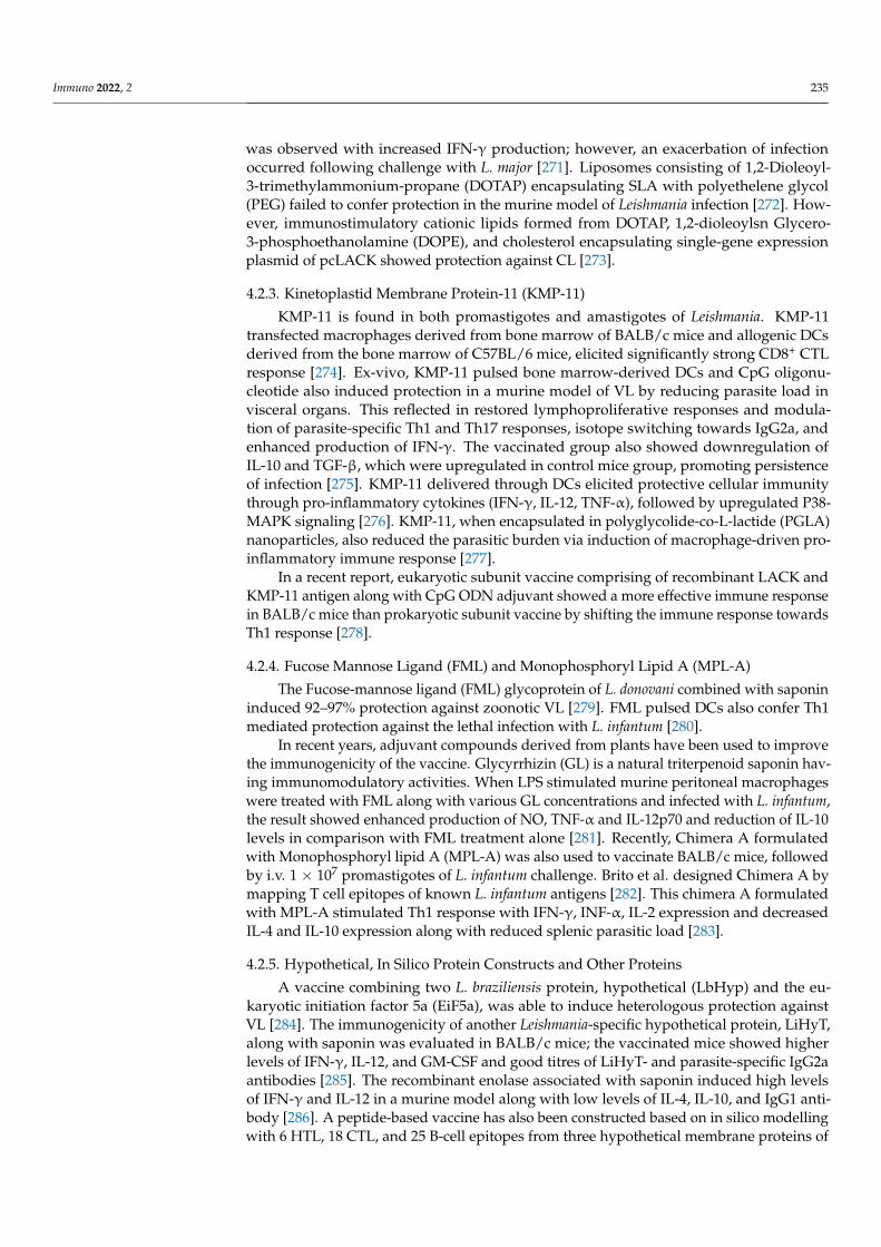
Immuno 2022, 2 235
was observed with increased IFN-γ production; however, an exacerbation of infectionoccurred following challenge with L. major [271]. Liposomes consisting of 1,2-Dioleoyl-3-trimethylammonium-propane (DOTAP) encapsulating SLA with polyethelene glycol(PEG) failed to confer protection in the murine model of Leishmania infection [272]. How-ever, immunostimulatory cationic lipids formed from DOTAP, 1,2-dioleoylsn Glycero-3-phosphoethanolamine (DOPE), and cholesterol encapsulating single-gene expressionplasmid of pcLACK showed protection against CL [273].
4.2.3. Kinetoplastid Membrane Protein-11 (KMP-11)
KMP-11 is found in both promastigotes and amastigotes of Leishmania. KMP-11transfected macrophages derived from bone marrow of BALB/c mice and allogenic DCsderived from the bone marrow of C57BL/6 mice, elicited significantly strong CD8+ CTLresponse [274]. Ex-vivo, KMP-11 pulsed bone marrow-derived DCs and CpG oligonu-cleotide also induced protection in a murine model of VL by reducing parasite load invisceral organs. This reflected in restored lymphoproliferative responses and modula-tion of parasite-specific Th1 and Th17 responses, isotope switching towards IgG2a, andenhanced production of IFN-γ. The vaccinated group also showed downregulation ofIL-10 and TGF-β, which were upregulated in control mice group, promoting persistenceof infection [275]. KMP-11 delivered through DCs elicited protective cellular immunitythrough pro-inflammatory cytokines (IFN-γ, IL-12, TNF-α), followed by upregulated P38-MAPK signaling [276]. KMP-11, when encapsulated in polyglycolide-co-L-lactide (PGLA)nanoparticles, also reduced the parasitic burden via induction of macrophage-driven pro-inflammatory immune response [277].
In a recent report, eukaryotic subunit vaccine comprising of recombinant LACK andKMP-11 antigen along with CpG ODN adjuvant showed a more effective immune responsein BALB/c mice than prokaryotic subunit vaccine by shifting the immune response towardsTh1 response [278].
4.2.4. Fucose Mannose Ligand (FML) and Monophosphoryl Lipid A (MPL-A)
The Fucose-mannose ligand (FML) glycoprotein of L. donovani combined with saponininduced 92–97% protection against zoonotic VL [279]. FML pulsed DCs also confer Th1mediated protection against the lethal infection with L. infantum [280].
In recent years, adjuvant compounds derived from plants have been used to improvethe immunogenicity of the vaccine. Glycyrrhizin (GL) is a natural triterpenoid saponin hav-ing immunomodulatory activities. When LPS stimulated murine peritoneal macrophageswere treated with FML along with various GL concentrations and infected with L. infantum,the result showed enhanced production of NO, TNF-α and IL-12p70 and reduction of IL-10levels in comparison with FML treatment alone [281]. Recently, Chimera A formulatedwith Monophosphoryl lipid A (MPL-A) was also used to vaccinate BALB/c mice, followedby i.v. 1 × 10⁷ promastigotes of L. infantum challenge. Brito et al. designed Chimera A bymapping T cell epitopes of known L. infantum antigens [282]. This chimera A formulatedwith MPL-A stimulated Th1 response with IFN-γ, INF-α, IL-2 expression and decreasedIL-4 and IL-10 expression along with reduced splenic parasitic load [283].
4.2.5. Hypothetical, In Silico Protein Constructs and Other Proteins
A vaccine combining two L. braziliensis protein, hypothetical (LbHyp) and the eu-karyotic initiation factor 5a (EiF5a), was able to induce heterologous protection againstVL [284]. The immunogenicity of another Leishmania-specific hypothetical protein, LiHyT,along with saponin was evaluated in BALB/c mice; the vaccinated mice showed higherlevels of IFN-γ, IL-12, and GM-CSF and good titres of LiHyT- and parasite-specific IgG2aantibodies [285]. The recombinant enolase associated with saponin induced high levelsof IFN-γ and IL-12 in a murine model along with low levels of IL-4, IL-10, and IgG1 anti-body [286]. A peptide-based vaccine has also been constructed based on in silico modellingwith 6 HTL, 18 CTL, and 25 B-cell epitopes from three hypothetical membrane proteins of

Immuno 2022, 2 236
L. donovani. An adjuvant was added at the N-terminus to enhance its immunogenicity. Thevaccine construct was found to be non-toxic, non-allergic, and thermally stable. Moleculardocking confirmed its binding with TLR-2 [287]. Recently, another fusion vaccine wasconstructed using immunoinformatics, targeting KMP-11 gene, and B-type flagellin (Fli-C)as an adjuvant; it was found to be a potent stimulator of the cellular and humoral immuneresponse against VL [288].
4.2.6. Vaccines against Canine Leishmaniasis
Immune regulation of L. infantum, the causative agent of most canine leishmaniasis(CanL), requires a balance between the pro-inflammatory CD4+ Th1 cells, which controlsparasite replication, and Tregs that induce immunosuppressive/regulatory response thatis required to dampen aggravated inflammation. The control of infection by L. infantum(syn. L. chagasi) in dogs is needed to stop the spread of zoonotic VL [289]. Four vaccines areavailable for the prevention of CanL: Leishmune® and Leish-Tec® in Brazil, and CaniLeish®
and LetiFend® in Europe [290].Leishmune vaccine is the first licensed vaccine against canine visceral leishmaniasis
(CVL) registered in Brazil in 2004. It is a FML glycoprotein preparation enriched withL. donovani promastigotes, with the adjuvant Quillaja saponaria saponin, and has undergonephase I, II, and III filed trials [291]. In phase III field trials, the vaccine demonstrated92% to 95% protection against CVL in the vaccinated group [292]. However, in 2014, theLeishmune® vaccine production and marketing license were withdrawn by the BrazilianMinistry of Agriculture as the company failed to comply with the technical regulationapproved in the Interministerial Normative Instruction 31 (IN-31/2007) [293]. Thus, cur-rently, there are three vaccines available against CanL in the market; (1) Leish-Tec® fromCeva Animal Health, Brazil, (2) CaniLeish® from Virbac Santé Animale, France, and (3)Letifend® from Laboratorios Leti, Spain. Their reported efficacies vary between 68.4% and80% [290,294].
Leish-Tec® comprises a recombinant protein A2 from L. donovani amastigotes andsaponin as vaccine adjuvant. It was licensed in Brazil in 2007 and is currently the onlyauthorized CanL vaccine in the country [290]. Leish-Tec® induced protective immunity inbeagle dogs against a high dose intravenous infection of L. chagasi; however, it providedonly partial protection [295]. Vaccination of sub-clinical dogs with LeishTec® was found tobe safe with a mild and site-specific adverse event of 3.06% [296,297]. A study conducted indogs under field conditions suggested that about 43% of Leish-Tec® recipients developeddisease over time [298].
CaniLeish (LiESP/QA-21) is the first European CanL vaccine, released in 2011. Itis composed of purified excreted–secreted proteins of L. infantum (LiESP), together withQuilaja saponaria saponin (QA-21) as an adjuvant. LiESP/QA-21 increased significantly IFN-γ secretion and IgG2 production in vaccinated dogs, concomitant with induction of iNOSpathway and production of NO derivatives [299]. Dogs vaccinated with LiESP/QA-21 orCaniLeish® developed Th1 immune response within 3 weeks. In a one-year randomizedcontrolled CaniLeish® vaccine field trial in the CanL Mediterranean endemic area with aheterogeneous canine population, no infection was observed during the first-year [300];however, the vaccine failed to maintain L. infantum specific IFN-γ at 9 months. Thus, thefield trial did not support the previously reported efficacy to prevent active L. infantuminfection in dogs from endemic areas and naturally exposed parasites [301].
In 2016, LetiFend® (Laboratorios LETI, Spain) was licensed for veterinary use inEurope [302]. The active component of LetiFend® is recombinant protein Q formed byfive antigenic fragments from four different L. infantum proteins (ribosomal proteins LiP2a,LiP2b and LiP0 and the histone H2A), without any adjuvant. Its efficacy was evaluated indifferent breeds of dogs in the endemic area of Spain and France in randomized, double-blind, placebo-controlled, and field trials. The vaccine reduced the risk of developing CanLfollowing natural infection with L. infantum [303,304].

Immuno 2022, 2 237
Use of salivary proteins has recently gained much attention for preparing a vaccineagainst CVL. A vaccine comprising L. braziliensis promastigote protein, sand fly glandextract (SGE) and saponin adjuvant was evaluated in a dog model. The vaccine exhibitedstrong antigenicity and induced SGE-specific humoral immune response. Increase in thecirculating CD21+ B-cells and CD5+ T-cells was observed [305]. Intradermal challenge withL. infantum plus salivary gland extract in dogs immunized with a vaccine composed ofL. braziliensis antigens plus saponin as an adjuvant (LBSap vaccine) was found to elevatetotal anti-Leishmania IgG as well as IgG1 and IgG2 isotypes in vaccinated dogs. Thenumber of circulating B cells (CD21+) as well as CD4+ and CD8+ T lymphocytes were alsohigh and exhibited a high level of IFN-γ and low IL-10 and TGF-β1 expression in spleenwith reduced parasite load [306].
LiESA-MDP is another second-generation dog vaccine candidate, based on a 54 kDaprotein derived from L. infantum. Holzmuller et al. showed that a combination of LiESApand muramyl dipeptide (MDP) induced total protection against experimental CL in 18healthy Beagle dogs when challenged with 108 metacyclic L. infantum promastigotes [307].The efficacy of LiESAP-MDP was evaluated in a randomized field trial in a large populationof dogs in an endemic area of France; dogs were vaccinated with 100 µg lipophilizedLiESAP antigen and 200 µg MDP. After 2 years, the vaccinated dogs showed 92% efficacy,as evident from the detection of Leishmanial DNA in bone marrow aspirates. Enhancementof NO mediated anti-leishmanial activity of canine macrophages were observed with higherIFN-γ production by Th1 cells [308,309].
Another heterologus therapeutic vaccine composed of L. braziliensis with MPL adju-vant (LBMPL) was tested in dogs naturally infected by L. infantum. Animals developedstrong antigen-specific lymphoproliferation mainly of CD4+ T cells and CD8+ T cells withhigh TNF-α and low IL-10 levels. It also blocked transmission of parasites to sandflies inLBMPL-vaccinated dogs [310,311].
Recently, phase I and phase II clinical trial in dogs were performed to evaluate thetoxicity of LBSap vaccine (comprising L. braziliansis antigen and saponin), in comparison toLeishmune® and Leish-tech®. The results showed an increase in CD21+ B lymphocytes incase of the Leishmune® group and CD14+ monocytes in LBSap and Leishmune® groups.In the LBSap group, an increase in both IFN-γ producing subpopulations of T cells wasobserved; in the Leish-Tec® group, an increase in IFN-γ producing CD8+ lymphocyteswere observed. The three vaccines, Leishmune®, LiESP/QA-21, and LBSap, did not induceadverse reactions and prevented CL [292].
4.3. Third-Generation Vaccine
Third-generation vaccines are DNA vaccines that provide an extremely flexible methodof vaccination for inducing stronger Th1 immune response. gp63, LACK and ORFF arethe most extensively studied DNA vaccine candidates against VL [312]. DNA vaccines areadvantageous as they are relatively simple to produce and administer, stable, and oftenvery immunogenic than their recombinant protein counterparts [313]. DNA vaccines areable to induce both humoral and cellular immune responses, which can be further tailoredthrough vector modifications, or by incorporation of immunomodulators such as CpGmotifs, GM-CSF, Flt3 ligands, and/or cytokine genes [314,315]. The concept of heterologousprime boosting has been employed successfully against Leishmania [316]. The first DNAvaccine had the gp63 gene expressed in S. typhimurium, which stimulated the CD4+ Th1subset without any detectable DTH response [317]. The level of protection was enhanced byadding gp46 and cysteine proteinase b (CPb) along with gp63 [318]. The use of aluminumphosphate [319] and plasmid encoding CD40L (as adjuvants) further increased the vaccineefficiency [320].
Multi-epitope vaccine comprising gp63, KMP-11, and amastin from Leishmania wasused as a prime-boost strategy against VL, where immune response was primed with DNAvaccine, then boosted with protein antigen/vector [321]. NH36 DNA, a main componentof FML, together with aluminium phosphate, induced a strong Th1 response providing

Immuno 2022, 2 238
protection in mice against L. chagasi, L. mexicana and L. amazonensis [319,322,322]. PlasmidDNA encoding TSA and LmSTI1 used alone or in tandem, protected mice against L. majorinfection by inducing specific CD4+ Th1 responses without requiring any adjuvant [323].Plasmid cocktails encoding LmSTI1, TSA, and LACK induced full protection against L. majorinfection [324]. Constructs pCMV3ISS-TSA and pCMV3ISS-LmSTI1 induced a comparablelevel of protection in mice infected with a highly virulent strain MHOM/TN/95/GLC94of L. major (Zymodeme MON-25; isolated from a Tunisian patient). Administration ofcombination plasmids containing LACKp24, TSA, LmsTI1 and CPa genes yielded thestrongest protective effects [325]. Immunization of dogs with multicomponent DNA vac-cine, which contained genes for L. donovani H1, H2A, H2B, H3, H4, p36 (LACK), PSA-2,TSA, STI1, and ARP-1, resulted in antigen-induced lymphoproliferative and IFN-γ (but notIL-4) response [326].
Leishmania histones are excellent immunogens for the host immune system. Plasmidscontaining L. infantum nucleosomal histones (H2A, H2B, H3, H4) (dose 25 µg per leg) havebeen tested in a murine model of L. major infection, which developed a strong Th1 responseagainst CL [327,328]. The intradermal vaccination increased IFN-γ production, in additionto CCXC29, CXCL10, and CC12 [329]. Hamsters immunized with recombinant histoneproteins of L. donovani (rLdh2-4) provided protection against VL (107amastigotes) [330].BALB/c mice, immunized with plasmids expressing H2A, H2B, H3, and H4 histoneproteins, and challenged with L. braziliensis, had lesion development prevented [331].
A recombinant lentiviral vaccine expressing a KMP11-Hydrophilic acylated surfaceprotein B (HASPB) fusion protein of L. infantum induced IFN-γ and IL-4, and a specificantibody response against L. infantum, reducing the parasite burden [332]. A DNA vaccinecomprising KMP11-HASPB genes of L. major induced a strong immune response in BALB/cmice and reduced the parasitic burden in spleen and lymph [333]. A pEGFP-N1-KMP11-gp96 DNA vaccine also induced a robust Th1 response and protected against L. major(MRHO/IR/75/ER) [334].
Leishmania sp. obtains heme from HbR-mediated endocytosis and is an importantglycolytic enzyme in the parasite. Thus, HbR-full length-DNA (HbR-FL) and HbR-N-terminal extracellular domain-DNA (HbR-N-DNA) have been tested in BALB/c mice andhamsters for protection against the AG83 strain of L. donovani; a total reduction in splenicand hepatic parasite burden matched with full protection against VL [335]. Another vaccine,LEISHDNAVAX, comprising plasmids coding for five leishmania antigenic determinantswith T cell restricted epitopes (KMP-11, TSA, CPA, CPB and P74), showed protection inmice against L. donovani infection [336].
Recombinant vaccine containing LEISH-F2 and LEISH-F3+, when administered witha TLR4 antagonist, has been shown to induce CD4+ mediated T cell immune response inanimal models and provide therapeutic efficacy in CL. RNA vaccines expressing F2 and F3+
sequences have also been found to enhance CD4+ and CD8+ T cell responses, protectingmice against L. donovani infection [337]. A DNA vaccine using LmxMBA gene lacking thetransmembrane regions of L. mexicana, when immunized prophylactically, reduced the sizeof the lesion and parasitic load in the mouse footpad, compared to the control groups [338].
LACK, when administered as a DNA vaccine, is able to protect against L. major and L.infantum in mice, and against L. infantum in Beagle dogs [339–343]. A combination constructcontaining LACK + Native thiol-specific antioxidant (TSA) increased survival upto 32 weeksfollowing challenge with 2 × 10⁶ L. major promastigotes, compared to the control micethat died within 11 weeks [344]. The protective effect correlated well with strong IFN-γresponse and reduced levels of IL-4 [345]. A multivalent vaccine containing LeIF, LACKand TSA genes against CL elicited stronger Th1 immune response, and delayed progressionof lesion size [346]. Another multivalent vaccine comprising the most immunogenicregions of L. major (MRHO/IR/75/ER) including TSA, LmST11, LACK and KMP11 wastested in BALB/c mice with or without vector containing IL-12. Mice immunized withlL-12 + multiple antigens constructs showed Th1 polarisation, and hence, reduced parasite

Immuno 2022, 2 239
burden [347]. However, the protective effect of a prime-boost vaccine using LACK constructin Beagle dogs against L. infantum was suppressed by IL-12 administration [348].
ChAd63-KH is a replication-defective simian adenoviral vector expressing a novel syn-thetic gene KH (a self-cleaving polyprotein) comprising KMP-11 and HASPB. In the phaseI human trial, it induced IFN-γ production significantly and DC activation, in addition toeliciting CD8+ T cells [349]. The phase IIa safety and immunogenicity trial was conductedin Sudanese patients with persistent PKDL. ChAd63-KH vaccine showed minimal adversereactions and induced potent innate and cell-mediated immune responses [350].
L. infantum pyridoxal kinase (PK) recombinant protein with saponin, or DNA vaccine,significantly reduced the parasite load in spleen, liver, draining lymph nodes and bonemarrow of the BALB/c mice challenged with L. infantum. The protective Th1 responsewas characterized by high levels of IFN-γ, IL-12, GM-CSF and specific IgG2a antibodies,whereas IL-4, IL-10 and parasite-specific IgG1 antibodies were lowered [351].
As an innovative strategy, an immunomodulatory drug, sirolimus (SIR), was also usedto boost a prophylactic DNA vaccine against leishmaniasis. Syrian hamsters were treatedwith SIR along a DNA vaccine consisting of four plasmids (LACK, TRYP, PAPLE22 andKMPII). Although the DNA vaccine on its own efficiently reduced the burden of parasitesin the skin and lymph nodes, SIR offered enhanced protection [352].
5. Conclusions
Early inflammatory events inducing macrophage dysfunction and T-cell dependentimmune response direct the species-specific pathogenesis in Leishmaniasis. The intra-cellular strategies by the parasite and the subtlities of the host–pathogen stand off thatallow the parasite to reside indefinitely inside the macrophage, make the cure challenging.Controlling Leishmaniasis through vaccination is also a challenge. Although a number ofvaccine candidates in the form of plasmid coding genes, defined subunits, live or killedor fractionated forms have been evaluated, only a small number of them have provenpromising. Several attenuated and protein-based vaccines have shown encouraging resultsin offering protection against different species of Leishmania in animal models, but theirpotential to revert back to virulent parasite is fraught with risk. Use of nano-carriers forLeishmania vaccine containing combination vaccines with adjuvants also seems promising;however, the cure from Leishmaniasis is still a dream.
Author Contributions: Conceptualization, H.Y and U.K.; writing—original draft preparation, H.Y.and A.A.; writing—review and editing, U.K., M.N.A.-A. and S.R. All authors have read and agreedto the published version of the manuscript.
Funding: This research received no external funding.
Institutional Review Board Statement: Not applicable.
Informed Consent Statement: Not applicable.
Data Availability Statement: Not applicable.
Acknowledgments: Syamal Roy acknowledges the Indian Council of Medical Research, Governmentof India for his Emeritus position. Illustrations were created using the Biorender tool by Hadida Yasmin.
Conflicts of Interest: The authors declare no conflict of interest.
References1. Ashford, R.W. Leishmaniasis reservoirs and their significance in control. Clin. Dermatol. 1996, 14, 523–532. [CrossRef]2. Weekly Epidemiological Report. 2020. Available online: http://www.who.int/wer (accessed on 10 January 2022).3. Herwaltd, B.L. Leishmaniasis. Lancet 1999, 354, 1191–1199. [CrossRef]4. Bailey, M.S.; Lockwood, D.N. Cutaneous leishmaniasis. Clin. Dermatol. 2007, 25, 203–211. [CrossRef] [PubMed]5. Weigle, K.; Saravia, N.G. Natural history, clinical evolution, and the host-parasite interaction in New World cutaneous Leishmania-
sis. Clin. Dermatol. 1996, 14, 433–450. [CrossRef]6. Burza, S.; Croft, S.L.; Boelaert, M. Leishmaniasis. Lancet 2018, 392, 951–970. [CrossRef]7. Murray, H.W.; Berman, J.D.; Davies, C.R.; Saravia, N.G. Advances in leishmaniasis. Lancet 2005, 366, 1561–1577. [CrossRef]

Immuno 2022, 2 240
8. Gregory, D.J.; Sladek, R.; Olivier, M.; Matlashewski, G. Comparison of the effects of Leishmania major or Leishmania do-novaniinfection on macrophage gene expression. Infect. Immun. 2008, 76, 1186–1192. [CrossRef]
9. Cruz, I.; Morales, M.A.; Noguer, I.; Rodríguez, A.; Alvar, J. Leishmania in discarded syringes from intravenous drug users. Lancet2002, 359, 1124–1125. [CrossRef]
10. Pagliano, P.; Carannante, N.; Rossi, M.; Gramiccia, M.; Gradoni, L.; Faella, F.S.; Gaeta, G.B. Visceral leishmaniasis in pregnancy: Acase series and a systematic review of the literature. J. Antimicrob. Chemother. 2005, 55, 229–233. [CrossRef]
11. Costa, C.H.N.; Gomes, R.B.B.; Silva, M.R.B.; Garcez, L.M.; Ramos, P.K.; Santos, R.S.; Shaw, J.J.; David, J.R.; Maguire, J.H.Competence of the Human Host as a Reservoir forLeishmania chagasi. J. Infect. Dis. 2000, 182, 997–1000. [CrossRef]
12. Chitimia, L.; Muñoz-García, C.; Sánchez-Velasco, D.; Lizana, V.; del Río, L.; Murcia, L.; Fisa, R.; Riera, C.; Giménez-Font, P.;Jiménez-Montalbán, P.; et al. Cryptic Leishmaniosis by Leishmania infantum, a feature of canines only? A study of natural infectionin wild rabbits, humans and dogs in southeastern Spain. Vet. Parasitol. 2011, 181, 12–16. [CrossRef] [PubMed]
13. Dipineto, L.; Manna, L.; Baiano, A.; Gala, M.; Fioretti, A.; Gravino, A.E.; Menna, L.F. Presence of Leishmania infantum in Red Foxes(Vulpes vulpes) in Southern Italy. J. Wildl. Dis. 2007, 43, 518–520. [CrossRef] [PubMed]
14. Chatzis, M.K.; Andreadou, M.; Leontides, L.; Kasabalis, D.; Mylonakis, M.; Koutinas, A.F.; Rallis, T.; Ikonomopoulos, J.;Saridomichelakis, M.N. Cytological and molecular detection of Leishmania infantum in different tissues of clinically normal andsick cats. Vet. Parasitol. 2014, 202, 217–225. [CrossRef] [PubMed]
15. Pereira, A.A.S.; Ferreira, E.; Lima, A.C.V.M.d.R.; Tonelli, G.B.; Rêgo, F.D.; Paglia, A.P.; Andrade-Filho, J.D.; Paz, G.F.; Gontijo,C.M.F. Detection of Leishmania spp in silvatic mammals and isolation of Leishmania (Viannia) braziliensis from Rattus rattus in anendemic area for leishmaniasis in Minas Gerais State, Brazil. PLoS ONE 2017, 12, e0187704. [CrossRef]
16. Singh, N.; Mishra, J.; Singh, R.; Singh, S. Animal Reservoirs of Visceral Leishmaniasis in India. J. Parasitol. 2013, 99, 64–67.[CrossRef]
17. Oryan, A.; Akbari, M. Worldwide risk factors in leishmaniasis. Asian Pac. J. Trop. Med. 2016, 9, 925–932. [CrossRef]18. Cloots, K.; Marino, P.; Burza, S.; Gill, N.; Boelaert, M.; Hasker, E. Visceral Leishmaniasis-HIV Coinfection as a Predictor of
Increased Leishmania Transmission at the Village Level in Bihar, India. Front. Cell. Infect. Microbiol. 2021, 11, 604117. [CrossRef]19. Aschale, Y.; Ayehu, A.; Worku, L.; Tesfa, H.; Birhanie, M.; Lemma, W. Malaria-visceral leishmaniasis co-infection and associated
factors among migrant laborers in West Armachiho district, North West Ethiopia: Community based cross-sectional study. BMCInfect. Dis. 2019, 19, 239. [CrossRef]
20. Shen, S.-S.; Qu, X.-Y.; Zhang, W.-Z.; Li, J.; Lv, Z.-Y. Infection against infection: Parasite antagonism against parasites, viruses andbacteria. Infect. Dis. Poverty 2019, 8, 49. [CrossRef]
21. Bates, P.A. Transmission of Leishmania metacyclic promastigotes by phlebotomine sand flies. Int. J. Parasitol. 2007, 37, 1097–1106.[CrossRef]
22. Rogers, M.E.; Ilg, T.; Nikolaev, A.V.; Ferguson, M.; Bates, P.A. Transmission of cutaneous leishmaniasis by sand flies is enhancedby regurgitation of fPPG. Nature 2004, 430, 463–467. [CrossRef] [PubMed]
23. Bates, P.; Rogers, M. New Insights into the Developmental Biology and Transmission Mechanisms of Leishmania. Curr. Mol. Med.2004, 4, 601–609. [CrossRef] [PubMed]
24. Gossage, S.M.; Rogers, M.E.; Bates, P.A. Two separate growth phases during the development of Leishmania in sand flies:Implications for understanding the life cycle. Int. J. Parasitol. 2003, 33, 1027–1034. [CrossRef]
25. Killick-Kendrick, R.; Molyneux, D.H.; Ashford, R.W. Leishmania in phlebotomid sandflies. I. Modifications of the flagellumassociated with attachment to the mid-gut and oesophageal valve of the sandfly. Proc. R Soc. Lond. B Biol. Sci. 1974, 187, 409–419.
26. Giraud, E.; Martin, O.; Yakob, L.; Rogers, M. Quantifying Leishmania Metacyclic Promastigotes from Individual Sandfly BitesReveals the Efficiency of Vector Transmission. Commun. Biol. 2019, 2, 84. [CrossRef]
27. Bates, P.A. Revising Leishmania’s life cycle. Nat. Microbiol. 2018, 3, 529–530. [CrossRef]28. Rogers, M.E.; Chance, M.L.; Bates, P.A. The role of promastigote secretory gel in the origin and transmission of the infective stage
of Leishmania mexicana by the sandfly Lutzomyia longipalpis. Parasitology 2002, 124, 495–507. [CrossRef]29. Sunter, J.; Gull, K. Shape, form, function and Leishmania pathogenicity: From textbook descriptions to biological understanding.
Open Biol. 2017, 7, 170165. [CrossRef]30. Rittig, M.G.; Schröppel, K.; Seack, K.H.; Sander, U.; N’Diaye, E.N.; Maridonneau-Parini, I.; Solbach, W.; Bogdan, C. Coiling
phag-ocytosis of trypanosomatids and fungal cells. Infect. Immun. 1998, 66, 4331–4339. [CrossRef]31. Petropolis, D.B.; Rodrigues, J.C.; Viana, N.B.; Pontes, B.; Pereira, C.F.; Silva-Filho, F.C. Leishmania amazonensis promastigotes in 3D
Collagen I culture: An in vitro physiological environment for the study of extracellular matrix and host cell interactions. PeerJ2014, 2, e317. [CrossRef]
32. Dedet, J.P.; Pratlong, F.; Lanotte, G.; Ravel, C. Cutaneous leishmaniasis. The parasite. Clin. Dermatol. 1999, 17, 261–268. [CrossRef]33. Yao, C.; Donelson, J.E.; Wilson, M.E. The major surface protease (MSP or GP63) of Leishmania sp. Biosynthesis, regulation of
expression, and function. Mol. Biochem. Parasitol. 2003, 132, 1–16. [CrossRef]34. Naderer, T.; McConville, M.J. The Leishmania-macrophage interaction: A metabolic perspective. Cell. Microbiol. 2008, 10, 301–308.
[CrossRef] [PubMed]35. Lo, S.K.; Bovis, L.; Matura, R.; Zhu, B.; He, S.; Lum, H.; Turco, S.J.; Ho, J.L. Leishmania lipophosphoglycan reduces monocyte
transendothelial migration: Modulation of cell adhesion molecules, intercellular junctional proteins, and chemoattractants. J.Immunol. 1998, 160, 1857–1865. [PubMed]
Immuno 2022, 2 241
36. Bogdan, C. Macrophages as host, effector and immunoregulatory cells in leishmaniasis: Impact of tissue micro-environment andmetabolism. Cytokine X 2020, 2, 100041. [CrossRef]
37. Liu, D.; Uzonna, J.E. The early interaction of Leishmania with macrophages and dendritic cells and its influence on the hostimmune response. Front. Cell. Infect. Microbiol. 2012, 2, 83. [CrossRef]
38. Novais, F.O.; Santiago, R.C.; Báfica, A.; Khouri, R.; Afonso, L.; Borges, V.; Brodskyn, C.; Netto, M.B.; Barral, A.; De Oliveira,C.I. Neutrophils and Macrophages Cooperate in Host Resistance against Leishmania braziliensis Infection. J. Immunol. 2009, 183,8088–8098. [CrossRef]
39. de Souza Carmo, E.V.; Katz, S.; Barbiéri, C.L. Neutrophils reduce the parasite burden in Leishmania (Leishmania) amazonensis-infected macrophages. PLoS ONE 2010, 3, e13815.
40. Pearson, R.D.; Steigbigel, R.T. Phagocytosis and killing of the protozoan Leishmania donovani by human polymorphonuclearleukocytes. J. Immunol. 1981, 127, 1438–1443.
41. Guimaraes-Costa, A.B.; Nascimento, M.T.C.; Froment, G.S.; Soares, R.P.P.; Morgado, F.N.; Conceicao-Silva, F.; Saraiva, E.M.Leishmania amazonensis promastigotes induce and are killed by neutrophil extracellular traps. Proc. Natl. Acad. Sci. USA 2009, 106,6748–6753. [CrossRef]
42. Titus, R.G.; Ribeiro, J.M. Salivary gland lysates from the sand fly Lutzomyia longipalpis enhance Leishmania infectivity. Science1988, 239, 1306–1308. [CrossRef]
43. Dey, R.; Joshi, A.B.; Oliveira, F.; Pereira, L.; Guimarães-Costa, A.B.; Serafim, T.D.; de Castro, W.; Coutinho-Abreu, I.V.; Bhattacharya,P.; Townsend, S.; et al. Gut Microbes Egested during Bites of Infected Sand Flies Augment Severity of Leishmaniasis viaInflammasome-Derived IL-1β. Cell Host Microbe 2018, 23, 134.e6–143.e6. [CrossRef] [PubMed]
44. Chagas, A.C.; Oliveira, F.; Debrabant, A.; Valenzuela, J.G.; Ribeiro, J.M.; Calvo, E. Lundep, a sand fly salivary endonucleasein-creases Leishmania parasite survival in neutrophils and inhibits XIIa contact activation in human plasma. PLoS Pathog. 2014, 10,e1003923. [CrossRef] [PubMed]
45. Guimaraes-Costa, A.B.; Shannon, J.P.; Waclawiak, I.; Oliveira, J.; Meneses, C.; de Castro, W.; Wen, X.; Brzostowski, J.; Serafim, T.D.;Andersen, J.F.; et al. A sand fly salivary protein acts as a neutrophil chemo-attractant. Nat. Commun. 2021, 12, 3213. [CrossRef][PubMed]
46. Sangare, M.; Coulibaly, Y.I.; Huda, N.; Vidal, S.; Tariq, S.; Coulibaly, M.E.; Coulibaly, S.Y.; Soumaoro, L.; Dicko, I.; Traore, B.; et al.Individuals co-exposed to sand fly saliva and filarial parasites exhibit altered monocyte function. PLoS Negl. Trop. Dis. 2021, 15,e0009448. [CrossRef]
47. Charmoy, M.; Megnekou, R.; Allenbach, C.; Zweifel, C.; Perez, C.; Monnat, K.; Breton, M.; Ronet, C.; Launois, P.; Tacchini-Cottier,F. Leishmania major induces distinct neutrophil phenotypes in mice that are resistant or susceptible to infection. J. Leukoc. Biol.2007, 82, 288–299. [CrossRef]
48. Ribeiro-Gomes, F.L.; Moniz-de-Souza, M.C.; Alexandre-Moreira, M.S.; Dias, W.B.; Lopes, M.F.; Nunes, M.P.; Lungarella, G.;DosReis, G.A. Neutrophils activate macrophages for intracellular killing of Leishmania major through recruitment of TLR4 byneutrophil elastase. J. Immunol. 2007, 179, 3988–3994. [CrossRef]
49. Ribeiro-Gomes, F.L.; Sacks, D. The influence of early neutrophil-Leishmania interactions on the host immune response to infec-tion.Front. Cell. Infect. Microbiol. 2012, 2, 59. [CrossRef]
50. Singh, T.P.; Carvalho, A.M.; Sacramento, L.A.; Grice, E.A.; Scott, P. Microbiota instruct IL-17A-producing innate lymphoid cells topromote skin inflammation in cutaneous leishmaniasis. PLOS Pathog. 2021, 17, e1009693. [CrossRef]
51. van Zandbergen, G.; Hermann, N.; Laufs, H.; Solbach, M.D.W.; Laskay, T. Leishmania Promastigotes Release a GranulocyteChemotactic Factor and Induce Interleukin-8 Release but Inhibit Gamma Interferon-Inducible Protein 10 Production by NeutrophilGranulocytes. Infect. Immun. 2002, 70, 4177–4184. [CrossRef]
52. Kostka, S.L.; Dinges, S.; Griewank, K.; Iwakura, Y.; Udey, M.C.; Von Stebut, E. IL-17 Promotes Progression of CutaneousLeishmaniasis in Susceptible Mice. J. Immunol. 2009, 182, 3039–3046. [CrossRef] [PubMed]
53. Boaventura, V.S.; Santos, C.S.; Cardoso, C.R.D.B.; De Andrade, J.; Dos-Santos, W.L.; Clarêncio, J.; Silva, J.S.; Borges, V.; Netto,M.B.; Brodskyn, C.I.; et al. Human mucosal leishmaniasis: Neutrophils infiltrate areas of tissue damage that express high levelsof Th17-related cytokines. Eur. J. Immunol. 2010, 40, 2830–2836. [CrossRef] [PubMed]
54. van Zandbergen, G.; Klinger, M.; Mueller, A.; Dannenberg, S.; Gebert, A.; Solbach, W.; Laskay, T. Cutting Edge: NeutrophilGranulocyte Serves as a Vector for Leishmania Entry into Macrophages. J. Immunol. 2004, 173, 6521–6525. [CrossRef] [PubMed]
55. Gueirard, P.; Laplante, A.; Rondeau, C.; Milon, G.; Desjardins, M. Trafficking of Leishmania donovani promastigotes in non-lyticcompartments in neutrophils enables the subsequent transfer of parasites to macrophages. Cell. Microbiol. 2008, 10, 100–111.[CrossRef]
56. Rossi, M.; Fasel, N. How to master the host immune system? Leishmania parasites have the solutions! Int. Immunol. 2018, 30,103–111. [CrossRef]
57. Beverley, S.M.; Turco, S.J. Lipophosphoglycan (LPG) and the identification of virulence genes in the protozoan parasite Leishmania.Trends Microbiol. 1998, 6, 135–140. [CrossRef]
58. Ferguson, M. The surface glycoconjugates of trypanosomatid parasites. Philos. Trans. R Soc. Lond. B Biol. Sci. 1997, 352, 1295–1302.[CrossRef]
59. McConville, M.J.; Ralton, J.E. Developmentally regulated changes in the cell surface architecture of Leishmania parasites. BehringInst. Mitt. 1997, 99, 34–43.

Immuno 2022, 2 242
60. Ferguson, M.A. The structure, biosynthesis and functions of glycosylphosphatidylinositol anchors, and the contributions oftrypanosome research. J. Cell Sci. 1999, 112, 2799–2809. [CrossRef]
61. Kane, M.M.; Mosser, D.M. Leishmania parasites and their ploys to disrupt macrophage activation. Curr. Opin. Hematol. 2000, 7,26–31. [CrossRef]
62. Joshi, P.B.; Kelly, B.L.; Kamhawi, S.; Sacks, D.L.; McMaster, W.R. Targeted gene deletion in Leishmania major identifies leish-manolysin (GP63) as a virulence factor. Mol. Biochem. Parasitol. 2002, 120, 33–40. [CrossRef]
63. Pelletier, I.; Hashidate, T.; Urashima, T.; Nishi, N.; Nakamura, T.; Futai, M.; Arata, Y.; Kasai, K.; Hirashima, M.; Hirabayashi, J.;et al. Specific recognition of Leishmania major poly-beta-galactosyl epitopes by galectin-9: Possible implication of galectin-9 ininteraction between L. major and host cells. J. Biol. Chem. 2003, 278, 22223–22230. [CrossRef] [PubMed]
64. Kamhawi, S.; Ramalho-Ortigao, M.; Pham, V.M.; Kumar, S.; Lawyer, P.G.; Turco, S.J.; Barillas-Mury, C.; Sacks, D.L.; Valenzuela,J.G. A Role for Insect Galectins in Parasite Survival. Cell 2004, 119, 329–341. [CrossRef] [PubMed]
65. Soulat, D.; Bogdan, C. Function of Macrophage and Parasite Phosphatases in Leishmaniasis. Front. Immunol. 2017, 8, 1838.[CrossRef] [PubMed]
66. Srivastava, S.; Pandey, S.P.; Jha, M.K.; Chandel, H.S.; Saha, B. Leishmania expressed lipophosphoglycan interacts with Toll-likereceptor (TLR)-2 to decrease TLR-9 expression and reduce anti-leishmanial responses. Clin. Exp. Immunol. 2013, 172, 403–409.[CrossRef]
67. Frankenburg, S.; Leibovici, V.; Mansbach, N.; Turco, S.J.; Rosen, G. Effect of glycolipids of Leishmania parasites on humanmonocyte activity. Inhibition by lipophosphoglycan. J. Immunol. 1990, 145, 4284–4289.
68. Ibraim, I.C.; de Assis, R.R.; Pessoa, N.L.; Campos, M.A.; Melo, M.N.; Turco, S.J.; Soares, R.P. Two biochemically distinctlipophosphoglycans from Leishmania braziliensis and Leishmania infantum trigger different innate immune responses in murinemacrophages. Parasites Vectors 2013, 6, 54. [CrossRef]
69. Barral-Netto, M.; Barral, A.; Brownell, C.E.; Skeiky, Y.A.; Ellingsworth, L.R.; Twardzik, D.R.; Reed, S.G. Transforming growthfactor-beta in leishmanial infection: A parasite escape mechanism. Science 1992, 257, 545–548. [CrossRef]
70. Chang, K.; Fong, D. Cell Biology of Host-Parasite Membrane Interactions in Leishmaniasis. Ciba Found. Symp. 1983, 99, 113–137.[CrossRef]
71. Forestier, C.-L.; Machu, C.; Loussert, C.; Pescher, P.; Späth, G.F. Imaging Host Cell-Leishmania Interaction Dynamics ImplicatesParasite Motility, Lysosome Recruitment, and Host Cell Wounding in the Infection Process. Cell Host Microbe 2011, 9, 319–330.[CrossRef]
72. Gluenz, E.; Ginger, M.L.; McKean, P.G. Flagellum assembly and function during the Leishmania life cycle. Curr. Opin. Microbiol.2010, 13, 473–479. [CrossRef] [PubMed]
73. Gluenz, E.; Höög, J.L.; Smith, A.E.; Dawe, H.R.; Shaw, M.K.; Gull, K. Beyond 9+0: Noncanonical axoneme structures characterizesensory cilia from protists to humans. FASEB J. 2010, 24, 3117–3121. [CrossRef] [PubMed]
74. Burchmore, R.J.; Barrett, M. Life in vacuoles—Nutrient acquisition by Leishmania amastigotes. Int. J. Parasitol. 2001, 31, 1311–1320.[CrossRef]
75. Dermine, J.-F.; Goyette, G.; Houde, M.; Turco, S.J.; Desjardins, M. Leishmania donovani lipophosphoglycan disrupts phagosomemicrodomains in J774 macrophages. Cell. Microbiol. 2005, 7, 1263–1270. [CrossRef] [PubMed]
76. Vinet, A.F.; Fukuda, M.; Turco, S.J.; Descoteaux, A. The Leishmania donovani lipophosphoglycan excludes the vesicular proton-ATPase from phagosomes by impairing the recruitment of synaptotagmin V. PLoS Pathog. 2009, 5, e1000628. [CrossRef]
77. Chakraborty, P.; Das, P.K. Suppression of macrophage lysosomal enzymes after Leishmania donovani infection. Biochem. Med.Metab. Biol. 1989, 41, 46–55. [CrossRef]
78. Henriques, C.; Atella, G.C.; Bonilha, V.L.; de Souza, W. Biochemical analysis of proteins and lipids found in parasitophorousvacuoles containing Leishmania amazonensis. Parasitol. Res. 2003, 89, 123–133.
79. Courret, N.; Fréhel, C.; Gouhier, N.; Pouchelet, M.; Prina, E.; Roux, P.; Antoine, J.C. Biogenesis of Leishmania-harbouringparasitophorous vacuoles following phagocytosis of the metacyclic promastigote or amastigote stages of the parasites. J. Cell Sci.2002, 115, 2303–2316. [CrossRef]
80. Körner, U.; Fuss, V.; Steigerwald, J.; Moll, H. Biogenesis of Leishmania major -Harboring Vacuoles in Murine Dendritic Cells. Infect.Immun. 2006, 74, 1305–1312. [CrossRef]
81. Podinovskaia, M.; Descoteaux, A. Leishmania and the macrophage: A multifaceted interaction. Future Microbiol. 2015, 10, 111–129.[CrossRef]
82. Mittra, B.; Andrews, N.W. IRONy OF FATE: Role of iron-mediated ROS in Leishmania differentiation. Trends Parasitol. 2013, 29,489–496. [CrossRef] [PubMed]
83. Mittra, B.; Cortez, M.; Haydock, A.; Ramasamy, G.; Myler, P.; Andrews, N. Iron uptake controls the generation of Leishmaniainfective forms through regulation of ROS levels. J. Exp. Med. 2013, 210, 401–416. [CrossRef] [PubMed]
84. Glaser, T.A.; Baatz, J.E.; Kreishman, G.P.; Mukkada, A.J. pH homeostasis in Leishmania donovani amastigotes and promastigotes.Proc. Natl. Acad. Sci. USA 1988, 85, 7602–7606. [CrossRef] [PubMed]
85. Schaible, U.E.; Schlesinger, P.H.; Steinberg, T.H.; Mangel, W.F.; Kobayashi, T.; Russell, D.G. Parasitophorous vacuoles of Leishmaniamexicana acquire macromolecules from the host cell cytosol via two independent routes. J. Cell Sci. 1999, 112, 681–693. [CrossRef]
86. Real, F.; Mortara, R.A.; Rabinovitch, M. Fusion between Leishmania amazonensis and Leishmania major Parasitophorous Vacuoles:Live Imaging of Coinfected Macrophages. PLoS Negl. Trop. Dis. 2010, 4, e905. [CrossRef]
Immuno 2022, 2 243
87. Chang, K.-P.; Dwyer, D. Leishmania Donovani. Hamster macrophage interactions in vitro: Cell entry, intracellular survival, andmultiplication of amastigotes. J. Exp. Med. 1978, 147, 515–530. [CrossRef]
88. Saha, S.; Basu, M.; Guin, S.; Gupta, P.; Mitterstiller, A.-M.; Weiss, G.; Jana, K.; Ukil, A. Leishmania donovani Exploits MacrophageHeme Oxygenase-1 To Neutralize Oxidative Burst and TLR Signaling–Dependent Host Defense. J. Immunol. 2018, 202, 827–840.[CrossRef]
89. Wei, X.-Q.; Charles, I.G.; Smith, A.C.; Ure, J.; Feng, G.-J.; Huang, F.-P.; Xu, D.; Muller, W.; Moncada, S.; Liew, F.Y. Altered immuneresponses in mice lacking inducible nitric oxide synthase. Nature 1995, 375, 408–411. [CrossRef]
90. Murray, H.W.; Nathan, C.F. Macrophage Microbicidal Mechanisms In Vivo: Reactive Nitrogen versus Oxygen Intermediates inthe Killing of Intracellular Visceral Leishmania donovani. J. Exp. Med. 1999, 189, 741–746. [CrossRef]
91. Henter, J.-I.; Horne, A.; Aricó, M.; Egeler, R.M.; Filipovich, A.H.; Imashuku, S.; Ladisch, S.; McClain, K.; Webb, D.; Winiarski, J.;et al. HLH-2004: Diagnostic and therapeutic guidelines for hemophagocytic lymphohistiocytosis. Pediatr. Blood Cancer 2007, 48,124–131. [CrossRef]
92. Mallory, F.B. A histological study of typhoid fever. J. Exp. Med. 1898, 3, 611–638. [CrossRef] [PubMed]93. Balasubramanian, S.; Kaarthigeyan, K.; Aparna, V.; Srinivas, S. Tuberculosis associated hemophagocytic syndrome in infancy.
Indian Pediatr. 2008, 45, 593–595. [PubMed]94. Slovut, D.P.; Benedetti, E.; Matas, A.J. Babesiosis and hemophagocytic syndrome in an asplenic renal transplant recipient.
Transplantation 1996, 62, 537–539. [CrossRef] [PubMed]95. Granert, C.; Elinder, G.; Ost, A.; Henter, J.I. Kala-azar in a one-year-old Swedish child. Diagnostic difficulties because of active
hemophagocytosis. Acta Paediatr. 1993, 82, 794–796. [CrossRef]96. Fathalla, M.; Hashim, J.; Alkindy, H.; Wali, Y. Cerebrospinal fluid involvement in a case of visceral leishmaniasis associated with
hemophagocytic lymphohistiocytosis. Sultan Qaboos Univ. Med. J. 2007, 7, 253–256.97. Kilani, B.; Ammari, L.; Kanoun, F.; Ben Chaabane, T.; Abdellatif, S.; Chaker, E. Hemophagocytic syndrome associated with
visceral leishmaniasis. Int. J. Infect. Dis. 2006, 10, 85–86. [CrossRef]98. Ozyurek, E.; Özçay, F.; Yilmaz, B.; Özbek, N. Hemophagocytic lymphohistiocytosis associated with visceral Leishmaniasis: A
Case Report. Pediatr. Hematol. Oncol. 2005, 22, 409–414. [CrossRef]99. Gagnaire, M.-H.; Galambrun, C.; Stéphan, J.L. Hemophagocytic Syndrome: A Misleading Complication of Visceral Leishmaniasis
in Children—A Series of 12 Cases. Pediatrics 2000, 106, e58. [CrossRef]100. Marom, D.; Offer, I.; Tamary, H.; Jaffe, C.L.; Garty, B.Z. Hemophagocytic Lymphohistocytosis associated with Visceral Leishmani-
asis. J. Pediatr. Hematol. Oncol. 2001, 18, 65–70. [CrossRef]101. Bode, S.; Bogdan, C.; Beutel, K.; Behnisch, W.; Greiner, J.; Henning, S.; Jorch, N.; Jankofsky, M.; Jakob, M.; Schmid, I.; et al.
Hemophagocytic Lymphohistiocytosis in Imported Pediatric Visceral Leishmaniasis in a Nonendemic Area. J. Pediatr. 2014, 165,147.e1–153.e1. [CrossRef]
102. Pilonieta, M.C.; Moreland, S.M.; English, C.N.; Detweiler, C.S. Salmonella enterica infection stimulates macrophages tohemophagocytose. mBio 2014, 5, e02211. [CrossRef] [PubMed]
103. Brown, D.E.; McCoy, M.W.; Pilonieta, M.C.; Nix, R.N.; Detweiler, C.S. Chronic Murine Typhoid Fever Is a Natural Model ofSecondary Hemophagocytic Lymphohistiocytosis. PLoS ONE 2010, 5, e9441. [CrossRef] [PubMed]
104. Jordan, M.B.; Hildeman, D.; Kappler, J.; Marrack, P. An animal model of hemophagocytic lymphohistiocytosis (HLH): CD8+ Tcells and interferon gamma are essential for the disorder. Blood 2004, 104, 735–743. [CrossRef] [PubMed]
105. Brisse, E.; Imbrechts, M.; Put, K.; Avau, A.; Mitera, T.; Berghmans, N.; Rutgeerts, O.; Waer, M.; Ninivaggi, M.; Kelchtermans, H.;et al. Mouse Cytomegalovirus Infection in BALB/c Mice Resembles Virus-Associated Secondary Hemophagocytic Lymphohistio-cytosis and Shows a Pathogenesis Distinct from Primary Hemophagocytic Lymphohistiocytosis. J. Immunol. 2016, 196, 3124–3134.[CrossRef]
106. Cnops, J.; De Trez, C.; Stijlemans, B.; Keirsse, J.; Kauffmann, F.; Barkhuizen, M.; Keeton, R.; Boon, L.; Brombacher, F.; Magez, S. NK-,NKT- and CD8-Derived IFNγ Drives Myeloid Cell Activation and Erythrophagocytosis, Resulting in Trypanosomosis-AssociatedAcute Anemia. PLoS Pathog. 2015, 11, e1004964. [CrossRef]
107. Morimoto, A.; Omachi, S.; Osada, Y.; Chambers, J.K.; Uchida, K.; Sanjoba, C.; Matsumoto, Y.; Goto, Y. Hemophagocytosis inExperimental Visceral Leishmaniasis by Leishmania donovani. PLoS Negl. Trop. Dis. 2016, 10, e0004505. [CrossRef]
108. Marquis, J.F.; Gros, P. Intracellular Leishmania: Your iron or mine? Trends Microbiol. 2007, 15, 93–95. [CrossRef]109. Blackwell, J.M.; Goswami, T.; Evans, C.A.; Sibthorpe, D.; Papo, N.; White, J.K.; Searle, S.; Miller, E.N.; Peacock, C.S.; Mohammed,
H.; et al. SLC11A1 (formerly NRAMP1) and disease resistance. Cell Microbiol. 2001, 3, 773–784. [CrossRef]110. Banerjee, S.; Datta, R. Leishmania infection triggers hepcidin-mediated proteasomal degradation of Nramp1 to increase phagolyso-
somal iron availability. Cell Microbiol. 2020, 22, e13253. [CrossRef]111. Das, N.K.; Biswas, S.; Solanki, S.; Mukhopadhyay, C.K. Leishmania donovani depletes labile iron pool to exploit iron uptake capacity
of macrophage for its intracellular growth. Cell Microbiol. 2009, 11, 83–94. [CrossRef]112. Borges, V.M.; Vannier-Santos, M.A.; De Souza, W. Subverted transferrin trafficking in Leishmania-infected macrophages. Parasitol
Res. 1998, 84, 811–822. [CrossRef] [PubMed]113. Srivastav, S.; Ball, W.B.; Gupta, P.; Giri, J.; Ukil, A.; Das, P.K. Leishmania donovani Prevents Oxidative Burst-mediated Apoptosis of
Host Macrophages through Selective Induction of Suppressors of Cytokine Signaling (SOCS) Proteins. J. Biol. Chem. 2014, 289,1092–1105. [CrossRef] [PubMed]

Immuno 2022, 2 244
114. Ghosh, J.; Guha, R.; Das, S.; Roy, S. Liposomal cholesterol delivery activates the macrophage innate immune arm to facilitateintracellular Leishmania donovani killing. Infect. Immun. 2014, 82, 607–617. [CrossRef] [PubMed]
115. Ghosh, J.; Lal, C.S.; Pandey, K.; Das, V.N.R.; Das, P.; Roychoudhury, K.; Roy, S. Human visceral leishmaniasis: Decrease in serumcholesterol as a function of splenic parasite load. Ann. Trop. Med. Parasitol. 2011, 105, 267–271. [CrossRef] [PubMed]
116. Messaoud, H.B.-B.; Guichard, M.; Lawton, P.; Delton, I.; Azzouz-Maache, S. Changes in Lipid and Fatty Acid Composition DuringIntramacrophagic Transformation of Leishmania donovani Complex Promastigotes into Amastigotes. Lipids 2017, 52, 433–441.[CrossRef]
117. Zhang, K.; Beverley, S.M. Phospholipid and sphingolipid metabolism in Leishmania. Mol. Biochem. Parasitol. 2010, 170, 55–64.[CrossRef]
118. Zhang, O.; Wilson, M.C.; Xu, W.; Hsu, F.F.; Turk, J.; Kuhlmann, F.M.; Wang, Y.; Soong, L.; Key, P.; Beverley, S.M.; et al. Deg-radationof host sphingomyelin is essential for Leishmania virulence. PLoS Pathog. 2009, 5, e1000692. [CrossRef]
119. Vallochi, A.L.; Teixeira, L.; Oliveira, K.D.S.; Maya-Monteiro, C.M.; Bozza, P.T. Lipid Droplet, a Key Player in Host-ParasiteIn-teractions. Front. Immunol. 2018, 9, 1022. [CrossRef]
120. De Cicco, N.N.; Pereira, M.G.; Corrêa, J.R.; Andrade-Neto, V.V.; Saraiva, F.B.; Chagas-Lima, A.C.; Gondim, K.C.; Torres-Santos,E.C.; Folly, E.; Saraiva, E.M.; et al. LDL uptake by Leishmania amazonensis: Involvement of membrane lipid microdomains. Exp.Parasitol. 2012, 130, 330–340. [CrossRef]
121. Das, P.; De, T.; Chakraborti, T. Leishmania donovani secretory serine protease alters macrophage inflammatory response via COX-2mediated PGE-2 production. Indian J. Biochem. Biophys. 2014, 51, 5542–5551.
122. Carregaro, V.; Valenzuela, J.G.; Cunha, T.M.; Verri, W.A., Jr.; Grespan, R.; Matsumura, G.; Ribeiro, J.M.; Elnaiem, D.E.; Silva, J.S.;Cunha, F.Q. Phlebotomine salivas inhibit immune inflammation-induced neutrophil migration via an autocrine DC-derived PGE2/IL-10 sequential pathway. J. Leukoc. Biol. 2008, 84, 104–114. [CrossRef] [PubMed]
123. Araújo-Santos, T.; Prates, D.B.; Andrade, B.B.; Nascimento, D.O.; Clarêncio, J.; Entringer, P.F.; Carneiro, A.B.; Silva-Neto, M.A.C.;Miranda, J.C.; Brodskyn, C.I.; et al. Lutzomyia longipalpis Saliva Triggers Lipid Body Formation and Prostaglandin E2 Productionin Murine Macrophages. PLoS Negl. Trop. Dis. 2010, 4, e873. [CrossRef] [PubMed]
124. Rabhi, S.; Rabhi, I.; Trentin, B.; Piquemal, D.; Regnault, B.; Goyard, S.; Lang, T.; Descoteaux, A.; Enninga, J.; Guizani-Tabbane, L.Lipid droplet formation, their localization and dynamics during Leishmania major macrophage infection. PLoS ONE 2016, 11, 1–19.[CrossRef] [PubMed]
125. Aoki, J.I.; Muxel, S.M.; Zampieri, R.A.; Müller, K.E.; Nerland, A.H.; Floeter-Winter, L.M. Differential immune response modulationin early Leishmania amazonensis infection of BALB/c and C57BL/6 macrophages based on transcriptome profiles. Sci. Rep. 2019, 9,19841. [CrossRef] [PubMed]
126. Tomiotto-Pellissier, F.; Bortoleti, B.; Assolini, J.P.; Gonçalves, M.D.; Carloto, A.C.M.; Miranda-Sapla, M.M.; Conchon-Costa, I.;Bordignon, J.; Pavanelli, W.R. Macrophage Polarization in Leishmaniasis: Broadening Horizons. Front. Immunol. 2018, 9, 2529.[CrossRef]
127. Kropf, P.; Fuentes, J.M.; Fähnrich, E.; Arpa, L.; Herath, S.; Weber, V.; Soler, G.; Celada, A.; Modolell, M.; Müller, I. Arginaseand polyamine synthesis are key factors in the regulation of experimental leishmaniasis in vivo. FASEB J. 2005, 19, 1000–1002.[CrossRef]
128. Kumar, A.; Das, S.; Mandal, A.; Verma, S.; Abhishek, K.; Kumar, A.; Kumar, V.; Ghosh, A.K.; Das, P. Leishmania infection activateshost mTOR for its survival by M2 macrophage polarization. Parasite Immunol. 2018, 40, e12586. [CrossRef]
129. Parmar, N.; Chandrakar, P.; Kar, S. Leishmania donovani Subverts Host Immune Response by Epigenetic Reprogramming ofMacrophage M(Lipopolysaccharides + IFN-γ)/M(IL-10) Polarization. J. Immunol. 2020, 204, 2762–2778. [CrossRef]
130. Descoteaux, A.; Matlashewski, G. c-fos and TNF gene expression in Leishmania donovani–infected macrophages. Mol. Cell. Biol.1989, 9, 5223–5227.
131. Reiner, N.E.; Ng, W.; McMaster, W.R. Parasite-accessory cell-interactions in murine leishmaniasis 2. Leishmania donovani suppressesmacrophage expression of class-I and class-II major histocompatibility complex gene-products. J. Immunol. 1987, 138, 1926–1932.
132. Olivier, M.; Tanner, C.E. Effect of cyclosporin A in murine leishmaniasis. Trop. Med. Parasitol. 1989, 40, 32–38. [PubMed]133. Hatzigeorgiou, D.E.; Geng, J.; Zhu, B.; Zhang, Y.; Liu, K.; Rom, W.N.; Fenton, M.J.; Turco, S.J.; Ho, J.L. Lipophosphoglycan from
Leishmania suppresses agonist-induced interleukin 1 beta gene expression in human monocytes via a unique promoter se-quence.Proc. Natl. Acad. Sci. USA 1996, 93, 14708–14713. [CrossRef] [PubMed]
134. Cillari, E.; Dieli, M.; Maltese, E.; Milano, S.; Salerno, A.; Liew, F.Y. Enhancement of macrophage IL-1 production by Leishmaniamajor infection in vitro and its inhibition by IFN-g. J. Immunol. 1989, 143, 2001–2005. [PubMed]
135. Matte, C.; Olivier, M. Leishmania-Induced Cellular Recruitment during the Early Inflammatory Response: Modulation ofProinflammatory Mediators. J. Infect. Dis. 2002, 185, 673–681. [CrossRef]
136. Ghalib, H.W.; Piuvezam, M.R.; Skeiky, Y.A.; Siddig, M.; Hashim, F.A.; El-Hassan, A.M.; Russo, D.M.; Reed, S.G. Interleukin 10production correlates with pathology in human Leishmania donovani infections. J. Clin. Investig. 1993, 92, 324–329. [CrossRef]
137. Nylen, S.R.; Maurya, L.; Eidsmo, K.D.; Manandhar, S.S.; Sacks, D. Splenic accumulation of IL-10 mRNA in T cells distinct fromCD4+CD25+ (Foxp3) regulatory T cells in human visceral leishmaniasis. J. Exp. Med. 2007, 204, 805–817. [CrossRef]
138. Panaro, M.; Acquafredda, A.; Lisi, S.; Lofrumento, D.; Mitolo, V.; Sisto, M.; Fasanella, A.; Trotta, T.; Bertani, F.; Consenti, B.; et al.Nitric oxide production by macrophages of dogs vaccinated with killed Leishmania infantum promastigotes. Comp. Immunol.Microbiol. Infect. Dis. 2001, 24, 187–195. [CrossRef]

Immuno 2022, 2 245
139. Pinelli, E.; Gebhard, D.; Mommaas, A.; van Hoeij, M.; Langermans, J.A.; Ruitenberg, E.; Rutten, V.P. Infection of a caninemacrophage cell line with Leishmania infantum: Determination of nitric oxide production and anti-leishmanial activity. Vet.Parasitol. 2000, 92, 181–189. [CrossRef]
140. Ghalib, H.W.; Whittle, J.A.; Kubin, M.; Hashim, F.A.; El-Hassan, A.M.; Grabstein, K.H.; Trinchieri, G.; Reed, S.G. IL-12 enhancesTh1-type responses in human Leishmania donovani infections. J. Immunol. 1995, 154, 4623–4629.
141. Sacks, D.L.; Lal, S.L.; Shrivastava, S.N.; Blackwell, J.; Neva, F.A. An analysis of T cell responsiveness in Indian kala-azar. J.Immunol. 1987, 138, 908–913.
142. Mosser, D.M.; Edelson, P.J. Activation of the alternative complement pathway by Leishmania promastigotes: Parasite lysisandattachment to macrophages. J. Immunol. 1984, 132, 1501–1505. [PubMed]
143. Mosser, D.M.; Burke, S.K.; Coutavas, E.E.; Wedgwood, J.F.; Edelson, P.J. Leishmania species: Mechanisms of complement activationby five strains of promastigotes. Exp. Parasitol. 1986, 62, 394–404. [CrossRef]
144. Laurenti, M.D.; Corbett, C.; Sotto, M.; Sinhorini, I.; Goto, H. The role of complement in the acute inflammatory process in the skinand in host–parasite interaction in hamsters inoculated with Leishmania (Leishmania) chagasi. Int. J. Exp. Pathol. 1996, 77, 15–24.[CrossRef] [PubMed]
145. Brittingham, A.; Mosser, D. Exploitation of the complement system by Leishmania promastigotes. Parasitol. Today 1996, 12, 444–447.[CrossRef]
146. Rosenthal, L.A.; Sutterwala, F.S.; Kehrli, M.E.; Mosser, D.M. Leishmania major-human macrophage interactions: Cooperationbetween Mac-1 (CD11b/CD18) and complement receptor type 1 (CD35) in promastigote adhesion. Infect. Immun. 1996, 64,2206–2215. [CrossRef] [PubMed]
147. Blackwell, J.M. Role of macrophage complement and lectin-like receptors in binding Leishmania parasites to host macrophages.Immunol. Lett. 1985, 11, 227–232. [CrossRef]
148. Ueno, N.; Wilson, M.E. Receptor-mediated phagocytosis of Leishmania: Implications for intracellular survival. Trends Parasitol.2012, 28, 335–344. [CrossRef] [PubMed]
149. Filho, A.A.P.; Nascimento, A.A.d.S.; Saab, N.A.A.; Fugiwara, R.T.; Pessoa, G.C.D.; Koerich, L.B.; Pereira, M.H.; Araújo, R.N.;Sant’Anna, M.R.V.; Gontijo, N.F. Evasion of the complement system by Leishmania through the uptake of factor H, a complementregulatory protein. Acta Trop. 2021, 224, 106152. [CrossRef]
150. Ambrosio, A.R.; Bavia, L.; Hiraiwa, P.M.; Tirado, T.C.; Figueiredo, F.B.; de Messias-Reason, I.J. The lectin pathway of complementand the initial recognition of Leishmania infantum promastigotes. Life Sci. 2021, 282, 119793. [CrossRef]
151. Vargas-Inchaustegui, D.A.; Tai, W.; Xin, L.; Hogg, A.E.; Corry, D.B.; Soong, L. Distinct roles for MyD88 and toll-like receptor 2during Leishmania braziliensis infection in mice. Infect. Immun. 2009, 77, 2948–2956. [CrossRef]
152. Argueta-Donohué, J.; Wilkins-Rodríguez, A.A.; Aguirre-García, M.; Gutiérrez-Kobeh, L. Differential phagocytosis of Leishmaniamexicana promastigotes and amastigotes by monocyte-derived dendritic cells. Microbiol. Immunol. 2016, 60, 369–381. [CrossRef][PubMed]
153. Sacramento, L.; Trevelin, S.C.; Nascimento, M.S.; Lima-Jùnior, D.S.; Costa, D.L.; Almeida, R.P.; Cunha, F.Q.; Silva, J.S.; Carregaro,V. Toll-like receptor 9 signaling in dendritic cells regulates neutrophil recruitment to inflammatory foci following Leishmaniainfantum infection. Infect. Immun. 2015, 83, 4604–4616. [CrossRef] [PubMed]
154. Liese, J.; Schleicher, U.; Bogdan, C. TLR9 signaling is essential for the innate NK cell response in murine cutaneous leishmaniasis.Eur. J. Immunol. 2007, 37, 3424–3434. [CrossRef] [PubMed]
155. Revest, M.; Donaghy, L.; Cabillic, F.; Guiguen, C.; Gangneux, J.-P. Comparison of the immunomodulatory effects of L. donovaniand L. major excreted–secreted antigens, particulate and soluble extracts and viable parasites on human dendritic cells. Vaccine2008, 26, 6119–6123. [CrossRef]
156. Ato, M.; Maroof, A.; Zubairi, S.; Nakano, H.; Kakiuchi, T.; Kaye, P.M. Loss of Dendritic Cell Migration and Impaired Resistance toLeishmania donovani Infection in Mice Deficient in CCL19 and CCL21. J. Immunol. 2006, 176, 5486–5493. [CrossRef]
157. Sato, N.; Ahuja, S.K.; Quinones, M.; Kostecki, V.; Reddick, R.L.; Melby, P.C.; Kuziel, W.A.; Ahuja, S.S. CC chemokine receptor(CCR)2 is required for langerhans cell migration and localization of T helper cell type 1 (Th1)-inducing dendritic cells. absence ofCCR2 shifts the Leishmania major-resistant phenotype to a susceptible state dominated by Th2 cytokin, B Cell Outgrowth, andSustained Neutrophilic Inflammation. J. Exp. Med. 2000, 192, 205–218.
158. von Stebut, E.; Belkaid, Y.; Jakob, T.; Sacks, D.L.; Udey, M.C. Uptake of Leishmania major amastigotes results in activation andin-terleukin 12 release from murine skin–derived dendritic cells: Implications for the initiation of Anti-Leishmania immunity. J.Exp. Med. 1998, 188, 1547–1552. [CrossRef]
159. Favali, C.; Tavares, N.; Clarencio, J.; Barral, A.; Barral-Netto, M.; Brodskyn, C. Leishmania amazonensis infection impairs differentia-tion and function of human dendritic cells. J. Leukoc. Biol. 2007, 82, 1401–1406. [CrossRef]
160. Resende, M.; Moreira, D.; Augusto, J.; Cunha, J.; Neves, B.; Cruz, M.T.; Estaquier, J.; Cordeiro-da-Silva, A.; Silvestre, R. Leishmania-infected MHC Class II high dendritic cells polarize CD4+ T cells toward a nonprotective T-bet+ IFN- + IL-10+ phenotype. J.Immunol. 2013, 191, 262–273. [CrossRef]
161. Amaral, V.F.; Ransatto, V.A.; Conceicão-Silva, F.; Molinaro, E.; Ferreira, V.; Coutinho, S.G.; McMahon-Pratt, D.; Grimaldi,G. Leishmania amazonensis: The Asian rhesus macaques (Macaca mulatta) as an experimental model for study of cutaneousleishmaniasis. Exp. Parasitol. 1996, 82, 34–44. [CrossRef]
Immuno 2022, 2 246
162. Gonçalves-de-Albuquerque, S.D.C.; Pessoa-E-Silva, R.; Trajano-Silva, L.A.M.; de Goes, T.C.; de Morais, R.C.S.; da C. Oliveira,C.N.; de Lorena, V.M.B.; de Paiva-Cavalcanti, M. The Equivocal Role of Th17 Cells and Neutrophils on Immunopathogenesis ofLeishmaniasis. Front. Immunol. 2017, 8, 1437. [CrossRef] [PubMed]
163. Alexander, J.; Brombacher, F. T helper 1/T helper 2 cells and resistance/susceptibility to Leishmania infection: Is this paradigmstill relevant? Front. Immunol. 2012, 2012. 3, 80.
164. Castes, M.; Cabrera, M.; Trujillo, D.; Convit, J. T-cell subpopulations, expression of interleukin-2 receptor, and productionof inter-leukin-2 and gamma interferon in human American cutaneous leishmaniasis. J. Clin. Microbiol. 1988, 26, 1207–1213.[CrossRef] [PubMed]
165. Frankenburg, S.; Kofsky, Y.; Gross, A. In vitro secretion of cytokines by human mononuclear cells of individuals during and aftercutaneous leishmaniasis infection. Parasite Immunol. 1993, 15, 509–512. [CrossRef] [PubMed]
166. Holaday, B.J.; Pompeu, M.M.D.L.; Evans, T.; Braga, D.N.D.M.; Texeira, M.J.; Sousa, A.D.Q.; Sadick, M.D.; Vasconcelos, A.W.;Abrams, J.S.; Pearson, R.D.; et al. Correlates of Leishmania-Specific Immunity in the Clinical Spectrum of Infection with Leishmaniachagasi. J. Infect. Dis. 1993, 167, 411–417. [CrossRef] [PubMed]
167. Amit, A.; Dikhit, M.R.; Singh, A.; Kumar, V.; Suman, S.S.; Singh, A.; Kumar, A.; Thakur, A.K.; Das, V.R.; Das, P.; et al. Immunizationwith Leishmania donovani protein disulfide isomerase DNA construct induces Th1 and Th17 dependent immune response andprotection against experimental visceral leishmaniasis in Balb/c mice. Mol. Immunol. 2017, 82, 104–113. [CrossRef] [PubMed]
168. McMahon-Pratt, D.; Alexander, J. Does the Leishmania major paradigm of pathogenesis and protection hold for New Worldcutaneous leishmaniases or the visceral disease? Immunol. Rev. 2004, 201, 206–224. [CrossRef]
169. Atri, C.; Guerfali, F.Z.; Laouini, D. Role of Human Macrophage Polarization in Inflammation during Infectious Diseases. Int. J.Mol. Sci. 2018, 19, 1801. [CrossRef]
170. Jaiswal, A.K.; Khare, P.; Joshi, S.; Kushawaha, P.K.; Sundar, S.; Dube, A. Th1 stimulatory proteins of Leishmania donovani:Comparative cellular and protective responses of rTriose phosphate isomerase, rProtein disulfide isomerase and rElongationfactor-2 in combination with rHSP70 against visceral leishmaniasis. PLoS ONE 2014, 9, e108556. [CrossRef]
171. Rodrigues, L.S.; Barreto, A.S.; Bomfim, L.G.S.; Gomes, M.C.; Ferreira, N.L.C.; da Cruz, G.S.; Magalhães, L.S.; de Jesus, A.R.;Palatnik-De-Sousa, C.B.; Corrêa, C.B.; et al. Multifunctional, TNF-α and IFN-γ-Secreting CD4 and CD8 T Cells and CD8High TCells Are Associated with the Cure of Human Visceral Leishmaniasis. Front. Immunol. 2021, 12, 773983. [CrossRef]
172. Hejazi, S.; Hoseini, S.; Javanmard, S.; Zarkesh, S.; Khamesipour, A. Interleukin-10 and Transforming Growth Factor-β in Earlyand Late Lesions of Patients with Leishmania major induced Cutaneous Leishmaniasis. Iran. J. Parasitol. 2012, 7, 53–60. [PubMed]
173. Bhowmick, S.; Ravindran, R.; Ali, N. IL-4 contributes to failure, and colludes with IL-10 to exacerbate Leishmania donovani infectionfollowing administration of a subcutaneous leishmanial antigen vaccine. BMC Microbiol. 2014, 14, 8. [CrossRef] [PubMed]
174. Muxel, S.M.; Aoki, J.I.; Fernandes, J.C.R.; Laranjeira-Silva, M.F.; Zampieri, R.A.; Acuña, S.M.; Müller, K.E.; Vanderlinde, R.H.;Floeter-Winter, L.M. Arginine and polyamines fate in Leishmania infection. Front. Microbiol. 2018, 15, 2682. [CrossRef] [PubMed]
175. Li, S.N.; Wang, W.; Fu, S.P.; Wang, J.F.; Liu, H.M.; Xie, S.S.; Liu, B.R.; Li, Y.; Lv, Q.K.; Li, Z.Q.; et al. IL-21 modulates release ofproinflammatory cytokines in LPS-stimulated macrophages through distinct signaling path-ways. Mediat. Inflamm. 2013, 2013,548073. [CrossRef]
176. Poudel, B.; Yorek, M.S.; Mazgaeen, L.; Brown, S.A.; Kanneganti, T.-D.; Gurung, P. Acute IL-4 Governs Pathogenic T Cell Responsesduring Leishmania major Infection. ImmunoHorizons 2020, 4, 546–560. [CrossRef]
177. Belkaid, Y.; Hoffmann, K.F.; Mendez, S.; Kamhawi, S.; Udey, M.C.; Wynn, T.; Sacks, D.L. The Role of Interleukin (IL)-10 in thePersistence of Leishmania major in the Skin after Healing and the Therapeutic Potential of Anti–IL-10 Receptor Antibody for SterileCure. J. Exp. Med. 2001, 194, 1497–1506. [CrossRef]
178. Belkaid, Y.; Piccirillo, C.A.; Mendez, S. CD4+ CD25+ regulatory T cells control Leishmania major persistence and immunity. Nature2002, 420, 633–637. [CrossRef]
179. Anderson, C.F.; Oukka, M.; Kuchroo, V.J.; Sacks, D. CD4+CD25-Foxp3- Th1 cells are source of IL-10-mediated immune suppressionin chronic cutaneous leishmaniasis. J. Exp. Med. 2007, 204, 285–297. [CrossRef]
180. Kane, M.M.; Mosser, D.M. The Role of IL-10 in Promoting Disease Progression in Leishmaniasis. J. Immunol. 2001, 166, 1141–1147.[CrossRef]
181. Gautam, S.; Kumar, R.; Maurya, R.; Nylén, S.; Ansari, N.; Rai, M.; Sundar, S.; Sacks, D. IL-10 Neutralization Promotes ParasiteClearance in Splenic Aspirate Cells from Patients with Visceral Leishmaniasis. J. Infect. Dis. 2011, 204, 1134–1137. [CrossRef]
182. Schwarz, T.; Remer, K.A.; Nahrendorf, W.; Masic, A.; Siewe, L.; Muller, W.; Roers, A.; Moll, H. T Cell-Derived IL-10 DeterminesLeishmaniasis Disease Outcome and Is Suppressed by a Dendritic Cell Based Vaccine. PLoS Pathog. 2013, 9, e1003476. [CrossRef][PubMed]
183. Gupta, G.; Majumdar, S.; Adhikari, A.; Bhattacharya, P.; Mukherjee, A.K.; Majumdar, S.B.; Majumdar, S. Treatment with IP-10induces host-protective immune response by regulating the T regulatory cell functioning in Leishmania donovani-infected mice.Med. Microbiol. Immunol. 2011, 200, 241–253. [CrossRef] [PubMed]
184. Mendez, S.; Reckling, S.K.; Piccirillo, C.A.; Sacks, D.L.; Belkaid, Y. Role for CD4+ CD25+ Regulatory T Cells in Reactivation ofPersistent Leishmaniasis and Control of Concomitant Immunity. J. Exp. Med. 2004, 200, 201–210. [CrossRef] [PubMed]
185. Ji, J.; Masterson, J.; Sun, J.; Soong, L. CD4+CD25+ regulatory T cells restrain pathogenic responses during Leishmania amazonen-sisinfection. J. Immunol. 2005, 174, 7147–7153. [CrossRef]

Immuno 2022, 2 247
186. Rai, A.K.; Thakur, C.P.; Singh, A.; Seth, T.; Srivastava, S.K.; Singh, P.; Mitra, D.K. Regulatory T Cells Suppress T Cell Activation atthe Pathologic Site of Human Visceral Leishmaniasis. PLoS ONE 2012, 7, e31551. [CrossRef]
187. Rodriguez-Pinto, D.; Navas, A.; Blanco, V.M.; Ramírez, L.; Garcerant, D.; Cruz, A.; Craft, N.; Saravia, N.G. Regulatory T Cells inthe Pathogenesis and Healing of Chronic Human Dermal Leishmaniasis Caused by Leishmania (Viannia) Species. PLoS Negl. Trop.Dis. 2012, 6, e1627. [CrossRef]
188. Rodriguez-Pinto, D.; Saravia, N.G.; McMahon-Pratt, D. CD4 T cell activation by B cells in human Leishmania (Viannia)infection.BMC Infect. Dis. 2014, 14, 108. [CrossRef]
189. Ehrlich, A.; Castilho, T.M.; Goldsmith-Pestana, K.; Chae, W.J.; Bothwell, A.L.; Sparwasser, T.; McMahon-Pratt, D. The im-munotherapeutic role of regulatory T cells in Leishmania (Viannia) panamensis infection. J. Immunol. 2014, 193, 2961–2970.[CrossRef]
190. Anderson, C.F.; Stumhofer, J.S.; Hunter, C.A.; Sacks, D. IL-27 regulates IL-10 and IL-17 from CD4+ cells in non-healing Leishmaniamajor infection. J. Immunol. 2009, 183, 4619–4627. [CrossRef]
191. Castilho, T.M.; Goldsmith-pestana, K.; Lozano, C.; Valderrama, L.; Saravia, N.G.; Mcmahon-pratt, D. Murine model of chronic L.(Viannia) panamensis infection: Role of IL-13 in disease. Eur. J. Immunol. 2010, 40, 2816–2829. [CrossRef]
192. Katara, G.K.; Raj, A.; Kumar, R.; Avishek, K.; Kaushal, H.; Ansari, N.A.; Bumb, R.A.; Salotra, P. Analysis of localized immuneresponses reveals presence of Th17 and Treg cells in cutaneous leishmaniasis due to Leishmania tropica. BMC Immunol. 2013, 14, 52.[CrossRef] [PubMed]
193. Espir, T.T.; Figueira, L.D.P.; Naiff, M.D.F.; Costa, A.; Ramalho-Ortigao, M.; Malheiro, A.; Franco, A.M.R. The Role of Inflammatory,Anti-Inflammatory, and Regulatory Cytokines in Patients Infected with Cutaneous Leishmaniasis in Amazonas State, Brazil. J.Immunol. Res. 2014, 2014, 481750. [CrossRef] [PubMed]
194. Asad, M.; Sabur, A.; Kamran, M.; Shadab, M.; Das, S.; Ali, N. Effector functions of Th17 cells are regulated by IL-35 and TGF-β invisceral leishmaniasis. FASEB J. 2021, 35, e21755. [CrossRef] [PubMed]
195. Khatonier, R.; Ahmed, G.; Sarmah, P.; Narain, K.; Khan, A.M. Immunomodulatory role of Th17 pathway in experimental visceralleishmaniasis. Immunobiology 2021, 226, 152148. [CrossRef]
196. Nateghi, R.M.; Keshavarz, V.H.; Eskandari, S.E.; Miramin, M.A.; Shahrestani, S.T.; Sarraf-Nejad, A.; Khamesipour, A. Differentialin vitro CD4+/CD8+ T-cell response to live vs. killed Leishmania major. Parasite Immunol. 2010, 32, 101–110. [CrossRef]
197. Santos, C.D.; Boaventura, V.; Ribeiro Cardoso, C.; Tavares, N.; Lordelo, M.J.; Noronha, A.; Costa, J.; Borges, V.M.; de Oliveira, C.I.;Van Weyenbergh, J.; et al. CD8(+) granzyme B(+)-mediated tissue injury vs. CD4(+)IFNgamma(+)-mediated parasite killing inhuman cutaneous leishmaniasis. J. Investig. Dermatol. 2013, 133, 1533–1540. [CrossRef]
198. Joshi, T.; Rodriguez, S.; Perovic, V.; Cockburn, I.; Stäger, S. B7-H1 Blockade Increases Survival of Dysfunctional CD8+ T Cells andConfers Protection against Leishmania donovani Infections. PLoS Pathog. 2009, 5, e1000431. [CrossRef]
199. Gautam, S.; Kumar, R.; Singh, N.; Singh, A.K.; Rai, M.; Sacks, D.; Sundar, S.; Nylén, S. CD8 T cell exhaustion in human visceralleishmaniasis. J. Infect. Dis. 2014, 209, 290–299. [CrossRef]
200. Clarencio, J.; de Oliveira, C.I.; Favali, C.; Medina, O.; Caldas, A.; Costa, C.H.; Costa, D.L.; Brodskyn, C.; Barral, A.; Barral-Netto,M. Could the lower frequency of CD8+CD18+CD45RO+ lymphocytes be biomarkers of human VL? Int. Immunol. 2009, 21,137–144. [CrossRef]
201. Bourreau, E.; Ronet, C.; Couppié, P.; Sainte-Marie, D.; Tacchini-Cottier, F.; Launois, P. IL-10 producing CD8+ T cells in humaninfection with Leishmania guyanensis. Microbes Infect. 2007, 9, 1034–1041. [CrossRef]
202. Saha, S.; Mondal, S.; Ravindran, R.; Bhowmick, S.; Modak, D.; Mallick, S.; Rahman, M.; Kar, S.; Goswami, R.; Guha, S.K.;et al. IL-10- and TGF-beta-mediated susceptibility in kala-azar and post-kala-azar dermal leishmaniasis: The significance ofamphotericin B in the control of Leishmania donovani infection in India. J. Immunol. 2007, 179, 5592–5603. [CrossRef] [PubMed]
203. Katara, G.K.; Ansari, N.A.; Verma, S.; Ramesh, V.; Salotra, P. Foxp3 and IL-10 Expression Correlates with Parasite Burden inLesional Tissues of Post Kala Azar Dermal Leishmaniasis (PKDL) Patients. PLoS Negl. Trop. Dis. 2011, 5, e1171. [CrossRef][PubMed]
204. Dong, H.; Zhu, G.; Tamada, K.; Chen, L. B7-H1, a third member of the B7 family, co-stimulates T-cell proliferation and interleukin-10 secretion. Nat. Med. 1999, 5, 1365–1369. [CrossRef] [PubMed]
205. Murphy, M.L.; Cotterell, S.E.; Gorak, P.M.; Engwerda, C.R.; Kaye, P.M. Blockade of CTLA-4 enhances host resistance to theintra-cellular pathogen, Leishmania donovani. J. Immunol. 1998, 161, 4153–4160. [PubMed]
206. Zubairi, S.; Sanos, S.L.; Hill, S.; Kaye, P.M. Immunotherapy with OX40L-Fc or anti-CTLA-4 enhances local tissue responses andkilling ofLeishmania donovani. Eur. J. Immunol. 2004, 34, 1433–1440. [CrossRef] [PubMed]
207. Prina, E.; Jouanne, C.; Lão, S.D.S.; Szabo, A.; Guillet, J.G.; Antoine, J.C. Antigen presentation capacity of murine macrophagesinfected with Leishmania amazonensis amastigotes. J. Immunol. 1993, 151, 2050–2061.
208. Lang, T.; de Chastellier, C.; Frehel, C.; Hellio, R.; Metezeau, P.; Leao, S.; Antoine, J.C. Distribution of MHC class I and of MHCclass II molecules in macrophages infected with Leishmania amazonensis. J. Cell. Sci. 1994, 107, 69–82. [CrossRef]
209. Fruth, U.; Solioz, N.; Louis, J.A. Leishmania major interferes with antigen presentation by infected macrophages. J. Immunol. 1993,150, 1857–1864.
210. De Almeida, M.; Cardoso, S.; Barral-Netto, M. Leishmania (Leishmania) chagasi infection alters the expression of cell adhesion andcostimulatory molecules on human monocyte and macrophage. Int. J. Parasitol. 2003, 33, 153–162. [CrossRef]

Immuno 2022, 2 248
211. Kaye, M.; Rogers, N.J.; Curry, A.J.; Scott, J.C. Deficient expression of co-stimulatory molecules on Leishmania infected macrophages.Eur. J. Immunol. 1994, 24, 2850–2854. [CrossRef]
212. Saha, B.; Das, G.; Vohra, H.; Ganguly, N.K.; Mishra, G.C. Macrophage-T cell interaction in experimental visceral leishmaniasis:Failure to express costimulatory molecules on Leishmania infected macrophages and its implication in the suppression ofcell-mediated immunity. Eur. J. Immunol. 1995, 25, 2492–2498. [CrossRef] [PubMed]
213. Soong, L.; Xu, J.-C.; Grewal, I.; Kima, P.; Sun, J.; Longley, B.; Ruddle, N.H.; McMahon-Pratt, D.; Flavell, R.A. Disruption ofCD40–CD40 Ligand Interactions Results in an Enhanced Susceptibility to Leishmania amazonensis Infection. Immunity 1996, 4,263–273. [CrossRef]
214. Kamanaka, M.; Yu, P.; Yasui, T.; Oshida, K.; Kawabe, T.; Horii, T.; Kishimoto, T.; Kikutani, H. Protective role of CD40 in Leishmaniamajor infection at two distinct phases of cell mediated immunity. Immunity 1996, 4, 275–281. [CrossRef]
215. Heinzel, F.P.; Rerko, R.M.; Hujer, A.M. Underproduction of Interleukin-12 in Susceptible Mice during Progressive LeishmaniasisIs Due to Decreased CD40 Activity. Cell. Immunol. 1998, 184, 129–142. [CrossRef]
216. Awasthi, A.; Mathur, R.; Khan, A.; Joshi, B.N.; Jain, N.; Sawant, S.; Boppana, R.; Mitra, D.; Saha, B. CD40 signaling is impaired inL. major-infected macrophages and is rescued by a p38MAPK activator establishing a host-protective memory T cell response. J.Exp. Med. 2003, 197, 1037–1043. [CrossRef]
217. Nyambura, L.W.; Jarmalavicius, S.; Walden, P. Impact of Leishmania donovani infection on the HLA I self peptide repertoire ofhuman macrophages. PLoS ONE 2018, 13, e0200297. [CrossRef]
218. Rezvan, H.; Khodadadi, A.; Ali, S.A. CTL responses to DCs stimulated with Leishmania antigens detected by DCs expressingLeishmania gp63. Iran. J. Immunol. 2014, 11, 65–73.
219. Imbert, S.; Palous, M.; Meyer, I.; Dannaoui, E.; Mazier, D.; Datry, A.; Fekkar, A. In VitroCombination of Voriconazole andMiltefosine against Clinically Relevant Molds. Antimicrob. Agents Chemother. 2014, 58, 6996–6998. [CrossRef]
220. Ashwin, H.; Sadlova, J.; Vojtkova, B.; Becvar, T.; Lypaczewski, P.; Schwartz, E.; Greensted, E.; Van Bocxlaer, K.; Pasin, M.; Lipinski,K.S.; et al. Characterization of a new Leishmania major strain for use in a controlled human infection model. Nat. Commun. 2021,12, 215. [CrossRef]
221. Gavron, D.; Saul, A. Pioneer of Tropical Medicine; Balaban Publishers: Rehovot, Israel, 1997; p. 92. ISBN 0-86689-045-9.222. Nadim, A.; Javadian, E.; Mohebali, M. The experience of leishmanization in the Islamic Republic of Iran. EMHJ East. Mediter-
Ranean Health J. 1997, 3, 284–289. [CrossRef]223. Koufman, Z.; Egoz, N.; Greenblatt, C.L.; Handman, E.; Montilio, B.; Even-Paz, Z. Observations on immunization against
cutaneous leish-maniasis in Israel. Isr. J. Med. Sci. 1978, 14, 218–222. [PubMed]224. Nadim, A.; Javadian, E.; Tahvildar-Bidruni, G.; Ghorbani, M. Effectiveness of leishmanization in the control of cutaneous
leishman-iasis. Bull. Soc. Pathol. Exot. Filiales. 1983, 76, 4377–4383.225. Seyed, N.; Peters, N.C.; Rafati, S. Translating Observations from Leishmanization into Non-Living Vaccines: The Potential of
Dendritic Cell-Based Vaccination Strategies Against Leishmania. Front. Immunol. 2018, 9, 1227. [CrossRef] [PubMed]226. Sacks, D.L. Vaccines against tropical parasitic diseases: A persisting answer to a persisting problem. Nat. Immunol. 2014, 15,
403–405. [CrossRef]227. Zabala-Peñafiel, A.; Todd, D.; Daneshvar, H.; Burchmore, R. The potential of live attenuated vaccines against Cutaneous
Leishmaniasis. Exp. Parasitol. 2020, 210, 107849. [CrossRef]228. Noazin, S.; Modabber, F.; Khamesipour, A.; Smith, P.G.; Moulton, L.H.; Nasseri, K.; Sharifi, I.; Khalil, E.A.; Bernal, I.D.V.; Antunes,
C.M.; et al. First generation leishmaniasis vaccines: A review of field efficacy trials. Vaccine 2008, 26, 6759–6767. [CrossRef]229. Daneshvar, H.; Coombs, G.H.; Hagan, P.; Phillips, R.S. Leishmania mexicana and Leishmania major: Attenuation of Wild? Type
Parasites and Vaccination with the Attenuated Lines. J. Infect. Dis. 2003, 187, 1662–1668. [CrossRef]230. Nagill, R.; Kaur, S. Vaccine candidates for leishmaniasis: A review. Int. Immunopharmacol. 2011, 11, 1464–1488. [CrossRef]231. De Oliveira, B.C.; Duthie, M.S.; Alves, P.V.R. Vaccines for leishmaniasis and the implications of their development for American
tegumentary leishmaniasis. Hum. Vaccin. Immunother. 2020, 16, 919–930. [CrossRef]232. Pandey, S.C.; Kumar, A.; Samant, M. Genetically modified live attenuated vaccine: A potential strategy to combat visceral
leishmaniasis. Parasite Immunol. 2020, 42, e12732. [CrossRef]233. Volpedo, G.; Huston, R.H.; Holcomb, E.A.; Pacheco-Fernandez, T.; Gannavaram, S.; Bhattacharya, P.; Nakhasi, H.L.; Satoskar, A.R.
From infection to vaccination: Reviewing the global burden, history of vaccine development, and recurring challenges in globalleishmaniasis protection. Expert Rev. Vaccines 2021, 20, 1431–1446. [CrossRef] [PubMed]
234. Titus, R.G.; Gueiros-Filho, F.J.; de Freitas, L.A.; Beverley, S.M. Development of a safe live Leishmania vaccine line by genereplacement. Proc. Natl. Acad. Sci. USA 1995, 92, 10267–11071. [CrossRef] [PubMed]
235. Veras, P.; Brodskyn, C.; Balestieri, F.; De Freitas, L.; Ramos, A.; Queiroz, A.; Barral, A.; Beverley, S.; Barral-Netto, M. A dhfr-ts-Leishmania major Knockout Mutant Cross-protects against Leishmania amazonensis. Mem. Inst. Oswaldo Cruz. 1999, 94, 491–496.[CrossRef] [PubMed]
236. Amaral, V.F.; Teva, A.; Oliveira-Neto, M.P.; Silva, A.J.; Pereira, M.S.; Cupolillo, E.; Porrozzi, R.; Coutinho, S.G.; Pirmez, C.;Beverley, S.M.; et al. Study of the safety, immunogenicity and efficacy of attenuated and killed Leishmania major vaccines in arhesus monkey (Macaca mulatta) model of the human disease. Mem. Inst. Oswaldo Cruz. 2002, 97, 1041–1048. [CrossRef]
237. Alexander, J.; Coombs, G.H.; Mottram, J. Leishmania mexicana cysteine proteinase-deficient mutants have attenuated virulence formice and potentiate a Th1 response. J. Immunol. 1998, 161, 6794–6801.

Immuno 2022, 2 249
238. Saravia, N.G.; Escorcia, B.; Osorio, Y.; Valderrama, L.; Brooks, D.; Arteaga, L.; Coombs, G.; Mottram, J.; Travi, B.L. Pathogenicityand protective immunogenicity of cysteine proteinase-deficient mutants of Leishmania mexicana in non-murine models. Vaccine2006, 24, 4247–4259. [CrossRef]
239. Selvapandiyan, A.; Debrabant, A.; Duncan, R.; Muller, J.; Salotra, P.; Sreenivas, G.; Salisbury, J.; Nakhasi, H.L. Centrin GeneDisruption Impairs Stage-specific Basal Body Duplication and Cell Cycle Progression in Leishmania. J. Biol. Chem. 2004, 279,25703–25710. [CrossRef]
240. Fiuza, J.A.; Santiago, H.; Selvapandiyan, A.; Gannavaram, S.; Ricci, D.; Bueno, L.L.; Bartholomeu, D.C.; Correa-Oliveira, R.;Nakhasi, H.L.; Fujiwara, R.T. Induction of immunogenicity by live attenuated L. donovani centrin parasite in dogs. Vaccine 2013,31, 1785–1792. [CrossRef]
241. Fiuza, J.A.; Gannavaram, S.; Santiago, H.D.C.; Selvapandiyan, A.; Souza, D.M.; Passos, L.S.A.; de Mendonça, L.Z.; Lemos-Giunchetti, D.D.S.; Ricci, N.D.; Bartholomeu, D.C.; et al. Vaccination using live attenuated Leishmania donovani centrin deletedparasites induces protection in dogs against Leishmania infantum. Vaccine 2015, 33, 280–288. [CrossRef]
242. Viana, K.F.; Fiuza, J.A.; Gannavaam, S.; Dey, R.; Selvapandiyan, A.; Bartholomeu, D.C.; da Silveira-Lemos, D.; Bueno, L.L.;Dutra, W.O.; Fujiwara, R.T.; et al. Application of rapid in vitro co-culture system of macrophages and T-cell subsets to assessthe immunogenicity of dogs vaccinated with live attenuated Leishmania donovani centrin deleted parasites (LdCen-/-). ParasitesVectors 2016, 9, 250. [CrossRef]
243. Bhattacharya, P.; Dey, R.; Dagur, P.K.; Joshi, A.B.; Ismail, N.; Gannavaram, S.; Debrabant, A.; Akue, A.D.; Kukuruga, M.A.;Selvapandiyan, A.; et al. Live Attenuated Leishmania donovani Centrin Knock Out Parasites Generate Non-inferior ProtectiveImmune Response in Aged Mice against Visceral Leishmaniasis. PLoS Negl. Trop. Dis. 2016, 10, e0004963. [CrossRef] [PubMed]
244. Ismail, N.; Kaul, A.; Bhattacharya, P.; Gannavaram, S.; Nakhasi, H.L. Immunization with Live Attenuated Leishmania donovaniCentrin-/- Parasites Is Efficacious in Asymptomatic Infection. Front. Immunol. 2017, 12, 1788. [CrossRef] [PubMed]
245. Fiuza, J.A.; Dey, R.; Davenport, D.; Abdeladhim, M.; Meneses, C.; Oliveira, F.; Kamhawi, S.; Valenzuela, J.G.; Gannavaram, S.;Nakhasi, H.L. Intradermal Immunization of Leishmania donovani Centrin Knock-Out Parasites in Combination with SalivaryProtein LJM19 from Sand Fly Vector Induces a Durable Protective Immune Response in Hamsters. PLoS Negl. Trop. Dis. 2016, 10,e0004322. [CrossRef] [PubMed]
246. Banerjee, A.; Bhattacharya, P.; Dagur, P.K.; Karmakar, S.; Ismail, N.; Joshi, A.B.; Akue, A.D.; KuKuruga, M.; McCoy, J.P., Jr.; Dey,R.; et al. Live Attenuated Leishmania donovani Centrin Gene–Deleted Parasites Induce IL-23–Dependent IL-17–Protective ImmuneResponse against Visceral Leishmaniasis in a Murine Model. J. Immunol. 2018, 200, 163–176. [CrossRef]
247. Afshar, M.J.A.; Elikaee, S.; Saberi, R.; Mohtasebi, S.; Mohebali, M. Expression analysis of centrin gene in promastigote andamastigote forms of Leishmania infantum iranian isolates: A promising target for live attenuated vaccine development againstcanine leishmaniasis. BMC Vet. Res. 2021, 17, 162. [CrossRef]
248. Dey, R.; Dagur, P.K.; Selvapandiyan, A.; McCoy, J.P.; Salotra, P.; Duncan, R.; Nakhasi, H.L. Live attenuated Leishmania donovanip27 gene knockout parasites are nonpathogenic and elicit long-term protective immunity in BALB/c mice. J. Immunol. 2013, 190,2138–2149. [CrossRef]
249. Elikaee, S.; Mohebali, M.; Rezaei, S.; Eslami, H.; Khamesipour, A.; Keshavarz, H.; Eshraghian, M.R. Development of a new liveattenuated Leishmania major p27 gene knockout: Safety and immunogenicity evaluation in BALB/c mice. Cell. Immunol. 2018, 332,24–31. [CrossRef]
250. Elikaee, S.; Mohebali, M.; Rezaei, S.; Eslami, H.; Khamesipour, A.; Keshavarz, H.; Eshraghian, M.R. Leishmania major p27 geneknockout as a novel live attenuated vaccine candidate: Protective immunity and efficacy evaluation against cutaneous andvisceral leishmaniasis in BALB/c mice. Vaccine 2019, 37, 3221–3228. [CrossRef]
251. Solana, J.C.; Ramírez, L.; Cook, E.C.; Hernández-García, E.; Sacristán, S.; Martín, M.E.; Manuel González, V.; Reguera, R.M.;Balaña-Fouce, R.; Fresno, M.; et al. Subcutaneous Immunization of Leishmania HSP70-II Null Mutant Line Reduces the Severity ofthe Experimental Visceral Leishmaniasis in BALB/c Mice. Vaccines 2020, 8, 141. [CrossRef]
252. Soto, M.; Ramírez, L.; Solana, J.C.; Cook, E.; Hernández-García, E.; Requena, J.M.; Iborra, S. Inoculation of the Leishmania infantumHSP70-II Null Mutant Induces Long-Term Protection against L. amazonensis Infection in BALB/c Mice. Microorganisms 2021, 9,363. [CrossRef]
253. Didwania, N.; Shadab, M.; Sabur, A.; Ali, N. Alternative to Chemotherapy—The Unmet Demand against Leishmaniasis. Front.Immunol. 2017, 8, 1779. [CrossRef] [PubMed]
254. Noazin, S.; Khamesipour, A.; Moulton, L.H.; Tanner, M.; Nasseri, K.; Modabber, F.; Sharifi, I.; Khalil, E.; Bernal, I.D.V.; Antunes,C.M.; et al. Efficacy of killed whole-parasite vaccines in the prevention of leishmaniasis—A meta-analysis. Vaccine 2009, 27,4747–4753. [CrossRef] [PubMed]
255. Bruhn, K.W.; Birnbaum, R.; Haskell, J.; Vanchinathan, V.; Greger, S.; Narayan, R.; Chang, P.-L.; Tran, T.A.; Hickerson, S.M.;Beverley, S.M.; et al. Killed but Metabolically Active Leishmania infantum as a Novel Whole-Cell Vaccine for Visceral Leishmaniasis.Clin. Vaccine Immunol. 2012, 19, 490–498. [CrossRef] [PubMed]
256. Rostamian, M.; Bahrami, F.; Niknam, H.M. Vaccination with whole-cell killed or recombinant leishmanial protein and toll-likereceptor agonists against Leishmania tropica in BALB/c mice. PLoS ONE 2018, 13, e0204491. [CrossRef] [PubMed]
257. Maarouf, M.; Abdlwahab, A.A. Investigating the Existence of Ribosomal Protein L5 Gene in Syrian Strain of Leishmania tropicaGenome: Sequencing It and Evaluating Its Immune Response as DNA Vaccine. J. Parasitol. Res. 2021, 2021, 6617270. [CrossRef]
Immuno 2022, 2 250
258. Mutiso, J.M.; Macharia, J.C.; Kiio, M.N.; Ichagichu, J.M.; Rikoi, H.; Gicheru, M.M. Development of Leishmania vaccines: Predictingthe future from past and present experience. J. Biomed. Res. 2013, 27, 85–102. [CrossRef]
259. Sinha, S.; Sundaram, S.; Singh, A.P.; Tripathi, A. A gp63 based vaccine candidate against Visceral Leishmaniasis. Bioinformation2011, 5, 320–325. [CrossRef]
260. Bhowmick, S.; Ravindran, R.; Ali, N. gp63 in Stable Cationic Liposomes Confers Sustained Vaccine Immunity to SusceptibleBALB/c Mice Infected with Leishmania donovani. Infect. Immun. 2008, 76, 1003–1015. [CrossRef]
261. Mazumder, S.; Maji, M.; Ali, N. Potentiating Effects of MPL on DSPC Bearing Cationic Liposomes Promote Recombinant GP63Vaccine Efficacy: High Immunogenicity and Protection. PLoS Negl. Trop. Dis. 2011, 5, e1429. [CrossRef]
262. Rezvan, H.; Rees, R.; Ali, S. Immunogenicity of MHC Class I Peptides Derived from Leishmania mexicana Gp63 in HLA-A2.1Transgenic (HHDII) and BALB/C Mouse Models. Iran. J. Parasitol. 2012, 7, 27–40.
263. Rezvan, H. Immunogenicity of HLA-DR1 Restricted Peptides Derived from Leishmania major gp63 Using FVB/N-DR1 TransgenicMouse Model. Iran. J. Parasitol. 2013, 8, 273–279. [PubMed]
264. Silva, L.P.; Paciello, M.O.; Teixeira, W.P.A.; Rivas, A.V.; Agular, R.W.S.; Cangussu, A.S.R.; Barbosa, L.C.B.; Marchetto, R.; Giunchetti,R.C.; Viana, K.F. Immunogenicity of HLA-DR1 and HLA-A2 peptides derived from Leishmania major Gp63 in golden hamsters.Parasite Immunol. 2020, 42, e12780. [CrossRef] [PubMed]
265. Zhang, J.; He, J.; Li, J.; Zhou, Q.; Chen, H.; Zheng, Z.; Chen, Q.; Chen, D.; Chen, J. The immunogenicity and protective immunity ofmulti-epitopes DNA prime-protein boost vaccines encoding Amastin-Kmp-11, Kmp11-Gp63 and Amastin-Gp63 against visceralleishmaniasis. PLoS ONE 2020, 15, e0230381. [CrossRef] [PubMed]
266. Motamedpour, L.; Dalimi, A.; Pirestani, M.; Ghafarifar, F. In silico analysis and expression of a new chimeric antigen as a vaccinecandidate against cutaneous leishmaniasis. Iran. J. Basic Med. Sci. 2020, 23, 1409–1418. [CrossRef] [PubMed]
267. Mougneau, E.; Altare, F.; Wakil, A.E.; Zheng, S.; Coppola, T.; Wang, Z.-E.; Waldmann, R.; Locksley, R.M.; Glaichenhaus, N.Expression Cloning of a Protective Leishmania Antigen. Science 1995, 268, 563–566. [CrossRef]
268. Hugentobler, F.; Yam, K.K.; Gillard, J.; Mahbuba, R.; Olivier, M.; Cousineau, B. Immunization against Leishmania major InfectionUsing LACK- and IL-12-Expressing Lactococcus lactis Induces Delay in Footpad Swelling. PLoS ONE 2012, 7, e30945. [CrossRef]
269. Hugentobler, F.; Di Roberto, R.B.; Gillard, J.; Cousineau, B. Oral immunization using live Lactococcus lactis co-expressing LACKand IL-12 protects BALB/c mice against Leishmania major infection. Vaccine 2012, 30, 5726–5732. [CrossRef]
270. Salari, S.; Sharifi, I.; Keyhani, A.R.; Almani, P.G.N. Evaluation of a new live recombinant vaccine against cutaneous leishmaniasisin BALB/c mice. Parasites Vectors 2020, 13, 415. [CrossRef]
271. Lakhal-Naouar, I.; Koles, N.; Rao, M.; Morrison, E.B.; Childs, J.M.; Alving, C.R.; Aronson, N.E. Transcutaneous immunizationusing SLA or rLACK skews the immune response towards a Th1 profile but fails to protect BALB/c mice against a Leishmaniamajor challenge. Vaccine 2019, 37, 516–523. [CrossRef]
272. Naseri, H.; Eskandari, F.; Jaafari, M.R.; Khamesipour, A.; Abbasi, A.; Badiee, A. PEGylation of cationic liposomes encapsulatingsoluble Leishmania antigens reduces the adjuvant efficacy of liposomes in murine model. Parasite Immunol. 2017, 39, e12492.[CrossRef]
273. Hezarjaribi, H.Z.; Soosaraei, M.; Fakhar, M.; Akhtari, J.; Rafiei, A.; Jorjani, O.N. Preparation and characterization of a nanoliposo-mal vaccine of pcLACK candidate against cutaneous leishmaniasis. Infect. Disord. Drug Targets 2020, 21, 527–533. [CrossRef][PubMed]
274. Basu, R.; Bhaumik, S.; Haldar, A.K.; Naskar, K.; De, T.; Dana, S.K.; Walden, P.; Roy, S. Hybrid Cell Vaccination Resolves Leishmaniadonovani Infection by Eliciting a Strong CD8 + Cytotoxic T-Lymphocyte Response with Concomitant Suppression of Interleukin-10(IL-10) but Not IL-4 or IL-13. Infect. Immun. 2007, 75, 5956–5966. [CrossRef]
275. Agallou, M.; Margaroni, M.; Karagouni, E. Cellular vaccination with bone marrow-derived dendritic cells pulsed with a peptideof Leishmania infantum KMP-11 and CpG oligonucleotides induces protection in a murine model of visceral leishmaniasis. Vaccine2011, 29, 5053–5064. [CrossRef] [PubMed]
276. Chaudhary, R.; Amit, A.; Kumar, A.; Dikhit, M.; Pandey, K.; Das, P.; Bimal, S. A New Vaccine Strategy of Dendritic Cell PresentedKinetoplastid Membrane (KMP-11) as Immunogen for Control against Experimental Visceral Leishmaniasis. Modern Res. Inflamm.2017, 6, 15–28. [CrossRef]
277. Santos, D.M.; Carneiro, M.W.; de Moura, T.R.; Soto, M.; Luz, N.F.; Prates, D.B.; Irache, J.M.; Brodskyn, C.; Barral, A.; Netto,M.B.; et al. PLGA nanoparticles loaded with KMP-11 stimulate innate immunity and induce the killing of Leishmania. Nanomed.Nanotechnol. Biol. Med. 2013, 9, 985–995. [CrossRef]
278. Salari, S.; Sharifi, I.; Bamorovat, M.; Almani, P.G.N. The immunity of the recombinant prokaryotic and eukaryotic subunit vaccinesagainst cutaneous leishmaniasis. Microb. Pathog. 2021, 153, 104807. [CrossRef]
279. Saraiva, E.M.; Barbosa, A.d.F.; Santos, F.N.; Borja-Cabrera, G.P.; Nico, D.; Souza, L.O.P.; Mendes-Aguiar, C.d.O.; de Souza, E.P.;Fampa, P.; Parra, L.E.; et al. The FML-vaccine (Leishmune®) against canine visceral leishmaniasis: A transmission blockingvaccine. Vaccine 2006, 24, 2423–2431. [CrossRef]
280. Foroughi-Parvar, F.; Hatam, G.-R.; Sarkari, B.; Kamali-Sarvestani, E. Leishmania infantum FML pulsed-dendritic cells induce aprotective immune response in murine visceral leishmaniasis. Immunotherapy 2015, 7, 3–12. [CrossRef]
281. Ahmadabad, H.N.; Shafiei, R.; Hatam, G.R.; Emameh, R.Z.; Aspatwar, A. Cytokine profile and nitric oxide levels in peritonealmacrophages of BALB/c mice exposed to the fucose-mannose ligand of Leishmania infantum combined with glycyrrhizin. ParasitesVectors 2020, 13, 363. [CrossRef]

Immuno 2022, 2 251
282. Brito, R.; Ruiz, J.; Cardoso, J.; Ostolin, T.; Reis, L.; Mathias, F.; Aguiar-Soares, R.; Roatt, B.; Corrêa-Oliveira, R.; Resende, D.;et al. Chimeric Vaccines Designed by Immunoinformatics-Activated Polyfunctional and Memory T Cells That Trigger Protectionagainst Experimental Visceral Leishmaniasis. Vaccines 2020, 8, 252. [CrossRef]
283. Ostolin, T.L.V.D.P.; Gusmão, M.R.; Mathias, F.A.S.; Cardoso, J.M.D.O.; Roatt, B.M.; Aguiar-Soares, R.D.D.O.; Ruiz, J.C.; Resende,D.D.M.; de Brito, R.C.F.; Reis, A.B. A chimeric vaccine combined with adjuvant system induces immunogenicity and protectionagainst visceral leishmaniasis in BALB/c mice. Vaccine 2021, 39, 2755–2763. [CrossRef] [PubMed]
284. Duarte, M.C.; Lage, D.P.; Martins, V.T.; Costa, L.E.; Carvalho, A.M.R.S.; Ludolf, F.; Santos, T.T.D.O.; Vale, D.L.; Roatt, B.; Menezes-Souza, D.; et al. A vaccine composed of a hypothetical protein and the eukaryotic initiation factor 5a from Leishmania braziliensiscross-protection against Leishmania amazonensis infection. Immunobiology 2017, 222, 251–260. [CrossRef]
285. Martins, V.T.; Lage, D.P.; Duarte, M.C.; Costa, L.E.; Garde, E.; Rodrigues, M.R.; Chávez-Fumagalli, M.A.; Menezes-Souza, D.;Roatt, B.M.; Tavares, C.A.P.; et al. A new Leishmania-specific hypothetical protein, LiHyT, used as a vaccine antigen againstvisceral leishmaniasis. Acta Trop. 2016, 154, 73–81. [CrossRef]
286. Santos, T.T.; Martins, V.T.; Lage, D.P.; Costa, L.E.; Salles, B.C.; Carvalho, A.M.; Dias, D.S.; Ribeiro, P.A.; Chávez-Fumagalli,M.A.; de Ávila, R.A.M.; et al. Probing the efficacy of a heterologous Leishmania/L. viannia braziliensis recombinant enolase as acandidate vaccine to restrict the development of L. infantum in BALB/c mice. Acta Trop. 2017, 171, 8–16. [CrossRef] [PubMed]
287. Yadav, S.; Prakash, J.; Shukla, H.; Das, K.C.; Tripathi, T.; Dubey, V.K. Design of a multi-epitope subunit vaccine for immune-protection against Leishmania parasite. Pathog. Glob. Heal. 2020, 114, 471–481. [CrossRef] [PubMed]
288. Atapour, A.; Ghalamfarsa, F.; Naderi, S.; Hatam, G. Designing of a Novel Fusion Protein Vaccine Candidate Against HumanVisceral Leishmaniasis (VL) Using Immunoinformatics and Structural Approaches. Int. J. Pept. Res. Ther. 2021, 27, 1885–1898.[CrossRef]
289. Reis, A.B.; Giunchetti, R.C.; Carrillo, E.; Martins-Filho, O.A.; Moreno, J. Immunity to Leishmania and the rational search forvaccines against canine leishmaniasis. Trends Parasitol. 2010, 26, 341–349. [CrossRef]
290. Velez, R.; Gállego, M. Commercially approved vaccines for canine leishmaniosis: A review of available data on their safety andefficacy. Trop. Med. Int. Health 2020, 25, 540–557. [CrossRef]
291. Parra, L.; Borja-Cabrera, G.P.; Santos, F.; Souza, L.; Palatnik-De-Sousa, C.; Menz, I. Safety trial using the Leishmune® vaccineagainst canine visceral leishmaniasis in Brazil. Vaccine 2007, 25, 2180–2186. [CrossRef]
292. Aguiar-Soares, R.; Roatt, B.; Mathias, F.; Reis, L.; Cardoso, J.; Brito, R.; Ker, H.; Corrêa-Oliveira, R.; Giunchetti, R.; Reis, A. Phase Iand II Clinical Trial Comparing the LBSap, Leishmune®, and Leish-Tec® Vaccines against Canine Visceral Leishmaniasis. Vaccines2020, 8, 690. [CrossRef]
293. Ministerio da Agricultura, Pecuaria e Abastecimento (MAPA). Suspensao da Licence de Fabricacao e Comercialixacao do ProdutoLeishmune®. Nota Tecnia n 038/2014/DFIP/SDA. Brasila. 2014. Available online: http://www.agricultura.gov.br/assuntos/politica-agricola/arquivos/nota-tecnica-dfip-38-14-leishmune.pdf/view (accessed on 11 November 2021).
294. Bongiorno, G.; Paparcone, R.; Manzillo, V.F.; Oliva, G.; Cuisinier, A.-M.; Gradoni, L. Vaccination with LiESP/QA-21 (CaniLeish®)reduces the intensity of infection in Phlebotomus perniciosus fed on Leishmania infantum infected dogs—A preliminary xenodiag-nosis study. Veter-Parasitol. 2013, 197, 691–695. [CrossRef] [PubMed]
295. Fernandes, A.P.; Costa, M.M.S.; Coelho, E.A.F.; Michalick, M.S.M.; de Freitas, E.; Melo, M.N.; Tafuri, W.L.; Resende, D.; Hermont,V.; Abrantes, C.D.F.; et al. Protective immunity against challenge with Leishmania (Leishmania) chagasi in beagle dogs vaccinatedwith recombinant A2 protein. Vaccine 2008, 26, 5888–5895. [CrossRef] [PubMed]
296. Toepp, A.J.; Petersen, C.A. The balancing act: Immunology of leishmaniosis. Res. Veter. Sci. 2020, 130, 19–25. [CrossRef] [PubMed]297. Ikeogu, N.M.; Akaluka, G.N.; Edechi, C.A.; Salako, E.S.; Onyilagha, C.; Barazandeh, A.F.; Uzonna, J.E. Leishmania Immunity:
Advancing Immunotherapy and Vaccine Development. Microorganisms 2020, 8, 1201. [CrossRef]298. Grimaldi, G., Jr.; Teva, A.; dos-Santos, C.B.; Santos, F.N.; Pinto, I.-S.; Fux, B.; Leite, G.R.; Falqueto, A. Field trial of efficacy of the
Leish-tec® vaccine against canine leishmaniasis caused by Leishmania infantum in an endemic area with high transmission rates.PLoS ONE 2017, 12, e0185438. [CrossRef]
299. Moreno, J.; Vouldoukis, I.; Martin, V.; McGahie, D.; Cuisinier, A.-M.; Gueguen, S. Use of a LiESP/QA-21 Vaccine (CaniLeish)Stimulates an Appropriate Th1-Dominated Cell-Mediated Immune Response in Dogs. PLoS Negl. Trop. Dis. 2012, 6, e1683.[CrossRef]
300. European Medicines Agency. CaniLeish: Summary of Product Characteristics. 2011. Available online: http://www.ema.europa.eu/docs/en_GB/document_library/EPAR_Summary_for_the_public/veterinary/002232/WC500104955.pdf (accessedon 20 January 2022).
301. Velez, R.; Domenech, E.; Rodríguez-Cortés, A.; Barrios, D.; Tebar, S.; Fernández-Arévalo, A.; Aguilar, R.; Dobaño, C.; Alberola, J.;Cairó, J.; et al. Evaluation of canine leishmaniosis vaccine CaniLeish® under field conditions in native dog populations from anendemic Mediterranean area–A randomized controlled trial. Acta Trop. 2020, 205, 105387. [CrossRef]
302. European Medicines Agency. Lentifend: Summary of Product Characteristics. Available online: https://www.ema.europa.eu/en/documents/product-information/letifend-epar-product-information_en.pdf (accessed on 20 January 2022).
303. Cotrina, J.F.; Iniesta, V.; Monroy, I.; Baz, V.; Hugnet, C.; Marañon, F.; Fabra, M.; Gómez-Nieto, L.C.; Alonso, C. A large-scalefield randomized trial demonstrates safety and efficacy of the vaccine LetiFend® against canine leishmanio-sis. Vaccine 2018, 36,1972–1982. [CrossRef]

Immuno 2022, 2 252
304. Cacheiro-Llaguno, C.; Parody, N.; Renshaw-Calderón, A.; Osuna, C.; Alonso, C.; Carnés, J. Vaccination with LetiFend® reducescirculating immune complexes in dogs experimentally infected with L. infantum. Vaccine 2020, 38, 890–896. [CrossRef]
305. Giunchetti, R.C.; Corrêa-Oliveira, R.; Martins-Filho, O.A.; Teixeira-Carvalho, A.; Roatt, B.M.; Aguiar-Soares, R.D.D.O.; Coura-Vital, W.; de Abreu, R.T.; Malaquias, L.C.C.; Gontijo, N.F.; et al. A killed Leishmania vaccine with sand fly saliva extract andsaponin adjuvant displays immunogenicity in dogs. Vaccine 2008, 26, 623–638. [CrossRef]
306. Roatt, B.M.; Aguiar-Soares, R.D.; Vitoriano-Souza, J.; Coura-Vital, W.; Braga, S.L.; Corrêa-Oliveira, R.; Martins-Filho, O.A.;Teixeira-Carvalho, A.; de Lana, M.; Figueiredo Gontijo, N.; et al. Performance of LBSap Vaccine after Intradermal Challenge withL. infantum and Saliva of Lu. longipalpis: Immunogenicity and Parasi-tological Evaluation. PLoS ONE 2012, 7, e49780. [CrossRef]
307. Lemesre, J.L.; Holzmuller, P.; Cavaleyra, M.; Gonçalves, R.B.; Hottin, G.; Papierok, G. Protection against experimental visceralleishmaniasis infection in dogs immunized with purified excreted secreted antigens of Leishmania infantum promastigotes. Vaccine2005, 23, 2825–2840. [CrossRef] [PubMed]
308. Lemesre, J.L.; Holzmuller, P.; Gonçalves, R.B.; Bourdoiseau, G.; Hugnet, C.; Cavaleyra, M.; Papierok, G. Long-lasting protectionagainst canine visceral leishmaniasis using the LiESAp-MDP vaccine in endemic areas of France: Double-blind randomisedefficacy field trial. Vaccine 2007, 25, 4223–4234. [CrossRef] [PubMed]
309. Holzmuller, P.; Cavaleyra, M.; Moreaux, J.; Kovacic, R.; Vincendeau, P.; Papierok, G.; Lemesre, J.-L. Lymphocytes of dogsimmunised with purified excreted-secreted antigens of Leishmania infantum co-incubated with Leishmania infected macrophagesproduce IFN gamma resulting in nitric oxide-mediated amastigote apoptosis. Vet. Immunol. Immunopathol. 2005, 106, 247–257.[CrossRef]
310. Roatt, B.M.; Aguiar-Soares, R.D.D.O.; Reis, L.E.S.; Cardoso, J.M.D.O.; Mathias, F.A.S.; de Brito, R.C.F.; da Silva, S.M.; Gontijo,N.D.F.; Ferreira, S.D.A.; Valenzuela, J.G.; et al. A Vaccine Therapy for Canine Visceral Leishmaniasis Promoted SignificantImprovement of Clinical and Immune Status with Reduction in Parasite Burden. Front. Immunol. 2017, 8, 217. [CrossRef]
311. Shermeh, A.S.; Zahedifard, F.; Habibzadeh, S.; Taheri, T.; Rafati, S.; Seyed, N. Evaluation of protection induced by in vitromaturated BMDCs presenting CD8+ T cell stimulating peptides after a heterologous vaccination regimen in BALB/c modelagainst Leishmania major. Exp. Parasitol. 2021, 223, 108082. [CrossRef]
312. Das, A.; Ali, N. Vaccine Development Against Leishmania donovani. Front. Immunol. 2012, 3, 99. [CrossRef]313. Liu, M.A.; Wahren, B.; Hedestam, G.B.K. DNA vaccines: Recent developments and future possibilities. Hum. Gene Ther. 2006, 17,
1051–1061. [CrossRef]314. Alarcon, J.B.; Waine, G.W.; McManus, D.P. DNA Vaccines: Technology and Application as Anti-parasite and Anti-microbial
Agents. Adv. Parasitol. 1999, 42, 343–410. [CrossRef]315. Dunning, N. Leishmania vaccines: From leishmanization to the era of DNA technology. Biosci. Horiz. 2009, 2, 73–82. [CrossRef]316. Tewary, P.; Jain, M.; Sahani, M.H.; Saxena, S.; Madhubala, R. A Heterologous Prime-Boost Vaccination Regimen Using ORFF
DNA and Recombinant ORFF Protein Confers Protective Immunity against Experimental Visceral Leishmaniasis. J. Infect. Dis.2005, 191, 2130–2137. [CrossRef] [PubMed]
317. Yang, D.M.; Fairweather, N.; Button, L.L.; McMaster, W.R.; Kahl, L.P.; Liew, F.Y. Oral Salmonella typhimurium (AroA-) vaccineexpressing a major leishmanial surface protein (gp63) preferentially induces T helper 1 cells and protective immunity againstleishmaniasis. J. Immunol. 1990, 145, 2281–2285.
318. Dumonteil, E.; Jesus, R.S.M.; Javier, E.O.; Del Rosario, G.M.M. DNA vaccines induce partial protection against Leishmania mexicana.Vaccine 2003, 21, 2161–2168. [CrossRef]
319. Rosado-Vallado, M.; Mut-Martin, M.; García-Miss Mdel, R.; Dumonteil, E. Aluminium phosphate potentiates the efficacy of DNAvaccines against Leishmania mexicana. Vaccine 2005, 23, 5372–5379. [CrossRef] [PubMed]
320. Tsagozis, P.; Karagouni, E.; Dotsika, E. Dendritic Cells Pulsed with Peptides of GP63 Induce Differential Protection againstExperimental Cutaneous Leishmaniasis. Int. J. Immunopathol. Pharmacol. 2004, 17, 343–352. [CrossRef]
321. Forster, K.M.; Hartwig, D.D.; Oliveira, T.L.; Bacelo, K.L.; Schuch, R.; Amaral, M.G.; Dellagostin, O.A. DNA prime-protein boostbased vaccination with a conserved region of leptospiral immunoglobulin-like A and B proteins enhances protection againstleptospirosis. Mem. Inst. Oswaldo Cruz 2015, 110, 989–995. [CrossRef]
322. Aguilar-Be, I.; Zardo, R.D.S.; de Souza, E.P.; Borja-Cabrera, G.P.; Vallado, M.E.R.; Mut-Martin, M.; García-Miss, M.D.R.; Palatnik-De-Sousa, C.B.; Dumonteil, E. Cross-Protective Efficacy of a Prophylactic Leishmania donovani DNA Vaccine against Visceral andCutaneous Murine Leishmaniasis. Infect. Immun. 2005, 73, 812–819. [CrossRef]
323. Campos-Neto, A.; Webb, J.R.; Greeson, K.; Coler, R.N.; Skeiky, Y.A.W.; Reed, S.G. Vaccination with Plasmid DNA EncodingTSA/LmSTI1 Leishmanial Fusion Proteins Confers Protection against Leishmania major Infection in Susceptible BALB/c Mice.Infect. Immun. 2002, 70, 2828–2836. [CrossRef]
324. Méndez, S.; Gurunathan, S.; Kamhawi, S.; Belkaid, Y.; Moga, M.A.; Skeiky, Y.A.W.; Campos-Neto, A.; Reed, S.; Seder, R.A.;Sacks, D. The Potency and Durability of DNA- and Protein-Based Vaccines Against Leishmania major Evaluated Using Low-Dose,Intradermal Challenge. J. Immunol. 2001, 166, 5122–5128. [CrossRef]
325. Ahmed, S.B.H.; Touihri, L.; Chtourou, Y.; Dellagi, K.; Bahloul, C. DNA based vaccination with a cocktail of plasmids encodingimmunodominant Leishmania (Leishmania) major antigens confers full protection in BALB/c mice. Vaccine 2009, 27, 99–106.[CrossRef]
326. Saldarriaga, O.A.; Travi, B.L.; Park, W.; Perez, L.E.; Melby, P.C. Immunogenicity of a multicomponent DNA vaccine againstvisceral leishmaniasis in dogs. Vaccine 2006, 24, 1928–1940. [CrossRef] [PubMed]

Immuno 2022, 2 253
327. Iborra, S.; Soto, M.; Carrión, J.; Alonso, C.; Requena, J.M. Vaccination with a plasmid DNA cocktail encoding the nucleosomalhistones of Leishmania confers protection against murine cutaneous leishmaniosis. Vaccine 2004, 22, 3865–3876. [CrossRef][PubMed]
328. Carrión, J. Mechanisms of immunity to Leishmania major infection in mice: The contribution of DNA vaccines coding for twonovel sets of histones (H2A–H2B or H3–H4). Comp. Immunol. Microbiol. Infect. Dis. 2011, 34, 381–386. [CrossRef] [PubMed]
329. Moreno, E.; Schwartz, J.; Calvo, A.; Blanco, L.; Larrea, E.; Irache, J.M.; Sanmartín, C.; Coulman, S.A.; Soto, M.; Birchall, J.C.; et al.Skin vaccination using microneedles coated with a plasmid DNA cocktail encoding nucleosomal histones of Leishmania spp. Int. J.Pharm. 2017, 533, 236–244. [CrossRef] [PubMed]
330. Baharia, R.K.; Tandon, R.; Sahasrabuddhe, A.A.; Sundar, S.; Dube, A. Nucleosomal Histone Proteins of L. donovani: A Combinationof Recombinant H2A, H2B, H3 and H4 Proteins Were Highly Immunogenic and Offered Optimum Prophylactic Efficacy againstLeishmania Challenge in Hamsters. PLoS ONE 2014, 9, e97911. [CrossRef]
331. Carneiro, M.W.; Santos, D.M.; Fukutani, K.F.; Clarencio, J.; Miranda, J.C.; Brodskyn, C.; Barral, A.; Barral-Netto, M.; Soto, M.;de Oliveira, C.I. Vaccination with L. infantum chagasi Nucleosomal Histones Confers Protection against New World CutaneousLeishmaniasis Caused by Leishmania braziliensis. PLoS ONE 2012, 7, e52296. [CrossRef]
332. Mortazavidehkordi, N.; Farjadfar, A.; Khanahmad, H.; Ghayour Najafabadi, Z.; Hashemi, N.; Fallah, A.; Najafi, A.; Kia, V.; Hejazi,S.H. Evaluation of a novel lentiviral vaccine expressing KMP11-HASPB fusion protein against Leishmania infantum in BALB/cmice. Parasite Immunol. 2016, 38, 670–677. [CrossRef]
333. Mortazavidehkordi, N.; Fallah, A.; Abdollahi, A.; Kia, V.; Khanahmad, H.; Najafabadi, Z.G.; Hashemi, N.; Estiri, B.; Roudbari, Z.;Najafi, A.; et al. A lentiviral vaccine expressing KMP11-HASPB fusion protein increases immune response to Leishmania major inBALB/C. Parasitol. Res. 2018, 117, 2265–2273. [CrossRef]
334. Dalimi, A.; Nasiri, V.D. Construction and Immunogenicity Assessment of pEGFP-N1-KMP11-GP96 (Fusion) as a DNA VaccineCandidate against Leishmania major Infection in BALB/c Mice. Iran. J. Parasitol. 2020, 15, 11–21. [CrossRef]
335. Guha, R.; Gupta, D.; Rastogi, R.; Vikram, R.; Krishnamurthy, G.; Bimal, S.; Roy, S.; Mukhopadhyay, A. Vaccination with LeishmaniaHemoglobin Receptor–Encoding DNA Protects Against Visceral Leishmaniasis. Sci. Transl. Med. 2013, 5, 202ra121. [CrossRef]
336. Das, S.; Freier, A.; Boussoffara, T.; Das, S.; Oswald, D.; Losch, F.O.; Selka, M.; Sacerdoti-Sierra, N.; Schönian, G.; Wiesmüller, K.-H.;et al. Modular Multiantigen T Cell Epitope–Enriched DNA Vaccine Against Human Leishmaniasis. Sci. Transl. Med. 2014, 6,234ra56. [CrossRef] [PubMed]
337. Duthie, M.; Van Hoeven, N.; Macmillen, Z.; Picone, A.; Mohamath, R.; Erasmus, J.; Hsu, F.-C.; Stinchcomb, D.T.; Reed, S.G.Heterologous Immunization with Defined RNA and Subunit Vaccines Enhances T Cell Responses That Protect Against Leishmaniadonovani. Front. Immunol. 2018, 9, 2420. [CrossRef] [PubMed]
338. Burgos-Reyes, M.A.; Baylón-Pacheco, L.; Espíritu-Gordillo, P.; Galindo-Gómez, S.; Tsutsumi, V.; Rosales-Encina, J.L. Effect ofProphylactic Vaccination with the Membrane-Bound Acid Phosphatase Gene of Leishmania mexicana in the Murine Model ofLocalized Cutaneous Leishmaniasis. J. Immunol. Res. 2021, 2021, 6624246. [CrossRef] [PubMed]
339. Gurunathan, S.; Sacks, D.L.; Brown, D.R.; Reiner, S.L.; Charest, H.; Glaichenhaus, N.; Seder, R.A. Vaccination with DNA Encodingthe Immunodominant LACK Parasite Antigen Confers Protective Immunity to Mice Infected with Leishmania major. J. Exp. Med.1997, 186, 1137–1147. [CrossRef]
340. Gonzalo, R.M.; Rodriguez, J.R.; Rodriguez, D.; Gonzalez-Aseguinolaza, G.; Larraga, V.; Esteban, M. Protective immune re-sponse against cutaneous leishmaniasis by prime/booster immunization regimens with vaccinia virus recombinants expressingLeishmania infantum p36/LACK and IL-12 in combination with purified p36. Microbes Infect. 2001, 3, 701–711. [CrossRef]
341. Ramos, I.; Alonso, A.; Marcen, J.; Peris, A.; Castillo, J.; Colmenares, M.; Larraga, V. Heterologous prime-boost vaccination with anon-replicative vaccinia recombinant vector expressing LACK confers protection against canine visceral leishmaniasis with apredominant Th1-specific immune response. Vaccine 2008, 26, 333–344. [CrossRef]
342. Ramiro, M.J.; Zárate, J.J.; Hanke, T.; Rodriguez, D.; Rodriguez, J.R.; Esteban, M.; Lucientes, J.; Castillo, J.A.; Larraga, V. Protectionin dogs against visceral leishmaniasis caused by Leishmania infantum is achieved by immunization with a heterologous prime-boostregime using DNA and vaccinia recombinant vectors expressing LACK. Vaccine 2003, 21, 2474–2484. [CrossRef]
343. Ramos, I.; Alonso, A.; Peris, A.; Marcen, J.; Abengozar, M.; Alcolea, P.; Castillo, J.; Larraga, V. Antibiotic resistance free plasmidDNA expressing LACK protein leads towards a protective Th1 response against Leishmania infantum infection. Vaccine 2009, 27,6695–6703. [CrossRef]
344. Jorjani, O.; Ghaffarifar, F.; Sharifi, Z.; Dalimi, A.; Ziaei-Hezarjaribi, H.; Talebi, B. LACK Gene’s Immune Response Induced byCocktail DNA Vaccine with IL-12 Gene Against Cutaneous Leishmaniasis in BALB/c Mice. Avicenna J. Med. Biotechnol. 2018, 10,134–140.
345. Ghaffarifar, F.; Jorjani, O.; Sharifi, Z.; Dalimi, A.; Hassan, Z.M.; Tabatabaie, F.; Khoshzaban, F.; Hezarjaribi, H.Z. Enhancement ofimmune response induced by DNA vaccine cocktail expressing complete LACK and TSA genes against Leishmania major. APMIS2013, 121, 290–298. [CrossRef]
346. Maspi, N.; Ghaffarifar, F.; Sharifi, Z.; Dalimi, A.; Dayer, M.S. Comparative Assessment of Induced Immune Responses FollowingIntramuscular Immunization with Fusion and Cocktail of LeIF, LACK and TSA Genes Against Cutaneous Leishmaniasis inBALB/c Mice. Arch. Immunol. Ther. Exp. 2018, 66, 55–64. [CrossRef] [PubMed]

Immuno 2022, 2 254
347. Salehi-Sangani, G.; Mohebali, M.; Jajarmi, V.; Khamesipour, A.; Bandehpour, M.; Mahmoudi, M.; Zahedi-Zavaram, H. Immuniza-tion against Leishmania major infection in BALB/c mice using a subunit-based DNA vaccine derived from TSA, LmSTI1, KMP11,and LACK predominant antigens. Iran. J. Basic Med. Sci. 2019, 22, 1493–1501. [PubMed]
348. Alcolea, P.J.; Alonso, A.; Esteban, A.; Peris, P.; Cortés, A.; Castillo, J.A.; Larraga, V. IL12 p35 and p40 subunit genes administeredas pPAL plasmid constructs do not improve protection of pPAL-LACK vaccine against canine leishmaniasis. PLoS ONE 2019, 14,e0212136. [CrossRef]
349. Osman, M.; Mistry, A.; Keding, A.; Gabe, R.; Cook, E.; Forrester, S.; Wiggins, R.; Di Marco, S.; Colloca, S.; Siani, L.; et al. A thirdgeneration vaccine for human visceral leishmaniasis and post kala azar dermal leishmaniasis: First-in-human trial of ChAd63-KH.PLoS Negl. Trop. Dis. 2017, 11, e0005527. [CrossRef]
350. Younis, B.M.; Osman, M.; Khalil, E.A.; Santoro, F.; Furini, S.; Wiggins, R.; Keding, A.; Carraro, M.; Musa, A.E.; Abdarahaman,M.A.; et al. Safety and immunogenicity of ChAd63-KH vaccine in post-kala-azar dermal leishmaniasis patients in Sudan. Mol.Ther. 2021, 29, 2366–2377. [CrossRef]
351. Oliveira-Da-Silva, J.A.; Lage, D.P.; Ramos, F.F.; Machado, A.S.; Tavares, G.S.; Mendonça, D.V.; Pereira, I.A.; Martins, V.T.; Carvalho,L.M.; Ludolf, F.; et al. Leishmania infantum pyridoxal kinase evaluated in a recombinant protein and DNA vaccine to protectsagainst visceral leishmaniasis. Mol. Immunol. 2020, 124, 161–171. [CrossRef]
352. Martínez-Flórez, A.; Martori, C.; Monteagudo, P.L.; Rodriguez, F.; Alberola, J.; Rodríguez-Cortés, A.; Muntsant, C.M. Sirolimusenhances the protection achieved by a DNA vaccine against Leishmania infantum. Parasites Vectors 2020, 13, 294. [CrossRef]