A Gift That Calls Us: The Meaning of Eucharistic Gift Exchange
Upload
khangminh22Category
view
1download
0
Phot
o cr
edit:
Roh
ana
Suba
sing
he/W
orld
Fish
Funded by
GIFT transfer risk management: Pathogen

i
AuthorsJ. Richard Arthur1
CitationThis publication should be cited as: Arthur JR. 2021. GIFT transfer risk management: Pathogen. Pathogen risk analysis and recommended risk management plan for transferring GIFT (Oreochromis niloticus) from Malaysia to Nigeria. Penang, Malaysia: WorldFish. Program Report: 2021-17.
AcknowledgmentsThis work was undertaken as part of the CGIAR Research Program on Fish Agri-Food Systems (FISH) led by WorldFish. The program is supported by contributors to the CGIAR Trust Fund.
Funding support for this work was provided by the Bill & Melinda Gates Foundation in the framework of the Aquaculture: Increasing Income, Diversifying Diets and Empowering Women in Bangladesh and Nigeria project [INV009865].
ContactWorldFish Communications and Marketing Department, Jalan Batu Maung, Batu Maung, 11960 Bayan Lepas, Penang, Malaysia. Email: [email protected]
Creative Commons License
Content in this publication is licensed under a Creative Commons Attribution-NonCommercial 4.0 International License (CC BY-NC 4.0), which permits non-commercial use, including reproduction, adaptation and distribution of the publication provided the original work is properly cited.
© 2021 WorldFish.
Photo credits
Front cover, Rohana Subasinghe/WorldFish; pages 4, 44, WorldFish; page 26, Chosa Mweemba/WorldFish; page 45, Heba El-Begawi/WorldFish; page 46, Finn Thilsted/WorldFish; page 48, Balaram Mahalder/WorldFish; page 62, Md. Habibur Rahman/WorldFish.
GIFT transfer risk management: Pathogen

ii
Table of contents
List of abbreviations iv
Executive summary 1
1. Introduction 3
1.1. Purpose 3
1.2. Terms of reference 3
1.3. Background 3
2. Commodity description 5
3. International and national context of the risk analysis 6
3.1. International context 6
3.1.1. Precautionary approach 6
3.1.2. Previous risk analyses for tilapia 6
3.2. National context 7
3.2.1. Relevant agreements and competent authority 7
3.2.2. Nigeria’s appropriate level of protection 8
3.2.3. Nigeria’s river systems 8
4. Risk analysis methods 9
4.1. General approach 9
4.2. Hazard identification 9
4.3. Assumptions of the risk analysis 9
4.4. Pathogen groups excluded from analysis 10
4.5. Risk management 10
4.6. Terminology 10
4.6.1. Terms used to describe the probability of an event occurring 10
4.6.2. Terms used to describe the consequences of an event occurring 10
4.7. Estimating likelihood of entry and exposure and estimating total risk 14
5. Diseases of Nile tilapia 15
5.1. Transboundary spread of tilapia diseases 15
5.2. Pathogens and parasites of Oreochromis niloticus 15
5.3. Pathogens and parasites of tilapia in Nigeria 15

iii
5.3.1. Viral diseases 15
5.3.2. Bacterial diseases 15
5.3.3. Fungi 16
5.3.4. Parasites 16
6. Health status of GIFT 23
7. Risk management measures 25
7.1. Use of fry derived from the GIFT breeding program broodstock 25
7.2. Documented testing for pathogens of GIFT broodstock and fry 25
7.3. High-security quarantine of imported fry 25
7.4. Monitoring and diagnostic testing of GIFT while in quarantine 25
7.5. Releasing only G17–F3 GIFT to NBCs 25
7.6. Diagnostic testing of Nigerian tilapia and catfish for some important pathogens 25
7.7. Expert risk management panel 26
7.8. Independent national scientific advisory team 26
7.9. Contingency planning 26
8. Hazard identification 27
8.1. Criteria to be considered a hazard 27
8.2. Results of the hazard identification: Preliminary screening 27
8.3. Pathogens considered further 27
8.4. Calculation of likelihood of pathogen entry and exposure 41
9. Assessment of risk to Nigeria (risk management: risk evaluation) 44
10. Uncertainty of risk estimates 45
11. Conclusions 46
12. Recommendations 47
12.1. Essential actions 47
12.2. Additional recommendations for WorldFish consideration 47
Notes 49
References 50
Author’s qualifications 62
Annex 1 63
Annex 2 87

iv
List of abbreviations
ALOP appropriate level of protection
ALOR acceptable level of risk
BIV Bohle iridovirus
CEV carp edema virus
EHNV epizootic hematopoietic necrosis virus
EUS epizootic ulcerative syndrome
FAO Food and Agriculture Organization of the United Nations
F Filian generation of fry produced from WorldFish broodstock
G Generation of WorldFish broodstock
GIFT Genetically Improved Farmed Tilapia
GON Government of Nigeria
ICES International Council for the Exploration of the Sea
ICLARM International Center for Living Aquatic Resources Management
KHV koi herpesvirus
IPNV infectious pancreatic necrosis virus
IRA import risk analysis
ISKNV infectious spleen and kidney necrosis virus
LCDV lymphocystis disease virus
NAQS Nigeria Agricultural Quarantine Service
NBC nucleus breeding center
OIE World Organisation for Animal Health

v
PCR polymerase chain reaction
PMP/AB Progressive Management Pathway for Improving Aquaculture Biosecurity
PRA pathogen risk analysis
PLOs Piscirickettsia-like organisms
RGNNV red grouper nervous necrosis virus
RSIV red seabream iridovirus
SOPs standard operating procedures
SPF specific pathogen free
SPS Agreement Sanitary and Phytosanitary Agreement
SVCV spring viremia of carp virus
TAAD transboundary aquatic animal disease
TiLV tilapia lake virus
TiPV tilapia parvovirus
TLEV tilapia larval encephalitis virus
VER viral encephalopathy and retinopathy
VNN viral nervous necrosis
VNNV viral nervous necrosis virus
WTO World Trade Organization

1
Executive summary
In summary, the proposal to transfer Genetically Improved Farmed Tilapia (GIFT) to Nigeria is characterized by a number of risk management measures that WorldFish has already implemented. WorldFish has also proposed additional biosecurity measures in its transfer plan and in the draft of a prospectus for transfer risk management. This document recommends additional risk management measures that will help to ensure a low risk of pathogen introduction. While there are a number of serious pathogens of Nile tilapia, the history of the GIFT stock, the risk management measures that WorldFish has already implemented or will implement, and the additional measures proposed in this document are sufficient to remove all of these pathogens from consideration as potential hazards.
This risk analysis examines the pathogen risks associated with the proposed importation of fry of GIFT, a strain of Nile tilapia (Oreochromis niloticus) developed by WorldFish that is characterized by its improved growth characteristics, to Nigeria for aquaculture development. It is one of three separate but related expert risk assessments commissioned by WorldFish Malaysia (1) to evaluate the genetic, ecological and pathogen risks associated with the proposed transfer and (2) to outline a risk management plan.
Only a few dozen comprehensive risk analyses have been conducted globally on the pathogen risks posed by the introduction of a live aquatic animal for aquaculture development. This is the second such risk analysis conducted for the introduction or transfer of tilapia to the African continent and its commissioning by WorldFish, along with the associated genetic and ecological risk assessments, demonstrates a high level of social responsibility.
This analysis highlights the high level of risk management measures that WorldFish has either implemented or proposed. These include the following:
• Using fry derived from the GIFT breeding program broodstock (generation 17, G17), which has a known production and health history.
• Conducting diagnostic testing of G17 and the fry to be imported into Nigeria (G17–F1) to show that they are free from key pathogens.
• Upon arrival into Nigeria, placing of imported GIFT fry into a high-security quarantine facility.
• Monitoring and testing of GIFT fry during quarantine for key pathogens and for any cause of significant mortality.
• Releasing only of G17–F3 GIFT into nucleus breeding centers and subsequent diagnostic testing.
• Diagnostic testing of tilapia and catfish currently cultured in the facility in Nigeria for key pathogens.
• Establishing an expert risk management panel.
• Assembling an independent national scientific advisory team.
These risk management measures are among the biosecurity arrangements comprising world’s best practice for the introduction or transfer of live aquatic animals.
This risk analysis considers 69 pathogens/pathogen groups (possible hazards) that have been reported globally from Nile tilapia. A number of these possible hazards are serious pathogens of Nile tilapia. However, this analysis suggests that the risk management measures proposed by WorldFish, further strengthened by the additional risk management measures outlined in this document, are likely to be sufficient to remove all these pathogens from consideration as hazards that could be released into the aquatic environment in Nigeria via the transfer of GIFT fry.
The additional risk management measures recommended in this document are placed into two categories: (1) actions that are essential for validating this risk assessment and (2) additional recommendations to be considered.

2
Essential actions
• Monitor implementation of risk management measures. As the risk assessment is highly dependent upon the actions proposed by WorldFish, monitoring systems should be established to ensure that all risk management measures are fully and effectively implemented.
• Conduct diagnostic testing for additional pathogens. Testing for viral nervous necrosis virus (VNNV), spring viremia of carp virus (SVCV), tilapia parvovirus (TiPV) and Aquabirnavirus should be included as part of the WorldFish protocols, as well as the health certification from the Government of Malaysia that will accompany the shipment of GIFT fry to Nigeria.
• Ensure that the receiving facility in Nigeria meets all standards for high-security quarantine. In preparation for transfer of GIFT, the receiving facility must remove all current stocks and disinfect all tanks, pipes, surfaces and fomites thoroughly, using an approved method. Specific standard operating procedures (SOPs) for the facility must be developed and put in place, and all staff must receive appropriate training to ensure that SOPs are strictly followed.
• Examine GIFT broodstock for direct life-cycle parasites. A few of the parasites that infect Nile tilapia, specifically Gyrodactylus and Dactylogyrus, may pose a significant risk to Nigeria, and a number of others are known to cause disease problems in aquaculture facilities. As such, the parent broodstock held in Penang should be subjected to full parasitological examination, as it may be possible, through appropriate treatment to minimize the chance that these pathogens will be transferred along with their hosts.
• Develop a detailed contingency plan. Serious exotic pathogens have escaped from similar high-biosecurity facilities. Thus WorldFish and the Government of Nigeria (GON) should develop a detailed contingency plan to deal with such an event, no matter how unlikely. This should include health monitoring of cultured stocks and diagnostic testing to determine the cause of any serious mortality events, as well as planning for efforts to restrict pathogen spread and, where possible, to implement eradication procedures.
Additional recommendations
• Study the diseases of fish species near GIFT aquaculture facilities. Baseline studies of the diseases of fish species in the vicinity of GIFT aquaculture facilities should be conducted, as such monitoring will help to detect any transfer of introduced exotic pathogens from GIFT to wild finfish populations.
• Commission an expert review of testing protocols. WorldFish may wish to have an independent expert in fish disease diagnostics review the proposed testing protocols for the final list of pathogens for which certification of GIFT fry will be conducted.
• Stress test GIFT to reveal hidden pathogens. Stress testing of broodstock and fry could be conducted to check for cryptic or unknown pathogens.
• Organize a collaborative study of Nigerian fish pathogens. WorldFish should consider developing an international project to conduct baseline surveys of key cultured species. Such studies should include viruses, bacteria and parasites.
• Prepare a fish parasite checklist. The preparation of a checklist of the parasites of Nigerian fish should be supported.
• Assist in a decision on Nigeria’s ALOP. WorldFish should initiate discussions with the GON and relevant stakeholders to determine a national appropriate level of protection (ALOP). A national consultation involving the plant, terrestrial animal and aquatic animal health sectors could be convened to assist in reaching a consensus among stakeholders.
• Develop a national strategy for aquatic animal health. WorldFish could assist the GON to prepare its national strategy for aquatic animal health and participate in the Food and Agriculture Organization of the United Nations (FAO) Progressive Management Pathway for Improving Aquaculture Biosecurity (PMP/AB) program.
• Implement measures to prevent future transfers of tilapia. The GON should take all possible measures to prevent future transfers of tilapia of unknown health or genetic status from outside the national territory.

3
1. Introduction
1.1. PurposeThis document analyzes the pathogen risks associated with the proposed transfer of GIFT from Malaysia to Nigeria. GIFT is a fast-growing strain of Nile tilapia (Oreochromis niloticus) developed by WorldFish, formerly known as the International Center for Living Aquatic Resources Management (ICLARM). The Honorable Minister of Agriculture and Rural Development in Nigeria made the request to import GIFT into Nigeria to facilitate the country’s national aquaculture development. This analysis is one of three separate but related expert studies that WorldFish has commissioned (a) to evaluate the possible (i) genetic, (ii) ecological and environmental and (iii) pathogen risks associated with the proposed transfer and (b) to develop associated risk management plans.
The details of the proposal are outlined in a strategic plan that WorldFish has developed for transferring GIFT from Malaysia to Nigeria, as well as the draft of a prospectus for transfer risk management. Briefly, using G17 GIFT as parents, swim-up fry (G17–F1) sourced from WorldFish’s GIFT broodstock facility in Penang, Malaysia, will be transferred to a high-level biosecurity quarantine facility to be established in Nigeria’s Ogun State through a WorldFish project. Following a successful quarantine, third generation fry (G17–F3) (non-sex reversed) will be moved to a nucleus breeding center (NBC) in Delta State and sex-reversed fry will be cultured in land-based and water-based systems (ponds and cages) in the two states. Subsequent generations of fry produced by NBCs will be made available to the private sector to help the GON establish a GIFT seed and grow-out industry.
1.2. Terms of referenceTerms of reference for the present consultancy are as follows:
The pathogen risk management plan will be developed by Dr. Richard Arthur from Canada. He will do the following:
• Examine and review the information provided by WorldFish on pathogen and disease aspects associated with the proposed transfer.
• Conduct, with the assistance of WorldFish (if requested), a detailed review of the relevant literature dealing with the pathogens and parasites of Nile tilapia.
• Follow best current practices for import risk analysis (IRA): in general, the methods outlined in FAO Fisheries and Aquaculture Technical Paper No. 519 (in particular, the paper by Arthur 2008), the IRA process as outlined in the Aquatic Animal Health Code of the World Organisation for Animal Health (OIE) (2019a) and the guidelines given in the International Council for the Exploration of the Sea’s (ICES) Code of Practice for the Introductions and Transfers of Marine Organisms (2005).
• Assess both direct and indirect pathogen risks to the receiving environment that may result from the proposed transfer.
• Provide a document summarizing the results of the risk analysis and outlining a pathogen risk management plan, including recommended risk management measures, that could be implemented prior to, during and after transferring GIFT from Malaysia to Nigeria.
1.3. BackgroundPast experience has amply demonstrated that the international movement of live aquatic animals (fish, crustaceans and shellfish) of unknown or uncertain health status is a high-risk activity that has been responsible for the spread of many serious aquatic animal diseases to new geographical areas, often with

4
serious economic consequences.2 It is clear that any proposal to transfer a new strain of any native species, such as Nile tilapia to Nigeria must include rigorous guarantees and risk management measures to ensure that the imported stock is free from serious transboundary aquatic animal diseases (TAADs).3
The range of pathogens and parasites infecting Nile tilapia is relatively well documented thanks to the global importance of this fish as an aquaculture species, which has generated hundreds of reports and publications. However, although Nile tilapia has been often introduced into new regions and countries for aquaculture development, there has been little consideration of the pathogens and parasites that could accompany them. Only one comprehensive IRA for this species can be found in the literature (i.e. Johnston 2008). Nile tilapia is susceptible to a number of untreatable serious diseases, mainly of viral etiology, several of which are listed by the OIE (2019a). The health status of Nile tilapia populations in Nigeria with regard to these serious pathogens has been little investigated. Thus a precautionary approach dictates that all possible measures should be taken to avoid their introduction into Nigeria through the use of appropriate biosecurity measures. One in particular is the repeated diagnostic testing of source broodstock and the fry to be transferred to demonstrate freedom from these pathogens (Section 7.1).
Countries considering either the introduction or transfer of live aquatic animals are recommended to follow the full ICES protocol for introductions and transfers of marine organisms (ICES 2005 and 2012). It should be emphasized that unlike the global shrimp culture industry, there exist no specific pathogen free (SPF) stocks of Nile tilapia that can be used to produce high health fry or juveniles originating from a production facility with guarantees that they are free from specific pathogens.
Genetically improved farmed tilapia (GIFT) in Jitra, Malaysia.
Phot
o cr
edit:
Wor
ldFi
sh
4

5
2. Commodity description
Table 1 defines the precise nature of the commodity to be transferred.
Species to be introduced: Oreochromis niloticus (Nile tilapia), Genetically Improved Farmed Tilapia (GIFT) strain
Proposed date of importation: June 2021
Life-cycle stage to be imported: Fry only (G17–F1)
Importer: Government of Nigeria
Exporter: WorldFish Malaysia
Source: WorldFish’s high-security GIFT broodstock facility in Penang, Malaysia
Proposed number of shipments:
1 (if the project is successful, subsequent shipment(s) may be needed to maintain stock quality)
Volume: 10,000 swim-up fry
Proposed destination:
A high-biosecurity quarantine facility to be established in Ogun State under a WorldFish project, with eventual release of progeny (G17–F3) into NBCs in Ogun and Delta states, followed by eventual release into the private sector for aquaculture development.
Table 1. Commodity description for the proposed transfer of Nile tilapia in Nigeria.

6
3. International and national context of the risk analysis
3.1. International contextRisk analysis is an internationally accepted standard method for assessing whether trade in a particular commodity, such as a live aquatic animal or its product, poses a significant risk to human, animal or plant health and, if so, what measures could be adopted to reduce that risk to an acceptable level. Several international factors have spurred the development of risk analysis. They include the liberalization of international trade through the General Agreement on Tariffs and Trade and the establishment of the World Trade Organization (WTO) and its Agreement on the Application of Sanitary and Phytosanitary Measures (SPS Agreement). WTO member countries are now required to use the risk analysis process as a means to justify any restrictions on international trade beyond those specified by the Aquatic Animal Health Code (OIE 2019a). These must be based on risks to human, animal or plant health (WTO 1994; Rodgers 2004).
3.1.1. Precautionary approachThe concept of the precautionary approach is widely used in fisheries management and elsewhere where governments must take action based on incomplete knowledge (Garcia 1996). The Code of Conduct for Responsible Fisheries, Section 7.5.1 (FAO 1995) states the following:
“States should apply the precautionary approach widely to conservation, management and exploitation of living aquatic resources in order to protect them and preserve the aquatic environment. The absence of adequate scientific information should not be used as a reason for postponing or failing to take conservation and management measures.”
In assessing potential pathogen-related risks associated with the proposed introduction or transfer of a live aquatic animal species, a precautionary approach requires that both the importing and exporting nations act responsibly and conservatively to avoid introducing potential “pest” species and spreading serious pathogens (Arthur et al. 2004).
Because WorldFish is a key global agency for advancing aquaculture, the proposed transfer of Nile tilapia to Nigeria should incorporate all feasible risk management measures and thus should exceed, to the extent possible, the minimal requirements of IRA. The lack of Nigerian expertise in key areas such as aquatic risk analysis, disease diagnosis and treatment, pathogen identification and biosecurity also places the burden on WorldFish to ensure a very low risk of disease transfer.
3.1.2. Previous risk analyses for tilapiaOnly a few pathogen risk analyses have been conducted for the importation of tilapia or their products. These are summarized below:
• Government of New Zealand, IRA for the importation of skinless boneless fillets of tilapia to New Zealand from China and Brazil (Johnston 2008). This is the most comprehensive risk assessment conducted to date. Because of the nature of the product to be imported (fillets only), this study was able to immediately rule out many species of parasite that have been reported from tilapia species. It identified 13 pathogens of potential concern. These included several viruses (iridoviruses, aquatic birnaviruses), bacteria (Aeromonas salmonicida, Flavobacterium spp., Streptococcus iniae, Edwardsiella spp., intracellular bacteria, Yersinia ruckeri), parasites (Henneguya spp., digenean metacercaria, larval nematodes), and fungi (Ichthyophonus hoferi and Aphanomyces invadans). After further assessment, however, none of these potential hazards were identified as requiring specific risk management measures. In this instance, separating the fillets from the rest of the carcass was considered effective in removing the majority of organisms that might be present in the live animal.
• IRA for the importation of Nile tilapia in South Africa (Government of South Africa n.d.). This brief report focused mainly on potential ecological risks, particularly the risk of introduced O. niloticus hybridizing with the native O. mossambicus. No detailed pathogen risk assessment was conducted,

7
and import permits were issued on an ad hoc basis following negotiations between the South African competent authority and its counterpart in the exporting country (Dr. D. Huchzermeyer, personal communication, 2020). However, the report noted that “Caution should be taken to ensure that [pathogens] are not transferred to new bodies of water along with Nile tilapia that is intended for aquaculture.”
• IRA for potential invasiveness of introduced GIFT tilapia to coastal marine ecosystems in Vanuatu (Welch 2020). This study also focused on potential environmental risks due a proposed introduction of GIFT. Although the potential for introducing pathogens was only briefly mentioned, the pathogen risk was considered “moderate.”
• Tilapia lake virus (TiLV). Expert knowledge elicitation risk assessment (FAO 2018). This study focused only on TiLV. The experts suggested that in terms of lower likelihood of entry, establishment and spread, and associated consequences, the risk of TiLV to Asia, Africa and South America was higher than that to the Pacific Island Countries and Territories and North America.
• Preliminary risk assessment for tilapia lake virus (TiLV) to the USA (USDA 2019). This study estimated that the risk of TiLV introduction to United States tilapia populations via the import of live tilapia and tilapia products was negligible for frozen tilapia fillets but high for imported tilapia fingerlings, germplasm and the associated shipping water. This is due to several factors: (a) the high degree of mortality when infection is present, (b) the lack of knowledge about how TiLV is spread, (c) the lack of regulations associated with importing tilapia fingerlings, and (d) the lack of a surveillance program and a response plan in the US should an outbreak occur.
• Risk assessment of parasitic helminths between cultured and wild Nile tilapia (Akoll et al. 2012). This study, conducted in Uganda, examined the potential risks posed by helminths infecting wild Nile tilapia to Nile tilapia cultured in cages and earthen ponds. The authors concluded that monogeneans are high-risk parasites, while heteroxenous helminths pose low to negligible threats to farmed fish.
3.2. National context
3.2.1. Relevant agreements and competent authorityThe general framework for an IRA for live aquatic animals and their products is laid out in the OIE’s Aquatic Animal Health Code (OIE 2019a). In addition, the GON, as a member of both the OIE and the WTO, is obligated to follow OIE and WTO procedures. Nigeria’s delegate to the OIE is the chief veterinary officer, currently Dr. Olaniran Alabi, director of the Veterinary and Pest Control Services at the Ministry of Agriculture and Rural Development.
Although not obligatory to the GON, the ICES Code (2005 and 2012) has wide global acceptance. The code is considered the key framework for assessing proposals to introduce exotic species into new environments outside their native range or to transfer new strains of established species to countries where they are either native or previously introduced. Among other risk analysis frameworks, the ICES Code addresses the evaluation of potential genetic, ecology and pathogen risks associated with the transfer of aquatic organisms. As such, conforming with the recommendations of the code can be considered best practice when introducing new species for aquaculture development. WorldFish has prepared both a draft strategic plan and a prospectus for transferring GIFT to Nigeria that addresses the issues and concerns outlined in the ICES Code.
The Nigeria Agricultural Quarantine Service (NAQS), under the Federal Ministry of Agriculture and Rural Development, was created to harmonize plant, veterinary and aquatic resources (fisheries) quarantine in Nigeria. Its purpose is to promote and regulate sanitary (animal and fisheries health) and phytosanitary (plant health) measures in connection with importing and exporting agricultural products, with a view to minimizing the risk to the agricultural economy, food safety and the environment. Among other duties, NAQS is responsible for preventing the introduction, establishment and spread of animal and zoonotic diseases, including diseases of aquatic animals. NAQS undertakes emergency protocols to control or manage disease outbreaks in collaboration with key stakeholders. It also ensures that Nigeria’s agricultural exports meet international standards, including those of the OIE, the WTO’s

8
SPS Agreement, and SPS conditions set by importing countries. Its operations are guided by the enabling legislation enacted by the National Assembly and SPS regulations and schedules.4
3.2.2. Nigeria’s appropriate level of protectionThe ALOP, also referred to as the acceptable level of risk (ALOR), is the level of protection that a country deems appropriate when establishing a sanitary or phytosanitary measure to protect human, animal or plant life or health within its territory (WTO 1994). As such, establishing an ALOP is a political decision, rather than a scientific one, and must be made at the highest level of government. Where no formal statement of ALOP exists, a country’s ALOP may often be defined by its practices in protecting its human, animal and plant life from hazards, as reflected in its legislation and other official documents, policies and procedures (Wilson 2000). The GON has apparently never formally declared a national ALOP. However, using the precautionary principle would dictate that in conducting the current pathogen risk
analysis (PRA) and recommending management measures to reduce pathogen risk, a conservative approach to protecting Nigeria’s aquatic animal health status should be followed. This means that a “high” ALOP should be applied. As such, the level of risk considered acceptable in this study should be characterized as “low” (AQIS 1999).
3.2.3. Nigeria’s river systemsNigeria is blessed with abundant aquatic resources. The Niger River Basin is home to more than 100 million people of West and Central Africa and is the continent’s third-largest river system (Anderson et al. 2005). Nine countries comprise the Niger River Basin: Benin, Burkina Faso, Cameroon, Chad, Cote d’Ivoire, Guinea, Mali, Niger and Nigeria (Figure 1). Within Nigeria, there are 11 distinct river basins, almost all of which drain into the Niger River (Amasu 1981; Ezenweani 2017). Because of this, ill-considered introductions or transfers of live aquatic animals into Nigeria have the potential to impact ecosystems and human populations over a large area of West Africa.
Source: Golitzen KG, Andersen I, Dione O and Jarosewich-Holder M. 2005. The Niger River Basin: A Vision for Sustainable Management. Directions in Development. Washington, DC: World Bank. © World Bank. https://openknowledge.worldbank.org/handle/10986/7397 License: CC BY 3.0 IGO.
Figure 1. The Niger River Basin in West Africa.

9
4. Risk analysis methods
4.1. General approachThe general approach used in the PRA follows that outlined by the AFFA (2001), Arthur et al. (2004 and 2009), Arthur and Bondad-Reantaso (2012) and the OIE (2019a).
The risk analysis process as outlined by the OIE (2019a) includes four components: hazard identification, risk assessment, risk management and risk communication. Risk communication will be the responsibility of WorldFish and the GON and is expected to include extensive stakeholder consultation.
4.2. Hazard identificationA hazard is any pathogenic agent that could produce adverse consequences upon importing a commodity (a live aquatic animal or its product), while hazard identification is the process of identifying pathogens that could potentially be introduced in the commodity considered for importation. As the potential pathogens of Nile tilapia, on a global basis, number in the hundreds, this risk analysis does not strictly follow the IRA framework as outlined by the OIE (2019a). Instead, it addresses hazard identification in the following fashion:
• “Possible hazards” (pathogens and parasites recorded from Nile tilapia globally) are listed and then evaluated against a set of criteria to determine those taxa that should be considered “potential hazards” (pathogens that pose a real risk to Nigeria). It is important to note that this initial step does not take into consideration the risk management measures in progress or proposed by WorldFish. However, the WorldFish risk management measures are taken into consideration in the subsequent steps of the risk assessment process.
• The potential pathogens are then individually evaluated using a stepwise pathways approach to estimate the likelihood of their entry into Nigeria (“entry assessment”).
• If the estimated likelihood of entry is “non-negligible,” then the likelihood of exposure is estimated (exposure assessment).
• If both of these estimates are non-negligible, then they are combined to give an estimate of the “likelihood of entry and exposure.”
• If this combined likelihood estimate is non-negligible, then the magnitude of the consequence of entry and exposure is estimated.
• If the estimate of consequence is non-negligible, then the two non-negligible estimates are combined to estimate the total risk posed by the hazard (risk = likelihood x consequence).
• The estimate of total risk posed by the hazard is then compared with the ALOP suggested for Nigeria to determine if the risk is acceptable.
• If the risk is unacceptable, then, where possible, additional risk management measures are suggested that will reduce the risk to an acceptable level.
Such an approach to IRA has been successfully used to minimize the risks associated with the introduction of penaeid shrimp species to the Kingdom of Saudi Arabia (Arthur et al. 2012) and the Sultanate of Oman (Aranguren and Arthur 2018a and 2018b). This approach has been accepted by independent expert review, stakeholder consultation and the respective competent authorities.
4.3. Assumptions of the risk analysisThis risk analysis is based on the following assumptions:
• As the source of the GIFT fry, WorldFish’s broodstock facility meets the standards for a high-security quarantine facility.5 In particular, it is assumed that standards of biosecurity are such that no potential pathogens have been able to enter the facility from the external environment (i.e. pathogens present in the fauna and aquatic environments of Malaysia have been excluded. As a result, the only pathogens that could be present in the facility

10
are those that may have accompanied the original stocks of Nile tilapia that were used to develop the GIFT strain. This assumption allows the risk analysis to remove from consideration:
• all possible pathogens having a life cycle that involves an obligatory alternate or intermediate host.
• the possibility that the broodstock has acquired new pathogens or parasites infecting the fish fauna of Malaysia through the facility’s water source or introduced via facility staff and their various activities.
• Additionally, this risk analysis takes a precautionary approach, by assuming that pathogens and parasites that are not reported as present in Nigeria and that are not known to be ubiquitous are to be considered absent from the country. The status of knowledge on pathogens and parasites of the Nigerian cichlids is summarized in Section 5.3.
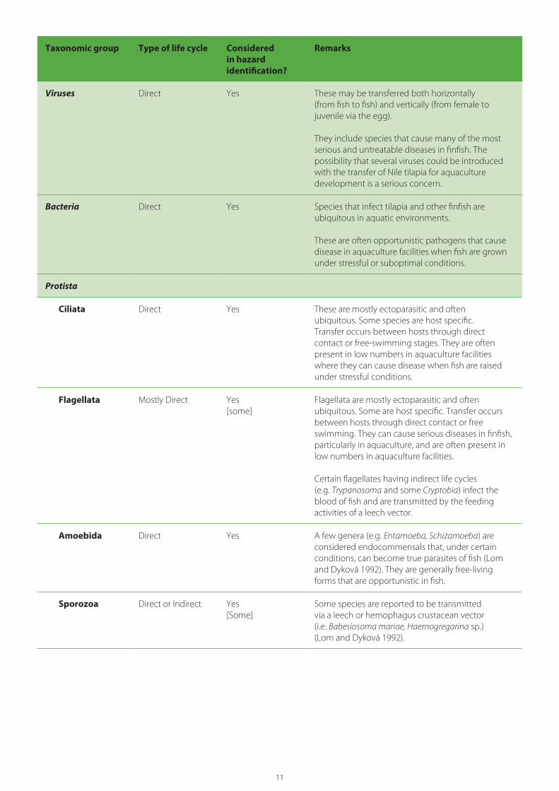
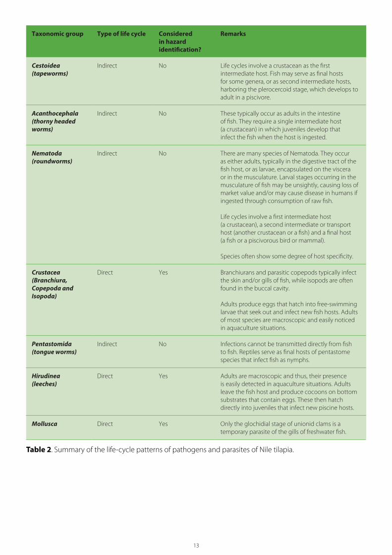
4.4. Pathogen groups excluded from analysisTable 2 lists the major taxonomic categories of potential pathogens infecting Nile tilapia and the nature of their life cycles (direct or indirect). The GIFT fry to be imported originate from broodstocks that have been cultured for many generations under conditions that would preclude infection by parasites with indirect life cycles. As such, the Myxozoa, Digenea, Cestoda, Nematoda, Acanthocephala, Pentastomida and some Protista have been excluded from further consideration.
4.5. Risk managementRisk management is the process of evaluating the estimated risk to determine if it is significant to the importing country, and if it is, of identifying, documenting and implementing measures that can be applied to reduce the level of risk to an acceptable level as expressed (explicitly or implicitly) in the country’s ALOP. Risk management measures for a given hazard (risk mitigation) are only considered when the estimated level of risk for the hazard exceeds the country’s ALOR. The level of unmitigated risk, the ALOR and the individual nature of the hazard will determine what risk management measures, if any, can be applied to reduce the risk to an acceptable level.
In this risk analysis, WorldFish has initiated or proposed extensive risk management measures to reduce the likelihood that serious pathogens will be introduced and become established in Nigeria. The risk analysis therefore takes these measures into consideration during the hazard evaluation process and will only suggest additional risk management measures should the proposed measures be insufficient to reduce the risk posed by a hazard to an acceptable level.
4.6. Terminology
4.6.1. Terms used to describe the probability of an event occurringThis risk analysis follows a five-category system to assess whether an adverse event is likely to occur:
1. High: very likely to occur
2. Moderate: an even probability of occurring
3. Low: unlikely to occur
4. Very low: very unlikely to occur
5. Negligible: almost certainly not to occur.
4.6.2. Terms used to describe the consequences of an event occurringThe terms used to describe the consequences of an adverse event occurring follow those outlined by AQIS (1999):
• Catastrophic: Establishment of disease would be expected to cause significant economic harm at a national level and/or cause serious and irreversible harm to the environment.
• High: Establishment of disease would have serious biological consequences (e.g. such as high mortality or morbidity) and would not be amenable to control or eradication. Such diseases could significantly harm economic performance at an industry level and/or may cause serious harm to the environment.
• Moderate: Establishment of disease would have less pronounced biological consequences and may be amenable to control or eradication. Such diseases could harm economic performance at an industry level and/or cause some environmental effects, which would not be serious or irreversible.
11
Taxonomic group Type of life cycle Considered in hazard identification?
Remarks
Viruses Direct Yes These may be transferred both horizontally (from fish to fish) and vertically (from female to juvenile via the egg).
They include species that cause many of the most serious and untreatable diseases in finfish. The possibility that several viruses could be introduced with the transfer of Nile tilapia for aquaculture development is a serious concern.
Bacteria Direct Yes Species that infect tilapia and other finfish are ubiquitous in aquatic environments.
These are often opportunistic pathogens that cause disease in aquaculture facilities when fish are grown under stressful or suboptimal conditions.
Protista
Ciliata Direct Yes These are mostly ectoparasitic and often ubiquitous. Some species are host specific. Transfer occurs between hosts through direct contact or free-swimming stages. They are often present in low numbers in aquaculture facilities where they can cause disease when fish are raised under stressful conditions.
Flagellata Mostly Direct Yes[some]
Flagellata are mostly ectoparasitic and often ubiquitous. Some are host specific. Transfer occurs between hosts through direct contact or free swimming. They can cause serious diseases in finfish, particularly in aquaculture, and are often present in low numbers in aquaculture facilities.
Certain flagellates having indirect life cycles (e.g. Trypanosoma and some Cryptobia) infect the blood of fish and are transmitted by the feeding activities of a leech vector.
Amoebida Direct Yes A few genera (e.g. Entamoeba, Schizamoeba) are considered endocommensals that, under certain conditions, can become true parasites of fish (Lom and Dyková 1992). They are generally free-living forms that are opportunistic in fish.
Sporozoa Direct or Indirect Yes[Some]
Some species are reported to be transmitted via a leech or hemophagus crustacean vector (i.e. Babesiosoma mariae, Haemogregarina sp.) (Lom and Dyková 1992).

12
Taxonomic group Type of life cycle Considered in hazard identification?
Remarks
Myxosporidia Indirect No These are common parasites of freshwater and marine fish that are often highly host specific.
Some genera produce macroscopic cysts in the musculature, gills and/or viscera (e.g. Henneguya, Myxobolus).
Life cycles require an obligate alternate host (a tubificid worm) in which the actinomyxin stage develops.
Some species are highly pathogenic to fish, but infections can be prevented in hatchery situations by elimination of tubificids and treatment of incoming water to kill actinomyxins.
Fungi
Septate fungi Direct Yes[1]
Most species are ubiquitous and opportunistic pathogens of fish. One species (Aphanomyces invadans), the cause of epizootic ulcerative syndrome (EUS), is a serious pathogen of freshwater and brackish-water fish.
Microspora Direct Yes Fish microspora are transmitted directly perorally, and the existence of a paratenic or intermediate host has not been shown (Lom and Dyková 1992).
Species are often highly host specific.
Monogenea (monogeneans)
Direct Yes Almost all species are ectoparasitic on the skin and/or gills. One genus occurring in tilapia species (Enterogyrus) is parasitic in the stomach.
Most species lay eggs that hatch to ciliated larvae; however, some genera (e.g. Gyrodactylus) are viviparous.
Monogeneans often are highly host specific, and there are many species that occur only on tilapia.
Digenea (digenetic trematodes)
Indirect No Digeneans infect Nile tilapia as either adults, occurring most often in the digestive tract, or as larvae (metacercariae) encysted in various parts of the body (i.e. the viscera, gills, musculature, eyes, etc.). One species occurring in tilapia (Transversotrema cichlidarum) is parasitic on the skin.
Life cycles of digeneans infecting fish typically involve three hosts: a first intermediate molluscan host (usually a snail), a second intermediate host (usually a crustacean or a fish) and a definitive (or final) host (a fish or a piscivorous bird or mammal).
13
Taxonomic group Type of life cycle Considered in hazard identification?
Remarks
Cestoidea (tapeworms)
Indirect No Life cycles involve a crustacean as the first intermediate host. Fish may serve as final hosts for some genera, or as second intermediate hosts, harboring the plerocercoid stage, which develops to adult in a piscivore.
Acanthocephala (thorny headed worms)
Indirect No These typically occur as adults in the intestine of fish. They require a single intermediate host (a crustacean) in which juveniles develop that infect the fish when the host is ingested.
Nematoda (roundworms)
Indirect No There are many species of Nematoda. They occur as either adults, typically in the digestive tract of the fish host, or as larvae, encapsulated on the viscera or in the musculature. Larval stages occurring in the musculature of fish may be unsightly, causing loss of market value and/or may cause disease in humans if ingested through consumption of raw fish.
Life cycles involve a first intermediate host (a crustacean), a second intermediate or transport host (another crustacean or a fish) and a final host (a fish or a piscivorous bird or mammal).
Species often show some degree of host specificity.
Crustacea (Branchiura, Copepoda andIsopoda)
Direct Yes Branchiurans and parasitic copepods typically infect the skin and/or gills of fish, while isopods are often found in the buccal cavity.
Adults produce eggs that hatch into free-swimming larvae that seek out and infect new fish hosts. Adults of most species are macroscopic and easily noticed in aquaculture situations.
Pentastomida(tongue worms)
Indirect No Infections cannot be transmitted directly from fish to fish. Reptiles serve as final hosts of pentastome species that infect fish as nymphs.
Hirudinea (leeches)
Direct Yes Adults are macroscopic and thus, their presence is easily detected in aquaculture situations. Adults leave the fish host and produce cocoons on bottom substrates that contain eggs. These then hatch directly into juveniles that infect new piscine hosts.
Mollusca Direct Yes Only the glochidial stage of unionid clams is a temporary parasite of the gills of freshwater fish.
Table 2. Summary of the life-cycle patterns of pathogens and parasites of Nile tilapia.
14
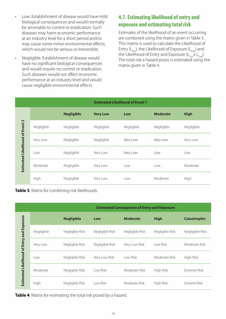
• Low: Establishment of disease would have mild biological consequences and would normally be amenable to control or eradication. Such diseases may harm economic performance at an industry level for a short period and/or may cause some minor environmental effects, which would not be serious or irreversible.
• Negligible: Establishment of disease would have no significant biological consequences and would require no control or eradication. Such diseases would not affect economic performance at an industry level and would cause negligible environmental effects.
Estimated Likelihood of Event 1
Esti
mat
ed L
ikel
ihoo
d of
Eve
nt 2
Negligible Very Low Low Moderate High
Negligible Negligible Negligible Negligible Negligible Negligible
Very Low Negligible Negligible Very Low Very Low Very Low
Low Negligible Very Low Very Low Low Low
Moderate Negligible Very Low Low Low Moderate
High Negligible Very Low Low Moderate High
Table 3. Matrix for combining risk likelihoods.
Estimated Consequence of Entry and Exposure
Esti
mat
ed L
ikel
ihoo
d of
Ent
ry a
nd E
xpos
ure
Negligible Low Moderate High Catastrophic
Negligible Negligible Risk Negligible Risk Negligible Risk Negligible Risk Negligible Risk
Very Low Negligible Risk Negligible Risk Very Low Risk Low Risk Moderate Risk
Low Negligible Risk Very Low Risk Low Risk Moderate Risk High Risk
Moderate Negligible Risk Low Risk Moderate Risk High Risk Extreme Risk
High Negligible Risk Low Risk Moderate Risk High Risk Extreme Risk
Table 4. Matrix for estimating the total risk posed by a hazard.
4.7. Estimating likelihood of entry and exposure and estimating total risk Estimates of the likelihood of an event occurring are combined using the matrix given in Table 3. This matrix is used to calculate the Likelihood of Entry (L
Ent), the Likelihood of Exposure (L
Exp) and
the Likelihood of Entry and Exposure (LEnt
x LExp
). The total risk a hazard poses is estimated using the matrix given in Table 4.

15
5. Diseases of Nile tilapia
5.1. Transboundary spread of tilapia diseases Nile tilapia are host to a wide range of pathogens and parasites (Table 5). Regarding aquaculture development, the most important are viruses, many of which the OIE (2019a) has listed as notifiable diseases having the potential to cause major economic, environmental and/or social impacts.
Many TAADs have been spread domestically, regionally and internationally, through both legal and illegal channels, by the ill-considered introduction and transfer of live aquatic animals to fuel the rapid expansion of aquaculture, often with disastrous consequences. The rapid emergence and spread of serious diseases of Nile tilapia on a regional and global basis is primarily the result of poor industry practices, including (a) the careless transboundary movement of broodstock and fry of unknown or poorly known health status, and (b) the common practice of siting both quarantine and production facilities near natural waterbodies where the likelihood of transferring pathogens between cultured stocks and wild cichlids is greatly increased. Also, the industry has often failed to recognize newly emerging diseases. This has resulted in the global spread of serious pathogens, such as TiLV, before reliable diagnostic methods have been developed to test for them.
5.2. Pathogens and parasites of Oreochromis niloticus6
As O. niloticus is by far the most widely cultured tilapia species globally, its pathogens and parasites are the most extensively documented among all cichlid species. Table 6 lists 69 taxa (species or species groups) of pathogens and parasites known to infect Nile tilapia and of potential concern to this risk analysis.
Of the diseases listed by the OIE, none has been reported from O. niloticus. Two have been reported from other tilapia species: (1) EUS from O. aureus and O. mossambicus from Zimbabwe and from Tilapia sp. from Botswana and (2) infectious pancreatic necrosis virus (IPNV) from Tilapia sp. from Kenya (Cefas 2020).
5.3. Pathogens and parasites of tilapia in NigeriaA detailed knowledge of a country’s aquatic viruses, bacteria, fungi and parasites, their pathogenicities and host and geographic distributions is fundamental to achieving healthy stocks in aquaculture production, protecting wild fish populations, and conducting pathogen risk analyses. Unfortunately, such knowledge remains very limited for Nigeria. The International Database on Aquatic Animal Diseases (Cefas 2020) contains no reports for Nigeria, either official or unofficial, of those aquatic animal diseases listed by the OIE. Sections 5.3.1 to 5.3.3 briefly discuss the state of knowledge, based on a comprehensive yet incomplete examination of the relevant literature.
5.3.1. Viral diseasesThere have been no detailed studies of the viral pathogens of Nigerian fish. The only viral disease reported from Nigeria is lymphocystis (Okaeme et al. 1988).
5.3.2. Bacterial diseasesBacterial infections are reported to be a major cause of losses to Nigerian aquaculture (Okaeme and Ibiwoye 1989; Ikpi and Offem 2010; Oladele et al. 2010 and 2015). However, there have been less than 50 studies of the bacteria causing disease in Nigerian fish culture (Annex 1). A few examples follow: In their review of problems in the culture of tilapia and Clarias in Nigerian freshwaters, Okaeme and Ibiwoye (1989) listed eight taxa (Pseudomonas putrifacium, Pseudomonas sp., Aeromonas hydrophila, Myxobacteria sp., Myxococcus sp., Staphylococcus sp., Proteus mirabilis and Enterobacter aerogenes) as common pathogens of tilapia. Shinkafi and Ukwaja (2010) identified the bacteria associated with fresh Nile tilapia purchased at a market in Sokoto, reporting six Gram-positive species (Bacillus megatanium, B. pumilus, B. alvei, B. licheniformis, Staphylococcus saprophyticus, Listeria monocytogenes), and three Gram-negative bacteria (Serratia mercescens, Providentia stuartii, Salmonella spp.). However, none of these taxa appear to have been associated

16
with disease outbreaks in aquaculture. Ashiru et al. (2011) isolated Aeromonas spp. (A. caviae, A. hydrophila, A. sobria) from the body surface and intestine of O. niloticus and/or Clarias batrachus purchased from a market in Lagos and reported on their resistance to several antibiotics. Oladele et al. (2012) implicated hemolytic strains of Staphylococcus aureus in outbreaks of jaundice syndrome in clariid catfish, while Oladele et al. (2011) associated A. sobria with arborescent organ necrosis syndrome. A major survey of bacteria present in cultured fish was conducted by Oladele et al. (2015). They isolated the following taxa from diseased fish examined from commercial catfish farms (hatcheries and grow-out ponds) in southwestern Nigeria: Staphylococcus aureus, Staphylococcus spp., Enterococcus spp., Luteococcus sanguinis, Klebsiella oxytoca, Aeromonas hydrophila, A. sobria, Streptococcus spp., Bacillus cereus, Vibrio alginolyticus, V. parahemolyticus, Bacillus spp. and Corynebacterium accolence.
5.3.3. FungiFilamentous fungi such as Saprolegnia spp. are frequent opportunistic pathogens of freshwater fish. Of the nearly 400 publications dealing with pathogens of Nigerian freshwater fish, only four studies deal with fungal infections (Annex 1). For example, Ogbonna and Alabi (1991) recorded a diversity (24 species belonging to 6 genera) of aquatic phycomycete fungi from infected fish in a freshwater pond.
5.3.4. ParasitesBecause of the vast literature dealing with Nigerian fish parasites (some 350 publications were encountered, see Annex 1), the inaccessibility of many of these publications and the limited time allowed for this study, this section is restricted to a consideration of the parasites of the cichlid species reported from Nigeria.
Nigerian workers have published several rather incomplete reviews dealing with the parasites of Nigerian freshwater fish. Adebambo (2020), for example, listed only 55 publications as containing original records. In a review of the parasites of catfish and tilapia in the wild and homestead ponds in Nigeria, Flourizel et al. (2019) cited only a single publication related to tilapia parasites. Okaeme et al.
(1988 and 2001) briefly reviewed the parasites reported from tilapia of the Lake Kainji area.
More general publications dealing with the parasites of freshwater fish of Africa include the following:
• the guides of Paperna (1996) and Scholz et al. (2020)
• the checklist of parasites of freshwater fish of Southern Africa by Van As and Basson (1984)
• the listing of pathogens and parasites of East African freshwater fish by Akoll and Mwanja (2012)
• the checklist of helminths of African freshwater fish by Khalil (1971), updated by Khalil and Polling (1997).
To date, there has not been a comprehensive, critical review or detailed checklist of the parasites reported from Nigerian fish.
Research on the fish parasites of Nigeria appears to have been hampered by a lack of specialized taxonomic expertise as well as limited access to essential literature. As a result, many studies have not been able to identify parasite taxa to species, while other studies appear to include incorrect identifications. Unfortunately, many of the papers published by Nigerian workers have appeared in online journals that have often provided inadequate peer review or skilled editing support. In only few cases have authors sent specimens to international experts to confirm identifications or deposited voucher specimens in a recognized museum so that their identifications could be verified later. Almost all parasitological attention has focused on a few species of catfish (genera Clarias, Chrysichthys and Synodontis) and tilapia (genera Oreochromis, Coptodon, Sarotherodon, Hemichromis, Chromidotilapia and Pelmatolapia). FishBase (Froese and Pauly 2019) provides an incomplete listing of the diverse fish fauna of Nigeria, with a total of 803 species, of which 478 are marine and 334 are freshwater species. Of these, 21 belong to the family Cichlidae (the tilapia species) and, of these, records of pathogens or parasites appear in the Nigerian literature for only 11 (Table 5). The end result is that despite considerable efforts, the parasites of Nigerian fish remain poorly known.

17
Table 5 lists the parasites reported from Nigerian cichlids. Although this list undoubtedly includes a number of misidentifications, it does provide an indication of the diverse parasite fauna present in
Taxon Host species References
Protista
“Amoeba” Oreochromis niloticus Okaeme et al. 1987
Babeiosoma sp. O. niloticus Biu et al. 2014
Chilodonella sp. O. niloticus Nyaku et al. 2007; Ashade et al. 2010; Osoh et al. 2017
Cryptobia sp. Coptodon zilliiO. niloticusSatherodon galilaeus
Osoh et al. 2017
Eimeria sp. C. zilliiO. niloticusS. galilaeus
Okaeme et al. 1987
Entamoeba sp. S. melanotheron Akinsanya et al. 2018a
Epistylis sp. O. niloticus Ashade et al. 2010
Haemogregarina sp. O. niloticus Biu et al. 2014
Hexamita sp. O. niloticus S. galilaeusS. melanotheron
Okaeme et al. 1987; Osoh et al. 2017; Akinsanya et al. 2018a
Ichthyobodo necatrix C. zilliiO. niloticusS. melanotheron
Omoniyi and Ojelade 2017
Ichthyophthirius multifillis C. zillii(syn: Tilapia melanopleura)O. niloticusS. galilaeus
Okaeme et al. 1987; Nyaku et al. 2007; Ashade et al. 2010; Abidemi-Iromini and Eze 2011; Bello-Olusoji et al. n.d.; Osoh et al. 2017; Enyidi and Uwanna 2019
Piscinoodinium sp. O. niloticus Nyaku et al. 2007
Tetrahymena sp. O. niloticus Nyaku et al. 2007
Trichodina acuta C. zilliiO. niloticus
Bello-Olusoji et al. n.d.; Abidemi-Iromini and Eze 2011
Trichodina heterodentata O. niloticus Enyidi and Uwanna 2019
Trypanosoma tincae C. zilliiO. niloticus
Bello-Olusoji et al. n.d.
Trypanosoma toddi Hemichromis sp. Abolarin 1970
Nigerian cichlids. This list includes some 78 genera of parasites, as follows: 14 Protista, 1 Myxosporea, 4 Monogenea, 11 Digenea, 13 Cestoda, 20 Nematoda, 5 Acanthocephala, 5 Crustacea and 5 Hirudinea.

18
Taxon Host species References
Myxosporea
Myxobolus agolus C. guineensis O. niloticusS. galilaeusand hybrids
Obiekezie and Okaeme 1990
Myxobolus brachyspora C. guineensis O. niloticusS. galilaeusand hybrids
Obiekezie and Okaeme 1990
Myxobolus cyprini C. zilliiO. niloticus
Bello-Olusoji et al. n.d.
Myxobolus equitoralis C. guineensis O. niloticusS. galilaeus
Obiekezie and Okaeme 1990
Myxobolus galilaeus O. niloticusS. galilaeusand hybrids
Obiekezie and Okaeme 1990
Myxobolus homeospora O. niloticus Obiekezie and Okaeme 1990
Myxobolus israelensis C. guineensis O. niloticusS. galilaeusand hybrids
Okaeme et al. 1987; Obiekezie and Okaeme 1990
Myxobolus kainjiae O. niloticusS. galilaeus
Obiekezie and Okaeme 1990
Myxobolus sarigi O. niloticusS. galilaeusand hybrids
Okaeme et al. 1987; Obiekezie and Okaeme 1990
Myxobolus tilapiae C. zillii O. niloticusS. galilaeus
Abolarin 1974; Obiekezie and Okaeme 1990
Monogenea
Dactylogyrus extensus O. niloticus Enyidi and Uwanna 2019
Enterogyrus cichlidarum O. niloticus Musa et al. 2007
Gyrodactylus malalai O. niloticus Adeshina et al. 2021
Gyrodactylus vastator[probable misidentification]
C. zilliiO. niloticus
Bello-Olusoji et al. n.d.
Macrogyrodactylus sp. O. niloticus Ashade et al. 2010
Digenea
Allocreadium ghanensis C. zilliiS. galilaeusS. melanotheron“cichlids”
Morenikeji and Adepeju 2009; Simon-Oke and Morenikeji 2015; Simon-Oke 2017

19
Taxon Host species References
Alloglossidium corti C. zilliiS. galilaeusS. melanotheron“cichlids”
Morenikeji and Adepeju 2009; Hassan et al. 2013; Simon-Oke and Morenikeji 2015; Simon-Oke 2017
Aranthocephalus sp. metacercaria [lapsus?]
O. niloticusS. galilaeus
Okaeme et al. 1987
Clinostomum complanatum metacercaria
C. guineensisC. zilliiO. niloticusS. melanotheron
Echi et al. 2009a, 2009b, 2012, 2014; Amaechi 2015
Clinostomum marginatum metacercaria
O. niloticus Ashade et al. 2010
Clinostomum tilapiae metacercaria
Chromidotilapia guntheriCoptodon guineensisC. zilliiHemichromis elongatusH. fasciatusO. niloticusS. galilaeusS. melanotheron Pelmatolapia mariae (syn. T. mariae)“cichlids” “tilapias”
Okaeme et al. 1987; Okaeme and Ibiwoye 1989; Musa et al. 2007; Adeyemo and Agbede 2008; Morenikeji and Adepeju 2009; Echi et al. 2009a, 2009b, 2012, 2014; Olurin et al. 2012; Hassan et al. 2013; Amaechi 2015; Ajala and Fawole 2015; Simon-Oke and Morenikeji 2015; Awharitoma and Ehigiator 2017; Simon-Oke 2017; Olugbotemi and Morenikeji 2018; Osimen and Anagha 2020
Crepidostomum metoecus [probable misidentification]
C. zillii Yakubu et al. 2002
Diplostomulum tregenna metacercaria
C. zillii Yakubu et al. 2002; Goselle et al. 2008
Euclinostomium heterostomum metacercaria
C. guineensis C. zilliiO. niloticusS. galilaeusS. melanotheron “cichlids”
Okaeme et al. 1987; Ukpai 2001; Morenikeji and Adepeju 2009; Echi et al., 2009a, 2009b, 2012, 2014; Ohaeri 2012; Hassan et al. 2013; Saliu et al. 2014; Amaechi 2015; Simon-Oke and Morenikeji 2015; Simon-Oke 2017
Heterophyes sp. metacercaria
O. niloticus Ashade et al. 2010
Neascus sp. metacercaria C. zilliiO. niloticusS. galilaeusS. melanotheron“cichlids”“tilapias”
Okaeme et al. 1987; Okaeme and Ibiwoye 1989; Morenikeji and Adepeju 2009; Hassan et al. 2013; Simon-Oke and Morenikeji 2015; Simon-Oke 2017; Abba et al. 2018a, 2018b
Phagicola longa C. zilliiH. bimaculatusH. fasciatusO. niloticusS. galilaeusS. melanotheron“cichlids”
Morenikeji and Adepeju 2009; Simon-Oke and Morenikeji 2015; Hassan et al. 2013; Simon-Oke 2017

20
Taxon Host species References
Sphaerostoma bramae[probable misidentification]
C. zillii Yakubu et al. 2002
Cestoda
Anomotaenia sp. larva H. fasciatus O. niloticus
Aderounmu and Aeniyi 1972
Biacetabulum [?] appendiculatum
Ch. guntheri Osimen and Anagha 2020
Bothriocephalus aegyptiacus O. niloticus Abba et al. 2018a, 2018b
Caryophyllaeides sp. C. zillii Omoniyi and Ojelade 2017
Caryophyllaeus sp. C. zillii“tilapia”
Ukpai 2001; Biu and Nkechi 2013
Diphyllobothrium latum plerocercoid [probable misidentification]
O. niloticus Edeh and Solomon 2016; Awosolu et al. 2018
Eubothrium crassum[probable misidentification]
C. zillii Yakubu et al. 2002
Hymenolepis nana [probable misidentification]
C. zillii Iyabo and Ijeoma 2017
Monobothrium sp. C. zillii Alade et al. 2015
Paradilepis sp. larvae C. guineensisO. niloticus
Ezeri 2002; Joseph et al. 2020
Proteocephalus sp. C. guineensisC. zilliiO. niloticus
Saliu et al. 2014; Onoja-Abutu et al. 2021
Polyonchobothrium clarias C. zillii Alade et al. 2015; Mgbemena et al. 2020
Wenyonia minuta S. melanotheron Akinsanya et al. 2018b
Nematoda
Ascaris sp. [probable misidentification]
C. zilliiO. niloticusS. galilaeus
Osoh et al. 2017
Camallanus polypteri C. zilliiO. niloticus
Ejere et al. 2014; Enyidi and Uwanna 2019
Capillaria cichlasomae C. zillii Ejere et al. 2014
Contracaecum osculatum Ch. guntheriC. zilliiO. niloticus
Osimen and Anagha 2020
Cucullanus barbi Ch. guntheri Edema et al. 2008
Cucullanus baylisi C. zilliiO. niloticusS. galilaeus
Ibiwoye et al. 2006

21
Taxon Host species References
Cucullanus sheilanensis C. zillii Akinsanya 2016
Dichelyne sp. Ch. guntheri Awharitoma and Ehigiator 2017
Eustronglydes sp. larvae C. zilliiO. niloticus
Ashade et al. 2010; Sikoki et al. 2013; Sani et al. 2019; Mgbemena et al. 2020
Gnathostoma spinigerum larvae
O. niloticus Awosolu et al. 2018
Goezia sigalasi O. niloticus Sikoki et al. 2013
Philonema sp. O. niloticus Sani et al. 2019
Paracamallanus cyathopharynx
S. galilaeus“tilapia”
Ukpai 2001; Ajala and Fawole 2015
Procamallanuis laevionchus C. guineensisC. zilliiO. niloticus S. galilaeus
Opara and Okon 2002; Ibiwoye et al. 2006; Okogwu et al. 2011; Alade et al. 2015; Ajala and Fawole 2015; Awharitoma and Ehigiator 2017; Abba et al. 2018a, 2018b; Osimen and Anagha 2020
Raphidascaroides sp. C. zillii Akinsanya et al. 2018b
Rhabdochona congolensis C. zilliiO. niloticus
Onoja-Abutu et al. 2021
Serradacnitis serrata O. niloticus Domo and Ester 2015
Spinitectus guntheri C. zilliiO. niloticus
Onoja-Abutu et al. 2021
Spirocamallanus spiralis H. elongatus Awharitoma and Ehigiator 2017
Spironoura petrei O. niloticusS. galilaeus
Ibiwoye et al. 2006
Trichiuris sp. [probable misidentification]
O. niloticus Ashade et al. 2010
Trichostrongylus sp. [probable misidentification]
C. zillii Iyabo and Ijeoma 2017
Acanthocephala
Acanthogyrus (Acanthosentis) tilapiae
Ch. guntheriC. zilliiH. bimaculatusH. elongatusH. fasciatusS. galilaeusS. melanotheronO. aureus(syn.: T. aurea) O. niloticusP. mariae“cichlids” “tilapias”
Shotter 1974; Hyslop 1988; Okaeme and Ibiwoye 1989; Morenikeji and Adepeju 2009; Matuoke et al. 2011; Hassan et al. 2013; Ajala and Fawole 2015; Simon-Oke and Morenikeji 2015; Awharitoma and Ehigiator 2017; Simon-Oke 2017; Ito 2017; Atalabi et al. 2018
Neoechinorhynchus rutili C. zilliiO. niloticusS. galilaeus
Olurin et al. 2012; Alade et al. 2015; Ajala and Fawole 2015; Abba et al. 2018a, 2018b
22
Taxon Host species References
Octospiniferoides sp. cichlids Nmor et al. 2004
Pomphorhynchus sp. O. niloticus Sani et al. 2019
Rhadinorhynchus horridus H. elongatus Awharitoma and Ehigiator 2017
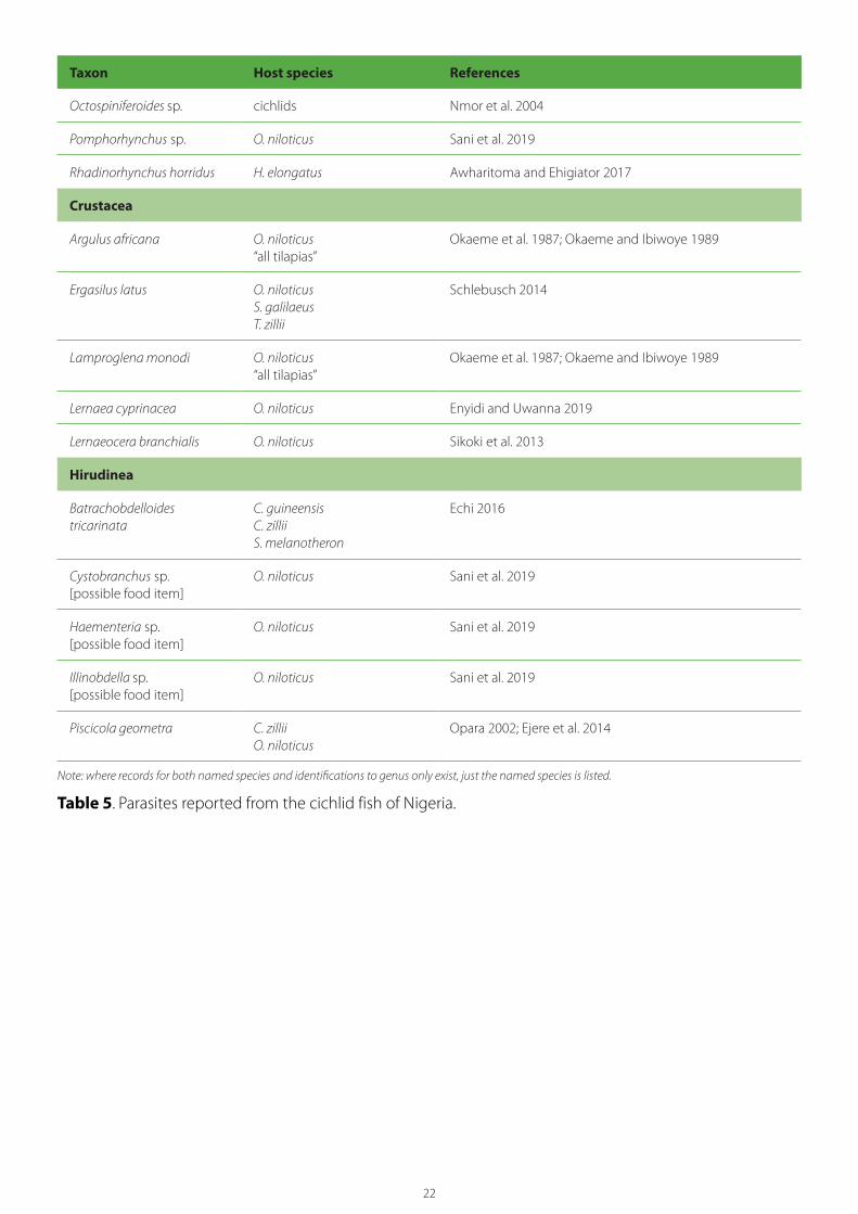
Crustacea
Argulus africana O. niloticus “all tilapias”
Okaeme et al. 1987; Okaeme and Ibiwoye 1989
Ergasilus latus O. niloticus S. galilaeus T. zillii
Schlebusch 2014
Lamproglena monodi O. niloticus“all tilapias”
Okaeme et al. 1987; Okaeme and Ibiwoye 1989
Lernaea cyprinacea O. niloticus Enyidi and Uwanna 2019
Lernaeocera branchialis O. niloticus Sikoki et al. 2013
Hirudinea
Batrachobdelloides tricarinata
C. guineensisC. zilliiS. melanotheron
Echi 2016
Cystobranchus sp.[possible food item]
O. niloticus Sani et al. 2019
Haementeria sp.[possible food item]
O. niloticus Sani et al. 2019
Illinobdella sp.[possible food item]
O. niloticus Sani et al. 2019
Piscicola geometra C. zillii O. niloticus
Opara 2002; Ejere et al. 2014
Note: where records for both named species and identifications to genus only exist, just the named species is listed.
Table 5. Parasites reported from the cichlid fish of Nigeria.
23
6. Health status of GIFT
The parental stock (G17) of the GIFT swim-up fry to be transferred to Nigeria will originate from WorldFish’s GIFT broodstock facility (a secure holding/breeding facility) in Malaysia. The stock is derived from WorldFish’s original core GIFT selective breeding stock maintained at Jitra in Malaysia. Following the first reports of TiLV in Malaysia in 2016 and considering the risks to WorldFish’s core GIFT selective breeding program at Jitra, a backup selective breeding program was established on the premises of WorldFish’s headquarters in Penang, Malaysia. In May 2017, a second transfer was made, with several families from G16 moved from Jitra to Penang.
Both transfers to the Penang facility took place well before the reported outbreak of TiLV that occurred in a GIFT stock contained in a single pond at the Jitra facility in February 2018. In November 2020, the Jitra facility was closed, and the Penang facility was transformed into WorldFish’s GIFT broodstock facility, where the GIFT breeding population is now maintained.
To confirm their health status and freedom from TiLV, screening of backup stocks belonging to G14, G15 and G16 was conducted. Following this, detailed planning and mating designs were done to use these backup stocks to produce the desired number of G17 families. A rigorous health screening was adopted throughout the process. Fertilized eggs, swim-up fry and fingerlings (at tagging) of all G17 families were screened for TiLV using the most appropriate diagnostic methodology (i.e. the TaqMan qPCR (realtime polymerase chain reaction (PCR)) method. In addition to G17, all parents used (coming from G14, G15 and G16) were sacrificed post-breeding to check for freedom from TiLV. This entire process, conducted between January 2019 and July 2020, involved testing some 4774 fish samples. The results confirmed that the Penang broodstock is free from TiLV and that the G17 families now being raised to produce G17–F1 are free from TiLV. Regular and rigorous screening for TiLV started in October 2019 and all laboratory records are available.
As part of WorldFish’s SOPs, broodstock are screened bi-annually for a list of pathogens (up to
12) by sampling up to 60 fish. Additionally, general health screening (based on wet smears and some histology) has been conducted on the fish held in the Penang facility since its establishment. Although no laboratory records have been kept, there have been no mortalities or TiLV outbreaks in this facility since its establishment in 2013.
The batch of swim-up fry to be transferred to Nigeria will originate from the health-screened G17 population in Penang. Pathogen screening will be conducted on samples taken from batches of fish that are being reared for export. The list of pathogens should take into consideration the OIE’s reportable disease list, the Malaysian national pathogen list, the WorldFish priority list, the recommendations of this risk analysis and any additional screening requested by the GON. This work will be linked to Malaysia Biosecurity as part of its work for issuing the health certificate that will accompany the GIFT fry being shipped to Nigeria.
The batch of GIFT swim-up fry to be transferred to Nigeria will be produced in a clean and regularly disinfected hatchery facility under artificial incubation procedure. Before packing for transfer, the fry will be surface disinfected using standard methodology and then bagged in clean water.
A summary of the pathogens for which screening of broodstock was conducted in October/November 2020 is given in Table 6. As provided by WorldFish, the results were negative for the presence of all pathogens for which testing was conducted. Another comprehensive health assessment will be conducted for the same batch of fish in March 2021, with the results expected around the end of April 2021.
It should be noted that broodstock held in or fry produced by WorldFish’s GIFT broodstock facility should not be referred to as “SPF” or “high health.” These are terms specific to certain stocks of penaeid shrimp that meet rigorous criteria with regard to their pathogen status. The definitions of these terms were formalized by the United States Marine Shrimp Farming Program in the 1980s and have recently been reviewed by Alday-Sanz et al. (2020). The SPF concept (in a slightly different form)

24
has also been applied in the salmonid culture industry, through the use of SPF eggs to prevent the movement of certain pathogens between facilities, and a stock of zebrafish (Danio rerio) that is SPF for a single pathogen is now available from Oregon State University (Kent et al. 2011). However, all stocks of aquatic animals, whether SPF or not, may still carry some pathogens.
In the development of SPF fishstocks, egg-laying fish provide an advantage over live-bearing fish in that pathogen exposure to the next generation can be reduced by separating eggs from their parents and disinfecting them before introducing
them into another facility. The introduction of new stocks via chlorine surface-disinfected eggs allows for the possibility of applying the same principles and methods routinely used in salmonid aquaculture for establishing SPF stocks to other fish species, such as screening broodstock and sex products and rearing fry in an environment completely separate from potentially infected fish, including broodstock (Kent et al. 2011). However, in the current transfer, as Nile tilapia are mouth brooders, and eggs will not be immediately separated from parent fish, there is a greatly increased opportunity for pathogens to be transferred directly from parent to offspring.
Population No. of samples
Type of samples Sampling date
Analysisdate
Pathogens screened1
Result
Penang G17 60 Pooled liver, spleen and kidney 15–6/10/2020 2–8/12/2020 Iridovirus Negative
ISKNV Negative
EUS Negative
SA Negative
SI Negative
KHV Negative
CEV Negative
EHNV Negative
TiLV Negative
1 ISKNV = infectious spleen and kidney necrosis virus, EUS = epizootic ulcerative syndrome caused by Aphanomyces invadans, SA = Streptococcus agalactiae, SI = S. iniae, KHV = koi herpesvirus, CEV = carp edema virus, EHNV = epizootic hematopoietic necrosis virus, TiLV = tilapia lake virus.
Note: baseline health assessment for 9 pathogens (6 virus, 2 bacteria and 1 fungus) using conventional PCR.
Table 6. Results of baseline health screening of GIFT broodstock (G17) held in Penang.
25
7. Risk management measures
WorldFish has proposed the risk management measures discussed in sections 7.1 to 7.9, with some already in progress.
7.1. Use of fry derived from the GIFT breeding program broodstockWorldFish proposes to biosecurely transfer an initial batch of 10,000 GIFT swim-fry from its GIFT broodstock facility in Malaysia to Nigeria in 2021. Fry will be produced using G17 GIFT as parents. Transferred fry will be kept in a designated land-based, secured quarantine facility in Nigeria’s Ogun State, where they will be raised with regular health checks. G17-F3 progeny produced from G17-F2 fish resulting from the originally transferred stock (G17-F1) will be transferred to Delta State for breeding (non-sex reversed fry weighing 10 g) and for grow-out (sex reversed all male fry weighing 2 g). If this plan is not achieved for any reason, at any state of the process, all remaining fish will be destroyed, and all facilities will be cleaned and fallowed adequately.
7.2. Documented testing for pathogens of GIFT broodstock and fryThe GIFT strain has been housed at the Penang facility without addition of new broodstock since 2017 and has been subjected to a number of diagnostic tests since the initial establishment of the facility in 20l3. A summary of the most recent diagnostic testing that has been performed on the GIFT stock is given in Table 6. In particular, extensive testing has been done to assure that the stock is free of TiLV (Section 6).
7.3. High-security quarantine of imported fryUpon arrival in Nigeria, the swim-up fry will be housed in a biosecure, land-based quarantine facility to be constructed in Ogun State that will prevent their escape and that of subsequent generations. Quarantine facilities will meet minimum standards of construction and will follow SOPs appropriate to such high-containment facilities, as outlined in Section 4 of Arthur et al. (2007). Construction and operating standards will also minimize the possibility of diseases that may be present in the external environment gaining entry to the facility.
7.4. Monitoring and diagnostic testing of GIFT while in quarantineTransferred stock and their progeny (G17-F1, F2, F3) kept in the biosecure quarantine facility in Ogun will be monitored for health on a daily basis. They will be tested for specified pathogens upon arrival and at 6-month intervals for 2 years before being released from the facility. Diagnostic testing will also be conducted should any unexplained mortalities occur. Any subpopulation infected with an untreatable exotic pathogen will be destroyed.
7.5. Releasing only G17–F3 GIFT to NBCsThe transferred GIFT will be used to produce G17–F2 and F3 progeny. All broodstock reared from the transferred fry used to develop the parent stock in Nigeria will be destroyed and disposed of in a sanitary manner (OIE 2019a) once they are no longer useful for breeding.
WorldFish will develop a list of pathogens of concern that will form the basis for subsequent diagnostic testing. Following successful testing, G17-F3 progeny will be released from the quarantine facility to be cultured in both land-based and water-based systems (ponds and cages) in Ogun and Delta states.
7.6. Diagnostic testing of Nigerian tilapia and catfish for some important pathogensWorldFish has arranged to conduct diagnostic testing on selected target organs taken from catfish and tilapia currently being held in the facility that will be upgraded to become the GIFT high-biosecurity quarantine facility in Ogun. A certified diagnostics laboratory in Malaysia will test for a number of pathogens causing important diseases in tilapia culture. These will include four viral pathogens (TiLV, ISKNV, KHV and VNNV), the fungal pathogen (Aphanomyces invadans) that causes EUS, and seven bacterial pathogens (Streptococcus agalactiae, S. iniae and S. dysagalactiae, which are the causative agents of streptococcosis and Aeromonas hydrophila, A. veronii, A. jandaei and A. schubertii, which are causative agents of hemorrhagic septicemia).

26
The test results will reveal any pathogens currently present in the culture facility that may have the potential to infect the transferred GIFT fry.
7.7. Expert risk management panelWorldFish has commissioned a panel of three experts to examine possible genetic, ecological and environmental, and pathogen risks and to recommend additional risk management measures to be incorporated into the strategic plan. This document is one of these studies.
7.8. Independent national scientific advisory teamWorldFish will set up an independent national advisory team consisting of representatives from key government agencies, the aquaculture sector and other stakeholders. Nigeria’s competent authority, the Department of Fisheries and Aquaculture, will lead the group meetings and dialogue toward building consensus on the proposed risk management strategy.
7.9. Contingency planningConsidering all precautionary measures to be taken during the transfer, breeding, seed dissemination and grow-out of GIFT in Nigeria,
WorldFish considers that two emergency situations are possible. One is the identification of an important exotic pathogen (disease) in the transferred or subsequent populations (G17–F1, F2, F3 stocks) held in the quarantine/breeding facility. During the first 3 years, all stocks will be regularly tested for important pathogens. If infection by an important and untreatable exotic pathogen is found, WorldFish will destroy the infected subpopulation(s) to ensure the pathogen is not transferred to the outside environment. In addition, WorldFish will continue regular testing of other subpopulations for 2 years to ensure the facility is free from the pathogen of concern.
The second emergency situation is the escape of an exotic pathogen from the high-biosecurity facility and its establishment in the external aquatic environment through a breach of biosecurity. WorldFish does not foresee the accidental escape of GIFT from the broodstock holding and breeding facility, as all measures (infrastructure, engineering and management) will be taken and in place following introduction.
Working with Great Lakes Products Limited in northern Zambia.
Phot
o cr
edit:
Cho
sa M
wee
mba
/Wor
ldFi
sh
26

27
8. Hazard identification
8.1. Criteria to be considered a hazardAs outlined in Section 4.2, hazard identification will proceed in several steps. The first step will consider the pathogens and parasites reported from Nile tilapia and assess whether these “possible hazards” should be considered “potential hazards” to be given further screening. For a possible hazard to be given further consideration in the risk analysis, the following criteria must be fulfilled:
1. The pathogen must have been reported to infect Nile tilapia.
2. The agent must be an obligate pathogen (i.e. it is not a ubiquitous free-living organism that is capable of becoming an opportunistic pathogen of Nile tilapia under certain environmental or culture conditions).
3. The agent must cause significant disease outbreaks and associated losses in populations of Nile tilapia or, if not a significant pathogen of this species, it must cause serious disease outbreaks in populations of other species of tilapine fish.
4. It must be plausible that that the agent might be in the WorldFish’s GIFT breeding facility in Malaysia and in the shipment of GIFT fry that will be approved for importation to Nigeria.
The second step in hazard identification will take into account the various risk management measures in process or proposed by WorldFish. To be considered a hazard that requires additional risk management measures, the following criteria will be applied:
5. Should the pathogen be present in the imported GIFT fry, it must be plausible that the risk management measures proposed by WorldFish would be insufficient to prevent its transfer to Nigeria with infected fry (“entry”) and its escape or release from quarantine and infection of cultured and/or wild fish populations in Nigeria (“exposure”).
8.2. Results of the hazard identification: Preliminary screeningThe results of this initial step are given in Table 7. Using criteria 1–4, a total of 69 “possible hazards” (pathogen species or species groups) have been considered, and 20 of these have been identified as “potential hazards” for further assessment. These include 7 viruses, 1 fungus, 8 protozoans, 2 monogeneans and 2 crustaceans.
8.3. Pathogens considered furtherPreliminary hazard identification has identified 20 potential pathogens that require further assessment. These include 7 viruses, 1 fungus, 8 protozoans, 2 monogeneans and 2 crustaceans.
The 20 possible hazards that meet all of the requirements of preliminary screening are listed in Table 8. The risk management measures in progress or proposed by WorldFish are taken into consideration during the assessment of each potential hazard’s (a) “likelihood of entry” (i.e. the likelihood that the pathogen is present in GIFT fry and will be transferred from Malaysia to the border of Nigeria along with the exported shipment of fish), and (b) the “likelihood of exposure” (i.e. the likelihood that the hazard, having gained entry to Nigeria, will escape from the quarantine facility and become established in cultured or wild populations). This likelihood estimate (entry x exposure) is then combined with an estimate of consequence to obtain an estimate of total risk. It is important to note that should the estimated risk for a given hazard become “negligible” at any point in the risk assessment process, then the risk assessment for that individual hazard is stopped.

28
Pathogen Infects Nile tilapia?
Infects Nile tilapia fry?
Causes significant disease?
Present in WorldFish facility?
Present in Nigeria?
Further consideration required?
Comments
VIRUSES
Tilapia lake virus (TiLV)
Yes Yes Yes Possible Possible Yes TiLV appears to be specific to tilapia. It has been reported from both cultured and wild tilapia, including Nile tilapia, hybrid tilapia (O. niloticus x O. aureus), red tilapia (O. niloticus x O. mossambicus), O. aureus, Sarotherodon galilaeus, Coprodon zillii and Tristamellasimonis intermedia (FAO 2018). All species of tilapia are thus likely to be susceptible.
Associated mortalities are reported as ranging from moderate (5%–20%) to high (80%–90%) (FAO 2018). Mortalities have been reported in juveniles and adults. Early developmental stages of tilapia (fertilized eggs, yolk-sac fish and fry) have also tested positive (Dong et al. 2017a).
TiLV has spread rapidly since it was initially reported in Israel in 2009 and is now confirmed in at least 16 countries on three continents (Africa, Asia and South America). Because live tilapia are highly traded internationally, it is probably also present in a many other countries.7
• In Africa: Angola, Egypt, Ghana (in co-infections with ISKNV and Streptococcus agalactiae 1b), Israel and Lake Victoria, which borders Uganda, Kenya and Tanzania (FAO 2018; Reantaso 2020; Lee and Arnaud 2020).
• In South America: Colombia, Ecuador and Peru (FAO 2018). It is now known that the cause of tilapia syncytial hepatitis in Ecuador is TiLV.
• In Asia: Bangladesh, India, Indonesia Malaysia, the Philippines, Thailand and Taiwan (Baoprasertkul 2020; FAO 2020; Somga 2020).
• In North America: detected in juvenile tilapia imported into the US and subsequently eradiated (Hartman 2020). Also reported from Mexico (Debnath et al. 2020).
An expert knowledge elicitation risk assessment for TiLV (FAO 2018) estimated that the overall risk to Africa from this pathogen is “high.” Nigeria was included on a list of 43 countries potentially at risk to TiLV due to importation of tilapia fry from known infected countries.
In February 2018, an outbreak of disease due to TiLV caused high mortality in GIFT housed in an outdoor pond at the WorldFish facility in Penang, Malaysia. Using data collected during this outbreak, Barria et al. (2020) reported significant and high host resistance to TiLV in a Nile tilapia breeding population of GIFT origin. These results highlight the significant potential of harnessing selective breeding to improve host resistance to TiLV in farmed Nile tilapia populations.

29
Pathogen Infects Nile tilapia?
Infects Nile tilapia fry?
Causes significant disease?
Present in WorldFish facility?
Present in Nigeria?
Further consideration required?
Comments
Infectious spleen and kidney necrosis virus (ISKNV)
Yes Yes Yes Possible Possible Yes ISKNV, an iridovirus, is the type species of the genus Megalocytivirus and causes disease in a range of freshwater and marine fish.
ISKNV is closely related to red sea bream iridovirus (RSIV), and both viruses are notifiable to the OIE (2019a). The OIE (2019a) considers ISKNV distinct from RSIV but as just one of the viruses causing RSID. In addition to mandarin fish, the OIE (2019b) also lists red drum, flathead mullet and Epinephelus sp. as susceptible to ISKNV.
Because of the many similar viruses, the host and geographical range of ISKNV remains unclear, and summaries often list ISKNV as being among the RSIV-like viruses.8
Although the OIE does not list tilapia as a susceptible species, recent reports from the US and Thailand suggest that it is susceptible and likely to suffer significant mortality (Ramírez-Paredes et al. 2021).
ISKNV appears to be an emerging pathogen with the potential to severely impact tilapia culture in Africa and elsewhere. Ramírez-Paredes et al. (2021) have recently reported very high mortality (>50% production) in intensive tilapia cage culture systems across Lake Volta in Ghana. Affected Nile tilapia showed darkening, erratic swimming and abdominal distension with associated ascites. Histopathological observations of tissues taken from moribund fish revealed lesions indicative of viral infection. These included hematopoietic cell nuclear and cytoplasmic pleomorphism with marginalization of chromatin and fine granulation.
Bohle iridovirus (BIV)
Possible Possible Yes No No No BIV is a Ranavirus that was first isolated from ornate burrowing frogs that died at the time of metamorphosis. Although rarely recognized in natural disease, experimental studies indicate that BIV is a potential pathogen of fish, amphibians and reptiles.
Areil and Owens (1997) reported periodic mortalities reaching 100% over a period of 60 days in O. mossambicus fry. Moribund fish exhibited rapid corkscrew-like swimming patterns, and the syndrome was successfully transmitted via cannibalism to naïve populations of tilapia fry. BIV-infected tilapia and those succumbing to “spinning tilapia syndrome” shared similarities in histopathological lesions of kidney and muscle. The barramundi Lates calcarifer bioassay indicated that the etiological agent of the epizootic was BIV.
The geographic distribution of BIV is restricted to Australia, with reports of infection limited to an Australian aquatic laboratory (Johnston 2008).

30
Pathogen Infects Nile tilapia?
Infects Nile tilapia fry?
Causes significant disease?
Present in WorldFish facility?
Present in Nigeria?
Further consideration required?
Comments
Spring viremia of carp virus (SVCV)
Yes [?] Possible Yes Possible No Yes SVCV is a member of the genus Vesiculovirus in the family Rhabdoviridae. It is the cause of spring viremia in carp (SVC), an OIE-listed disease causing an acute hemorrhagic and contagious viremia in several carp species and some other cyprinids and ictalurids.
Although SVCV was reported to have been isolated from Nile tilapia from Egypt by Soliman et al. (2008), the OIE (2019b) notes that immuno-histochemistry constituted the sole basis for this identification; electron microscopy purported to show the virus in the nucleus, which is not a feature of SVCV infection. Ibrahim (2020) suggested that Nile tilapia is more resistant to SVCV than the susceptible hosts (Cyprinidae). However, under stressful conditions leading to immunosuppression, it can be infected with the virus and serve as a carrier. There have been no subsequent reports of isolation of SVCV from tilapia or other cichlids. As such, the ability of SVCV to infect Nile tilapia requires confirmation. SVCV has not been reported from Southeast Asia or West Africa. The OIE (2019b) notes that its confirmed distribution includes most European countries, and certain Independent States of the former Soviet Union (Belarus, Georgia, Lithuania, Moldova, Russia and the Ukraine), Brazil, the US, Canada and China.
Confirmation of the isolation of SVCV from rainbow trout and Indian carp in Iran, and from Nile tilapia in Egypt awaits more data.
Tilapia larval encephalitis virus (TLEV)
Possible Possible Yes No No No TLEV is a herpes-like virus (family Herpesviridae) causing viral encephalitis of tilapia larvae, characterized by neurological signs and increased mortality of laboratory-reared larvae of blue tilapia (O. aureus). It has only been reported from Israel (Shlapobersky et al. 2010); there have been no reports of similar neurological conditions elsewhere and no identification of this virus outside Israel.
Aquabirnavirus (IPNV-like virus)
Possible Possible Yes Possible Possible Yes Infectious pancreatic necrosis virus (IPNV) is the type species of the genus Aquabirnavirus (family Birnaviridae) and is the cause of infectious pancreatic necrosis, an acute and highly contagious disease of juvenile salmonids.
According to Dopazo (2020), the term IPNV is strictly used for those strains affecting salmonids that develop specific clinical signs. Where the virus affects non-salmonids, with different clinical signs, the term “IPNV-like” is applied. In general terms, the name aquabirnaviruses is employed.
Aquabirnaviruses are widespread in the aquatic environment and are known to infect cichlids (Johnston 2008). Tilapia have been shown to be experimentally susceptible to birnaviruses, and an Aquabirnavirus related to the Ab serotype of IPNV was isolated from O. mossambicus in Taiwan.
Mulei et al. (2018) have recently reported infections of IPNV in cultured rainbow trout and tilapia in Kenya that are identical to European isolates; however, infections were not associated with any clinical signs of disease.
Aquabirnavirus has been reported from O. mossambicus and Tilapia spp., so it is likely that Nile tilapia is susceptible to infection.
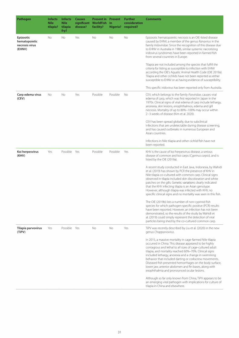
31
Pathogen Infects Nile tilapia?
Infects Nile tilapia fry?
Causes significant disease?
Present in WorldFish facility?
Present in Nigeria?
Further consideration required?
Comments
Epizootic hematopoietic necrosis virus (EHNV)
No No Yes No No No Epizootic hematopoietic necrosis is an OIE-listed disease caused by EHNV, a member of the genus Ranavirus in the family Iridoviridae. Since the recognition of this disease due to EHNV in Australia in 1986, similar systemic necrotizing iridovirus syndromes have been reported in farmed fish from several countries in Europe.
Tilapia are not included among the species that fulfill the criteria for listing as susceptible to infection with EHNV according the OIE’s Aquatic Animal Health Code (OIE 2019a). Tilapia and other cichlids have not been reported as either susceptible to EHNV or as having evidence of susceptibility.
This specific iridovirus has been reported only from Australia.
Carp edema virus (CEV)
No No Yes Possible Possible No CEV, which belongs to the family Poxviridae, causes viral edema of carp, which was first reported in Japan in the 1970s. Clinical signs of viral edema of carp include lethargy, anorexia, skin lesions, enophthalmos, edema and gill necrosis. Mortality of up to 80%−100% may occur within 2−3 weeks of disease (Kim et al. 2020).
CEV has been spread globally, due to subclinical infections that are undetectable during disease screening, and has caused outbreaks in numerous European and Asian countries.
Infections in Nile tilapia and other cichlid fish have not been reported.
Koi herpesvirus (KHV)
Yes Possible Yes Possible Possible Yes KHV is the cause of koi herpesvirus disease, a serious disease of common and koi carps (Cyprinus carpio), and is listed by the OIE (2019a).
A recent study conducted in East Java, Indonesia, by Wahidi et al. (2019) has shown by PCR the presence of KHV in Nile tilapia co-cultured with common carp. Clinical signs observed in tilapia included skin discoloration and white patches on the gills. Genetic variations clearly indicated that the KHV infecting tilapia is an Asian genotype. However, although tilapia was infected with KHV, no specific clinical signs and no mortality was seen in this fish.
The OIE (2019b) lists a number of non-cyprinid fish species for which pathogen-specific positive (PCR) results have been reported. However, an infection has not been demonstrated, so the results of the study by Wahidi et al. (2019) could simply represent the detection of viral particles being shed by the co-cultured common carp.
Tilapia parvovirus (TiPV)
Yes Possible Yes No No Yes TiPV was recently described by Liu et al. (2020) in the new genus Chapparvovirus.
In 2015, a massive mortality in cage-farmed Nile tilapia occurred in China. This disease appeared to be highly contagious and lethal to all sizes of cage-cultured adult tilapia, and mortality reached 60%–70%. Clinical signs included lethargy, anorexia and a change in swimming behavior that included darting or corkscrew movements. Diseased fish presented hemorrhages on the body surface, lower jaw, anterior abdomen and fin bases, along with exophthalmia and pronounced ocular lesions.
Although so far only known from China, TiPV appears to be an emerging viral pathogen with implications for culture of tilapia in China and elsewhere.
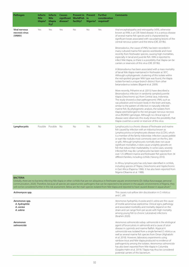
32
Pathogen Infects Nile tilapia?
Infects Nile tilapia fry?
Causes significant disease?
Present in WorldFish facility?
Present in Nigeria?
Further consideration required?
Comments
Viral nervous necrosis virus (VNNV)
Yes Yes Yes No No Yes Viral encephalopathy and retinopathy (VER), otherwise known as VNN, is an OIE-listed disease. It is a serious disease of several marine fish species and is characterized by significant losses associated with vacuolating lesions of the central nervous system and the retina (OIE 2019b).
Betanodavirus, the cause of VNN, has been recorded in many cultured marine fish species worldwide and more recently from freshwater species, causing high mortalities, especially in larval and juvenile fish. VNN is reported to infect Nile tilapia, so there is a possibility that tilapia can be carriers or reservoirs of this virus (OIE 2019b).
A Betanodavirus has been associated with a mass mortality of larval Nile tilapia maintained in freshwater at 30oC. Although a phylogenetic clustering of this isolate within the red-spotted grouper NNV type was found, the tilapia isolate formed a unique branch distinct from other betanodavirus isolates (Bigarré et al. 2009).
More recently, Prihartini et al. (2015) have described a Betanodavirus infection in randomly sampled juvenile tilapia (Oreochromis sp.) from Central Java, Indonesia. The study showed a clear pathogenomic VNN, such as vacuolization and inclusion body in the brain and eyes, similar to the pattern of infection in naturally infected marine fish. By phylogenetic analysis, the isolates from tilapia seed belonged to the red grouper nervous necrosis virus (RGNNV) genotype. Although no clinical signs of disease were observed, this study shows the possibility that tilapia could be a carrier or reservoir of this virus.
Lymphocystis Possible Possible No No Yes No Lymphocystis is a chronic disease of freshwater and marine fish caused by infection with an iridovirus known as Lymphocystivirus or lymphocystis disease virus (LCDV), which is a member of the family Iridoviridae. Infection causes pebble or wart-like nodules most commonly seen on the fins, skin or gills. Although lymphocystis normally does not cause significant mortalities, it does cause unsightly growths on fish that reduce their marketability. In some cases, severely infected fish may die. Lymphocystis has been reported in over 125 different marine and freshwater fish species from 34 different families, including cichlids (Yanong 2010).
In Africa, lymphocystis has only been identified in cichlids, including species of Tilapia, Oreochromis and Haplochromis in East Africa (Paperna 1996). It has also been reported from Nigeria (Okaeme et al. 1988).
BACTERIAGlobally, there are no bacteria infecting Nile tilapia or other cichlids that are not ubiquitous in freshwater aquatic environments (Dr. Iddiya Karunasagar, personal communication, 2020). Therefore, because all species are opportunistic pathogens that can be expected to be present in the aquatic environments of Nigeria, they will not be considered further in this risk assessment. Below are the main species isolated from Nile tilapia and reported to have caused disease in aquaculture.9
Actinomyces spp. This causes rust-yellow skin discoloration in O. niloticus and C. zillii.
Aeromonas spp.A. hydrophila A. cavieaA. sobria
Aeromonas salmonicida
Aeromonas hydrophila, A.caviea and A. sobria are the cause of motile aeromonas septicemia. Clinical signs, pathology and associated morbidity and mortality depend on the strain and can range from per-acute with high mortality among young fish to chronic (ulcerative) infections (Ibrahim 2020).
Aeromonas salmonicida subsp. salmonicida is the etiological agent of furunculosis in salmonids and a cause of ulcer disease in cyprinids and marine flatfish. Atypical A. salmonicida was isolated from a single farmed O. niloticus as well as several marine fish species from Oman (Alghabshi et al. 2018). However, laboratory experiments using rainbow trout and Nile tilapia produced no evidence of pathogenicity among the isolates. Aeromomas salmonicida has also been reported from Nile tilapia in Columbia (Grajales-Hahn et al. 2019). Tilapia may thus be considered potential carriers of this bacterium.

33
Pathogen Infects Nile tilapia?
Infects Nile tilapia fry?
Causes significant disease?
Present in WorldFish facility?
Present in Nigeria?
Further consideration required?
Comments
Edwardsiella tarda This species is ubiquitous and widespread globally, although it is considered exotic to some countries.
Edwardsiella tarda causes edwardsiellosis in freshwater and marine fish species globally and mass mortalities in fish and gastro-intestinal infections in humans. This bacterium has been reported to cause economically significant mortalities in Nile tilapia in different geographical areas (Ibrahim 2020).
It has also been reported as an opportunistic pathogen causing septicemia in O. niloticus in Brazil and noted by Johnston (2008) to have serious consequences and zoonotic potential. It has also been reported in O. mossambicus.
Epitheliocystis Epitheliocystis is a condition affecting the gills and skin of fish and has been reported from more than 90 freshwater and marine species. It is caused by intracellular Gram-negative bacteria, most commonly belonging to the phylum Chlamydiae; however, non-chlamydial bacteria have also been implicated (Seth-Smith et al. 2016).
Mortalities have been associated with infections in cultured fish, and it is the cause of gill lesions in O. niloticus.
Flavobacterium columnare
Like other bacteria infecting tilapia, F. columnare is ubiquitous in freshwater environments globally. It is an opportunistic pathogen of fish, causing skin and fin lesions and columnaris disease (cotton-wool disease). As with most fish-infecting bacteria, disease is often the result of environmental and other stress factors, such as high stocking density, high levels of ammonia and organic load (Ibrahim 2020).
Mycobacterium spp. Mycobacteriosis (fish tuberculosis), a subacute to chronic granulomatous disease, has been reported from Nile tilapia in Kenya and Egypt (Ibrahim 2020).
In Mexico, M. fortuitum and M. marinum were associated with massive mortality of Nile tilapia in 2008 (Lara-Flores et al. 2014).
Piscirickettsia-like organisms (PLOs)
These are intercellular bacteria. PLOs have wreaked havoc on tilapia farms in Hawaii and, especially, Taiwan, where farms suffered mortalities of up to 95%.
PLOs have been reported in various Oreochromis and Tilapia spp., including O. niloticus. The pathogen appears to be widely distributed in tilapia culture, having been reported in Taiwan, Japan, Jamaica, Indonesia, Central America and the US (Johnston 2008). Most reports of PLOs appear to involve intracellular bacteria belonging to the genus Francisella.
Pasteurella multocida
This bacterium is a cause of septicemia. Infections in fish are extremely rare, with only one reported case in hybrid tilapia (O. aureus x O. niloticus) in Israel.
Plesiomonas shigelloides
This species has reportedly caused mortality in fry of Oreochromis spp. in Taiwan.
Providencia rettgeri This bacterium was isolated from the kidney of O. niloticus in Egypt.
Pseudomonas spp.P. fluorescensP. putida
These species can cause chronic infection and acute septicemia.
In Egypt, Pseudomonas spp. were isolated from 30.8% of lake-cultured O. niloticus examined in association with a disease outbreak; during the episode of mass mortality, some fish showed signs typical of Pseudomonas septicemia (Ibrahim 2020).
Staphylococcus epidermidis; Staphyloccocus sp.
These bacteria have caused septicemia and mass mortalities of Oreochromis spp. in Taiwan and Egypt.

34
Pathogen Infects Nile tilapia?
Infects Nile tilapia fry?
Causes significant disease?
Present in WorldFish facility?
Present in Nigeria?
Further consideration required?
Comments
Streptococcus spp.S. agalactiae 1b S. iniae
Streptococcosis is considered the most economically important disease of Nile tilapia. It has led to private-sector efforts to develop Streptococcus-resistant strains of O. niloticus.10
Verner-Jeffreys et al. (2018), who conducted a comprehensive disease investigation in tilapia farmed in Lake Volta, Ghana, found that S. agalactiae multilocus sequence type 261 was a major cause of mortality.
Streptococcus iniae has been reported from at least 27 species of freshwater and marine fish and is a major cause of losses in aquaculture, including tilapia culture. Diseased fish display lethargy and dark coloration, with variable degrees of other signs of septicemia (ascites, eye hemorrhage, skin hemorrhage, behavioral changes etc.). This species is widely distributed geographically, but apparently exotic to some countries (e.g. New Zealand).
Vibrio spp. V. anguillarum V. ordaliiV. damselaV. vulnificus
Vibrio spp. are ubiquitous globally in both freshwater and marine environments. In Egypt, V. vulnificus was noted to cause septicemia and ulceration in Oreochromis spp., while V. anguillarum, as the cause of economically damaging infectious disease, was isolated from 62% of clinically affected Nile tilapia. The latter was also associated with mass mortalities (nearly 98%) of Nile tilapia compromised by exposure to extreme cold water (5.2ºC) at a farm in the north of the country (Ibrahim 2020).
Yersinia ruckeri Yersinia ruckeri is ubiquitous globally in freshwater environments and a cause of bacterial septicemia in O. niloticus and other fish species.
PROTISTA
Flagellata
Cryptobia branchialis,Cryptobia spp.
Yes Yes Possible Possible Yes Yes Most species are ubiquitous and non-pathogenic commensals on the gills of freshwater and marine fish. However, the freshwater species C. branchialis, reported from Nile tilapia in the Philippines (Arthur and Lumanlan-Mayo 1997) is associated with the disease “gill cryptobiosis.” Cryptobia iubilans, which infects the intestine of cichlid fish (Lom and Dyková 1992) is also reported to be pathogenic. The latter species has been reported from Nigeria in catfish (see Omeji et al. 2011; Iyabo et al. 2015)
This genus is listed as infecting O. niloticus in Africa (Scholz et al. 2018), and an unidentified Cryptobia has been reported from this host in Nigeria (Table 5).
Hexamita spp. Yes Yes Possible Possible Yes Yes In Nigeria, Hexamita sp. has been reported from S. melanotheron (Table 5), while H. intestinalis has been reported from the catfish Chrysichthys nigrodigitatus by Iyabo et al. (2015).
The genus is listed as infecting O. niloticus in Africa (Scholz et al. 2018).
Ichthyobodo necatrix Yes Yes Possible Yes Yes Yes These are ubiquitous ectoparasites that infect the skin and gills of many freshwater fish species.
In Nigeria, I. necatrix has been reported from Nile tilapia and other cichlids by Omoniyi and Ojelade (2017), and from the catfish Chrysichthys nigrodigitatus by Iyabo et al. (2015).
Ichthyobodo necatrix causes ichthyobodosis, a disease affecting mainly young fish that is treatable in enclosed aquaculture facilities (Lom and Dyková 1992).
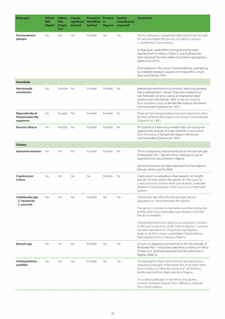
35
Pathogen Infects Nile tilapia?
Infects Nile tilapia fry?
Causes significant disease?
Present in WorldFish facility?
Present in Nigeria?
Further consideration required?
Comments
Piscinoodinium pillulare
Yes Yes Yes Possible Yes Yes This is a ubiquitous ectoparasite affecting the skin and gills of many freshwater fish species, including O. niloticus, C. rendalli and O. mossambicus.
In Nigeria, an unidentified Piscinoodinium has been reported from O. niloticus (Table 5), and P. pillulare has been reported from the catfish Chrysichthys nigrodigitatus (Iyabo et al. (2015).
Piscinoodinium is the cause of piscinoodinosis, a dangerous but treatable disease in aquaria and tropical fish culture (Lom and Dyková 1992).
Ameobida
Hartmanella vermiformis
Yes Possible No Possible Possible No Hartmanella vermiformis is a common, free-living amoeba that is widespread in nature. It has been isolated from soil, freshwater, air and a variety of engineered water systems (Lom and Dyková 1992). It was also isolated from the kidney tissue of farmed Nile tilapia in the former Czechoslovakia (Dyková et al. 1997).
Mayorella-like & Platyamoeba-like organisms
Yes Possible No Possible Possible No These are free-living amoebae that were reported to infect the liver of farmed Nile tilapia in the former Czechoslovakia (Dyková et al. 1997).
Rosculus ithacus Yes Possible No Possible Possible No This species is a free-living amoeba that is an occasional opportunistic parasite of snakes and fish. It was isolated from the kidney of farmed Nile tilapia in the former Czechoslovakia (Dyková et al. 1997).
Ciliatea
Apiosoma minutum Yes Yes No Possible Possible No This is a ubiquitous commensal found on the skin and gills of freshwater fish.11 Species of this ciliate genus can be expected to be encountered in Nigeria.
Apiosoma minutum has been reported from Nile tilapia in Vietnam (Arthur and Te 2006).
Cryptocaryon irritans
Yes Yes Yes No Possible No Cryptocaryon is a ubiquitous ciliate parasitic on the gills and skin of many marine fish species. It is the cause of cryptocaryonosis (marine white spot disease), a treatable disease in marine aquaria. It does not occur in freshwater systems.
Chilodonella spp.C. hexastichaC. piscicola
Yes Yes Yes Possible Yes Yes Chilodonella spp. infect the skin and gills, and are ubiquitous on many freshwater fish species.
This genus is common in hatcheries and other production facilities and may occasionally cause disease in stressed fish, but is treatable.
Chilodonella hexasticha is listed as occurring on O. niloticus in Africa by Scholz et al. (2018), while in Nigeria, C. uncinata has been reported from Chrysichthys nigrodigitatus (Iyabo et al. 2015) and an unidentified Chilodonella has been reported from O. niloticus (Table 5).
Epistylis spp. Yes Yes No Possible Yes No Epistylis is a ubiquitous commensal on the skin and gills of freshwater fish.12 It has been listed from O. niloticus in Africa (Scholz et al. 2018) and reported from the same host in Nigeria (Table 5).
Ichthyophthirius multifiliis
Yes Yes Yes Possible Yes Yes This pathogenic ciliate infects the skin and gills and is a ubiquitous pathogen of freshwater fish. It has been listed from O. niloticus in Africa by Scholz et al. (2018) and is widely reported from tilapia species in Nigeria.
It is a serious pathogen in hatcheries and aquaria, causing “whitespot disease,” and is difficult to eradicate from closed systems.

36
Pathogen Infects Nile tilapia?
Infects Nile tilapia fry?
Causes significant disease?
Present in WorldFish facility?
Present in Nigeria?
Further consideration required?
Comments
Paratrichodina spp.P. africanaP. incissa
Yes Yes No Possible Possible No Paratrichodina africana is listed as infecting O. niloticus in Africa (Scholz et al. 2018), while P. incissa occurs on the same host in Vietnam (Arthur and Te 2006).
This genus does not appear to have been associated with disease in tilapia culture.
Tetrahymena spp. Yes Yes Possible Possible Yes Yes Tetrahymena spp. are saprozoic and occasional pathogens of freshwater fish. Tetrahymena corlissi causes “tet disease” in freshwater fish held in aquaria (Lom and Dyková 1992).
An unidentified species of Tetrahymena has been reported from Nile tilapia in Nigeria (Table 5).
Trichodinella spp.T. epizootica
Possible Possible No Possible Possible No This genus has been recorded from the gills of O. mossambicus.
Trichodinella epizootica is ubiquitous and shows little host specificity. It is common in hatcheries and other production facilities and may occasionally proliferate massively, causing disease in stressed fish (Lom and Dyková 1992), but it is treatable.
Trichodina spp.T. acutaT. centrostrigataT. cichidarumT. compactaT. fultoniT. heterodentataT. magnaT. migalaT. mutabilisT. nigraT. oreochromisiT. orientalisT. pediculusT. rectuncinataT. reticulataT. siluriT. tilapiaeT. truttaeT. velasquezae
Yes Yes Yes Possible Yes Yes Trichodina is a ubiquitous genus containing more than 100 described species that infect the skin and/or gills and occasionally the urinary bladder of freshwater and marine fish. Certain species many have limited geographical distributions and/or show high host specificity.
Globally, at least 16 species are reported from Nile tilapia, with T. acuta and T. heterodentata being reported from O. niloticus in Nigeria (Table 5). Many other species are likely to infect Nigerian freshwater fish.
Trichodina spp. are common in hatcheries and other production facilities and occasionally cause trichodinosis in stressed fish, which is a treatable disease. Pathogenicity may vary between parasite and host species.
Tripartiella spp.T. bulbosaT. cichlidarumT. clavodontaT. leptospinaT. nanaT. obtusaT. orthodensT. spatulaT. tilapiae
Yes Yes No Possible Possible No Tripartiella spp. Are ubiquitous and commensal. The genus is common on the gills of freshwater fish, with at least nine species occurring on Nile tilapia.
Trichophrya spp. Yes Yes No Possible Possible No This ubiquitous ciliate is ectocommensal on the gills of freshwater fish (Lom and Dyková 1992).
Coccidia
Goussia cichlidarum Yes Possible No Possible Possible No Goussia cichlidarum is common in the epithelial lining of the swim bladder of tilapia in Israel and Uganda (Lom and Dyková 1992). It has been listed from O. niloticus in Africa (Scholz et al. 2018).

37
Pathogen Infects Nile tilapia?
Infects Nile tilapia fry?
Causes significant disease?
Present in WorldFish facility?
Present in Nigeria?
Further consideration required?
Comments
FUNGI
Aphanomyces invadans
Yes Possible Yes Possible Possible Yes Aphanomyces invadans is the cause of EUS, a serious transboundary disease affecting many species of freshwater fish with near global distribution that is listed by the OIE.
Tilapia species are generally considered resistant but are more accurately characterized as having low susceptibility (Johnston 2008) and thus are potential carriers.
Branchiomyces demigrans
Yes Yes Yes No Possible No This non-septate filamentous fungus is an opportunistic pathogen of freshwater fish and has been reported to cause gill rot and mortalities of cultured Nile tilapia in Egypt (Khalil et al. 2015).
Fusarium oxysporum Possible Yes No No Possible No Fusarium oxysporum has a global distribution and occurs chiefly as a soil saprophyte. An opportunistic pathogen of marine fish causing subcutaneous lesions, F. oxysporum in co-infection with Aeromonas hydrophila has been associated with mortalities in Nile tilapia fry held in freshwater culture in Spain (Cutuli et al. 2015).
Ichthyophonus hoferi
Yes Possible Yes No Possible No Ichthyophonosis, caused by the parasitic fungus I. hoferi is an important disease of marine and anadromous fish. It has a broad host range and global distribution (El-Ghany and Alla 2008; Kocan 2018). It is listed by Johnston (2008) as reported from O. niloticus.
Infections range from subclinical, with no gross or microscopic signs of disease, to clinical disease that can present as external ulcers, extensive tissue and organ damage and heavy mortality (Kocan 2018).
Loma camerouensis Yes Possible Possible No No No The genus Loma belongs to the Microsporida, which were formerly considered protozoans but are now recognized as fungi. Loma camerounensis was reported from xenomas in the gut of cultured O. niloticus in Cameroon by Fomena et al. (1992).
Rhizomucor spp. Possible Possible No No Possible No Rhizomucor is a genus of saprophytic fungus that is ubiquitous in the environment but is considered a rare opportunistic pathogen of freshwater fish. It is listed by Johnston (2008) from the skin of O. niloticus x O. aureus x O. mossambicus from the US.
Saprolegnia spp. Yes Yes No No Yes No Saprolegnia spp. are ubiquitous and opportunistic pathogens of freshwater fish. In Nigeria, several species of this genus have been associated with infections in freshwater fish species (Ogbonna and Alabi 1991).
Johnston (2008) listed Saprolegnia sp. from the skin of O. niloticus and O. aureus from Egypt.
MONOGENEA
Cichlidogyrus spp. C. aegypticusC. arthracanthus C. cirratusC. doussouiC. halli C. haplochromiC. levequeiC. mbirizeiC. nematocirrusC. rognoniC. sclerosusC. thurstonaeC. tiberianusC. tilapiaeC. tubicirrus magnus
Yes Yes No Possible Possible No This is a large genus containing some 125 described species, almost all of which are only known from cichlid fish (le Roux and Avenant-Oldewage 2010; Gereates et al. 2020). The species listed herein are only those that have been reported from Nile tilapia. Many of these species are undoubtedly present in Nigeria.
Cichlidogyrus spp. are gill parasites with a direct life cycle. They have often been translocated to new geographic areas along with the movement of infected tilapia for aquaculture development.
Members of this genus do not appear to have been reported to cause problems in aquaculture, although their potential pathogenicity and the possibility of host switching from introduced to native species has been mentioned (Gereates et al. 2020).
As with other monogeneans, infections in enclosed aquaculture facilities should be treatable.

38
Pathogen Infects Nile tilapia?
Infects Nile tilapia fry?
Causes significant disease?
Present in WorldFish facility?
Present in Nigeria?
Further consideration required?
Comments
Dactylogyrus spp.D. extensus
Yes Possible Yes Possible Yes Yes The genus Dactylogyrus has occasionally been reported to infect Nile tilapia. However, species identifications have not been made, and most or all of these records may involve the misidentification of Cichlidogyrus spp.
Dactylogryrus extensus has been reported from Nile tilapia in Nigeria (Table 5) and there are also several reports of unidentified Dactylogyrus spp. from tilapia species.
Enterogyrus spp.E. cichlidarumE. coronatusE. hemi-haplochromiiE. malmbergiE. niloticus
Yes Possible No Possible Possible No Enterogyrus spp. are parasites in the stomach of cichlid fish, with 12 species currently described within this genus (Madanire-Moyo and Avenant-Oldewage 2014; Luus-Powell et al. 2020).
They are host specific to cichlids and have often been translocated to new geographic areas along with the movement of infected tilapia for aquaculture development.
Enterogyrus spp. infecting Nile tilapia do not appear to cause problems in aquaculture. Madanire-Moyo and Avenant-Oldewage (2015) found that histopathological changes caused by the E. coronatus in the stomach of wild Pseudocrenilabrus philander were mild and restricted to the vicinity of haptoral attachment.
It is possible that veterinary drugs such as ivermectin that are used to treat internal parasites may be effective against these monogeneans, but this has not yet been investigated.
Gyrodactylus spp.G. cichlidarum (syn.: G. niloticus)G. elegansG. ergensi G. hildaeG. malalaiG. nyanzaeG.occupatusG. pariselleiG. shariffiG. sprostonaeG. yacatli
Yes Yes Yes Possible Possible Yes The genus Gyrodactylus comprises some 409 potentially valid species (Harris et al. 2004). Many species are host specific. Gyrodactylus spp. are viviparous.
Gyrodactylus spp. are frequent parasites of the skin and gills of freshwater fish, with members of this genus often causing problems in fish cultured in closed systems.
Gyrodactylus spp. have often been translocated to new geographic areas along with the movement of infected fish for aquaculture development. García-Vásquez et al. (2011) noted that Gyrodactylus-associated mortality of juvenile, pond-reared O. niloticus has been reported across several continents and stressed that extreme caution should be exercised in the translocation of commercial tilapiine species into areas where cichlids are already resident.
Scholz et al. (2018) listed seven species occurring on Nile tilapia in Africa.
As with other monogeneans infecting the gills or skin of freshwater fish cultured in closed systems, infections are treatable.
Macrogyrodactylus spp.
Yes[?] ? No No Yes No Macrogyrodactylus spp. are enzootic to Africa and have been described mainly from the gills and fins of Clarias and Polyopterus (Prikrylová and Gelnar 2008). The report of an unidentified species from Nile tilapia in Nigeria by Ashade et al. (2010) requires confirmation, as it appears to be the only finding of this genus on a cichlid fish.
Scutogyrus spp.S. longicornis (syn.: C. longicornis, C. longicornis longicornis)S. minus
Yes Possible No Possible Possible No This genus is closely related to Cichlidogyrus and shares many of its characteristics. Two species have been reported from O. niloticus in Africa (Scholz et al. 2018).

39
Pathogen Infects Nile tilapia?
Infects Nile tilapia fry?
Causes significant disease?
Present in WorldFish facility?
Present in Nigeria?
Further consideration required?
Comments
CRUSTACEA
Brachiura
Argulus spp.A, africanusA. japonicusA. rhipidiophorus
Yes Yes Yes No Yes No Scholz et al. (2018) list two species of Argulus as infecting Nile tilapia in Africa, while A. africanus has been reported from this host in Nigeria (Table 5).
Argulus spp. are macroscopic parasites on the skin of freshwater fish and are easily seen by hatchery workers.
Dolops ranarum
Yes Possible Possible No Possible No This brachiuran was listed from O. niloticus in Africa by Scholz et al. (2018).
Closely related to Argulus, these macroscopic parasites are easily seen in hatchery situations.
Copepoda
Caligus epidemicus Yes Possible Yes No No No This is a marine and brackish-water parasite of Nile tilapia raised in brackish water in the Philippines (Arthur and Lumanlan-Mayo 1997).
They are macroscopic parasites that are easily seen by aquaculturists.
Ergasilus latus Yes Possible Possible Possible Yes No Ergasilus spp. are parasitic copepods that infect the gills of freshwater fish. Heavy infections can cause gill pathology and ergasilosis.
Ergasilus latus is primarily a parasite of cichlid fish and is known only from Africa. It has been reported from O. niloticus and other tilapia in Nigeria (Table 5).
Lamproglena monodi
Yes Possible No No Yes No Lamproglena monodi is a copepod parasitic on the gills of cichlid fish. Originally described from cichlids from the Democratic Republic of Congo, this species has also been reported in Egypt, Zimbabwe, Namibia and Uganda, and recorded from Nile tilapia in Nigeria (Table 5).
It was recently introduced to Brazil, where it was recorded from Nile tilapia and other species (Kozlowiski de Azevedo et al. 2012; Hassan et al. 2013).
Parasitic on the skin and gills, it is a macroscopic parasite that is easily seen in closed aquaculture systems. It reportedly does not cause serious gill pathology or mortalities in cichlids (Hassan et al. 2013).
Lernaea spp. L. barnimiana L. cyprinacea L. hardingi L. tilapiae
Yes Yes Yes No Yes Yes Anchor worms (Lernaea spp.) are common parasites of freshwater fish. Lernaea cyprinacea has been reported from Nile tilapia in Nigeria (Table 5). Although highly pathogenic, especially to young fish, these macroscopic parasites are easily seen in hatchery situations, where they may cause lernaeosis and mortalities.
Some species may show host specificity.
Lernaeocera branchialis
Yes Possible Yes No Yes No This pathogen is a marine species of parasitic copepod that has been reported from Nile tilapia in Nigeria (Table 5).
Opistholernaealaterobrachialis
Yes Possible Yes No Possible Yes Opistholernaea laterobrachialis is listed as a parasite of O. niloticus in Africa (Scholz et al. 2018).
These are macroscopic parasites that are easily seen in hatchery situations. Infections in mouthbreeding cichlids are reported to impair breeding (Paperna 1996).

40
VirusesViruses are the most serious threats to tilapia culture. The following seven are given further consideration due to their pathogenicity and the potential harm their introduction might cause to Nigerian aquaculture and wild fish populations:
OIE-listed viruses
• Koi herpesvirus (KHV)
• Viral nervous necrosis virus (VNNV)
• Spring viremia of carp virus (SVCV)
Other viruses
• Tilapia lake virus (TiLV)
• Infectious spleen and kidney necrosis virus (ISKNV)
• Tilapia parvovirus (TiPV)
• Aquabirnavirus (IPNV-like virus)
BacteriaGlobally, there are no bacteria infecting Nile tilapia that are not ubiquitous in freshwater aquatic environments. Species causing disease in Nile
tilapia culture, such as Aeromonas hydrophila, Streptococcus agalactiae and S. iniae, are globally distributed. As previously noted, all bacteria considered in this risk analysis have either reportedly been found or are expected to be present in Nigeria; thus, bacterial pathogens are not considered further in this risk assessment.
FungiMost of the fungi causing disease in tilapia culture are opportunistic species present in freshwater environments and have broad geographic distributions. One serious fungal disease requires further consideration:
• Aphanomyces invadans (epizootic ulcerative syndrome, EUS)
ProtistaEight protozoan taxa that have direct life cycles and are pathogenic to Nile tilapia and other freshwater fish require further consideration:
• Cryptobia spp.
• Hexamita spp.
• Ichthyobodo necatrix
Pathogen Infects Nile tilapia?
Infects Nile tilapia fry?
Causes significant disease?
Present in WorldFish facility?
Present in Nigeria?
Further consideration required?
Comments
Isopoda
Alitropis typicus,Nerocila orbignyi
Yes Yes Yes Possible No No These are large parasites found in the buccal cavity of tilapia and other freshwater fish that are easily detected by aquaculturists.
Alitropis typicus is reported to have caused mortalities of 40%–80% in Nile tilapia cultured in net cages in the Philippines, with only 5% of juveniles in Taal Lake surviving infection (Arthur and Lumanlan-Mayo 1997). Nerocila orbignyi has been reported from tilapia in Egypt (Ibrahim 2020).
HIRUDINEA
Piscicola geometra Yes Yes Yes No Yes No This leech is a widespread European parasite of freshwater fish. It has been reported from Nile tilapia in a rainforest pond in southeastern Nigeria by Opara (2002).
The macroscopic size of this leech and the fact that it must leave its fish host to lay cocoons on substrate would make it easily detected in closed aquaculture facilities.
MOLLUSCA
Cristaria plicata Yes Yes Yes No Possible No Glochidia of C. plicata have been reported as temporary parasites on the gills of Nile tilapia in the Philippines (Arthur and Lumanlan-Mayo 1997).
Adults are free-living freshwater clams.
Table 7. Possible pathogens of Nile tilapia having direct life cycles and their screening against the criteria listed in Section 8.1.
41
• Piscinoodinium pillulare
• Chilodonella spp.
• Ichthyophthirius multifiliis
• Tetrahymena spp.
• Trichodina spp.
MonogeneaTwo monogenean taxa require further consideration:
• Dactylogyrus spp.
• Gyrodactylus spp.
CrustaceaTwo parasitic copepods require further assessment:
• Lernaea spp.
• Opistholernaea laterobrachialis
8.4. Calculation of likelihood of pathogen entry and exposure13
The following eight-step pathway required for a potential pathogen to enter Nigeria, escape from quarantine and become established in cultured or wild fish populations has been used to estimate likelihood of entry and exposure (Table 8):
Entry
1. Pathogen is present in G17 broodstock.
2. Pathogen escapes detection in G17 broodstock.
3. Pathogen is transferred to G17–F1 fry and establishes infection.
4. Pathogen is present in the subpopulation of G17–F1 fry selected for transfer.
5. Pathogen survives transfer to Nigeria.
Exposure
6. Pathogen is not detected by diagnostic testing of G17–F1 or subsequent generations.
7. Pathogen escapes the Nigerian biosecure quarantine facility due to a lapse in biosecurity or remains undetected and is released from the facility via fry transferred to the NBCs.
8. Pathogen contacts and infects cultured or wild fish populations and becomes established in Nigeria.
Table 8 presents the results of estimation of likelihood of entry (L
Ent), likelihood of exposure
(LExp
) and likelihood of both entry and exposure (L
Ent X L
Exp) occurring for the 20 hazards.
The results of this phase of the risk assessment revealed negligible likelihood of entry (L
Ent) for 3
hazards (1 virus and 2 crustaceans), very low LEnt
for 7 hazards (6 viruses and 1 fungus), and moderate L
Ent for 10 hazards (all 8 protozoans and both
monogeneans).
The risk assessment then estimated the likelihood of exposure (L
Exp)
for those 17 hazards having
non-negligible LEnt
(Table 8). The results showed a very low L
Exp for 7 hazards (6 viruses and 1 fungus),
and a high LExp
for 10 hazards (all 8 protozoans and both monogeneans).
The risk assessment then proceeded to calculate the likelihood of both entry and exposure (L
Ent X L
Exp) for the 17 hazards. Seven hazards
(all 6 viruses and the fungus) were estimated to have negligible L
Ent X L
Exp, while the remaining
10 hazards (8 protists and 2 monogeneans) were estimated to have non-negligible L
Ent X L
Exp.
The next step in the risk assessment process is the estimation of magnitude of the consequence of entry and exposure for the 10 hazards having non-negligible likelihood of entry and exposure (L
Ent X L
Exp) (Table 9).
The estimated consequence of LEnt
X LExp
was low for all 8 protozoans, low to moderate for 1 monogenean (Dactylogyrus spp.), and low to high for the other monogenean (Gyrodactylus spp.)
As all 10 hazards were estimated to have non-negligible consequence, they were all taken through the final step in the risk assessment, which is the calculation of their total risk (Table 9). Total risk was estimated as low for all 8 protozoans, low to moderate for Dactylogyrus spp., and low to high for Gyrodactylus spp.

42
Pathway step: Hazard (Pathogen)
1 2 3 4 5 LEnt 6 7 8 LExp LEnt X LExp
VIRUSES
Koi herpesvirus (KHV) L VL H H H VL VL L H VL N
Viral nervous necrosis virus (VNNV) VL L H H H VL VL L H VL N
Spring viremia of carp virus (SVCV) VL H H H H VL VL L H VL N
Tilapia lake virus (TiLV) VL VL N
Infectious spleen and kidney necrosis virus (ISKNV)
L VL H H H VL VL L H VL N
Tilapia parvovirus (TiPV) VL L H H H VL VL L H VL N
Aquabirnavirus (IPNV-like virus) VL L H H H VL VL L H VL N
FUNGI
Aphanomyces invadans (EUS) VL L H H H VL VL L H VL N
PROTISTA
Cryptobia spp. M H H H H M H H H H M
Hexamita sp. M H H H H M H H H H M
Ichthyobodo necatrix M H H H H M H H H H M
Piscinoodinium pillulare M H H H H M H H H H M
Chilodonella spp. M H H H H M H H H H M
Ichthyophthirius multifiliis M H H H H M H H H H M
Tetrahymena spp. M H H H H M H H H H M
Trichodina spp. M H H H H M H H H H M
MONOGENEA
Dactylogyrus spp. M H H H H M H H H H M
Gyrodactylus spp. M H H H H M H H H H M
CRUSTACEA
Lernaea spp. VL VL N
Opistholernaea laterobrachialis VL VL N
Note: likelihood estimates are combined using Table 3 (N = Negligible, VL = Very Low, L = Low, M = Moderate, H = High); LEnt = L1 X L2 X L3 X L4 X L5, LExp = L6 X L7 X L8; Likelihood of Entry and Exposure = LEnt X LExp).
Table 8. Calculation of estimates of likelihood of entry (LEnt
) and exposure (LExp
).

43
Potential hazard
Estimatedlikelihood of entry and exposure
Estimated consequence of entry and exposure
Estimated total risk
Remarks
PROTISTA
Cryptobia spp. M L L Genus is known to be present in Nigeria (Table 5). Introduction would have only localized effects on receiving aquaculture facilities where infections could be controlled.
Hexamita spp. M L L Genus is known to be present in Nigeria (Table 5). Introduction would have only localized effects on receiving aquaculture facilities where infections could be controlled.
Ichthyobodo necatrix
M L L Species is known to be present in Nigeria (Table 5). Introduction would have only localized effects on receiving aquaculture facilities where infections could be controlled.
Piscinoodinium pillulare
M L L Genus is known to be present in Nigeria (Table 5). Introduction would have only localized effects on receiving aquaculture facilities where infections could be controlled.
Chilodonella spp.
M L L Genus has been reported from Nigeria (Table 5). Introduction would have only localized effects on receiving aquaculture facilities where infections could be controlled. A recent study by Bu et al. (2021) suggests that C. uncinata is a facultative parasite.
Ichthyophthirius multifiliis
M L L Species has been reported from Nigeria (Table 5). Introduction would have only localized effects on receiving aquaculture facilities where infections could be controlled.
Tetrahymena spp.
M L L Genus is ubiquitous, so it is expected to be present in Nigeria. Introduction would have only localized effects on receiving aquaculture facilities where infections could be controlled.
Trichodina spp. M L L Genus is ubiquitous and present in Nigeria (Table 5). Introduction would have localized effects on receiving aquaculture facilities where infections could be controlled. However, with the movement of GIFT into Nigeria, the possibility of introducing new species, some of which could be pathogenic to native fish species, should be avoided.
MONOGENEA
Dactylogyrus spp.
M L to M L to M Although the genus has been reported from Nigeria (Table 5), the potential exists to introduce an exotic species of high pathogenicity, so additional risk management measures are recommended.
Gyrodactylus spp.
M L to H L to H Although the genus has been reported from Nigeria (Table 5), the potential exists to introduce an exotic species of high pathogenicity, so additional risk management measures are recommended.
Table 9. Estimate of total risk for those potential hazards having non-negligible likelihood of entry and exposure (L
Ent X L
Exp) (calculated using Table 4).

44
9. Assessment of risk to Nigeria (risk management: risk evaluation)
This risk analysis takes a highly precautionary approach by assuming that the ALOP for Nigeria should be “high,” giving an ALOR that is “low.”
Comparison of estimates of total risk with the suggested ALOR shows that the risk is acceptable
for all eight protozoans, but may be above the ALOR for the two monogeneans (Dactylogyrus spp. and Gyrodactylus spp.). Thus, additional risk management measures for these two hazards are recommended (Section 12.1).
Genetically improved farmed tilapia (GIFT).
Phot
o cr
edit:
Wor
ldFi
sh
44

45
10. Uncertainty of risk estimates
Due to a lack of basic knowledge and the need for precaution, there is always an element of uncertainty in establishing risk estimates for pathogens. In this hazard identification, cells in Table 7 that have a “possible” ranking, particularly for the presence of a hazard in the WorldFish facility, have a high degree of uncertainty. The reason is that this preliminary evaluation does not take into consideration the impact of risk management measures (particularly diagnostic testing) in decreasing uncertainty. A similar ranking for presence of a pathogen in Nigeria is given with a low degree of uncertainty, as the pathogens receiving this ranking, although not reported in the literature for Nigeria, are ubiquitous organisms of wide geographical and host distribution and are highly likely to be present in the country. In Table 8,
which takes to account the risk management measures that WorldFish has either implemented or proposed (Section 7), risk estimates for seven viruses and one fungus are given with a low degree of uncertainty, and all parasites have a moderate level of uncertainty. This latter is due to the complete lack of diagnostic information on the status of WorldFish GIFT stocks with regard to these pathogens. This uncertainty is addressed in Section 12 through the recommendation that GIFT stocks be subjected to parasitological examination. Note that risk for all direct life-cycle parasites, except the monogeneans Dactylogyrus and Gyrodactylus, is calculated with a low level of uncertainty as being “negligible," due to these hazards being both ubiquitous globally and treatable in closed aquaculture facilities.
Hatchery workers harvest Abbassa nile tilapia from a hatchery in Egypt.
Phot
o cr
edit:
Heb
a El
-Beg
awi/W
orld
Fish
45

46
11. Conclusions
Global experience has amply shown that escapes of introduced or transferred species from aquaculture facilities are inevitable. However, good biosecurity should prevent the escape of GIFT from the high-biosecurity quarantine facility to be established in Ogun during the first 2–3 years of the project. During this period of high-biosecurity confinement, the health status of the GIFT can be confirmed before any relocation to other facilities is permitted. In the event that a serious and untreatable disease occurs, the affected subpopulations or entire stock can be destroyed and the facility disinfected.
The risk analysis considered a total of 69 pathogens/pathogen groups as possible hazards for the transfer of GIFT fry from Malaysia to Nigeria. The results show that, should WorldFish and its Nigerian counterparts fully and correctly implement all of WorldFish’s in progress and proposed risk management measures as well as the additional measures listed in Section 12, the risk posed by all hazards is likely to fall within the recommended ALOR.
The risk posed by monogeneans belonging to the genera Gyrodactylus and Dactylogyrus is problematic. This is mainly due to the lack of information as to their possible presence at low prevalence in the WorldFish broodstock, and the large number of species in these genera, which may differ considerably in their pathogenicity to Nile tilapia and some of which may be exotic to Nigeria. It is thus recommended that WorldFish confirm the absence of these parasites through appropriate parasitological examination of GIFT broodstock and fry.
Fish pathogenic bacteria, such as Aeromonas hydrophila, Streptococcus agalactiae and S. iniae, are globally distributed and opportunistic pathogens of freshwater fish. Thus, although these organisms are an important cause of disease and mortality in aquaculture facilities, they are not relevant to this risk analysis. It is also worth noting that currently there are no PCR tests that can differentiate between pathogenic and non-pathogenic strains of these bacteria. This is because pathogenicity is complex, involving several genes, none of which has the properties to be considered a specific virulence factor (Dr. Iddiya Karunasagar, personal communication, 2020).
Tilapia Hatchery in Noakhali, Bangladesh.
Phot
o cr
edit:
Fin
n Th
ilste
d/W
orld
Fish
46

47
12. Recommendations
The following recommendations are made to WorldFish and its Nigerian counterparts to improve the biosecurity management practices that are either in progress or are proposed by WorldFish. They are divided into two categories: (1) those whose implementation is considered essential and (2) those that are desirable.
12.1. Essential actions• Monitor implementation of risk
management measures. As the risk assessment is highly dependent upon the risk management measures proposed by WorldFish, monitoring systems should be established to ensure that all risk management measures are fully and effectively implemented.
• Conduct diagnostic testing for the following additional pathogens. Viral nervous necrosis virus (VNNV), spring viremia of carp virus (SVCV), tilapia parvovirus (TiPV) and Aquabirnavirus. Include the testing as part of the WorldFish protocols, as well as the health certification from the Government of Malaysia that will accompany the shipment of GIFT fry to Nigeria.
• Ensure the biosecurity of the receiving facility in Nigeria. WorldFish should ensure that the receiving facility in Nigeria meets all standards for a high-security quarantine facility, as detailed, for example, in Arthur et al. (2007). In preparation for the transfer of GIFT, the receiving facility, which currently houses Nigerian stocks of tilapia and catfish, must have all stocks removed, and all tanks, pipes, surfaces and fomites must then be thoroughly disinfected using an approved method. The holding facility must be upgraded, as necessary, to meet the standards of a high-level biosecurity facility (see Arthur et al. 2007). Specific SOPs for the facility must be developed and in place, and all staff must receive appropriate training to ensure that SOPs are strictly followed.
• Examine GIFT broodstock for direct life-cycle parasites and treat them, if required. Many studies have shown that Nile tilapia introduced into new countries many generations ago may still be infected with a wide variety of parasites having direct life cycles. Some of these are host specific to tilapia and thus have clearly been introduced to new environments along with the introduction of their hosts.14 This risk analysis has determined that a few of these parasites (i.e. Gyrodactylus, Dactylogyrus) may pose a significant risk to the receiving country, while a number of others, although known or likely to be present in Nigeria, are known to cause disease problems in aquaculture facilities. It is thus recommended that the parent broodstock held in Penang be subjected to full parasitological examination, as it may be possible, through appropriate treatment to minimize the chance that these pathogens will be transferred to Nigeria where they may cause problems in the receiving facility.
• Develop a detailed contingency plan. As serious exotic pathogens are known to have escaped from similar high-biosecurity facilities, WorldFish and the GON should develop a detailed contingency plan to deal with such an event, no matter how unlikely. This should include health monitoring of cultured stocks and diagnostic testing to determine the cause of any serious mortality events, as well as planning for efforts to restrict pathogen spread and, where possible, to implement eradication procedures (Arthur et al. 2005).
12.2. Additional recommendations for WorldFish considerationThe following recommendations are not regarded as essential to pathogen risk management but are suggested as useful activities that could be incorporated into project design:

48
• Study the diseases of fish species near GIFT aquaculture facilities. To better understand the potential for pathogen transfer between wild and cultured stocks, baseline studies of the diseases of fish species in the vicinity of GIFT aquaculture facilities in Nigeria should be conducted. Such monitoring will also help detect any transfer of introduced exotic pathogens from GIFT to wild finfish populations.
• Commission an expert review of testing protocols. WorldFish may wish to have an independent expert in fish disease diagnostics review the proposed testing protocols for the final list of pathogens used to certify GIFT fry.
• Stress test GIFT to reveal hidden pathogens. Stress testing of GIFT broodstock and fry could be conducted to check for cryptic or unknown pathogens.
• Organize a collaborative study of Nigerian fish pathogens. As the pathogens of cultured fish of Nigeria remain poorly known, WorldFish should consider developing an international project to conduct baseline surveys of key cultured species. Such studies should include viruses, bacteria and parasites.
• Prepare a fish parasite checklist. As there is no comprehensive listing or critical review of the parasites reported from Nigerian fish, a starting point would be the preparation of a checklist of the parasites of Nigerian fish. The preliminary list of publications on the diseases of Nigerian fish given in Annex 1 could serve as a starting point for this work.
• Assist in a decision on Nigeria’s ALOP. WorldFish should initiate discussions with the GON and relevant stakeholders with the aim of determining the national ALOP. A national consultation involving the plant, terrestrial animal and aquatic animal health sectors could be convened to assist in reaching a consensus among stakeholders.
• Develop a national strategy for aquatic animal health. WorldFish could assist the GON in developing a national strategy for aquatic animal health and in participation in the FAO’s PMP/AB program.
• Implement measures to prevent future transfers of tilapia. The GON should take all possible measures to prevent the future transfer of tilapia of unknown health or genetic status from outside the national territory.
Tilapia brooding female.
Phot
o cr
edit:
Bal
aram
Mah
alde
r/W
orld
Fish
48

49
Notes
1 Box 1216, Barriere, B.C., Canada, V0E 1E0; e-mail: [email protected]
2 See, for example, Briggs et al. 2004; Bondad-Reantaso and Subasinghe 2005; Bondad-Reantaso et al. 2005a and 2005b; Flegel 2006; Johnston 2008; Rodgers et al. 2011; Tavares-Dias and Martins 2017; Shinn et al. 2018.
3 ICES (2005) defines a “transferred species” as “Any species intentionally or accidentally transported and released within areas of established populations, and continuing genetic flow where it occurs,” while an “introduced species” is defined as “Any species transported intentionally or accidentally by a human-mediated vector into aquatic habitats outside its native range.”
4 https://naqsportal.net/
5 See, for example, Section 4 in Arthur et al. 2008.
6 The nomenclature for fish used in this document follows Froese and Pauly 2019.
7 See, for example, Dong et al. 2017b.
8 See, for example, DAWE 2020.
9 For additional information, see the review of Ibrahim 2020.
10 See, for example, The Fish Site 2021.
11 See, for example, Arthur and Lumanlan-Mayo 1997.
12 See, for example, Arthur and Lumanlan-Mayo 1997.
13 The OIE’s Aquatic Animal Health Code (2019a) defines entry assessment as consisting of describing the biological pathway(s) necessary for an importation activity to introduce a pathogen into a particular environment, as well as estimating the probability of that complete process occurring, either qualitatively (in words) or quantitatively (as a numerical estimate). An exposure assessment is defined as consisting of describing the biological pathway(s) necessary for exposure of animals and humans in the importing country to the hazards (in this case the pathogenic agents) from a given risk source, and estimating the probability of these exposure(s) occurring, either qualitatively (in words) or quantitatively (as a numerical estimate). Most countries consider that the “entry” pathways terminate and the “exposure” pathways begin at the importing country’s border, a practice that is followed in this risk analysis.
14 See, for example, Natividad et al. 1986.

50
References
Abba AM, Abdulhamid Y, Omenesa RL and Mudassir I. 2018a. Impact of helminth parasites on length-weight ratio and condition factor of fishes in Ajiwa and Jibia reservoirs, Katsina State, Nigeria. Research Journal of Zoology, 2018, 1(2), 6 pp.
Abba AM, Emere MC and Appah J. 2018b. Comparative study on helminth parasites of Oreochromis niloticus and Clarias gariepinus in Ajiwa Earth Dam Katsina State, Nigeria. World Journal of Fish and Marine Sciences 10(1):5–11.
Abidemi-Iromini AO and Eze RN. 2011. Comparative assessment of parasite infestation of tilapia in natural and cultured environments. In Liping L and Fitzsimmons K, eds. Better science, better fish, better life. Proceedings of the Ninth International Symposium on Tilapia in Aquaculture, Shanghai, China, April 22–24 2011, 56–59.
Abolarin MO. 1970. A note on the trypanosomes from the African freshwater fish and some comments on the possible relationship between taxonomy and pathology in trypanosomes. Bulletin of Epizootic Diseases of Africa 18(3):221–28. (not seen)
Abolarin MO. 1974. Myxobolus tilapiae sp. nov. (Protozoa: Myxosporidia) from three species of freshwater Tilapia in Nigeria. Journal of the West African Science Association 19:109–14. (not seen)
Adebambo AAR. 2020. Fish species parasites: A review in Nigerian water bodies. Journal of Research in Forestry, Wildlife and Environment 12(3):223–34.
Aderounmu EA and Adeniyi F. 1972. Cestodes in fish from a pond at Ile-Ife, Nigeria. African Journal of Tropical Hydrobiology and Fisheries 2(2):151–56.
Adeshina I, Tiamiyu LO, Akpoilih BU, Jenyo-Oni A and Ajani EK. 2021. Dietary Mitracarpus scaber leaves extract improved growth, antioxidants, non-specific immunity, and resistance of Nile tilapia, Oreochromis niloticus to Gyrodactylus malalai infestation. Aquaculture 535:736377. (abstract only seen)
Adeyemo AO and Agbede SA. 2008. Histopathology of tilapia tissues harbouring Clinostomum tilapiae parasites. African Journal of Biomedical Research 11(1):115–18.
[AFFA] Agriculture, Fisheries and Forestry Australia. 2001. Guidelines for import risk analysis. Canberra, Australia: AFFA.
Ajala OO and Fawole OO. 2015. Diets and enteroparasitic infestation in Sarotherodon galilaeus (Linnaeus, 1758) (Cichlidae) in Oba Reservoir Ogbomoso, Nigeria. International Journal of Fisheries and Aquatic Studies 2(6):3–10.
Akinasanya B. 2016. Studies on Cucullanus sp. (Nematoda: Cucullanidae) parasitic in Tilapia zillii (Gervais, 1848) from Lekki Lagoon, Lagos, Nigeria. Egyptian Journal of Aquatic Biology and Fisheries 20(2):79–87.
Akinsanya B, Abiodun KT, Ukwa UD and Saliu JK. 2018a. Gastrointestinal parasites of Sarotherodon melanotheron (Ruppel, 1852) histopathological alterations and organochlorine pesticides pollution from Lagos, Lagoon, Nigeria. Egyptian Academic Journal of Biological Sciences E. Medical Entomology & Parasitology 10(2):15–28.
Akinsanya B, Adebusoye SA, Alinson T and Ukwa UD. 2018b. Bioaccumulation of polycyclic aromatic hydrocarbons, histopathological alterations and parasito-fauna in bentho-pelagic host from Snake Island, Lagos, Nigeria. Journal of Basic and Applied Zoology 79:40, 18 pp.

51
Akinsanya B, Hassan AA and Fawole OO. 2002. Prevalence of parasitic infections in cichlids from Eleyele River, Ibadan, Nigeria. Bioscience Research Communications 14(1):93–100.
Akinsanya B, Isibor PO, Ademola E, Dada E, Saliu J and Olasehinde G. 2020b. Accumulation of PCBs and infections of parasitic helminthes in Synodontis filamentosus (Boulenger, 1901) and Tilapia zillii (Gervais, 1848) of Epe Lagoon, Lagos, Nigeria. Egyptian Journal of Aquatic Biology and Fisheries 24(1):49–63.
Akoll P, Konecny R, Mwanja WW and Schiemer F. 2012. Risk assessment of parasitic helminths on cultured Nile tilapia (Oreochromis niloticus, L.). Aquaculture 356/357:123–27.
Akoll P and Mwanja WW. 2012. Fish health status, research and management in East Africa: Past and present. African Journal of Aquatic Science 37(2):117–29.
Alade AO, Whab AO and Okunlola DO. 2015. Incidence of parasites in some fresh water fish species Inoyo Town, Oyo State, Nigeria: A threat to national food security. International Journal of Engineering Science 12(4):50–53.
Alday-Sanz V, Brock J, Flegel TW, McIntosh R, Bondad-Reantaso MG, Salazar M and Subasinghe R. 2020. Facts, truths and myths about SPF shrimp in aquaculture. Reviews in Aquaculture 12(1):76–84.
Alghabshi A, Austin B and Crumlish M. 2018. Aeromonas salmonicida isolated from wild and farmed fish and invertebrates in Oman. International Aquatic Research 10(1). https://doi.org/10.1007/s40071-018-0195-4
Amaechi CE. 2015. Prevalence, intensity and abundance of endoparasites in Oreochromis niloticus and Tilapia zilli [sic] (Pisces: Cichlidae) from Asa Dam, Ilorin, Nigeria. Cuadernos de Investigación UNED 7(1):67–70.
Amasu I. 1981. Fisheries development and management in the river basins of Nigeria. In Kapetsky J, ed. Seminar on river basin management and development, Blantyre, Malawi, December 8–10, 1980. CIFA Technical Paper No. 8 (CIFA/T8). Rome: FAO.
Andersen I, Dione O, Jarosewich-Holder M and Olivry J. 2005. The Niger River Basin: A vision for sustainable management. Washington, D.C.: World Bank; International Bank for Reconstruction and Development.
[AQIS] Australian Quarantine and Inspection Service. 1999. Import risk analysis on non-viable salmonids and non-salmonid marine finfish. Canberra, Australia: AQIS.
Aranguren LF and Arthur JR. 2018a. Pathogen risk analysis for the introduction of whiteleg shrimp (Litopenaeus vannamei) to the Sultanate of Oman. Consultancy report prepared for Ministry of Agriculture and Fisheries Wealth, Muscat, Sultanate of Oman.
Aranguren LF and Arthur JR. 2018b. Pathogen risk analysis for the introduction of giant tiger prawn (Penaeus monodon) to the Sultanate of Oman. Consultancy report prepared for Ministry of Agriculture and Fisheries Wealth, Muscat, Sultanate of Oman.
Ariel E and Owens L. 1997. Epizootic mortalities in tilapia Oreochromis mossambicus. Diseases of Aquatic Organisms 29(1):1–6.
Arthur JR. 2008. General principles of the risk analysis process and its application to aquaculture. In Bondad-Reantaso MG, Arthur JR and Subasinghe RP, eds. Understanding and applying risk analysis in aquaculture. FAO Fisheries Technical Paper No. 519. Rome: FAO. 3–8.
Arthur JR, Alday-Sanz V, Doyle RW, Mather PM, Hurwood D and Nandlal S. 2012. Proposal to introduce whiteleg shrimp (Litopenaeus vannamei) to the Kingdom of Saudi Arabia for aquaculture development. Jeddah, Saudi Arabia: Saudi Aquaculture Society.
Arthur JR, Baldock FC, Subasinghe RP and McGladdery SE. 2005. Preparedness and response to aquatic animal health emergencies in Asia: Guidelines. FAO Fisheries Technical Paper No. 486. Rome: FAO.

52
Arthur JR and Bondad-Reantaso MG. 2012. Introductory training course on risk analysis for movements of live aquatic animals. Samoa: FAO SAP.
Arthur JR, Bondad-Reantaso M, Baldock FC, Rodgers CJ and Edgerton BF. 2004. Manual on risk analysis for the safe movement of aquatic animals (FWG/01/2002). APEC/DoF/NACA/FAO. APEC Publication No. APEC #203- FS-03.1.
Arthur JR, Bondad-Reantaso MG, Campbell ML, Hewitt CL, Phillips MJ and Subasinghe RP. 2009. Understanding and applying risk analysis in aquaculture. A manual for decision-makers. FAO Fisheries and Aquaculture Technical Paper No. 519/1. Rome: FAO.
Arthur JR, Bondad-Reantaso MG and Subasinghe RP. 2007. Procedures for the quarantine of live aquatic animals: A manual. FAO Fisheries Technical Paper No. 502. Rome: FAO.
Arthur JR and Lumanlan-Mayo S. 1997. Checklist of the parasites of fishes of the Philippines. FAO Fisheries Technical Paper No. 369. Rome: FAO.
Arthur JR and Te BQ. 2006. Checklist of the parasites of fishes of the Viet Nam. FAO Fisheries Technical Paper No. 369/2. Rome: FAO.
Ashade OO, Osineye OM and Kumaye EA. 2010. Isolation, identification and prevalence of parasites on Oreochromis niloticus from three selected river systems. FSN-AQ 0015. FISON ECO, 2010. Proceedings of the Fisheries Society of Nigeria (FISON), Badagry, Fisheries Society of Nigeria, October 25–29, 2010, 78–87. (republished in 2013. Journal of Fisheries and Aquatic Sciences 8(1):115–21).
Ashiru AW, Uaboi-Egbeni PO, Oguntowo JE and Idika CN. 2011. Isolation and antibiotic profile of Aeromonas species from tilapia fish (Tilapia nilotica) and catfish (Clarias betrachus [sic]). Pakistan Journal of Nutrition 10(10):982–86.
Atalabi TE, Awharitoma AO and Akinluyi FO. 2018. Prevalence, intensity, and exposed variables of infection with Acanthocephala parasites of the gastrointestinal tract of Coptodon zillii (Gervais, 1848) [Perciformes: Cichlidae] in Zobe Dam, Dutsin-Ma Local Government Area, Katsina State, Nigeria. Journal of Basic and Applied Zoology 79(1), Article No. 29, 7 pp.
Awharitoma AO and Ehigiator FAR. 2017. Effects of climatic changes on fish diversity and abundance and prevalence of fish parasitic infections in southern Nigeria. NISEB Journal 17(3):112–18.
Awharitoma AO and Okaka CE. 1999. Observations on the cichlid fishes in Ikpoba River and their parasitic infections. Nigerian Journal of Parasitology 20:129–37.
Awosolu OB, Simon-Oke IA and Oyelere AA. 2018. Studies on the prevalence and distribution of parasites of tilapia fish (Oreochromis niloticus) from Igbokoda River, Ondo State, Nigeria. Molecular Pathogens 9(1):1–4.
Baoprasertkul P. 2020. Thailand: Lessons learned in response to aquatic animal disease emergencies in Thailand. In FAO round-table discussion: Moving forward through lessons learned on response actions to aquatic animal disease emergencies, December 16–18, 2019. FAO Fisheries and Aquaculture Report No. 1333. Rome: FAO. 9–10 (presentation summary).
Barría A, Trinh TQ, Mahmuddin M, Benzie JAH, Chadag VM and Houston RD. 2020. Genetic parameters for resistance to tilapia lake virus (TiLV) in Nile tilapia (Oreochromis niloticus). Aquaculture 522 (May 30, 2020). https://doi.org/10.1016/j.aquaculture.2020.73512
Bello-Olusoji OA, Aderiye BK, Borede AA and Oyekanmi FB. n.d. Ectoparasitic studies of pond cultured and wild tilapias in major cocoa producing area of Nigeria. 4 pp. ag.arizona.edu › ista › OlusojiBello- Ectoparasite.
53
Bigarré L, Cabon J, Baud M, Heimann M, Body A, Lieffrig F and Castric J. 2009. Outbreak of betanodavirus infection in tilapia, Oreochromis niloticus (L.), in fresh water. Journal of Fish Diseases 32(8):667–73.
Biu AA, Diyaware MY, Yakaka W and Joseph E. 2014. Survey of parasites infesting the Nile tilapia (Oreochromis niloticus Linnaeus, 1758) from Lake Alau, Maiduguri, Nigeria. Nigerian Journal of Fisheries and Aquaculture 2(2):6–12.
Biu AA and Nkechi OP. 2013. Prevalence of gastrointestinal helminths of Tilapia zilli [sic] b(Gervais 1848) in Maiduguri, Nigeria. Nigerian Journal of Fisheries and Aquaculture 1(1):20–24.
Bondad-Reantaso MG and Subasinghe RP. 2005. Minimizing the risks of aquatic animal disease incursions: Current strategies in Asia-Pacific. In Walker PJ, Lester RG and Bondad-Reantaso MG, eds. Diseases in Asian aquaculture V. Proceedings of the 5th Symposium on Diseases in Asian Aquaculture, Fish Health Section. Manila, Philippines: Asian Fisheries Society. 47–62.
Bondad-Reantaso MG, Lovell ER, Arthur JR, Hurwood D and Mather PB. 2005a. Pathogen and ecological risk analysis for the introduction of blue shrimp, Litopenaeus stylirostris from Brunei Darussalam to Fiji. SPC Aquaculture Technical Papers. Noumea Cedex, New Caledonia: Secretariat of the Pacific Community, Aquaculture Section.
Bondad-Reantaso MG, Subasinghe RP, Arthur JR, Ogawa K, Chinabut S, Adlard R, Tan Z and Shariff M. 2005b. Disease and health management in Asian aquaculture. Veterinary Parasitology 132(3–4):249–72.
Briggs M, Funge-Smith S, Subasinghe R and Phillips M. 2004. Introductions and movement of Litopenaeus vannamei and Penaeus stylirostris into Asia and the Pacific. Bangkok: Food and Agriculture Organization of the United Nations, Regional Office for Asia and the Pacific. Bangkok. RAP Publ. 2004/10.
[Cefas] Centre for Environment, Fisheries and Aquaculture Science. 2020. International database on aquatic animal diseases. Accessed December 2020. https://www.cefas.co.uk/international-database-on-aquatic-animal-diseases/species-data/?id=272
Cutuli MT, Gibello A, Rodriguez-Bertos A, Blanco MM, Villarroel M, Giraldo A and Guarro J. 2015. Skin and subcutaneous mycoses in tilapia (Oreochromis niloticus) caused by Fusarium oxysporum in coinfection with Aeromonas hydrophila. Medical Mycology Case Reports 9:7–11.
[DAWE] Australian Government Department of Agriculture, Water and the Environment. 2020. Infection with infectious spleen and kidney necrosis virus (ISKNV)-like viruses. In Aquatic animal diseases significant to Australia: Identification field guide 5th Edition, 6 pp.
Debnath PP, Delamare-Deboutteville J, Jansen MD, Phiwsaiya K, Dalia A, Hasan MA, Senapin S, Mohan CV, Dong HT and Rodkhum C. 2020. Two-year surveillance of tilapia lake virus (TiLV) reveals its wide circulation in tilapia farms and hatcheries from multiple districts of Bangladesh. Journal of Fish Diseases 43(11):1381–89.
Domo GA and Ester ST. 2015. Prevalence of the helminth parasites of Oreochromis niloticus and Clarias gariepinus in Lake Geriyo Jimeta Yola, Adamawa State. Journal of Novel Applied Sciences 4(1):1–6.
Dong HT, Ataguba P, Khunrae T, Rattanarojpong T and Serapin S. 2017a. Evidence of TiLV infection in tilapia hatcheries from 2012 to 2017 reveals probable global spread of the disease. Aquaculture 479(1):579–83.
Dong HT, Rattanarojpong T and Senapin S. 2017b. Urgent update on possible worldwide spread of tilapia lake virus (TiLV). Bangkok: Network of Aquaculture Centers in Asia-Pacific. https://enaca.org/?id=870&title=urgent-update-on-possible-worldwide-spread-of-tilapia-lake-virus-tilv
Dopazo CP. 2020. The infectious pancreatic necrosis virus (IPNV) and its virulence determinants: What is known and what should be known. Pathogens 9(2):94. https://doi.org/10.3390/pathogens9020094

54
Dyková I, Machackova B and Peckova H. 1997. Amoebae isolated from organs of farmed tilapias, Oreochromis niloticus. Folia Parasitologia 44(2):81–90.
Echi PC. 2016. Recurrent attachment of Batrachobdelloides tricarinata (Rhynchobdellae: Glossiphoniidae) on cichlids in an undisturbed lake, southeast, Nigeria. International Journal of Fisheries and Aquatic Studies 4(3):343–45.
Echi PC, Eyo JE and Okafor FC. 2009a. Co-parasitism and morphometrics of three clinostomatids (Digenea: Clinostomatidae) in Sarotherodon melanotheron from a tropical freshwater lake, Nigeria. Animal Research International 6(2):982–86.
Echi PC, Iyaji FO, Ejere VC and Abuh SJ. 2014. Dynamics of synchronized clinostomatids infections in cichlids. Environment Conservation Journal 15(1/2):49–54.
Echi PC, Okafor FC and Eyo JE. 2009b. Co-infection and morphometrics of three clinostomatids (Digenea: Clinostomatidae) in Tilapia guinensis Bleeker, 1862 from Opi Lake, Nigeria. Bio-Research 7(1):432–36.
Echi PC, Eyo JE, Okafor FC, Onyishi GC and Ivoke N. 2012. First record of co-infection of three clinostomatid parasites in cichlids (Osteichthyes: Cichlidae) in a tropical freshwater lake. Iranian Journal of Public Health 41(7):86–90.
Edeh C and Solomon RJ. 2016. Endoparasites of Oreochromis niloticus and Clarias gariepinus found in Utako flowing gutter. Direct Research Journal of Agriculture and Food Science 4(12):361–73.
Edema CU, Okaka CE, Oboh IP and Okogub BO. 2008. A preliminary study of parasitic infections of some fishes from Okhuo River, Benin City, Nigeria. International Journal of Biomedical and Health Sciences 4(3):107–16.
Ejere VC, Aguzie OI, Ivoke N, Ekeh FN, Ezenwaji NE, Onoja US and Eyo JE. 2014. Parasitofauna of five freshwater fishes in a Nigerian freshwater ecosystem. Croatian Journal of Fisheries 72:17–24.
El-Ghany NA and Alla HMLA. 2008. A trial for treatment of ichthyophonosis in cultured Oreochromis niloticus using fucus and neem plants. In 8th International Symposium on Tilapia in Aquaculture 2008, Cairo, Egypt, October 12–14, 2008, 1329–49.
Enyidi UD and Eneje UL. 2015. Parasites of African catfish Clarias gariepinus, cultured in homestead ponds. Researchjournali’s Journal of Agriculture 2(12):1–10.
Enyidi U and Uwanna P. 2019. Parasites of African catfish Clarias gariepinus and Oreochromis niloticus polycultured in earthen pond. Aquaculture Studies 19(2):81–89.
Ezenweani RA. 2017. A comprehensive collation of river basins in Nigeria: Benefits and river basin development planning and management. International Journal of Scientific and Engineering Research 8(11):1587–95.
Ezeri GNO. 2002. Infection by larval cestodes of the genus Paradilepis in cultured Oreochromis niloticus (L). Journal of Aquatic Sciences 17:60–62.
[FAO] Food and Agriculture Organization. 1995. Code of conduct for responsible fisheries. Rome: FAO.
[FAO] Food and Agriculture Organization. 2018. Tilapia lake virus. Expert knowledge elicitation risk assessment. Animal Health Risk Analysis, Assessment No. 7.
[FAO] Food and Agriculture Organization. 2020. FAO round-table discussion: Moving forward through lessons learned on response actions to aquatic animal disease emergencies, December, 16–18, 2019. FAO Fisheries and Aquaculture Report No. 1333. Rome: FAO.
55
Flegel TW. 2006. The special danger of viral pathogens in shrimp translocated for aquaculture. ScienceAsia 32(3):215–21.
Flourizel I, Sadiq HO and Eyiseh TE. 2019. A review of the parasites of catfishes and tilapias in the wild and homestead ponds in Nigeria. International Journal of Fisheries and Aquatic Studies 7(5):307–10.
Fomena A, Coste F and Bouix G. 1992. Loma camerounensis sp. nov. (Protozoa: Microsporida) a parasite of Oreochromis niloticus Linnaeus, 1757 (Teleost: Cichlidae) in fish-rearing ponds in Melen, Yaoundé, Cameroon. Parasitology Research 78(3):201–08.
Froese R and Pauly D, eds. 2019. FishBase. World Wide Web electronic publication. Accessed December 2019. www.fishbase.org
Garcia S. 1996. The precautionary approach to fisheries and its implications for fishery research, technology and management: An updated review. In Precautionary approach to fisheries. Part 2: Scientific papers. Prepared for the Technical Consultation on the Precautionary Approach to Capture Fisheries (Including Species Introductions), Lysekil, Sweden, June 6–13, 1995. FAO Fisheries Technical Paper No. 350, Part 2.
García-Vásquez A, Hansen H, Christison KW, Bron JE and Shinn AP. 2011. Description of three new species of Gyrodactylus von Nordmann, 1832 (Monogenea) parasitising Oreochromis niloticus niloticus (L.) and O. mossambicus (Peters) (Cichlidae). Acta Parasitologia 56(1):20–33.
Geraerts M, Bukinga FM, Vanhove MPM, Pariselle A, Manda AC, Vreven E, Huyse T and Artois T. 2020. Six new species of Cichlidogyrus Paperna, 1960 (Platyhelminthes: Monogenea) from the gills of cichlids (Teleostei: Cichliformes) from the Lomami River Basin (DRC: Middle Congo). Parasites & Vectors 13, Article number 187 https://pubmed.ncbi.nlm.nih.gov/32272977/
Goselle ON, Shir GI, Udeh EO, Abelau M and Imandeh GN. 2008. Helminth parasites of Clarias gariepinus and Tilapia zilli [sic] at Lamingo Dam, Jos, Nigeria. Scientific World Journal 3(4):23–28.
Government of South Africa. n.d. Risk assessment for Nile tilapia Oreochromis niloticus in South Africa. Government of South Africa. Forestry, Fisheries and the Environment. 12 pp.
Grajales-Hahn S, Hahn-von-Hessberg CM & Grajales Quintero A. 2019. Reporte de caso de Aeromonas salmonicida en tilapia nilótica (Oreochromis niloticus) en Caldas, Colombia. Boletín Científico Centro de Museos Museo de Historia Natural 22(1):76–85.
Hahn-von-Hessberg CM and Grajales Quintero A. 2019. Reporte de caso de Aeromonas salmonicida en tilapia nilótica (Oreochromis niloticus) en Caldas, Colombia. Boletín Científico Centro de Museos Museo de Historia Natura 22(1):76–85.
Harris PD, Shinn AP, Cable J and Bakke TA. 2004. Nominal species of the genus Gyrodactylus von Nordmann 1832 (Monogenea: Gyrodactylidae), with a list of principal host species. Systematic Parasitology 59(1):1–27.
Hartman K. 2020. United States of America: Aquatic animal disease emergencies – lessons learned. In FAO. FAO round-table discussion: Moving forward through lessons learned on response actions to aquatic animal disease emergencies, December 16–18, 2019. FAO Fisheries and Aquaculture Report No. 1333. Rome: FAO. 10–11. (presentation summary).
Hassan AA, Sosanya B and Akinsanya B. 2013. The influence of extrinsic factors in freshwater bodies on helminth parasite abundance and diversity in cichlid fishes from Ibadan, Oyo State, Nigeria. Tropical Veterinarian 31(3):87–104.
Hassan ES, Mahmoud MM, Metwally AM and Mokhtar DM. 2013. Lamproglena monodi (Copepoda: Lernaeidae), infesting gills of Oreochromis niloticus and Tilapia zillii. Global Journal of Fisheries and. Aquaculture Research 6(6):1–16.

56
Hyslop, E.J. 1988. First occurrence of Acanthogyrus tilapiae (Baylis 1948) in Hemichromis species. Journal of Fish Biology 33:491–92.
Ibiwoye TII, Nweke SU and Sogbesan AO. 2006. Parasitic fauna of the gastrointestinal tract of Clarias anguillaris (Geoffrey, Pisces: Clariidae) in Onitsha Area of Nigeria. In Proceedings of the 20th Annual Conference of the Fishes Society of Nigeria (FISON), Port Harcourt, Nigeria, November 14–18, 2005. 266–71.
Ibrahim T. 2020. Diseases of Nile tilapia with special emphasis on water pollution. Journal of Environmental Science and Technology 13(1):29–56.
[ICES] International Council for the Exploration of the Sea. 2005. ICES Code of Practice on the Introductions and Transfers of Marine Organisms. Copenhagen: ICES.
[ICES] International Council for the Exploration of the Sea. 2012. Annex 6. Appendices to the ICES Code of Practice (CoP) on the Introductions and Transfers of Marine Organisms (2005), Appendix C: Quarantine, 263–65.
Ikpi G and Offem B. 2010. Bacterial infection of mudfish Clarias gariepinus (Siluriformes: Clariidae) fingerlings in tropical nursery ponds. Revista de Biología Tropical 59(2):751–59.
Ito E. 2017. Survey of parasites of two fish species (Tilapia zillii and Clarias gariepinus) in Ase River Catchment, Delta State, Nigeria. Journal of Coastal Life Medicine 5(10):417–21.
Iyabo UB, Cosmos U and Chukwudi O. 2015. Protozoan parasites of Chrysichthys nigrodigitatus (Lacepede: 1803) in the Mid-Cross River Flood System, south eastern Nigeria. American Journal of Microbiology and Biotechnology 2(4):51–56.
Iyabo, UB and Ijeoma JL. 2017. Prevalence of helminth parasites of Tilapia zilli [sic] in Ebonyi River, southeastern Nigeria: Implication for health management and policy. AASCIT Journal of Bioscience 3(5:)47–51.
Joseph F, Amuzie CC and Moslen M. 2020. Report of Paradilepis sp. (Cestoda) and proximate composition of Tilapia guineensis from Rumuola Borrow Pit, Port Harcourt-Nigeria. Journal of Environmental Science and Public Health 4(4):334–48.
Johnston C. 2008. Import risk analysis: Frozen, skinless and boneless fillet meat of Oreochromis spp. from China and Brazil for human consumption. MAF Biosecurity New Zealand. Wellington, New Zealand: Ministry of Agriculture and Forestry.
Kent ML, Buchner C, Watral VG, Sanders JL, LaDu J, Peterson TS and Tanguay RL. 2011. Development and maintenance of a specific pathogen free (SPF) zebrafish research facility for Pseudoloma neurophilia. Diseases of Aquatic Organisms 95(1):73–79.
Keremah RI and Inko-Tariah MB. 2013. Comparative study of ectoparasites on Nile tilapia (Oreochromis niloticus) cultured under integrated and unintegrated pond systems. African Journal of Biotechnology 12(19):2711–14.
Khalil LF. 1970. Check list of the helminth parasites of African freshwater fishes. Technical Communication No. 42 of the Commonwealth Institute of Helminthology. St Albans, UK: Commonwealth Agricultural Bureaux.
Khalil LF and Polling L. 1997. Check list of the helminth parasites of African freshwater fishes. (2nd ed.). Pietersburg, South Africa: University of the North.
Khalil RH, Saad TT, Abo Selema TAM and Abdel-Latif HMR. 2015. Branchiomyces demigrans infection in farm-
reared common carp (Cyprinus carpio L.) and Nile tilapia (Oreochromis niloticus) at different localities in Egypt, with special emphasis to the role of environmental stress factors. International Journal of Innovative Studies in Aquatic Biology and Fisheries 1(1):15–23.

57
Kim SW, Giri SS, Kim SG, Kwon J, Oh WT and Park SC. 2020. Carp edema virus and cyprinid herpesvirus-3 coinfection is associated with mass mortality of koi (Cyprinus carpio haematopterus) in the Republic of Korea. Pathogens 9(3):222.
Kocan RM. 2018. Transmission models for the fish pathogen Ichthyophonus: Synthesis of field observations and empirical studies. Canadian Journal of Fisheries and Aquatic Sciences 76(4):636–42.
Kozlowiski de Azevedo R, Abdallah VD, José da Silva R, Pegado de Azevedo TM, Martins ML and Luque JL. 2012. Expanded description of Lamproglena monodi (Copepoda: Lernaeidae), parasitizing native and introduced fishes in Brazil. Revista Brasileira de Parasitologia Veterinária 21(3):263–69.
Lara-Flores M, Aguirre-Guzman G, Balan-Zetina SB, Sonda-Santos KY and Zapata AA. 2014. Identification of Mycobacterium agent isolated from tissues of Nile tilapia (Oreochromis niloticus). Turkish Journal of Fisheries and Aquatic Science 14:575–80.
Lee YS and Arnaud C. 2020. MSD: Ghana tilapia ISKNV case study. In FAO. FAO round-table discussion: Moving forward through lessons learned on response actions to aquatic animal disease emergencies, December 16–18, 2019. FAO Fisheries and Aquaculture Report No. 1333 Rome: FAO. 19–20. (presentation summary).
le Roux LE and Avenant-Oldewage A. 2010. Checklist of the fish parasitic genus Cichlidogyrus (Monogenea), including its cosmopolitan distribution and host species. African Journal of Aquatic Science 35(1):21–36.
Liu W, Zhang Y, Ma J, Jiang N, Fan Y, Zhou Y, Cain K, Yi M, Jia K and Wen H. 2020. Determination of a novel parvovirus pathogen associated with massive mortality in adult tilapia. PLoS Pathogens 16(9):e1008765. https://doi.org/10.1371/journal.ppat.1008765
Lom J and Dyková I. 1992. Protozoan Parasites of Fishes. New York: Elsevier.
Luus-Powell WJ, Madanire-Moyo GN, Matla MM and Přikrylová I. 2020. Monogenean parasites from the stomach of Oreochromis mossambicus from South Africa: Two new species of Enterogyrus (Dactylogyridae: Ancyrocephalinae). Parasitology Research 119:1505–14.
Madanire-Moyo GN and Avenant-Oldewage A. 2014. A new locality and host record for Enterogyrus coronatus (Pariselle Lambert & Euzet 1991) in South Africa and a review of the morphology and distribution of Enterogyrus (Ancyrocephalidae) species. Helminthologia 51(1):13–22.
Madanire-Moyo GN and Avenant-Oldewage A. 2015. Histopathology of Enterogyrus coronatus Pariselle, Lambert & Euzet, 1991 (Monogenoidea) in the stomach of the southern mouthbrooder Pseudocrenilabrus philander (Weber, 1897) (Cichlidae). African Zoology 50(2):175–80.
Matouke MM, Aken’Ova TO and Nock IH. 2011. Acanthocephalan infections of cichlids and mormyrids in River Galma, Zaria, Nigeria. Journal of Tropical Biosciences 9:56–62.
Mgbemena A, Arimoro F, Omalu I and Keke U. 2020. Prevalence of helminth parasites of Clarias gariepinus and Tilapia zillii in relation to age and sex in an afrotropical stream. Egyptian Journal of Aquatic Biology and Fisheries 24(5):1–11.
Morenikeji OA and Adepeju AI. 2009. Helminth communities in cichlids in natural and man-made ponds in south-west Nigeria. Researcher 1(3):84–92.
Mulei IR, Nyaga PN, Mbuthia PG, Waruiru RM, Njagi LW, Mwihia EW, Gamil AAA, Evensen Ø and Mutoloki S. 2018. Infectious pancreatic necrosis virus isolated from farmed rainbow trout and tilapia in Kenya is identical to European isolates. Journal of Fish Diseases 41(8):1191–1200.
Musa SO, Absalom KV, Adeyongo CM and Pam DD. 2007. Prevalence of helminth infection in Oreochromis niloticus (L) in a fish farm, Jos Nigeria. Animal Production Research Advances 3(1):78–81. (abstract only seen)

58
Natividad JM, Bondad-Reantaso MG and Arthur JR. 1986. Parasites of Nile tilapia (Oreochromis niloticus) in the Philippines. In Maclean JL, Dizon LB and Hosillos LV, eds. The First Asia Fisheries Forum. Manila, Philippines: Asian Fisheries Society. 255–59.
Nmor JC, Egwunyenga AO and Ake JEG. 2004. Observations on the intestinal helminth parasites of cichlids in the upper reaches of River Orogodo, a freshwater body in Delta State, southern Nigeria. Tropical Freshwater Biology 12/13:131–36.
Nyaku RE, Okayi RG, Kolndadacha OD and Abdulraham M. 2007. A survey of ectoparasites associated with 3 species of fish Auchenoglanis occidentals, Oreochromis niloticus and Bagrus bajad in River Benue, Makurdi, Benu State, Nigeria. In Proceedings of the 22nd Annual Conference of the Fisheries Society of Nigeria (FISON), Kebbi, Nigeria, November 2–16, 2007. 10–14.
Obiekezie AI and Okaeme AN. 1990. Myxosporea (Protozoa) infections of cultured tilapias in Nigeria. Revue de Zoologie Africaine 104:77–91.
Ogbonna CIC. 1989. Fungi associated with diseases of freshwater fishes in Plateau State, Nigeria. Journal of Aquatic Sciences 4:59–62.
Ogbonna CIC and Alabi RO. 1991. Studies on species of fungi associated with mycotic infections of fish in a Nigerian freshwater fish pond. Hydrobiologia 220:131–35.
Ohaeri CC. 2012. Gut helminths parasites and host influence in Nile tilapia, Oreochromis nilloticus [sic]. Journal of Biological Science and Bioconservation 4:38–43.
[OIE] World Organisation for Animal Health. 2019a. Aquatic animal health code (2019). Paris: OIE. https://www.oie.int/standard-setting/aquatic-code/
[OIE] World Organisation for Animal Health. 2019b. Manual of diagnostics tests for aquatic animals (2019). Paris: OIE. https://www.oie.int/standard-setting/aquatic-manual/access-online/
Okaeme AN. 1991. Helminth fauna of the tilapia of Lake Kainji in pre and post impoundment condition. Journal of Aquaculture in the Tropics 6:1–8.
Okaeme AN and Ibiwoye TII. 1986. Hints on disease problems, prevention and control in the culture of tilapias and Clarias sp. in freshwater systems in Nigeria. NTFFR Technical Report 18. New Bussa, Nigeria: National Institute for Freshwater Fisheries Research.
Okaeme AN and Ibiwoye TII. 1989. Hints on disease problems, prevention and control in the culture of tilapias and Clarias species in freshwater systems in Nigeria. National Institute for Freshwater Fisheries Research Technical Report No. 18.
Okaeme AN, Obiekezie AI and Ogbondeminu FS 1987. The economic impact of diseases and parasitic problems in freshwater fish production. In 5th Annual Conference of the Fisheries Society of Nigeria (FISON), Ilorin, Nigeria, September 22–25, 1986. 368–74.
Okaeme AN, Obiekezie AI, Lehman J, Antai EB and Madu CT. 1988. Parasites and diseases of cultured fish of Lake Kainji area Nigeria. Journal of Fish Biology 37(3):479–81. (abstract only seen)
Okaeme AN, Olufemi BE and Obiekezi A. 2001. Parasites and non-fish predators of tilapia with particular reference to the sustainable management of fisheries of Lake Kainji Nigeria. In 14th Annual Conference of the Fisheries Society of Nigeria (FISON), Ibadan, Nigeria, January 19–23, 1998. 145–51.
Okogwu OI, Ani CO and Uneke IB. 2011. Distribution of Procamallanus laevionchus in relation to environmental variables in three tropical African rivers: The role of the host and ecosystem. Acta Zoologica Lituanica 21(2):145–52.

59
Oladele OO, Ajayi OL, Olude OO, Stephen OO, Adediji AA, Arasi IO and Ntiwunka UG. 2012. Jaundice syndrome in African sharp-tooth catfish, Clarias gariepinus (Burchell), associated with haemolytic Staphylococcus aureus. Journal of Fish Diseases 35(12):945–47.
Oladele OO, Olarinmoye AO, Ntiwunka UG and Akintomide TO. 2015. Survey of bacterial isolates from cases of fish disease outbreaks and their antibiotic susceptibility patterns. Nigerian Journal of Fisheries 12(2):901–06.
Oladele OO, Olufemi BE, Agbato OA, Yunusa H and Adebowale TK. 2010. High mortality in Clarias gariepinus fry associated with Klebsiella pneumoniae: A case report. Tropical Veterinarian 28(4):40–46.
Oladele OO, Olufemi BE, Oladosu GA, Ajayi OL, Adediji AA and Arasi IO. 2011. Arborescent organ necrosis syndrome in catfish Clarias gariepinus (Burchell): A case report. Journal of Fish Diseases 34(10):801–04.
Olagunju FI, Adesiyan IO and Ezekiel AA. 2007. Economic viability of catfish production in Oyo State, Nigeria. Journal of Human Ecology 21(2):121–24.
Olugbotemi CI and Morenikeji OA. 2018. Helminth parasites of Clarias gariepinus (Burchell, 1822) and Oreochromis niloticus (Linnaeus, 1758) from Esa Odo Reservoir, Esa Odo, south-west Nigeria. Researcher 10(8):44–52.
Olurin K, Okafor J, Alade A, Asiru R, Ademiluwa J, Owonifari K and Oronaye O. 2012. Helminth parasites of Sarotherodon galilaeus and Tilapia zillii (Pisces: Cichlidae) from River Oshun, southwest Nigeria. International Journal of Aquatic Science 3(2):49–55.
Omeji S, Solomon SG and Idoga ES. 2011. A Comparative study of the common protozoan parasites of Clarias gariepinus from the wild and cultured environments in Benue State, Nigeria. Journal of Parasitology Research Article ID 916489. https://doi.org/10.1155/2011/916489
Omoniyi IT and Ojelade OC. 2017. Parasites of the cichlid fishes in water reservoir of Federal University of Agriculture, Abeokuta, Nigeria. Journal of Agricultural Science and Environment 17(2):20–27.
Onoja-Abutu AE, Okpanachi MA, Yaro CA, Nasif O, Alharbi SA and Batiha GE. 2021. Branchial chamber and gastrointestinal tracts parasites of fish species in rivers Niger and Benue at Lokoja, north central, Nigeria. Research Square, 11 pp. https://doi.org/10.21203/rs.3.rs-145833/v1.
Opara KN. 2002. Population dynamics of Piscicola geometra (Hirudinea, Rhynchobdellida) in Oreochromis niloticus (Cichlidae) cultured in a rainforest fish pond south eastern Nigeria. Journal of Environmental Sciences 14(4):536–40.
Opara KN and Okon AO. 2002. Studies on the parasites of Oreochromis niloticus (Cichlidae) in a rainforest fish pond south eastern Nigeria. Journal of Aquatic Sciences 17(1):17–20.
Osho FE. 2017. Parasitic helminth fauna of Parachanna obscura in River Ogun, southwest Nigeria. African Journal of Fisheries and Aquatic Resources Management 2:79–85.
Osimen EC and Anagha LI. 2020. Endoparasites of fresh water fishes from rivers in Edo State, Nigeria. Sokoto Journal of Veterinary Sciences 18(4):197–204.
Paperna I. 1996. Parasites, infections and diseases of fishes in Africa – An update. CIFA Technical Paper 31.
Prihartini NC, Yanuhar U and Maftuch. 2015. Betanodavirus infections in tilapia seed (Oreochromis sp.), in Indonesia. Journal of Life Science and Biomedicine 5(4):106–09.
Prikrylová I and Gelnar M. 2008. The first record of Macrogyrodactylus species (Monogenea, Gyrodactylidae) on freshwater fishes in Senegal with the description of Macrogyrodactylus simentiensis sp. nov., a parasite of Polypterus senegalus Cuvier. Acta Parasitologia 53(1):1–8.

60
Ramírez-Paredes JG, Paley RK, Hunt W, Feist SW, Stone DM, Field T, Haydon DJ, Ziddah PA, Wallis T and Verner-Jeffreys D.W. 2021. First detection of infectious spleen and kidney necrosis virus (ISKNV) associated with massive mortalities in farmed tilapia in Duodu SAfrica. Transboundary and Emerging Diseases 68(3):1550–63.
Reantaso M. 2020. Food and Agriculture Organization of the United Nations: FIAA Technical Assistance to FAO Member States Re: Response actions to aquatic disease emergencies. In FAO. FAO round-table discussion: Moving forward through lessons learned on response actions to aquatic animal disease emergencies, December 16–18, 2019. FAO Fisheries and Aquaculture Report No. 1333. Rome: FAO. 15–16 (presentation summary).
Rodgers CJ. 2004. Risk analysis in aquaculture and aquatic animal health. In Arthur JR and Bondad-Reantaso MG, eds. Capacity and awareness building on import risk analysis (IRA) for aquatic animals. Proceedings of the workshops, Bangkok, Thailand, April 1–6, 2002, and Mazatlan, Mexico, August 12–17, 2002. APEC FWG 01/2002. Bangkok: NACA. 59–64.
Rodgers CJ, Mohan CV and Peeler EJ. 2011. The spread of pathogens through trade in aquatic animals and their products. Revue Scientifique et Technique Office International des Épizooties 30(1):241–56.
Saliu JK, Akinsanya B, Ukwa UD, Odeozie J and Ganiu Y. 2014. Host condition, parasite interaction and metal accumulation in Tilapia guineensis from Iddo area of Lagos Lagoon, Nigeria. Iranian Journal of Ichthyology 1(4):289–97.
Sani KA, Obaroh IO, Nabila L and Hafsat AL. 2019. Survey of gastrointestinal parasite [sic] of tilapia fish in Birnin Kebbi Central Market. Journal of Innovative Research in Life Sciences 1(1):28–32.
Schlebusch R. 2014. Phylogeny of the African genus Ergasilus (Copepoda: Poecilostomatoida). [PhD thesis] University of the Free State, Bloemfontein, South Africa.
Scholz T, Vanhove MPM, Smit N, Jayasundera Z and Gelnar M, eds. 2018. A guide to the parasites of African freshwater fishes. Abc Taxa, (Vol. 18). http://www.abctaxa.be/volumes/volume_18_Guide-Parasites-African-Freshwater-Fishes.
Seth-Smith H, Dourala N, Fehr A, Qu W, Katharios P, Ruetten M, Mateos JM, Nufer L, Weilenmann R, Ziegler U et al. 2016. Emerging pathogens of gilthead seabream: Characterisation and genomic analysis of novel intracellular β-proteobacteria. ISME Journal 10:1791–1803.
Shinkafi SA and Ukwaja VC. 2010. Bacteria associated with fresh tilapia fish (Oreochromis niloticus) sold at Sokoto Central Market in Sokoto, Nigeria. Nigerian Journal of Basic and Applied Sciences 18(2):217–21.
Shinn AP, Pratoomyot J, Griffiths D, Trong TQ, Vu NT, Jiravanichpaisal J and Briggs M. 2018. Asian shrimp production and the economic costs of disease. Asian Fisheries Science 31S(2018):29–58.
Shlapobersky M, Sinyakov MS, Katzenellenbogen M, Sarid R, Don J and Avtalion RR. 2010. Viral encephalitis of tilapia larvae: Primary characterization of a novel herpes-like virus. Virology 399(2):239–47.
Shotter RA. 1974. Seasonal variation in the occurrence of the acanthocephalan Acanthogyrus (Acanthosentis) tilapiae (Baylis) in the intestine of the cichlid fish Tilapia zilli (Gervais) from a river and a lake at Zaria in northern Nigeria. Proceedings of the 3rd International Congress of Parasitology, Munich, Germany, August 25–31, 1974. 1(Sect. B3) 399. (not seen)
Sikoki F, Nzeako S and Nchege B. 2013. Evaluation of nematode parasitemia in Oreochromis niloticus from lower new Calabar River, Port Harcourt, Niger Delta, Nigeria. International Journal of Research in Environmental Science 1(10):263–67.
Simon-Oke IA. 2017. Diversity, intensity and prevalence of parasites of cichlids in polluted and unpolluted sections of Eleyele Dam, Ibadan, Nigeria. Cuadernos de Investigación UNED 9(1):45–50.

61
Simon-Oke IA and Morenikeji OA. 2015. Pathogenic conditions of cichlids in natural and man-made ponds in Ibadan. Journal of Biology, Agriculture and Healthcare 5(5):24–26.
Somga JR. 2020. Philippines: Response to aquatic animal disease emergencies. In FAO. FAO round-table discussion: Moving forward through lessons learned on response actions to aquatic animal disease emergencies, December 16–18, 2019. FAO Fisheries and Aquaculture Report No. 1333. Rome: FAO. 8–9 (presentation summary).
Soliman MK, Aboeisa MM, Mohamed SG and Saleh WD. 2008. First record of isolation and identification of spring viraemia of carp virus from Oreochromis niloticus in Egypt. In 8th International Symposium on Tilapia in Aquaculture 2008, Cairo, Egypt, October 12–14, 2008. 1287–1306.
Tavares-Dias M and Martins ML. 2017. An overall estimation of losses caused by diseases in the Brazilian fish farms. Journal of Parasitic Diseases 41(4):913–18.
Ukpai MO. 2001. Observations on the helminth parasites of wild and cultured tilapia in Okigwe area of Imo State, Nigeria. Global Journal of Pure and Applied Sciences 7(1):23–28.
[USDA] United States Department of Agriculture. 2019. Brief summary. Preliminary risk assessment for tilapia lake virus (TiLV). United States Department of Agriculture. 5 pp.
Van As JG and Basson L. 1984. Checklist of freshwater fish parasites from southern Africa. South African Journal of Wildlife Research 14(2):49–61.
Verner-Jeffreys DW, Wallis TJ, Cano Cejas I, Ryder D , Haydon DJ , Domazoro JF, Dontwi J, Field TR, Adjei-Boteng D, Wood G et al. 2018. Streptococcus agalactiae multilocus sequence Type 261 is associated with mortalities in the emerging Ghanaian tilapia industry. Journal of Fish Diseases 41(1):157–79.
Wahidi BR, Yanuhar U, Fadjar M and Andayani S. 2019. Clinical and molecular study of koi herpesvirus (KHV) emerged in Oreochromis niloticus from Indonesia. Asian Journal of Scientific Research 12:316–22.
Welch DJ. 2020. Assessing the risk of tilapia from proposed aquaculture ponds establishing and becoming invasive in coastal marine ecosystems at Port Resolution (Tanna Island), Vanuatu. Report to the Secretariat of the Pacific Regional Environment Programme (SPREP) under the Pacific Ecosystem-based Adaptation to Climate Change (PEBACC) project. Port Vila, Vanuatu: C
2O Fisheries.
Wilson D. 2000. The appropriate level of protection. In Quarantine and Market Access: Playing by the WTO Rules. A forum held in Canberra, Australia, September 6–7, 2000. Department of Agriculture Fisheries & Forestry Australia. 159–64.
[WTO] World Trade Organization. 1994. Agreement on the Application of Sanitary and Phytosanitary Measures. In The results of the Uruguay Round of multilateral trade negotiations: The legal texts. General Agreement on Tariffs and Trade (GATT). Geneva: World Trade Organization. 69–84.
Yakubu DP, Omoregie E, Wade JW and Faringoro DU. 2002. A comparative study of the gut heminths of Tilapia zilli [sic] and Clarias gariepinus from River Uke, Plateau State, Nigeria. Journal of Aquatic Sciences 17(2):137–39.
Yanong RPE. 2010. Lymphocystis disease in fish. University of Florida, IFAS Extension, FA181. https://edis.ifas.ufl.edu/pdffiles/FA/FA18100.pdf
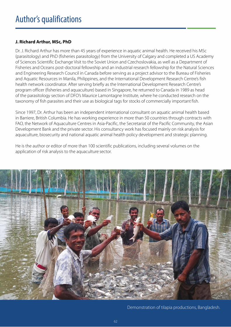
62
Author’s qualifications
J. Richard Arthur, MSc, PhD
Dr. J. Richard Arthur has more than 45 years of experience in aquatic animal health. He received his MSc (parasitology) and PhD (fisheries parasitology) from the University of Calgary and completed a US Academy of Sciences Scientific Exchange Visit to the Soviet Union and Czechoslovakia, as well as a Department of Fisheries and Oceans post-doctoral fellowship and an industrial research fellowship for the Natural Sciences and Engineering Research Council in Canada before serving as a project advisor to the Bureau of Fisheries and Aquatic Resources in Manila, Philippines, and the International Development Research Centre’s fish health network coordinator. After serving briefly as the International Development Research Centre’s program officer (fisheries and aquaculture) based in Singapore, he returned to Canada in 1989 as head of the parasitology section of DFO’s Maurice Lamontagne Institute, where he conducted research on the taxonomy of fish parasites and their use as biological tags for stocks of commercially important fish.
Since 1997, Dr. Arthur has been an independent international consultant on aquatic animal health based in Barriere, British Columbia. He has working experience in more than 50 countries through contracts with FAO, the Network of Aquaculture Centres in Asia-Pacific, the Secretariat of the Pacific Community, the Asian Development Bank and the private sector. His consultancy work has focused mainly on risk analysis for aquaculture, biosecurity and national aquatic animal health policy development and strategic planning.
He is the author or editor of more than 100 scientific publications, including several volumes on the application of risk analysis to the aquaculture sector.
Demonstration of tilapia productions, Bangladesh.
Phot
o cr
edit:
Md.
Hab
ibur
Rah
man
/Wor
ldFi
sh
62

63
Annex 1
Preliminary bibliography on the pathogens and parasites of Nigerian fish
BACTERIAAbidemi-Iromini AO and Fofah OJ. 2016. Bacteria occurrence in Sarotherodon malarotheron [sic] and Chrysichthys nigrodigitatus in brackish water environment. Applied Tropical Agriculture 21(1):1–6.
Abidemi-Iromini AO and Ogundipe ET. 2016. Occurrence of Aeromonas hydrophila in internal organs of wild and cultured Clarias gariepinus (Burchell, 1822). Journal of Sustainable Technology 7(1): 94–101.
Adah DA, Saidu L, Oniye SJ, Adah SA, Daodu OB, David SM and Olatunde AO. 2020. Microbiota of gills and antibiotic susceptibility patterns of bacteria isolates from Clarias gariepinus in different holding facilities. Sokoto Journal of Veterinary Sciences 18(3): 119–28.
Adeshina I, Abdrahman SA and Adewale AY. 2016. Occurrence of Klebsiella species in cultured african catfish in Oyo State, south-west Nigeria. Nigerian Veterinary Journal 37(1): 13–21.
Adeshina I, Umma SB, Adesanmi O and Adewale YA. 2016. Distribution of bacteria load in fish tissues, pond water and sediment in Oyo State, Nigeria. Alexandria Journal of Veterinary Sciences 51(1):1–9.
Ajayi AO. 2012. Bacteriological study of catfish, Clarias gariepinus, from fish pond sources in Akungba – Akoko Community Nigeria. British Microbiology Research Journal 2(1):1–9.
Ashiru AW, Uaboi-Egbeni PO, Oguntowo JE and Idika CN. 2011. Isolation and antibiotic profile of Aeromonas species from tilapia fish (Tilapia nilotica) and catfish (Clarias betrachus [sic]). Pakistan Journal of Nutrition 10(10):982–86.
Danba EP, Bichi AH, Ishaku S, Ahmad MK, Buba U, Bingari MS, Barau BW and Fidelis UF. 2014. Occurrence of pathogenic bacteria associated with Clarias gariepinus in selected fish farms of Kumbotso Local Governement Area of Kano State, Nigeria. Bayero Journal of Pure and Applied Sciences 7(2): 145–49.
Efuntoye MO, Olurin KB and Jegede GC. 2012. Bacterial flora from healthy Clarias gariepinus and their antimicrobial resistance pattern. Advance Journal of Food Science Technology 4(3):121–125.
Ekanem AP, Inyang-Etoh AP and Inyang-Etoh PC. 2011. Evaluation of the antibacterial efficacy of seven plant extracts against Aeromonas and Pseudomonas bacteria of farmed catfish (Heterobranchus longifilis). Veterinary Science Development 1(1):e11.
Emikpe BO, Adebis T and Adedeji OB. 2011. Bacteria load on skin and stomach of Clarias gariepinus and Oreochromis niloticus from Ibadan, south west Nigeria: Public health implications. Journal of Microbiology and Biotechnology Research 1(1):52–59
Enyidi UD and Maduakor CJ. 2017. Prevalence of bacteria and nematode parasites in African catfish Clarias gariepinus cultured in small holder concrete ponds in Nigeria. Journal of Biology and Nature 7: 169–176.
Ezeri GNO. 2001a. Haematological response of Clarias gariepinus to bacteria infection and prophylactic treatment with antibiotics. Journal of Aquatic Sciences 16:22–24.
Ezeri GNO. 2001b. Comparative bacteria count of skins of healthy and diseased Clarias gariepinus from two fish farms. Delta Agriculture 7:32–42.
Ikpi GU and Offem BO. 2008. Bacterial infection of cultural fish in the fish farm of the Cross River University of Technology. Egyptian Journal of Microbiology 21:57–63.

64
Ikpi G and Offem B. 2011. Bacterial infection of mudfish Clarias gariepinus (Siluriformes: Clariidae) fingerlings in tropical nursery ponds. Revista de Biologia Tropical 59(2):751–59.
Mohammed YM and Adamu KM. 2019. Bacteria associated with some freshwater fishes in Dangana Lake Lapai, Nigeria. Jewel Journal of Scientific Research 4(1&2):83–90.
Objajuru LOC and Ogbulie JN. 2006. Bacteriological quality of some fishes and crabs from rivers within Imo River basin. Journal of Aquatic Sciences 21(1):9–14.
Obot EA. 1989. The macrophytic flora of the draw-down area of Lake Kainji. African Journal of Ecology 27(2):173–77.
Ogbondeminu FS. 1993. Bacterial flora associated with the production of mudfish (Clarias anguillaris L.) in a hatchery. Biorsearch Communications 5:33–38.
Ogbondeminu FS, Madu CT and Okaeme AN. 1991. Bacteriological aspects of cultured fingerlings of Clarias anguillaris L. in a hatchery complex in Nigeria. Journal of Aquaculture in the Tropics 6:45–54.
Ogbulie JN and Okpokwasili GC. 1999. Haematological and histological responses of Clarias gariepinus and Heterobranchus bidorsalis to some bacterial diseases in Rivers State, Nigeria. Journal of the National Science Foundation of Sri Lanka 27(1):1–16.
Okaeme AN. 1989. Bacteria associated with mortality in tilapias, Heterobranchus bidorsalis, and C. lazera in indoor hatcheries and outdoor ponds. Journal of Aquaculture in the Tropics 4:143–46.
Okaeme AN and Obiekezie AO. 1990. Some emergent diseases and management problems of Oreochromis niloticus, Sarotherodon galileus and Clarias species in Nigeria. Journal of Aquatic Sciences 2:153–61.
Oladele OO, Ajayi OL, Olude OO, Stephen OO, Adediji AA, Arasi IO and Ntiwunka UG. 2012. Jaundice syndrome in African sharp-tooth catfish, Clarias gariepinus (Burchell), associated with haemolytic Staphylococcus aureus. Journal of Fish Diseases 35:945–47.
Oladele OO, Olarinmoye AO, Ntiwunka UG and Akintomide TO. 2015. Survey of bacterial isolates from cases of fish disease outbreaks and their antibiotic susceptibility patterns. Nigerian Journal of Fisheries 12(2):901–06.
Oladele, OO, Olufemi BE, Agbato OA, Yunusa H and Adebowale TK. 2010. High mortality in Clarias gariepinus fry associated with Klebsiella pneumoniae: A case report. Tropical Veterinarian 28(4):40–46.
Oladele OO, Olufemi BE, Oladosu GA, Ajayi OL, Adediji AA and Arasi IO. 2011. Arborescent organ necrosis syndrome in catfish Clarias gariepinus (Burchell): A case report. Journal of Fish Diseases 34:801–04.
Oladosu GA, Ayinla A and Ajiboye MO. 1994a. Aetiology, epizootiology and pathology of ‘rusty yellow’ skin discolouration in tilapia species Oreochromis niloticas [sic] and Tilapia zilii [sic]. Journal of Applied Ichthyology 10(2-3): https://doi.org/10.1111/j.1439-0426.1994.tb00159.x
Oladosu GA, Ayinla A and Ajiboye MO. 1994b. Isolation and pathogenicity of Bacillus sp. associated with a septicaemic condition in some tropical freshwater fish species. Journal of Applied Ichthyology 10(2–3). https://doi.org/10.1111/j.1439-0426.1994.tb00144.x
Oladosu GA, Tijani MO, Akpavie SO and Adedeji OB. 2013. Clinicopathological features of arborescent organ necrosis syndrome in Clarias gariepinus (Burchell 1822). Tropical Veterinarian 31(3):113–20.
Olayemi AB, Adebayo O and Ojo AO. 1991. Microbial flora of six freshwater fish species from Asa River, Ilorin, Nigeria. Revista de Biologia Tropical 39:165–67.
Olufemi BE, Akinlabi DA and Agbede SA. 1991. Aerobic bacterial pathogens isolated from the African catfish Clarias gariepinus (Burch). Tropical Veterinarian 9:177–80.

65
Olurin KB, Efuntoye M, Olojo EAA and Opomulero SO. 2006. Bacteria from eutrophic concrete fish tanks and earthen ponds in Ogun State, south-west Nigeria. Ecology, Environment and Conservation 12(1):167–70.
Omeje VO and Chukwu CC. 2014. Prevalence of Aeromonas hydrophila isolates in cultured and feral Clarias gariepinus of the Kainji Lake area, Nigeria. Nigerian Veterinary Journal 35(1):948–55.
Osho FE, Jenyo-Oni A, Ajani KE, Okpokpo AN and Okeke EO. 2020. Skin and gut microbiota of the African snakehead, Parachanna obscura, Gunther, 1861 from River Ogun, southwest Nigeria. Ife Journal of Agriculture 32(2):67–78.
Shinkafi SA and Ukwaja VC. 2010. Bacteria associated with fresh tilapia fish (Oreochromis niloticus) sold at Sokoto Central Market in Sokoto, Nigeria. Nigerian Journal of Basic and Applied Sciences 18(2):217–21.
Udeze AO, Talatu M, Ezediokpu MN, Nmwanze JC, Onoh C and Ononko IO. 2012. The effect of Klebsiella pneumoniae on catfish (Clarias gariepinus). Researcher 4(4):51–59.
Ugwuzor GN. 1996. Isolation and characterization of gliding bacteria from infected Clarias gariepinus (Burchell, 1822). Nigerian Journal of Science 30:89–95.
Ugwuzor GN. 1997. Sensitivity of gliding bacteria isolated from Clarias gariepinus (Burchell, 1822) to some antibiotics. Nigerian Journal of Science 31:61–64.
Ugwuzor NG, Anadu DL and Ejike C. 1990. Pseudomonad infection of catfish of the genus Clarias gariepinus (Teugels 1984). Journal of Aquatic Sciences 5:11–13.
Ugwuzor GN, Anadu DI and Ejike C. 1991. Fish bacteria of the genus Pseudomonas as a potential pathogen of mammals. Nigerian Journal of Agricultural Science 6:1–4. FUNGIAgina SE and Kpu RS. 1988. A survey of aquatic phycomycetes of Rockwater fish farms in Jos-Plateau State Nigeria. Nigerian Journal of Applied Fisheries and Hydrobiology 3:39–44.
Ogbonna CIC. 1989. Fungi associated with diseases of freshwater fishes in Plateau State, Nigeria. Journal of Aquatic Sciences 4:59–62.
Ogbonna CIC and Alabi RO. 1991. Studies on species of fungi associated with mycotic infections of fish in a Nigerian freshwater fish pond. Hydrobiologia 220:131–35.
Salawudeen MT, Kazeem HM, Raji MA, Oniye SJ, Kwanashie CN and Ibrahim MJ. 2017. Isolation and identification of fungi from apparently healthy and diseased Clarias gariepinus from freshwater in Zaria, Kaduna State, Nigeria. Microbiology Research International 5(1):8–15.
PARASITESAbba AM, Abdulhamid Y, Omenesa RL and Mudassir I. 2018a. Impact of helminth parasites on length-weight ratio and condition factor of fishes in Ajiwa and Jibia reservoirs, Katsina State, Nigeria. Research Journal of Zoology, 2018, 1(2), 6 pp.
Abba AM, Emere MC and Appah J. 2018b. Comparative study on helminth parasites of Oreochromis niloticus and Clarias gariepinus in Ajiwa Earth Dam Katsina State, Nigeria. World Journal of Fish and Marine Sciences 10(1):5–11.
Abidemi-Iromini AO and Eze RN. 2011. Comparative assessment of parasite infestation of tilapia in natural and cultured environments. In Liping L and Fitzsimmons K, eds. Better science, better fish, better life. Proceedings of the Ninth International Symposium on Tilapia in Aquaculture, Shanghai, China, April 22–24 2011. 56–59.
66
Abolarin MO. 1970. A note on the trypanosomes from the African freshwater fish and some comments on the possible relationship between taxonomy and pathology in trypanosomes. Bulletin of Epizootic Diseases of Africa 18(3):221–28.
Abolarin MO. 1971a. Preliminary study of Myxosporida (Protozoa) from the African carps in Nigeria. Nigerian Journal of Science 5:21–26.
Abolarin MO. 1971b. A new species of Henneguya (Myxosporida, Protozoa) from West African catfish, Clarias lazera Val., with a review of the genus Henneguya Thelohan, 1892. African Journal of Tropical Hydrobiology and Fisheries 2(1):93–105.
Abolarin MO. 1974. Myxobolus tilapiae sp. nov. (Protozoa: Myxosporidia) from three species of freshwater Tilapia in Nigeria. Journal of the West African Science Association 19:109–14.
Abowei JFN and Ezekiel EN. 2011. A review of Acanthocephala, leeches, parasite crustaceans and some other parasites of miscellaneous taxa infections in African fish. International Journal of Animal and Veterinary Advances 3(5):337–51.
Abraham JT and Akpan PA. 2012. Prevalence of Henneguya chrysichthys and its infection effect on Chrysichthys nigrodigitatus fecundity. AFRREV STECH 1(3):231–51.
Abraham JT, Akpan PA and Effiom OE. 2011. Prevalence of Henneguya chrysichthys (flagellated Protozoa: cyst) and haematological changes due to the infection in Chrysichthys nigrodigitatus. African Research Review 5(4):124–34.
Abraham JT, Akpan PA and Okon OE. 2004. Occurrence of parasites in Pseudotolithus elongatus and Cynoglossus seneglensis [sic] in Cross River estuary Nigeria. Global Journal of Pure and Applied Sciences 11(1):45–49. (Reprinted from Vol. 10(4) due to omission of third author’s name.)
Abraham JT, Ndome CB, Okon OE, Ekpenyong E and Ibor CI. 2013. Capillariasis in Chrysichthys nigrodigitatus (catfish), Cynoglossus senegalensis (sole) and Pseudotolthus [sic] elongatus (bobo croaker) from Cross River estuary and adjacent coastal waters. AFRREV STECH 2(1):83–93.
Absalom KV, Makpo JK and Mustapha AJ. 2018. Prevalence of gastrointestinal helminth parasites of Clarias gariepinus at River Gudi, Akwanga Local Government Area of Nasarawa State, Nigeria. International Journal of Fisheries and Aquaculture Research 4(1):9–15.
Abu S, Okoye I, Obiezue NNR and Ofoezie E. 2016. Influence of host weight and size on the prevalence and abundance of parasites of fish. International Journal of Biological Research 1(5):1–4.
Adebambo AAR. 2020. Fish species parasites: A review in Nigerian water bodies. Journal of Research in Forestry, Wildlife and Environment 12(3):223–34.
Adeogun OA, Oladosu GA, Akinwale MMA, Okunade OA, Akintayo IA, Idika N, Adeiga AA, Ezeugwu SMC, Afocha EE, Peters OS and Odusanya AF. 2013. 23. Identification, distribution and prevalence of ecto-parasites associated with cultured fish in Ogun State, Nigeria. In Proceedings of the FISON 28th Annual Conference, November 25–30, 2013. 2–13. (Republished in 2014 in Journal of Fisheries and Aquatic Sciences 9(5):413–18.)
Aderounmu EA and Adeniyi F. 1972. Cestodes in fish from a pond at Ile-Ife, Nigeria. African Journal of Tropical Hydrobiology and Fisheries 2(2):151–56.
Adeshina I, Jenyo-Oni A. Ajani EK and Adewale YA. 2016. Natural occurrence of Diplostomum spp. in farm raised African catfish (Clarias gariepinus) in Oyo State. International Journal of Veterinary Science and Medicine 5(2):1–8.
Adeshina I, Tiamiyu LO, Akpoili, BU, Jenyo-Oni A and Ajani EK. 2021. Dietary Mitracarpus scaber leaves extract improved growth, antioxidants, non-specific immunity, and resistance of Nile tilapia, Oreochromis niloticus to Gyrodactylus malalai infestation. Aquaculture 535:736377.

67
Adeyemo AO and Agbede SA. 2008. Histopathology of tilapia tissues harbouring Clinostomum tilapiae parasites. African Journal of Biomedical Research 11(1):115–18.
Adeyemo AO and Falaye AE. 2007. Parasitic incidence in cultured Clarias gariepinus. Animal Research International 4(2): 702–04.
Adegoroye F, Omobhude M and Morenikeji O. 2019. Helminth parasites of Synodontis clarias (Linnaeus, 1758), Chrysichthys nigrodigitatus (Lacepede, 1802) and Chrysichthys auratus (Geoffrey Saint-Hilaire, 1808) in Asejire Dam, south-west Nigeria. International Journal of Aquatic Science 10(1):37–47.
Adikwu IA and Ibrahim BA. 2004. Studies on the endoparasites in the gastro-intestinal tract of Clarias gariepinus (Tugels) in Wase Dam, Kano State, Nigeria. African Journal of Applied Zoology and Environmental Biology 6:36–40.
Ajala OO and Fawole OO. 2012. A study of helminth species assemblages at different host scales in Clarias gariepinus (Burchell, 1822) as a bio-indicator of aquatic water quality. In Conference Proceedings, World Academy of Science, Engineering and Technology (WASET), Singapore, September 2012. 741–50.
Ajala OO and Fawole OO. 2014a. Multiple infections of helminths in the stomach and intestine of Clarias gariepinus (Burchell, 1822) in Oba Reservoir, Oyo State, Nigeria. IOSR Journal of Pharmacy and Biological Sciences 9(3):5–12.
Ajala OO and Fawole OO. 2014b. Multiple infections of helminths in the alimentary system of Clarias gariepinus (Burchell, 1822) in a tropical reservoir. International Journal of Fisheries and Aquaculture 6(6):62–70.
Ajala OO and Fawole OO. 2015. Diets and enteroparasitic infestation in Sarotherodon galilaeus (Linnaeus, 1758) (Cichlidae) in Oba Reservoir Ogbomoso, Nigeria. International Journal of Fisheries and Aquatic Studies 2(6):3–10.
Aken’ova T. 2000a. Copepod parasites of the gills of Clarias species in two lakes and a river in Zaria, Nigeria. Nigerian Journal of Parasitology 20:99–112.
Aken’ova T. 2000b. Helminth infection of the gills of Clarias species in Zaria, Nigeria. Nigerian Journal of Parasitology 20:113–21.
Aken’ova TO and Shotter RA. 1988. Ectoparasitic protozoans of the gills of Clarias species. Nigerian Journal of Parasitology 9(11):129–37.
Akinsanya B. 2007. Histopathological study on the parasitised visceral organs of some fishes of Lekki Lagoon, Lagos, Nigeria. Life Science Journal 4(3):70–76.
Akinsanya B. 2015. Two fish species study of the parasitic helminth fauna of Synodontis filamentosus (Boulenger, 1901) and Calamoichthys calabaricus (Smith, 1865) from Lekki Lagoon, Lagos, Nigeria. Ife Journal of Science 17(1):97–108.
Akinasanya B. 2016. Studies on Cucullanus sp. (Nematoda: Cucullanidae) parasitic in Tilapia zillii (Gervais, 1848) from Lekki Lagoon, Lagos, Nigeria. Egyptian Journal of Aquatic Biology and Fisheries 20(2):79–87.
Akinsanya B, Abayomi A, Iyabo A and Giwa M. 2019. Parasitic fauna, histopathological alterations, and organochlorine pesticides contamination in Chrysichthys nigrodigitatus (Lacepede, 1803) (Bagridae) from Lagos, Lagoon, Nigeria. Scientific African 5:e00130. https://doi.org/10.1016/j.sciaf.2019.e00130
Akinsanya B, Abiodun KT, Ukwa UD and Saliu JK. 2018. Gastrointestinal parasites of Sarotherodon melanotheron (Ruppel, 1852) histopathological alterations and organochlorine pesticides pollution from Lagos, Lagoon, Nigeria. Academic Journal of Biological Sciences E. Medical Entomology and Parasitology 10(2):15–28.

68
Akinsanya B, Adebusoye SA, Alinson T and Ukwa UD. 2018. Bioaccumulation of polycyclic aromatic hydrocarbons, histopathological alterations and parasito-fauna in bentho-pelagic host from Snake Island, Lagos, Nigeria. Journal of Basic and Applied Zoology 79:40, 18 pp.
Akinsanya B, Ade-Ademilua OE, Idris O, Ukwa UD and Saliu JK. 2016. Toxicological evaluation of plant crude extracts on helminth parasites of Clarias gariepinus using host low observed effect concentration (LOEC). Egyptian Journal of Aquatic Biology and Fisheries 20(2):69–77.
Akinsanya B, Ayanda IO, Fadipe AO, Onwuka B and Saliu JK. 2020. Heavy metals, parasitologic and oxidative stress biomarker investigations in Heterotis niloticus from Lekki Lagoon, Lagos, Nigeria. Toxicology Reports 7:1075–82.
Akinsanya B and Hassan AA. 2002. Excystment of the metacerceria of the trematode Clinostomum marginatum. Bioscience Research Communications 14(4):445–50.
Akinsanya B and Hassan AA. 2012. Ultrastructural and histopathological studies of the digenetic trematode Siphodera ghanensis (Cryptogonimidae), parasite of Chrysichthys nigrodigitatus (Lacepede, 1802) from Lekki Lagoon, Lagos, Nigeria. Nigerian Journal of Parasitology 33(1):103–09.
Akinsanya B, Hassan AA and Adeogun AO. 2008. Gastrointestinal helminth parasites of the fish Synodontis clarias (Siluriformes: Mochokidae) from Lekki Lagoon, Lagos, Nigeria. Revista de Biología Tropical 56(4):2021–26.
Akinsanya B, Hassan AA and Adeogun AO. 2015. SEM study on morphology and surface topography of Wenyonia minuta Woodland 1923 (Cestoda: Caryophyllidea) and the histopathological consequences on Synodontis filamentosus (Boulenger, 1901) from Lekki Lagoon, Lagos – Nigeria. Egyptian Academic Journal of Biological Sciences, D. Histology & Histochemistry 7(1):11–18.
Akinsanya B, Hassan A and Adeogun A. 2016. Pathological changes and description of Procamallanus (Spirocamallanus) aspiralis Baylis, 1923 from the freshwater fish, Parachanna obscura Gunther, 1861. Journal of Coastal Life Medicine 4(9):693–97.
Akinsanya B, Hassan AA and Fawole OO. 2002. Prevalence of parasitic infections in cichlids from Eleyele River, Ibadan, Nigeria. Bioscience Research Communications 14(1):93–100.
Akinsanya B, Hassan AA and Ibidapo CA. 2010. The parasitic helminth fauna of Parachanna obscura from Lekki Lagoon, Lagos, Nigeria. Researcher 2(9):78–84.
Akinsanya B, Hassan AA and Otubanjo OA. 2007. A comparative study of the parasitic helminth fauna of Gymnarchus niloticus (Gymnarchidae) and Heterotis niloticus (Osteoglossidae) from Lekki Lagoon, Lagos, Nigeria. Pakistan Journal of Biological Sciences 10(3):427–32.
Akinsanya B, Hassan AA and Ukwa UD. 2014. Effect of host condition on intestinal parasite load and prevalence in Malapterurus electricus (Gmelin, 1789) (Siluriformes: Malapteruridae) in Lekki Lagoon. Nigerian Journal of Parasitology 35(1&2):59–64.
Akinsanya B, Isibor PO, Ademola E, Dada D, Saliu J and Olasehinde G. 2020. Accumulation of PCBs and infections of parasitic helminthes in Synodontis filamentosus (Boulenger, 1901) and Tilapia zillii (Gervais, 1848) of Epe Lagoon, Lagos, Nigeria. Egyptian Journal of Aquatic Biology and Fisheries 24(1):49–63.
Akinsanya B, Isibor PO, Kuton MP, Dada EO and Saliu JK. 2019. Comparative partition coefficients of BTX and OCPs between Synodontis clarias and parasite Weynonia acuminata. Journal of Basic and Applied Zoology 80(38):1–9.
Akinsanya B, Isibor PO, Kuton MP, Saliu JK and Dada EO. 2019. Aspidogastrea africanus infections, comparative assessment of BTEX and heavy metals bioaccumulation, and histopathological alterations as biomarker response in Chrysichthyes nigrodigitatus (Lacépède, 1803) of Lekki Lagoon, Nigeria. Scientific African 3:e90060.

69
Akinsanya B, Isibor PO, Olaleru F, Abayomi A, Akeredolu E, Ohazulike MI and Saliu JK. 2021. Bioaccumulation of pyrethroid in parasite Wenyonia acuminata (Cestoda: Caryophyllaeidae) and host fish Synodontis clarias (Linnaeus, 1758) from Lekki Lagoon, Lagos Nigeria. Brazilian Journal of Biology https://doi.org/10.1590/1519-6984.236427. Online ahead of print. PMID: 32965342.
Akinsanya B, Isibor PO, Pentho KM [Kuton MP], Mulikat K and Saliu JK. 2019. Parasite prevalence and bioaccumulation of polycyclic aromatic hydrocarbons as stressors in the silver catfish, Chrysichthys nigrodigitatus (Siluriformes: Claroteidae). Scientific African 7:e00225.
Akinsanya B and Kuton MP. 2016a. Bioaccumulation of heavy metals and parasitic fauna in Synodontis clarias (Linnaeus, 1758) and Chrysichthys nigrodigitatus (Lacepede, 1803) from Lekki Lagoon, Lagos, Nigeria. Asian Pacific Journal of Tropical Disease 6(8):615–21.
Akinsanya B and Kuton MP. 2016b. Parasitic diseases and heavy metal analysis in Parachanna obscura (Gunther 1861) and Clarias gariepinus (Burchell 1901) from Epe Lagoon, Lagos, Nigeria. Asian Pacific Journal of Tropical Disease 6(9):685–90.
Akinsanya B, Kuton MP, Arowoshafe RA, Lawal Are AO and Akhiromen DI. 2018. The Caryophylleadae [sic] cestodes, Wenyonia spp Woodland, 1923 bioaccumulates high quantities of a specific PCBs congener in the fish host, Synodontis clarias (Linnaeus, 1758), with histopathological alterations as biomarker response. Egyptian Academic Journal of Biological Sciences, D. Histology & Histochemistry 10(1):27–45.
Akinsanya B, Kuton MP, Iniobong AD, Uchenna ND, Saliu JK and David UU. 2020. Bioaccumulation of polychlorinated biphenyls (PCBs) in fish host-parasite bentho-pelagic food chain in Epe Lagoon, Lagos, Nigeria. Bulletin of Environmental Contamination and Toxicology 105(1). https://doi.org/10.1007/s00128-020-02893-y
Akinsanya B, Kuton MP, Saliu JK, Oyebola L and Ukwa UD. 2015. Condition factor and gastrointestinal parasitic fauna of three fish species as stress indicators in Lekki Lagoon, Lagos, Nigeria. Egyptian Academic Journal of Biological Sciences, D. Histology & Histochemistry 7(1):1–13.
Akinsanya B, Kuton MP, Saliu JK and Ukwa UD. 2015. Host relative condition factor and the prevalence of intestinal parasites of Parachanna obscura and Sarotherodon melanotheron as indicators of environmental stress in Lekki Lagoon, Lagos, Nigeria. Nigerian Journal of Life Sciences 5(2):37–46.
Akinsanya B, Olaleru F, Samuel OB, Akeredolu E, Isibor PO, Adeniran OS, Saliu JK and Akhiromen DI. 2020. Bioaccumulation of organochlorine pesticides, Procamallanus sp. (Baylis, 1923) infections, and microbial colonization in African snakehead fish sampled from Lekki Lagoon, Lagos, Nigeria. Brazilian Journal of Biology, 11 pp. https://doi.org/10.1590/1519-6984.237312
Akinsanya B and Otubanjo OA. 2006. Helminth parasites of Clarias gariepinus (Clariidae) in Lekki Lagoon, Lagos, Nigeria. Revista de Biología Tropical 54(1):93–99.
Akinsanya B, Otubanjo OA and Hassan AA. 2007. Helminth parasites of Malapterurus electricus (Malapteruridae) from Lekki Lagoon, Lagos, Nigeria. Journal of American Science 3(3):1–5.
Akinsanya B, Otubanjo OA and Ibidapo CA. 2007. Helminth biload of Chrysichthys nigrodigitatus (Lacepede 1802) from Lekki Lagoon, Lagos, Nigeria. Turkish Journal of Fisheries and Aquatic Sciences 7:83–87.
Akinsanya B, Ukwa UD, Kuton MP and Saliu JK. 2015. Parasite load, condition factor, and heavy metal accumulation as multiple stressors on fish with public health significance in Lekki Lagoon, Lagos, Nigeria. Journal of Public and Allied Health Sciences 1:21–33.
Akinsanya B, Utoh OU and Ukwa UD. 2016. Toxicological, phytochemical and anthelminthic properties of rich plant extracts on Clarias gariepinus. Journal of Basic and Applied Zoology 74:75–86.

70
Akogun OB and Gaddard JP. 1991. Parasites of Clarias submarginatus and Clarias gariepinus from the Benue River at Yola, Nigeria. Annals of Borno 6/7:115–20.
Alfred-Ockiya JF. 1985. Preliminary survey of parasites of Clarias in Niger Delta area, Nigeria. In Proceedings of the 4th Annual Conference of the Fisheries Society of Nigeria (FISON), Port Harcourt, November 26–29, 1985. 210–15.
Alfred-Ockiya JF and Ovuru SS. 1995. A comparative study of incident of parasitic infestation of the African catfish (Clarias gariepinus) in Rivers State. Nigerian Journal of Agriculture Teacher Education 4:65–73.
Alade AO, Whab AO and Okunlola DO. 2015. Incidence of parasites in some fresh water fish species Inoyo Town, Oyo State, Nigeria. A threat to national food security. International Journal of Engineering Science 12(4):50–53.
Aliyu MD and Solomon JR. 2012. The intestinal parasites of Clarias gariepinus found at lower Usman Dam, Abuja. Researcher 4(9):38–44.
Allumma MI and Idowu RT. 2011 Prevalence of gills helminth of Clarias gariepinus in Baga Side of Lake Chad. Journal of Applied Sciences and Environmental Management 5(1):47–50.
Ama-Abasi DE and Obiekezie AI. 2002. Trichodina (Emberg Ciliphora Petrichida) infection of clariid catfishes cultured in Nigeria. African Journal of Fisheries and Aquaculture 3:1–9.
Amaechi CE. 2015. Prevalence, intensity and abundance of endoparasites in Oreochromis niloticus and Tilapia zilli [sic] (Pisces: Cichlidae) from Asa Dam, Ilorin, Nigeria. Cuadernos de Investigación UNED 7(1):67–70.
Amos SO, Eyiseh TE and Michael ET. 2018. Parasitic infection and prevalence in Clarias gariepinus in Lake Gerio, Yola, Adamawa State. MOJ Anatomy and Physiology 5(6):376–81.
Ani OC, Nnamonu EI and Ejiogu C. 2017. Prevalence of intestinal parasites of fish farmed and harvested in Abakiliki, Nigeria: A pointer to the level of their vulnerability. International Journal of Research in Pharmacy and Biosciences 4(9):7–10.
Anosike JG, Omoregie E, Ofojekwu PC and Nweke IE. 1992. A survey of the helminth parasites of Clarias gariepinus in Plateau State, Nigeria. Journal of Aquatic Sciences 7:39–43.
Anyanwu AO. 1983. Parasitic infestations of Pseudotolithus spp. off the coast of Lagos, Nigeria. Journal of Fish Biology 22(1):29–33.
Arimoro FO and Utebor KE. 2013. Relevance of nematode parasitic burden in channid fishes of Orogodo River, southern Nigeria to organic pollution. Annual Review & Research in Biology 3(4):584–95.
Ashade OO, Osineye OM and Kumaye EA. 2010. Isolation, identification and prevalence of parasites on Oreochromis niloticus from three selected river systems. FSN-AQ 0015. FISON ECO, 2010. In Proceedings of the Fisheries Society of Nigeria (FISON), Ascon, Badagry, October 25–29, 2010. 78–87. (Republished in 2013 in the Journal of Fisheries and Aquatic Sciences 8(1):115–21).
Atalabi TE, Awharitoma AO and Akinluyi FO. 2018. Prevalence, intensity, and exposed variables of infection with Acanthocephala parasites of the gastrointestinal tract of Coptodon zillii (Gervais, 1848) [Perciformes: Cichlidae] in Zobe Dam, Dutsin-Ma Local Government Area, Katsina State, Nigeria. Journal of Basic and Applied Zoology 79(1), Article No. 29, 7 pp.
Auta J, Oniye SJ and Adakole JA. 1999. The helminthes parasites of the gastrointestinal tract of Synodontis species in Zaria, Nigeria. Zuma: Journal of Pure and Applied Sciences 2(2):47–53.

71
Awa J, Anyanwu P and Ezenwa B. 1988. Incidence of parasitic infection of pond raised tilapia spp. and some cultivable fish species from three ecological areas of Lagos State. NIOMR Technical Paper. No. 32.
Awa JU, Ugumba A and Odaibo AB. 1996. Parasites of Oreochromis nilotica of Ita Lake, Ibadan, Oyo State, Nigeria. Nigerian Journal of Parasitology 17:115–20.
Awachie JBE. 1965. Preliminary notes on the parasites of fish in the area of the Kainji Reservoir. In White E, ed. First Scientific Report of Kainji Biological Research Team. Ile-Ife, University of Ife. 65–69.
Awachie JBE. 1972. On a didymozoid trematode parasite in the eye socket of African carps, Labeo sp. from area of Kainji man-made lake in Nigeria. Acta Parasitologica Polonica 20:492–98.
Awachie JBE, Ilozumba, PCO and Azugo WI. 1977. Fish parasites in the ecology, management and productivity of river and flood plain fisheries in Africa. In Welcomme RL, ed. CIFA/77 Symposium. 14th Symposium on River and Flood Plain Fisheries in Africa. Bujumbura, Burundi, November 21–23, 1977. Review and Experience Papers. CIFA Technical Paper No. 5.
Awharitoma AO and Ehigiator FAR. 2012. Helminth parasites of fishes from some rivers in southern Nigeria. African Scientist 13(2):65–69.
Awharitoma AO and Ehigiator FAR. 2017. Effects of climatic changes on fish diversity and abundance and prevalence of fish parasitic infections in southern Nigeria. NISEB Journal 17(3):112–18.
Awharitoma AO and Okaka CE. 1999. Observations on the cichlid fishes in Ikpoba River and their parasitic infections. Nigerian Journal of Parasitology 20:129–37.
Awharitoma AO and Okaka CE. 2004. Parasites of fishes from Ikpoba River and Ogba River fish farm. Nigerian Journal of Applied Science 22:355–62.
Awosolu OB, Simon-Oke IA and Oyelere AA. 2018. Studies on the prevalence and distribution of parasites of tilapia fish (Oreochromis niloticus) from Igbokoda River, Ondo State, Nigeria. Molecular Pathogens 9(1):1–4.
Ayanda OI. 2008. Comparative parasitic helminth infection between cultured and wild species of Clarias gariepinus in Ilorin, north - central Nigeria. Scientific Research and Essays 4:18–21.
Ayanda OI. 2009. Comparison of parasitic helminthes infection between the sexes of Clarias gariepinus from Asa Dam Ilorin, north-central Nigeria. Scientific Research and Essays 4(4):357–60.
Ayanwu AO. 1983. Parasitic infestations of Pseudotolithus spp. off the coast of Lagos, Nigeria. Journal of Fish Biology 22(1):29–33.
Ayotunde EO, Ochang SN and Okey IB. 2007. Parasitological examinations and food composition in the gut of feral African carp, Labeo coubie in the Cross River, south eastern, Nigeria. African Journal of Biotechnology 6(5):625–30.
Balogun O and Solomon JR. 2015. Prevalence of endoparasitic infection of Clarias gariepinus fed chicken droppings. African Journal of Environmental Pollution and Health 11:26–33.
Bello-Olusoji OA, Aderiye BK, Borede AA and Oyekanmi FB. n.d. Ectoparasitic studies of pond cultured and wild tilapias in major cocoa producing area of Nigeria. ag.arizona.edu › ista › OlusojiBello- Ectoparasite.
Bichi AH. 2006. A survey of external and intestinal parasites of fishes found around the burrow pits of Kano metropolis. African Scientist 7(4):161–64.
Bichi AH and Bizi AG. 2002. Survey of ecto and endo parasites of fishes of Challawa George Dam. NISEB Journal 2(3):219–22.

72
Bichi AH and Dawaki SS. 2010. A survey of the ectoparasites on the gills, skin and fins of Oreochromis niloticus at Bagauda fish farm, Kano, Nigeria. Bayero Journal of Pure and Applied Sciences 3(1):83–86.
Bichi AH and Ibrahim AA. 2009a. A survey of ecto and intestinal parasites of Tilapia zillii (Gervias) [sic] in Tiga Lake, Kano, northern Nigeria. Bayero Journal of Pure and Applied Sciences 2(1):79–82.
Bichi AH and Ibrahim AA. 2009b. Survey of ecto and endo parasites of fishes in Tiga Lake, Kano. NISEB Journal 2(3):219–222.
Bichi AH and Yelwa SI. 2010. Incidence of piscine parasites on the gills and gastrointestinal tract of Clarias gariepinus (Teugels) at Bagauda fish farm, Kano. Bayero Journal of Pure and Applied Sciences 3(1):104–07.
Biu AA and Akorede GJ. 2013. Prevalence of endoparasites of Clarias gariepinus (Burchell 1822) in Maiduguri, Nigeria. Nigerian Journal of Fisheries and Aquaculture 1(1):1–6.
Biu AA, Diyaware MY, Yakaka W and Joseph E. 2014. Survey of parasites infesting the Nile tilapia (Oreochromis niloticus Linnaeus, 1758) from Lake Alau, Maiduguri, Nigeria. Nigerian Journal of Fisheries and Aquaculture 2(2):6–12.
Biu AA, Diyaware MY, Yakaka W and Rita DJ. 2014. Incidence of parasites of Clarias gariepinus (Burchell, 1822) caught from Lake Alau, Maiduguri, Borno State, Nigeria. Nigerian Journal of Fisheries and Aquaculture 2(1):74–80.
Biu AA and Nkechi OP. 2013. Prevalence of gastrointestinal helminths of Tilapia zilli [sic] (Gervais 1848) in Maiduguri, Nigeria. Nigerian Journal of Fisheries and Aquaculture 1(1):20–24.
Caffara M, De Vita V, Florio D, Gustinelli A, Quaglio F and Fioravanti ML. 2007. Infections due to Unicauda spp. (Myxozoa, Myxobolidae) in elephantnose fish (Gnathonemus petersii) imported to Italy. Bulletin of the European Association of Fish Pathologists 27(4):142–47.
Caffara M, Locke SA, Echi PC, Halajian A, Benini D, Luus-Powell WJ, Tavakol S and Fioravanti ML. 2017. A morphological and molecular study of clinostomid metacercariae from African fish with a redescription of Clinostomum tilapiae. Parasitology 144(11):1519–29.
Caffara M, Locke SA, Echi PC, Halajian A, Luus-Powell WJ, Benini D, Tedesco P and Fioravanti ML. 2020. A new species of Clinostomum Leidy, 1856 based on molecular and morphological analysis of metacercariae from African siluriform fishes. Parasitology Research 119:885–92.
Chessed A. 2019. Prevalence of gastrointestinal parasites and condition factor of Auchenoglanis biscutatus (Geoffrey St. Hilarie [sic], 1827) in Kiri Reservoir, Shelleng, Adamawa State, Nigeria. Corpus ID: 208073163.
Dan-kishiya AS, Oboh A and Ibrahim UB. 2013. The prevalence of helminth parasites in the gastro-intestinal tract of wild African sharptooth catfish Clarias gariepinus (Siluriformes: Clariidae) in Gwagwalada, Nigeria. Cuadernos de Investigación UNED 5(1):83–87.
Dankishiya AS and Zakari M. 2007. Study on the gastrointestinal helminth parasites of Clarias gariepinus (Teugels) in Gwagwalada, FCT, Nigeria. Biological and Environmental Sciences Journal for the Tropics 4(2):79–81.
Dauda J, Lawal JR, Bello AM, Majama YB, Lekko YM, Mshelia ES and Biu AA. 2016. Prevalence of gastrointestinal helminths of Tilapia zilli [sic] (Gervias) [sic] in Gombe, northeastern Nigeria. Journal of Animal Science and Veterinary Medicine 1(3):74–80.
Domo GA and Ester ST. 2015. Prevalence of the helminth parasites of Oreochromis niloticus and Clarias gariepinus in Lake Geriyo Jimeta Yola, Adamawa State. Journal of Novel Applied Sciences 4(1):1–6.
Dönges J and Harder W. 1966. Nesolecithus africanus n.sp. Cestodaria, Amphilinidae) aus dem Coelom von Gymnarchus niloticus Cuvier, 1829 (Teleostei). Zeitschrift für Parasitenkunde 28:125–41.

73
Echi PC. 2016a. Recurrent attachment of Batrachobdelloides tricarinata (Rhynchobdellae: Glossiphoniidae) on cichlids in an undisturbed lake, southeast, Nigeria. International Journal of Fisheries and Aquatic Studies 4(3):343–45.
Echi PC. 2016b. The overall differential morphometric parts among clinostomatids (Clinostomatidae) in the micro-habitats of Tilapia zillii and other cichlids. Journal of Fisheries and Aquatic Studies 4(4):304–06.
Echi PC. 2017. Glycogen and protein profiles of Clinostomum tilapiae Ukoli, 1966 (Digenea: Clinostomidae) and putative species Clinostomum Morphotype1 in a tropical river (Anambra River Basin, Otuocha, Nigeria). Brazilian Journal of Biological Sciences 4(8):317–21.
Echi PC, Eyo JE and Okafor FC. 2009. Co-parasitism and morphometrics of three clinostomatids (Digenea: Clinostomatidae) in Sarotherodon melanotheron from a tropical freshwater lake, Nigeria. Animal Research International 6(2):982–86.
Echi PC, Eyo JE, Okafor FC, Onyishi GC and Ivoke N. 2012. First record of co-infection of three clinostomatid parasites in cichlids (Osteichthyes: Cichlidae) in a tropical freshwater lake. Iranian Journal of Public Health 41(7):86–90.
Echi PC and Ezenwaji HMG. 2010. The parasites fauna of characids’ (Osteichthyes: Characidae) Anambra River, Nigeria. African Journal of Ecology 48(1):1–4.
Echi PC, George S and Kumar S. 2016. Differential size-biased parasitism between Polyacanthorhynchus nigerianus (nomen nudum) and Polyacanthorhynchus echiyensis (nomen nudum) (Acanthocephala: Polyacanthocephala). Brazilian Journal of Biological Sciences 3(5):113–19.
Echi PC, Iyaji FO, Ejere VC and Abuh SJ. 2014. Dynamics of synchronized clinostomatids infections in cichlids. Environment Conservation Journal 15(1/2):49–54.
Echi PC and Ojebe MO. 2016. Differences in host-infection patterns of clinostomatids (Clinostomatidae) in human disturbed aquatic ecosystems against undisturbed lake. Brazilian Journal of Biological Sciences 3(5):97–103.
Echi PC, Okafor FC and Eyo JE. 2009. Co-infection and morphometrics of three clinostomatids (Digenea: Clinostomatidae) in Tilapia guinensis Bleeker, 1862 from Opi Lake, Nigeria. Bio-Research 7(1):432–36.
Echi PC, Suresh K, Sanil G, Iyaji FO, Nwani CD and and Ejere VC. 2015. Mitochondrial DNA resolution of two new sequences Polyacanthorhynchus echiyensis n. sp. and Polyacanthorhynchus nigerianus n. sp. (Polyacanthocephala: Acanthocephala) in a parentenic host from a tropical river. Environmental Conservation Journal 16(1/2):13–17.
Edeh C and Solomon RJ. 2016. Endoparasites of Oreochromis niloticus and Clarias gariepinus found in Utako flowing gutter. Direct Research Journal of Agriculture and Food Science 4(12):361–73.
Edema CU, Okaka CE, Oboh IP and Okogub BO. 2008. A preliminary study of parasitic infections of some fishes from Okhuo River, Benin City, Nigeria. International Journal of Biomedical and Health Sciences 4(3):107–16.
Egwunyenga AO, Nmorsi OPG and Igbinosun AF. 1999. Haematological changes in cichlids of Ethiope River (Niger Delta Nigeria) due to intestinal helminthiasis. Bioscience Research Communications 11(4):361–65.
Ejere VC, Aguzie OI, Ivoke N, Ekeh FN, Ezenwaji NE, Onoja US and Eyo JE. 2014. Parasitofauna of five freshwater fishes in a Nigerian freshwater ecosystem. Croatian Journal of Fisheries 72:17–24.
Ekanem AP. 2010. Incidence and abundance of trichodiniasis of Clarias gariepinus in the University of Calabar fish farm, Calabar, Nigeria. Tropical Environmental Research 9:566–70.

74
Ekanem AP and Brisibe EA. 2010. Effects of ethanol extract of Artemisia annua L. against monogenean parasites of Heterobranchus longifilis. Parasitology Research 106(5):1135–39.
Ekanem AP, Eyo VO and Sampson AF. 2011. Parasites of landed fish from Great Kwa River, Calabar, Cross River State, Nigeria. International Journal of Fisheries and Aquaculture 3(12):225–30.
Ekanem AP, Eyo VO, Udoh JP and Okon JA. 2014. Endoparasites of food-fish landing from the Calabar River, Cross River State, Nigeria. Journal of Scientific Research and Reports 3(6):810–17.
Ekanem AP and Obiekezie AI. 2000. Antiparasitic effects of Piper guineense (Husaini) on the juvenile of Heterobranchus longifilis (Cuvier & Valenciennes). African Journal of Fisheries and Aquaculture 2:68–74.
Ekanem AP and Obiekezie AI. 2013. Utilization of medicinal plants and their products in the treatment and control of disease in fish. In Juliani HR, Simon JE and Ho C-T, eds. African natural plant products. Vol. II: Discoveries and Challenges in Chemistry, Health, and Nutrition. ACS Symposium Series 1127, Washington, D.C., American Chemical Society. Chapter 7, 93–102.
Ekanem AP, Obiekezie A, Kloas W and Knopf K. Effects of crude extracts of Mucuna pruriens (Fabaceae) and Carica papaya (Caricaceae) against the protozoan fish parasite Ichthyophthirius multifiliis. Parasitology Research 92(5):361–66.
Ekanem AP, Wang M, Simon JE, Obiekezie AI and Morah F. 2004. In vivo and in vitro activities of the seed extract of Piper guineense Schum. and Thonn. against skin and gill monogenean parasites of goldfish (Carassius auratus auratus). Phytotherapy Research 18(10):793–97.
Ekanem DA and Obiekezie AI. 1996. Growth reduction in African catfish fry infected with Trichodina maritinkae Basson & Van As 1991. Journal of Aquaculture in the Tropics 11(2):91–96.
Emere MC. 2000. Parasitic infection of the Nile perch Lates niloticus (L) in River Kaduna. Journal of Aquatic Sciences 15:51–54.
Emere MC and Egbe NEL. 2006. Protozoan parasites of Synodontis clarias (a fresh water fish) in River Kaduna. The Best Journal 3(3):58–64.
Enyidi UD and Eneje UL. 2015. Parasites of African catfish Clarias gariepinus, cultured in homestead ponds. Researchjournali’s Journal of Agriculture 2(12):1–10.
Enyidi UD and Maduakor CJ. 2017. Prevalence of bacteria and nematode parasites in African catfish Clarias gariepinus cultured in small holder concrete ponds in Nigeria. Journal of Biology and Nature 7(4):169–76.
Enyidi U and Uwanna P. 2019. Parasites of African catfish Clarias gariepinus and Oreochromis niloticus polycultured in earthen pond. Aquaculture Studies 19(2):81–89.
Esiest ULP. 2013. Length-weight relationship and parasites of Chrysichthys nigrodigitatus in Cross River Estuary Itu local government area Akwa Ibom State, Nigeria. Basic Research Journal of Agricultural Science and Review 2:154–65.
Eyo VO, Edet TA and Ekanem AP. 2015. Monogenean parasites of the African catfish Clarias gariepinus from two fish farms in Calabar, Cross River State, Nigeria. Journal of Coastal Life Medicine 3(6):4338–37.
Eyo JE and Iyaji FO. 2014. Parasites of Clarotes laticeps (Ruppell, 1832 Siluriformes, Bagridae) at rivers Niger-Benue confluence, Lokoja, Nigeria. Journal of Fisheries and Aquatic Science 9:125–33.
Eyo JE, Iyaji FO and Obiekezie AI. 2012. Parasitic infestation of Synodontis batensoda (Ruppell 1832, Siluriformes, Mockokidae) at rivers Niger-Benue confluence, Nigeria. African Journal of Biotechnology 12(20):3029–39.

75
Eyo VO, Xavier UJ and Udobong BE. 2018. Incidence of Trichodina heterodentata in Clarias gariepinus from two fish farms in Calabar, Cross River State, Nigeria. Journal of Aquatic Sciences 33(1):9–16.
Ezenwaji HMG and Ilozumba PCO. 1992. Helminthofauna of four West African ‘small’ Clarias species (Osteichthys: Clariidae) from Nigeria. Journal of African Zoology 106:391–400.
Ezewanji NE, Aguigwo JNI, Philip CO and Ezewanji HMG. 2005. Helminth endo-parasites of mochokids in a tropical rainforest river system. Animal Research International 2(2):346–52.
Ezeri GNO. 2002. Infection by larval cestodes of the genus Paradilepis in cultured Oreochromis niloticus (L). Journal of Aquatic Sciences 17:60–62.
Fafioye OO, Odusola AA and Oladunjoy RY. 2017. Susceptibility of helminth in fishes of River Ogun, Abeokuta, Nigeria. Book of Abstract of 1st International Conference of Faculty of Science, Olabisi Onabanjo University.
Fawole OO and Akinsanya B. 2000. Prevalence of parasitic infection on Sarotherodon galilaeus (Artedi) from Opa Reservoir, Ile-Ife, Nigeria. World Journal of Biotechnology 2:136–40.
The Fish Site. 2021. Genomar develops streptococcosis-resistant tilapia. Accessed December 2020. https://thefishsite.com/articles/genomar-develops-streptococcosis-resistant-tilapia
Flourizel I, Sadiq HO and Eyiseh TE. 2019. A review of the parasites of catfishes and tilapias in the wild and homestead ponds in Nigeria. International Journal of Fisheries and Aquatic Studies 7(5):307–10.
Goselle ON, Shir GI, Udeh EO, Abelau M and Imandeh GN. 2008. Helminth parasites of Clarias gariepinus and Tilapia zilli [sic]bat Lamingo Dam, Jos, Nigeria. Scientific World Journal 3(4):23–28.
Hassan AA, Akinsanya B and Adegbaju WA. 2007. Haemoparasites of Clarias gariepinus and Synodontis clarias from Lekki Lagoon, Lagos Nigeria. Journal of American Science 3(3):61–66.
Hassan AA, Akinsanyan B and Adegbaju WA. 2010. Impacts of helminth parasites on Clarias gariepinus and Synodontis clarias from Lekki, Lagoon, Lagos, Nigeria. Report and Opinion 2(11):42–48.
Hassan AA, Akinsanya B and Adegbaju WA. 2012. Comparative study of the parasitic helminth fauna of Clarias gariepinus and Synodontis clarias from Lekki Lagoon, Lagos, Nigeria. Nigerian Journal of Parasitology 33(1):49–53.
Hassan AA, Sosanya B and Akinsanya B. 2013. The influence of extrinsic factors in freshwater bodies on helminth parasite abundance and diversity in cichlid fishes from Ibadan, Oyo State, Nigeria. Tropical Veterinarian 31(3):87–104.
Hyslop EJ. 1988. First occurrence of Acanthogyrus tilapiae (Baylis 1948) in Hemichromis species. Journal of Fish Biology 33(3):491–92.
Ibiwoye TII, Balogun AM, Ogunsisi RA and Agbontale JJ. 2004. Determination of the infection densities of Eustrongylides in mudfish, Clarias gariepinus and C. anguillaris from Bida floodplain of Nigeria. Journal of Applied Sciences and Environmental Management 8(2):39–44.
Ibiwoye TII, Nweke SU and Sogbesan AO. 2006. Parasitic fauna of the gastrointestinal tract of Clarias anguillaris (Geoffrey, Pisces: Clariidae) in Onitsha Area of Nigeria. In Proceedings of the 20th Annual Conference of the Fisheries Society of Nigeria (FISON), Port Harcourt, Nigeria, November 14–18, 2005. 266–71.
Ibiwoye TII, Okaeme AN, Balogun AM and Ogunsusi RA. 2000. Updating the helminth parasites fauna of freshwater fishes in Nigeria in the new millennium. First occurrence of Eustrongyloides africanus (Khalil and Thurston, 1973) larvae in Clarias species of Nigeria. In Proceedings of the 15th Annual Conference of the Fishes Society of Nigeria (FISON) Jos, Plateau State, March 19–24, 2000.

76
Ibiwoye TII, Owolabi OO, Ajala AA, Oketoki TO, Adio SM, Adedapo AO, Ajeka PO and Agbontale JJ. 2006. Helminthes parasites in freshwater fish species from Jebba Lake and Bida flood plain area of River Niger, Nigeria. In Proceedings of the 21st Annual Conference of the Fisheries Society of Nigeria (FISON), Calabar, November 13–17, 2006. 13–20.
Ibiwoye TII, Sule AM, Okojie PUA and Agbontal JJ. 1999. Prevalence of endoparasite in some commercially important fresh water fishes of the Bida Area, Niger State. New Bussa, Niger State: National Institute for Freshwater Fisheries Research (NIFFRI).
Ibrahim T. 2020. Diseases of Nile tilapia with special emphasis on water pollution. Journal of Environmental Science and Technology 13(1):29–56.
Ibrahim ZA, Aken’Ova T and Luka SA. 2020. Nematode infections of clariid and claroteid catfish species in River Galma, Zaria, Nigeria. Nigerian Journal of Parasitology 41(1):14–19.
Idika IK, Eke GC, Obi CF and Nwosu CO. 2017. Prevalence of parasites of cultured catfish (Clarias gariepinus) in southeastern Nigeria. Journal of Applied Veterinary Sciences 7(1):1–6.
Ikechukwu IC, Solomon RJ and Wilfred-Ekprikpo PC. 2017. Endoparasites found in Clarias gariepinus (Clariidae) that are found in Kubwa Market. New York Science Journal 10(4):104–11.
Ikem CO, Abu SJ, Obiezue NR and Ofoezie IE. 2014. Prevalence and seasonality of parasites of fish in Agulu Lake, southeast Nigeria. African Journal of Biotechnology 13:502–08.
Imam TS and Dewu RA. 2010. Survey of piscine ecto- and intestinal parasites of Clarias species sold at Galadima Road Fish Market, Kano metropolis, Nigeria. Bioscience Research Communications 22(4):209–14.
Imevbore AMA and Bakare O. 1970. The food and feeding habits of non-cichlid fishes of the River Niger in Kainji Reservoir area in Kainji: A Nigerian man-made studies. In Visser SA, ed. Ecology. Ibadan, Nigeria: Nigerian Institute of Social and Economic Research. 49–64.
Isibor PO, Akeredolu E, Samuel OB, Abayomi A, Olaleru F, Akinsanya B, Emezie P, Are FN and Saliu JK. 2020. Comparative bioaccumulation of PAH and BTEX in Malapterurus electricus (Siluriformes: Malapteruridae) and its enteric parasite, Electrotaenia malopteruri sampled from Lekki Lagoon, Lagos, Nigeria. Brazilian Journal of Biology 81(4):1081–109.
Isibor PO, Akinsanya B, Sogbamu T, Olaleru F, Excellence A, Komolafe B and Kayode SJ. 2020. Nilonema gymnarchi (Nematoda: Philometridae) and trace metals in Gymnarchus niloticus of Epe Lagoon in Lagos State, Nigeria. Heliyon 6(9):e04959. https://doi.org/10.1016/j.heliyon.2020.e04959
Isibor P, Bamidele A, Olukolajo, S, Atinuke O, Pento, K and Kayode S. 2020 Raphidascaroides brasiliensis (Nematoda: Anisakidae) infection and bioaccumulation of polycyclic aromatic hydrocarbons in Gymnarchus niloticus (Cuvier, 1829) in Lekki Lagoon, Nigeria. Egyptian Journal of Aquatic Biology and Fisheries 24(1):99–118.
Ito E. 2017. Survey of parasites of two fish species (Tilapia zillii and Clarias gariepinus) in Ase River Catchment, Delta State, Nigeria. Journal of Coastal Life Medicine 5(10):417–21.
Iyabo UB. 2015. Gut helminth parasites of Citharinus citharus in Anambra River flood system, southeastern Nigeria. American Journal of Agricultural Science 2(2):63–69.
Iyabo UB, Cosmos U and Chukwudi O. 2015. Protozoan parasites of Chrysichthys nigrodigitatus (Lacepede: 1803) in the Mid-Cross River Flood System, south eastern Nigeria. American Journal of Microbiology and Biotechnology 2(4):51–56.
77
Iyabo UB and Ijeoma JL. 2017. Prevalence of helminth parasites of Tilapia zilli [sic] in Ebonyi River, southeastern Nigeria: Implication for health management and policy. AASCIT Journal of Bioscience 3(5):47–51.
Iyabo UB and Joy E. 2015. Isolation of intestinal parasites of Schilbe mystus from the mid Cross River flood system southeastern Nigeria. AASCIT Journal of Health 2(4):26–31.
Iyaji FO, Etim L and Eyo JE. 2009. Parasite assemblages in fish hosts. Bio-Research 7(2):561–70.
Iyaji FO and Eyo JE. 2008. Parasites and their freshwater fish host. Bio-Research 6(1): 328–38.
Iyaji FO and Eyo JE. 2014. Parasites of Malapterurus electricus (Gmelin, 1789, Siluriformes, Malapteruridae) at rivers Niger-Benue confluence, Nigeria. Journal of Science and Multidisciplinary Research 2(2):33–40.
Iyaji FO, Eyo JE, Falola OO and Okpanachi MA. 2015. Parasites of Synodontis sorex (Gunther 1866 Mochokidae Siluriformes) in rivers Niger and Benue at the confluence area in Lokoja, Nigeria. FUTA Journal in Research in Sciences 11(1):87–94.
Joseph F, Amuzie CC and Moslen M. 2020. Report of Paradilepis sp. (Cestoda) and proximate composition of Tilapia guineensis from Rumuola Borrow Pit, Port Harcourt-Nigeria. Journal of Environmental Science and Public Health 4(4):334–48.
Kawe SM, God’spower RO, Balarabe MR and Akaniru RI. 2016. Prevalence of gastrointestinal helminth parasites of Clarias gariepinus in Abuja, Nigeria. Sokoto Journal of Veterinary Sciences 14(2):26–33.
Keremah RI and Inko-Tariah MB. 2013. Comparative study of ectoparasites on Nile tilapia (Oreochromis niloticus) cultured under integrated and unintegrated pond systems. African Journal of Biotechnology 12(19):2711–14.
Khalil LF. 1971. Check list of the helminth parasites of African freshwater fishes. Technical Communication No. 42 of the Commonwealth Institute of Helminthology. St Albans, UK: Commonwealth Agricultural Bureaux.
Khalil LF and Polling L. 1997. Check List of the Helminth Parasites of African Freshwater Fishes (2nd ed). Pietersburg, South Africa: University of the North.
Kuton M, Akinsanya B, Saliu JK and Ukwa UD. 2015. Relevance of intestinal helminth parasites of Parachanna obscura and Sarotherodon melanotheron on the host metal accumulation in Lekki Lagoon, Lagos, Nigeria. UNILAG Journal of Medicine, Science & Technology 3(1):125–36.
Magami IM, Enodiana I and Ladan MU. 2016. Prevalence of gastrointestinal helminthes in Clarias gariepinus and its fishing practices at River Rima, Sokoto Nigeria. International Journal of Pure & Applied Bioscience 4(6):9–15.
Manbe MY, Mohammed AK, Abdulfatai I, Muaz U and Hussaini K. 2020. Prevalence of protozoan parasites in some freshwater fishes of Dangana Lake Lapai Niger State, Nigeria. International Journal of Veterinary Sciences and Animal Husbandry 5(2):13–16.
Matouke MM, Aken’Ova TO and Nock IH. 2011. Acanthocephalan infections of cichlids and mormyrids in River Galma, Zaria, Nigeria. Journal of Tropical Biosciences 9:56–62.
Mgbemena A, Arimoro F, Omalu I and Keke U. 2020. Prevalence of helminth parasites of Clarias gariepinus and Tilapia zillii in relation to age and sex in an afrotropical stream. Egyptian Journal of Aquatic Biology and Fisheries 24(5):1–11.
Morenikeji OA and Adepeju I. 2009. Helminth communities in cichlids in natural and man-made ponds in south west Nigeria. Researcher 1(3):84–92.

78
Musa SO, Absalom KV, Adeyongo CM and Pam DD. 2007. Prevalence of helminth infection in Oreochromis niloticus (L) in a fish farm, Jos Nigeria. Animal Production Research Advances 3(1):78–81.
Ndifon GT and Jimeta RS. 1990. Preliminary observation on the parasites of Chryrichyes aurantus (Geoffery) in Tiga Lake, Kano, Nigeria. Nigerian Journal of Parasitology 9(11):139–44.
Nmor JC, Egwunyenga AO and Ake JEG. 2004. Observation of the intestinal helminth parasites of cichlids in the upper reaches of River Orogodo, a freshwater body in Delta State, southern Nigeria. Tropical Freshwater Biology 12/13:131–36.
Nnadi EI. 2012. A study of parasitic infections of Clarias gariepinus in natural waters of Owerri, Imo State Nigeria. International Journal of Agriculture and Rural Development 15(2):976–81.
Nnadi EI and Eze CN. 2013. A study of pathogenic organisms habitation preferences in fish organs. Journal of Research in National Development 11(1):275–83.
Nnadi EI and Nnadi KU. 2010. Fish parasites, fish food, and the marine environment. Journal of Research in National Development 7. 10.4314/jorind.v7i2.50963.
Nnabuchi UO, Ejikeme OG, Didiugwu NC, Ncha OS, Onahs SP and Amarachi AC. 2015. Effect of parasites on the biochemical and haematological indices of some clariid (Siluriformes) catfishes from Anambra River, Nigeria. International Journal of Fisheries and Aquatic Studies 3(2):331–36.
Nwaba LA, Ewuin SC and Chukwura EI. 1999. Studies on the effect of helminth parasites of Tilapia monody (Blache ) on public health. Journal of Science, Engineering and Technology 6:2084–91.
Nwani, CD and Ude EF. 2006. Parasites of three Distichodus species in Anambara River Nigeria. Journal of Agricultural Science, Food Technology and Environment 6:77–84.
Nwani CD, Oti EE, Odoh GE and Ezenwaji HMG. 2008. Endo-parasitic helminthes of four mormyrid species (Osteichthyes: Mormyridae) from a West African flood river system. Pakistan Journal of Biological Sciences 11: 202–07.
Nyaku RE, Okayi RG, Kolndadacha OD and Abdulraham M. 2007. A survey of ectoparasites associated with 3 species of fish Auchenoglanis occidentalis, Oreochromis niloticus and Bagrus bayad in River Benu, Makurdi, Benu State, Nigeria. In Proceedings of the 22nd Annual Conference of the Fisheries Society of Nigeria (FISON), Kebbi, Nigeria, November 12–16, 2007. 106–14.
Obano EE, Ezeri GNO and Aniyie UK. 2010. Studies on the parasitic infections in fishes in Ovia River at Ikoro, Ovia – South West Local Government Area of Edo State, Nigeria. Bioscience Research Communications 22(3):137.
Obano EE and Odiko AE. 2004. Endoparasites of some culturable fish species in Ogba River, Benin City, Edo State, Nigeria. Nigerian Journal of Applied Science 22:341–43.
Obano EE, Odiko AE and Edoh DO. 2010. Helminthes parasitic infection of fishes from Okhuaihe River Benin City, Nigeria. Bioscience Research Communications 22(3):129.
Obaroh IO, Danladi YK, Attah DD and Dauda N. 2013. Intestinal parasites of Synodontis clarias from River Dukku, north west Nigeria. Elixir Applied Biology 65: 19982–84
Obiekezie AI. 1986a. Goussia ethmalosa n. sp. (Apicomplexa: Sporozoea), a coccidian parasite of the West African shad, Ethmalosa fimbriata Bowdich 1825, (Pisces: Clupeidae). Zeitschrift für Parasitenkunde 72(6): 827–29.
Obiekezie AI. 1986b. Philometra (Ranjhinema) beninensis sp. nov. (Nematoda: Philometridae) from the giant African threadfin, Polydactylus quadrifiliis Cuvier, 1829 (Teleostei: Polynemidae). Revue de Zoologie Africaine 100(3):357–61.

79
Obiekezie AI. 1986c. Haematozoa of the tongue sole, Cynoglossus senegalensis (Kaup, 1858) (Teleostei, Cynoglossidae) from the West African coast. Revue de Zoologie Africaine 99(3):255–61.
Obiekezie AI. 1987. Protomicrocotyle ivoriensis Wahl, 1972 and Microcotyle oronensis sp. nov., polyopisthocotylean parasites of fish in the Cross River Estuary, Nigeria. Zoologischer Anzeiger 218(1&2):75–80.
Obiekezie AI. 1988. Some species of Diplectanum Diesing, 1858 (Monogenea: Dactylogyroidae), parasites of marine fishes from the Nigerian coast. Revue de Zoologie Africaine 102(2):133–41.
Obiekezie AI. 1995. Chemotherapy regimens for diseases management in the nursery phase of African catfishes (Clariidae). First African Fisheries Congress (Fish ’95), Nairobi, Kenya, July 31–August 5, 1995. (extended abstract).
Obiekezie, AI and Anders K. 1991. Scanning electron microscope studies on Philometra (Ranjhinema) beninensis Obiekezie, 1986 (Nematoda: Philometridae). Folia Parasitologia 38:371–74.
Obiekezie, AI, Anders K, Lick R and Möller H. 1987. Kudoa sp. infection in the musculature of wild tongue sole, Cynoglossus senegalensis (Kaup, 1858) from the coast of West Africa. Bulletin of the European Association of Fish Pathologists 7:38–41.
Obiekezie, AI, Anders K, Lick R, Möller H and Palm H. 1992. External lesions and flesh parasites in commercial fishes of Nigerian inshore waters. Aquatic Living Resources 5(3):173–83.
Obiekezie AI and Ekanem D. 1995. Experimental infection of Heterobranchus longifilis (Teleosti: Clariidae) with Trichodina maritinkae (Ciliophora: Peritrichida). Aquatic Living Resources 8(4):439–43.
Obiekezie AI and Enyenihi UK. 1986. Protozoa: Myxozoa. Parasites of the African estuarine catfish, Chrysichthys nigrodigitatus (LaCepede) Pisces: Bagridae. Revue de Zoologie Africaine 1:10–17.
Obiekezie AI and Enyenihi UK. 1988. Henneguya chrysichthyii Nov. (Protozoa: Myxozoa) from the gills of estuarine catfish Chrysichthys nigrodigitatus (LaCepede) (Pisces: Bagridae) in Nigeria. Journal of African Zoology 102:33–42.
Obiekezie AI and Lick R. 1994. Kudoa cynoglossi n. sp., a new species of Kudoa Meglitsch (Myxosporea: Multivalvulida) from the West African tongue sole, Cynoglossus senegalensis (Kaup) (Teleostei: Cynoglossidae). Archiv für Protistenkunde 144(2):201–05.
Obiekezie AI, Moller H and Anders K. 1988. Diseases of the African estuarine catfish Chrysichthys nigrodigitatus (Lacépède) from the Cross River Estuary. Journal of Fish Biology 32(2):207–21.
Obiekezie AI and Okaeme AN. 1987. Myxobilatus accessobranchialis n. sp. (Protozoa: Myxozoa) from the accessory breathing organ of cultured Heterobranchus bidorsalis Saint-Hilaire, 1809. Archiv für Protistenkunde 134(4):409–14.
Obiekezie AI and Okaeme AN. 1990. Myxosporea (Protozoa) infections of cultured tilapias in Nigeria. Journal of African Zoology 104:77–91.
Obiekezie A. and Schmahl G. 1993. Henneguya laterocapsulata Landsberg, 1987 (Myxosporea, Myxozoa) in cultured hybrid African catfish: Ultrastructure of the parasite-host interface. European Journal of Protistology 29(1):38–41.
Obiekezie AI and Taege M. 1991. Mortalities in hatchery-reared fry of the African catfish, Clarias gariepinus (Burchell) caused by Gyrodactylus groschafti, Ergens, 1973. Bulletin of the European Association of Fish Pathologists 11(2):82–85.

80
Oden EM, Ama-Abasi D and Ndome C. 2015. Incidence of nematode parasites in snakehead, Parachanna obscura of the lower Cross River system, Nigeria. International Journal of Fisheries and Aquatic Studies 2(4):3319–36.
Oden EM, Job BE and Bassey CE. 2020. Gill parasites of the silver catfish (Chrysichthys nigrodigitatus) (Bagridae) (Lacepede, 1803) in Cross River Estuary, Nigeria. Report and Opinion 12(2):33–40.
Odoh VU, Abuh OO, Haruna MM, Yisa MA, Bids AA and Zaliya IW. 2019. Medically important parasites of Clarias garipienus (catfish) in Nigeria. Advances in Biotechnology & Microbiology 15(1):555904. https://doi.org/10.19080/AIBM.2019.15.555904
Odunze FC, Ibiwoye TIII and Iyolnyoon PA. 2003. Citrus lime (Citrus aurantifolia) as an efficacious bio-therapy in the control of macrogyrodactylosis in the African catfishes, Clarias spp. Nigerian Journal of Fisheries 1:99–102.
Ogbulie JN, Obiajuru IOC and Ogbulie TE. 2003. Bacterial and helminth bioload of cultured Channa obscura fish. Journal of Aquatic Sciences 18(2):93–100.
Ogbeibu AE and Arazu VN. 2009. Parasites of Clarias gariepinus obtained from culture and wild specimens of Onitsha urban stretch of River Niger. Amambra State, Nigeria. Tropical Freshwater Biology 18(1): https://doi.org/10.4314/tfb.v18i1.56617
Ogbeibu AE, Okaka CE and Oribhabor BJ. 2014. Gastrointestinal helminth parasites community of fish species in a Niger Delta tidal creek, Nigeria. Journal of Ecosystems Article ID 246283. https://doi.org/10.1155/2014/246283
Ohaeri CC. 2012. Gut helminthes parasites and host influence in Nile tilapia, Oreochromis niloticus. Journal of Biological Science and Bioconservation 4:38–43.
Okaeme AN. 1991. Helminthes fauna of the tilapia of Lake Kainji in pre and post impoundment condition. Journal of Aquaculture in the Tropics 6(1):1–8.
Okaeme AN and Ibiwoye TII. 1989. Hints on disease problems, prevention and control in the culture of tilapias and Clarias species in freshwater systems in Nigeria. National Institute for Freshwater Fisheries Research, Technical Report No. 18.
Okaeme AN, Obiekezie AI, Lehman J, Antai EB and Madu CT. 1988. Parasites and diseases of cultured fish of Lake Kainji area Nigeria. Journal of Fish Biology 37(3):479–81.
Okaeme AN, Obiekezie AI and Ogbondeminu FS. 1987a. The economic impact of diseases and parasitic problems in freshwater fish production. In 5th Annual Conference of the Fisheries Society of Nigeria (FISON), Ilorin, Nigeria, September 22–25, 1986. 368–74.
Okaeme AN, Obiekezie AI and Ogbondeminu FS. 1987b. The economic impact of diseases. Journal of Aquaculture in the Tropics 64:1–8.
Okaeme AN, Okojie PU, Agbontale JA and Obiekezie A. 1990. Host range of Myxospora of fish with particular reference to Myxobolus ovariae of the tilapia. National Institute for Freshwater Fisheries Research Annual Report 1989. 29–31.
Okaeme AN, Olufemi B and Amubode F. 1999. A review of diseases associated with cage culture systems: Diagnosis and control in small-scale fish farming. In 13th Annual Conference of the Fisheries Society of Nigeria (FISON), New Bussa, Nigeria, November 3–8, 1996. 92–96.
Okaeme AN, Olufemi BE and Obiekezi A. 2001. Parasites and non-fish predators of tilapia with particular reference to the sustainable management of fisheries of Lake Kainji Nigeria. In 14th Annual Conference of the Fisheries Society of Nigeria (FISON), Ibadan, Nigeria, January 19–23, 1998. 145–51.

81
Okaka CE 1991. A survey into helminth parasites of fishes of Asa River and its dam at Ilorin, Nigeria. Journal of Experimental and Applied Biology 3:12–18.
Okaka CE. 1998. Plerocercosis and other helminthic infections among freshwater fishes of Osiomo and Benin rivers in southern Nigeria. Tropical Freshwater Biology 7:73–89.
Okaka CE. 1999. Helminth parasites of some tropical freshwater fish from Osse River in Benin, southern Nigeria. Tropical Freshwater Biology 8:41–48.
Okaka CE. 2005. A survey into the helminth parasites of fishes of Asa River and the impoundment at Asa Dam, Ilorin, Nigeria. Rivista di Parassitologia 22(3):207–14.
Okaka CE and Akhigbe JE. 1999. Helminth parasites of some tropical freshwater fish from Osse River in Benin, southern Nigeria. Tropical Freshwater Biology 8(1):41–48.
Okaka CE and Omoigberale OM. 2002. Parasites of fishes of Okhuaihe River Edo State. African Scientist 34(1):1–5.
Okogwu OI, Ani CO and Uneke IB. 2011. Distribution of Procamallanus laevionchus in relation to environmental variables in three tropical African rivers: The role of the host and ecosystem. Acta Zoologica Lituanica 21(2):145–52.
Okoye IC, Abu SJ, Obiezue NNR, Agbu RA, Okoh FN and Okoro JO. 2016. Impact of stomach state of fish on the intensity and abundance of parasites. International Journal of Zoology Studies 1(7):58–62.
Okoye IC, Abu SJ, Obiezue NNR and Ofoezie IE. 2014. Prevalence and seasonality of infections of fish in Agulu Lake, southeast Nigeria. African Journal of Biotechnology 3(3):502–08.
Okoye UO, Ndupuh EE and Adeleye SA. 2016. A survey on endo-parasites of Clarias gariepinus in some selected fish farms in Owerri west local government area of Imo State, Nigeria. International Journal of Fisheries and Aquatic Studies 4(5):624–31.
Okpasuo OJ, Ezenwaji NE, Onah IE, Ekeh FN and Ngwu GI. 2016. Parasites of freshwater and condition factor of bagrid fishes in Anambra River Basin, Nigeria. International Journal of Pharmacy and Biological Sciences 6:13–26.
Oladosu, OO, Oladosu GA and Hart AI. 2012. Incidence of parasitic infestation on the skin and gills of Clarias gariepinus (Burchell) reared under different culture systems. In 26th Annual Conference of the Fisheries Society of Nigeria (FISON), Minna, Nigeria, November 28–December 2, 2011. 149–63.
Olagunju FI, Adesiyan IO and Ezekiel AA. 2007. Economic viability of catfish production in Oyo State, Nigeria. Journal of Human Ecology 21(2):121–24.
Olofintoye LK. 2006. Parasitofauna in some freshwater fish species in Ekiti State, Nigeria. Pakistan Journal of Nutrition 5(4):359–62.
Olufemi DO. 2008. Endoparasitic helminths of the upside-down catfish, Synodontis membranaceus in Jebba Lake, Nigeria. International Journal of Zoological Research 4(3):181–88.
Olugbotemi CI and Morenikeji OA. 2018. Helminth parasites of Clarias gariepinus (Burchell, 1822) and Oreochromis niloticus (Linnaeus, 1758) from Esa Odo Reservoir, Esa Odo, south-west Nigeria. Researcher 10(8): 44–52.
Olurin KB, Okafor J, Alade A, Asiru R, Ademiluwa J, Owonifari K and Oronaye O. 2012. Helminth parasites of Sarotherodon galilaeus and Tilapia zillii (Pisces: Cichlidae) from River Oshun, south west Nigeria. International Journal of Aquatic Science 3(2):49–55.

82
Olurin KB and Somorin CA. 2006. Intestinal helminthes of the fishes of Owa stream, south west Nigeria. Research Journal of Fisheries and Hydrobiology 1(1):61–69.
Omeji S. 2012. Gastrointestinal helminth parasites of Auchenoglanis occidentalis and Synodontis clarias from lower river Benue, Makurdi, Nigeria. Advances in Agriculture, Sciences and Engineering Research 2(12):544–49.
Omeji, S, Obande R and Member ST. 2015. Prevalence of endoparasites of Synodontis shcall and Synodontis ocellifer (upside-down cat fish) from lower River Benue, Nigeria. International Journal of Animal Biology 1(5): 176–81.
Omeji S, Obande R and Solomon SG. 2017. Morphological and parasitological variations of African lungfish, Protopterus annectens in dry and rainy seasons. Journal of Research in Forestry, Wildlife & Environment 9(1):104–13.
Omeji S, Solomon SG and Idoga ES. 2011. A Comparative study of the common protozoan parasites of Clarias gariepinus from the wild and cultured environments in Benue State, Nigeria. Journal of Parasitology Research Article ID 916489. https://doi.org/10.1155/2011/916489
Omeji S, Solomon SG and Uloko C. 2013. Comparative study on the endo-parasitic infestation in Clarias gariepinus collected from earthen and concrete ponds in Makurdi, Benue State, Nigeria. IOSR Journal of Agriculture and Veterinary Science 2(1):45–49.
Omeji S, Tiamiyu LO, Annune PA and Solomon SG. 2014a. Ecto and intestinal parasites of Malapterus electricus from Upper River Benue. Journal of Global Biosciences 3(6):891–903.
Omeji S, Tiamiyu LO, Annune PA and Solomon SG. 2014b. Parasites species spectrum of Clarotes macrocephalus from lower and upper River Benue Nigeria. International Journal of Advanced Research in Biological Sciences 1(7):22–29.
Omoniyi IT and Ojelade OC. 2017. Parasites of the cichlid fishes in water reservoir of Federal University of Agriculture, Abeokuta, Nigeria. Journal of Agricultural Science and Environment 17(2):20–27.
Omoniyi IT and Olofintoye LK. 2001. A survey of endohelminth parasites of fishes from water reservoir and Elemi River in Ado-Ekiti, Ekiti State, Nigeria. Bioscience Research Communications 13(1):87–94.
Oniye SJ, Adebote DA and Ayanda OI. 2004. Helminth parasites of Clarias gariepinus (Teugels) in Zaria, Nigeria. Journal of Aquatic Sciences 19(2):71–75.
Oniye SJ and Aken’Ova TO. 2002. The dynamics of adult and larval stages of Rhadinorhynchus horridus (Luhe, 1912) in Hyperopisus bebe occidentalis (Gunther) in Zaria Dam. The Zoologist 1(1):41–48.
Oniye SJ and Annune PA. 1993. Common fish diseases: Prevention and control. In Proceedings of National Workshop on Fisheries Extension Delivery 1:26–29.
Onoja-Abutu AE, Okpanachi MA, Yaro CA, Nasif O, Alharbi SA and Batiha GE 2021. Branchial chamber and gastrointestinal tracts parasites of fish species in rivers Niger and Benue at Lokoja, north central, Nigeria. International Journal of Zoology Vol. 2021, Article ID 6625332, 10 pp. https://doi.org/10.1155/2021/6625332
Onusiriuka BC. 2001. Incidence of helminth parasites on the electric fish, Malapterurus electricus, in River Kaduna, Nigeria. Journal of Aquatic Sciences 18(2):144–46.
Onusiriuka B. 2002. The effect of parasitism on the protein and fatty acid content of the Nile tilapia, Oreochromis niloticus and African catfish, Clarias gariepinus (Teugals) in River Kaduna, Nigeria. Journal of Aquat Sciences 17. https://doi.org/10.4314/jas.v17i2.19926
Onwuliri COE and Mgbemena MO. 1987. The parasitic fauna of some fresh water fish from Jos Plateau, Nigeria. African Journal of Tropical Hydrobiology and Fisheries 2:33–37.

83
Onyedineke NS, Obi U, Ofoegbu PU and Ukogo I. 2010. Helminth parasites of some freshwater fish from River Niger at Ilushi, Edo State, Nigeria. Journal of American Science 6(3):16–21.
Opara KN. 2002. Population dynamics of Piscicola geometra (Hirudinea, Rhynchobdellida) in Oreochromis niloticus (Cichlidae) cultured in a rainforest fish pond south eastern Nigeria. Journal of Environmental Sciences 14(4):536–40.
Opara KN and Okon AO. 2002. Studies on the parasites of Oreochromis niloticus (Cichlidae) in a rainforest fish pond south eastern Nigeria. Journal of Aquatic Sciences 17(1):17–20.
Oribhabor BJ, Ogbeibu AE and Okaka CE. 2010. The gastrointestinal helminth parasites of some scianid species (croakers) in a Niger Delta mangrove creek, Nigeria. Tropical Freshwater Biology 19(1):15–23.
Oribhabor BJ, Ogbeibu AE and Okaka CE. 2012. The gastrointestinal helminth parasites of the threadfin fish, Polydactylus quadrifilis (family: Polynemidae) in a Niger Delta mangrove creek, Nigeria. International Journal of Animal and Veterinary Advances 4(4):240–43.
Osho FE. 2017. Parasitic helminth fauna of Parachanna obscura in River Ogun, southwest Nigeria. African Journal of Fisheries and Aquatic Resources Management 2:79–85.
Osimen EC and Anagha LI. 2020. Endoparasites of fresh water fishes from rivers in Edo State, Nigeria. Sokoto Journal of Veterinary Sciences 18(4):197–204.
Oso JA, Idowu EO, Adewumi AA and Longe DO. 2017. Prevalence of parasites infection of resident fish species in a tropical reservoir. Asian Journal of Biology 2(3):1–7.
Osuigwe DI, Okor VDC, Adaka GS, Obiekezie AI and Ogunji JO. 2011. The parasites of cultured and feral nile tilapia (niloticus) in Umudike, Nigeria. In Annual Conference on Tropical and Subtropical Agricultural and Natural Resource Management (Tropentag) 2011. Development on the margin, October 5–7, 2011, University of Bonn, Bonn, Germany. 4 pp.
Otor ED, Banjo A, Gyelkul K and Otor ME. 2016. Prevalence of intestinal helminth parasites of some common culturable fish species in River Benue, Makurdi, Nigeria. International Journal of Science and Applied Research 1(1):58–66.
Owolabi OD. 2008. Endoparasitic helminths of the upside-down catfish Synodontis membranaceus (Geoffroy Saint Hilarie [sic]) in Jebba Lake, Nigeria. International Journal of Zoological Research 4:181–88.
Palm H, Obiekezie A and Möller H. 1994. Trypanorhynchid cestodes of commercial inshore fishes of the West African coast. Aquatic Living Resources 7(3):153–64.
Salawu MT, Morenikeji OA, Sowunmi AA and Odaibo AB. 2013. Comparative survey of helminth parasites of Clarias gariepinus and Clarias pachynema from Ogun River and Asejire Dam in south west Nigeria. International Journal of Fisheries and Aquatic Studies 5(1):7–11.
Saliu JK and Akinsanya B. 2014. Heavy metal accumulation in some commercial fish species of Lekki Lagoon and their parasitic fauna. Nigerian Journal of Parasitology 34(2):6–13.
Saliu JK, Akinsanya B, Ukwa UD, Odozie J and Ganiu Y. 2014. Host condition, parasite interaction and metal accumulation in Tilapia guineensis from Iddo area of Lagos Lagoon, Nigeria. Iranian Journal of Ichthyology 1(4):286–95.
Sani KA, Obaroh IO, Nabila L and Hafsat AL. 2019. Survey of gastrointestinal parasite of tilapia fish in Birnin Kebbi Central Market. Journal of Innovative Research in Life Sciences 1(1):28–32.
Schmahl G and Obiekezie A. 1991. Fine structure of spermatogenesis in polyopisthocotylid monogeneans (Protomicrocotyle ivoriensis, Gastrocotyle sp.). Parasitology Research 77(2):115–22.
84
Schmahl G, Obiekezie A and Raether W. 1993. Treatment of fish parasites. 2011. Morphogenesis of Henneguya laterocapsulata Landsberg, 1987 (Myxosporea, Myxozoa), and the effects of a new triazine derivative, HOE 092 V, on its developmental stages: a light and electron microscopy study. Parasitology Research 79(8): 667–74.
Shotter RA. 1974. Copepod parasites of fish from Zaria in northern Nigeria. In Proceedings, Third International Congress of Parasitology, Munich, August 25–31, 1974, Vienna, Facta Publication, 3:1634–35.
Shotter RA. 1974. Seasonal variation in the occurrence of the acanthocephalan Acanthogyrus (Acanthosentis) tilapiae (Baylis) in the intestine of the cichlid fish Tilapia zilli (Gervais) from a river and a lake at Zaria in northern Nigeria. In Proceedings, 3rd International Congress of Parasitology, Munich, Germany, August 25–31, 1974, Vienna, Facta Publication, 1(Sect. B3), 399.
Shotter RA. 1977. Copepod parasites of fishes from northern Nigeria. Bulletin de l’Institut Fondamental d’Afrique Noire, Ser. A 39(3):583–600.
Shotter RA. 1980. Aspects of the parasitology of the catfish Clarias anguillaris (L.) from a river and a lake at Zaria, Kaduna State, Nigeria. Bulletin de l’Institut Fondamental d’Afrique Noire, Ser. A 42(4):836–59.
Shotter RA and Medaiyedu JA. 1977. The parasites of Polypterus endlicheri Heckel (Pisces: Polypteridae) from the River Galma at Zaria, Nigeria, with a note on it food. Bulletin de l’Institut Fondamental d’Afrique Noire, Ser. A 39(1):177–89.
Sikoki F, Nzeako S and Nchege B. 2013. Evaluation of nematode parasitemia in Oreochromis niloticus from lower new Calabar River, Port Harcourt, Niger Delta, Nigeria. International Journal of Research in Environmental Science 1(10):263–67.
Simon-Oke IA. 2017. Diversity, intensity and prevalence of parasites of cichlids in polluted and unpolluted sections of Eleyele Dam, Ibadan, Nigeria. Cuadernos de Investigación UNED 9(1):45–50.
Simon-Oke IA and Morenikeji OA. 2015. Pathogenic conditions of cichlids in natural and man-made ponds in Ibadan. Journal of Biology, Agriculture and Healthcare 5(5):24–26.
Solomon S, Omeji S and Atta AF. 2018. Endoparasitic helminths of Bagrus bayad from lower river Benue Makurdi, Nigeria. International Journal of Fisheries and Aquatic Research 3(3):50–53.
Tachia MU, Omeji S and Odeh L. 2012. A survey of ectoparasites of Clarias gariepinus caught from the University of Agriculture research fish farm, Makurdi. Journal of Research in Forestry, Wildlife and Environment (4)2:30–37.
Thelma AO. 1999. Helminth infection of the gills of Clarias species in Zaria. Nigerian Journal of Parasitology 20:1134–121.
Ubong G and Aniema IE. 2018. Parasitic fauna of landed fishes from Qua Iboe River Estuary, South-South, Nigeria: Its application in bio-monitoring studies. New York Science Journal 11(3):54–63.
Uche A, Sikoki F and Nzeako S. 2014. Endoparasitaemia of Chrysichthys nigrodigitatus in a tidal freshwater body in the Niger Delta, Nigeria. International Journal of Scientific Research in Environmental Sciences 2(7):250–60.
Udechukwu CU, Panda SM, Sunday ID and Bello FA. 2018. Parasites associated with Clarias gariepinus (African catfish) from dam, plastic and concrete ponds in Bauchi metropolis, Bauchi State, Nigeria. GSC Biological and Pharmaceutical Sciences 2(2):1–5.
Uchechukwu E. 2015. Parasites of African catfish Clarias gariepinus cultured in homestead ponds. Agricultural Research Journal (1)1:3–11.

85
Ugbomeh AP and Nwosu OR. 2016. Survey of the cymothoid parasites associated with some estuarine fishes (Actinopterygii) from the upper reaches of the Bonny Estuary Nigeria: Their use in ecotoxicological studies. Research Journal of Soil Biology 4(2):34–41.
Ugbor ON, Odo GE, Nwani CD, Ochang SN, Somdare PO and Agbakwo CA. 2014. Parasitic fauna of two dominant clariid (Siluriformes) catfishes in a tropical freshwater ecosystem, Nigeria. Nigerian Journal of Fisheries 2(1&2):745–55.
Ugwuzor GN. 1985. A preliminary survey of the helminth fish parasites in Imo River. In 4th Annual Conference of the Fisheries Society of Nigeria (FISON), Port Harcourt, Nigeria, November 26–29, 1985. 207–09.
Ugwuzor GN. 1987. A survey of the helminthic parasites of fish in Imo River. Nigerian Journal of Applied Fisheries and Hydrobiology 2:25–30.
Uhuo AC, Uneke BI, Nwele DE, Azi SO, Ogiji ED and Okereke CN. 2014. The prevalence of acanthocephalan parasites of tilapian species in Cross River Basin Indibe Beach, Afikpo North Ebonyi State, Nigeria. Sky Journal of Biochemistry Research 3(4):42–45.
Ukoli FMA. 1965. Preliminary report on helminth infection in the River Niger. In White E, ed. The First Scientific Report of the Kainji Biological Research Team, Liverpool, University of Liverpool. 70–73.
Ukoli FMA. 1966. On Clinostomum tilapiae n.sp. and Clinostomum phalacrocoracis Dubois, 1931, from Ghana and a discussion of the systematics of the genus Clinostomum Leidy, 1956. Journal of. Helminthology 40(1–2):187–214.
Ukoli FMA. 1969a. On the life history, growth and development from the metacercarial stage to adulthood of Clinostomum tilapiae. Journal of Helminthology 40(1–2):215–26.
Ukoli FMA. 1969b. Preliminary report of the helminth infection of fish in the River Niger at Shagnum. In Obeng LE, ed. Man-made Lakes: The Accra Symposium. Accra, Ghana: University Press of Ghana Academy of Sciences. 269–83.
Ukoli FMA. 1970. On the adhesive mechanism of Apharyngostriages simplex and Clinostomum tilapiae. Nigerian Journal of Science 4:77–79.
Ukoli FMA. 1972a. On two amphistome trematodes, Brevicaecum niloticum McClelland, 1957 and Sandonia sudanensis McClelland, 1957 of fishes of the River Niger. Nigerian Journal of Science 6(1):3–12.
Ukoli FMA. 1972b. Occurrence, morphology and systematics of caryophyllaeid cestodes of the genus Wenyonia Woodland, 1923 from fishes in River Niger, Nigeria. Journal of the West African Science Association 17(1):49–67.
Ukpai MO. 2001. Observations on the helminth parasites of wild and cultured tilapia in Okigwe area of Imo State, Nigeria. Global Journal of Pure and Applied Sciences 7(1):23–28.
Ukwa UD. 2015. Role of parasitism in fish host nutrient uptake associated with biomarkers of heavy metal exposure – a review. In SETPOM International Conference, University of Lagos, Lagos, Nigeria. Abstract SIC/EH/15-11.
Ukwa UD, Akinsanya B and Saliu JK. 2015. Parasite load, condition factor, and heavy metal accumulation as multiple stressors on fish in Lekki Lagoon, Lagos, Nigeria. In 1st Annual SETAC Central and West Africa Conference, University of Nigeria, Nsuka, Enugu, Nigeria, January 28–30, 2015. Book of Abstracts, No. 1410.
Ukwa UD, Saliu JK and Osibona AO. 2018. Combined effects of intestinal infestation and extrinsic stress on host energy in Malapterurus electricus host-parasite system in Lekki Lagoon, Nigeria. Iranian Journal of Ichthyology 5(1):43–54.

86
Umuoeren NA, Onwuliri COE and Anadu DI. 1988. Comparative studies on endohelminth parasites of cultured and uncultured fish from Plateau State. Nigerian Journal of Applied Fisheries and Hydrobiology 3:45–48.
Uneke BI, Uhuo C and Obi C. 2015. Protozoan parasites of Chrysicthys nigrodigitatus (Lacepede: 1803) in the mid-Cross River flood system, south eastern Nigeria. American Journal of Microbiology and Biotechnology 2(4):51–56.
Uruku MN and Adikwu IA. 2017. Seasonal prevalence of parasites of clariids fishes from the lower Benue River Nigeria. Nigerian Journal of Fisheries and Aquaculture 5(2):11–19.
Usip LPE, Udoidiong OM, Ekpo IE and Ukut II. 2014. Parasites of cultured Clarias gariepinus (Burchell, 1822) from three farms, Uyo, Nigeria. Global Advanced Research Journal of Food Science and Technology 2:84–89.
Wogu MD and Okaka CE. 2012. A comparative study of the gastrointestinal helminth parasites infection of fresh and brackish water fishes from Warri River, southern Nigeria. African Research Review 6(2):13–23.
Yakubu DP, Omoregie E, Wafe JW and Faringoro DU. 2002. A comparative study of the gut heminths of Tilapia zilli [sic] and Clarias gariepinus from River Uke, Plateau State, Nigeria. Journal of Aquatic Sciences 17(27):137–39.

87
Annex 2
Outline of a risk management plan
Preshipment activities
A. By WorldFish, Penang (pre-border activities)
• Successful completion of first diagnostic testing of G17 Broodstock (completed).
• Successful completion of second diagnostic testing of G17 Broodstock.
• Review the expert risk analysis.
• Review/revision of the pathogen list.
• Successful completion of diagnostic testing of G17–F1 swim-up fry.
• Examination of G17 broodstock for direct life-cycle parasites (recommended).
• Successful certification by Malaysia Competent Authority of fry to be shipped.
B. By Nigerian counterparts, in cooperation with WorldFish
• Establishment of a high-biosecurity quarantine facility (HSQF) in Ogun.
• Development of HSQF SOPs for arrival and transfer of fry, destruction or sanitary disposal of shipping containers, shipping water and any mortalities, etc.
• Training of HSQF staff.
• Establishment of an independent national scientific advisory team, including drafting TOR.
• Development of a monitoring and diagnostic testing program for G17–F1 to F3.
• Development of a monitoring program for wild cichlid populations.
• Development of a detailed contingency plan, including designated emergency funds.
Border activities
• Clearance of GIFT fry shipment by the GON.
Post-border activities
• Secure transfer of GIFT fry to the Ogun facility.
• Sanitary disposal of transportation water and packaging.
• Monitoring of health status of G17–F1 to F3.
• Conducting of diagnostic testing at prescribed intervals.
• Development of SOPs and other standards for NBCs.

88
About WorldFish
WorldFish is a nonprofit research and innovation institution that creates, advances and translates scientific research on aquatic food systems into scalable solutions with transformational impact on human well-being and the environment. Our research data, evidence and insights shape better practices, policies and investment decisions for sustainable development in low- and middle-income countries.
We have a global presence across 20 countries in Asia, Africa and the Pacific with 460 staff of 30 nationalities deployed where the greatest sustainable development challenges can be addressed through holistic aquatic food systems solutions.
Our research and innovation work spans climate change, food security and nutrition, sustainable fisheries and aquaculture, the blue economy and ocean governance, One Health, genetics and AgriTech, and it integrates evidence and perspectives on gender, youth and social inclusion. Our approach empowers people for change over the long term: research excellence and engagement with national and international partners are at the heart of our efforts to set new agendas, build capacities and support better decision-making on the critical issues of our times.
WorldFish is part of One CGIAR, the world’s largest agricultural innovation network.
For more information, please visit www.worldfishcenter.org
Copyright © 2022 FDOKUMEN