Menin Interacts with the AP1 Transcription Factor JunD and Represses JunD-Activated Transcription
TH
EJ
OU
RN
AL
OF
CE
LL
BIO
LO
GY
©
The Rockefeller University Press $8.00The Journal of Cell Biology, Vol. 167, No. 5, December 6, 2004 903–913http://www.jcb.org/cgi/doi/10.1083/jcb.200407031
JCB: ARTICLE
JCB 903
HIV-1 Nef disrupts MHC-I trafficking by recruiting AP-1 to the MHC-I cytoplasmic tail
Jeremiah F. Roeth,
1
Maya Williams,
1
Matthew R. Kasper,
2
Tracey M. Filzen,
3
and Kathleen L. Collins
1,2,3
1
Graduate Program in Cellular and Molecular Biology,
2
Department of Microbiology and Immunology, and
3
Department of Internal Medicine, University of Michigan, Ann Arbor, MI 48109
o avoid immune recognition by cytotoxic T lympho-cytes (CTLs), human immunodeficiency virus (HIV)-1Nef disrupts the transport of major histocompatibil-
ity complex class I molecules (MHC-I) to the cell surface inHIV-infected T cells. However, the mechanism by whichNef does this is unknown. We report that Nef disruptsMHC-I trafficking by rerouting newly synthesized MHC-Ifrom the trans-Golgi network (TGN) to lysosomal com-partments for degradation. The ability of Nef to targetMHC-I from the TGN to lysosomes is dependent on ex-
T
pression of the
�
1 subunit of adaptor protein (AP) AP-1A,a cellular protein complex implicated in TGN to endoly-sosomal pathways. We demonstrate that in HIV-infectedprimary T cells, Nef promotes a physical interactionbetween endogenous AP-1 and MHC-I. Moreover, wepresent data that this interaction uses a novel AP-1 bindingsite that requires amino acids in the MHC-I cytoplasmictail. In sum, our evidence suggests that binding of AP-1 tothe Nef–MHC-I complex is an important step required forinhibition of antigen presentation by HIV.
Introduction
The human immunodeficiency virus (HIV) establishes a chronicinfection in T lymphocytes by evading the host immune re-sponse. To prevent recognition and killing by host cytotoxic Tlymphocytes (CTLs), HIV reduces the cell surface expressionof major histocompatibility complex class I molecules (MHC-I)(Schwartz et al., 1996), which are needed to present antigenicpeptides to CTLs. The HIV Nef protein is required for thereduced MHC-I cell surface expression (Schwartz et al., 1996)and to thereby protect HIV-infected primary T cells from anti-HIV CTLs (Collins et al., 1998).
HIV-1 Nef is a 27–34-kD multifunctional protein that hasno apparent enzymatic activity and is thought to function by act-ing as an adaptor protein (AP). Consistent with this, Nef bindsto cytoplasmic tail domains (Harris and Neil, 1994; Greenway etal., 1995; Grzesiek et al., 1996; Rossi et al., 1996; Schaefer etal., 2000; Williams et al., 2002), and contains a number of po-tential protein–protein interaction domains that are requiredeither for MHC-I or CD4 downmodulation. An NH
2
-terminal
�
helical domain, an acidic domain and a polyproline repeat are allrequired for MHC-I downmodulation (Greenberg et al., 1998b;
Mangasarian et al., 1999). In contrast, dileucine and diacidicmotifs within Nef are necessary for removal of cell surface CD4(Aiken et al., 1994; Greenberg et al., 1998b).
Thus far, Nef has been shown to downmodulate allMHC-I HLA-A and HLA-B allotypes. However, Nef does notdownmodulate MHC-I HLA-C and HLA-E (Le Gall et al.,1998; Cohen et al., 1999). This selective downmodulation re-sults from amino acid sequence variation in the Nef bindingdomain within the cytoplasmic tails of these molecules (Wil-liams et al., 2002). Because HLA-C and HLA-E are known toinhibit natural killer cell lysis, it has been proposed that mainte-nance of their cell surface expression may allow survival of theinfected cell (Le Gall et al., 1998; Cohen et al., 1999).
The current model for how Nef affects CD4 cell surfaceexpression is that Nef binds to the CD4 cytoplasmic tail and linksit to cellular trafficking proteins that accelerate its endocytosis.Indeed, Nef is known to interact with a number of proteins thatregulate intracellular trafficking. For example, Nef interactswith the heterotetrameric clathrin APs (AP-1, AP-2, and/orAP-3; Bresnahan et al., 1998; Greenberg et al., 1998a; Le Gallet al., 1998; Piguet et al., 1998; Craig et al., 2000; Erdtmann etal., 2000; Janvier et al., 2003a,b), through the same dileucinemotif that is needed for CD4 downmodulation (Bresnahan etal., 1998; Greenberg et al., 1998a; Craig et al., 2000; Janvier etal., 2003a,b).
The adaptin complexes (Robinson and Bonifacino, 2001;Traub, 2003; Robinson, 2004) are each composed of four sub-units; two large subunits (
�
1,
�
2,
�
3 plus
�
,
�
, or
�
), medium
The online version of this article includes supplemental material.Correspondence to Kathleen L. Collins: [email protected] used in this paper: AP, adaptor protein; CTLs, cytotoxic T lym-phocytes; endo H, endoglycosidase H; HIV, human immunodeficiency virus;HLA, human leukocyte antigen; LAMP-1, lysosome-associated membrane pro-tein; MHC-I, major histocompatibility complex class I molecules; MOI, multiplic-ity of infection; MPR, mannose 6-phosphate receptor; PACS-1, phosphofurinacidic cluster sorting protein; P/S/G, penicillin, streptomycin, and glutamine.

JCB • VOLUME 167 • NUMBER 5 • 2004904
subunit (
�
1A,
�
1B,
�
2,
�
3) and one small subunit (
�
1,
�
2,
�
3) for AP-1 (A or B), AP-2 and AP-3, respectively. There isevidence that all of these complexes sort proteins by bindingrecognition sequences in their cytoplasmic tails and linkingthem to clathrin. AP-1 is localized to the TGN and endosomes.Thus, it is thought to be important for trafficking between thesecompartments (Doray et al., 2002; Waguri et al., 2003), and foreventual sorting of some proteins (e.g., lysosomal hydrolases)to the lysosomes. AP-3 is localized to endosomes (Peden et al.,2004) and is needed for proper targeting of other proteins (e.g.,lysosome-associated membrane protein [LAMP-1]) to lyso-somes. Finally, AP-2 is localized to the cell surface and plays arole in endocytosis (Traub, 2003). Thus, one model is that Nef-dependent CD4 endocytosis occurs via recruitment of AP-2through Nef’s dileucine motif. Nef-dependent CD4 endocyto-sis may additionally require interaction with a subunit of thevacuolar ATPase (Lu et al., 1998), which may indirectly pro-mote an association of Nef with AP-2 (Geyer et al., 2002).However, the mechanistic details of how interactions of Nefwith various adaptin molecules accelerate CD4 endocytosisand degradation are not well understood.
There is increasing evidence that Nef disrupts MHC-Icell surface expression by a different mechanism than it uses todownmodulate CD4. The diacidic motif needed for Nef to in-teract with the vacuolar ATPase to promote CD4 down-regula-tion is not required for Nef to disrupt MHC-I trafficking(Greenberg et al., 1998b; Lu et al., 1998). In addition, the dileu-cine motif within Nef that is required for Nef to directly bindadaptin complexes is also dispensable (Greenberg et al., 1998b;Mangasarian et al., 1999). Moreover, in HIV-infected T lym-phocytes and astrocytic cells, the primary effect of Nef onMHC-I is to disrupt its transport to the cell surface rather thanto promote its endocytosis (Swann et al., 2001; Kasper andCollins, 2003). The molecular mechanism underlying this ef-fect of Nef is unknown.
To determine how Nef disrupts the transport of MHC-I tothe cell surface, we used biochemical and cell biological ap-proaches to examine MHC-I trafficking in Nef-expressing Tcells. We found that Nef redirected MHC-I from the TGN tolysosomes. Moreover, we demonstrated that the AP, AP-1,which is required for proper sorting of lysosomal hydrolasesfrom the TGN to lysosomes, was required for Nef to disruptMHC-I trafficking. Finally, in HIV-infected primary T cells,we found that Nef stabilized an interaction between MHC-Iand AP-1. This interaction used a novel domain that requiredsequences from the NH
2
-terminal
�
helix of Nef and from theMHC-I cytoplasmic tail.
Results
To perform large scale biochemical experiments to examinethe effects of Nef on MHC-I trafficking in T cells, we devel-oped a system that permitted transient, uniform, expression ofNef in T cells. This was accomplished by using a replication-defective adenoviral vector expressing HIV-1 Nef, which dis-rupts MHC-I transport in a manner that is indistinguishablefrom HIV (Kasper and Collins, 2003). In addition, to specifi-
cally detect an allotype of MHC-I that is responsive to Nef, weattached an HA tag to the NH
2
-terminal extracellular domain ofHLA-A2. Based on a number of biochemical measurements,the tagged molecule was folded properly, matured through thesecretory pathway normally, and was equally responsive toHIV-1 Nef (Fig. S1, available at http://www.jcb.org/cgi/content/full/jcb.200407031/DC1).
Trafficking of MHC-I in Nef-expressing T cells
It has been previously shown that Nef does not affect thetransport of MHC-I into the medial Golgi apparatus (Schwartzet al., 1996), but does inhibit its transport to the cell surface(Kasper and Collins, 2003). To further define where in thesecretory pathway Nef exerts its effects, we asked whetherMHC-I was transported normally into the TGN. This was ac-complished by monitoring the rate at which HLA-A2 ac-quired sialic acid residues in this compartment, which can bemeasured by testing sensitivity to neuraminidase digestion.We detected sialylation of HLA-A2 as a slight increase inHLA-A2 molecular weight (Fig. 1 A, lanes 4 and 7) that waseliminated by neuraminidase treatment (Fig. 1 A, lanes 5 and8). Based on these data (quantified in Fig. 1 B), Nef does notdelay the transport of HLA-A2 into the TGN. As a control,we also confirmed the previously published observation thatNef does not affect transit into the medial Golgi (Schwartz etal., 1996), as assessed by sensitivity to endoglycosidase H(endo H) digestion (Fig. 1, A and B).
HIV-1 Nef causes HLA-A2 to be degraded in lysosomal compartments
There is consensus that Nef causes the accumulation of MHC-Iin the TGN of multiple non-T cell lines (Schwartz et al., 1996;Greenberg et al., 1998b; Le Gall et al., 1998; Piguet et al.,2000; Swann et al., 2001; Blagoveshchenskaya et al., 2002).However, it is controversial as to whether MHC-I is ultimatelytargeted to other organelles in which it is degraded at an accel-erated rate (Schwartz et al., 1996; Blagoveshchenskaya et al.,2002; Williams et al., 2002). In agreement with other studiesperformed in T cells (Schwartz et al., 1996), we found that Nefexpression accelerated the degradation of mature, endo H re-sistant HLA-A2, but did not affect the stability of immature,endo H–sensitive molecules (Fig. S2, available at http://www.jcb.org/cgi/content/full/jcb.200407031/DC1). In addition, wefound that two inhibitors of lysosomal protein degradation,ammonium chloride and bafilomycin A1, blocked HLA-A2degradation. Whereas, lactacystin, a proteasome inhibitor, hadno effect on HLA-A2 degradation (Fig. 1 C). Thus, after reach-ing the TGN, HLA-A2 appears to be targeted for lysosomaldegradation in Nef-expressing T cells.
To provide supporting evidence that MHC-I is ultimatelytargeted to lysosomes, we examined the colocalization ofHLA-A2 with LAMP-1, a marker of lysosomal compartments(Fig. 1 D). In T cells expressing Nef, we observed a very smallamount of colocalization of HLA-A2 with LAMP-1 (Fig. 1 D,10). The majority colocalized with markers of the TGN (notdepicted). However, when lysosomal degradation was inhib-

NEF TARGETS MHC-I INTO AN AP-1 PATHWAY • ROETH ET AL.
905
ited by treatment of cells with bafilomycin A1, the amount ofHLA-A2 colocalizing with LAMP-1 dramatically increased inNef-expressing cells (Fig. 1 D, compare 10 with 12). Thesedata support the model that MHC-I is ultimately directed to ly-sosomal compartments in Nef-expressing T cells, where it israpidly degraded.
Nef targets HLA-A2 to lysosomes using an AP-1A–dependent pathway
Transport of lysosomal hydrolases and LAMP-1 to the lyso-somes is known to require the APs AP-1 and AP-3, respec-
tively. Interestingly, both of these adaptors are known tointeract with HIV-1 Nef. Therefore, to explore a possiblerequirement for these complexes, we transfected cells withsiRNAs directed at the
�
subunit of AP-1A and AP-3. Asshown in Fig. 2 (A and B), we were able to reduce expressionof these molecules in both astrocytic cells and T cells. The ef-fect was more dramatic in astrocytic cells, probably becausewe were able to transfect them more efficiently than T cells.In both cell types, we found that inhibiting the expression ofAP-1A, but not that of AP-3, reduced the effect of Nef onMHC-I cell surface expression (Fig. 2, C and D). In addition,
Figure 1. HIV-1 Nef redirects MHC-I from the TGNto lysosomes. (A and B) Nef does not affect the matu-ration of HLA-A2 through the TGN. CEM HA-HLA-A2cells were transduced with a control adenovirus (nef�)or an adenovirus expressing HIV-1 Nef (nef�) andsubjected to a pulse-chase metabolic labeling assayto follow proteins through the biosynthetic pathway.HA-HLA-A2 species with different gel mobilities wereidentified based on enzymatic digestion profiles: (a)N-glycosylated and sialylated HA-HLA-A2; (b) N-glyco-sylated HA-HLA-A2; and (c) core HA-HLA-A2 protein.(B) Quantitation of the relative percentage of endo Hresistant and sialylated HA-HLA-A2. The mean per-centage SD from two independent experiments isplotted over time. (C) Nef-induced degradation ofmature MHC-I is blocked by inhibitors of acidic deg-radation. CEM HA-HLA-A2 cells were treated withadenovirus, pulsed with radioactive amino acids, andwere either collected immediately (lanes 1 and 2) orchased for 4 h in media containing the indicatedchemical inhibitor (lanes 3–12). Cellular lysates weredivided equally, and HA-HLA-A2 or transferrin recep-tor was recovered by immunoprecipitation. All sampleswere digested with endo H before SDS-PAGE. Theresults are representative of three independent experi-ments. (D) Bafilomycin A1 treatment increases thedegree of colocalization between HLA-A2 and LAMP-1.Cells transduced with the indicated adenovirus weretreated with either solvent alone (DMSO) or bafilomycinA1 (Baf A1) for 4 h before staining. HLA-A2 andLAMP-1 were detected by indirect immunofluores-cence using mAbs as described in Materials andmethods. Arrows indicate colocalization betweenHLA-A2 and LAMP-1. Images were collected using aconfocal microscope. Individual z-sections are shown.Bar, 5 �m.
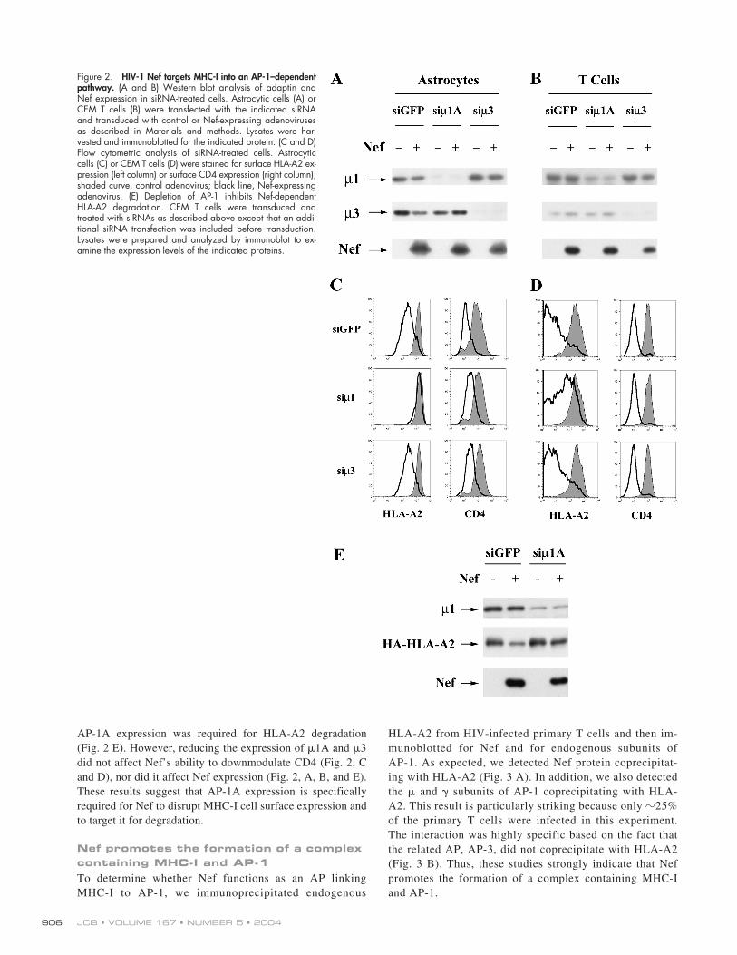
JCB • VOLUME 167 • NUMBER 5 • 2004906
AP-1A expression was required for HLA-A2 degradation(Fig. 2 E). However, reducing the expression of
�
1A and
�
3did not affect Nef’s ability to downmodulate CD4 (Fig. 2, Cand D), nor did it affect Nef expression (Fig. 2, A, B, and E).These results suggest that AP-1A expression is specificallyrequired for Nef to disrupt MHC-I cell surface expression andto target it for degradation.
Nef promotes the formation of a complex containing MHC-I and AP-1
To determine whether Nef functions as an AP linkingMHC-I to AP-1, we immunoprecipitated endogenous
HLA-A2 from HIV-infected primary T cells and then im-munoblotted for Nef and for endogenous subunits ofAP-1. As expected, we detected Nef protein coprecipitat-ing with HLA-A2 (Fig. 3 A). In addition, we also detectedthe
�
and
�
subunits of AP-1 coprecipitating with HLA-A2. This result is particularly striking because only
�
25%of the primary T cells were infected in this experiment.The interaction was highly specific based on the fact thatthe related AP, AP-3, did not coprecipitate with HLA-A2(Fig. 3 B). Thus, these studies strongly indicate that Nefpromotes the formation of a complex containing MHC-Iand AP-1.
Figure 2. HIV-1 Nef targets MHC-I into an AP-1–dependentpathway. (A and B) Western blot analysis of adaptin andNef expression in siRNA-treated cells. Astrocytic cells (A) orCEM T cells (B) were transfected with the indicated siRNAand transduced with control or Nef-expressing adenovirusesas described in Materials and methods. Lysates were har-vested and immunoblotted for the indicated protein. (C and D)Flow cytometric analysis of siRNA-treated cells. Astrocyticcells (C) or CEM T cells (D) were stained for surface HLA-A2 ex-pression (left column) or surface CD4 expression (right column);shaded curve, control adenovirus; black line, Nef-expressingadenovirus. (E) Depletion of AP-1 inhibits Nef-dependentHLA-A2 degradation. CEM T cells were transduced andtreated with siRNAs as described above except that an addi-tional siRNA transfection was included before transduction.Lysates were prepared and analyzed by immunoblot to ex-amine the expression levels of the indicated proteins.

NEF TARGETS MHC-I INTO AN AP-1 PATHWAY • ROETH ET AL.
907
AP-1 binds to the Nef–MHC-I complex in a dileucine-independent manner
Previous studies that have examined the direct interaction ofNef with AP-1 have found that it depends on the Nef dileucinemotif (Bresnahan et al., 1998; Craig et al., 2000; Janvier et al.,2003a,b). However, we found that the dileucine motif was notneeded for coprecipitation of AP-1 with MHC-I in CEM Tcells treated with adeno-Nef (Fig. 4 A) or HIV (Fig. S3, avail-
able at http://www.jcb.org/cgi/content/full/jcb.200407031/DC1). These data are consistent with the fact that the dileucinemotif is not necessary for disruption of MHC-I trafficking(Greenberg et al., 1998a; Mangasarian et al., 1999), and sug-gests that there is an alternative AP-1 binding site in the Nef–MHC-I complex.
To rule out the possibility that the interaction we observedwas nonspecific, and to verify that it was dependent on Nef do-
Figure 3. Nef expression results in coprecipi-tation of AP-1 with HLA-A2. (A) HLA-A2� pri-mary human T cells or control CEM-SS cellsthat did not express HLA-A2 (Ctrl) were trans-duced with an HIV molecular clone expressingNef (nef�), or a matched control HIV that didnot express Nef (nef�). Cells were harvested,and HLA-A2 was immunoprecipitated as de-scribed in Materials and methods. Proteins thatcoprecipitated with HLA-A2 were detected byWestern blotting as indicated. Western blotsof protein inputs are also shown as a controlfor relative protein levels in each sample beforeimmunoprecipitation (Input Controls). Resultsare typical of two independent experiments.(B) AP-3 does not coprecipitate with HLA-A2.CEM-SS cells (lanes 1 and 4) or CEM HA-HLA-A2 cells (lanes 2, 3, 5, and 6) were infectedwith HIV, and HLA-A2 was recovered by im-munoprecipitation as described in Materialsand methods. Coprecipitating proteins weredetected by Western blotting (lanes 1–3). Afraction of the protein input before immunopre-
cipitation was also analyzed to ensure similar protein expression levels (lanes 4–6). Western blots of �-adaptin (AP-3) and �-adaptin (AP-1) were performedsimultaneously using the same antibody concentrations. Apparent molecular masses of protein standards are denoted in kilodaltons on the left.
Figure 4. Analysis of MHC-I and Nef domains that contribute to AP-1 recruitment. (A) Recruitment of AP-1 requires an intact Nef–MHC-I complex and isindependent of the dileucine motif in Nef. CEM-SS cells (lane 1) or CEM HA-HLA-A2 cells (lanes 2–5) were transduced with the indicated adenoviral vector.The cells were harvested at 72 h and subjected to the coimmunoprecipitation assay. Cells were transduced with each adenovirus to achieve equivalentNef expression levels (Input Controls): wild-type NL4-3 Nef (MOI 25); NL4-3 Nef with a NH2-terminal deletion (V10E�17-26, MOI 300); HXB Nefwith a mutation in the dileucine motif (LL164,165AA, MOI 25). (As shown, HXB Nef migrates more slowly on SDS PAGE than NL4-3 Nef. Wild-type HXBNef and HXB NefLL164,165AA are directly compared in Fig. S3.) All results are typical of at least two independent experiments. (B) HLA-A2 tyrosine 320(Y320) is required for efficient Nef binding, AP-1 recruitment, and targeting for degradation. CEM-SS (lanes 1 and 2), CEM HA-HLA-A2 (lanes 3 and 4), orCEM cells expressing the MHC-I mutant, HA-HLA-A2 Y320A (lanes 5 and 6), were transduced with a control (�) or Nef-expressing adenovirus (�). At 72 hafter transduction, HLA-A2 immunoprecipitations were recovered and analyzed by Western blotting as described in Fig. 3.

JCB • VOLUME 167 • NUMBER 5 • 2004908
mains important for MHC-I trafficking to lysosomes, we askedwhether AP-1 would coprecipitate with MHC-I in cells express-ing a Nef mutant (V
10
E
�
17-26) that was unable to affect MHC-Itrafficking and to bind MHC-I (Mangasarian et al., 1999; Wil-liams et al., 2004). As shown in Fig. 4 A, we found that AP-1did not coprecipitate with HLA-A2 under these conditions.
In addition, we asked whether AP-1 would coprecipitatewith an HLA-A2 mutant lacking a tyrosine residue at position320 that is necessary for responsiveness to Nef (Greenberg etal., 1998b; Le Gall et al., 1998). As shown in Fig. 4 B, this mu-tant was expressed well (Fig. 4 B, input controls, bottom).However, it was resistant to Nef-dependent MHC-I degrada-tion, coprecipitated Nef less efficiently, and was defective inAP-1 recruitment (Fig. 4 B). Thus, these studies indicate thatcoprecipitation of AP-1 with HLA-A2 in Nef-expressing cellswas highly specific and depended on amino acid sequences inNef and MHC-I that are functionally important for Nef’s ef-fects on MHC-I cell surface expression.
Recruitment of AP-1 requires both the cytoplasmic tail of HLA-A2 and the NH
2
-terminal
�
-helix in Nef
The domains of HIV Nef specifically involved in MHC-I traf-ficking have been well characterized. It is known that disruptionof Nef’s NH
2
-terminal
�
helix by deletion (V
10
E
�
17-26) orpoint mutation (M
20
A), mutation of an acidic cluster (E
62-65
A),or mutation of a polyproline repeat (P
69/72/75/78
A) specifically af-
fects this activity (Greenberg et al., 1998b; Mangasarian et al.,1999). We have recently reported that each of these mutantsfails to coprecipitate with MHC-I (Williams et al., 2004) andthus binding to MHC-I may be the primary role of these do-mains. However, it is also possible that one or more of these do-mains plays an additional role. To determine whether any ofthese domains might also be involved in AP-1 recruitment di-rectly, we bypassed the requirement for Nef binding by directlyfusing Nef to the COOH terminus of the HLA-A2 cytoplasmictail (A2/Nef; Fig. 5 A). As expected, Nef was able to reduce thecell surface expression of HLA-A2 in cis (Fig. 5 B, left), and topromote its colocalization with markers of the Golgi apparatus(Fig. 5 C). In addition, the A2/Nef fusion protein was able to ef-ficiently coprecipitate AP-1 from T cell lysates in a manner thatwas independent of the Nef dileucine motif (Fig. 6 A). However,the fusion protein was not capable of reducing MHC-I surfaceexpression in trans (Fig. 5 B, right). Thus, A2/Nef was a usefulreagent to explore which amino acids in Nef and MHC-I wereimportant for AP-1 recruitment to the Nef–MHC-I complex.
As shown in Fig. 6 A, these studies revealed that muta-tion of amino acids in the NH
2
-terminal
�
-helix (A2/V
10
E
�
17-26 and A2/M
20
A) inhibited coprecipitation of AP-1 with the fu-sion protein. In contrast, disruption of the acidic (A2/E
62-65
Q)and polyproline (A2/P
72/75
A and A2/P
75/78
A) domains in Nefhad no effect (Fig. 6, A and B). Surprisingly, deletion of theHLA-A2 cytoplasmic tail (A2
�
Tail/Nef) or mutation of tyro-sine 320 in the HLA-A2 cytoplasmic tail (A2Y
320
A/Nef) also
Figure 5. Characterization of the A2/Nef fusion protein.(A) Schematic diagram of the A2/Nef fusion protein. (B)The addition of Nef to the COOH terminus of HLA-A2 re-sults in a reduction of cell surface expression in cis but nottrans. In the left panel, the effect of Nef in cis was mea-sured by transducing CEM-SS cells with a bi-cistronicmurine retrovirus encoding GFP and HLA-A2 (shadedcurve), GFP and A2/Nef (black line), or GFP alone (grayline). HLA-A2 was detected by staining with mAb BB7.2and using flow cytometry to gate on the GFP-positivecells. In the right panel, the effect of A2/Nef in trans wasmeasured by examining the effect of the fusion protein(black line) or wild-type HLA-A2 (shaded curve) on stablyexpressed HA tagged HLA-A2 (HA-HLA-A2). The negativecontrol is CEM-SS cells transduced with virus expressingGFP only (gray line). HA-HLA-A2 was detected by stain-ing with an mAb directed against HA and using flowcytometry to gate on the GFP-positive cells. Results arerepresentative of three independent experiments. (C) TheA2/Nef fusion protein accumulates in the TGN. CEM-SScells that stably expressed a tagged late Golgi residentprotein (EYFP-Golgi) were transduced with retroviral con-structs (A2 or A2/Nef). Cells were stained for HLA-A2and analyzed by confocal microscopy as described inMaterials and methods. Bar, 5 �m.

NEF TARGETS MHC-I INTO AN AP-1 PATHWAY • ROETH ET AL.
909
dramatically inhibited coprecipitation of AP-1 by the fusionprotein (Fig. 6 C). Thus, sequences in the Nef NH
2
-terminal
�
helix as well as in the HLA-A2 cytoplasmic tail are necessaryto create a functional AP-1 binding site.
Discussion
In sum, we have found that in T cells, Nef disrupts MHC-I traf-ficking after it reaches the TGN to prevent MHC-I cell surfaceexpression and to promote its degradation in lysosomes. RNAitreatment of Nef-expressing T cells and astrocytic cells re-vealed that this activity of Nef required the expression of AP-1A, but not AP-3. The requirement for AP-1A was further sup-
ported by the fact that we were able to isolate complexes con-taining MHC-I and AP-1 from Nef-expressing, HIV-infectedprimary T cells. The dileucine motif in Nef, which is necessaryfor Nef to directly bind AP-1 in other assay systems, was notneeded for AP-1 to bind the Nef–MHC-I complex, suggestingan alternative means of association. Interestingly, when Nefwas fused to HLA-A2, AP-1 was only recruited when the NH
2
-terminal
�
helical domain of Nef was intact and when a full-length, wild-type HLA-A2 cytoplasmic tail was present. Thus,amino acids within both Nef and MHC-I were necessary to cre-ate a binding site for AP-1 in the Nef–MHC-I complex.
The normal function of AP-1 is to sort proteins, such aslysosomal enzyme receptors at the TGN by linking clathrin tothe cytoplasmic tails of cargo (for review see Dell’Angelica andPayne, 2001; Robinson and Bonifacino, 2001; Traub, 2003;Robinson, 2004). For example, the mannose 6-phosphate recep-tor (MPR), which binds soluble lysosomal enzymes, is sorted atthe TGN by AP-1. It is then transported in vesicles to endo-somes where the enzymes dissociate and are carried to the lyso-somes. The MPRs are then recycled back to the TGN to trans-port more cargo. Thus, Nef-induced AP-1 binding of MHC-Iwould be expected to promote TGN accumulation and/or tar-geting of MHC-I into the endolysosomal pathway (Fig. 7).
Using the yeast two hybrid system (Le Gall et al., 1998;Piguet et al., 1998; Craig et al., 2000; Erdtmann et al., 2000;Janvier et al., 2003b), GST-Nef pull-downs (Bresnahan et al.,1998; Le Gall et al., 1998; Piguet et al., 1998; Janvier et al.,2003a) or overexpression of a Nef-CD8 chimera in 293 cells(Bresnahan et al., 1998), a number of investigators have foundthat the HIV-1 Nef protein interacts with adaptin subunits andwhole adaptin complexes. A consensus binding domain foradaptin protein binding (D/EXXXLL) (Bonifacino and Traub,2003) can be found in Nef and this motif is required for GST-Nef to pull down AP-1 complexes from mammalian cell ly-sates (Bresnahan et al., 1998; Janvier et al., 2003a). In addition,the leucine residues in this motif are required for Nef to down-modulate CD4, but not MHC-I (Greenberg et al., 1998a; Man-
Figure 6. The cytoplasmic domain of HLA-A2 and the NH2-terminal � helixof Nef are both required for AP-1 binding. (A and B) The NH2-terminal�-helix in Nef is necessary for AP-1 recruitment. CEM-SS cells were trans-duced with the murine retrovirus encoding HLA-A2 or the indicated A2/Nef fusion constructs. Coprecipitating proteins were detected by Westernblotting of anti-HLA-A2 immunoprecipitations. (C) AP-1 binding to the A2/Nef fusion protein requires tyrosine 320 (Y320) in the cytoplasmic tail ofHLA-A2. The indicated mutations in the A2/Nef fusion protein were testedfor involvement in AP-1 binding using the assay described above. A2�Tailis an A2/Nef fusion protein that lacks the entire HLA-A2 cytoplasmic tail.All results are representative of at least three independent experiments.White lines indicate that intervening lanes have been spliced out.
Figure 7. Model for MHC-I trafficking in Nef-expressing T cells. To disruptMHC-I trafficking, Nef first binds to the MHC-I cytoplasmic tail in thesecretory pathway. The formation of the Nef–MHC-I complex creates abinding site for AP-1 that is independent of the Nef dileucine motif. Thistertiary complex leads to the recruitment of MHC-I into AP-1–positiveclathrin-coated vesicles destined for degradation in the lysosomes.

JCB • VOLUME 167 • NUMBER 5 • 2004910
gasarian et al., 1999). Thus, the dileucine-dependent binding toadaptins appears to be important for Nef’s effects on CD4.
Our data showing that the dileucine motif is not requiredfor Nef to promote an interaction between HLA-A2 and AP-1indicates that there is an alternative binding site for AP-1 in theNef–MHC-I complex. It should be noted that there is evidencefor a second AP-1 binding site within Nef that is active in yeasttwo hybrid systems (Craig et al., 2000; Erdtmann et al., 2000).However, existing data suggest that this motif is not able tosupport coprecipitation of AP-1 with Nef in mammalian sys-tems (Bresnahan et al., 1998; Janvier et al., 2003a). These datacan be explained by our results showing that AP-1 recruitmentto HLA-A2 in Nef-expressing cells appeared to result from thecreation of a new binding site for AP-1 that was only activewhen Nef was bound to wild-type MHC-I.
The MHC-I cytoplasmic tail itself does not contain a con-sensus AP-1 binding site (D/EXXXLL or YXX
�
) (Bonifacinoand Traub, 2003). However, the tyrosine residue at position320 (Y
320
) that we show is necessary for Nef to recruit AP-1, isalso required for the normal targeting of MHC-I to endolysoso-mal compartments in dendritic cells (Lizee et al., 2003). Themechanism by which MHC-I is normally targeted to thesecompartments is unknown, and it is interesting to speculate thatin these cell types Y320 recruits an AP, such as AP-1, that tar-gets MHC-I to the endolysosomal compartment. Thus, Nefmay be taking advantage of an existing pathway for MHC-Itrafficking that is normally only active in certain cell types.
Previous studies describing the mechanism of Nef’s ef-fects on MHC-I have focused on the ability of Nef to accelerateMHC-I endocytosis, rather than on the TGN to lysosomal path-way we describe (Greenberg et al., 1998b; Le Gall et al., 1998,2000; Piguet et al., 2000; Blagoveshchenskaya et al., 2002;Kasper and Collins, 2003). This is most likely due to the factthat Nef is more active at disrupting transport from the TGN inT cells than it is in more commonly used cell lines, such asHeLa cells (Kasper and Collins, 2003). The effect of Nef onendocytosis has been attributed to the small GTPase ADP-ribo-sylation factor-6 (Blagoveshchenskaya et al., 2002), which isnormally involved in MHC-I turnover from the cell surface(Caplan et al., 2002). However, there is new evidence that theeffect of ADP-ribosylation factor-6 may be indirect (Larsen etal., 2004). In addition, the phosphofurin acidic cluster sortingprotein (PACS-1), which binds Nef via the acidic domain, maytarget Nef and/or endocytosed Nef-MHC-I to the TGN in somecell types (Piguet et al., 2000; Blagoveshchenskaya et al.,2002). It is known that PACS-1 interacts with AP-1 to promoteTGN recycling (Crump et al., 2001). However, it is unlikelythat the AP-1 interaction we observed here is secondary toPACS-1 binding, because the PACS-1 binding region (i.e., theacidic domain) was not needed to recruit AP-1 in our system.
It should also be noted that Nef associates with lipid raftsand that this localization is important for its effects on MHC-I traf-ficking (Alexander et al., 2004). It has been proposed that Nef as-sociation with rafts in the Golgi could trap MHC-I there and per-haps promote its association with required cellular proteins, suchas AP-1. Further studies are needed to determine whether lipidrafts are important for Nef–MHC-I–AP-1 complex formation.
In sum, our results indicate that in T cells, the primary ef-fect of Nef on MHC-I is to promote TGN retention and to ulti-mately direct MHC-I to lysosomes. We have provided evi-dence that Nef accomplishes this by acting as an AP, whichstabilizes a physical interaction between AP-1 and MHC-I inorder to target MHC-I into the endolysosomal pathway. Theseresults have important implications for understanding HIV dis-ease pathogenesis and for possible pharmaceutical approachesin combating the development of AIDS.
Materials and methodsCell cultureCEM T cell lines were maintained in R10 (RPMI supplemented with 10%FBS, 10 mM Hepes, 2 mM penicillin, streptomycin, and glutamine (P/S/G).The astrocytic cell lines stably expressing CD4 were maintained in DMEsupplemented with 10% FBS and P/S/G. Stable astrocytic and CEM T celllines were generated by transduction with MSCV retroviral constructspseudotyped with VSV-G as described previously (Kasper and Collins,2003). Primary T lymphocytes were isolated from buffy coats as describedpreviously (Collins et al., 1998).
DNA constructsA nine–amino acid HA epitope tag (YPYDVPDYA) was added to the NH2
terminus of HLA-A2 and HLA-B71 (just after the leader sequence cleavagesite and before the NaeI site; Swann et al., 2001) using the followingprimers and HLA-B3501 as the DNA template: forward primer, 5 -GGG-AATTCCTCAGAATCTCCTCAGACGCCGAG-3 ; reverse primer, 5 -TCC-CGCCGGCATAGTCGGGTACGTCATACGGATAGGACCCGGCCCAG-GTCTCGGT-3 . The PCR product was then digested with EcoRI and NaeIand subcloned into a shuttle vector (New England Biolabs, Inc.). Next, thefollowing primers were used to amplify the 3 region of HLA-A2 and HLA-B71: HLA-A2 forward primer, 5 -GAATTCATGGTACCGTGCACG-3 ;HLA-B71 forward primer, 5 -GGCTCGAGTCAAGCTGTGAGAGACA-CATC-3 ; HLA-A2 reverse primer, 5 -CCGCTCGAGTCACACTTTACAA-GCTGT-3 ; HLA-B71 reverse primer, 5 -CTGGGCCGGCTCCCACTCCAT-GAGGTATTT-3 . The resulting PCR products were digested with NaeI andXhoI. Finally, the HLA-A2/B71 PCR products and the EcoRI–NaeI frag-ment purchased from New England Biolabs, Inc. were ligated into MSCV2.1 (cut with EcoRI and XhoI) in a three-way ligation to generate MSCVHA-HLA-A2. MSCV HA-HLA-A2 Y320A was cloned by amplifying a portionof HLA-A2 Y320A (Swann et al., 2001) using the following primers: for-ward primer, 5 -GCAGCTCAGACCACCAAGCACAAG-3 ; reverse primer5 -CCGCTCGAGTCACACTTTACAAGCTGT-3 . The PCR product wasthen digested with PmlI and XhoI, and was ligated into the same sites inMSCV HA-HLA-A2 to replace the wild-type sequence.
An adenoviral vector expressing HXB Nef LL164,165AA was gener-ated by PCR mutagenesis of the HIV molecular clone, HXB-PI (Chen et al.,1996), using the following primers: mutant forward primer, 5 -AAT-AAAGGAGAGAACACCAGCGCTGCTCACCCTGTGAGCCTGCATGG-3 ;with reverse primer (3 hxb-xba), 5 -GCTCTAGATGCTAGAGATTTTCCA-CACTG-3 , and mutant reverse primer, 5 -TCCATGCAGGCTCACA-GGGTGAGCAGCGCTGGTGTTCTCTCCTTTATT-3 ; with forward primer(5 nef) 5 -CGGGATCCATGGGTGGCAAGTGGTCAAA-3 . The resultingmutant PCR products were mixed and reamplified with 3 hxb-xba and 5 nef primers, digested with XbaI and XhoI and cloned into the same sites ofHXB-PI. The mutated Nef was then amplified and cloned into an adeno-viral vector shuttle plasmid as described previously (Swann et al., 2001).The adenoviral vector expressing Nef V10E�17-26 has been describedpreviously (Williams et al., 2004).
An HIV molecular clone containing the Nef LL164,165AA mutationwas generated by PCR mutagenesis of the HIV molecular clone, HXB-EP(Chen et al., 1996), using the following primers: mutant forward primer,5 -GAAGGAGAGAACACCCGCGCGGCACACCCTGTGAGCCTGCAT-3 ; with reverse primer (3 hxb-xba), 5 -GCTCTAGATGCTAGAGATTTTC-CACACTG-3 , and mutant reverse primer, 5 -ATGCAGGCTCACAGGGT-GTGCCGCGCGGGTGTTCTCTCCTTC-3 ; with forward primer (5 nef) 5 -GACAGATCCATTCGATTAGTG-3 . The resulting mutant PCR productswere mixed and reamplified with 3 hxb-xba and 5 nef primers, digestedwith XbaI and BamHI and cloned into the same sites of HXB-EP.
The A2/Nef fusion constructs were generated as follows: a PCR prod-

NEF TARGETS MHC-I INTO AN AP-1 PATHWAY • ROETH ET AL. 911
uct containing the HLA-A2 sequence was generated with the primers; HLA-A2 5 -CGGGATCCACCATGGTACCGTGCACG-3 and 5 -GGACTAGT-CACTTTACAAGCTGTGAGAGA-3 . (For HLA-A2�Tail, a different 3 oligo-nucleotide was used to generate the truncated molecule 5 -GGACTAGTCT-TCCTCCTCCACATCACAGC-3 ). A PCR product containing wild-type ormutant Nef sequence (Williams et al., 2004) was generated with the prim-ers; Nef 5 -GGACTAGTATGGGTGGCAAGTGGTCAAAA-3 and 5 -GCGAATTCTCAGCAGTTCTTGAAGTACTC-3 . The HLA-A2 PCR productwas digested with BamHI and SpeI, the Nef PCR product was digested withSpeI and EcoRI. These fragments were then cloned into the BglII and EcoRIsites of a bi-cistronic retroviral vector expressing an IRES GFP cassette(pMIG) (Van Parijs et al., 1999) in a three-way ligation. The control HLA-A2IRES GFP construct was generated using the following primers 5 -CGG-GATCCACCATGGTACCGTGCACG-3 and 5 -GGTCAACTAGTCACTTTA-CAAGCTGTGAGAGA-3 . This PCR product was digested with BamHI andligated into the BglII and HpaI sites of pMIG. A2 Y320A/Nef was generatedfrom the A2/Nef template using the following mutant primers: 5 primer; 5 -AGAAAAGGAGGGAGCGCCTCTCAGGCTGCA-3 , 3 primer; 5 -TGCAG-CCTGAGAGGCGCTCCCTCCTTTTCT-3 . A2/LL164,165AA was generatedfrom the A2/Nef template using the following mutant primers: 5 primer; 5 -ATGCAGGCTCACAGGGTGTGCCGCGCTGGTGTTCTCTCCTTT-3 , 3 primer; 5 -AAAGGAGAGAACACCAGCGCGGCACACCCTGTGAGCCT-GCAT-3 .
MSCV-EYFP-Golgi was constructed as follows: EYFP-Golgi (CLON-TECH Laboratories, Inc.) was digested with NheI and HpaI. This fragmentwas ligated into the XbaI and EcoRV sites of pShuttle (Stratagene). The re-sulting construct was then digested with XhoI and BglII and ligated into thesame sites in MSCV 2.1.
Viral transduction of T cellsCD8-depleted PHA-activated primary T cells or CEM-SS cells were trans-duced with VSV-G-pseudotyped HIV molecular clones (HXB-EP nef� andnef�), VSVG-pseudotyped murine retroviruses or adenoviral vectors as de-scribed previously (Collins et al., 1998; Swann et al., 2001; Kasper andCollins, 2003). Recombinant replication defective E3-deleted adenovirusexpressing HIV-1 NL4-3 Nef, Nef V10E�17-26, Nef LL164,165AA, or no in-sert was generated by the University of Michigan Vector Core. For adeno-viral transductions, CEM cell lines were resuspended in R2 media (RPMI,2% FBS, P/S/G) at a concentration of 106 cells/ml. Adenovirus stockswere added to a relative multiplicity of infection (MOI; based on 293 cellinfectivity) of 50–200 virus particles/cell. Cells were transferred to 96-wellplates [100 �l (105 cells) per well]. After 2 h at 37�C, 200 �l of R10 me-dia was added. Cells were harvested at 24–72 h after transduction.
Metabolic labeling and immunoprecipitationAdeno-transduced CEM T cells were collected and washed twice with D-PBS(Invitrogen). Cells were incubated in prelabel media [RPMI –Cys –Met (Spe-cialty Media, Inc.) � 10% dialyzed FBS (Invitrogen)] for 15 min at 37�C.Cells were pulsed with labeling media [prelabel media � 150 �Ci/ml Pro-mix-L [35S] (�1,000 Ci/mmol; Amersham Biosciences)] for 15 min at 37�C.Cells were washed twice with R10 and chased in R10 for the indicated pe-riod of time (5 � 106 cells were used per time point). For some pulse-chaseexperiments, bafilomycin A1 (100 nM; Sigma-Aldrich), NH4Cl (25 mM;Fisher Scientific), or lactacystin (20 �M; Sigma-Aldrich) was included in thechase media. Cells were collected and washed once with D-PBS and lysedin 1 ml of lysis buffer (50 mM Tris, pH 8, 1% NP-40 (vol/vol), 5 mM MgCl2,1 mM PMSF) for 30 min on ice, followed by centrifugation to remove insolu-ble material. Cellular lysates were precleared overnight at 4�C with 1.5�g of mouse IgG1 (BD Biosciences) and 30 �l of protein A/G agarose(50% slurry; Calbiochem). For immunoprecipitations, 5 �g of anti–HLA-A2(BB7.2) or 3.5 �g of anti-transferrin receptor antibody (Oncogene), wasadded to the precleared lysate, followed by the addition to 30 �l of proteinA/G agarose beads. After 2 h at 4�C, the beads were washed three timeswith 1 ml RIPA buffer (Kasper and Collins, 2003). Endo Hf (New EnglandBiolabs, Inc.) or neuraminidase (New England Biolabs, Inc.) digestion of im-munoprecipitated material was performed according to the manufacturer’sprotocols. Samples were separated by 10% SDS-PAGE and the gels weredried and exposed to BioMaxMS films at �80�C. Radio-labeled proteinswere quantitated using a Phosphor storage screen and Typhoon Scannerfollowed by processing with Image Quant software.
Immunofluorescence microscopyCEM cells were allowed to adhere to chambered glass slides coated withpoly-L-lysine (Sigma-Aldrich) for 1 h at 37�C. Cells were then fixed at RTfor 15 min in D-PBS � 2% PFA. Next, cell membranes were permeabi-lized by incubation in D-PBS � 0.2% Tween 20 for 15 min at 37�C. Cells
were washed twice with wash buffer (D-PBS, 10 mM Hepes, 10% goat se-rum [Sigma-Aldrich], 0.025% sodium azide) followed by blocking inwash buffer (adjusted to 10% goat serum and 10% human serum; Sigma-Aldrich) for 30 min on ice. For Fig. 1, mouse mAbs were used to detectLAMP-1 (clone H4A3; BD Biosciences; 1:500) and HLA-A2 (BB7.2, 20�g/ml). Isotype-specific secondary antibodies were used to distinguish be-tween these antibodies (anti–mouse IgG1 Alexa-Fluor546 [1:250; Molec-ular Probes] and anti–mouse IgG2b Alexa-Fluor488 [1:250; MolecularProbes], respectively). All antibody incubations were performed in washbuffer on ice for 20 min. Slides were mounted with coverslips using Pro-long antifade reagent (Molecular Probes). Images were collected using aconfocal microscope (model LSM 510; Carl Zeiss MicroImaging, Inc.) andprocessed using Adobe Photoshop 6.0 software. For inhibitor treatment,adeno-transduced CEM HA-HLA-A2 cells were resuspended in fresh R10containing 100 nM bafilomycin A1 (Sigma-Aldrich) or solvent alone (DMSO)and incubated at 37�C for 3 h before adherence.
RNAi treatmentThe following duplex siRNAs (Ambion) were used in this study: siGFP,sense 5 -GCUGACCCUGAAGUUCAUCTT-3 , antisense 5 -GAUGAAC-UUCAGGGUCAGCTT-3 ; si�3, sense 5 -GGACUACUUUGGUGAGU-GUTT-3 , antisense 5 -ACACUCACCAAAGUAGUCCTG-3 . The �1AsiRNA was described previously (Hirst et al., 2003). Astrocytoma cells sta-bly expressing CD4 (373-CD4) were transfected with annealed duplexsiRNAs using Lipofectamine 2000 (Invitrogen) following the manufac-turer’s protocol. In brief, cells were plated onto 6-well plates (105 cells perwell) and the next day each well was transfected using 0.16 nmol siRNAand 4 �l of Lipofectamine 2000 reagent. 24 h later, cells were replatedonto 6-well plates (105 cells per well) and the cells were transfected withsiRNA again 24 h after replating. 4–6 h after transfection, the media wasremoved and the cells were transduced with adenovirus (MOI 50). Cellswere harvested and analyzed 24 h after transduction.
For experiments using CEM T cells, the cells were first transducedwith adenovirus (MOI 50). 24 h later, 5 � 106 cells were electropo-rated with 1 nmole siRNA according to the manufacturer’s protocol(Amaxa Biosystems). The cells were then incubated for 48 h before FACSand Western blot assays. For Fig. 2 E, the cells were subjected to an ini-tial siRNA transfection 1 d before the adenoviral transduction, and the ex-periment was then performed as described above.
Antibodies and Western blot analysesIP-Western experiments were performed as described previously (Williamset al., 2004). HA-HLA-A2 was detected using the anti-HA antibody,HA.11 (1:5,000; Covance). Coprecipitating Nef protein was detected byWestern blotting using antibodies obtained via the NIH AIDS Researchand Reference Reagent Repository, Division of AIDS, NIAID, NIH: poly-clonal anti-Nef antibody (2949, 1:5,000, a gift from R. Swanstrom, TheUniversity of North Carolina at Chapel Hill, Chapel Hill, NC; Shugars etal., 1993) or monoclonal anti-Nef (AG11, 1:500 for IP-Westerns and1:10,000 for crude lysate, a gift from J. Hoxie, University of Pennsylvania,Philadelphia, PA; Chang et al., 1998). The adaptin subunit, �1, was de-tected using RY/1 (1:2,500, a gift from L. Traub, University of Pittsburgh,Pittsburgh, PA; Traub et al., 1995). Other antibodies to AP subunits werepurchased from BD Biosciences: �-adaptin (1:100 for IP-Westerns; 1:1,000for crude lysate); �-adaptin (1:100 for IP-Westerns; 1:1,000 for crude ly-sate); and �3 (p47A, 1:500). The HLA-A2–specific mAb, BB7.2 (Parhamand Brodsky, 1981), and anti-Nef antibody, AG11 (Chang et al., 1998),were purified from ascites fluid as described previously (Kasper and Col-lins, 2003).
Online supplemental materialFig. S1 shows that the addition of an HA tag does not affect MHC-I folding,maturation through the secretory pathway and responsiveness to Nef. Fig.S2 demonstrates that Nef targets mature MHC-I for degradation in HIV-infected T cells. Fig. S3 is coimmunoprecipitation and Western blot analy-sis demonstrating that the dileucine motif is not required for the Nef-MHC-Ito recruit AP-1 in HIV-infected T cells. Online supplemental material is avail-able at http://www.jcb.org/cgi/content/full/jcb.200407031/DC1.
We are grateful to Dr. Linton Traub for antibody to �1, to the NIH AIDS Re-agent Repository for antibodies, to the University of Michigan vector core foradenovirus preparation, to Dr. Randy Schekman for helpful suggestions and toDrs. John Moran and Mark Benson for critical reading of the manuscript.
This research was supported by National Institutes of Health grant RO1AI46998. M.R. Kasper was supported by the University of Michigan Genet-ics Training Program. M. Williams and J. Roeth were supported by the Univer-
JCB • VOLUME 167 • NUMBER 5 • 2004912
sity of Michigan Cellular and Molecular Biology Training Program. M. Wil-liams was also supported by a Rackham predoctoral fellowship and theMicrobial Pathogenesis Training Program.
Submitted: 6 July 2004Accepted: 20 October 2004
ReferencesAiken, C., J. Konner, N.R. Landau, M.E. Lenburg, and D. Trono. 1994. Nef in-
duces CD4 endocytosis: requirement for a critical dileucine motif in themembrane-proximal CD4 cytoplasmic domain. Cell. 76:853–864.
Alexander, M., Y.C. Bor, K.S. Ravichandran, M.L. Hammarskjold, and D. Re-kosh. 2004. Human immunodeficiency virus type 1 Nef associates withlipid rafts to downmodulate cell surface CD4 and class I major histocom-patibility complex expression and to increase viral infectivity. J. Virol.78:1685–1696.
Blagoveshchenskaya, A.D., L. Thomas, S.F. Feliciangeli, C.H. Hung, and G.Thomas. 2002. HIV-1 Nef downregulates MHC-I by a PACS-1- andPI3K-regulated ARF6 endocytic pathway. Cell. 111:853–866.
Bonifacino, J.S., and L.M. Traub. 2003. Signals for sorting of transmembraneproteins to endosomes and lysosomes. Annu. Rev. Biochem. 72:395–447.
Bresnahan, P.A., W. Yonemoto, S. Ferrell, D. Williams-Herman, R. Geleziunas,and W.C. Greene. 1998. A dileucine motif in HIV-1 Nef acts as an inter-nalization signal for CD4 downregulation and binds the AP-1 clathrinadaptor. Curr. Biol. 8:1235–1238.
Caplan, S., N. Naslavsky, L.M. Hartnell, R. Lodge, R.S. Polishchuk, J.G.Donaldson, and J.S. Bonifacino. 2002. A tubular EHD1-containing com-partment involved in the recycling of major histocompatibility complexclass I molecules to the plasma membrane. EMBO J. 21:2557–2567.
Chang, A.H., J.A. Hoxie, S. Cassol, M. O’Shaughnessy, and F. Jirik. 1998. Con-struction of single-chain antibodies that bind an overlapping epitope ofHIV-1 Nef. FEBS Lett. 441:307–312.
Chen, B., R. Gandhi, and D. Baltimore. 1996. CD4 down-modulation during in-fection of human T cells with human immunodeficiency virus type 1 in-volves independent activities of vpu, env, and nef. J. Virol. 70:6044–6053.
Cohen, G.B., R.T. Gandhi, D.M. Davis, O. Mandelboim, B.K. Chen, J.L.Strominger, and D. Baltimore. 1999. The selective downregulation ofclass I major histocompatibility complex proteins by HIV-1 protectsHIV-infected cells from NK cells. Immunity. 10:661–671.
Collins, K., B. Chen, S. Kalams, B. Walker, and D. Baltimore. 1998. HIV-1 Nefprotein protects infected primary human cells from killing by cytotoxic Tlymphocytes. Nature. 391:397–401.
Craig, H.M., T.R. Reddy, N.L. Riggs, P.P. Dao, and J.C. Guatelli. 2000. Interac-tions of HIV-1 nef with the mu subunits of adaptor protein complexes 1,2, and 3: role of the dileucine-based sorting motif. Virology. 271:9–17.
Crump, C.M., Y. Xiang, L. Thomas, F. Gu, C. Austin, S.A. Tooze, and G.Thomas. 2001. PACS-1 binding to adaptors is required for acidic clustermotif-mediated protein traffic. EMBO J. 20:2191–2201.
Dell’Angelica, E.C., and G.S. Payne. 2001. Intracellular cycling of lysosomalenzyme receptors: cytoplasmic tails’ tales. Cell. 106:395–398.
Doray, B., P. Ghosh, J. Griffith, H.J. Geuze, and S. Kornfeld. 2002. Cooperationof GGAs and AP-1 in packaging MPRs at the trans-Golgi network. Sci-ence. 297:1700–1703.
Erdtmann, L., K. Janvier, G. Raposo, H.M. Craig, P. Benaroch, C. Berlioz-Tor-rent, J.C. Guatelli, R. Benarous, and S. Benichou. 2000. Two indepen-dent regions of HIV-1 Nef are required for connection with the endocyticpathway through binding to the mu 1 chain of AP1 complex. Traffic.1:871–883.
Geyer, M., H. Yu, R. Mandic, T. Linnemann, Y.H. Zheng, O.T. Fackler, andB.M. Peterlin. 2002. Subunit H of the V-ATPase binds to the mediumchain of adaptor protein complex 2 and connects Nef to the endocyticmachinery. J. Biol. Chem. 277:28521–28529.
Greenberg, M., L. DeTulleo, I. Rapoport, J. Skowronski, and T. Kirchhausen.1998a. A dileucine motif in HIV-1 Nef is essential for sorting into clath-rin-coated pits and for downregulation of CD4. Curr. Biol. 8:1239–1242.
Greenberg, M., A. Iafrate, and J. Skowronski. 1998b. The SH3 domain-bindingsurface and an acidic motif in HIV-1 Nef regulate trafficking of class IMHC complexes. EMBO J. 17:2777–2789.
Greenway, A., A. Azad, and D. McPhee. 1995. Human immunodeficiency virustype 1 Nef protein inhibits activation pathways in peripheral bloodmononuclear cells and T-cell lines. J. Virol. 69:1842–1850.
Grzesiek, S., S.J. Stahl, P.T. Wingfield, and A. Bax. 1996. The CD4 determi-nant for downregulation by HIV-1 Nef directly binds to Nef. Mapping ofthe Nef binding surface by NMR. Biochemistry. 35:10256–10261.
Harris, M.P., and J.C. Neil. 1994. Myristoylation-dependent binding of HIV-1
Nef to CD4. J. Mol. Biol. 241:136–142.
Hirst, J., A. Motley, K. Harasaki, S.Y. Peak Chew, and M.S. Robinson. 2003.EpsinR: an ENTH domain-containing protein that interacts with AP-1.Mol. Biol. Cell. 14:625–641.
Janvier, K., H. Craig, D. Hitchin, R. Madrid, N. Sol-Foulon, L. Renault, J. Cher-fils, D. Cassel, S. Benichou, and J. Guatelli. 2003a. HIV-1 Nef stabilizesthe association of adaptor protein complexes with membranes. J. Biol.Chem. 278:8725–8732.
Janvier, K., Y. Kato, M. Boehm, J.R. Rose, J.A. Martina, B.Y. Kim, S. Venkate-san, and J.S. Bonifacino. 2003b. Recognition of dileucine-based sortingsignals from HIV-1 Nef and LIMP-II by the AP-1 �-sigma1 and AP-3�-sigma3 hemicomplexes. J. Cell Biol. 163:1281–1290.
Kasper, M.R., and K.L. Collins. 2003. Nef-mediated disruption of HLA-A2transport to the cell surface in T cells. J. Virol. 77:3041–3049.
Larsen, J.E., R.H. Massol, T.J. Nieland, and T. Kirchhausen. 2004. HIV Nef-mediated major histocompatibility complex class I down-modulation isindependent of Arf6 activity. Mol. Biol. Cell. 15:323–331.
Le Gall, S., L. Erdtmann, S. Benichou, C. Berlloz-Torrent, L. Liu, R. Benarous,J. Heard, and O. Schwartz. 1998. Nef interacts with mu subunit of clath-rin adaptor complexes and reveals a cryptic sorting signal in MHC I mol-ecules. Immunity. 8:483–495.
Le Gall, S., F. Buseyne, A. Trocha, B.D. Walker, J.M. Heard, and O. Schwartz.2000. Distinct trafficking pathways mediate Nef-induced and clathrin-dependent major histocompatibility complex class I down-regulation. J.Virol. 74:9256–9266.
Lizee, G., G. Basha, J. Tiong, J.P. Julien, M. Tian, K.E. Biron, and W.A. Jeffer-ies. 2003. Control of dendritic cell cross-presentation by the major his-tocompatibility complex class I cytoplasmic domain. Nat. Immunol.4:1065–1073.
Lu, X., H. Yu, S. Liu, F. Brodsky, and B. Peterlin. 1998. Interactions betweenHIV1 Nef and vacuolar ATPase facilitate the internalization of CD4. Im-munity. 8:647–656.
Mangasarian, A., V. Piguet, J.K. Wang, Y.L. Chen, and D. Trono. 1999. Nef-induced CD4 and major histocompatibility complex class I (MHC-I)down-regulation are governed by distinct determinants: N-terminal alphahelix and proline repeat of Nef selectively regulate MHC-I trafficking. J.Virol. 73:1964–1973.
Parham, P., and F.M. Brodsky. 1981. Partial purification and some properties ofBB7.2. A cytotoxic monoclonal antibody with specificity for HLA-A2and a variant of HLA-A28. Hum. Immunol. 3:277–299.
Peden, A.A., V. Oorschot, B.A. Hesser, C.D. Austin, R.H. Scheller, and J.Klumperman. 2004. Localization of the AP-3 adaptor complex defines anovel endosomal exit site for lysosomal membrane proteins. J. Cell Biol.164:1065–1076.
Piguet, V., Y.L. Chen, A. Mangasarian, M. Foti, J.L. Carpentier, and D. Trono.1998. Mechanism of Nef-induced CD4 endocytosis: Nef connects CD4with the mu chain of adaptor complexes. EMBO J. 17:2472–2481.
Piguet, V., L. Wan, C. Borel, A. Mangasarian, N. Demaurex, G. Thomas, and D.Trono. 2000. HIV-1 Nef protein binds to the cellular protein PACS-1 todownregulate class I major histocompatibility complexes. Nat. Cell Biol.2:163–167.
Robinson, M.S. 2004. Adaptable adaptors for coated vesicles. Trends Cell Biol.14:167–174.
Robinson, M.S., and J.S. Bonifacino. 2001. Adaptor-related proteins. Curr.Opin. Cell Biol. 13:444–453.
Rossi, F., A. Gallina, and G. Milanesi. 1996. Nef-CD4 physical interactionsensed with the yeast two-hybrid system. Virology. 217:397–403.
Schaefer, T.M., I. Bell, B.A. Fallert, and T.A. Reinhart. 2000. The T-cell recep-tor zeta chain contains two homologous domains with which simian im-munodeficiency virus Nef interacts and mediates down-modulation. J.Virol. 74:3273–3283.
Schwartz, O., V. Marechal, S. Le Gall, F. Lemonnier, and J. Heard. 1996. En-docytosis of major histocompatibility complex class I molecules is in-duced by the HIV-1 Nef protein. Nat. Med. 2:338–342.
Shugars, D.C., M.S. Smith, D.H. Glueck, P.V. Nantermet, F. Seillier-Moisei-witsch, and R. Swanstrom. 1993. Analysis of human immunodeficiencyvirus type 1 nef gene sequences present in vivo. J. Virol. 67:4639-4650.
Swann, S.A., M. Williams, C.M. Story, K.R. Bobbitt, R. Fleis, and K.L. Collins.2001. HIV-1 Nef blocks transport of MHC class I molecules to the cellsurface via a PI 3-kinase-dependent pathway. Virology. 282:267–277.
Traub, L.M. 2003. Sorting it out: AP-2 and alternate clathrin adaptors in en-docytic cargo selection. J. Cell Biol. 163:203–208.
Traub, L.M., S. Kornfeld, and E. Ungewickell. 1995. Different domains of theAP-1 adaptor complex are required for Golgi membrane binding andclathrin recruitment. J. Biol. Chem. 270:4933–4942.
Van Parijs, L., Y. Refaeli, J.D. Lord, B.H. Nelson, A.K. Abbas, and D. Balti-more. 1999. Uncoupling IL-2 signals that regulate T cell proliferation,
NEF TARGETS MHC-I INTO AN AP-1 PATHWAY • ROETH ET AL. 913
survival, and Fas-mediated activation-induced cell death. Immunity. 11:281–288.
Waguri, S., F. Dewitte, R. Le Borgne, Y. Rouille, Y. Uchiyama, J.F. Du-bremetz, and B. Hoflack. 2003. Visualization of TGN to endosome traf-ficking through fluorescently labeled MPR and AP-1 in living cells. Mol.Biol. Cell. 14:142–155.
Williams, M., J.F. Roeth, M.R. Kasper, R.I. Fleis, C.G. Przybycin, and K.L.Collins. 2002. Direct binding of human immunodeficiency virus type 1Nef to the major histocompatibility complex class I (MHC-I) cytoplas-mic tail disrupts MHC-I trafficking. J. Virol. 76:12173–12184.
Williams, M., J.F. Roeth, M.R. Kasper, T.M. Filzen, and K.L. Collins. 2004.HIV-1 Nef domains required for disruption of MHC-I trafficking are alsonecessary for coprecipitation of Nef with HLA-A2. J. Virol. In press.
Copyright © 2022 FDOKUMEN