Emerging roles of ATF2 and the dynamic AP1 network in cancer
29
Emerging roles of ATF2 and the dynamic AP1 network in cancer Pablo Lopez-Bergami, Instituto de Biologia y Medicina Experimental, Vuelta de Obligado 2490, Buenos Aires1428, Argentina, [email protected] Eric Lau, and Signal Transduction Program, Burnham Institute for Medical Research, La Jolla, CA 92037, USA, [email protected] Ze'ev Ronai Signal Transduction Program, Burnham Institute for Medical Research, La Jolla, CA 92037, USA Abstract Cooperation among transcription factors is central for their ability to execute specific transcriptional programmes. The AP1 complex exemplifies a network of transcription factors that function in unison under normal circumstances and during the course of tumour development and progression. This Perspective summarizes our current understanding of the changes in members of the AP1 complex and the role of ATF2 as part of this complex in tumorigenesis. Activator protein 1 (AP1) 1,2 functions in almost all areas of eukaryotic cellular behaviour, from cell cycle proliferation and development to stress response and apoptosis. Indeed, AP1 is activated in response to a plethora of extracellular signals from cytokines and growth factors to stress and inflammation 3,4 . The expansive transcriptional repertoire executed by AP1 complexes is propagated from the diverse compositional array of homodimeric or heterodimeric combinations formed by members of the Jun, Atf, Fos and Maf transcription factor families (BOX 1). The dimeric combinations and transcriptional activity observed in vivo are largely influenced by the tissue-specific expression patterns of the individual proteins, and importantly by their specific activating mechanisms and post-translational modifications that facilitate their individual ability to dimerize with other basic leucine zipper (bZIP) domain proteins. This inherently diverse composition of AP1 complexes and their central role in transcriptional regulation places AP1 complexes at a functional epicenter for pathological signal relay in disease, particularly in the context of malignant cellular transformation in which AP1 proteins are often deregulated by oncoprotein signalling 4–6 . This Perspective describes the function and cooperation of Jun, Fos and Atf family members in tumour cells, and the emerging function of ATF2 as part of the dynamic AP1 complex. Competing interests statement: The authors declare no competing financial interests. Databases: Entrez Gene: http://www.ncbi.nlm.nih.gov/gene JUN UniProtKB: http://www.uniprot.org ATF2 | ATF3 | ATF4 | cathepsin L | CREB1 | cyclin A | ezrin | FBXW7 | FOS | FOSB | FRA1 | FRA2 | HMGA1 | JNK1 | JNK2 | JUNB | JUND | MMP1 | MMP2 | MMP3 | MMP9 | SENP1 | Stathmin | VEGFA Pathway interaction Database: http://pid.nci.nih.gov/ ATF2 Further Information: Ze'ev Ronai's homepage: http://www.ronailab.net Supplementary Information: See online article: S1 (table) | S2 (table) | S3 (table) ALL LINKS ARE ACTIVE IN THE ONLINE PDF NIH Public Access Author Manuscript Nat Rev Cancer. Author manuscript; available in PMC 2011 January 1. Published in final edited form as: Nat Rev Cancer. 2010 January ; 10(1): 65–76. doi:10.1038/nrc2681. NIH-PA Author Manuscript NIH-PA Author Manuscript NIH-PA Author Manuscript
-
Upload
independent -
Category
Documents
-
view
1 -
download
0
Transcript of Emerging roles of ATF2 and the dynamic AP1 network in cancer

Emerging roles of ATF2 and the dynamic AP1 network in cancer
Pablo Lopez-Bergami,Instituto de Biologia y Medicina Experimental, Vuelta de Obligado 2490, Buenos Aires1428,Argentina, [email protected]
Eric Lau, andSignal Transduction Program, Burnham Institute for Medical Research, La Jolla, CA 92037, USA,[email protected]
Ze'ev RonaiSignal Transduction Program, Burnham Institute for Medical Research, La Jolla, CA 92037, USA
AbstractCooperation among transcription factors is central for their ability to execute specific transcriptionalprogrammes. The AP1 complex exemplifies a network of transcription factors that function in unisonunder normal circumstances and during the course of tumour development and progression. ThisPerspective summarizes our current understanding of the changes in members of the AP1 complexand the role of ATF2 as part of this complex in tumorigenesis.
Activator protein 1 (AP1)1,2 functions in almost all areas of eukaryotic cellular behaviour,from cell cycle proliferation and development to stress response and apoptosis. Indeed, AP1is activated in response to a plethora of extracellular signals from cytokines and growth factorsto stress and inflammation3,4. The expansive transcriptional repertoire executed by AP1complexes is propagated from the diverse compositional array of homodimeric orheterodimeric combinations formed by members of the Jun, Atf, Fos and Maf transcriptionfactor families (BOX 1). The dimeric combinations and transcriptional activity observed invivo are largely influenced by the tissue-specific expression patterns of the individual proteins,and importantly by their specific activating mechanisms and post-translational modificationsthat facilitate their individual ability to dimerize with other basic leucine zipper (bZIP) domainproteins. This inherently diverse composition of AP1 complexes and their central role intranscriptional regulation places AP1 complexes at a functional epicenter for pathologicalsignal relay in disease, particularly in the context of malignant cellular transformation in whichAP1 proteins are often deregulated by oncoprotein signalling4–6. This Perspective describesthe function and cooperation of Jun, Fos and Atf family members in tumour cells, and theemerging function of ATF2 as part of the dynamic AP1 complex.
Competing interests statement: The authors declare no competing financial interests.Databases: Entrez Gene: http://www.ncbi.nlm.nih.gov/geneJUNUniProtKB: http://www.uniprot.orgATF2 | ATF3 | ATF4 | cathepsin L | CREB1 | cyclin A | ezrin | FBXW7 | FOS | FOSB | FRA1 | FRA2 | HMGA1 | JNK1 | JNK2 | JUNB| JUND | MMP1 | MMP2 | MMP3 | MMP9 | SENP1 | Stathmin | VEGFAPathway interaction Database: http://pid.nci.nih.gov/ATF2Further Information: Ze'ev Ronai's homepage: http://www.ronailab.netSupplementary Information: See online article: S1 (table) | S2 (table) | S3 (table)ALL LINKS ARE ACTIVE IN THE ONLINE PDF
NIH Public AccessAuthor ManuscriptNat Rev Cancer. Author manuscript; available in PMC 2011 January 1.
Published in final edited form as:Nat Rev Cancer. 2010 January ; 10(1): 65–76. doi:10.1038/nrc2681.
NIH
-PA Author Manuscript
NIH
-PA Author Manuscript
NIH
-PA Author Manuscript

Box 1
The Ap1 transcription factor complex
The mammalian AP1 proteins are homodimers and heterodimers composed of proteins fromthe Jun (JUN, JUNB and JUND) and Fos (FOS, FOSB, FRA1 and FRA2) families, and theclosely related activating transcription factor (Atf and Creb) subfamily and the Mafsubfamily5. AP1 constituent proteins are structurally distinguished by a basic leucine zipper(bZIP) domain that is composed of leucine zipper and basic domains. It is through thesedomains that AP1 proteins dimerize and bind to DNA. These proteins are typically activatedthrough phosphorylation by the indicated upstream kinases. The different AP1 dimers bindto DNA with varying affinities and differ in their transactivation efficiencies8,15. Junproteins can form stable dimers that bind to the AP1 DNA recognition element 5′-TGAC/GTCA-3′ (also known as TPA response element (TRE)) based on their ability to mediatetranscriptional induction in response to the phorbol ester tumour promoter TPA2,15. Atfproteins, conversely, form dimers that preferentially bind to cyclic AMP responsiveelements (CRE; 5′-TGACGTCA-3′)15. AP1 proteins also dimerize efficiently with othertranscription factors, including some that are not members of the bZIP family193.
JUN and the Jun familyJUN was originally identified as the normal cellular counterpart of the avian sarcoma (ASV17)viral Jun oncoprotein (v-jun)7. The Jun family consists of JUN, JUNB and JUND, and eachprotein has distinct characteristics. JUN is important for cell proliferation, survival andapoptosis, and accordingly mice lacking JUN die between day 12.5 and 13.5 of embryonaldevelopment owing to hepatic failure and heart defects8,9. Similarly, JUNB is essential forembryonal development, however, JUND is dispensable10. Although the JUN locus is notmutated in human cancer, it was recently shown to be a target of 1p32 amplifications inundifferentiated and aggressive human sarcomas11,12. Moreover, many human cancers exhibitoverexpression of JUN and/or other Jun family members (TABLE 1), which in most cases, isthe result of upstream oncogene activation13. There is now good evidence that JUN activationis a crucial contributing factor for transformation and tumorigenesis, rather than an indirecteffect of oncogenesis.
JUN activationJUN is an ‘immediate early gene’ and is responsive to mitogenic stimuli, as well as DNAdamage and stress. JUN expression levels are tightly controlled by a combination of proteinstability and a short mRNA half-life of 20–25 minutes, owing to an AU-rich RNA destabilizingelement in the 3′-untranslated region. Post-translational modifications trigger a positiveautoregulatory loop that involves the binding of AP1 dimers to a phorbol TPA response element(TRE; also known as a JUN1 site) and a cyclic AMP responsive element CRE (also known asa JUN2 site) in the JUN promoter resulting in increased transcription14. The AP1 dimersprimarily involved in JUN transcription are JUN– FOS and JUN–ATF2 for TRE and CRE,respectively15. JUN transcription is also induced by SP1, nuclear factor-κB (NF-κB), ternarycomplex factors (TCFs), MEF2 or CCAAT-binding transcription factors16,17. ERK contributesto JUN transcription by activating FOS, TCFs and MEF217. The signals that trigger JUNtranscription also activate RNA-binding proteins that increase JUN mRNA stability. In allcases, JUN exhibits a rapid, but transient upregulation, which effectively stimulates thetranscription of genes important for entry into the G1 and S phases of the cell cycle such ascyclin D1 (REF. 11), cyclin A18 and cyclin E. However, JUN also controls anti-proliferative cellcycle regulators such as p53 (REFS 19,20), p21, INK4A13 and ARF19,21 (for an extensive reviewof JUN transcriptional targets see REF. 8; representative JUN targets are shown in
Lopez-Bergami et al. Page 2
Nat Rev Cancer. Author manuscript; available in PMC 2011 January 1.
NIH
-PA Author Manuscript
NIH
-PA Author Manuscript
NIH
-PA Author Manuscript
Supplementary information S1 (table)). Despite the high degree of sequence homology sharedbetween the three Jun proteins, they have distinct transactivation properties and biologicaleffects, mainly attributed to the lower degree of conservation of the amino-terminal region(residues 1–95). Although JUN and JUND have strong transactivation activity, thetranscriptional activity of JUNB is much weaker22, and unlike JUN, both JUNB and JUNDcan repress transcription22–24.
Consistent with its important role in cell cycle regulation, JUN levels and N-terminalphosphorylation (which is crucial for its activation) are cell cycle regulated25,26. Ectopicoverexpression of JUN promotes cell growth in many cell lines27,28, whereas mouse fibroblastslacking JUN and cancer cells expressing TAM67 — a dominant-negative form of JUN thatlacks the transactivation domain encompassing amino acids 3–122 — exhibit severely impairedor inhibited proliferation, demonstrating the central role that JUN has in cell growth andtumorigenesis20,29–31. It should be noted that TAM67 generally inhibits AP1, regardless ofthe specific binding partner, owing to its promiscuous interaction with most AP1 proteins. Therole of JUNB and JUND is more complex, and they can promote or inhibit growth underdifferent conditions. In Jun-knockout mice Junb can rescue Jun-induced phenotypes in a dose-dependent manner32 and prevent p53 expression. These experiments suggest that, in theabsence of Jun, Junb exhibits proliferative effects and its anti-proliferative activity requiresthe formation of a growth-inhibiting JUN–JUNB heterodimer. Similarly, Jund can suppressp53-induced senescence and apoptosis in fibroblasts19, although it has also been observed tofunction as a tumour suppressor10,13. It is thought that this dual role depends on an interactionwith the tumour suppressor Menin33. However, most of the evidence indicates that JUNDantagonizes JUN in cell growth regulation and transformation19. Notably, JUNB and JUNDare regulated by different protein kinases than the kinases that regulate JUN. Therefore, stimulithat differentially activate JUNB and JUND regulatory kinases might dictate positive ornegative effects on the interaction of JUNB or JUND with JUN and so on their correspondingresponse elements.
JUN function, stability34 and transactivation potential35 are crucially enhanced byphosphorylation of Ser63 and Ser73 by JUN N-terminal kinase (JNK)17, which docks withJUN primarily through the δ-domain (amino acids 34–60) of JUN8 (FIG. 1). Increasingevidence implicates differential regulation of JUN by JNK1 compared with JNK2, which mayexplain the variable binding affinity that JUN exhibits to JNK family members36,37. JNK-mediated phosphorylation can stimulate JUN transcriptional activity by promoting eitherinteraction with basal transcriptional machinery or co-activators38–40, or by promotingdissociation of transcriptional repressor complexes containing histone deacetylase 3 (REF. 41).Therefore, JUN can regulate gene transcription through the regulation of transcriptionalmachinery including RNA polymerase II, as well as other co-activators or co-repressors, andchromatin structural changes. Although JNK phosphorylation sites on Ser63 and Ser73 areconserved, the Jun proteins differ markedly in their regulation by JNK42. JUNB has a JNKdocking site, but the lack of phospho-acceptor residues prevents its phosphorylation by JNK.By contrast, JUND lacks an effective docking site, resulting in only weak phosphorylation byJNK42. These residues of JUND can still be phosphorylated by ERK1 and ERK2 (REF. 43).Whether these phosphorylations affect differential dimerization with other AP1 partnersremains largely unexplored.
JUN degradationIn most cells, JUN is a labile protein (with a half-life of approximately 2 hours), and itsexpression levels are tightly regulated by polyubiquitylation on multiple lysine residues andconcomitant degradation by the 26S proteasome34 (FIG. 2). Stabilization of JUN occursfollowing inactivation of GSK3, attributable to ERK and PI3K–Akt signalling cascades44,45.
Lopez-Bergami et al. Page 3
Nat Rev Cancer. Author manuscript; available in PMC 2011 January 1.
NIH
-PA Author Manuscript
NIH
-PA Author Manuscript
NIH
-PA Author Manuscript

Inhibition of GSK3-dependent phosphorylation of JUN on Ser243 prevents binding of the E3ligase F-box and WD domain repeated 7 (FBXW7), which targets JUN for polyubiquitylationand proteasomal degradation46,47. Whether FBXW7 degrades active (N-terminallyphosphorylated by JNK) or inactive JUN molecules, is controversial47,48. JUN stability isincreased after phosphorylation by JNK, which promotes degradation of JUN under non-stressed conditions17,36,49,50. JUN is also subject to sumoylation, which reduces thetranscriptional activity of the JUN–FOS heterodimer51. Interestingly, the SUMO protease,SENP1, increases JUN-mediated transcription through the desumoylation of the p300 CRD1domain, offering an alternative mechanism for the regulation of JUN transcription52. Althoughubiquitin and SUMO modifications were also identified for other members of the AP1 family,the role of these modifications remains largely unexplored. For example, JUNB seems toundergo ubiquitylation-mediated proteasomal degradation, although the ubiquitin ligase thatcontrols this modification is still unknown53. JUNB was shown to be sumoylated in T cells,resulting in its transcriptional activation54. Although JUND is also ubiquitylated, theconsequences of this modification are yet to be determined, as its ubiquitylation does not resultin its degradation55.
FOS and the Fos familyThe Fos family of transcription factors is composed of FOS, FOSB, FOS-related antigen 1(FRA1; also known as FOSL1) and FRA2 (also known as FOSL2). Apart from the classic bZIPdomain and basic DNA binding domain in other AP1 proteins, FOS and FOSB also have strongtransactivation domains, which FRA1 and FRA2 do not possess56. Fos family members canheterodimerize with JUN and some Atf family members, giving rise to complexes withdifferent biochemical and transcriptional behaviour15. The negative charge of residues adjacentto the hydrophobic interphase of their leucine zipper electrostatically destabilizes Foshomodimers57 and favours the formation of JUN–FOS heterodimers, which exhibit increasedstability, DNA-binding activity and greater transforming potential.
Fos activation and degradationLike Jun family members, FOS and FOSB are immediate early genes expressed at low orundetectable levels in most cell types, with rapid and transient transcriptional activationfollowing mitogenic stimuli or cellular stress58. Within minutes of growth factor stimulationand subsequent ERK activation, transcription of both genes is induced by ELK1, the cyclicAMP response element-binding protein (CREB) and serum-response factor (SRF). Althoughtranscription of FRA1 and FRA2 also increases as a result of mitogenic stimulation throughTRE, SRE, MYC and Atf sites, they are often expressed under non-stimulated conditions59.Similar to JUN, FRA1 transcription is partly autoregulated by an AP1 site60.
FOS activity and degradation are primarily regulated by phosphorylation. The major phospho-acceptor sites include Thr325, Thr331 and Ser374, which are phosphorylated by ERK, Ser362,phosphorylated by RSK1 and RSK2 (which are substrates of ERK), and Thr232,phosphorylated by an unknown kinase61. Transient activation of ERK alone results in Ser374and Ser362 phosphorylation and stabilization of FOS, but these are insufficient to increase itstranscriptional activity. Rather, these two modifications expose a docking site for ERK, whichfacilitates ERK-mediated phosphorylation of Thr331 and Thr325 that increases FOStranscriptional activity62. FOS is also phosphorylated by p38 at Thr232, Thr325, Thr331 andSer374 in response to ultraviolet light treatment63. Unlike JUN, FOS is primarily degraded bythe proteasome through ubiquitin-independent mechanisms. FOS degradation is differentiallyregulated by autonomous degrons at its N-terminal and carboxy-terminal ends. The activity ofthe C-terminal degron is reduced by phosphorylation of Ser362 and Ser374 (REF. 64). Similarly,FRA1 stabilization relies on the inhibition of a C-terminal degron by ERK-mediatedphosphorylation of Ser252 and Ser265 (REF. 61). FOS shuttles between the nucleus and the
Lopez-Bergami et al. Page 4
Nat Rev Cancer. Author manuscript; available in PMC 2011 January 1.
NIH
-PA Author Manuscript
NIH
-PA Author Manuscript
NIH
-PA Author Manuscript

cytoplasm owing to the presence of two nuclear localization signals. Dimerization with Junproteins inhibits FOS nuclear export (notably, the strongest nuclear retention of FOS isobserved when dimerized with JUN), thereby preventing the degradation of monomeric FOSin the cytoplasm65. Like other integral members of the AP1 transcriptional complex, Fos familymembers are reportedly deregulated in numerous human pathologies, and particularly in cancer(Supplemental information S2 (table)).
ATF2ATF2 is one of 16 members of the Atf and Creb group of bZIP transcription factors thatcontribute to multiple cellular functions, from development to cellular responses to stresses,such as hypoxia or DNA damage response66–68. Although particularly enriched in braintissue69, ATF2 is an ubiquitously expressed protein that is implicated in transcriptional control,chromatin remodelling and the DNA damage response70–72. Complete somatic loss of Atf2results in postnatal lethality, whereas partial deregulation of ATF2 is implicated in cancer73–79.
ATF2 is located on chromosome 2q32 and comprises 12 exons, and in its full-length form, istranslated into a protein 505 amino acids in length80. Like JUN and FOS, ATF2 is alsocharacterized by a basic structural region and a leucine zipper domain that are crucial for AP1homodimerization and heterodimerization81. ATF2 contains two canonical nuclearlocalization sequences (NLS) and one export sequence (NES) in its basic and leucine zipperregions, respectively. Its nuclear export has been shown to be CRM1-dependent82. Furthercomplexity is added by tissue-specific expression of ATF2 splice variants, although to datestudies evaluating the function of the splice variants have been limited (BOX 2).
ATF2 phosphorylationATF2 is negatively regulated by intramolecular auto-inhibitory binding of its C-terminal DNAbinding domain to its N-terminal activation domain83. This prevents ATF2 monomers fromdimerizing with partner proteins during unstimulated (unstressed) conditions. Whether ATF2monomers have a cellular function is unknown. In response to stress stimuli or cytokines, ATF2is phosphorylated on Thr69 and/or Thr71 by either JNK or p38. Certain growth factors havealso been shown to induce ERK-dependent phosphorylation of ATF2 on Thr71 followed byRALGDS–SRC–p38-dependent phosphorylation of Thr69 (REF. 84). In all cases,phosphorylation of these residues is required to de-repress ATF2 intramolecular inhibitionallowing its homodimerization or heterodimerization with other members of the AP1transcription factor family, such as JUN (Supplemental information S3 (table)), CREB, Fosand Fra85. The N-terminal phosphorylation of ATF2 and its dimerization, which facilitateATF2 transcriptional function, also promote its ubiquitylation and degradation — a mechanismthat limits ATF2 transcriptional output. Indeed, ATF2 mutants that are incapable ofdimerization exhibit enhanced protein stability86–88. Phosphorylation of ATF2 at C-terminalSer490 and Ser498 by ataxia-telangectasia mutated (ATM) is required for the contribution ofATF2 to the DNA damage response. ATM phosphorylation of ATF2 is important for the intra-S phase checkpoint following ionizing radiation (IR), essential for halting entry into the DNAreplication phase of cell cycle. Furthermore, this phosphorylation was also found to promoteATF2 localization at irradiation-induced foci where it localizes with components of the DNArepair machinery, including MRE11, RAD50 and NBS1 (REF. 89). Another kinase shown tophosphorylate ATF2 on Ser121 is PKC, and this is essential for ATF2-mediated late-phaseresponse to stress90.
Lopez-Bergami et al. Page 5
Nat Rev Cancer. Author manuscript; available in PMC 2011 January 1.
NIH
-PA Author Manuscript
NIH
-PA Author Manuscript
NIH
-PA Author Manuscript

ATF2 transcriptional targetsThe basic DNA binding region of ATF2 homodimers exhibits binding specificity for CREsequences, TGACGTCA91. However, depending on specific stimulus and cell type context,ATF2 can interact with other promoter elements including, but not limited to, other AP1sequences, the proximal promoter of interferon-γ, stress-response element and the UREpromoter. Like Jun and Fos family members, ATF2 dimerization with different partnerssignificantly influences DNA binding specificity and affinity15,78,92,93, and ultimately thetranscriptional outcome.
Although it is unknown whether ATF2 itself is cell cycle regulated, ATF2 does regulate cellcycle progression through the transcriptional control of several key genes, including RB1,cyclin A, cyclin D, GADD45A, GADD45B and maspin (TABLE 2). ATF2 can further enhanceproliferation by promoting survival through regulation of Bcl2 expression in certain celltypes94. ATF2 transcriptionally regulates a wide array of gene targets controlling other cellularpathways, ranging from Atf, Jun and Fos transcription factors, to extracellular, cytokine andintracellular signalling pathways (TABLE 2).
JUN, FOS and ATF2 in tumorigenesisJUN overexpression in vitro is sufficient, in certain cases, to transform mammalian cells8.Consistently, loss of JUN decreases the incidence of papilloma outgrowth by abrogation ofepidermal growth factor receptor (EGFR) signalling in skin subjected to the two-stage skincarcinogenesis protocol95. However, transformation of other cells, such as rat embryonicfibroblasts, require the presence of either additional oncogenes such as Ras and SRC or otherAP1 components such as FRA1 (REFS 8,96). Despite its oncogenic potential in vitro, JUNoverexpression in transgenic mice does not result in the development of tumours8,13. HRAS-induced transformation of immortalized mouse fibroblasts requires JUN expression, astransformation is suppressed in the absence of JUN or the presence of a dominant-negativeJUN97. Fibroblasts with JUN Ala63 and Ala73 can be efficiently transformed by v-ras, butshow reduced tumorigenicity in nude mice98. This is consistent with the ability of v-jun tocontribute to cell transformation despite a lack of phosphorylation on the Ser63 and Ser73 sites.The expression of oncogenic HRAS can increase AP1 transcriptional activity by activatingERK and JNK, leading to increased expression of Fos proteins and N-terminal phosphorylationof JUN99. An alternative mechanism was recently proposed by Talotta et al.100, who showedthat HRAS can trigger a positive AP1 feedback loop in solid tumours through promoting JUN–FRA1 heterodimer formation with subsequent upregulation of microRNA-21. As a result,miR-21 causes the downregulation of tumour suppressors and negative AP1 regulators,including the tumour suppressor PTEN and programmed cell death 4 (PDCD4)100.
Fos family proteins have oncogenic potential both in vitro and in vivo by regulatingproliferation and transformation, angiogenesis, tumour invasion and metastasis101. Expressionof FRA1 confers anchorage-independent growth in rat fibroblasts in vitro and promotes tumourdevelopment in athymic mice102. Similar to JUN, FOS overexpression correlates with tumourgrade and adverse outcome in some cancers. Its overexpression alone is sufficient to transformchicken embryonic fibroblasts103, and its oncogenicity is linked to JUN, as immortalizedfibroblasts expressing v-ras, v-fos and a non-JNK phosphorylatable JUN mutant showedreduced tumorigenicity in nude mice98. Transgenic expression of Fos promoted thetransformation of chondroblasts and osteoblasts, resulting in chondrogenic and osteogenictumour formation in mice104. Overexpression of FRA2 in mice can also induce tumourformation in the pancreas, thymus and lung4. Knockdown or dominant-negative mutants ofFOS can abrogate transformation by upstream oncogenes, such as activated Ras105,106.However, in contrast to their oncogenic contributions, recent reports suggest a possible tumoursuppressor role for the Fos family. For instance, ubiquitous FRA1 overexpression accelerates
Lopez-Bergami et al. Page 6
Nat Rev Cancer. Author manuscript; available in PMC 2011 January 1.
NIH
-PA Author Manuscript
NIH
-PA Author Manuscript
NIH
-PA Author Manuscript

osteoblast differentiation and subsequent osteosclerosis107, whereas overexpression of FOSinhibits cell cycle progression, stimulating mouse hepatocyte cell death and strongly suppressestumour formation in vivo108. Based on findings from human familial breast and ovarian cancer,one possible mechanism for the tumour-suppressor activity of FOS could be its potentialinvolvement in the regulation of BRCA1 (REF. 109). The function of FOS in apoptosis mightalso influence its capacity to suppress tumour formation (see below). Together, these datahighlight the functional duality of Fos family transcription factors and the importance of theirtissue-specific context and resulting heterodimerization partners.
Box 2
ATF2 splice variants
Differential splicing or promoter usage in a tissue-specific manner can result in theexpression of alternative splice isoforms of ATF2. Of the studied isoforms, most areubiquitously expressed, with particular variants exhibiting tissue-specific enrichment.Studies on murine T cells revealed three isoforms (CRE-BP1, CRE-BP2 and CRE-BP3) ofATF2. The basic leucine zipper domain (bZIP) domain is conserved between these isoformsand variation between them resides mostly in their amino- and extreme carboxy-termini194,195, where ATF2 is frequently post-translationally modified and regulated85.Whereas CRE-BP2 lacks exons 1–7 and most of exon 12, CRE-BP2 varies from CRE-BP1by an 8 amino acid substitution for the first 15 amino acids of CRE-BP1 only. This diversitysuggests conservation of the transcription factor function between these isoforms, butvariation in their regulation. ATF2-sm is an intriguing isoform of ATF2 that lacks all majorbZIP functional domains and comprises the first and last two exons of full-length ATF2only. It has been shown to be transcriptionally active, and exhibits polarized expressionpatterns in myometrial tissue and is differentially regulated before and during pregnancyand labour. Such differential expression patterns suggest that different ATF2 isoforms havetissue and temporal-specific functions, an idea that is supported by the finding that ATF2and ATF2-sm transcriptionally regulate distinct subsets of genes196.
Evidence to date indicates that ATF2 can elicit tumour suppressor or oncogene activities in acell- and tissue-dependent context4. For example, in melanoma, inhibition of ATF2 activityby ATF2 inhibitory peptides results in the suppression of tumorigenesis and metastasis,concomitant with sensitization of melanoma tumour cells to genotoxic stress in vitro and invivo74,93,110,111. Consistent with its cell cycle regulatory role, increased expression of ATF2increased cell proliferation in mouse cancer models112–114. By contrast, expression oftranscriptionally inactive ATF2 in the presence of oncogene activation (such as Ras mutations)in non-melanoma skin cancers increases papilloma formation owing to the deregulatedexpression of genes that promote proliferation, such as CTNNB1 (REF. 115). In agreement withthis, mammary tumour formation rates are accelerated in Atf2 heterozygous mice that also carrya mutant allele of Trp53 (REF. 116). Such results indicate that depending on the tissue type,ATF2 can elicit a tumour suppressive function, and that loss of ATF2 can cooperate withoncogenes and mutation of tumour suppressor genes to promote tumorigenesis115. As the lossof ATF2 alone does not induce tumour formation, but rather predisposes mouse models tomore rapid onset and increased tumour incidence with additional genetic mutations, thefunctional loss of ATF2 might have a cooperative role as opposed to an initiator role inmultistage tumorigenic processes75,115.
A phenomenon that might shed light on the divergent function of ATF2 is its differentialsubcellular localization. Immunohistochemical studies have demonstrated an upregulation andactivation of ATF2 in the nuclear compartment in certain cancer types75,117–119. Furthermore,immunohistochemical analysis of patient-derived tumour tissue microarrays found enriched
Lopez-Bergami et al. Page 7
Nat Rev Cancer. Author manuscript; available in PMC 2011 January 1.
NIH
-PA Author Manuscript
NIH
-PA Author Manuscript
NIH
-PA Author Manuscript

nuclear localization of ATF2 in advanced metastatic melanoma samples, which correlated withpoor prognosis and survival120. By contrast, melanoma samples exhibiting strong cytoplasmiclocalization correlated with primary tumours and favourable prognosis. Analysis of tissuemicroarrays from patient-derived squamous and basal cell carcinoma samples revealed reducednuclear levels and increased cytoplasmic levels of ATF2, further substantiating the idea thatATF2 transcriptional activity may be attenuated in non-melanoma and papillary tumours115.Recent studies using IR of prostate cancer cells showed that IR can induce cytoplasmiclocalization of ATF2, in contrast to its predominant nuclear localization during basalconditions121. Notably, cytoplasmic accumulation of ATF2 was associated with the appearanceof a neuroendocrine-like (differentiation) phenotype. As ATF2 is known to promotedifferentiation in certain tissue contexts when dimerized with JUN, it is also possible thatenhanced ATF2 binding with JUN might outcompete JUN binding with other factors, such asFOS and FRA2, both of which enhance cell cycle re-entry and progression122. Although thesignificance of the cytosolic localization of ATF2 is not known, its distinct distribution andactivities probably depend on post-translational modification with available heterodimericpartners of the AP1 network (FIG. 3). Dimerization with JUN has been shown to promotenuclear import of ATF2 while monomeric ATF2 remained cytoplasmic, suggesting thatmonomeric forms of ATF2 in the cytoplasm have an alternative function82.
AP1 in tumorigenesisChronic exposure to certain environmental or dietary carcinogens can promote tumorigenesisthrough the stimulation of a wide array of signalling pathways, ranging from inflammatory topro-proliferative and survival pathways23, and carcinogens have been observed to induce orat least correlate with increased AP1 activity. Long-term exposure to tobacco smoke ornicotine, for instance, activates AP1 activity in mouse brain or epithelial cell lines, andspecifically, FOS and JUN are upregulated in rat and hamster cell lines during chronic asbestosexposure123–126. Chronic ethanol exposure of human neuroblastoma cells enhances AP1activity127. In several studies, AP1 activity is crucial for tumorigenesis, as inhibition of AP1function by dominant-negative JUN mutants or AP1 decoys, for example, effectively inhibitstumour formation in vivo. Such studies have also enabled the identification of AP1 target genesinvolved in different aspects of carcinogenesis30,31,128,129. Interestingly, AP1 activity isreported to be upregulated in certain tumour cell lines that acquire drug resistance after chronicanti-oestrogen therapy or cisplatin treatment, suggesting the possibility that somechemotherapeutic agents, similar to long-term carcinogenic stimuli, can elicit AP1 activationthat can facilitate tumour survival and render them refractory to long-term treatments130,131.Numerous studies have shown the importance of AP1 in tumorigenesis.
Invasion and metastasisExtensive evidence suggests that JUN and other AP1 proteins coordinate multigene expressionprogrammes required for invasive and metastatic behaviour (Supplementary information S1,S3 (tables)). For example, AP1 has consistently been linked to invasive properties of aggressivebreast cancer132. Overexpression of JUN in MCF7 breast cancer cells increased tumourformation in nude mice, as well as motility, invasiveness and liver metastasis27. Enforcedexpression of JUN in human bronchial epithelial cells significantly increased cell viability andcolony formation in soft agar, whereas expression of TAM67 inhibited their anchorage-independent growth29,133. Similarly, the treatment of oral squamous cell carcinoma cells withAP1 decoys attenuates their invasiveness134. Consistently, cells from conditional Jun-knockout mice exhibit increased cellular adhesion, stress fibre formation and reduced cellularmigration, a phenotype that was reverted by addition of stem cell factor (SCF; a JUN targetgene)135. Among genes that are regulated by JUN and may mediate these changes are genesencoding Stathmin, HMGA1 or cyclin A18,136,137. Other JUN-induced genes that may
Lopez-Bergami et al. Page 8
Nat Rev Cancer. Author manuscript; available in PMC 2011 January 1.
NIH
-PA Author Manuscript
NIH
-PA Author Manuscript
NIH
-PA Author Manuscript

contribute to enhanced tumorigenesis are involved in cellular migration and invasion as wellas inflammation (Supplementary information S1 (table)). These findings suggest that increasedexpression of JUN, as well as FOS (see below), may be involved in the acquisition of anchorageindependence in the process of human carcinogenesis.
FOS family members transcriptionally regulate numerous genes involved in cell movementand invasiveness, such as the genes encoding matrix metalloproteinase 1 (MMP1), MMP3,cathepsin L and ezrin138,139. Therefore, it is not surprising that epithelial–mesenchymaltransition can be induced by FOS140. As FRA1-containing complexes can activate transcriptionfrom both TRE and CRE elements, most of the genes shown to be activated by FOS have alsobeen shown to be activated by FRA1 (REFS 141,142).
Several observations support a contribution of ATF2 to the regulation of cellular invasivenessand migration. For example, increased expression and phosphorylation of ATF2 correlateswith increased tumour invasiveness in patients with extramammary Paget's disease143.Furthermore, in vitro studies with MCF10A cells demonstrate that ATF2 signalling driven byp38 mediates the transcription of MMP2, thereby influencing invasive migration78. Othermembers of the Atf and Creb family are also implicated in invasion and migration. For example,the expression of a dominant-negative CREB mutant impairs the invasiveness of MeWomelanoma cells144.
AngiogenesisActivated JUN is predominantly found at the invasive front of tumours and is associated withreplicating cells, microvessel density and vascular endothelial growth factor A (VEGFA)expression145. Targeting JUN by catalytic DNA molecules known as DNAzymes blockedendothelial cell proliferation, migration, chemoinvasion and tubule formation in mouse tumourmodels146. The same DNAzymes also suppressed the growth and angiogenesis of solidsquamous cell carcinomas in severe combined immunodeficient (SCID) mice by inhibitingMMP2, MMP9, VEGFA and fibroblast growth factor 2 expression147. This is consistent withprevious data showing that a transactivation domain deletion mutant of JUN attenuated theformation of squamous cell carcinoma148. JUN was also found to control expression ofproliferin, an angiogenic placental hormone that also has a role in tumour angiogenesis149.Recently, it was shown that interleukin-7 (IL-7) promotes lymphangiogenesis in lung cancerby inducing VEGFD expression that is dependent on FOS–JUN dimers150.
Survival and apoptosisThe pro-apoptotic or anti-apoptotic function of JUN is cell type specific and dependent on boththe type of external or internal stimuli and the potential JUN binding partners (Supplementaryinformation S1 (table)). In part, the overexpression of JUN has been implicated in the inductionof apoptosis in neurons151, endothelial152, myeloma cells153 and fibroblasts154, although theJUN targets have remained mostly unidentified. Activation of JNK and subsequent JUNphosphorylation has been associated to apoptotic cell death. However, Atf family members,such as ATF3, can cooperatively promote survival with JUN through the induction of heatshock protein 27 in neurons during injury, indicating that the heterodimerization of JUN withother AP1 factors can antagonize its pro-apoptotic functions.
Enhanced growth conferred by induction of JUN has been attributed not only to cell cyclealterations, but also to enhanced cell survival that is concomitant with the reduction of celldeath. Among the mediators for cell survival signalling, JUN suppresses PTEN155, whichinhibits cell growth through negative regulation of the Akt survival pathway. Eferl et al.156
showed that mice with a targeted disruption of Jun in hepatocytes156 presented with reducedliver tumour mass and higher survival rates than control mice in a chemically induced
Lopez-Bergami et al. Page 9
Nat Rev Cancer. Author manuscript; available in PMC 2011 January 1.
NIH
-PA Author Manuscript
NIH
-PA Author Manuscript
NIH
-PA Author Manuscript

hepatocellular carcinoma model. JUN deficiency resulted in the accumulation of p53 andincreased apoptosis without affecting the proliferation rate of these cells, confirming a role forJUN in cell survival. This function of JUN seems to be distinct from its role in proliferation,which also involves p53 (REF. 157). Therefore, JUN may promote tumorigenesis byantagonizing the proapoptotic and anti-proliferative activities of p53 through differentmechanisms.
FOS also seems to have a pro-apoptotic function, as do FRA1 and FRA2. FOS mediates MYC-induced cell death, probably through the p38 MAPK pathway and the induction of CD95L andtumour necrosis factor-related apoptosis-inducing ligand (TRAIL)158,159. Moreover, reducedlevels of FOS, FRA1 or FRA2 might potentiate chemoresistance160–162. The proapoptoticdownstream molecular targets of FOS are mostly poorly understood. Interestingly, JUN andFRA1 also induce the expression of the tumour suppressor ARF — a key link betweenoncogenic signalling and the p53 pathway — which resulted in the induction of growth arrestin primary mouse fibroblasts21.
In contrast to its pro-proliferative function, ATF2 has been found to mediate apoptosis invarious circumstances, although specific signalling mechanisms remain largely understudied.For example, in chondrocytes, ATF2 and CREB1 can heterodimerize to directly regulate theBcl2 promoter94. ATF2 was shown to mediate apoptosis in non-differentiated PC12 cells163.
With strong evidence to support the role of JUN in normal and tumour cell biology, and theobservations that JUN can also induce apoptosis, under certain conditions111,154,164, it is likelythat these diverse functions are dependent on the nature of its heterodimeric partners. Forexample, abrogation of ATF2 interaction with JNK sensitizes melanoma cells to anisomycin-induced cell death that is JUN- and JUND-dependent, suggesting that a pro-apoptotic JUNand/or JUND-containing AP1 complex is enriched in the absence of active ATF2 (REF. 111).In neurons, JUN dimerization to ATF2 promotes apoptosis through the transcription of harakiri,whereas this is abrogated by increases in FOS expression, probably because FOS competeswith JUN for binding to ATF2 (REF. 165). Conversely, FOS–JUN dimers have been shown toinduce apoptosis in prostate cancer cells by the transcriptional repression of the anti-apoptoticmolecule CASP8 and FADD-like apoptosis regulator (FLIP)166. These data imply thatperturbations in the fine balance between the tissue-specific compositions of AP1 dimers aresufficient to alter transcriptional programmes for either cell survival or cell death, and are likelyto have a major role during tumorigenesis.
Stem cell self-renewal and differentiationSeveral in vitro and in vivo studies have indicated that AP1 function is involved in stem celland tumour cell self-renewal or differentiation. Whereas JUN is stabilized at the protein andmRNA levels during induced differentiation of teratocarcinoma and erythroid cells,respectively, other Jun family members, such as JUNB, can negatively regulate proliferationof long-term repopulating myeloid stem cells167,168. JUN is particularly implicated inhepatogenesis and cardiac development9,169, and consistent with this JUN and FOS areimplicated in icariin-induced cardiomyocyte differentiation of mouse embryonic stemcells170. Downregulation of cyclin A during differentiation of human embryonic carcinomacells depends on promoter depletion of ATF1 and ATF2 (REF. 113). By contrast, ATF2 alsointeracts with undifferentiated embryonic cell transcription factor 1 (UTF1), an importanttranscriptional co-activator during early embryogenesis that apparently enhances ATF2-dependent transcription in F9 embryonic carcinoma cells171, indicating specific AP1transcriptional programme switches during differentiation.
Lopez-Bergami et al. Page 10
Nat Rev Cancer. Author manuscript; available in PMC 2011 January 1.
NIH
-PA Author Manuscript
NIH
-PA Author Manuscript
NIH
-PA Author Manuscript

Jun, Fos and ATF2 in human cancerMany human cancers exhibit overexpression of JUN and/or other Jun family members (TABLE1), which is predominantly the result of activation of upstream oncogenes, including Ras,BRAF and EGFR. Activating mutations of NRAS or BRAF, which occur in >70% ofmelanomas, super-activate ERK, driving increased expression of JUN by increasing itstranscription and stability44. Moreover, inhibition of JUN function consistently attenuates thegrowth of various human tumour cell lines both in vitro and in mouse xenografts172. Consistentwith the idea that JUN promotes tumorigenicity is its overexpression in some of the moreaggressive CD30-positive lymphomas173,174. Similarly, increased JUN levels correlate withmore advanced tumour stage and poor prognosis in prostate cancer175. In breast cancer, otheraltered pathways, including RB, VEGF and EGFR have been implicated in inducing increasedJUN expression.
Interestingly, altered FOS expression in tumours depends on the tissue of origin. Its increasedexpression is associated with poor clinical outcomes in osteosarcoma and endometrialcarcinoma, and loss of FOS expression is associated with tumour progression and adverseoutcome in ovarian carcinoma and gastric carcinoma176,177. FRA1 overexpression isassociated with diverse tumours, including thyroid, breast, lung, brain, nasopharyngeal,oesophageal, endometrial, prostate and colon carcinomas, along with glioblastomas andmesotheliomas, and so may hold prognostic value178 (Supplementary information S2 (table)).
Overexpression and activation (phosphorylation) of ATF2, altered subcellular localization andenhanced interaction with other AP1 proteins, in particular with oncogenic JUN, is observedin many cancer types and transformation models46,117. Such increased expression of ATF2might be of diagnostic value in the clinic118. However, loss of ATF2 function is also observedin cancer. Although germline mutations in ATF2 are infrequent, mutations that inactivate ATF2have been observed in certain cancer types such as lung and breast cancer, andneuroblastoma76,77.
The dynamic network of AP1 signallingThe diverse functions attributed to AP1 complexes have proved difficult to discern as each isdictated by the distinct heterodimeric combination that can be assembled from an array ofpotential complexes that these proteins can form. ATF2, for example, was reported to form 8different complexes with other members of the Atf, Jun and Fos family, whereas JUN can form15 different dimeric complexes. FOS was reported to form heterodimeric complexes with allJun members, ATF2, ATF4, CREB1 and all Maf members. Differential dimerization betweenJUN, ATF2 and FOS with different family members is sufficient to alter their promoter-bindingspecificity, drastically changing the transcriptional capacity, protein stability and localization,and ultimately the transcriptional repertoire of these proteins179. Such dimerization andconsequent functional differences are largely attributed to tissue- and cell type-specificexpression levels of the individual AP1 proteins and the degree of activation of upstreampathways such as MAPK or SAPK pathways. For example, whereas JUN–ATF2180,181 and/or JUN–FOS dimers182 can promote proliferation in some cell types, in skin and breast cancer,ATF2 suppresses tumour outgrowth115,116. In addition to cell- and tissue-specific conditions,AP1 dimer composition is subject to influence by cell cycle progression and specific stimuli.For example, mitogenic stimulation upregulates FOS–JUN dimers, which are later displacedby FRA1 and FRA2 in accordance with the duration of ERK1 and ERK2 activity183.
Apart from variation in transcriptional activity that can be attributed to altered promoterbinding, individual AP1 proteins (particularly those exhibiting weak transactivation potential,such as JUNB, JUND, FRA1 and FRA2) can function as transcriptional repressors, bycompetitively out-binding partners of transcriptionally active AP1 complexes165,184,185,186.
Lopez-Bergami et al. Page 11
Nat Rev Cancer. Author manuscript; available in PMC 2011 January 1.
NIH
-PA Author Manuscript
NIH
-PA Author Manuscript
NIH
-PA Author Manuscript

This explains why some dimers activate, and others repress transcription through binding atthe same DNA promoter sites187. Accordingly, as the composition of the AP1 complexes isparamount to their function, deregulation of this composition in favour of more oncogenicpartnerships may account for the transcriptional alterations observed during tumorigenesis.
A revealing example of altered AP1 composition relevant for cancer is that observed duringRas transformation. Oncogenic Ras constitutively activates ERK and increases transcriptionof JUN, JUNB, FRA1 and FRA2, but not of FOS, increasing the population of JUN–FRA1dimers and thus increasing AP1 activity188. Similarly, the adenoviral protein E1A alters AP1composition by promoting ATF2–JUN dimer formation, resulting in strong activation ofATF2, but weak activation of ATF1 or CREB1 (REFS 189–191). The implication of this dynamicheterodimerization is exemplified by CREB1 dimerization with ATF2, which abrogates theability of E1A to bind to ATF2, presumably suppressing that particular axis of E1A-mediatedtransformation189,192.
Collectively, these studies support the idea that manipulation of AP1 composition might havetherapeutic applications in cancer treatment. The oncogenicity of JUN in the studies discussedabove largely depends on its binding to ATF2, which is consistent with earlier studies showingthat mutant v-jun with enhanced ATF2-binding capacity and mutant ATF2 that binds JUN withincreased affinity can both enhance growth factor independence and tumorigenicity invivo111,180. JUN–FOS and JUN–ATF2 dimers clearly have crucial roles in promotingtumorigenesis, however, the antagonistic effects of FOS- and ATF2-containing AP1complexes implies that distinct cellular pathways are activated by each of these complexes.The complex changes in AP1 members at the transcriptional, translational and post-translational levels that enable their dynamic interchange during tumour development are stillsubject to intense investigation. The availability of genomic and proteomic technologiescombined with the power of systems biology will, we hope, reveal the composition andtherefore the mechanisms underlying this dynamic network in the near future.
Supplementary MaterialRefer to Web version on PubMed Central for supplementary material.
AcknowledgmentsWe thank members of the Ronai laboratory for discussions and critical reading. We thank K. Wright for editorialassistance. Support by US National Cancer Institute (NCI) grants CA099961, CA051995, CA117927 (to Z. R.) andby Roemmers Foundation, The Harry J Lloyd Charitable Trust and ANPCyT (PICT-2007-01010) (to P.L.B.) isgratefully acknowledged. E.L. was supported by NCI grant T32 CA121929 and by the American Cancer Society(ACS), Illinois Division, Postdoctoral Fellowship, PF-09-112-01-GMC.
References1. Lee W, Mitchell P, Tjian R. Purified transcription factor AP1 interacts with TPA-inducible enhancer
elements. Cell 1987;49:741–752. [PubMed: 3034433]2. Angel P, et al. Phorbol ester-inducible genes contain a common cis element recognized by a TPA-
modulated trans-acting factor. Cell 1987;49:729–739. [PubMed: 3034432]3. Wisdom R. AP1: One switch for many signals. Exp Cell Res 1999;253:180–185. [PubMed: 10579922]4. Eferl R, Wagner EF. AP1: a double-edged sword in tumorigenesis. Nature Rev Cancer 2003;3:859–
868. [PubMed: 14668816]5. Angel P, Karin M. The role of Jun, Fos and the AP1 complex in cell-proliferation and transformation.
Biochem Biophys Acta 1991;1072:129–157. [PubMed: 1751545]6. Vlahopoulos SA, et al. The role of ATF-2 in oncogenesis. Bioessays 2008;30:314–327. [PubMed:
18348191]
Lopez-Bergami et al. Page 12
Nat Rev Cancer. Author manuscript; available in PMC 2011 January 1.
NIH
-PA Author Manuscript
NIH
-PA Author Manuscript
NIH
-PA Author Manuscript

7. Maki Y, Bos C, Davis C, Starbuck M, Vogt P. Avian sarcoma virus 17 carries the jun oncogene. ProcNatl Acad Sci USA 1987;84:2848–2852. [PubMed: 3033666]
8. Vogt PK. Jun, the oncoprotein. Oncogene 2001;20:2365–2377. [PubMed: 11402333]9. Eferl R, et al. Functions of c-Jun in liver and heart development. J Cell Biol 1999;145:1049–1061.
[PubMed: 10352021]10. Mechta-Grigoriou F, Gerald D, Yaniv M. The mammalian Jun proteins: redundancy and specificity.
Oncogene 2001;20:2378–2389. [PubMed: 11402334]11. Bakiri L, Lallemand D, Bossy-Wetzel E, Yaniv M. Cell cycle-dependent variations in c-Jun and JunB
phosphorylation: a role in the control of cyclin D1 expression. EMBO J 2000;19:2056–2068.[PubMed: 10790372]
12. Mariani O, et al. JUN Oncogene amplification and overexpression block adipocytic differentiationin highly aggressive sarcomas. Cancer Cell 2007;11:361–374. [PubMed: 17418412]
13. Shaulian E, Karin M. AP1 in cell proliferation and survival. Oncogene 2001;20:2390–2400. [PubMed:11402335]
14. Angel P, Hattori K, Smeal T, Karin M. The jun proto-oncogene is positively autoregulated by itsproduct, Jun/AP1. Cell 1988;55:875–885. [PubMed: 3142689]
15. van Dam H, Castellazzi M. Distinct roles of Jun:Fos and Jun:ATF dimers in oncogenesis. Oncogene2001;20:2453–2464. [PubMed: 11402340]
16. Sng JCG, Taniura H, Yoneda Y. A tale of early response genes. Biol Pharm Bull 2004;27:606–612.[PubMed: 15133230]
17. Whitmarsh AJ, Davis RJ. Regulation of transcription factor function by phosphorylation. Cell MolLife Sci 2000;57:1172–1183. [PubMed: 11028910]
18. Katabami M, et al. Cyclin A is a c-Jun target gene and is necessary for c-Jun-induced anchorage-independent growth in RAT1a cells. J Biol Chem 2005;280:16728–16738. [PubMed: 15737994]
19. Weitzman JB, Fiette L, Matsuo K, Yaniv M. JunD protects cells from p53-dependent senescence andapoptosis. Mol Cell 2000;6:1109–1119. [PubMed: 11106750]
20. Schreiber M. Control of cell cycle progression by c-Jun is p53 dependent. Genes Dev 1999;13:607–619. [PubMed: 10072388]
21. Ameyar-Zazoua M, et al. AP1 dimers regulate transcription of the p14/p19ARF tumor suppressorgene. Oncogene 2005;24:2298–306. [PubMed: 15688012]
22. Deng T, Karin M. JunB differs from c-Jun in its DNA-binding and dimerization domains, andrepresses c-Jun by formation of inactive heterodimers. Genes Dev 1993;7:479–490. [PubMed:8383624]
23. Aggarwal BB, Gehlot P. Inflammation and cancer: how friendly is the relationship for cancer patients?Curr Opin Pharmacol 2009;9:351–369. [PubMed: 19665429]
24. Passegue E, Wagner EF. JunB suppresses cell proliferation by transcriptional activation ofp16INK4a expression. EMBO J 2000;19:2969–2979. [PubMed: 10856241]
25. Ryseck RP, Hirai SI, Yaniv M, Bravo R. Transcriptional activation of c-jun during the G0/G1transition in mouse fibroblasts. Nature 1988;334:535–537. [PubMed: 3136397]
26. Mayo MW, Steelman LS, McCubrey JA. Phorbol esters support the proliferation of a hematopoieticcell line by upregulating c-jun expression. Oncogene 1994;9:1999–2008. [PubMed: 8208545]
27. Zhang Y, et al. Critical role of c-Jun overexpression in liver metastasis of human breast cancerxenograft model. BMC Cancer 2007;7:145. [PubMed: 17672916]
28. Jin X, et al. Blockade of AP1 activity by dominant-negative TAM67 can abrogate the oncogenicphenotype in latent membrane protein 1-positive human nasopharyngeal carcinoma. Mol Carcinog2007;46:901–911. [PubMed: 17477349]
29. Shimizu Y, et al. Growth inhibition of non-small cell lung cancer cells by AP1 blockade using a cJundominant-negative mutant. Br J Cancer 2008;98:915–922. [PubMed: 18283312]
30. Shen Q, et al. The AP1 transcription factor regulates breast cancer cell growth via cyclins and E2Ffactors. Oncogene 2008;27:366–377. [PubMed: 17637753]
31. Suto R, et al. Dominant-negative mutant of c-Jun gene transfer: a novel therapeutic strategy forcolorectal cancer. Gene Therapy 2004;11:187–193. [PubMed: 14712303]
Lopez-Bergami et al. Page 13
Nat Rev Cancer. Author manuscript; available in PMC 2011 January 1.
NIH
-PA Author Manuscript
NIH
-PA Author Manuscript
NIH
-PA Author Manuscript
32. Passegue E, Jochum W, Behrens A, Ricci R, Wagner EF. JunB can substitute for Jun in mousedevelopment and cell proliferation. Nature Genet 2002;30:158–166. [PubMed: 11818961]
33. Agarwal SK, et al. Transcription factor JunD, deprived of menin, switches from growth suppressorto growth promoter. Proc Natl Acad Sci USA 2003;100:10770–10775. [PubMed: 12960363]
34. Laine A, Ronai Z. Ubiquitin chains in the ladder of MAPK signaling. Sci STKE 2005;281:re5.[PubMed: 15855411]
35. Dérijard B, et al. JNK1: A protein kinase stimulated by UV light and Ha-Ras that binds andphosphorylates the c-Jun activation domain. Cell 1994;76:1025–1037. [PubMed: 8137421]
36. Sabapathy K, et al. Distinct roles for JNK1 and JNK2 in regulating JNK activity and c-Jun-dependentcell proliferation. Mol Cell 2004;15:713–725. [PubMed: 15350216]
37. Sabapathy K, Wagner EF. JNK2: a negative regulator of cellular proliferation. Cell Cycle2004;3:1520–1523. [PubMed: 15611655]
38. Lively TN, Ferguson HA, Galasinski SK, Seto AG, Goodrich JA. c-Jun binds the N terminus ofHuman TAFII250 to derepress RNA polymerase II transcription in vitro. J Biol Chem2001;276:25582–25588. [PubMed: 11316804]
39. Franklin CC, McCulloch AV, Kraft AS. In vitro association between the Jun protein family and thegeneral transcription factors, TBP and TFIIB. Biochem J 1995;305:967–974. [PubMed: 7848298]
40. Karin M, Liu ZG, Zandi E. AP1 function and regulation. Curr Opin Cell Biol 1997;9:240–246.[PubMed: 9069263]
41. Weiss C, et al. JNK phosphorylation relieves HDAC3-dependent suppression of the transcriptionalactivity of c-Jun. EMBO J 2003;22:3686–3695. [PubMed: 12853483]
42. Kallunki T, Deng T, Hibi M, Karin M. c-Jun can recruit JNK to phosphorylate dimerization partnersvia specific docking interactions. Cell 1996;87:929–939. [PubMed: 8945519]
43. Gallo A, et al. Menin uncouples Elk-1, JunD and c-Jun phosphorylation from MAP kinase activation.Oncogene 2002;21:6434–6445. [PubMed: 12226747]
44. Lopez-Bergami P, et al. Rewired ERK-JNK signaling pathways in melanoma. Cancer Cell2007;11:447–460. [PubMed: 17482134]
45. Morton S, Davis RJ, McLaren A, Cohen P. A reinvestigation of the multisite phosphorylation of thetranscription factor c-Jun. EMBO J 2003;22:3876–3886. [PubMed: 12881422]
46. Bhoumik A, Ronai Z. ATF2: a transcription factor that elicits oncogenic or tumor suppressor activities.Cell Cycle 2008;7:2341–2345. [PubMed: 18677098]
47. Wei W, Jin J, Schlisio S, Harper JW, Kaelin WG. The v-Jun point mutation allows c-Jun to escapeGSK3-dependent recognition and destruction by the Fbw7 ubiquitin ligase. Cancer Cell 2005;8:25–33. [PubMed: 16023596]
48. Nateri AS, Riera-Sans L, Da Costa C, Behrens A. The ubiquitin ligase SCFFbw7 antagonizesapoptotic JNK signaling. Science 2004;303:1374–1378. [PubMed: 14739463]
49. Davis RJ. Signal transduction by the JNK group of MAP kinases. Cell 2000;103:239–252. [PubMed:11057897]
50. Fuchs SY, Dolan L, Davis RJ, Ronai Z. Phosphorylation-dependent targeting of c-Jun ubiquitinationby Jun N-kinase. Oncogene 1996;13:1531–1535. [PubMed: 8875991]
51. Bossis G, et al. Down-regulation of c-Fos/c-Jun AP1 dimer activity by sumoylation. Mol Cell Biol2005;25:6964–6979. [PubMed: 16055710]
52. Cheng J, Perkins ND, Yeh ET. Differential regulation of c-Jun-dependent transcription by SUMO-specific proteases. J Biol Chem 2005;280:14492–14498. [PubMed: 15701643]
53. Farras R, Bossis G, Andermarcher E, Jariel-Encontre I, Piechaczyk M. Mechanisms of delivery ofubiquitylated proteins to the proteasome: new target for anti-cancer therapy? Crit Rev Oncol Hematol2005;54:31–51. [PubMed: 15780906]
54. Garaude J, et al. SUMOylation regulates the transcriptional activity of JunB in T lymphocytes. JImmunol 2008;180:5983–5990. [PubMed: 18424718]
55. Musti AM, Treier M, Peverali FA, Bohmann D. Differential regulation of c-Jun and JunD by ubiquitin-dependent protein degradation. Biol Chem 1996;377:619–624. [PubMed: 8922589]
56. Tulchinsky E. Fos family members: regulation, structure and role in oncogenic transformation. HistolHistopathol 2000;15:921–928. [PubMed: 10963134]
Lopez-Bergami et al. Page 14
Nat Rev Cancer. Author manuscript; available in PMC 2011 January 1.
NIH
-PA Author Manuscript
NIH
-PA Author Manuscript
NIH
-PA Author Manuscript

57. Halazonetis TD, Georgopoulos K, Greenberg ME, Leder P. c-Jun dimerizes with itself and with c-Fos, forming complexes of different DNA binding affinities. Cell 1988;55:917–924. [PubMed:3142692]
58. Greenberg ME, Ziff EB. Stimulation of 3T3 cells induces transcription of the c-fos proto-oncogene.Nature 1984;311:433–438. [PubMed: 6090941]
59. Kovary K, Bravo R. The jun and fos protein families are both required for cell cycle progression infibroblasts. Mol Cell Biol 1991;11:4466–4472. [PubMed: 1908553]
60. Adiseshaiah P, Peddakama S, Zhang Q, Kalvakolanu DV, Reddy SP. Mitogen regulated inductionof FRA-1 proto-oncogene is controlled by the transcription factors binding to both serum and TPAresponse elements. Oncogene 2005;24:4193–4205. [PubMed: 15806162]
61. Basbous J, Jariel-Encontre I, Gomard T, Bossis G, Piechaczyk M. Ubiquitin-independent- versusubiquitin-dependent proteasomal degradation of the c-Fos and Fra-1 transcription factors: is there aunique answer? Biochimie 2008;90:296–305. [PubMed: 17825471]
62. Pellegrino MJ, Stork PJ. Sustained activation of extracellular signal-regulated kinase by nerve growthfactor regulates c-fos protein stabilization and transactivation in PC12 cells. J Neurochem2006;99:1480–1493. [PubMed: 17223854]
63. Tanos T, et al. Phosphorylation of c-Fos by members of the p38 MAPK family. Role in the AP1response to UV light. J Biol Chem 2005;280:18842–18852. [PubMed: 15708845]
64. Basbous J, Chalbos D, Hipskind R, Jariel-Encontre I, Piechaczyk M. Ubiquitin-independentproteasomal degradation of Fra-1 is antagonized by Erk1/2 pathway-mediated phosphorylation of aunique C-terminal destabilizer. Mol Cell Biol 2007;27:3936–3950. [PubMed: 17371847]
65. Malnou CE, et al. Heterodimerization with Jun family members regulates c-Fos nucleocytoplasmictraffic. J Biol Chem 2007;282:31046–31059. [PubMed: 17681951]
66. Hai T, Hartman MG. The molecular biology and nomenclature of the activating transcription factor/cAMP responsive element binding family of transcription factors: activating transcription factorproteins and homeostasis. Gene 2001;273:1–11. [PubMed: 11483355]
67. Bhoumik A, Lopez-Bergami P, Ronai Z. ATF2 on the double — activating transcription factor andDNA damage response protein. Pigment Cell Res 2007;20:498–506. [PubMed: 17935492]
68. Breitwieser W, et al. Feedback regulation of p38 activity via ATF2 is essential for survival ofembryonic liver cells. Genes Dev 2007;21:2069–2082. [PubMed: 17699753]
69. Takeda J, et al. Expression of the CRE-BP1 transcriptional regulator binding to the cyclic AMPresponse element in central nervous system, regenerating liver, and human tumors. Oncogene1991;6:1009–1014. [PubMed: 1829805]
70. Kim HS, Choi ES, Shin JA, Jang YK, Park SD. Regulation of Swi6/HP1-dependent heterochromatinassembly by cooperation of components of the mitogen-activated protein kinase pathway and ahistone deacetylase Clr6. J Biol Chem 2004;279:42850–42859. [PubMed: 15292231]
71. Bruhat A, et al. ATF2 is required for amino acid-regulated transcription by orchestrating specifichistone acetylation. Nucleic Acids Res 2007;35:1312–1321. [PubMed: 17267404]
72. Agelopoulos M, Thanos D. Epigenetic determination of a cell-specific gene expression program byATF-2 and the histone variant macroH2A. EMBO J 2006;25:4843–4853. [PubMed: 17036053]
73. Maekawa T, et al. Mouse ATF-2 null mutants display features of a severe type of meconium aspirationsyndrome. J Biol Chem 1999;274:17813–17819. [PubMed: 10364225]
74. Papassava P, et al. Overexpression of activating transcription factor-2 is required for tumor growthand progression in mouse skin tumors. Cancer Res 2004;64:8573–8584. [PubMed: 15574764]
75. Zoumpourlis V, et al. High levels of phosphorylated c-Jun, Fra-1, Fra-2 and ATF-2 proteins correlatewith malignant phenotypes in the multistage mouse skin carcinogenesis model. Oncogene2000;19:4011–4021. [PubMed: 10962557]
76. Woo IS, Kohno T, Inoue K, Ishii S, Yokota J. Infrequent mutations of the activating transcriptionfactor-2 gene in human lung cancer, neuroblastoma and breast cancer. Int J Oncol 2002;20:527–531.[PubMed: 11836564]
77. Maekawa T, et al. Reduced levels of ATF-2 predispose mice to mammary tumors. Mol Cell Biol2007;27:1730–1744. [PubMed: 17189429]
Lopez-Bergami et al. Page 15
Nat Rev Cancer. Author manuscript; available in PMC 2011 January 1.
NIH
-PA Author Manuscript
NIH
-PA Author Manuscript
NIH
-PA Author Manuscript
78. Song H, Ki SH, Kim SG, Moon A. Activating transcription factor 2 mediates matrixmetalloproteinase-2 transcriptional activation induced by p38 in breast epithelial cells. Cancer Res2006;66:10487–10496. [PubMed: 17079470]
79. Reimold AM, et al. Chondrodysplasia and neurological abnormalities in ATF-2-deficient mice.Nature 1996;379:262–265. [PubMed: 8538792]
80. Ozawa K, Sudo T, Soeda E, Yoshida MC, Ishii S. Assignment of the human CREB2 (CRE-BP1)gene to 2q32. Genomics 1991;10:1103–1104. [PubMed: 1833307]
81. Landschulz WH, Johnson PF, McKnight SL. The leucine zipper: a hypothetical structure common toa new class of DNA binding proteins. Science 1988;240:1759–1764. [PubMed: 3289117]
82. Liu H, et al. Mutual regulation of c-Jun and ATF2 by transcriptional activation and subcellularlocalization. EMBO J 2006;25:1058–1069. [PubMed: 16511568]
83. Li XY, Green MR. Intramolecular inhibition of activating transcription factor-2 function by its DNA-binding domain. Genes Dev 1996;10:517–527. [PubMed: 8598283]
84. Ouwens DM, et al. Growth factors can activate ATF2 via a two-step mechanism: phosphorylation ofThr71 through the Ras-MEK-ERK pathway and of Thr69 through RalGDS-Src-p38. EMBO J2002;21:3782–3793. [PubMed: 12110590]
85. Gupta S, Campbell D, Derijard B, Davis RJ. Transcription factor ATF2: regulation by the JNK signaltransduction pathway. Science 1995;267:389–393. [PubMed: 7824938]
86. Firestein R, Feuerstein N. Association of activating transcription factor 2 (ATF2) with the ubiquitin-conjugating enzyme hUBC9. Implication of the ubiquitin/proteasome pathway in regulation of ATF2in T cells. J Biol Chem 1998;273:5892–5902. [PubMed: 9488727]
87. Fuchs SY, Ronai Z. Ubiquitination and degradation of ATF2 are dimerization dependent. Mol CellBiol 1999;19:3289–3298. [PubMed: 10207054]
88. Fuchs SY, Tappin I, Ronai Z. Stability of the ATF2 transcription factor is regulated byphosphorylation and dephosphorylation. J Biol Chem 2000;275:12560–12564. [PubMed: 10777545]
89. Bhoumik A, et al. ATM-dependent phosphorylation of ATF2 is required for the DNA damageresponse. Mol Cell 2005;18:577–587. [PubMed: 15916964]
90. Yamasaki T, Takahashi A, Pan J, Yamaguchi N, Yokoyama KK. Phosphorylation of activationtranscription factor-2 at serine 121 by protein kinase C controls c-Jun-mediated activation oftranscription. J Biol Chem 2009;284:8567–8581. [PubMed: 19176525]
91. Hai TW, Liu F, Coukos WJ, Green MR. Transcription factor ATF cDNA clones: an extensive familyof leucine zipper proteins able to selectively form DNA-binding heterodimers. Genes Dev1989;3:2083–2090. [PubMed: 2516827]
92. Kerppola TK, Curran T. Selective DNA bending by a variety of bZIP proteins. Mol Cell Biol1993;13:5479–5489. [PubMed: 8355695]
93. Ronai Z, et al. ATF2 confers radiation resistance to human melanoma cells. Oncogene 1998;16:523–531. [PubMed: 9484842]
94. Ma Q, et al. Activating transcription factor 2 controls Bcl-2 promoter activity in growth platechondrocytes. J Cell Biochem 2007;101:477–487. [PubMed: 17219413]
95. Zenz R, et al. c-Jun regulates eyelid closure and skin tumor development through EGFR signaling.Dev Cell 2003;4:879–889. [PubMed: 12791272]
96. Shaulian E, Karin M. AP1 as a regulator of cell life and death. Nature Cell Biol 2002;4:E131–E136.[PubMed: 11988758]
97. Johnson R, Spiegelman B, Hanahan D, Wisdom R. Cellular transformation and malignancy inducedby ras require c-jun. Mol Cell Biol 1996;16:4504–4511. [PubMed: 8754851]
98. Behrens A, Jochum W, Sibilia M, Wagner EF. Oncogenic transformation by ras and fos is mediatedby c-Jun N-terminal phosphorylation. Oncogene 2000;19:2657–2663. [PubMed: 10851065]
99. Binetruy B, Smeal T, Karin M. Ha-Ras augments c-Jun activity and stimulates phosphorylation ofits activation domain. Nature 1991;351:122–127. [PubMed: 1903181]
100. Talotta F, et al. An autoregulatory loop mediated by miR-21 and PDCD4 controls the AP1 activityin RAS transformation. Oncogene 2009;28:73–84. [PubMed: 18850008]
101. Milde-Langosch K. The Fos family of transcription factors and their role in tumourigenesis. Eur JCancer 2005;41:2449–2461. [PubMed: 16199154]
Lopez-Bergami et al. Page 16
Nat Rev Cancer. Author manuscript; available in PMC 2011 January 1.
NIH
-PA Author Manuscript
NIH
-PA Author Manuscript
NIH
-PA Author Manuscript

102. Bergers G, Graninger P, Braselmann S, Wrighton C, Busslinger M. Transcriptional activation of thefra-1 gene by AP1 is mediated by regulatory sequences in the first intron. Mol Cell Biol1995;15:3748–3758. [PubMed: 7791782]
103. Jenuwein T, Muller R. Structure-function analysis of fos protein: a single amino acid change activatesthe immortalizing potential of v-fos. Cell 1987;48:647–657. [PubMed: 3028645]
104. Sunters A, McCluskey J, Grigoriadis AE. Control of cell cycle gene expression in bone developmentand during c-Fos-induced osteosarcoma formation. Dev Genet 1998;22:386–397. [PubMed:9664690]
105. Ledwith BJ, Manam S, Kraynak AR, Nichols WW, Bradley MO. Antisense-fos RNA causes partialreversion of the transformed phenotypes induced by the c-Ha-ras oncogene. Mol Cell Biol1990;10:1545–1555. [PubMed: 1690847]
106. Olive M, et al. A dominant negative to activation protein-1 (AP1) that abolishes DNA binding andinhibits oncogenesis. J Biol Chem 1997;272:18586–18594. [PubMed: 9228025]
107. Jochum W, et al. Increased bone formation and osteosclerosis in mice overexpressing thetranscription factor Fra-1. Nature Med 2000;6:980–984. [PubMed: 10973316]
108. Mikula M, et al. The proto-oncoprotein c-Fos negatively regulates hepatocellular tumorigenesis.Oncogene 2003;22:6725–6738. [PubMed: 14555986]
109. Graves ML, Zhou L, MacDonald G, Mueller CR, Roskelley CD. Regulation of the BRCA1 promoterin ovarian surface epithelial cells and ovarian carcinoma cells. FEBS Lett 2007;581:1825–1833.[PubMed: 17434164]
110. Abbas S, et al. Preclinical studies of celastrol and acetyl isogambogic acid in melanoma. Clin CancerRes 2007;13:6769–6778. [PubMed: 18006779]
111. Bhoumik A, Jones N, Ronai Z. Transcriptional switch by activating transcription factor 2-derivedpeptide sensitizes melanoma cells to apoptosis and inhibits their tumorigenicity. Proc Natl AcadSci USA 2004;101:4222–4227. [PubMed: 15010535]
112. Vale-Cruz DS, Ma Q, Syme J, LuValle PA. Activating transcription factor-2 affects skeletal growthby modulating pRb gene expression. Mech Dev 2008;125:843–856. [PubMed: 18638549]
113. Nakamura T, et al. Down-regulation of the cyclin A promoter in differentiating human embryonalcarcinoma cells is mediated by depletion of ATF-1 and ATF-2 in the complex at the ATF/CRE site.Exp Cell Res 1995;216:422–430. [PubMed: 7843287]
114. Beier F, Taylor AC, LuValle P. Activating transcription factor 2 is necessary for maximal activityand serum induction of the cyclin A promoter in chondrocytes. J Biol Chem 2000;275:12948–12953. [PubMed: 10777595]
115. Bhoumik A, et al. Suppressor role of activating transcription factor 2 (ATF2) in skin cancer. ProcNatl Acad Sci USA 2008;105:1674–1679. [PubMed: 18227516]
116. Maekawa T, et al. ATF-2 controls transcription of Maspin and GADD45α genes independently fromp53 to suppress mammary tumors. Oncogene 2008;27:1045–1054. [PubMed: 17700520]
117. Chen SY, et al. Overexpression of phosphorylated-ATF2 and STAT3 in cutaneous squamous cellcarcinoma, Bowen's disease and basal cell carcinoma. J Dermatol Sci 2008;51:210–215. [PubMed:18547788]
118. Knippen S, et al. Expression and prognostic value of activating transcription factor 2 (ATF2) andits phosphorylated form in mammary carcinomas. Anticancer Res 2009;29:183–189. [PubMed:19331149]
119. Lewis JS, et al. Activation of cyclin D1 by estradiol and spermine in MCF-7 breast cancer cells: amechanism involving the p38 MAP kinase and phosphorylation of ATF-2. Oncol Res 2005;15:113–128. [PubMed: 16050133]
120. Berger AJ, et al. Subcellular localization of activating transcription factor 2 in melanoma specimenspredicts patient survival. Cancer Res 2003;63:8103–8107. [PubMed: 14678960]
121. Deng X, et al. Ionizing radiation induces prostate cancer neuroendocrine differentiation throughinterplay of CREB and ATF2: implications for disease progression. Cancer Res 2008;68:9663–9670. [PubMed: 19047143]
122. Daury L, et al. Opposing functions of ATF2 and Fos-like transcription factors in c-Jun-mediatedmyogenin expression and terminal differentiation of avian myoblasts. Oncogene 2001;20:7998–8008. [PubMed: 11753683]
Lopez-Bergami et al. Page 17
Nat Rev Cancer. Author manuscript; available in PMC 2011 January 1.
NIH
-PA Author Manuscript
NIH
-PA Author Manuscript
NIH
-PA Author Manuscript

123. Chu M, Guo J, Chen CY. Long-term exposure to nicotine, via ras pathway, induces cyclin D1 tostimulate G1 cell cycle transition. J Biol Chem 2005;280:6369–6379. [PubMed: 15574422]
124. Manna SK, et al. Long term environmental tobacco smoke activates nuclear transcription factor-κB, activator protein-1 and stress responsive kinases in mouse brain. Biochem Pharmacol2006;71:1602–1609. [PubMed: 16569398]
125. Heintz NH, Janssen YM, Mossman BT. Persistent induction of c-fos and c-jun expression byasbestos. Proc Natl Acad Sci USA 1993;90:3299–3303. [PubMed: 8386370]
126. Janssen YM, Heintz NH, Marsh JP, Borm PJ, Mossman BT. Induction of c-fos and c-jun proto-oncogenes in target cells of the lung and pleura by carcinogenic fibers. Am J Respir Cell Mol Biol1994;11:522–530. [PubMed: 7946382]
127. Fried U, Kotarsky K, Alling C. Chronic ethanol exposure enhances activating protein-1transcriptional activity in human neuroblastoma cells. Alcohol 2001;24:189–195. [PubMed:11557304]
128. Matthews CP, et al. Dominant-negative activator protein 1 (TAM67) targets cyclooxygenase-2 andosteopontin under conditions in which it specifically inhibits tumorigenesis. Cancer Res2007;67:2430–2438. [PubMed: 17363560]
129. Young MR, et al. Transgenic mice demonstrate AP1 (activator protein-1) transactivation is requiredfor tumor promotion. Proc Natl Acad Sci USA 1999;96:9827–9832. [PubMed: 10449779]
130. Astruc ME, Chabret C, Bali P, Gagne D, Pons M. Prolonged treatment of breast cancer cells withantiestrogens increases the activating protein-1-mediated response: involvement of the estrogenreceptor. Endocrinology 1995;136:824–832. [PubMed: 7867590]
131. Brozovic A, et al. Long-term activation of SAPK/JNK, p38 kinase and fas-L expression by cisplatinis attenuated in human carcinoma cells that acquired drug resistance. Int J Cancer 2004;112:974–985. [PubMed: 15386344]
132. Ozanne BW, Spence HJ, McGarry LC, Hennigan RF. Transcription factors control invasion: AP1the first among equals. Oncogene 2007;26:1–10. [PubMed: 16799638]
133. Maeno K, et al. Altered regulation of c-jun and its involvement in anchorage-independent growthof human lung cancers. Oncogene 2005;25:271–277. [PubMed: 16158054]
134. Shiratsuchi T, Ishibashi H, Shirasuna K. Inhibition of epidermal growth factor-induced invasion bydexamethasone and AP1 decoy in human squamous cell carcinoma cell lines. J Cell Physiol2002;193:340–348. [PubMed: 12384986]
135. Katiyar S, Jiao X, Wagner E, Lisanti MP, Pestell RG. Somatic excision demonstrates that c-Juninduces cellular migration and invasion through induction of stem cell factor. Mol Cell Biol2007;27:1356–1369. [PubMed: 17145782]
136. Hommura F, et al. HMG-I/Y is a c-Jun/activator protein-1 target gene and is necessary for c-Jun-induced anchorage-independent growth in Rat1a cells. Mol Cancer Res 2004;2:305–314. [PubMed:15192124]
137. Kinoshita I, et al. Identification of cJun-responsive genes in Rat-1a cells using multiple techniques:Increased expression of stathmin is necessary for cJun-mediated anchorage-independent growth.Oncogene 2003;22:2710–2722. [PubMed: 12743595]
138. Jooss KU, Muller R. Deregulation of genes encoding microfilament-associated proteins during Fos-induced morphological transformation. Oncogene 1995;10:603–608. [PubMed: 7845686]
139. Westermarck J, et al. Activation of fibroblast collagenase-1 expression by tumor cells of squamouscell carcinomas is mediated by p38 mitogen-activated protein kinase and c-Jun NH2-terminalkinase-2. Cancer Res 2000;60:7156–7162. [PubMed: 11156425]
140. Reichmann E, et al. Activation of an inducible c-FosER fusion protein causes loss of epithelialpolarity and triggers epithelial-fibroblastoid cell conversion. Cell 1992;71:1103–1116. [PubMed:1473147]
141. Belguise K, Kersual N, Galtier F, Chalbos D. FRA-1 expression level regulates proliferation andinvasiveness of breast cancer cells. Oncogene 2005;24:1434–1444. [PubMed: 15608675]
142. Ramos-Nino ME, Scapoli L, Martinelli M, Land S, Mossman BT. Microarray analysis and RNAsilencing link fra-1 to cd44 and c-met expression in mesothelioma. Cancer Res 2003;63:3539–3545.[PubMed: 12839939]
Lopez-Bergami et al. Page 18
Nat Rev Cancer. Author manuscript; available in PMC 2011 January 1.
NIH
-PA Author Manuscript
NIH
-PA Author Manuscript
NIH
-PA Author Manuscript

143. Chen SY, et al. Concordant overexpression of phosphorylated ATF2 and STAT3 in extramammaryPaget's disease. J Cutan Pathol 2009;36:402–408. [PubMed: 19278424]
144. Jean D, Bar-Eli M. Regulation of tumor growth and metastasis of human melanoma by the CREBtranscription factor family. Mol Cell Biochem 2000;212:19–28. [PubMed: 11108132]
145. Vleugel MM, Greijer AE, Bos R, van der Wall E, van Diest P. J c-Jun activation is associated withproliferation and angiogenesis in invasive breast cancer Hum Pathol 2006;37:668–674.
146. Zhang G, et al. Effect of deoxyribzymes targeting c-Jun on solid tumor growth and angiogenesis inrodents. J Natl Cancer Inst 2004;96:683–696. [PubMed: 15126605]
147. Zhang G, et al. Squamous cell carcinoma growth in mice and in culture is regulated by c-Jun andits control of matrix metalloproteinase-2 and -9 expression. Oncogene 2006;25:7260–7266.[PubMed: 16785994]
148. Bowden GT, Schneider B, Domann R, Kulesz-Martin M. Oncogene activation and tumor suppressorgene inactivation during multistage mouse skin carcinogenesis. Cancer Res 1994;54:1882–1885.
149. Toft DJ, Rosenberg SB, Bergers G, Volpert O, Linzer DIH. Reactivation of proliferin geneexpression is associated with increased angiogenesis in a cell culture model of fibrosarcoma tumorprogression. Proc Natl Acad Sci USA 2001;98:13055–13059. [PubMed: 11606769]
150. Ming J, Zhang Q, Qiu X, Wang E. Interleukin 7/interleukin 7 receptor induce c-Fos/c-Jun-dependentvascular endothelial growth factor-D up-regulation: a mechanism of lymphangiogenesis in lungcancer. Eur J Cancer 2009;45:866–873. [PubMed: 19136250]
151. Ham J, et al. A c-Jun dominant negative mutant protects sympathetic neurons against programmedcell death. Neuron 1995;14:927–939. [PubMed: 7748560]
152. Wang N, et al. c-Jun triggers apoptosis in human vascular endothelial cells. Circ Res 1999;85:387–393. [PubMed: 10473668]
153. Podar K, et al. Up-regulation of c-Jun inhibits proliferation and induces apoptosis via caspase-triggered c-Abl cleavage in human multiple myeloma. Cancer Res 2007;67:1680–1688. [PubMed:17308109]
154. Bossy-Wetzel E, Bakiri L, Yaniv M. Induction of apoptosis by the transcription factor c-Jun. EMBOJ 1997;16:1695–1709. [PubMed: 9130714]
155. Hettinger K, et al. c-Jun promotes cellular survival by suppression of PTEN. Cell Death Differ2006;14:218–229. [PubMed: 16676006]
156. Eferl R, et al. Liver Tumor development: c-Jun antagonizes the proapoptotic activity of p53. Cell2003;112:181–192. [PubMed: 12553907]
157. Stepniak E, et al. c-Jun/AP1 controls liver regeneration by repressing p53/p21 and p38 MAPKactivity. Genes Dev 2006;20:2306–2314. [PubMed: 16912279]
158. Kalra N, Kumar V. c-Fos is a mediator of the c-myc-induced apoptotic signaling in serum-deprivedhepatoma cells via the p38 mitogen-activated protein kinase pathway. J Biol Chem2004;279:25313–25319. [PubMed: 15078869]
159. Siegmund D, et al. Fas-associated death domain protein (FADD) and caspase-8 mediate up-regulation of c-Fos by Fas ligand and tumor necrosis factor-related apoptosis-inducing ligand(TRAIL) via a FLICE inhibitory protein (FLIP)-regulated pathway. J Biol Chem 2001;276:32585–32590. [PubMed: 11384965]
160. Kustikova O, et al. Fra-1 induces morphological transformation and increases in vitro invasivenessand motility of epithelioid adenocarcinoma cells. Mol Cell Biol 1998;18:7095–7105. [PubMed:9819396]
161. Shirsat NV, Shaikh SA. Overexpression of the immediate early gene fra-1 inhibits proliferation,induces apoptosis, and reduces tumourigenicity of c6 glioma cells. Exp Cell Res 2003;291:91–100.[PubMed: 14597411]
162. Jochum W, Passegue E, Wagner EF. AP1 in mouse development and tumorigenesis. Oncogene2001;20:2401–2412. [PubMed: 11402336]
163. Leppa S, Eriksson M, Saffrich R, Ansorge W, Bohmann D. Complex functions of AP1 transcriptionfactors in differentiation and survival of PC12 cells. Mol Cell Biol 2001;21:4369–4378. [PubMed:11390664]
164. Behrens A, Sibilia M, Wagner EF. Amino-terminal phosphorylation of c-Jun regulates stress-induced apoptosis and cellular proliferation. Nature Genet 1999;21:326–329. [PubMed: 10080190]
Lopez-Bergami et al. Page 19
Nat Rev Cancer. Author manuscript; available in PMC 2011 January 1.
NIH
-PA Author Manuscript
NIH
-PA Author Manuscript
NIH
-PA Author Manuscript

165. Yuan Z, et al. Opposing roles for ATF2 and c-Fos in c-Jun-mediated neuronal apoptosis. Mol CellBiol. 2009
166. Li W, Zhang X, Olumi AF. MG-132 sensitizes TRAIL-resistant prostate cancer cells by activatingc-Fos/c-Jun heterodimers and repressing c-FLIPL. Cancer Res 2007;67:2247–2255. [PubMed:17332355]
167. Adunyah SE, Chander R, Barner VK, Cooper RS, Copper RS. Regulation of c-jun mRNA expressionby hydroxyurea in human K562 cells during erythroid differentiation. Biochim Biophys Acta1995;1263:123–132. [PubMed: 7640302]
168. Santaguida M, et al. JunB protects against myeloid malignancies by limiting hematopoietic stemcell proliferation and differentiation without affecting self-renewal. Cancer Cell 2009;15:341–352.[PubMed: 19345332]
169. Hilberg F, Aguzzi A, Howells N, Wagner EF. c-jun is essential for normal mouse development andhepatogenesis. Nature 1993;365:179–181. [PubMed: 8371760]
170. Wo Y, Zhu D, Yu Y, Lou Y. Involvement of NF-κB and AP1 activation in icariin promoted cardiacdifferentiation of mouse embryonic stem cells. Eur J Pharmacol 2008;586:59–66. [PubMed:18423597]
171. Okuda A, et al. UTF1, a novel transcriptional coactivator expressed in pluripotent embryonic stemcells and extra-embryonic cells. EMBO J 1998;17:2019–2032. [PubMed: 9524124]
172. Rapp UR, Troppmair J, Beck T, Birrer MJ. Transformation by Raf and other oncogenes renderscells differentially sensitive to growth inhibition by a dominant negative c-jun mutant. Oncogene1994;9:3493–3498. [PubMed: 7970709]
173. Mathas S, et al. Aberrantly expressed c-Jun and JunB are a hallmark of Hodgkin lymphoma cells,stimulate proliferation and synergize with NF-κB. EMBO J 2002;21:4104–4113. [PubMed:12145210]
174. Drakos E, et al. c-Jun expression and activation are restricted to CD30+ lymphoproliferativedisorders. Am J Surg Pathol 2007;31:447–453. [PubMed: 17325487]
175. Ouyang X, et al. Activator protein-1 transcription factors are associated with progression andrecurrence of prostate cancer. Cancer Res 2008;68:2132–2144. [PubMed: 18381418]
176. Mahner S, et al. C-Fos expression is a molecular predictor of progression and survival in epithelialovarian carcinoma. Br J Cancer 2008;99:1269–1275. [PubMed: 18854825]
177. Jin SP, et al. Prognostic significance of loss of c-fos protein in gastric carcinoma. Pathol Oncol Res2007;13:284–289. [PubMed: 18158562]
178. Young MR, Colburn NH. Fra-1 a target for cancer prevention or intervention. Gene 2006;379:1–11. [PubMed: 16784822]
179. Ramirez-Carrozzi V, Kerppola T. Asymmetric recognition of nonconsensus AP1 sites by Fos-Junand Jun-Jun influences transcriptional cooperativity with NFAT1. Mol Cell Biol 2003;23:1737–1749. [PubMed: 12588992]
180. Huguier S, Baguet J, Perez S, van Dam H, Castellazzi M. Transcription factor ATF2 cooperateswith v-Jun to promote growth factor-independent proliferation in vitro and tumor formation invivo. Mol Cell Biol 1998;18:7020–7029. [PubMed: 9819389]
181. van Dam H, et al. Autocrine growth and anchorage independence: two complementing Jun-controlled genetic programs of cellular transformation. Genes Dev 1998;12:1227–1239. [PubMed:9553051]
182. Sunters A, Thomas DP, Yeudall WA, Grigoriadis AE. Accelerated cell cycle progression inosteoblasts overexpressing the c-fos proto-oncogene: induction of cyclin A and enhanced CDK2activity. J Biol Chem 2004;279:9882–9891. [PubMed: 14699150]
183. Chalmers CJ, Gilley R, March HN, Balmanno K, Cook SJ. The duration of ERK1/2 activitydetermines the activation of c-Fos and Fra-1 and the composition and quantitative transcriptionaloutput of AP1. Cell Signal 2007;19:695–704. [PubMed: 17052890]
184. De Cesare D, et al. Heterodimerization of c-Jun with ATF-2 and c-Fos is required for positive andnegative regulation of the human urokinase enhancer. Oncogene 1995;11:365–376. [PubMed:7624151]
Lopez-Bergami et al. Page 20
Nat Rev Cancer. Author manuscript; available in PMC 2011 January 1.
NIH
-PA Author Manuscript
NIH
-PA Author Manuscript
NIH
-PA Author Manuscript

185. Lemaigre FP, Ace CI, Green MR. The cAMP response element binding protein, CREB, is a potentinhibitor of diverse transcriptional activators. Nucleic Acids Res 1993;21:2907–2911. [PubMed:8332500]
186. Rutberg SE, et al. CRE DNA binding proteins bind to the AP1 target sequence and suppress AP1transcriptional activity in mouse keratinocytes. Oncogene 1999;18:1569–1579. [PubMed:10102627]
187. Chiu R, Angel P, Karin M. JunB differs in its biological properties from, and is a negative regulatorof c-Jun. Cell 1989;59:979–986. [PubMed: 2513128]
188. Mechta F, Lallemand D, Pfarr CM, Yaniv M. Transformation by ras modifies AP1 composition andactivity. Oncogene 1997;14:837–847. [PubMed: 9047391]
189. Flint KJ, Jones NC. Differential regulation of three members of the ATF/CREB family of DNA-binding proteins. Oncogene 1991;6:2019–2026. [PubMed: 1658708]
190. Hagmeyer BM, Angel P, van Dam H. Modulation of AP1/ATF transcription factor activity by theadenovirus-E1A oncogene products. Bioessays 1995;17:621–629. [PubMed: 7646484]
191. Pospelova TV, et al. E1A+ cHa-ras transformed rat embryo fibroblast cells are characterized by highand constitutive DNA binding activities of AP1 dimers with significantly altered composition. GeneExpr 1999;8:19–32. [PubMed: 10543728]
192. Abdel-Hafiz HA, Chen CY, Marcell T, Kroll DJ, Hoeffler JP. Structural determinants outside of theleucine zipper influence the interactions of CREB and ATF-2: interaction of CREB with ATF-2blocks E1a-ATF-2 complex formation. Oncogene 1993;8:1161–1174. [PubMed: 8479741]
193. Chinenov Y, Kerppola TK. Close encounters of many kinds: Fos-Jun interactions that mediatetranscription regulatory specificity. Oncogene 2001;20:2438–2452. [PubMed: 11402339]
194. Kara CJ, Liou HC, Ivashkiv LB, Glimcher LH. A cDNA for a human cyclic AMP response element-binding protein which is distinct from CREB and expressed preferentially in brain. Mol Cell Biol1990;10:1347–1357. [PubMed: 2320002]
195. Georgopoulos K, Morgan BA, Moore DD. Functionally distinct isoforms of the CRE-BP DNA-binding protein mediate activity of a T-cell-specific enhancer. Mol Cell Biol 1992;12:747–757.[PubMed: 1531087]
196. Bailey J, Europe-Finner GN. Identification of human myometrial target genes of the c-Jun NH2-terminal kinase (JNK) pathway: the role of activating transcription factor 2 (ATF2) and a novelspliced isoform ATF2-small. J Mol Endocrinol 2005;34:19–35. [PubMed: 15691875]
197. Ransone LJ, Verma IM. Nuclear proto-oncogenes fos and jun. Annu Rev Cell Biol 1990;6:539–557.[PubMed: 2125830]
198. Wei W, Jin J, Schlisio S, Harper JW, Kaelin JWG. The v-Jun point mutation allows c-Jun to escapeGSK3-dependent recognition and destruction by the Fbw7 ubiquitin ligase. Cancer Cell 2005;8:25–33. [PubMed: 16023596]
199. Huang CC, et al. Calcineurin-mediated dephosphorylation of c-Jun Ser-243 is required for c-Junprotein stability and cell transformation. Oncogene 2007;27:2422–2429. [PubMed: 17952113]
200. Wang YN, Chen YJ, Chang WC. Activation of extracellular signal-regulated kinase signaling byepidermal growth factor mediates c-Jun activation and p300 recruitment in keratin 16 geneexpression. Mol Pharmacol 2006;69:85–98. [PubMed: 16214953]
201. Wertz IE, et al. Human De-etiolated-1 regulates c-Jun by assembling a CUL4A ubiquitin ligase.Science 2004;303:1371–1374. [PubMed: 14739464]
202. Szabo E, Riffe ME, Steinberg SM, Birrer MJ, Linnoila RI. Altered cJun expression: an early eventin human lung carcinogenesis. Cancer Res 1996;56:305–315. [PubMed: 8542585]
203. Rangatia J, et al. Elevated c-Jun expression in acute myeloid leukemias inhibits C/EBPα DNAbinding via leucine zipper domain interaction. Oncogene 2003;22:4760–4764. [PubMed:12879022]
204. Zhang W, Hart J, McLeod HL, Wang HL. Differential expression of the AP1 transcription factorfamily members in human colorectal epithelial and neuroendocrine neoplasms. Am J Clin Path2005;124:11–19. [PubMed: 15923159]
205. Tessari G, et al. The expression of proto-oncogene c-jun in human pancreatic cancer. AnticancerRes 1999;19:863–867. [PubMed: 10216507]
Lopez-Bergami et al. Page 21
Nat Rev Cancer. Author manuscript; available in PMC 2011 January 1.
NIH
-PA Author Manuscript
NIH
-PA Author Manuscript
NIH
-PA Author Manuscript

206. Acay RR, Santos Ed, Machado de Sousa SO. Correlation between c-Jun and human papillomavirusin oral premalignant and malignant lesions. Oral Oncology 2008;44:698–702. [PubMed: 18061529]
207. Assimakopoulou M, Varakis J. AP1 and heat shock protein 27 expression in human astrocytomas.J Cancer Res Clin Oncol 2001;127:727–732. [PubMed: 11768612]
208. Papachristou DJ, Batistatou A, Sykiotis GP, Varakis I, Papavassiliou AG. Activation of the JNK-AP1 signal transduction pathway is associated with pathogenesis and progression of humanosteosarcomas. Bone 2003;32:364–371. [PubMed: 12689679]
209. Assimakopoulou M, Kondyli M, Gatzounis G, Maraziotis T, Varakis J. Neurotrophin receptorsexpression and JNK pathway activation in human astrocytomas. BMC Cancer 2007;7:202.[PubMed: 17971243]
210. Langer S, et al. Jun and Fos family protein expression in human breast cancer: correlation of proteinexpression and clinicopathological parameters. Eur J Gynaecol Oncol 2006;27:345–352. [PubMed:17009623]
211. Mao X, Orchard G. Abnormal AP1 protein expression in primary cutaneous B-cell lymphomas. BrJ Dermatol 2008;159:145–151. [PubMed: 18460027]
212. Wang H, Birkenbach M, Hart J. Expression of Jun family members in human colorectaladenocarcinoma. Carcinogenesis 2000;21:1313–1317. [PubMed: 10874008]
213. Linardopoulos S, et al. Human lung and bladder carcinoma tumors as compared to their adjacentnormal tissue have elevated AP1 activity associated with the retinoblastoma gene promoter.Anticancer Res 1993;13:257–262. [PubMed: 8476221]
214. Bamberger AM, Milde-Langosch K, Rossing E, Goemann C, Loning T. Expression pattern of theAP1 family in endometrial cancer: correlations with cell cycle regulators. J Cancer Res Clin Oncol2001;127:545–550. [PubMed: 11570575]
215. Prusty BK, Das BC. Constitutive activation of transcription factor AP1 in cervical cancer andsuppression of human papillomavirus (HPV) transcription and AP1 activity in HeLa cells bycurcumin. Int J Cancer 2005;113:951–960. [PubMed: 15514944]
216. Cirillo G, et al. Role of distinct mitogen-activated protein kinase pathways and cooperation betweenEts-2, ATF-2, and Jun family members in human urokinase-type plasminogen activator geneinduction by interleukin-1 and tetradecanoyl phorbol acetate. Mol Cell Biol 1999;19:6240–6252.[PubMed: 10454570]
217. Lin DW, et al. Transforming growth factor beta up-regulates cysteine-rich protein 2 in vascularsmooth muscle cells via activating transcription factor 2. J Biol Chem 2008;283:15003–15014.[PubMed: 18387947]
218. Read MA, et al. Tumor necrosis factorα-induced E-selectin expression is activated by the nuclearfactor-kappaB and c-JUN N-terminal kinase/p38 mitogen-activated protein kinase pathways. J BiolChem 1997;272:2753–2761. [PubMed: 9006914]
219. Herr I, et al. Autoamplification of apoptosis following ligation of CD95-L, TRAIL and TNF-α.Oncogene 2000;19:4255–4262. [PubMed: 10980599]
220. Kool J, et al. Induction of ATF3 by ionizing radiation is mediated via a signaling pathway thatincludes ATM, Nibrin1, stress-induced MAPkinases and ATF-2. Oncogene 2003;22:4235–4242.[PubMed: 12833146]
221. Bhat NR, Feinstein DL, Shen Q, Bhat AN. p38 MAPK-mediated transcriptional activation ofinducible nitric-oxide synthase in glial cells. Roles of nuclear factors, nuclear factor κB, cAMPresponse element-binding protein, CCAAT/enhancer-binding protein-β, and activatingtranscription factor-2. J Biol Chem 2002;277:29584–29592. [PubMed: 12048217]
222. Penix LA, et al. The proximal regulatory element of the interferon-γ promoter mediates selectiveexpression in T cells. J Biol Chem 1996;271:31964–31972. [PubMed: 8943243]
223. Chen KD, Hung JJ, Huang HL, Chang MD, Lai YK. Rapid induction of the Grp78 gene bycooperative actions of okadaic acid and heat-shock in 9L rat brain tumor cells — involvement of acAMP responsive element-like promoter sequence and a protein kinase A signaling pathway. EurJ Biochem 1997;248:120–129. [PubMed: 9310369]
224. Suzuki T, Yamakuni T, Hagiwara M, Ichinose H. Identification of ATF-2 as a transcriptionalregulator for the tyrosine hydroxylase gene. J Biol Chem 2002;277:40768–40774. [PubMed:12196528]
Lopez-Bergami et al. Page 22
Nat Rev Cancer. Author manuscript; available in PMC 2011 January 1.
NIH
-PA Author Manuscript
NIH
-PA Author Manuscript
NIH
-PA Author Manuscript

225. Akimoto T, et al. Exercise stimulates Pgc-1α transcription in skeletal muscle through activation ofthe p38 MAPK pathway. J Biol Chem 2005;280:19587–19593. [PubMed: 15767263]
226. Kawasaki H, et al. p300 and ATF-2 are components of the DRF complex, which regulates retinoicacid- and E1A-mediated transcription of the c-jun gene in F9 cells. Genes Dev 1998;12:233–45.[PubMed: 9436983]
227. Zhang JY, et al. The JNK/AP1/ATF2 pathway is involved in H2O2-induced acetylcholinesteraseexpression during apoptosis. Cell Mol Life Sci 2008;65:1435–1445. [PubMed: 18385943]
Lopez-Bergami et al. Page 23
Nat Rev Cancer. Author manuscript; available in PMC 2011 January 1.
NIH
-PA Author Manuscript
NIH
-PA Author Manuscript
NIH
-PA Author Manuscript
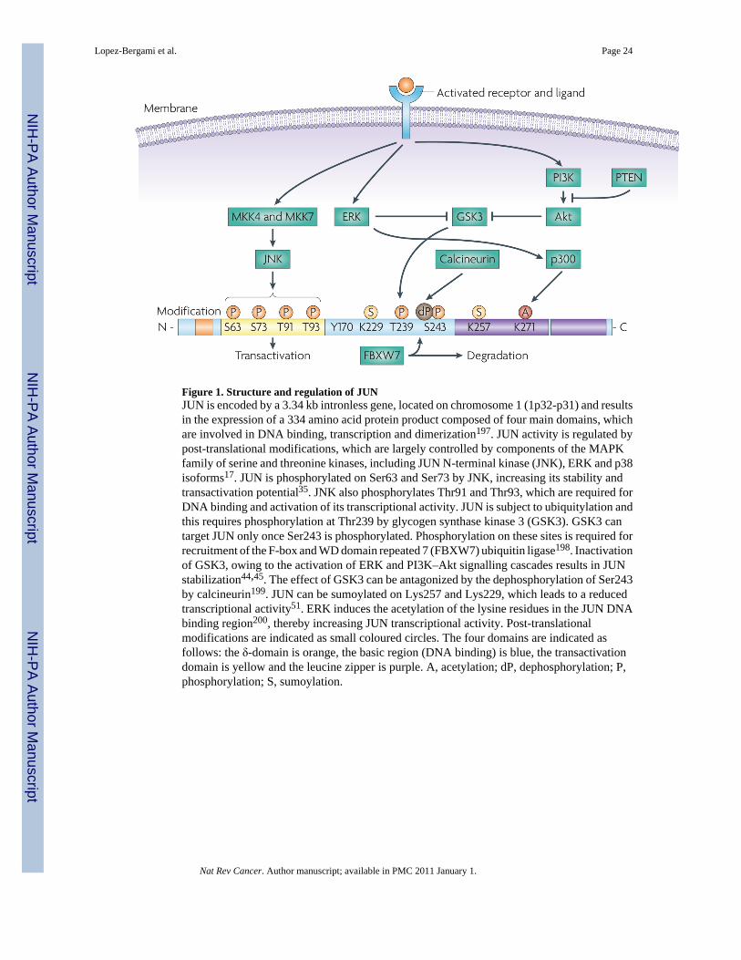
Figure 1. Structure and regulation of JUNJUN is encoded by a 3.34 kb intronless gene, located on chromosome 1 (1p32-p31) and resultsin the expression of a 334 amino acid protein product composed of four main domains, whichare involved in DNA binding, transcription and dimerization197. JUN activity is regulated bypost-translational modifications, which are largely controlled by components of the MAPKfamily of serine and threonine kinases, including JUN N-terminal kinase (JNK), ERK and p38isoforms17. JUN is phosphorylated on Ser63 and Ser73 by JNK, increasing its stability andtransactivation potential35. JNK also phosphorylates Thr91 and Thr93, which are required forDNA binding and activation of its transcriptional activity. JUN is subject to ubiquitylation andthis requires phosphorylation at Thr239 by glycogen synthase kinase 3 (GSK3). GSK3 cantarget JUN only once Ser243 is phosphorylated. Phosphorylation on these sites is required forrecruitment of the F-box and WD domain repeated 7 (FBXW7) ubiquitin ligase198. Inactivationof GSK3, owing to the activation of ERK and PI3K–Akt signalling cascades results in JUNstabilization44,45. The effect of GSK3 can be antagonized by the dephosphorylation of Ser243by calcineurin199. JUN can be sumoylated on Lys257 and Lys229, which leads to a reducedtranscriptional activity51. ERK induces the acetylation of the lysine residues in the JUN DNAbinding region200, thereby increasing JUN transcriptional activity. Post-translationalmodifications are indicated as small coloured circles. The four domains are indicated asfollows: the δ-domain is orange, the basic region (DNA binding) is blue, the transactivationdomain is yellow and the leucine zipper is purple. A, acetylation; dP, dephosphorylation; P,phosphorylation; S, sumoylation.
Lopez-Bergami et al. Page 24
Nat Rev Cancer. Author manuscript; available in PMC 2011 January 1.
NIH
-PA Author Manuscript
NIH
-PA Author Manuscript
NIH
-PA Author Manuscript

Figure 2. Mechanisms of JUN degradationSeveral mechanisms exist to limit JUN stablity. Under non-stressed conditions, JUN N-terminal kinase (JNK) is tightly bound to JUN, targeting JUN for ubiquitylation anddegradation. Activation of JNK by stress results in JUN phosphorylation and dissociation fromJNK, enhancing its protein stability, although phosphorylation on Ser63 and Ser73 promotesthe F-box and WD domain repeated 7 (FBXW7)-mediated degradation of JUN48. As part of afeedforward mechanism, JNK also phosphorylates the E3 ligase Itch in T cells after stimulation,accelerating degradation of JUN and JUNB, independently of Ser63 and Ser73phosphorylation34. Phosphorylation on JUN at Thr239 by glycogen synthase kinase 3 (GsK3)(FIG. 1) allows FBXW7 binding and ubiquitin-mediated degradation by a Skp1-Cullin1-F-box(SCF) complex. Following osmotic stress, MEKK1 can activate JUN N-terminalphosphorylation by activating MKK4, an upstream kinase for JNKs that can also function asa JUN E3 ubiquitin ligase, promoting its ubiquitin–proteasome-dependent degradation34 (notshown). De-etiolated 1 (DET1) contributes to JUN degradation by promoting the formation ofa ubiquitin ligase complex containing DNA damage binding protein 1 (DDB1), cullin 4A(CUL4A), regulator of cullins 1 (ROC1) and constitutively photomorphogenic 1 (COP1)201.Other mechanisms modulating JUN ubiquitin-mediated degradation have been reported34. P,phosphorylation; Ub, ubiquitylation.
Lopez-Bergami et al. Page 25
Nat Rev Cancer. Author manuscript; available in PMC 2011 January 1.
NIH
-PA Author Manuscript
NIH
-PA Author Manuscript
NIH
-PA Author Manuscript

Figure 3. Network of AP1 signallingERK, JUN N-terminal kinase (JNK) and p38 are predominantly responsible for thephosphorylation and activation of FOS, JUN and ATF2 respectively (black arrows) in responseto stress, mitogens or oncogene activation. ERK and JNK also regulate FOS and JUNdegradation, respectively (not depicted) and participate in ATF2 activation (dashed arrows).ERK also induces the transcription of FOS through the activation of non-AP1 transcriptionfactors (TFs). Transcription of some AP1 proteins (that is, JUN and FRA1) is regulated bycrosstalk among AP1 complexes (red arrows) as well as by autoregulation (circular red arrows).
Lopez-Bergami et al. Page 26
Nat Rev Cancer. Author manuscript; available in PMC 2011 January 1.
NIH
-PA Author Manuscript
NIH
-PA Author Manuscript
NIH
-PA Author Manuscript

NIH
-PA Author Manuscript
NIH
-PA Author Manuscript
NIH
-PA Author Manuscript
Lopez-Bergami et al. Page 27
Table 1Deregulation of Jun family members in cancer
Observation Tumour type Method Refs
JUN amplification and JUN overexpression Liposarcoma CGH, FISH, Real-time PCR and WB 12
Increased JUN expression Non-small-cell lung cancer IHC 29,202
Acute myeloid leukaemia Microarray 203
Colorectal neoplasm IHC 204
Pancreatic cancer IHC 205
Oral squamous cell carcinomas IHC 206
Increased levels of phosphorylated JUN Astrocytoma IHC 207
Invasive breast cancer IHC 146
Osteosarcoma IHC 208
Glioblastoma IHC 209
Melanoma WB 204
JUN and FOS overexpression Prostate cancer TMA 175
JUN and JUND overexpression Breast cancer TMA 210
JUN and JUNB overexpression CD30+ lymphoma* TMA and IHC 174
JUND overexpression Primary cutaneous B cell lymphoma IHC 211
JUNB and JUND overexpression Colorectal adenocarcinoma IHC and WB 212
Increased AP1 activity Lung and bladder carcinoma EMSA 213
Increased AP1 binding activity (FOS–JUNB) Endometrial cancer WB 214
Cervical cancer EMSA and WB 215
CGH, comparative genomic hybridization; EMSA, electrophoretic mobility shift assay; FISH, fluorescence in situ hybridization; IHC,immunohistochemistry; TMA, tumour microarray; WB, western blot.
*Classic Hodgkin lymphoma, anaplastic large cell lymphoma, diffuse large B cell lymphoma.
Nat Rev Cancer. Author manuscript; available in PMC 2011 January 1.
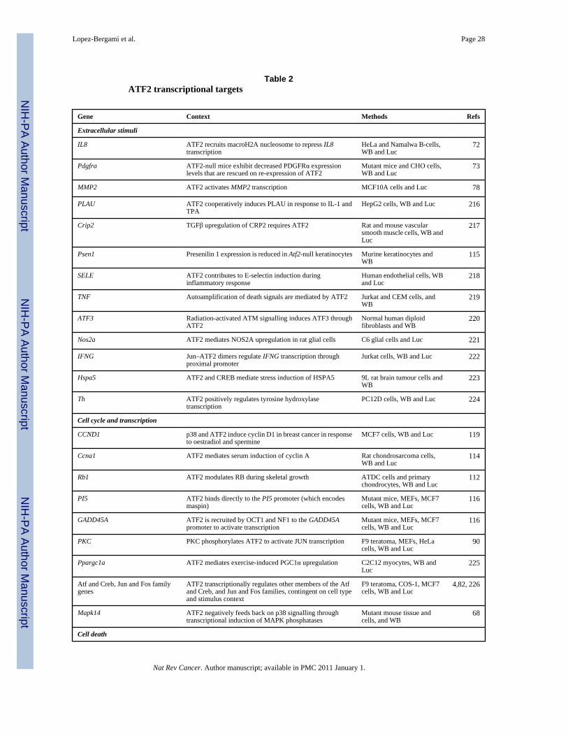
NIH
-PA Author Manuscript
NIH
-PA Author Manuscript
NIH
-PA Author Manuscript
Lopez-Bergami et al. Page 28
Table 2ATF2 transcriptional targets
Gene Context Methods Refs
Extracellular stimuli
IL8 ATF2 recruits macroH2A nucleosome to repress IL8transcription
HeLa and Namalwa B-cells,WB and Luc
72
Pdgfra ATF2-null mice exhibit decreased PDGFRα expressionlevels that are rescued on re-expression of ATF2
Mutant mice and CHO cells,WB and Luc
73
MMP2 ATF2 activates MMP2 transcription MCF10A cells and Luc 78
PLAU ATF2 cooperatively induces PLAU in response to IL-1 andTPA
HepG2 cells, WB and Luc 216
Crip2 TGFβ upregulation of CRP2 requires ATF2 Rat and mouse vascularsmooth muscle cells, WB andLuc
217
Psen1 Presenilin 1 expression is reduced in Atf2-null keratinocytes Murine keratinocytes andWB
115
SELE ATF2 contributes to E-selectin induction duringinflammatory response
Human endothelial cells, WBand Luc
218
TNF Autoamplification of death signals are mediated by ATF2 Jurkat and CEM cells, andWB
219
ATF3 Radiation-activated ATM signalling induces ATF3 throughATF2
Normal human diploidfibroblasts and WB
220
Nos2a ATF2 mediates NOS2A upregulation in rat glial cells C6 glial cells and Luc 221
IFNG Jun–ATF2 dimers regulate IFNG transcription throughproximal promoter
Jurkat cells, WB and Luc 222
Hspa5 ATF2 and CREB mediate stress induction of HSPA5 9L rat brain tumour cells andWB
223
Th ATF2 positively regulates tyrosine hydroxylasetranscription
PC12D cells, WB and Luc 224
Cell cycle and transcription
CCND1 p38 and ATF2 induce cyclin D1 in breast cancer in responseto oestradiol and spermine
MCF7 cells, WB and Luc 119
Ccna1 ATF2 mediates serum induction of cyclin A Rat chondrosarcoma cells,WB and Luc
114
Rb1 ATF2 modulates RB during skeletal growth ATDC cells and primarychondrocytes, WB and Luc
112
PI5 ATF2 binds directly to the PI5 promoter (which encodesmaspin)
Mutant mice, MEFs, MCF7cells, WB and Luc
116
GADD45A ATF2 is recruited by OCT1 and NF1 to the GADD45Apromoter to activate transcription
Mutant mice, MEFs, MCF7cells, WB and Luc
116
PKC PKC phosphorylates ATF2 to activate JUN transcription F9 teratoma, MEFs, HeLacells, WB and Luc
90
Ppargc1a ATF2 mediates exercise-induced PGC1α upregulation C2C12 myocytes, WB andLuc
225
Atf and Creb, Jun and Fos familygenes
ATF2 transcriptionally regulates other members of the Atfand Creb, and Jun and Fos families, contingent on cell typeand stimulus context
F9 teratoma, COS-1, MCF7cells, WB and Luc
4,82, 226
Mapk14 ATF2 negatively feeds back on p38 signalling throughtranscriptional induction of MAPK phosphatases
Mutant mouse tissue andcells, and WB
68
Cell death
Nat Rev Cancer. Author manuscript; available in PMC 2011 January 1.

NIH
-PA Author Manuscript
NIH
-PA Author Manuscript
NIH
-PA Author Manuscript
Lopez-Bergami et al. Page 29
Gene Context Methods Refs
Bcl2 ATF2 controls Bcl2 promoter activation in chondrocytes Mutant mice, WB and Luc 94
TRAIL Auto-amplification of death signals are mediated by ATF2 Jurkat and CEM cells, andWB
219
ACHE ATF2 mediates hydrogen peroxide-induction ofacetylcholinesterase
293T cells, WB and Luc 227
ACHE, acetylcholinesterase; ATM, ataxia-telangectasia mutated; CCND1, cyclin D1; CHO, Chinese hamster ovary; Crip2, cysteine-rich intestinalprotein 2; Hspa5, heat shock protein A5; IFNG, interferon (IFN)-γ; IL8, interleukin-8; Luc, luciferase assay; MEF, mouse embryonic fibroblast;MMP2, matrix metalloproteinase 2; Nos2a, nitric oxide synthase 2A; Pdgfra, platelet derived growth factor A; PI5, protease inhibitor 5; PLAU, urinarypalminogen activator; Ppargc1a, peroxisome proliferator-activated receptor-γ coactivator 1α; Psen1, presenilin 1; SELE, E selectin; TGFβ,transforming growth factor-β; Th, tyrosine hydroxylase; TNF, tumour necrosis factor; TRAIL, tumour necrosis factor-related apoptosis-inducingligand; WB, western blot.
Nat Rev Cancer. Author manuscript; available in PMC 2011 January 1.