Defining “Sexually Explicit” Content - The University of North ...
Upload
khangminh22Category
view
1download
0
Neisseria gonorrhoeae
Sexually TransmittedBacterial Pathogen for which there are
Increasing AntimicrobialResistance Concerns
Sexually Transmitted Bacterial Pathogen
with Increasing Antim
icrobial Resistance
Neisseria gonorrhoeae | 63
N eisseria gonorrhoeae, also commonly referred to as “gonococcus” or “GC”,causes an estimated 62 million cases of gonorrhea worldwide each year[Gerbase et al., 1998]. Spread by sexual intercourse, N. gonorrhoeae may
infect the mucosal surfaces of urogenital sites (cervix, urethra, rectum) and theoro- and nasopharynx (throat), causing symptomatic or asymptomatic infections.GC is always pathogenic and, if untreated, gonorrhea is a major cause of pelvicinflammatory disease (PID), tubal infertility, ectopic pregnancy, chronic pelvicpain and/or disseminated gonococcal infection (DGI). The probability of co-infection with other sexually transmitted infections (STIs) may be high in somepatient populations. Neonates may acquire gonococcal infection of the conjunctivaduring birth. The diagnosis of gonorrhea in older infants and young children isoften associated with allegations of sexual abuse; transmission through neithernonsexual human nor fomite contact has been documented. Epidemiologicalstudies provide strong evidence that gonococcal infections facilitate HIVtransmission [Fleming and Wasserheit 1999]. Extended-spectrum cephalosporins,fluoroquinolones and spectinomycin are recognized as the most effectiveantibiotics for the treatment of gonorrhea in most areas of the world.
Antimicrobial resistance in N. gonorrhoeae is the most significant challenge tocontrolling gonorrhea. Gonococcal strains may be resistant to penicillins,tetracyclines, spectinomycin, and, recently, resistance to the fluoroquinolones(ciprofloxacin and ofloxacin) and the macrolide azithromycin has emerged[Handsfield 1994; Knapp et al. 1997; Young et al. 1997; CDC 1999]. Resistance tothe penicillins and tetracyclines is conferred by chromosomal and/or plasmid-mediated mechanisms. Resistance to spectinomycin, fluoroquinolones andazithromycin is chromosomally mediated, and certain types of chromosomalmutations may contribute to resistance to several classes of antibioticssimultaneously.
Neisseria gonorrhoeaeCONFIRMATORY IDENTIFICATION AND ANTIMICROBIAL SUSCEPTIBILITY TESTING
CHAPTER VI

64 | Manual for Identification and Antimicrobial Susceptibility Testing
Agents used for the treatment of bacterial infections, including co-infecting STIs,may select for resistance in N. gonorrhoeae. For example, whereas a 1-gram dose of azithromycin is sufficient for treatment of infections with C. trachomatis and H. ducreyi, this dose is sub-optimal for the treatment of N. gonorrhoeae and mayresult in the incidental selection and spread of resistant gonococcal strains.At the time of writing of this manual (2002), the broad-spectrum cephalosporins(ceftriaxone, cefixime, etc.) are the only class of antimicrobial agents to whichgonococci have not developed confirmed resistance, although a few isolated strainshave exhibited decreased susceptibility to cefixime [CDC 2000; Wang 2002].
It is of great importance to perform laboratory surveillance of antimicrobialresistance in N. gonorrhoeae in order to assess the effectiveness of locallyrecommended therapies. Only measurement of the in vitro susceptibilities of theinfecting organism will provide objective information to help determine if a post-treatment isolate is truly resistant to the antimicrobial agent being used to treat theinfection, as opposed to infection which fails to respond to treatment due toinadequate absorption of the agent, non-compliance with therapy, or re-exposure.At the population level, surveillance is key for the monitoring of local, regional andinternational trends in antimicrobial resistance, which can help inform and shapepublic health policy. Comparison between antimicrobial susceptibilities ofgonococci isolated in different geographical areas provides information about the distribution and temporal spread of resistant isolates. Thus, changes inrecommended antimicrobial therapies can be anticipated, and surveillance can beenhanced to guide timely changes in these therapies at the local level.
Presumptive identification of N. gonorrhoeae
After the specimen has been collected from the patient, it should be labeled with aunique identifier assigned in tandem with the demographic and clinicalinformation so it can be linked for epidemiological studies. Methods for streakingfor isolation from specimen swabs, primary culture methodology, and isolatestorage and transport are included in Appendices 8, 11 and 12.
Because N. gonorrhoeae is highly susceptible to adverse environmental conditions(as described in Table 28 of Appendix 8), strains must always be incubated at35˚–36.5˚C in a humid, CO2-enriched atmosphere. Subculture colonies that appearto be gonococcal (gram-negative diplococci growing in pinkish-brown colonies 0.5– 1 mm in diameter, see Appendix 8) from the primary selective medium to a non-selective medium, such as GC-chocolate agar with 1% defined supplement, toobtain a pure culture of the isolate. (Specimens from normally sterile sites, such asthe conjunctiva, are cultured on nonselective medium for primary isolation;subculture for purity if examination of the plate shows evidence of contaminants.)If the subcultured isolate is not pure, continue to perform serial subcultures ofindividual colonies of gram-negative diplococci until a pure culture is obtained.

Neisseria gonorrhoeae | 65
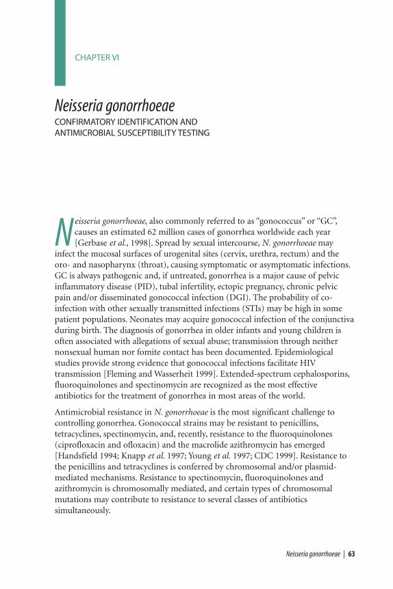
A presumptive diagnosis of N. gonorrhoeae originally isolated on selective mediumcan be made based upon colonial morphology, the observation of typical (gram-negative) diplococci in pairs, tetrads or clusters upon Gram stain or simple singlestain with Loeffler’s methylene blue, and a positive oxidase reaction. A presumptivediagnosis of N. gonorrhoeae originally isolated on nonselective medium can bemade based upon these characteristics plus an appropriate reaction in at least onesupplemental biochemical or enzymatic test (e.g., superoxol 4+ reaction, see‘Supplemental Tests’). A flowchart of tests required for presumptive identificationof isolates from sites with normal flora (i.e., isolated on selective media such asMTM, ML, or GC-Lect) and isolates from normally sterile sites (i.e., isolated onnonselective medium, such as GC-chocolate agar) is presented in Figure 19.
Oxidase test
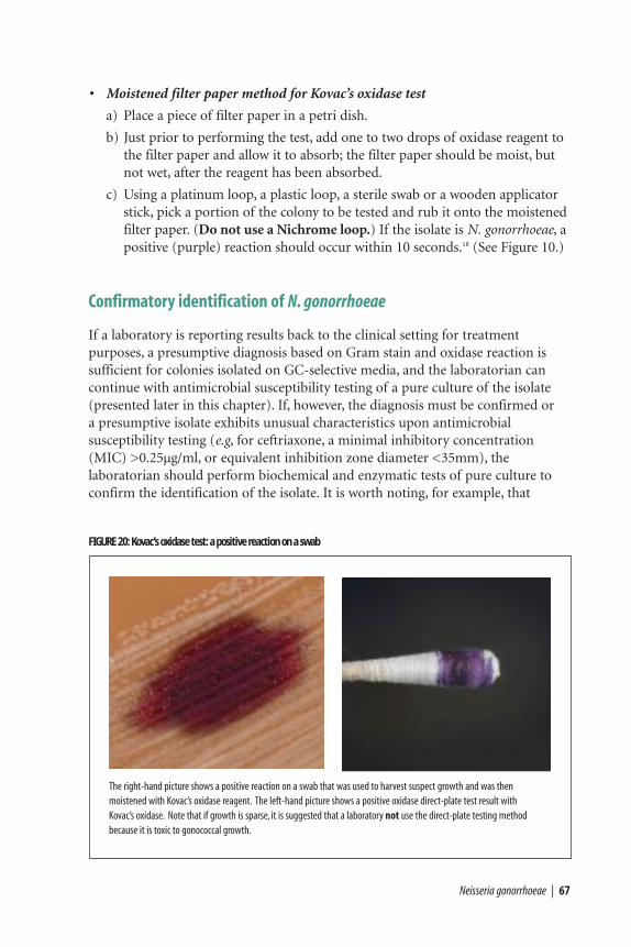
The oxidase test uses Kovac’s reagent (a 1% (wt/vol) solution of N, N, N’, N’–tetramethyl-ρ-phenylenediamine dihydrochloride)18 to detect the presence ofcytochrome c in a bacterial organism’s respiratory chain; if the oxidase reagent iscatalyzed, it turns purple. Neisseria species give a positive oxidase reaction, andgram-negative oxidase-positive diplococci isolated on gonococcal selective mediamay be identified presumptively as N. gonorrhoeae. Preparation of oxidase reagentand appropriate quality control methods are included in Appendix 2.
Perform an oxidase test on growth of representative colonies that stained as (gram-negative) diplococci. Because the oxidase reagent is toxic for bacteria, it isrecommended to perform the oxidase test on a sterile swab and not directly on theculture plate, particularly if there are only a few suspect colonies. Alternatively, onecan use filter paper in place of a swab for this test. Do not perform the oxidasetest with a Nichrome loop, as it may produce a false-positive reaction. If a sterileswab was used to make a smear for the Gram stain (as described in Appendix 4),the swab can then be used to conduct the oxidase test. The oxidase test should onlybe performed on freshly grown (18–24 hour) organisms.
• Swab method for Kovac’s oxidase test
a) Select suspect colonies from the culture plate (selective or nonselectivemedium) with the swab.
b) Use a Pasteur pipette to add one drop of oxidase reagent to the swab.
c) If the isolate is N. gonorrhoeae, a positive (purple) reaction should occurwithin 10 seconds.18 (See Figure 20).
18 Some laboratories may use a different reagent, Gordon and MacLeod’s reagent, (1% [wt/vol] dimethyl-ρ-phenylenediamene dihydrocholoride; “dimethyl reagent”) to perform the oxidase test. The dimethyl reagent is more stable than the tetramethyl reagent (Kovac’s reagent), but the reaction with the dimethyl reagent isslower than that with the tetramethyl reagent. If the laboratory is using the dimethyl- reagent, a positive reaction will be indicated by a color change to blue on the filter paper (not purple, as with the tetramethylreagent), and with the dimethyl reagent it will take 10 – 30 minutes for a positive reaction to develop.
66 | Manual for Identification and Antimicrobial Susceptibility Testing
oxidase-positive
= suspect N. gonorrhoeae
FIGURE 19: Flowchart for isolation and presumptive identification of Neisseria gonorrhoeae
Colonies on selective media (e.g., Martin-Lewis[ML] or Modified Thayer-Martin [MTM]) are
pinkish-brown and translucent, with smooth consistency and defined margins, and are typically
0.5 – 1.0 mm in diameter.*
Colonies on GC-chocolate agar are pinkish-brown and translucent, exhibit
smooth consistency and defined margins,and are typically 0.5 – 1.0 mm in
diameter.*
Sterile site specimens(e.g., conjunctiva)
Non-sterile-site specimens(e.g., urethra, cervix, vagina, rectum, and pharynx)
* If a presumptive isolate exhibits unusual characteristics upon antimicrobial susceptibility testing, confirm the identification with biochemical and enzymatic tests.
Antimicrobial susceptibility testing on
GC-susceptibility test medium
* Fastidious strains of N. gonorrhoeae may producesmall, ~0.25-mm “pinpoint” colonies
(If primary isolation was on nonselective medium)
* Fastidious strains of N. gonorrhoeae may producesmall, ~0.25-mm “pinpoint” colonies
Other morphology = negative
(Gram-negative)
bean-shaped diplococci
= suspect N. gonorrhoeae
oxidase negative = negative
• An isolate from selective medium (MTM, ML) is considered ‘presumptive GC’ when it is an oxidase-positive, (gram-negative) diplococcus.
• An isolate from nonselective medium can beconsidered presumptive GC when it is an oxidase-positive, (gram-negative) diplococcusand gives an appropriate reaction in at least onesupplemental test (e.g., superoxol 4+ reaction).
Note: it is acceptable practice to perform antimicrobial susceptibility testing on presumptiveisolates of N. gonorrhoeae (GC) for treatment purposes.*
Oxidase test
Reactions typical of N. gonorrhoeaein supplemental tests:
Superoxol/Catalase: positive
Colistin resistance: positive (resistant)
Nitrate reduction: negative
Polysaccharide production:negative
Acid production: acid from glucose only
Enzyme substrate:hydroxyprolylaminopeptidase +
Gram stain or simple single stain(e.g., Loeffler’s methylene blue stain)
Neisseria gonorrhoeae | 67
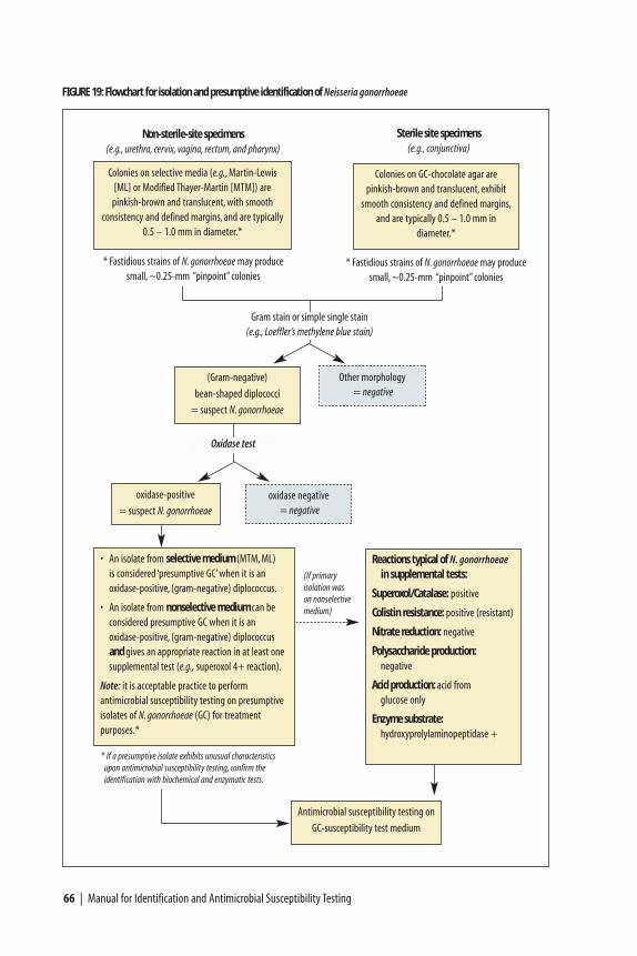
• Moistened filter paper method for Kovac’s oxidase test
a) Place a piece of filter paper in a petri dish.
b) Just prior to performing the test, add one to two drops of oxidase reagent tothe filter paper and allow it to absorb; the filter paper should be moist, butnot wet, after the reagent has been absorbed.
c) Using a platinum loop, a plastic loop, a sterile swab or a wooden applicatorstick, pick a portion of the colony to be tested and rub it onto the moistenedfilter paper. (Do not use a Nichrome loop.) If the isolate is N. gonorrhoeae, apositive (purple) reaction should occur within 10 seconds.18 (See Figure 10.)
Confirmatory identification of N. gonorrhoeae
If a laboratory is reporting results back to the clinical setting for treatmentpurposes, a presumptive diagnosis based on Gram stain and oxidase reaction issufficient for colonies isolated on GC-selective media, and the laboratorian cancontinue with antimicrobial susceptibility testing of a pure culture of the isolate(presented later in this chapter). If, however, the diagnosis must be confirmed or a presumptive isolate exhibits unusual characteristics upon antimicrobialsusceptibility testing (e.g, for ceftriaxone, a minimal inhibitory concentration(MIC) >0.25µg/ml, or equivalent inhibition zone diameter <35mm), thelaboratorian should perform biochemical and enzymatic tests of pure culture toconfirm the identification of the isolate. It is worth noting, for example, that
FIGURE 20: Kovac’s oxidase test: a positive reaction on a swab
The right-hand picture shows a positive reaction on a swab that was used to harvest suspect growth and was then moistened with Kovac’s oxidase reagent. The left-hand picture shows a positive oxidase direct-plate test result with Kovac’s oxidase. Note that if growth is sparse, it is suggested that a laboratory not use the direct-plate testing methodbecause it is toxic to gonococcal growth.
because men who have sex with men (referred to in literature as “MSM”) havehigher rates of non-gonococcal neisserial infections in the urethra than do otherpopulations, the epidemiology could lead a clinician to request a confirmeddiagnosis. Another example of a situation where the diagnosis requires definitiveconfirmation would be a case of suspected sexual abuse; the discussion of therelated social, medical and legal issues with which a laboratory could be involvedgoes beyond the scope of this laboratory manual.19
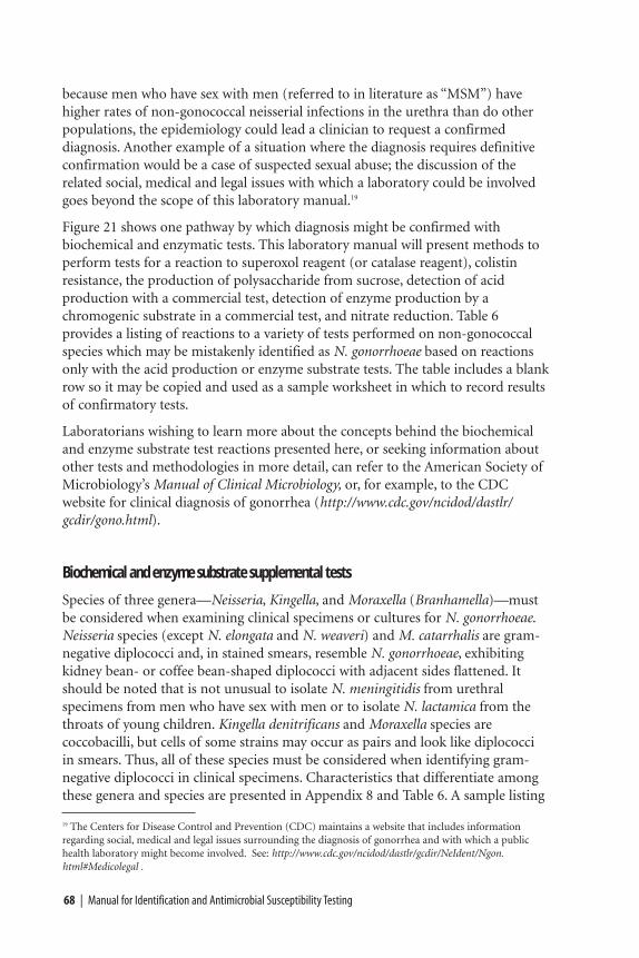
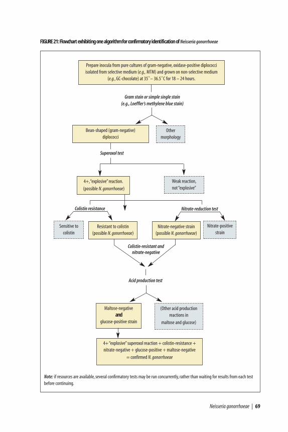
Figure 21 shows one pathway by which diagnosis might be confirmed withbiochemical and enzymatic tests. This laboratory manual will present methods toperform tests for a reaction to superoxol reagent (or catalase reagent), colistinresistance, the production of polysaccharide from sucrose, detection of acidproduction with a commercial test, detection of enzyme production by achromogenic substrate in a commercial test, and nitrate reduction. Table 6provides a listing of reactions to a variety of tests performed on non-gonococcalspecies which may be mistakenly identified as N. gonorrhoeae based on reactionsonly with the acid production or enzyme substrate tests. The table includes a blankrow so it may be copied and used as a sample worksheet in which to record resultsof confirmatory tests.
Laboratorians wishing to learn more about the concepts behind the biochemicaland enzyme substrate test reactions presented here, or seeking information aboutother tests and methodologies in more detail, can refer to the American Society ofMicrobiology’s Manual of Clinical Microbiology, or, for example, to the CDCwebsite for clinical diagnosis of gonorrhea (http://www.cdc.gov/ncidod/dastlr/ gcdir/gono.html).
Biochemical and enzyme substrate supplemental tests
Species of three genera—Neisseria, Kingella, and Moraxella (Branhamella)—mustbe considered when examining clinical specimens or cultures for N. gonorrhoeae.Neisseria species (except N. elongata and N. weaveri) and M. catarrhalis are gram-negative diplococci and, in stained smears, resemble N. gonorrhoeae, exhibitingkidney bean- or coffee bean-shaped diplococci with adjacent sides flattened. Itshould be noted that is not unusual to isolate N. meningitidis from urethralspecimens from men who have sex with men or to isolate N. lactamica from thethroats of young children. Kingella denitrificans and Moraxella species arecoccobacilli, but cells of some strains may occur as pairs and look like diplococci in smears. Thus, all of these species must be considered when identifying gram-negative diplococci in clinical specimens. Characteristics that differentiate amongthese genera and species are presented in Appendix 8 and Table 6. A sample listing
68 | Manual for Identification and Antimicrobial Susceptibility Testing
19 The Centers for Disease Control and Prevention (CDC) maintains a website that includes informationregarding social, medical and legal issues surrounding the diagnosis of gonorrhea and with which a publichealth laboratory might become involved. See: http://www.cdc.gov/ncidod/dastlr/gcdir/NeIdent/Ngon.html#Medicolegal .
Other morphology
Bean-shaped (gram-negative) diplococci
Gram stain or simple single stain (e.g., Loeffler’s methylene blue stain)
Superoxol test
Neisseria gonorrhoeae | 69
FIGURE 21: Flowchart exhibiting one algorithm for confirmatory identification of Neisseria gonorrhoeae
Resistant to colistin (possible N. gonorrhoeae)
Nitrate-negative strain(possible N. gonorrhoeae)
Prepare inocula from pure cultures of gram-negative, oxidase-positive diplococci isolated from selective medium (e.g., MTM) and grown on non-selective medium
(e.g., GC-chocolate) at 35˚– 36.5˚C for 18 – 24 hours.
Maltose-negative and
glucose-positive strain
(Other acid productionreactions in
maltose and glucose)
Sensitive to colistin
Nitrate-positive strain
Acid production test
Weak reaction,not “explosive”
4+,“explosive” reaction.
(possible N. gonorrhoeae)
Colistin resistance Nitrate-reduction test
4+ “explosive” superoxol reaction + colistin-resistance +nitrate-negative + glucose-positive + maltose-negative
= confirmed N. gonorrhoeae
Colistin-resistant and nitrate-negative
Note: if resources are available, several confirmatory tests may be run concurrently, rather than waiting for results from each testbefore continuing.
70 | Manual for Identification and Antimicrobial Susceptibility Testing
Cell
Supe
roxo
lPr
oduc
tion
of a
cid fr
om:
Redu
ctio
n of
Poly
sacc
harid
eSp
ecie
sm
orph
olog
y{C
atal
ase}
Colis
tinGL
UM
ALLA
CSU
CNO
3(N
itrat
e)fro
m su
cros
e
Test
isol
ate:
N.go
norrh
oeae
aGN
D4+
{+}
R+
––
––
–
N.m
enin
gitid
isGN
D1+
to 4
+{+
}R
++
––
––
N.la
ctam
icaGN
D1+
to 3
+
{+}
R+
++
––
–
N.cin
erea
bGN
D2+
{+
}(R
)–
––
––
–
N.po
lysac
char
eaGN
D1+
to 3
+
{+}
(R)
++
––
––
N.su
bfla
vab
GND
2+
{+}
(R)
++
–V
–V
N.sic
caGN
D2+
{+
}S
++
–+
–+
N.m
ucos
aGN
D2+
{+
}S
++
–+
++
N.fla
vesc
ens
GND
2+
{+}
S–
––
––
+
N.elo
ngat
aGN
R–
{–}
S–
––
––
–
M.c
atar
rhal
isGN
D1+
to 4
+{+
}(R
)–
––
–+
–
K.de
nitri
fican
scGN
C–
{–}
R+
––
–+
–
Sym
bols
and
Abb
revi
atio
ns:
+,s
train
s typ
ically
pos
itive
but
gen
etic
mut
ants
may
be n
egat
ive;–
,stra
ins t
ypica
lly n
egat
ive;V
,bio
var d
epen
dent
(stra
ins b
elon
ging
to b
iova
rs fla
vaan
d su
bflav
a do
not p
rodu
ce ac
id fr
om su
crose
or p
rodu
ce p
olys
acch
arid
e fro
m su
crose
);GL
U,gl
ucos
e;M
AL,m
alto
se;L
AC,la
ctos
e;SU
C,su
crose
;GND
,gra
m-n
egat
ive d
iplo
cocc
i;GNR
,gr
am-n
egat
ive ro
ds;G
NC,g
ram
-neg
ative
cocc
obac
illi;R
,res
istan
t;(R
),so
me s
train
s res
istan
t and
may
gro
w o
n go
noco
ccal
sele
ctive
med
ia;S
,sus
cept
ible
(ins
uffic
ient
dat
a to
sugg
est
that
isol
ates
may
gro
w o
n go
noco
ccal
sele
ctive
med
ia co
ntai
ning
colis
tin).
a Inclu
des N
.gon
orrh
oeae
subs
pecie
s koc
hii w
hich
exhi
bit c
hara
cter
istics
of b
oth
N.go
norrh
oeae
and
N.m
enin
gitid
is(b
ut w
ill b
e ide
ntifi
ed as
N.g
onor
rhoe
aeby
test
s rou
tinel
y use
d fo
r th
e ide
ntifi
catio
n of
Neis
seria
spec
ies)
.b
Inclu
des b
iova
rs su
bflav
a,fla
va,a
nd p
erfla
va.S
train
s bel
ongi
ng to
the b
iova
r flav
a pro
duce
acid
from
glu
cose
,mal
tose
and
fruct
ose;
stra
ins b
elon
ging
to th
e bio
var s
ubfla
va p
rodu
ce
acid
onl
y fro
m g
luco
se an
d m
alto
se.
c Cocc
obac
illus
;som
e stra
ins o
ccur
in p
airs
whi
ch re
sem
ble g
ram
-neg
ative
dip
loco
cci
TABL
E6:
Resu
lts o
f bio
chem
ical a
nd e
nzym
atic
test
s for
Nei
sser
ia g
onor
rhoe
aean
d re
late
d sp
ecie
s with
sim
ilar c
olon
ial m
orph
olog
y
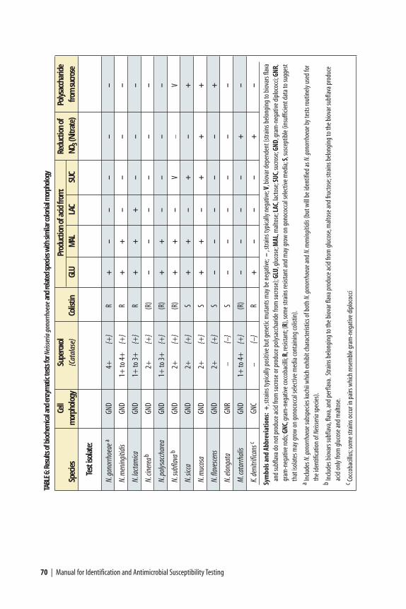
of quality control strains for the supplemental tests described in this manual forthe identification of N. gonorrhoeae is included in Table 7.
In a reference laboratory setting, the tests described below are best performedconcurrently since they all require an inoculum prepared from fresh (18–24 hour)growth. However, when resources are limited, laboratorians may choose to screenisolates with a subset of these tests to detect isolates resembling N. gonorrhoeaeprior to further testing. Sequential testing practices can conserve resources bylimiting the use of more costly commercial tests (e.g., acid production or enzymesubstrate) to only those isolates resistant to colistin and exhibiting a strongsuperoxol reaction. When choosing the screening approach, it is important toremember that tests performed on successive days will require a fresh (18–24 hour)subculture of the isolate.
Neisseria gonorrhoeae | 71
Test Positive control Negative control
Superoxol (or Catalase) test N. gonorrhoeae ATCC 49226 [4+] K. denitrificans ATCC 33394N. cinerea ATCC 14685 [weak, 2+] (no reaction in superoxol)
(positive reaction in superoxol)
Colistin resistance test N. gonorrhoeae ATCC 49226 N. cinerea ATCC 14685K. denitrificans ATCC 33394 N. mucosa ATCC 19696(resistant to colistin) (susceptible to colistin)
Polysaccharide N. polysaccharea ATCC 43768 N. gonorrhoeae ATCC 49226production test N. mucosa ATCC 19696 N. cinerea ATCC 14685
(produce polysaccharide) (do not produce polysaccharide)
Nitrate reduction test K. denitrificans ATCC 33394 N. gonorrhoeae ATCC 49226N. mucosa ATCC 19696 N. cinerea ATCC 14685(able to reduce nitrate) (unable to reduce nitrate)
Acid production test Use the QC strains recommended by the test manufacturer* plus N. cinerea.* If the manufacturer has not designated specific strains for QC:
• N. gonorrhoeae (ATCC 49226) produces acid from glucose• N. meningitidis (ATCC 13077) produces acid from glucose and maltose• N. lactamica (ATCC 23970) produces acid from glucose, maltose, and lactose• N. mucosa (ATCC 19696) produces acid from glucose, maltose, and sucrose• N. cinerea (ATCC 14685) glucose negative, but may produce a weak glucose
reaction; does not produce acid from the other sugars.
Enzyme substrate test Use the QC strains recommended by the test manufacturer.** If the manufacturer has not designated specific strains for QC:
• N. gonorrhoeae (ATCC 49226) produces hydroxyprolylaminopeptidase.• N. meningitidis (ATCC 13077) produces γ-glutamylaminopeptidase.• N. lactamica (ATCC 23970) produces ß-galactosidase.• M. catarrhalis (ATCC 25238) produces none of these enzymes.
Note: Laboratorians should follow QC strain designations provided by manufacturers of (commercial) tests; however, if specific strain numbers are not provided, those included in this table can be used for guidance.
TABLE 7: Examples of quality control (QC) strains for supplemental tests used to identify Neisseria gonorrhoeae
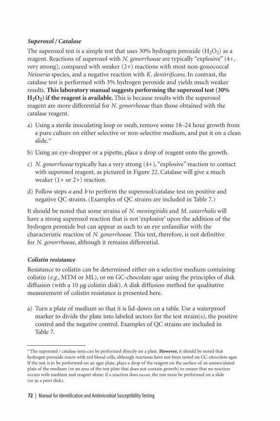
Superoxol / Catalase
The superoxol test is a simple test that uses 30% hydrogen peroxide (H2O2) as areagent. Reactions of superoxol with N. gonorrhoeae are typically “explosive” (4+,very strong), compared with weaker (2+) reactions with most non-gonococcalNeisseria species, and a negative reaction with K. denitrificans. In contrast, thecatalase test is performed with 3% hydrogen peroxide and yields much weakerresults. This laboratory manual suggests performing the superoxol test (30%H2O2) if the reagent is available. This is because results with the superoxol reagent are more differential for N. gonorrhoeae than those obtained with thecatalase reagent.
a) Using a sterile inoculating loop or swab, remove some 18–24 hour growth froma pure culture on either selective or non-selective medium, and put it on a cleanslide.20
b) Using an eye-dropper or a pipette, place a drop of reagent onto the growth.
c) N. gonorrhoeae typically has a very strong (4+), “explosive” reaction to contactwith superoxol reagent, as pictured in Figure 22. Catalase will give a muchweaker (1+ or 2+) reaction.
d) Follow steps a and b to perform the superoxol/catalase test on positive andnegative QC strains. (Examples of QC strains are included in Table 7.)
It should be noted that some strains of N. meningitidis and M. catarrhalis will have a strong superoxol reaction that is not ‘explosive’ upon the addition of thehydrogen peroxide but can appear as such to an eye unfamiliar with thecharacteristic reaction of N. gonorrhoeae. This test, therefore, is not definitive for N. gonorrhoeae, although it remains differential.
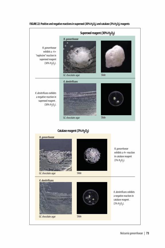
Colistin resistance
Resistance to colistin can be determined either on a selective medium containingcolistin (e.g., MTM or ML), or on GC-chocolate agar using the principles of diskdiffusion (with a 10 µg colistin disk). A disk diffusion method for qualitativemeasurement of colistin resistance is presented here.
a) Turn a plate of medium so that it is lid-down on a table. Use a waterproofmarker to divide the plate into labeled sectors for the test strain(s), the positivecontrol and the negative control. Examples of QC strains are included in Table 7.
72 | Manual for Identification and Antimicrobial Susceptibility Testing
20 The superoxol / catalase tests can be performed directly on a plate. However, it should be noted that hydrogen peroxide reacts with red blood cells, although reactions have not been noted on GC-chocolate agar.If the test is to be performed on an agar plate, place a drop of the reagent on the surface of an uninoculatedplate of the medium (or an area of the test plate that does not contain growth) to ensure that no reactionoccurs with medium and reagent alone; if a reaction does occur, the test must be performed on a slide (or in a petri dish).
K. denitrificans
GC chocolate agar Slide
K. denitrificans exhibitsa negative reaction in
superoxol reagent .(30% H2O2).
Neisseria gonorrhoeae | 73
FIGURE 22: Positive and negative reactions in superoxol (30% H2O2) and catalase (3% H2O2) reagents
N. gonorrhoeae
GC chocolate agar Slide
Superoxol reagent (30% H2O2)
N. gonorrhoeaeexhibits a 4+
“explosive” reaction insuperoxol reagent
(30% H2O2).
K. denitrificans
GC chocolate agar Slide
K. denitrificans exhibitsa negative reaction incatalase reagent .(3% H2O2).
N. gonorrhoeae
GC chocolate agar Slide
Catalase reagent (3% H2O2)
N. gonorrhoeaeexhibits a 4+ reaction in catalase reagent(3% H2O2).
• A 100-mm plate can be divided into four sectors, permitting testing of twoclinical strains alongside the positive and negative controls. If there aremultiple clinical strains requiring the colistin resistance test at once, and thecolistin disks are from the same batch, it is appropriate to run the positiveand negative controls on only one plate.
b) Prepare a suspension of a pure overnight culture (approximately equal to a 0.5McFarland turbidity standard) in Mueller-Hinton broth or phosphate bufferedsaline (PBS).
c) Using a sterile swab or inoculating loop, inoculate the GC-chocolate agar plateevenly with a swab. Allow the plate to dry so that there is no visible surfacemoisture.
d) Apply a colistin disk (10 µg) to the center of the plate, tapping it down toensure even contact with the surface. Incubate at 35˚–36.5˚C in 5% CO2 andincreased humidity for 18–24 hours.
After incubation, examine the plate for inhibition of growth around the colistindisk. N. gonorrhoeae is colistin-resistant, and will grow all the way up to the disk,as will all strains of N. meningitidis, N. lactamica and K. denitrificans. In contrast,strains of commensal Neisseria species, most of which are colistin-susceptible, willexhibit zones of inhibition at least 10 mm in diameter with a non-standardizedinoculum. Some strains of N. subflava biovars, N. cinerea, and M. catarrhalis maybe sufficiently resistant to colistin so as to also grow up to the disk. Thus, thecolistin resistance test is not definitive for N. gonorrhoeae but will aid indifferentiating between this species and many commensal species.
Polysaccharide production test
Some species produce a starch-like polysaccharide when grown on a mediumcontaining sucrose. Upon addition of a drop of Gram’s iodine to the growth,this starch will immediately stain dark blue-purple to brown or black. This test iseasy to perform and is a useful differential test to be used in combination withothers (e.g., superoxol, colistin resistance, acid production) in the identification ofN. gonorrhoeae. It is not possible to detect polysaccharide in the sucrose-containing medium of rapid acid-detection tests. The methods for preparation ofthe medium appropriate for this test (tryptone-based soy agar [TSA] containing1% sucrose) can be found in Appendix 2.
a) Turn a plate of sucrose medium so that it is lid-down on a table. Use awaterproof marker to divide the plate into labeled sectors for the test strain(s),the positive control and the negative control. (Examples of QC strains areincluded in Table 7.)
• A 100-mm plate can be divided into four sectors, permitting testing of twoclinical strains alongside the positive and negative controls. If there aremultiple clinical strains requiring the polysaccharide test at once, and the
74 | Manual for Identification and Antimicrobial Susceptibility Testing
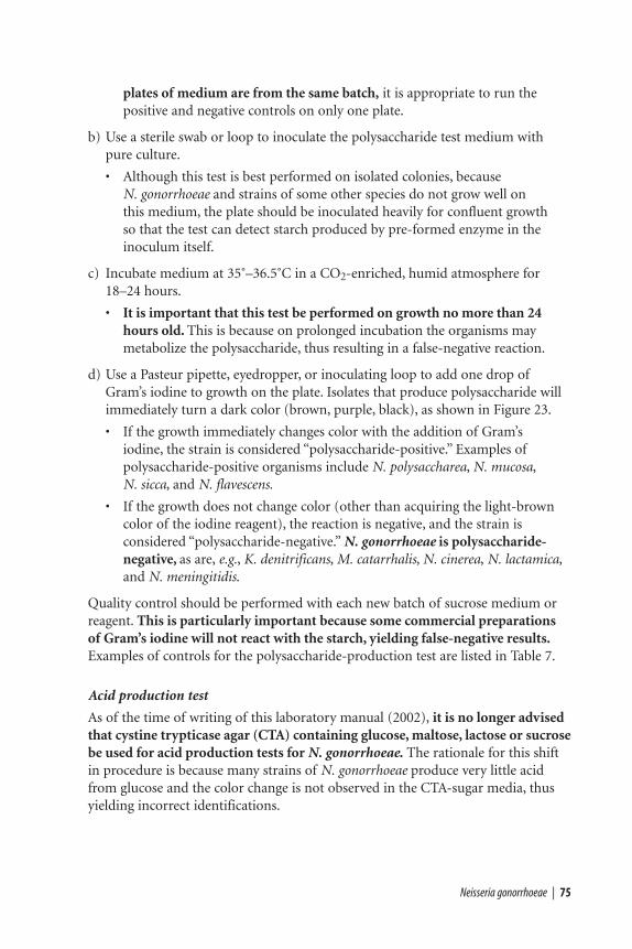
plates of medium are from the same batch, it is appropriate to run thepositive and negative controls on only one plate.
b) Use a sterile swab or loop to inoculate the polysaccharide test medium withpure culture.
• Although this test is best performed on isolated colonies, because N. gonorrhoeae and strains of some other species do not grow well on this medium, the plate should be inoculated heavily for confluent growth so that the test can detect starch produced by pre-formed enzyme in theinoculum itself.
c) Incubate medium at 35˚–36.5˚C in a CO2-enriched, humid atmosphere for18–24 hours.
• It is important that this test be performed on growth no more than 24hours old. This is because on prolonged incubation the organisms maymetabolize the polysaccharide, thus resulting in a false-negative reaction.
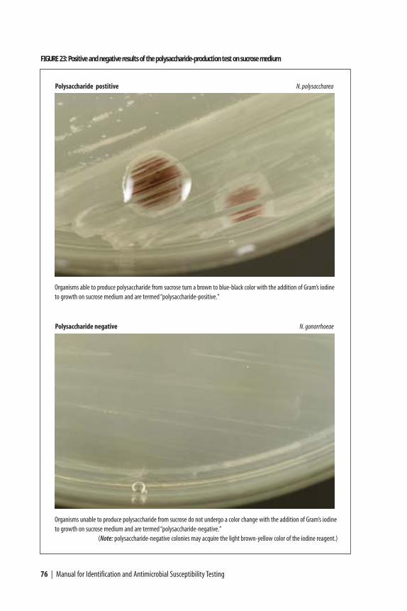
d) Use a Pasteur pipette, eyedropper, or inoculating loop to add one drop ofGram’s iodine to growth on the plate. Isolates that produce polysaccharide willimmediately turn a dark color (brown, purple, black), as shown in Figure 23.
• If the growth immediately changes color with the addition of Gram’s iodine, the strain is considered “polysaccharide-positive.” Examples ofpolysaccharide-positive organisms include N. polysaccharea, N. mucosa,N. sicca, and N. flavescens.
• If the growth does not change color (other than acquiring the light-browncolor of the iodine reagent), the reaction is negative, and the strain is considered “polysaccharide-negative.” N. gonorrhoeae is polysaccharide-negative, as are, e.g., K. denitrificans, M. catarrhalis, N. cinerea, N. lactamica,and N. meningitidis.
Quality control should be performed with each new batch of sucrose medium orreagent. This is particularly important because some commercial preparationsof Gram’s iodine will not react with the starch, yielding false-negative results.Examples of controls for the polysaccharide-production test are listed in Table 7.
Acid production test
As of the time of writing of this laboratory manual (2002), it is no longer advisedthat cystine trypticase agar (CTA) containing glucose, maltose, lactose or sucrosebe used for acid production tests for N. gonorrhoeae. The rationale for this shiftin procedure is because many strains of N. gonorrhoeae produce very little acidfrom glucose and the color change is not observed in the CTA-sugar media, thusyielding incorrect identifications.
Neisseria gonorrhoeae | 75
76 | Manual for Identification and Antimicrobial Susceptibility Testing
FIGURE 23: Positive and negative results of the polysaccharide-production test on sucrose medium
Organisms able to produce polysaccharide from sucrose turn a brown to blue-black color with the addition of Gram’s iodineto growth on sucrose medium and are termed “polysaccharide-positive.”
Organisms unable to produce polysaccharide from sucrose do not undergo a color change with the addition of Gram’s iodineto growth on sucrose medium and are termed “polysaccharide-negative.”
(Note: polysaccharide-negative colonies may acquire the light brown-yellow color of the iodine reagent.)
Polysaccharide negative N. gonorrhoeae
Polysaccharide postitive N. polysaccharea
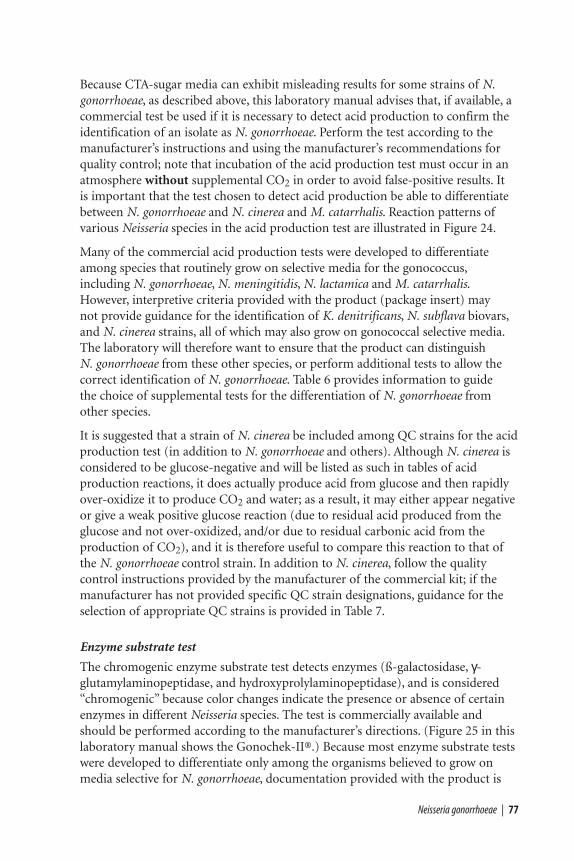
Because CTA-sugar media can exhibit misleading results for some strains of N.gonorrhoeae, as described above, this laboratory manual advises that, if available, acommercial test be used if it is necessary to detect acid production to confirm theidentification of an isolate as N. gonorrhoeae. Perform the test according to themanufacturer’s instructions and using the manufacturer’s recommendations forquality control; note that incubation of the acid production test must occur in anatmosphere without supplemental CO2 in order to avoid false-positive results. It is important that the test chosen to detect acid production be able to differentiatebetween N. gonorrhoeae and N. cinerea and M. catarrhalis. Reaction patterns ofvarious Neisseria species in the acid production test are illustrated in Figure 24.
Many of the commercial acid production tests were developed to differentiateamong species that routinely grow on selective media for the gonococcus,including N. gonorrhoeae, N. meningitidis, N. lactamica and M. catarrhalis.However, interpretive criteria provided with the product (package insert) may not provide guidance for the identification of K. denitrificans, N. subflava biovars,and N. cinerea strains, all of which may also grow on gonococcal selective media.The laboratory will therefore want to ensure that the product can distinguish N. gonorrhoeae from these other species, or perform additional tests to allow thecorrect identification of N. gonorrhoeae. Table 6 provides information to guide the choice of supplemental tests for the differentiation of N. gonorrhoeae fromother species.
It is suggested that a strain of N. cinerea be included among QC strains for the acidproduction test (in addition to N. gonorrhoeae and others). Although N. cinerea isconsidered to be glucose-negative and will be listed as such in tables of acidproduction reactions, it does actually produce acid from glucose and then rapidlyover-oxidize it to produce CO2 and water; as a result, it may either appear negativeor give a weak positive glucose reaction (due to residual acid produced from theglucose and not over-oxidized, and/or due to residual carbonic acid from theproduction of CO2), and it is therefore useful to compare this reaction to that ofthe N. gonorrhoeae control strain. In addition to N. cinerea, follow the qualitycontrol instructions provided by the manufacturer of the commercial kit; if themanufacturer has not provided specific QC strain designations, guidance for theselection of appropriate QC strains is provided in Table 7.
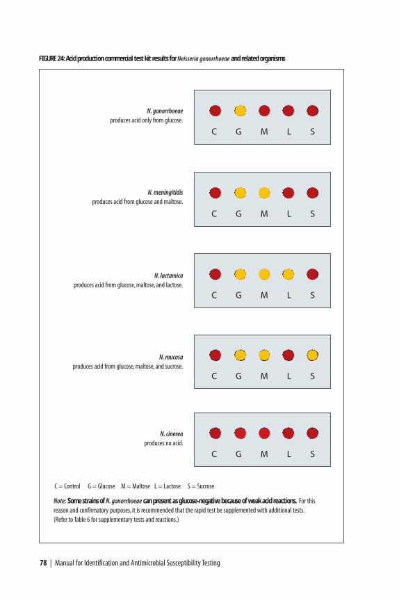
Enzyme substrate test
The chromogenic enzyme substrate test detects enzymes (ß-galactosidase, γ-glutamylaminopeptidase, and hydroxyprolylaminopeptidase), and is considered“chromogenic” because color changes indicate the presence or absence of certainenzymes in different Neisseria species. The test is commercially available andshould be performed according to the manufacturer’s directions. (Figure 25 in thislaboratory manual shows the Gonochek-II®.) Because most enzyme substrate testswere developed to differentiate only among the organisms believed to grow onmedia selective for N. gonorrhoeae, documentation provided with the product is
Neisseria gonorrhoeae | 77
78 | Manual for Identification and Antimicrobial Susceptibility Testing
FIGURE 24: Acid production commercial test kit results for Neisseria gonorrhoeae and related organisms
Note: Some strains of N. gonorrhoeae can present as glucose-negative because of weak acid reactions. For this reason and confirmatory purposes, it is recommended that the rapid test be supplemented with additional tests.(Refer to Table 6 for supplementary tests and reactions.)
N. gonorrhoeaeproduces acid only from glucose.
N. meningitidisproduces acid from glucose and maltose.
N. lactamicaproduces acid from glucose, maltose, and lactose.
N. mucosaproduces acid from glucose, maltose, and sucrose.
N. cinereaproduces no acid.
C G M L S
C G M L S
C G M L S
C G M L S
C G M L S
C = Control G = Glucose M = Maltose L = Lactose S = Sucrose

usually limited to distinguishing between N. gonorrhoeae (which produces onlyhydroxyprolylaminopeptidase), N. meningitidis (which produces γ-glutamyl-aminopeptidase), N. lactamica (which produces ß-galactosidase), and M. catarrhalis (which produces none of these three enzymes). It is now known thatstrains of several commensal Neisseria species can grow on selective GC media andalso produce only hydroxyprolylaminopeptidase. The chromogenic enzymesubstrate test is therefore not definitive for the identification of N. gonorrhoeae.Table 6 provides information to guide the choice of supplemental tests for thedifferentiation of N. gonorrhoeae from other species. Follow the quality controlinstructions provided by the manufacturer of the commercial kit; if themanufacturer has not provided specific QC strain designations, guidance for theselection of appropriate QC strains is provided in Table 7.
Nitrate reduction test
The nitrate reduction test is available commercially or can be made easily in thelaboratory. This test distinguishes between species that can reduce nitrate (NO3) tonitrite (NO2) or nitrogenous gases. In the context of this chapter, the test is usefulfor differentiating between strains of N. gonorrhoeae (nitrate-negative) and K.denitrificans or M. catarrhalis (two nitrate-positive species sometimes misidentifiedas N. gonorrhoeae).
The nitrate reduction test uses a medium containing nitrate and three differentreagents: sulfanilic acid (“Nitrate Reagent A”), α-naphthylamine (“Nitrate ReagentB”), and zinc powder (“Zn+2 dust”). Bacteria able to reduce nitrate from themedium into either nitrite or into nitrogenous gases are “nitrate-positive,” whilebacteria that lack enzymes to reduce nitrate are “nitrate-negative.”
In practical terms, the nitrate reduction test centers around the colorimetricdetection of nitrite in the test medium. Nitrite forms a compound with sulfanilicacid, which when reacted with α-naphthylamine gives a pink-to-red colordepending upon the concentration of nitrite in the medium; the addition ofNitrate Reagents A and B is therefore only able to detect the presence of nitrite inthe medium. If a pink-red color is detected after the addition of Nitrate Reagents A and B, the organism is considered to be “nitrate-positive.” However, if there is nocolor change in the medium after the addition of these reagents, it is necessary todetermine whether nitrate was ever reduced to nitrite, or whether the nitriteproduced was completely reduced to nitrogenous gases. This is accomplished byusing a small amount of zinc powder, which chemically catalyzes the reduction ofnitrate to nitrite and nitrite to nitrogenous gases. (It is therefore critical to use onlya very small amount of zinc powder so that if nitrate has not been reduced byenzymes produced by the bacteria, the reaction catalyzed by the zinc powder is notso strong as to reduce the nitrate completely to nitrogenous gases so rapidly that itis not possible to detect the nitrite produced in the catalytic reaction in themedium.) Nitrate-negative strains will exhibit a color change to red after
Neisseria gonorrhoeae | 79
incubation with zinc powder (nitrate is reduced to nitrite by the zinc powder, andthe nitrite is detected by Nitrate Reagents A and B already in the medium, yieldinga color change to pink-red). Nitrate-positive strains do not exhibit a color changeafter incubation with zinc powder because nitrate in the medium will have alreadybeen reduced beyond nitrite to nitrogenous gases. To summarize:
• Bacteria that reduce nitrate to nitrite may be identified when addition ofNitrate Reagents A and B causes the medium to change color from clear topink-red; no additional testing with zinc powder is required. Results should berecorded as “nitrate-positive.”
• Bacteria that reduce nitrate to nitrite and then further reduce the nitrite tonitrogenous gases are identified when there is no color change in the mediumafter either the addition of Nitrate Reagents A and B, or after incubation withzinc powder. Results should be recorded as “nitrate-positive.”
80 | Manual for Identification and Antimicrobial Susceptibility Testing
FIGURE 25: Reactions of Neisseria gonorrhoeae and related organisms in a commercial enzyme substrate test
The enzyme substrate test is a chromogenic test used to identify organisms that produce γ-glutamylaminopeptidase (indicated in this example of commercial test kit results as a color change to yellow), ß-galactosidase (indicated in thisexample as a color change to blue), hydroxyprolylaminopeptidase (indicated in this example as a color change to red-pink),and none of these enzymes (indicated by an absence of change of color in the cell suspension in the tube).
Test organism insuspension
Color change to yellowprior to exposure to substrate in red cap
No color changeoccurs prior to exposure to substrate in red cap:
Color change to blue prior to exposure to substrate in red cap
Organism produces γγ-glutamylaminopeptidase:N. meningitidisN. mucosaN. subflava biovar perflava
1) Remove white cap.2) Insert red cap
into tube.3) Invert tube to
expose the suspension to the substrates in the red cap.
Organism producesß-galactosidase:
N. lactamica
No colorchange
Color change to red-pink
Organism produces none of these three enzymes;suspension is colorless-to-yellow:M. catarrhalis
Organism produceshydroxyprolylamino-peptidase:N. gonorrhoeae K. denitrificansN. cinereaN. polysaccharea *N. flavescens *N. subflava biovars *N. sicca *N. mucosa *N. elongata *
(* Species which do not usually grow on selective media for N. gonorrhoeae but may do so occasionally and give a positivehydroxyprolylaminopeptidase reaction in an enzyme substrate test.)
• Bacteria unable to reduce nitrate at all are identified when there is no colorchange with the addition of Nitrate Reagents A and B, but there is a colorchange in the medium from clear to pink-red after incubation with zincpowder. Results should be recorded as “nitrate-negative.”
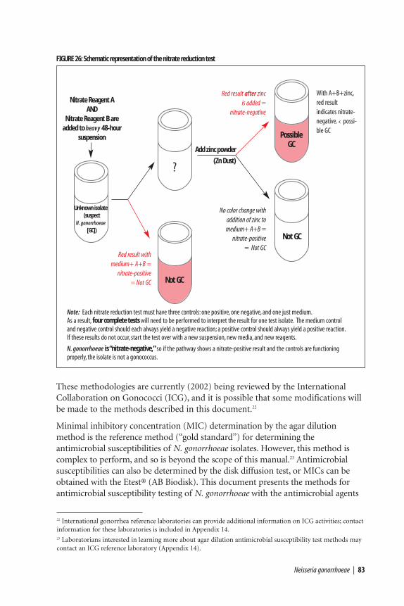
The nitrate test is performed in a standard nitrate broth which is inoculatedheavily to give a dense suspension of organisms because many Neisseria speciesmay not grow in this medium; the reaction for these species will therefore dependupon preformed enzymes in the inoculum. The test must be performed exactly asdescribed; if not performed correctly, the test results may be inaccurate and anincorrect identification made. A schematic representation of the nitrate reductiontest is shown in Figure 26. Media and reagents required for this test are describedin Appendix 2.
Nitrate reduction occurs only under anaerobic conditions; it is therefore importantto ensure a low surface-area to depth ratio to limit the diffusion of oxygen into themedium during the test. These conditions will be met by dispensing 5 ml ofmedium into a 13 mm diameter screw-cap tube.
It is important to run a medium control and both negative- and positive- controlsas the test is complex and the controls have known outcomes to indicate if themedia and reagents are reacting appropriately. Quality control tests should beperformed each time clinical isolates are tested, using QC strains included inTable 7.
Methods
a) Using colonies from a fresh, pure culture on GC-chocolate agar, prepare a heavysuspension in nitrate broth.
b) Remove the screw-cap top from the tube of nitrate test medium and inoculatethe medium to give heavy turbidity. Replace the screw-cap top.
c) Incubate the inoculated tubes and an uninoculated medium control tube at35˚–36.5˚C (without supplemental CO2) for 48 hours.
d) After incubation for 48 hours, remove the screw-cap top from the tube. Add 5drops of Nitrate Reagent A to each tube (including the uninoculated controlmedium). Shake each tube gently back and forth to mix Reagent A with themedium, add 5 drops of Nitrate Reagent B to each tube (again including theuninoculated control medium), and again shake each tube gently back andforth to mix Reagent B with the medium.
• If the uninoculated control medium turns pink-red, the test is invalid, anda new batch of media must be prepared.
• If the uninoculated control medium shows no color change, proceed to step e.
Neisseria gonorrhoeae | 81
e) Examine the test medium and controls for a pink-red color; this color shoulddevelop within a few minutes if the medium is still warm. The reaction maytake a little longer if the medium has cooled before the reagents are added.
• The negative control medium should show no color change.
• The positive control medium may or may not exhibit a color change to pink-red, depending upon whether nitrate was reduced to nitrite or furtherreduced to nitrogenous gases.
• If the test medium turns pink-red after the addition of Nitrate Reagents Aand B, the reaction is positive and the test is completed. If a pink-red colordevelops, do not perform step f and record the reaction as nitrate-positive.
f) If the medium is still colorless after the addition of Nitrate Reagents A and B,add a very small amount of zinc powder to the medium. (A convenient methodto estimate the amount of zinc powder required for the test is to use the sharppoint of a knife to pick up the powder; the pile of zinc powder should notexceed 4–5 mg, or 2–3 mm in diameter.) Shake the tube vigorously back andforth to mix well, and allow it to stand at room temperature for 10–15 minutes.
• If the negative control turns pink-red after the addition of zinc powder,the amount of zinc added is sufficient for the reaction to occur (and not somuch as to cause rapid over-reduction of nitrate to nitrogenous gases).Continue by interpreting the reactions in the test media.
• If the medium remains colorless after the addition of zinc powder, the testresult is positive (nitrate has been reduced to nitrite and further reduced tonitrogenous gases). Record the result for the isolate as “nitrate-positive.”
• If the medium turns pink-red after the addition of zinc powder, the result is negative. Record the result for the isolate as “nitrate-negative.”N. gonorrhoeae is nitrate-negative.
No identification of genus or species can be made on the basis of any of the abovebiochemical and enzymatic tests alone, but performing a combination (e.g., aspresented in Figure 21) can lead to a definitive identification of N. gonorrhoeae.
Antimicrobial susceptibility testing of N. gonorrhoeae
The methods presented in this laboratory manual are those recommended byNCCLS (an international, interdisciplinary, nonprofit, educational organizationthat develops updated consensus standards and guidelines for the healthcarecommunity on an annual basis),21 although a variety of methods are usedinternationally to determine antimicrobial susceptibilities of N. gonorrhoeae.
82 | Manual for Identification and Antimicrobial Susceptibility Testing
21 Formerly known as the “National Committee for Clinical Laboratory Standards,” NCCLS is now known solely by its acronym.
These methodologies are currently (2002) being reviewed by the InternationalCollaboration on Gonococci (ICG), and it is possible that some modifications willbe made to the methods described in this document.22
Minimal inhibitory concentration (MIC) determination by the agar dilutionmethod is the reference method (“gold standard”) for determining theantimicrobial susceptibilities of N. gonorrhoeae isolates. However, this method iscomplex to perform, and so is beyond the scope of this manual.23 Antimicrobialsusceptibilities can also be determined by the disk diffusion test, or MICs can beobtained with the Etest® (AB Biodisk). This document presents the methods forantimicrobial susceptibility testing of N. gonorrhoeae with the antimicrobial agents
Neisseria gonorrhoeae | 83
FIGURE 26: Schematic representation of the nitrate reduction test
Nitrate Reagent AAND
Nitrate Reagent B are added to heavy 48-hour
suspension
With A+B+zinc,red result indicates nitrate-negative.‹ possi-ble GC
Unknown isolate(suspect
N. gonorrhoeae[GC])
?
Red result withmedium+ A+B =
nitrate-positive= Not GC Not GC
PossibleGC
Red result after zinc is added =
nitrate-negative
Not GC
No color change withaddition of zinc tomedium+ A+B =
nitrate-positive= Not GC
Note: Each nitrate reduction test must have three controls: one positive, one negative, and one just medium.As a result, four complete tests will need to be performed to interpret the result for one test isolate. The medium controland negative control should each always yield a negative reaction; a positive control should always yield a positive reaction.If these results do not occur, start the test over with a new suspension, new media, and new reagents.
N. gonorrhoeae is “nitrate-negative,” so if the pathway shows a nitrate-positive result and the controls are functioningproperly, the isolate is not a gonococcus.
22 International gonorrhea reference laboratories can provide additional information on ICG activities; contactinformation for these laboratories is included in Appendix 14.23 Laboratorians interested in learning more about agar dilution antimicrobial susceptibility test methods maycontact an ICG reference laboratory (Appendix 14).
Add zinc powder
(Zn Dust)
currently recommended by WHO for the primary therapy of gonorrhea:ciprofloxacin, azithromycin, ceftriaxone, cefixime, and spectinomycin [WHO 2001].
Factors such as testing medium, inoculum size, incubation atmosphere, andantimicrobial disk concentrations may affect the antimicrobial susceptibility valuesobtained. Thus, quality control is of great importance and, with every test run,laboratory personnel must include reference strains with known antimicrobialsusceptibilities to ensure that the susceptibility results for test isolates are accurate.It should be noted that for methods that determine MICs, the MIC will beaccurate plus or minus (±) one dilution. For example, an organism with an MIC ofpenicillin of 0.25 µg/ml may exhibit an MIC of 0.125 µg/ml to 0.5 µg/ml, but itwould be found upon repeated testing that most antimicrobial susceptibility values(i.e., the modal MIC) for this organism and drug would be 0.25 µg/ml. Diskdiffusion results (i.e., inhibition zone diameters, mm) exhibit a similar normaldistribution upon repeated testing of the same isolates. It is important to keepthese variations of measurement in mind as laboratories typically perform onlyone complete set of antimicrobial susceptibility tests per isolate, and not repeatedmeasures for the same antimicrobial agent unless there is a specific reason to do so,such as confirming an unusual antimicrobial susceptibility result.
WHO has recommended a number of isolates for quality control (QC), althoughthese do not adequately represent the variety of resistance patterns now known toexist for N. gonorrhoeae. Consequently, most international laboratories haveincluded additional QC strains exhibiting resistance and intermediate resistance tofluoroquinolones and emerging resistance to azithromycin. Only one strain, N.gonorrhoeae ATCC 49226, is designated by NCCLS for QC of antimicrobialsusceptibility testing of gonococcal isolates. At the Centers for Disease Control andPrevention (CDC), the NCCLS-recommended QC strain and supplemental QCstrains are routinely made available to investigators (see Appendix 14 for contactinformation). Strains of N. gonorrhoeae are currently being tested under ICGsponsorship to establish an international reference panel for QC of antimicrobialsusceptibility testing that represents the known range of resistances in this species.
Once the susceptibility of a gonococcal strain to an antimicrobial agent has beenmeasured in vitro, the strain is then classified as susceptible, intermediate, orresistant to each antimicrobial agent tested to indicate whether the infection mayeither respond fail to respond to therapy with that agent. For clinical applications(i.e., prescribing appropriate therapy for individual patients), antimicrobialsusceptibilities are always interpreted strictly according to standardized guidelines,such as the NCCLS interpretive criteria. These criteria must be specific for the doseof the agent used to treat the infection [Knapp et al. 1995]. For example, NCCLScriteria for the interpretation of susceptibility of N. gonorrhoeae to ciprofloxacinwere developed to correspond to treatment with the recommended 500-mg ofciprofloxacin in a single oral dose; assessment of treatment efficacy of a single oraldose of 250-mg of ciprofloxacin would require the development of separateinterpretive criteria.
84 | Manual for Identification and Antimicrobial Susceptibility Testing
Organism-antimicrobial-dose interactions are categorized into one of two levels ofclassification based on the clinical efficacy of the antimicrobial agent. One levelapplies to antimicrobial agents to which an organism has not yet developedclinically significant resistance, and uses the categories “susceptible” and “decreasedsusceptibility.” The second level is used when the organism has developed clinicallysignificant resistance resulting in failure of the infection to respond to therapy withthe recommended dose of the antimicrobial agent (“treatment failures”), and usesthe categories “susceptible”, “intermediate,” and “resistant.” For example:
• At the time of writing (2002), gonococcal infections have not been confirmed tofail to respond to therapy with extended-spectrum cephalosporins, such ascefixime (400-mg in a single oral dose). The NCCLS has established aninterpretive criterion of “susceptible” as an MIC of ≤ 0.25 mg/ml of cefixime(corresponding disk diffusion zone of inhibition diameter with a 5-µg cefiximedisk, ≥ 31mm). Organisms with a higher MIC (or smaller inhibition zonediameter) are classified as exhibiting “decreased susceptibility” to cefixime.
• When infections fail to respond to recommended therapies with specificantimicrobial agents, a “resistant” category is established for that organism-antimicrobial-dose combination by NCCLS. Breakpoints are set for in vitrodetermination of this category based on testing of a variety of isolates resistantto the recommended therapeutic treatment. For example, gonococcal infectionscaused by organisms for which the ciprofloxacin MICs are ≥ 1.0 mg/ml(corresponding disk diffusion zone diameter of inhibition with a 5 mgciprofloxacin disk, ≤ 27mm) have failed to respond to therapy with the WHO-recommended single oral ciprofloxacin dose of 500-mg. NCCLS had previouslydefined the “susceptible” breakpoint for ciprofloxacin as an MIC of ≤ 0.06mg/ml (zone inhibition diameter ≥ 41 mm), so the “intermediate” designationapplies to those isolates for which the MICs are in the range between thesusceptible and resistant categories, i.e., 0.125 µg/ml – 0.5 µg/ml (28 mm – 40 mm). For gonococcal infections, it should be noted that organisms in the“intermediate” category for an antimicrobial agent have rarely been associatedwith confirmed instances of treatment failure with that agent.
NCCLS interpretive criteria are designed to define antimicrobial susceptibility testresult categories when NCCLS methodology is used to perform the tests, aspresented in this laboratory manual.24 The additional QC reference strainsincluded in this laboratory manual for antimicrobial agents not currently includedin NCCLS guidelines have been validated by the Gonorrhea Research Branch(Neisseria Reference Laboratory) at the CDC, and may be used alongside NCCLScriteria with the methods presented here until an ICG-sponsored international QC
Neisseria gonorrhoeae | 85
24 NCCLS methods are presented in this document, and are strongly recommended. However, if a laboratoryuses different antimicrobial susceptibility testing methodologies for N. gonorrhoeae, and all quality control references are consistently in check with the NCCLS interpretive criteria for QC strain ATCC 49226, the laboratory may consider interpreting results for the alternate testing methodology according to the NCCLSinterpretive criteria.
reference panel is designated. Tables 9 and 10 provide summaries of QC rangesand interpretive criteria for clinical isolates.25
In resource-limited geographic areas or in local clinical laboratories, antimicrobialsusceptibility test results should be determined for current antimicrobialtherapies and also the alternate antimicrobial agent(s) that would be used ifresistance emerged to the current regimen. Not all local laboratories will have thecapacity to perform antimicrobial susceptibility testing on isolates. National orlarge regional laboratories acting in the capacity of a reference laboratory shouldbe able not only to provide assistance to local laboratories and health authorities(clinical applications), but also to perform the most extensive susceptibility testingto a broad range of antimicrobial agents in order to compare susceptibilities ofisolates at the regional, national and international levels (surveillance activities).26
In a local laboratory, if it is not feasible to perform prospective surveillance, thelaboratory should at least determine susceptibilities of post-treatment “treatmentfailure” isolates which could either be truly resistant treatment failures or elsesusceptible isolates acquired by re-infection. If a laboratory is unable to performantimicrobial susceptibility testing, isolates should be sent to a laboratory that canperform such testing. (Methods for preservation and storage of isolates areincluded in Appendix 11; transport of isolates is addressed in Appendix 12.)
In addition to providing immediate assistance to local and regional laboratoriesand public health authorities in efforts to control gonorrhea by determiningantimicrobial susceptibilities to the recommended therapies, reference laboratoriesmay want to conduct more extensive antimicrobial susceptibility testing in order todevelop a global perspective on antimicrobial resistance in N. gonorrhoeae.Determination of antimicrobial susceptibilities to a wide range of agents—penicillin, tetracycline, spectinomycin, extended-spectrum cephalosporins (e.g.,ceftriaxone and cefixime), fluoroquinolones (e.g., ciprofloxacin, ofloxacin, andlevofloxacin), and the macrolide azithromycin—allows for the comparison ofisolates from the population served by the testing laboratory with isolates fromother regions.
Laboratory-based surveillance for antimicrobial resistance may be conducted atone of two basic levels. When resources are limited, surveillance may be performedfor susceptibilities to antimicrobial agents being used for primary and secondarytherapy of gonorrhea, i.e., the primary agent being used to treat infections and thealternative therapeutic agent(s) that would be used to treat infections not treatedeffectively by the primary regimen. In this instance, antimicrobial susceptibilities
86 | Manual for Identification and Antimicrobial Susceptibility Testing
25 If antimicrobial susceptibility test QC results for a locally developed testing method are consistent but do notagree with those obtained by NCCLS-recommended methods, the testing laboratory may want to consult withthe ICG for assistance with the development of standard interpretive criteria appropriate to the situation.26 Laboratorians interested in learning more about the methods used for the surveillance of antimicrobialresistance in N. gonorrhoeae isolates can find links to various protocols through the following internet address:http://www.cdc.gov/ncidod/dastlr/gcdir/gono.html
would be interpreted by the standards used for clinical applications, e.g., byNCCLS standards.
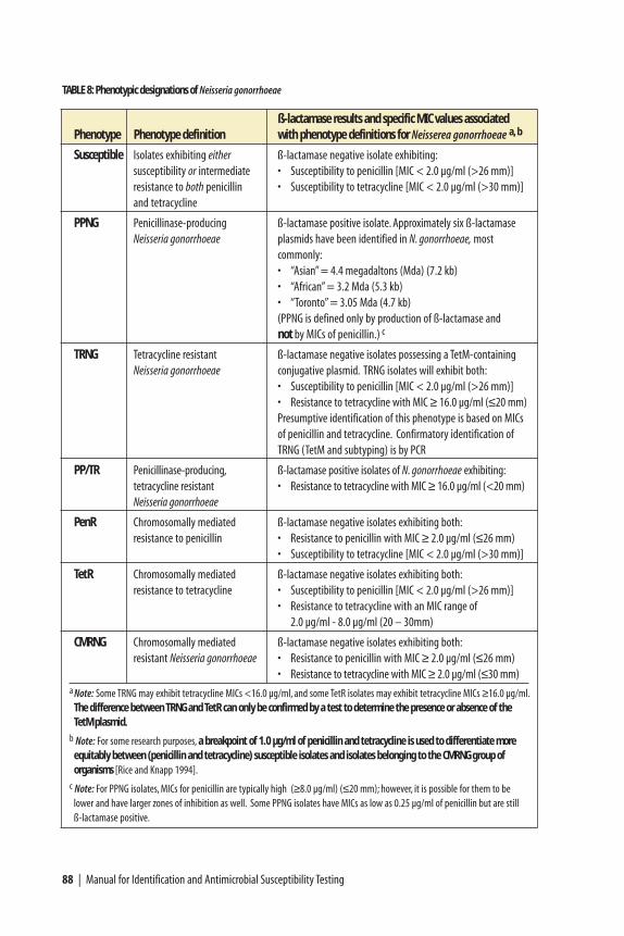
When investigators wish to compare the antimicrobial susceptibilities of N.gonorrhoeae strains in their geographic locality with those in other geographicareas, susceptibilities are usually determined to a larger number of antimicrobialagents than those used locally for treatment. A typical panel might include thefollowing: penicillin, tetracycline, spectinomycin, an extended-spectrumcephalosporin (e.g., ceftriaxone or cefixime), a fluoroquinolone (e.g., ciprofloxacin,ofloxacin, or levofloxacin), and a macrolide (e.g., azithromycin). For broadsurveillance purposes, gonococcal isolates are described first by theirsusceptibilities to penicillin and tetracycline (although these drugs should not beused treat gonorrhea) and by a simple test to detect the production of ß-lactamase(described below). This is because, based on the level of resistance to penicillin andtetracycline and the detection of ß-lactamase, it is possible to predict whether themechanisms of resistance to penicillin and tetracycline are chromosomallymediated or plasmid-mediated.
A specialized classification and terminology with standard acronyms has beendeveloped to describe patterns of penicillin-tetracycline resistance and designatepenicillin-tetracycline resistance phenotypes, as presented in Table 8. Organismsthat are ß-lactamase-negative and resistant to penicillin but not tetracycline, forexample, use the NCCLS designation “penicillin-resistant,” and are designated“PenR.” Other acronyms do not use NCCLS designations in their names, althoughNCCLS methods are used to identify the resistances. For example, “CMRNG”(chromosomally mediated resistant N. gonorrhoeae) describes organisms that havechromosomally mediated resistance to both penicillin (MIC ≥ 2.0 mg/ml, orequivalent inhibition zone diameter ≤ 26 mm) and tetracycline (MIC ≥ 2.0 mg/ml,or equivalent inhibition zone diameter ≤ 30 mm) and do not produce ß-lactamase.It should be noted that while plasmid-mediated resistance to penicillin can bedetected and confirmed with a simple test to detect ß-lactamase, plasmid-mediatedresistance to tetracycline can only be identified presumptively with susceptibilityresults and must be confirmed with a complex test demonstrating the presence ofthe TetM-conjugative plasmid (e.g., by laboratories performing molecularepidemiologic testing).
The basic penicillin-tetracycline resistance phenotype acronym characterizessusceptibilities only of penicillin and tetracycline. For other therapeutic agents,NCCLS (or equivalent) standardized criteria are used to classify susceptibilities tothese agents, and antimicrobial resistance (including “intermediate” or “decreasedsusceptibility” categories) to these additional antimicrobial agents is appended tothe penicillin-tetracycline resistance phenotype. For example, a CMRNG isolateexhibiting resistance to ciprofloxacin (CipR) would be cited as “CMRNG, CipR.”Such descriptive designations permit one to rapidly appreciate the fact thatciprofloxacin resistance is occurring in an organism already resistant to penicillin
Neisseria gonorrhoeae | 87
88 | Manual for Identification and Antimicrobial Susceptibility Testing
ß-lactamase results and specific MIC values associated Phenotype Phenotype definition with phenotype definitions for Neisserea gonorrhoeae a, b
Susceptible Isolates exhibiting either ß-lactamase negative isolate exhibiting:susceptibility or intermediate • Susceptibility to penicillin [MIC < 2.0 µg/ml (>26 mm)]resistance to both penicillin • Susceptibility to tetracycline [MIC < 2.0 µg/ml (>30 mm)]and tetracycline
PPNG Penicillinase-producing ß-lactamase positive isolate. Approximately six ß-lactamaseNeisseria gonorrhoeae plasmids have been identified in N. gonorrhoeae, most
commonly:• “Asian” = 4.4 megadaltons (Mda) (7.2 kb)• “African” = 3.2 Mda (5.3 kb)• “Toronto” = 3.05 Mda (4.7 kb)(PPNG is defined only by production of ß-lactamase and not by MICs of penicillin.) c
TRNG Tetracycline resistant ß-lactamase negative isolates possessing a TetM-containingNeisseria gonorrhoeae conjugative plasmid. TRNG isolates will exhibit both:
• Susceptibility to penicillin [MIC < 2.0 µg/ml (>26 mm)]• Resistance to tetracycline with MIC ≥ 16.0 µg/ml (≤20 mm)Presumptive identification of this phenotype is based on MICs of penicillin and tetracycline. Confirmatory identification of TRNG (TetM and subtyping) is by PCR
PP/TR Penicillinase-producing, ß-lactamase positive isolates of N. gonorrhoeae exhibiting:tetracycline resistant • Resistance to tetracycline with MIC ≥ 16.0 µg/ml (<20 mm)Neisseria gonorrhoeae
PenR Chromosomally mediated ß-lactamase negative isolates exhibiting both:resistance to penicillin • Resistance to penicillin with MIC ≥ 2.0 µg/ml (≤26 mm)
• Susceptibility to tetracycline [MIC < 2.0 µg/ml (>30 mm)]
TetR Chromosomally mediated ß-lactamase negative isolates exhibiting both:resistance to tetracycline • Susceptibility to penicillin [MIC < 2.0 µg/ml (>26 mm)]
• Resistance to tetracycline with an MIC range of 2.0 µg/ml - 8.0 µg/ml (20 – 30mm)
CMRNG Chromosomally mediated ß-lactamase negative isolates exhibiting both:resistant Neisseria gonorrhoeae • Resistance to penicillin with MIC ≥ 2.0 µg/ml (≤26 mm)
• Resistance to tetracycline with MIC ≥ 2.0 µg/ml (≤30 mm)a Note: Some TRNG may exhibit tetracycline MICs <16.0 µg/ml, and some TetR isolates may exhibit tetracycline MICs ≥16.0 µg/ml.
The difference between TRNG and TetR can only be confirmed by a test to determine the presence or absence of the TetM plasmid.
b Note: For some research purposes, a breakpoint of 1.0 µg/ml of penicillin and tetracycline is used to differentiate more equitably between (penicillin and tetracycline) susceptible isolates and isolates belonging to the CMRNG group of organisms [Rice and Knapp 1994].
c Note: For PPNG isolates, MICs for penicillin are typically high (≥8.0 µg/ml) (≤20 mm); however, it is possible for them to be lower and have larger zones of inhibition as well. Some PPNG isolates have MICs as low as 0.25 µg/ml of penicillin but are still ß-lactamase positive.
TABLE 8: Phenotypic designations of Neisseria gonorrhoeae
and tetracycline. The use of penicillin-tetracycline resistance phenotypes also haspractical applications for monitoring susceptibilities to the extended-spectrumcephalosporins: gonococcal isolates exhibiting chromosomally mediated, highlevels of resistance to penicillin (PenR) or penicillin and tetracycline (CMRNG)exhibit higher—but still susceptible—MICs of ceftriaxone and cefixime.
Aggregation and analysis of phenotypic data permit investigators to monitorchanges in the prevalence of resistant strain populations and their geographicpatterns of spread, and these surveillance tools may be used to help anticipate theneed to revise treatment recommendations before resistance becomes endemic in aregion and undermines the effectiveness of local gonorrhea control measures.
Further characterization of resistant strains
An area of research in which reference laboratories may be interested inparticipating is the further subtype characterization27 of isolates exhibiting thesame antimicrobial resistance phenotypes. Subtyping methods are resource-intensive, however, and so it is not expected that every reference laboratory will beable to adopt these techniques. Genotypic and phenotypic subtyping characterizesindividual strains and facilitates a refined interpretation of the antimicrobialresistance data. By assigning strain subtype designations, investigators may be ableto differentiate between the strain types which are sporadically imported andcoincidentally exhibit the same resistance phenotype as a local strain. Strainsubtyping coupled with information about social-sexual networks may facilitateproactive disease control interventions.
Methods for detecting antimicrobial resistance in N. gonorrhoeae
As detailed above, there are two different approaches taken when defining for whatantimicrobial agents susceptibility tests should be performed. When performingantimicrobial susceptibility testing for clinical purposes, susceptibilities should bedetermined to the antimicrobial agents currently used for treatment of gonorrheaand the alternate antimicrobial agent(s) that would be prescribed if the primarycourse were to be ineffective. When performing antimicrobial susceptibility testingfor surveillance purposes, however, the clinical testing is supplemented with anexpanded panel of antimicrobial agents in conjunction with ß-lactamase testing,providing the laboratory with phenotypic data appropriate for internationalcomparisons.
Tests identifying gonococcal strains that produce ß-lactamase are used inconjunction with MICs as an integral component of surveillance to differentiatebetween chromosomally mediated and plasmid-mediated resistance to penicillin for
Neisseria gonorrhoeae | 89
27 Examples of phenotypic typing include auxotyping (determination of nutrients required for growth of astrain), serotyping, ß-lactamase plasmid typing, and TetM plasmid typing. Examples of genotypic typinginclude Lip subtyping, RFLP-related typing, and Opa typing.
N. gonorrhoeae, as explained above. The nitrocefin test is a qualitative test used todetect production of ß-lactamase; it can be performed using the same culture onGC-chocolate agar used to prepare the inoculum for MIC (or disk diffusion) tests.
Test for ß-lactamase production by N. gonorrhoeae
The most reliable way to detect ß-lactamase-producing strains of N. gonorrhoeae isto use the nitrocefin test. Reactions are strongest when the test is performed oncultures recently removed from an incubator and still warm. The nitrocefin test isperformed either with a liquid reagent or with a treated disk. Because the liquidreagent can be expensive, the disk method is preferable if relatively few isolates areto be tested. Positive and negative controls should be run each time this test isperformed. Positive and negative control strains may be selected from among thoselisted in Table 7.
Nitrocefin disk method
a) Use sterile forceps or tweezers to place a nitrocefin disk on a clean slide.
b) Add a drop of distilled water to the disk and allow it to absorb so the disk ismoistened, but not wet.
c) Touch a sterile swab or loop to a characteristic colony in fresh, pure, 18–24 hourculture.
d) Rub the swab on the moistened disk so that the growth goes into the filterpaper of the disk.
e) Examine the disk: if the reaction is positive, the areas of the disk containinggrowth will turn a characteristic red/pink color. Reactions typically occur withinfive minutes.
f) Record results. Strains for which the inoculum on the nitrocefin disk turns red/ pink are considered “ß-lactamase positive”. Strains for which the inoculum onthe nitrocefin disk does not change color are considered “ß-lactamase negative.”
Nitrocefin liquid reagent
If it is anticipated that a large number of isolates will be tested, laboratoriansshould investigate obtaining nitrocefin powder and preparing the liquid reagent.The nitrocefin test using liquid reagent is performed either by dropping reagentdirectly on colonies growing on selective or nonselective media, or by diluting thereagent and using it as a suspension medium for bacterial growth in a tube.Although the former method is easier as it involves fewer steps, the advantage ofthe latter method is that it uses lesser amounts of the costly liquid reagent.(Methods for the different preparations of the nitrocefin reagent as used for eachof these tests are included in Appendix 2.)
To perform the test for ß-lactamase production with liquid nitrocefin reagentusing the plate method, use an eyedropper, Pasteur pipette or inoculating loop to
90 | Manual for Identification and Antimicrobial Susceptibility Testing
place a drop of the undiluted reagent directly onto fresh gonococcal coloniesgrowing on selective or nonselective culture media. After several minutes, thecolonies will turn pink if the gonococcal strain is producing ß-lactamase, andshould be recorded as “ß-lactamase positive.” If, after ten minutes, no color changehas occurred on the colonies dropped with reagent, the gonococcal strain isconsidered “ß-lactamase negative,” and should be recorded as such.
To perform the test for ß-lactamase production with liquid nitrocefin reagentusing the tube method, dispense dilute nitrocefin solution (25 mg/L prepared in0.1M phosphate buffer) into a test tube, and use it to prepare a heavy suspension(~ McFarland 2) of the suspect gonococcal colonies from 18–24 hour culture. Ifß-lactamase producing organisms are present, the suspension should change colorfrom colorless/yellow to pink within 15 seconds; record a strain exhibiting thiscolor change as ‘ß-lactamase positive.’ If after five minutes no color change hasoccurred in the suspension, record the strain as ‘ß-lactamase negative.’
Results of ß-lactamase tests are used in conjunction with results of antimicrobialsusceptibility tests performed according to NCCLS methodologies.
Antimicrobial susceptibility testing of N. gonorrhoeae using NCCLS methodologies
Antimicrobial susceptibility testing by both disk diffusion and the antimicrobialgradient strip Etest® method are conducted on the same standardized medium.Because gonococci are fastidious, antimicrobial susceptibility tests for mostantimicrobial agents are performed on a GC agar base medium supplemented withIsoVitaleX or an equivalent supplement. Mueller-Hinton medium, on whichsusceptibilities of most aerobic bacteria are determined, is not suitable fordetermining gonococcal susceptibilities; however, Mueller-Hinton broth can beused to prepare the gonococcal cell suspensions that will be tested. Furthermore,gonococcal susceptibilities should not be determined on media containingchocolatized blood or hemoglobin because of the variability of blood products(which may affect susceptibilities of N. gonorrhoeae to various antimicrobialagents). Antimicrobial susceptibility test results for N. gonorrhoeae should only beinterpreted when tested on GC-susceptibility test medium, a standard qualitycontrolled GC agar base medium plus 1% defined supplement.
A sample form for recording the results of antimicrobial susceptibility tests for N. gonorrhoeae is included in Figure 27.
Antimicrobial susceptibility testing of N. gonorrhoeae by disk diffusion
Disk diffusion testing should be carried out as defined by the NCCLS performancestandards and with the NCCLS quality control strain N. gonorrhoeae ATCC 49226.It is recommended that laboratories obtain additional gonococcal reference strainsexhibiting resistance patterns not exhibited by ATCC 49226: supplemental QCstrains, tested routinely by disk diffusion and agar dilution methods with
Neisseria gonorrhoeae | 91
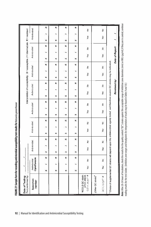
92 | Manual for Identification and Antimicrobial Susceptibility Testing
FIGU
RE27
:Sam
ple
form
for r
ecor
ding
ant
imicr
obia
l sus
cept
ibili
ty te
st re
sults
for N
eiss
eria
gon
orrh
oeae
Note
:Afte
r 20-
24 h
ours
of in
cuba
tion,
chec
k the
resu
lts fo
r the
qua
lity c
ontro
l (“Q
C”) s
train
s aga
inst
the a
ccep
tabl
e ran
ge o
f inh
ibiti
on zo
ne d
iam
eter
s (m
m) o
r MIC
s (µg
/ml);
if th
ey ar
e in
cont
rol,c
ontin
uere
adin
g re
sults
for t
he te
st iso
late.
(Inhi
bitio
n zo
ne ra
nges
and b
reak
poin
ts fo
r int
erpr
etat
ion
of re
sults
may
be fo
und i
n Tab
les 9
and 1
0.)
consistent results, may be obtained from the Neisseria Reference Laboratory,Gonorrhea Research Branch, CDC (see Appendix 14). Quality control values fordisk diffusion zone diameter sizes for these strains are presented in Table 9.
Methods
a) Label one plate of GC-chocolate agar for each clinical isolate and QC strain tobe tested.
b) Inoculate plates with each test strain and streak for isolation. Incubateinoculated plates at 35˚–36.5˚C in a CO2-supplemented atmosphere withincreased humidity for 16–20 hours.
• Note: if isolates are maintained in culture prior to inoculation forantimicrobial susceptibility testing, they must be subcultured every 24 hoursprior to being tested.
• Note: if isolates are stored frozen prior to inoculation for antimicrobialsusceptibility testing, they must be subcultured at least once after initialculture from the frozen preparation prior to being tested.
c) Suspend isolated colonies (from the overnight cultures prepared in steps a andb) in 1.0–2.0 ml of Mueller-Hinton broth (or PBS). Mix the suspensionthoroughly on a vortex mixer to break up clumps of growth as much aspossible.
• It is easier to prepare the suspensions with a swab28 than with an inoculatingloop. The best method to avoid excessive clumping of growth in thesuspension is to roll the swab over the colonies rather than to use a scrapingmethod to harvest cells.
d) Adjust the turbidity of the cell suspension to the turbidity of a 0.5 McFarlandstandard by comparing tubes against black and white lines and adding broth orculture as needed (see Figures 51and 52 in Appendix 2). The suspension mustbe used to inoculate the plate within 15–20 minutes after preparation, or elseit must be discarded and a new suspension prepared.
• Note: The inoculation step must be completed within 15–20 minutesbecause the organisms will begin to die within a short time after thesuspension is prepared, and even though the suspension will be visuallycomparable to the McFarland standard, the viability of the inoculumdelivered onto the test medium may be too low to produce reliableantimicrobial susceptibility test results.
• If there are many cultures to test, they should be done in small batches (e.g.,five or six isolates at a time) to avoid loss of viability.
Neisseria gonorrhoeae | 93
28 Notes on the survival of N. gonorrhoeae with different swab materials are included in Table 29, Appendix 8.
e) Pour 60 ml of GC base medium containing 1% defined supplement into a 150-mm diameter plate to a uniform depth of 3–4 mm (in order to assure properconditions for disk diffusion results). The number of plates required for thetesting of each strain will be dependent upon the number and type ofantimicrobial agents to be tested, as some have larger inhibition zone sizes thanothers and the zones of inhibition must not overlap. Generally speaking, GCsusceptibility tests have no more than 3 disks on each plate.
Plates to be used for antimicrobial susceptibility testing must have warmed toroom temperature before they are inoculated with the cell suspension. Thesurface of the plate must also be dry prior to inoculation; if not, invert theplates and dry them with lids slightly open either in an incubator at 35˚–36.5˚C,or in a biohazard hood. There should be no visible drops of moisture on thesurface of the agar when the plates are inoculated.
f) Moisten a sterile applicator swab28 in the standardized cell suspension andremove any excess moisture by rotating the swab against the glass above theliquid in the tube. Inoculate the entire surface of each plate three times, rotatingthe plate 60˚ each time to ensure confluent growth (Figure 34).
g) Store the inoculated plates at room temperature for 3–5 minutes to allow themedium to absorb the moisture from the inoculum. It is essential that thesurface of the medium is dry before the antimicrobial disks are applied. Platesmay be dried in an incubator or biohazard hood as described in step e. (If ittakes longer than 15 minutes for the inoculum to dry, use a smaller volume /express more suspension from the swab in the future.)
h) Using sterile forceps, tweezers or a disk dispenser, apply disks of the selectedantimicrobial agents to the surface of the inoculated medium; tap them toensure they are in complete contact with the surface of the agar. Once a diskhas touched the agar surface, diffusion begins and it must not be moved.All disks should be placed approximately the same distance from the edge ofthe plate and from each other (Figure 28).
i) Cover and invert the inoculated plates and incubate them at 35˚–36.5˚C in a3%–5% CO2 atmosphere (in a CO2-incubator or candle-extinction jar) for20–24 hours.
j) At 20–24 hours after inoculation and incubation, read the results of theantimicrobial susceptibility tests.
• Examine the plates from the back, viewed from the top down against a blackbackground and illuminated with reflected light (so hazy growth is moreeasily seen). Measure the diameter of the zone of inhibition with calipers, aruler, or a ruler on a stick (see Figure 6).
• Read the results for ATCC 49226 and compare them to the values in Table 9;if they are in control, continue reading and comparing results for the other
94 | Manual for Identification and Antimicrobial Susceptibility Testing
QC strains tested. If these are also in control, continue to read and recordresults for the clinical isolates.
k) Interpret the results. Table 10 presents zone inhibition diameters and equivalentminimal inhibitory concentrations (MICs) for test strains, along with theNCCLS standard interpretations of those zones diameters as sensitive,intermediate, or resistant.
After interpreting results, report them back to the primary laboratory.
Antimicrobial susceptibility testing of N. gonorrhoeae by Etest® antimicrobialgradient strip
Antimicrobial susceptibility testing with the Etest® antimicrobial gradient strip29 istechnically as simple to perform as the disk diffusion test, but provides semi-quantitative MIC results. The strip is impregnated with a standard gradient ofantimicrobial agent, and the front of the strip has MIC values that are to be read incorrespondence with inhibition of growth on the plate after incubation. Alwaysread the insert in the package of Etest® strips, and follow the manufacturer’sinstructions for performance of the test.
Antimicrobial susceptibility testing of N. gonorrhoeae is performed on GC basemedium plus 1% defined growth supplement; methods for the preparation andQC of this medium are included in Appendix 2 (“Media, Reagents and QualityControl”). The standardization of the inoculum and methods for the inoculationof the test plate are the same for the Etest® as they are for the disk diffusion test forN. gonorrhoeae; follow steps a through g above, and then continue with step h,below. Strict quality control practices are of extreme importance in order for theproper performance and appropriate interpretation of the antimicrobialsusceptibility test. If conditions cannot be controlled and standardized, it is betterthat the laboratory not perform the antimicrobial susceptibility test at all, becausethe results obtained cannot be interpreted according standardized criteria.Inaccurate results are useless to the clinician, can even cause harm to a case-patient, and should not be recorded for use in public health policy treatmentdecision-making.
Laboratorians should ensure that the Etest® strips used for antimicrobialsusceptibility testing of N. gonorrhoeae strains cover the appropriate range ofantibiotic concentrations for these organisms.30
Neisseria gonorrhoeae | 95
29 The Etest® can be expensive; contact the manufacturer (AB BIODISK) to inquire about discounts availablefor laboratories in resource-poor settings (see Appendix 13).30 Please note that for certain antimicrobial agents (particularly some ß-lactams), the Etest® is available in ahigh- and low range of concentrations. For testing of N. gonorrhoeae with the ceftriaxone Etest®, for example,it is recommended that laboratories use the low-range concentration (0.002 µg/ml – 32 µg/ml) rather than thehigh-range concentration (0.016 µg/ml – 256 µg/ml). A complete list of strips and ranges of concentrations isavailable from AB Biodisk (at http://www.abbiodisk.se/productsservice/product.htm ).
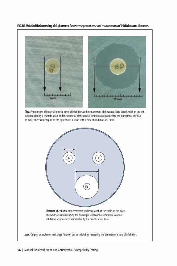
96 | Manual for Identification and Antimicrobial Susceptibility Testing
FIGURE 28: Disk diffusion testing: disk placement for Neisseria gonorrhoeae and measurements of inhibition zone diameters
Top: Photographs of bacterial growth, zones of inhibition, and measurement of the zones. Note that the disk on the leftis surrounded by a resistant strain and the diameter of the zone of inhibition is equivalent to the diameter of the disk (6 mm), whereas the figure on the right shows a strain with a zone of inhibition of 17 mm.
Te P
Cip
Bottom: The shaded area represents uniform growth of the strain on the plate;the white areas surrounding the disks represent zones of inhibition. Zones ofinhibition are measured as indicated by the double-arrow lines.
Note: Calipers or a ruler on a stick (see Figure 6) can be helpful for measuring the diameter of a zone of inhibition.
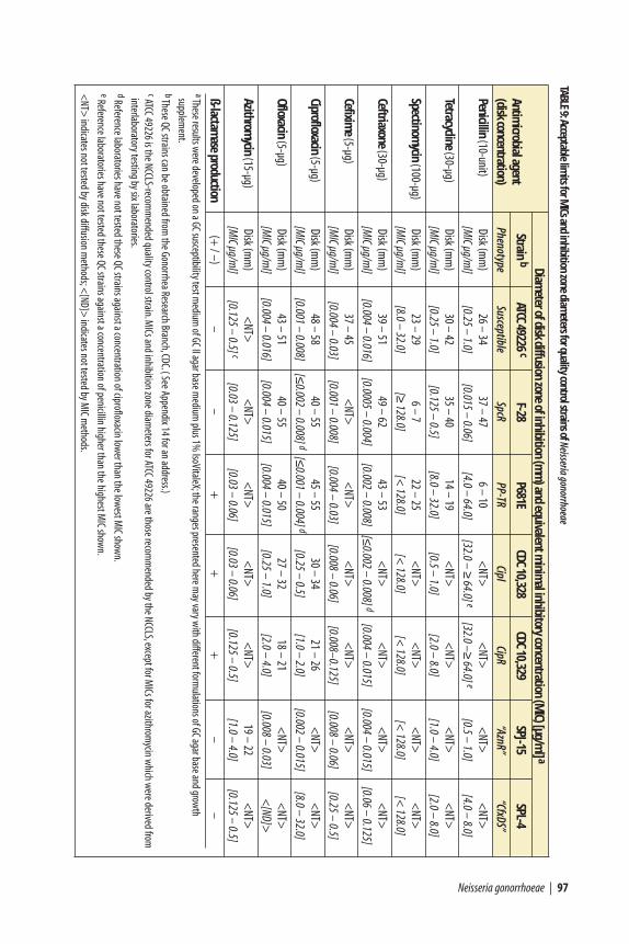
Neisseria gonorrhoeae | 97
Diameter of disk diffusion zone of inhibition (m
m) and equivalent m
inimal inhibitory concentration (M
IC) [µg/ml] a
Antimicrobial agent
Strain bATCC 49226 c
F-28P681E
CDC 10,328CDC 10,329
SPJ-15SPL-4
(disk concentration)Phenotype
SusceptibleSpcR
PP-TRCipI
CipR“AznR”
“CfxDS”
Penicillin(10-unit)
Disk (mm
)26 – 34
37 – 476 – 10
<NT>
<NT>
<NT>
<NT>
[MIC µg/m
l][0.25 – 1.0]
[0.015 – 0.06] [4.0 – 64.0]
[32.0 – ≥64.0] e
[32.0 –≥64.0] e
[0.5 – 1.0] [4.0 – 8.0]
Tetracycline(30-µg)
Disk (mm
)30 – 42
35 – 40 14 – 19
<NT>
<NT>
<NT>
<NT>
[MIC µg/m
l][0.25 – 1.0]
[0.125 – 0.5][8.0 – 32.0]
[0.5 – 1.0][2.0 – 8.0]
[1.0 – 4.0][2.0 – 8.0]
Spectinomycin
(100-µg)Disk (m
m)
23 – 29 6 – 7
22 – 25<
NT><
NT><
NT><
NT>[M
IC µg/ml]
[8.0 – 32.0][≥
128.0] [<
128.0][<
128.0][<
128.0][<
128.0][<
128.0]
Ceftriaxone(30-µg)
Disk (mm
)39 – 51
49 – 6243 – 53
<NT>
<NT>
<NT>
<NT>
[MIC µg/m
l][0.004 – 0.016]
[0.0005 – 0.004][0.002 – 0.008]
[≤0.002 – 0.008] d
[0.004 – 0.015][0.004 – 0.015]
[0.06 – 0.125]
Cefixime
(5-µg)Disk (m
m)
37 – 45<
NT><
NT><
NT><
NT><
NT><
NT>[M
IC µg/ml]
[0.004 – 0.03][0.001 – 0.008]
[0.004 – 0.03][0.008 – 0.06]
[0.008–0.125][0.008 – 0.06]
[0.25 – 0.5]
Ciprofloxacin(5-µg)
Disk (mm
)48 – 58
40 – 5545 – 55
30 – 3421 – 26
<NT>
<NT>
[MIC µg/m
l][0.001 – 0.008]
[≤0.002 – 0.008] d
[≤0.001 – 0.004] d
[0.25 – 0.5][1.0 – 2.0]
[0.002 – 0.015] [8.0 – 32.0]
Ofloxacin(5-µg)
Disk (mm
)43 – 51
40 – 5540 – 50
27 – 3218 – 21
<NT>
<NT>
[MIC µg/m
l][0.004 – 0.016]
[0.004 – 0.015][0.004 – 0.015]
[0.25 – 1.0][2.0 – 4.0]
[0.008 – 0.03]<
[ND]>
Azithromycin
(15-µg)Disk (m
m)
<NT>
<NT>
<NT>
<NT>
<NT>
19 – 22<
NT>[M
IC µg/ml]
[0.125 – 0.5] c[0.03 – 0.125]
[0.03 – 0.06][0.03 – 0.06]
[0.125 – 0.5][1.0 – 4.0]
[0.125 – 0.5]
ß-lactamase
production(+
/ –)–
–+
++
––
aThese results were developed on a GC susceptibility test m
edium of GC II agar base m
edium plus 1%
IsoVitaleX;the ranges presented here may vary w
ith different formulations of GC agar base and grow
th supplem
ent.bThese QC strains can be obtained from
the Gonorrhea Research Branch,CDC.( See Appendix 14 for an address.)cATCC 49226 is the NCCLS-recom
mended quality control strain.M
ICs and inhibition zone diameters for ATCC 49226 are those recom
mended by the NCCLS,except for M
ICs for azithromycin w
hich were derived from
interlaboratory testing by six laboratories.dReference laboratories have not tested these QC strains against a concentration of ciprofloxacin low
er than the lowest M
IC shown.
eReference laboratories have not tested these QC strains against a concentration of penicillin higher than the highest MIC show
n.
<NT>
indicates not tested by disk diffusion methods;<
[ND]> indicates not tested by M
IC methods.
TABLE9:Acceptable lim
its for MICs and inhibition zone diam
eters for quality control strains of Neisseria gonorrhoeae
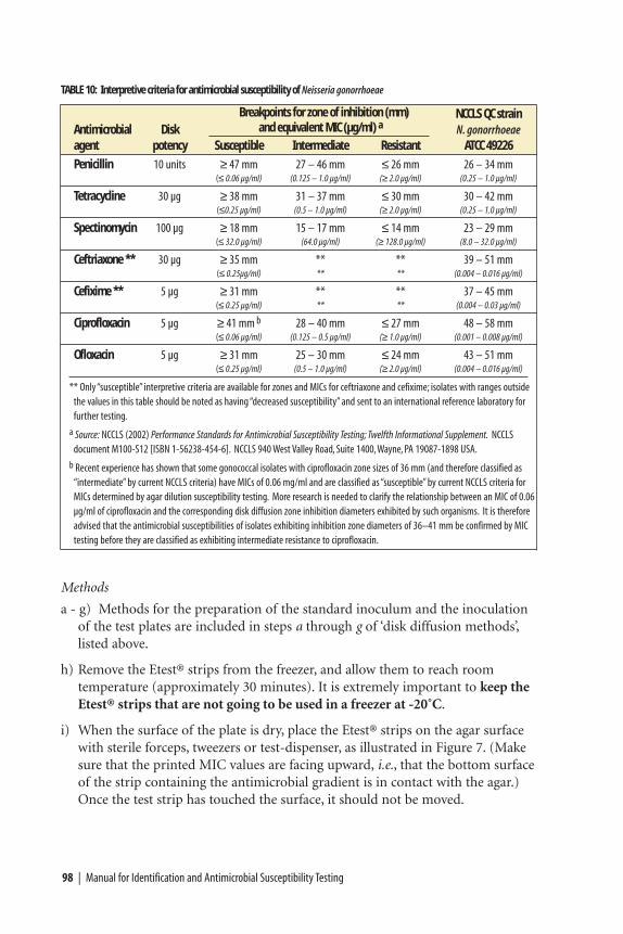
Methods
a - g) Methods for the preparation of the standard inoculum and the inoculationof the test plates are included in steps a through g of ‘disk diffusion methods’,listed above.
h) Remove the Etest® strips from the freezer, and allow them to reach roomtemperature (approximately 30 minutes). It is extremely important to keep theEtest® strips that are not going to be used in a freezer at -20˚C.
i) When the surface of the plate is dry, place the Etest® strips on the agar surfacewith sterile forceps, tweezers or test-dispenser, as illustrated in Figure 7. (Makesure that the printed MIC values are facing upward, i.e., that the bottom surfaceof the strip containing the antimicrobial gradient is in contact with the agar.)Once the test strip has touched the surface, it should not be moved.
98 | Manual for Identification and Antimicrobial Susceptibility Testing
TABLE 10: Interpretive criteria for antimicrobial susceptibility of Neisseria gonorrhoeae
Breakpoints for zone of inhibition (mm) NCCLS QC strainAntimicrobial Disk and equivalent MIC (µg/ml) a N. gonorrhoeaeagent potency Susceptible Intermediate Resistant ATCC 49226
Penicillin 10 units ≥ 47 mm 27 – 46 mm ≤ 26 mm 26 – 34 mm(≤ 0.06 µg/ml) (0.125 – 1.0 µg/ml) (≥ 2.0 µg/ml) (0.25 – 1.0 µg/ml)
Tetracycline 30 µg ≥ 38 mm 31 – 37 mm ≤ 30 mm 30 – 42 mm(≤0.25 µg/ml) (0.5 – 1.0 µg/ml) (≥ 2.0 µg/ml) (0.25 – 1.0 µg/ml)
Spectinomycin 100 µg ≥ 18 mm 15 – 17 mm ≤ 14 mm 23 – 29 mm(≤ 32.0 µg/ml) (64.0 µg/ml) (≥ 128.0 µg/ml) (8.0 – 32.0 µg/ml)
Ceftriaxone ** 30 µg ≥ 35 mm ** ** 39 – 51 mm(≤ 0.25µg/ml) ** ** (0.004 – 0.016 µg/ml)
Cefixime ** 5 µg ≥ 31 mm ** ** 37 – 45 mm(≤ 0.25 µg/ml) ** ** (0.004 – 0.03 µg/ml)
Ciprofloxacin 5 µg ≥ 41 mm b 28 – 40 mm ≤ 27 mm 48 – 58 mm(≤ 0.06 µg/ml) (0.125 – 0.5 µg/ml) (≥ 1.0 µg/ml) (0.001 – 0.008 µg/ml)
Ofloxacin 5 µg ≥ 31 mm 25 – 30 mm ≤ 24 mm 43 – 51 mm(≤ 0.25 µg/ml) (0.5 – 1.0 µg/ml) (≥ 2.0 µg/ml) (0.004 – 0.016 µg/ml)
** Only “susceptible” interpretive criteria are available for zones and MICs for ceftriaxone and cefixime; isolates with ranges outsidethe values in this table should be noted as having “decreased susceptibility” and sent to an international reference laboratory forfurther testing.
a Source: NCCLS (2002) Performance Standards for Antimicrobial Susceptibility Testing; Twelfth Informational Supplement. NCCLS document M100-S12 [ISBN 1-56238-454-6]. NCCLS 940 West Valley Road, Suite 1400,Wayne, PA 19087-1898 USA.
b Recent experience has shown that some gonococcal isolates with ciprofloxacin zone sizes of 36 mm (and therefore classified as “intermediate” by current NCCLS criteria) have MICs of 0.06 mg/ml and are classified as “susceptible” by current NCCLS criteria forMICs determined by agar dilution susceptibility testing. More research is needed to clarify the relationship between an MIC of 0.06µg/ml of ciprofloxacin and the corresponding disk diffusion zone inhibition diameters exhibited by such organisms. It is thereforeadvised that the antimicrobial susceptibilities of isolates exhibiting inhibition zone diameters of 36–41 mm be confirmed by MICtesting before they are classified as exhibiting intermediate resistance to ciprofloxacin.
• Although the manufacturer’s insert for the Etest® says that up to two stripscan be used on a 100-mm plate and up to six on a 150-mm plate, becausegonococci can have such wide zones of inhibition, this laboratory manualadvises using only one Etest® strip per 100 mm plate for N. gonorrhoeae.The number of strips on a 150-mm plate will be determined by thecombination of drugs being tested; zones of inhibition must not overlap.(Once laboratorians have determined the range of susceptibilities of localgonococcal isolates to various antimicrobial agents with the Etest® on 100-mm plates, they can assess which combinations of antimicrobial agents canbe tested on a 150-mm plate without overlapping zones of inhibition,usually 3 or 4 antimicrobial agents.)
j) Incubate the inoculated Etest® plate according to the manufacturer’sinstructions (usually 20–24 hours at 35˚–36.5˚C in a 5% CO2 atmosphere).
k) After incubation for 20–24 hours, there will be an ellipse of inhibition ofbacterial growth on the plate around the Etest® strip, and the MIC values canbe read (see Figure 8).
• The graduation marks on the Etest® strip correspond to the standardconcentrations for the agar dilution method, but also include incrementsbetween those standard values. The standard values (see Table 27 inAppendix 7) are used for interpretation and reporting of antimicrobialsusceptibility test results. It is advised that both the actual reading of thevalue from the strip and the next-higher standard value (i.e., the value to beused for interpretation) be included in the laboratory records for testing ofthe strain. For example, if testing susceptibility of a gonococcal isolate tociprofloxacin, an MIC recorded from the graduations on the Etest® stripmight be 0.094 µg/ml; however, the reported MIC would be 0.125 µg/ml andthe organism would be interpreted as exhibiting intermediate resistance tociprofloxacin.
l) Read the results for ATCC 49226 and compare them to the values in Table 9; ifthey are in control, continue reading and comparing results for the other QCstrains tested. If these are also in control, continue to read and record results forthe clinical isolates. It is essential to review the MIC results of the qualitycontrol strains prior to interpreting the MICs of the clinical isolates.
m)Read and interpret the results for the test strains. Table 10 presents the NCCLSinterpretive criteria (susceptible, intermediate, resistant) for differentantimicrobials, including those currently recommended for the primary therapyof uncomplicated gonorrhea.
A reading guide for interpretation of Etest® antimicrobial susceptibility results andguidance in reading MICs from the Etest® strip is presented in Figure 8. The guide,included with the permission of AB Biodisk, shows how growth appears aroundthe strip and provides guidance for how the test should be interpreted.
Neisseria gonorrhoeae | 99
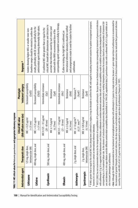
100 | Manual for Identification and Antimicrobial Susceptibility Testing
TABL
E11
:MIC
criti
cal v
alue
s for
Nei
sser
ia g
onor
rhoe
aean
d ap
prop
riate
labo
rato
ry re
spon
se
Criti
cal M
IC va
lue
Phen
otyp
ic An
timicr
obia
l age
ntTh
erap
eutic
dos
e(d
isk d
iffus
ion
zone
size
)‘re
sista
nce’
cate
gory
Resp
onse
a
Ceft
riaxo
ne12
5 m
g,sin
gle d
ose,
MIC
> 0
.25
µg/m
lDe
creas
ed su
scep
tibili
tyin
tram
uscu
lar (
IM)
(<35
mm
)(C
roDS
)
Cefix
ime
400
mg,
singl
e dos
e,or
alM
IC >
0.2
5 µg
/ml
Decre
ased
susc
eptib
ility
(<
31 m
m)
(Cfx
DS)
MIC
0.1
25 –
0.5
µg/
ml
Inte
rmed
iate
resis
tanc
e
Cipr
oflo
xacin
500
mg,
singl
e dos
e,or
al(2
8 - 4
0 m
m)
(Cip
I)
MIC
≥1.
0 µg
/ml
Resis
tanc
e(≤
27m
m)
(Cip
R)
MIC
0.5
– 1
.0 µ
g/m
lIn
term
edia
te re
sista
nce
Oflo
xacin
400
mg,
singl
e dos
e,or
al(2
5 –
30 m
m)
(Ofx
I)
MIC
≥2.
0 µg
/ml
Resis
tanc
e(≤
24 m
m)
(Ofx
R)
Azith
rom
ycin
2 g,
singl
e dos
e,or
alM
IC ≥
1.0
µg/m
l b‘R
esist
ance
’b
(≤25
mm
) b(‘A
znR’
)
Spec
tinom
ycin
2 g,
singl
e dos
e,M
IC ≥
128.
0 µg
/ml
Resis
tanc
ein
tram
uscu
lar (
IM)
(≤14
mm
)(S
pcR)
aIf
MIC
s gre
ater
than
or e
qual
to th
e crit
ical M
IC h
ave a
lread
y bee
n co
nfirm
ed in
a re
gion
,it m
ay b
e des
irabl
e to
conf
irm th
e MIC
with
rega
rd to
eval
uatin
g tre
atm
ent o
utco
me f
or p
atie
nt m
anag
emen
t pur
pose
s;ho
wev
er,it
is n
ot n
eces
sary
to re
-con
tact
an in
tern
atio
nal r
efer
ence
labo
rato
ry.
b NC
CLS [
2002
] has
not
reco
mm
ende
d cr
iteria
for t
he in
terp
reta
tion
of su
scep
tibili
ties o
f N.g
onor
rhoe
aeto
azith
rom
ycin
.Bec
ause
ther
e are
limite
d ou
tcom
e dat
a cor
resp
ondi
ng to
trea
tmen
t of g
onoc
occa
l infe
ctio
nsw
ith az
ithro
myc
in (2
g),
it is
sugg
este
d th
at la
bora
toria
ns an
d cli
nicia
ns co
llabo
rate
to co
rrelat
e tre
atm
ent o
utco
me d
ata w
ith in
vitro
susc
eptib
ility
resu
lts to
esta
blish
an in
terp
retiv
e bre
akpo
int f
or re
sista
nce
corre
spon
ding
to a
5% tr
eatm
ent f
ailu
re ra
te.I
n th
e abs
ence
of c
onclu
sive c
linica
l out
com
e dat
a,th
is do
cum
ent p
rese
nts a
‘wor
king’
inte
rpre
tive c
riter
ion
for r
esist
ance
bas
ed o
n a l
imite
d nu
mbe
r of t
reat
men
t fai
l-ur
es an
d la
bora
tory
in vi
troag
ar d
ilutio
n an
d di
sk d
iffus
ion
antim
icrob
ial s
usce
ptib
ility
dat
a [Ha
ndsfi
eld
et al
.199
4;CD
C,un
publ
ished
dat
a]:N
.gon
orrh
oeae
strai
ns w
ith a
conf
irmed
MIC
of ≥
1.0
µg/m
l exh
ibit
an in
vitro
‘resis
tant
’phe
noty
pe to
azith
rom
ycin
.
- Not
e:Al
thou
gh th
e WHO
-reco
mm
ende
d do
se fo
r tre
atm
ent o
f unc
ompl
icate
d go
norrh
ea w
ith az
ithro
myc
in is
2 g
ram
s in
a sin
gle o
ral d
ose,
beca
use a
1-g
ram
sing
le o
ral d
ose o
f azit
hrom
ycin
is re
com
men
ded
for
the t
reat
men
t of g
enita
l Chl
amyd
iatra
chom
atis
infe
ctio
ns,t
his d
ose m
ay b
e use
d in
ciden
tally
to tr
eat g
onoc
occa
l co-
infe
ctio
ns.E
valu
atio
n of
clin
ical t
reat
men
t out
com
es h
as in
dica
ted
that
gon
ococ
cal i
nfec
tions
caus
ed b
y stra
ins w
ith M
ICs o
f ≥0.
125
µg/m
l may
fail
to re
spon
d to
trea
tmen
t with
the 1
-gra
m d
ose o
f azit
hrom
ycin
[You
nget
al.
1997
].
Isola
tes s
how
ing
high
er M
ICs o
r sm
alle
r zon
e size
sth
anth
ese r
ange
s sho
uld
be re
teste
d to
conf
irm th
ere
sults
,pre
fera
bly w
ith Q
C st
rain
s spe
cifica
lly fo
r the
antim
icrob
ial a
gent
show
ing
abno
rmal
ly hi
gh va
lues
.
A co
nfirm
ed M
IC va
lue g
reat
er th
an o
r equ
al to
the
criti
cal M
IC sh
ould
aler
t lab
orat
oria
ns an
d cli
nicia
ns to
asce
rtain
that
infe
ctio
ns ca
used
by s
uch
stra
ins (
and
treat
ed w
ith th
e WHO
-reco
mm
ende
d do
se o
f the
co
rresp
ondi
ng ag
ent)
resp
onde
d su
cces
sfully
to th
erap
y.
If,af
ter r
e-te
stin
g,th
e hig
h M
IC is
conf
irmed
,an
inte
rnat
iona
l ref
eren
ce la
bora
tory
shou
ld b
e not
ified
and
arra
ngem
ents
mad
e to
send
the i
sola
te fo
r fur
ther
conf
irmat
ion.
Neisseria gonorrhoeae | 101
When reading any antimicrobial susceptibility test results for N. gonorrhoeae, thelaboratorian should be aware of critical values that indicate a need for retesting.Table 11 presents a listing of critical antimicrobial susceptibility test values for thelaboratory to be aware of. If the MIC results for an organism are higher than thoselisted for the specific antimicrobial agent, the reference laboratory should re-testthe isolate. If the results are still atypical, confirm the identification of theorganism, ensure the test is being performed properly, and then re-test the isolateagain. If it still produces a high MIC, notify the national and an internationalreference laboratory and send the isolate for further investigation. Instructions forthe preservation and storage of isolates are presented in Appendix 11, whileAppendix 12 includes the instructions for regulation-compliant packaging andshipping of isolates.
When the susceptibility value is confirmed upon re-test, the confirmatorylaboratory should notify the submitting laboratory and then other laboratories inthe regional and international network. If the isolate represents a newantimicrobial resistance phenotype, it is important that the confirming referencelaboratory disseminate preserved cultures of the isolate to other referencelaboratories for inclusion among susceptibility quality control strains. Isolatesshowing a previously undescribed resistance pattern should not be used forscientific research (such as the determination of the resistance mechanisms)without permission from the originating clinician and/or laboratory.
Data for decision-making
Antimicrobial susceptibility testing can be performed on an isolate presumptivelyidentified as N. gonorrhoeae, although confirmatory testing should be completedbefore antimicrobial susceptibility test results exceeding the critical MIC values arereported. (For example, before reporting results, laboratories should confirm theidentification of an organism showing an unexpectedly high MIC to, e.g.,ceftriaxone.) Once the laboratory has determined the antimicrobial susceptibilities,the information should be reported back to public health officials in a timelymanner. Factors to consider in the development of a treatment policy include:
• Laboratories should screen and report values for antimicrobial agents currentlyin use for primary therapy of gonorrhea in the region and, ideally, also for thesecond line drugs.
• MIC “critical values” can be useful tools to initiate (enhanced) surveillance andepidemiological investigations to determine if there is an association betweenthe in vitro susceptibility of a strain and the clinical outcome.
• Extended-spectrum cephalosporins, fluoroquinolones, and spectinomycin arerecognized as the most effective antimicrobial agents for the treatment ofgonorrhea in most geographic areas of the world.
• The antimicrobial agent and dose chosen should be effective against at least95% of local gonococcal strains.
• The antimicrobial agent chosen should be affordable.
• The antimicrobial agent chosen should be readily available.
• It should be possible to store the chosen antimicrobial agent under conditions(e.g., refrigeration) that will maintain the activity of the drug.
It is important to consider the above factors when making decisions relating totreatment of gonorrhea. Determination of antimicrobial susceptibilities totherapeutic agents will help public health officials review the appropriateness oftreatment recommendations for local populations, and surveillance ofantimicrobial susceptibilities will promote effective disease control.
102 | Manual for Identification and Antimicrobial Susceptibility Testing

Salmonella serotype TyphiShigella
Vibrio cholerae
Bacterial Agents ofEnteric Diseases of
Public Health Concern
Bacterial Agents of Enteric Diseases ofPublic Health Concern
Salmonella serotype Typhi | 103
S almonella serotype Typhi (S. Typhi), the etiologic agent of typhoid fever,causes an estimated 16.6 million cases and 600,000 deaths worldwide eachyear. A syndrome similar to typhoid fever is caused by “paratyphoidal”
serotypes of Salmonella. The paratyphoid serotypes (i.e., S. Paratyphi A, S.Paratyphi B, and S. Paratyphi C) are isolated much less frequently than S. Typhi.Rarely, other serotypes of Salmonella, such as S. Enteritidis, can also cause “entericfever.” Like other enteric pathogens, S. Typhi is transmitted through food or waterthat has been contaminated with feces from either acutely infected persons,persistent excretors, or from chronic asymptomatic carriers. Humans are the onlyhost for S. Typhi; there are no environmental reservoirs.
Effective antimicrobial therapy reduces morbidity and mortality from typhoidfever. Without therapy, the illness may last for 3–4 weeks and case-fatality ratesmay exceed 10%. With appropriate treatment, clinical symptoms subside within afew days, fever recedes within 5 days, and mortality is reduced to approximately1%. Relapses, characterized by a less severe but otherwise typical illness, occur in10%–20% of patients with typhoid fever, usually after an afebrile period of 1–2weeks. Relapses may still occur despite antimicrobial therapy.
S. Typhi is most frequently isolated from blood during the first week of illness, butit can also be present during the second and third weeks of illness, during the firstweek of antimicrobial therapy, and during clinical relapse. Fecal cultures arepositive in approximately half the cases during the first week of fever, but thelargest number of positive cultures occurs during the second and third weeks ofdisease. Bone marrow cultures are frequently positive (90% of cases) and are morelikely to yield S. Typhi than are cultures from any other site, especially when thepatient has already received antimicrobial therapy. Organisms can also be isolatedfrom duodenal aspirates, rose spots, and infrequently (i.e., in approximately 25%of cases) from urine cultures.
Salmonella serotype TyphiIDENTIFICATION AND ANTIMICROBIAL SUSCEPTIBILITY TESTING
CHAPTER VII
104 | Manual for Identification and Antimicrobial Susceptibility Testing
In typhoid fever, serologic responses to O, H, and Vi antigens usually occur by theend of the first week of illness. The Widal test, which measures antibody responses to H and O antigens, can suggest the diagnosis, but the results are not definitive andmust be interpreted with care because titers also may be elevated in response to anumber of other infections. High-titer, single serum specimens from adults living inareas of endemic disease have little diagnostic value. Even when paired sera are used, the results must be interpreted in light of the patient’s history of typhoidimmunization and previous illness, the stage of the illness when the first serumspecimen was obtained, the use of early antimicrobial therapy, and the reagents used.
There are currently (2002) at least two effective vaccines available for typhoid,both of which were recently licensed for use in the United States. The oral liveattenuated vaccine (for use in children aged 6 years and older) and the parenteral(i.e., injectable) capsular polysaccharide vaccine (for use in children aged 2 yearsand older) each have efficacy of 50%–80% and fewer adverse events associated with their use than earlier typhoid vaccines. A team doing research in Vietnamreported promising preliminary success of a new conjugate vaccine in early 2001.The two U.S.-licensed vaccines have been widely and effectively used by travelers to typhoid-endemic regions, though the expense and limited experience with theiruse as a public health intervention in countries with high endemic rates of typhoidfever precludes the widespread use of these vaccines in countries with limitedresources. Nonetheless, it is good policy for laboratory technicians who may beworking with this organism to supplement their laboratory safety practices andensure that their vaccination status against typhoid fever remains current.
In developing countries, typhoid fever is frequently diagnosed solely on clinicalgrounds; however, isolation of the causative organism is necessary for a definitivediagnosis. Isolation of the agent is also a necessity for the performance ofantimicrobial susceptibility testing.
Resistance to the antimicrobial agents amoxicillin, trimethoprim-sulfamethoxazole,and chloramphenicol is being increasingly reported among S. Typhi isolates;quinolone resistance has been reported from the Indian subcontinent andSoutheast Asia. Determining antimicrobial resistance patterns is essential inrecommending treatment. In areas where resistance to these agents is commonamong circulating S. Typhi strains, fluoroquinolones and parenteral third-generation cephalosporins are probably the best choice for empiric treatment oftyphoid fever. Cefixime may be recommended in some cases as a less expensive,oral alternative to parenteral ceftriaxone.
Identification of S. Typhi
A preliminary report of typhoid can be issued to a clinician as soon as apresumptive identification of S. Typhi is obtained. Methods for the isolation of
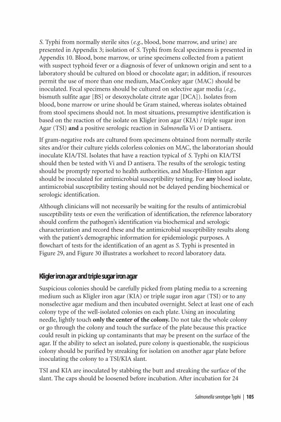
Salmonella serotype Typhi | 105
S. Typhi from normally sterile sites (e.g., blood, bone marrow, and urine) arepresented in Appendix 3; isolation of S. Typhi from fecal specimens is presented inAppendix 10. Blood, bone marrow, or urine specimens collected from a patientwith suspect typhoid fever or a diagnosis of fever of unknown origin and sent to alaboratory should be cultured on blood or chocolate agar; in addition, if resourcespermit the use of more than one medium, MacConkey agar (MAC) should beinoculated. Fecal specimens should be cultured on selective agar media (e.g.,bismuth sulfite agar [BS] or desoxycholate citrate agar [DCA]). Isolates fromblood, bone marrow or urine should be Gram stained, whereas isolates obtainedfrom stool specimens should not. In most situations, presumptive identification isbased on the reaction of the isolate on Kligler iron agar (KIA) / triple sugar ironAgar (TSI) and a positive serologic reaction in Salmonella Vi or D antisera.
If gram-negative rods are cultured from specimens obtained from normally sterilesites and/or their culture yields colorless colonies on MAC, the laboratorian shouldinoculate KIA/TSI. Isolates that have a reaction typical of S. Typhi on KIA/TSIshould then be tested with Vi and D antisera. The results of the serologic testingshould be promptly reported to health authorities, and Mueller-Hinton agarshould be inoculated for antimicrobial susceptibility testing. For any blood isolate,antimicrobial susceptibility testing should not be delayed pending biochemical orserologic identification.
Although clinicians will not necessarily be waiting for the results of antimicrobialsusceptibility tests or even the verification of identification, the reference laboratoryshould confirm the pathogen’s identification via biochemical and serologiccharacterization and record these and the antimicrobial susceptibility results alongwith the patient’s demographic information for epidemiologic purposes. Aflowchart of tests for the identification of an agent as S. Typhi is presented in Figure 29, and Figure 30 illustrates a worksheet to record laboratory data.
Kligler iron agar and triple sugar iron agar
Suspicious colonies should be carefully picked from plating media to a screeningmedium such as Kligler iron agar (KIA) or triple sugar iron agar (TSI) or to anynonselective agar medium and then incubated overnight. Select at least one of eachcolony type of the well-isolated colonies on each plate. Using an inoculatingneedle, lightly touch only the center of the colony. Do not take the whole colonyor go through the colony and touch the surface of the plate because this practicecould result in picking up contaminants that may be present on the surface of theagar. If the ability to select an isolated, pure colony is questionable, the suspiciouscolony should be purified by streaking for isolation on another agar plate beforeinoculating the colony to a TSI/KIA slant.
TSI and KIA are inoculated by stabbing the butt and streaking the surface of theslant. The caps should be loosened before incubation. After incubation for 24
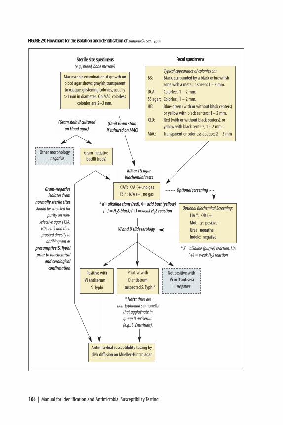
KIA or TSI agar biochemical tests
106 | Manual for Identification and Antimicrobial Susceptibility Testing
Positive with
D antiserum
= suspected S. Typhi*
FIGURE 29: Flowchart for the isolation and identification of Salmonella ser.Typhi
Macroscopic examination of growth on blood agar shows grayish, transparentto opaque, glistening colonies, usually>1 mm in diameter. On MAC, colorless
colonies are 2–3 mm.
Typical appearance of colonies on:
BS: Black, surrounded by a black or brownishzone with a metallic sheen; 1 – 3 mm.
DCA: Colorless; 1 – 2 mm.
SS agar: Colorless; 1 – 2 mm.
HE: Blue-green (with or without black centers)or yellow with black centers; 1 – 2 mm.
XLD: Red (with or without black centers), or yellow with black centers; 1 – 2 mm.
MAC: Transparent or colorless opaque; 2 – 3 mm
Fecal specimens Sterile site specimens(e.g., blood, bone marrow)
Antimicrobial susceptibility testing by
disk diffusion on Mueller-Hinton agar
KIA*: K/A (+), no gas
TSI*: K/A (+), no gas
Not positive withVi or D antisera
= negative
(Gram stain if cultured on blood agar)
Other morphology = negative
Gram-negative bacilli (rods)
(Omit Gram stainif cultured on MAC)
Optional screening
Optional Biochemical Screening:
LIA *: K/K (+)
Motility: positive
Urea: negative
Indole: negative
* K= alkaline slant (red); A= acid butt (yellow)[+] = H2S black; (+) = weak H2S reaction
Gram-negative isolates from
normally sterile sitesshould be streaked for
purity on non-selective agar (TSA,
HIA, etc.) and then proceed directly to
antibiogram as presumptive S. Typhiprior to biochemical
and serological confirmation
Vi and D slide serology
* K= alkaline (purple) reaction, LIA(+) = weak H2S reaction
Positive with
Vi antiserum =
S. Typhi
* Note: there are non-typhoidal Salmonella
that agglutinate in group D antiserum (e.g., S. Enteritidis).
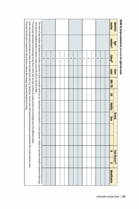
Salmonella serotype Typhi | 107
FIGURE30:Sam
ple worksheet for Salm
onellaser.Typhi test results
Specimen
Agar Gram
OPTIONAL
SLIDESEROLOGY d
number
medium
aColony
bstain
cKIA / TSI
LIAM
otilityUrea
ViD
Identification
123123123123123aThe choice of agar m
edium w
ill depend on whether a specim
en is from a norm
ally sterile site (e.g.,blood,bone marrow,urine) or is a fecal specim
en.Sterile site specimens should be cultured on blood agar;
fecal specimens should be plated on selective m
edia (such as BS,DCA,SS,HE,XLD,or MAC).
bIf macroscopic exam
ination of morphology reveals there is m
ore than one type of colony on a plate,perform tests to identify each of the different isolates.
cGram stain should only be perform
ed on isolates from norm
ally sterile sites (e.g.,blood,bone marrow,urine) and should not be perform
ed on growth from
MAC or other selective m
edia.dIf prom
pt identification is required for clinical decision-making,slide serology m
ay precede biochemical testing.
hours at 35˚–37˚C, the TSI or KIA slants are observed for reactions typical ofSalmonella. On TSI or KIA slants, S. Typhi characteristically produces an alkalineslant (red, “K”), an acid butt (yellow, “A”), and a small amount of blackening ofthe agar (H2S, +) at the site of the stab on the slant and in the stab line (Figure31); no gas (G) is produced. It is worth noting that occasionally S. Typhi isolatesdo not produce H2S. S. Paratyphi A isolates on TSI/KIA are usually K/AG and donot produce H2S. Most other Salmonella serotypes produce a K/AG+ reaction,indicating that glucose is fermented with gas and H2S production. Table 12summarizes the reactions of Salmonella on screening biochemicals.
Additional screening biochemicals for the identification of S. Typhi
Isolates can be identified biochemically as Salmonella by traditional tubed mediaor commercial biochemical systems. Table 12 lists biochemical reactions of thetests that are helpful screening tests for S. Typhi. After performing the tests, readand record the results, then compare them to results for presumptive S. Typhi. Ifthey match, then proceed by confirming with serologic testing if it has not alreadybeen performed.
Lysine iron agar
Lysine iron agar (LIA) is a useful screening medium because most Salmonellaisolates decarboxylate lysine and produce H2S, whereas gas production varies byserotype. Preparation and quality control (QC) of this medium are described inAppendix 2. Inoculate LIA by stabbing the butt and streaking the surface of theslant; read and interpret the reactions after incubation for 24 hours at 35˚–37˚C for24 hours.
On LIA, Salmonella typically give an alkaline (purple) reaction on the slant andbutt and may produce gas and H2S (blackening of medium) as well, as indicated inTable 12. When the reaction in the butt of the tube is alkaline, the lysine isdecarboxylated and the isolate is termed “lysine-positive.” Unlike most otherSalmonella, S. Paratyphi A isolates are lysine-negative and appear yellow on LIA.
If a diagnosis of infection with S. Typhi is suspected and prompt diagnosis isrequired to identify appropriate treatment, suspect isolates should be screened withantisera prior to biochemical identification. However, in the setting of a publichealth study, because slide serology can be performed using growth from KIA, TSI,or LIA, performing serology after those tests and saving antiserum for only thoseisolates showing biochemical characteristics typical of S. Typhi is more cost-effective.
Motility agar
Motility agar should be inoculated with a straight inoculating needle, making asingle stab about 1–2 cm down into the medium. The surface of the motility agar
108 | Manual for Identification and Antimicrobial Susceptibility Testing
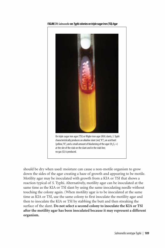
Salmonella serotype Typhi | 109
FIGURE 31: Salmonella ser. Typhi colonies on triple sugar iron (TSI) Agar
On triple sugar iron agar (TSI) or Kligler iron agar (KIA) slants, S. Typhicharacteristically produces an alkaline slant (red,“K”), an acid butt(yellow,“A”), and a small amount of blackening of the agar (H2S, +)at the site of the stab on the slant and in the stab line;no gas (G) is produced.
should be dry when used: moisture can cause a non-motile organism to growdown the sides of the agar creating a haze of growth and appearing to be motile.Motility agar may be inoculated with growth from a KIA or TSI that shows areaction typical of S. Typhi. Alternatively, motility agar can be inoculated at thesame time as the KIA or TSI slant by using the same inoculating needle withouttouching the colony again. (When motility agar is to be inoculated at the sametime as KIA or TSI, use the same colony to first inoculate the motility agar andthen to inoculate the KIA or TSI by stabbing the butt and then streaking thesurface of the slant. Do not select a second colony to inoculate the KIA or TSIafter the motility agar has been inoculated because it may represent a differentorganism.

Examine after overnight incubation at 35°–37°C. Motility is indicated by thepresence of diffuse growth (appearing as clouding of the medium) away from theline of inoculation (Figure 39). Non-motile organisms do not grow out from theline of inoculation. Motility reactions may be difficult for inexperiencedlaboratorians to read; therefore, reactions should be compared with positive andnegative control strains. S. Typhi is usually motile (+ 97%).
Sulfide-indole-motility medium is a combination medium that is commerciallyavailable in dehydrated form (see Appendix 2, “Media, Reagents, and QualityControl”). It can be used in place of motility medium.
Urea medium
Urea medium screens out urease-producing organisms (e.g., Klebsiella andProteus). Urea agar is inoculated heavily over the entire surface of the slant. Loosencaps before incubating overnight at 35°–37°C. Urease-positive cultures producean alkaline reaction in the medium, evidenced by a pinkish-red color (Figure40). Urease-negative organisms do not change the color of the medium, which is apale yellowish-pink. S. Typhi is always urease negative.
Slide serology for S. Typhi identification
TSI/KIA cultures that are suspicious for S. Typhi should be screened serologicallywith Salmonella Vi and group D “O” antisera. Because Vi is a capsular antigen, if itis present, it may mask the somatic “O” group reaction. Therefore, S. Typhi isolateswill usually either be positive in the Vi or the D antisera (though it is possible they
110 | Manual for Identification and Antimicrobial Susceptibility Testing
TABLE 12. Typical reactions of Salmonella spp. in screening biochemicals
Nontyphoidal Salmonella orScreening medium Salmonella Typhi Salmonella Paratyphi A Salmonella Paratyphi B or C
Triple sugar iron agar (TSI) K/A(+)a K/AG-a K/AG+a
Kligler iron agar (KIA) K/A(+)a K/AG-a K/AG+a
Lysine iron agar (LIA) K/K(+)b K/AG-b K/K+b
Hydrogen sulfide (H2S) (weak)c negative positive
Urea negative negative negative
Motility positivec positive positive
Indole negative negative negativea for KIA / TSI: K = alkaline (red); A = acid (yellow); G = gas production; + = black H2S produced (weak); – = no H2S b for LIA: K= alkaline (purple); A= acid (yellow); G = gas production; + = black H2S produced (weak); – = no H2S
~ An alkaline reaction (purple) in the butt of the medium indicates that lysine was decarboxylated.~ An acid reaction (yellow) in the butt of the medium indicates that lysine was not decarboxylated.
c this reaction occurs 97% of the time.
will be weakly positive in both). Occasionally, S. Paratyphi C will also be positive inVi antiserum, but because it produces gas from glucose and is H2S positive,reactions on KIA/TSI allow for the differentiation of S. Paratyphi C from S. Typhi.
Serologic agglutination tests may be performed in a petri dish or on a clean glassslide.
a) Use an inoculating loop or needle, sterile applicator stick, or toothpick toremove a portion of the growth from the surface of KIA, TSI, LIA, or othernonselective agar medium. Serologic testing should not be done on growthfrom selective media (e.g., MAC, DCA, BS, or XLD) because selective mediamay yield false-negative serologic results.
b) Emulsify the growth in three small drops of physiological saline and mixthoroughly.
c) Add a small drop of O group D antiserum to one of the suspensions and a smalldrop of Vi antiserum to a second. The third suspension is used as a control forautoagglutination (roughness). Usually approximately equal volumes ofantiserum and growth suspension are mixed, but the volume of suspension maybe as much as double the volume of the antiserum. To conserve antiserum,volumes as small as 10 ml can be used. A bent inoculating loop may be used todispense small amounts of antisera if micropipettors are not available (Figure32).
d) Mix the suspension and antiserum thoroughly and then tilt the slide back andforth to observe for agglutination. It will be easier to see the agglutination if theslide is observed under a bright light and over a black background; if thereaction is positive, clumping will appear within 30 seconds to 1 minute (Figure42). Examine the saline suspension carefully to ensure that it is even and doesnot show clumping caused by autoagglutination. If autoagglutination occurs,the culture is termed “rough” and cannot be serotyped. Strong agglutinationreactions are read as positive.
Cultures that have a TSI/KIA reaction typical of S. Typhi and that react serologicallyin either the Vi or the D antisera can be presumptively identified as S. Typhi. Thetube agglutination for the “d” flagellar antigen or further biochemical tests may beconducted by reference laboratories to confirm the identification as S. Typhi.
Antimicrobial susceptibility testing of S. Typhi
Treatment with an appropriate antimicrobial agent is crucial for patients withtyphoid. Because recent reports have noted an increasing level of resistance to oneor more antimicrobial agents in S. Typhi strains, isolates should undergoantimicrobial susceptibility testing as soon as possible. The disk diffusion method
Salmonella serotype Typhi | 111
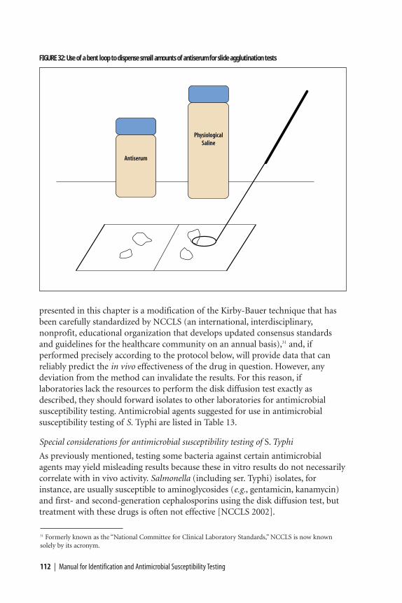
presented in this chapter is a modification of the Kirby-Bauer technique that hasbeen carefully standardized by NCCLS (an international, interdisciplinary,nonprofit, educational organization that develops updated consensus standardsand guidelines for the healthcare community on an annual basis),31 and, ifperformed precisely according to the protocol below, will provide data that canreliably predict the in vivo effectiveness of the drug in question. However, anydeviation from the method can invalidate the results. For this reason, iflaboratories lack the resources to perform the disk diffusion test exactly asdescribed, they should forward isolates to other laboratories for antimicrobialsusceptibility testing. Antimicrobial agents suggested for use in antimicrobialsusceptibility testing of S. Typhi are listed in Table 13.
Special considerations for antimicrobial susceptibility testing of S. Typhi
As previously mentioned, testing some bacteria against certain antimicrobialagents may yield misleading results because these in vitro results do not necessarilycorrelate with in vivo activity. Salmonella (including ser. Typhi) isolates, forinstance, are usually susceptible to aminoglycosides (e.g., gentamicin, kanamycin)and first- and second-generation cephalosporins using the disk diffusion test, buttreatment with these drugs is often not effective [NCCLS 2002].
112 | Manual for Identification and Antimicrobial Susceptibility Testing
FIGURE 32: Use of a bent loop to dispense small amounts of antiserum for slide agglutination tests
31 Formerly known as the “National Committee for Clinical Laboratory Standards,” NCCLS is now knownsolely by its acronym.
Salmonella serotype Typhi | 113
TABLE 13: Antimicrobial agents suggested for use in antimicrobial susceptibility testing of Salmonella ser. Typhi
Antimicrobial agents for susceptibility testing of Salmonella serotype Typhi
Ampicillin
Chloramphenicol
Trimethoprim-sulfamethoxazole (cotrimoxazole)
Nalidixic acid*
*If resistant to nalidixic acid, the isolate should be tested for susceptibility to ciprofloxacin,
and will probably exhibit reduced susceptibility to ciprofloxacin.
It is also worth noting that sometimes the result of one antimicrobialsusceptibility test will indicate the need for additional tests to confirm anexpected result. For example, when an isolate of S. Typhi is resistant to nalidixicacid, it will usually exhibit reduced susceptibility to ciprofloxacin; this scenariomay translate in the clinical setting to need for a longer course of treatment.Isolates exhibiting resistance to nalidixic acid should be tested for susceptibility tociprofloxacin.
Agar disk diffusion testing of S. Typhi
Mueller-Hinton agar medium is the only antimicrobial susceptibility test mediumthat has been validated by NCCLS. Mueller-Hinton agar should always be used fordisk diffusion susceptibility testing according to NCCLS and internationalguidelines. Because the way in which Mueller-Hinton is prepared can affect diskdiffusion test results, this medium should be prepared strictly according to themethods and quality control instructions presented in Appendix 2. A summary ofthe disk diffusion method of antimicrobial susceptibility testing is presented inFigure 33.
A 0.5 McFarland turbidity standard should be prepared and quality controlled priorto beginning antimicrobial susceptibility testing (see Appendix 2, Figure 50). Iftightly sealed to prevent evaporation and stored in the dark, the turbidity standardcan be stored for up to 6 months. The 0.5 McFarland turbidity standard is used toadjust the turbidity of the inoculum for the antimicrobial susceptibility test.
Preparation of inoculum
Each culture to be tested should be streaked onto a non-inhibitory agar medium(e.g., blood agar, brain heart infusion agar, or tryptone soy agar [TSA]) to obtainisolated colonies. After incubation at 35˚C overnight, select four or five well-isolated colonies with an inoculating needle or loop, and transfer the growth to atube of sterile saline or nonselective broth (e.g., Mueller-Hinton broth, heartinfusion broth, or tryptone soy broth [TSB]) and vortex thoroughly. The bacterial
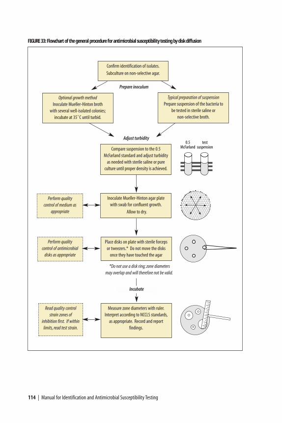
114 | Manual for Identification and Antimicrobial Susceptibility Testing
FIGURE 33: Flowchart of the general procedure for antimicrobial susceptibility testing by disk diffusion
Confirm identification of isolates.
Subculture on non-selective agar.
Optional growth method Inoculate Mueller-Hinton broth
with several well-isolated colonies;incubate at 35˚C until turbid.
Typical preparation of suspension Prepare suspension of the bacteria to
be tested in sterile saline or non-selective broth.
Prepare inoculum
Compare suspension to the 0.5McFarland standard and adjust turbidity
as needed with sterile saline or pure culture until proper density is achieved.
Adjust turbidity
Inoculate Mueller-Hinton agar platewith swab for confluent growth.
Allow to dry.
Place disks on plate with sterile forcepsor tweezers.* Do not move the disks
once they have touched the agar
Measure zone diameters with ruler.Interpret according to NCCLS standards,
as appropriate. Record and report findings.
Perform quality control of medium as
appropriate
Perform quality control of antimicrobial
disks as appropriate
Read quality controlstrain zones of
inhibition first. If within limits, read test strain.
*Do not use a disk ring; zone diametersmay overlap and will therefore not be valid.
Incubate

suspension should then be compared to the 0.5 McFarland turbidity standard. Thiscomparison can be made more easily if the tubes are viewed against a sheet ofwhite paper on which sharp black lines are drawn (see Appendix 2, Figures 51 and52). The turbidity standard should be agitated on a vortex mixer immediately priorto use. If the bacterial suspension does not appear to be the same density as the 0.5McFarland turbidity standard, the turbidity can be reduced by adding sterile salineor broth or increased by adding more bacterial growth.
Alternatively, the growth method may be used to prepare the inoculum. Pick fouror five colonies from overnight growth on agar and inoculate them into broth(Mueller-Hinton broth, heart infusion broth, or TSB). Incubate the broth at 35˚Cuntil turbid (usually 16–24 hours), and then adjust the turbidity to the properdensity.
Inoculation procedure
Within 15 minutes after adjusting the turbidity of the inoculum suspension, dip asterile cotton swab into the suspension. Pressing firmly against the inside wall ofthe tube just above the fluid level, rotate the swab to remove excess liquid. Streakthe swab over the entire surface of the medium three times, rotating the plateapproximately 60 degrees after each application to ensure an even distribution ofthe inoculum (Figure 34). Finally, swab around the entire edge of the agar surface.
If the bacterial colonies used to prepare the suspension are picked off a platecontaining mixed growth (i.e., if isolated colonies are picked from a plate that doesnot contain pure culture, as may occur when working with cultures from stoolspecimens), laboratorians may choose to prepare a purity plate to ensure thesuspension used for antimicrobial susceptibility testing is pure. To prepare thepurity plate, after inoculating the Mueller-Hinton agar plate for confluent growth,label (a portion of) a separate TSA plate (or other non-selective medium) and usethe same swab of suspension with which the Mueller-Hinton was inoculated tostreak for isolation; do not place the swab back into the suspension. Severalinocula can be streaked on different sections of a properly labeled purity plate,but the streaks must not overlap.
Quality control
To verify that antimicrobial susceptibility test results are accurate, at least onecontrol organism should be included with each test. (ATCC 25922 is the E. colicontrol strain used when testing S. Typhi and other Enterobacteriaceae.) Zonediameters obtained for ATCC 25922 should be compared with NCCLS publishedlimits; Table 14 includes the diameters of the zones of inhibition for ATCC 25922.If zones produced by the control strain are out of the expected ranges, thelaboratorian should consider possible sources of error.
Antimicrobial susceptibility tests are affected by variations in media, inoculumsize, incubation time, temperature, and other factors. The medium used may be a
Salmonella serotype Typhi | 115
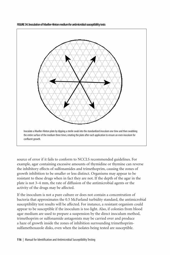
116 | Manual for Identification and Antimicrobial Susceptibility Testing
FIGURE 34: Inoculation of Mueller-Hinton medium for antimicrobial susceptibility tests
Inoculate a Mueller-Hinton plate by dipping a sterile swab into the standardized inoculum one time and then swabbingthe entire surface of the medium three times, rotating the plate after each application to ensure an even inoculum forconfluent growth.
source of error if it fails to conform to NCCLS recommended guidelines. Forexample, agar containing excessive amounts of thymidine or thymine can reversethe inhibitory effects of sulfonamides and trimethoprim, causing the zones ofgrowth inhibition to be smaller or less distinct. Organisms may appear to beresistant to these drugs when in fact they are not. If the depth of the agar in theplate is not 3–4 mm, the rate of diffusion of the antimicrobial agents or theactivity of the drugs may be affected.
If the inoculum is not a pure culture or does not contain a concentration ofbacteria that approximates the 0.5 McFarland turbidity standard, the antimicrobialsusceptibility test results will be affected. For instance, a resistant organism couldappear to be susceptible if the inoculum is too light. Also, if colonies from bloodagar medium are used to prepare a suspension by the direct inoculum method,trimethoprim or sulfonamide antagonists may be carried over and produce a haze of growth inside the zones of inhibition surrounding trimethoprim-sulfamethoxazole disks, even when the isolates being tested are susceptible.
If antimicrobial disks are not stored properly or are used beyond the statedexpiration date, their potency may decrease; this will usually be indicated by adecrease in the size of the inhibition zone around the control strain.
Antimicrobial disks
The working supply of antimicrobial disks should be stored in a refrigerator (4˚C).Upon removal of the disks from the refrigerator, the package containing thecartridges should be left unopened at room temperature for approximately 1 hourto allow the temperature to equilibrate; this reduces the amount of condensationon the disks. If a disk-dispensing apparatus is used, it should have a tight-fittingcover, be stored in the refrigerator, and be allowed to warm to room temperaturebefore use.
Apply the antimicrobial disks to the plates as soon as possible, but no longer than15 minutes after inoculation. The plate surface should be dry, with no liquidremaining. Place the disks individually with sterile forceps or with mechanicaldispensing apparatus, and then gently press down onto the agar. In general, nomore than 12 disks should be placed on a 150-mm plate and no more than fourdisks should be placed on a 100-mm plate to prevent overlapping of the zones ofinhibition and possible resultant error in measurement. Diffusion of the drug inthe disk begins immediately; therefore, once a disk contacts the agar surface, thedisk should not be moved.
Salmonella serotype Typhi | 117
TABLE 14: Inhibition zone diameter size interpretive standards for Enterobacteriaceae (for selected antimicrobial disks appropriate for Salmonella ser. Typhi)
Diameter of zone of inhibition (mm) andAntimicrobial Disk equivalent MIC breakpoint (µg/ml) NCCLS QC strainagent potency Susceptible Intermediate Resistant E. coli ATCC 25922
Ampicillin 10 µg ≥ 17 mm 14 – 16 mm ≤ 13 mm 16 – 22 mm(≤ 8 µg/ml) (16 µg/ml) (≥ 32 µg/ml) (2–8 µg/ml)
Chloramphenicol 30 µg ≥ 18 mm 13 – 17 mm ≤ 12 mm 21 – 27 mm(≤ 8 µg/ml) (16 µg/ml) (≥ 32 µg/ml) (2–8 µg/ml)
Trimethoprim- 1.25 / 23.75 µg ≥ 16 mm 11 – 15 mm ≤ 10 mm 23 – 29 mmsulfamethoxazole (≤ 2/38 µg/ml) (4/76 µg/ml) (≥ 8/152 µg/ml) (≤ 0.5/9.5 µg/ml)
(cotrimoxazole)
Nalidixic acid 30 µg ≥ 19 mm 14 – 18 mm ≤ 13 mm 22 – 28 mm(≤ 8 µg/ml) (16 µg/ml) (≥ 32 µg/ml) (1–4 µg/ml)
Ciprofloxacin 5 µg ≥ 21 mm 16 – 20 mm ≤ 15 mm 30 – 40 mm(≤ 1 µg/ml) (2 µg/ml) (≥ 4 mg/ml) (0.004–0.016 µg/ml)
Source: NCCLS (2002) Performance Standards for Antimicrobial Susceptibility Testing; Twelfth Informational Supplement.NCCLS document M100-S12 [ISBN 1-56238-454-6]. NCCLS 940 West Valley Road, Suite 1400,Wayne, PA 19087 USA.
118 | Manual for Identification and Antimicrobial Susceptibility Testing
Recording and interpreting results
After the disks are placed on the plate, invert the plate and incubate at 35˚C for16–18 hours; if a purity plate was prepared, incubate it under the same conditions.After incubation, measure the diameter of the zones of complete inhibition(including the diameter of the disk) (Figure 28) and record it in millimeters. (Asample worksheet is included in Figure 35.) The measurements can be made withcalipers or a ruler on the undersurface of the plate without opening the lid. Withsulfonamides and trimethoprim-sulfamethoxazole, a slight amount of growth mayoccur within the inhibition zone; in this instance, slight growth (approximately80% inhibition) should be ignored and the zone diameter should be measured tothe margin of heavy growth. For S. Typhi, zones of growth inhibition should becompared with the zone-size interpretative table for Enterobacteriaceae (Table 14),and recorded as susceptible, intermediate, or resistant to each drug tested.
Colonies growing within the clear zone of inhibition may represent resistantvariants or a mixed inoculum. Measure the distance from the inner-most colonies(i.e., those closest to the disk) to the center of the antimicrobial disk, and doublethis measurement to obtain the diameter; record the measurement andinterpretation of antimicrobial susceptibility (Figure 35). If there is both an inner-and outer zone of inhibition of growth around the antimicrobial disk:
a) If a purity plate was prepared, check the streak to confirm the culture was pure.(Step a is optional.)
b) Record the diameter and interpretation of antimicrobial susceptibility of thosecolonies in the outer zone (i.e., in addition to those in the inner zone).
c) Pick the colonies inside the zone, streak for isolation on a new plate, confirmtheir identification, and perform the disk diffusion test again to confirm theprevious results.
The presence of colonies within a zone of inhibition may predict eventualresistance to that antimicrobial agent.
Data for decision-making
Once the laboratory has assessed the identity and antimicrobial susceptibilitypatterns of S. Typhi isolates, the information should be reported promptly topublic health officials. Factors to consider in the development of a treatment policyinclude:
• The antimicrobial agent should be affordable.
• The antimicrobial agent chosen should be available locally (or be able to beobtained quickly).
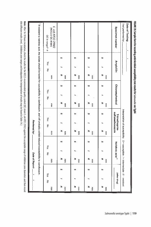
Salmonella serotype Typhi | 119
FIGURE35:Sam
ple form for recording antim
icrobial susceptibility test results for Salmonella
ser.Typhi
Note:After 16-18 hours incubation,check the results for the NCCLS-recomm
ended quality control (QC) strain E.coli ATCC 25922 against the acceptable range of inhibition zone diameters and then record
disk diffusion results (mm
).(Inhibition zone ranges and breakpoints for interpretation of results may be found in Table 14.)
120 | Manual for Identification and Antimicrobial Susceptibility Testing
• Immunization with available typhoid fever vaccines should be considered onlyfor high-risk populations where epidemic or high endemic rates of multi-drugresistant S. Typhi infections are a major cause of morbidity and mortality, andwhere vaccine effectiveness can be formally evaluated.
Consideration of such factors when making decisions based on data will helppublic health officials meet needs in a manner appropriate to the local situationand antimicrobial susceptibility profile.
Shigella | 121
The genus Shigella is divided into four subgroups: Shigella dysenteriae(Subgroup A), Shigella flexneri (Subgroup B), Shigella boydii (Subgroup C),and Shigella sonnei (Subgroup D). Each of these subgroups, with the exception
of S. sonnei, has several serotypes (Table 15). In general, S. sonnei is more commonin developed countries and S. flexneri and S. dysenteriae serotype 1 occur morefrequently in developing countries. The proportions of each subgroup varies fromcountry to country, though epidemic dysentery in developing countries is usuallycaused by S. dysenteriae 1, an unusually virulent pathogen. The hallmark ofinfection with Shigella is diarrhea with blood, often termed “dysentery.” However,in almost half of cases, Shigella causes acute non-bloody diarrheas that cannot bedistinguished clinically from diarrhea caused by other enteric pathogens. Severityof symptoms appears to be dose-related.
Shigella dysenteriae serotype 1 differs from other Shigella in several ways:
• Only S. dysenteriae 1 causes large and prolonged epidemics of dysentery.
• Infection with S. dysenteriae 1 causes more severe, more prolonged, and morefrequently fatal illness than does infection with other Shigella.
• Antimicrobial resistance develops more quickly and occurs more frequently inS. dysenteriae 1 than in other Shigella groups.
This section of the laboratory manual focuses on the isolation, identification, andantimicrobial susceptibility testing of Shigella.
Identification of Shigella
Methods for the collection and transport of fecal specimens and the primaryisolation and presumptive identification on selective agar are included in
ShigellaIDENTIFICATION AND ANTIMICROBIAL SUSCEPTIBILITY TESTING
CHAPTER VIII
122 | Manual for Identification and Antimicrobial Susceptibility Testing
Appendices 9 and 10. Suspect Shigella isolates should be subcultured to anonselective medium (e.g., Kligler iron agar [KIA] or triple sugar iron agar [TSI])in preparation for identification by slide serology and biochemical tests. Figure 36presents a flowchart for the isolation and identification of an isolate as Shigella,and Figure 37 provides a sample worksheet which can be used to record testresults.
Biochemical screening tests
Identification of Shigella subgroups involves both biochemical and serologictesting. The use of biochemical screening media is usually advisable to avoidwasting antisera. For most laboratories, KIA or TSI will be the single most helpfulmedium for screening suspected Shigella isolates. If an additional test is desired,motility medium can be used to screen isolates before serologic testing isperformed.
Kligler iron agar and triple sugar iron agar
To obtain true reactions in KIA, TSI, or other biochemical tests, a pure culturemust be used to inoculate the medium. Carefully select at least one of each type ofwell-isolated colony on each type of plate that was streaked for isolation (i.e., ifsuspect lactose-nonfermenting colonies that differ in macroscopic appearance arepresent, a separate test should be run for each.) Using an inoculating needle, lightlytouch only the center of the colony. Do not take the whole colony or go throughthe colony and touch the surface of the plate because doing so can pick upcontaminants that may be on the surface of the agar. If there is doubt that aparticular colony is sufficiently isolated from surrounding colonies, purify thesuspicious colony by streaking on another agar plate; afterwards, the KIA slant orTSI slant may be inoculated. Only one colony should be inoculated into each testmedium.
KIA and TSI tubes are inoculated by stabbing the butt and streaking the surface ofthe slant. After incubation for 18–24 hours at 35°–37°C, the slants are observed forreactions typical of Shigella. When incubating most biochemicals, caps should beloosened before placement in the incubator. This is particularly important whenusing KIA and TSI. If the caps are too tight and anaerobic conditions exist inKIA or TSI, the characteristic reactions of Shigella may not occur and amisleading result could be exhibited. In addition, the KIA and TSI tubes must beprepared so that the tubes have a deep butt (i.e., approximately 3.5 cm) and a longslant (i.e., approximately 2.5 cm). Shigella characteristically produces an alkaline(red) slant and an acid (yellow) butt, little or no gas, and no H2S (see Table 15 andFigure 38). A few strains of S. flexneri serotype 6 and very rare strains of S. boydiiproduce gas in KIA or TSI.
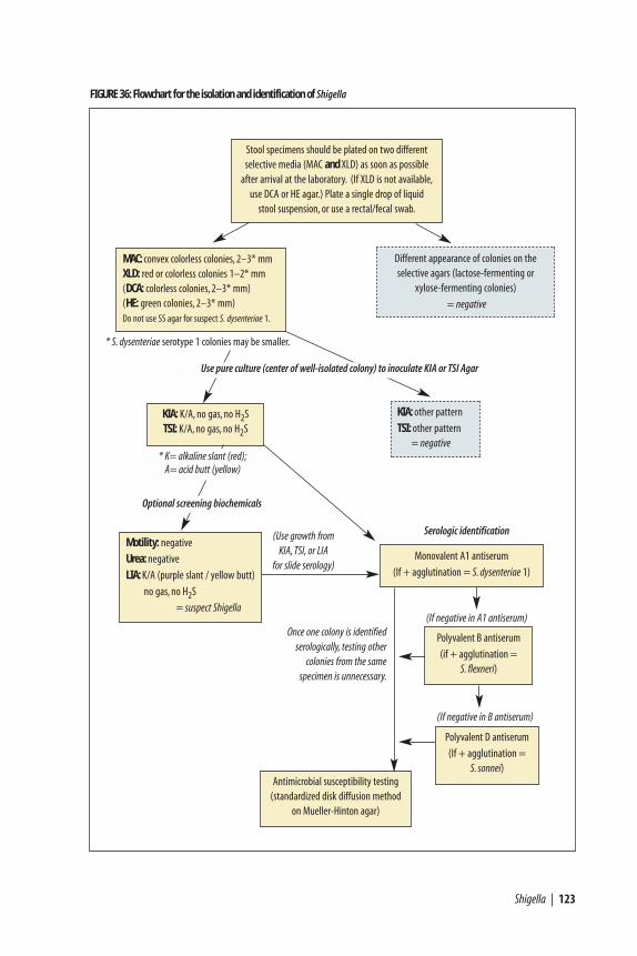
Shigella | 123
Polyvalent B antiserum
(if + agglutination = S. flexneri)
FIGURE 36: Flowchart for the isolation and identification of Shigella
Stool specimens should be plated on two differentselective media (MAC and XLD) as soon as possible
after arrival at the laboratory. (If XLD is not available,use DCA or HE agar.) Plate a single drop of liquid
stool suspension, or use a rectal/fecal swab.
Antimicrobial susceptibility testing(standardized disk diffusion method
on Mueller-Hinton agar)
KIA: K/A, no gas, no H2STSI: K/A, no gas, no H2S
Different appearance of colonies on the selective agars (lactose-fermenting or
xylose-fermenting colonies)
= negative
MAC: convex colorless colonies, 2–3* mmXLD: red or colorless colonies 1–2* mm(DCA: colorless colonies, 2–3* mm)(HE: green colonies, 2–3* mm)Do not use SS agar for suspect S. dysenteriae 1.
Optional screening biochemicals
* K= alkaline slant (red);A= acid butt (yellow)
Motility: negative
Urea: negative
LIA: K/A (purple slant / yellow butt)
no gas, no H2S
= suspect Shigella
(Use growth from KIA, TSI, or LIA
for slide serology)
* S. dysenteriae serotype 1 colonies may be smaller.
Use pure culture (center of well-isolated colony) to inoculate KIA or TSI Agar
KIA: other pattern
TSI: other pattern= negative
Once one colony is identifiedserologically, testing other
colonies from the same specimen is unnecessary.
Serologic identification
Monovalent A1 antiserum
(If + agglutination = S. dysenteriae 1)
Polyvalent D antiserum
(If + agglutination = S. sonnei)
(If negative in A1 antiserum)
(If negative in B antiserum)
124 | Manual for Identification and Antimicrobial Susceptibility Testing
FIGU
RE37
:Sam
ple
wor
kshe
et fo
r Shi
gella
test
resu
lts
Spec
imen
XYL/
XYL/
OPTI
ONAL
SLID
ESE
ROLO
GYc
num
ber
Med
ium
LA
C-a
LAC+
aCo
lony
bKI
A / T
SIM
otili
tyUr
eaLI
AA1
BD
Iden
tifica
tion
X1
XLD
X2 X3 M1
MAC
M2
M3 X1
XLD
X2 X3 M1
MAC
M2
M3 X1
XLD
X2 X3 M1
MAC
M2
M3
a XYL/
LAC–
:Xylo
se o
r Lac
tose
-neg
ative
colo
nies
XY
L/LA
C+ :X
ylose
or L
acto
se -p
ositi
ve co
loni
es
bId
entif
icatio
n of
onl
y one
colo
ny fr
om ea
ch su
spec
t cas
e mus
t be c
onfir
med
as Sh
igell
a.
c A1 =
Mon
oval
ent a
ntise
rum
for S
hige
lla d
ysen
teria
e(S
erog
roup
A) s
erot
ype 1
B =
Pol
yval
ent a
ntise
rum
for S
hige
lla fl
exne
ri(S
erog
roup
B)
D =
Pol
yval
ent a
ntise
rum
for S
hige
lla so
nnei
(Ser
ogro
up D
)
Shigella | 125
Motility agar
Motility agar should be inoculated with a straight inoculating needle, making asingle stab about 1–2 cm down into the medium. The surface of the motility agarshould be dry when used: moisture can cause a non-motile organism to growdown the sides of the agar, creating a haze of growth and appearing to be motile.Motility agar may be inoculated with growth from a KIA or TSI that shows areaction typical of Shigella. Alternatively, motility agar can be inoculated at thesame time as the KIA or TSI slant by using the same inoculating needle withouttouching the colony again. (When motility agar is to be inoculated at the sametime as KIA or TSI, use the same colony to first inoculate the motility agar andthen to inoculate the KIA or TSI by stabbing the butt and then streaking thesurface of the slant. Do not select a second colony to inoculate the KIA or TSIafter the motility agar has been inoculated because it may represent a differentorganism.
Examine after overnight incubation at 35°–37°C. Motility is indicated by thepresence of diffuse growth (appearing as clouding of the medium) away from theline of inoculation (Figure 39). Non-motile organisms do not grow out from theline of inoculation. Motility reactions may be difficult for inexperiencedlaboratorians to read; therefore, reactions should be compared with positive andnegative control strains. Shigella are always non-motile (Table 15).
Sulfide-indole-motility medium is a combination medium that is commerciallyavailable in dehydrated form (see Appendix 2, “Media, Reagents, and QualityControl”). It can be used in place of motility medium.
Additional biochemical screening tests
Other biochemical tests (e.g., urea medium and lysine iron agar [LIA]) may beused for additional screening of isolates before testing with antisera (Table 15).
TABLE 15: Reactions of Shigella in screening biochemicals
Screening medium Shigella reaction Figure number
Kligler iron agar (KIA) K/A, no gas produced (red slant/yellow butt) a Figure 38
Triple sugar iron agar (TSI) K/A, no gas produced (red slant/yellow butt) a ~
H2S (on KIA or TSI) Negative (positive reaction would be blackened medium) ~
Motility Negative Figure 39
Urea Negative Figure 40
Indole Positive or Negative ~
Lysine iron agar (LIA) K/A (purple slant/yellow butt) b Figure 41a K = alkaline (red); A = acid (yellow). Some strains of S. flexneri serotype 6 and S. boydii produce gas from glucose.b K = alkaline (purple); A = acid (yellow). An alkaline reaction in the butt of the medium indicates that lysine was
decarboxylated; an acid reaction in the butt indicates that lysine was not decarboxylated.
126 | Manual for Identification and Antimicrobial Susceptibility Testing
FIGURE 38: Reaction typical of Shigella in Kligler iron agar (KIA): alkaline slant and acid butt
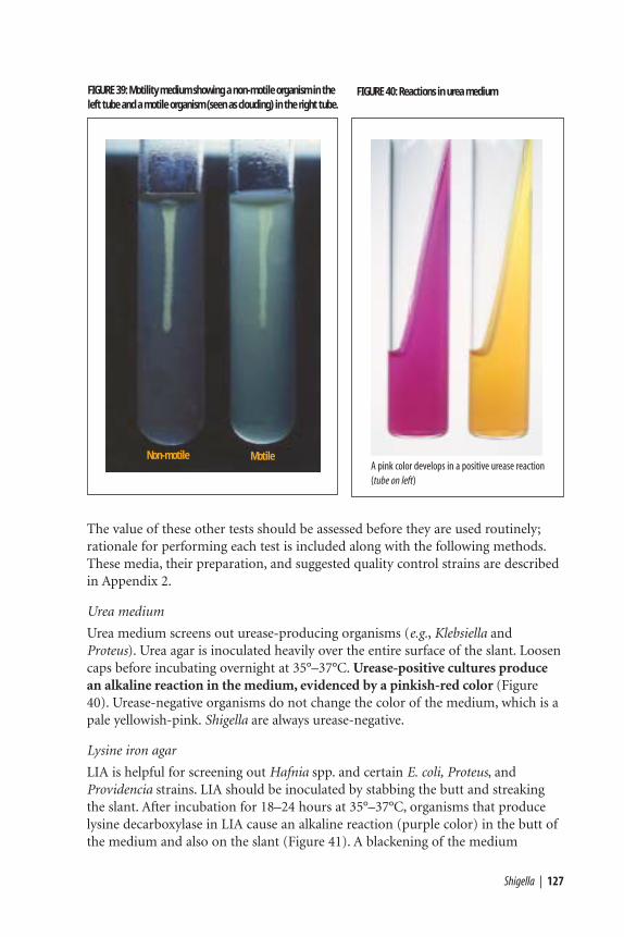
Shigella | 127
The value of these other tests should be assessed before they are used routinely;rationale for performing each test is included along with the following methods.These media, their preparation, and suggested quality control strains are describedin Appendix 2.
Urea medium
Urea medium screens out urease-producing organisms (e.g., Klebsiella andProteus). Urea agar is inoculated heavily over the entire surface of the slant. Loosencaps before incubating overnight at 35°–37°C. Urease-positive cultures producean alkaline reaction in the medium, evidenced by a pinkish-red color (Figure40). Urease-negative organisms do not change the color of the medium, which is apale yellowish-pink. Shigella are always urease-negative.
Lysine iron agar
LIA is helpful for screening out Hafnia spp. and certain E. coli, Proteus, andProvidencia strains. LIA should be inoculated by stabbing the butt and streakingthe slant. After incubation for 18–24 hours at 35°–37°C, organisms that producelysine decarboxylase in LIA cause an alkaline reaction (purple color) in the butt ofthe medium and also on the slant (Figure 41). A blackening of the medium
FIGURE 39: Motility medium showing a non-motile organism in theleft tube and a motile organism (seen as clouding) in the right tube.
FIGURE 40: Reactions in urea medium
A pink color develops in a positive urease reaction(tube on left)
Non-motile Motile
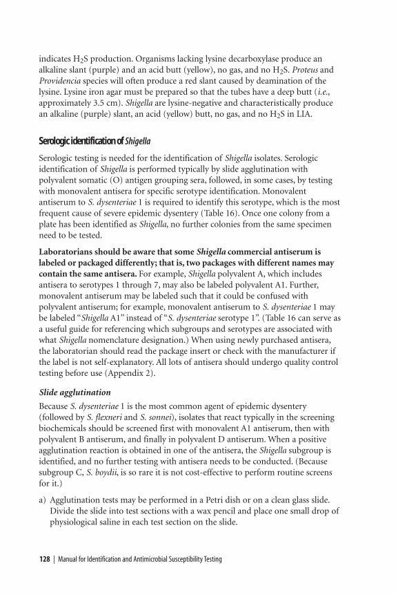
indicates H2S production. Organisms lacking lysine decarboxylase produce analkaline slant (purple) and an acid butt (yellow), no gas, and no H2S. Proteus andProvidencia species will often produce a red slant caused by deamination of thelysine. Lysine iron agar must be prepared so that the tubes have a deep butt (i.e.,approximately 3.5 cm). Shigella are lysine-negative and characteristically producean alkaline (purple) slant, an acid (yellow) butt, no gas, and no H2S in LIA.
Serologic identification of Shigella
Serologic testing is needed for the identification of Shigella isolates. Serologicidentification of Shigella is performed typically by slide agglutination withpolyvalent somatic (O) antigen grouping sera, followed, in some cases, by testingwith monovalent antisera for specific serotype identification. Monovalentantiserum to S. dysenteriae 1 is required to identify this serotype, which is the mostfrequent cause of severe epidemic dysentery (Table 16). Once one colony from aplate has been identified as Shigella, no further colonies from the same specimenneed to be tested.
Laboratorians should be aware that some Shigella commercial antiserum islabeled or packaged differently; that is, two packages with different names maycontain the same antisera. For example, Shigella polyvalent A, which includesantisera to serotypes 1 through 7, may also be labeled polyvalent A1. Further,monovalent antiserum may be labeled such that it could be confused withpolyvalent antiserum; for example, monovalent antiserum to S. dysenteriae 1 maybe labeled “Shigella A1” instead of “S. dysenteriae serotype 1”. (Table 16 can serve asa useful guide for referencing which subgroups and serotypes are associated withwhat Shigella nomenclature designation.) When using newly purchased antisera,the laboratorian should read the package insert or check with the manufacturer ifthe label is not self-explanatory. All lots of antisera should undergo quality controltesting before use (Appendix 2).
Slide agglutination
Because S. dysenteriae 1 is the most common agent of epidemic dysentery(followed by S. flexneri and S. sonnei), isolates that react typically in the screeningbiochemicals should be screened first with monovalent A1 antiserum, then withpolyvalent B antiserum, and finally in polyvalent D antiserum. When a positiveagglutination reaction is obtained in one of the antisera, the Shigella subgroup isidentified, and no further testing with antisera needs to be conducted. (Becausesubgroup C, S. boydii, is so rare it is not cost-effective to perform routine screensfor it.)
a) Agglutination tests may be performed in a Petri dish or on a clean glass slide.Divide the slide into test sections with a wax pencil and place one small drop ofphysiological saline in each test section on the slide.
128 | Manual for Identification and Antimicrobial Susceptibility Testing
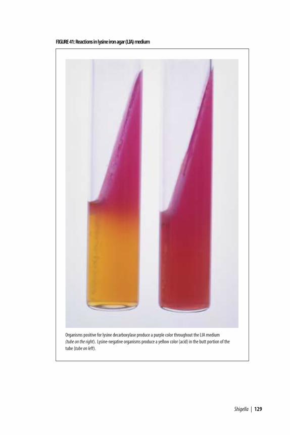
Shigella | 129
FIGURE 41: Reactions in lysine iron agar (LIA) medium
Organisms positive for lysine decarboxylase produce a purple color throughout the LIA medium (tube on the right). Lysine-negative organisms produce a yellow color (acid) in the butt portion of the tube (tube on left).
b) Use an inoculating loop or needle, sterile applicator stick, or toothpick toremove a portion of the growth from the surface of KIA, TSI, heart-infusionagar (HIA), or other non-selective agar medium. (Serologic testing should notbe done on growth from selective media such as MacConkey or XLD agarbecause selective media may yield false-negative serologic results.) Emulsify thegrowth in each drop of physiological saline on the slide and mix thoroughly tocreate a moderately milky suspension.
c) Add a small drop of antiserum to one of the suspensions; the secondsuspension serves as the control. To conserve antiserum, volumes as small as 10 µl can be used; a bent inoculating loop may be used to dispense smallamounts of antisera if micropipettors are not available (Figure 32). Usuallyapproximately equal volumes of antiserum and growth suspension are mixed,although the volume of suspension may be as much as double the volume ofthe antiserum.
d) Mix the suspension and antiserum well and then tilt the slide back and forth toobserve for autoagglutination (Figure 2). The agglutination is more visible if theslide is observed under a bright light and over a black background. If thereaction is positive, clumping will appear within 30 seconds to 1 minute (Figure42). Examine the saline suspension carefully to ensure that it is even and doesnot show clumping resulting from autoagglutination. If autoagglutinationoccurs, the culture is termed “rough” and cannot be serotyped.
Cultures that react serologically and show no conflicting results in the biochemicalscreening tests are reported as positive for Shigella.
Antimicrobial susceptibility testing of Shigella
As antimicrobial resistance increases in many parts of the world, monitoring theantimicrobial susceptibility of Shigella becomes increasingly important. The disk
130 | Manual for Identification and Antimicrobial Susceptibility Testing
TABLE 16: Subgroup and serotype designations of Shigella
Shigella Subgroup designation Serotypes (Label on commercial(common name) (polyvalent antisera) (monovalent antisera) antiserum may also say)
S. dysenteriae Group A 1 – 15 a,b (A1, A2, A3, …, A13)
S. flexneri Group B 1 – 6, X,Y (B1, B2, B3, B4, B5, B6)
S. boydii c Group C 1 – 19 b (C1, C2, C3, … , C18)
S. sonnei Group D 1 (D1)a Detection of S. dysenteriae 1 is of particular importance since it is unusually virulent and causes endemic or epidemic dysentery
with high death rates. Monovalent antiserum (absorbed) is required to identify S. dysenteriae 1.b Additional provisional serotypes have been reported, but antisera to these new serotypes were not commercially available at the
time this manual was printed.c Because S. boydii is so rare it is not cost-effective to perform routine screens for it.
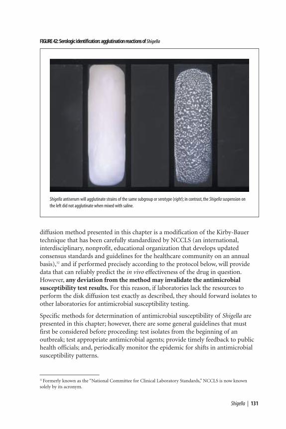
Shigella | 131
FIGURE 42: Serologic identification: agglutination reactions of Shigella
Shigella antiserum will agglutinate strains of the same subgroup or serotype (right); in contrast, the Shigella suspension onthe left did not agglutinate when mixed with saline.
diffusion method presented in this chapter is a modification of the Kirby-Bauertechnique that has been carefully standardized by NCCLS (an international,interdisciplinary, nonprofit, educational organization that develops updatedconsensus standards and guidelines for the healthcare community on an annualbasis),32 and if performed precisely according to the protocol below, will providedata that can reliably predict the in vivo effectiveness of the drug in question.However, any deviation from the method may invalidate the antimicrobialsusceptibility test results. For this reason, if laboratories lack the resources toperform the disk diffusion test exactly as described, they should forward isolates toother laboratories for antimicrobial susceptibility testing.
Specific methods for determination of antimicrobial susceptibility of Shigella arepresented in this chapter; however, there are some general guidelines that mustfirst be considered before proceeding: test isolates from the beginning of anoutbreak; test appropriate antimicrobial agents; provide timely feedback to publichealth officials; and, periodically monitor the epidemic for shifts in antimicrobialsusceptibility patterns.
32 Formerly known as the “National Committee for Clinical Laboratory Standards,” NCCLS is now known solely by its acronym.
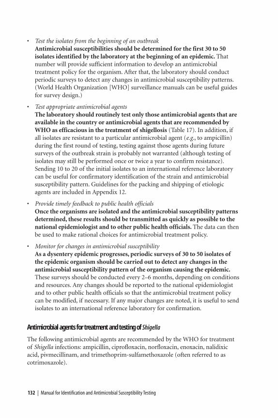
• Test the isolates from the beginning of an outbreakAntimicrobial susceptibilities should be determined for the first 30 to 50isolates identified by the laboratory at the beginning of an epidemic. Thatnumber will provide sufficient information to develop an antimicrobialtreatment policy for the organism. After that, the laboratory should conductperiodic surveys to detect any changes in antimicrobial susceptibility patterns.(World Health Organization [WHO] surveillance manuals can be useful guidesfor survey design.)
• Test appropriate antimicrobial agentsThe laboratory should routinely test only those antimicrobial agents that areavailable in the country or antimicrobial agents that are recommended byWHO as efficacious in the treatment of shigellosis (Table 17). In addition, ifall isolates are resistant to a particular antimicrobial agent (e.g., to ampicillin)during the first round of testing, testing against those agents during futuresurveys of the outbreak strain is probably not warranted (although testing ofisolates may still be performed once or twice a year to confirm resistance).Sending 10 to 20 of the initial isolates to an international reference laboratorycan be useful for confirmatory identification of the strain and antimicrobialsusceptibility pattern. Guidelines for the packing and shipping of etiologicagents are included in Appendix 12.
• Provide timely feedback to public health officialsOnce the organisms are isolated and the antimicrobial susceptibility patternsdetermined, these results should be transmitted as quickly as possible to thenational epidemiologist and to other public health officials. The data can thenbe used to make rational choices for antimicrobial treatment policy.
• Monitor for changes in antimicrobial susceptibilityAs a dysentery epidemic progresses, periodic surveys of 30 to 50 isolates ofthe epidemic organism should be carried out to detect any changes in theantimicrobial susceptibility pattern of the organism causing the epidemic.These surveys should be conducted every 2–6 months, depending on conditionsand resources. Any changes should be reported to the national epidemiologistand to other public health officials so that the antimicrobial treatment policycan be modified, if necessary. If any major changes are noted, it is useful to sendisolates to an international reference laboratory for confirmation.
Antimicrobial agents for treatment and testing of Shigella
The following antimicrobial agents are recommended by the WHO for treatmentof Shigella infections: ampicillin, ciprofloxacin, norfloxacin, enoxacin, nalidixicacid, pivmecillinam, and trimethoprim-sulfamethoxazole (often referred to ascotrimoxazole).
132 | Manual for Identification and Antimicrobial Susceptibility Testing
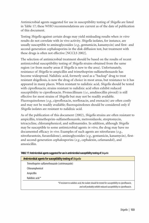
Antimicrobial agents suggested for use in susceptibility testing of Shigella are listedin Table 17; these WHO recommendations are current as of the date of publicationof this document.
Testing Shigella against certain drugs may yield misleading results when in vitroresults do not correlate with in vivo activity. Shigella isolates, for instance, areusually susceptible to aminoglycosides (e.g., gentamicin, kanamycin) and first- andsecond-generation cephalosporins in the disk diffusion test, but treatment withthese drugs is often not effective [NCCLS 2002].
The selection of antimicrobial treatment should be based on the results of recentantimicrobial susceptibility testing of Shigella strains obtained from the sameregion (or from nearby areas if Shigella is new to the area). Unfortunately,resistance of Shigella to ampicillin and trimethoprim-sulfamethoxazole hasbecome widespread. Nalidixic acid, formerly used as a “backup” drug to treatresistant shigellosis, is now the drug of choice in most areas, but resistance to it hasappeared in many places. When resistant to nalidixic acid, Shigella should be testedwith ciprofloxacin; strains resistant to nalidixic acid often exhibit reducedsusceptibility to ciprofloxacin. Pivmecillinam (i.e., amdinocillin pivoxil) is stilleffective for most strains of Shigella but may not be readily available.Fluoroquinolones (e.g., ciprofloxacin, norfloxacin, and enoxacin) are often costlyand may not be readily available; fluoroquinolones should be considered only ifShigella isolates are resistant to nalidixic acid.
As of the publication of this document (2002), Shigella strains are often resistant toampicillin, trimethoprim-sulfamethoxazole, metronidazole, streptomycin,tetracycline, chloramphenicol, and sulfonamides. In addition, although Shigellamay be susceptible to some antimicrobial agents in vitro, the drug may have nodocumented efficacy in vivo. Examples of such agents are nitrofurans (e.g.,nitrofurantoin, furazolidone), aminoglycosides (e.g., gentamicin, kanamycin), first-and second-generation cephalosporins (e.g., cephalexin, cefamandol), andamoxicillin.
Shigella | 133
TABLE 17: Antimicrobial agents suggested for use in antimicrobial susceptibility testing of Shigella
Antimicrobial agents for susceptibility testing of Shigella
Trimethoprim-sulfamethoxazole (cotrimoxazole)
Chloramphenicol
Ampicillin
Nalidixic acid *
*If resistant to nalidixic acid, the isolate should be tested for susceptibility to ciprofloxacin,
and will probably exhibit reduced susceptibility to ciprofloxacin.
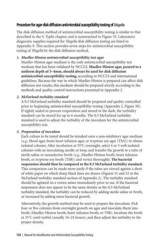
134 | Manual for Identification and Antimicrobial Susceptibility Testing
Procedure for agar disk diffusion antimicrobial susceptibility testing of Shigella
The disk diffusion method of antimicrobial susceptibility testing is similar to thatdescribed in the S. Typhi chapter, and is summarized in Figure 33. Laboratorydiagnostic supplies required for Shigella disk diffusion testing are listed inAppendix 9. This section provides seven steps for antimicrobial susceptibilitytesting of Shigella by the disk diffusion method.
1. Mueller-Hinton antimicrobial susceptibility test agar Mueller-Hinton agar medium is the only antimicrobial susceptibility testmedium that has been validated by NCCLS. Mueller-Hinton agar, poured to auniform depth of 3–4mm, should always be used for disk diffusionantimicrobial susceptibility testing, according to NCCLS and internationalguidelines. Because the way in which Mueller-Hinton is prepared can affect diskdiffusion test results, this medium should be prepared strictly according to themethods and quality control instructions presented in Appendix 2.
2. McFarland turbidity standardA 0.5 McFarland turbidity standard should be prepared and quality controlledprior to beginning antimicrobial susceptibility testing (Appendix 2, Figure 50).If tightly sealed to prevent evaporation and stored in the dark, the turbiditystandard can be stored for up to 6 months. The 0.5 McFarland turbiditystandard is used to adjust the turbidity of the inoculum for the antimicrobialsusceptibility test.
3. Preparation of inoculumEach culture to be tested should be streaked onto a non-inhibitory agar medium(e.g., blood agar, brain heart infusion agar, or tryptone soy agar [TSA]) to obtainisolated colonies. After incubation at 35°C overnight, select 4 or 5 well-isolatedcolonies with an inoculating needle or loop, and transfer the growth to a tube ofsterile saline or nonselective broth (e.g., Mueller-Hinton broth, heart infusionbroth, or tryptone soy broth [TSB]) and vortex thoroughly. The bacterialsuspension should then be compared to the 0.5 McFarland turbidity standard.This comparison can be made more easily if the tubes are viewed against a sheetof white paper on which sharp black lines are drawn (Figures 51 and 52 in theMcFarland turbidity standard section of Appendix 2). The turbidity standardshould be agitated on a vortex mixer immediately prior to use. If the bacterialsuspension does not appear to be the same density as the 0.5 McFarlandturbidity standard, the turbidity can be reduced by adding sterile saline or broth,or increased by adding more bacterial growth.
Alternatively, the growth method may be used to prepare the inoculum. Pickfour or five colonies from overnight growth on agar and inoculate them intobroth (Mueller-Hinton broth, heart infusion broth, or TSB). Incubate the brothat 35˚C until turbid (usually 16–24 hours), and then adjust the turbidity to theproper density.
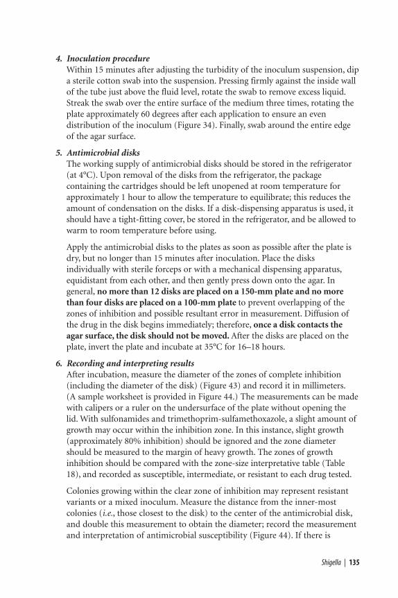
4. Inoculation procedureWithin 15 minutes after adjusting the turbidity of the inoculum suspension, dipa sterile cotton swab into the suspension. Pressing firmly against the inside wallof the tube just above the fluid level, rotate the swab to remove excess liquid.Streak the swab over the entire surface of the medium three times, rotating theplate approximately 60 degrees after each application to ensure an evendistribution of the inoculum (Figure 34). Finally, swab around the entire edgeof the agar surface.
5. Antimicrobial disksThe working supply of antimicrobial disks should be stored in the refrigerator(at 4°C). Upon removal of the disks from the refrigerator, the packagecontaining the cartridges should be left unopened at room temperature forapproximately 1 hour to allow the temperature to equilibrate; this reduces theamount of condensation on the disks. If a disk-dispensing apparatus is used, itshould have a tight-fitting cover, be stored in the refrigerator, and be allowed towarm to room temperature before using.
Apply the antimicrobial disks to the plates as soon as possible after the plate isdry, but no longer than 15 minutes after inoculation. Place the disksindividually with sterile forceps or with a mechanical dispensing apparatus,equidistant from each other, and then gently press down onto the agar. Ingeneral, no more than 12 disks are placed on a 150-mm plate and no morethan four disks are placed on a 100-mm plate to prevent overlapping of thezones of inhibition and possible resultant error in measurement. Diffusion ofthe drug in the disk begins immediately; therefore, once a disk contacts theagar surface, the disk should not be moved. After the disks are placed on theplate, invert the plate and incubate at 35°C for 16–18 hours.
6. Recording and interpreting resultsAfter incubation, measure the diameter of the zones of complete inhibition(including the diameter of the disk) (Figure 43) and record it in millimeters.(A sample worksheet is provided in Figure 44.) The measurements can be madewith calipers or a ruler on the undersurface of the plate without opening thelid. With sulfonamides and trimethoprim-sulfamethoxazole, a slight amount ofgrowth may occur within the inhibition zone. In this instance, slight growth(approximately 80% inhibition) should be ignored and the zone diametershould be measured to the margin of heavy growth. The zones of growthinhibition should be compared with the zone-size interpretative table (Table18), and recorded as susceptible, intermediate, or resistant to each drug tested.
Colonies growing within the clear zone of inhibition may represent resistantvariants or a mixed inoculum. Measure the distance from the inner-mostcolonies (i.e., those closest to the disk) to the center of the antimicrobial disk,and double this measurement to obtain the diameter; record the measurementand interpretation of antimicrobial susceptibility (Figure 44). If there is
Shigella | 135
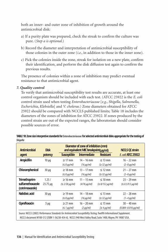
136 | Manual for Identification and Antimicrobial Susceptibility Testing
both an inner- and outer zone of inhibition of growth around theantimicrobial disk:
a) If a purity plate was prepared, check the streak to confirm the culture waspure. (Step a is optional.)
b) Record the diameter and interpretation of antimicrobial susceptibility ofthose colonies in the outer zone (i.e., in addition to those in the inner zone).
c) Pick the colonies inside the zone, streak for isolation on a new plate, confirmtheir identification, and perform the disk diffusion test again to confirm theprevious results.
The presence of colonies within a zone of inhibition may predict eventualresistance to that antimicrobial agent.
7. Quality control To verify that antimicrobial susceptibility test results are accurate, at least onecontrol organism should be included with each test. (ATCC 25922 is the E. colicontrol strain used when testing Enterobacteriaceae [e.g., Shigella, Salmonella,Escherichia, Klebsiella] and V. cholerae.) Zone diameters obtained for ATCC25922 should be compared with NCCLS published limits; Table 18 includes thediameters of the zones of inhibition for ATCC 25922. If zones produced by thecontrol strain are out of the expected ranges, the laboratorian should considerpossible sources of error.
TABLE 18: Zone size interpretive standards for Enterobacteriaceae for selected antimicrobial disks appropriate for the testing ofShigella
Diameter of zone of inhibition (mm)Antimicrobial Disk and equivalent MIC breakpoint(µg/ml) NCCLS QC strainagent potency Susceptible Intermediate Resistant E. coli ATCC 25922
Ampicillin 10 µg ≥ 17 mm 14 – 16 mm ≤ 13 mm 16 – 22 mm(≤ 8 µg/ml) (16 µg/ml) (≥ 32 µg/ml) (2–8 µg/ml)
Chloramphenicol 30 µg ≥ 18 mm 13 – 17 mm ≤ 12 mm 21 – 27 mm(≤ 8 µg/ml) (16 µg/ml) (≥ 32 µg/ml) (2–8 µg/ml)
Trimethoprim- 1.25 / ≥ 16 mm 11 – 15 mm ≤ 10 mm 23 – 29 mmsulfamethoxazole 23.75 µg (≤ 2/38 µg/ml) (4/76 µg/ml) (≥ 8/152 µg/ml) (≤ 0.5/9.5 µg/ml)
(cotrimoxazole)
Nalidixic acid 30 µg ≥ 19 mm 14 – 18 mm ≤ 13 mm 22 – 28 mm(≤ 8 µg/ml) (16 µg/ml) (≥ 32 µg/ml) (1–4 µg/ml)
Ciprofloxacin 5 µg ≥ 21 mm 16 – 20 mm ≤ 15 mm 30 – 40 mm(≤ 1 µg/ml) (2 µg/ml) [≥ 4 µg/ml) (0.004–0.016 µg/ml)
Source: NCCLS (2002) Performance Standards for Antimicrobial Susceptibility Testing; Twelfth Informational Supplement.
NCCLS document M100-S12 [ISBN 1-56238-454-6]. NCCLS 940 West Valley Road, Suite 1400,Wayne, PA 19087 USA.

Antimicrobial susceptibility tests are affected by variations in media,inoculum size, incubation time, temperature, and other factors. The mediumused may be a source of error if it fails to conform to NCCLS recommendedguidelines. For example, agar containing excessive amounts of thymidine orthymine can reverse the inhibitory effects of sulfonamides and trimethoprim,causing the zones of growth inhibition to be smaller or less distinct. Organismsmay appear to be resistant to these drugs when in fact they are not. If the depthof the agar in the plate is not 3–4 mm, the rate of diffusion of the antimicrobialagents or the activity of the drugs may be affected.
If the inoculum is not a pure culture or does not contain a concentration ofbacteria that approximates the 0.5 McFarland turbidity standard, theantimicrobial susceptibility test results will be affected. For instance, aresistant organism could appear to be susceptible if the inoculum is too light.
Shigella | 137
FIGURE 43: Sample results of the disk diffusion assay
This Shigella isolate is resistant to trimethoprim-sulfamethoxazole (cotrimoxazole) and is growing up to the disk(SXT), the zone of which is recorded as 6 mm. In addition to the zones of inhibited growth, note how evenly distributed the growth is on the plate.
138 | Manual for Identification and Antimicrobial Susceptibility Testing
FIGU
RE44
:Sam
ple
form
for r
ecor
ding
ant
imicr
obia
l sus
cept
ibili
ty te
st re
sults
for S
hige
llaiso
late
s
Note
:Afte
r 16–
18 h
ours
incu
batio
n,ch
eck t
he re
sults
for t
he N
CCLS
-reco
mm
ende
d qu
ality
cont
rol (
QC) s
train
E.co
li ATC
C 259
22 ag
ains
t the
acce
ptab
le ra
nge o
f inh
ibiti
on zo
ne d
iam
eter
s and
then
reco
rd d
isk d
iffus
ion
resu
lts (m
m).
(Inhi
bitio
n zo
ne ra
nges
and
brea
kpoi
nts f
or in
terp
reta
tion
of re
sults
may
be f
ound
in Ta
ble 1
8.)
Also, if colonies from blood agar medium are used to prepare a suspension by the direct inoculum method, trimethoprim or sulfonamide antagonists maybe carried over and produce a haze of growth inside the zones of inhibitionsurrounding trimethoprim-sulfamethoxazole disks even when the isolates being tested are susceptible.
If antimicrobial disks are not stored properly or are used beyond the statedexpiration date, their potency may decrease; this will usually be indicated by a decrease in the size of the inhibition zone around the control strain.
Data for decision-making: informed epidemic response
Once the laboratory has assessed the identity and antimicrobial susceptibilitypatterns of the Shigella isolates, the information should be reported to publichealth officials in a timely manner. Factors to consider in the development of atreatment policy include:
• The antimicrobial agent chosen should be effective against at least 80% oflocal Shigella strains.
• The antimicrobial agent chosen should be able to be given by mouth.
• The antimicrobial agent chosen should be affordable.
• The antimicrobial agent chosen should be available locally (or be able to beobtained quickly).
Consideration of such factors when making data-based decisions will help publichealth officials meet needs in a manner appropriate to the local situation andantimicrobial susceptibility profile.
Shigella | 139

Vibrio cholerae | 141
Most Vibrio cholerae isolated during cholera outbreaks will be toxigenicserogroup O1 or O139. Because the cultural and biochemical characteristicsof these two serogroups are identical, the isolation and identification
methods described below apply to both O1 and O139. Both serogroups must beidentified using O-group-specific antisera.
Isolates of V. cholerae serogroup O1 are classified into two biotypes, El Tor andclassical, on the basis of several phenotypic characteristics. Currently, the El Torbiotype is responsible for virtually all of the cholera cases throughout the world,and classical isolates are not encountered outside of India or Bangladesh. Inaddition, V. cholerae O1 is classified into two serotypes (Inaba and Ogawa) on thebasis of agglutination in antiserum. A possible third serotype, Hikojima, has beendescribed, but it occurs only rarely. During an outbreak or epidemic, it is worthdocumenting the biotype and serotype of the isolate; however, it is not necessary to know this information to respond appropriately to the epidemic.
V. cholerae serogroup O139 appeared in India in late 1992. It quickly spread toBangladesh and other Asian countries, although the rate of spread has slowed afterthe initial outbreaks. Through 1998, 11 countries have officially reportedtransmission of V. cholerae O139 to the World Health Organization (WHO).Imported cases have been reported from the United States and other countries.At this time, endemic V. cholerae O139 appears to be confined to Asia.
Fluid replacement is the cornerstone of cholera treatment, and rehydration therapyis a necessity. Antimicrobial therapy is helpful, although not essential, in treatingcholera patients. Antimicrobial agents reduce the duration of illness, the volume ofstool, and the duration of shedding of vibrios in the feces. When antimicrobialagents are used, it is essential to choose one to which the organism is susceptible.Antimicrobial agents recommended by WHO for treating cholera patients as of thedate of publication of this document include tetracycline, doxycycline,
Vibrio choleraeIDENTIFICATION AND ANTIMICROBIAL SUSCEPTIBILITY TESTING
CHAPTER IX
142 | Manual for Identification and Antimicrobial Susceptibility Testing
furazolidone, trimethoprim-sulfamethoxazole, erythromycin, or chloramphenicol.Ciprofloxacin and norfloxacin are also effective. Because antimicrobial resistancehas been a growing problem in many parts of the world, the susceptibility of V.cholerae O1 strains to antimicrobial agents should be determined at the beginningof an epidemic and be monitored periodically. Methods for antimicrobialsusceptibility testing of V. cholerae are addressed in this chapter of the manual,after identification. Isolation and presumptive identification of V. cholerae fromfecal specimens are included in Appendix 10.
Public health authorities in regions that experience outbreaks of cholera may findthat the manual Laboratory Methods for the Diagnosis of Epidemic Dysentery andCholera [CDC 1999] provides additional helpful discussions about choleraepidemiology and laboratory decision-making in resource-limited regions. Thedocument is available from WHO in English and French; details for ordering areincluded in Appendix 15.
Identification of V. cholerae
Methods for the collection and transport of fecal specimens and the primaryisolation and presumptive identification on selective agar are included inAppendices 9 and 10. Suspect V. cholerae isolates should be subcultured to a non-selective medium (e.g., heart infusion agar [HIA] or tryptone soy agar [TSA]) inpreparation for identification by slide serology and biochemical tests. V. choleraerequires 0.5% NaCl (i.e., salt) for optimal growth on agar media; somecommercially available formulations of nutrient agar do not contain salt andshould not be used for culture of V. cholerae. In general, screening withbiochemical tests prior to testing with O1 and O139 antisera is not necessary forsuspected V. cholerae isolates from fecal specimens. However, if the supply of O-antigen antisera is limited, biochemical tests may be useful for additionalscreening of isolates before testing them with antisera. Screening tests and slideserology must be performed with growth from nonselective media. Figure 45presents a flowchart for isolation and identification of an isolate as V. cholerae, andFigure 46 provides a sample worksheet that can be used to record screening testresults.
Oxidase test
The oxidase test uses Kovac’s reagent (a 1% [wt/vol] solution of N, N, N’, N’–tetramethyl-ρ-phenylenediamine dihydrochloride) to detect the presence ofcytochrome c in a bacterial organism’s respiratory chain; if the oxidase reagent iscatalyzed, it turns purple. The oxidase test can be performed on filter paper or on a swab.
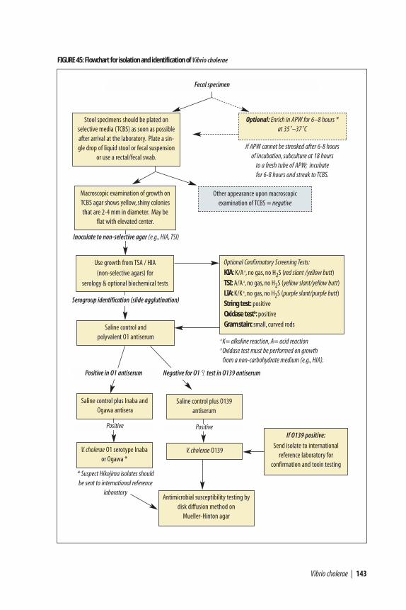
Vibrio cholerae | 143
If O139 positive:
Send isolate to international reference laboratory for
confirmation and toxin testing
FIGURE 45: Flowchart for isolation and identification of Vibrio cholerae
Antimicrobial susceptibility testing bydisk diffusion method on
Mueller-Hinton agar
Use growth from TSA / HIA
(non-selective agars) for
serology & optional biochemical tests
Stool specimens should be plated on selective media (TCBS) as soon as possibleafter arrival at the laboratory. Plate a sin-gle drop of liquid stool or fecal suspension
or use a rectal/fecal swab.
* Suspect Hikojima isolates shouldbe sent to international reference
laboratory
Fecal specimen
if APW cannot be streaked after 6-8 hours of incubation, subculture at 18 hours
to a fresh tube of APW; incubate for 6-8 hours and streak to TCBS.
Optional: Enrich in APW for 6–8 hours * at 35˚–37˚C
Other appearance upon macroscopic examination of TCBS = negative
Macroscopic examination of growth onTCBS agar shows yellow, shiny coloniesthat are 2-4 mm in diameter. May be
flat with elevated center.
Optional Confirmatory Screening Tests:
KIA: K/A a, no gas, no H2S (red slant /yellow butt)
TSI: A/A a, no gas, no H2S (yellow slant/yellow butt)
LIA: K/K a, no gas, no H2S (purple slant/purple butt)
String test: positive
Oxidase testb: positive
Gram stain: small, curved rods
a K= alkaline reaction, A= acid reactionb Oxidase test must be performed on growth
from a non-carbohydrate medium (e.g., HIA).
Saline control and polyvalent O1 antiserum
Saline control plus Inaba andOgawa antisera
V. cholerae O1 serotype Inabaor Ogawa *
Positive in O1 antiserum Negative for O1 Ÿ test in O139 antiserum
Saline control plus O139antiserum
Positive
V. cholerae O139
Inoculate to non-selective agar (e.g., HIA, TSI)
Positive
Serogroup identification (slide agglutination)
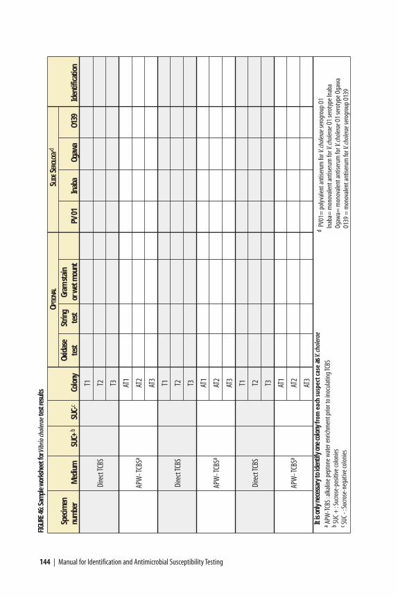
144 | Manual for Identification and Antimicrobial Susceptibility Testing
FIGU
RE46
:Sam
ple
wor
kshe
et fo
rVib
rio ch
oler
ae te
st re
sults
OPTI
ONAL
SLID
ESE
ROLO
GYd
Spec
imen
Oxid
ase
Strin
gGr
am st
ain
num
ber
Med
ium
SU
C+b
SUC-
cCo
lony
test
test
or w
et m
ount
PV 0
1In
aba
Ogaw
aO1
39Id
entif
icatio
n
T1
Dire
ct TC
BST2 T3 AT
1
APW
- TCB
SaAT
2
AT3
T1
Dire
ct TC
BST2 T3 AT
1
APW
- TCB
SaAT
2
AT3
T1
Dire
ct TC
BST2 T3 AT
1
APW
- TCB
SaAT
2
AT3
It is
only
nec
essa
ry to
iden
tify
one
colo
ny fr
om e
ach
susp
ect c
ase
as V.
chol
erae
aAP
W-T
CBS :
alka
line p
epto
ne w
ater
enric
hmen
t prio
r to
inoc
ulat
ing T
CBS
bSU
C +
:Suc
rose
-pos
itive
colo
nies
c SUC
- :Su
crose
-neg
ative
colo
nies
dPV
01=
pol
yval
ent a
ntise
rum
for V
.cho
lerae
sero
grou
p O1
Inab
a= m
onov
alen
t ant
iseru
m fo
r V.c
holer
aeO1
sero
type
Inab
aOg
awa=
mon
oval
ent a
ntise
rum
for V
.cho
lerae
O1 se
roty
pe O
gaw
aO1
39 =
mon
oval
ent a
ntise
rum
for V
.cho
lerae
sero
grou
p O1
39

Vibrio cholerae | 145
Perform the oxidase test with fresh growth from an HIA or TSA slant or any non-selective, non-carbohydrate-containing medium; do not use growth fromthiosulfate citrate bile-salts sucrose [TCBS] agar because it may yield either false-negative or false-positive results. Do not perform this test with a Nichrome loop,as it may produce a false-positive reaction. Positive and negative controls should betested at the same time as the test isolate for quality control purposes. Preparationof the oxidase reagent is described in Appendix 2.
Moistened filter paper method
a) Add two to three drops of Kovac’s oxidase reagent to a piece of filter paper in apetri dish and allow it to absorb; the filter paper should be moist (but not wet)after the reagent has been absorbed.
b) Using a platinum loop, a plastic loop, a sterile swab, or a sterile woodenapplicator stick or toothpick, pick a portion of the colony to be tested fromnon-selective media and rub it onto the moistened filter paper. (Do not use aNichrome loop.)
c) If the isolate is V. cholerae, a positive (purple) reaction should occur in theregion where the growth has been smeared within 10 seconds (Figure 10).
Swab method
a) Pick up suspect colonies from a non-selective culture plate or growth from anon-selective agar slant with the swab.
b) Use a Pasteur pipette to add one drop of Kovac’s oxidase reagent to the swab.
c) If the isolate is V. cholerae, a positive (purple) reaction should occur within 10seconds. (See Figure 20).
If an isolate has not turned purple within 10 seconds of adding the Kovac’s oxidasereagent, it is not considered oxidase-positive. Organisms of the genera Vibrio(Table 19), Neisseria, Campylobacter, Aeromonas, Plesiomonas, Pseudomonas, andAlcaligenes are all oxidase-positive; all Enterobacteriaceae are oxidase-negative.
Additional biochemical screening tests
The string reaction, Kligler iron agar (KIA) or triple sugar iron agar (TSI), lysineiron agar (LIA), Gram stain, and wet mount for motility are other possible teststhat may be used for additional screening of isolates before testing with antisera(Table 19). The value of these other tests should be assessed before they are usedroutinely; rationale for performing each test (e.g., use of the string test to rule outAeromonas) is included along with the following methods. These media, theirpreparation, and suggested quality control strains are described in Appendix 2.
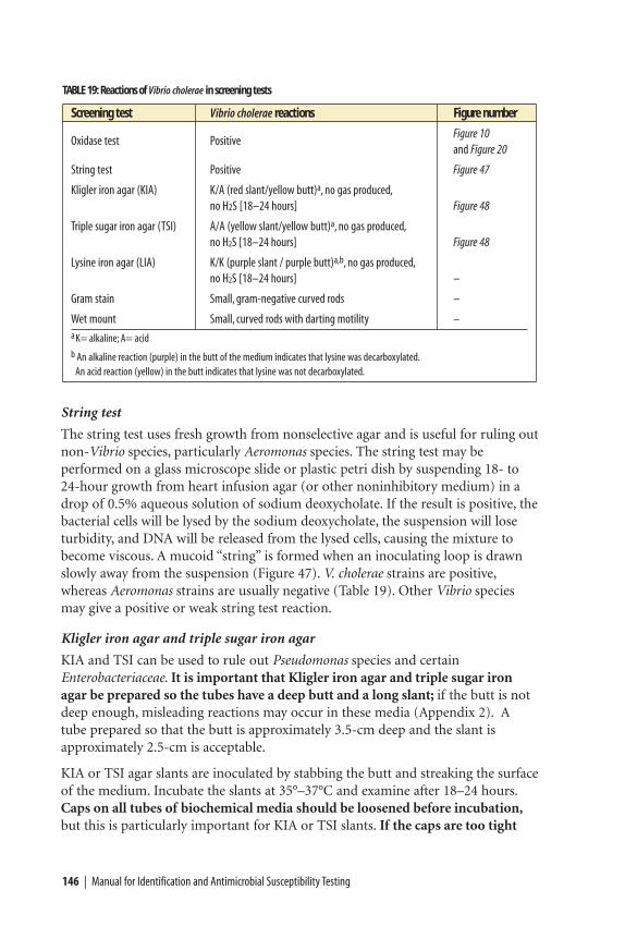
String test
The string test uses fresh growth from nonselective agar and is useful for ruling outnon-Vibrio species, particularly Aeromonas species. The string test may beperformed on a glass microscope slide or plastic petri dish by suspending 18- to24-hour growth from heart infusion agar (or other noninhibitory medium) in adrop of 0.5% aqueous solution of sodium deoxycholate. If the result is positive, thebacterial cells will be lysed by the sodium deoxycholate, the suspension will loseturbidity, and DNA will be released from the lysed cells, causing the mixture tobecome viscous. A mucoid “string” is formed when an inoculating loop is drawnslowly away from the suspension (Figure 47). V. cholerae strains are positive,whereas Aeromonas strains are usually negative (Table 19). Other Vibrio speciesmay give a positive or weak string test reaction.
Kligler iron agar and triple sugar iron agar
KIA and TSI can be used to rule out Pseudomonas species and certainEnterobacteriaceae. It is important that Kligler iron agar and triple sugar ironagar be prepared so the tubes have a deep butt and a long slant; if the butt is notdeep enough, misleading reactions may occur in these media (Appendix 2). Atube prepared so that the butt is approximately 3.5-cm deep and the slant isapproximately 2.5-cm is acceptable.
KIA or TSI agar slants are inoculated by stabbing the butt and streaking the surfaceof the medium. Incubate the slants at 35°–37°C and examine after 18–24 hours.Caps on all tubes of biochemical media should be loosened before incubation,but this is particularly important for KIA or TSI slants. If the caps are too tight
146 | Manual for Identification and Antimicrobial Susceptibility Testing
TABLE 19: Reactions of Vibrio cholerae in screening tests
Screening test Vibrio cholerae reactions Figure number
Oxidase test PositiveFigure 10and Figure 20
String test Positive Figure 47
Kligler iron agar (KIA) K/A (red slant/yellow butt)a, no gas produced,no H2S [18–24 hours] Figure 48
Triple sugar iron agar (TSI) A/A (yellow slant/yellow butt)a, no gas produced,no H2S [18–24 hours] Figure 48
Lysine iron agar (LIA) K/K (purple slant / purple butt)a,b, no gas produced,no H2S [18–24 hours] –
Gram stain Small, gram-negative curved rods –
Wet mount Small, curved rods with darting motility –a K= alkaline; A= acidb An alkaline reaction (purple) in the butt of the medium indicates that lysine was decarboxylated.
An acid reaction (yellow) in the butt indicates that lysine was not decarboxylated.
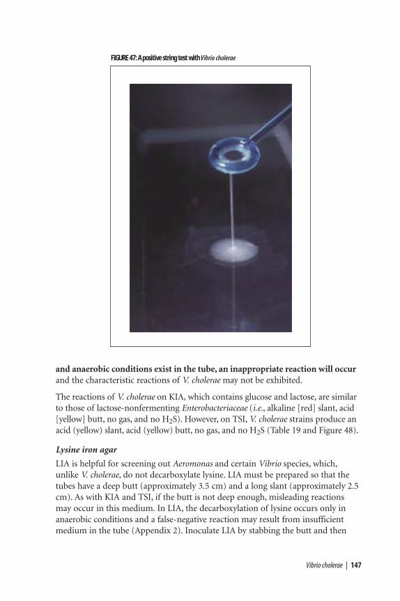
Vibrio cholerae | 147
and anaerobic conditions exist in the tube, an inappropriate reaction will occurand the characteristic reactions of V. cholerae may not be exhibited.
The reactions of V. cholerae on KIA, which contains glucose and lactose, are similarto those of lactose-nonfermenting Enterobacteriaceae (i.e., alkaline [red] slant, acid[yellow] butt, no gas, and no H2S). However, on TSI, V. cholerae strains produce anacid (yellow) slant, acid (yellow) butt, no gas, and no H2S (Table 19 and Figure 48).
Lysine iron agar
LIA is helpful for screening out Aeromonas and certain Vibrio species, which,unlike V. cholerae, do not decarboxylate lysine. LIA must be prepared so that thetubes have a deep butt (approximately 3.5 cm) and a long slant (approximately 2.5cm). As with KIA and TSI, if the butt is not deep enough, misleading reactionsmay occur in this medium. In LIA, the decarboxylation of lysine occurs only inanaerobic conditions and a false-negative reaction may result from insufficientmedium in the tube (Appendix 2). Inoculate LIA by stabbing the butt and then
FIGURE 47: A positive string test with Vibrio cholerae
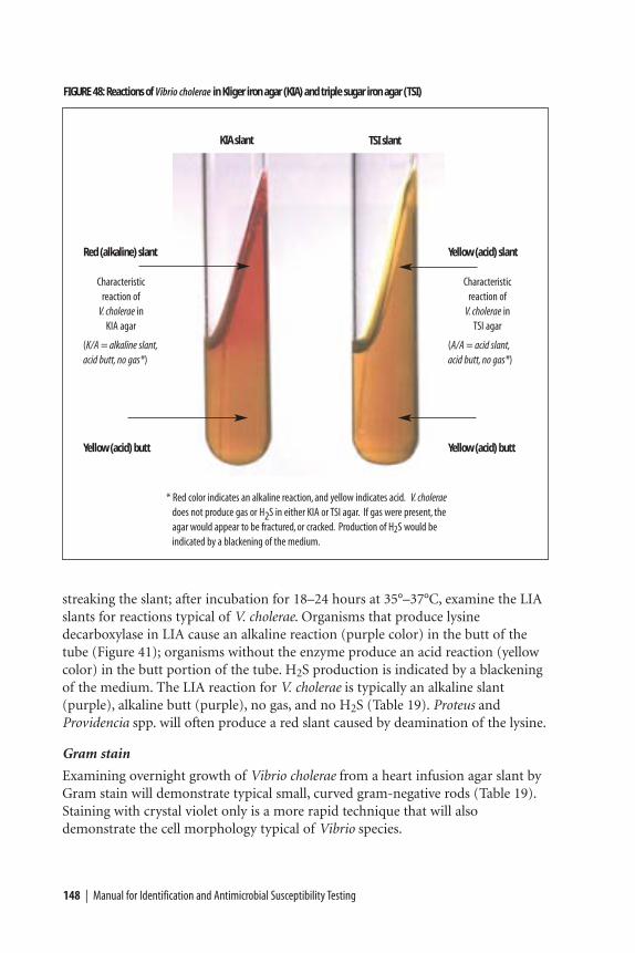
streaking the slant; after incubation for 18–24 hours at 35°–37°C, examine the LIAslants for reactions typical of V. cholerae. Organisms that produce lysinedecarboxylase in LIA cause an alkaline reaction (purple color) in the butt of thetube (Figure 41); organisms without the enzyme produce an acid reaction (yellowcolor) in the butt portion of the tube. H2S production is indicated by a blackeningof the medium. The LIA reaction for V. cholerae is typically an alkaline slant(purple), alkaline butt (purple), no gas, and no H2S (Table 19). Proteus andProvidencia spp. will often produce a red slant caused by deamination of the lysine.
Gram stain
Examining overnight growth of Vibrio cholerae from a heart infusion agar slant byGram stain will demonstrate typical small, curved gram-negative rods (Table 19).Staining with crystal violet only is a more rapid technique that will alsodemonstrate the cell morphology typical of Vibrio species.
148 | Manual for Identification and Antimicrobial Susceptibility Testing
FIGURE 48: Reactions of Vibrio cholerae in Kliger iron agar (KIA) and triple sugar iron agar (TSI)
Yellow (acid) slant
Characteristic reaction of
V. cholerae in TSI agar
(A/A = acid slant,acid butt, no gas*)
Yellow (acid) butt
* Red color indicates an alkaline reaction, and yellow indicates acid. V. choleraedoes not produce gas or H2S in either KIA or TSI agar. If gas were present, theagar would appear to be fractured, or cracked. Production of H2S would be indicated by a blackening of the medium.
Red (alkaline) slant
Characteristic reaction of
V. cholerae in KIA agar
(K/A = alkaline slant,acid butt, no gas*)
Yellow (acid) butt
KIA slant TSI slant
Wet mount
Dark-field and phase-contrast microscopy have been used for screening suspectedisolates of V. cholerae. With these techniques, saline suspensions are microscopicallyexamined for the presence of organisms with typical small, curved rods and darting(i.e., “shooting star”) motility (Table 19).
Serologic identification of V. cholerae O1 and O139
Following presumptive biochemical identification of the agent as V. cholerae, it isappropriate to confirm identification with serology. If an epidemic of cholera issuspected, the most common cause is V. cholerae O1. If V. cholerae O1 is notisolated, the laboratory should test for V. cholerae O139. If neither of theseorganisms is isolated, arrangements should be made to send stool specimens to areference laboratory. Local and regional laboratories should send isolates requiringtesting with O139 antiserum to the national reference laboratory; if the nationalreference laboratory is still unable to confirm the identification of a V. choleraeisolate as O1 or O139, an international reference laboratory can provide guidance.
To conserve resources, the laboratory can first test V. cholerae for somatic O1antigens, and then test with O139 antiserum only if the isolate does not yield apositive agglutination reaction in the O1 antiserum.
Presumptive identification using O1 and O139 antisera
For slide agglutination testing with polyvalent O1 or O139 antisera, fresh growthof suspected V. cholerae from a nonselective agar medium should be used. (Usinggrowth from thiosulfate citrate bile salts sucrose (TCBS) agar may result infalse-negative reactions.) After 5–6 hours of incubation, growth on the surface ofthe slant is usually sufficient to perform slide agglutination with antisera; if not,incubate for a longer period. If the isolate does not agglutinate in O1 antiserum,test in O139 antiserum. If it is positive in the polyvalent O1 or in the O139antiserum, the isolate may be reported as presumptive V. cholerae O1 or O139.Presumptive V. cholerae O1 isolates should be tested in monovalent Ogawa andInaba antisera (methods follow this section). Once one colony from a plate hasbeen identified as V. cholerae O1 or O139, no further colonies from the same plateneed to be tested. [Refer to Appendix 2 for a discussion on quality control ofantisera.]
Confirmation of V. cholerae O1 using Inaba and Ogawa antisera
The O1 serogroup of V. cholerae has been further divided into three serotypes:Inaba, Ogawa, and Hikojima (which is very rare). Serotype identification is basedon agglutination in monovalent antisera to type-specific O antigens (Table 20). Apositive reaction in either Inaba or Ogawa antiserum is sufficient to confirm theidentification of a V. cholerae O1 isolate. Isolates that agglutinate weakly or slowlywith serogroup O1 antiserum but do not agglutinate with either Inaba or Ogawa
Vibrio cholerae | 149
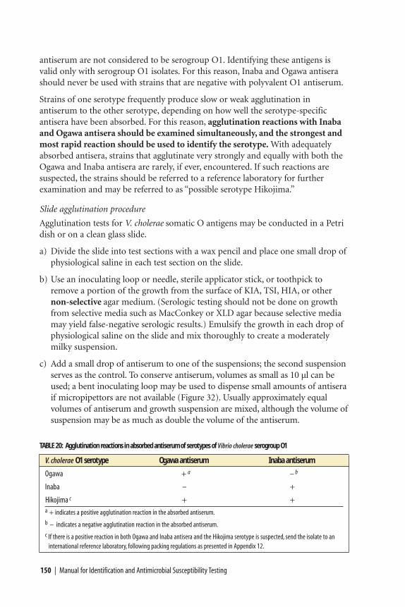
antiserum are not considered to be serogroup O1. Identifying these antigens isvalid only with serogroup O1 isolates. For this reason, Inaba and Ogawa antiserashould never be used with strains that are negative with polyvalent O1 antiserum.
Strains of one serotype frequently produce slow or weak agglutination inantiserum to the other serotype, depending on how well the serotype-specificantisera have been absorbed. For this reason, agglutination reactions with Inabaand Ogawa antisera should be examined simultaneously, and the strongest andmost rapid reaction should be used to identify the serotype. With adequatelyabsorbed antisera, strains that agglutinate very strongly and equally with both theOgawa and Inaba antisera are rarely, if ever, encountered. If such reactions aresuspected, the strains should be referred to a reference laboratory for furtherexamination and may be referred to as “possible serotype Hikojima.”
Slide agglutination procedure
Agglutination tests for V. cholerae somatic O antigens may be conducted in a Petridish or on a clean glass slide.
a) Divide the slide into test sections with a wax pencil and place one small drop ofphysiological saline in each test section on the slide.
b) Use an inoculating loop or needle, sterile applicator stick, or toothpick toremove a portion of the growth from the surface of KIA, TSI, HIA, or othernon-selective agar medium. (Serologic testing should not be done on growthfrom selective media such as MacConkey or XLD agar because selective mediamay yield false-negative serologic results.) Emulsify the growth in each drop ofphysiological saline on the slide and mix thoroughly to create a moderatelymilky suspension.
c) Add a small drop of antiserum to one of the suspensions; the second suspensionserves as the control. To conserve antiserum, volumes as small as 10 µl can beused; a bent inoculating loop may be used to dispense small amounts of antiseraif micropipettors are not available (Figure 32). Usually approximately equalvolumes of antiserum and growth suspension are mixed, although the volume ofsuspension may be as much as double the volume of the antiserum.
150 | Manual for Identification and Antimicrobial Susceptibility Testing
TABLE 20: Agglutination reactions in absorbed antiserum of serotypes of Vibrio cholerae serogroup O1
V. cholerae O1 serotype Ogawa antiserum Inaba antiserum
Ogawa + a – b
Inaba – +
Hikojima c + +a + indicates a positive agglutination reaction in the absorbed antiserum.b – indicates a negative agglutination reaction in the absorbed antiserum.c If there is a positive reaction in both Ogawa and Inaba antisera and the Hikojima serotype is suspected, send the isolate to an
international reference laboratory, following packing regulations as presented in Appendix 12.
Vibrio cholerae | 151
d) Mix the suspension and antiserum well and then tilt the slide back and forth toobserve for autoagglutination (Figure 2). The agglutination is more visible if theslide is observed under a bright light and over a black background. If thereaction is positive, clumping will appear within 30 seconds to 1 minute (Figure42). Examine the saline suspension carefully to ensure that it is even and doesnot show clumping resulting from autoagglutination. If autoagglutinationoccurs, the culture is termed “rough” and cannot be serotyped.
Confirmation of V. cholerae O139
A suspected V. cholerae isolate that reacts in O139 antiserum but not in polyvalentO1 antiserum should be sent to a reference laboratory. Confirmation of V. choleraeO139 includes testing for production of cholera enterotoxin and verification ofthe O139 antigen by slide agglutination with O139 antiserum. No serotypes havebeen identified in the O139 serogroup. Enterotoxin assays (e.g., PCR, EIA, DNAprobing) are complex and beyond the scope of this manual. Few laboratories arecapable of doing these tests, and they are performed mainly by internationalreference laboratories. (See Appendix 12 for packing and shipping regulations and Appendix 14 for a list of international reference laboratories.)
Following identification of the agent, it is appropriate for the laboratorian tocommence testing for antimicrobial susceptibility patterns if antimicrobial agentsare to be used to treat the cholera outbreak.
Antimicrobial susceptibility testing of V. cholerae
As antimicrobial resistance increases in many parts of the world, monitoring theantimicrobial susceptibility of Vibrio cholerae O1 and O139 has become increasinglyimportant. The disk diffusion method presented in this chapter is a modification ofthe Kirby-Bauer technique that has been carefully standardized by NCCLS (aninternational, interdisciplinary, nonprofit, educational organization that developsupdated consensus standards and guidelines for the healthcare community on anannual basis),33 and if performed precisely according to the protocol below, willprovide data that can reliably predict the in vivo effectiveness of the drug inquestion. However, any deviation from the testing method may invalidate theantimicrobial susceptibility test results. For this reason, if laboratories lack theresources to perform the disk diffusion test exactly as described, they shouldforward isolates to other laboratories for antimicrobial susceptibility testing.
Specific methods for determination of antimicrobial susceptibility patterns ofV. cholerae are presented in this manual; however, there are some general guidelinesthat must first be considered before proceeding: test isolates from the beginning of
33 Formerly known as the “National Committee for Clinical Laboratory Standards,” NCCLS is now known solely by itsacronym.

an outbreak; test appropriate antimicrobial agents; provide timely feedback topublic health officials; and, periodically monitor the epidemic for shifts inantimicrobial susceptibility patterns.
• Test the isolates from the beginning of the outbreak Antimicrobial susceptibilities should be determined for the first 30 to 50isolates identified by the laboratory at the beginning of an epidemic. Thatnumber will provide sufficient information to develop an antimicrobialtreatment policy for the organism. After that, the laboratory should conductperiodic surveys to detect any changes in antimicrobial susceptibility patterns.WHO surveillance manuals can provide guidance in the development of suchsurveys.
• Test appropriate antimicrobial agentsThe laboratory should not routinely test antimicrobial agents that are notavailable in the country or antimicrobial agents that are not recommended byWHO as efficacious in the treatment of cholera (Table 21). In addition, if allisolates are resistant to a particular antimicrobial agent (e.g., to ampicillin)during the first round of testing, testing against those agents during futuresurveys of the outbreak strain is probably not warranted (although testing ofisolates may still be performed once or twice a year to confirm resistance).Sending 10 to 20 of the initial isolates to an international reference laboratory(Appendix 14) can be useful for confirmatory identification of the strain andantimicrobial susceptibility pattern. Guidelines for the packing and shipping ofetiologic agents are included in Appendix 12.
• Provide timely feedback to public health officialsOnce the organisms are isolated and the antimicrobial susceptibility patternsdetermined, these results should be transmitted as quickly as possible to thenational epidemiologist and to other public health officials. The data can thenbe used to make rational choices for antimicrobial treatment policy.
• Monitor for changes in antimicrobial susceptibilityAs a cholera epidemic progresses, periodic surveys of 30 to 50 isolates of theepidemic organism should be carried out to detect any changes in theantimicrobial susceptibility pattern of the organism causing the epidemic.These surveys should be conducted every 2–6 months, depending on conditionsand resources. Any changes should be reported to the national epidemiologistand to other public health officials so that the antimicrobial treatment policycan be modified, if necessary. If any major changes are noted, it is useful to sendisolates to an international reference laboratory for confirmation.
The antimicrobial agents recommended by the WHO for testing of V. cholerae areincluded in Table 21; these recommendations are current as of 2002.
In addition to the general principles of antimicrobial susceptibility testingpresented in the previous section, there are several special considerations to beheeded when performing disk diffusion testing of Vibrio cholerae:
152 | Manual for Identification and Antimicrobial Susceptibility Testing
• Although the disk diffusion technique is the most commonly used method for antimicrobial susceptibility testing, zone size interpretive criteria for V. cholerae O1 and O139 have been established by NCCLS only for ampicillin,chloramphenicol, sulfonamides, tetracycline, and trimethoprim-sulfamethoxazole. The interpretations of susceptible, intermediate and resistantfor isolates tested against these drugs by disk diffusion correlate well with theminimum inhibitory concentration (MIC) results determined by brothmicrodilution.
• Disk diffusion tests should not be used for doxycycline and erythromycinbecause the results for these drugs are frequently inaccurate for V. cholerae O1and O139 strains. Therefore, these agents should not be tested using thismethod.
• The results from the tetracycline disk should be used to predict susceptibility todoxycycline. If a strain is susceptible to tetracycline, it will also be susceptible todoxycycline.
• At this time there is no in vitro method to accurately determine susceptibility toerythromycin.
• The reliability of disk diffusion results for other antimicrobial agents, includingciprofloxacin, furazolidone, and nalidixic acid, has not been validated.
3 Until interpretive criteria have been established for V. cholerae, disk diffusionmay be used to screen V. cholerae for resistance to ciprofloxacin, usingNCCLS interpretive criteria for the Enterobacteriaceae (Table 22).
3 Tentative breakpoints have been proposed for testing furazolidone andnalidixic acid with V. cholerae based on multi-laboratory studies using NCCLStesting methodologies. When screening with the disk diffusion method forthese agents, results should be interpreted with caution (Table 22).
Vibrio cholerae | 153
TABLE 21: Antimicrobial agents suggested for use in susceptibility testing of Vibrio cholerae O1 and O139
Antimicrobial agents for susceptibility testing of V. cholerae
Trimethoprim-sulfamethoxazole (cotrimoxazole)
Furazolidone
Tetracycline a
Nalidixic acid b
a The results from the tetracycline disk are also used to predict susceptibility to doxycycline.b If resistant to nalidixic acid, the isolate should be tested for susceptibility to ciprofloxacin,
and will probably exhibit reduced susceptibility to ciprofloxacin.
154 | Manual for Identification and Antimicrobial Susceptibility Testing
Procedure for agar disk diffusion antimicrobial susceptibility testing of V. cholerae
Laboratory diagnostic supplies required for V. cholerae disk diffusion testing arelisted in Appendix 9. Figure 33 summarizes the disk diffusion method ofantimicrobial susceptibility testing for enteric bacterial pathogens. The followingsection provides seven steps for antimicrobial susceptibility testing of Vibriocholerae by the disk diffusion method.
1. Mueller-Hinton antimicrobial susceptibility test agar
Mueller-Hinton agar medium is the only antimicrobial susceptibility testmedium that has been validated by NCCLS. Mueller-Hinton agar, poured to a uniform depth of 3–4 mm, should always be used for disk diffusionantimicrobial susceptibility testing, according to NCCLS and internationalguidelines. Because the way in which Mueller-Hinton is prepared can affect diskdiffusion test results, this medium should be prepared strictly according to themethods and quality control instructions presented in Appendix 2.
2. McFarland turbidity standard
A 0.5 McFarland turbidity standard should be prepared and quality controlledprior to beginning antimicrobial susceptibility testing (Appendix 2, Figure 50).If tightly sealed to prevent evaporation and stored in the dark, the turbiditystandard can be stored for up to 6 months. The 0.5 McFarland turbiditystandard is used to adjust the turbidity of the inoculum for the antimicrobialsusceptibility test.
3. Preparation of inoculum
Each culture to be tested should be streaked onto a non-inhibitory agarmedium (e.g., blood agar, brain heart infusion agar, or tryptone soy agar [TSA])to obtain isolated colonies. After incubation at 35°C overnight, select 4 or 5well-isolated colonies with an inoculating needle or loop, and transfer thegrowth to a tube of sterile saline or nonselective broth (e.g., Mueller-Hintonbroth, heart infusion broth, or tryptone soy broth [TSB]) and vortexthoroughly. The bacterial suspension should then be compared to the 0.5McFarland turbidity standard. This comparison can be made more easily ifthe tubes are viewed against a sheet of white paper on which sharp black linesare drawn (Figures 51 and 52 in the McFarland turbidity standard section ofAppendix 2). The turbidity standard should be agitated on a vortex mixerimmediately prior to use. If the bacterial suspension does not appear to be thesame density as the 0.5 McFarland turbidity standard, the turbidity can bereduced by adding sterile saline or broth, or increased by adding more bacterialgrowth.
Alternatively, the growth method may be used to prepare the inoculum. Pickfour or five colonies from overnight growth on agar and inoculate them into
broth (Mueller-Hinton broth, heart infusion broth, or TSB). Incubate the brothat 35˚C until turbid (usually 16–24 hours), and then adjust the turbidity to theproper density.
4. Inoculation procedure
Within 15 minutes after adjusting the turbidity of the inoculum suspension, dipa sterile cotton swab into the suspension. Pressing firmly against the inside wallof the tube just above the fluid level, rotate the swab to remove excess liquid.Streak the swab over the entire surface of the medium three times, rotating theplate approximately 60 degrees after each application to ensure an evendistribution of the inoculum (Figure 34). Finally, swab around the entire edgeof the agar surface.
If the bacterial colonies used to prepare the suspension are picked off a platecontaining mixed growth (i.e., if isolated colonies are picked from a plate thatdoes not contain pure culture), laboratorians may choose to prepare a purityplate to ensure the suspension used for antimicrobial susceptibility testing ispure. To prepare the purity plate, after inoculating the Mueller-Hinton agarplate for confluent growth, label (a portion of) a separate TSA plate (or othernon-selective medium) and use the same swab of suspension with which theMueller-Hinton was inoculated to streak for isolation; do not place the swabback into the suspension. Several inocula can be streaked on different sectionsof a properly labeled purity plate, but the streaks must not overlap.
5. Antimicrobial disks
The working supply of antimicrobial disks should be stored in the refrigerator(at 4˚C). Upon removal of the disks from the refrigerator, the packagecontaining the cartridges should be left unopened at room temperature forapproximately 1 hour to allow the temperature to equilibrate. This reduces theamount of condensation on the disks. If a disk-dispensing apparatus is used, itshould have a tight-fitting cover, be stored in the refrigerator, and be allowed towarm to room temperature before use.
Apply the antimicrobial disks to the plates as soon as possible after they are dry,but no longer than 15 minutes after inoculation. Place each disk individuallywith sterile forceps or with a mechanical dispensing apparatus, and then gentlypress down onto the agar. In general, no more than 12 disks are placed on a150-mm plate and no more than four disks are placed on a 100-mm plate toprevent overlapping of the zones of inhibition and possible resultant error inmeasurement. Diffusion of the drug in the disk begins immediately; therefore,once a disk contacts the agar surface, the disk should not be moved. After thedisks are placed on the plate, invert the plate and incubate at 35°C for 16–18hours; if a purity plate was prepared, incubate it under the same conditions.
Vibrio cholerae | 155
156 | Manual for Identification and Antimicrobial Susceptibility Testing
6. Recording and interpreting results
After incubation, measure and the diameter of the zones of complete inhibition(including the diameter of the disk) in millimeters. (Figure 43 shows growth,Figure 6 and Figure 28 show how to measure the zones, and Figure 49 presentsa sample worksheet in which to record data.) The measurements can be madewith calipers or a ruler on the under-surface of the plate without opening thelid. With sulfonamides and trimethoprim-sulfamethoxazole, a slight amount ofgrowth may occur within the inhibition zone. In this instance, slight growth(approximately 80% inhibition) should be ignored and the zone diametershould be measured to the margin of heavy growth. The zones of growthinhibition should be compared with the zone-size interpretative table (Table22), and recorded as susceptible, intermediate, or resistant to each drug tested.
Colonies growing within the clear zone of inhibition may represent resistantvariants or a mixed inoculum. Measure the distance from the inner-mostcolonies (i.e., those closest to the disk) to the center of the antimicrobial disk,and double this measurement to obtain the diameter; record the measurementand interpretation of antimicrobial susceptibility (Figure 49). If there is both aninner- and outer zone of inhibition of growth around the antimicrobial disk:
a) If a purity plate was prepared, check the streak to confirm the culture waspure. (Step a is optional.)
b) Record the diameter and interpretation of antimicrobial susceptibility ofthose colonies in the outer zone (i.e., in addition to those in the inner zone).
c) Pick the colonies inside the zone, streak for isolation on a new plate, confirmtheir identification, and perform the disk diffusion test again to confirm theprevious results.
The presence of colonies within a zone of inhibition may predict eventualresistance to that antimicrobial agent.
7. Quality control of agar disk diffusion antimicrobial susceptibility testing
To verify that antimicrobial susceptibility test results are accurate, at least onecontrol organism should be included with each test. (ATCC 25922 is the E. colicontrol strain used when testing Enterobacteriaceae [e.g., Shigella, Salmonella,Escherichia, Klebsiella] and V. cholerae.) Zone diameters obtained for ATCC25922 should be compared with NCCLS published limits; Table 22 includes thediameters of the zones of inhibition for ATCC 25922. If zones produced by thecontrol strain are out of the expected ranges, the laboratorian should considerpossible sources of error.
Antimicrobial susceptibility tests are affected by variations in media, inoculumsize, incubation time, temperature, and other factors. The medium used may be asource of error if it fails to conform to NCCLS recommended guidelines. For
Vibrio cholerae | 157
FIGURE49:Sam
ple form for recording antim
icrobial susceptibility results for Vibrio cholerae
Note:After 16–18 hours incubation,check the results for the NCCLS-recomm
ended quality control (QC) strain E.coli ATCC 25922 against the acceptable range of inhibition zone diameters
and then record disk diffusion results (mm
).(Inhibition zone ranges and breakpoints for interpretation of results may be found in Table 22.)
158 | Manual for Identification and Antimicrobial Susceptibility Testing
example, agar containing excessive amounts of thymidine or thymine can reversethe inhibitory effects of sulfonamides and trimethoprim, causing the zones ofgrowth inhibition to be smaller or less distinct. Organisms may appear to beresistant to these drugs when in fact they are not. If the depth of the agar in theplate is not 3–4 mm, the rate of diffusion of the antimicrobial agents or the activityof the drugs may be affected.
If the inoculum is not a pure culture or does not contain a concentration ofbacteria that approximates the 0.5 McFarland turbidity standard, theantimicrobial susceptibility test results will be affected. For instance, a resistantorganism could appear to be susceptible if the inoculum is too light. Also, ifcolonies from blood agar medium are used to prepare a suspension by the directinoculum method, trimethoprim or sulfonamide antagonists may be carried overand produce a haze of growth inside the zones of inhibition surroundingtrimethoprim-sulfamethoxazole disks even the isolates being tested are susceptible.Again, as mentioned above, erythromycin tested against V. cholerae will give
TABLE 22: Interpretive standards for antimicrobial susceptibility testing of Vibrio cholerae with selected antimicrobial disks
Diameter of zone of inhibition (mm) and
Antimicrobial Disk equivalent MIC breakpoint (µg/ml) (NCCLS QC strainagent potency Susceptible Intermediate Resistant E. coli ATCC 25922
Ampicillin a 10 µg ≥ 17 mm 14 – 16 mm ≤ 13 mm 16 – 22 mm(≤ 8 µg/ml) (16 µg/ml) (≥ 32 µg/ml) (2–8 µg/ml)
Chloramphenicol a,b 30 µg ≥ 18 mm 13 – 17 mm ≤ 12 mm 21 – 27 mm(≤8 µg/ml) (16 µg/ml) (≥ 32 µg/ml) (2–8 µg/ml)
Furazolidone c 100 µg ≥ 18 mm - < 18 mm 22 – 26 mm d
-
Nalidixic acid c 30 µg ≥ 19 mm - < 19 mm 22 – 28 mm(1–4 µg/ml)
Ciprofloxacin e 5 µg ≥ 21 mm 16 – 20 mm ≤ 15 mm 30 – 40 mm(≤1 µg/ml) (2 µg/ml) (≥ 4 µg/ml) (0.004–0.016 µg/ml)
Tetracycline a 30 µg ≥ 19 mm 15 – 18 mm ≤ 14 mm 18 – 25 mm(≤ 4 µg/ml) (8 µg/ml) (> 16 µg/ml) ( 0.5–2 µg/ml)
Trimethoprim- 1.25 / ≥ 16 mm 11 – 15 mm ≤ 10 mm 23 – 29 mmsulfamethoxazole a 23.75 µg (≤ 2/38 µg/ml) (4/76 µg/ml) (≥ 8/152 µg/ml) (≤ 0.5/9.5 µg/ml)
(cotrimoxazole)a Source: NCCLS (2002) Performance Standards for Antimicrobial Susceptibility Testing; Twelfth Informational Supplement.
NCCLS document M100-S12 [ISBN 1-56238-454-6]. NCCLS 940 West Valley Road, Suite 1400,Wayne, PA 19087 USA.b Use these interpretive standards for chloramphenicol with caution because the disk diffusion test may misclassify many
organisms (high minor error rate) [NCCLS 2002].c Proposed interpretative criteria based on multi-laboratory studies; criteria have not been established for V. cholerae by NCCLS.d Quality control inhibition zone diameter ranges for furazolidone have not been validated by NCCLS; the ranges presented in this
table are based on those suggested by the manufacturer of the antimicrobial disks.e Criteria for interpretation of susceptibility of V. cholerae to ciprofloxacin have not been developed; this table presents tentative
interpretive criteria based on NCCLS interpretive criteria for Enterobacteriaceae.
misleading results because these in vitro results do not necessarily correlate with invivo activity.
If antimicrobial disks are not stored properly or are used beyond the statedexpiration date, their potency may decrease; this will usually be indicated by adecrease in the size of the inhibition zone around the control strain.
Data for decision-making: informed epidemic response
Once the laboratory has assessed the identity and antimicrobial susceptibilitypatterns of the V. cholerae O1 or O139 isolates, the information should be reportedback to public health officials in a timely manner. Factors to consider in thedevelopment of a treatment policy include:
• The antimicrobial agent chosen should be effective against at least 80% of localV. cholerae O1/O139 strains. Evidence of clinical efficacy is the most importantcriterion, especially for a drug such as erythromycin, which cannot be testedin vitro.
• The antimicrobial agent chosen should be able to be given by mouth.
• The antimicrobial agent chosen should be affordable.
• The antimicrobial agent chosen should be available locally (or be able to beobtained quickly).
Consideration of such factors when making data-based decisions will help publichealth officials meet needs in manner appropriate to the local situation andantimicrobial susceptibility profile.
Vibrio cholerae | 159

Conclusion | 161
The techniques and media described in this manual adhere to internationallyrecognized clinical standards. The procedures provide laboratorians fromregions with limited resources with the methodological tools needed for the
quality-controlled detection of antimicrobial resistance in seven pathogens causingacute bacterial infections of public health importance. Application of thesemethods will enable laboratorians to make valid comparisons and interpretationsof their findings within countries and across borders.
This manual addresses the identification and antimicrobial susceptibility testing ofbacterial pathogens that cause acute respiratory infections, meningitis, febrileillness, diarrheal disease, and sexually transmitted infections of public healthconcern. Haemophilus influenzae, Neisseria meningitidis, and Streptococcuspneumoniae are three pathogens contributing to a substantial proportion ofbacterial pneumonia and meningitis morbidity and mortality; commonly usedantimicrobial agents (e.g., penicillin and trimethoprim-sulfamethoxazole) arebecoming decreasingly effective for treatment of these pathogens. Laboratory dataon antimicrobial susceptibility and serotype (or serogroup) distributions can helpdetermine not only if antibiotic treatment or prophylaxis is appropriate, but also ifvaccination would be efficacious. Antimicrobial resistance in Neisseria gonorrhoeaeis a growing concern not only because of its direct health effects on thereproductive tract, but also because epidemiological evidence indicates thatgonorrhea infection facilitates transmission of HIV/AIDS. Typhoid fever, a diseasecaused by Salmonella serotype Typhi, is endemic in many developing countries,and outbreaks of multi-drug resistant strains have been reported worldwide.Shigella is frequently the agent of epidemic bloody diarrhea and has becomeprogressively more resistant to commonly available and affordable treatmentregimens. Cholera, an internationally reportable disease caused by Vibrio choleraeO1 and O139 that is clinically recognized by the presentation of abundant waterydiarrhea, must be treated primarily with rehydration therapy, but antimicrobial
Conclusion
CHAPTER X
162 | Manual for Identification and Antimicrobial Susceptibility Testing
agents contribute to the reduction of stool volume. As antimicrobial resistantstrains of disease spread throughout communities and more people becomeinfected with less-treatable bacteria, the burden on public health and on social and economic development will continue to grow.
A goal of this manual has been to provide public health reference laboratories witha tool to produce standardized antimicrobial susceptibility test results that can beused for public health decision-making. Individual results of antimicrobialsusceptibility tests are important for clinical treatment plans; adequate informationmust be provided to health-care providers. Laboratorians have the power andresponsibility to contribute to the shaping of local policy for prevention, control,and treatment of disease by communicating patterns of a pathogen’s antimicrobialsusceptibility to public health officials. Concerted public health efforts are neededto reduce the frequency and spectrum of antimicrobial resistant disease in bothhospital and community settings.
Copyright © 2022 FDOKUMEN