An mRNA m7G Cap Binding-like Motif within Human Ago2 Represses Translation
Neuron
Article
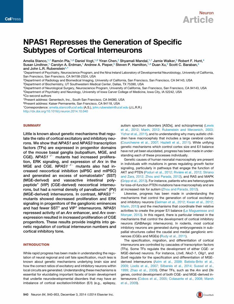
NPAS1 Represses the Generation of SpecificSubtypes of Cortical InterneuronsAmelia Stanco,1,* Ramon Pla,1,6 Daniel Vogt,1,6 Yiran Chen,2 Shyamali Mandal,1,7 Jamie Walker,3 Robert F. Hunt,4
Susan Lindtner,1 Carolyn A. Erdman,1 Andrew A. Pieper,5 Steven P. Hamilton,1,8 Duan Xu,2 Scott C. Baraban,4
and John L.R. Rubenstein1,*1Department of Psychiatry, Neuroscience Program, and the Nina Ireland Laboratory of Developmental Neurobiology, University of California,San Francisco, San Francisco, CA 94158-2324, USA2Department of Radiology and Biomedical Imaging, University of California, San Francisco, San Francisco, CA 94143, USA3Department of Biochemistry, UT Southwestern Medical Center, Dallas, TX 75390, USA4Department of Neurological Surgery, Neuroscience Program, University of California, San Francisco, San Francisco, CA 94143, USA5Department of Psychiatry and Neurology, University of Iowa Carver College of Medicine, Iowa City, IA 52242, USA6Co-second authors7Present address: Genentech, Inc., South San Francisco, CA 94080, USA8Present address: Kaiser Permanente, San Francisco, CA 94118, USA*Correspondence: [email protected] (A.S.), [email protected] (J.L.R.R.)
http://dx.doi.org/10.1016/j.neuron.2014.10.040
SUMMARY
Little is known about genetic mechanisms that regu-late the ratio of cortical excitatory and inhibitory neu-rons. We show that NPAS1 and NPAS3 transcriptionfactors (TFs) are expressed in progenitor domainsof the mouse basal ganglia (subpallium, MGE, andCGE). NPAS1�/� mutants had increased prolifera-tion, ERK signaling, and expression of Arx in theMGE and CGE. NPAS1�/� mutants also had in-creased neocortical inhibition (sIPSC and mIPSC)and generated an excess of somatostatin+ (SST)(MGE-derived) and vasoactive intestinal poly-peptide+ (VIP) (CGE-derived) neocortical interneu-rons, but had a normal density of parvalbumin+ (PV)(MGE-derived) interneurons. In contrast, NPAS3�/�
mutants showed decreased proliferation and ERKsignaling in progenitors of the ganglionic eminencesand had fewer SST+ and VIP+ interneurons. NPAS1repressed activity of an Arx enhancer, and Arx over-expression resulted in increased proliferation of CGEprogenitors. These results provide insights into ge-netic regulation of cortical interneuron numbers andcortical inhibitory tone.
INTRODUCTION
While rapid progress has been made in understanding the regu-
lation of neural regional and cell fate specification, much less is
known about genetic mechanisms underlying brain size and
how the correct ratios of excitatory and inhibitory neurons within
local circuits are generated. Understanding thesemechanisms is
essential for elucidating important facets of brain development
that underlie neurodevelopmental disorders that may have an
imbalance of cortical excitation/inhibition (E/I) (e.g., epilepsy,
940 Neuron 84, 940–953, December 3, 2014 ª2014 Elsevier Inc.
autism spectrum disorders [ASDs], and schizophrenia) (Lewis
et al., 2012; Marın, 2012; Rubenstein and Merzenich, 2003;
Yizhar et al., 2011), and to understanding why many autistic chil-
dren have macrocephaly that includes a large cerebral cortex
(Courchesne et al., 2007; Hazlett et al., 2011). While unifying
genetic mechanisms which control cortex size and E/I balance
have not yet been elucidated, progress has beenmade in under-
standing each of these processes individually.
Genetic causes of human neonatal macrocephaly are present
in individuals with mutations in genes regulating growth factor
signaling, particularly in pathways that engage the function of
AKT and PTEN (Poduri et al., 2012; Riviere et al., 2012; Striano
and Zara, 2012; Zhou and Parada, 2012), and RAS and MAPK
(Gripp et al., 2013). For instance, patients who are heterozygotes
for loss-of-function PTENmutations have macrocephaly and are
at increased risk for autism (Zhou and Parada, 2012).
Likewise, progress has been made in understanding the
mechanisms that control the generation of cortical excitatory
and inhibitory neurons (Gelman et al., 2012; Kwan et al., 2012;
Marın, 2013) and the mechanisms that coordinate their relative
activities to create the proper E/I balance (Le Magueresse and
Monyer, 2013). In this regard, there is particular interest in the
mechanisms that control the development of cortical inhibitory
neurons (GABAergic interneurons). In rodents, most cortical
inhibitory neurons are generated during embryogenesis in sub-
pallial structures called the caudal and medial ganglionic emi-
nences (CGEs and MGEs) (Rudy et al., 2011).
The specification, migration, and differentiation of cortical
interneurons are controlled by cascades of transcription factors
(TFs). Some TFs regulate the development of either CGE- or
MGE-derived neurons. For instance, Lhx6, Nkx2-1, Olig1, and
Sox6 regulate for the specification and differentiation of MGE-
derived interneurons (Azim et al., 2009; Batista-Brito et al.,
2009; Liodis et al., 2007; Silbereis et al., 2014; Sussel et al.,
1999; Zhao et al., 2008). Other TFs, such as the Arx and Dlx
genes, control development of both CGE- and MGE-derived in-
terneurons (Cobos et al., 2005; Colasante et al., 2008; Marsh
et al., 2009).

Neuron
NPAS1 Represses Interneuron Generation
Herein, we present evidence that themouseNPAS basic helix-
loop-helix (bHLH)-PAS TF genes (Erbel-Sieler et al., 2004; Pieper
et al., 2005; Zhou et al., 1997) are regulators of cortex size and E/I
balance.NPAS1 andNPAS3 are expressed in telencephalic pro-
genitor domains of the cortex, and the CGE and MGE, and later
in immature andmature cortical interneurons (Batista-Brito et al.,
2008; Erbel-Sieler et al., 2004; Zhao et al., 2008). Vertebrate
NPAS function in embryonic neural progenitors may be related
to the function of Trachealess, the NPAS Drosophila homolog.
Trachealess modulates fibroblast growth factor (FGF) signaling
by transcriptional regulation of the FGF receptor (Ohshiro and
Saigo, 1997). In the adult mouse hippocampus,NPAS3 regulates
expression of FGFR1 to control proliferation of hippocampal
granule neurons (Pieper et al., 2005). Here, we have found that
NPAS1 negatively regulates proliferation and MAPK signaling
in CGE and MGE progenitors, not by regulating FGF receptor
expression but through an unexpected mechanism, repression
of Arx expression. As a result, NPAS1�/� mutants generated
excessive cortical interneurons prenatally, which persisted into
adulthood. NPAS1�/� mutants also had increased neocortical
inhibition (sIPSC and mIPSC) and generated an excess of
SST+ (MGE-derived) and VIP+ (CGE-derived) neocortical inter-
neurons, but had normal numbers of PV+ (MGE-derived) inter-
neurons. In contrast, NPAS3�/� mutants had a complementary
phenotype, with reduced proliferation and MAPK signaling in
progenitors of the ganglionic eminences, and the postnatal cor-
tex had fewer SST+ and VIP+ interneurons.
We propose that our analysis of NPAS1 and NPAS3 functions
in mice provides mechanistic insights into human neuropsychi-
atric disorders, as NPAS3 dysfunction is implicated in schizo-
phrenia (Kamnasaran et al., 2003; Macintyre et al., 2010).
Furthermore, we have identified sporadic nonsynonymous mu-
tations in NPAS1 and NPAS3 in autistic individuals.
RESULTS
NPAS1 and NPAS3 Expression during InterneuronDevelopmentThe subpallium generates neocortical interneurons (Flandin
et al., 2011; Marın, 2012; Rudy et al., 2011). We examined
NPAS1 and NPAS3 RNA expression by in situ hybridization
(ISH) at E13.5, E15.5, and P5 (Figure 1) and assessed NPAS1
and NPAS3 expression by immunofluorescence at P0, P5,
P15, and P30 (Figure 1, and see Figures S1 and S2 and Tables
S1 and S2 available online). At E13.5, both had pallial and sub-
pallial ventricular zone (VZ) expression. NPAS1 showed notable
expression in the VZ and subventricular zone (SVZ) of the dorsal
and ventral MGE, and CGE. By E15.5, NPAS3 was expressed in
the MGE mantle zone; NPAS1 expression was prominent in the
pallial and subpallial SVZ.
Previous studies have described coexpression of NPAS1 or
NPAS3with cortical interneurons using GABA, GAD-67, or calre-
tinin antibodies in the adultmouse brain (Erbel-Sieler et al., 2004).
We have extended coexpression analysis of NPAS1 or NPAS3
with various interneuron markers during cortical interneuron
development and in the adult (Figures 1, S1, and S2; Tables S1
and S2). At P0, NPAS1 was expressed in neocortical interneu-
rons; �100% of NPAS1+ cells express GAD67-GFP; �30%
had MGE-like properties (Lhx6-GFP+) (Figures 1D, 1F, S1A,
and S1B). By P5, NPAS1 and NPAS3 were expressed in rostro-
caudal gradients in neocortical interneurons; we are unaware of
other TFswith this property. Virtually all neocortical NPAS1+ cells
(99% ± 0.29%) and the majority of NPAS3+ cells (67% ± 2.94%)
expressed GAD67-GFP at P5 (Figures S1C and S1D). By P15,
NPAS1 and NPAS3 were expressed by a majority of reelin+
(NPAS1, 68% ± 2.78%; NPAS3, 79% ± 4.79%) and SST+
(NPAS1, 65% ± 1.95%; NPAS3, 75% ± 0.44%) interneurons.
Both NPAS1 and NPAS3 were expressed in a small proportion
of PV+ cells (NPAS1, 6% ± 0.85%) (NPAS3, 13% ± 1.29%) (Fig-
ures S1E, S1F, S1H–S1J, and S1L). At P30, NPAS1+ cells coex-
pressed reelin, SST, calretinin (CR), or neuropeptide Y (NPY) but
rarely coexpressed PV (reelin, 42%± 1.94%; SST, 36%± 3.72%;
CR, 28%± 2.89%; NPY, 12%± 0.70%; PV, 5%± 1.24%). On the
other hand, NPAS3+ was expressed in a large fraction of all inter-
neuron subtypes assayed, including PV (reelin, 74% ± 2.31%;
SST, 75% ± 3.52%; CR, 51% ± 1.23%; PV, 43% ± 1.40%) (Fig-
ures S2B–S2E and S2G–S2J; data not shown).
Increased Numbers of Neocortical Interneurons inNPAS1–/– MutantsWe studied the effect of an NPAS1 null allele (NPAS1�/�) (Erbel-Sieler et al., 2004) on neocortical interneuron development using
a GAD67-GFP allele to label all of the interneurons (Tamamaki
et al., 2003). By E15.5 there was an increased density of GFP+
interneurons through the intermediate zone and then throughout
the cortical wall at E17.5 and P0 (26%–41%; E15.5, 26% ±
4.67%, p = 0.00099; E17.5, 35% ± 2.08%, p = 9.08E-8; P0,
41% ± 4.46%, p = 2.48E-6) (Figures 2A–2C and 2E–2G). Even
though there was �2-fold increased interneuron cell death at
P7 (activated caspase-3, Figures S3A and S3B), P30 mice main-
tained �15% (15% ± 5.43%, p = 0.044) more interneurons
(Figures 2D and 2H). Surprisingly, while SST+, VIP+, NPY+, and
reelin+ interneuron subtypes were increased (28%–44%; SST,
32% ± 5.07%, p = 0.001; VIP, 44% ± 7.99%, p = 0.014; NPY,
34% ± 7.42%, p = 0.0019; reelin, 28% ± 3.01%, p = 2.7E-5),
PV+ interneuron density was normal (Figures 3A–3E, 3A0–3E0,and 3F–3J). MRI quantification showed that P30 cortical volume
was increased 10% (10% ± 1.40%, p = 0.0055) (Figures S3F and
S3G; Table S3). Nissl section analysis supported the MRI find-
ings, and showed 11% increased (11%± 3.13%, p = 0.03) rostral
neocortical width (Figures S2A and S2B). Assessment of NeuN+
(neuronal marker) cells in the P21 somatosensory cortex sug-
gests that the enhancement in cortical volume may be the result
of increased neuron numbers. In NPAS1�/�mutants, the density
of NeuN+ cells was increased by 11% (cortex, 11% ± 2.21%, p =
0.0011; layer I, 22% ± 7.34%, p = 0.030; layer IV, 7% ± 2.09%,
p = 0.025; layer V, 16% ± 3.32%, p = 0.0024; layer VI, 13% ±
2.66%, p = 0.0052) (Figures S4A and S4B).
Increased Synaptic Inhibition onto NeocorticalPyramidal Neurons in NPAS1–/–
To test whether the increase in cortical interneurons altered the
physiology of the NPAS1�/� cortex, we performed patch-clamp
recordings of layer II/III somatosensory pyramidal neurons in
slices from P21-30 NPAS1�/� mice and wild-type (WT) litter-
mates (Figures 4A–4C). Compared to WT littermates, pyramidal
Neuron 84, 940–953, December 3, 2014 ª2014 Elsevier Inc. 941

Figure 1. Forebrain Expression of Mouse NPAS1 and NPAS3 during Embryonic and Neonatal Stages(A and B) ISH on rostrocaudal series of coronal hemisections at E13.5 (A) and E15.5 (B). Note strong NPAS1 expression in dMGE and CGE.
(C) ISH on neonatal sagittal sections at P5. Note the rostral bias of cortical expression.
(D–G) Quantification of percentage of NPAS1+ (immuno-stained) cortical interneurons (GAD67-GFP+ and Lhx6-GFP+) at P0. n = 3 animals for (D)–(G). Abbre-
viations are as follows: CGE, caudal ganglionic eminence; Cx, cortex (rCX, rostral; cCx, caudal); GP, globus pallidus; Hi, hippocampus; MGE, medial ganglionic
eminence (dMGE, dorsal; vMGE, ventral). Scale bars, (A) 1.21 mm, (B) 1.74 mm, and (C) 3 mm. See also Figures S1 and S2.
Neuron
NPAS1 Represses Interneuron Generation
neurons from NPAS1�/� mice showed an increase in the fre-
quency of spontaneous IPSCs (sIPSCs) (WT, 8.17 ± 1.29 Hz;
NPAS1�/�, 11.18 ± 1.26 Hz; p = 0.042). However, the amplitudes
and kinetics of inhibitory events were not significantly changed
(Figures 4A and 4B). To further characterize the increase in
GABA-mediated synaptic inhibition, tetrodotoxin (TTX) was
added to the ACSF to isolate miniature IPSCs (mIPSCs). Similar
to spontaneous events, an increase in mIPSC frequencies was
observed in NPAS1�/� mice (WT, 6.2 ± 1.24 Hz; NPAS1�/�,8.74 ± 1.02 Hz; p = 0.036); amplitudes and kinetics of mIPSCs
were not significantly changed (Figure 4C).
942 Neuron 84, 940–953, December 3, 2014 ª2014 Elsevier Inc.
Next, we evaluated the ratio of interneuron numbers to total
neuron numbers by quantifying the percentage of NeuN+ cells
(total neurons) that express GAD67-GFP in each layer of P21
somatosensory cortex (Figures S4B and S4C). At P21, all
NeuN+ cells were GAD67-GFP+ in layer I somatosensory cortex.
NPAS1�/� mutant mice displayed a 19% increase (19% ±
5.94%, p = 0.025) in GAD67-GFP+ interneurons within layer I
(Figure S4C). In layer II/III, the percentage of NeuN+ cells that
express GAD67-GFP was increased by 18% in NPAS1�/� mu-
tants (NPAS1+/+, 27% ± 1.13%; NPAS1�/�, 32% ± 0.96%, p =
0.0024) (Figures 4D–4I). We also estimated the excitatory to

Figure 2. NPAS1–/– Mice Have Increased Numbers and Density of GAD67-GFP+ Cortical Interneurons
(A–D) Increase in GAD67-GFP+ cortical interneurons (somatosensory Cx) beginning by E15.5 shown on coronal cortical sections at E15.5 (A), E17.5 (B), P0 (C),
and P30 (D) in NPAS1�/� mice.
(E–H) Quantification of GAD67-GFP+ neurons/104 mm2 at E15.5 (E), E17.5 (F), P0 (G), and GAD67-GFP+ neurons/1 mm2 at P30 (H). n = 3 animals per genotype for
(E)–(H). Abbreviations are as follows: CP, cortical plate; Cx, cortex; IZ, intermediate zone; MZ, marginal zone; SVZ, subventricular zone. *p < 0.05. ***p < 0.001.
Scale bars, (A)–(C), 200 mm; (D), 400 mm. See also Figure S3.
Neuron
NPAS1 Represses Interneuron Generation
inhibitory cell ratio (E/I) in each cortical layer at P21. The E/I
ratio, determined as the ratio of NeuN+ and GAD67� cells to
GAD67+ cells, was decreased by 23% in layer II/III of the
NPAS1�/� somatosensory cortex (NPAS1+/+, 2.74 ± 0.17;
NPAS1�/�, 2.12 ± 0.091, p = 0.0040) (Figure S4D). No signifi-
cant change in the E/I cell ratio was found in deep cortical layers
(IV, V, or VI) (Figure S4D). These findings were supported by
stereological quantification of NeuN+ and GAD67+ cells within
the cerebral cortex. The NPAS1�/� mutant cerebral cortex ex-
hibited a 35% increase (35% ± 5.96%, p = 0.0053) in NeuN+
cells and a 57% increase (57% ± 10.49%, p = 0.0012) in
GAD67+ cells at 3 months of age (Figures S4E–S4G). Cortical
pyramidal cell density was unchanged in P21 NPAS1�/� mu-
tants as indicated by Cux1+, Ctip2+, and Tbr1+ immunofluores-
cence staining which labels layer II/III, layer V, and layer VI,
respectively (Figures S4H–S4K). Thus, the enhanced level of
synaptic inhibition onto layer II/III neocortical pyramidal neurons
in the NPAS1�/� mutant was associated with an increased frac-
tion of inhibitory neurons.
NPAS1–/– andNPAS3–/–HadOpposite Effects on CorticalInterneuron NumbersNPAS3�/� mice display reduced proliferation and size of the
adult hippocampal dentate gyrus (Pieper et al., 2005). Given
that NPAS1�/� mutants have increased cortical volume (Figures
S3F and S3G) and interneuron numbers (Figures 3A–3C, 3E, 3A0–3C0, 3E0, 3F–3H, and 3J), we hypothesized that NPAS1 and
NPAS3 may have opposing functions on interneuron develop-
ment. Therefore, we compared interneuron numbers in
NPAS1�/�, NPAS3�/�, and NPAS1/3�/� P30 neocortices. While
NPAS1�/� mutants had increased density of cortical interneu-
rons (Figures 3A–3C, 3E, 3A0–3C0, 3E0, 3F–3H, and 3J),
NPAS3�/� mutants had reduced density of SST+, VIP+, NPY+,
and reelin+ interneurons (Figures 3A-3C, 3E, 3A0 0–3C0 0, 3E0 0,
Neuron 84, 940–953, December 3, 2014 ª2014 Elsevier Inc. 943

Figure 3. NPAS1–/– Mice Have Increased,
whereas NPAS3–/– and NPAS1/3–/– Mice
Have Decreased, Cortical Interneurons
(A–E0 0 0) Interneurons expressing VIP, reelin, NPY,
and somatostatin are increased inNPAS1�/�mice
but decreased in NPAS3�/� and NPAS1/3�/�
mice. All mutants have a normal density of
parvalbumin+ interneurons. Immunofluorescence
staining of coronal neocortical sections (somato-
sensory Cx) at P30.
(F–J) Quantification of VIP+ (F), reelin+ (G), NPY+
(H), parvalbumin+ (I), and somatostatin+ (J) neu-
rons/1 mm2. n = 3 animals per genotype for (F)–(J).
*p < 0.05. ***p < 0.001. Scale bar, (A)–(E0 0 0) 400 mm.
Neuron
NPAS1 Represses Interneuron Generation
3F–3H, and 3J). LikeNPAS1�/�mutants, there was not a change
in the density of PV+ interneurons (Figures 3D–3D0 0 and 3I).
MRI quantification revealed that P30 cortical volume was
increased in NPAS1�/� mutant mice, as opposed to a 21%
decrease (21% ± 1.47%, p = 0.0039) in NPAS3�/� mutants.
Also, we found differential changes in P30 NPAS1�/� and
NPAS3�/� mutant basal ganglia volumes by MRI. NPAS1�/�
mutant mice displayed a 12% increase (12% ± 1.74%, p =
0.042) and NPAS3�/� mutants a 25% decrease (25% ± 2.24%,
p = 0.0087) in basal ganglia volume (Figures S3F–S3H; Table S3).
NPAS1/3�/� and NPAS3�/� mutants had similar interneuron
phenotypes (Figures 3A-3E, 3A0 0–3E0 0, 3A0 0 0–3E0 0 0, and 3F–3J).
944 Neuron 84, 940–953, December 3, 2014 ª2014 Elsevier Inc.
In addition, we observed probable
NPAS1�/�, NPAS3�/�, and NPAS1/3�/�
mutant basal ganglia phenotypes (n =
1), which include subtle cholinergic de-
fects (NPAS1 and NPAS3 are expressed
in the pallidum) (Figure 1 and data not
shown) (Flandin et al., 2010; Nobrega-
Pereira et al., 2010). The distribution of
choline acetyltransferase (ChAT)+ cells
around the globus pallidus (GP) was
abnormal in NPAS1/3�/� and NPAS3�/�
mutants. We detected increased ChAT+
cells inside the GP and reduced numbers
in the ventral pallidum.While parvalbumin
(PV)+ expression in the GP was not
grossly abnormal in the mutants, the GP
may be enlarged in the NPAS1�/� mutant
and smaller in the NPAS3�/� mutant. PV+
interneuron numbers in the striatum and
basolateral nucleus of the amygdala ap-
peared grossly normal in all three mutant
genotypes (data not shown).
NPAS1–/– and NPAS3–/– GanglionicEminences Had OppositeProliferation and ERK-SignalingPhenotypesGiven that NPAS1 and NPAS3 are ex-
pressed in MGE and CGE progenitors
(Figures 1A and 1B), we explored whether
changes in interneuron numbers in
NPAS1�/� andNPAS3�/� mutants were due to changes in prolif-
eration using a marker of M phase cells (phosphohistone H3,
PH3). We found that NPAS1�/� mutants had a 1.6-fold increase
(1.61 ± 0.13, p = 0.0007) in PH3+ cells in the VZ, and a 1.3-fold in-
crease (1.31 ± 0.039, p = 0.0033) in the SVZ of the CGE at E13.5;
at E15.5 NPAS1�/� mutants had a 1.3-fold increase (VZ, 1.32 ±
0.042, p = 1.09E-5; SVZ, 1.30 ± 0.035, p = 0.022) in PH3+ cells
in the VZ and SVZ of the MGE (Figures 5A, 5B, 5E, 5F, 5I, and
5K). In contrast, NPAS3�/� progenitors displayed reduced prolif-
eration in the E13.5 MGE, including a 1.3-fold decrease (1.26 ±
0.043, p = 0.010) in PH3+ cells in the VZ and a 1.4-fold decrease
(1.44 ± 0.031, p = 6.54E-5) in the SVZ (Figures 5Q, 5R, and 5W).

Figure 4. Synaptic Inhibition to Neocortical Pyramidal Cells Is Increased in NPAS1–/– Mice
(A) Representative traces of sIPSCs recorded from layer II/III pyramidal neurons in somatosensory neocortex of WT and NPAS1�/� mice.
(B andC)Mean sIPSC (B) andmIPSC (C) frequencieswere higher in pyramidal neurons recorded fromNPAS1�/�mice than in controls. Numbers of recorded cells
are shown in each bar. No significant changes in sIPSC or mIPSC amplitude, 10%–90% rise time, or decay time constant were detected. For (B) and (C), n = 7WT
and 9 NPAS1�/� animals.
(D–F) Increase in GAD67-GFP+ interneurons (D) and percentage of neurons (NeuN+) that are inhibitory (GAD67-GFP+) (F) within layer II/III of NPAS1�/� so-
matosensory cortex (white boxes) shown on coronal sections at P21. The density of NeuN+ cells within layer II/III is not significantly increased in NPAS1�/�
mutants (E).
(G–I) Quantification of GAD67-GFP+ (G) andNeuN+ (H) neurons/105 mm2 and percentage of NeuN+ cells that expressGAD67-GFPwithin that area (I). n = 3 animals
per genotype for (G)–(I). *p < 0.05. **p < 0.01. ***p < 0.001. Scale bar, (D)–(F), 200 mm. See also Figure S4.
Neuron
NPAS1 Represses Interneuron Generation
BrdU labeling analysis of E13.5 MGE and CGE progenitors,
utilized to detect cells in S phase, supported the PH3 results.
One hour BrdU pulse labeling showed increased proliferation
in the SVZ of the NPAS1�/� mutant MGE (total number, 31% ±
1.26%, p = 0.0007; density, 48% ± 2.15%, p = 0.0004) and
CGE (total number, 19% ± 3.31%, p = 0.039; density, 34% ±
5.24%, p = 0.022), while NPAS3�/� mutant mice exhibited
decreased proliferation in the MGE SVZ (total number, 26% ±
2.05%, p = 0.0018; density, 21% ± 3.08%, p = 0.023) (Figures
S5A–S5E). In addition, examination of proliferation in the hippo-
campal dentate gyrus following intraperitoneal injection of BrdU
once daily for 12 days revealed a 31% increase (31% ± 7.89%,
p = 0.003) in BrdU+ cells in 5- to 10-week-old NPAS1�/� mice
(Figures S5F and S5G).
The NPAS Drosophila homolog Trachealess regulates FGF
signaling (Ohshiro and Saigo, 1997); thus we examined activa-
tion of the MAP kinase pathway by measuring the level of phos-
pho-ERK immunofluorescence. NPAS1 mutants had a 1.3-fold
increase (E13.5 CGE, 1.32 ± 0.043, p = 0.0016; E15.5 MGE,
1.35 ± 0.063, p = 0.00017) in phospho-ERK expression in the
E13.5 CGE and E15.5 MGE, whereas NPAS3 mutants exhibited
a 1.3-fold decrease (1.26 ± 0.060, p = 0.0079) in phospho-ERK
expression in the E13.5 CGE and a 1.2-fold decrease (1.17 ±
0.024, p = 0.023) in the E13.5 MGE (Figures 5C, 5D, 5G, 5H,
5J, 5L, 5O, 5P, 5S, 5T, 5V, and 5X).
NPAS1–/– Ganglionic Eminences Displayed IncreasedArxExpressionwhichMediated an Increase in SubpallialMAP Kinase Activity and ProliferationWe focused on identifying molecular mechanisms underlying the
increased proliferation and phospho-ERK expression in theMGE
and CGE of NPAS1 mutants by ISH screening of the MGE and
CGE for expression of genes that are implicated in these pro-
cesses (Arx, COUP-TFI, Cyclin D1, Cyclin D2, Dlx1, ErbB4,
FGFR1, FGFR3, Lhx2, Lhx6, NPAS3, Olig1, Sp8, and Sprouty2)
(Figures 6 and S5; data not shown). While we did not find robust
changes formost of these, including FGF-signaling components,
there were some interesting changes in Arx and Sp8 expression.
In accordance with the increase in CGE proliferation, there was a
subtle increase in Sp8+ (LGE/CGE marker) migrating interneu-
rons at E13.5 (Figure 6B) (Ma et al., 2012). We also observed a
clear increase of Arx TF RNA at E13.5 and E15.5 in regions of
the MGE and CGE where NPAS1 is normally expressed (Figures
6A and 6C). Arx function is necessary for proper interneuron
migration and maturation (Colombo et al., 2007; Kitamura
et al., 2002; Marsh et al., 2009), as well as neocortical neuron
Neuron 84, 940–953, December 3, 2014 ª2014 Elsevier Inc. 945

Figure 5. NPAS1–/– Progenitors Have Increased Proliferation and MAP Kinase Activity, while NPAS3–/– Progenitors Exhibit Decreased
Proliferation and MAP Kinase Activity
(A–H) Immunofluorescence assays of coronal sections demonstrate increased proliferation (phosphohistone H3+ M phase cells) and MAP kinase activity
(phospho-ERK) in the E13.5 CGE (A–D), and E15.5 MGE (E–H) of NPAS1�/� progenitors.
(I–L) Quantification of phosphohistone H3+ (I and K) cells and phospho-ERK (J and L) levels in NPAS1�/� mutants. n = 3 animals per genotype for (I)–(L).
(M–T)NPAS3�/� progenitors display reduced proliferation andMAP kinase activity in the E13.5MGE (Q–T). WhileMAP kinase activity was decreased in the E13.5
CGE (O and P), proliferation of progenitors was unaltered in the E13.5 CGE (M and N) of NPAS3�/� mutant mice.
(U–X) Quantification of phosphohistone H3+ (U and W) cells and phospho-ERK (V and X) levels in NPAS3�/� mutants. Proliferation was measured in the VZ and
SVZ (white boxes). Phospho-ERK levels in the CGE/MGE (white box) were normalized by comparison with cortical levels (yellow box). n = 3 animals per genotype
for (U)–(X). Abbreviations are as follows: CGE, caudal ganglionic eminence; Cx, cortex; MGE, medial ganglionic eminence; SVZ, subventricular zone; VZ, ven-
tricular zone. *p < 0.05. **p < 0.01. ***p < 0.001. Scale bar, (A)–(H) and (M)–(T), 250 mm. See also Figure S5.
Neuron
NPAS1 Represses Interneuron Generation
progenitor proliferation (Friocourt et al., 2008; Kitamura et al.,
2002).
To test whether NPAS1 and its cofactor, ARNT, can directly
repress Arx expression through a known Arx subpallial enhancer
element (Colasante et al., 2008; Visel et al., 2013), we performed
luciferase assays in CGE primary cultures (Flandin et al., 2011).
We found that this enhancer had two putative NPAS1/ARNT
sites, designated A and B (Figure S7D) (Hogenesch et al.,
1997). Consistent with the aforementioned phenotype, NPAS1/
ARNT induced an �2-fold repression of the Arx enhancer
(0.58 ± 0.035, p = 6.69E-5). Mutation of the Arx enhancer at
site A, but not site B, resulted in a rescue of the repression
induced by NPAS1/ARNT (p = 0.034) (Figures 7C and 7D).
Next, to investigate whether the increase in Arx expression
(Figures 6A and 6C) could alter subpallial development, we
used ultrasound to guide injection into the E13.5 CGE of a lenti-
946 Neuron 84, 940–953, December 3, 2014 ª2014 Elsevier Inc.
virus encoding either Arx and GFP, or GFP alone. At E15.5, we
compared the number of PH3+/GFP+ cells and observed that
the Arx-encoding virus increased PH3+/GFP+ cells by �2-fold
(2.18 ± 0.039, p = 0.042) (Figures 7A, 7B, and S7A). We also as-
sessed the level of phospho-ERK expression in GFP+ regions of
the CGE ventricular zone and found that Arx overexpression
increased phospho-ERK expression by 1.4-fold (1.39 ± 0.12,
p = 0.046) (Figures S7B and S7C). These findings provide evi-
dence that NPAS1 repression of Arx can regulate CGE prolifera-
tion and MAP kinase activity.
Nonsynonymous Mutations in NPAS1 and NPAS3 AreFound in Humans with ASDNPAS3 mutations are observed in patients with schizophrenia
(Kamnasaran et al., 2003; Macintyre et al., 2010). Given that
ASDs and schizophrenia share some genetic risk factors

Figure 6. NPAS1–/– GEs Have Increased Arx
Expression, whereas NPAS1–/– Cortices
Reveal Increased Sp8 Expression
(A) ISH assay on coronal hemisections arrayed in a
rostrocaudal series reveal that NPAS1�/� pro-
genitors have increased Arx expression in the VZ/
SVZ regions of the E13.5 MGE (arrows) and
CGE(arrowheads) (compare white arrow and
arrowhead in bottom tier [NPAS1�/� ] to middle
tier [WT]) where NPAS1 is expressed (black arrow
and arrowhead in top tier).
(B) At E13.5, Sp8 expression appears increased in
a pattern consistent with tangentially migrating
cortical interneurons (arrows). Data are presented
in coronal sections at three rostrocaudal planes
(left to right).
(C) ISH assay on coronal hemisections displayed
in a rostrocaudal series shows that NPAS1�/�
progenitors have increased Arx expression in the
VZ/SVZ regions of the E15.5 MGE (arrows) and
CGE (arrowheads) (compare white arrow and
arrowhead in bottom tier [NPAS1�/�] to middle tier
[WT]) where NPAS1 is expressed (black arrow and
arrowhead in top tier). Abbreviations are as fol-
lows: CGE, caudal ganglionic eminence; MGE,
medial ganglionic eminence. Scale bar, (A) 1 mm,
(B) 723 mm, and (C) 1.25 mm. See also Figure S6.
Neuron
NPAS1 Represses Interneuron Generation
(Murdoch and State, 2013) and, like NPAS1 mutant mice,
enlarged brain size is frequent in ASD children (Butler et al.,
2005; Hazlett et al., 2011), we screened 947 ASD probands for
mutations inNPAS1 andNPAS3 exons, introns, and 50 and 30 un-translated regions, and found ten nonsynonymous variants in
NPAS1 and seven inNPAS3 (see the Supplemental Information).
We assayed 13 of those in 190 control subjects (unscreened for
autism) and found that five of seven in NPAS1 and all six in
NPAS3 were absent in the controls. Although the data lack sta-
Neuron 84, 940–953,
tistical power and do not unambiguously
implicate these variants in genetic sus-
ceptibility to ASD, these results support
the possibility that NPAS1 and NPAS3
mutations are related to ASD risk.
DISCUSSION
NPAS1�/� mice had increased numbers
of neocortical interneurons, whereas
NPAS3�/� mice have fewer interneurons
(Figures 3A–3C, 3E, 3A0–3C0, 3E0, 3A0 0–3C0 0, 3E0 0, 3F–3H, and 3J). These changes
in interneuron numbers were associated
with altered ERK signaling and cell prolif-
eration within the MGE and CGE of
NPAS1�/� and NPAS3�/� mutants (Fig-
ure 5). In addition, the increased numbers
of neocortical interneurons in NPAS1�/�
mice resulted in enhanced synaptic inhi-
bition onto neocortical pyramidal neurons
in layer II/III of the somatosensory cortex
(Figures 4A–4C). We focused on how NPAS1 regulates this
process in the MGE and CGE and found that NPAS1�/� mutants
had increased Arx expression in these embryonic progenitor
zones (Figures 6A and 6C). Using in utero transduction of Arx
into the CGE, we showed that increased Arx expression was suf-
ficient to increase subpallial proliferation andMAP kinase activity
(Figures 7A, 7B, S7B, and S7C). We also demonstrated that
NPAS1 could directly repress a bone fideArx subpallial enhancer
(Figures 7C, 7D, and S7D) (Colasante et al., 2008).
December 3, 2014 ª2014 Elsevier Inc. 947

Figure 7. Increased Arx Expression in the CGE Mediates Increased Subpallial Proliferation
(A and B) In utero lentiviral transduction of Arx into the E13.5 CGE results in increased cell proliferation (PH3). Arrowheads in (A) indicate GFP+/PH3+ cells. n = 3
animals per experimental condition for (B).
(C) Arx locus on mouse X chr, showing evolutionarily conserved domains, one of which is a subpallial enhancer (UAS3) (Colasante et al., 2008; Visel et al., 2013)
that has two predicted NPAS1/ARNT sites (Figure S6).
(D) Transcription assay of E12.5 CGE primary culture shows that NPAS1/ARNT expression represses activity of the Arx subpallial enhancer. Mutation
of the Arx enhancer at site A, but not site B, resulted in a rescue of the repression induced by NPAS1/ARNT. n = 3 independent experiments, each
performed in triplicate for (D). Abbreviations are as follows: CGE, caudal ganglionic eminence. *p < 0.05. **p < 0.01. Scale bar, (A) 70 mm. See also
Figure S7.
Neuron
NPAS1 Represses Interneuron Generation
Our results provide insights into mechanisms that control the
number of specific subtypes of cortical inhibitory neurons,
showing that homologous TF-encoding genes NPAS1 and
948 Neuron 84, 940–953, December 3, 2014 ª2014 Elsevier Inc.
NPAS3 have opposing functions in regulating the numbers of
SST+ and VIP+ interneurons (Figures 3A–3A0 0, 3E–3E0 0, 3F, and3J). Thus, we have provided evidence for a cell-autonomous

Neuron
NPAS1 Represses Interneuron Generation
mechanism that controls interneuron numbers, a process pro-
posed to exist based on results from the MGE-transplantation
assay (Southwell et al., 2012).
NPAS1 and NPAS3 Regulate the Proliferation ofGanglionic Eminence ProgenitorsProliferation and ERK signaling in progenitor domains of the
MGE and CGE were increased and decreased in NPAS1�/�
and NPAS3�/� mice, respectively (Figure 5). Vertebrate NPAS
function in embryonic neural precursors may be related to the
function of Trachealess, the NPAS Drosophila homolog. Tra-
chealess promotes FGF-signaling by increasing expression of
the FGF receptor (Ohshiro and Saigo, 1997). This program is
evolutionarily conserved based on the observation that
NPAS3�/� mice have reduced FGFR1 expression in the adult
hippocampus (Pieper et al., 2005). However, NPAS3�/� mutant
mice did not show this phenotype in the prenatal telencephalon
(data not shown). Also, NPAS1�/� mutants did not exhibit
decreased FGFR1 expression in the adult hippocampus (Pieper
et al., 2005), or in the prenatal telencephalon (Figure S6C). Thus,
there is no evidence that changes in phospho-ERK levels, in the
subpallium of NPAS1�/� and NPAS3�/� mutants (Figures 5C,
5D, 5G, 5H, 5J, 5L, 5O, 5P, 5S, 5T, 5V, and 5X), are related to
altered FGF signaling.
Our study reveals insights into NPAS1 regulation of cell prolif-
eration. Here, we have found that the increase in proliferation and
MAPK signaling in NPAS1�/� mutant CGE and MGE progenitors
was associated with increased Arx expression. Previous studies
of Arx mutant mice show that lack of this TF resulted in reduced
cortical proliferation (Friocourt et al., 2008; Kitamura et al., 2002).
We demonstrated that increased Arx expression was sufficient
to enhance subpallial proliferation andMAP kinase activity, using
in utero transduction of Arx into the CGE (Figures 7A, 7B, S7B,
and S7C). Furthermore, we showed that NPAS1 could directly
inhibit a bone fide Arx subpallial enhancer (Figures 7C, 7D, and
S7D) (Colasante et al., 2008).
Given that NPAS1 and NPAS3 have opposite effects on prolif-
eration, an imbalance of either NPAS1 or NPAS3 expression
and/or function could potentially be compensated by changes
in the expression of the other gene. However, we did not observe
changes of NPAS3 expression in the NPAS1�/� E13.5 and P0
forebrain (data not shown).
NPAS1 and NPAS3 Modified the Production of SpecificSubtypes of Cortical InterneuronsThe increase in Arx and phospho-ERK expression and the in-
crease in proliferation in theMGEandCGEofNPAS1�/�mutants
were associated with an increase in subpallially derived cortical
interneurons (Figure 2). As a result, NPAS1 mutants had exces-
sive SST+ and VIP+ interneurons, products of the MGE and
CGE, respectively (Figures 3A, 3A0, 3E, 3E0, 3F, and 3J) (Rudy
et al., 2011). In contrast, decreased phospho-ERK expression
and cell proliferation in the MGE and CGE of NPAS3�/� mutant
mice resulted in reduced numbers of SST+ and VIP+ interneurons
(Figures 3A, 3A0 0, 3E, 3E0 0, 3F, and 3J).
Both NPAS1 and NPAS3mutants had a normal density of PV+
interneurons (Figures 3D–3D0 0 and 3I). PV and SST are MGE
derived (Rudy et al., 2011). NPAS1, along with NPAS3, COUP-
TFI,Dlx1, and SatB1 (Cobos et al., 2005; Denaxa et al., 2012; Lo-
dato et al., 2011), define a set of TFs that differentially regulate
the numbers of interneurons that express PV and SST (Figures
3D–3D0 0, 3E–3E0 0, 3I, and 3J). At least one PV+ subtype, the
Chandelier neuron, is generated late in gestation (Inan et al.,
2012; Taniguchi et al., 2013); perhaps NPAS1 and NPAS3 re-
gulation of MGE proliferation is time dependent, and thus pre-
ferentially affects early-born SST+ interneurons. Based on the
embryonic phenotypes of NPAS1�/� and NPAS3�/� mutants,
we propose that NPAS1 and NPAS3 control the SST/PV ratio
via their functions in MGE progenitor cells. Future studies are
needed to examine the roles of NPAS1 and NPAS3 in regulating
interneurons via their expression in maturing and mature inter-
neurons (Figure 1C).
The significance of transcriptional repression in the regulation
of cortical interneuron number is becoming apparent. A recent
study demonstrates that Olig1 inhibits the generation of
GABAergic interneurons produced in the MGE by repression of
Dlx expression through the Dlx1/2 I12b intergenic enhancer (Sil-
bereis et al., 2014). Likewise, NPAS1 inhibits the generation of
cortical interneurons by repression of Arx expression through
the subpallial Arx enhancer, which mediates CGE progenitor
cell proliferation (Figure 7) (Colasante et al., 2008).
NPAS1 Function in Interneuron Generation ControlsCortical Excitation/InhibitionNPAS1�/� mutant mice display increased numbers of GAD67-
GFP+ interneurons in layer I (Figure S4C). It is likely that an
excess of GABAergic neurons in layer I would have an effect
on the inhibitory tone of pyramidal cell dendrites within this layer.
NPAS1 mutants also exhibit excessive SST+ and VIP+ interneu-
rons. VIP+ interneurons are enriched in layers II/III (Kawaguchi
and Kubota, 1997; Miyoshi et al., 2010) and form synapses
with pyramidal cell somata and dendrites (Kawaguchi and Ku-
bota, 1996) and other interneurons (Acsady et al., 1996; David
et al., 2007; Hajos et al., 1996). Thus, with an increase in both
SST+ and VIP+ interneurons, it is difficult to deduce the net phys-
iological effect a priori. We found that NPAS1 mutants had
enhanced cortical inhibition in layer II/III of the somatosensory
cortex (Figure 4), implying that the increase in interneurons re-
sulted in a net increase in inhibition in superficial cortical layers.
The enhanced cortical inhibition in layer II/III somatosensory cor-
tex of NPAS1�/� mutants suggests a decreased E/I ratio in layer
II/III somatosensory cortex. This is supported by estimation of
the cellular E/I ratio within this cortical region of NPAS1�/�
mutant mice (Figure S4D). While increased cortical inhibition is
likely to reduce noise in the cortex, and it could be protective
for epilepsy, it may be detrimental to cortical function by damp-
ening cortical signals and thereby predispose to neuropsychi-
atric disorders.
NPAS1 and NPAS3 in Human NeuropsychiatricDisordersOur examination of NPAS1 and NPAS3 functions in mice
provides mechanistic insights into human neuropsychiatric
disorders.NPAS3�/�mutant mice display behavioral and neuro-
anatomical deficits associated with human schizophrenia (Erbel-
Sieler et al., 2004), and NPAS3 mutations are observed in
Neuron 84, 940–953, December 3, 2014 ª2014 Elsevier Inc. 949

Neuron
NPAS1 Represses Interneuron Generation
patients with schizophrenia (Kamnasaran et al., 2003; Macintyre
et al., 2010). Furthermore, NPAS1 mutant mice exhibit an in-
crease in brain size (Figures S3D–S3G), similar to the enlarge-
ment seen in some children with ASD (Butler et al., 2005; Hazlett
et al., 2011). Notably, we have found sporadic nonsynonymous
mutations in NPAS1 and NPAS3 in individuals with ASD, which
suggests that NPAS1 and NPAS3mutations may be associated
with ASD risk. However, our analyses lack the statistical power
to conclude that these alleles contribute to ASD. Nonetheless,
these data, and the experimental analyses of the mouse mu-
tants, strongly suggest that further genetic analyses of NPAS1
and NPAS3 are warranted in human neuropsychiatric disorders.
EXPERIMENTAL PROCEDURES
Mice
NPAS1+/� and NPAS3+/� mice were obtained from Steven McKnight (Univer-
sity of Texas Southwestern Medical Center) and genotyped as described by
Erbel-Sieler and colleagues (Erbel-Sieler et al., 2004). NPAS1+/� mice were
maintained in a C57BL/6J strain background. NPAS1+/�; NPAS3+/� females
mated to NPAS1+/�; NPAS3+/� males also produced C57Bl/6J strain litters.
NPAS3�/� mutant mice obtained from this cross were utilized for BrdU anal-
ysis of the ganglionic eminences, assessment of cortical layer-specific
markers, and in the examination of interneuron markers in the P30 somato-
sensory cortex. However, due to increased mortality of NPAS3�/� mutants
maintained on a C57BL/6J strain background, subsequent experiments
were performed with NPAS3�/� mutant mice maintained on a CD-1 strain
background. These experiments include assessment of apoptosis within the
somatosensory cortex, examination of PH3 and phospho-ERK expression in
the ganglionic eminences, and MRI analysis.
The Lhx6-GFP BAC transgenic mouse line was obtained from the Gene
Expression Nervous System Atlas Project (GENSAT) at The Rockefeller
University (New York). GAD67-GFP mice were genotyped as described by
Tamamaki and colleagues (Tamamaki et al., 2003). As a note on nomenclature,
experimental samples designated as NPAS1+/+ indicate that the NPAS1 allele
is unaltered; however, data from these mice are not referred to asWT because
they have either the Lhx6-GFP or GAD67-GFP alleles. CD-1 WT mice were
obtained from Charles River Laboratories. WT littermate mice were used as
controls for all experiments. For staging of embryos, midday of the vaginal
plug was calculated as embryonic day 0.5 (E0.5). Mouse colonies were main-
tained at the University of California, San Francisco, and University of Texas
Southwestern Medical Center, in accordance with National Institutes of
Health, UCSF, and UT Southwestern guidelines.
Histology
See the Supplemental Experimental Procedures for details on immunohisto-
chemistry, ISH, BrdU labeling, and Nissl stain analysis.
MRI
P30 4% PFA-fixed brains were washed twice in 20 ml PBS for a total of 24 hr
and imaged in Fluorinert FC-40 (Sigma Aldrich) for null background signal. The
imaging was done on an 600 MHz NMR spectrometer (Agilent Technologies
Inc.) with imaging gradients and the following parameters: 3D gradient echo,
TE/TR 15/75 ms, eight averages, field of view (FOV) 12.8 mm isotropic, reso-
lution of 50 mm3 50 mm3 100 mm, and a total scan time of 5.5 hr. The acquired
images were converted on the Varian console to the DICOM format and refor-
matted into the same orientation as the histology sections using OsiriX, an
open source image viewer. Volumetric measurements were made using
custom-built software in MATLAB. Cortical and basal ganglia volumes were
statistically analyzed in three mice of each genotype. Results are presented
as mean ± SEM. Statistical differences between experimental groups were as-
sessedwith the Student’s t test using SPSS 15 software (IBM). See Table S3 in
the Supplemental Experimental Procedures for details on cortical and basal
ganglia volumes in control, NPAS1�/�, and NPAS3�/� mutants.
950 Neuron 84, 940–953, December 3, 2014 ª2014 Elsevier Inc.
Electrophysiology
Coronal brain slices (300 mm) were prepared from P21-30 WT and NPAS1�/�
mice. Slices were submerged in the recording chamber and continuously
perfused with oxygenated ACSF (32 –34�C) containing (in mM) 124 NaCl, 3
KCl, 1.25 NaH2PO4-H2O, 2 MgSO4-7H2O, 26 NaHCO3, 10 dextrose, and 2
CaCl2 (pH 7.2–7.4, 300–305 mOsm/kg). Whole-cell patch-clamp recordings
from layer II/III pyramidal cells in somatosensory neocortex were performed
at 403 using an upright, fixed-stage microscope (Olympus BX50WI) equipped
with infrared, differential interference contrast (IR-DIC). Patch pipettes (2–4
MU) were filled with an internal solution, containing (in mM) 140 CsCl, 1
MgCl2, 10 HEPES, 11 EGTA, 2 NaATP, 0.5 Na2GTP, and 1.25 QX-314 (pH
7.28). Recordings were obtained with an Axopatch 1D amplifier, filtered at 5
kHz, and recorded to pClamp 10.2 software (Clampfit, Axon Instruments).
Spontaneous (s) andminiature (m) IPSCs were examined at a holding potential
of �70 mV. GABAergic currents were isolated by adding 1 mM kynurenic acid
to the ACSF to block glutamate receptors, and tetrodotoxin (TTX, 2 mM) was
added to isolate mIPSCs. Series resistance was typically <15 MU and was
monitored throughout the recordings. Data were only used for analysis if the
series resistance remained <20MU and changed by%20%during the record-
ings. Data analysis was performed using pClamp 10.2 (Clampfit, Axon Instru-
ments), MiniAnalysis 6.0 (Synaptosoft), Microsoft Excel, and Sigmaplot 12.3
programs. Events characterized by a typical fast rising phase and exponential
decay phase were manually detected using MiniAnalysis. A 2 min sample
recording per cell was used for measuring IPSC frequency, amplitude, 10%–
90% rise time, and decay time constant. Kinetic analysis of the IPSCswas per-
formed with a single-exponential function. The threshold for event detection
was currents with amplitudes greater than three times the root mean square
(rms) noise level. Spontaneous IPSC measurements were recorded from 18
wild-type and 20 NPAS1�/� pyramidal cells. Miniature IPSC measurements
were recorded from 14 WT and 16 NPAS1�/� pyramidal cells. Seven WT
and nine NPAS1�/� animals were used for sIPSC and mIPSC measurements.
Results are expressed as mean ± SEM. Statistical differences between exper-
imental groups were assessed with the Mann-Whitney U test using SigmaPlot
12.3 software (SYSTAT).
Primary Cell Culture and Luciferase Assays
CGE tissue was dissected from E12.5 WT embryos and mechanically disso-
ciated with a P1000 pipette tip. A total of 200,000 cells were seeded into
tissue culture dishes precoated with poly-L-lysine 10 mg/ml, Sigma) and
then laminin (5 mg/ml, Sigma), and grown in N5 media (DMEM-F-12 with glu-
tamax, N2 supplement, 35 mg/ml bovine pituitary extract, 20 ng/ml bFGF,
and 20 ng/ml EGF) as previously described for MGE cells (Flandin et al.,
2011).
CGE primary cultures were transfected using Fugene 6 (Promega) with
DNA expression vectors and reporters comprising four conditions: (1) empty
firefly-luciferase reporter (PGL4.23) and a control DNA expression vector
only expressing GFP (CMV-GFP), (2) empty PGL4.23 and CMV-NPAS1, (3)
Arx enhancer (UAS3-PGL4.23) luciferase reporter and CMV-GFP, and (4)
UAS3-PGL4.23 and CMV-NPAS1. All conditions also included the same
amounts of CMV-ARNT (NPAS cofactor) and Renilla-luciferase (normalization
control). Cell lysates were collected at 2 days posttransfection and procedures
were performed according to the manufacturer’s protocol (Promega, dual
luciferase assay system). Enhancer activity for each condition was determined
from three independent experiments, each performed in triplicate. Results
were calculated as mean ± SEM and presented as fold change relative to
the basal condition containing CMV-GFP and the PGL4.23 empty luciferase
reporter vector. Statistical differences between experimental groups were
determined with the Student’s t test using SPSS 15 software (IBM). See the
Supplemental Experimental Procedures for details on expression and lucif-
erase reporter vectors.
Virus Production and In Utero Injection
HEK293T cells grown in DMEM H21 with 10% FBS were transfected with four
plasmids to generate lentivirus particles, (pVSV-g, pRSVr, pMDLg-pRRE, and
the lentiviral vector). Cells were transfected at �70% confluency, and media
was completely replaced 4 hr after transfection, then cultured for 4 days
days before harvesting. On day 4 of culture, all the media (�35 ml) was

Neuron
NPAS1 Represses Interneuron Generation
collected and filtered through a 0.45 low protein binding membrane to remove
cells and large debris. The filtered media was pooled and ultracentrifuged at
100,000 3 g for 2.5 hr at 4�C. After the ultracentrifuge step, supernatant was
removed, and the pellet was resuspended overnight in sterile PBS, then stored
at �80�C until use. For infection, recipient pregnant females (E13.5) were
anesthetized with isoflurane, their uterine horns exposed, and mounted under
an ultrasound microscope (Vevo 770, VisualSonics). A beveled glass micropi-
pette (�50 mm diameter) was front loaded with CMV-GFP or CMV-GFP-T2a-
Arx lentivirus and inserted into the VZ of the CGE under real-time ultrasound
guidance imaging. Approximately 1 ml of solution was injected into the VZ of
the CGE. Three successfully injected animals were analyzed 2 days later
(E15.5) for each experimental condition. Image analysis of CMV-GFP+/PH3+,
CMV-GFP+/phospho-ERK+, CMV-GFP-T2a-Arx+/PH3+, or CMV-GFP-T2a-
Arx+/phospho-ERK+ coexpression was performed on images acquired on a
confocal microscope (LSM 510 META NLO, Carl Zeiss International) with a
203 objective. The percentage of CMV-GFP+ or CMV-GFP-T2a-Arx+ that ex-
presses PH3 was calculated in a 10,000 mm2 area of the CGE VZ. Results are
presented as mean ± SEM. Statistical differences between experimental
groups were assessed with the chi-square test using SPSS 15 software
(IBM). Phospho-ERK expression levels were measured in GFP+ regions of
the CGE VZ with the aid of Adobe Photoshop CS4 software. Expression levels
were determined as a ratio of the integrated density of phospho-ERK+ CGE VZ
cells in a 4,000 mm2 area to the integrated density of phospho-ERK+ cortical VZ
cells in a 4,000 mm2 area. Integrated density is equal to the sum of the pixel
values in a selected area. Results were calculated as mean ± SEM and pre-
sented as fold change relative to control. Statistical differences between
experimental groups were determined with the Student’s t test using SPSS
15 software (IBM). See the Supplemental Experimental Procedures for details
on lentiviral vectors.
Human DNA Sequencing
See the Supplemental Experimental Procedures for details on human DNA
sequencing.
SUPPLEMENTAL INFORMATION
Supplemental Information includes seven figures, seven tables, Supplemental
Experimental Procedures, and Supplemental Text and can be found with this
article online at http://dx.doi.org/10.1016/j.neuron.2014.10.040.
ACKNOWLEDGMENTS
We are grateful to S. McKnight and his lab at University of Texas Southwestern
Medical Center for the generous gift of NPAS1 and NPAS3 antibodies, the
NPAS1+/� and NPAS3+/� mice, the NPAS1 ISH construct (J. Walker), and
the CMV-ARNT and CMV-NPAS1 mammalian expression vectors (L. Wu).
We acknowledge the Autism Genetic Resource Exchange (AGRE) and Simons
Foundation AutismResearch Initiative (SFARI) for ASD genomic DNA samples,
and SFARI as well for phenotypic data made available on SFARI Base.We also
wish to thank all the families who donated samples, and the principal investi-
gators involved in their collection (A. Beaudet, R. Bernier, J. Constantino, E.
Cook, E. Fombonne, D. Geschwind, D. Grice, A. Klin, D. Ledbetter, C. Lord,
C. Martin, D. Martin, R. Maxim, J. Miles, O. Ousley, B. Peterson, J. Piggot,
C. Saulnier, M. State, W. Stone, J. Sutcliffe, C. Walsh, and E. Wijsman).
Approved researchers can obtain the Simons Simplex Collection (SSC) popu-
lation data set by applying at https://base.sfari.org. This work was supported
by research grants to J.L.R.R. (Simons Foundation, Nina Ireland, Althea Foun-
dation, NIMH R37 MH049428, Weston Havens Foundation), to A.S. (NIMHT32
MH089920), to S.C.B. (R01NS071785), to R.F.H. (F32NS077747), to D.X.
(R01EB009756), to S.P.H. (Jaffe Family Foundation), and to A.A.P. and Steven
L. McKnight (NIMH 5-RO1-MH087986). This work was also funded in part by
an unrestricted endowment provided to Steven L. McKnight by an anonymous
donor, used as support for J.W.
Accepted: October 2, 2014
Published: November 20, 2014
REFERENCES
Acsady, L., Arabadzisz, D., and Freund, T.F. (1996). Correlated morphological
and neurochemical features identify different subsets of vasoactive intestinal
polypeptide-immunoreactive interneurons in rat hippocampus. Neuroscience
73, 299–315.
Azim, E., Jabaudon, D., Fame, R.M., and Macklis, J.D. (2009). SOX6 controls
dorsal progenitor identity and interneuron diversity during neocortical devel-
opment. Nat. Neurosci. 12, 1238–1247.
Batista-Brito, R., Machold, R., Klein, C., and Fishell, G. (2008). Gene expres-
sion in cortical interneuron precursors is prescient of their mature function.
Cereb. Cortex 18, 2306–2317.
Batista-Brito, R., Rossignol, E., Hjerling-Leffler, J., Denaxa, M., Wegner, M.,
Lefebvre, V., Pachnis, V., and Fishell, G. (2009). The cell-intrinsic requirement
of Sox6 for cortical interneuron development. Neuron 63, 466–481.
Butler, M.G., Dasouki, M.J., Zhou, X.P., Talebizadeh, Z., Brown, M.,
Takahashi, T.N., Miles, J.H., Wang, C.H., Stratton, R., Pilarski, R., and Eng,
C. (2005). Subset of individuals with autism spectrum disorders and extreme
macrocephaly associated with germline PTEN tumour suppressor gene muta-
tions. J. Med. Genet. 42, 318–321.
Cobos, I., Calcagnotto, M.E., Vilaythong, A.J., Thwin, M.T., Noebels, J.L.,
Baraban, S.C., and Rubenstein, J.L.R. (2005). Mice lacking Dlx1 show sub-
type-specific loss of interneurons, reduced inhibition and epilepsy. Nat.
Neurosci. 8, 1059–1068.
Colasante, G., Collombat, P., Raimondi, V., Bonanomi, D., Ferrai, C., Maira,
M., Yoshikawa, K., Mansouri, A., Valtorta, F., Rubenstein, J.L., and Broccoli,
V. (2008). Arx is a direct target of Dlx2 and thereby contributes to the tangential
migration of GABAergic interneurons. J. Neurosci. 28, 10674–10686.
Colombo, E., Collombat, P., Colasante, G., Bianchi, M., Long, J., Mansouri, A.,
Rubenstein, J.L., and Broccoli, V. (2007). Inactivation of Arx, the murine ortho-
log of the X-linked lissencephaly with ambiguous genitalia gene, leads to se-
vere disorganization of the ventral telencephalon with impaired neuronal
migration and differentiation. J. Neurosci. 27, 4786–4798.
Courchesne, E., Pierce, K., Schumann, C.M., Redcay, E., Buckwalter, J.A.,
Kennedy, D.P., and Morgan, J. (2007). Mapping early brain development in
autism. Neuron 56, 399–413.
David, C., Schleicher, A., Zuschratter, W., and Staiger, J.F. (2007). The inner-
vation of parvalbumin-containing interneurons by VIP-immunopositive inter-
neurons in the primary somatosensory cortex of the adult rat. Eur. J.
Neurosci. 25, 2329–2340.
Denaxa, M., Kalaitzidou, M., Garefalaki, A., Achimastou, A., Lasrado, R.,
Maes, T., and Pachnis, V. (2012). Maturation-promoting activity of SATB1 in
MGE-derived cortical interneurons. Cell Rep. 2, 1351–1362.
Erbel-Sieler, C., Dudley, C., Zhou, Y., Wu, X., Estill, S.J., Han, T., Diaz-Arrastia,
R., Brunskill, E.W., Potter, S.S., andMcKnight, S.L. (2004). Behavioral and reg-
ulatory abnormalities in mice deficient in the NPAS1 and NPAS3 transcription
factors. Proc. Natl. Acad. Sci. USA 101, 13648–13653.
Flandin, P., Kimura, S., and Rubenstein, J.L. (2010). The progenitor zone of the
ventral medial ganglionic eminence requires Nkx2-1 to generate most of the
globus pallidus but few neocortical interneurons. J. Neurosci. 30, 2812–2823.
Flandin, P., Zhao, Y., Vogt, D., Jeong, J., Long, J., Potter, G.,Westphal, H., and
Rubenstein, J.L. (2011). Lhx6 and Lhx8 coordinately induce neuronal expres-
sion of Shh that controls the generation of interneuron progenitors. Neuron 70,
939–950.
Friocourt, G., Kanatani, S., Tabata, H., Yozu, M., Takahashi, T., Antypa, M.,
Raguenes, O., Chelly, J., Ferec, C., Nakajima, K., and Parnavelas, J.G.
(2008). Cell-autonomous roles of ARX in cell proliferation and neuronal migra-
tion during corticogenesis. J. Neurosci. 28, 5794–5805.
Gelman, D.M., Marin, O., and Rubenstein, J.L.R. (2012). The generation of
cortical interneurons. In Jasper’s Basic Mechanisms of the Epilepsies, J.L.
Noebels, M. Avoli, M.A. Rogawski, R.W. Olsen, and A.V. Delgado-Escueta,
eds. (Bethesda, MD: National Center for Biotechnology Information).
Neuron 84, 940–953, December 3, 2014 ª2014 Elsevier Inc. 951

Neuron
NPAS1 Represses Interneuron Generation
Gripp, K.W., Zand, D.J., Demmer, L., Anderson, C.E., Dobyns, W.B., Zackai,
E.H., Denenberg, E., Jenny, K., Stabley, D.L., and Sol-Church, K. (2013).
Expanding the SHOC2 mutation associated phenotype of Noonan syndrome
with loose anagen hair: structural brain anomalies and myelofibrosis. Am. J.
Med. Genet. A. 161A, 2420–2430.
Hajos, N., Acsady, L., and Freund, T.F. (1996). Target selectivity and neuro-
chemical characteristics of VIP-immunoreactive interneurons in the rat dentate
gyrus. Eur. J. Neurosci. 8, 1415–1431.
Hazlett, H.C., Poe, M.D., Gerig, G., Styner, M., Chappell, C., Smith, R.G.,
Vachet, C., and Piven, J. (2011). Early brain overgrowth in autism associated
with an increase in cortical surface area before age 2 years. Arch. Gen.
Psychiatry 68, 467–476.
Hogenesch, J.B., Chan,W.K., Jackiw, V.H., Brown, R.C., Gu, Y.Z., Pray-Grant,
M., Perdew, G.H., and Bradfield, C.A. (1997). Characterization of a subset of
the basic-helix-loop-helix-PAS superfamily that interacts with components
of the dioxin signaling pathway. J. Biol. Chem. 272, 8581–8593.
Inan, M., Welagen, J., and Anderson, S.A. (2012). Spatial and temporal bias in
the mitotic origins of somatostatin- and parvalbumin-expressing interneuron
subgroups and the chandelier subtype in the medial ganglionic eminence.
Cereb. Cortex 22, 820–827.
Kamnasaran, D., Muir, W.J., Ferguson-Smith, M.A., and Cox, D.W. (2003).
Disruption of the neuronal PAS3 gene in a family affected with schizophrenia.
J. Med. Genet. 40, 325–332.
Kawaguchi, Y., and Kubota, Y. (1996). Physiological and morphological iden-
tification of somatostatin- or vasoactive intestinal polypeptide-containing cells
among GABAergic cell subtypes in rat frontal cortex. J. Neurosci. 16, 2701–
2715.
Kawaguchi, Y., and Kubota, Y. (1997). GABAergic cell subtypes and their syn-
aptic connections in rat frontal cortex. Cereb. Cortex 7, 476–486.
Kitamura, K., Yanazawa, M., Sugiyama, N., Miura, H., Iizuka-Kogo, A., Kusaka,
M., Omichi, K., Suzuki, R., Kato-Fukui, Y., Kamiirisa, K., et al. (2002). Mutation
of ARX causes abnormal development of forebrain and testes in mice and X-
linked lissencephaly with abnormal genitalia in humans. Nat. Genet. 32,
359–369.
Kwan, K.Y., Sestan, N., and Anton, E.S. (2012). Transcriptional co-regulation
of neuronal migration and laminar identity in the neocortex. Development
139, 1535–1546.
Le Magueresse, C., andMonyer, H. (2013). GABAergic interneurons shape the
functional maturation of the cortex. Neuron 77, 388–405.
Lewis, D.A., Curley, A.A., Glausier, J.R., and Volk, D.W. (2012). Cortical parval-
bumin interneurons and cognitive dysfunction in schizophrenia. Trends
Neurosci. 35, 57–67.
Liodis, P., Denaxa, M., Grigoriou, M., Akufo-Addo, C., Yanagawa, Y., and
Pachnis, V. (2007). Lhx6 activity is required for the normal migration and spec-
ification of cortical interneuron subtypes. J. Neurosci. 27, 3078–3089.
Lodato, S., Tomassy, G.S., De Leonibus, E., Uzcategui, Y.G., Andolfi, G.,
Armentano, M., Touzot, A., Gaztelu, J.M., Arlotta, P., Menendez de la Prida,
L., and Studer, M. (2011). Loss of COUP-TFI alters the balance between caudal
ganglionic eminence- and medial ganglionic eminence-derived cortical inter-
neurons and results in resistance to epilepsy. J. Neurosci. 31, 4650–4662.
Ma, T., Zhang, Q., Cai, Y., You, Y., Rubenstein, J.L., and Yang, Z. (2012). A
subpopulation of dorsal lateral/caudal ganglionic eminence-derived neocor-
tical interneurons expresses the transcription factor Sp8. Cereb. Cortex 22,
2120–2130.
Macintyre, G., Alford, T., Xiong, L., Rouleau, G.A., Tibbo, P.G., and Cox, D.W.
(2010). Association of NPAS3 exonic variation with schizophrenia. Schizophr.
Res. 120, 143–149.
Marın, O. (2012). Interneuron dysfunction in psychiatric disorders. Nat. Rev.
Neurosci. 13, 107–120.
Marın, O. (2013). Cellular and molecular mechanisms controlling the migration
of neocortical interneurons. Eur. J. Neurosci. 38, 2019–2029.
Marsh, E., Fulp, C., Gomez, E., Nasrallah, I., Minarcik, J., Sudi, J., Christian,
S.L., Mancini, G., Labosky, P., Dobyns, W., et al. (2009). Targeted loss of
952 Neuron 84, 940–953, December 3, 2014 ª2014 Elsevier Inc.
Arx results in a developmental epilepsymousemodel and recapitulates the hu-
man phenotype in heterozygous females. Brain 132, 1563–1576.
Miyoshi, G., Hjerling-Leffler, J., Karayannis, T., Sousa, V.H., Butt, S.J.,
Battiste, J., Johnson, J.E., Machold, R.P., and Fishell, G. (2010). Genetic
fate mapping reveals that the caudal ganglionic eminence produces a large
and diverse population of superficial cortical interneurons. J. Neurosci. 30,
1582–1594.
Murdoch, J.D., and State, M.W. (2013). Recent developments in the genetics
of autism spectrum disorders. Curr. Opin. Genet. Dev. 23, 310–315.
Nobrega-Pereira, S., Gelman, D., Bartolini, G., Pla, R., Pierani, A., and Marın,
O. (2010). Origin and molecular specification of globus pallidus neurons.
J. Neurosci. 30, 2824–2834.
Ohshiro, T., and Saigo, K. (1997). Transcriptional regulation of breathless FGF
receptor gene by binding of TRACHEALESS/dARNT heterodimers to three
central midline elements in Drosophila developing trachea. Development
124, 3975–3986.
Pieper, A.A., Wu, X., Han, T.W., Estill, S.J., Dang, Q., Wu, L.C., Reece-
Fincanon, S., Dudley, C.A., Richardson, J.A., Brat, D.J., and McKnight, S.L.
(2005). The neuronal PAS domain protein 3 transcription factor controls
FGF-mediated adult hippocampal neurogenesis in mice. Proc. Natl. Acad.
Sci. USA 102, 14052–14057.
Poduri, A., Evrony, G.D., Cai, X., Elhosary, P.C., Beroukhim, R., Lehtinen, M.K.,
Hills, L.B., Heinzen, E.L., Hill, A., Hill, R.S., et al. (2012). Somatic activation of
AKT3 causes hemispheric developmental brain malformations. Neuron 74,
41–48.
Riviere, J.B., Mirzaa, G.M., O’Roak, B.J., Beddaoui, M., Alcantara, D.,
Conway, R.L., St-Onge, J., Schwartzentruber, J.A., Gripp, K.W., Nikkel,
S.M., et al.; Finding of Rare Disease Genes (FORGE) Canada Consortium
(2012). De novo germline and postzygotic mutations in AKT3, PIK3R2 and
PIK3CA cause a spectrum of related megalencephaly syndromes. Nat.
Genet. 44, 934–940.
Rubenstein, J.L., and Merzenich, M.M. (2003). Model of autism: increased ra-
tio of excitation/inhibition in key neural systems. Genes Brain Behav. 2,
255–267.
Rudy, B., Fishell, G., Lee, S., and Hjerling-Leffler, J. (2011). Three groups of in-
terneurons account for nearly 100% of neocortical GABAergic neurons. Dev.
Neurobiol. 71, 45–61.
Silbereis, J.C., Nobuta, H., Tsai, H.H., Heine, V.M., McKinsey, G.L., Meijer,
D.H., Howard, M.A., Petryniak, M.A., Potter, G.B., Alberta, J.A., et al. (2014).
Olig1 function is required to repress dlx1/2 and interneuron production in
Mammalian brain. Neuron 81, 574–587.
Southwell, D.G., Paredes, M.F., Galvao, R.P., Jones, D.L., Froemke, R.C.,
Sebe, J.Y., Alfaro-Cervello, C., Tang, Y., Garcia-Verdugo, J.M., Rubenstein,
J.L., et al. (2012). Intrinsically determined cell death of developing cortical in-
terneurons. Nature 491, 109–113.
Striano, P., and Zara, F. (2012). Genetics: mutations in mTOR pathway linked
to megalencephaly syndromes. Nat. Rev. Neurol. 8, 542–544.
Sussel, L., Marin, O., Kimura, S., and Rubenstein, J.L. (1999). Loss of Nkx2.1
homeobox gene function results in a ventral to dorsal molecular respecification
within the basal telencephalon: evidence for a transformation of the pallidum
into the striatum. Development 126, 3359–3370.
Tamamaki, N., Yanagawa, Y., Tomioka, R., Miyazaki, J., Obata, K., and
Kaneko, T. (2003). Green fluorescent protein expression and colocalization
with calretinin, parvalbumin, and somatostatin in the GAD67-GFP knock-in
mouse. J. Comp. Neurol. 467, 60–79.
Taniguchi, H., Lu, J., and Huang, Z.J. (2013). The spatial and temporal origin of
chandelier cells in mouse neocortex. Science 339, 70–74.
Visel, A., Taher, L., Girgis, H., May, D., Golonzhka, O., Hoch, R.V., McKinsey,
G.L., Pattabiraman, K., Silberberg, S.N., Blow, M.J., et al. (2013). A high-reso-
lution enhancer atlas of the developing telencephalon. Cell 152, 895–908.
Yizhar, O., Fenno, L.E., Prigge, M., Schneider, F., Davidson, T.J., O’Shea, D.J.,
Sohal, V.S., Goshen, I., Finkelstein, J., Paz, J.T., et al. (2011). Neocortical

Neuron
NPAS1 Represses Interneuron Generation
excitation/inhibition balance in information processing and social dysfunction.
Nature 477, 171–178.
Zhao, Y., Flandin, P., Long, J.E., Cuesta, M.D., Westphal, H., and Rubenstein,
J.L.R. (2008). Distinct molecular pathways for development of telencephalic
interneuron subtypes revealed through analysis of Lhx6 mutants. J. Comp.
Neurol. 510, 79–99.
Zhou, J., and Parada, L.F. (2012). PTEN signaling in autism spectrum disor-
ders. Curr. Opin. Neurobiol. 22, 873–879.
Zhou, Y.D., Barnard, M., Tian, H., Li, X., Ring, H.Z., Francke, U., Shelton, J.,
Richardson, J., Russell, D.W., andMcKnight, S.L. (1997). Molecular character-
ization of twomammalian bHLH-PAS domain proteins selectively expressed in
the central nervous system. Proc. Natl. Acad. Sci. USA 94, 713–718.
Neuron 84, 940–953, December 3, 2014 ª2014 Elsevier Inc. 953
Copyright © 2022 FDOKUMEN