
The let-7 microRNA represses cell proliferation pathways in human cells
11
The let-7 MicroRNA Represses Cell Proliferation Pathways in Human Cells Charles D. Johnson, 1 Aurora Esquela-Kerscher, 3 Giovanni Stefani, 3 Mike Byrom, 1 Kevin Kelnar, 1 Dmitriy Ovcharenko, 1 Mike Wilson, 1 Xiaowei Wang, 2 Jeffrey Shelton, 1 Jaclyn Shingara, 1 Lena Chin, 3 David Brown, 1 and Frank J. Slack 3 1 Asuragen, Inc.; 2 Ambion, Inc., Austin, Texas, and 3 Department of Molecular, Cellular and Developmental Biology, Yale University, New Haven, Connecticut Abstract MicroRNAs play important roles in animal development, cell differentiation, and metabolism and have been implicated in human cancer. The let-7 microRNA controls the timing of cell cycle exit and terminal differentiation in Caenorhabditis elegans and is poorly expressed or deleted in human lung tumors. Here, we show that let-7 is highly expressed in normal lung tissue, and that inhibiting let-7 function leads to increased cell division in A549 lung cancer cells. Over- expression of let-7 in cancer cell lines alters cell cycle progression and reduces cell division, providing evidence that let-7 functions as a tumor suppressor in lung cells. let-7 was previously shown to regulate the expression of the RAS lung cancer oncogenes, and our work now shows that multiple genes involved in cell cycle and cell division functions are also directly or indirectly repressed by let-7. This work reveals the let-7 microRNA to be a master regulator of cell proliferation pathways. [Cancer Res 2007;67(16):7713–22] Introduction Hundreds of microRNAs (miRNA) are encoded in animal genomes, where they provide important regulatory functions in development, apoptosis, life span, and metabolism (1, 2). A number of miRNAs have also been linked to human cancer (3–8): we refer to this class of miRNAs as ‘‘oncomirs’’ (9). These are roughly divided into two groups, those miRNAs that are up-regulated or amplified in cancer and are likely to be acting as oncogenes, and those miRNAs deleted or down-regulated in cancer that are likely to be acting as tumor suppressors. Like most animal miRNAs, oncomirs act by binding to complementary sequences in the mRNAs of their target genes, e.g., oncogenes or tumor suppressors, to repress protein expression from the target mRNA (10), but miRNAs can also destabilize target mRNAs (11, 12). The let-7 miRNA is a founding member of the miRNA family and is conserved in invertebrates and vertebrates, including humans, where the let-7 family consists of 11 very closely related genes (13–15). In Caenorhabditis elegans, let-7 is temporally regulated and controls the timing of terminal differentiation, acting as a master temporal regulator of multiple genes required for cell cycle exit in seam cells (5, 15–18). Many human let-7 genes map to regions altered or deleted in human tumors (3), indicating that these genes may function as tumor suppressors. In fact, let-7g maps to 3p21, which has been implicated in the initiation of lung cancers (19). Previous work has shown that let-7 may also play a role in lung cancer progression (4, 5, 20). For example, let-7 is expressed at lower levels in lung tumors than in normal tissue for patients with both adenocarcinoma and squamous cell carcinoma (4, 5, 20). Under- scoring the potential importance of let-7 in lung cancer is the observation that postoperative survival time in lung cancer patients directly correlated with let-7 expression levels (4, 20): patients with lower let-7 expression survived for less time than those with higher let-7 expression. Our previous work showed that let-7 may directly control cellular proliferation by negatively regulating the human RAS genes (5), which are known lung cancer oncogenes (21). let-7 is complementary to multiple sequences in the 3¶-untranslated regions (3¶UTR) of human RAS genes, and let-7 represses the expression of KRAS and NRAS in tissue culture through their 3¶UTRs (5). Moreover, in lung squamous cell carcinoma, low let-7 levels correlates with high RAS expression, consistent with let-7 negatively regulating RAS protein levels in vivo (5). Taken together, this previous work (3–5) suggested that let-7 is a candidate tumor suppressor in lung, but little direct evidence existed to show this. Furthermore, little is known about the extent of the involvement of this miRNA in cancer or mechanisms by which this miRNA functions in cancer. In this new work, we show that let-7 is robustly expressed in the lung and directly affects the proliferation of A549 lung cancer cells. We also show that let-7 reduces cell cycle progression of a liver cancer cell line. Our studies indicate that let-7 directly regulates multiple cell cycle oncogenes in addition to RAS and, thus, directly represses cell proliferation pathways. Materials and Methods Plasmids. To generate the luciferase fusion to the human 3¶UTRs, we subcloned the 3¶UTR fragments downstream of firefly luciferase (luc ) in pGL3 control (Promega). Details are shown in Supplementary Data. Transfections and luciferase assays are described in Supplementary Data. Tissue culture, transfections, and cellular assays. HeLa, A549, and HepG2 cells were obtained from the American Type Culture Collection and grown in 90% DMEM with 10% fetal bovine serum (Invitrogen) at 37jC under 5% CO 2 . All cell lines were reverse transfected with either pre-miRs or anti-miRs (Ambion, Inc.) as indicated at 30 nmol/L final concentration using NeoFx (Ambion, Inc.) under manufacturer-recommended conditions. Cell proliferation. To measure the effects on cellular proliferation rates, cells were incubated in 10% AlamarBlue diluted in normal culture media until visual color conversion appears. Proliferation rates were determined at 48 h post-transfection, and quantification was done on a BMG POLARstar Optima fluorescent plate reader under manufacturer-recommended protocol. Note: Supplementary data for this article are available at Cancer Research Online (http://cancerres.aacrjournals.org/). C. Johnson, A. Esquela-Kerscher, and G. Stefani contributed equally to this work. Requests for reprints: Frank J. Slack, Department of Molecular, Cellular and Developmental Biology, P.O. Box 208103, Yale University, New Haven, CT 06520. Phone: 203-432-3492; Fax: 203-432-6161; E-mail: [email protected] or David Brown, Asuragen, Inc., 2150 Woodward St., Austin, TX 78744. E-mail: [email protected]. I2007 American Association for Cancer Research. doi:10.1158/0008-5472.CAN-07-1083 www.aacrjournals.org 7713 Cancer Res 2007; 67: (16). August 15, 2007 Research Article Research. on August 13, 2015. © 2007 American Association for Cancer cancerres.aacrjournals.org Downloaded from
-
Upload
independent -
Category
Documents
-
view
1 -
download
0
Transcript of The let-7 microRNA represses cell proliferation pathways in human cells

The let-7 MicroRNA Represses Cell Proliferation
Pathways in Human Cells
Charles D. Johnson,1Aurora Esquela-Kerscher,
3Giovanni Stefani,
3Mike Byrom,
1Kevin Kelnar,
1
Dmitriy Ovcharenko,1Mike Wilson,
1Xiaowei Wang,
2Jeffrey Shelton,
1Jaclyn Shingara,
1
Lena Chin,3David Brown,
1and Frank J. Slack
3
1Asuragen, Inc.; 2Ambion, Inc., Austin, Texas, and 3Department of Molecular, Cellular and Developmental Biology, Yale University,New Haven, Connecticut
Abstract
MicroRNAs play important roles in animal development, celldifferentiation, and metabolism and have been implicated inhuman cancer. The let-7 microRNA controls the timing of cellcycle exit and terminal differentiation in Caenorhabditiselegans and is poorly expressed or deleted in human lungtumors. Here, we show that let-7 is highly expressed in normallung tissue, and that inhibiting let-7 function leads toincreased cell division in A549 lung cancer cells. Over-expression of let-7 in cancer cell lines alters cell cycleprogression and reduces cell division, providing evidence thatlet-7 functions as a tumor suppressor in lung cells. let-7 waspreviously shown to regulate the expression of the RAS lungcancer oncogenes, and our work now shows that multiplegenes involved in cell cycle and cell division functions are alsodirectly or indirectly repressed by let-7. This work reveals thelet-7 microRNA to be a master regulator of cell proliferationpathways. [Cancer Res 2007;67(16):7713–22]
Introduction
Hundreds of microRNAs (miRNA) are encoded in animalgenomes, where they provide important regulatory functions indevelopment, apoptosis, life span, and metabolism (1, 2). A numberof miRNAs have also been linked to human cancer (3–8): we referto this class of miRNAs as ‘‘oncomirs’’ (9). These are roughly dividedinto two groups, those miRNAs that are up-regulated or amplifiedin cancer and are likely to be acting as oncogenes, and thosemiRNAs deleted or down-regulated in cancer that are likely to beacting as tumor suppressors. Like most animal miRNAs, oncomirsact by binding to complementary sequences in the mRNAs of theirtarget genes, e.g., oncogenes or tumor suppressors, to repressprotein expression from the target mRNA (10), but miRNAs canalso destabilize target mRNAs (11, 12).The let-7 miRNA is a foundingmember of themiRNA family and is
conserved in invertebrates and vertebrates, including humans,where the let-7 family consists of 11 very closely related genes(13–15). In Caenorhabditis elegans, let-7 is temporally regulated andcontrols the timing of terminal differentiation, acting as a mastertemporal regulator of multiple genes required for cell cycle exit in
seam cells (5, 15–18). Many human let-7 genesmap to regions alteredor deleted in human tumors (3), indicating that these genes mayfunction as tumor suppressors. In fact, let-7g maps to 3p21, whichhas been implicated in the initiation of lung cancers (19). Previouswork has shown that let-7 may also play a role in lung cancerprogression (4, 5, 20). For example, let-7 is expressed at lower levelsin lung tumors than in normal tissue for patients with bothadenocarcinoma and squamous cell carcinoma (4, 5, 20). Under-scoring the potential importance of let-7 in lung cancer is theobservation that postoperative survival time in lung cancer patientsdirectly correlated with let-7 expression levels (4, 20): patients withlower let-7 expression survived for less time than those with higherlet-7 expression. Our previous work showed that let-7 may directlycontrol cellular proliferation by negatively regulating the humanRAS genes (5), which are known lung cancer oncogenes (21). let-7is complementary to multiple sequences in the 3¶-untranslatedregions (3¶UTR) of human RAS genes, and let-7 represses theexpression of KRAS and NRAS in tissue culture through their 3¶UTRs(5). Moreover, in lung squamous cell carcinoma, low let-7 levelscorrelates with high RAS expression, consistent with let-7 negativelyregulating RAS protein levels in vivo (5).Taken together, this previous work (3–5) suggested that let-7 is
a candidate tumor suppressor in lung, but little direct evidenceexisted to show this. Furthermore, little is known about the extentof the involvement of this miRNA in cancer or mechanisms bywhich this miRNA functions in cancer. In this new work, we showthat let-7 is robustly expressed in the lung and directly affects theproliferation of A549 lung cancer cells. We also show that let-7reduces cell cycle progression of a liver cancer cell line. Our studiesindicate that let-7 directly regulates multiple cell cycle oncogenesin addition to RAS and, thus, directly represses cell proliferationpathways.
Materials and Methods
Plasmids. To generate the luciferase fusion to the human 3¶UTRs, we
subcloned the 3¶UTR fragments downstream of firefly luciferase (luc) inpGL3 control (Promega). Details are shown in Supplementary Data.
Transfections and luciferase assays are described in Supplementary Data.
Tissue culture, transfections, and cellular assays. HeLa, A549, andHepG2 cells were obtained from the American Type Culture Collection and
grown in 90% DMEM with 10% fetal bovine serum (Invitrogen) at 37jCunder 5% CO2. All cell lines were reverse transfected with either pre-miRs or
anti-miRs (Ambion, Inc.) as indicated at 30 nmol/L final concentrationusing NeoFx (Ambion, Inc.) under manufacturer-recommended conditions.
Cell proliferation. To measure the effects on cellular proliferation rates,
cells were incubated in 10% AlamarBlue diluted in normal culture media
until visual color conversion appears. Proliferation rates were determined at48 h post-transfection, and quantification was done on a BMG POLARstar
Optima fluorescent plate reader under manufacturer-recommended protocol.
Note: Supplementary data for this article are available at Cancer Research Online(http://cancerres.aacrjournals.org/).
C. Johnson, A. Esquela-Kerscher, and G. Stefani contributed equally to this work.Requests for reprints: Frank J. Slack, Department of Molecular, Cellular and
Developmental Biology, P.O. Box 208103, Yale University, New Haven, CT 06520. Phone:203-432-3492; Fax: 203-432-6161; E-mail: [email protected] or David Brown,Asuragen, Inc., 2150 Woodward St., Austin, TX 78744. E-mail: [email protected].
I2007 American Association for Cancer Research.doi:10.1158/0008-5472.CAN-07-1083
www.aacrjournals.org 7713 Cancer Res 2007; 67: (16). August 15, 2007
Research Article
Research. on August 13, 2015. © 2007 American Association for Cancercancerres.aacrjournals.org Downloaded from

Cell cycle assay. Cells were reverse transfected in triplicate in six-well
plate format by complexing 30 nmol/L small interfering RNA (siRNA) or
miRNA and 4 AL of NeoFX transfection reagent (Ambion) in Opti-MEMserum-free medium (Invitrogen) in a total volume of 100 AL for 20 min.
HepG2, or A549 cells (200,000 cells in 1.9 mL of complete growth medium
per well), were plated by overlaying the transfection complexes. Seventy-two
hours post-transfection, cells were harvested, and flow cytometry analysiswas done using a GUAVA PCA-96 instrument following the manufacturer’s
recommended protocol. Data were collected and processed using the
GUAVA Cell Cycle Analysis Software.Western blots. HeLa cells growing in six-well plates were transfected
with pre-miRs (Ambion) to a final concentration of 60 nmol/L with
LipofectAMINE RNAiMAX (Invitrogen). Cells were harvested 24 h after
transfection and lysated in radioimmunoprecipitation assay buffer[50 mmol/L Tris (pH, 7.4), 150 mmol/L NaCl, 1% Triton X-100, 0.5%
deoxycholic acid, 0.1% SDS], containing complete proteases inhibitor mix
(Roche). Proteins were analyzed by Western blot using antibodies against
CDK6, CDC25a, and actin (DCS-90 from Sigma, DCS-120, 121 from Labvision,
and C4 from MP Biomedicals). Bands were visualized by chemiluminescence
using a Typhoon 8600 Imaging System (GE Healthcare). All values of
CDC25a and CDK6 were normalized for the corresponding values of actin.Reverse transcription-PCR detection of let-7 expression in cell lines.
Details are found in Supplementary Data.
let-7 pathway analysis using GeneChip mRNA array analysis. HepG2and HeLa cells were transfected with pre-miRs specific to let-7b , miR-124,negative control 1, and negative control 2 at 30 nmol/L final concentration
using NeoFx (Ambion, Inc.) under the manufacturer’s recommended
conditions. At time points indicated in the figure legends, the sampleswere lysed, and total RNA was isolated using the RNAqueous RNA isolation
system (Ambion).
All Affymetrix U133 plus 2 GeneChips used in the cell comparisons were
processed according to the robust multichip analysis (RMA) backgroundsubtraction, normalization, and expression summary method (22). Irizarry
et al. (22) showed that RMA has better precision for lower expression values
and provides a greater than 5-fold reduction of the within-replicate variance
as compared with other commonly used methods.
Figure 1. let-7 expression in lung tissue. A, Northern blot analysis of total RNA taken from adult mouse tissues was probed for let-7c . U6 RNA is shown as aloading control. B, quantification of normalized RNA expression of let-7c shown in (A). Note that let-7c is highly expressed in the murine lung and lowest inthe testis. C, in situ hybridization analysis of an E12.5 mouse embryo for let-7a shows expression in a variety of organ systems. Boxed region shows intense stainingin the embryonic lung. D, higher magnification of the boxed region in (C ) shows that let-7c expression is prominent in the developing bronchial system of themouse (black arrows ). a, atrium of heart; drg, dorsal root ganglion; f, forebrain; fnp, frontal nasal prominence; h, hindbrain; k, kidney; liv, liver; m, midbrain;p, palate; s, stomach; v, heart ventricle.
Cancer Research
Cancer Res 2007; 67: (16). August 15, 2007 7714 www.aacrjournals.org
Research. on August 13, 2015. © 2007 American Association for Cancercancerres.aacrjournals.org Downloaded from

Assessment of statistically significant differential expression was carriedout using a one-way ANOVA for each tissue type using Partek Genomic
Solutions 6.2 (Partek Inc.). Given the nature of the data and the statistical
tests selected, adjusting for multiple testing errors is critical. To account
for the increased probability of type 1 error, a false discovery rates (FDR)P value adjustment was used (23). The FDR is defined to be the expected
value of the ratio of the number of erroneously rejected true hypotheses
over the number of rejected hypotheses. Benjamini and Hochberg (23)
step-up procedure rejects H(1). . . H(k) with k being the largest i for whichP(i) V q � i/m , and this procedure controls the FDR at level q when P(i)
are independent.
We did pairwise comparison for the differentially expressed genes
identified by ANOVA to determine the probe set that has significantdifferences between groups. For each pair of treatments, a two-sample t test
was carried out for those genes that possessed a significant ANOVA main
effect after FDR adjustment. This method is referred to as Fisher’s protectedleast significant difference. We identified 50% of the same mir-124 repressed
genes as previously published (12), suggesting that our microarray
technology is accurate.
Time course study GeneChip array analysis. Affymetrix U133 plus 2GeneChips that were used in the time course study were processed using
Affymetrix MAS 5.0 algorithm as the scaling (value set to 500) and
summarization method (Affymetrix Statistical Algorithms Description
Document Part Number 701137 Rev 3). Because the time course studywas unreplicated, the Wilcoxon signed-rank test (24) as implemented in the
Affymetrix GCOS 1.4 software, was used to determine those genes that weredifferentially expressed relative to time 0. Those genes that were calculated
to be absent in 100% of time points were discarded.
Gene ontology analysis. Details are found in Supplementary Data.
Northern blot analysis. Approximately 20.0 Ag of total RNA wasobtained from various adult mouse organs for Northern blot analysis using
methods described previously (15). A probe used to detect RNA levels of
let-7c (5¶-AACCATACAACCTACTACCTCA-3¶) was made using the StarFire
oligonucleotide labeling system (IDT). The Northern blot was subsequentlystripped and reprobed with U6 to normalize lanes for loading. pU6 (5¶-
GCAGGGGCCATGCTAATCTTCTCTGTATTG-3¶; ref. 15), was 5¶-end labeled
with g-32P ATP.
In situ hybridization analysis. Mouse tissue was collected in PBS andfixed in 4% paraformaldehyde. Samples were then soaked in 0.5 mol/L
sucrose/PBS, embedded in OCT compound (Tissue-Tek), frozen, and
sectioned at 12 Am thickness with a microtome cryostat. In situhybridization analysis was done using digoxigenin-labeled LNA probes
(miRcury probes, Exiqon; UTP/DIG Oligo Tailing Kit, Roche) corresponding
to let-7a (5¶-ACTATACAACCTACTACCTCA-3¶) and let-7c (5¶-AACCATA-
CAACCTACTACCTCA-3¶) on frozen sections as described (25), withblocking and antibody incubation steps as described in ref. 26, except that
miRNA probes were hybridized at 48jC. Slides were mounted in a water-
based medium (Aquamount) and photographed.
Results
let-7 is highly expressed in normal lung tissue. Previous workstrongly implicated let-7 as a tumor suppressor in lung tissue (3–5).In support of this notion, we now show that let-7 is highlyexpressed in the adult mouse lung as well as in the developing lungduring mouse embryogenesis (Fig. 1A–D). Northern analysisshowed that of all adult tissues, let-7 is expressed with the highestrelative level in the lung (Fig. 1B) and lower levels in other adulttissues (Fig. 1A). In addition, we detected let-7 expression inmultiple tissues in developing mouse embryos using in situhybridization (Fig. 1C ; Supplementary Fig. S1), with intenseexpression in the developing lung (Fig. 1D). In contrast, a controlprobe detected little signal (Supplementary Fig. S1). let-7a andlet-7c probes revealed an almost identical expression pattern(Fig. 1, Supplementary Fig. S1), which could reflect the difficulty inspecifically detecting individual let-7 family members that onlydiffer by one nucleotide. We also detected let-7 expression in adultmurine lung epithelium (Supplementary Fig. S2). Consistent withthis, we detected expression of all human let-7 family members(let-7a, b, c, d, e, f, g , and i) in normal adult human lung samples(Supplementary Fig. S3).In contrast, let-7 levels are reduced in non–small cell lung
tumors relative to normal adjacent tissue (4, 5) and in eight testedlung cancer cell lines relative to normal lung samples (Supple-mentary Fig. S3). We found that with a few rare exceptions, thelung cancer lines tested showed reduced expression of all humanlet-7 molecules. This analysis led us to pick A549 cells as arepresentative lung cancer cell line for the remainder of this study.let-7 represses cell proliferation in lung cells. Consistently
reduced let-7 expression in the tumors of lung cancer patientssuggests that the miRNA is either being affected as a consequenceof the disease or is itself contributing to the development of thetumor. To experimentally distinguish between these possibilities,we examined the role of let-7 on cellular growth and proliferationin mammalian cells by manipulating let-7 levels using exogenouslytransfected pre–let-7 RNAs (to overexpress let-7) and anti–let-72’OMe oligonucleotides (to reduce let-7 activity). We transfectedcultured human A549 lung cancer cells and HepG2 liver cancer
Figure 2. Impact of let-7 on proliferation in lung cancer cells. A, all syntheticpre–let-7 molecules tested reduced cell number in transfected A549 lung cancercells compared with a negative control miRNA (NC ). A siRNA against MYCserved as a positive control. B, inhibition of let-7 with an anti–let-7 moleculeresults in an f100% increase in the number of A549 cells, compared witha control transfection and other tested anti-miRNA (anti-oncomirs). gapAS isa control transfection with a siRNA against glyceraldehyde-3-phosphatedehydrogenase.
let-7 Represses Cell Proliferation
www.aacrjournals.org 7715 Cancer Res 2007; 67: (16). August 15, 2007
Research. on August 13, 2015. © 2007 American Association for Cancercancerres.aacrjournals.org Downloaded from
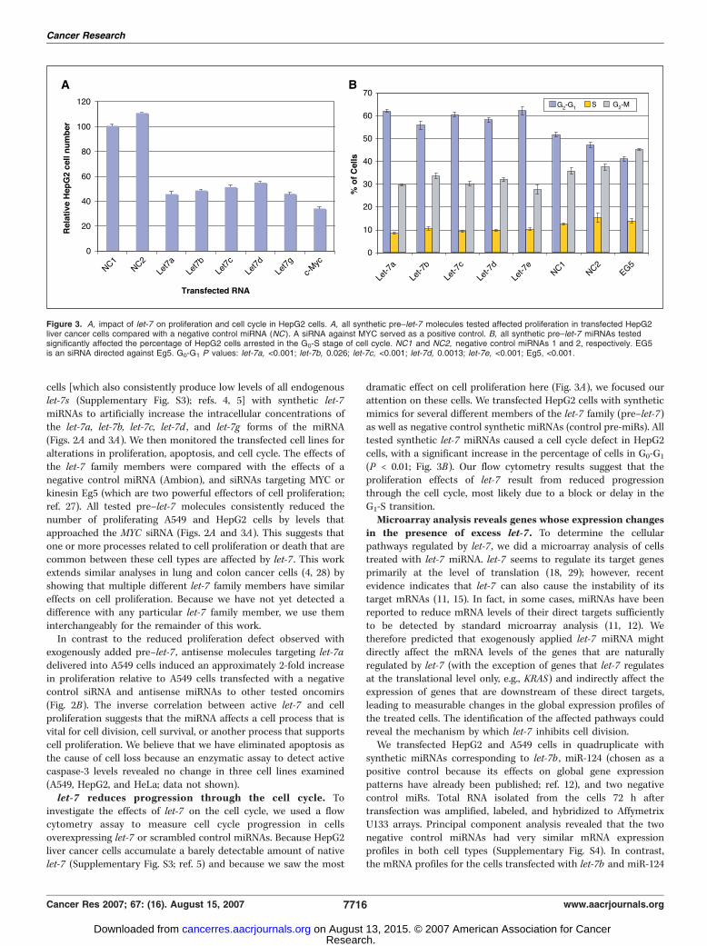
cells [which also consistently produce low levels of all endogenouslet-7s (Supplementary Fig. S3); refs. 4, 5] with synthetic let-7miRNAs to artificially increase the intracellular concentrations ofthe let-7a, let-7b, let-7c, let-7d , and let-7g forms of the miRNA(Figs. 2A and 3A). We then monitored the transfected cell lines foralterations in proliferation, apoptosis, and cell cycle. The effects ofthe let-7 family members were compared with the effects of anegative control miRNA (Ambion), and siRNAs targeting MYC orkinesin Eg5 (which are two powerful effectors of cell proliferation;ref. 27). All tested pre–let-7 molecules consistently reduced thenumber of proliferating A549 and HepG2 cells by levels thatapproached the MYC siRNA (Figs. 2A and 3A). This suggests thatone or more processes related to cell proliferation or death that arecommon between these cell types are affected by let-7 . This workextends similar analyses in lung and colon cancer cells (4, 28) byshowing that multiple different let-7 family members have similareffects on cell proliferation. Because we have not yet detected adifference with any particular let-7 family member, we use theminterchangeably for the remainder of this work.In contrast to the reduced proliferation defect observed with
exogenously added pre–let-7 , antisense molecules targeting let-7adelivered into A549 cells induced an approximately 2-fold increasein proliferation relative to A549 cells transfected with a negativecontrol siRNA and antisense miRNAs to other tested oncomirs(Fig. 2B). The inverse correlation between active let-7 and cellproliferation suggests that the miRNA affects a cell process that isvital for cell division, cell survival, or another process that supportscell proliferation. We believe that we have eliminated apoptosis asthe cause of cell loss because an enzymatic assay to detect activecaspase-3 levels revealed no change in three cell lines examined(A549, HepG2, and HeLa; data not shown).let-7 reduces progression through the cell cycle. To
investigate the effects of let-7 on the cell cycle, we used a flowcytometry assay to measure cell cycle progression in cellsoverexpressing let-7 or scrambled control miRNAs. Because HepG2liver cancer cells accumulate a barely detectable amount of nativelet-7 (Supplementary Fig. S3; ref. 5) and because we saw the most
dramatic effect on cell proliferation here (Fig. 3A), we focused ourattention on these cells. We transfected HepG2 cells with syntheticmimics for several different members of the let-7 family (pre–let-7)as well as negative control synthetic miRNAs (control pre-miRs). Alltested synthetic let-7 miRNAs caused a cell cycle defect in HepG2cells, with a significant increase in the percentage of cells in G0-G1
(P < 0.01; Fig. 3B). Our flow cytometry results suggest that theproliferation effects of let-7 result from reduced progressionthrough the cell cycle, most likely due to a block or delay in theG1-S transition.Microarray analysis reveals genes whose expression changes
in the presence of excess let-7 . To determine the cellularpathways regulated by let-7 , we did a microarray analysis of cellstreated with let-7 miRNA. let-7 seems to regulate its target genesprimarily at the level of translation (18, 29); however, recentevidence indicates that let-7 can also cause the instability of itstarget mRNAs (11, 15). In fact, in some cases, miRNAs have beenreported to reduce mRNA levels of their direct targets sufficientlyto be detected by standard microarray analysis (11, 12). Wetherefore predicted that exogenously applied let-7 miRNA mightdirectly affect the mRNA levels of the genes that are naturallyregulated by let-7 (with the exception of genes that let-7 regulatesat the translational level only, e.g., KRAS) and indirectly affect theexpression of genes that are downstream of these direct targets,leading to measurable changes in the global expression profiles ofthe treated cells. The identification of the affected pathways couldreveal the mechanism by which let-7 inhibits cell division.We transfected HepG2 and A549 cells in quadruplicate with
synthetic miRNAs corresponding to let-7b , miR-124 (chosen as apositive control because its effects on global gene expressionpatterns have already been published; ref. 12), and two negativecontrol miRs. Total RNA isolated from the cells 72 h aftertransfection was amplified, labeled, and hybridized to AffymetrixU133 arrays. Principal component analysis revealed that the twonegative control miRNAs had very similar mRNA expressionprofiles in both cell types (Supplementary Fig. S4). In contrast,the mRNA profiles for the cells transfected with let-7b and miR-124
Figure 3. A, impact of let-7 on proliferation and cell cycle in HepG2 cells. A, all synthetic pre–let-7 molecules tested affected proliferation in transfected HepG2liver cancer cells compared with a negative control miRNA (NC ). A siRNA against MYC served as a positive control. B, all synthetic pre–let-7 miRNAs testedsignificantly affected the percentage of HepG2 cells arrested in the G0-S stage of cell cycle. NC1 and NC2, negative control miRNAs 1 and 2, respectively. EG5is an siRNA directed against Eg5. G0-G1 P values: let-7a, <0.001; let-7b, 0.026; let-7c, <0.001; let-7d, 0.0013; let-7e, <0.001; Eg5, <0.001.
Cancer Research
Cancer Res 2007; 67: (16). August 15, 2007 7716 www.aacrjournals.org
Research. on August 13, 2015. © 2007 American Association for Cancercancerres.aacrjournals.org Downloaded from

Table 1. Cell cycle, cell division, and cell proliferation genes that respond to excess let-7
Gene Product Function*
A. Repressed in both HepG2 and A549 cells
CCNA2 Cyclin A2 Binds CDK2 and CDC2 to promote cell cycle
G1-S and G2-M phase transition; aberrantlyexpressed in acute myeloid and promyelocytic
leukemias
CDC34 Cell division defective 34 Modifies CDKN1B, increases the ubiquinationand degradation of CDKN1B
ASK/DBF4 activator of S-phase kinase Binds to and activates kinase activity of CDC7,
required for the initiation of DNA replication
at the G1 to S transitionAURKA/STK6 and AURKB/STK12 Aurora A and Aurora B
kinases
Maximally expressed during G2-M phases and
may function in cytokinesis, up-regulated in
multiple neoplasms
E2F5 E2F transcription factor 5 Oncogenic in primary rodent cells and isamplified in human breast tumors
CDK8 CDK8 Forms a complex with cyclin C that
phosphorylates cyclin H (CCNH), plays a role
in the regulation of transcription and ascomponent of the RNA polymerase II
holoenzyme
PLAGL1 and PLAGL2 Pleomorphic adenoma gene-liketranscription factors
Transcription activators, regulate cellproliferation
LIN28B Homologue of heterochronic protein LIN-28 Putative RNA binding protein. Mutated in
hepatocellular carcinoma
DICER1 RNaseIII RNase processes pre-miRNAs and dsRNAGMNN Geminin Geminin, regulates DNA replication and
proliferation, binds to the licensing factor
CDT1 and negatively regulates its
ubiquitination, up-regulated in breast, colon,rectal, and biliary tract neoplasms
NRAS Ras GTPase Signaling molecule, mutated in
multiple tumorsHMGA2 Chromatin protein Regulates proliferation. Chromosomal
translocations in multiple tumors
B. Repressed in HepG2 cells only
CDC2 Cell division cycle 2, a CDK Binds B-type cyclins, regulates G2 to M phasetransition, promotes cell proliferation
CDC25A Cell division cycle 25A, a protein
tyrosine-threonine phosphatase
Binds cyclins and regulates G1-S phase transition,
overexpressed in many cancers
CCNB1 Cyclin B1 Regulatory subunit of the CCNB1-CDC2maturation-promoting factor complex that
mediates G2-M phase transition, up-regulated
in various cancersCCNE2 Cyclin E2 G1-specific CDK regulatory subunit that interacts
with CDK2 and CDK3, overexpressed in
transformed cells and up-regulated in breast
and lung cancerCCNF Cyclin F A member of the cyclin family of CDK kinase
regulatory subunits, forms a complex with
cyclin B1 (CCNB1) and CDC2
CCNJ Cyclin J Protein containing cyclin COOH-terminal andNH2-terminal domains have a region of low
similarity to a region of cyclin A2
SKP2 S-phase kinase-associated protein 2 A component of a ubiquitin E3 ligase complex,
mediates cell cycle regulatory proteindegradation, promotes cell proliferation and
invasion, inhibits cell adhesion and apoptosis;
overexpressed in many cancers
(Continued on the following page)
let-7 Represses Cell Proliferation
www.aacrjournals.org 7717 Cancer Res 2007; 67: (16). August 15, 2007
Research. on August 13, 2015. © 2007 American Association for Cancercancerres.aacrjournals.org Downloaded from

Table 1. Cell cycle, cell division, and cell proliferation genes that respond to excess let-7 (cont’d)
Gene Product Function*
GeneCKS1B CDC28 protein kinase regulatory
subunit 1B
Binds SKP2 and targets it to its substrates, required for
ubiquitination of p21 Cip1 (CDKN1A) and p27 Kip1
(CDKN1B), highly expressed in non–small cell lung,
gastric, and colon carcinomaCDC20 Cell division cycle 20 Activates the mitotically phosphorylated form of the
anaphase promoting complex as well as the mitotic
spindle checkpoint, overexpressed in gastric cancer
CDCA1 Cell division cycle associated 1 Mediates stable attachment of microtubules to thekinetochore during mitosis and plays a role in the
spindle checkpoint
CDAC2 Cell division cycle associated 2 Novel proteinCDAC3/TOME1 Cell division cycle associated 3/trigger
of mitotic entry 1
A cytosolic protein that is degraded during G1 phase and
whose gene promoter activity is stimulated at the
G2-M phase
CDCA5 Cell division cycle associated 5 Novel proteinCDAC7 Cell division cycle associated 7 A nuclear protein expressed highly in thymus and small
intestine, has a role in anchorage-dependent growth,
up-regulated in Burkitt lymphoma cell lines; gene may
be a MYC targetCDCA8 Cell division cycle associated 8 (borealin) A chromosomal passenger complex component
may target survivin (BIRC5) and INCENP to
centromere, required for kinetochore function,
mitotic spindle stability, and metaphasechromosome alignment during mitosis
RRM1 and RRM2 Ribonucleotide reductase M1 and M2
polypeptides
DNA synthesis
CDC6 Encoding cell division cycle 6 homologue DNA replication, up-regulated in cervical intraepithelial
neoplasia and cervical cancer
CDC45L Cell division cycle 45 like Associates with ORC2L, MCM7, and
POLA2, predicted to be involved in the initiation ofDNA replication
CDT1 Chromatin licensing factor Ensures replication occurs once per cell cycle,
up-regulated in non–small cell lung
carcinomasORC1L and ORC6L Origin recognition complex proteins DNA replication
MCM2/3/4/5/6/7/8/10 Mini chromosome maintenance-deficient
complex
DNA replication, up-regulated in multiple cancers
RFC2/3/4/5 Replication factor C complex DNA replication
E2F6 and E2F8 E2F transcription factors Regulators of cell cycle
CHEK1 Checkpoint homologue 1 kinase Required for mitotic G2 checkpoint in response to
radiation-induced DNA damage, associated withlung cancer
BUB1 and BUB1B Budding uninhibited by benzimidazoles
1 homologues
Acts in spindle assembly checkpoint and
chromosome congression, may regulate
vesicular traffic; mutations areassociated with lung cancer, T cell
leukemia, and colorectal cancer cell
chromosomal instability; a proteinkinase of the mitotic spindle checkpoint, inhibits
anaphase-promoting complex activation
MAD2L1 MAD2 mitotic arrest deficient-like 1 Component with BUB1B
CDC23 Cell division cycle 23 A putative component of the anaphase-promotingcomplex (APC), considered a tumor antigen in ovarian
carcinoma; mutation in corresponding gene is
associated with colon cancer
FANCD2 Fanconi anemia complementation group D2 Involved in DNA damage responseBRCA1 and BRCA2 Breast Cancer Susceptibility loci Tumor suppressors; mutations are linked to breast and
ovarian cancer
(Continued on the following page)
Cancer Research
Cancer Res 2007; 67: (16). August 15, 2007 7718 www.aacrjournals.org
Research. on August 13, 2015. © 2007 American Association for Cancercancerres.aacrjournals.org Downloaded from

are clearly distinct from the cells transfected with the negativecontrol miRNAs as well as from each other, indicating that the twomiRNAs are uniquely affecting the global gene expression profilesof the two cell types. For both cell types, the number of genesdetermined to be altered by treatment was calculated by filteringall genes by fold change relative to both control transfections, andstatistical significance was assessed by a t test after the omnibusF test was shown to be significant. In HepG2 liver cancer cells, weidentified 1,334 (698 repressed and 636 up-regulated) genes whoseexpression varied by at least 1.93-fold and were statisticallysignificant at a 0.05 FDR between the cells transfected with let-7and the negative control miRNAs (Supplementary Table S1). InA549 lung cancer cells, we identified 629 (244 repressed and 385 up-regulated) altered genes, all of which were statistically significant ata 0.05 FDR (Supplementary Table S2). let-7 addition affected 200genes in common between both cell types (Supplementary TablesS1 and S2). In both cell types, we identified NRAS and HMGA2 ,known downstream targets of let-7 (5, 30), indicating thatour analysis could appropriately reveal let-7 downstream genes(Table 1).let-7 repressed multiple cell cycle associated genes. Genes
found to be differentially expressed in either the HepG2 and A549cell lines were grouped by their assigned biological functions usingthe Gene Ontology (GO) database (Fig. 4A ; Supplementary TableS4). These results show that let-7 directly or indirectly affects theexpression of many cell cycle–related genes. In fact, the primaryGO classes associated with the differentially expressed genes in theHepG2 cells are linked with the cell cycle (Fig. 4A), which areconsistent with our earlier observations (Figs. 2 and 3). Cellproliferation genes repressed directly or indirectly by excess let-7 inboth cell types include the genes for cyclin A2, which promotesG1-S and G2-M phase transitions; CDC34, which promotes thedegradation of cyclin-dependent kinase (CDK) inhibitor 1B; theASK activator of S-phase kinase, required for the initiation of DNAreplication at the G1 to S transition; the Aurora A and B kinases; theE2F5 transcription factor; CDK8; PLAGL2, the pleomorphicadenoma gene-like transcription factor; and Geminin, a regulatorof DNA replication and proliferation (Table 1). Additional cell cyclegenes repressed directly or indirectly by let-7 in HepG2 cells includethe genes for various CDKs, cyclins, the CDC25A phosphatase, theS-phase kinase–associated protein 2, SKP2; the CDC28 kinaseregulatory subunit 1B, and various cell division cycle–associated
proteins (Table 1). let-7 also repressed genes coding for DNAsynthesis and DNA replication functions (Table 1), e.g., ribonucle-otide reductase subunits, multiple DNA replication initiation andorigin recognition complex proteins, members of the Minichromosome maintenance-deficient complex, and multiple mem-bers of the replication factor C complex (Table 1).Although the vast majority of altered cell cycle genes exhibited
reduced expression following let-7 application, a few cell cyclegenes were up-regulated under the same conditions. Because let-7is currently only known to function as a repressor of geneexpression, this likely indicates a 2j (or 3j or more) effect of let-7application. These genes included those encoding CDK inhibitor2B; the MAX-interacting protein 1, MXI1, a transcription regulatorthat antagonizes MYC; and cyclin G2, which is down-regulated inthyroid papillary carcinoma (31), showing that it has the propensityto act as a tumor antagonist (Table 1). In let-7–deficient tumorcells, these three genes would be predicted to be down-regulated,which might disable their tumor-suppressing functions.Interestingly, we also found that let-7 addition repressed the
expression of a number of known and putative tumor suppressorgenes (Table 1) such as BRCA1, BRCA2, FANCD2, PLAGL1, E2F6 , andE2F8 and the cell cycle checkpoint genes CHEK1, BUB1, BUB1B,MAD2L1 , and CDC23. The significance of these findings remains tobe determined, but may be relevant to the small number of recentlyidentified tumors where let-7 is actually up-regulated (32, 33).Identification of likely direct let-7 targets. To enrich for genes
modulated directly by let-7 , we reasoned that the expression ofdirect let-7 downstream genes might be affected earlier thanindirect downstream genes. Therefore, in addition to analyzing thechanges in global mRNA expression 72 h after let-7 transfection, weused a time course assay featuring microarray analysis of HepG2cells harvested at 4, 8, 16, 24, 36, 48, 72, and 128 h after transfection(Fig. 4B ; Supplementary Table S3). We found no change in geneexpression at times 4 and 8 h after transfection, but detected thefirst changes 16 h post-transfection. We detected a total of 176genes that were down-regulated within 36 h of let-7 addition: wedenote these as early repressed genes (Supplementary Table S3;Fig. 4B). Like the original microarray analysis, these genes wereenriched in GO categories for cell cycle and cell division(Supplementary Table S4). These included many of the samewell-known cell cycle genes already mentioned (CCNA2, CDC25A,CDK8, SKP2, AURKA/STK6), but also included some that were
Table 1. Cell cycle, cell division, and cell proliferation genes that respond to excess let-7 (cont’d)
Gene Product Function*
C. Up-regulated in both cell types
RRM2B Ribonucleotide reductase M2B DNA synthesis, up-regulated by p53
EIF2C2 Argonaute Functions in RNAi and miRNA pathwaysD. Up-regulated in HepG2 cells only
CDKN2B CDK inhibitor 2B Interacts with the D type CDK4 and CDK6, inhibits cell
proliferation; gene deletion and promoter hypermethylationare associated with many different neoplasms
CCNG2 cyclin G2 Down-regulated in thyroid papillary carcinoma
MXI1 MAX-interacting protein 1 Transcription regulator, antagonizes MYC, tumor suppressor
in prostatic neoplasms
*Functional annotations were derived from the Human Proteome Survey Database (HumanPSD; ref. 48).
let-7 Represses Cell Proliferation
www.aacrjournals.org 7719 Cancer Res 2007; 67: (16). August 15, 2007
Research. on August 13, 2015. © 2007 American Association for Cancercancerres.aacrjournals.org Downloaded from

missed in the 72-h experiment detailed above because theirexpression was presumably repressed early, and then their levelsreturned to normal by 72 h. These included the cell cycle genesCDC16 and CDK6 . Other early repressed mRNAs include the DNAsynthesis gene RRM2 and the cell proliferation regulator, MINA. Ofthese 176 early repressed genes, 127 genes first appeared down-regulated at time 16 h (Supplementary Table S3, pink; CDC16,CDK6, AURKA/STK6; RRM2 , and MINA). An additional set of 37genes was first observed down-regulated at 24 h (SupplementaryTable S3, orange ; including SKP2, CDCA7 , and CDC25A), andanother 12 were first detected as repressed at 36 h (SupplementaryTable S3, yellow ; including CCNA2 and E2F6). Interestingly, a
number of other transcription factors besides E2F6 , including ID2,CBFB, ZNF336, SMAD4, SOX9, NR1H4, ARID3A, PLAGL2, YAP1 , andGTF2I , were among the early repressed genes, suggesting that theymight also propagate the let-7 effects to their downstream targets.To assess how many of the early let-7–repressed genes might
constitute direct target genes, we examine the 3¶UTRs of this groupfor let-7 complementary sites (LCS) that displayed features of LCSsin validated let-7 target genes (5, 15–18, 29, 34) and also used apublished miRNA prediction program, PicTar, to predict let-7targets (35). We found that at least 25 of the early repressed genescontained LCSs in their 3¶UTRs (Fig. 4B), and we propose that theseconstitute direct let-7 targets. This set includes the cell cycleregulators CDK6, CDC25A, AURKB/STK6, CDCA7 , and the DNAsynthesis regulator RRM2 . CDK6 interacts with D-type cyclins andphosphorylates RB1 to activate the G1 phase of the cell cycle (36).CDK6 is also overexpressed or amplified in numerous cancersincluding non–small cell lung cancer. CDC25A is a serine-tyrosinephosphatase that activates CDKs by removing inhibitory phos-phate groups. Like CDK6, CDC25A is up-regulated in multiplecancers, including lung cancers (37). Aurora kinase B regulateschromosome segregation and cytokinesis during mitosis. CDCA7encodes a novel protein up-regulated in multiple cancers, includinglung cancer, which, like CDC25A , is a downstream target of MYC ,and participates in the cell proliferation effects of MYC (38). Thelist of likely direct let-7 targets also includes eight transcriptionfactors CBFB, PLAGL2, E2F6, SOX9, ZNF336, YAP1, GTF2I , andARID3A , consistent with the enrichment for transcription factorsseen as let-7 targets in C. elegans (16). We conclude that the non–LCS-containing genes with altered expression upon let-7 additionare likely to be downstream genes indirectly affected by let-7expression, perhaps as downstream targets of the transcriptionfactors affected directly by let-7 . For example, we found thatmultiple members of the MCM and RFC DNA synthesis complexeswere repressed only at later time points and could therefore betargets of these transcription factors.let-7 negatively regulates the protein levels of CDK6 and
CDC25A . We analyzed the native expression of our two top scoringcell cycle regulators (Fig. 5), CDK6 and CDC25A , in cells transfectedwith pre-miRs. Consistent with the microarray analysis, we foundthat protein levels of both CDK6 and CDC25A decrease in cellstransfected with pre–let-7 compared with cells transfected witha control pre-miRNA (Fig. 5A). When quantified, we found thatpre–let-7 transfection resulted in approximately a 50% reduction ofprotein compared with the normal levels of CDK6 and CDC25A(P < 0.001, CDC25A; P < 0.002, CDK6 ; Fig. 5B).To provide further validation for these cell cycle genes as direct
let-7 targets, we did reporter assays where we independentlyfused the 3¶UTRs of CDK6 and CDC25A downstream of fireflyluciferase (Fig. 5C). In luciferase assays, both 3¶UTRs conferredlet-7–dependent repression of the reporter gene (Fig. 5D) comparedwith a negative control anti-miR. The effect on the CDC25a andCDK6 3¶UTRs caused by the anti–let-7 was similar to a knownlet-7 target gene, NRAS (Fig. 5D). Thus, like NRAS , these resultsshow that these genes are also likely to be directly regulated by thelet-7 miRNA. Given the close working relationship between CDK6and cyclin D (36, 39) in promoting the G1 to S transition, and thefact that CCND2 (encoding cyclin D2) is the highest scoring cellcycle gene predicted as a let-7 target by PicTar (35), we alsotested the CCND2 3¶UTR in the same assay. We found a similarresult to CDK6 (Fig. 5D), suggesting that CCND2 is also a directtarget of let-7 .
Figure 4. Microarray analysis of let-7–treated human cancer cells. A, tableof the most affected GO categories after let-7 overexpression in HepG2 cellsfor 72 h. mRNAs whose expression was affected by greater than 2-foldwith P values below 0.05 were identified and classified using GO categories.*, P values were calculated with hypergeometric tests to determine whetherthere is a significant enrichment of affected genes in a GO category whencompared with all genes represented on the arrays. B, cell cycle targets for let-7 .Potential direct downstream targets of let-7. Right column, number of LCSs.Pink, first observed repression at 16 h; orange, first observed repression at24 h; yellow, first observed repression at 36 h; blue, first observed repressionafter 36 h. Some of these genes, e.g., CDK6 and RRM2, were missed in PicTarbecause the rodent 3¶UTRs are not complete. Known let-7 targets NRAS andHMGA2 are repressed after 36 h in our assay.
Cancer Research
Cancer Res 2007; 67: (16). August 15, 2007 7720 www.aacrjournals.org
Research. on August 13, 2015. © 2007 American Association for Cancercancerres.aacrjournals.org Downloaded from

Discussion
During mouse embryogenesis, let-7 is first detected around thetime of initiation of lung development (40), and expression thenpersists into adulthood (Fig. 1A ; Supplementary Fig. S3). Theseexpression studies suggest that let-7 may control a variety ofprocesses both during development and in the maintenance ofadult tissue homeostasis. In contrast to robust let-7 expression innormal human lung tissue (14), let-7 is poorly expressed in lungtumors and lung cancer cell lines (Supplementary Fig. S3; refs. 4, 5).Poor let-7 expression may thus be a powerful diagnostic marker forlung tumors (20).let-7 in control of cell proliferation. Proliferation and survival
pathways are frequently altered in tumors (41). We have shown thatlet-7 overexpression causes human cancer cells to decrease cellcycle progression (Figs. 2 and 3). In addition, our microarray datashow that let-7 directly or indirectly regulates multiple cellproliferation genes and strongly suggest that let-7 is a key regulatorof cell cycle progression, consistent with the cell cycle assay resultsdescribed earlier. We show that let-7 directly regulates a few keycell cycle proto-oncogenes, e.g., RAS, CDC25a, CDK6, and cyclin D(Fig. 5), thus controlling cell proliferation by reducing flux throughthe pathways promoting the G1 to S transition. Because many ofthese let-7–responsive genes are known oncogenes or are over-expressed in tumors, one prediction is that in cancer cells with let-7deletions or poor let-7 expression, many of these genes would beup-regulated, which is likely to stimulate cell cycle and DNAsynthesis and, hence, cell division.The class of let-7 target genes that are solely repressed at the
level of translation will not be identified via microarray analysis,but rather will need to be identified via proteomic or other means.For example, let-7 does not affect KRAS mRNA levels (42), and
consequently, the human let-7 target gene KRAS (5) did not emergefrom our microarray analysis. This shows that our analysis did notprovide a complete picture of all let-7 targets and implies that let-7might confer varying degrees of translational inhibition versusmRNA instability depending on specific target genes. In addition,CCND2 was also missed in our microarray analysis, but confirmedas a target by luciferase assays (Fig. 5D).Nevertheless, our microarray analysis seems to have enriched
for potential direct let-7 targets. Just over 14% (25 out of 176 totalrepressed) of our early misregulated genes are likely to be directlyregulated by the let-7 miRNA, better than the f1% of all PicTarpredicted let-7a targets as a function of the whole genome(243:25,000; enrichment P value = 5.4E�22 by hypergeometrictest).Our data strongly support the assertion that let-7 is a tumor
suppressor miRNA. Although this role is most likely in lung cellswhere there is normally high let-7 expression and a strongcorrelation between let-7 loss and lung cancer, our studies findthat let-7 causes cell cycle defects in a non–lung cancer cell line aswell, implying that let-7 may function as a tumor suppressor inother tissues. This idea is supported by the observation that let-7genes map to loci deleted in multiple types of cancers, such asbreast, ovary, urothelial, and cervical cancers (3).Our work reveals a miRNA to be a master regulator of proto-
oncogene expression and cancer pathways. This important role inthe control of a fundamental process such as cell cycle may providean explanation for why let-7 has been 100% conserved more than600 million years of evolution (14). These experiments also suggestthat let-7 may prove to be a valuable tool in interventions aimed attreating and diagnosing many cancers.let-7 affects expression of homologues of C. elegans
heterochronic genes. In C. elegans, let-7 is a member of the
Figure 5. Native and reporter gene validation oflet-7 target genes. A, Western analysis of theindicated proteins following pre–let-7b or pre-controlmiR transfection of HeLa cells. Three independenttransfections are shown for each pre-miR.B, quantification of relative levels of CDK6 andCDC25A proteins normalized to actin and thenegative control pre-miR. C, schematicrepresentations of the 3¶UTR fragments cloneddownstream of a luciferase reporter 3¶UTR (gray,3¶UTR; wide gray, subcloned fragments). Arrows,positions of LCSs. D, relative fold induction ofreporter gene expression in HeLa cells transfectedwith an anti-let7b oligonucleotide compared with atransfection with a negative control miRNA.Triplicate repeats of this experiment were done.Each value of luciferase activity in the samplestreated with let-7b anti-miR was normalized by theaverage luciferase activity value of the samplestreated with negative control anti-miR (value = 1).NRAS is the NRAS long 3¶UTR shown here as apositive control.
let-7 Represses Cell Proliferation
www.aacrjournals.org 7721 Cancer Res 2007; 67: (16). August 15, 2007
Research. on August 13, 2015. © 2007 American Association for Cancercancerres.aacrjournals.org Downloaded from

heterochronic pathway, which regulates the timing of cell fatedetermination during development (43, 44) and requires dcr-1 andalg-1/alg-2 for this role (45). Interestingly, we identified threehuman homologues of C. elegans heterochronic genes as let-7–responsive genes in both A549 and HepG2 cells (Table 1), includingLIN28B, DICER1 , and EIF2C2/AGO2 . In addition, EIF2C4/AGO4 wasalso affected in HepG2 cells overexpressing let-7. EIF2C2, EIF2C4 ,and alg-1/2 encode Argonaute proteins (46), which function withmiRNAs in the RNAi-induced silencing complex (RISC) to mediatetarget-specific gene silencing. DICER1 processes let-7 (and othermiRNAs) and our microarray data suggest that let-7 may negativelyfeedback on its own expression by modulating DICER1 expression.LIN28B is altered in human hepatocellular carcinoma and, like the
C. elegans lin-28 , has LCSs in its 3¶UTR (47). It is therefore possiblethat conserved pathways function to control the timing of cellproliferation during the development of nematodes and mammals.
Acknowledgments
Received 3/21/2007; revised 5/17/2007; accepted 6/18/2007.Grant support: G. Stefani was supported by a Yale postdoctoral fellowship and the
Anna Fuller Fund. F.J. Slack was supported by NIH grant GM62594 and NationalScience Foundation grant IBN-03444429. A.E. Kerscher was supported by a NIH/National Research Service Award postdoctoral fellowship (F32GM071157).
The costs of publication of this article were defrayed in part by the payment of pagecharges. This article must therefore be hereby marked advertisement in accordancewith 18 U.S.C. Section 1734 solely to indicate this fact.
We thank Drs. Joanne Weidhaas and Iain Dawson for critical reading of thismanuscript.
References1. Bartel DP. MicroRNAs: genomics, biogenesis, mecha-nism, and function. Cell 2004;116:281–97.
2. Boehm M, Slack F. A developmental timing microRNAand its target regulate life span in C. elegans . Science2005;310:1954–7.
3. Calin GA, Sevignani C, Dumitru CD, et al. HumanmicroRNA genes are frequently located at fragile sitesand genomic regions involved in cancers. Proc NatlAcad Sci U S A 2004;101:2999–3004.
4. Takamizawa J, Konishi H, Yanagisawa K, et al. Reducedexpression of the let-7 microRNAs in human lungcancers in association with shortened postoperativesurvival. Cancer Res 2004;64:3753–6.
5. Johnson SM, Grosshans H, Shingara J, et al. RAS is regu-lated by the let-7 microRNA family. Cell 2005;120:635–47.
6. He L, Thomson JM, Hemann MT, et al. A microRNApolycistron as a potential human oncogene. Nature2005;435:828–33.
7. Cimmino A, Calin GA, Fabbri M, et al. miR-15 andmiR-16 induce apoptosis by targeting BCL2. Proc NatlAcad Sci U S A 2005;102:13944–9.
8. Iorio MV, Ferracin M, Liu CG, et al. MicroRNA geneexpression deregulation in human breast cancer. CancerRes 2005;65:7065–70.
9. Esquela-Kerscher A, Slack FJ. Oncomirs: microRNAswith a role in cancer. Nat Rev Cancer 2006;6:259–69.
10. Pillai RS, Bhattacharyya SN, Artus CG, et al.Inhibition of translational initiation by Let-7 MicroRNAin human cells. Science 2005;309:1573–6.
11. Bagga S, Bracht J, Hunter S, et al. Regulation by let-7and lin-4 miRNAs results in target mRNA degradation.Cell 2005;122:553–63.
12. Lim LP, Lau NC, Garrett-Engele P, et al. Microarrayanalysis shows that some microRNAs downregulate largenumbers of target mRNAs. Nature 2005;433:769–73.
13. Lagos-Quintana M, Rauhut R, Yalcin A, Meyer J,Lendeckel W, Tuschl T. Identification of tissue-specificmicroRNAs from mouse. Curr Biol 2002;12:735–9.
14. Pasquinelli AE, Reinhart BJ, Slack F, et al. Conserva-tion of the sequence and temporal expression of let-7heterochronic regulatory RNA. Nature 2000;408:86–9.
15. Reinhart B, Slack F, Basson M, et al. The 21nucleotide let-7 RNA regulates C. elegans developmentaltiming. Nature 2000;403:901–6.
16. Grosshans H, Johnson T, Reinert KL, Gerstein M,Slack FJ. The temporal patterning microRNA let-7regulates several transcription factors at the larval toadult transition in C. elegans . Dev Cell 2005;8:321–30.
17. Lin SY, Johnson SM, Abraham M, et al. The C. eleganshunchback homolog, hbl-1, controls temporal pattern-ing and is a probable microRNA target. Dev Cell 2003;4:639–50.
18. Slack FJ, Basson M, Liu Z, Ambros V, Horvitz HR,Ruvkun G. The lin-41 RBCC gene acts in the C. elegansheterochronic pathway between the let-7 regulatoryRNA and the lin-29 transcription factor. Mol Cell 2000;5:659–69.
19. Minna JD, Roth JA, Gazdar AF. Focus on lung cancer.Cancer Cell 2002;1:49–52.
20. Yanaihara N, Caplen N, Bowman E, et al. UniquemicroRNA molecular profiles in lung cancer diagnosisand prognosis. Cancer Cell 2006;9:189–98.
21. Malumbres M, Barbacid M. RAS oncogenes: the first30 years. Nat Rev Cancer 2003;3:459–65.
22. Irizarry RA, Bolstad BM, Collin F, Cope LM, Hobbs B,Speed TP. Summaries of Affymetrix GeneChip probelevel data. Nucleic Acids Res 2003;31:1–8.
23. Benjamini Y, Hochberg Y. Controlling the false dis-covery rate: a practical and powerful approach to mul-tiple testing. J R Stat Soc Ser B Method 1995;57:289–300.
24. Wilcoxon F. Individual comparisons by rankingmethods. Biometrics 1945;1:80–3.
25. Wilkinson DG, Nieto MA. Detection of messengerRNA by in situ hybridization to tissue sections andwhole mounts. Methods Enzymol 1993;225:361–73.
26. Heller S, Sheane CA, Javed Z, Hudspeth AJ. Molecularmarkers for cell types of the inner ear and candidategenes for hearing disorders. Proc Natl Acad Sci U S A1998;95:11400–5.
27. Weil D, Garcon L, Harper M, Dumenil D, Dautry F,Kress M. Targeting the kinesin Eg5 to monitor siRNAtransfection in mammalian cells. Biotechniques 2002;33:1244–8.
28. Akao Y, Nakagawa Y, Naoe T. let-7 microRNAfunctions as a potential growth suppressor inhuman colon cancer cells. Biol Pharm Bull 2006;29:903–6.
29. Vella MC, Choi EY, Lin SY, Reinert K, Slack FJ. TheC. elegans microRNA let-7 binds to imperfect let-7complementary sites from the lin-41 3¶UTR. Genes Dev2004;18:132–7.
30. Mayr C, Hemann MT, Bartel DP. Disrupting thepairing between let-7 and Hmga2 enhances oncogenictransformation. Science 2007;315:1576–9.
31. Ito Y, Yoshida H, Uruno T, et al. Decreased expressionof cyclin G2 is significantly linked to the malignanttransformation of papillary carcinoma of the thyroid.Anticancer Res 2003;23:2335–8.
32. Brueckner B, Stresemann C, Kuner R, et al. Thehuman let-7a-3 locus contains an epigenetically regu-lated microRNA gene with oncogenic function. CancerRes 2007;67:1419–23.
33. Wang T, Zhang X, Obijuru L, et al. A micro-RNAsignature associated with race, tumor size, and targetgene activity in human uterine leiomyomas. GenesChromosomes Cancer 2007;46:336–47.
34. Vella MC, Reinert K, Slack FJ. Architecture of avalidated microRNA:target interaction. Chem Biol 2004;11:1619–23.
35. Lall S, Grun D, Krek A, et al. A genome-wide map ofconserved microRNA targets in C. elegans . Curr Biol2006;16:460–71.
36. Russo AA, Tong L, Lee JO, Jeffrey PD, Pavletich NP.Structural basis for inhibition of the cyclin-dependentkinase Cdk6 by the tumour suppressor p16INK4a.Nature 1998;395:237–43.
37. Galaktionov K, Lee AK, Eckstein J, et al. CDC25phosphatases as potential human oncogenes. Science1995;269:1575–7.
38. Osthus RC, Karim B, Prescott JE, et al. The Myc targetgene JPO1/CDCA7 is frequently overexpressed in humantumors and has limited transforming activity in vivo .Cancer Res 2005;65:5620–7.
39. Grossel MJ, Hinds PW. From cell cycle to differen-tiation: an expanding role for cdk6. Cell Cycle 2006;5:266–70.
40. Schulman BR, Esquela-Kerscher A, Slack FJ. Recip-rocal expression of lin-41 and the microRNAs let-7 andmir-125 during mouse embryogenesis. Dev Dyn 2005;234:1046–54.
41. Hanahan D, Weinberg RA. The hallmarks of cancer.Cell 2000;100:57–70.
42. Chu CY, Rana TM. Translation repression in humancells by microRNA-induced gene silencing requiresRCK/p54. PLoS Biol 2006;4:1122–36.
43. Ambros V, Horvitz HR. Heterochronic mutants of thenematode Caenorhabditis elegans . Science 1984;226:409–16.
44. Banerjee D, Slack F. Control of developmentaltiming by small temporal RNAs: a paradigm for RNA-mediated regulation of gene expression. Bioessays 2002;24:119–29.
45. Grishok A, Pasquinelli AE, Conte D, et al. Genes andmechanisms related to RNA interference regulateexpression of the small temporal RNAs that control C.elegans developmental timing. Cell 2001;106:23–34.
46. Carmell MA, Xuan Z, Zhang MQ, Hannon GJ. TheArgonaute family: tentacles that reach into RNAi,developmental control, stem cell maintenance, andtumorigenesis. Genes Dev 2002;16:2733–42.
47. Guo Y, Chen Y, Ito H, et al. Identification andcharacterization of lin-28 homolog B (LIN28B) inhuman hepatocellular carcinoma. Gene 2006;384:51–61.
48. Hodges PE, Carrico PM, Hogan JD, et al. Annotat-ing the human proteome: the Human ProteomeSurvey Database (HumanPSD) and an in-depth targetdatabase for G protein-coupled receptors (GPCR-PD)from Incyte Genomics. Nucleic Acids Res 2002;30:137–41.
Cancer Research
Cancer Res 2007; 67: (16). August 15, 2007 7722 www.aacrjournals.org
Research. on August 13, 2015. © 2007 American Association for Cancercancerres.aacrjournals.org Downloaded from

2007;67:7713-7722. Cancer Res Charles D. Johnson, Aurora Esquela-Kerscher, Giovanni Stefani, et al. Human Cells
MicroRNA Represses Cell Proliferation Pathways inlet-7The
Updated version
http://cancerres.aacrjournals.org/content/67/16/7713
Access the most recent version of this article at:
Material
Supplementary
http://cancerres.aacrjournals.org/content/suppl/2007/08/14/67.16.7713.DC1.html
Access the most recent supplemental material at:
Cited articles
http://cancerres.aacrjournals.org/content/67/16/7713.full.html#ref-list-1
This article cites 48 articles, 16 of which you can access for free at:
Citing articles
http://cancerres.aacrjournals.org/content/67/16/7713.full.html#related-urls
This article has been cited by 100 HighWire-hosted articles. Access the articles at:
E-mail alerts related to this article or journal.Sign up to receive free email-alerts
Subscriptions
Reprints and
To order reprints of this article or to subscribe to the journal, contact the AACR Publications
Permissions
To request permission to re-use all or part of this article, contact the AACR Publications
Research. on August 13, 2015. © 2007 American Association for Cancercancerres.aacrjournals.org Downloaded from