The Lin28 cold-shock domain remodels pre-let-7 microRNA
15
The Lin28 cold-shock domain remodels pre-let-7 microRNA Florian Mayr 1,2 , Anja Schu ¨ tz 3 , Nadine Do ¨ ge 1 and Udo Heinemann 1,2, * 1 Crystallography, Max-Delbru ¨ ck Center for Molecular Medicine, Robert-Ro ¨ ssle Straße 10, 13125, 2 Institute for Chemistry and Biochemistry, Freie Universita ¨ t Berlin, Takustraße 6, 14195 and 3 Helmholtz-Protein Sample Production Facility, Max-Delbru ¨ ck Center for Molecular Medicine, Robert-Ro ¨ ssle Straße 10, 13125 Berlin, Germany Received November 11, 2011; Revised April 5, 2012; Accepted April 10, 2012 ABSTRACT The RNA-binding protein Lin28 regulates the pro- cessing of a developmentally important group of microRNAs, the let-7 family. Lin28 blocks the bio- genesis of let-7 in embryonic stem cells and thereby prevents differentiation. It was shown that both RNA-binding domains (RBDs) of this protein, the cold-shock domain (CSD) and the zinc-knuckle domain (ZKD) are indispensable for pri- or pre-let-7 binding and blocking its maturation. Here, we sys- tematically examined the nucleic acid-binding pref- erences of the Lin28 RBDs and determined the crystal structure of the Lin28 CSD in the absence and presence of nucleic acids. Both RNA-binding domains bind to single-stranded nucleic acids with the ZKD mediating specific binding to a conserved GGAG motif and the CSD showing only limited sequence specificity. However, only the isolated Lin28 CSD, but not the ZKD, can bind with a reason- able affinity to pre-let-7 and thus is able to remodel the terminal loop of pre-let-7 including the Dicer cleavage site. Further mutagenesis studies reveal that the Lin28 CSD induces a conformational change in the terminal loop of pre-let-7 and thereby facilitates a subsequent specific binding of the Lin28 ZKD to the conserved GGAG motif. INTRODUCTION MicroRNAs (miRNAs) are small regulatory RNAs, typ- ically 22 nt in length, which regulate target mRNAs through complementary base pairing. The biogenesis of miRNAs can be divided into two major steps. First, the nuclear localized ribonuclease III Drosha, in a complex with DGCR8 (Pasha in invertebrates), cleaves a long primary miRNA transcript (pri-miRNA), creating a characteristic 60–80 nt hairpin structure with a 2-nt 3 0 overhang (1,2). After export of this precursor-miRNA (pre-miRNA) by exportin-5:RanGTP (3,4), it is further cleaved by Dicer to yield the mature miRNA that func- tions in the RNA-induced silencing complex (RISC) (5–7). The highly conserved let-7 miRNA is known to regulate developmental timing and proliferation (8–10). Mature let-7 is absent or only present in low amounts in various stem and progenitor cells but strongly induced upon tissue differentiation. Multiple oncogenes, such as RAS, HMGA2, c-MYC, Cdc25a and Cdk6 are targets of let-7, and it has been reported that let-7 can act as a tumor suppressor in lung cancers (11–13). Interestingly, mature let-7 is downregulated in various cancer and stem cells while levels of pri- and pre-let-7 are not affected, suggest- ing a regulation of let-7 maturation rather than at tran- scription levels (14). The RNA-binding protein Lin28 (from cell lineage abnormal 28) was shown to regulate the maturation of let-7 and is involved in the regulation of pluripotency, development and differentiation (15–18). In Caenor- habditis elegans, mutations in the LIN28 gene disturbed the developmental progression of the worm (19). Further experiments revealed that Lin28 is abundant early in nematode development but is downregulated during larval stages L1–L3 (20,21), whereas the amount of mature let-7 shows an opposite pattern (11,22). This opposing expression pattern is also valid for mammalian cells, in which the corresponding homologs Lin28a and Lin28b are mainly expressed in undifferentiated embry- onic stem cells (18,23,24). Strikingly, mature let-7 itself is involved in the post-transcriptional repression of LIN28 upon differentiation. It can bind to the 3 0 -untranslated region (UTR) of the Lin28 mRNA and thereby enables a double-negative feedback loop (17,25). Reciprocally, Lin28 was used to reprogram adult human fibroblasts to induced pluripotent stem cells in cooperation with Oct4, Sox2 and Nanog (26). These observations, in conjunction with the fact that LIN28b and less frequently LIN28a are *To whom correspondence should be addressed. Tel: +49 30 9406 3420; Fax: +49 30 9406 2548; Email: [email protected] 7492–7506 Nucleic Acids Research, 2012, Vol. 40, No. 15 Published online 8 May 2012 doi:10.1093/nar/gks355 ß The Author(s) 2012. Published by Oxford University Press. This is an Open Access article distributed under the terms of the Creative Commons Attribution Non-Commercial License (http://creativecommons.org/licenses/ by-nc/3.0), which permits unrestricted non-commercial use, distribution, and reproduction in any medium, provided the original work is properly cited.
-
Upload
mdc-berlin -
Category
Documents
-
view
1 -
download
0
Transcript of The Lin28 cold-shock domain remodels pre-let-7 microRNA

The Lin28 cold-shock domain remodelspre-let-7 microRNAFlorian Mayr1,2, Anja Schutz3, Nadine Doge1 and Udo Heinemann1,2,*
1Crystallography, Max-Delbruck Center for Molecular Medicine, Robert-Rossle Straße 10, 13125, 2Institute forChemistry and Biochemistry, Freie Universitat Berlin, Takustraße 6, 14195 and 3Helmholtz-Protein SampleProduction Facility, Max-Delbruck Center for Molecular Medicine, Robert-Rossle Straße 10, 13125 Berlin,Germany
Received November 11, 2011; Revised April 5, 2012; Accepted April 10, 2012
ABSTRACT
The RNA-binding protein Lin28 regulates the pro-cessing of a developmentally important group ofmicroRNAs, the let-7 family. Lin28 blocks the bio-genesis of let-7 in embryonic stem cells andthereby prevents differentiation. It was shown thatboth RNA-binding domains (RBDs) of this protein,the cold-shock domain (CSD) and the zinc-knuckledomain (ZKD) are indispensable for pri- or pre-let-7binding and blocking its maturation. Here, we sys-tematically examined the nucleic acid-binding pref-erences of the Lin28 RBDs and determined thecrystal structure of the Lin28 CSD in the absenceand presence of nucleic acids. Both RNA-bindingdomains bind to single-stranded nucleic acids withthe ZKD mediating specific binding to a conservedGGAG motif and the CSD showing only limitedsequence specificity. However, only the isolatedLin28 CSD, but not the ZKD, can bind with a reason-able affinity to pre-let-7 and thus is able to remodelthe terminal loop of pre-let-7 including theDicer cleavage site. Further mutagenesis studiesreveal that the Lin28 CSD induces a conformationalchange in the terminal loop of pre-let-7 and therebyfacilitates a subsequent specific binding of the Lin28ZKD to the conserved GGAG motif.
INTRODUCTION
MicroRNAs (miRNAs) are small regulatory RNAs, typ-ically �22 nt in length, which regulate target mRNAsthrough complementary base pairing. The biogenesis ofmiRNAs can be divided into two major steps. First, thenuclear localized ribonuclease III Drosha, in a complexwith DGCR8 (Pasha in invertebrates), cleaves a longprimary miRNA transcript (pri-miRNA), creating a
characteristic 60–80 nt hairpin structure with a 2-nt 30
overhang (1,2). After export of this precursor-miRNA(pre-miRNA) by exportin-5:RanGTP (3,4), it is furthercleaved by Dicer to yield the mature miRNA that func-tions in the RNA-induced silencing complex (RISC) (5–7).
The highly conserved let-7 miRNA is known to regulatedevelopmental timing and proliferation (8–10). Maturelet-7 is absent or only present in low amounts in variousstem and progenitor cells but strongly induced upon tissuedifferentiation. Multiple oncogenes, such as RAS,HMGA2, c-MYC, Cdc25a and Cdk6 are targets of let-7,and it has been reported that let-7 can act as a tumorsuppressor in lung cancers (11–13). Interestingly, maturelet-7 is downregulated in various cancer and stem cellswhile levels of pri- and pre-let-7 are not affected, suggest-ing a regulation of let-7 maturation rather than at tran-scription levels (14).
The RNA-binding protein Lin28 (from cell lineageabnormal 28) was shown to regulate the maturation oflet-7 and is involved in the regulation of pluripotency,development and differentiation (15–18). In Caenor-habditis elegans, mutations in the LIN28 gene disturbedthe developmental progression of the worm (19). Furtherexperiments revealed that Lin28 is abundant early innematode development but is downregulated duringlarval stages L1–L3 (20,21), whereas the amount ofmature let-7 shows an opposite pattern (11,22). Thisopposing expression pattern is also valid for mammaliancells, in which the corresponding homologs Lin28a andLin28b are mainly expressed in undifferentiated embry-onic stem cells (18,23,24). Strikingly, mature let-7 itself isinvolved in the post-transcriptional repression of LIN28upon differentiation. It can bind to the 30-untranslatedregion (UTR) of the Lin28 mRNA and thereby enablesa double-negative feedback loop (17,25). Reciprocally,Lin28 was used to reprogram adult human fibroblasts toinduced pluripotent stem cells in cooperation with Oct4,Sox2 and Nanog (26). These observations, in conjunctionwith the fact that LIN28b and less frequently LIN28a are
*To whom correspondence should be addressed. Tel: +49 30 9406 3420; Fax: +49 30 9406 2548; Email: [email protected]
7492–7506 Nucleic Acids Research, 2012, Vol. 40, No. 15 Published online 8 May 2012doi:10.1093/nar/gks355
� The Author(s) 2012. Published by Oxford University Press.This is an Open Access article distributed under the terms of the Creative Commons Attribution Non-Commercial License (http://creativecommons.org/licenses/by-nc/3.0), which permits unrestricted non-commercial use, distribution, and reproduction in any medium, provided the original work is properly cited.

reactivated in many tumor cells, indicate the functionalrelevance of Lin28 in maintaining and reconstitutingpluripotency (27).
Recent studies revealed the molecular basis forthe selective inhibition of let-7 biogenesis by Lin28.According to these, Lin28 can inhibit the maturation oflet-7 in two different ways. First, Lin28 binds specificallythe terminal loop of pri- and pre-let-7, therebyinterfering with cleavage by Drosha and Dicer, respect-ively (16–18,28). Second, Lin28 is thought to promoteterminal uridylation of the 30-end of pre-let-7 by recruitinga terminal uridyl-transferase (TUT4/Zcchc11 inmammals, PUP-2 in C. elegans) (15,29,30). The uridylatedpre-let-7 is resistant to cleavage by Dicer and thus servesas a signal for degradation. Furthermore, it was shownthat a conserved GGAG motif within the terminal loop(pre-element, preE; Figure 1) of pre-let-7 is essential forefficient binding and uridylation (31). Recent structuralstudies showed that this sequence-specific interaction ismediated by the two CCHC zinc knuckles forming thezinc-knuckle domain (ZKD) of Lin28 (32,33). However,because the observed ZKD-binding site is inaccessiblewithin a double-helical stem region in most pre-let-7(Figure 1B), binding of Lin28 requires an extensiveremodeling of preE. The N-terminal cold-shock domain(CSD) may mediate this remodeling, as CSDs are knownto bind to a wide range of single-stranded nucleic acidsand can act as RNA chaperones (34–37).
To obtain functional and mechanistic insights intoLin28-mediated inhibition of let-7 maturation, we under-took a detailed structural and biochemical analyses of theLin28 nucleic acid interactions. The data presented here
demonstrate how Lin28 can associate with a wide range ofdifferent preE RNAs with diverse secondary structure andspecifically inhibit the maturation of pre-let-7. We showthat Lin28 binds in a stepwise manner to pre-let-7, aprocess that can be subdivided into an initial binding ofthe CSD followed by an extensive remodeling of preE andsubsequent binding of the ZKD. Further mutagenesisexperiments in context of the isolated Lin28b RNA-binding domains (RBDs) and full-length Lin28b supportour conclusion that the CSD is responsible for remodelingpre-let-7, while the ZKD ensures a specific and directionalbinding.
MATERIALS AND METHODS
Cloning and protein overexpression
The cDNAs for human Lin28b and Xenopus tropicalis(Xtr) Lin28 homologs A and B (hLin28b, XtrLin28a andXtrLin28b) were obtained from Source Bioscience(Nottingham) and used for designing the following con-structs: full-length protein (XtrLin28b2�253), an N- andC-terminally truncated protein containing the CSDand ZKD (XtrLin28b CSD+ZKD27�175), CSD-only con-structs (XtrLin28b CSD27�114 and hLin28b CSD24�111)and a ZKD-only construct (XtrLin28b ZKD125�175).The coding sequence of the respective constructs werepolymerase chain reaction (PCR)-amplified and clonedinto the bacterial expression vector pQLinkH (38) viaBamHI and NotI restriction sites. Mutants of Xtrlin28band its isolated domains were generated using theQuikChange Site-Directed Mutagenesis Kit (Stratagene)according the manufacturer’s protocol. The N-terminallyHis7-tagged proteins were overexpressed in Escherichiacoli Rosetta (DE3) cells at 293K overnight. Cellswere grown in Terrific Broth medium and induced at anOD600 of 1.5 with 0.5mM isopropyl b-D-1-thiogala-ctopyranoside. After harvesting cells by centrifugation,pellets were either directly used for purification or storedat 193K.
Protein purification
The purification procedure comprised the following steps:(i) Ni/Zn affinity chromatography on a 5ml HisTrap FFcrude column (GE Healthcare); (ii) cleavage of the His7tag by tobacco etch virus (TEV) protease; (iii) reapplica-tion of the cleaved protein on the Ni/Zn affinity column;(iv) chromatography using a 5ml heparin column(GE Healthcare); and (v) size-exclusion chromatographyon a Superdex 75 prep-grade column (16� 60 cm or26� 60 cm, GE Healthcare). All purification steps werecarried out on ice or at 277K. For constructs containingthe ZKD, Ni2+HisTrap columns were charged with Zn2+
according to the manufacturer’s protocol to avoid a Zn2+
to Ni2+ exchange within the zinc finger. In addition, puri-fication buffers were supplemented with 10 mM ZnSO4 forthese proteins.Escherichia coli cells overexpressing the protein of
interest were resuspended in phosphate-buffered saline(PBS) lysis buffer [1� PBS, pH 7.4, 0.5M NaCl,5% (v/v) glycerol and 1mM dithiothreitol (DTT)],
Figure 1. Schematic representation of the domain structure of Lin28band the processing of pre-let-7 by Dicer. (A) Domain structure ofXtrLin28a and b and human (h) Lin28b with the molecular weightand amino acid positions indicated. The truncated Lin28 variantsused in this study are shown below the corresponding domain repre-sentation. (B) Lin28 inhibits the Dicer-mediated pre-let-7 processing bybinding to the pre-element (preE).
Nucleic Acids Research, 2012, Vol. 40, No. 15 7493

supplemented with 1 mg/ml DNase I (Roche), 1U/mlBenzonase (Novagen) and one tablet of EDTA-freeComplete Protease Inhibitor (Roche). After mechanicallysis of the bacterial suspension using a microfluidizer(Microfluidics) or sonication (SONOPULS HD 2200,Bandelin), the soluble fraction of the lysate wasprepared by centrifugation at 50 000g for 45min. Thesupernatant was filtered through a 0.2 mm pore size filterand applied on a 5ml HisTrap column, equilibrated withbuffer I (20 mM Tris/HCl, pH 8.0, 0.5M NaCl, 1mMDTT and 5mM imidazole). After washing with at least10 column volumes of buffer I supplemented with25mM imidazole, the protein was eluted using a linearimidazole gradient (25–250mM). The eluate was thendialyzed overnight against buffer I in the presence of1mg TEV protease per 20mg protein and reapplied on a5ml HisTrap FF column. The flow-through was collected,dialyzed for at least 4 h against buffer II (20mM Bis–Tris,pH 6.5, 1mMDTT) and applied on a 5ml heparin columnequilibrated with buffer II. The target protein was elutedusing a linear gradient of 0–1M NaCl, dialyzed againstbuffer III (20mM Tris/HCl, pH 7.5, 1mM DTT and100mM KCl/360mM NaCl, respectively), concentratedto a volume of 5–13ml and applied to the final size-exclusion chromatography using buffer III as runningbuffer. The peak fractions were pooled and concentratedto about 10mg/ml. After flash-freezing in liquid nitrogen,the proteins were stored at 193K.
Protein crystallization
The purified XtrLin28b CSD was crystallized either aloneor in complex with the ssDNA oligonucleotides dT7 (TTTTTT T) or dT6 (TTT TTT) or the ssRNA oligonucleotiderU6 (UUU UUU). ssDNA oligonucleotides were obtainedfrom Biotez (Berlin) and ssRNA oligonucleotides fromBiomers (Ulm). All crystallization trials were performedusing the vapor diffusion method by mixing equal volumesof protein or protein–oligonucleotide complex solutionand reservoir buffer at either 277K for the XtrLin28bCSD–oligonucleotide complexes and hLin28b CSD apoprotein or at 293K for the XtrLin28b CSD apo protein.Crystals of the XtrLin28b CSD apo protein could beobtained with 10mg/ml protein (in 20mM Tris/HCl, pH8.0, 360mM NaCl and 5mM DTT) and 2.5M sodiumacetate, 0.1M HEPES, pH 7.0 as reservoir solution. ThehLin28b CSD apo protein was crystallized at 11mg/ml (in20mM Tris/HCl, pH 8.0, 340mM NaCl and 5mM DTT)using 2M ammonium sulfate, 0.2M NaCl and 0.1MMES, pH 6.5 as reservoir solution. For co-crystallizationof XtrLin28b CSD with DNA or RNA oligonucleotides,the protein was mixed with a 1.2-fold molar excess of thecorresponding oligonucleotide, followed by gel filtrationon a Superose S12 10–300 GL column (GE Healthcare)to separate the complex from unbound oligonucleotides.Crystals for XtrLin28b CSD in complex with dT6 weregrown using 0.75mM protein–DNA complex (in 20mMTris/HCl, pH 7.5, 100mM KCl, 2mM MgCl2 and 1mMDTT) and 0.1mM Bis–Tris, pH 5.5, 15% (w/v) PEG 3350and 0.1M sodium thiocyanate as reservoir buffer.The XtrLin28b CSD/dT7 complex (1mM in 20mM Tris,
pH 7.5, 100mM KCl and 1mM DTT) was crystallizedusing 17% (w/v) PEG 3350, 0.2M sodium thiocyanateas reservoir buffer, whereas the crystallization ofXtrLin28b CSD bound to rU6 could be achieved undersimilar conditions but using a reservoir buffer containing20% (w/v) PEG 3350 and 0.2M potassium thiocyanate.After soaking in a cryoprotectant consisting of reservoirsolution supplemented with 25–30% (v/v) glycerol, thecrystals were flash-frozen in liquid nitrogen.
Data collection, structure determination and refinement
X-ray diffraction data were collected at 100K and a wave-length of 0.9184 A at beamline BL 14.1 or BL 14.2 of theHelmholtz-Zentrum Berlin fur Materialien und Energieand Freie Universitat Berlin at BESSY (39). Initialindexing and determination of an optimal data collectingstrategy was performed using iMOSFLM (40). Therecorded diffraction data for the XtrLin28b CSDcrystals were indexed, integrated and scaled with X-raydetector software (XDS) (41) and for the hLin28b CSDcrystals with HKL2000 (42). For the native protein, thephase problem could be solved by molecular replacementusing Phaser and Salmonella thyphimurium cold-shockprotein (3I2Z) as a template. The structures of hlin28bCSD and the RNA- or DNA-bound XtrLin28b CSDswere solved similarly but using the apo XtrLin28b CSDas a template. After initial model building by ARP/wARP(43), the structure was refined using REFMAC 5.5 (44) inmultiple rounds, while the DNA or RNA oligonucleotideswere placed manually into the density using COOT (45).Statistics on data collection and refinement aresummarized in Supplementary Tables S4 and S5. Thegraphics program COOT was used for model buildingand structure analysis. Molecular drawings androot-mean-square deviation (RMSD) values were createdor calculated with PyMOL Molecular Graphics System(Version 1.3, Schrodinger, LLC). Hydrogen bonding wasanalyzed using the University of California at SanFrancisco Chimera program (46). The resulting structuresof the XtrLin28b CSD alone or bound to dT6, dT7 or rU6
as well as the apo hLin28b CSD structure were depositedin the RCSB Protein Data Bank (http://www.pdb.org).
Fluorescence titration of Lin28 CSD with oligonucleotides
The intrinsic fluorescence of Trp39 of XtrLin28b CSD isquenched upon binding of nucleic acids and was thereforeused as a probe to examine the DNA-binding preferencesof the protein. For determination of the equilibrium dis-sociation constants, 50 nM XtrLin28b CSD was titratedwith increasing amounts of oligonucleotide dependingon dissociation constants in preliminary experiments in atotal volume of 1ml. After addition of DNA oligonucleo-tides from a highly concentrated stock solution, themixture was equilibrated for 1 min under constantstirring. Finally, the fluorescence of Trp39 was measuredat 350 nm upon excitation at 280 nm. All experiments wereperformed using a Cary-Eclipse fluorescence spectro-fluorimeter at 293K in 20mM Tris/HCl, pH 7.5, 60mMKCl, supplemented with 40 mM N-acetyltryptophanamide.The fluorescence was corrected for inner filter effects,
7494 Nucleic Acids Research, 2012, Vol. 40, No. 15

buffer and oligonucleotide fluorescence and dilution. Thechanges of fluorescence were analyzed according to thefollowing binding equation (47,48):
Q ¼ QmaxA�
ffiffiffiffiffiffiffiffiffiffiffiffiffiffiffiffiffiffiffiffiffiffiffiffiffiffiffiffiffiffiffiffiffiffiffiffiffiffiffiffiffiffiffiffiffiffiffiffiffiffiA2 � 4� n� P0½ � � L0½ �
p
2� L0½ �
with A=KD+[P0]+[L0], where Q represents thequenching of the intrinsic Trp39 fluorescence after eachaddition of oligonucleotide, Qmax the maximum quen-ching upon saturation of the protein with ligand, n thestoichiometric ratio of the protein–ligand complex, KD
the equilibrium dissociation constant and [P0] and[L0] the overall concentrations of either XtrLin28b CSDand the respective oligonucleotide.
Isothermal titration calorimetry
All isothermal titration calorimetry (ITC) experimentswere carried out at 288K in a VP-ITC (Microcal) in20mM Tris/HCl, pH 8.0 and 60mM KCl. Prior to themeasurement, all protein constructs and synthetic oligo-nucleotides were dialyzed against this buffer. Dependingon the construct and the respective oligonucleotide,protein concentrations between 4 and 15 mM were used.All binding isotherms and thermodynamic parameterswere fitted and calculated using the MICROCALORIGIN software.
Electrophoretic mobility shift assay
All RNA constructs used for electrophoretic mobility shiftassay (EMSA) were in vitro transcribed using the T7Transcription Kit from Roboklon (Berlin) according tothe manufacturer’s protocol. To ensure optimal transcrip-tion, slightly shortened pre-let-7 templates with a G at the50-end were designed. However, this change in sequencehad no effect on pre-let-7 secondary structure according toMfold (49). All templates were cloned and fused to a T7RNA-polymerase promoter (TAATA CGACT CACTATAG) and amplified via PCR. Radiolabeling was per-formed during in vitro transcription using a-32P-ATP(Perkin Elmer) or afterward using g-32P UTP (PerkinElmer) for 50-end labeling.
Binding reactions were conducted using 1 nM ofa-32P-ATP (Perkin Elmer) labeled pre-miRNA as aprobe in a total volume of 20 ml together with 10 mg ofyeast tRNA and the indicated amounts of recombinantLin28 protein. The labeled RNA was first heated to368K for 2min and immediately cooled on ice for3min. Binding buffer contained 20mM Tris/HCl, pH7.5, 60mM KCl, 10U of Ribonuclease Inhibitor(Roboklon) and 1mM DTT. After 30min of incubationat room temperature, the EMSA sample was mixed in a1:1 ratio with loading buffer (similar to binding buffer butsupplemented with 50% (v/v) glycerol, 0.1% (w/v)bromophenol blue and 0.1% (w/v) xylene cyanol) andresolved on a native 10% (w/v) polyacrylamide gel.RNA was visualized by phosphorimaging, and bandintensities of scanned gels were quantified usingImageQuant (GE Healthcare). The total amount ofprobe in each binding reaction was normalized against
the unbound probe (in the absence of recombinantprotein). The data were fitted to a one-site specificbinding function with Hill slope, thereby taking apossible cooperative binding into account. The followingbinding equation of the nonlinear curve fitting method ofGraphPad Prism (version 5.0a for Mac, GraphPadSoftware, La Jolla, CA, USA) was used:
B ¼Bmax � P½ �
h
KD+P½ �h
where B represents the amount of complex as a functionof the protein concentration [P], Bmax the observedmaximum fraction of complex, h the Hill coefficient andKD the dissociation constant.
In vitro pre-miRNA processing assay
The 10 000 cpm in vitro transcribed and 32P 50-end labeledpre-miRNA was incubated with the indicated amounts ofthe corresponding Lin28 variant for 30min on ice in20mM Tris/HCl, pH 7.5, 60mM KCl, 10mM MgCl2and 1mM DTT. Two units of recombinant humanDicer (Genlantis) were added to each sample andincubated for 2 h at 310K. The reaction was stopped bymixing 4 ml Dicer Stop Solution (Genlantis) with 20 mlsample and heated to 368K for 5min. All samples wereresolved on 10% (w/v) TBE–polyacrylamide gel supple-mented with 8M urea and visualized by autoradiography.
RNA remodeling assay
A truncated Xtr-pre-let-7 g* was synthesized by ThermoScientific. This construct contained a 50-dabcyl and a30-fluorescein label that are in close proximity to eachother according to secondary structure predictions usingMfold (Figure 5A). For determination of the remodelingactivity of the used Lin28 constructs, 200 nM of Xtr-pre-let-7 g* was titrated with increasing amounts of protein ina total volume of 1ml. After addition of protein from ahighly concentrated stock solution, the mixture wasequilibrated for 1 min under constant stirring. Finally,the fluorescence of fluorescein was measured at 522 nmupon excitation at 495 nm. All experiments wereperformed using a Cary-Eclipse fluorescence spectro-fluorimeter at 293K in 20mM Tris/HCl, pH 7.5, 60mMKCl, 10mM MgCl2 and 1mM DTT. The fluores-cence was corrected for inner filter effects, buffer anddilution.
Kinetic measurements of RNA remodeling
The kinetics of Lin28-mediated Xtr-pre-let-7 g* remodel-ing were measured using 15 mM Lin28 variant and 100 nMXtr-pre-let-7g* (final concentration for each) on aChirascan SF.3 spectrometer (Applied Photophysics).The path length of the observation chamber was 10mm.The kinetic was followed by fluorescence above 515 nmafter excitation at 490 nm (5 nm bandwidth). The experi-ments were performed at 293K in 20mM Tris/HCl,pH 7.5, 60mM KCl, 10mM MgCl2 and 1mM DTT.The fluorescence was corrected for buffer and dilution.Kinetic curves were measured at least 12 times under
Nucleic Acids Research, 2012, Vol. 40, No. 15 7495

identical conditions and fitted either as mono- or biexpo-nential curves, respectively, and analyzed assuming a 1:1stoichiometry.
RESULTS
The Lin28 ZKD binds with high specificity tosingle-stranded GGAG or GGUG sequences
To understand the binding preferences of Lin28 to DNAand RNA, we first examined the binding of the isolatedRBDs of XtrLin28b (Figure 1) to single-stranded oligo-nucleotides. It has been reported that Lin28 variants withmutations in the ZKD were impaired in pre-let-7 binding(28,31). Further experiments using electrophoreticmobility shift (EMSA) and in vitro uridylation assaysshowed that the conserved GGAG or GGUG motifwithin preE is critical for Lin28 binding and Lin28-mediated uridylation by TUT4 (31).To confirm that the Lin28 ZKD mediates this binding
preference, we performed isothermal titration calorimetry(ITC) experiments with the isolated ZKD and smallssRNA oligonucleotides. The data show that XtrLin28bZKD binds tightly to GGAG- and GGUG-containingoligonucleotides with dissociation constants (KD) of 45or 32 nM, respectively (Table 1 and SupplementaryFigure S1). A single G-to-A mutation at the first orsecond position within the GGAG motif impairedbinding only slightly (rZ3, rZ4), while mutating both Gsled to a >250-fold decrease in binding affinity (rZ5). Amutation of the fourth position strongly impaired thebinding of the ZKD (rZ6, rZ7). This is consistent withpreviously observed NMR structures of the ZKD of thenucleocapsid protein of HIV type 1 (HIV-1 NC), in which
each zinc knuckle mediates a specific contact to G-2 orG-4 of the GGAG/GGUG motif, respectively (50,51). Onthe protein level, key residues for a specific interactionwithin the GGAG motif are Phe133 in the first andHis155 in the second zinc knuckle, because a mutationtoward A caused a severe loss of binding affinity. Bothresidues were shown to interact with each other and atleast one G of the GGAG motif, thereby establishing akinked conformation in the RNA backbone (32,33).
Remarkably, no binding was observable for adeno-sine or uridine oligomers (rA8, rU7), and XtrLin28bZKD did not bind to a GGGG-containing oligonucleo-tide, indicating that G itself cannot act as a spacingresidue (rZ8). Taken together, the data show that twoGs spaced by at least one nucleotide other than G arespecifically recognized by the Lin28 ZKD.
The Lin28 CSD preferentially binds to pyrimidine-richheptanucleotides with high affinity but limited sequencespecificity
We and others demonstrated previously that bacterialcold-shock proteins (Csps) and eukaryotic CSDs show apreference for pyrimidine-rich ssDNA/ssRNA oligo-nucleotides with KD values in the low nanomolar range(34–37). To test whether XtrLin28b CSD shows a similarsequence selectivity and affinity, we systematicallyanalyzed the binding preferences of XtrLin28b CSD tossDNA oligonucleotides using fluorescence titrationexperiments.
First, we addressed the question how many nucleotidesare bound by XtrLin28b CSD. Therefore, we usedoligothymidines in the range from 5 to 9 nt and analyzedthe respective KD values. The binding affinity constantlyincreases from 5 - to 7-mers with KD values ranging from680 to 58 nM at a 1:1 stoichiometric ratio. No furthersubstantial increase in binding strength could beobtained for longer ssDNA oligonucleotides, indicatingthat XtrLin28b CSD preferentially binds hepta- oroctanucleotides (Supplementary Figure S2 and Table 2).However, XtrLin28b CSD shows lower affinity towardssDNA oligonucleotides than bacterial Csps. In the caseof Bacillus subtilis CspB or Bacillus caldolyticus Csp, dT7
was bound 25 - to 50-fold tighter (35,37).Next, the oligonucleotide sequence was altered progres-
sively using heptathymidine (dT7) as a reference. Thebinding analysis showed that XtrLin28b CSD has thehighest affinity toward oligonucleotides containing a Gat position 1 (dG1; Table 2). This nucleotide exchangeled to a 5-fold decrease in the KD. Furthermore, at pos-itions 2 and 5, a T is preferred over A or C (dT7 versusdA2/5 and dC2/5). On the other hand, mutations at pos-itions 3, 4, 6 and 7 caused only subtle changes in thebinding affinities with KD values ranging from 39 to147 nM, indicating that XtrLin28b CSD has only limitedsequence specificity. However, as purine heptanucleotidesdid not bind to XtrLin28b CSD, the protein seems to havea slight preference for pyrimidine-rich ssDNA (dT7 versusdG7 and dA7).
To verify the KD values obtained earlier by an inde-pendent method, we performed ITC measurements with
Table 1. Equilibrium dissociation constants of XtrLin28b ZKD
binding to ssRNA oligonucleotides derived from ITC experimentsa
ZKD
Ligand Sequence KD (nM)
rZ1 AA GGAG AA 45±4rZ2 AA GGUG AA 32±2rZ3 AA AGAG AA 200±30rZ4 AA GAAG AA 600±170rZ5 AA AAAG AA 11 900±2200rZ6 AA GGAA AA n.d.b
rZ7 AA AGAA AA 6800±2100rZ8 AA GGGG AA n.d.b
rA8 AA AAAA AA n.d.b
rU7 UU UUUU U n.d.b
ZKD variants
Mutation Sequence KD (nM)
Y133A AA GGAG AA 2200±700H155A AA GGAG AA 2600±600
Additionally calculated thermodynamic parameters are listed in thesupporting information (Supplementary Table S1).aAll experiments were performed at 288K in 20mM Tris/HCl, pH 8.0and 60mM KCl.bNot determined due to insufficient affinity.
7496 Nucleic Acids Research, 2012, Vol. 40, No. 15

ssDNA and ssRNA oligonucleotides. As observed before,XtrLin28b CSD displayed limited sequence specificityexcept for a preferential binding of G at position 1 andT at position 2 (dG1, dG1b; Supplementary Table S2). Onthe RNA level, binding of the oligouridines rU6 and rU7
was significantly impaired with an associated 10 - to15-fold increase in KD with respect to dT6 and dT7
(Table 3 and Supplementary Table S3). A U-to-Gexchange at position 1, however, led to similar KD
values as for the corresponding DNA oligonucleotide(rG1; Table 3, versus dG1, Supplementary Table S2),showing that the differences between rU6/rU7 and dT6/dT7 can be traced back to this particular bindingsubsite. Interestingly, a heptameric ssRNA sequencederived from a single-stranded part of the Xtr-pre-let-7fpreE showed the highest affinity with a KD of 13 nM(rlet-7f1; Table 3 and Supplementary Figure S3A). Thisshort and rather pyrimidine-rich sequence motif containsthe relevant G at position 1 and U at positions 2 and 5.Upon mutation of these nucleotides against A, the bindingof XtrLin28b CSD is dramatically impaired (rlet-7f2,3).
Next, we analyzed the optimal RNA strand length forCSD binding using the natural preE-let-7f as a template.While an additional G at the 50-end slightly impairedbinding, an extra C at the 30-end (position 8) increasedthe binding affinity considerably (rlet-7f, rlet-74 andrlet-75). Adding both nucleotides led to a similar KD asseen for rlet-75 (Table 3 and Supplementary Figure S3B).
Taken together, XtrLin28b CSD preferentially bindssingle-stranded pyrimidine-rich oligonucleotides with upto eight bases. However, it only has moderate sequencespecificity, as sequence-specific binding is only mediated atpositions 1, 2 and, partially, 5.
The structure of the Lin28 CSD reveals a preformednucleic acid-binding platform
To investigate the structural basis for the observedsequence preferences, we crystallized human hLin28bCSD in the apo and XtrLin28b CSD in the apo and innucleotide-bound forms. First, we succeeded in deter-mining the crystal structure of apo XtrLin28b CSD andhlin28b CSD at 1.06 and 1.95 A resolution, respectively(Supplementary Tables S4 and S5). Both structures areremarkably similar with an RMSD of the backbone Ca
atoms of 0.2 A (Figure 2A). The monomeric Lin28bCSDs reveal the CSD-typical oligosaccharide/oligo-nucleotide-binding fold (52–56), which consists of ab-barrel composed of five anti-parallel b-strands(Supplementary Figure S4). In general, the overall archi-tecture is quite similar to those of other CSDs; however,both Lin28b CSDs show a higher structural similarityto bacterial Csps than to eukaryotic CSD-containingproteins.Consistent with this, the globular units of both Lin28b
CSDs resemble their bacterial homologs in having
Table 3. Equilibrium dissociation constants of the binding of
XtrLin28b CSD and XtrLin28b CSD variants to ssRNA oligonucleo-
tides derived from ITC experimentsa
CSD
Ligand Sequence KD (nM)
rU6 UUU UUU 2760±790rU7 U UUU UUU 530±65rG1 G UUU UUU 35±2rG1b G UCU UUU 27±3rGA6 G AAA AAA n.d.b
rlet-7f G UCA CAC 46±3rlet-7f1 G UCA UAC 13±2rlet-7f2 G UCA AAC 54±9rlet-7f3 G ACA AAC 1360±600rlet-7f4 GG UCA CAC 66±9rlet-7f5 G UCA CAC C 18±2rlet-7f6 GG UCA CAC C 21±4
CSD variants
Mutation Sequence KD (nM)
W39A G UCA CAC C 1130±110F48A G UCA CAC C 1610±340F66A G UCA CAC C 1440±200H68A G UCA CAC C 540±50F77A G UCA CAC C 1180±60
Additionally calculated thermodynamic parameters are listed in thesupporting information (Supplementary Table S3).aAll experiments were performed at 288K in 20mM Tris/HCl, pH 8.0and 60mM KCl.bNot determined due to insufficient affinity.
Table 2. Equilibrium dissociation constants of XtrLin28b
CSD:ssDNA complexes derived from fluorescence titration
experiments assuming a 1:1 stoichiometry
Ligand Sequence KD (nM)
dT5 TTT TT 680±40dT6 TTT TTT 169±30dT7 TTT TTT T 59±5
dT8 TTT TTT TT 32±1dT9 TTT TTT TTT 44±3dA1
ATT TTT T 84±5dA2 TAT TTT T 311±24dA3 TTA TTT T 137±10dA4 TTT ATT T 124±12dA5 TTT TAT T 176±8dA6 TTT TTA T 147±15dA7 TTT TTT A 113±15dA7 AAA AAA A –dG1 GTT TTT T 12±1dG2 TGT TTT T 39±4dG3 TTG TTT T 139±8dG4 TTT GTT T 119±16dG5 TTT TGT T 45±4dG6 TTT TTG T 81±10dG7 TTT TTT G 80±10dG7 GGG GGG G –dC1 CTT TTT T 45±4dC2 TCT TTT T 137±22dC3 TTC TTT T 71±3dC4 TTT CTT T 73±6dC5 TTT TCT T 154±26dC6 TTT TTC T 87±14dC7 TTT TTT C 59±4
All experiments were performed at 293K in 20mM Tris/HCl, pH 7.5,50mM KCl and 40 mM N-acetyltryptophanamide.
Nucleic Acids Research, 2012, Vol. 40, No. 15 7497
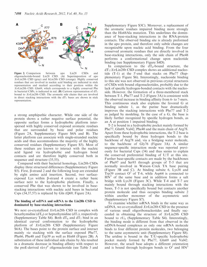
a strong amphipathic character. While one side of theprotein shows a rather negative surface potential, theopposite surface forms a hydrophobic platform inter-spersed with highly conserved exposed aromatic residuesthat are surrounded by basic and polar residues(Figure 2A, Supplementary Figure S4A and B). Thelatter platform can associate with single-stranded nucleicacids and thus accommodates the majority of the highlyconserved residues (Supplementary Figure S5). Most ofthese residues are known to interact with the nucleicacid ligand via hydrophobic and hydrogen bondcontacts and are therefore highly conserved both insequence and structure (35,55).Compared with their bacterial homologs, Lin28b CSDs
display three structural differences (Supplementary FigureS5). First, b-strand 2 and the following loop are extendedby eight amino acid insertion. Second, two surface-exposed Lys within b-strand 4 create a rather basicsurface next to the hydrophobic platform. Finally, aconserved Phe that was shown to be involved in base-stacking interactions with nucleic acid bases in bacterialCsps (34,37,57) is replaced by Gln (Q69; Figure 2A).
The binding of ssDNA and ssRNA to the Lin28b CSD isdominated by base-stacking interactions
We next co-crystallized XtrLin28b CSD in complex withhexathymidine (dT6) or heptathymidine (dT7), respectively(Supplementary Table S4). Both dT6 and dT7 bind in anidentical curved conformation to the hydrophobicplatform of XtrLin28b CSD (Supplementary FigureS6A). The bases point to the protein surface and interactmainly via stacking with the surface exposed Phe77,Phe66, Phe48 and Trp39 as well as His68 (Figure 2B). Asubstitution of these individual side chains by Ala resultedin a dramatic decrease in binding affinity with respect tothe preE-derived rlet-f5 oligonucleotide (see Table 3 and
Supplementary Figure S3C). Moreover, a replacement ofthe aromatic residues impaired binding more stronglythan the His68Ala mutation. This underlines the domin-ance of base-stacking interactions in the RNA:proteincontacts. The observed binding site is already preformedin the apo protein, and therefore only subtle changes arerecognizable upon nucleic acid binding. From the fourconserved aromatic residues that are directly involved inbase-stacking interactions, only the side chain of Phe48performs a conformational change upon nucleotidebinding (see Supplementary Figure S4D).
In comparison to the dT6-bound structure, thedT7:XtrLin28b CSD complex shows an additional nucleo-tide (T-1) at the 50-end that stacks on Phe77 (Sup-plementary Figure S6). Interestingly, nucleoside bindingto this site was not observed in previous crystal structuresof CSDs with bound oligonucleotides, probably due to thelack of specific hydrogen-bonded contacts with the nucleo-side. However, the formation of a three-membered stackbetween T-1, Phe77 and T-2 (Figure 3A) is consistent withthe observed increase in binding affinity from 6- to 7-mers.This continuous stack also explains the favored G atbinding subsite 1, as the purine base dramaticallyimproves the stacking interactions with Phe77 and T-2as judged by modeling. In the case of a G, the base islikely further recognized by specific hydrogen bonds, asan A at position 1 impaired binding.
T-2 is bound in a hydrophobic pocket mainly formed byPhe77, Gln69, Val42, Phe40 and the main chain of Arg78.Apart from these hydrophobic interactions, the T-2 base isspecifically bound by three hydrogen bonds to thebackbone of Arg78 and, via a bridging water molecule,to the backbone of Gly76 (Figure 3A). A similarsequence-specific interaction mode was reported previ-ously for bacterial Csps (34) and thus seems to explainthe conserved preferential binding of U/T at this site.Further base-specific contacts are made by the backbonesof Phe97 and Ser93 through groups of T-5 that arenormally involved in Watson–Crick TA base pairing(Figure 3B and C). At binding subsite 6, Lys38 andTrp39 contact O2 of T-6, while Asp64 is connected toHN3 of the same base and in addition forms a saltbridge with Lys38 (Figure 3C). While T-4 and T-7 aremainly bound through stacking interactions with thebases, T-3 is not specifically bound but contacts anotherprotein molecule and thus occupies binding subsite 2within another monomer of the asymmetric unit(Supplementary Figure S7).
To examine whether ssRNA binds in the same way asssDNA, we co-crystallized XtrLin28b CSD in the presenceof various small oligoribonucleotides and finally suc-ceeded in obtaining the structure of XtrLin28b CSDbound to rU6 (Supplementary Table S4). Interestingly,the binding mode is different from that observed in thessDNA-bound complexes as only one ssRNA moleculebinds to four different protein molecules, two belongingto the same asymmetric unit (Supplementary Figure S8).The uridine is bound in the T/U-specific hydrophobicpocket mainly formed by Phe77, Gln69 and Val42.However, the uracil base adopts a different orientationand is bound through hydrogen bonds to O2 and HN3
Figure 2. Comparison between apo Lin28 CSDs andoligonucleotide-bound Lin28 CSD. (A) Superimposition of apoXtrLin28b CSD (gray) and hLin28b CSD (orange). Highly conservedresidues that are involved in nucleic acid binding are indicated in stickrepresentation and marked according to their position within theXtrLin28b CSD. Gln69, which corresponds to a highly conserved Phein bacterial CSPs, is indicated in red. (B) Cartoon representation of dT7
bound to XtrLin28b CSD. The aromatic side chains that are involvedin direct stacking interactions with the dT7 bases are shown in stickrepresentation.
7498 Nucleic Acids Research, 2012, Vol. 40, No. 15

via the backbone of Arg78. Probably, the unique bindinggeometry observed in this structure is a crystallizationartefact, because a 4:1 stoichiometry between RNA andLin28 was not observed in any of the solution bindingexperiments, which are completely in agreement with thebinding mode observed in dT7-bound XtrLin28b CSD.
Determinants of Lin28:pre-let-7 interactions
To understand how the observed binding preferences ofthe individual Lin28 RBDs result in a specific recognitionof the natural substrate, we performed EMSAs and Dicerin vitro processing assays with Xtr-pre-let-7f. First, wetested if other parts than the Lin28b RBDs contribute tobinding of pre-let-7f. Therefore, we created a variantlacking the C-terminal 27 and the N-terminal 78 residuesof the protein (XtrLin28b CSD+ZKD; see Figure 1). Thisvariant shows a similar binding affinity as the full-lengthprotein with a KD of 1.6±0.1 mM for each (Figure 4Aand C). Moreover, binding of both proteins is highly co-operative, as the binding data could best be fitted to a Hillequation with a Hill coefficient higher than 2 (seeSupplementary Figure S10). Unlike the isolateddomains, XtrLin28b CSD+ZKD is able to inhibit the pro-cessing of pre-let-7 by Dicer to a similar extent(Figure 4D) as the full-length protein. Moreover, thedegree of inhibition strongly depends on the Lin28 con-centration and thus reflects the competitive nature
between Dicer and Lin28 with respect to the accessibilityto the Dicer cleavage site within Xtr-pre-let-7 (Figure 4E).Next, we checked the binding of the isolated ZKD to
Xtr-pre-let-7f. Surprisingly, hardly any binding is observ-able for this variant, even though, according to the lowestenergy structure by Mfold (49), the GGAG motif shouldbe single stranded (Figure 4B). Thus, the data imply thatthe bases of the conserved GGAG motif are not freelyaccessible for the ZKD, since the presence of the GGAGmotif is essential for a proper binding of the full-lengthprotein (Figure 4A).The isolated XtrLin28b CSD, on the other hand, binds
to Xtr-pre-let-7f with about nine times lower affinitycompared to the full-length protein. Moreover, up totwo differently migrating complexes are visible, indicatingthat the CSD can bind to more than one site. Consideringthe binding preference of XtrLin28 CSD for single-stranded pyrimidine-rich sequences, we mutated twopotential CSD-binding sites within preE of Xtr-pre-let-7f: one in the terminal hairpin loop, harboring therlet-7f5 sequence (preE-mut I), and one in a bulge closeto the upper stem region and adjacent to the Dicercleavage site (preE-mut II). Interestingly, both mutationsimpair CSD binding, as hardly any complex band is visiblein both cases. Consistent with this, these mutations cause acomparable loss of binding affinity in context of thewild-type (WT) protein, even though only a combination
Figure 3. Molecular interactions that promote binding of dT7 to XtrLin28b CSD. The electrostatic surface potential of XtrLin28b CSD uponbinding to dT7 was calculated with APBS for pH 7 with a range from �10 (red) to +10 kT (blue). The bound oligonucleotide is shown incartoon representation. Water molecules are shown as gray spheres. (A) Detailed view of T-2 bound to the hydrophobic pocket at bindingsubsite 2. Besides hydrophobic contacts including a three-membered stack with Phe77 and T-1, T/U-specific contacts are mediated via thebackbone atoms of Arg78 and Phe77. (B) Detailed view of T-5 bound in the hydrophobic cleft created by Lys95 and Phe66. Ser93 and Phe97form hydrogen bonds with O4 and HN3 of T-5. Binding of T-4 is only mediated via stacking interactions with His68 and a water molecule-mediatedhydrogen bond. (C) Detailed view of binding subsites 6 and 7. T-6 and T-7 are mainly bound via stacking interactions with Phe48 and Trp39,respectively.
Nucleic Acids Research, 2012, Vol. 40, No. 15 7499

of both mutations (preE-mut III) has a similar effect as theGGAG-mut. Nam et al. (32) showed that the CSDdomain of mLin28 indeed binds to a sequence correspond-ing to preE-mut I. However, as preE-mut II also had a
considerable influence on binding, we suggest thatadditional elements within preE have an influence onthe binding reaction of both the CSD and full-lengthLin28.
Figure 4. Determinants of Lin28:pre-let-7f interactions. (A) EMSAs with Xtr-pre-let-7f as a probe, mixed with increasing concentrations of protein(for XtrLin28b and XtrLin28b CSD+ZKD: 0, 0.4, 0.8, 1.6, 3.2, 6.4, 13, 26 mM; for XtrLin28b CSD and ZKD: 0, 0.8, 1.6, 3.2, 6.4, 13, 26, 51 mM). AllEMSAs were performed using 1 nM a-32P-ATP-labeled RNA. (B) The terminal loop sequence and secondary structure of the Xtr-pre-let-7f mutantsas predicted by Mfold (49). Regions that were mutated are shaded. (C) Results from EMSA experiments shown in (A). The binding affinity wasscored according to the following dissociation constant ranges: +++++, 0.8–1.6 mM; ++++, 1.6–3.2 mM; +++, 3.2–6.4 mM; ++, 6.4–12.8 mM; +,12.8–25.6mM; �, >25.6mM. (D and E) In vitro pre-miRNA processing reaction on 32P 50-end-labeled Xtr-pre-let-7f. The indicated concentrations(in micromolar) of XtrLin28b, XtrLin28b RBDs, XtrLin28b CSD and XtrLin28b ZKD were added to 1 nM Xtr-pre-let-7f in the presence or absenceof human Dicer. The samples were resolved on a 10% (w/v) denaturing PAGE and visualized by autoradiography.
7500 Nucleic Acids Research, 2012, Vol. 40, No. 15

The Lin28 CSD remodels the terminal loop and the upperstem of pre-let-7 while the ZKD ensures a stable bindingand thus efficient blocking of pre-let-7 processing
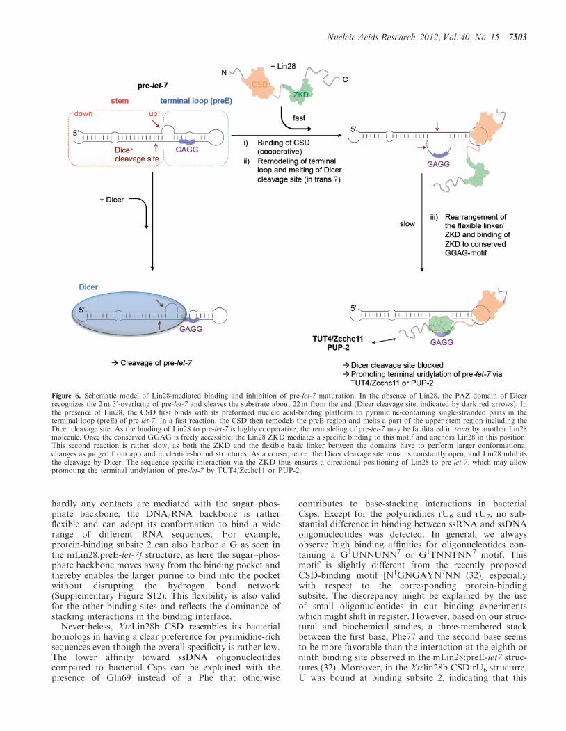
The observation that the isolated CSD, but not the ZKD,binds to Xtr-pre-let-7f with a reasonable affinity suggests amultistep binding reaction in which the CSD first bindsand imposes a structural change within preE and therebyfacilitates a subsequent binding of the ZKD. To confirmthis hypothesis, we performed an RNA remodeling assayusing a truncated Xtr-pre-let-7g* that contains the entireterminal loop and five bases of the double-stranded upperstem region. The blunt end of Xtr-pre-let-7g* wasmodified with a fluorescence quencher pair to monitor apotential RNA unwinding of the double-stranded upperstem region upon protein binding (Figure 5A). Bothfull-length XtrLin28 isoforms and XtrLin28b CSD showa clear increase in fluorescence and thus an RNA remodel-ing activity, while for the ZKD only a slight increase influorescence is observable (Figure 5B). The increase influorescence is again cooperative for WT proteins andthe isolated CSD and reflects their different bindingaffinities to Xtr-pre-let-7g (Figure 5B, D and E).
The ability of the isolated Lin28 CSD to remodel RNAimplies that mutations in the CSD that impair theobserved RNA interactions surface should lead to astronger loss of remodeling activity than mutations inthe Lin28 ZKD. Indeed, this is the case for all examinedCSD mutations even though the KD values of the analyzedvariants with respect to Xtr-pre-let-7g vary only slightly(Figure 5C–E). Upon closer inspection, the Lin28 muta-tions can be clustered into three classes. First, the ZKDvariants XtrLin28b Tyr133Ala and His155Ala have asimilar remodeling activity as the WT protein at low con-centrations but only reach up to 70% of the WT’s activityat saturation. Second, variations in the CSD affectingbinding subsites 5–7 (XtrLin28b Phe66Ala, Phe48Alaand Trp39Ala) still show modest remodeling activitythat almost reaches the level of the ZKD variants athigh concentrations. Finally, XtrLin28b His68Ala(binding subsite 4), Phe77Ala and the double variantXtrLin28b Phe77Ala/Arg78Gly (binding subsites 1 and2) show hardly any remodeling activity (Figure 5C). Thisobservation is highly interesting, as in the correspondingEMSAs all analyzed variants still shifted Xtr-pre-let-7gcompletely at high concentrations (Figure 5D) eventhough the CSD-binding site within pre-let-7g (32) is in-accessible according to the predicted secondary structure(Figure 5A). Consequently, XtrLin28b Phe77Ala andHis68Ala might bind in an alternative mode to Xtr-pre-let-7g. Nevertheless, all variants are impaired in binding toXtr-pre-let-7g and consequently lead to an increased pre-let-7 processing by Dicer (Figure 5D–F).
To gain further insights into the observed remodelingreaction, we measured the kinetics of remodeling bystopped-flow mixing experiments performed with theWT protein, Lin28 variants of each class and theisolated CSD under saturation conditions. Mixing ofWT with Xtr-pre-let-7g* resulted in an associationreaction with two phases (Figure 5G and SupplementaryFigure S11). A major fast reaction (85% of total
amplitude; time constant of the reaction �1=20ms) isfollowed by a minor slow phase (�2=210ms). The timecourse of the XtrLin28b Tyr133Ala remodeling activitycould be fitted to either a monoexponential (�=28ms)or a biexponential equation (�1=26 ms, �2=510ms)with a very weak second time constant making up only3.7% of the total amplitude. This indicates that the secondrate observed in the WT can be traced back to the bindingof the ZKD. Considering the flexible linker between bothRBDs and the structural difference between apo Lin28ZKD and RNA-bound Lin28 ZKD (32,33), the slowsecond time constant may reflect the necessary structuralrearrangement in these regions to facilitate binding. Theisolated CSD shows a one-phase association reaction witha similar time constant as observed for XtrLin28bTyr133Ala (�1=29ms), even though the amplitude ismuch lower. For XtrLin28b Trp39Ala, after a fast associ-ation reaction (�1=15ms) that is completed in 80ms, aslow dissociation reaction is followed (�2=110ms). Thedecrease of fluorescence might reflect the impaired bindingof this construct and thus a higher dissociation ratecompared to WT protein, as the fast remodeling rate isnot impaired. For XtrLin28b Phe77Ala also the fast ratewas considerably impaired, indicating that the corres-ponding residues are crucial for the remodeling reaction.Taken together, the data clearly demonstrate that the
XtrLin28b CSD imposes a structural change within preEof Xtr-pre-let-7g, which facilitates a subsequent bindingof the ZKD. However, as the isolated CSD is unable toblock the processing of Xtr-pre-let-7g alone, the ZKDprobably serves as an anchor for directional binding andthus ensures a constant opening of the Dicer cleavage site(Figure 6).
DISCUSSION
We found that the Lin28 CSDs bind with high affinity tosingle-stranded nucleic acids via a conserved nucleicacid-binding platform mainly formed of exposed sidechains that are surrounded by basic surface residues.This binding platform is already preformed in the apoprotein, and consequently only subtle changes areobserved upon nucleic acid binding. The crystal structureof the XtrLin28b CSD bound to dT7 reveals the binding ofup to seven nucleotides. This observation is in good agree-ment with the binding data, as the CSD showed thehighest affinity for 7- to 9-mers. Such binding preferenceshave also been described for bacterial Csps (34–37,58–60)and recently also for mLin28a in complex with morecomplete preE structures (32). The presence of a closedRNA loop as well as elements of the basic linker regionsin these structures probably triggers the binding of anadditional eighth and ninth nucleotide.Regarding sequence specificity, our structural data
reveal sequence-specific binding at positions 2 and 5only, because at these binding sites only T/U-specifichydrogen bond contacts are mediated with the base.Moreover, at position 6, the presence of a salt bridgebetween Lys38 and Asp64, which is important for the for-mation of hydrogen bonds to T-6, limits the flexibility andconsequently the size of this binding pocket. However, as
Nucleic Acids Research, 2012, Vol. 40, No. 15 7501

Figure 5. The Lin28 CSD remodels the terminal loop of pre-let-7 and facilitates a subsequent binding of the ZKD to the conserved GGAG motif.(A–C) RNA remodeling assay. A truncated Xtr-pre-let-7g that contains only the preE sequence and 5 bp of the upper stem region (Xtr-pre-let-7g*)was incubated with increasing concentrations of the corresponding Lin28 protein. The 50-end of the RNA was modified with the quencher dabcyl(Dab), and the adjacent 30-end harbored a fluorescein (FAM) label. The sequence and secondary structure of the RNA are indicated and the knownZKD- and CSD-binding sites are marked in red and green, respectively. The increase of fluorescence was plotted as a function of titrated Lin28protein. All experiments were performed using a Cary-Eclipse fluorescence spectrometer at 293K in 20mM Tris (pH 7.5), 60mM KCl, 10mM MgCl2and 1mM DTT. (D) EMSAs with Xtr-pre-let-7g as a probe, mixed with increasing concentrations of XtrLin28a and the indicated XtrLin28b variants(0, 0.4, 0.8, 1.6, 3.2, 6.4, 13, 26 mM). All EMSAs were performed using 1 nM a-32P-ATP-labeled RNA. (E) Apparent dissociation constants fromEMSAs shown in (D). Band intensities were quantitated from three independent experiments and used to generate the binding data. (F) In vitropre-miRNA processing reaction on Xtr-pre-let-7g. 10 mM of the indicated XtrLin28b variant was added to 1 nM 32P 50-end-labeled Xtr-pre-let-7g inthe presence or absence of human Dicer. The samples were resolved on a 10% (w/v) denaturing PAGE and visualized by autoradiography.(G) Pre-steady-state kinetics of Lin28-mediated binding and remodeling of Xtr-pre-let-7g*. After rapid mixing of Xtr-pre-let-7g* with 15 mM (finalconcentration) of the indicated XtrLin28b variant, the change of FAM fluorescence was monitored for 1 s using a Chirascan stopped-flow instru-ment. Traces of at least eight replicates were fitted to a mono- (Y133A) or biexponential curve (Xtrlin28b), respectively (solid lines). For W39A, thetime courses of the first 80ms were fitted to a single exponential association curve, while traces from 90ms to 1 s were fitted to a one-phase decaycurve (see Supplementary Figure S11).
7502 Nucleic Acids Research, 2012, Vol. 40, No. 15
hardly any contacts are mediated with the sugar–phos-phate backbone, the DNA/RNA backbone is ratherflexible and can adopt its conformation to bind a widerange of different RNA sequences. For example,protein-binding subsite 2 can also harbor a G as seen inthe mLin28:preE-let-7f structure, as here the sugar–phos-phate backbone moves away from the binding pocket andthereby enables the larger purine to bind into the pocketwithout disrupting the hydrogen bond network(Supplementary Figure S12). This flexibility is also validfor the other binding sites and reflects the dominance ofstacking interactions in the binding interface.
Nevertheless, XtrLin28b CSD resembles its bacterialhomologs in having a clear preference for pyrimidine-richsequences even though the overall specificity is rather low.The lower affinity toward ssDNA oligonucleotidescompared to bacterial Csps can be explained with thepresence of Gln69 instead of a Phe that otherwise
contributes to base-stacking interactions in bacterialCsps. Except for the polyuridines rU6 and rU7, no sub-stantial difference in binding between ssRNA and ssDNAoligonucleotides was detected. In general, we alwaysobserve high binding affinities for oligonucleotides con-taining a G1UNNUNN7 or G1TNNTNN7 motif. Thismotif is slightly different from the recently proposedCSD-binding motif [N1GNGAYN7NN (32)] especiallywith respect to the corresponding protein-bindingsubsite. The discrepancy might be explained by the useof small oligonucleotides in our binding experimentswhich might shift in register. However, based on our struc-tural and biochemical studies, a three-membered stackbetween the first base, Phe77 and the second base seemsto be more favorable than the interaction at the eighth orninth binding site observed in the mLin28:preE-let7 struc-tures (32). Moreover, in the Xtrlin28b CSD:rU6 structure,U was bound at binding subsite 2, indicating that this
Figure 6. Schematic model of Lin28-mediated binding and inhibition of pre-let-7 maturation. In the absence of Lin28, the PAZ domain of Dicerrecognizes the 2 nt 30-overhang of pre-let-7 and cleaves the substrate about 22 nt from the end (Dicer cleavage site, indicated by dark red arrows). Inthe presence of Lin28, the CSD first binds with its preformed nucleic acid-binding platform to pyrimidine-containing single-stranded parts in theterminal loop (preE) of pre-let-7. In a fast reaction, the CSD then remodels the preE region and melts a part of the upper stem region including theDicer cleavage site. As the binding of Lin28 to pre-let-7 is highly cooperative, the remodeling of pre-let-7 may be facilitated in trans by another Lin28molecule. Once the conserved GGAG is freely accessible, the Lin28 ZKD mediates a specific binding to this motif and anchors Lin28 in this position.This second reaction is rather slow, as both the ZKD and the flexible basic linker between the domains have to perform larger conformationalchanges as judged from apo and nucleotide-bound structures. As a consequence, the Dicer cleavage site remains constantly open, and Lin28 inhibitsthe cleavage by Dicer. The sequence-specific interaction via the ZKD thus ensures a directional positioning of Lin28 to pre-let-7, which may allowpromoting the terminal uridylation of pre-let-7 by TUT4/Zcchc11 or PUP-2.
Nucleic Acids Research, 2012, Vol. 40, No. 15 7503

particular binding subsite has a high affinity toward U.Therefore, it is likely that the oligonucleotides used forsolution binding experiments in our study indeed boundas seen in the Xtrlin28b CSD:dT7 structure.The broad spectrum of potential CSD-binding
sequences probably reflects the low sequence conservationwithin its natural substrate, the preE of pre-let-7 (seeSupplementary Figure S9). Consequently, sequence-specific binding of Lin28 is likely mediated through theZKD. Structural studies with homologous ZKDsrevealed a specific binding of a GGAG- or GGAG-likemotif (50,51). Interestingly, this motif is highly conservedwithin preEs of pre-let-7. However, we could show thatthe association of Lin28 with pre-let-7 is also strongly de-pendent on the Lin28 CSD, as the isolated XtrLin28bZKD could hardly bind to Xtr-pre-let-7f in vitro. Thus,a mutation of the GGAG motif to AAAA did notdisrupt binding of WT XtrLin28b completely. Moreover,our biochemical analysis of XtrLin28b ZKD showed thatit binds with high affinity to motifs containing two Gs,which are preferentially spaced by one nucleotide otherthan G. This reflects the observation that the GGAGmotif is present in variable forms in pre-let-7 such asGGUG, GGCG, GGAG and UGXG. It seems likelythat the ZKD specifically recognizes this variable motifsas well and thereby contributes to a stable binding of pre-let-7 and facilitates a subsequent TUT4-mediateduridylation of pre-let-7, as this process was shown to bedependent on the GGAG motif (31).A recent study proposed that Lin28 induces a structural
change within preE of pre-let-7g and thus directly preventsthe processing of pre-let-7g by Dicer (61). Using enzymaticfootprinting, the authors reported that upon Lin28binding a part of the upper stem of pre-let-7 becomesmore susceptible to cleavage by single-strand-specific ribo-nucleases. We now could demonstrate that the structuralchange within pre-let-7g is mediated through the action ofthe CSD, as only mutations within this domain impairedthe remodeling activity significantly. The function ofCSDs as protein domains that can induce local secondarystructural changes within RNA was already proposed forbacterial Csps in the process of transcription anti-termination (62–64). In the case of E. coli CspE, amelting pathway was suggested, in which two conservedPhe and one His, resembling Phe46 and Phe66 (bindingsubsite 5) and His68 (binding subsite 4) in XtrLin28bCSD, intercalate between bases of the stem. Indeed,His68 and Phe77 (binding subsites 1 and 2) are essentialfor the remodeling activity of Lin28, and it is likely that asimilar melting pathway exists for the Lin28 CSD.Keeping in mind that most preE secondary structures of
pre-let-7 differ considerably from the structure observed inthe mLin28:preE complexes, the CSD-dependent remodel-ing seems to be essential for a directional binding asproposed by Nam et al. (32). According to Mfold, in6 (pre-let-7 a-1, a-2, b, c, g, f-2) out of 11 pre-let-7miRNAs, the GGAG motif is inaccessible. Moreover,often multiple secondary structures with almost the sameenergy exist. Based on our data, we propose a multi-stepbinding model according to which Lin28 CSD first binds apyrimidine-rich single-stranded sequence within preE and
induces a conformational change that facilitates a subse-quent binding of the ZKD to the conserved GGAGmotif. As the binding of XtrLin28b and the isolatedXtrLin28b CSD to Xtr-pre-let-7f is highly cooperative[Supplementary Figure S10 (65)], it is possible that theremodeling is performed in trans by another Lin28molecule. Moreover, the action of the Lin28 CSD alsoleads to a melting of the upper stem region including theDicer cleavage site (32,61). The sequence-specific bindingof the ZKD to the GGAG motif adjacent to this site mayensure a constant opening of the corresponding site andthereby directly inhibit the processing by Dicer. Thiswould also explain why both intact RBDs are requiredfor an efficient inhibition of pre-let-7 processing byDicer even though the isolated CSD and most of theanalyzed variants are able to bind and remodel pre-let-7.
Apart from inhibiting let-7 biogenesis, Lin28 alsoexhibits let-7-independent functions. Multiple studiesshowed that Lin28 binds to a variety of mRNAs andstimulates the translation of factors associated with cellgrowth and survival (66–70). This let-7-independentfunction of Lin28 is mediated via protein interactions.As an N- and C-terminally truncated construct ofXtrLin28b, harboring the two RBDs only, was still ableto bind to pre-let-7 with similar affinity as the full-lengthprotein, we conclude that regions outside the RBDs haveother functions than RNA binding and may be involved inprotein–protein interactions.
ACCESSION NUMBERS
PDB 3ulj, 4a75, 4a76, 4alp and 4a4i.
SUPPLEMENTARY DATA
Supplementary Data are available at NAR Online:Supplementary Tables 1–5 and Supplementary Figures1–12.
ACKNOWLEDGEMENTS
We thank Tracy Dornblut, Janett Tischer, Ingrid Bergerand Michaela Naschke for excellent technical assistance.We are also thankful to Yvette Roske, who assisted indiffraction data collection and processing, and acknow-ledge Uwe Muller and the beamline support by the staffof the Helmholtz-Zentrum Berlin fur Materialien undEnergie at BESSY. The Protein Sample ProductionFacility at the Max Delbruck Center is funded by theHelmholtz Association of German Research Centres.U.H., A.S. and F.M. conceived and coordinated thestudy. F.M., A.S. and N.D. performed the experiments.F.M. and A.S. analyzed the data. F.M. wrote the manu-script. All authors read and approved the manuscript.
FUNDING
Funding for open access charge: Max-Delbruck-Centrumfur Molekulare Medizin Berlin-Buch.
Conflict of interest statement. None declared.
7504 Nucleic Acids Research, 2012, Vol. 40, No. 15

REFERENCES
1. Landthaler,M., Yalcin,A. and Tuschl,T. (2004) The humanDiGeorge syndrome critical region gene 8 and its D.melanogaster homolog are required for miRNA biogenesis.Curr. Biol., 14, 2162–2167.
2. Lee,Y., Ahn,C., Han,J., Choi,H., Kim,J., Yim,J., Lee,J.,Provost,P., Radmark,O., Kim,S. et al. (2003) The nuclearRNase III Drosha initiates microRNA processing. Nature,425, 415–419.
3. Bohnsack,M.T., Czaplinski,K. and Gorlich,D. (2004) Exportin 5is a RanGTP-dependent dsRNA-binding protein that mediatesnuclear export of pre-miRNAs. RNA, 10, 185–191.
4. Yi,R., Qin,Y., Macara,I.G. and Cullen,B.R. (2003) Exportin-5mediates the nuclear export of pre-microRNAs and short hairpinRNAs. Genes Dev., 17, 3011–3016.
5. Hutvagner,G., McLachlan,J., Pasquinelli,A.E., Balint,E., Tuschl,T.and Zamore,P.D. (2001) A cellular function for theRNA-interference enzyme Dicer in the maturation of the let-7small temporal RNA. Science, 293, 834–838.
6. Ketting,R.F., Fischer,S.E., Bernstein,E., Sijen,T., Hannon,G.J.and Plasterk,R.H. (2001) Dicer functions in RNA interferenceand in synthesis of small RNA involved in developmental timingin C. elegans. Genes Dev., 15, 2654–2659.
7. Knight,S.W. and Bass,B.L. (2001) A role for the RNase IIIenzyme DCR-1 in RNA interference and germ line developmentin Caenorhabditis elegans. Science, 293, 2269–2271.
8. Grosshans,H., Johnson,T., Reinert,K.L., Gerstein,M. andSlack,F.J. (2005) The temporal patterning microRNA let-7regulates several transcription factors at the larval to adulttransition in C. elegans. Dev. Cell, 8, 321–330.
9. Pasquinelli,A.E., Reinhart,B.J., Slack,F., Martindale,M.Q.,Kuroda,M.I., Maller,B., Hayward,D.C., Ball,E.E., Degnan,B.,Muller,P. et al. (2000) Conservation of the sequence and temporalexpression of let-7 heterochronic regulatory RNA. Nature, 408,86–89.
10. Reinhart,B.J., Slack,F.J., Basson,M., Pasquinelli,A.E.,Bettinger,J.C., Rougvie,A.E., Horvitz,H.R. and Ruvkun,G. (2000)The 21-nucleotide let-7 RNA regulates developmental timing inCaenorhabditis elegans. Nature, 403, 901–906.
11. Bussing,I., Slack,F.J. and Grosshans,H. (2008) let-7 microRNAsin development, stem cells and cancer. Trends Mol. Med., 14,400–409.
12. Johnson,S., Grosshans,H., Shingara,J., Byrom,M., Jarvis,R.,Cheng,A., Labourier,E., Reinert,K., Brown,D. and Slack,F.(2005) RAS is regulated by the let-7 microRNA family. Cell, 120,635–647.
13. Mayr,C., Hemann,M.T. and Bartel,D.P. (2007) Disrupting thepairing between let-7 and Hmga2 enhances oncogenictransformation. Science, 315, 1576–1579.
14. Wulczyn,F., Smirnova,L., Rybak,A., Brandt,C., Kwidzinski,E.,Ninnemann,O., Strehle,M., Seiler,A., Schumacher,S. andNitsch,R. (2007) Post-transcriptional regulation of the let-7microRNA during neural cell specification. FASEB J., 21,415–426.
15. Heo,I., Joo,C., Cho,J., Ha,M., Han,J. and Kim,V.N. (2008) Lin28mediates the terminal uridylation of let-7 precursor MicroRNA.Mol. Cell, 32, 276–284.
16. Newman,M.A., Thomson,J.M. and Hammond,S.M. (2008) Lin-28interaction with the Let-7 precursor loop mediates regulatedmicroRNA processing. RNA, 14, 1539–1549.
17. Rybak,A., Fuchs,H., Smirnova,L., Brandt,C., Pohl,E., Nitsch,R.and Wulczyn,F. (2008) A feedback loop comprising lin-28 andlet-7 controls pre-let-7 maturation during neural stem-cellcommitment. Nat. Cell Biol., 10, 987–993.
18. Viswanathan,S.R., Daley,G.Q. and Gregory,R.I. (2008) Selectiveblockade of microRNA processing by Lin28. Science, 320,97–100.
19. Ambros,V. and Horvitz,H. (1984) Heterochronic mutants of thenematode Caenorhabditis elegans. Science, 226, 409–416.
20. Moss,E.G., Lee,R.C. and Ambros,V. (1997) The cold shockdomain protein LIN-28 controls developmental timing in C.
elegans and is regulated by the lin-4 RNA. Cell, 88, 637–646.
21. Seggerson,K., Tang,L. and Moss,E.G. (2002) Two genetic circuitsrepress the Caenorhabditis elegans heterochronic gene lin-28 aftertranslation initiation. Dev. Biol., 243, 215–225.
22. Reinhart,B., Slack,F., Basson,M., Pasquinelli,A., Bettinger,J.,Rougvie,A., Horvitz,H. and Ruvkun,G. (2000) The 21-nucleotidelet-7 RNA regulates developmental timing in Caenorhabditiselegans. Nature, 403, 901–906.
23. Richards,M., Tan,S.-P., Tan,J.-H., Chan,W.-K. and Bongso,A.(2004) The transcriptome profile of human embryonic stem cellsas defined by SAGE. Stem Cells, 22, 51–64.
24. Yang,D.H. and Moss,E.G. (2003) Temporally regulatedexpression of Lin-28 in diverse tissues of the developing mouse.Gene Expr. Patterns, 3, 719–726.
25. Yang,X., Lin,X., Zhong,X., Kaur,S., Li,N., Liang,S., Lassus,H.,Wang,L., Katsaros,D., Montone,K. et al. (2010) Double-negativefeedback loop between reprogramming factor LIN28 andmicroRNA let-7 regulates aldehyde dehydrogenase 1-positivecancer stem cells. Cancer Res., 70, 9463–9472.
26. Yu,J., Vodyanik,M., Smuga-Otto,K., Antosiewicz-Bourget,J.,Frane,J., Tian,S., Nie,J., Jonsdottir,G., Ruotti,V., Stewart,R.et al. (2007) Induced pluripotent stem cell lines derived fromhuman somatic cells. Science, 318, 1917–1920.
27. Viswanathan,S., Powers,J., Einhorn,W., Hoshida,Y., Ng,T.,Toffanin,S., O’Sullivan,M., Lu,J., Phillips,L., Lockhart,V. et al.(2009) Lin28 promotes transformation and is associated withadvanced human malignancies. Nat. Genet., 41, 843–848.
28. Piskounova,E., Viswanathan,S.R., Janas,M., LaPierre,R.J.,Daley,G.Q., Sliz,P. and Gregory,R.I. (2008) Determinants ofmicroRNA processing inhibition by the developmentally regulatedRNA-binding protein Lin28. J. Biol. Chem., 283, 21310–21314.
29. Hagan,J., Piskounova,E. and Gregory,R. (2009) Lin28 recruits theTUTase Zcchc11 to inhibit let-7 maturation in mouse embryonicstem cells. Nat. Struct. Mol. Biol., 16, 1021–1025.
30. Lehrbach,N.J., Armisen,J., Lightfoot,H.L., Murfitt,K.J.,Bugaut,A., Balasubramanian,S. and Miska,E.A. (2009) LIN-28and the poly(U) polymerase PUP-2 regulate let-7 microRNAprocessing in Caenorhabditis elegans. Nat. Struct. Mol. Biol.,16, 1016–1020.
31. Heo,I., Joo,C., Kim,Y.-K., Ha,M., Yoon,M.-J., Cho,J.,Yeom,K.-H., Han,J. and Kim,V.N. (2009) TUT4 in concertwith Lin28 suppresses microRNA biogenesis throughpre-microRNA uridylation. Cell, 138, 696–708.
32. Nam,Y., Chen,C., Gregory,R.I., Chou,J.J. and Sliz,P. (2011)Molecular basis for interaction of let-7 microRNAs with Lin28.Cell, 147, 1080–1091.
33. Loughlin,F.E., Gebert,L.F., Towbin,H., Brunschweiger,A., Hall,J.and Allain,F.H. (2012) Structural basis of pre-let-7 miRNArecognition by the zinc knuckles of pluripotency factor Lin28.Nat. Struct. Mol. Biol., 19, 84–89.
34. Max,K.E.A., Zeeb,M., Bienert,R., Balbach,J. and Heinemann,U.(2006) T-rich DNA single strands bind to a preformed site on thebacterial cold shock protein Bs-CspB. J. Mol. Biol., 360, 702–714.
35. Max,K.E.A., Zeeb,M., Bienert,R., Balbach,J. and Heinemann,U.(2007) Common mode of DNA binding to cold shock domains.Crystal structure of hexathymidine bound to the domain-swappedform of a major cold shock protein from Bacillus caldolyticus.FEBS J., 274, 1265–1279.
36. Zeeb,M. and Balbach,J. (2003) Single-stranded DNA binding ofthe cold-shock protein CspB from Bacillus subtilis: NMRmapping and mutational characterization. Protein Sci., 12,112–123.
37. Zeeb,M., Max,K.E.A., Weininger,U., Low,C., Sticht,H. andBalbach,J. (2006) Recognition of T-rich single-stranded DNA bythe cold shock protein Bs-CspB in solution. Nucleic Acids Res.,34, 4561–4571.
38. Scheich,C., Kummel,D., Soumailakakis,D., Heinemann,U. andBussow,K. (2007) Vectors for co-expression of an unrestrictednumber of proteins. Nucleic Acids Res., 35, e43.
39. Heinemann,U., Bussow,K., Mueller,U. and Umbach,P. (2003)Facilities and methods for the high-throughput crystal structuralanalysis of human proteins. Acc. Chem. Res., 36, 157–163.
40. Leslie,A. (1992) Recent changes to the MOSFLM package forprocessing film and image plate data. Joint CCP4 + ESF-EAMCB Newsletter on Protein Crystallography, No. 26.
Nucleic Acids Research, 2012, Vol. 40, No. 15 7505

41. Kabsch,W. (2010) XDS. Acta Crystallogr. D Biol. Crystallogr.,66, 125–132.
42. Otwinowski,Z. and Minor,W. (1997) Processing of X-raydiffraction data collected in oscillation mode. Method Enzymol.,276, 307–326.
43. Perrakis,A., Langer,G., Cohen,S.X. and Lamzin,V.S. (2008)Automated macromolecular model building for X-raycrystallography using ARP/wARP version 7. Nat. Protoc., 3,1171–1179.
44. Murshudov,G.N., Vagin,A.A. and Dodson,E.J. (1997) Refinementof macromolecular structures by the maximum-likelihood method.Acta Crystallogr. D Biol. Crystallogr., 53, 240–255.
45. Emsley,P. and Cowtan,K. (2004) Coot: model-building tools formolecular graphics. Acta Crystallogr. D Biol. Crystallogr., 60,2126–2132.
46. Ferrin,T.E., Pettersen,E.F., Goddard,T.D., Huang,C.C.,Couch,G.S., Greenblatt,D.M. and Meng,E.C. (2004) UCSFchimera—a visualization system for exploratory research andanalysis. J. Comput. Chem., 25, 1605–1612.
47. Eftink,M.R. (1997) Fluorescence methods for studyingequilibrium macromolecule-ligand interactions. Methods Enzymol.,278, 221–257.
48. Lohman,T.M. and Bujalowski,W. (1991) Thermodynamicmethods for model-independent determination of equilibriumbinding isotherms for protein-DNA interactions—spectroscopicapproaches to monitor binding. Methods Enzymol., 208,258–290.
49. Zuker,M. (2003) Mfold web server for nucleic acid folding andhybridization prediction. Nucleic Acids Res., 31, 3406–3415.
50. Amarasinghe,G.K., De Guzman,R.N., Turner,R.B.,Chancellor,K.J., Wu,Z.R. and Summers,M.F. (2000) NMRstructure of the HIV-1 nucleocapsid protein bound to stem-loopSL2 of the psi-RNA packaging signal. Implications for genomerecognition. J. Mol. Biol., 301, 491–511.
51. De Guzman,R.N., Wu,Z.R., Stalling,C.C., Pappalardo,L.,Borer,P.N. and Summers,M.F. (1998) Structure of the HIV-1nucleocapsid protein bound to the SL3 psi-RNA recognitionelement. Science, 279, 384–388.
52. Rajkowitsch,L., Chen,D., Stampfl,S., Semrad,K., Waldsich,C.,Mayer,O., Jantsch,M.F., Konrat,R., Blasi,U. and Schroeder,R.(2007) RNA chaperones, RNA annealers and RNA helicases.RNA Biol., 4, 118–130.
53. Schindelin,H., Jiang,W., Inouye,M. and Heinemann,U. (1994)Crystal structure of CspA, the major cold shock protein ofEscherichia coli. Proc. Natl. Acad. Sci. USA., 91,5119–5123.
54. Chaikam,V. and Karlson,D.T. (2010) Comparison of structure,function and regulation of plant cold shock domain proteinsto bacterial and animal cold shock domain proteins. BMB Rep.,43, 1–8.
55. Horn,G., Hofweber,R., Kremer,W. and Kalbitzer,H.R. (2007)Structure and function of bacterial cold shock proteins.Cell. Mol. Life Sci., 64, 1457–1470.
56. Mihailovich,M., Militti,C., Gabaldon,T. and Gebauer,F. (2010)Eukaryotic cold shock domain proteins: highly versatile regulatorsof gene expression. Bioessays, 32, 109–118.
57. Max,K.E.A., Wunderlich,M., Roske,Y., Schmid,F.X. andHeinemann,U. (2007) Optimized variants of the cold shockprotein from in vitro selection: structural basis of their highthermostability. J. Mol. Biol., 369, 1087–1097.
58. Bienert,R., Zeeb,M., Dostal,L., Feske,A., Magg,C., Max,K.,Welfle,H., Balbach,J. and Heinemann,U. (2004) Single-strandedDNA bound to bacterial cold-shock proteins: preliminarycrystallographic and Raman analysis. Acta Crystallogr.D Biol. Crystallogr., 60, 755–757.
59. Lopez,M.M. and Makhatadze,G.I. (2000) Major cold shockproteins, CspA from Escherichia coli and CspB from Bacillussubtilis, interact differently with single-stranded DNA templates.Biochim. Biophys. Acta, 1479, 196–202.
60. Lopez,M.M., Yutani,K. and Makhatadze,G.I. (2001) Interactionsof the cold shock protein CspB from Bacillus subtilis withsingle-stranded DNA. Importance of the T base content andposition within the template. J. Biol. Chem., 276, 15511–15518.
61. Lightfoot,H.L., Bugaut,A., Armisen,J., Lehrbach,N.J., Miska,E.A.and Balasubramanian,S. (2011) A LIN28-dependent structuralchange in pre-let-7g directly inhibits dicer processing.Biochemistry, 50, 7514–7521.
62. Phadtare,S., Inouye,M. and Severinov,K. (2004) The mechanismof nucleic acid melting by a CspA family protein. J. Mol. Biol.,337, 147–155.
63. Phadtare,S. (2011) Unwinding activity of cold shock proteins andRNA metabolism. RNA Biol., 8, 394–397.
64. Phadtare,S. and Severinov,K. (2005) Nucleic acid melting byEscherichia coli CspE. Nucleic Acids Res., 33, 5583–5590.
65. Desjardins,A., Yang,A., Bouvette,J., Omichinski,J.G. andLegault,P. (2012) Importance of the NCp7-like domain in therecognition of pre-let-7g by the pluripotency factor Lin28. NucleicAcids Res., 40, 1767–1777.
66. Balzer,E., Heine,C., Jiang,Q., Lee,V. and Moss,E. (2010) LIN28alters cell fate succession and acts independently of the let-7microRNA during neurogliogenesis in vitro. Development, 137,891–900.
67. Peng,S., Chen,L.-L., Lei,X.-X., Yang,L., Lin,H., Carmichael,G.G.and Huang,Y. (2011) Genome-wide studies reveal that Lin28enhances the translation of genes important for growth andsurvival of human embryonic stem cells. Stem Cells, 29, 496–504.
68. Qiu,C., Ma,Y., Wang,J., Peng,S. and Huang,Y. (2010)Lin28-mediated post-transcriptional regulation of Oct4 expressionin human embryonic stem cells. Nucleic Acids Res., 38,1240–1248.
69. Xu,B. and Huang,Y. (2009) Histone H2a mRNA interacts withLin28 and contains a Lin28-dependent posttranscriptionalregulatory element. Nucleic Acids Res., 37, 4256–4263.
70. Xu,B., Zhang,K. and Huang,Y. (2009) Lin28 modulates cellgrowth and associates with a subset of cell cycle regulatormRNAs in mouse embryonic stem cells. RNA, 15, 357–361.
7506 Nucleic Acids Research, 2012, Vol. 40, No. 15