MicroRNA amplification and detection technologies - Nature
18
Laboratory Investigation (2019) 99:452–469 https://doi.org/10.1038/s41374-018-0143-3 REVIEW ARTICLE MicroRNA amplification and detection technologies: opportunities and challenges for point of care diagnostics Vivek Priy Dave 1,4 ● Tien Anh Ngo 1 ● Anna-Karin Pernestig 2 ● Diana Tilevik 2 ● Krishna Kant 3 ● Trieu Nguyen 1 ● Anders Wolff 3 ● Dang Duong Bang 1 Received: 17 May 2018 / Revised: 3 August 2018 / Accepted: 30 August 2018 / Published online: 12 December 2018 © United States & Canadian Academy of Pathology 2018 Abstract The volume of point of care (POC) testing continues to grow steadily due to the increased availability of easy-to-use devices, thus making it possible to deliver less costly care closer to the patient site in a shorter time relative to the central laboratory services. A novel class of molecules called microRNAs have recently gained attention in healthcare management for their potential as biomarkers for human diseases. The increasing interest of miRNAs in clinical practice has led to an unmet need for assays that can rapidly and accurately measure miRNAs at the POC. However, the most widely used methods for analyzing miRNAs, including Northern blot-based platforms, in situ hybridization, reverse transcription qPCR, microarray, and next-generation sequencing, are still far from being used as ideal POC diagnostic tools, due to considerable time, expertize required for sample preparation, and in terms of miniaturizations making them suitable platforms for centralized labs. In this review, we highlight various existing and upcoming technologies for miRNA amplification and detection with a particular emphasis on the POC testing industries. The review summarizes different miRNA targets and signals amplification-based assays, from conventional methods to alternative technologies, such as isothermal amplification, paper-based, oligonucleotide-templated reaction, nanobead-based, electrochemical signaling- based, and microfluidic chip-based strategies. Based on critical analysis of these technologies, the possibilities and feasibilities for further development of POC testing for miRNA diagnostics are addressed and discussed. Introduction MicroRNAs (miRNAs) are evolutionary conserved, ~18–24 nucleotides long non-coding RNA, playing a significant role in controlling human gene expression by post-transcriptional gene regulation or silencing [1]. Further, aberrant expression of a single miRNA can regulate the activity of multiple genes [2]. Previous studies have demonstrated that changes in miRNA expression contribute to a wide variety of human disease states and disorders such as cancer, cardiovascular, autoimmune, neurodegenerative, liver, and inflammatory diseases [3–5]. The miRNAs not only circulate in the human peripheral blood in a remarkably stable form [6–9], they are also widely present in other body tissues and fluids such as urine, saliva, milk, and cerebrospinal fluid [9–11]. These characteristics indicate that the miRNAs are potential biomarkers for diagnostic purposes. The miRNAs are involved in disease origin and development, and are pathology-specific, thus altered miRNA expression have been addressed for early detection and diagnostics, classifi- cation, prognostics, as well as predictive diagnostics. These authors contributed equally: Vivek Priy Dave and Tien Anh Ngo * Tien Anh Ngo [email protected] 1 Laboratory of Applied Micro and Nanotechnology (LAMINATE), Division of Microbiology and Production, National Food Institute, Technical University of Denmark, 2800 Kgs Lyngby, Denmark 2 Systems Biology Research Centre, School of Bioscience, University of Skövde, Skövde, Sweden 3 Department of Micro- and Nanotechnology, Technical University of Denmark, 2800 Kgs Lyngby, Denmark 4 Present address: Boditech Med Inc, 43 Geodudanji 1-gil, Dongnae-myeon, 24398 Chuncheon-si, Gangwon-do, South Korea Supplementary material The online version of this article (https:// doi.org/10.1038/s41374-018-0143-3) contains supplementary material, which is available to authorized users. 1234567890();,: 1234567890();,:
-
Upload
khangminh22 -
Category
Documents
-
view
1 -
download
0
Transcript of MicroRNA amplification and detection technologies - Nature

Laboratory Investigation (2019) 99:452–469https://doi.org/10.1038/s41374-018-0143-3
REVIEW ARTICLE
MicroRNA amplification and detection technologies: opportunitiesand challenges for point of care diagnostics
Vivek Priy Dave1,4 ● Tien Anh Ngo 1● Anna-Karin Pernestig2
● Diana Tilevik2 ● Krishna Kant3 ● Trieu Nguyen1●
Anders Wolff3 ● Dang Duong Bang1
Received: 17 May 2018 / Revised: 3 August 2018 / Accepted: 30 August 2018 / Published online: 12 December 2018© United States & Canadian Academy of Pathology 2018
AbstractThe volume of point of care (POC) testing continues to grow steadily due to the increased availability of easy-to-usedevices, thus making it possible to deliver less costly care closer to the patient site in a shorter time relative to thecentral laboratory services. A novel class of molecules called microRNAs have recently gained attention in healthcaremanagement for their potential as biomarkers for human diseases. The increasing interest of miRNAs in clinical practice hasled to an unmet need for assays that can rapidly and accurately measure miRNAs at the POC. However, the most widelyused methods for analyzing miRNAs, including Northern blot-based platforms, in situ hybridization, reverse transcriptionqPCR, microarray, and next-generation sequencing, are still far from being used as ideal POC diagnostic tools, due toconsiderable time, expertize required for sample preparation, and in terms of miniaturizations making them suitableplatforms for centralized labs. In this review, we highlight various existing and upcoming technologies for miRNAamplification and detection with a particular emphasis on the POC testing industries. The review summarizes differentmiRNA targets and signals amplification-based assays, from conventional methods to alternative technologies, suchas isothermal amplification, paper-based, oligonucleotide-templated reaction, nanobead-based, electrochemical signaling-based, and microfluidic chip-based strategies. Based on critical analysis of these technologies, the possibilities andfeasibilities for further development of POC testing for miRNA diagnostics are addressed and discussed.
Introduction
MicroRNAs (miRNAs) are evolutionary conserved, ~18–24nucleotides long non-coding RNA, playing a significant rolein controlling human gene expression by post-transcriptionalgene regulation or silencing [1]. Further, aberrant expressionof a single miRNA can regulate the activity of multiplegenes [2]. Previous studies have demonstrated that changesin miRNA expression contribute to a wide variety of humandisease states and disorders such as cancer, cardiovascular,autoimmune, neurodegenerative, liver, and inflammatorydiseases [3–5]. The miRNAs not only circulate in the humanperipheral blood in a remarkably stable form [6–9], they arealso widely present in other body tissues and fluids such asurine, saliva, milk, and cerebrospinal fluid [9–11]. Thesecharacteristics indicate that the miRNAs are potentialbiomarkers for diagnostic purposes. The miRNAs areinvolved in disease origin and development, andare pathology-specific, thus altered miRNA expression havebeen addressed for early detection and diagnostics, classifi-cation, prognostics, as well as predictive diagnostics.
These authors contributed equally: Vivek Priy Dave and Tien AnhNgo
* Tien Anh [email protected]
1 Laboratory of Applied Micro and Nanotechnology (LAMINATE),Division of Microbiology and Production, National Food Institute,Technical University of Denmark, 2800 Kgs Lyngby, Denmark
2 Systems Biology Research Centre, School of Bioscience,University of Skövde, Skövde, Sweden
3 Department of Micro- and Nanotechnology, Technical Universityof Denmark, 2800 Kgs Lyngby, Denmark
4 Present address: Boditech Med Inc, 43 Geodudanji 1-gil,Dongnae-myeon, 24398 Chuncheon-si, Gangwon-do, South Korea
Supplementary material The online version of this article (https://doi.org/10.1038/s41374-018-0143-3) contains supplementarymaterial, which is available to authorized users.
1234
5678
90();,:
1234567890();,:
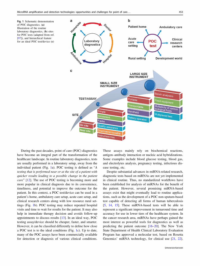
During the past decades, point of care (POC) diagnosticshave become an integral part of the transformation of thehealthcare landscape. In routine laboratory diagnostics, testsare usually performed in a laboratory setup, away from theindividual patient (Fig. 1a). POC testing is defined as “Atesting that is performed near or at the site of a patient withquicker results leading to a possible change in the patientcare” [12]. The use of POC testing is becoming more andmore popular in clinical diagnosis due to its convenience,timeliness, and potential to improve the outcome for thepatient. In this context, a POC test/device can be used in apatient’s home, ambulatory care setup, acute care setup, andclinical research centers along with low resource rural set-tings (Fig. 1b). POC testing may reduce repeated hospitalvisits and time to wait for results for the patient. It may alsohelp in immediate therapy decision and avoids follow-upappointments to discuss results [13]. In an ideal way, POCtesting assay/device should be cheaper, faster, and smarter.However, it can be classified differently to define how closea POC test is to the ideal conditions (Fig. 1c). Up to date,many of the POC assays have been commercially availablefor detection or diagnosis of various clinical conditions.
These assays mainly rely on biochemical reactions,antigen–antibody interaction or nucleic acid hybridizations.Some examples include blood glucose testing, blood gas,and electrolytes analysis, pregnancy testing, infectious dis-ease testing, etc.
Despite substantial advances in miRNA-related research,diagnostic tests based on miRNAs are not yet implementedin clinical routine. Thus, no standardized workflows havebeen established for analysis of miRNAs for the benefit ofthe patient. However, several promising miRNA-basedassays exist that might eventually lead to routine applica-tions, such as the development of a POC non-sputum-basedtest capable of detecting all forms of human tuberculosis[5, 14, 15]. These miRNA-based tests will be able torepresent a significant improvement in turnaround time andaccuracy for use in lower tiers of the healthcare system. Inthe cancer research area, miRNAs have perhaps gained themost interest as powerful tools for diagnostics as well aspredicting the patient outcome [16–20]. The New YorkState Department of Health Clinical Laboratory EvaluationProgram has approved a molecular test, based on RosettaGenomics’ miRNA technology, for clinical use [21, 22].
Fig. 1 Schematic demonstrationof POC diagnostics. (a)Illustration of the routinelaboratory diagnostics, (b) sitesfor POC tests (adapted from ref.[97]), and hierarchical featurefor an ideal POC test/device (c)
MicroRNA amplification and detection technologies: opportunities and challenges for point of care. . . 453

When using this system to test a sample of a patient’stumor, the test classifies squamous-cell carcinoma of thelung with a sensitivity of 96% and specificity of 90%.Columbia University Medical Center developed and vali-dated this assay. Food and Drug Administration (FDA) hasalready approved a handful of miRNA-based tests aimed atguiding therapeutic management. For instance, for theclassification of cancer of unknown primary origin, for lungcancer type, for thyroid cancer stratification, and for breastcancer metastasis [23, 24]. Although the results are pro-mising and may be used to guide decisions regardingtreatment that leads to improved patient outcomes, this fieldis still in the infant stage, and the adoption of these tests bythe clinical practice requires more clinical studies. Recentreviews have also emphasized the role of circulating miR-NAs as potential and emerging biomarkers for diagnosis ofhuman infectious diseases [5]. The increased amount ofstudies on miRNAs as promising diagnostic biomarkers hasalso been followed by a growing number of public data-bases providing information on the relationships betweenmiRNA and diseases [25–27]. Thus, developing POCassays and diagnostics tools for detection of known miRNAholds a promising area of research in the coming decade. Inthis review, we will summarize and discuss the existingassays based on conventional methods for miRNA ampli-fication and detection, and highlight upcoming technologieswith a particular emphasis on the POC testing industries.Based on critical analysis of these technologies, the possi-bilities and feasibilities for further development of POCtesting for miRNA diagnostics are addressed and discussed.
Conventional miRNA detection methods
Moving into the field of biomarkers and therapeutics,miRNA is a rising star. The relatively new field of miRNAhas gained an accelerated growth regarding prognostic,diagnostic, functional, and therapeutic bioanalysis. Existingassays such as Northern blot (NB), in situ hybridization(ISH), reverse transcription qPCR (RT-qPCR), microarray,and next-generation sequencing (NGS) are, however, farfrom the ideal POC diagnostic assays. The advantages anddisadvantages of these assays are summarized in Table 1.
Northern blotting
NB for miRNA analysis is a readily available technologyfor molecular biology laboratories. To perform an NBanalysis, the RNA in a sample is size-separated throughdenaturing gel electrophoresis, transferred and cross-linkedto a membrane and hybridized using a nucleic acid probecomplementary to the target RNA. NB technique can beused to analyze the expression levels of individual miRNAsTa
ble1Con
ventionalmiRNA
detectionmetho
ds
Detectio
nmetho
dAdv
antages
Disadvantages
References
Northernblottin
g-H
ighspecificity-Readily
availableandeasy-to-perform
-Low
-throu
ghpu
t-Low
sensitivity-Laborious
andvery
time
consum
ing
[49,
86,91
,92]
Insitu
hybridization(ISH)with
locked
nucleicacid
prob
es-A
bleto
mon
itorthecellu
larandsub-cellu
lardistribu
tions
andto
determ
inethespatiotempo
ralexpression
profi
leof
miRNAs
-Low
-throu
ghpu
t-Sem
i-qu
antitativeanalysisof
miRNA
expression
[90]
Reverse
transcriptionqP
CR
(RT-
qPCR)
-Highsensitivity
andspecificity
-Suitableformeasuring
smaller
miRNA
panels-Can
beused
forabsolute
quantifi
catio
n-Easyto
incorporateinto
theworkfl
owforlabo
ratories
that
areused
toqP
CR-Suitableforroutinemeasurementsin
labo
ratory
setting
s-Can
beautomated
toahigh
degree
-Canno
tidentifyno
velmiRNAs-M
edium-throu
ghpu
twith
respect
tothenu
mberof
samples
that
canbe
processedperday-O
ptim
alreactio
ncond
ition
smay
differ
considerably
betweenmiRNAsdu
eto
sequ
ence-specificdifferencesin
prim
erannealing
[29,
32,36
,86,
87]
Microarray
-Ratherlow-costandhigh
-throu
ghpu
tconsideringthenu
mberof
samples
that
canbe
processedperday-Suitableforcomparing
the
relativ
eabun
danceof
specificmiRNAsbetweentwostates
(e.g.,
‘diseased’
versus
‘non
-diseased)
-Low
sensitivity
andspecificity
-Lon
gturnarou
ndtim
e-Poo
rdegree
ofautomation-Not
suitableforabsolute
quantifi
catio
n-Canno
tidentifyno
velmiRNAs
[41,86–88
]
Nextgeneratio
nsequ
encing
(NGS)-W
idestrang
eof
applications
-Veryhigh
sensitivity-H
ighaccuracy
indistingu
ishing
variantsof
miRNAsthat
arevery
similarin
sequ
ence
-Sub
stantialcom
putatio
nalsup
portneeded
fordataanalysis-Canno
tbe
used
forabsolute
quantifi
catio
n-Ratherexpensiveandno
tavailableeveryw
here-N
otso
suitableforhigh
-throu
ghpu
tlabo
ratory
setting
syet
[50,
86,87
,89]
454 V. P. Dave et al.

[28]. NB analysis has the advantage of simultaneouslydetecting the mature miRNAs and miRNA precursors, but itis expensive, very labor-intensive and time-consumingapproach which also requires radio-labeling. In addition,NB is not typically used for quantitative analysis. It isconsidered to be semi-quantitative and providing informa-tion regarding relative RNA expression within a sampleor across samples [29, 30]. For detection, both radioactiveand non-radioactive probes can be used where labelingstrategies include end labeling as well as uniform probelabeling [31, 32]. Due to its high sensitivity, radioactivelabeling with 32P is most commonly utilized. However,the health hazard, safety measurements, and special trainingassociated with radiation use, as well as the short half-life of the probe are a significant disadvantage. WhileNB is widely used throughout the field, alternative techni-ques, such as RT-qPCR, nuclease protection assays,fluorescent in situ hybridization (FISH), and microarrayshave advantages over it. Mainly, these techniques areconsidered to be more sensitive, and they also tend tobe less sensitive to RNA degradation. For example, RT-qPCR, nuclease protection assays, and FISH allowexamination of multiple genes at once, and microarraysare high-throughput. FISH does not require the isolationof RNA. In addition, it provides information aboutRNA localization within the cell or tissue. Even thoughthere are a number of disadvantages in NB, it is the onlymethod that provides information regarding both sequenceand length. For this reason, NB is still widely used forRNA detection and to validate results obtained with othermethods.
In situ hybridization
ISH is a commonly used method to visualize expression andlocalization of molecules within a cell, tissue, or embryo.ISH techniques involve the use of labeled complementarynucleic acid probes to detect single-stranded DNA or RNAin tissue sections or fixed cells [33] and were used fordetection of miRNA for the first time in 2006 [34]. Theprimary advantage of ISH in comparison to other miRNAdetection methods is its ability to monitor the cellular andsub-cellular distributions, as well as to determine the spa-tiotemporal expression profile of miRNAs. This can beimportant to determine the spatiotemporal expression ofmiRNAs in elucidating their biological role as well as theirpathologic involvement in numerous diseases. At present,ISH is the only technique for miRNA profiling that pre-serves RNA integrity and identifies the native locations ofmiRNA in a single cell, inside tissues, or in cell compart-ments [35]. RNA FISH is a cell-based technique fordetecting mRNA transcripts. Recent advances in variousmethods for signal amplification and super-resolution
imaging have enabled the development of single moleculeRNA FISH (smRNA-FISH) techniques. Several approa-ches, including branched DNA probes, tyramide signalamplification, quantum dots (QDs), and padlock-rollingcircle amplification (RCA), have been used for signalenhancement [36]. Although the low-throughput natureremains a major limitation of this technique, the recentdevelopment of directly labeled fluorescence probes hasallowed this method to detect multiple miRNAs per reaction[37]. FISH technology has made significant progress withthe innovation of novel labeling methods and the intro-duction of super-resolution imaging systems for fine map-ping of intra-nuclear genomic structures and single cellssingle molecule profiling of cytoplasmic RNA transcription.Even with the introduction of genomic technologies, suchas microarray analysis and exome sequencing, the FISHanalysis still cannot be substituted in the field of geneticdiagnosis [38–40]. Micro-fluidic devices for miniaturizedand automatic FISH applications are currently underdevelopment [41–43]. Further research on the identificationof more diseases specific probes and labeling methods willgive strength to validate these platforms in diagnostics FISHPOC settings.
Reverse transcription qPCR
RT-qPCR is a gold standard approach to quantify circulat-ing miRNAs [44], for which there are some protocols andcommercial assays such as miRCURY LNA qPCR fromExiqon, TaqMan assays from Applied Biosystems/FisherThermo Scientific, and two-tailed RT-qPCR miRNA assaysfrom TATAA Biocenter. The principle of these RT-qPCRmethods focuses on two steps: cDNA synthesis using RTfollowed by detection of amplified products using a con-ventional qPCR with either intercalating dye or TaqManprobe. Non-common sequence features are used for theenrichment and amplification due to the short length oftarget miRNAs. Additionally, the mature miRNA sequenceis present in both pre-miRNAs and pri-miRNAs. Therefore,the design of miRNA primers in both steps is critical toobtain the specificity and sensitivity of RT-qPCR assays fordetection of mature miRNAs. In the step of cDNA synth-esis, target-miRNAs could be reverse transcribed by usinguniversal [45–47] or specific RT primers [48–51] for highlyaccurate miRNA quantification. To utilize universal primersfor RT, all 3′-end of miRNAs are first tailed to possess acommon sequence, such as poly(A or U) by using poly(A orU) polymerase at its 3′-end as the pre-treatment step, andthen reverse transcribed by a universal primer [45–47]. Inthis case, the RT primer is designed an oligo (dT or dA)sequence at 3′-end and a universal primer-binding sequenceat 5′-end that is used to amplify the target sequence cDNAin the qPCR step. To reduce the pre-treatment step above,
MicroRNA amplification and detection technologies: opportunities and challenges for point of care. . . 455

the target miRNAs could be reverse transcribed directly intocDNA by using specific primers, such as stem-loop RTprimers [48], linear RT primers [49], DNA pincer probe[50], and two-tailed RT primer [51]. By designing a specificprobe, this method can specifically quantify mature miR-NAs from their precursors, and discriminate miRNAhomologs from the same miRNA family. Recently,Androvic et al. have developed a novel method based ontwo-tailed RT primer for highly accurate miRNA quantifi-cation, namely two-tailed RT-qPCR [51]. In this method,the stem-loop RT primers are designed to have twohemiprobes at both 3′-end and 5′-end that precisely hybri-dize to two different parts of the miRNA. The methoddemonstrates a dynamic range of seven logs, high sensi-tivity with a detection limit of 10 targeted miRNA mole-cules. The method can discriminate between similarmiRNAs, and ability to quantify iso-miRNAs [51].
In an alternative approach, Li et al. developed an RT-freeqPCR based on enzymatic ligation of DNA stem-loop pri-mers for miRNA detection, in which a pair of stem-loopprimers is designed to hybridize with the target miRNA[52]. The resulting joint DNA is then used as a template forthe PCR amplification. This method is a simple, accurate,and inexpensive qPCR for miRNAs detection. However,the method is limited by low ligation efficiency and lowsensitivity [52] compared to RT-qPCR assays [53].
It is known that RT-qPCR is a powerful tool to identifymiRNAs. However, this technique is time-consuming,complicated, and requires sophisticated, large, and expen-sive thermal cycling equipment for amplification andquantification and is not suitable to apply for POC testing.Developing a portable, easy-to-use miRNA amplificationdetection system with sufficient selectivity and sensitivityfor POC diagnostics is not a simple task. A portable PCRsystem requires temperature cycling, which is a major bat-tery drainer. Moving towards POC, isothermal miRNAamplification may serve as a promising technique, which wewill discuss in the next section. Another challenging ismicro-fluidics. Transportation of the PCR reaction from thePCR chamber into the microarray plate in a portable devicerequires the timely managing of a few microliters over a fewmillimeters. Dispensing the rinsing solutions and allreagents in a timely fashion complicates the design further.Finally, the problem of the detection system, which requireshighly sensitive charge-coupled device (CCD) cameras thatare big-sized, expensive, requiring cooling, and sophisti-cated electronics. Another possibility is the use of photo-diode arrays [54]. Photodiodes are small, very sensitive,and simple to operate photodetectors capable of trans-forming light into either current or voltage. Taking collec-tive measurements on the challenges, RT-qPCR could bea significant technology for the miRNA POC testingsshortly.
Microarrays
Microarray-based methods are widely used for geneexpression analysis studies and were among the first tech-nologies to be utilized for parallel analysis of a vast numberof miRNAs. Microarrays rely on nucleic acid hybridizationbetween target molecules and their corresponding com-plementary probes. Several technical variants of theapproach have been developed independently in the pastfew years, including the probe design, immobilizationchemistry, sample labeling, and microarray chip signal-detection methods [55, 56]. The strength of microarray-based approaches is their ability to quantify large numbersof miRNAs simultaneously in a single experiment. Micro-arrays are often chosen for identification and validation ofnovel miRNA signatures and are still one of the mostwidely used as high-throughput methods for detectingmiRNA levels [57]. Measurement of differential expressionof various miRNAs is crucial to determine the physiologicaland pathophysiological status of cellular microenvironment.In this context, GeneChip miRNA Array (Thermo FisherScientific) is one of the well-known platforms for studyingand deciphering the role of miRNAs. This array has com-prehensive coverage designed to interrogate all maturemiRNA sequences in miRBase Release-20, while onlyrequiring a low sample input of 130 ng total RNA. Thehigh-resolution automated GeneChip Scanner 3000 7G hassignificantly improved the miRNA applications. It fits easilyinto a benchtop environment. Its solid-state laser eliminatesthe need for an external laser power supply or a specialcooling system under the bench. Target preparation on theNimbus instrument helps minimize run-to-run variabilityand the labor burden associated with complicated manualpipetting. Despise all the facts; this platform is limited to acentralized lab or research lab as much more miniaturizationis required to move these platforms to POC settings.
Next-generation sequencing
Sequence-based methods for miRNA profiling determinethe nucleotide sequence of miRNAs and involve RNAisolation, ligation of linkers to the 3′ and 5′-ends, RT, andPCR amplification. Traditionally, sequence-based methodswere time-consuming and expensive methods for miRNAdetection [58]. Since 2007, NGS instruments that can pro-cess millions of sequence reads in parallel in a few dayshave driven the discovery of most miRNAs [59]. In com-parison with other high-throughput methods, such asmicroarray, the NGS technologies do not suffer from pro-blems like background noise and cross-hybridization. TheNGS also offers other significant advantages such as thepossibility to generate comprehensive and definitive ana-lyses of all miRNAs in samples, including those derived
456 V. P. Dave et al.

from sera and plasma [60]. The techniques do not requireknowledge of target miRNAs or specific probes or primers,and it can, therefore, detect both known and unknownmiRNAs. NGS is the best platform for miRNA discoverysince unambiguous libraries of miRNAs are generated fromhumans and other organisms [61, 62]. However, the tech-nique has some limitations: (1) NGS is still too expensivefor routine laboratory work and (2) the technique requirescomputational infrastructure for data analysis and inter-pretation. It is therefore not yet suitable for POC testing.Companies within the space of personalized health care aredesigning instruments to target specific needs across theclinical diagnostic spectrum. Therefore, they are addingNGS capabilities to their portfolio. As long as the genomicera interfaces with personalized medicine, NGS uptake willcontinue. Several NGS companies, such as Genapsys,Gnubio (acquired by Biorad), and Genia (acquired byRoche), have innovative technologies in the pipeline with afocus on iPad/chip-sized devices using semiconductortechnology providing results with a short turnaround time.Such tests will find their place clinics, and hence NGS willsoon be offered in clinical settings. Further POC mode ofsequencing will provide simplified handling, enable fastaccurate results, reduce sample volumes/reagent and pro-duce minimal waste without the need of expertize.
miRNA amplification and detection methodsfor POC diagnostics
Each of the assays described in the previous section has itsown individual limitations (Table 1). Although NB is easyto perform, it is laborious and time-consuming. Further-more, it requires radio-labeling, which can introduce sig-nificant contamination and has low detection efficiency.RT-qPCR and microarrays have good detection limits, butthe requirement of equipment, such as thermocycler and ascanner, is not affordable by small and medium scale clin-ics, thus limiting their use. The development of methodswith highly efficient and low-cost are needed due to thelimitations mentioned in the current existing assays. Keep-ing in view the fact of limitations, the concept called lab-on-a-chip (LOC) may become the dominant miRNA POCtesting technology in the future. The development of LOC,sometimes referred to microchip, is on the way to growfrom the microelectronics industry through techniques ofminiaturization and micro-fabrication. Such devices havebeen defined as ones that perform analysis at microscopicscales, i.e., 1–500 µm and incorporate microfilters, micro-channels, microarrays, micropumps, microvalves, andbioelectronics chips [63]. Thus, these core technologies mayplay a pivotal role in the foundation of POC testing ofmiRNA. In addition, more attention is currently paid to
alternative amplification and detection technologies at thePOC industries (Table 2). A common theme of these newlydeveloped methods is a combination of a multi-step-signalamplification with some sensitive signal output unit toachieve excellent detection efficiency for on-site or near-sitePOC miRNA diagnostics.
Isothermal amplification-based assays
Isothermal amplification has emerged as a robust methodfor quantification of nucleic acids and has already provenits utility for developing highly specific and sensitivemiRNA assays. Compared to RT-qPCR, isothermalamplification can be performed without precise control oftemperature cycling and is well fitted for detecting shortRNA or DNA. Isothermal amplification can rapidly andefficiently amplify the target nucleic acids or the signal ofa recognition event at constant temperature under simpleconditions. A large family of isothermal amplificationtechniques has successively been developed, such asRCA, exponential amplification reaction (EXPAR),hybridization chain reaction (HCR), catalytic hairpinassembly (CHA), strand-displacement amplification(SDA), duplex-specific nuclease signal amplification(DSNSA), and loop-mediated isothermal amplification(LAMP). These methods mainly rely on enzyme-basedreplication, digestion, or enzyme-free strand displacementprocesses.
Jia et al. presented a typical example of the isothermalamplification of miRNAs, where the target-miRNA wasdesigned to be the trigger of the isothermal amplification[64]. The template was designed to contain two identicalsegments: (1) complementary to the target-miRNA and (2)adjacent to the nicking site, with one segment upstream andone segment downstream. With this design, not onlyextension and cleavage occur at the nicking site, but thereleased sequence could also serve repeatedly as the trigger.By this way, the target-miRNA is exponentially amplified.Using a fluorescence dye–SYBR Green I, detection couldoccur in real-time (Fig. 2a). To further simplify the detec-tion system, the authors [65] developed another detectionbased on the LAMP amplification [66], which only con-tained one single enzyme—the Bst DNA polymerase.Along with its DNA polymerase activity, the Bst enzymealso displays RNA polymerase (using a DNA template) andstrand displacement activities. The loop-stem-like extensionproducts could be detected using SYBR Green I. Withrepetitive strand extension and displacement, the stem-loopDNA products are further amplified. Here, the targetmiRNA also contributed to triggering the amplificationprocess. In this method, the design of the templates andprimers are complicated, and one set of probes is suitablefor use with only one target. These factors increase the
MicroRNA amplification and detection technologies: opportunities and challenges for point of care. . . 457

Table2miRNA
amplificatio
nanddetectionmetho
dsforPOCdiagno
stics
Detectio
nmetho
dSpecificatio
nsTargetmiRNA
Lim
itof
detection
(LOD)
Linearrang
eRealsample
Detectio
ntim
eRef.
Isotherm
alam
plificatio
n-basedassays
Real-tim
efluo
rescence
detectionof
expo
nentialam
plificatio
nreactio
n(EXPAR)prod
ucts.T
hisreactio
nisused
toam
plifyshortoligon
ucleotides
bya
combinatio
nof
polymerasestrand
extensionandsing
le-strandnicking.
let‐7miRNA
family
(let-7a–i)
0.1zm
ol(100
copies)per10
µLreactio
n
0.1zm
olto
1.0
pmol
Syn
thetic
miRNA
≤30min
[52]
One-steploop
-mediatedisotherm
alam
plificatio
n(LAMP).
let‐7miRNA
family
(let-7a–e)
1.0am
olper
10µL
reactio
n1.0am
olto
1.0pm
olSyn
thetic
miRNA
≤90min
[53]
Assay
basedon
atarget-primed
branched
rolling
-circleam
plificatio
n(BRCA)
reactio
nandfluo
rescence
quantifi
catio
n.
let‐7miRNA
family
(let-7a–c)
10fM
0.05–0.5pM
Spikedsynthetic
miRNA
intotal
human
lung
RNA
≤90min
[56]
Lateral
flow
assay-based
system
s
DNA-goldnano
particle
(DNA-G
NP)-
basedlateralflow
nucleicacid
biosensor
forvisual
detectionof
miRNA.
Particularly,thesand
wich-type
hybridizationreactio
nsam
ongGNP-
labeledDNA
prob
e,miRNA,andbiotin-
mod
ified
DNA
prob
eson
thelateralflow
device
enablesthevisual
detectionof
miRNA
basedon
theaccumulationof
GNPson
thetestlin
eof
thebiosensor.
miRNA-21
5.0am
olper
50µL
reactio
n5.0–10
0am
olSyn
thetic
miRNA
~70min
[60]
Enzym
e-am
plified
lateralflow
biosensor
basedon
anenzyme-baseddu
al-labeled
nano
prob
e.Particularly,theGNPssurface
was
functio
nalized
with
detectionprob
eandho
rseradishperoxidase
(HRP).The
captureDNA
prob
esareim
mob
ilizedon
thetestzone
ofthelateralflow
biosensor.
InmiRNA
positiv
esamples,theenzyme-
baseddu
al-labeled
nano
prob
esarecaptured
byform
ingthe“sand
wichstructure”
inthe
testzone
ofthelateralflow
biosensor,
enablin
gvisualizationof
thedetected
miRNA.
miRNA-244
7.5pM
7.5pM
–75
nMSyn
thetic
miRNA
andmiRNA-224
inA54
9celllysate
≤30min
[61]
pH-respo
nsivemetho
dbasedon
the
byprod
uctio
nof
H+,a
sign
ificant
change
ofthepH
inthesystem
during
themiRNA
amplificatio
nbasedRCA
techniqu
e.
miRNA-21
9.3fM
20fM
to20
pMSyn
thetic
miRNA,atumor-
associated
miRNA,spiked
serum
samples
andcells
≤70min
[62]
Olig
onucleotide-
templated
reactio
n(O
TR)
The
miRNA
ofinterestserves
asamatrix
tocatalyze
anotherw
isehigh
lyun
favo
rable
fluo
rogenicreactio
nbetweenchem
ically
miRNA-141
,miRNA-375
,miRNA-132
60.77nM
50nM
to1µM
miRNA
inhu
man
bloo
dsamples
Not
repo
rted
[64]
458 V. P. Dave et al.

Table2(con
tinued)
Detectio
nmetho
dSpecificatio
nsTargetmiRNA
Lim
itof
detection
(LOD)
Linearrang
eRealsample
Detectio
ntim
eRef.
engineered
peptidenu
cleicacid
hybridizationprob
es.
Nanob
ead-based
system
smiRNA
quantifi
catio
nbasedon
the
enzymatic
processing
ofDNA
prob
esim
mob
ilizedon
PEGylated
GNPs.The
fluo
rescence
ofFAM-labeled
DNA
prob
esisinitially
quenched
vianano
material
surfaceenergy
transfer
bytheprox
imity
tothego
ldsurface.
miRNA-203
,miRNA-21
0.2–
0.3
fmol
or5–
8pM
5–30
0pM
CancerrelatedmiRNA-203
and
miRNA-21in
samples
ofextracted
totalRNA
from
cellcultu
res
5h
[66]
DNA
functio
nalized
Fe 3O
4@Agcore-shell
magnetic
NPsformiRNA
captureand
DSNsign
alam
plificatio
nto
bedetected
via
surface-enhanced
Ram
anspectroscopy
(SERS)spectroscopy
.
miRNA-let7b
0.3fM
1fM
–1nM
Syn
thetic
miRNA
Not
repo
rted
[67]
Electrochem
ical-
basedsystem
sOlig
onucleotidecaptureprob
eson
the
surfaceof
indium
tinox
ideelectrod
eshy
bridizeto
target
miRNA
specifically
.After
form
ingaDNA/m
iRNA
duplex
throug
hhy
bridization,
ison
iazid-capp
edOsO
2nano
particlesareintrod
uced
toform
achem
ical
ligationbetweentagmiRNA
andOsO
2nano
particles.Con
sequ
ently
,this
electrocatalytic
system
generatesa
measurablecurrentthat
enablesto
detect
miRNA.
let‐7miRNA
family
(let-7b,
7c),
miRNA-106
,miRNA
139
80fM
80–20
0pM
miRNA
expression
analysisof
HeL
acells
~60min
[70]
Amperometricmagnetobiosensors
invo
lvingRNA-binding
viralproteinp1
9enable
toqu
antifymiRNA.
miRNA-21
0.4fm
olin
10µL
ofsample
0.14–10
nMSyn
thetic
miRNA
andendo
geno
usmiR-21in
totalRNA
extractedfrom
cancer
cells
andhu
man
breast-tum
orspecim
ens.
~2h
[71]
Amperometricmagnetobiosensors
invo
lvinguseof
magnetic
beads(M
Bs)
mod
ified
with
aspecificDNA-RNA
antib
odyas
forthedeterm
inationof
miRNAs.
miRNA-211
,miRNA-205
,miRNA-223
,miRNA-155
2.4pM
8.2–25
0pM
Syn
thetic
miRNA
andmiRNA
intotalRNA
extractedfrom
metastatic
cancer
celllin
esandhu
man
tumor
tissues.
2h
[72]
miRNA
detectionbasedon
hybridization
protectio
nagainstnu
clease
S1digestion.
miRNA-319
a1.8pM
5–10
00pM
Syn
thetic
miRNA
Not
repo
rted
[74]
Microfluidic
chip-based
The
power-freemicrofluidicdevice
driven
bydegassed
poly-di-methy
l-siloxane
(PDMS)was
appliedfortarget
miRNA
detectionby
sand
wichhy
bridizationtaking
advantageof
thecoaxialstacking
effect.
miRNA-21
0.62
nMNot
repo
rted
Syn
thetic
miRNA
20min
[78]
MicroRNA amplification and detection technologies: opportunities and challenges for point of care. . . 459

complexity and cost, especially when considering theapplication of this method in POC diagnostics (Fig. 2b).
RCA is a site-anchored isothermal nucleic-acid signalamplification. The reaction is based on the rolling circlereplication mechanism. It was first applied to detect miRNAby Jonstrup and co-workers [67]. Subsequently, animproved design, called branched rolling-circle amplifica-tion (BRCA) (Fig. 2c) was developed [68]. This method iswell-established and widely used for the specific and sen-sitive detection of short nucleotide sequences, and severalsubsequent improvements are made on its basis [69].However, through this method, a single nucleotide mis-match at the middle position could easily be distinguished,but a terminal mismatch would lead to poor discrimination.
Lateral flow assay (LFA)-based systems
LFA-based POC devices are one of the most favorableapproaches used for detection of target molecules of interest[70, 71]. The method provides a visual response usuallybased on the immune-chromatographic concept. LFAtechnology possesses many advantages due to its lowoperational cost, simple instrumentation, rapidity, and onestep analysis, user-friendly format, high specificity andsensitivity, fewer interferences due to chromatographicseparation, and portability [70, 71]. Additionally, the LFApaper-based devices use capillary force enabling usagewithout any external power. The white background of thepaper also allows colorimetric and fluorescent-baseddetection. Furthermore, different types of labels, includinggold, silver, and selenium nanoparticles (NPs), QDs, up-converting phosphors, fluorescent, and luminescent mate-rials are utilized as visualizing markers for developingLFAs with improved sensitivity of detection. The size of allthese diagnostic substances is adjustable for optical prop-erties to develop specific LFA-based detection systems [71].Recently, several lateral flow strip biosensors have beendeveloped to detect many different targets, such as DNAs,mRNAs, proteins, biological agents, and chemical con-taminants [70, 71]. Nevertheless, there are very few reportson the use of lateral flow strip biosensors for miRNA ana-lysis. Fig. 3a represents generalized strategies to detectmiRNAs using the LFA.
Hou et al. developed DNA–gold nanoparticle(DNA–GNP)-based lateral flow nucleic acid biosensor forvisual detection of miRNA-21 [72]. They have usedsandwich-type hybridization reactions among GNP-labeledDNA probe, miRNA-21 and biotin-modified DNA probeson the lateral flow device (Fig. 3b). The accumulation ofGNPs on the test line of the biosensor enables the visualdetection of miRNA-21. To avoid non-specific signals, amung-bean nuclease, which catalyzes the degradation of thecapture probe in miRNA negative samples, was used ifTa
ble2(con
tinued)
Detectio
nmetho
dSpecificatio
nsTargetmiRNA
Lim
itof
detection
(LOD)
Linearrang
eRealsample
Detectio
ntim
eRef.
The
immob
ilizatio
nof
miRNA
capture
prob
eon
totheglasssurface,
and
microchannelsconv
eythesampleto
the
immob
ilizedprob
esinitiatingthemiRNA
hybridizationresulting
intheam
plified
sign
alby
laminar
flow
-assisteddend
ritic
amplificatio
n.
miRNA-21
0.5pM
Not
repo
rted
Syn
thetic
miRNA
20min
[79]
Afully
integrated
fluo
rescence
reader
into
amicrofluidicdevice
forsuccessful
screeninganddetectionof
breastcancer
bybloo
dtestusingmiRNA
beacon
prob
e,which
was
design
edto
includ
emultip
lemiRNA
todetect
multip
lemiRNA
levels.
miRNA-21
Not
repo
rted
Not
repo
rted
Blood
samples
30min
[81]
460 V. P. Dave et al.

there is no tested miRNA in the samples. Gao et al. alsoreported an enzyme-amplified lateral flow biosensor basedon an enzyme-based dual-labeled nanoprobe for visualdetection of miRNA-224 [73]. In this method, the GNPssurface was functionalized with detection probe andhorseradish peroxidase (HRP). The capture DNA probeswere immobilized on the test zone of the lateral flow bio-sensor. In miRNA positive samples, the enzyme-baseddual-labeled nanoprobes are captured by forming the“sandwich structure” in the test zone of the lateral flowbiosensor, enabling visual detection of miRNA-224(Fig. 3c) by TMB/H2O2 enzymatic substrate reaction inthe test zone [73]. Recently, Feng et al. have developed a
pH-responsive miRNA amplification method to amplify andquantify miRNA-21 in cancer cells just by using a pH testpaper [74]. The operation is easy, and no other costlyinstrument is involved, making the method very attractive.The developed assay exploits the properties of highly effi-cient isothermal amplification of miRNA based on RCAtechnique. Large amounts of H+ are produced as a bypro-duct of the amplification inducing significant changes inpH, which can be monitored directly using a pH test paperor pH-sensitive indicators (Fig. 3d). The degree of colorchanges depends on the amount of miRNA, making itpossible for quantitative analysis. The results agreed wellwith that of RT-qPCR analysis.
Fig. 2 Approaches based on Isothermal amplification. a Schematicrepresentation of the exponential amplification reaction using let-7amiRNA as the amplification template. P indicates a phosphate group(adapted from ref. [64]). b Illustration of the LAMP reaction initiated
by the target miRNA let-7a. Reproduced with permission from ref.[65]. c Illustration of the target-primed branched rolling circle ampli-fication reaction. Reproduced with permission from ref. [68]
MicroRNA amplification and detection technologies: opportunities and challenges for point of care. . . 461

Oligonucleotide-templated reaction
Oligonucleotide templated reactions (OTRs) are verydynamic and have been successfully applied to a broadrange of chemistries. The OTRs concept relies uponsequence‐specific Watson–Crick base‐pairing to bringtogether two reactive moieties or probe‐heads, each attachedto the end of an oligonucleotide. Because of their intrinsicspecificity and high programmability, OTRs have foundvaluable applications in controlled organic synthesis, DNA‐encoded chemistry for nucleic acid sensing, both in vitroand in vivo. For sensing applications, OTRs have beenengineered whereby only the nucleic acid target of interestacts as a template to catalyze, which can be monitoredoptically. Metcalf et al. have recently reported a novelsensing strategy based on OTRs [75] where only the
miRNA of interest serves as a matrix to catalyze anotherwise highly unfavorable fluorogenic reaction betweenchemically engineered peptide nucleic acid (PNA) hybri-dization probes (Fig. 4a) [76]. This method enables thequantitative detection of endogenous concentrations of cir-culating miRNA biomarkers, such as miR-375, miR-141,and miR-132, in blood samples and do not require anyamplification step [76]. The process is isothermal andhighly cost-effective.
Nanobead-based systems
Several innovative approaches based on the unique prop-erties of nanostructured materials and devices haveappeared in the scientific literature to support the wide-spread adoption of miRNA [77]. Typically, nanobead-based
Fig. 3 Approaches based onLateral flow assay. aGeneralized strategies to detectmiRNAs using the lateral flowassay (LFA). b Schematicdescription of miRNA detectionusing lateral flow nucleic acidstrips with gold nanoparticles(adapted from ref. [72]).c Schematic diagram of thehybridization reaction betweenthe miRNA-capture probe-detector followed by enzyme-mediated signal enhancement ofthe lateral flow strip (adaptedfrom ref. [73]). d The principleof the pH-responsive NRCA-based colorimetric assay fordetection of miRNA followed bythe equation of the reactioncatalyzed by Bst DNApolymerase. The color changesof the pH-responsive indicatorsunder different pH (figureadapted with permission fromref. [74])
462 V. P. Dave et al.

miRNA detection systems exploit the physiochemicalproperties of nanostructure materials, which induce trans-duction mechanisms after hybridization of target miRNAwith the capture probe coupled nanobeads leading toquantifiable signals. In this context, several nano-particulatematerials have excellent optical properties making themideally suited for the development of sensing strategies.Some NPs are bright and stable fluorescence emitters, e.g.,silver nanoclusters (AgNCs) and QDs, and can be usedeither directly or in fluorescence resonance energy transferstrategies. Other NPs, such as GNPs and carbonaceous NPs,can be used as efficient fluorescence quenchers in fluores-cence recovery approaches.
Degliangeli et al. reported a real-time fluorescencerecovery strategy for miRNA quantification based on the
enzymatic processing of DNA probes immobilized onPEGylated GNPs [78]. The fluorescence of FAM-labeledDNA probes is initially quenched via nanomaterial surfaceenergy transfer by the proximity to the gold surface. Uponhybridization with target miRNA, DNA:miRNA hetero-duplexes are formed and become a substrate for the enzymeduplex-specific nuclease (DSN), which selectively cleavesthe DNA strand leaving the miRNA untouched, resulting intarget recycling amplification. The DNA-probe hydrolysisresults in fluorescence recovery due to the release of thefluorophores in solution (Fig. 4b). Pang et al. also proposedDNA functionalized Fe3O4@Ag core–shell magnetic NPsfor miRNA capture and DSN signal amplification to bedetected via surface-enhanced Raman spectroscopy (SERS)[79]. The detection occurs in a separate step following
Fig. 4 Approaches based on oligonucleotide-templated reaction andnanobead. a Complementary Watson–Crick base-pairing between theRNA target and two engineered PNA probes catalyzesoligonucleotide-templated reaction where a fluorogenic Michaeladdition reaction and unleashes the quenched fluorescence of a cou-marin derivative. Adapted with permission from ref. [76]. b DSN is ahighly stable, nonspecific endonuclease, which possesses a strongpreference for double-stranded DNA and DNA in DNA−RNA het-eroduplexes. The substrate specificity of this enzyme is ideal for the
development of miRNA sensing strategies. Since target-miRNAremains intact during this process, under conditions of effectiveenzymatic activity, target-recycling amplification leads to significantsignal enhancement. Adapted with permission from ref. [77].c Schematic illustration of miRNA capturing using magnetic nano-particles where mix RNAt sample with Cy-3-DNA modifiedFe3O4@Ag NPs concentrate samples by a magnet and add DSN forincubation after wash away the Cy-3-DNA fragments and read outRaman signal (adapted from ref. [79])
MicroRNA amplification and detection technologies: opportunities and challenges for point of care. . . 463

magnetic separation of the DSN-treated Fe3O4@Ag. Thepresence of target-miRNA produces an attenuation of theSERS signal (Fig. 4c).
Electrochemical-based systems
Electrochemical methodologies for miRNA detection havebeen developed to substitute classical methods [80]. Typi-cally, an electrochemical genosensor-based hybridization[81] is established from two main components: (1) anelectrode tethered short, single-stranded DNA/RNA probe,which can specifically hybridize with the target sequences,and (2) an electroactive hybridization indicator that trans-lates the hybridization signal into the measurable current(amperometry, voltammetry) or charge accumulation(potentiometry) (Fig. 5a). For the first generation of miRNAelectrochemical sensor, Gao and Yang developed a system(Fig. 5b) in which the oligonucleotide capture probes on thesurface of indium tin oxide electrodes hybridize to targetmiRNA specifically [82]. After forming a DNA/miRNAduplex through hybridization, isoniazid-capped OsO2 NPsare introduced to form a chemical ligation between tagmiRNA and OsO2 NPs. Consequently, this electrocatalyticsystem generates a measurable current that enables detec-tion of miRNA at the femtomolar level. Following thisprinciple, some modified miRNA electrochemical sensorshave been developed for the specificity and sensitivity of awide range of miRNA detection [80].
In 2014, Pringarrón et al. reported another version ofmiRNA electrochemical biosensor platform, namely theamperometric magnetosensor [83], enabling the detection ofa synthetic target-miRNA (miRNA-21) at 0.4 fmol in rawsamples without any amplification, preconcentration, andpurification for diagnosis of various human cancers. In thissystem, the RNA-binding viral protein p19 immobilized-chitin magnetic bead was utilized as the biorecognitionreceptor for capturing target miRNAs/anti-miRNA duplexselectively. Particularly, a biotinylated antimiRNA-21 probewas introduced to hybridize to the miRNA-21 target spe-cifically. The resulting miRNA-21/biotinylated antiRNA-21duplex was then labeled with streptavidin protein con-jugated to HRP for measurement of the catalytic current inthe presence of H2O2 and hydroquinone (HQ) at a dis-posable screen-printed carbon electrode (SPCE) (Fig. 5c).Instead of viral protein p19, the specific antibody-functionalized magnetic bead was used as the bioreceptorfor surface-confined DNA/RNA duplexes to develop asecond generation of amperometric magnetosensor fordetection of target miRNAs in clinical samples with adetection limit of 60 amol [84]. Similar to the first version, abiotinylated DNA probe was precisely and efficientlyhybridized to the target-miRNAs in solution. The formationof DNA/miRNA duplex was then captured by a specific
DNA–RNA antibody/protein G-functionalized magneticbeads and detected amperometric on the SPCE in the pre-sent of H2O2/HQ (Fig. 5c).
Zhou et al. also reported a simple electrochemical bio-sensor for miRNA detection based on hybridization pro-tection against nuclease S1 digestion [85] (Fig. 5d) [86].The principle of this method is based on the change of theelectrochemical response in the absence or present of spe-cific hybridization process between the target-miRNA toDNA probe on GNPs/Au surface of the electrode whenusing nuclease S1 and redox probe of Fe(CN)6
3−/4−. It isknown that electrochemical methods have demonstrated avariety of advantages, such as specificity, sensitivity, sim-plicity, portability, as well as a low cost which makes thesemethods suited for POC diagnostics [87]. By using theseapproaches, miRNA can be directly detected in the clinicalsamples without any amplification.
Microfluidic chip-based
In the last two decades, LOC technology was the main erafor microfabrication researchers and the LOC technologydrew significant interest from the industries for its biome-dical applications. The major advantages of LOC platformsare cost-effectiveness, short processing time, and multi-plexed detection of the biological sample. These qualities ofLOC attract researchers to develop advanced technologiesand bring these technologies out of laboratory bench-top tothe pocket of end users as POC system. It is also importantto mention that micro-total analysis systems (MicroTAS)and micro-electro-mechanical systems (MEMS) play anequally important role in the development of POC systemby providing a miniaturized electronic readout for suchdevices [88, 89]. In the development of POC system formiRNA detection, Arata et al. were first to develop a power-free microfluidic device for detection of miRNA from asmall sample volume (Fig. 6a) [90]. The device is driven byenergy that is stored in degassed poly-di-methyl-siloxane(PDMS) in advance, eliminating the need for externalpower sources for fluid pumping. In addition, using anapproach called laminar flow-assisted dendritic amplifica-tion (LFDA), the signal amplification is achieved onto themicrofluidic device [91]. The miRNA is detected by sand-wich hybridization technique, where a miRNA captureprobe is immobilized onto the glass surface, and micro-channels convey the sample to the immobilized probes. ThemiRNA hybridization takes place, and the signal is ampli-fied by LFDA (Fig. 6b). Together with the advantages ofself-reliance, this device might contribute substantially tothe future development of miRNA POC diagnostics. Zhanget al. demonstrated a hand-powered electricity-free cen-trifugal microfluidic platform with sample multiplexingcapability using molecular label detection to identify
464 V. P. Dave et al.

bacterial pathogen with a detection limit of 2 × 10² cells/μL[92]. This device is inspired by the top spinning technologyand can be used as a POC device in remote areas. In thisdevice, all the steps from nucleic acid purification, target
amplification, and detection have been integrated on themicrofluidic disc. The manipulation and mixing of reagentsare done by a centrifugal mixing of the pre-loaded reagents.LAMP reaction is used with a low-temperature range from
Fig. 5 Electrochemical-basedapproaches. a A schematicillustration of an electrochemicalsensor for miRNA detection.b Representation of miRNAassay using electrocatalyticnanoparticles tags. Reproducedwith permission from ref. [82]copyright 2006, AmerianChemical Society. (c, left)Schematic demonstration of thep19-based amperometricmagnetosensor designed for thedetermination of miR-21.Reproduced with permissionfrom ref. [83] copyright 2014WILEY‐VCH Verlag GmbH &Co. KGaA, Weinheim. (c, right)Illustration of the antibody-functionalized magnetic bead-based amperometricmagnetosensor designed for thedetermination of miRNAs.Reproduced with permissionfrom ref. [84] Copyright 2016,American Chemical Society.d Schematic illustration of thedetection strategy for microRNAassay based on hybridizationprotection against nuclease S1digestion (adapted from ref.[86])
MicroRNA amplification and detection technologies: opportunities and challenges for point of care. . . 465

30 to 60 °C through portable pocket warmer-based heating,and a small UV flashlight is used for fluorescence signalsvisualization. The authors presented the successful detectionof P. aeruginosa, S. typhimurium, and S. iniae, as well asthe absence of V. parahaemolyticus, V. vulnificus, and V.alginolyticus in artificial urine samples. However, a clinicalevaluation of this POC system is not yet performed. It isimportant to optimize and evaluate the performance of thesystem and compare with gold standard technologies. Salimet al. took a further step in this direction by using a miRNA-based microfluidic POC system. This device is fully inte-grated with fluorescence reader. Using this system withspecially designed of miRNA beacon probes, it is possibleto screen and to detect miRNA-21 in the blood samples ofbreast cancer patient within 30 min [93]. The molecularbeacon probes were designed to include multiple miRNAsso that multiple miRNA levels in the sample can be detectedin a single test to enhance true positive and true negativewhile eliminating the possibility of other diseases. Usingthis POC device to test 51 blood samples in which 30 werefrom healthy, and 21 were from breast cancer patients,miRNA-21 was chosen as the biomarker since it showed
four-fold overexpression in the serum of breast cancerpatients [93]. The performance of this system was validatedagainst the RT-qPCR. The study revealed that the use ofthis test could help to depict the stage of cancer, whichwould be useful for clinician deciding a therapeutic regime.It is very crucial to address the present state of the POCsystem for clinical diagnosis. Fernández-Carballo et al.were first to report a quantitative and continuous-flow RT-PCR-based microfluidic platform for detection of RNA-based virus [94]. This disposable system is suitable forindustrial mass production. This developed POC systemwas used to test and to evaluate against a conventional PCRusing a commercial kit to detect Ebola virus. The results ofthis study showed that the POC system was able to performfaster RT-qPCR with the same analytical sensitivity andefficiency. The amplification and sensitivity are purelydepending on the PCR master mixture used, but this systemshows fairly high sensitivity in short turnaround time of30–50 min. This system opens up the possibility of itsapplication for detection of other RNA viruses, such as Zikavirus or Chikungunya virus, for remote settings with lowmedical infrastructure and outbreak control.
Summary and future outlooks
Over the last decade, the involvement of miRNAs in reg-ulating gene expression has been studied and demonstratedin many diseases [1, 6, 7]. With the advancement of micro-nano fabrication technologies and life science, the devel-opment of POC systems for detection of miRNA as diag-nostic and prognostic markers have attracted attention fromresearchers, clinicians, and industries. The POC diagnosticsis a growing field to achieve early diagnostics and betterpatient outcome [13, 95]. Despite the fast-growing area ofPOC testing platforms related to antigen–antibody andDNA-based, the development of POC for miRNA detectionis still at the very early stage and needs further developmentand progress. The current strategies for miRNA quantifi-cation detection like RT-qPCR, microarray, NGS, NB, andISH have some limitations to adapt to POC diagnostics.Thus, alternative amplification and novel detection tech-nologies are desirable and on their way to achieve the goalsto fit the criteria for POC testing.
The miRNA amplification strategies using the isothermalamplifications are crucial in the development of POC sys-tems and can provide better sensitivity than other existingnucleic acid-based POC systems [64, 65]. However, thesmall size of miRNAs is a significant obstacle to designprimers and probes for development of isothermal amplifi-cation technologies, such as RCA or LAMP-based ampli-fication. When designing the RCA system, the specificityof the method is one of the most critical factors since
Fig. 6 Microfluidic chip-based approaches. a The power-free micro-fluidic device in which PDMS absorbs air in the outlet chamber thusbeing a self-stand pumping device. Probe DNA is immobilized ontothe glass surface, and microchannels convey the sample to the probeand miRNA hybridization and detection takes place. b An enlargedview of a laminar flow in the microchannel. The laminar flow conveysF-SA and B-anti-SA. Sandwich hybridization and dendritic amplifi-cation take place at the intersection between the probe DNA-patternedsurface and the interface of the laminar flow. F-SA FITC-labeledstreptavidin, B-anti-SA biotinylated anti-streptavidin. Reproducedwith permission from refs. [90, 91]
466 V. P. Dave et al.
template-dependent ligation could lead to non-specificamplification. Thus, optimization of different conditionsand careful selection of ligase are required. Similarly inLAMP technology, designing the multiplexing strategiesand internal amplification control are challenging. In addi-tion, to develop more sensitive isothermal detection tech-nologies for POC, a miniaturized fluorescent reader isrequired, and such requirement does indeed imply greattechnological challenges. Development of portable opticaldetection system in combination with precise thermal con-trol and innovative isothermal amplification may lead to anew innovative generation of miRNA POC systems fornear-site and low resource setting laboratories.
Paper-based POC testing method for the detection ofmiRNAs holds excellent potential for diagnostics [72, 73],since it is a simple, fast, selective, and sensitive approachfor detection of miRNA without any complex sampletreatment and expensive instrument. Interestingly, the targetmiRNA could easily be detected by observing the change ofthe color and can be quantified by a simple “strip reader”instrument. Further, the signals could easily be enhanced bythe use of fluorescent/GNP or enzyme coupled NP. The pHindicator-based miRNA detection could serve as an idealon-site POC assay for miRNA diagnostics [74]. However,further studies to include the mismatched detection inmiRNA and to improve the capability for multiplexing onthe lateral flow strips are required.
Recently, nanotechnology has shown the potential toprovide advanced materials and surfaces to address variousanalytical problems related to miRNA diagnostics. Thenanobead-based miRNA detection system can be performedin solutions and thus overcome the design of the lateralflow strip. However, the development of multiplex detectionis still a challenge. For futuristic miRNA POC diagnostics,electrochemical signaling-based assay [80, 96] could beintegrated with the nanotechnology. Lastly, but more critical,miniaturization of miRNA diagnostics assays/device on asmall chip using the micro-fluidics could help to developa comprehensive miRNA POC diagnostics [90, 91].
Despite the many advancements in technologies formiRNA detection, we need to stress a few critical pointsabout POC miRNA diagnostics. The small size and a veryminute fraction of target-miRNA in biological samples posean urgent need for the development of a better miRNAextraction system, an innovative miRNA amplificationstrategy, and signal enhancement approach in minimal time.Additionally, miRNA sequences may differ in just one tofew nucleotides. Thus, the precise design of moleculardiscrimination is needed. Numerous designs and strategiesfor miRNA detection have been developed, but they are stillat an infant stage for usage in a clinical setting, and furtherdevelopments are needed to integrate these techniques intoPOC diagnostics. The in situ detection of multiple miRNAs
directly in the biological samples, such as blood, plasma,serum, etc., may serve as an ideal miRNA diagnostic assayand can be integrated into a POC platform to realize fullyintegrated POC for clinical diagnosis industries in thenear future.
Acknowledgements This work has received funding from theEuropean Research Council (ERC) under the European Union’sHorizon 2020 research and innovation program (grant agreementno. 68797).
Compliance with ethical standards
Conflict of interest The authors declare that they have no conflict ofinterest.
References
1. Essandoh K, Fan GC. Role of extracellular and intracellularmicroRNAs in sepsis. Biochim Biophys Acta. 2014;1842:2155–2162.
2. Lim LP, Lau NC, Garrett-Engele P, et al. Microarray analysisshows that some microRNAs downregulate large numbers oftarget mRNAs. Nature. 2005;433:769–773.
3. Calin GA, Croce CM. MicroRNA signatures in human cancers.Nat Rev Cancer. 2006;6:857–866.
4. Wang J, Chen J, Sen S. MicroRNA as biomarkers and diagnostics.J Cell Physiol. 2016;231:25–30.
5. Correia CN, Nalpas NC, McLoughlin KE, et al. CirculatingmicroRNAs as potential biomarkers of infectious disease. FrontImmunol. 2017;8:1–17.
6. Chen X, Ba Y, Ma L, et al. Characterization of microRNAs inserum: a novel class of biomarkers for diagnosis of cancer andother diseases. Cell Res. 2008;18282:997–1006.
7. Mitchell PS, Parkin RK, Kroh EM, et al. Circulating microRNAsas stable blood-based markers for cancer detection. Proc NatlAcad Sci USA. 2008;105:10513–10518.
8. Guidelines for microRNA profiling in biofluids. http://www.exiqon.com/ls/Documents/Scientific/microRNA-serum-plasma-guidelines.pdf?
9. Sempere LF. Tissue slide-based microRNA characterization oftumors: how detailed could diagnosis become for cancer medi-cine? Expert Rev Mol Diagn. 2014;14:853–869.
10. Galimberti D, Villa C, Fenoglia C, et al. Circulating miRNAs aspotential biomarkers in alzheimer’s disease. J Alzheimers Dis.2014;42:1261–1267.
11. Weber JA, Baxter DH, Zhang S, et al. The microRNA spectrum in12 body fluids. Clin Chem. 2010;56:1733–1741.
12. Kost GJ, Tran NK, Louie RF. Point-of-Care Testing: principles,practice, and critical-emergency-disaster medicine. Encycl AnalChem 2008:1–45.
13. Luppa PB, Bietenbeck A, Beaudoin C, et al. Clinically relevantanalytical techniques, organizational concepts for application andfuture perspectives of point-of-care testing. Biotechnol Adv.2016;34:139–160.
14. Meyerhans A, Latorre I, Meese E, et al. Micro-RNA-baseddiagnosis of tuberculosis. European patent application 3026121A1.2016-06-01.?
15. Zhou M, Yu G, Yang X, et al. Circulating microRNAs as bio-markers for the early diagnosis of childhood tuberculosis infec-tion. Mol Med Rep. 2016;13:4620–4626.
16. Paranjape T, Slack FJ, Weidhaas JB. MicroRNAs: tools for cancerdiagnostics. Gut. 2009;58:1546–1554.
MicroRNA amplification and detection technologies: opportunities and challenges for point of care. . . 467

17. Iorio MV, Croce CM. MicroRNA dysregulation in cancer: diag-nostics, monitoring and therapeutics. A comprehensive review.EMBO Mol Med. 2012;4:143–159.
18. Price C, Chen J. MicroRNAs in cancer biology and therapy:current status and perspectives. Genes Dis. 2014;1:53–63.
19. D’angelo B, Benedetti E, Cimini A, et al. MicroRNAs: a puzzlingtool in cancer diagnostics and therapy. Anticancer Res. 2016;36:5571–5576.
20. Cheerla N, Gevaert O. MicroRNA based pan-cancer diagnosis andtreatment recommendation. BMC Bioinform. 2017;18:1–11.
21. PDANEWS. Diagnostic test based on MicroRNA technologyreceives approval. PDANEWS 2008. https://www.fdanews.com/articles/108756-diagnostic-test-based-on-microrna-technology-receives-approval?
22. Gilad S, Lithwick-Yanai G, Barshack I, et al. Classification of thefour main types of lung cancer using a microRNA-based diag-nostic assay. J Mol Diagn. 2012;14:510–517.
23. Egatz-Gomez A, Wang C, Klacsmann F. Future microfluidic andnanofluidic modular platforms for nucleic acid liquid biopsyin precision medicine. Biomicrofluidics. 2016;10:032902(1)–032902(27).
24. Lu J, Getz G, Miska EA, et al. MicroRNA expression profilesclassify human cancers. Nature. 2005;435:834–838.
25. Bolha L, Ravnik-Glavac M, Glavac D. Circular RNAs: biogen-esis, function, and a role as possible cancer biomarkers. Int JGenomics. 2017;2017:6218353(1)–6218353(19).
26. Kai K, Dittmar RL, Sen S. Secretory microRNAs as biomarkers ofcancer. Semin Cell Dev Biol. 2017;78:22–36.
27. Leggio L, Vivarelli S, L’Episcopo F, et al. microRNAs in Par-kinson’s disease: from pathogenesis to novel diagnostic andtherapeutic approaches. Int J Mol Sci. 2017;18:E2698.
28. Tang G, Reinhart BJ, Bartel DP, et al. A biochemical frameworkfor RNA silencing in plants. Genes Dev. 2003;2:49–63.
29. Böhm-Hofstätter H, Tschernutter M, Kunert R. Comparison ofhybridization methods and real-time PCR: their value in animalcell line characterization. Appl Microbiol Biotechnol. 2010;87:419–425.
30. Reue K. mRNA quantitation techniques: considerations forexperimental design and application. J Nutr. 1998;128:2038–2044.
31. Novogrodsky A, Tal M, Traub A, et al. The and enzymaticphosphorylation deoxyribonucleic acid of ribonucleic acid. J BiolChem. 1966;241:2933–2943.
32. Hilario E. End labeling procedures: an overview. Mol Biotechnol.2004;28:77–80.
33. McDougall JK, Dunn AR, Jones KW. In situ hybridization ofadenovirus RNA and DNA. Nature. 1972;236:346–348.
34. Nelson PT, Baldwin DA, Kloosterman WP, et al. RAKE andLNA-ISH reveal microRNA expression and localization in RAKEand LNA-ISH reveal microRNA expression and localization inarchival human brain. RNA. 2006;12:187–191.
35. Kloosterman WP, Wienholds E, de Bruijn E, et al. In situ detec-tion of miRNAs in animal embryos using LNA-modified oligo-nucleotide probes. Nat Methods. 2006;3:27–29.
36. Kwon S. Single-molecule fluorescence in situ hybridization:quantitative imaging of single RNA molecules. BMB Rep.2013;46:65–72.
37. Hanna JA, Wimberly H, Kumar S, et al. Quantitative analysis ofmicroRNAs in tissue microarrays by in situ hybridization. Bio-techniques. 2012;52:235–245.
38. Parisi F, Micsinai M, Strino F, et al. Integrated analysis of tumorsamples sheds light on tumor heterogeneity. Yale J Biol Med.2012;85:347–361.
39. Wei Y, Xu F, Li P. Technology-driven and evidence-basedgenomic analysis for integrated pediatric and prenatal geneticsevaluation. J Genet Genom. 2013;40:1–14.
40. Martin CL, Warburton D. Detection of chromosomal aberrationsin clinical practice: from karyotype to genome sequence. AnnuRev Genom Hum Genet. 2015;16:309–326.
41. Vedarethinam I, Shah P, Dimaki M, et al. Metaphase FISH on achip: miniaturized microfluidic device for fluorescence in situhybridization. Sensors. 2010;10:9831–9846.
42. Kwasny D, Vedarethinam I, Shah P, et al. Advanced micro-technologies for detection of chromosome abnormalities byfluorescent in situ hybridization. Biomed Microdevices. 2012;14:453–460.
43. Kao KJ, Tai CH, Chang WH, et al. A fluorescence in situhybridization (FISH) microfluidic platform for detection ofHER2 amplification in cancer cells. Biosens Bioelectron. 2015;69:272–279.
44. Fan JB. Next-generation MicroRNA expression profilingtechnology: methods and protocols. New York: Humana Press;2012.
45. Shi R, Chiang VL. Facile means for quantifying microRNAexpression by real-time PCR. Biotechniques. 2005;39:519–524.
46. Mei Q, Li X, Meng Y, Wu Z, et al. A facile and specific assay forfuantifying microRNA by an optimized RT-qPCR approach.PLoS ONE. 2012;7:e46890.
47. Ge Q, Tian F, Zhou Y, et al. A universal linker-RT PCR basedquantitative method for the detection of circulating miRNAs.Anal Methods. 2014;6:9101–9107.
48. Chen C, Ridzon DA, Broomer AJ, et al. Real-time quantificationof microRNAs by stem-loop RT-PCR. Nucleic Acids Res.2005;33:e179.
49. Raymond CK, Robert BS, Garrett-Engele P, et al. Simple, quan-titative primer-extension PCR assay for direct monitoringof microRNAs and short-interfering RNAs. RNA. 2005;11:1737–1744.
50. Huang T, Yang J, Liu G, et al. Quantification of mature Micro-RNAs using pincer probes and real-time PCR amplification. PLoSONE. 2015;10:e0120160.
51. Androvic P, Valihrach L, Elling J, et al. Two-tailed RT-qPCR: anovel method for highly accurate miRNA quantification. NucleicAcids Res. 2017;45:e144.
52. Li J, Yao B, Huang H, et al. Real-time polymerase chain reactionmicroRNA detection based on enzymatic stem-loop probes liga-tion. Anal Chem. 2009;81:5446–5451.
53. Tang F, Hajkova P, Barton SC, et al. MicroRNA expressionprofiling of single whole embryonic stem cells. Nucleic AcidsRes. 2006;34:e9.
54. Aikens RS, Agard DA, Sedat JW. Solid-state imagers for micro-scopy. Methods Cell Biol. 1988;29:291–313.
55. Jiang L, Duan D, Shen Y, et al. Direct microRNA detection withuniversal tagged probe and time-resolved fluorescence technol-ogy. Biosens Bioelectron. 2012;34:291–295.
56. Duan D, Zheng KX, Chen Y, et al. Label-free high-throughputmicroRNA expression profiling from total RNA. Nucleic AcidsRes. 2011;39:e154.
57. Yin JQ, Zhao RC, Morris KV. Profiling microRNA expressionwith microarrays. Trends Biotechnol. 2008;26:70–76.
58. Lu J, Shen Y, Wu Q, et al. The birth and death of microRNAgenes in Drosophila. Nat Genet. 2008;40:351–355.
59. Friedländer MR, Chen W, Adamidi C, et al. Discovering micro-RNAs from deep sequencing data using miRDeep. Nat Bio-technol. 2008;26:407–415.
60. Diehl P, Fricke A, Sander L, et al. Microparticles: major transportvehicles for distinct microRNAs in circulation. Cardiovasc Res.2012;93:633–644.
61. Moldovan L, Batte KE, Trgovcich J, et al. Methodologicalchallenges in utilizing miRNAs as circulating biomarkers. J CellMol Med. 2014;18:371–390.
468 V. P. Dave et al.

62. Huang J, Borchert GM, Dou D, et al. Bioinformatics in Micro-RNA research. New York: Humana Press; 2017.
63. Price CP, John AS, Kricka LJ. Point-of-care testing: needs,opportunity, and innovation. Washington, DC: AACC Press; 2010.
64. Jia H, Li Z, Liu C, et al. Ultrasensitive detection of microRNAs byexponential isothermal amplification. Angew Chem Int Ed Engl.2010;49:5498–5501.
65. Li C, Li Z, Jia H, et al. One-step ultrasensitive detection ofmicroRNAs with loop-mediated isothermal amplification(LAMP). Chem Commun. 2011;47:2595–2597.
66. Notomi T, Okayama H, Masubuchi H, et al. Loop-mediated iso-thermal amplification of DNA. Nucleic Acids Res. 2000;28:e63.
67. Jonstrup SP, Koch J, Kjems J. A microRNA detection systembased on padlock probes and rolling circle amplification. RNA.2006;12:1747–1752.
68. Cheng Y, Zhang X, Li Z, et al. Highly sensitive determination ofmicroRNA using target-primed and branched rolling-circleamplification. Angew Chem Int Ed Engl. 2009;48:3268–3272.
69. Goo NI, Kim DE. Rolling circle amplification as isothermal geneamplification in molecular diagnostics. BioChip J. 2016;10:262–271.
70. Sajid M, Kawde AN, Daud M. Designs, formats and applicationsof lateral flow assay: a literature review. J Saudi Chem Soc. 2015;19:689–705.
71. Tripathi P, Upadhyay N, Nara S. Recent advancements in lateralflow immunoassays: a journey for toxin detection in food. CritRev Food Sci Nutr. 2018;58:1715–1734.
72. Hou SY, Hsiao YL, Lin MS, et al. MicroRNA detection usinglateral flow nucleic acid strips with gold nanoparticles. Talanta.2012;99:375–379.
73. Gao X, Xu LP, Wu T, et al. An enzyme-amplified lateral flow stripbiosensor for visual detection of microRNA-224. Talanta.2016;146:648–654.
74. Feng C, Mao X, Shi H, et al. Detection of microRNA: a point-of-care testing method based on a pH-responsive and highly efficientisothermal amplification. Anal Chem. 2017;89:6631–6636.
75. Choi Y, Metcalf G, Sleiman MH, et al. Oligonucleotide-templatedreactions based on Peptide Nucleic Acid (PNA) probes: conceptand biomedical applications. Bioorg Med Chem. 2014;22:4395–4398.
76. Metcalf GAD, Shibakawa A, Pate H, et al. Amplification-freedetection of circulating microRNA biomarkers from body fluidsbased on fluorogenic oligonucleotide-templated reaction betweenengineered peptide nucleic acid probes: application to prostatecancer diagnosis. Anal Chem. 2016;88:8091–8098.
77. Degliangeli F, Pompa PP, Fiammengo R. Nanotechnology-basedstrategies for the detection and quantification of microRNA.Chemistry. 2014;20:9476–9492.
78. Degliangeli F, Kshirsagar P, Brunetti V, et al. Absolute and sirectmicroRNA quantification using DNA-gold nanoparticle probes. JAm Chem Soc. 2014;136:2264–2267.
79. Pang Y, Wang C, Wang J, et al. Fe3O4@Ag magnetic nano-particles for microRNA capture and duplex-specific nucleasesignal amplification based SERS detection in cancer cells. BiosensBioelectron. 2016;79:574–580.
80. Hamidi-asl E, Palchetti I, Hasheminejad E, et al. A review on theelectrochemical biosensors for determination of microRNAs.Talanta. 2013;115:74–83.
81. Labuda J, Brett AMO, Evtugyn G, et al. Electrochemical nucleicacid-based biosensors: concepts, terms, and methodology (IUPACTechnical Report). Pure Appl Chem. 2010;82:1161–1187.
82. Gao Z, Yang Z. Detection of microRNAs using electrocatalyticnanoparticle tags. Anal Chem. 2006;78:1470–1477.
83. Campuzano S, Torrente-Rodríguez RM, López-Hernández E,et al. Magnetobiosensors based on viral protein p19 for micro-RNA determination in cancer cells and tissues. Angew Chem IntEd Engl. 2014;53:6168–6171.
84. Torrente-Rodríguez RM, Montiel VRV, Campuzano S, et al.Fast electrochemical miRNAs determination in cancer cells andtumor tissues with antibody-functionalized magnetic micro-carriers. ACS Sensors. 2016;1:896–903.
85. Vogt VM. Purification and properties of single-strand-specificnuclease from Aspergillus oryzae. Eur J Biochem. 1973;33:192–200.
86. Zhou Y, Wang M, Yang Z, et al. Electrochemical biosensorfor microRNA detection based on hybridization protectionagainst nuclease S1 digestion. J Solid State Electrochem. 2016;20:413–419.
87. Wang J. Electrochemical biosensors: towards point-of-care cancerdiagnostics. Biosens Bioelectron. 2006;21:1887–1892.
88. Whitesides GM. The origins and the future of microfluidics.Nature. 2006;442:368–373.
89. Craighead H. Future lab-on-a-chip technologies for interrogatingindividual molecules. Nature. 2006;442:387–393.
90. Arata H, Komatsu H, Han A, Hosokawa K, Maeda M. RapidmicroRNA detection using power-free microfluidic chip: coaxialstacking effect enhances the sandwich hybridization. Analyst.2012;137:3234–3237.
91. Arata H, Komatsu H, Hosokawa K. Rapid and sensitiveMicroRNA detection with laminar flow-assisted dendritic ampli-fication on power-free microfluidic chip. PLoS ONE. 2012;7:e48329.
92. Zhang L, Tian F, Liu C, et al. Hand-powered centrifugal micro-fluidic platforms inspired by a spinning top for sample-to-answerdiagnostics of nucleic acids. Lab Chip. 2018;18:610–619.
93. Salim B, Athira MV, Kandaswamy A, et al. Microfluidic devicefor novel breast cancer screening by blood test using miRNAbeacon probe. Biomed Micro. 2017;19:89.
94. Fernández-Carballo BL, McBeth C, McGuiness I, et al. Con-tinuous-flow, microfluidic, qRT-PCR system for RNA virusdetection. Anal Bioanal Chem. 2018;410:33–43.
95. Vashist SK, Luppa PB, Yeo LY, et al. Emerging technologies fornext-generation point-of-care testing. Trends Biotechnol. 2015;33:692–705.
96. Teo AKL, Lim CL, Gao Z. The development of electrochemicalassays for microRNAs. Electrochim Acta. 2014;126:19–30.
97. King KR, Grazette LP, Paltoo DN, et al. Point-of-care technolo-gies for precision cardiovascular care and clinical research:National Heart, Lung, and Blood Institute Working Group. JACCBasic Transl Sci. 2016;1:73–86.
MicroRNA amplification and detection technologies: opportunities and challenges for point of care. . . 469