Structural basis of microRNA length variety
12
Structural basis of microRNA length variety Julia Starega-Roslan, Jacek Krol, Edyta Koscianska, Piotr Kozlowski, Wojciech J. Szlachcic, Krzysztof Sobczak and Wlodzimierz J. Krzyzosiak* Laboratory of Cancer Genetics, Institute of Bioorganic Chemistry, Polish Academy of Sciences, Noskowskiego 12/14 Street, 61-704 Poznan, Poland Received March 3, 2010; Revised July 29, 2010; Accepted August 2, 2010 ABSTRACT The biogenesis of human microRNAs (miRNAs) includes two RNA cleavage steps in which the activities of the RNases Drosha and Dicer are involved. miRNAs of diverse lengths are generated from different genes, and miRNAs that are hetero- geneous in length are produced from a single miRNA gene. We determined the solution structures of many miRNA precursors and analysed the struc- tural basis of miRNA length diversity using a new measure: the weighted average length of diced RNA (WALDI). We found that asymmetrical struc- tural motifs present in precursor hairpins are primarily responsible for the length diversity of miRNAs generated by Dicer. High-resolution northern blots of miRNAs and their precursors revealed that both Dicer and Drosha cleavages of imperfect specificity contributed to the miRNA length heterogeneity. The relevance of these findings to the dynamics of the dicing complex, mRNA regulation by miRNA, RNA interference and miRNA technologies are discussed. INTRODUCTION In animal cells, the post-transcriptional regulation of gene expression by microRNAs (miRNAs) is initiated by the binding of the effector complex miRISC to partially com- plementary sequences in the 3 0 -UTR of mRNAs. Binding typically results in mRNA repression by translation inhi- bition or by deadenylation and degradation (1). The majority of human mRNAs have been predicted to be regulated by miRNAs (2), and the number of experimen- tally validated targets increases constantly (3). Proteomics studies (4,5) have shown the complexity of miRNA regu- latory networks in a new light, and recent technological advances have allowed for the determination of miRNA– mRNA interaction maps in vivo (6). In human cells, most miRNA genes are transcribed by RNA polymerase II (Pol II), and the main miRNA matu- ration pathway begins with the nuclear processing of primary precursors (pri-miRNAs) by a microprocessor complex (7,8). The microprocessor core contains the ribo- nuclease type III enzyme Drosha and the DGCR8 protein, which is an RNA-binding protein. DGCR8 binds to the junction of the double-stranded stem of miRNA- containing hairpins and its single-stranded flanks to form a platform to which Drosha binds (9). Pri-miRNA cleavage by Drosha occurs one helical turn away from the junction, towards the hairpin terminal loop. The product of Drosha cleavage, which is typically a 60-nt pre- miRNA hairpin, is exported from the nucleus by Exportin-5 and Ran-GTP (10). In the cytoplasm, the pre-miRNA is handed over to the RISC-loading complex (RLC), which contains another ribonuclease III family enzyme (Dicer), the Tar-RNA-binding protein (TRBP) and an argonaute family protein (AGO1-4) (11–13). Dicer anchors itself via its PAZ domain to the 3 0 -terminus of the pre-miRNA, and the pre-miRNA is cleaved about two helical turns away by a single RNA processing center formed by two RNase III domains (14). A typical product of the pre-miRNA cleavage is an imperfect duplex composed of miRNA and miRNA* strands that contain 5 0 -monophosphates and free OH groups at the 2-nt 3 0 -protruding ends. Mature miRNAs generated from different miRNA genes may differ in length (miRNA length diversity) (15), and individual miRNA genes may give rise to several miRNA species that differ in length (miRNA length heterogeneity). The rapidly growing interest in miRNA heterogeneity is caused by the fact that it diversifies and enriches the miRNA universe and increases the regulatory potential of miRNAs (16), even if the miRNA fraction undergoes some 5 0 -end nucleotide biases at the AGO2 loading step (17). In addition, this type of miRNA variation has important practical impli- cations for the construction of miRNA precursor-based expression cassettes, which are designed to release *To whom correspondence should be addressed. Tel: +48 61 8528503; Fax:+48 61 8520532; Email: [email protected] Present address: Jacek Krol, Friedrich Miescher Institute for Biomedical Research, Maulbeerstrasse 66, CH-4002 Basel, Switzerland. Published online 25 August 2010 Nucleic Acids Research, 2011, Vol. 39, No. 1 257–268 doi:10.1093/nar/gkq727 ß The Author(s) 2010. Published by Oxford University Press. This is an Open Access article distributed under the terms of the Creative Commons Attribution Non-Commercial License (http://creativecommons.org/licenses/ by-nc/2.5), which permits unrestricted non-commercial use, distribution, and reproduction in any medium, provided the original work is properly cited.
-
Upload
independent -
Category
Documents
-
view
2 -
download
0
Transcript of Structural basis of microRNA length variety

Structural basis of microRNA length varietyJulia Starega-Roslan, Jacek Krol, Edyta Koscianska, Piotr Kozlowski,
Wojciech J. Szlachcic, Krzysztof Sobczak and Wlodzimierz J. Krzyzosiak*
Laboratory of Cancer Genetics, Institute of Bioorganic Chemistry, Polish Academy of Sciences,Noskowskiego 12/14 Street, 61-704 Poznan, Poland
Received March 3, 2010; Revised July 29, 2010; Accepted August 2, 2010
ABSTRACT
The biogenesis of human microRNAs (miRNAs)includes two RNA cleavage steps in which theactivities of the RNases Drosha and Dicer areinvolved. miRNAs of diverse lengths are generatedfrom different genes, and miRNAs that are hetero-geneous in length are produced from a singlemiRNA gene. We determined the solution structuresof many miRNA precursors and analysed the struc-tural basis of miRNA length diversity using a newmeasure: the weighted average length of dicedRNA (WALDI). We found that asymmetrical struc-tural motifs present in precursor hairpins areprimarily responsible for the length diversity ofmiRNAs generated by Dicer. High-resolutionnorthern blots of miRNAs and their precursorsrevealed that both Dicer and Drosha cleavagesof imperfect specificity contributed to the miRNAlength heterogeneity. The relevance of thesefindings to the dynamics of the dicing complex,mRNA regulation by miRNA, RNA interference andmiRNA technologies are discussed.
INTRODUCTION
In animal cells, the post-transcriptional regulation of geneexpression by microRNAs (miRNAs) is initiated by thebinding of the effector complex miRISC to partially com-plementary sequences in the 30-UTR of mRNAs. Bindingtypically results in mRNA repression by translation inhi-bition or by deadenylation and degradation (1). Themajority of human mRNAs have been predicted to beregulated by miRNAs (2), and the number of experimen-tally validated targets increases constantly (3). Proteomicsstudies (4,5) have shown the complexity of miRNA regu-latory networks in a new light, and recent technologicaladvances have allowed for the determination of miRNA–mRNA interaction maps in vivo (6).
In human cells, most miRNA genes are transcribed byRNA polymerase II (Pol II), and the main miRNA matu-ration pathway begins with the nuclear processing ofprimary precursors (pri-miRNAs) by a microprocessorcomplex (7,8). The microprocessor core contains the ribo-nuclease type III enzyme Drosha and the DGCR8 protein,which is an RNA-binding protein. DGCR8 binds tothe junction of the double-stranded stem of miRNA-containing hairpins and its single-stranded flanks toform a platform to which Drosha binds (9). Pri-miRNAcleavage by Drosha occurs one helical turn away from thejunction, towards the hairpin terminal loop. The productof Drosha cleavage, which is typically a �60-nt pre-miRNA hairpin, is exported from the nucleus byExportin-5 and Ran-GTP (10). In the cytoplasm, thepre-miRNA is handed over to the RISC-loadingcomplex (RLC), which contains another ribonuclease IIIfamily enzyme (Dicer), the Tar-RNA-binding protein(TRBP) and an argonaute family protein (AGO1-4)(11–13). Dicer anchors itself via its PAZ domain to the30-terminus of the pre-miRNA, and the pre-miRNA iscleaved about two helical turns away by a single RNAprocessing center formed by two RNase III domains(14). A typical product of the pre-miRNA cleavage is animperfect duplex composed of miRNA and miRNA*strands that contain 50-monophosphates and free OHgroups at the 2-nt 30-protruding ends.Mature miRNAs generated from different miRNA
genes may differ in length (miRNA length diversity)(15), and individual miRNA genes may give rise toseveral miRNA species that differ in length (miRNAlength heterogeneity). The rapidly growing interest inmiRNA heterogeneity is caused by the fact that itdiversifies and enriches the miRNA universe and increasesthe regulatory potential of miRNAs (16), even if themiRNA fraction undergoes some 50-end nucleotidebiases at the AGO2 loading step (17). In addition, thistype of miRNA variation has important practical impli-cations for the construction of miRNA precursor-basedexpression cassettes, which are designed to release
*To whom correspondence should be addressed. Tel: +48 61 8528503; Fax: +48 61 8520532; Email: [email protected] address:Jacek Krol, Friedrich Miescher Institute for Biomedical Research, Maulbeerstrasse 66, CH-4002 Basel, Switzerland.
Published online 25 August 2010 Nucleic Acids Research, 2011, Vol. 39, No. 1 257–268doi:10.1093/nar/gkq727
� The Author(s) 2010. Published by Oxford University Press.This is an Open Access article distributed under the terms of the Creative Commons Attribution Non-Commercial License (http://creativecommons.org/licenses/by-nc/2.5), which permits unrestricted non-commercial use, distribution, and reproduction in any medium, provided the original work is properly cited.

siRNAs or artificial miRNAs with specific sequences. Inspite of the importance of these issues, the question howthe structure of miRNA precursors predetermines thelength diversity and heterogeneity of miRNAs has notyet been formally addressed.In this study, we focused on the structural aspects of
miRNA biogenesis. We investigated the Dicer step inmiRNA biogenesis, the structural diversity ofpre-miRNAs and the relationship between the structureof precursors and the specificity of Dicer cleavage. Weexamined recombinant Dicer reactions with numeroussynthetic pre-miRNAs and some of their mutants andidentified the primary determinants of miRNA length di-versity. Then, we addressed the following question: whatare the roles of cleavages generated by endogenousDrosha and Dicer in generating non-uniform miRNAends? We monitored the effects of these two miRNAprecursor processing events in a cellular system thatoverexpresses various pri-miRNAs. We concluded thatboth processing steps involving Drosha and Dicergenerate substantial miRNA length heterogeneity thatcan be reduced to some extent by AGO2 binding.Finally, we discuss the importance of these observationsfor the biological function of miRNAs and their implica-tions for the RNAi and miRNA technologies.
Materials and Methods
Cell culture and RNA isolation
HeLa, HT29, MCF7 and HEK293T cells were obtainedfrom the ATCC collection and cultured according tosupplier’s instructions. Total RNA was isolated from thecell lines using the Tri-Reagent method (MRC).
Northern blots of miRNA and pre-miRNA
RNA (30–40 mg) was resolved in a 12%-denaturing poly-acrylamide gel in 0.5� TBE. The XC dye migrated 10 cmand 30 cm during miRNA and pre-miRNA detection,respectively. The marker lanes contained a mixture ofradio-labelled RNA oligonucleotides (17, 19, 21, 23 and25 nt) and the RNA Low Molecular Weight Marker (USBCorp.). RNA was transferred onto GeneScreen Plusmembrane (Perkin Elmer) using semi-dry electroblotting(Sigma-Aldrich) and immobilized using UV irradiation(UVP). The membrane was probed with [g32P] ATP(5000Ci/mmol; Hartmann Analytics)-labelled oligo-nucleotides (Supplementary Table S1) that were comple-mentary to either miRNA or miRNA*. The hybridizationwas performed overnight at 37�C in a buffer containing1% SDS, 5� SSC and 1�Denhardt’s Solution. The radio-active signals were quantified by PhosphorImager (MultiGauge; Fujifilm).
Oligonucleotides
The DNA oligonucleotides used in this study wereobtained from IBB Warsaw. Chemically synthesizedRNAs were purchased from Curevac and Metabion.
Preparation of RNA substrates
DNA templates for the pre-miR-19b-1, pre-miR-24-1,pre-miR-27b, pre-miR-31, pre-miR-33a, pre-miR-124-2,pre-miR-187, pre-miR-208a, pre-miR-210, pre-miR-214and pre-miR-496 transcripts were obtained by chemicalsynthesis and purified by polyacrylamide gel electrophor-esis. Each oligomer contained the SP6 or T7 RNA poly-merase promoter sequence at the 30-end (SupplementaryTable S2). The experimental strategy and protocols forthe preparation of DNA templates and in vitro RNAtranscripts that are free of detectable 50-end heterogeneityhave been described earlier (18,19). The RNA se-quences of the chemically synthesized pre-miR-132, pre-miR-136, pre-miR-139, pre-miR-367, pre-miR-526b,pre-miR-549, pre-miR-591, pre-miR-637 and the mutanttranscripts pre-miR-19b-1-Mut, pre-miR-33a-Mut,pre-miR-136-Mut and pre-miR-549-Mut are also shown(Supplementary Table S2). Transcripts were 50-end-labelled with T4 polynucleotide kinase (Epicenter) and[g32P]ATP (4500Ci/mmol; ICN), gel-purified and storedat �80�C until use. Pre-miR-496-C and pre-miR-526b-C(chemically synthesised RNA, both lacking the 30-terminalC-residue) were 30-end-labelled using T4 RNA ligase(Fermentas) and [g32P]pCp (3000Ci/mmole; HartmannAnalytics) followed by dephosphorylation of the 50- and30-ends with alkaline phosphatase (Pharmacia) and phos-phorylation of the 50-end with T4 polynucleotide kinase.The product was purified by polyacrylamide gel electro-phoresis and stored at �80�C until use.
Nuclease digestions and metal ion-inducedcleavages of RNA
Prior to probing the structures, the 32P-labelled transcriptswere mixed with an excess of homologous unlabelledRNA and then denatured and renatured by heating at90�C for 1min, which was followed by slow cooling to37�C. The RNA was subjected to limited digestion at37�C in a solution resembling the intracellular environ-ment (10mM Tris–HCl, pH 7.2, 40mM NaCl and 1mMMgCl2) or in an optimal buffer for Dicer activity (20mMTris–HCl, pH 8.0, 150mM NaCl and 2.5mM MgCl2;0.5mM ZnCl2 was also present in the reactions withnuclease S1).
Structure determination was performed as describedearlier (20). Briefly, 8 ml of the RNA prepared in the ap-propriate buffer described above (50 000 c.p.m., �15 fmol)was mixed with 2 ml of a probe at different concentrations.The final concentrations of the probes in the reactionswere as follows: S1 nuclease—0.3, 0.6, 1.2U/ml; T1 ribo-nuclease—0.1, 0.2, 0.3U/ml; T2 ribonuclease—0.03, 0.04,0.05U/ml; V1 ribonuclease—0.03, 0.04, 0.05U/ml; Pb2+
ions—0.1, 0.2mM. The reactions with the nucleasesand lead ions were stopped after 10min by the additionof an equal volume of a stop solution containing 7.5Murea and 20mM EDTA with dyes. The samples wereelectrophoresed in a 15%-polyacrylamide gel underdenaturing conditions at 1500V, followed by autoradiog-raphy at �80�C using an intensifying screen. The productsof the structure-probing reactions were also visualized
258 Nucleic Acids Research, 2011, Vol. 39, No. 1

and analysed by PhosphorImager (ImageQuant v5.1—Molecular Dynamics).
Electrophoresis in nondenaturing conditions
RNA structure homogeneity was analysed for each of theinvestigated transcripts by electrophoresing the radio-labelled samples in a 10%-nondenaturing polyacrylamidegel (150/140/1mm; acrylamide/bisacrylamide—29:1)buffered with 10mM Tris–HCl, pH 7.2, 40mM NaCland 1mM MgCl2 or 20mM Tris–HCl, pH 8.0, 150mMNaCl and 2.5mM MgCl2 at a fixed temperature of 37�C.Prior to gel electrophoresis, the 32P-labelled transcripts(�5000 c.p.m.) were denatured and renatured as describedin the preceding section and mixed with an equal volumeof solution containing 7% sucrose and dyes. The electro-phoresis was performed at 100V with buffer circulation at2 l/h and was followed by phosphorimager analysis.
RNA cleavage assay using recombinant Dicer
Prior to the reaction with Dicer (Ambion), the50-end-labelled RNAs were denatured and renatured byheating the sample at 80�C for 1min and then slowlycooling to 37�C. The RNAs (50 000 c.p.m., �5 fmol)were incubated with human Dicer (�0.5 pmol) at 37�Cfor different amounts of time, as specified in the figurelegends. The reactions were stopped by the addition ofan equal volume of gel loading buffer (7.5M urea and20mM EDTA with dyes). The samples were analysed byelectrophoresis in a 15%-polyacrylamide 7.5M urea gelthat was run in 1� TBE buffer along with the productsof the alkaline hydrolysis and limited T1, S1 or P1nuclease digestion of the same RNA molecule. Theproducts were detected by autoradiography and thenquantitatively analysed with a PhosphorImager(ImageQuant v5.1—Molecular Dynamics).
To visualize Dicer cleavage reactions by northern blot,�4 pmol of unlabelled 50-phosphorylated pre-miRNAswere subjected to the reaction with recombinant Dicer asdescribed earlier, except that the time of the reaction wasextended to 60min. Small aliquots were separated in a12%-polyacrylamide gel, transferred to nylon membranesand hybridization was performed with the miRNA ormiRNA* specific probes (Supplementary Table S1) asdescribed earlier.
Preparation of the ladders
The alkaline hydrolysis ladder was generated byincubating the labelled RNA in formamide buffer contain-ing 0.5mM MgCl2 at 100�C for 10min. The RNAs(�50 000 c.p.m.) were partially digested with T1 ribonucle-ase under semi-denaturing conditions (10mM sodiumcitrate, pH 4.5, 0.5mM EDTA and 3.5M urea) for10min at 55�C using 0.2U/ml of the enzyme. The S1ladder was generated by incubating 50-end-labelled RNAwith 0.2U/ml of S1 in a buffer containing 10mM Tris–HCl, pH 7.2, 40mM NaCl, 1mM ZnCl2 and 1mMMgCl2 for 1min at 75�C. The P1 ladder was generatedby incubating the labelled RNA with 1U/ml of P1 ribo-nuclease in a buffer containing 6.5M urea, 20mM sodium
phosphate, pH 9.0 and 1mM ZnCl2 for 20min at 55�C asdescribed earlier (21).
DNA transfection
HeLa and HEK293T cells were grown to 90% conflu-ence and transfected, using Lipofectamine 2000(Invitrogen), with 10 mg of the plasmid construct (SystemBiosciences, Open Biosystems, GeneCopoeia, CellBiolabs) that encoded an appropriate miRNA precursor(Supplementary Table S3). The efficiency of the transfec-tion was monitored using a fluorescent reporter gene.The cells were harvested 24 h after transfection, and theisolated RNA was used for northern blot and primerextension analyses.
Primer extension
Prior to primer extension, each primer (SupplementaryTable S4) was gel-purified. For the primer extensionassay, 10 mg of the total RNA and 300 fmol of the50-end-labelled [g32P]ATP (5000Ci/mmol; HartmannAnalytics) primer were used. After the primer wasannealed at 42�C for 10min, it was extended for 30minat 42�C using 2U of AMV Reverse Transcriptase(Promega). The reaction was stopped by addition of anequal volume (10ml) of formamide loading dye, and theproducts were separated by electrophoresis in a15%-polyacrylamide gel with 7.5M urea along with sizemarker. Radioactive signals were quantified using aPhosphorImager (Multi Gauge, Fujifilm).
Statistical analysis
In all of the tests where the P-value was calculated,P� 0.05 was considered significant. The Student’s t-testwith Welch’s correlation for unequal variance wascalculated using Statistica (StatSoft) or Prism v. 4.0(GraphPad Software). End-heterogeneity (in the Dicerin vitro cleavage test), 50-end-heterogeneity (in the primerextension experiment) and length-heterogeneity (using thenorthern blots) were calculated using the equationH=1�Fmax, where H represents the heterogeneity andFmax is the fraction of the most abundant product (%).To generate a bubble-chart graph (MS Excel), we
analysed the lengths of the Dicer cleavage products, andthe cleavage intensities were represented as bubble areas[as calculated from the densitometric analysis(ImageQuant v5.1—Molecular Dynamics)].
RESULTS
The biogenesis machinery of miRNAs typically generatesheterogeneous products
miRNAs isolated from various organisms and tissuesfrequently show length heterogeneity. This can be seenin the northern blot analysis of several human miRNAsthat are not derived from members of miRNA families(Figure 1). In the three analysed cell lines, HeLa, MCF7and HT29, miR-191 was detected in three length variants,miR-16-1, miR-21 and miR-31 were detected in two lengthvariants, and miR-25 was represented by a single band.
Nucleic Acids Research, 2011, Vol. 39, No. 1 259
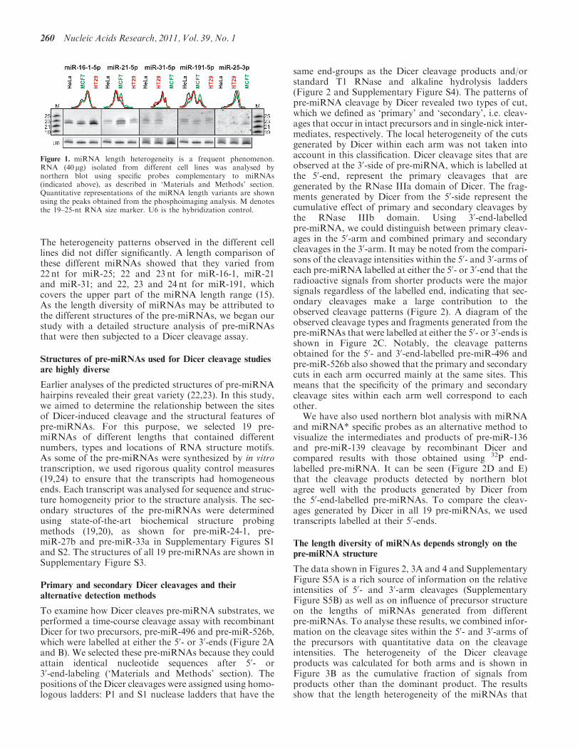
The heterogeneity patterns observed in the different celllines did not differ significantly. A length comparison ofthese different miRNAs showed that they varied from22 nt for miR-25; 22 and 23 nt for miR-16-1, miR-21and miR-31; and 22, 23 and 24 nt for miR-191, whichcovers the upper part of the miRNA length range (15).As the length diversity of miRNAs may be attributed tothe different structures of the pre-miRNAs, we began ourstudy with a detailed structure analysis of pre-miRNAsthat were then subjected to a Dicer cleavage assay.
Structures of pre-miRNAs used for Dicer cleavage studiesare highly diverse
Earlier analyses of the predicted structures of pre-miRNAhairpins revealed their great variety (22,23). In this study,we aimed to determine the relationship between the sitesof Dicer-induced cleavage and the structural features ofpre-miRNAs. For this purpose, we selected 19 pre-miRNAs of different lengths that contained differentnumbers, types and locations of RNA structure motifs.As some of the pre-miRNAs were synthesized by in vitrotranscription, we used rigorous quality control measures(19,24) to ensure that the transcripts had homogeneousends. Each transcript was analysed for sequence and struc-ture homogeneity prior to the structure analysis. The sec-ondary structures of the pre-miRNAs were determinedusing state-of-the-art biochemical structure probingmethods (19,20), as shown for pre-miR-24-1, pre-miR-27b and pre-miR-33a in Supplementary Figures S1and S2. The structures of all 19 pre-miRNAs are shown inSupplementary Figure S3.
Primary and secondary Dicer cleavages and theiralternative detection methods
To examine how Dicer cleaves pre-miRNA substrates, weperformed a time-course cleavage assay with recombinantDicer for two precursors, pre-miR-496 and pre-miR-526b,which were labelled at either the 50- or 30-ends (Figure 2Aand B). We selected these pre-miRNAs because they couldattain identical nucleotide sequences after 50- or30-end-labeling (‘Materials and Methods’ section). Thepositions of the Dicer cleavages were assigned using homo-logous ladders: P1 and S1 nuclease ladders that have the
same end-groups as the Dicer cleavage products and/orstandard T1 RNase and alkaline hydrolysis ladders(Figure 2 and Supplementary Figure S4). The patterns ofpre-miRNA cleavage by Dicer revealed two types of cut,which we defined as ‘primary’ and ‘secondary’, i.e. cleav-ages that occur in intact precursors and in single-nick inter-mediates, respectively. The local heterogeneity of the cutsgenerated by Dicer within each arm was not taken intoaccount in this classification. Dicer cleavage sites that areobserved at the 30-side of pre-miRNA, which is labelled atthe 50-end, represent the primary cleavages that aregenerated by the RNase IIIa domain of Dicer. The frag-ments generated by Dicer from the 50-side represent thecumulative effect of primary and secondary cleavages bythe RNase IIIb domain. Using 30-end-labelledpre-miRNA, we could distinguish between primary cleav-ages in the 50-arm and combined primary and secondarycleavages in the 30-arm. It may be noted from the compari-sons of the cleavage intensities within the 50- and 30-arms ofeach pre-miRNA labelled at either the 50- or 30-end that theradioactive signals from shorter products were the majorsignals regardless of the labelled end, indicating that sec-ondary cleavages make a large contribution to theobserved cleavage patterns (Figure 2). A diagram of theobserved cleavage types and fragments generated from thepre-miRNAs that were labelled at either the 50- or 30-ends isshown in Figure 2C. Notably, the cleavage patternsobtained for the 50- and 30-end-labelled pre-miR-496 andpre-miR-526b also showed that the primary and secondarycuts in each arm occurred mainly at the same sites. Thismeans that the specificity of the primary and secondarycleavage sites within each arm well correspond to eachother.
We have also used northern blot analysis with miRNAand miRNA* specific probes as an alternative method tovisualize the intermediates and products of pre-miR-136and pre-miR-139 cleavage by recombinant Dicer andcompared results with those obtained using 32P end-labelled pre-miRNA. It can be seen (Figure 2D and E)that the cleavage products detected by northern blotagree well with the products generated by Dicer fromthe 50-end-labelled pre-miRNAs. To compare the cleav-ages generated by Dicer in all 19 pre-miRNAs, we usedtranscripts labelled at their 50-ends.
The length diversity of miRNAs depends strongly on thepre-miRNA structure
The data shown in Figures 2, 3A and 4 and SupplementaryFigure S5A is a rich source of information on the relativeintensities of 50- and 30-arm cleavages (SupplementaryFigure S5B) as well as on influence of precursor structureon the lengths of miRNAs generated from differentpre-miRNAs. To analyse these results, we combined infor-mation on the cleavage sites within the 50- and 30-arms ofthe precursors with quantitative data on the cleavageintensities. The heterogeneity of the Dicer cleavageproducts was calculated for both arms and is shown inFigure 3B as the cumulative fraction of signals fromproducts other than the dominant product. The resultsshow that the length heterogeneity of the miRNAs that
Figure 1. miRNA length heterogeneity is a frequent phenomenon.RNA (40 mg) isolated from different cell lines was analysed bynorthern blot using specific probes complementary to miRNAs(indicated above), as described in ‘Materials and Methods’ section.Quantitative representations of the miRNA length variants are shownusing the peaks obtained from the phosphoimaging analysis. M denotesthe 19–25-nt RNA size marker. U6 is the hybridization control.
260 Nucleic Acids Research, 2011, Vol. 39, No. 1

Figure 2. Different experimental systems used to analyse Dicer cleavage products. A comparative analyses of the cleavage patterns generated byrecombinant Dicer (Ambion) in a time-dependent reaction: 1, 2 and 5min for pre-miR-496 (A) and pre-miR-526b (B) labelled at the 50- (left panel) or30-end (right panel). Lane Ci, incubation control without Dicer; lane S, S1 ladder; lane P, P1 ladder; lane T, guanine-specific ladder; lane F,formamide ladder. The positions of the selected cleavage sites (marked with black boxes) and G-residues were counted from the 50-end of theprecursors in all autoradiograms. In the middle, the structures of these precursors are shown. Asterisks represent the labeling site on the 50-end (red)and the 30-end (blue). The Dicer cleavage sites (marked with red arrowheads in the precursor labelled at the 50-end or blue arrowheads in the30-end-labelled precursor, the size of which is proportional to the radioactive signal intensity: very strong, strong, medium and weak) are shown inthe secondary structure models, where the reported miRNA sequences are marked in red. (C) The scheme of pre-miRNA processing by recombinantDicer labelled at the 50- (left, red) and 30-end (right, blue). From left to right: unprocessed precursor (substrate); single-nick intermediate products;precursor that is cleaved in both arms (product). The expected electrophoretic band migrations of the Dicer cleavage products that were labelled ateither 50- or 30-ends are shown schematically below. (D and E) The products of the Dicer cleavage assay of the unlabelled pre-miR-136 (D) andpre-miR-139 (E) were subjected to northern blot with specific 50- or 30-end probes detecting miRNA derived from 50- or 30-arm, respectively (lanes 1and 2 for pre-miR-136 and pre-miR-139, respectively). For comparison, on the right, the structure of the pre-miRNA and the result of the Dicerin vitro cleavage assay of the labelled precursors are shown. The blue arrowheads represent the cleavages by Dicer in the 30-arm being only primarycuts. The red arrowheads mark the cleavages in the 50 precursor arm (the sum of primary and secondary cuts). The blue and red arrows indicate thecorresponding cleavage sites. The migration of the miRNA, intermediates and pre-miRNA in the northern blot are represented with black lines onthe right side. M1 denotes the Low Molecular Weight Marker (USB Corp.); M2 denotes the 17–25 nt RNA marker. Other designations are the sameas in (A) and (B).
Nucleic Acids Research, 2011, Vol. 39, No. 1 261

are generated from the precursor 50-arm (combinedprimary and secondary cleavage products) was verysimilar to that of miRNAs generated from the 30-arm(primary cleavage products) [0.31 and 0.35, respectively
(Figure 3B)]. We could not identify any correlationbetween pre-miRNA structure and miRNA heterogeneityif the presence of asymmetrical versus symmetrical motifswas considered. We hypothesize that more subtle features
Figure 3. Dicer generates heterogeneous products of diverse lengths. (A) An analysis of Dicer cleavages in selected 50-end-labelled pre-miRNAs thatwere incubated with recombinant Dicer (Ambion) for 1, 2 and 5min. Below each precursor arm, the WALDI parameter is presented for the Dicercleavage products generated from this arm. The designations are the same as in Figure 2A and B. (B) The heterogeneity of the products that weregenerated from the 50-end-labelled precursors from the 50 (the sum of primary and secondary cleavages) and 30-arms (primary cleavages) wascalculated based on the data presented in Figures 2, 3A, 4 and Supplementary Figure S5A as described in ‘Materials and Methods’ section. Dotsand horizontal lines indicate the individual and average values of miRNA heterogeneity, respectively. Grey dots represent the heterogeneity ofproducts generated from symmetrical precursors, while white dots represent those generated from asymmetrical precursors. Signals that accountedfor <5% of all the cleavage products were not considered in the analysis. (C) A bubble-chart (MS Excel) depicting the fractions of cleavage productsthat were generated by Dicer in the 50-arm (left-hand side) and 30-arm (right-hand side) of the 50-end-labelled precursors analysed (y-axis). Note thatthe left diagram represents the sum of the primary and secondary cleavage products, while the right diagram shows the primary cleavage productsonly. The x-axis represents the lengths of the products, and the bubble size (area) represents the fraction of the product that was generated from agiven arm (as calculated from the densitometric analysis of the results presented in Figures 2, 3A, 4 and Supplementary Figure S5A). The values ofWALDI parameter from the 50- and 30-arms are shown as red vertical lines. The precursors were sorted according to the increasing value of theWALDI parameter generated from the 50-arm. White and grey bubbles indicate products that were diced from the ‘asymmetrical’ (i.e. including atleast one asymmetrical motif in the stem; labelled ASYM) and ‘symmetrical’ precursors (labelled SYM), respectively. The insets (i.e. the bar charts)present average WALDI values for the ‘symmetrical’ and ‘asymmetrical’ precursors with error-bars representing the standard deviation (SD). Thesevalues were formally compared using a Student’s t-test with Welch’s correlation for unequal variance, and the P-values are shown in the insets.Signals that accounted for <5% of all the cleavage products were not considered in graph (C).
262 Nucleic Acids Research, 2011, Vol. 39, No. 1

of pre-miRNA architecture may be involved in determiningmiRNA heterogeneity.The pre-miRNAs that harbor either symmetrical RNA
structural motifs (i.e. mismatches or symmetrical internalloops) or both symmetrical and asymmetrical motifs (i.e.bulges or asymmetrical internal loops) were ranked ac-cording to the weighted average length of diced RNA(WALDI), and the results are presented in Figure 3C(vertical red lines). Although the ranges and standard de-viations of the WALDI parameter are lower for cleavageproducts within the 50-arm than for the products withinthe 30-arm (SD: 0.52 versus 0.85, respectively), the averagelengths of these products are very similar (21.8 and 21.9 nt,respectively). When the cleavages that were generated inthe pre-miRNAs that contained only symmetrical RNAstructure motifs (grey bubbles in Figure 3C) werecompared with those in pre-miRNAs that contained atleast one asymmetrical motif (white bubbles), it becameapparent that the latter gave rise to significantly longerproducts (average WALDI: 21.5 nt versus 22.2 nt, and21.6 nt versus 22.2 nt, for 50- and 30-arms, respectively,see Figure 3C inset).
Longer miRNAs derive from pre-miRNA arms containingexcessive nucleotides
To illustrate the general observations described in theprevious paragraph using specific examples, we notethat the structures of pre-miR-132 and pre-miR-136(Figures 2D and 3A) differ in a single C-bulge present inthe 50-arm of the latter and that otherwise their hairpinstems have similar architectures. These precursors havenearly identical cleavage patterns, but the products ofthe Dicer cleavages in the 50-arm of pre-miR-136 are1-nt longer than those generated from pre-miR-132(WALDI: 22.5 and 21.6 nt, respectively). This length dif-ference may suggest that only the consecutive base pairsare counted by Dicer during cleavage site determination.Dicer cut in the 50-arm of pre-miR-637 and generated pre-dominantly a 22-nt fragment (WALDI: 21.9 nt) and cut inits 30 arm to release a 24-nt fragment (Figure 3A). Again,the difference in lengths between the released fragmentsmay be explained by the different number of bulged nu-cleotides along the hairpin stem in both precursor arms.Conversely, the same total number of bulged nucleotidesin the stem portion that spans the Dicer anchoring site andthe cleavage site explains the generation of the 23-ntfragment from each arm of pre-miR-19b-1 (Figure 4).
Pre-miRNA mutagenesis supports the conclusions fromthe analysis of natural precursors
To demonstrate the influence of asymmetrical struc-tural motifs on the lengths of miRNAs generated byDicer, we created specific pre-miRNA mutants. The asym-metrical motif was removed from pre-miR-19b-1 andpre-miR-136. In pre-miR-549, a 2-nt UU bulge wasinserted, and in pre-miR-33a, the symmetrical internalloop AGUU/UUCC was replaced with a fully pairedsequence (Figure 4). Natural precursors and theirmutants were 50-end-labelled and subjected to Dicercleavage assay. In all cases, products derived from
Figure 4. In vitro Dicer cleavages in the pre-miRNAs and theirmutants. Cleavages by recombinant Dicer of the 50-end-labelledpre-miR-19b-1, pre-miR-33a, pre-miR-136, pre-miR-549 and themutant variants. The sequences that were mutated in the precursorsare marked with rectangles. Below each precursor arm, the WALDIparameter is presented for Dicer cleavage products generated from thisarm. Other designations and symbols are the same as in Figure 2.
Nucleic Acids Research, 2011, Vol. 39, No. 1 263

pre-miRNA arms containing excessive nucleotides werelonger than those released from arms not containingthese nucleotides, which was reflected by the differencesin the corresponding WALDI parameters. Conversely, nosubstantial change in the length of released fragments wasobserved between pre-miR-33a to pre-miR-33a-Mut, inwhich a symmetrical internal loop was eliminated(Figure 4). Taken together, the results of the mutationalanalysis provided additional evidence that the asymmet-rical motifs had a much stronger effect on the lengths ofmiRNAs released by Dicer than symmetrical motifs. Ourexperimental observations were supported by the resultsof a comparative analysis of the predicted secondarystructures of all human pre-miRNAs and the lengths ofmiRNAs deposited in miRBase version 14. The presenceof excessive nucleotides in any pre-miRNA arm gave riseto longer miRNAs generated from this arm (Figure 5).
Drosha cleavages contribute to miRNA lengthheterogeneity
To gain insight into Drosha andDicer cleavages in cells, weoverexpressed a number of pri-miRNAs (SupplementaryTable S3) in HEK293T cells and analysed their processingproducts using high-resolution northern blot analysis(Figure 6). Overexpression of the primary transcripts didnot change the processing specificity characteristics of theendogenousmiRNAs (Supplementary Figure S6). The pro-cessing efficiency of different primary transcripts andpre-miRNAs varied considerably, as shown by the differentproportions of the pre-miRNA and miRNA fractions inthe total northern blot signal. Both Drosha and Dicercleavages were highly efficient in the case of pre-miR-214,for which >95% of the total signal was in the miRNAfraction.The northern blot experiments confirmed the frequent
occurrence of miRNA length heterogeneity (Figure 6A).Most of the analysed miRNAs were heterogeneousin length, and only miR-136 and miR-148a were
homogeneous. The mean value of the heterogeneity was0.28 for the 14 miRNAs shown in Figure 6A and C. Wealso analysed the pre-miRNA fraction using a high-resolution northern blot from long-electrophoretic runs(Figure 6B). Of the 14 pre-miRNAs, six (pre-miR-25,pre-miR-31, pre-miR-136, pre-miR-139, pre-miR-141and pre-miR-432) did not show length heterogeneity. Inall other cases, the heterogeneity ranged from 0.08 to 0.49.Overall, the average pre-miRNA length heterogeneity was0.18 (Figure 6B and 6C). We also determined the hetero-geneity of the miRNAs generated from the sixpre-miRNAs that were homogeneous in length. Dicergenerated heterogeneous products from five of six pre-miRNAs (the average heterogeneity of these sixmiRNAs was 0.23) (Figure 6C). To confirm the northernblot results, the primer extension assay was performed(Figure 6D) on RNA isolated from HEK293T cells trans-fected with pri-miRNA encoding plasmids. We analysedthe status of the 50-ends of the miRNAs generated from14 pre-miRNAs by either Drosha or Dicer. The analysis ofthe radioactive signal indicated that the 50-ends generatedby Drosha were less heterogeneous than those generatedby Dicer (average heterogeneity: 0.09 versus 0.18, respec-tively). The case of homogeneous miRNA derived fromheterogeneous pre-miRNA was noteworthy. The northernblot results showed that from heterogeneouspre-miR-148a (heterogeneity 0.42), only one dominantmature miRNA, or alternatively a population ofmiRNA variants of the same length, was generated fromthe pre-miRNA 50 arm (Supplementary Figure S7). Theprimer extension results showed that the 50-end of themature miRNA was heterogeneous (heterogeneity 0.24).These results allowed us to conclude that (i) more thanone length variant of pre-miR-148a is a substrate forDicer; (ii) at least two mature miR-148a variants ofthe same length exists (shift in sequence by 1 nt); and(iii) the 30-end of pre-miRNAs is more heterogeneousthan the 50-end (Supplementary Figure S7). Taken
Figure 5. The correlation between miRNA lengths and the presence of excessive nucleotides in pre-miRNA arms. The lengths of miRNAs generatedfrom the pre-miRNA arms that did not contain asymmetrical motifs (0) compared to the lengths of miRNA generated from pre-miRNA armscontaining at least one asymmetrical motif (A), containing one asymmetrical motif (A1) and containing more than one asymmetrical motif (A> 1).The results are shown separately for the 50- and 30-arms. N, number of analysed miRNAs; the Mann–Whitney test P-values are indicated in thegraphs. Standard deviations are shown as error bars. The results are based on a bioinformatics analysis of 721 human miRNA sequences andpre-miRNA structures.
264 Nucleic Acids Research, 2011, Vol. 39, No. 1

Figure 6. Analysis of the length heterogeneity of miRNAs and pre-miRNAs. (A) Northern blots showing the efficiency of precursor processing andthe length variation of the miRNAs. RNA (30 mg) that was isolated from HEK293T cells after transfection with different plasmid-encodedpri-miRNAs (indicated above the gel) was analysed in the blot. We analysed the total miRNA fraction in the total northern blot hybridizationsignals (miRNA and pre-miRNA fraction) quantitatively, and the result is shown below the gel (calculated as the processing efficiency in percent).The fraction of the miRNA variants other than the most abundant miRNA (i.e. the miRNA length heterogeneity) was calculated as in the Figure 3B,
Continued
Nucleic Acids Research, 2011, Vol. 39, No. 1 265

together, our data demonstrate that both processing stepscontribute to miRNA length heterogeneity and that therole of Dicer cleavage inaccuracy may be somewhatgreater.
DISCUSSION
In this study, the specificity of cleavages induced inmiRNA precursors by both Drosha and Dicer wasanalysed by high-resolution northern blot and primer ex-tension analyses. In most cases, the considerable hetero-geneity of the miRNAs was observed, which most likelyresulted from imprecise cleavages by Drosha and Dicerand could be further biased by AGO2 binding (17). Theinaccurate generation of the miRNA 50-end by Drosha orDicer, which has also been observed in miRNA discoveryefforts by deep sequencing (25–27), has important func-tional implications, even if the 50-end variability is reducedby AGO2 binding (17). The miRNAs that have shifted50-ends have different seed sequences, and they mayregulate different sets of targets (16,27,28). We have pre-dicted the target pools for the 50-end variants of miR-214,miR-17, miR-191 and miR-25 (Supplementary Figure S8).Our in silico predictions together with both bioinformatics(16) and experimental (29) results obtained by otherssupport the notion that not one but more miRNA50-end variants should be annotated because they allmay contribute to miRNA function.The imprecise processing of miRNA precursors also has
important implications for the rapidly developing RNAiand miRNA technologies. The vector-based siRNAs orartificial miRNAs that are expressed from pri-miRNAshuttles are thought to have the same sequence and exertthe same effects as the effective siRNAs or miRNAmimics that are obtained by chemical synthesis.However, the relaxed specificity of the Drosha and Dicercleavages will result in only a fraction of the silencingreagent with the desired sequence; the rest will haveshifted sequences. The release of the right allele-specificsiRNA from the pri-miRNA type vector is still morechallenging. The results of the analysis of the processingproducts of 14 different pri-miRNAs (Figure 6) couldserve as a guide for the selection of suitable shuttles thatexpress artificial miRNAs or siRNAs in cells.
The role of the structure of the pre-miRNA hairpins asan intrinsic factor in the regulation of mammalianmiRNA biogenesis has not been previously analysed indetail. In this study, we determined the secondary struc-tures of 19 pre-miRNAs and demonstrated that theirdiverse structures strongly influence the specificity ofDicer cleavage. In particular, the presence of bulges andasymmetrical internal loops within the approximately twohelical turns that span the Dicer anchoring site andcleavage site were the major source of miRNA length di-versity (Figure 7A). Cleavage by Dicer alone has also beenshown to generate the substantial length heterogeneity ofits products (Figure 7B). The model suggests that RNAstructure plays a substantial role in determining miRNAlength diversity and that Dicer flexibility plays the preva-lent role in determining miRNA heterogeneity. In cells,both RNA and Dicer are likely to exhibit structural flexi-bility during active complex formation. The WALDIparameter introduced in this study to characterize theproducts of Dicer, which acts as a ruler, allowed us toevaluate the specificity of Dicer cleavage in a quantitativemanner and may be useful for further studies of this kind.
Neither the crystal structure of human Dicer nor that ofthe Dicer complex with its accessory proteins (TRBP andAGO) and/or pre-miRNAs has been reported thus far.Most of the relevant structural information comes frombiochemical studies (14) and from the crystal structures ofDicer from Giardia intestinalis (30,31) and bacterialRNase III (32). Recent electron microscopy imaging ofthe human Dicer–TRBP complex (33) and the humanRLC complex (34) provided low-resolution structuralinformation about the molecular architecture of thesecomplexes and suggested how pre-miRNA dockingmight occur. The results of our study show thatpre-miRNA structure may substantially contribute tothe dynamics of the dicing complex. The stems ofhuman pre-miRNA hairpins are typically mosaics ofbase pairs and internal loops of various types and sizesand have, on average, 2.7 structure-disturbing motifs perprecursor (22). We propose that the unmatched nucleo-tides of pre-miRNAs that are accommodated within thesubstrate channel are often not counted by Dicer when itmeasures the distance to its cleavage site. Thus, the accu-mulation of structural imperfections in pre-miRNA
Figure 6. Continuedand the results are presented below the gel. A quantitative representation of the miRNA length variants is shown using the peaks obtained from thephosphoimaging analysis; M1 denotes the Low Molecular Weight Marker (USB Corp.); M2 denotes the 17–25 nt RNA marker. (B) Northern blotspresenting the length variation of the pre-miRNAs. The experiments were performed as in (A), but longer electrophoretic runs were applied. Thefractions of the pre-miRNA variants other than the most abundant pre-miRNA were determined as in Figure 3B and are shown below the gel.The quantitative representation of the pre-miRNA length variants is shown using the peaks obtained from the phosphoimaging analysis. M1 denotesthe Low Molecular Weight Marker (USB Corp.) M3 and M4 denote markers of the same sequence but differ in length by 1 nt (53 and 54 nt, and 58and 59 nt, respectively). (C) Dots and black horizontal lines indicate the individual and average values of the length heterogeneity of all pre-miRNAsand miRNAs, respectively. The numbers represent the average length heterogeneity. The black dots represent pre-miRNAs that are heterogeneous inlength. The grey dots represent either precursors that were homogeneous in length or miRNAs that were generated from these precursors. The greyline represents the average values of the miRNA length heterogeneity for those miRNAs that were generated from the homogeneous in lengthpre-miRNAs. The white dots show miRNAs that were generated from heterogeneous precursors. The dotted line represents the average value of thelength heterogeneity of the miRNAs that were generated from heterogeneous precursors. (D) Primer extension analyses of RNA isolated from cellsthat had been transfected with the same plasmid-encoded pri-miRNAs as in Figure 6A. Oligodeoxynucleotide primers (Supplementary Table S4)complementary to either the miRNA or miRNA* sequences (which detect miRNAs derived from the 50-arm-5p or the 30-arm-3p) were annealed tothe RNA (10mg). M denotes the 16–22 nt DNA marker. The fraction of miRNA 50-end variants other than the most abundant primer extensionproduct was calculated, and the result is shown below the gel. N, products not detected or detected at level too low to analyse. The migration of thefree probe and a fraction of the extended products are represented by black lines on the right side.
266 Nucleic Acids Research, 2011, Vol. 39, No. 1

hairpins may result in the higher plasticity of the precursorstructures, and this may be considered another successfulstrategy for the enrichment of miRNA diversity andincreased complexity of miRNA regulatory networks.
SUPPLEMENTARY DATA
Supplementary Data are available at NAR Online.
ACKNOWLEDGEMENTS
The authors thank Witek Filipowicz for encouragementand Marek Napierala for kindly providing the pri-miRNAencoding plasmids. We also thank Katarzyna Czubala,
Natalia Mokrzecka and Tomasz Witkos for theircontributions.
FUNDING
Sixth Research Framework Program of the EuropeanUnion, Project RIGHT (LSHB-CT-2004-005276);Ministry of Science and Higher Education (grantnumbers N301 112 32/3910, N N301 523 038, N N301284 837); European Regional Development Fund withinInnovative Economy Programme (grant numberPOIG.01.03.01-00-098/08); European Social Fund (POKL/8.2.2 to J.S.-R.). Funding for open access charge:European Regional Development Fund withinInnovative Economy Programme (grant numberPOIG.01.03.01-00-098/08).
Conflict of interest statement. None declared.
REFERENCES
1. Filipowicz,W., Bhattacharyya,S.N. and Sonenberg,N. (2008)Mechanisms of post-transcriptional regulation by microRNAs: arethe answers in sight? Nat. Rev. Genet., 9, 102–114.
2. Friedman,R.C., Farh,K.K., Burge,C.B. and Bartel,D.P. (2009)Most mammalian mRNAs are conserved targets of microRNAs.Genome Res., 19, 92–105.
3. Papadopoulos,G.L., Reczko,M., Simossis,V.A., Sethupathy,P. andHatzigeorgiou,A.G. (2009) The database of experimentallysupported targets: a functional update of TarBase. Nucleic AcidsRes., 37, D155–D158.
4. Baek,D., Villen,J., Shin,C., Camargo,F.D., Gygi,S.P. andBartel,D.P. (2008) The impact of microRNAs on protein output.Nature, 455, 64–71.
5. Selbach,M., Schwanhausser,B., Thierfelder,N., Fang,Z., Khanin,R.and Rajewsky,N. (2008) Widespread changes in protein synthesisinduced by microRNAs. Nature, 455, 58–63.
6. Chi,S.W., Zang,J.B., Mele,A. and Darnell,R.B. (2009) ArgonauteHITS-CLIP decodes microRNA-mRNA interaction maps. Nature,460, 479–486.
7. Denli,A.M., Tops,B.B., Plasterk,R.H., Ketting,R.F. andHannon,G.J. (2004) Processing of primary microRNAs by theMicroprocessor complex. Nature, 432, 231–235.
8. Gregory,R.I., Yan,K.P., Amuthan,G., Chendrimada,T.,Doratotaj,B., Cooch,N. and Shiekhattar,R. (2004) TheMicroprocessor complex mediates the genesis of microRNAs.Nature, 432, 235–240.
9. Han,J., Lee,Y., Yeom,K.H., Nam,J.W., Heo,I., Rhee,J.K.,Sohn,S.Y., Cho,Y., Zhang,B.T. and Kim,V.N. (2006) Molecularbasis for the recognition of primary microRNAs by theDrosha-DGCR8 complex. Cell, 125, 887–901.
10. Lund,E., Guttinger,S., Calado,A., Dahlberg,J.E. and Kutay,U.(2004) Nuclear export of microRNA precursors. Science, 303,95–98.
11. Chendrimada,T.P., Gregory,R.I., Kumaraswamy,E., Norman,J.,Cooch,N., Nishikura,K. and Shiekhattar,R. (2005) TRBP recruitsthe Dicer complex to Ago2 for microRNA processing and genesilencing. Nature, 436, 740–744.
12. Haase,A.D., Jaskiewicz,L., Zhang,H., Laine,S., Sack,R.,Gatignol,A. and Filipowicz,W. (2005) TRBP, a regulator ofcellular PKR and HIV-1 virus expression, interacts with Dicerand functions in RNA silencing. EMBO Rep., 6, 961–967.
13. Maniataki,E. and Mourelatos,Z. (2005) A human,ATP-independent, RISC assembly machine fueled by pre-miRNA.Genes Dev., 19, 2979–2990.
14. Zhang,H., Kolb,F.A., Jaskiewicz,L., Westhof,E. andFilipowicz,W. (2004) Single processing center models for humanDicer and bacterial RNase III. Cell, 118, 57–68.
Figure 7. Schematic representations of the different phenomenainvolved in creating miRNA length variation. (A) miRNA length di-versity—miRNAs of diverse lengths are generated from different pre-cursors. Symmetrical or asymmetrical structural motifs in pre-miRNAprecursor are represented by single base mismatch or bulges, respect-ively. Dicer RNaseIII domain A (RIIIA) is indicated using a greentriangle, and domain B (RIIIB) is indicated using a violet triangle.The PAZ domain is shown as a red rectangle. The arrows andnumbers represent the Dicer cleavage sites and the lengths of theDicer products, respectively. (B) miRNA length heterogeneity—miRNAs of heterogeneous lengths are generated from individual pre-cursor. All representations and symbols are the same as in (A). Forsimplicity, the pre-miRNA is shown as flexible in generating lengthdiversity (A), and Dicer is shown as flexible in generating miRNAlength heterogeneity (B).
Nucleic Acids Research, 2011, Vol. 39, No. 1 267
15. Griffiths-Jones,S., Saini,H.K., van Dongen,S. and Enright,A.J.(2008) miRBase: tools for microRNA genomics. Nucleic AcidsRes., 36, D154–D158.
16. Chiang,H.R., Schoenfeld,L.W., Ruby,J.G., Auyeung,V.C.,Spies,N., Baek,D., Johnston,W.K., Russ,C., Luo,S., Babiarz,J.E.et al. (2010) Mammalian microRNAs: experimental evaluation ofnovel and previously annotated genes. Genes Dev., 24, 992–1009.
17. Frank,F., Sonenberg,N. and Nagar,B. (2010) Structural basis for5’-nucleotide base-specific recognition of guide RNA by humanAGO2. Nature, 465, 818–822.
18. Sobczak,K., de Mezer,M., Michlewski,G., Krol,J. andKrzyzosiak,W.J. (2003) RNA structure of trinucleotide repeatsassociated with human neurological diseases. Nucleic Acids Res.,31, 5469–5482.
19. Krol,J. and Krzyzosiak,W.J. (2006) Structure analysis ofmicroRNA precursors. Methods Mol. Biol., 342, 19–32.
20. Krol,J., Sobczak,K., Wilczynska,U., Drath,M., Jasinska,A.,Kaczynska,D. and Krzyzosiak,W.J. (2004) Structural features ofmicroRNA (miRNA) precursors and their relevance to miRNAbiogenesis and small interfering RNA/short hairpin RNA design.J. Biol. Chem., 279, 42230–42239.
21. Cruz-Reyes,J., Piller,K.J., Rusche,L.N., Mukherjee,M. andSollner-Webb,B. (1998) Unexpected electrophoretic migration ofRNA with different 30 termini causes a RNA sizing ambiguitythat can be resolved using nuclease P1-generated sequencingladders. Biochemistry, 37, 6059–6064.
22. Kozlowski,P., Starega-Roslan,J., Legacz,M., Magnus,M. andKrzyzosiak,W.J. (2008) In Ying,S.-Y. (ed.), Current Perspectivesin microRNAs (miRNA). Springer, Netherlands, pp. 1–16.
23. Krol,J., Starega-Roslan,J., Milanowska,K., Nowak,D.,Kubiaczyk,E., Nowak,M., Majorek,K., Kaminska,K. andKrzyzosiak,W.J. (2006) In Clarke,N. and Sanseau,P. (eds),microRNA: Biology, Function & Expression. DNA Press,Eagleville, PA, pp. 95–110.
24. Krzyzosiak,W.J., Napierala,M. and Drozdz,M. (1999)In Barciszewski,J. and Clark,B.F.C. (eds), RNA Biochemistry andBiotechnology. Kluwer Academic Publishers, Dordrecht,pp. 303–314.
25. Morin,R.D., O’Connor,M.D., Griffith,M., Kuchenbauer,F.,Delaney,A., Prabhu,A.L., Zhao,Y., McDonald,H., Zeng,T.,Hirst,M. et al. (2008) Application of massively parallel sequencingto microRNA profiling and discovery in human embryonic stemcells. Genome Res., 18, 610–621.
26. Calabrese,J.M., Seila,A.C., Yeo,G.W. and Sharp,P.A. (2007)RNA sequence analysis defines Dicer’s role in mouseembryonic stem cells. Proc. Natl Acad. Sci. USA, 104,18097–18102.
27. Wu,H., Neilson,J.R., Kumar,P., Manocha,M., Shankar,P.,Sharp,P.A. and Manjunath,N. (2007) miRNA profiling of naive,effector and memory CD8 T cells. PLoS One, 2, e1020.
28. Seitz,H., Ghildiyal,M. and Zamore,P.D. (2008) Argonaute loadingimproves the 5’ precision of both MicroRNAs and their miRNA*strands in flies. Curr. Biol., 18, 147–151.
29. Azuma-Mukai,A., Oguri,H., Mituyama,T., Qian,Z.R., Asai,K.,Siomi,H. and Siomi,M.C. (2008) Characterization of endogenoushuman Argonautes and their miRNA partners in RNA silencing.Proc. Natl Acad. Sci. USA, 105, 7964–7969.
30. MacRae,I.J., Li,F., Zhou,K., Cande,W.Z. and Doudna,J.A. (2006)Structure of Dicer and mechanistic implications for RNAi. ColdSpring Harb. Symp. Quant. Biol., 71, 73–80.
31. MacRae,I.J., Zhou,K., Li,F., Repic,A., Brooks,A.N., Cande,W.Z.,Adams,P.D. and Doudna,J.A. (2006) Structural basisfor double-stranded RNA processing by Dicer. Science, 311,195–198.
32. Gan,J., Tropea,J.E., Austin,B.P., Court,D.L., Waugh,D.S. andJi,X. (2006) Structural insight into the mechanism ofdouble-stranded RNA processing by ribonuclease III. Cell, 124,355–366.
33. Lau,P.W., Potter,C.S., Carragher,B. and MacRae,I.J. (2009)Structure of the human Dicer-TRBP complex by electronmicroscopy. Structure, 17, 1326–1332.
34. Wang,H.W., Noland,C., Siridechadilok,B., Taylor,D.W., Ma,E.,Felderer,K., Doudna,J.A. and Nogales,E. (2009) Structuralinsights into RNA processing by the human RISC-loadingcomplex. Nat. Struct. Mol. Biol., 16, 1148–1153.
268 Nucleic Acids Research, 2011, Vol. 39, No. 1