Mechanisms of Enhanced -Adrenergic Reserve From Cardiac Resynchronization Therapy

Journal of Computer-Aided Molecular Design, 10 (1996) 545-557 ESCOM
545
J-CAMD 362
Pharmacophore development for antagonists at a, adrenergic receptor subtypes
J.B. Bremner, B. Coban and R. Griffith* Department of Chemistry, University of Wollongong, Northjields Avenue, Wollongong, NS W 2522, Australia
Received 13 March 1996 Accepted 6 July 1996
Keywords: Drug design; Molecular modelling; Selectivity
Summary
Many receptors, including a, adrenergic receptors, have a range of subtypes. This offers possibilities for the development of highly selective antagonists with potentially fewer detrimental effects. Antagonists developed for olA receptors, for example, would have potential in the treatment of benign prostatic hyperplasia. As part of the molecular design process, structural features necessary for the selective affinity for alA and c1,, adrenergic receptors have been investigated. The molecular modelling software (particularly the Apex module) of Molecular Simulations, Inc. was used to develop pharmacophore models for these two subtypes. Low-energy conformations of a set of known antagonists were used as input, together with a classification of the receptor affinity data. The biophores proposed by the pro- gram were evaluated and pharmacophores were proposed. The pharmacophore models were validated by testing the fit of known antagonists, not included in the training set. The critical structural feature for selectivity between the crlA and a,, adrenergic receptor sites is the distance between the basic nitrogen atom and the centre of an aromatic ring system. This will be exploited in the design and synthesis of structurally new selective antagonists for these sites.
Introduction
Receptors of the a, adrenergic subtype are members of the G-protein-coupled receptor (GPCR) superfamily. At least three distinct a, receptor subtypes have been report- ed on the basis of both pharmacology and molecular biology [l-6]. The GPCRs are integral membrane pro- teins and contain seven hydrophobic domains which are believed to be arranged in seven membrane spanning helices [7-91. These helices are responsible for passing signals across the cell membrane after interaction with their ligand on the extracellular side. The binding of the ligand elicits a response felt by the G-protein on the intra- cellular side. The activated G-protein then sets a second messenger system in train. Different GPCRs recognize different ligands. For adrenergic receptors, the activating ligands are norepinephrine and epinephrine. There are, however, only a few different types of G-proteins, each of which is used by many different receptors and to initiate a specific type of intracellular signalling. In addition, each
*To whom correspondence should be addressed.
0920-654x/$ 6.00 + 1 .OO 0 1996 ESCOM Science Publishers B.V.
receptor type may use more than one G-protein for sig- nalling, and recent studies have indicated that stimulation of different a, adrenergic receptor subtypes, for example, can lead to different physiological effects, e.g. a decrease in the propensity for abnormal heart rhythms (alB sub- type [IO]) versus promotion of arrhythmias and automa- ticity during myocardial ischaemia and the development of benign prostatic hyperplasia (alA subtype [ 111). There- fore, the development of a, subtype specific drugs to either block or enhance receptor function would be im- portant for disorders thought to be related to one subtype only, such as benign prostatic hyperplasia.
In order to design such ligands, structural information on the receptor subtypes, especially their binding sites, would be invaluable. There is, however, no X-ray struc- ture available for any GPCRs. Many GPCR transmem- brane domains have been modelled on the basis of the known structure of bacteriorhodopsin, but these models are only qualitative in nature and are still under develop- ment [7-91. From these models, which also take into
546
TABLE 1 AFFINITY ESTIMATE (pKJ VALUES OF CI, ADRENERGIC ANTAGONISTS
Antagonists PY Reference
Prazosin 10.1 10.2 2
X=0, (S)-WB4101 10.1 8.8 2 X = S, (S)-Benoxathian 9.1 8.4 2
MeO”@-Ng
2
(+)-Niguldipine 9.6 1.6 2
5-Methyluropidil 9.2 1.6 2
Me0
Spiperone 8.1 9.3 2
Risperidone 6.6 8.6 26 8.6 8.1 23
1.5 2 Me Phentolamine 8.8
KMD-3213 10.4 1.1 21
‘OH
Inactive prazosin analogue No adrenergic activity 22
Inactive WB4101 analogue 3.9 for cI, 17
NH2
OMe (R)-WB4101 analogue 1.4 for a, 28 ..d+o
Me0

547
TABLE 1 (continued)
Antagonists %A PY % Reference
16
Me’
OMe
17 Me0
Me0
OMe
22
(R)-YM-12617 (S)-YM-12617
YM-617=Tamsulosin
10.5 9.2 29 8.4 7.0 19
Corynanthine 6.8 6.3 30
(S)-(+)-Dicentrine 8.3 for CL, 25
Racemic IQC 8.6 for cc, 31
Abanoquil
Indoramin
10.4
8.4
10.1 11
7.4 11
SNAP 5089 9.7 6.7 32
AH 1lllOA 5.6 7.1 24

Prazosin (1) 9WB4101 (2) S-Benoxathian (3)
8 (+)-Niguldipine (4) 5Methyluropidil (5) Spiperone (6)
Risperidone (7) Phentolamine (8) KMD-3213 (9) Fig. 1. Global minimum structures for antagonists l-9, calculated as described in the text. Light grey: hydrogens; grey: carbons; black: hetero- atoms.
account mutagenesis and other experimental data, it is believed that the agonist binding pocket is located in the transmembrane domain near the extracellular face and that an aspartate side chain in the third transmembrane helix binds the hydrogen of the protonated amine of both agonists and antagonists. Close to this residue is also a large hydrophobic pocket containing conserved aromatic and serinelcysteine residues believed to bind the catechol ring of the agonists.
Another commonly adopted approach to drug design [ 12,131 relies on establishing the structure-activity rela- tionships of a number of known ligands of a receptor in order to develop a pharmacophore, i.e. the essential mo- lecular fragment which selectively recognizes the binding site. This is the approach adopted here using the Apex- 3D software package by Molecular Simulations, Inc. (formerly Biosym Technologies, San Diego, CA, U.S.A.). This program ‘employs advanced statistical techniques and 3D pattern-matching algorithms to draw its own generalizations from existing chemical data’ [ 141.
There have been attempts in the literature to develop a pharmacophore for the a, adrenergic receptors for both agonists and antagonists [15-191, but no mention has been made of the specific structural requirements for
affinity to the various a, subtypes. Table 1 shows a var- iety of antagonists and their affinities for the two sub- types considered here.
This paper reports studies directed towards defining antagonist pharmacophores for the alA and an, adrenergic receptors.
Methods
The molecular modelling software of Molecular Simu- lations, Inc. was used, specifically the following modules: INSIGHT II, BUILDER, DISCOVER, Search-Compare and Apex-3D. The CVFF force field was used throughout.
Antagonists l-9 from Table 1 were built and their geometry was optimized. The ligand molecules were then further investigated by using dynamics simulations (900
TABLE 2 ACTIVITY CLASSES USED IN APEX-SD
Class name a,* condition (PKJ
Very active >9 Active >8and<9 Inactive <8
alB condition (PK,)
>8 >6and<8 <6
549
K, time = 20 ps, time step = 1.0 fs, sample conformation taken every 2 ps). The 10 conformations sampled were minimized again (until the rms derivative was smaller than 0.001 kcal/A). The final 10 conformations chosen were all within a 10 kcal/mol energy window with respect to the global minimum.
The global minimum conformations so determined are depicted in Fig. 1.
The protonated forms of the nine antagonists (l-9 in Table 1) were also investigated in the same way and two inactive derivatives of (S)-WB4101 and prazosin were also considered (10 and 11 in Table 1). The inactive ligands were chosen from previous modelling studies on a, adre- noceptor antagonists [17,18] and were included to assist the program to induce features which are probably not related to biological activity [14]. The global minimum conformations for compounds 10 and 11 and for the test compounds were determined as above. For selected anta- gonists, structure basis sets containing more than 10 conformations per molecule were also prepared by sys- tematic conformational searching in the following way.
An optimized conformation was used and the bonds around which systematic rotations were to be performed were identified (bonds in rings cannot be treated in this way). The increment for the rotations was selected (usual- ly 60” or 90°) in such a way as to obtain an estimate of about a few hundred sterically allowed conformers. These were then minimized (until the rms derivative was smaller than 0.001 kcal/A) and duplicates removed. This resulted in about 50-100 conformers for the molecules chosen, again all conformations were within 10 kcaVmo1 from the lowest conformation, which in all cases coincided with the one previously determined (by dynamics).
The Apex-3D software [14,20,21] takes as input a number of molecules which bind to the same receptor site (or are presumed to do so) and a classification of each molecule according to its activity. The thresholds used for this classification are based on the pK, values in Table 1 and are listed in Table 2. The program then identifies common functional groups (descriptor centres) for these molecules. The descriptor centres can be quantitative, such as electron donor and acceptor reactivity indices (these are calculated using the semiempirical MOPAC method), or they can qualitatively describe an atom (het- eroatom, hydrogen-bond donor or acceptor, etc.). De- scriptor centres can also refer to pseudoatoms such as ring centroids. Because the bioactive conformation of the ligands is not generally known, a set of representative conformations is generated for each molecule as described above, and the property and distance matrices are calcu- lated for each conformer. The next step is then a search for common 3D arrangements of the descriptor centres identified. The inductive inference procedures used are based on similarity (common structural patterns in differ- ent compounds with similar affinity), dissimilarity (differ-
@ Putative hydrogen donor atom on receptor
+
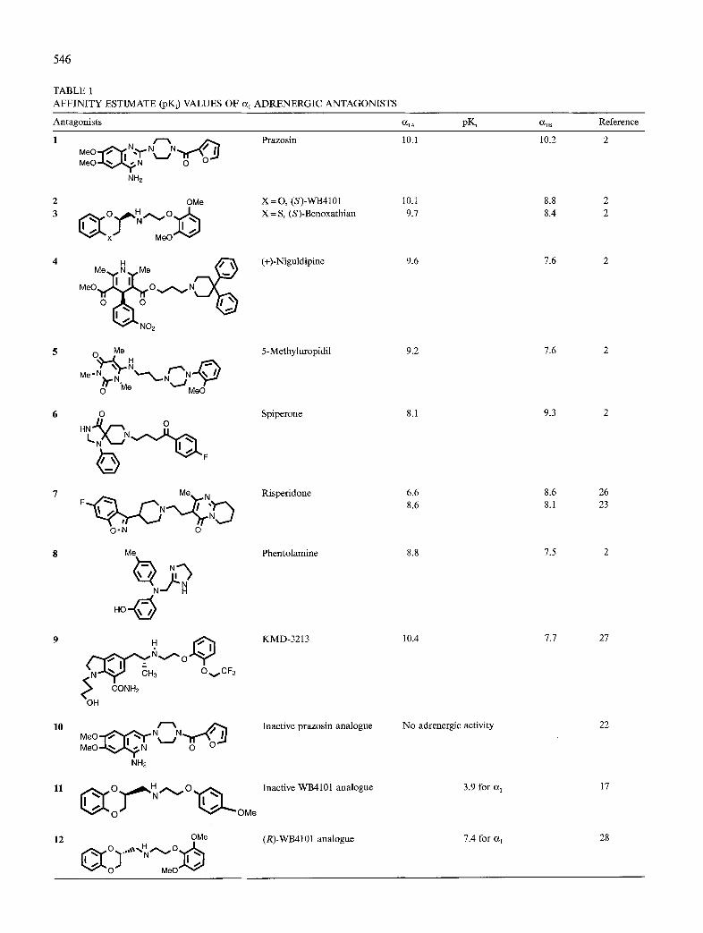
Fig. 2. Independent descriptor centres. A hypothetical biophore is shown which would be listed as containing three descriptor centres: a heteroatom (l), a heteroatom (2) and a hydrogen acceptor (2). To- gether with a hydrogen acceptor atom, the program always shows the approximate position of the hydrogen donor atom on the receptor (3). Since two of the three descriptor centres are connected to the same atom, they are not independent and this biophore is viewed as con- taining only two descriptor centres.
ent structural patterns in compounds with different affin- ity) and concomitant variations (variations in structural patterns in compounds with varying affinity). This ap- proach works best for a set of molecules which are struc- turally different and include subsets of structurally related compounds with very different activity. The software usu- ally finds a large number of common 3D arrangements of varying numbers of common descriptor centres. These have been called ‘biophores’ [14,21]. The program pro- vides statistical filtering options for these biophores, but further evaluation proved necessary to be able to propose pharmacophore models.
Results and Discussion
Inclusive pharmacophores
As a first attempt, 10 conformations each of all nine antagonists and the two inactive molecules (compounds l-11 in Table 1) were used together with the classification data from Table 2. Because the binding conformation of the antagonists at the receptors is not known and cannot be assumed to coincide with the global minimum confor- mation, a number of minimum-energy conformations of different geometries need to be taken into consideration for each molecule. This was done by using 10 conforma- tions within an energy window of 10 kcallmol (see also the Restricted pharmacophores section).
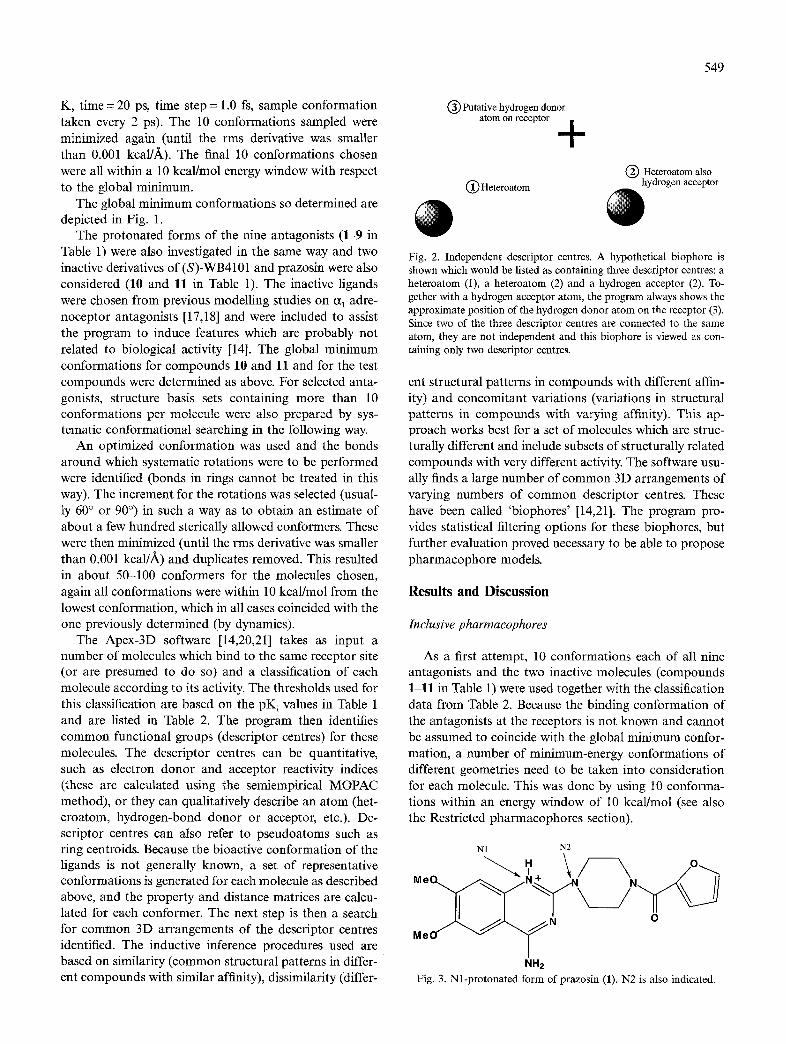
Nl N2
Me
Me
Fig. 3. Nl-protonated form of prazosin (1). N2 is also indicated.

550
Fig. 4. Inclusive biophores for the alA and I+, adrenergic receptor subtypes. For clarity, only two molecules are shown for each biophore: prazosin (1) and WB4101 (2). Green: carbon; white: hydrogen; red: oxygen; blue: nitrogen. The white spheres depict the descriptor centres for each biophore, the number 1 next to a descriptor centre denotes a basic nitrogen common to prazosin and WB4101, and the number 2 similarly denotes the centre of an aromatic ring. White crosses are connected with heteroatom descriptor centres that can function as hydrogen-bond donors; the cross denotes the estimated position of the hydrogen donor on the receptor,
Apex produced hundreds of biophores for each sub- type from this input, even when only the biophores con- nected to the class of very active ligands were considered, and it was clearly necessary to use the automatic filtering the software provides in order to reduce this number. The filters selected were:
for the alA subtype - p 2 0.6, size 2 3 and active 2 5; for the alB subtype - p 2 0.6, size 2 3 and active 2 4.
This means that the probability (p) that a novel compound possessing the biophore under consideration will be a ‘very active’ compound is only moderate. p is defined as follows:
P = (mrk + lY(m, + 2)
where mrk is the number of very active compounds pos- sessing the given biophore and m, is the total number of compounds possessing the given biophore. Only bio- phores with at least three descriptor centres were consid- ered (size 2 3) and the biophores were also required to contain at least five (a,*) or four (a& very active mol- ecules (active > 5 or 4).
This still left a large number of biophores (87 in the case of the IX,, subtype). These were now further edited by manually removing all biophores containing one of the two inactive molecules (risperidone (7) was classified as
inactive for the a,, subtype, but biophores containing this ligand were not discarded at this stage, because in com- parison with the truly inactive analogues 10 and 11, 7 shows some affinity for the alA receptor site) and also any biophores with less than three independent descriptor centres (see Fig. 2). The next step was to check if the biophores conformed to the idea that the most basic nitrogen atom in each molecule will be the one to be protonated most easily and therefore also the one to interact with the highly conserved aspartate residue in the third transmembrane helix of the receptor. For prazosin (l), this nitrogen has been determined both experimental- ly and theoretically [22]; we also confirmed that the most stable protonated form of prazosin is the one shown in Fig. 3, where the hydrogen ion is attached to Nl.
Therefore, a further evaluation criterion for the bio- phores was the requirement that the Nl of prazosin be one of the descriptor centres and that the nitrogen in WB4101 (2) be located at the same spot.
This left only two biophores for each subtype, which are depicted in Fig. 4. Only one of these four biophores (1038) contains an aromatic ring centre as a descriptor. This has been proposed as a pharmacophore element by De Marinis et al. [15]. None of the biophores has the most basic nitrogens of all molecules aligned in the same spot as would have to be the case for a credible pharma-

551
Fig. 5. olA inclusive biophore 1042 (a,s) with 5methyluropidil (in purple), benoxathian (in red) and KMD-3213 (in yellow). The most basic nitrogen atom of each compound is shown in light blue, illustrating the fact that these atoms do not overlap in the biophore.
cophore for a bioamine receptor (see Fig. 5 for an ex- al. [15], which suffers from the same problem. To make ample). This problem is further illustrated by a discussion prazosin fit their proposed pharmacophore, the authors of the pharmacophore for a, adrenergic receptor antagon- align it so that the nitrogen in the piperazine ring (N2 in ists (no subtype specified) as proposed by De Marinis et Fig. 3) is the basic nitrogen used for interaction with the
Fig. 6. Restricted biophores for the alA subtype. KMD-3213 (9): red; 5-methyluropidil (5): yellow; niguldipine (4): green. Symbols as in Fig. 4.

552
Basic nitrogen Basic nitrogen Basic nitrogen
(+)-Niguldipine (4) SMethyluropidil (5) KMD-3213 (9)
Fig. 7. Chemical structures of 4, 5 and 9, with various important structural features highlighted.
receptor. They point out that ‘only protonation at the 2- amino group gives a proton-aromatic distance and an N- H to aromatic plane angle that resembles those found in the structural classes previously discussed’. From previous studies it is quite unlikely that this nitrogen is protonated. De Benedetti et al. [22] estimated that the protonation energy difference for protonation at Nl and N2 is about 20 kcal/mol. The very high (but subtype-unselective) affinity of prazosin for a, adrenergic receptors cannot be explained by such a pharmacophore which necessitates the involvement of a very much less favourable proton- ated form. The protonated forms of the antagonists were also investigated, but the results are not reported here because of the problem of intramolecular hydrogen bond- ing when looking at isolated molecules (gas phase). These interactions heavily biased the conformations towards unrealistically folded ones where the hydrogen on the nitrogen would not be available for interaction with the receptor. Apex does not allow one to use solvated mol- ecules in its structure base, but the study of solvated protonated bases will still be undertaken with the aim of finding reasonable conformations for treatment with the Apex module.
Restricted pharmacophores
Because of the problems outlined above when trying to include all nine antagonists and the two inactive molecu- les in the training set, a different approach was taken whereby only the most selective and very active antagon- ists for each subtype were used to try and develop a phar- macophore, but a larger number of conformations were utilized for each molecule chosen.
Results for the ulA subtype Eighty-nine conformations of KMD-3213 (9) and 99
conformations each of 5-methyluropidil (5) and nigul- dipine (4) were used for this task. The probability was chosen to be 2 0.8 and all the most basic nitrogens were required to be an element in the biophores and to coin- cide as well. This left nine biophores, all except one (3931)
having an aromatic ring as a descriptor centre as well (Fig. 6). The distances between these two critical descrip- tors are very similar in all eight biophores (5.2-5.8 f!~) and also agree well with the pharmacophore proposed by De Marinis. 5-Methyluropidil and niguldipine are always aligned in the same way in the proposed biophores (Fig. 7): aromatic centres Rl overlapping. In KMD-3213, both possible aromatic rings (Rl and R2 in Fig. 7) have been used in the alignments. For seven of the biophores in Fig. 6, there is a third (and sometimes fourth) descriptor centre at the other end of the molecules for a heteroatom associ- ated with the second aromatic (or unsaturated six-mem- bered ring) centre. This is also in agreement with the pharmacophore of De Marinis.
The pharmacophore this suggests for antagonists bind- ing to this receptor is sketched in Fig. 8. It consists of an aromatic ring centre 5.2-5.8 A away from the basic nitro- gen and, on the other side of the molecule, 6-8 A away from the basic nitrogen, of an aromatic or six-membered unsaturated ring with polar substituents, and it is essen- tially the same as the pharmacophore proposed by De Marinis et al.
Results for the CI,, subtype Forty conformations of risperidone (7) and 42 confor-
mations of spiperone (6) were used for this task. The probability was chosen to be 2 0.75, the number of de- scriptor centres was 2 3 and all the most basic nitrogens were required to be an element in the biophores and to coincide as well. This left 12 biophores, all but one of which (138) contain an aromatic ring centre. Figure 9 shows nine of these biophores (the remaining three bio-
Q-c. +*+N/ Aromatic ring -+-.. 1
Basic nitrogen
Fig. 8. Pharmacophore model for alA adrenergic receptor antagonists. Numbers as in Fig. 4.
553
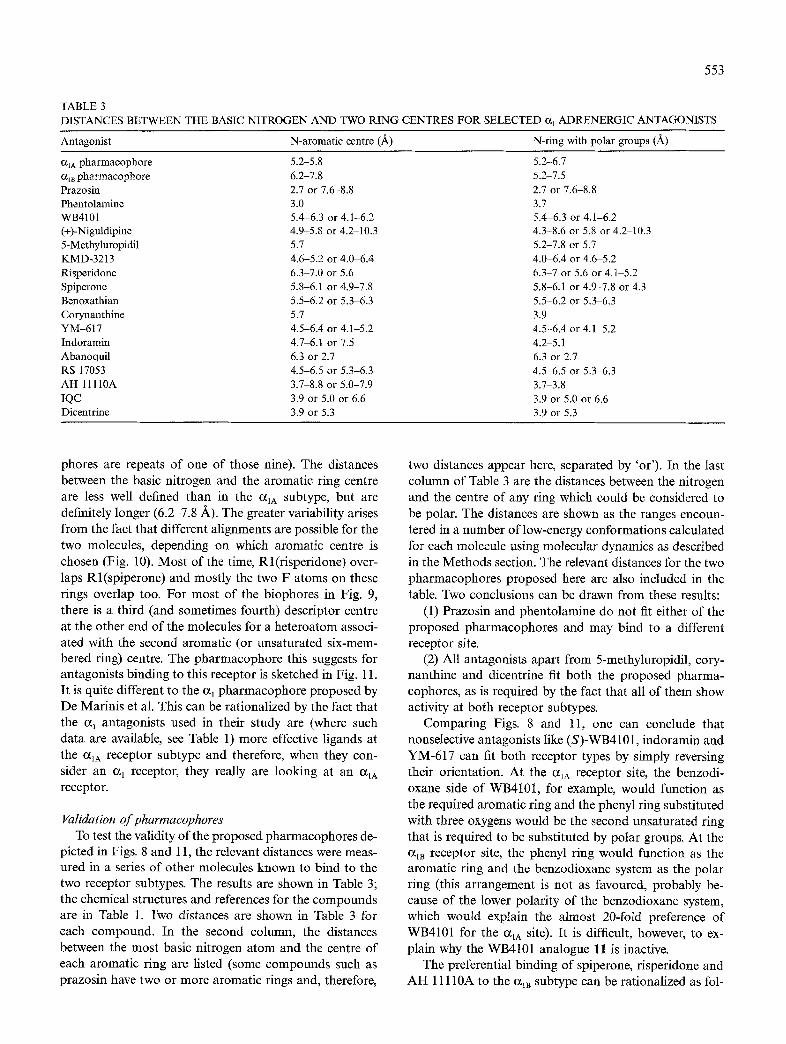
TABLE 3 DISTANCES BETWEEN THE BASIC NITROGEN AND TWO RING CENTRES FOR SELECTED CL, ADRENERGIC ANTAGONISTS
Antagonist N-aromatic centre (A) N-ring with polar groups (A)
CL,* pharmacophore Q,~ pharmacophore Prazosin Phentolamine WB4101 (+)-Niguldipine 5-Methyluropidil KMD-3213 Risperidone Spiperone Benoxathian Corynanthine YM-617 Indoramin Abanoquil RS 17053 AH 1lllOA IQC Dicentrine
5.2-5.8 6.2-7.8 2.7 or 7.6-8.8 3.0 5.4-6.3 or 4.1-6.2 4.9-5.8 or 4.2-10.3 5.7 4.6-5.2 or 4.0-6.4 6.3-7.0 or 5.6 5.8-6.1 or 4.9-7.8 5.5-6.2 or 5.336.3 5.7 4.5-6.4 or 4.1-5.2 4.7-6.1 or 7.5 6.3 or 2.7 4.5-6.5 or 5.3-6.3 3.7-8.8 or 5.0-7.9 3.9 or 5.0 or 6.6 3.9 or 5.3
5.2-6.7 5.2-7.5 2.7 or 7.6-8.8 3.1 5.4-6.3 or 4.1-6.2 4.3-8.6 or 5.8 or 4.2-10.3 5.2-7.8 or 5.7 4.0-6.4 or 4.6-5.2 6.3-7 or 5.6 or 4.1-5.2 5.8-6.1 or 4.9-7.8 or 4.3 5.5-6.2 or 5.3-6.3 3.9 4.5-6.4 or 4.1-5.2 4.2-5.1 6.3 or 2.7 4.5-6.5 or 5.3-6.3 3.7-3.8 3.9 or 5.0 or 6.6 3.9 or 5.3
phores are repeats of one of those nine). The distances between the basic nitrogen and the aromatic ring centre are less well defined than in the alA subtype, but are definitely longer (6.2-7.8 A). The greater variability arises from the fact that different alignments are possible for the two molecules, depending on which aromatic centre is chosen (Fig. 10). Most of the time, Rl(risperidone) over- laps Rl(spiperone) and mostly the two F atoms on these rings overlap too. For most of the biophores in Fig. 9, there is a third (and sometimes fourth) descriptor centre at the other end of the molecules for a heteroatom associ- ated with the second aromatic (or unsaturated six-mem- bered ring) centre. The pharmacophore this suggests for antagonists binding to this receptor is sketched in Fig. Il. It is quite different to the a, pharmacophore proposed by De Marinis et al. This can be rationalized by the fact that the a, antagonists used in their study are (where such data are available, see Table 1) more effective ligands at the alA receptor subtype and therefore, when they con- sider an a, receptor, they really are looking at an alA receptor.
Validation of pharmacophores To test the validity of the proposed pharmacophores de-
picted in Figs. 8 and 11, the relevant distances were meas- ured in a series of other molecules known to bind to the two receptor subtypes. The results are shown in Table 3; the chemical structures and references for the compounds are in Table 1. Two distances are shown in Table 3 for each compound. In the second column, the distances between the most basic nitrogen atom and the centre of each aromatic ring are listed (some compounds such as prazosin have two or more aromatic rings and, therefore,
two distances appear here, separated by ‘or’). In the last column of Table 3 are the distances between the nitrogen and the centre of any ring which could be considered to be polar. The distances are shown as the ranges encoun- tered in a number of low-energy conformations calculated for each molecule using molecular dynamics as described in the Methods section. The relevant distances for the two pharmacophores proposed here are also included in the table. Two conclusions can be drawn from these results:
(1) Prazosin and phentolamine do not fit either of the proposed pharmacophores and may bind to a different receptor site.
(2) All antagonists apart from 5-methyluropidil, cory- nanthine and dicentrine fit both the proposed pharma- cophores, as is required by the fact that all of them show activity at both receptor subtypes.
Comparing Figs. 8 and 11, one can conclude that nonselective antagonists like (S)-WB4101, indoramin and YM-617 can fit both receptor types by simply reversing their orientation. At the alA receptor site, the benzodi- oxane side of WB4101, for example, would function as the required aromatic ring and the phenyl ring substituted with three oxygens would be the second unsaturated ring that is required to be substituted by polar groups. At the alB receptor site, the phenyl ring would function as the aromatic ring and the benzodioxane system as the polar ring (this arrangement is not as favoured, probably be- cause of the lower polarity of the benzodioxane system, which would explain the almost 20-fold preference of WB4101 for the alA site). It is difficult, however, to ex- plain why the WB4101 analogue 11 is inactive.
The preferential binding of spiperone, risperidone and AH 11llOA to the a,, subtype can be rationalized as fol-

554
lows. At the alA site, risperidone would have to use the aromatic five-membered heterocycle as the aromatic centre, which is apparently not as favourable as using a larger six-membered aromatic ring. Therefore, the binding of risperidone to the alA site is much reduced. Recently, the selectivity of risperidone has been questioned [23] on the basis of results with recombinant receptors. The com- pound is, however, still listed in a very recent review [5] as an a,,-selective agent.
Compound AH 1lllOA has only very recently been reported as an a,,-selective antagonist [24]. As would be expected from its moderate selectivity, it fits both phar- macophores, with the imine nitrogen being the most basic site. In most of its low-energy conformations, the aro- matic rings are too far from the imine nitrogen to fit comfortably on the alA site. It has fairly low affinity (pK = 7.1 for a,& which may be due to the fact that it does not really have a polar group attached to the end of
Basic nitrogen
Spiperone (6) Risperidone (7)
Fig. 10. Chemical structures of 6 and 7, with various important structural features highlighted.
Fig. 9. Restricted biophores for the alB subtype. Risperidone (7): red; spiperone (6): yellow. Symbols as in Fig. 4.
the molecule away from the aromatic ring. The fluoro- substituted aromatic ring of spiperone is too far away in most low-energy conformations to comfortably fit on the alA site.
Similarly, the preferential binding of KMD-3213, 5- methyluropidil, RS 17053, niguldipine and the very close- ly related SNAP 5089 to the alA subtype can be rational- ized as follows. The reduced crlB activity of niguldipine can be explained by the fact that this molecule has only one polar end, and the phenyl rings at the other end are not substituted by the required polar groups. The phar- macophore is still not refined enough to account for the much reduced affinity of SNAP 5089 to the alB site, making this compound much more alA selective than niguldipine. 5-Methyluropidil, on the other hand, has only one aromatic ring, which does not fit very well on the a,, site. This compound may utilize the pyrimidine dione ring as an ‘aromatic’ ring when binding to an alB
Basic, nitrogen

555
site. The two aromatic rings of KMD-3213 are not far enough from the nitrogen to fit comfortably on the cxlB site and, in most of the conformations encountered for RS 17053, both aromatic rings are too close to the nitro- gen to fit the CX,~ site either.
The differences in selectivity for the (R) and (S) forms of WB4101 and YM-617 have not yet been scrutinized. Abanoquil appears to be more closely related to prazosin than to the other types of antagonists. Corynanthine was used by De Marinis et al. [15] for the development of their pharmacophore and will be discussed in detail later, together with IQC and dicentrine.
Results for a prazosin- WB4101 study
It was decided to study the prazosin problem more closely by building a task consisting of 99 conformations of prazosin and 99 conformations of WB4101. Filtering the resulting biophores using p 2 0.75 and size 2 3 resulted in 58 biophores. Requiring the Nl of prazosin to be an element of the biophores reduced that number to nine biophores. Only three of those nine biophores also con- tained the nitrogen of WB4101 and only one of them had both nitrogen atoms coinciding (11 in Fig. 12). This bio- phore did not include an aromatic ring as a descriptor centre and covered only a part of each molecule. This finding confirms the suggestion put forward above that prazosin binds to a different site at the receptors under consideration here than do the other antagonists such as WB4101.
Basic nitrogen
Fig. 11. Pharmacophore model for alB adrenergic receptor antagon- ists. Numbers as in Fig. 4.
Comparison of the proposed pharmacophores with the pharmacophoreproposedfor agonists at bioamine receptors
De Marinis et al. [15] reviewed the structureeactivity relationships of a, adrenergic receptor agonists of the phenethylamine type and concluded that ‘The dominant factors in the correlation of structure and activity for all phenethylamines have been embodied in the Easson-Sted- man hypothesis...‘. They also point out, however, that this hypothesis ‘does not correctly predict the effects observed with the imidazolines or the 2-aminotetralins’.
A common feature of both types of agonists is, how- ever, a ‘two-point mode of attachment involving only the phenyl ring and one nitrogen’. Figure 13 shows this com- mon pharmacophore for a, receptor agonists and it can be seen that the distance between the phenyl ring and the nitrogen is the same as that required by the proposed pharmacophore for a,, antagonists, but quite different to the proposed pharmacophore for alB antagonists.
It therefore appears that the alA antagonists make use of the same two points of attachment as used by all the
Fig. 12. Nine biophores from the prazosin-WB4101 study. Prazosin (1): red; WB4101 (2): yellow. Symbols as in Fig. 4.

556
agonists and then extend further to a pocket accommo- dating their polar rings. The aiB antagonists and prazosin, on the other hand, use only the common aspartate point of attachment and extend to other as yet unidentified regions of the receptor.
Both the proposed antagonist pharmacophores are also in qualitative agreement with the modelling studies under- taken by Trumpp-Kallmeyer et al. and others [8,9] on the a, adrenergic receptor agonist binding pocket as described in the Introduction section. These studies do not provide any distances between the hydrophobic pocket for aro- matic rings and the aspartate residue to bind the hydrogen on the basic nitrogen, but they do suggest the importance of the basic nitrogen close to an aromatic ring system.
Design of more selective antagonists
From the above considerations, it can be summarized that the ideal a,, antagonist should have (i) a basic nitro- gen that is accessible and can easily be protonated at physiological pH; (ii) a six-membered aromatic ring at a distance of 5.2-5.8 A from the nitrogen atom; and (iii) a (preferably nonaromatic) ring substituted with polar groups at a distance of 6-8 A from the nitrogen atom.
On the other hand, an ideal aiB antagonist should have (i) a basic nitrogen that is accessible and can easily be protonated at physiological pH; (ii) a six-membered aro- matic ring at a distance of 6.2-7.8 A from the nitrogen atom; and (iii) a (preferably nonaromatic) ring substituted with polar groups at a distance of 5-6 A from the nitro- gen atom.
Conformationally restricted a, antagonists
Conformationally constrained molecules can be used as probe molecules and to simplify the problem of finding a pharmacophore. De Marinis et al. used this approach in the development of their pharmacophore and it will be used here to design and synthesize potentially more sub- type-selective a, antagonists based on the IQC (reduced isoquinolino[8,1-ablcarbazole derivative, 17) and dicen- trine (16) molecules. One analogue, where the distance between the nitrogen and the aromatic ring is too short for the a,, site, is currently being synthesized, and ana- logues where this critical distance is too long to fit the alA site are being designed.
Dicentrine and IQC show very similar affinity for a, sites, whereas corynanthine is considerably less active. In all the three cases, the distance between the polar ring centre and the basic nitrogen is 3.9 A, which is much shorter than the ones proposed in Figs. 8 (6-8 A) and 11 (5-6 A). This is, however, in agreement with the findings of De Marinis that the polar group(s) at the other end of the molecule relative to the aromatic ring is (are) much less sterically sensitive.
distance = 5 A
Fig. 13. Schematic representation of the Easson-Stedman hypothesis.
So far, the ‘pharmacophore angle’ between the two distance vectors in each proposed pharmacophore has not been studied in detail. This angle can vary in the flexible ligands such as WB4101, whereas it is fixed for the three constrained molecules. In this context, it is interesting to note that a much more acute angle, such as in IQC and dicentrine, is associated with higher affinity compared to corynanthine.
Dicentrine itself should be selective for the alA subtype, because it has no aromatic ring far enough from the nitrogen to be able to fit into the alB site comfortably. This has not been tested yet. Yu et al. [25] tested dicen- trine in human prostate tissue, which contains predomi- nantly the alA receptor subtype [l 11. Similarly, corynan- thine should be selective for the alA subtype, but this is not so. It has, however, much lower affinities for both subtypes than any other compound classified as active in this study. IQC would need to use the five-membered indolic ring to fit on the aiA site and it is therefore pre- dicted that this molecule should show some selectivity for the alB subtype.
Conclusions
Pharmacophores for antagonists were developed for the alA and air, adrenergic receptor subtypes. The dis- tance between the basic nitrogen atom and the aromatic ring centre was found to be the critical feature for distin- guishing selective antagonists for the two subtypes, and this will be exploited in the design and synthesis of struc- turally new selective antagonists. These pharmacophores describe the structural features necessary for the binding of selective antagonists, such as KMD-3213, niguldipine, S-methyluropidil, risperidone and spiperone. They also explain the binding of nonselective antagonists, such as WB4101, benoxathian, YM-617 and others. Further re-

557
finement of these pharmacophores is required, however, to account for the differences in binding of the optical isomers of WB4101, YM-617 and others and to explore the requirements of the polar groups in greater depth.
The nonselective, but strongly binding, antagonist prazosin does not conform to either of the proposed pharmacophores and it is suggested that this ligand may utilize a different binding site which is common to both subtypes. The pharmacophore for prazosin and related compounds will be investigated in the future.
Acknowledgements
The authors would like to thank Molecular Simula- tions, Inc. for the loan of the Apex module of their mol- ecular modelling software, and to specially thank Dr. A. Palmer at the Sydney office of Biosym/MSI for her help. The award of a scholarship from the Zonguldak Karael- mas University in Turkey to Burak Coban, and research support from the ARC are gratefully acknowledged.
References
1 TiPS Receptor and Ion Channel Nomenclature Supplement, Trends Pharmacol. Sci. (1996) 10.
2 Ford, A.P.D.W., Williams, T.J., Blue, D.R. and Clarke, D.E., Trends Pharmacol. Sci., 15 (1994) 167.
3 Hieble, J.P., Bondinell, W.E. and Ruffolo Jr., R.R., J. Med. Chem., 38 (1995) 3415.
4 Graham, R.M., Perez, D.M., Piascik, M.T., Riek, R.P. and Hwa, J., Pharmacol. Commun., 6 (1995) 15.
5 Graham, R.M., Perez, D.M., Hwa, J. and Piascik, M.T., Circ. Res., 78 (1996) 737.
6 Hieble, J.I?, Bylund, D.B., Clarke, D.E., Eikenburg, D.C., Langer, S.Z., Lefkowitz, R.J., Minneman, K.P and Ruffolo Jr., R.R., Pharmacol. Rev., 47 (1995) 267.
7 Baldwin, J.M., EMBO J., 12 (1993) 1693. 8 Riek, R.P., Handschumacher, M.D., Sung, S.-S., Tan, M.,
Glynias, M.J., Schluchter, M.D., Novotny, J. and Graham, R.M., J. Theor. Biol., 172 (1995) 245.
9 Trumpp-Kallmeyer, S., Hoflack, J., Bruinvels, A. and Hibert, M.J., J. Med. Chem., 35 (1992) 3448.
10 Anyuhovsky, E.P. and Rosen, M.R., Circulation, 83 (1991) 2076. 11 Forray, C., Bard, J.A., Wetzel, J.M., Chiu, G., Shapiro, E., Tang,
R., Leper, H., Hartig, P.R., Weunshank, R.L., Branchek, T.A. and Gluchowski, C., Mol. Pharmacol., 45 (1994) 703.
12 Van de Waterbeemd, H. (Ed.) Advanced Computer-Assisted Tech- niques in Drug Discovery, VCH, Weinheim, Germany, 1995.
13 Cohen, N.C., Blaney, J.M., Humblet, C., Gund, P. and Barry, D.C., J. Med. Chem., 33 (1990) 883.
14 Apex-3D User Guide, v. 1.4, Biosym Technologies, San Diego, CA, U.S.A., 1993.
15 De Marinis, R.M., Wise, M., Hieble, J.P. and Ruffolo Jr., R.R., In Ruffolo Jr., R.R. (Ed.) The Alpha-l Adrenergic Receptor, Humana Press, Clifton, NJ, U.S.A., 1987.
16 Ruffolo Jr., R.R., Bondinell, W. and Hieble, J.P., J. Med. Chem., 38 (1995) 3416, 3681.
17 Venture& M., Menziani, M.C., Cocchi, M., Fanelli, F. and De Benedetti, P.G., J. Mol. Struct., 276 (1992) 327.
18 Rastelli, G. and De Benedetti, P.G., J. Mol. Struct., 251 (1991) 307.
19 Cocchi, M., Menziani, M.C., Fanelli, F. and De Benedetti, PG., J. Mol. Struct., 331 (1995) 79.
20 Golender, V.E. and Vorpagel, E.R., In Kubinyi, H. (Ed.) 3D QSAR in Drug Design: Theory, Methods and Applications, ESCOM, Leiden, The Netherlands, 1993, pp. 137-149.
21 Golender, V.E., Verterman, B. and Vorpagel, E., Net. Sci. January (1996) URL: http://www.awod.com/netsci/Issues/Jan96/feature3. html.
22 De Benedetti, PG., Menziani, M.C., Rastelli, G. and Cocchi, M.J., J. Mol. Struct., 233 (1991) 343.
23 Giardina, D., Crucianelli, M., Melchiorre, C., Taddei, C. and Testa, R., Eur. J. Pharmacol., 287 (1995) 13.
24 King, H.K., Goetz, A.S., Ward, S.D.C. and Saussy Jr., D.L., Sot. Neurosci. Abstr., 20 (1994) 526.
25 Yu, S., Ko, F., Chueh, S., Chen, J., Chen, S., Chen, C. and Teng, C., Eur. J. Pharmacol., 252 (1994) 29.
26 Slight, A.J., Koek, W. and Bigg, D.C.H., Eur. J. Pharmacol., 238 (1993) 407.
27 Shibata, K., Foglar, R., Horie, K., Obika, K., Sakamoto, A., Ogava, S. and Tsujimoto, G., Mol. Pharmacol., 48 (1995) 250.
28 Villa, L., Valoti, E., Villa, A.M., Pallavicini, M. and Ferri, V., 11 Farmaco, 49 (1994) 587.
29 Foglar, R., Shibata, K., Horie, K., Hirasawa, A. and Tsujimoto, G., Eur. J. Pharmacol., 288 (1995) 201.
30 Lomasney, J.W., Cotecchia, S., Lorenz, W., Leung, W.-Y., Schwinn, D.A., Yang-Feng, T.L., Brownstein, M., Lefkowitz, R.J. and Caron, M.G., J. Biol. Chem., 266 (1991) 6365.
31 Groenewoud, K.M., B.Sc. Honours Thesis, Department of Chem- istry, University of Tasmania, 1991.
32 Gluchowski, C., Wetzel, J.M., Chiu, G., Marzabadi, M.R., Wong, W.C. and Nagarathnam, D., Dihydropyridines and new uses thereof, International Patents Appl., No. WO 94/22829, 13 Octo- ber 1994.
33 Ford, A.P.D.W., Arredondo, N.F., Blue Jr., D.R., Bonhaus, D.W., Jasper, J., Kava, MS., Lesnick, J., Pfister, J.R., Shieh, A., Vimont, R.L., Williams, T.J., McNeal, J.E., Stamey, T.A. and Clarke, D.E., Mol. Pharmacol., 49 (1996) 209.
Copyright © 2022 FDOKUMEN