A Virtual Reality Scenario for All Seasons: The Virtual Classroom
Upload
khangminh22Category
view
1download
0
Review
Pharmacophore Models and Pharmacophore-BasedVirtual Screening: Concepts and ApplicationsExemplified on Hydroxysteroid Dehydrogenases
Teresa Kaserer 1,†, Katharina R. Beck 2,†, Muhammad Akram 1, Alex Odermatt 2,* andDaniela Schuster 1,*
Received: 19 November 2015; Accepted: 9 December 2015; Published: 19 December 2015Academic Editor: Peter Willett
1 Institute of Pharmacy/Pharmaceutical Chemistry and Center for Molecular Biosciences Innsbruck (CMBI),Computer Aided Molecular Design Group, University of Innsbruck, Innrain 80/82, 6020 Innsbruck, Austria;[email protected] (T.K.); [email protected] (M.A.)
2 Swiss Center for Applied Human Toxicology and Division of Molecular and Systems Toxicology,Department of Pharmaceutical Sciences, Pharmacenter, University of Basel, Klingelbergstrasse 50,4056 Basel, Switzerland; [email protected]
* Correspondence: [email protected] (A.O.); [email protected] (D.S.);Tel.: +41-61-267-1530 (A.O.); +43-512-507-58253 (D.S.); Fax: +41-61-267-1515 (A.O.); +43-512-507-58299 (D.S.)
† These authors contributed equally to this work.
Abstract: Computational methods are well-established tools in the drug discovery process andcan be employed for a variety of tasks. Common applications include lead identification andscaffold hopping, as well as lead optimization by structure-activity relationship analysis andselectivity profiling. In addition, compound-target interactions associated with potentially harmfuleffects can be identified and investigated. This review focuses on pharmacophore-based virtualscreening campaigns specifically addressing the target class of hydroxysteroid dehydrogenases.Many members of this enzyme family are associated with specific pathological conditions, andpharmacological modulation of their activity may represent promising therapeutic strategies.On the other hand, unintended interference with their biological functions, e.g., upon inhibition byxenobiotics, can disrupt steroid hormone-mediated effects, thereby contributing to the developmentand progression of major diseases. Besides a general introduction to pharmacophore modelingand pharmacophore-based virtual screening, exemplary case studies from the field of short-chaindehydrogenase/reductase (SDR) research are presented. These success stories highlight the suitabilityof pharmacophore modeling for the various application fields and suggest its application also infutures studies.
Keywords: pharmacophore; virtual screening; ligand protein interactions; hydroxysteroiddehydrogenase; oxidoreductase
1. Introduction
Pharmacophore Modeling
The concept of “pharmacophores” dates back to the late 19th century, when Paul Ehrlichsuggested that specific groups within a molecule are responsible for its biological activity [1,2].The pharmacophore definition, as currently used, was developed over time, with many researchersactively participating in the process (for a detailed history of pharmacophores, please refer toGüner and Bowen [2]). However, Schueler provided the basis for our modern understanding ofa pharmacophore [2,3], which is defined by the International Union of Pure and Applied Chemistry
Molecules 2015, 20, 22799–22832; doi:10.3390/molecules201219880 www.mdpi.com/journal/molecules

Molecules 2015, 20, 22799–22832
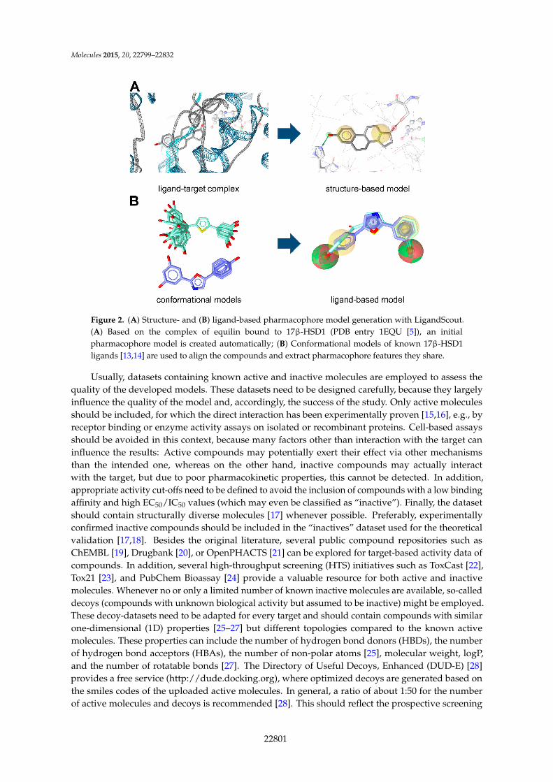
(IUPAC) as “the ensemble of steric and electronic features that is necessary to ensure the optimalsupra-molecular interactions with a specific biological target structure and to trigger (or to block) itsbiological response” [4]. According to this definition, the interaction patterns of bioactive moleculeswith their targets are represented via a three-dimensional (3D) arrangement of abstract features thatdefine interaction types rather than specific functional groups. These interaction types can, for example,include the formation of hydrogen bonds, charged interactions, metal interactions, or hydrophobic(H) and aromatic (AR) contacts (Figure 1). Besides that, many pharmacophore modeling programsallow for the addition of steric constraints. These so-called exclusion volumes (XVols) mimic thegeometry of the binding pocket and prevent the mapping of compounds that would be inactive in theexperimental assessment due to clashes with the protein surface. In its entirety, a pharmacophore modelrepresents one binding mode of ligands with a specific target, as exemplified on 17β-hydroxysteroiddehydrogenase (HSD) type 1 (Figure 1).
Figure 1. Pharmacophore models based on the estrogen equilin co-crystallized with 17β-hydroxysteroiddehydrogenase type 1 (PDB entry 1EQU [5]) and generated with LigandScout [6] (*), DiscoveryStudio [7] (#), and Molecular Operating Environment (MOE) [8] (§). H, hydrophobic feature; HBD,hydrogen bond donor; HBA, hydrogen bond acceptor; XVols, exclusion volume.
Pharmacophore models can be generated using two different approaches (Figure 2) dependingon the input data employed for model construction. In the structure-based approach, the interactionpattern of a molecule and its targets are directly extracted from experimentally determinedligand-target complexes (Figure 2A). An important source for these complexes, e.g., derived fromNMR-spectroscopy or X-ray crystallography, represents the Protein Data Bank (PDB, www.pdb.org) [9].To date (access date 2 November 2015), more than 113,000 macromolecular structures are stored inthis online repository. However, not all of these structures were solved in a complex with a boundligand, and in the case of induced fit, the binding of different ligands to an enzyme or receptor canlead to different interactions that are not covered by a single structure. To address this limitation, somepharmacophore modeling programs, e.g., Discovery Studio [7] and LigandScout [6], also provide toolsto create pharmacophore models based exclusively on the topology of the binding site and in theabsence of a ligand [10]. In Discovery Studio, for example, the binding site can be defined manually byselecting residues within the desired cavity or by applying implemented binding site identificationtools. Once the binding site is defined, the program automatically calculates pharmacophore featuresbased on the residues lining the active site. This initial ensemble of pharmacophore features can then beadapted to construct the final hypothesis [10]. In addition, structure-based pharmacophore models canalso be generated with computationally derived ligand-target complexes. In the course of a dockingrun, known active compounds are fitted into the empty binding pocket of the target [11]. These dockedbinding poses can then directly be employed to extract the interaction patterns. For further refinementof the initial docking poses, molecular dynamics (MD) simulations can be conducted [12] prior tomodel generation.
In the course of ligand-based modeling, three-dimensional (3D) structures of two or more knownactive molecules are aligned and common pharmacophore features shared among these training setmolecules are identified (Figure 2B). In a ligand-based approach, all of the common chemical features fromthe pharmacophore have to be presumed as essential, whereas in a structure-based approach, it can beconsidered whether a chemical feature of a molecule is directly involved in the ligand binding or not.
22800
Molecules 2015, 20, 22799–22832
Figure 2. (A) Structure- and (B) ligand-based pharmacophore model generation with LigandScout.(A) Based on the complex of equilin bound to 17β-HSD1 (PDB entry 1EQU [5]), an initialpharmacophore model is created automatically; (B) Conformational models of known 17β-HSD1ligands [13,14] are used to align the compounds and extract pharmacophore features they share.
Usually, datasets containing known active and inactive molecules are employed to assess thequality of the developed models. These datasets need to be designed carefully, because they largelyinfluence the quality of the model and, accordingly, the success of the study. Only active moleculesshould be included, for which the direct interaction has been experimentally proven [15,16], e.g., byreceptor binding or enzyme activity assays on isolated or recombinant proteins. Cell-based assaysshould be avoided in this context, because many factors other than interaction with the target caninfluence the results: Active compounds may potentially exert their effect via other mechanismsthan the intended one, whereas on the other hand, inactive compounds may actually interactwith the target, but due to poor pharmacokinetic properties, this cannot be detected. In addition,appropriate activity cut-offs need to be defined to avoid the inclusion of compounds with a low bindingaffinity and high EC50/IC50 values (which may even be classified as “inactive”). Finally, the datasetshould contain structurally diverse molecules [17] whenever possible. Preferably, experimentallyconfirmed inactive compounds should be included in the “inactives” dataset used for the theoreticalvalidation [17,18]. Besides the original literature, several public compound repositories such asChEMBL [19], Drugbank [20], or OpenPHACTS [21] can be explored for target-based activity data ofcompounds. In addition, several high-throughput screening (HTS) initiatives such as ToxCast [22],Tox21 [23], and PubChem Bioassay [24] provide a valuable resource for both active and inactivemolecules. Whenever no or only a limited number of known inactive molecules are available, so-calleddecoys (compounds with unknown biological activity but assumed to be inactive) might be employed.These decoy-datasets need to be adapted for every target and should contain compounds with similarone-dimensional (1D) properties [25–27] but different topologies compared to the known activemolecules. These properties can include the number of hydrogen bond donors (HBDs), the numberof hydrogen bond acceptors (HBAs), the number of non-polar atoms [25], molecular weight, logP,and the number of rotatable bonds [27]. The Directory of Useful Decoys, Enhanced (DUD-E) [28]provides a free service (http://dude.docking.org), where optimized decoys are generated based onthe smiles codes of the uploaded active molecules. In general, a ratio of about 1:50 for the numberof active molecules and decoys is recommended [28]. This should reflect the prospective screening
22801

Molecules 2015, 20, 22799–22832
database, where usually only a few active molecules are also distributed among a vast amount ofinactive molecules (Figure 3).
Figure 3. Enrichment of active molecules in the virtual hit list. Usually, the majority of compounds ina screening database are inactive molecules, while a small pool of bioactive molecules is contained.Pharmacophore-based virtual screening can help to enrich active molecules in the hit list compared toa random selection of test compounds.
The preliminary models generated with both approaches need further improvement in themajority of cases [16,29] to facilitate the recovery of the active molecules and concomitantly excludethe inactive compounds in the dataset from the hit list. Basic model refinement steps include thedeletion or addition of pharmacophore features and adaptations concerning the feature weight andsize. Selected features can also be defined as optional and, therefore, can but do not have to be mappedby a molecule. In addition, a user-defined number of omitted features can be specified in manypharmacophore modeling programs. More sophisticated modifications comprise the modification offeature definitions, i.e., the functional groups covered by a pharmacophore feature.
The aim of pharmacophore-based virtual screening (VS) is to enrich active molecules in a screeningdatabase in the virtual hit list (Figure 3). Multiple quality metrics are available that help to evaluatethe quality of the developed pharmacophore model, for example the enrichment factor [30] (theenrichment of active molecules compared to random selection), yield of actives (the percentage ofactive compounds in the virtual hit list), specificity (the ability to exclude inactive compounds) andsensitivity (the ability to identify active molecules), and the area under the curve of the ReceiverOperating Characteristic plot (ROC-AUC) [31]. For detailed descriptions of commonly applied qualityparameters we refer to earlier work [15,16,26,32]. The ultimate proof of a model’s quality and value, i.e.,whether it is indeed capable of proposing novel active molecules, can, however, only be determined ina prospective experiment, as will be explained in more detail below. A workflow summarizing theindividual steps of pharmacophore model generation and application is depicted in Figure 4.
As outlined below, refined, high quality pharmacophore models can then be employed formultiple tasks.
22802

Molecules 2015, 20, 22799–22832
Figure 4. The different consecutive steps in pharmacophore model generation, refinement, andprospective application.
2. Applications of Pharmacophore-Based VS
In the course of a VS run, a pharmacophore model is screened against large chemical libraries,and molecules mapping the model are collected in a virtual hit list. These molecules fulfill therequirements of the model and therefore have a high likelihood to be active in the experimental testing.Accordingly, VS can be used to filter promising compounds out of large compound collections andenrich active molecules in chemical databases selected for experimental investigations. VS is considereda valuable support for classical HTS campaigns [33,34], because true positive hit rates are usuallymuch higher than in those “random” testing strategies [35–37]. Reported hit rates from prospectivepharmacophore-based virtual screening vary between individual studies, but are typically in the rangeof 5% to 40% (an excellent collection of prospective studies has been presented earlier [16]). On the otherside, the hit rates of identifying active molecules upon random selection of test compounds are typicallybelow 1% and have been described, for example, as 0.55% for glycogen synthase kinase-3β [36],0.075% for peroxisome proliferator-activated receptor (PPAR) γ [38], and 0.021% for protein tyrosinephosphatase-1B [37].
2.1. Drug Discovery
Pharmacophore-based VS is widely applied in different steps of the drug discovery process andfacilitates the initial selection of compound classes as well as the optimization of compound propertiesas outlined below.
2.1.1. Lead Identification
The most common application of pharmacophore-based virtual screening concerns leadidentification, the so-called cherry-picking approach. Virtual screening is often deployed in these
22803
Molecules 2015, 20, 22799–22832
projects to prioritize molecules for testing and minimizing the number of compounds to be investigatedin biological screens. The ultimate aim is the identification of novel lead compounds for a specificdisease-related target, which can be developed into drug candidates for the treatment of the intendeddisease, with numerous studies during the last years describing such applications [39–44]. For example,Ha et al. reported the discovery of novel ligands for the chemokine receptor CXCR2 by using aligand-based pharmacophore modeling approach [45]. In the course of a pharmacophore-based virtualscreening for novel histamine H3 receptor antagonists, Lepailleur et al. identified novel compoundsadditionally binding to the 5HT4 receptor [46]. Both activities were considered beneficial for thetreatment of Alzheimer’s disease and the authors were the first to report compounds with this dualmechanism of action [46].
2.1.2. Structure-Activity Relationships
As mentioned in the introduction, a pharmacophore model represents the putative binding modeof active molecules to their target. It therefore describes the crucial functionalities required for acompound’s activity. A pharmacophore model is trained to discriminate between active and inactivemolecules (in the best case even between members of the same chemical series), which makes it highlyvaluable for establishing structure-activity relationships (SARs). Differences in the experimentallyobserved biological activities of a set of compounds can be rationalized based on the presence/absenceof chemical groups, represented by pharmacophore features, in the respective molecules. SARscan be established during model building, thereby elucidating the underlying mechanisms for the(absent) biological activity. For example, Ferreira et al. employed pharmacophore models to elucidateimportant features responsible for the interaction of compounds with the P-glycoprotein drug bindingsite [47]. Previous studies suggested a crucial role for a nitrogen atom in the modulators; however,active constituents from Euphorbia species isolated in-house did not contain such a moiety. Theauthors generated multiple refined pharmacophore models and evaluated them against a dataset ofliterature-derived modulators, the in-house collection, and inactive molecules. Their final modelhighlighted the important role of hydrophobic contacts and the presence of a HBA feature forP-glycoprotein modulators and showed that mapping of the most active compounds was alsopreserved when a further HBA/HBD feature was added [47]. In addition, pharmacophore models canbe employed to reflect previously elucidated SARs for the identification of novel bioactive molecules. In2002, Flohr et al. used the endogenous peptide urotensin II and synthetic analogues to experimentallyidentify interactions that are crucial for binding to the urotensin II receptor [48]. Based on theestablished SAR, pharmacophore models were built and employed to screen a chemical librarycontaining small drug-like compounds. Subsequent experimental testing of the virtual hits led to theidentification of six novel scaffold classes, which, importantly, contained non-peptic molecules [48].
2.1.3. Scaffold Hopping
A pharmacophore feature describes abstract chemical functionalities rather than specific functionalgroups. Additionally, pharmacophore models only demand local functional similarity of activecompounds and virtual hits at 3D locations essential for biological activity. Therefore, there areno specifications concerning the actual two-dimensional (2D) structures of mapping compounds.Although the composition of a pharmacophore model is influenced by the 2D structure of the moleculesemployed for model generation and refinement, it still allows for mapping of structurally distincthits. This makes pharmacophore modeling broadly applicable for the investigation of moleculesoriginating from a diverse chemical space such as natural products and synthetic compounds.Importantly, it also allows for the identification of novel scaffolds that have not been associated withthe target of interest before, a strategy that is called scaffold hopping. An earlier review extensivelydiscussed pharmacophore modeling in the context of scaffold hopping [49]. A recent study employedpharmacophore modeling for the discovery of novel transient receptor potential vanilloid type 1channel ligands [50]. Although the initial hits only weakly interacted with the target, they represent an
22804

Molecules 2015, 20, 22799–22832
interesting starting point for further chemical optimization. Such studies mostly emphasized novelchemical scaffolds and retrieved low similarity scores compared to the highly active compounds in thetheoretical validation dataset [50].
Scaffold hopping is certainly relevant for the pharmaceutical industry that needs to explorecompounds which are not yet covered by intellectual property issues. Of relevance for the generalpublic, scaffold hopping facilitates the identification of chemicals with only limited available data.This is often the case for environmental pollutants and chemicals from consumer products that areoften not drug-like by their nature.
2.1.4. Selectivity Profiling
For some projects, it may be of the utmost importance to identify compounds that selectivelymodulate the activity of one or more isoforms of an enzyme (family) to trigger the desired biologicaleffect. For example, steroidal core structures are frequently found in endogenous and exogenousbioactive compounds; however, these compounds often lack selectivity. To identify selectivecompounds, specific chemical substitutions leading to additional hydrophobic or ionic interactionsand hydrogen bonds have to be implemented. It has to be emphasized that these specific chemicalmodifications allow for distinguishing between the enzyme of interest and its related enzymes.
For example, 17β-HSD1 inhibitors are promising drug candidates for the treatment ofhormone-sensitive breast cancer as well as endometriosis because they block the activation of estroneto the highly potent endogenous estrogen receptor (ER) agonist estradiol [51–53]. On the other side,the converse reaction, (i.e., inactivation of estradiol) mediated via 17β-HSD2, should not be blockedby these molecules. Ideally, bioassays of all relevant members within a given protein family wouldbe employed to assess a compound’s selectivity. Additionally, proteins sharing structural similarityin the domain that contains the ligand binding pocket rather than sequence similarity should beconsidered in the selectivity assessment of compounds [54,55]. Thus, a huge number of proteinsneed to be covered in this resource- and time-consuming approach. In a first step, parallel screeningusing a large collection of pharmacophores, covering the most relevant proteins, allows for an initialcharacterization of a compound’s activity profile and facilitates the prioritization of the bioassays to bechosen for further biological analyses.
However, selectivity may not be limited to different isoforms. As exemplified by a study fromGuasch et al., it can even address the biological effect exerted via the same target [56]. The authorsfocused on the exclusive discovery of novel PPARγ partial agonists. The retrieval of full agonistswas avoided to prevent the side effects accompanying full receptor activation. For this purpose, apharmacophore model for full agonists (called the anti-pharmacophore) was generated and used toremove all potential full agonists from the screening database. In the second step, a partial agonistpharmacophore model was applied to identify potential partial agonists in the compound library.After several additional filtering steps, eight compounds were finally subjected to biological testingand five of them could be confirmed as novel PPARγ ligands displaying partial agonistic effects [56].
2.1.5. Combination with Other Techniques
Pharmacophore models are also often used together with other methods to further increasethe number of active molecules in the hit list via the application of a consensus approach.Commonly employed combinations comprise docking, shape-based modeling, and MD simulation.
In addition, a number of filters are available that help to limit the virtual hits to those with thedesired properties and eliminate unwanted actions or molecules. Probably the most prominent filterrepresents the Lipinski’s, describing properties that are shared by approved and orally administereddrugs [57]. In particular, these comprise a number of ď5 HBDs, ď10 HBAs, a molecular weight of ď500,and a cLogP ď5. Since all descriptors are either five or a multiple of five, Lipinski et al. referred to it asthe “rule of five”. Although the rule of five was initially developed to predict the oral bioavailabilityof molecules, it is also widely applied as a general drug-like filter. Veber et al. suggested two other
22805

Molecules 2015, 20, 22799–22832
criteria for the oral bioavailability of compounds: First, compounds should have a number of ď10rotatable bonds and, second, either a polar surface area of ď140 Å2 or ď12 HBAs and HBDs [58].
In analogy to Lipinski’s rule of five, Congreve et al. introduced the “rule of three” for theidentification of promising hit compounds in fragment-based drug discovery [59]. Their analysisrevealed that most of the small compounds that were successfully optimized to potent lead-likecandidates had a molecular weight of ď300, a number of HBDs ď3, a number of HBAs ď3, and acLogP ď3 [59].
More recently, a substructure filter was developed to identify highly problematic compounds thatnotoriously produce false positive assay read-outs [60]. Baell and Holloway analyzed high-throughputtesting results and observed that a group of molecules were prone to unspecifically interfere with someexperimental test systems. The subsequently developed substructure filter can help to detect thesepan-assay-interference compounds (PAINS) [60] prior to spending time and resources in investigatingand optimizing such molecules [61].
Multiple of these methods and filters can be included as well. As an example, Noha et al. employeda variety of computational techniques in a sequential manner to identify novel inhibitors of microsomalprostaglandin E2 synthase-1 [62]. The workflow included multiple prefilters, among them also theLipinski filter, a pharmacophore-based virtual screening procedure, and molecular docking. Out of the17 molecules finally selected for testing, two showed good activity in the experimental assay, and twofurther had moderate effects. Temml et al. used a combination of pharmacophore- and shape-basedvirtual screening to identify novel liver X receptor agonists [44]. In their study mentioned above [56],Guasch et al. not only applied pharmacophore models, but also a multistep protocol comprised ofelectrostatic and shape similarity and molecular docking to identify novel PPARγ partial agonists.
2.2. The Short-Chain Dehydrogenase/Reductase Superfamily
The short-chain dehydrogenase/reductase (SDR) enzyme family are nicotinamide adeninedinucleotide NAD (phosphate (P))-dependent enzymes sharing a common core structure of up to sevenparallel stranded β–sheets flanked by three to four α–helices on each side, the so-called Rossmannfold, for NAD(P) binding and a catalytic center characterized by a Tyr-(Xaa)3-Lys motif. This motif isoften found in combination with a conserved serine residue that stabilizes the orientation of the boundsubstrate (Figure 5) [63]. SDRs typically share a low sequence identity between 20%–30%, but withconsiderable structural similarity in the core domain.
The SDR family contains HSDs that play key roles in adrenal and gonadal steroidogenesis aswell as in the metabolism of steroids in peripheral tissues [64]. Some of these HSDs are consideredas promising therapeutic targets for the treatment of estrogen- and androgen-dependent diseasessuch as osteoporosis, endometriosis, and breast and prostate cancer, and other enzymes gainedinterest regarding the treatment of corticosteroid-related diseases such as diabetes, visceral obesity anddyslipidemia, atherosclerosis, wound healing, glaucoma, neurodegenerative disease, and cognitiveimpairment [53,65–67].
The development of specific SDR inhibitors needs to take into account the structural similarityof the various SDR enzymes in order to exclude the inhibition of members causing adverse effects,so-called off-targets. Suitable enzyme activity assays are fundamental for selectivity testing of potentialinhibitors. Koch et al. proposed that structural similarity rather than primary sequence similarityshould be chosen as the criterion for whether a certain chemical affects the activity of a relatedenzyme [54]. Therefore, the closest structurally related enzymes should be included for selectivitytesting—using pharmacophore models and cell-based assays. Another application of the modelingapproaches is the identification of toxic xenobiotics including industrial and environmentally relevantchemicals [68–70]. The role of several SDRs in xenobiotics metabolism and in steroid synthesis andmetabolism makes them prone as targets for endocrine disruption [71–76].
22806

Molecules 2015, 20, 22799–22832
Figure 5. The general structure of SDR enzymes exemplified on 17β-HSD1 (PDB entry 1EQU [5]).(A) The Rossmann fold consists of parallel stranded β-sheets (yellow), which are flanked by α-helices onboth sides (green). This structural domain forms the binding site of the co-factor NADP+. The residuesTyr155 and Lys159 of the Tyr-(Xaa)3-Lys motif as well as the conserved Ser142 are highlighted in rose;(B) 2D depiction of 17β-HSD1 (PDB entry 1EQU). Yellow triangles display β-sheets and barrel symbolsα-helices. Apart from the Rossmann fold, structurally conserved regions are highlighted in red. Theconserved glycine-rich motif GxxxGxG is important for cofactor binding and the + indicates a positivecharged residue crucial for cofactor (NADP+) stabilization.
3. Examples from the SDR Family
3.1. 11β-Hydroxysteroid Dehydrogenase Type 1
The two isoenzymes of 11β-HSD catalyze the interconversion of the biologically inactivecortisone and the active cortisol (Figure 6). The 11β-HSD1 is ubiquitously expressed and mediatesthe regeneration of active glucocorticoids [77,78], whereas 11β-HSD2 catalyzes the inactivation ofglucocorticoids mainly in the kidney, colon and placenta. There is evidence for beneficial effects of11β-HSD1 inhibition in the metabolic syndrome [79–87], atherosclerosis [88–91], osteoporosis [66,92],glaucoma [93–95], cognitive functions [96–100], skin aging [101], and wound healing [102,103]. Thus,inhibition of 11β-HSD1 has substantial therapeutic potential for glucocorticoid-related diseases.Numerous 11β-HSD1 inhibitors have already been identified and some have reached the clinicalphase, but to date still no 11β-HSD1 inhibitor is on the market [104]. Although structural variety isprevalent among the 11β-HSD1 inhibitors, the crystal structures are rather similar [105]. Nevertheless,the observed differences are useful in selecting a structure for further in silico evaluations. To date, 27human, four mouse, and three guinea pig 11β-HSD1 crystal structures are accessible through the PDB;however, there is currently no 3D structure of human 11β-HSD1 in -complex with a substrate available.In addition, structural information about 11β-HSD2 is entirely missing.
Figure 6. Interconversion of cortisone and cortisol catalyzed by the 11β-HSD enzymes.
22807
Molecules 2015, 20, 22799–22832
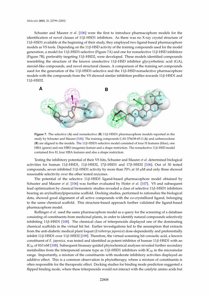
Schuster and Maurer et al. [106] were the first to introduce pharmacophore models for theidentification of novel classes of 11β-HSD1 inhibitors. As there was no X-ray crystal structure of11β-HSD1 available at the beginning of their study, they employed two ligand-based pharmacophoremodels as VS tools. Depending on the 11β-HSD activity of the training compounds used for the modelgeneration, a model for 11β-HSD1-selective (Figure 7A) and one for nonselective 11β-HSD inhibitors(Figure 7B), preferably targeting 11β-HSD2, were developed. These models identified compoundsresembling the structure of the known unselective 11β-HSD inhibitor glycyrrhetinic acid (GA),steroid-like compounds, and novel structural classes. A comparison of the training set compoundsused for the generation of the 11β-HSD1-selective and the 11β-HSD-nonselective pharmacophoremodels with the compounds from the VS showed similar inhibition profiles towards 11β-HSD1 and11β-HSD2.
Figure 7. The selective (A) and nonselective (B) 11β-HSD1 pharmacophore models reported in thestudy by Schuster and Maurer [106]. The training compounds CAS 376638-65-2 (A) and carbenoxolone(B) are aligned to the models. The 11β-HSD1-selective model consisted of four H features (blue), oneHBA (green) and one HBD (magenta) feature and a shape restriction. The nonselective 11β-HSD modelcontained five H, four HBA features and also a shape restriction.
Testing the inhibitory potential of their VS hits, Schuster and Maurer et al. determined biologicalactivities for human 11β-HSD1, 11β-HSD2, 17β-HSD1 and 17β-HSD2 [106]. Out of 30 testedcompounds, seven inhibited 11β-HSD1 activity by more than 70% at 10 µM and only three showedreasonable selectivity over the other tested enzymes.
The potential of the selective 11β-HSD1 ligand-based pharmacophore model obtained bySchuster and Maurer et al. [106] was further evaluated by Hofer et al. [107]. VS and subsequentlead optimization by classical bioisosteric studies revealed a class of selective 11β-HSD1 inhibitorsbearing an arylsulfonylpiperazine scaffold. Docking studies, performed to rationalize the biologicaldata, showed good alignment of all active compounds with the co-crystallized ligand, belongingto the same chemical scaffold. This structure-based approach further validated the ligand-basedpharmacophore model.
Rollinger et al. used the same pharmacophore model as a query for the screening of a databaseconsisting of constituents from medicinal plants, in order to identify natural compounds selectivelyinhibiting 11β-HSD1 [108]. The chemical class of triterpenoids displayed one of the dominatingchemical scaffolds in the virtual hit list. Earlier investigations led to the assumption that extractsfrom the anti-diabetic medical plant loquat (Eriobotrya japonica) dose-dependently and preferentiallyinhibit 11β-HSD1 over 11β-HSD2 [109]. Therefore, the virtual screening hit corosolic acid, a knownconstituent of E. japonica, was tested and identified as potent inhibitor of human 11β-HSD1 with anIC50 of 810 nM [108]. Subsequent bioassay-guided phytochemical analyses revealed further secondarymetabolites from the triterpenoid ursane type as 11β-HSD1 inhibitors with IC50 in the micromolarrange. Importantly, a mixture of the constituents with moderate inhibitory activities displayed anadditive effect. This is a common observation in phytotherapy, where a mixture of constituents isoften responsible for the therapeutic effect. Docking studies for binding mode prediction suggested aflipped binding mode, where these triterpenoids would not interact with the catalytic amino acids but
22808

Molecules 2015, 20, 22799–22832
with Thr124 and Tyr177 (Figure 8). Based on the most active compounds, a pharmacophore modelwas generated that enriched active molecules on the top of the hit list and successfully reflected thesubstructures important for binding. Additionally, this study demonstrates a further application in thedrug discovery process—finding inhibitors from natural origins.
Figure 8. The docking pose of the potent inhibitor corosolic acid in the binding pocket of 11β-HSD1(PDB entry 2BEL [110]) suggests interactions with Thr124 and Tyr177.
Considering the ongoing search for novel 11β-HSD1 inhibitors, high predictivity and performanceof pharmacophores are essential. Thus, to maintain high quality standards, pharmacophore modelshave to be continuously re-evaluated and improved. Vuorinen et al. [29] performed a refinement studyof the 11β-HSD pharmacophore models previously described by Schuster and Maurer et al. [106] andKratschmar et al. [78]. In a first step, the selective 11β-HSD1 model was refined by exchanging achemical feature and removing shape restriction using literature data. Whereas the unrefined modelwas only able to recognize two out of 14 test compounds, the refined model found 13. Subsequentprospective VS and biological testing revealed better performance of the refined model. However,although the refinement improved the sensitivity of the model and more active compounds werefound, it decreased specificity and also more inactive compounds fitted into the model. Addinga shape restriction, following newly identified selective 11β-HSD1 inhibitors, increased specificity,whereas the sensitivity remained the same. For additional testing of the model quality on a differentdataset, literature-based validation was performed with structurally diverse compounds, which hadnot been used in the model development. Specificity was increased, whereas sensitivity decreased.This illustrates that improvement of model quality is accompanied by balancing the specificity andsensitivity of a model. Refinement of the 11β-HSD2-selective model was equally conducted. Since thereis no 3D structure of 11β-HSD2 available and only a few selective, mainly triterpenoid scaffold-based11β-HSD2 inhibitors are known, the 11β-HSD2 model data are biased. They were, however, able toimprove 11β-HSD2 model quality, and novel active scaffolds selectively inhibiting both 11β-HSD1(Figure 9A) and 11β-HSD2 (Figure 9B) were discovered [29].
Using the refined 11β-HSD1 model, Vuorinen et al. applied a VS to filter a database consisting ofconstituents from medicinal plants to identify potential 11β-HSD1 inhibitors focusing on triterpenoidspresent in Pistacia lentiscus (P. lentiscus), so-called mastic gum that is used in traditional Greek medicinefor the treatment of diabetes [111]. The VS hit list contained eight hits of P. lentiscus constituents. Thetwo main constituents of mastic gum, masticadienonic acid and isomasticadienonic acid, were chosenfor further biological evaluation. Both compounds were shown to selectively inhibit 11β-HSD1 over11β-HSD2 with IC50 values of 2.51 µM for masticadienonic acid and 1.94 µM for isomasticadienonicacid, respectively. Examination of the whole resin’s activity revealed half the IC50 value of the singlemolecules, suggesting an additive inhibitory effect. Thus, the hypothesis of 11β-HSD1 involvementin the antidiabetic activity of mastic gum was supported. Analyzing the binding orientation of the
22809

Molecules 2015, 20, 22799–22832
two substances by docking revealed interactions comparable to that of the co-crystallized ligandcarbenoxolone, suggesting a competitive binding mode. Thus, the refined pharmacophore model hasproven its ability to identify novel 11β-HSD1 inhibitors from natural sources.
Figure 9. Both the refined 11β-HSD1 (A) and 11β-HSD2 (B) model identified novel scaffolds [29].The inhibitor fenofibrate maps the 11β-HSD1 model (A) and ketoconazole matches the 11β-HSD2model (B). Both models were screened with one omitted feature. The 2D structures of the novelinhibitors are depicted underneath the alignments.
Yang et al. performed a study using different 11β-HSD1 crystal structures in order to identifysynthetic 11β-HSD1 inhibitors [112]. They applied a combined approach of molecular dockingand ligand-based pharmacophore modeling. For virtual docking calculations the crystal structure1XU9 [113] and the program DOCK4.0 [114] were used to screen a commercial compound database.The 3000 compounds with the highest docking score were selected for a second docking run usingGlide [115]. Additionally, a ligand-based pharmacophore model for selective 11β-HSD1 inhibitorswas constructed using Catalyst 4.10 [116], which was used for screening the 3000 compounds withthe Best Flexible Search mode. Compounds with high docking and good fit score were furtherevaluated by filtering for drug likeness and finally selected for biological testing on human andmouse 11β-HSD1. Importantly, other studies showed significant species-specific variability in thepotency of various 11β-HSD1 inhibitors, indicating significant differences in the 3D organizationof the hydrophobic substrate-binding pocket of human and mouse 11β-HSD1 [117,118]. Due tothis issue, the tested compounds showed different inhibition profiles for the mouse and humanenzyme. Eleven out of 121 tested compounds inhibited the human 11β-HSD1 with IC50 values of0.26–14.6 µM, whereas six molecules inhibited the mouse 11β-HSD1 with IC50 values of 0.48–12.49 µM.Two substances displayed overlapping hits with IC50 for the human 11β-HSD1 of 0.69 µM and3.57 µM and for the mouse isoenzyme of 0.48 µM and 2.09 µM, respectively. In order to test theselectivity over 11β-HSD2 for subsequent animal studies, only compounds inhibiting mouse 11β-HSD1were tested for the inhibition of mouse 11β-HSD2. All compounds selectively inhibited 11β-HSD1.Nevertheless, selectivity assessment needs to include human 11β-HSD2 and, ideally, other SDRs.Cross-species activity would be the optimal situation for preclinical evaluation in the development ofnovel drug candidates.
A consecutive in silico study of Yang et al. includes virtual screening with 11β-HSD1structure-based pharmacophore models and subsequent docking for hit selection [119].Compounds chosen in the docking process were able to form hydrogen bonds with the amino acidsTyr183 and Ser170 from the catalytic triade. Nine out of 56 enzymatically tested compounds exhibiteddose-dependent and selective inhibition of human 11β-HSD1 with IC50 values between 0.85–7.98 µMand six substances inhibited the mouse 11β-HSD1 with IC50 values between 0.44 µM and 8.48 µM.
22810

Molecules 2015, 20, 22799–22832
Four substances inhibited both isoenzymes with similar IC50 values. In contrast, during their first11β-HSD1 in silico study, Yang et al. identified 11 out of 121 tested compounds from the same databaseas actives against 11β-HSD1, with IC50 values between 0.26–14.6 µM [113]. Four of the identified11β-HSD1 inhibitors incorporate an arylsulfamido scaffold, an already reported scaffold to inhibit11β-HSD1 [118]. Besides, three new scaffolds were identified as displayed in Figure 10.
Figure 10. The three new identified scaffolds by Yang et al. [119].
Table 1 summarizes the pharmacophore-based virtual screening studies and illustrates thescaffold-hopping of the different 11β-HSD1 inhibitors.
3.1.1. 17β-Hydroxysteroid Dehydrogenase Type 1
To date, 14 different human 17β-hydroxysteroid dehydrogenase (17β-HSD) enzymes have beenreported, all of which except the aldo-keto reductase (AKR) member 17β-HSD5 (AKR1C3) belong tothe SDR family [120]. The 17β-HSDs essentially regulate the local metabolism and activity of estrogensand androgens (Figure 11).
Figure 11. 17β-HSDs involved in sex steroid metabolism.
22811

Molecules 2015, 20, 22799–22832
Table 1. 11β-HSD1 pharmacophore-based virtual screening studies summarized.
Reference Study Aim PharmacophoreModel
Database Usedfor VS
Hits Biological Testing
Most Active Hit Number ofVirtual Hits
Tested inVitro Actives Assay IC50 Selectivity
Schuster andMaurer et al. [106]11β-HSD1 inhibitors
Ligand-basedusing Catalyst
Asinex Gold andPlatinum, Bionet2003, ChemBridgeDBS, Clab andIDC, Enamine 03,Interbioscreen 03nat and syn,Maybridge 2003,NCI, Specs 09 03
Molecules 2015, 20, page–page
14
Table 1. 11β-HSD1 pharmacophore-based virtual screening studies summarized.
Reference Study AimPharmacophore
Model Database Used
for VS
Hits Biological Testing
Most Active Hit Number of Virtual Hits
Testedin Vitro
Actives Assay IC50 Selectivity
Schuster and Maurer et al. [106] 11β-HSD1 inhibitors
Ligand-based using Catalyst
Asinex Gold and Platinum, Bionet 2003, ChemBridge DBS, Clab and IDC, Enamine 03, Interbioscreen 03 nat and syn, Maybridge 2003, NCI, Specs 09 03
16/20304 15 2 Lysate 2.03 and 7.59 µM
Against 11β-HSD2, 17β-HSD1, and 17β-HSD2
11β-HSD1 selective (4 H, 1 HBA, 1 HBD, and shape restriction)
11β-HSD unselective (5 H, 4 HBA and shape restriction)
107/1776579 15 5 Lysate 11β-HSD1 0.144–2.81 µM 11β-HSD2 0.06–3.95 µM
Most of them against 17β-HSD1 and 17β-HSD2
Hofer et al. [107] Lead optimization
11β-HSD1 selective from Schuster and Maurer et al. [106]
In-house database
- - - Lysate 0.7 µM Against 11β-HSD2
Rollinger et al. [108] Natural compounds inhibiting 11β-HSD1
11β-HSD1 selective from Schuster and Maurer et al. [106]
DIOS (Natural products in-house database)
corosolic acid
172 1 1 Lysate 0.81 µM Against 11β-HSD2
Vuorinen et al. [29] Refinement study
Refined models from Schuster and Maurer et al. [106] Using Discovery Studio
In-house database, DIOS
fenofibrate
463 9 3
Lysate
Considered as active if remaining enzyme activity ≤55% at test substance concentration of 20 µM or ≤65% at test substance concentration of 10 µM 5%–40%
Two preferentially inhibited 11β-HSD2, one was unselective
11β-HSD1 selective
11β-HSD2 selective In-house database, Specs, Maybridge
ketoconazole
444 25 2 11%–61% Enzyme rest activity
One preferentially inhibited 11β-HSD1 and one was unselective
11β-HSD unselective EDC, In-house database
38 4 36%–49% Enzyme rest activity
Two preferentially inhibited 11β-HSD1 one preferentially inhibited 11β-HSD2
16/20304 15 2 Lysate 2.03 and 7.59 µMAgainst11β-HSD2,17β-HSD1, and17β-HSD2
11β-HSD1 selective(4 H, 1 HBA, 1 HBD,and shape restriction)
11β-HSD unselective(5 H, 4 HBA andshape restriction)
Molecules 2015, 20, page–page
14
Table 1. 11β-HSD1 pharmacophore-based virtual screening studies summarized.
Reference Study AimPharmacophore
Model Database Used
for VS
Hits Biological Testing
Most Active Hit Number of Virtual Hits
Testedin Vitro
Actives Assay IC50 Selectivity
Schuster and Maurer et al. [106] 11β-HSD1 inhibitors
Ligand-based using Catalyst
Asinex Gold and Platinum, Bionet 2003, ChemBridge DBS, Clab and IDC, Enamine 03, Interbioscreen 03 nat and syn, Maybridge 2003, NCI, Specs 09 03
16/20304 15 2 Lysate 2.03 and 7.59 µM
Against 11β-HSD2, 17β-HSD1, and 17β-HSD2
11β-HSD1 selective (4 H, 1 HBA, 1 HBD, and shape restriction)
11β-HSD unselective (5 H, 4 HBA and shape restriction)
107/1776579 15 5 Lysate 11β-HSD1 0.144–2.81 µM 11β-HSD2 0.06–3.95 µM
Most of them against 17β-HSD1 and 17β-HSD2
Hofer et al. [107] Lead optimization
11β-HSD1 selective from Schuster and Maurer et al. [106]
In-house database
- - - Lysate 0.7 µM Against 11β-HSD2
Rollinger et al. [108] Natural compounds inhibiting 11β-HSD1
11β-HSD1 selective from Schuster and Maurer et al. [106]
DIOS (Natural products in-house database)
corosolic acid
172 1 1 Lysate 0.81 µM Against 11β-HSD2
Vuorinen et al. [29] Refinement study
Refined models from Schuster and Maurer et al. [106] Using Discovery Studio
In-house database, DIOS
fenofibrate
463 9 3
Lysate
Considered as active if remaining enzyme activity ≤55% at test substance concentration of 20 µM or ≤65% at test substance concentration of 10 µM 5%–40%
Two preferentially inhibited 11β-HSD2, one was unselective
11β-HSD1 selective
11β-HSD2 selective In-house database, Specs, Maybridge
ketoconazole
444 25 2 11%–61% Enzyme rest activity
One preferentially inhibited 11β-HSD1 and one was unselective
11β-HSD unselective EDC, In-house database
38 4 36%–49% Enzyme rest activity
Two preferentially inhibited 11β-HSD1 one preferentially inhibited 11β-HSD2
107/1776579 15 5 Lysate 11β-HSD1 0.144–2.81 µM11β-HSD2 0.06–3.95 µM
Most of themagainst 17β-HSD1and 17β-HSD2
Hofer et al. [107]Lead optimization
11β-HSD1 selectivefrom Schuster andMaurer et al. [106]
In-house database
Molecules 2015, 20, page–page
14
Table 1. 11β-HSD1 pharmacophore-based virtual screening studies summarized.
Reference Study AimPharmacophore
Model Database Used
for VS
Hits Biological Testing
Most Active Hit Number of Virtual Hits
Testedin Vitro
Actives Assay IC50 Selectivity
Schuster and Maurer et al. [106] 11β-HSD1 inhibitors
Ligand-based using Catalyst
Asinex Gold and Platinum, Bionet 2003, ChemBridge DBS, Clab and IDC, Enamine 03, Interbioscreen 03 nat and syn, Maybridge 2003, NCI, Specs 09 03
16/20304 15 2 Lysate 2.03 and 7.59 µM
Against 11β-HSD2, 17β-HSD1, and 17β-HSD2
11β-HSD1 selective (4 H, 1 HBA, 1 HBD, and shape restriction)
11β-HSD unselective (5 H, 4 HBA and shape restriction)
107/1776579 15 5 Lysate 11β-HSD1 0.144–2.81 µM 11β-HSD2 0.06–3.95 µM
Most of them against 17β-HSD1 and 17β-HSD2
Hofer et al. [107] Lead optimization
11β-HSD1 selective from Schuster and Maurer et al. [106]
In-house database
- - - Lysate 0.7 µM Against 11β-HSD2
Rollinger et al. [108] Natural compounds inhibiting 11β-HSD1
11β-HSD1 selective from Schuster and Maurer et al. [106]
DIOS (Natural products in-house database)
corosolic acid
172 1 1 Lysate 0.81 µM Against 11β-HSD2
Vuorinen et al. [29] Refinement study
Refined models from Schuster and Maurer et al. [106] Using Discovery Studio
In-house database, DIOS
fenofibrate
463 9 3
Lysate
Considered as active if remaining enzyme activity ≤55% at test substance concentration of 20 µM or ≤65% at test substance concentration of 10 µM 5%–40%
Two preferentially inhibited 11β-HSD2, one was unselective
11β-HSD1 selective
11β-HSD2 selective In-house database, Specs, Maybridge
ketoconazole
444 25 2 11%–61% Enzyme rest activity
One preferentially inhibited 11β-HSD1 and one was unselective
11β-HSD unselective EDC, In-house database
38 4 36%–49% Enzyme rest activity
Two preferentially inhibited 11β-HSD1 one preferentially inhibited 11β-HSD2
- - - Lysate 0.7 µM Against11β-HSD2
Rollinger et al. [108]Natural compoundsinhibiting 11β-HSD1
11β-HSD1 selectivefrom Schuster andMaurer et al. [106]
DIOS (Naturalproducts in-housedatabase)
Molecules 2015, 20, page–page
14
Table 1. 11β-HSD1 pharmacophore-based virtual screening studies summarized.
Reference Study AimPharmacophore
Model Database Used
for VS
Hits Biological Testing
Most Active Hit Number of Virtual Hits
Testedin Vitro
Actives Assay IC50 Selectivity
Schuster and Maurer et al. [106] 11β-HSD1 inhibitors
Ligand-based using Catalyst
Asinex Gold and Platinum, Bionet 2003, ChemBridge DBS, Clab and IDC, Enamine 03, Interbioscreen 03 nat and syn, Maybridge 2003, NCI, Specs 09 03
16/20304 15 2 Lysate 2.03 and 7.59 µM
Against 11β-HSD2, 17β-HSD1, and 17β-HSD2
11β-HSD1 selective (4 H, 1 HBA, 1 HBD, and shape restriction)
11β-HSD unselective (5 H, 4 HBA and shape restriction)
107/1776579 15 5 Lysate 11β-HSD1 0.144–2.81 µM 11β-HSD2 0.06–3.95 µM
Most of them against 17β-HSD1 and 17β-HSD2
Hofer et al. [107] Lead optimization
11β-HSD1 selective from Schuster and Maurer et al. [106]
In-house database
- - - Lysate 0.7 µM Against 11β-HSD2
Rollinger et al. [108] Natural compounds inhibiting 11β-HSD1
11β-HSD1 selective from Schuster and Maurer et al. [106]
DIOS (Natural products in-house database)
corosolic acid
172 1 1 Lysate 0.81 µM Against 11β-HSD2
Vuorinen et al. [29] Refinement study
Refined models from Schuster and Maurer et al. [106] Using Discovery Studio
In-house database, DIOS
fenofibrate
463 9 3
Lysate
Considered as active if remaining enzyme activity ≤55% at test substance concentration of 20 µM or ≤65% at test substance concentration of 10 µM 5%–40%
Two preferentially inhibited 11β-HSD2, one was unselective
11β-HSD1 selective
11β-HSD2 selective In-house database, Specs, Maybridge
ketoconazole
444 25 2 11%–61% Enzyme rest activity
One preferentially inhibited 11β-HSD1 and one was unselective
11β-HSD unselective EDC, In-house database
38 4 36%–49% Enzyme rest activity
Two preferentially inhibited 11β-HSD1 one preferentially inhibited 11β-HSD2
corosolic acid
172 1 1 Lysate 0.81 µM Against11β-HSD2
Vuorinen et al. [29]Refinement study
Refined models fromSchuster andMaurer et al. [106]Using DiscoveryStudio
In-housedatabase, DIOS
Molecules 2015, 20, page–page
14
Table 1. 11β-HSD1 pharmacophore-based virtual screening studies summarized.
Reference Study AimPharmacophore
Model Database Used
for VS
Hits Biological Testing
Most Active Hit Number of Virtual Hits
Testedin Vitro
Actives Assay IC50 Selectivity
Schuster and Maurer et al. [106] 11β-HSD1 inhibitors
Ligand-based using Catalyst
Asinex Gold and Platinum, Bionet 2003, ChemBridge DBS, Clab and IDC, Enamine 03, Interbioscreen 03 nat and syn, Maybridge 2003, NCI, Specs 09 03
16/20304 15 2 Lysate 2.03 and 7.59 µM
Against 11β-HSD2, 17β-HSD1, and 17β-HSD2
11β-HSD1 selective (4 H, 1 HBA, 1 HBD, and shape restriction)
11β-HSD unselective (5 H, 4 HBA and shape restriction)
107/1776579 15 5 Lysate 11β-HSD1 0.144–2.81 µM 11β-HSD2 0.06–3.95 µM
Most of them against 17β-HSD1 and 17β-HSD2
Hofer et al. [107] Lead optimization
11β-HSD1 selective from Schuster and Maurer et al. [106]
In-house database
- - - Lysate 0.7 µM Against 11β-HSD2
Rollinger et al. [108] Natural compounds inhibiting 11β-HSD1
11β-HSD1 selective from Schuster and Maurer et al. [106]
DIOS (Natural products in-house database)
corosolic acid
172 1 1 Lysate 0.81 µM Against 11β-HSD2
Vuorinen et al. [29] Refinement study
Refined models from Schuster and Maurer et al. [106] Using Discovery Studio
In-house database, DIOS
fenofibrate
463 9 3
Lysate
Considered as active if remaining enzyme activity ≤55% at test substance concentration of 20 µM or ≤65% at test substance concentration of 10 µM 5%–40%
Two preferentially inhibited 11β-HSD2, one was unselective
11β-HSD1 selective
11β-HSD2 selective In-house database, Specs, Maybridge
ketoconazole
444 25 2 11%–61% Enzyme rest activity
One preferentially inhibited 11β-HSD1 and one was unselective
11β-HSD unselective EDC, In-house database
38 4 36%–49% Enzyme rest activity
Two preferentially inhibited 11β-HSD1 one preferentially inhibited 11β-HSD2
463 9 3
Lysate
Considered as activeif remaining enzymeactivity ď55% at testsubstance concentrationof 20 µM or ď65% at testsubstance concentrationof 10 µM 5%–40%
Two preferentiallyinhibited11β-HSD2, onewas unselective
11β-HSD1 selective fenofibrate
11β-HSD2 selectiveIn-housedatabase, Specs,Maybridge
Molecules 2015, 20, page–page
14
Table 1. 11β-HSD1 pharmacophore-based virtual screening studies summarized.
Reference Study AimPharmacophore
Model Database Used
for VS
Hits Biological Testing
Most Active Hit Number of Virtual Hits
Testedin Vitro
Actives Assay IC50 Selectivity
Schuster and Maurer et al. [106] 11β-HSD1 inhibitors
Ligand-based using Catalyst
Asinex Gold and Platinum, Bionet 2003, ChemBridge DBS, Clab and IDC, Enamine 03, Interbioscreen 03 nat and syn, Maybridge 2003, NCI, Specs 09 03
16/20304 15 2 Lysate 2.03 and 7.59 µM
Against 11β-HSD2, 17β-HSD1, and 17β-HSD2
11β-HSD1 selective (4 H, 1 HBA, 1 HBD, and shape restriction)
11β-HSD unselective (5 H, 4 HBA and shape restriction)
107/1776579 15 5 Lysate 11β-HSD1 0.144–2.81 µM 11β-HSD2 0.06–3.95 µM
Most of them against 17β-HSD1 and 17β-HSD2
Hofer et al. [107] Lead optimization
11β-HSD1 selective from Schuster and Maurer et al. [106]
In-house database
- - - Lysate 0.7 µM Against 11β-HSD2
Rollinger et al. [108] Natural compounds inhibiting 11β-HSD1
11β-HSD1 selective from Schuster and Maurer et al. [106]
DIOS (Natural products in-house database)
corosolic acid
172 1 1 Lysate 0.81 µM Against 11β-HSD2
Vuorinen et al. [29] Refinement study
Refined models from Schuster and Maurer et al. [106] Using Discovery Studio
In-house database, DIOS
fenofibrate
463 9 3
Lysate
Considered as active if remaining enzyme activity ≤55% at test substance concentration of 20 µM or ≤65% at test substance concentration of 10 µM 5%–40%
Two preferentially inhibited 11β-HSD2, one was unselective
11β-HSD1 selective
11β-HSD2 selective In-house database, Specs, Maybridge
ketoconazole
444 25 2 11%–61% Enzyme rest activity
One preferentially inhibited 11β-HSD1 and one was unselective
11β-HSD unselective EDC, In-house database
38 4 36%–49% Enzyme rest activity
Two preferentially inhibited 11β-HSD1 one preferentially inhibited 11β-HSD2
ketoconazole
444 25 2 11%–61% Enzyme restactivity
One preferentiallyinhibited11β-HSD1 andone wasunselective
11β-HSD unselective EDC, In-housedatabase 38 4 36%–49% Enzyme rest
activity
Two preferentiallyinhibited11β-HSD1 onepreferentiallyinhibited11β-HSD2
22812

Molecules 2015, 20, 22799–22832
Table 1. Cont.
Reference Study Aim PharmacophoreModel
Database Usedfor VS
Hits Biological Testing
Most Active Hit Number ofVirtual Hits
Tested inVitro Actives Assay IC50 Selectivity
Vuorinen et al. [112]Mode of action study
Refined 11β-HSD1model fromVuorinen et al. [29]
DIOS
Molecules 2015, 20, page–page
15
Table 1. Cont.
Reference Study AimPharmacophore
Model Database Used
for VS
Hits Biological Testing
Most Active Hit Number of Virtual Hits
Testedin Vitro
Actives Assay IC50 Selectivity
Vuorinen et al. [112] Mode of action study
Refined 11β-HSD1 model from Vuorinen et al. [29]
DIOS 305/6702 2 2 Lysate 1.94 µM and 2.15 µM
Against 11β-HSD2
Yang et al.[113] 11β-HSD1 inhibitors
Ligand-based Using Catalyst (4 H, 1 HBA, 1 AR)
SPECS
Active against human
11β-HSD1
Active against human and
mouse 11β-HSD1
3000 Selected by docking (these 3000 were fitted in the pharmacophore model)
121 (39 out of docking and 82 from pharmacophore modeling)
11 Scintillation proximity assay
Human 11β-HSD1 0.26–14.6 µM Nine compounds Mouse 11β-HSD1 0.48–12.49 µM
Only tested against mouse 11β-HSD2 not tested toward the human 11β-HSD2
Yang et al. [119] 11β-HSD1 inhibitors
Two structure-based models using LigandScout (PDB code 2IRW) (3 H, 1HBD, 1 HBA)
SPECS
1000 Selected for each model
56 Nine human and six mouse
Scintillation proximity assay
Human 11β-HSD1 0.85–7.98 µM Mouse 11β-HSD1 0.44–8.48 µM
Against 11β-HSD2
305/6702 2 2 Lysate 1.94 µM and 2.15 µM Against11β-HSD2
Yang et al. [113]11β-HSD1 inhibitors
Ligand-basedUsing Catalyst(4 H, 1 HBA, 1 AR)
SPECS
Molecules 2015, 20, page–page
15
Table 1. Cont.
Reference Study AimPharmacophore
Model Database Used
for VS
Hits Biological Testing
Most Active Hit Number of Virtual Hits
Testedin Vitro
Actives Assay IC50 Selectivity
Vuorinen et al. [112] Mode of action study
Refined 11β-HSD1 model from Vuorinen et al. [29]
DIOS 305/6702 2 2 Lysate 1.94 µM and 2.15 µM
Against 11β-HSD2
Yang et al.[113] 11β-HSD1 inhibitors
Ligand-based Using Catalyst (4 H, 1 HBA, 1 AR)
SPECS
Active against human
11β-HSD1
Active against human and
mouse 11β-HSD1
3000 Selected by docking (these 3000 were fitted in the pharmacophore model)
121 (39 out of docking and 82 from pharmacophore modeling)
11 Scintillation proximity assay
Human 11β-HSD1 0.26–14.6 µM Nine compounds Mouse 11β-HSD1 0.48–12.49 µM
Only tested against mouse 11β-HSD2 not tested toward the human 11β-HSD2
Yang et al. [119] 11β-HSD1 inhibitors
Two structure-based models using LigandScout (PDB code 2IRW) (3 H, 1HBD, 1 HBA)
SPECS
1000 Selected for each model
56 Nine human and six mouse
Scintillation proximity assay
Human 11β-HSD1 0.85–7.98 µM Mouse 11β-HSD1 0.44–8.48 µM
Against 11β-HSD2
Active against human11β-HSD1
Molecules 2015, 20, page–page
15
Table 1. Cont.
Reference Study AimPharmacophore
Model Database Used
for VS
Hits Biological Testing
Most Active Hit Number of Virtual Hits
Testedin Vitro
Actives Assay IC50 Selectivity
Vuorinen et al. [112] Mode of action study
Refined 11β-HSD1 model from Vuorinen et al. [29]
DIOS 305/6702 2 2 Lysate 1.94 µM and 2.15 µM
Against 11β-HSD2
Yang et al.[113] 11β-HSD1 inhibitors
Ligand-based Using Catalyst (4 H, 1 HBA, 1 AR)
SPECS
Active against human
11β-HSD1
Active against human and
mouse 11β-HSD1
3000 Selected by docking (these 3000 were fitted in the pharmacophore model)
121 (39 out of docking and 82 from pharmacophore modeling)
11 Scintillation proximity assay
Human 11β-HSD1 0.26–14.6 µM Nine compounds Mouse 11β-HSD1 0.48–12.49 µM
Only tested against mouse 11β-HSD2 not tested toward the human 11β-HSD2
Yang et al. [119] 11β-HSD1 inhibitors
Two structure-based models using LigandScout (PDB code 2IRW) (3 H, 1HBD, 1 HBA)
SPECS
1000 Selected for each model
56 Nine human and six mouse
Scintillation proximity assay
Human 11β-HSD1 0.85–7.98 µM Mouse 11β-HSD1 0.44–8.48 µM
Against 11β-HSD2
Active against humanand mouse 11β-HSD1
3000 Selected bydocking (these3000 were fittedin thepharmacophoremodel)
121 (39 out ofdocking and82 frompharmacophoremodeling)
11Scintillationproximityassay
Human 11β-HSD10.26–14.6 µMNine compounds Mouse11β-HSD1 0.48–12.49 µM
Only testedagainst mouse11β-HSD2 nottested toward thehuman 11β-HSD2
Yang et al. [119]11β-HSD1 inhibitors
Two structure-basedmodels usingLigandScout (PDBcode 2IRW)(3 H, 1HBD, 1 HBA)
SPECS
Molecules 2015, 20, page–page
15
Table 1. Cont.
Reference Study AimPharmacophore
Model Database Used
for VS
Hits Biological Testing
Most Active Hit Number of Virtual Hits
Testedin Vitro
Actives Assay IC50 Selectivity
Vuorinen et al. [112] Mode of action study
Refined 11β-HSD1 model from Vuorinen et al. [29]
DIOS 305/6702 2 2 Lysate 1.94 µM and 2.15 µM
Against 11β-HSD2
Yang et al.[113] 11β-HSD1 inhibitors
Ligand-based Using Catalyst (4 H, 1 HBA, 1 AR)
SPECS
Active against human
11β-HSD1
Active against human and
mouse 11β-HSD1
3000 Selected by docking (these 3000 were fitted in the pharmacophore model)
121 (39 out of docking and 82 from pharmacophore modeling)
11 Scintillation proximity assay
Human 11β-HSD1 0.26–14.6 µM Nine compounds Mouse 11β-HSD1 0.48–12.49 µM
Only tested against mouse 11β-HSD2 not tested toward the human 11β-HSD2
Yang et al. [119] 11β-HSD1 inhibitors
Two structure-based models using LigandScout (PDB code 2IRW) (3 H, 1HBD, 1 HBA)
SPECS
1000 Selected for each model
56 Nine human and six mouse
Scintillation proximity assay
Human 11β-HSD1 0.85–7.98 µM Mouse 11β-HSD1 0.44–8.48 µM
Against 11β-HSD2
1000 Selected foreach model 56
Ninehuman andsix mouse
Scintillationproximityassay
Human 11β-HSD10.85–7.98 µMMouse 11β-HSD10.44–8.48 µM
Against11β-HSD2
22813

Molecules 2015, 20, 22799–22832
The enzyme 17β-HSD1 catalyzes the NADP (H)-dependent reduction of the weak estrogenestrone to the potent estradiol and to a minor extent of dehydroepiandrosterone (DHEA) to5-androstene-3β,17β-diol [121]. 17β-HSD1 is predominantly expressed in the human placenta,ovaries, and mammary gland, and is of major importance for the peripheral and gonadal estradiolsynthesis [122]. Several studies provide evidence for the association of 17β-HSD1 with breastcancer [123–125], endometriosis [52,126], endometrial cancer [127] and uterine leiomyoma [128].
Despite the recently increasing numbers of reported 17β-HSD1 inhibitors, still no compoundreached clinical trials. To date, more than 20 crystal structures have been published. The bindingpocket of 17β-HSD1 is an elongated hydrophobic tunnel, with key roles for Leu149, Val225, Phe226,and Phe259, and polar areas at each end formed by His221 and Glu282 on one side and the catalyticallyessential residues Ser142 and Tyr155 on the other side. The active site is limited by a flexible loop(amino acids 188–201), which is not well resolved in the crystal structures (Figure 12) [13].
Figure 12. Shape binding site of 17β-HSD1 with equilin as co-crystallized ligand, key residues, aflexible loop and the cofactor NADP+ (PDB 1EQU).
In 2001, Hoffren et al. were the first to report structure-based pharmacophore models for thediscovery of 17β-HSD1 inhibitors [129]. The pharmacophore models were validated to specificallyrecognize compounds possessing the structural and chemical features of steroids and flavonoids.Coumestrol displayed the most potent 17β-HSD1 inhibiting activity among the test compoundsused for model validation. However, coumestrol also inhibited 17β-HSD5 and is, therefore, notselective [130]. Unfortunately, the virtual hits were not confirmed by biological validation [129].
To support the development of therapeutic inhibitors, database creation for pharmacophore modelvalidation should focus on selective inhibitors to increase model selectivity and sensitivity. Sincesteroidal inhibitors and natural phytoestrogens, including flavonoids, often exhibit cross-reactivitywith other enzymes and hormone receptors involved in the steroidogenesis, non-steroidal scaffoldsare more favorable for virtual screening and drug development. However, although highly selectiveinhibitors are needed for many therapeutic applications, polyvalent inhibitors acting on synergisticpathways may be advantageous in some situations.
The 17β-HSD1 can be inhibited by several modes: competing reversibly and irreversibly with thenatural substrate for its binding site, competing with NADP(H) for its binding site at the Rossmannfold or occupying the ligand and the cofactor binding site by so-called hybrid compounds consisting ofa steroidal core and extended side-chains of NADP(H) moieties [131,132]. Since only crystal structurescontaining steroidal inhibitors were available at that time, Schuster and Nashev et al. generatedstructure-based pharmacophore models based on steroidal inhibitors [133]. They developed twopharmacophore models, representing, on one hand, reversible competitive inhibitors based on the
22814

Molecules 2015, 20, 22799–22832
steroidal core equilin (Figure 13A) and, on the other hand, hybrid inhibitors (Figure 13B). Whereasthe first model was suggested to be suitable as a general screening tool, expecting many false positivehits, the hybrid model was more restrictive due to the unique scaffold of the underlying hybridinhibitors. VS and subsequent in vitro validation of 14 selected compounds from the virtual hit listrevealed, amongst others, two nonsteroidal hits with IC50 of 5.7 µM and 19 µM, respectively. Asmentioned above, the SDR enzymes share substantial structural similarity. For selectivity assessment,11β-HSD1, 11β-HSD2, 17β-HSD2, 17β-HSD3 and the AKR 17β-HSD5 were tested. Two additionalinhibitors were selective. One was a steroidal compound with an IC50 of 3.8 µM for 11β-HSD1 and47 µM for 17β-HSD1, and one a nonsteroidal 11β-HSD1 inhibitor with IC50 of 6.2 µM and comparableactivity on 17β-HSD3. These observations emphasize the importance of including structurally relatedenzymes for selectivity assessment. In addition to the biological selectivity assessment, Schusterand Nashev et al. applied pharmacophore models of structurally related enzymes as an alternativestrategy to identify unspecific inhibitors [133]. These pharmacophores should act as initial filters toeliminate compounds with a low degree of selectivity that may exhibit off-target effects. Screening thecompounds identified as actives for 11β-HSD1 with their previously established selective 11β-HSD1pharmacophore model resulted in retrieving one hit [106]. By deleting the shape restriction, the secondhit was found as well and, at the same time, showed higher best fit values than an inactive compound.Thus, screening of pharmacophore models of related enzymes may facilitate the discrimination ofselective and nonselective inhibitors and the virtual hit selection for in vitro testing, similar to the studyby Guasch et al. described above [56].
Figure 13. (A) 17β-HSD1 model based on the equilin crystal structure (PDB entry 1EQU [5]); (B) Thepotent inhibitor STX 1040 maps the hybrid 17β-HSD1 pharmacophore model [133].
For pharmacophore model generation, Sparado et al. [134] superimposed five 17β-HSD1 crystalstructures, covering most of the chemical space occupied by the co-crystallized ligands. Performinga VS of an in-house compound library led to the identification of one virtual hit with moderateinhibitory activity against 17β-HSD1. Application of the rigidification strategy, scaffold hoppingand further SAR analysis resulted in two far more potent benzothiazole-scaffold-bearing inhibitorswith IC50 in cell lysates of 44 and 243 nM, respectively. Both hits were selective against 17β-HSD2.Furthermore, the less active compound still potently inhibited estrogen formation, with a comparableIC50 value to the lysates, in a human cell model endogenously expressing 17β-HSD1. The more potentcompound showed pronounced affinity to bind to ERα and ERβ. Depending on whether binding toERα and ERβ results in agonistic or antagonistic effects, this could cause beneficial or adverse effects.Interestingly, although the two hits differ only in a carbonyl and amide bridge, respectively, bindingmode investigations by docking showed a 180˝ flipped orientation of the two molecules (Figure 14). Theobservation of a flipped binding mode was also discovered for corosolic acid and other triterpenoidesin the binding pocket of 11β-HSD1 as described earlier [108]. A follow-up lead optimization study toimprove activity and selectivity of the two compounds for in vivo applications, without the help ofmolecular modeling techniques, led to the discovery of two new lead compounds [135]. They showedselectivity over 17β-HSD2, no ER binding and promising activity in the intact cell model.
22815

Molecules 2015, 20, 22799–22832
Figure 14. 17β-HSD1 in complex with the two hits from Sparado et al. [134], (doi:10.1371/journal.pone.0029252.g010, doi:10.1371/journal.pone.0029252.g011) showing a 180˝ flipped orientation. IC50
values of 44 nM (A) and 243 nM (B).
Table 2 shows a summary of the prospective pharmacophore-based virtual screening studies andillustrates the scaffold-hopping potential for 17β-HSD1 inhibitors.
Structure-based and ligand-based pharmacophore modeling was performed by Karkola et al. [136].They generated four pharmacophore models with different methods based on a crystal structure, arelaxed crystal structure, alignment of thienopyrimidinone inhibitors, and a docked complex of17β-HSD1 with a potent inhibitor. By VS, they found several compounds fitting into the active site of17β-HSD1 without determining the activity of the hits. However, to validate these hits as 17β-HSD1inhibitors, biological testing is needed. In addition, they could apply their differently generatedpharmacophore models to calculate selectivity and sensitivity.
3.1.2. 17β-Hydroxysteroid Dehydrogenase Type 2
The oxidative inactivation of estradiol to estrone is predominantly catalyzed by 17β-HSD2.Additionally, 17β-HSD2 is capable of converting testosterone into 4-androstene-3,17-dione(androstenedione), 5α-dihydrotestosterone (DHT) into 5α-androstanedione, 5-androstene-3β,17β-diolto DHEA, and 20α-dihydroprogesterone into progesterone using the cofactor NAD+ [137,138].The 17β-HSD2 is expressed in various tissues such as bone, placenta, endometrium, breast, uterus,prostate, stomach, small intestine, and colon epithelium [139,140]. The current treatment options forosteoporosis bear several limitations. Since 17β-HSD2 is expressed in osteoblasts, its inhibition mayprovide a new approach to treat osteoporosis by increasing the local availability of estradiol.
Since 17β-HSD2 contains an N-terminal transmembrane anchor, the experimental 3D structuredetermination remains a challenge and, to date, still no crystal structure is available. Due to thislack, Vuorinen et al. constructed three ligand-based pharmacophore models as virtual screeningfilters [141]. Virtual hit-testing in a cell-free assay revealed seven out of 29 compounds with IC50 valuesagainst 17β-HSD2 ranging between 0.24 µM and 33 µM. Most of the active compounds representedphenylbenzene-sulfonamides and -sulfonates. With the new structural classes of 17β-HSD2 inhibitors,they performed a SAR study using two different approaches: first, by a 2D similarity search withoutfitting the compounds into the pharmacophore models, and second, using a pharmacophore modelfor VS. From the 2D search, one out of 16 compounds inhibited 17β-HSD2 with an IC50 of 3.3 µM,whereas the VS showed five out of 14 compounds with IC50 between 1–15 µM. Selectivity of allactive compounds was tested against inhibition of 17β-HSD1, 17β-HSD3, 11β-HSD1, and 11β-HSD2.The activity data of the phenylbenzene-sulfonamide and -sulfonate inhibitors revealed a phenolichydroxyl group with hydrogen bond donor functionality, which was important for 17β-HSD2inhibition. This feature was confirmed by a ligand-based pharmacophore model that was developedbased on several of the newly identified active compounds (Figure 15). Furthermore, to improvethe initial pharmacophore model, a refinement database was created, including the original test setcompounds and the newly identified inhibitors as well as the inactive compounds. The specificity ofthe model was increased by adding exclusion volumes. This approach is an important step to enhancea model’s ability to enrich active compounds from a database.
22816

Molecules 2015, 20, 22799–22832
Figure 15. The selective 17β-HSD2 model contains a HBD feature (green sphere), which is importantfor 17β-HSD2 inhibitors such as the newly identified phenylbenzene-sulfonamide derivative 13 [141].
3.1.3. 17β-Hydroxysteroid Dehydrogenase Type 3
The 17β-HSD3 is almost exclusively expressed in the testes and catalyzes the reduction ofandrostenedione to testosterone in the presence of NADPH [142]. Although 17β-HSD3 is mainlyfound in the testes, there is evidence for 17β-HSD3 mRNA up-regulation in prostate cancer [143].Co-expression of 17β-HSD5, catalyzing the same reaction, might limit the therapeutic efficacyof 17β-HSD3 inhibitors and a combined treatment with inhibitors against both enzymes shouldbe envisaged.
The enzyme is anchored through an N-terminal transmembrane domain to the endoplasmicreticulum, and, like 17β-HSD2, its catalytic domain faces the cytoplasmic compartment [144,145].As for 17β-HSD2, there is still no crystal structure available for the membrane protein 17β-HSD3.
Figure 16. (A) The novel 17β-HSD3 inhibitor 1–7 was identified with the steroid-based model consistingof two HBAs (green) and four H features (blue); (B) The non-selective inhibitor 2-2 mapped thenonsteroid-based 17β-HSD3 model containing two HBAs, two AR (orange), one H and one H-ARfeature [146].
Two ligand-based pharmacophore models, based on steroidal and nonsteroidal 17β-HSD3inhibitors, were developed by Schuster et al. [146] (Figure 16). These ligand-based models supportedthe observations by Vicker et al. of a highly hydrophobic active site of 17β-HSD3 [147]. The modelswere then used to screen eight commercial databases and the hit list was further filtered prior to theselection of hits. Enzymatic tests showed that, from the steroid-based model, two out of 15 testedsubstances inhibited 17β-HSD3, with one also inhibiting 17β-HSD1 [146]. At the same time, threeother compounds inhibiting the AKR 17β-HSD5 were identified. The 17β-HSD5 is a multifunctionalenzyme and, like 17β-HSD3, catalyzes the conversion of androstenedione into testosterone. The mostpotent compound was not selective and also inhibited 11β-HSD1 and 11β-HSD2. Similar results wereobtained with the nonsteroidal model. The nonsteroidal model and its training compounds displayedseveral overlapping features with the lead compound identified earlier by Vicker et al. [147]; thus,the examination of their compounds for 17β-HSD5 inhibitory activity would be interesting. Theseobservations again emphasize the importance of including structurally related enzymes, independentlyof their enzymatic classes, for selectivity profiling. A summary of the 17β-HSD3 pharmacophore-basedvirtual screening study presented by Schuster et al. is provided in Table 3.
22817

Molecules 2015, 20, 22799–22832
Table 2. 17β-HSD1 pharmacophore-based virtual screening studies summarized.
ReferenceStudy Aim Pharmacophore Model Database Used
for VS
Hits Biological Testing
Most Active Hit Number ofVirtual Hits
Testedin Vitro Actives Assay IC50 Selectivity
Schuster andNashev et al. [133]17β-HSD1inhibitors
Structure-based UsingLigandScout andCatalyst 1I5R model (4 H,2HBA, 2 HBD) Based ona hybrid inhibitor
NCI, SPECS
Molecules 2015, 20, page–page
20
Table 2. 17β-HSD1 pharmacophore-based virtual screening studies summarized.
Reference Study Aim
Pharmacophore Model Database
Used for VS
Hits Biological Testing
Most Active Hit Number of Virtual Hits
Tested in Vitro
Actives Assay IC50 Selectivity
Schuster and Nashev et al. [133] 17β-HSD1 inhibitors
Structure-based Using LigandScout and Catalyst 1I5R model (4 H, 2HBA, 2 HBD) Based on a hybrid inhibitor
NCI, SPECS 1559/340042 14 4, IC50 < 50 µM
Lysates 5.7–47 µM
Selective over 17β-HSD2, 17β-HSD3, 17β-HSD5 and 11β-HSD1, except one compound, which was not selective towards 17β-HSD5 and 11β-HSD1 However, one compound inhibited 17β-HSD3 and 11β-HSD1 but not 17β-HSD1 and another compound inhibited 11β-HSD1 only
Sparado et al. [134] 17β-HSD1 inhibitors and lead optimization
Ligand-based By superimposing co-crystallized ligands using MOE (5 H, 3 HBA, 1 HBD, 1 AR)
In-house database
-/37 - 1 Cell-free
34% Enzyme inhibition with 10 µM test compounds
Selectivity of optimized compounds tested against 17β-HSD2 and ERα and ERβ
Table 3. Summary of the 17β-HSD3 pharmacophore-based virtual screening study.
Reference Study Aim
Pharmacophore Model
Database Used for VS
Hits Biological Testing
Most Active Hit Number of Hitsafter Filtering
Testedin Vitro
Actives Assay Enzyme Inhibition Selectivity
Schuster et al. [146] 17β-HSD3 inhibitors
Ligand-based Using Catalyst
Asinex Gold and Platinum, ChemBridge, Enamine, IF-Labs, Maybridge, Specs, Vitas-M
3921/1712102 15 2
Lysates
Inhibition >40% with 2 µM test compoundsas threshold 41.3% and 50.8%
Selective over 17β-HSD2, 17β-HSD4, 17β-HSD7, 11β-HSD1, and 11β-HSD2, acceptable selectivity over 17β-HSD1 and 17β-HSD5. However, several hits inhibited 17β-HSD5 more potently than 17β-HSD3
Model 1: steroidal training compounds (four H, two HBA)
Model 2: non-steroidal training compounds (one H, two HBA, two AR, one H-AR)
8190/1712102 16 2 55.6% and 57.5%
Selective over 17β-HSD2, 17β-HSD4, 17β-HSD7, and 11β-HSD2, acceptable selectivity over 17β-HSD1 One hit was not selective over 17β-HSD5 and the other not over 11β-HSD1. However, several hits inhibited 17β-HSD5 more potently than 17β-HSD3
1559/340042 14 4, IC50 < 50 µM Lysates 5.7–47 µM
Selective over 17β-HSD2, 17β-HSD3,17β-HSD5 and 11β-HSD1, except onecompound, which was not selectivetowards 17β-HSD5 and 11β-HSD1However, one compound inhibited17β-HSD3 and 11β-HSD1 but not17β-HSD1 and another compoundinhibited 11β-HSD1 only
Sparado et al. [134]17β-HSD1inhibitors and leadoptimization
Ligand-based Bysuperimposingco-crystallized ligandsusing MOE (5 H, 3 HBA,1 HBD, 1 AR)
In-housedatabase
Molecules 2015, 20, page–page
20
Table 2. 17β-HSD1 pharmacophore-based virtual screening studies summarized.
Reference Study Aim
Pharmacophore Model Database
Used for VS
Hits Biological Testing
Most Active Hit Number of Virtual Hits
Tested in Vitro
Actives Assay IC50 Selectivity
Schuster and Nashev et al. [133] 17β-HSD1 inhibitors
Structure-based Using LigandScout and Catalyst 1I5R model (4 H, 2HBA, 2 HBD) Based on a hybrid inhibitor
NCI, SPECS 1559/340042 14 4, IC50 < 50 µM
Lysates 5.7–47 µM
Selective over 17β-HSD2, 17β-HSD3, 17β-HSD5 and 11β-HSD1, except one compound, which was not selective towards 17β-HSD5 and 11β-HSD1 However, one compound inhibited 17β-HSD3 and 11β-HSD1 but not 17β-HSD1 and another compound inhibited 11β-HSD1 only
Sparado et al. [134] 17β-HSD1 inhibitors and lead optimization
Ligand-based By superimposing co-crystallized ligands using MOE (5 H, 3 HBA, 1 HBD, 1 AR)
In-house database
-/37 - 1 Cell-free
34% Enzyme inhibition with 10 µM test compounds
Selectivity of optimized compounds tested against 17β-HSD2 and ERα and ERβ
Table 3. Summary of the 17β-HSD3 pharmacophore-based virtual screening study.
Reference Study Aim
Pharmacophore Model
Database Used for VS
Hits Biological Testing
Most Active Hit Number of Hitsafter Filtering
Testedin Vitro
Actives Assay Enzyme Inhibition Selectivity
Schuster et al. [146] 17β-HSD3 inhibitors
Ligand-based Using Catalyst
Asinex Gold and Platinum, ChemBridge, Enamine, IF-Labs, Maybridge, Specs, Vitas-M
3921/1712102 15 2
Lysates
Inhibition >40% with 2 µM test compoundsas threshold 41.3% and 50.8%
Selective over 17β-HSD2, 17β-HSD4, 17β-HSD7, 11β-HSD1, and 11β-HSD2, acceptable selectivity over 17β-HSD1 and 17β-HSD5. However, several hits inhibited 17β-HSD5 more potently than 17β-HSD3
Model 1: steroidal training compounds (four H, two HBA)
Model 2: non-steroidal training compounds (one H, two HBA, two AR, one H-AR)
8190/1712102 16 2 55.6% and 57.5%
Selective over 17β-HSD2, 17β-HSD4, 17β-HSD7, and 11β-HSD2, acceptable selectivity over 17β-HSD1 One hit was not selective over 17β-HSD5 and the other not over 11β-HSD1. However, several hits inhibited 17β-HSD5 more potently than 17β-HSD3
-/37 - 1 Cell-free
34% Enzymeinhibition with10 µM testcompounds
Selectivity of optimized compoundstested against 17β-HSD2 and ERαand ERβ
Table 3. Summary of the 17β-HSD3 pharmacophore-based virtual screening study.
ReferenceStudy Aim
PharmacophoreModel
Database Usedfor VS
Hits Biological Testing
Most Active Hit Number of Hitsafter Filtering
Testedin Vitro Actives Assay Enzyme
Inhibition Selectivity
Schuster et al. [146]17β-HSD3 inhibitors
Ligand-basedUsing Catalyst
Asinex Gold andPlatinum,ChemBridge,Enamine, IF-Labs,Maybridge, Specs,Vitas-M
Molecules 2015, 20, page–page
20
Table 2. 17β-HSD1 pharmacophore-based virtual screening studies summarized.
Reference Study Aim
Pharmacophore Model Database
Used for VS
Hits Biological Testing
Most Active Hit Number of Virtual Hits
Tested in Vitro
Actives Assay IC50 Selectivity
Schuster and Nashev et al. [133] 17β-HSD1 inhibitors
Structure-based Using LigandScout and Catalyst 1I5R model (4 H, 2HBA, 2 HBD) Based on a hybrid inhibitor
NCI, SPECS 1559/340042 14 4, IC50 < 50 µM
Lysates 5.7–47 µM
Selective over 17β-HSD2, 17β-HSD3, 17β-HSD5 and 11β-HSD1, except one compound, which was not selective towards 17β-HSD5 and 11β-HSD1 However, one compound inhibited 17β-HSD3 and 11β-HSD1 but not 17β-HSD1 and another compound inhibited 11β-HSD1 only
Sparado et al. [134] 17β-HSD1 inhibitors and lead optimization
Ligand-based By superimposing co-crystallized ligands using MOE (5 H, 3 HBA, 1 HBD, 1 AR)
In-house database
-/37 - 1 Cell-free
34% Enzyme inhibition with 10 µM test compounds
Selectivity of optimized compounds tested against 17β-HSD2 and ERα and ERβ
Table 3. Summary of the 17β-HSD3 pharmacophore-based virtual screening study.
Reference Study Aim
Pharmacophore Model
Database Used for VS
Hits Biological Testing
Most Active Hit Number of Hitsafter Filtering
Testedin Vitro
Actives Assay Enzyme Inhibition Selectivity
Schuster et al. [146] 17β-HSD3 inhibitors
Ligand-based Using Catalyst
Asinex Gold and Platinum, ChemBridge, Enamine, IF-Labs, Maybridge, Specs, Vitas-M
3921/1712102 15 2
Lysates
Inhibition >40% with 2 µM test compoundsas threshold 41.3% and 50.8%
Selective over 17β-HSD2, 17β-HSD4, 17β-HSD7, 11β-HSD1, and 11β-HSD2, acceptable selectivity over 17β-HSD1 and 17β-HSD5. However, several hits inhibited 17β-HSD5 more potently than 17β-HSD3
Model 1: steroidal training compounds (four H, two HBA)
Model 2: non-steroidal training compounds (one H, two HBA, two AR, one H-AR)
8190/1712102 16 2 55.6% and 57.5%
Selective over 17β-HSD2, 17β-HSD4, 17β-HSD7, and 11β-HSD2, acceptable selectivity over 17β-HSD1 One hit was not selective over 17β-HSD5 and the other not over 11β-HSD1. However, several hits inhibited 17β-HSD5 more potently than 17β-HSD3
3921/1712102 15 2
Lysates
Inhibition >40%with 2 µM testcompounds asthreshold 41.3%and 50.8%
Selective over 17β-HSD2, 17β-HSD4,17β-HSD7, 11β-HSD1, and 11β-HSD2,acceptable selectivity over 17β-HSD1 and17β-HSD5. However, several hits inhibited17β-HSD5 more potently than 17β-HSD3
Model 1: steroidaltraining compounds(four H, two HBA)
Model 2:non-steroidaltraining compounds(one H, two HBA,two AR, one H-AR)
Molecules 2015, 20, page–page
20
Table 2. 17β-HSD1 pharmacophore-based virtual screening studies summarized.
Reference Study Aim
Pharmacophore Model Database
Used for VS
Hits Biological Testing
Most Active Hit Number of Virtual Hits
Tested in Vitro
Actives Assay IC50 Selectivity
Schuster and Nashev et al. [133] 17β-HSD1 inhibitors
Structure-based Using LigandScout and Catalyst 1I5R model (4 H, 2HBA, 2 HBD) Based on a hybrid inhibitor
NCI, SPECS 1559/340042 14 4, IC50 < 50 µM
Lysates 5.7–47 µM
Selective over 17β-HSD2, 17β-HSD3, 17β-HSD5 and 11β-HSD1, except one compound, which was not selective towards 17β-HSD5 and 11β-HSD1 However, one compound inhibited 17β-HSD3 and 11β-HSD1 but not 17β-HSD1 and another compound inhibited 11β-HSD1 only
Sparado et al. [134] 17β-HSD1 inhibitors and lead optimization
Ligand-based By superimposing co-crystallized ligands using MOE (5 H, 3 HBA, 1 HBD, 1 AR)
In-house database
-/37 - 1 Cell-free
34% Enzyme inhibition with 10 µM test compounds
Selectivity of optimized compounds tested against 17β-HSD2 and ERα and ERβ
Table 3. Summary of the 17β-HSD3 pharmacophore-based virtual screening study.
Reference Study Aim
Pharmacophore Model
Database Used for VS
Hits Biological Testing
Most Active Hit Number of Hitsafter Filtering
Testedin Vitro
Actives Assay Enzyme Inhibition Selectivity
Schuster et al. [146] 17β-HSD3 inhibitors
Ligand-based Using Catalyst
Asinex Gold and Platinum, ChemBridge, Enamine, IF-Labs, Maybridge, Specs, Vitas-M
3921/1712102 15 2
Lysates
Inhibition >40% with 2 µM test compoundsas threshold 41.3% and 50.8%
Selective over 17β-HSD2, 17β-HSD4, 17β-HSD7, 11β-HSD1, and 11β-HSD2, acceptable selectivity over 17β-HSD1 and 17β-HSD5. However, several hits inhibited 17β-HSD5 more potently than 17β-HSD3
Model 1: steroidal training compounds (four H, two HBA)
Model 2: non-steroidal training compounds (one H, two HBA, two AR, one H-AR)
8190/1712102 16 2 55.6% and 57.5%
Selective over 17β-HSD2, 17β-HSD4, 17β-HSD7, and 11β-HSD2, acceptable selectivity over 17β-HSD1 One hit was not selective over 17β-HSD5 and the other not over 11β-HSD1. However, several hits inhibited 17β-HSD5 more potently than 17β-HSD3
8190/1712102 16 2 55.6% and 57.5%
Selective over 17β-HSD2, 17β-HSD4,17β-HSD7, and 11β-HSD2, acceptableselectivity over 17β-HSD1One hit was not selective over 17β-HSD5and the other not over 11β-HSD1. However,several hits inhibited 17β-HSD5 morepotently than 17β-HSD3
22818

Molecules 2015, 20, 22799–22832
3.2. Applications in Toxicology
3.2.1. Anti-Target Screening
Although the actual virtual screening process is analogous to lead identification, anti-targetscreening pursues a different aim. Lead identification focuses on the discovery of ligands fortherapeutically relevant targets, whereas anti-target screening aims at predicting the interactionof molecules with macromolecules mediating potentially harmful effects (so-called anti-targets). Theseinvestigations support the identification of (serious) adverse events already at an early stage in drugdevelopment. This strategy is powerful, as recently shown by Kratz et al. [148], who successfullyapplied pharmacophore models to identify inhibitors of the human ether-a-go-go-related gene (hERG)potassium channel, thereby predicting the cardiotoxic potential of the investigated molecules [148].
3.2.2. Parallel Screening
Parallel screening represents an extension to lead identification and anti-target screening protocols.It investigates not a single target but a whole collection of macromolecules with the aim of obtainingactivity profiles of compounds of interest in order to prioritize further investigation. Thus, the focus ofthis technique shifts from the target of interest to the compound of interest, which is screened againsta collection of pharmacophores, representing a plethora of different targets. Parallel screening hasthe potential to identify macromolecular interaction partners of the investigated molecule, therebyproviding novel insight into its biological activities. These activities may include beneficial (i.e.,therapeutic) and harmful (i.e., toxic) effects. Therefore, the results support the evaluation of acompound both with regard to the occurrence of adverse events and potential novel applicationfields (whenever this aspect represents the main aim of the parallel screening, this technique is alsoreferred to as drug repurposing or drug repositioning). In the attempt to explore the biologicalactivity of leoligin, a lignan isolated from the alpine plant Edelweiss (Leontopodium alpinum), thecompound was screened against the Inte:Ligand pharmacophore collection in the course of a parallelscreening [149]. Among the proposed targets, wascholesteryl ester transfer protein (CETP), a targetinvolved in lipoprotein metabolism, was shown to be activated by leoligin in subsequent experimentaltesting. On the other side, leoligin was also predicted to inhibit the cytochrome P450 (CYP) isoforms1A2, 2C9, and 3A4 [150], which are involved in the metabolic clearance of exogenous compounds.While it was not active on CYP1A2, it was a weak inhibitor on CYP2C9, and a sub-micromolar IC50
was determined for CYP3A4 [150]. Inhibition of CYP enzymes can cause severe drug-drug interactionsthat may lead to serious adverse effects and eventually require the termination of a drug developmentproject. Accordingly, both potentially beneficial (CETP activation) and potentially harmful (CYPinhibition) effects can be detected during a parallel screening.
3.2.3. Examples
There is a great demand for improved methods for the safety assessment of man-made chemicalsreleased into the environment [68,151]. Endocrine-disrupting chemicals (EDCs) are exogenoussubstances interfering with hormone synthesis, metabolism and/or hormonal regulation, therebyadversely affecting human health by contributing to developmental and reproductive disorders,cardio-metabolic diseases, cancer, and immune-related diseases and psychiatric disorders [152]. EDCsinclude substances used in agriculture, industrial production, dyes, food preservatives, or body careproducts and cosmetics. Several SDRs are essentially involved in the control of the local availability ofactive glucocorticoids, androgens and estrogens, and these enzymes should therefore be considered inthe assessment of potential EDCs. In silico tools are well established in the drug discovery process;however, they can also display a valuable part in the identification of new EDCs or the mechanism ofaction of known EDCs [68].
Nashev and Vuorinen et al. [70] reported a pharmacophore-based virtual screening using aligand-based 11β-HSD pharmacophore model preferentially focusing on 11β-HSD2 [106]. The 11β-HSD2
22819

Molecules 2015, 20, 22799–22832
protects the mineralocorticoid receptor (MR) from activation by cortisol and renders specificity for themuch less abundant aldosterone to activate this receptor. Genetic defects of this enzyme cause thesyndrome of apparent mineralocorticoid excess (AME), characterized by hypokalemia, hypernatremia,and severe hypertension [153,154]. In addition, placental 11β-HSD2 protects the fetus from enhancedmaternal cortisol exposure [155,156]. Therefore, disrupting corticosteroid action by EDCs can be expectedto cause substantial adverse health effects. VS of an EDC database predicted 29 compounds fitting into themodel of which five hits were selected for biological evaluation. Two compounds were found to inhibit11βHSD2, the silane coupling agent AB110873 and the antibiotic lasalocid, with IC50 values of 6.1 µM and14 µM, respectively. The silane AB110873 is widely used as a rubber additive for the production of tires,mechanical goods, or shoe soles and lasalocid is used as a feed additive for the prevention of infections inthe breeding of chicken and turkeys. Docking studies were implemented to understand the binding modeof AB110873 in 11β-HSD2 (Figure 17) and MR.
Figure 17. Docking of silane into the homology model of 11β-HSD2 [78] suggests hydrogen bondinteractions with Ser219 and Tyr232 [70].
Genetic defects resulting in 17β-HSD3 deficiency cause 46,XY disorder of sex development [142,157,158].Inhibition of 17β-HSD3 activity by EDCs might reduce plasma testosterone levels, thereby interferingwith male sexual development and contributing to male reproductive disorders. To identify potentialEDCs inhibiting 17β-HSD3, Nashev and Schuster et al. generated a ligand-based pharmacophoremodel [74]. VS of an EDC database predicted several organic UV filters containing a benzophenoneas a bioactive chemical scaffold. UV filters are a structurally diverse class of chemicals widely used insunscreens and cosmetics as well as plastic additives. In vitro testing of selected virtual hits and similarenvironmentally relevant derivatives led to the identification of benzophenone-1 (BP-1) as the mostpotent 17β-HSD3 inhibitor with an IC50 of 1.05 µM in intact cells. BP-2,3-benzylidene camphor (3-BC)and 4-methylbenzylidene camphor (4-MBC) moderately inhibited 17β-HSD3 with IC50 values between10.7 µM and 33.3 µM, but showed substantial inhibitory activity on 17β-HSD2 with IC50 between 5.9 µMand 10.3 µM. Importantly, the most active compound, BP-1, as well as 3-BC and 4-MBC were not includedin the initial virtual hit list but added to the biological testing due to their use as UV filters. Hence, VSdisplays an initial filter for the identification of potential EDC compound classes and aims at prioritizingthe compounds to be included for biological investigations. In analogy to the drug discovery process,it is important to test structurally related enzymes in order to know whether they are affected by agiven EDC. Importantly, major metabolites should also be included in the analysis. For example, BP-3showed no activity against 17β-HSD3, but it is demethylated in vivo to the potent inhibitor BP-1 [159].To explain the differential inhibitory activities of the tested UV filters, Schuster and Nashev conductedpharmacophore-based SAR studies, suggesting that the ether group on BP-3 and BP-8 instead of ahydroxyl group on BP-1 and BP-2 was the reason for the loss of activity of BP-3 and BP-8 (Figure 18). Tofurther study the toxicological relevance of 17β-HSD3 inhibition by BP-1, concentrations reached in vivo,especially in the testes, need to be determined.
22820

Molecules 2015, 20, 22799–22832
Figure 18. SAR analysis revealed that the etherification of the hydroxyl group (as indicated by thearrows) was responsible for the loss of activity observed for BP-3 and BP-8 [74]. *Remaining enzymeactivity at a compound concentration of 20 µM compared to vehicle control.
4. Limitations
As with every method, pharmacophore modeling and pharmacophore-based virtual screeningalso have their limits. A recent study compared the performances of two pharmacophore modelingprograms, LigandScout and Discovery Studio, on the identification of novel cyclooxygenaseinhibitors [160]. Intriguingly, although both programs succeeded in the identification of novel bioactivemolecules, the virtual hit lists retrieved with the two tools were highly complementary. It is of notethat not a single overlap in the hit lists was observed, even when the identical crystal structure of aligand-target complex was employed for model generation. This illustrates that neither of the twoprograms was capable of comprehensively covering the active space and that models from differentprograms need to be combined whenever a more complete retrieval of active molecules is required. Theauthors suggested that the reasons for this finding may be found in the different screening algorithmsand feature definitions deployed by the programs.
Feature definitions can be improved in general, as highlighted by the treatment of halogens.Some pharmacophore modeling programs consider halogens solely as hydrophobic moieties in thedefault settings [7,8,161]. LigandScout, in addition, matches fluorine to HBA features [6]. In 2013,a study by Sirimulla et al. [162] revealed that in many ligand-target complexes, halogens participate instrong halogen bond formation, e.g., with aromatic rings, and thereby considerably contribute to theinteraction between the ligand and the target. These types of interactions, although often employed bymedicinal chemists to improve the binding affinity of compounds [162], are not yet implemented incommon pharmacophore-based virtual screening tools.
A major limitation, not only for pharmacophore modeling but for virtual screening tools in general,is the fact that the quality of a pharmacophore model critically depends on the data employed formodel generation, refinement, and theoretical validation. Many public data repositories are availablethat can be explored to build a model. However, caution is required as, apparently, parts of the dataare erroneous. Fourches et al. investigated six different datasets, and after curation, up to 10% ofthe original structures were removed [163]. Besides several other preventive measures, the authorssuggest to include a final manual inspection step to check the structures of the input compounds. Wefully support this recommendation and would even go one step further: Not only the structures of thecompounds included in the modeling dataset need to be critically evaluated, but also the annotatedbiological data. This starts with inclusion/exclusion criteria for appropriate/inappropriate testingsystems applied to determine the biological activity of a compound (for example, data obtained fromintact cell assays or from animal tissue preparations are of limited use for human enzyme models),the application of suitable activity cut-offs (distinguishing between specific and unspecific effects,depending on the investigated target), and ends at a critical comparison with the original literatureas errors can also happen during the transfer of data to depositories. These procedures may be quiteelaborate; nevertheless, they are crucial for the generation of high quality models and every modeler iswell advised to carefully review the data on which the models are based.
Another limitation is a lack of compounds confirmed as inactive for a specific target. Resultson proven inactives for model validation are often not accessible because, unfortunately, negative
22821

Molecules 2015, 20, 22799–22832
results are rarely published. The information from confirmed inactive compounds is importantfor the balancing between selectivity and sensitivity of a model during the validation step. Inthe drug development process, restrictive models are required because, finally, only one or a fewlead compounds are selected for further optimization steps, whereas in a toxicology screening, it isimportant to correctly find preferably all of the potentially harmful substances. Albeit considerablymore successful than random screening, the success rate of VS may still be a limiting factor fortoxicological projects. However, it has the ability to identify structural compound classes that then canbe further evaluated. Obviously, the database used for VS might be self-limiting as not all potentialactive compounds are included.
One caveat of pharmacophore modeling is that a modeler needs to be aware of concernsabout detailed interaction patterns of the active compounds in the dataset. Although high qualityexperimental data confirming their binding and activity may be available for these ligands, the exactbinding site is still not clearly defined for most of them. Many molecules may occupy a similar yetslightly different part of the binding pocket, e.g., compared to the co-crystallized ligand in an X-raycrystallographic complex employed for model generation. Accordingly, the interaction patterns maydiffer. This factor is even more pronounced in ligands affecting the function of a protein by binding toallosteric sites, disrupting conformational changes, or interfering with post-translational modifications.Similar concerns also apply for the experimental validation of the in silico predictions, as it is, in theend, often not known whether the newly identified compounds indeed exert the predicted bindingmode [164]. An X-ray crystal structure of the ligand-target complex would provide the ultimateconfirmation of the exact interaction patterns; however, 3D structure resolution of transmembraneproteins by crystallization remains difficult. Several SDRs belong to this class of proteins such as, forinstance, 11βHSD2, 17βHSD2, and 17βHSD3. For these proteins, structure-based pharmacophoremodeling is currently not possible, and homology modeling remains challenging due to the lowsequence similarity of SDRs. However, for these cases, ligand-based pharmacophore modelingdisplays an elegant solution.
Pharmacophore-based VS proved to be a powerful tool to support drug discovery anddevelopment, especially concerning the enrichment of active molecules among test compounds.Nevertheless, expectations concerning the results of VS need to remain realistic. Although sometimespotent compounds are discovered via VS, the majority of virtual hits usually display only weak activity.For this concern, the initial virtual hits from VS should be considered similar to initial experimentalhits discovered in a HTS campaign, which also require further chemical optimization steps to developto potential drug candidates [164].
5. Conclusions
The current work summarizes prospective pharmacophore-based studies conducted in the fieldof steroid biology, with special focus on SDRs, and highlights success stories reported in this area.Pharmacophore models are suitable to address a wide range of issues relevant for both drug discoveryand toxicology. This is of special relevance for SDRs, because members of this target class are bothassociated with therapeutic value (e.g., 17β-HSD1 inhibition for the treatment of hormone-sensitivecancers) and toxicological liabilities (disruption of 11β-HSD2 actions). Although the method itselfstill has room for improvement as pointed out in the “Limits” section, the caveats associated withpharmacophore modeling largely also apply for other virtual screening techniques. In addition, in caseof a lack of available structural data on macromolecular targets, ligand-based modeling strategies offera useful alternative. The identification of structurally diverse molecules may, to a certain extent, berestricted to the data employed for model generation and refinement. However, the extraction of crucialinteractions and their representation via abstract chemical features proved to be a powerful approachto step beyond the initial chemical space. As highlighted in this review, pharmacophore-based VS is avaluable scaffold-hopping tool. Importantly, this allows for the application of pharmacophore-basedvirtual screening also for compound classes that do not fall into the category of “small drug-like
22822

Molecules 2015, 20, 22799–22832
compounds” or whose properties differ from that of synthetic compounds: For example, naturalproducts provide a vast resource for bioactive compounds that can be exploited for therapeuticpurposes. On the other hand, the in silico-driven investigation of environmental chemicals, which oftenchemically differ from drug-like molecules, facilitates the rapid identification of potentially harmfulcompounds that need to be prioritized for experimental evaluation. Given the many application fieldsof pharmacophore-based virtual screening and the successful examples summarized in this review, anincreasing number of studies, also in the field of SDR research, can be expected in the future.
Acknowledgments: We are grateful for financial support from the Austrian Science Fund (FWF, project P26782“Safety of environmental chemicals”. A.O. was supported as Chair for Molecular and Systems Toxicology by theNovartis Research Foundation.
Author Contributions: Teresa Kaserer, Katharina R. Beck, Muhammad Akram, Alex Odermatt and DanielaSchuster were involved in literature search, data analysis and interpretation, and writing of the manuscript.
Conflicts of Interest: The authors declare no conflict of interest.
Abbreviations
The following abbreviations are used in this manuscript:
3-BC 3-benzylidene camphor4-MBC 4-methylbenzylidene camphorAKR aldo-keto reductaseAME apparent mineralocorticoid excessAR aromatic featuresBP benzophenoneCETP cholesterylester transfer proteinCYP cytochrome P450DHEA dehydroepiandrosteroneDHT 5α-dihydrotestosteroneDUD-E Directory of Useful Decoys, EnhancedEDCs endocrine disrupting chemicalsER estrogen receptorGA glycyrrhetinic acidH hydrophobic featureHBA hydrogen bond acceptorHBD hydrogen bond donorhERG human ether-a-go-go related geneHSD hydroxysteroid dehydrogenaseHTS high-throughput screeningIUPAC International Union of Pure and Applied ChemistryMD molecular dynamicsMOE Molecular Operating EnvironmentMR mineralocorticoid receptorNADP nicotinamide adenine dinucleotide phosphatePAINS Pan-Assay Interference CompoundsPDB Protein Data BankPPAR γ peroxisome proliferator-activated receptor γROC-AUC area under the receiver operating characteristics curveSAR structure-activity relationshipSDR short-chain dehydrogenase/reductase
22823

Molecules 2015, 20, 22799–22832
VS virtual screeningXVols exclusion volumes
References
1. Ehrlich, P. Über die constitution des diphtheriegiftes. Deutsch. Med. Wochschr. 1898, 24, 597–600. [CrossRef]2. Güner, O.F.; Bowen, J.P. Setting the record straight: The origin of the pharmacophore concept. J. Chem.
Inf. Model. 2014, 54, 1269–1283. [CrossRef] [PubMed]3. Schueler, F.W. Chemobiodynamics and Drug Design; McGraw-Hill: New York, NY, USA, 1960.4. Wermuth, G.; Ganellin, C.R.; Lindberg, P.; Mitscher, L.A. Glossary of terms used in medicinal chemistry
(iupac recommendations 1998). Pure Appl. Chem. 1998, 70, 1129–1143. [CrossRef]5. Sawicki, M.W.; Erman, M.; Puranen, T.; Vihko, P.; Ghosh, D. Structure of the ternary complex of human
17β-hydroxysteroid dehydrogenase type 1 with 3-hydroxyestra-1,3,5,7-tetraen-17-one (equilin) and NADP+.Proc. Natl. Acad. Sci. USA 1999, 96, 840–845. [CrossRef] [PubMed]
6. Wolber, G.; Langer, T. Ligandscout: 3-D pharmacophores derived from protein-bound ligands and their useas virtual screening filters. J. Chem. Inf. Model. 2005, 45, 160–169. [CrossRef] [PubMed]
7. Dassault Systèmes BIOVIA. Discovery Studio Modeling Environment; Dassault Systèmes: San Diego, CA,USA, 2015.
8. Molecular Operating Environment (MOE); Chemical Computing Group Inc.: Montreal, QC, Canada, 2015.9. Berman, H.; Westbrook, J.; Feng, Z.; Gilliland, G.; Bhat, T.; Weissig, H.; Shindyalov, I.; Bourne, P. The Protein
Data Bank. Nucleic Acids Res. 2000, 28, 235–242. [CrossRef] [PubMed]10. Sutter, J.; Li, J.; Maynard, A.J.; Goupil, A.; Luu, T.; Nadassy, K. New features that improve the pharmacophore
tools from accelrys. Curr. Comput.-Aided Drug Des. 2011, 7, 173–180. [CrossRef] [PubMed]11. Kitchen, D.B.; Decornez, H.; Furr, J.R.; Bajorath, J. Docking and scoring in virtual screening for drug discovery:
Methods and applications. Nat. Rev. Drug Discov. 2004, 3, 935–949. [CrossRef] [PubMed]12. Shen, J.; Zhang, W.; Fang, H.; Perkins, R.; Tong, W.; Hong, H. Homology modeling, molecular docking,
and molecular dynamics simulations elucidated alpha-fetoprotein binding modes. BMC Bioinform. 2013, 14(Suppl. 14), S6.
13. Bey, E.; Marchais-Oberwinkler, S.; Kruchten, P.; Frotscher, M.; Werth, R.; Oster, A.; Algül, O.; Neugebauer, A.;Hartmann, R.W. Design, synthesis and biological evaluation of bis(hydroxyphenyl) azoles as potent andselective non-steroidal inhibitors of 17β-hydroxysteroid dehydrogenase type 1 (17β-HSD1) for the treatmentof estrogen-dependent diseases. Bioorg. Med. Chem. 2008, 16, 6423–6435. [CrossRef] [PubMed]
14. Oster, A.; Hinsberger, S.; Werth, R.; Marchais-Oberwinkler, S.; Frotscher, M.; Hartmann, R.W. Bicyclicsubstituted hydroxyphenylmethanones as novel inhibitors of 17β-hydroxysteroid dehydrogenase type 1(17β-hsd1) for the treatment of estrogen-dependent diseases. J. Med. Chem. 2010, 53, 8176–8186. [CrossRef][PubMed]
15. Akram, M.; Kaserer, T.; Schuster, D. Pharmacophore modeling and screening. In In silico Drug Discovery andDesign: Theory, Methods, Challenges and Applications; Cavasotto, C., Ed.; CRC Press: Boca Raton, FL, USA,2015; pp. 123–153.
16. Vuorinen, A.; Schuster, D. Methods for generating and applying pharmacophore models as virtual screeningfilters and for bioactivity profiling. Methods 2015, 71, 113–134. [CrossRef] [PubMed]
17. Lagarde, N.; Zagury, J.-F.; Montes, M. Benchmarking data sets for the evaluation of virtual ligand screeningmethods: Review and perspectives. J. Chem. Inf. Model. 2015, 55, 1297–1307. [CrossRef] [PubMed]
18. Heikamp, K.; Bajorath, J. Comparison of confirmed inactive and randomly selected compounds as negativetraining examples in support vector machine-based virtual screening. J. Chem. Inf. Model. 2013, 53, 1595–1601.[CrossRef] [PubMed]
19. Gaulton, A.; Bellis, L.J.; Bento, A.P.; Chambers, J.; Davies, M.; Hersey, A.; Light, Y.; McGlinchey, S.;Michalovich, D.; Al-Lazikani, B.; et al. Chembl: A large-scale bioactivity database for drug discovery.Nucleic Acids Res. 2011, 40, D1100–D1107. [CrossRef] [PubMed]
20. Wishart, D.S.; Knox, C.; Guo, A.C.; Shrivastava, S.; Hassanali, M.; Stothard, P.; Chang, Z.; Woolsey, J.Drugbank: A comprehensive resource for in silico drug discovery and exploration. Nucleic Acids Res. 2006,34, D668–D672. [CrossRef] [PubMed]
22824
Molecules 2015, 20, 22799–22832
21. Williams, A.J.; Harland, L.; Groth, P.; Pettifer, S.; Chichester, C.; Willighagen, E.L.; Evelo, C.T.; Blomberg, N.;Ecker, G.; Goble, C.; et al. Open phacts: Semantic interoperability for drug discovery. Drug Discov. Today2012, 17, 1188–1198. [CrossRef] [PubMed]
22. Dix, D.J.; Houck, K.A.; Martin, M.T.; Richard, A.M.; Setzer, R.W.; Kavlock, R.J. The toxcast program forprioritizing toxicity testing of environmental chemicals. Toxicol. Sci. 2007, 95, 5–12. [CrossRef] [PubMed]
23. Kavlock, R.J.; Austin, C.P.; Tice, R.R. Toxicity testing in the 21st century: Implications for human health riskassessment. Risk Anal. 2009, 29, 485–487. [CrossRef] [PubMed]
24. Wang, Y.; Xiao, J.; Suzek, T.O.; Zhang, J.; Wang, J.; Zhou, Z.; Han, L.; Karapetyan, K.; Dracheva, S.;Shoemaker, B.A.; et al. Pubchem’s bioassay database. Nucleic Acids Res. 2012, 40, D400–D412. [CrossRef][PubMed]
25. Verdonk, M.L.; Berdini, V.; Hartshorn, M.J.; Mooij, W.T.M.; Murray, C.W.; Taylor, R.D.; Watson, P. Virtualscreening using protein´ligand docking: Avoiding artificial enrichment. J. Chem. Inf. Comput. Sci. 2004, 44,793–806. [CrossRef] [PubMed]
26. Kirchmair, J.; Markt, P.; Distinto, S.; Wolber, G.; Langer, T. Evaluation of the performance of 3d virtualscreening protocols: Rmsd comparisons, enrichment assessments, and decoy selection—What can we learnfrom earlier mistakes? J. Comput. Aided Mol. Des. 2008, 22, 213–228. [CrossRef] [PubMed]
27. Huang, N.; Shoichet, B.K.; Irwin, J.J. Benchmarking sets for molecular docking. J. Med. Chem. 2006, 49,6789–6801. [CrossRef] [PubMed]
28. Mysinger, M.M.; Carchia, M.; Irwin, J.J.; Shoichet, B.K. Directory of useful decoys, enhanced (DUD-E): Betterligands and decoys for better benchmarking. J. Med. Chem. 2012, 55, 6582–6594. [CrossRef] [PubMed]
29. Vuorinen, A.; Nashev, L.G.; Odermatt, A.; Rollinger, J.M.; Schuster, D. Pharmacophore model refinementfor 11β-xydroxysteroid dehydrogenase inhibitors: Search for modulators of intracellular glucocorticoidconcentrations. Mol. Inf. 2014, 33, 15–25. [CrossRef]
30. Güner, F.; Henry, R. Metric for analyzing hit-lists and pharmacophores. In Pharmacophore Perception,Development, and Use in Drug Design; Güner, O.F., Ed.; International University Line: La Jolla, CA, USA, 2000;pp. 193–212.
31. Triballeau, N.; Acher, F.; Brabet, I.; Pin, J.-P.; Bertrand, H.-O. Virtual screening workflow development guidedby the “receiver operating characteristic” curve approach. Application to high-throughput docking onmetabotropic glutamate receptor subtype 4. J. Med. Chem. 2005, 48, 2534–2547. [CrossRef] [PubMed]
32. Braga, R.C.; Andrade, C.H. Assessing the performance of 3d pharmacophore models in virtual screening:How good are they? Curr. Top. Med. Chem. 2013, 13, 1127–1138. [CrossRef] [PubMed]
33. Bajorath, J. Integration of virtual and high-throughput screening. Nat. Rev. Drug Discov. 2002, 1, 882–894.[CrossRef] [PubMed]
34. Tanrikulu, Y.; Krüger, B.; Proschak, E. The holistic integration of virtual screening in drug discovery.Drug Discov. Today 2013, 18, 358–364. [CrossRef] [PubMed]
35. Schuster, D.; Spetea, M.; Music, M.; Rief, S.; Fink, M.; Kirchmair, J.; Schütz, J.; Wolber, G.; Langer, T.;Stuppner, H.; et al. Morphinans and isoquinolines: Acetylcholinesterase inhibition, pharmacophoremodeling, and interaction with opioid receptors. Bioorganic Med. Chem. 2010, 18, 5071–5080. [CrossRef][PubMed]
36. Polgár, T.; Baki, A.; Szendrei, G.I.; Keseruu, G.M. Comparative virtual and experimental high-throughputscreening for glycogen synthase kinase-3β inhibitors. J. Med. Chem. 2005, 48, 7946–7959. [CrossRef][PubMed]
37. Doman, T.N.; McGovern, S.L.; Witherbee, B.J.; Kasten, T.P.; Kurumbail, R.; Stallings, W.C.; Connolly, D.T.;Shoichet, B.K. Molecular docking and high-throughput screening for novel inhibitors of protein tyrosinephosphatase-1b. J. Med. Chem. 2002, 45, 2213–2221. [CrossRef] [PubMed]
38. Wu, B.; Gao, J.; Wang, M. Development of a complex scintillation proximity assay for high throughputscreening of ppar[gamma] modulators. Acta Pharmacol. Sin. 2005, 26, 339–344. [CrossRef] [PubMed]
39. Murgueitio, M.S.; Henneke, P.; Glossmann, H.; Santos-Sierra, S.; Wolber, G. Prospective virtual screening in asparse data scenario: Design of small-molecule tlr2 antagonists. ChemMedChem 2014, 9, 813–822. [CrossRef][PubMed]
40. Krautscheid, Y.; Senning, C.J.Å.; Sartori, S.B.; Singewald, N.; Schuster, D.; Stuppner, H. Pharmacophoremodeling, virtual screening, and in vitro testing reveal haloperidol, eprazinone, and fenbutrazate asneurokinin receptors ligands. J. Chem. Inf. Model. 2014, 54, 1747–1757. [CrossRef] [PubMed]
22825
Molecules 2015, 20, 22799–22832
41. Joung, J.Y.; Lee, H.Y.; Park, J.; Lee, J.-Y.; Chang, B.H.; No, K.T.; Nam, K.-Y.; Hwang, J.S. Identification of novelrab27a/melanophilin blockers by pharmacophore-based virtual screening. Appl. Biochem. Biotechnol. 2014,172, 1882–1897. [CrossRef] [PubMed]
42. Lu, P.; Wang, Y.; Ouyang, P.K.; She, J.; He, M. 3d-qsar based pharmacophore modeling and virtual screeningfor identification of novel g protein-coupled receptor 40 agonists. Curr. Comput.-Aided Drug Des. 2015, 11,51–56. [CrossRef] [PubMed]
43. Singh, N.; Tiwari, S.; Srivastava, K.K.; Siddiqi, M.I. Identification of novel inhibitors of mycobacteriumtuberculosis pkng using pharmacophore based virtual screening, docking, molecular dynamics simulation,and their biological evaluation. J. Chem. Inf. Model. 2015, 55, 1120–1129. [CrossRef] [PubMed]
44. Temml, V.; Voss, C.V.; Dirsch, V.M.; Schuster, D. Discovery of new liver x receptor agonists by pharmacophoremodeling and shape-based virtual screening. J. Chem. Inf. Model. 2014, 54, 367–371. [CrossRef] [PubMed]
45. Ha, H.; Debnath, B.; Odde, S.; Bensman, T.; Ho, H.; Beringer, P.M.; Neamati, N. Discovery of novel cxcr2inhibitors using ligand-based pharmacophore models. J. Chem. Inf. Model. 2015, 55, 1720–1738. [CrossRef][PubMed]
46. Lepailleur, A.; Freret, T.; Lemaître, S.; Boulouard, M.; Dauphin, F.; Hinschberger, A.; Dulin, F.;Lesnard, A.; Bureau, R.; Rault, S. Dual histamine h3r/serotonin 5-ht4r ligands with antiamnesic properties:Pharmacophore-based virtual screening and polypharmacology. J. Chem. Inf. Model. 2014, 54, 1773–1784.[CrossRef] [PubMed]
47. Ferreira, R.J.; dos Santos, D.J.V.A.; Ferreira, M.-J.U.; Guedes, R.C. Toward a better pharmacophore descriptionof p-glycoprotein modulators, based on macrocyclic diterpenes from euphorbia species. J. Chem. Inf. Model.2011, 51, 1315–1324. [CrossRef] [PubMed]
48. Flohr, S.; Kurz, M.; Kostenis, E.; Brkovich, A.; Fournier, A.; Klabunde, T. Identification of nonpeptidicurotensin ii receptor antagonists by virtual screening based on a pharmacophore model derived fromstructure´activity relationships and nuclear magnetic resonance studies on urotensin ii. J. Med. Chem. 2002,45, 1799–1805. [CrossRef] [PubMed]
49. Hessler, G.; Baringhaus, K.-H. The scaffold hopping potential of pharmacophores. Drug Discov. Today Technol.2010, 7, e263–e269. [CrossRef] [PubMed]
50. Goldmann, D.; Pakfeifer, P.; Hering, S.; Ecker, G.F. Novel scaffolds for modulation of trpv1 identified withpharmacophore modeling and virtual screening. Future Med. Chem. 2015, 7, 243–256. [CrossRef] [PubMed]
51. Ayan, D.; Maltais, R.; Roy, J.; Poirier, D. A new nonestrogenic steroidal inhibitor of 17beta-hydroxysteroiddehydrogenase type i blocks the estrogen-dependent breast cancer tumor growth induced by estrone.Mol. Cancer Ther. 2012, 11, 2096–2104. [CrossRef] [PubMed]
52. Delvoux, B.; D’Hooghe, T.; Kyama, C.; Koskimies, P.; Hermans, R.J.; Dunselman, G.A.; Romano, A. Inhibitionof type 1 17beta-hydroxysteroid dehydrogenase impairs the synthesis of 17beta-estradiol in endometriosislesions. J. Clin. Endocr. Metab. 2014, 99, 276–284. [CrossRef] [PubMed]
53. Marchais-Oberwinkler, S.; Henn, C.; Moller, G.; Klein, T.; Negri, M.; Oster, A.; Spadaro, A.; Werth, R.;Wetzel, M.; Xu, K.; et al. 17beta-hydroxysteroid dehydrogenases (17beta-hsds) as therapeutic targets: Proteinstructures, functions, and recent progress in inhibitor development. J. Steroid Biochem. Mol. Biol. 2011, 125,66–82. [CrossRef] [PubMed]
54. Koch, M.A.; Wittenberg, L.O.; Basu, S.; Jeyaraj, D.A.; Gourzoulidou, E.; Reinecke, K.; Odermatt, A.;Waldmann, H. Compound library development guided by protein structure similarity clustering andnatural product structure. Proc. Natl. Acad. Sci. USA 2004, 101, 16721–16726. [CrossRef] [PubMed]
55. Koch, M.A.; Schuffenhauer, A.; Scheck, M.; Wetzel, S.; Casaulta, M.; Odermatt, A.; Ertl, P.; Waldmann, H.Charting biologically relevant chemical space: A structural classification of natural products (sconp).Proc. Natl. Acad. Sci. USA 2005, 102, 17272–17277. [CrossRef] [PubMed]
56. Guasch, L.; Sala, E.; Castell-Auví, A.; Cedó, L.; Liedl, K.R.; Wolber, G.; Muehlbacher, M.; Mulero, M.;Pinent, M.; Ardévol, A.; et al. Identification of ppargamma partial agonists of natural origin (i): Developmentof a virtual screening procedure and in vitro validation. PLoS ONE 2012, 7, e50816. [CrossRef] [PubMed]
57. Lipinski, C.A.; Lombardo, F.; Dominy, B.W.; Feeney, P.J. Experimental and computational approaches toestimate solubility and permeability in drug discovery and development settings. Adv. Drug Deliv. Rev. 1997,23, 3–25. [CrossRef]
58. Veber, D.F.; Johnson, S.R.; Cheng, H.-Y.; Smith, B.R.; Ward, K.W.; Kopple, K.D. Molecular properties thatinfluence the oral bioavailability of drug candidates. J. Med. Chem. 2002, 45, 2615–2623. [CrossRef] [PubMed]
22826

Molecules 2015, 20, 22799–22832
59. Congreve, M.; Carr, R.; Murray, C.; Jhoti, H. A “rule of three” for fragment-based lead discovery?Drug Discov. Today 2003, 8, 876–877. [CrossRef]
60. Baell, J.B.; Holloway, G.A. New substructure filters for removal of pan assay interference compounds (pains)from screening libraries and for their exclusion in bioassays. J. Chem. Med. 2010, 53, 2719–2740. [CrossRef][PubMed]
61. Baell, J.; Walters, M.A. Chemistry: Chemical con artists foil drug discovery. Nature 2014, 513, 481–483.[CrossRef] [PubMed]
62. Noha, S.M.; Fischer, K.; Koeberle, A.; Garscha, U.; Werz, O.; Schuster, D. Discovery of novel, non-acidicmPGES-1 inhibitors by virtual screening with a multistep protocol. Bioorganic Med. Chem. 2015, 23, 4839–4845.[CrossRef] [PubMed]
63. Kavanagh, K.L.; Jornvall, H.; Persson, B.; Oppermann, U. Medium- and short-chain dehydrogenase/reductasegene and protein families: The SDR superfamily: Functional and structural diversity within a family of metabolicand regulatory enzymes. Cell. Mol. Life Sci. 2008, 65, 3895–3906. [CrossRef] [PubMed]
64. Miller, W.L.; Auchus, R.J. The molecular biology, biochemistry, and physiology of human steroidogenesisand its disorders. Endocr. Rev. 2011, 32, 81–151. [CrossRef] [PubMed]
65. Yang, S.Y.; He, X.Y.; Isaacs, C.; Dobkin, C.; Miller, D.; Philipp, M. Roles of 17β-hydroxysteroid dehydrogenasetype 10 in neurodegenerative disorders. J. Steroid Biochem. Mol. Biol. 2014, 143, 460–472. [CrossRef] [PubMed]
66. Gathercole, L.L.; Lavery, G.G.; Morgan, S.A.; Cooper, M.S.; Sinclair, A.J.; Tomlinson, J.W.; Stewart, P.M.11β-hydroxysteroid dehydrogenase 1: Translational and therapeutic aspects. Endocr. Rev. 2013, 34, 525–555.[CrossRef] [PubMed]
67. Luu-The, V. Assessment of steroidogenesis and steroidogenic enzyme functions. J. Steroid Biochem. Mol. Biol.2013, 137, 176–182. [CrossRef] [PubMed]
68. Vuorinen, A.; Odermatt, A.; Schuster, D. In silico methods in the discovery of endocrine disrupting chemicals.J. Steroid Biochem. Mol. Biol. 2013, 137, 18–26. [CrossRef] [PubMed]
69. Vitku, J.; Starka, L.; Bicikova, M.; Hill, M.; Heracek, J.; Sosvorova, L.; Hampl, R. Endocrine disruptors andother inhibitors of 11β-hydroxysteroid dehydrogenase 1 and 2: Tissue-specific consequences of enzymeinhibition. J. Steroid Biochem. Mol. Biol. 2016, 155, 207–216. [CrossRef] [PubMed]
70. Nashev, L.G.; Vuorinen, A.; Praxmarer, L.; Chantong, B.; Cereghetti, D.; Winiger, R.; Schuster, D.; Odermatt, A.Virtual screening as a strategy for the identification of xenobiotics disrupting corticosteroid action. PLoS ONE2012, 7, e46958. [CrossRef] [PubMed]
71. Odermatt, A.; Nashev, L.G. The glucocorticoid-activating enzyme 11β-hydroxysteroid dehydrogenase type1 has broad substrate specificity: Physiological and toxicological considerations. J. Steroid Biochem. Mol. Biol.2010, 119, 1–13. [CrossRef] [PubMed]
72. Maser, E.; Oppermann, U.C. Role of type-1 11β-hydroxysteroid dehydrogenase in detoxification processes.Eur. J. Biochem. 1997, 249, 365–369. [CrossRef] [PubMed]
73. Maser, E. Xenobiotic carbonyl reduction and physiological steroid oxidoreduction. The pluripotency ofseveral hydroxysteroid dehydrogenases. Biochem. Pharmacol. 1995, 49, 421–440. [CrossRef]
74. Nashev, L.G.; Schuster, D.; Laggner, C.; Sodha, S.; Langer, T.; Wolber, G.; Odermatt, A. The uv-filterbenzophenone-1 inhibits 17β-hydroxysteroid dehydrogenase type 3: Virtual screening as a strategy toidentify potential endocrine disrupting chemicals. Biochem. Pharmacol. 2010, 79, 1189–1199. [CrossRef][PubMed]
75. Yuan, K.; Zhao, B.; Li, X.W.; Hu, G.X.; Su, Y.; Chu, Y.; Akingbemi, B.T.; Lian, Q.Q.; Ge, R.S. Effects ofphthalates on 3β-hydroxysteroid dehydrogenase and 17beta-hydroxysteroid dehydrogenase 3 activities inhuman and rat testes. Chem.-Biol. Interact. 2012, 195, 180–188. [CrossRef] [PubMed]
76. Zhao, B.; Chu, Y.; Hardy, D.O.; Li, X.K.; Ge, R.S. Inhibition of 3β- and 17β-hydroxysteroid dehydrogenaseactivities in rat leydig cells by perfluorooctane acid. J. Steroid Biochem. Mol. Biol. 2009, 118, 13–17. [CrossRef][PubMed]
77. Chapman, K.; Holmes, M.; Seckl, J. 11beta-hydroxysteroid dehydrogenases: Intracellular gate-keepers oftissue glucocorticoid action. Physiol. Rev. 2013, 93, 1139–1206. [CrossRef] [PubMed]
78. Kratschmar, D.V.; Vuorinen, A.; Da Cunha, T.; Wolber, G.; Classen-Houben, D.; Doblhoff, O.; Schuster, D.;Odermatt, A. Characterization of activity and binding mode of glycyrrhetinic acid derivatives inhibiting11beta-hydroxysteroid dehydrogenase type 2. J. Steroid Biochem. Mol. Biol. 2011, 125, 129–142. [CrossRef][PubMed]
22827

Molecules 2015, 20, 22799–22832
79. Kannisto, K.; Pietilainen, K.H.; Ehrenborg, E.; Rissanen, A.; Kaprio, J.; Hamsten, A.; Yki-Jarvinen, H.Overexpression of 11β-hydroxysteroid dehydrogenase-1 in adipose tissue is associated with acquired obesityand features of insulin resistance: Studies in young adult monozygotic twins. J. Clin. Endocrinol. Metab. 2004,89, 4414–4421. [CrossRef] [PubMed]
80. Kotelevtsev, Y.; Holmes, M.C.; Burchell, A.; Houston, P.M.; Schmoll, D.; Jamieson, P.; Best, R.; Brown, R.;Edwards, C.R.; Seckl, J.R.; et al. 11beta-hydroxysteroid dehydrogenase type 1 knockout mice show attenuatedglucocorticoid-inducible responses and resist hyperglycemia on obesity or stress. Proc. Natl. Acad. Sci. USA1997, 94, 14924–14929. [CrossRef] [PubMed]
81. Lindsay, R.S.; Wake, D.J.; Nair, S.; Bunt, J.; Livingstone, D.E.; Permana, P.A.; Tataranni, P.A.; Walker, B.R.Subcutaneous adipose 11β-hydroxysteroid dehydrogenase type 1 activity and messenger ribonucleic acidlevels are associated with adiposity and insulinemia in pima indians and caucasians. J. Clin. Endocrinol. Metab.2003, 88, 2738–2744. [CrossRef] [PubMed]
82. Masuzaki, H.; Paterson, J.; Shinyama, H.; Morton, N.M.; Mullins, J.J.; Seckl, J.R.; Flier, J.S. A transgenic modelof visceral obesity and the metabolic syndrome. Science 2001, 294, 2166–2170. [CrossRef] [PubMed]
83. Masuzaki, H.; Yamamoto, H.; Kenyon, C.J.; Elmquist, J.K.; Morton, N.M.; Paterson, J.M.; Shinyama, H.;Sharp, M.G.; Fleming, S.; Mullins, J.J.; et al. Transgenic amplification of glucocorticoid action in adiposetissue causes high blood pressure in mice. J. Clin. Investig. 2003, 112, 83–90. [CrossRef] [PubMed]
84. Paterson, J.M.; Morton, N.M.; Fievet, C.; Kenyon, C.J.; Holmes, M.C.; Staels, B.; Seckl, J.R.; Mullins, J.J.Metabolic syndrome without obesity: Hepatic overexpression of 11β-hydroxysteroid dehydrogenase type 1in transgenic mice. Proc. Natl. Acad. Sci. USA 2004, 101, 7088–7093. [CrossRef] [PubMed]
85. Paulmyer-Lacroix, O.; Boullu, S.; Oliver, C.; Alessi, M.C.; Grino, M. Expression of the mRNA coding for11β-hydroxysteroid dehydrogenase type 1 in adipose tissue from obese patients: An in situ hybridizationstudy. J. Clin. Endocrinol. Metab. 2002, 87, 2701–2705. [CrossRef] [PubMed]
86. Rask, E.; Walker, B.R.; Soderberg, S.; Livingstone, D.E.; Eliasson, M.; Johnson, O.; Andrew, R.;Olsson, T. Tissue-specific changes in peripheral cortisol metabolism in obese women: Increased adipose11β-hydroxysteroid dehydrogenase type 1 activity. J. Clin. Endocrinol. Metab. 2002, 87, 3330–3336. [CrossRef][PubMed]
87. Valsamakis, G.; Anwar, A.; Tomlinson, J.W.; Shackleton, C.H.; McTernan, P.G.; Chetty, R.; Wood, P.J.;Banerjee, A.K.; Holder, G.; Barnett, A.H.; et al. 11β-hydroxysteroid dehydrogenase type 1 activity in leanand obese males with type 2 diabetes mellitus. J. Clin. Endocrinol. Metab. 2004, 89, 4755–4761. [CrossRef][PubMed]
88. Kipari, T.; Hadoke, P.W.; Iqbal, J.; Man, T.Y.; Miller, E.; Coutinho, A.E.; Zhang, Z.; Sullivan, K.M.; Mitic, T.;Livingstone, D.E.; et al. 11β-hydroxysteroid dehydrogenase type 1 deficiency in bone marrow-derived cellsreduces atherosclerosis. FASEB J. 2013, 27, 1519–1531. [CrossRef] [PubMed]
89. Hermanowski-Vosatka, A.; Balkovec, J.M.; Cheng, K.; Chen, H.Y.; Hernandez, M.; Koo, G.C.; Le Grand, C.B.;Li, Z.; Metzger, J.M.; Mundt, S.S.; et al. 11β-HSD1 inhibition ameliorates metabolic syndrome and preventsprogression of atherosclerosis in mice. J. Exp. Med. 2005, 202, 517–527. [CrossRef] [PubMed]
90. Garcia, R.A.; Search, D.J.; Lupisella, J.A.; Ostrowski, J.; Guan, B.; Chen, J.; Yang, W.P.; Truong, A.; He, A.;Zhang, R.; et al. 11β-hydroxysteroid dehydrogenase type 1 gene knockout attenuates atherosclerosis andin vivo foam cell formation in hyperlipidemic apoe´/´ mice. PLoS ONE 2013, 8, e53192. [CrossRef][PubMed]
91. Luo, M.J.; Thieringer, R.; Springer, M.S.; Wright, S.D.; Hermanowski-Vosatka, A.; Plump, A.; Balkovec, J.M.;Cheng, K.; Ding, G.J.; Kawka, D.W.; et al. 11β-HSD1 inhibition reduces atherosclerosis in mice by alteringproinflammatory gene expression in the vasculature. Physiol. Genom. 2013, 45, 47–57. [CrossRef] [PubMed]
92. Wu, L.; Qi, H.; Zhong, Y.; Lv, S.; Yu, J.; Liu, J.; Wang, L.; Bi, J.; Kong, X.; Di, W.; et al. 11β-hydroxysteroiddehydrogenase type 1 selective inhibitor bvt.2733 protects osteoblasts against endogenous glucocorticoidinduced dysfunction. Endocr. J. 2013, 60, 1047–1058. [CrossRef] [PubMed]
93. Rauz, S.; Cheung, C.M.; Wood, P.J.; Coca-Prados, M.; Walker, E.A.; Murray, P.I.; Stewart, P.M. Inhibition of11β-hydroxysteroid dehydrogenase type 1 lowers intraocular pressure in patients with ocular hypertension.QJM 2003, 96, 481–490. [CrossRef] [PubMed]
94. Rauz, S.; Walker, E.A.; Shackleton, C.H.; Hewison, M.; Murray, P.I.; Stewart, P.M. Expression and putativerole of 11β-hydroxysteroid dehydrogenase isozymes within the human eye. Investig. Ophthalmol. Visual Sci.2001, 42, 2037–2042.
22828
Molecules 2015, 20, 22799–22832
95. Anderson, S.; Carreiro, S.; Quenzer, T.; Gale, D.; Xiang, C.; Gukasyan, H.; Lafontaine, J.; Cheng, H.; Krauss, A.;Prasanna, G. In vivo evaluation of 11β-hydroxysteroid dehydrogenase activity in the rabbit eye. J. Ocul.Pharmacol. Ther. 2009, 25, 215–222. [CrossRef] [PubMed]
96. Sooy, K.; Webster, S.P.; Noble, J.; Binnie, M.; Walker, B.R.; Seckl, J.R.; Yau, J.L. Partial deficiency or short-terminhibition of 11β-hydroxysteroid dehydrogenase type 1 improves cognitive function in aging mice. J. Neurosci.2010, 30, 13867–13872. [CrossRef] [PubMed]
97. Yau, J.L.; McNair, K.M.; Noble, J.; Brownstein, D.; Hibberd, C.; Morton, N.; Mullins, J.J.; Morris, R.G.; Cobb, S.;Seckl, J.R. Enhanced hippocampal long-term potentiation and spatial learning in aged 11β-hydroxysteroiddehydrogenase type 1 knock-out mice. J. Neurosci. 2007, 27, 10487–10496. [CrossRef] [PubMed]
98. Yau, J.L.; Noble, J.; Seckl, J.R. 11β-hydroxysteroid dehydrogenase type 1 deficiency prevents memory deficitswith aging by switching from glucocorticoid receptor to mineralocorticoid receptor-mediated cognitivecontrol. J. Neurosci. 2011, 31, 4188–4193. [CrossRef] [PubMed]
99. Sooy, K.; Noble, J.; McBride, A.; Binnie, M.; Yau, J.L.; Seckl, J.R.; Walker, B.R.; Webster, S.P. Cognitive anddisease-modifying effects of 11ss-hydroxysteroid dehydrogenase type 1 inhibition in male tg2576 mice, amodel of Alzheimer’s disease. Endocrinology 2015, 156, 4592–4603. [CrossRef] [PubMed]
100. Mohler, E.G.; Browman, K.E.; Roderwald, V.A.; Cronin, E.A.; Markosyan, S.; Scott Bitner, R.; Strakhova, M.I.;Drescher, K.U.; Hornberger, W.; Rohde, J.J.; et al. Acute inhibition of 11β-hydroxysteroid dehydrogenasetype-1 improves memory in rodent models of cognition. J. Neurosci. 2011, 31, 5406–5413. [CrossRef][PubMed]
101. Tiganescu, A.; Tahrani, A.A.; Morgan, S.A.; Otranto, M.; Desmouliere, A.; Abrahams, L.; Hassan-Smith, Z.;Walker, E.A.; Rabbitt, E.H.; Cooper, M.S.; et al. 11β-hydroxysteroid dehydrogenase blockade preventsage-induced skin structure and function defects. J. Clin. Investig. 2013, 123, 3051–3060. [CrossRef] [PubMed]
102. Tiganescu, A.; Hupe, M.; Uchida, Y.; Mauro, T.; Elias, P.M.; Holleran, W.M. Increased glucocorticoid activationduring mouse skin wound healing. J. Endocrinol. 2014, 221, 51–61. [CrossRef] [PubMed]
103. Youm, J.K.; Park, K.; Uchida, Y.; Chan, A.; Mauro, T.M.; Holleran, W.M.; Elias, P.M. Local blockade ofglucocorticoid activation reverses stress- and glucocorticoid-induced delays in cutaneous wound healing.Wound Repair Regen. 2013, 21, 715–722. [CrossRef] [PubMed]
104. Scott, J.S.; Goldberg, F.W.; Turnbull, A.V. Medicinal chemistry of inhibitors of 11β-hydroxysteroiddehydrogenase type 1 (11β-HSD1). J. Med. Chem. 2014, 57, 4466–4486. [CrossRef] [PubMed]
105. Thomas, M.P.; Potter, B.V. Crystal structures of 11beta-hydroxysteroid dehydrogenase type 1 and their use indrug discovery. Future Med. Chem. 2011, 3, 367–390. [CrossRef] [PubMed]
106. Schuster, D.; Maurer, E.M.; Laggner, C.; Nashev, L.G.; Wilckens, T.; Langer, T.; Odermatt, A. The discovery ofnew 11β-hydroxysteroid dehydrogenase type 1 inhibitors by common feature pharmacophore modelingand virtual screening. J. Med. Chem. 2006, 49, 3454–3466. [CrossRef] [PubMed]
107. Hofer, S.; Kratschmar, D.V.; Schernthanner, B.; Vuorinen, A.; Schuster, D.; Odermatt, A.; Easmon, J. Synthesisand biological analysis of benzazol-2-yl piperazine sulfonamides as 11β-hydroxysteroid dehydrogenase 1inhibitors. Bioorg. Med. Chem. Lett. 2013, 23, 5397–5400. [CrossRef] [PubMed]
108. Rollinger, J.M.; Kratschmar, D.V.; Schuster, D.; Pfisterer, P.H.; Gumy, C.; Aubry, E.M.; Brandstötter, S.;Stuppner, H.; Wolber, G.; Odermatt, A. 11β-hydroxysteroid dehydrogenase 1 inhibiting constituentsfrom eriobotrya japonica revealed by bioactivity-guided isolation and computational approaches.Bioorganic Med. Chem. 2010, 18, 1507–1515. [CrossRef] [PubMed]
109. Gumy, C.; Thurnbichler, C.; Aubry, E.M.; Balazs, Z.; Pfisterer, P.; Baumgartner, L.; Stuppner, H.; Odermatt, A.;Rollinger, J.M. Inhibition of 11β-hydroxysteroid dehydrogenase type 1 by plant extracts used as traditionalantidiabetic medicines. Fitoterapia 2009. [CrossRef] [PubMed]
110. Wu, X.; Kavanagh, K.; Svensson, S.; Elleby, B.; Hult, M.; Von Delft, F.; Marsden, B.; Jornvall, H.; Abrahmsen, L.;Oppermann, U. Structure of human 11β-hydroxysteroid dehydrogenase in complex with nadp andcarbenoxolone. PDB Entry 2BEL 2004. [CrossRef]
111. Vuorinen, A.; Seibert, J.; Papageorgiou, V.P.; Rollinger, J.M.; Odermatt, A.; Schuster, D.; Assimopoulou, A.N.Pistacia lentiscus oleoresin: Virtual screening and identification of masticadienonic and isomasticadienonicacids as inhibitors of 11β-hydroxysteroid dehydrogenase 1. Planta Med. 2015, 81, 525–532. [CrossRef][PubMed]
22829

Molecules 2015, 20, 22799–22832
112. Yang, H.; Dou, W.; Lou, J.; Leng, Y.; Shen, J. Discovery of novel inhibitors of 11β-hydroxysteroiddehydrogenase type 1 by docking and pharmacophore modeling. Bioorg. Med. Chem. Lett. 2008, 18,1340–1345. [CrossRef] [PubMed]
113. Hosfield, D.J.; Wu, Y.; Skene, R.J.; Hilgers, M.; Jennings, A.; Snell, G.P.; Aertgeerts, K. Conformationalflexibility in crystal structures of human 11β-hydroxysteroid dehydrogenase type i provide insights intoglucocorticoid interconversion and enzyme regulation. J. Biol. Chem. 2005, 280, 4639–4648. [CrossRef][PubMed]
114. Ewing, T.J.A.; Makino, S.; Skillman, A.G.; Kuntz, I.D. DOCK 4.0: Search strategies for automated moleculardocking of flexible molecule databases. J. Comput.-Aided Mol. Des. 2001, 15, 411–428. [CrossRef] [PubMed]
115. Friesner, R.A.; Banks, J.L.; Murphy, R.B.; Halgren, T.A.; Klicic, J.J.; Mainz, D.T.; Repasky, M.P.; Knoll, E.H.;Shaw, D.E.; Shelley, M.; et al. Glide: A new approach for rapid, accurate docking and scoring. 1. Method andassessment of docking accuracy. J. Med. Chem. 2004, 47, 1739–1749. [CrossRef] [PubMed]
116. Catalyst Version 4.10; Accelrys Software Inc.: San Diego, CA, USA, 2005.117. Arampatzis, S.; Kadereit, B.; Schuster, D.; Balazs, Z.; Schweizer, R.A.; Frey, F.J.; Langer, T.; Odermatt, A.
Comparative enzymology of 11β-hydroxysteroid dehydrogenase type 1 from six species. J. Mol. Endocrinol.2005, 35, 89–101. [CrossRef] [PubMed]
118. Barf, T.; Vallgarda, J.; Emond, R.; Haggstrom, C.; Kurz, G.; Nygren, A.; Larwood, V.; Mosialou, E.;Axelsson, K.; Olsson, R.; et al. Arylsulfonamidothiazoles as a new class of potential antidiabetic drugs.Discovery of potent and selective inhibitors of the 11beta-hydroxysteroid dehydrogenase type 1. J. Med. Chem.2002, 45, 3813–3815. [CrossRef] [PubMed]
119. Yang, H.; Shen, Y.; Chen, J.; Jiang, Q.; Leng, Y.; Shen, J. Structure-based virtual screening for identification ofnovel 11β-HSD1 inhibitors. Eur. J. Med. Chem. 2009, 44, 1167–1171. [CrossRef] [PubMed]
120. Moeller, G.; Adamski, J. Integrated view on 17β-hydroxysteroid dehydrogenases. Mol. Cell. Endocrinol. 2009,301, 7–19. [CrossRef] [PubMed]
121. Poirier, D. Inhibitors of 17β-hydroxysteroid dehydrogenases. Curr. Med. Chem. 2003, 10, 453–477. [CrossRef][PubMed]
122. Lukacik, P.; Kavanagh, K.L.; Oppermann, U. Structure and function of human 17β-hydroxysteroiddehydrogenases. Mol. Cell. Endocrinol. 2006, 248, 61–71. [CrossRef] [PubMed]
123. Jansson, A. 17β-hydroxysteroid dehydrogenase enzymes and breast cancer. J. Steroid Biochem. Mol. Biol.2009, 114, 64–67. [CrossRef] [PubMed]
124. Oduwole, O.O.; Li, Y.; Isomaa, V.V.; Mantyniemi, A.; Pulkka, A.E.; Soini, Y.; Vihko, P.T. 17β-hydroxysteroiddehydrogenase type 1 is an independent prognostic marker in breast cancer. Cancer Res. 2004, 64, 7604–7609.[CrossRef] [PubMed]
125. Miyoshi, Y.; Ando, A.; Shiba, E.; Taguchi, T.; Tamaki, Y.; Noguchi, S. Involvement of up-regulationof 17β-hydroxysteroid dehydrogenase type 1 in maintenance of intratumoral high estradiol levels inpostmenopausal breast cancers. Int. J. Cancer 2001, 94, 685–689. [CrossRef] [PubMed]
126. Smuc, T.; Pucelj, M.R.; Sinkovec, J.; Husen, B.; Thole, H.; Rizner, T.L. Expression analysis of the genesinvolved in estradiol and progesterone action in human ovarian endometriosis. Gynecol. Endocrinol. 2007, 23,105–111. [CrossRef] [PubMed]
127. Cornel, K.M.; Kruitwagen, R.F.; Delvoux, B.; Visconti, L.; van de Vijver, K.K.; Day, J.M.; van Gorp, T.;Hermans, R.J.; Dunselman, G.A.; Romano, A. Overexpression of 17β-hydroxysteroid dehydrogenase type 1increases the exposure of endometrial cancer to 17β-estradiol. J. Clin. Endocrinol. Metab. 2012, 97, E591–E601.[CrossRef] [PubMed]
128. Kasai, T.; Shozu, M.; Murakami, K.; Segawa, T.; Shinohara, K.; Nomura, K.; Inoue, M. Increased expressionof type i 17β-hydroxysteroid dehydrogenase enhances in situ production of estradiol in uterine leiomyoma.J. Clin. Endocrinol. Metab. 2004, 89, 5661–5668. [CrossRef] [PubMed]
129. Hoffren, A.M.; Murray, C.M.; Hoffmann, R.D. Structure-based focusing using pharmacophores derivedfrom the active site of 17β-hydroxysteroid dehydrogenase. Curr. Pharm. Des. 2001, 7, 547–566. [CrossRef][PubMed]
130. Krazeisen, A.; Breitling, R.; Moller, G.; Adamski, J. Phytoestrogens inhibit human 17β-hydroxysteroiddehydrogenase type 5. Mol. Cell. Endocrinol. 2001, 171, 151–162. [CrossRef]
131. Berube, M.; Poirier, D. Synthesis of simplified hybrid inhibitors of type 1 17β-hydroxysteroid dehydrogenasevia cross-metathesis and sonogashira coupling reactions. Org. Lett. 2004, 6, 3127–3130. [CrossRef] [PubMed]
22830

Molecules 2015, 20, 22799–22832
132. Fournier, D.; Poirier, D.; Mazumdar, M.; Lin, S.X. Design and synthesis of bisubstrate inhibitors of type 117β-hydroxysteroid dehydrogenase: Overview and perspectives. Eur. J. Med. Chem. 2008, 43, 2298–2306.[CrossRef] [PubMed]
133. Schuster, D.; Nashev, L.G.; Kirchmair, J.; Laggner, C.; Wolber, G.; Langer, T.; Odermatt, A. Discovery ofnonsteroidal 17β-hydroxysteroid dehydrogenase 1 inhibitors by pharmacophore-based screening of virtualcompound libraries. J. Med. Chem. 2008, 51, 4188–4199. [CrossRef] [PubMed]
134. Spadaro, A.; Negri, M.; Marchais-Oberwinkler, S.; Bey, E.; Frotscher, M. Hydroxybenzothiazoles as newnonsteroidal inhibitors of 17beta-hydroxysteroid dehydrogenase type 1 (17β-HSD1). PLoS ONE 2012, 7,e29252. [CrossRef] [PubMed]
135. Spadaro, A.; Frotscher, M.; Hartmann, R.W. Optimization of hydroxybenzothiazoles as novel potent andselective inhibitors of 17β-HSD1. J. Med. Chem. 2012, 55, 2469–2473. [CrossRef] [PubMed]
136. Karkola, S.; Alho-Richmond, S.; Wahala, K. Pharmacophore modelling of 17β-HSD1 enzyme based on activeinhibitors and enzyme structure. Mol. Cell. Endocrinol. 2009, 301, 225–228. [CrossRef] [PubMed]
137. Wu, L.; Einstein, M.; Geissler, W.M.; Chan, H.K.; Elliston, K.O.; Andersson, S. Expression cloning andcharacterization of human 17β-hydroxysteroid dehydrogenase type 2, a microsomal enzyme possessing 20α-hydroxysteroid dehydrogenase activity. J. Biol. Chem. 1993, 268, 12964–12969. [PubMed]
138. Puranen, T.J.; Kurkela, R.M.; Lakkakorpi, J.T.; Poutanen, M.H.; Itaranta, P.V.; Melis, J.P.; Ghosh, D.;Vihko, R.K.; Vihko, P.T. Characterization of molecular and catalytic properties of intact and truncatedhuman 17β-hydroxysteroid dehydrogenase type 2 enzymes: Intracellular localization of the wild-typeenzyme in the endoplasmic reticulum. Endocrinology 1999, 140, 3334–3341. [CrossRef] [PubMed]
139. Dong, Y.; Qiu, Q.Q.; Debear, J.; Lathrop, W.F.; Bertolini, D.R.; Tamburini, P.P. 17β-hydroxysteroiddehydrogenases in human bone cells. J. Bone Miner. Res. 1998, 13, 1539–1546. [CrossRef] [PubMed]
140. Vihko, P.; Isomaa, V.; Ghosh, D. Structure and function of 17β-hydroxysteroid dehydrogenase type 1 andtype 2. Mol. Cell. Endocrinol. 2001, 171, 71–76. [CrossRef]
141. Vuorinen, A.; Engeli, R.; Meyer, A.; Bachmann, F.; Griesser, U.J.; Schuster, D.; Odermatt, A.Ligand-based pharmacophore modeling and virtual screening for the discovery of novel 17β-hydroxysteroiddehydrogenase 2 inhibitors. J. Med. Chem. 2014, 57, 5995–6007. [CrossRef] [PubMed]
142. Geissler, W.M.; Davis, D.L.; Wu, L.; Bradshaw, K.D.; Patel, S.; Mendonca, B.B.; Elliston, K.O.;Wilson, J.D.; Russell, D.W.; Andersson, S. Male pseudohermaphroditism caused by mutations of testicular17β-hydroxysteroid dehydrogenase 3. Nat. Genet. 1994, 7, 34–39. [CrossRef] [PubMed]
143. Koh, E.; Noda, T.; Kanaya, J.; Namiki, M. Differential expression of 17β-hydroxysteroid dehydrogenaseisozyme genes in prostate cancer and noncancer tissues. Prostate 2002, 53, 154–159. [CrossRef] [PubMed]
144. Legeza, B.; Balazs, Z.; Nashev, L.G.; Odermatt, A. The microsomal enzyme 17β-hydroxysteroiddehydrogenase 3 faces the cytoplasm and uses NADPH generated by glucose-6-phosphate dehydrogenase.Endocrinology 2013, 154, 205–213. [CrossRef] [PubMed]
145. Tsachaki, M.; Birk, J.; Egert, A.; Odermatt, A. Determination of the topology of endoplasmic reticulummembrane proteins using redox-sensitive green-fluorescence protein fusions. Biochim. Biophys. Acta 2015,1853, 1672–1682. [CrossRef] [PubMed]
146. Schuster, D.; Kowalik, D.; Kirchmair, J.; Laggner, C.; Markt, P.; Aebischer-Gumy, C.; Strohle, F.; Moller, G.;Wolber, G.; Wilckens, T.; et al. Identification of chemically diverse, novel inhibitors of 17β-hydroxysteroiddehydrogenase type 3 and 5 by pharmacophore-based virtual screening. J. Steroid Biochem. Mol. Biol. 2011,125, 148–161. [CrossRef] [PubMed]
147. Vicker, N.; Sharland, C.M.; Heaton, W.B.; Gonzalez, A.M.; Bailey, H.V.; Smith, A.; Springall, J.S.; Day, J.M.;Tutill, H.J.; Reed, M.J.; et al. The design of novel 17β-hydroxysteroid dehydrogenase type 3 inhibitors.Mol. Cell. Endocrinol. 2009, 301, 259–265. [CrossRef] [PubMed]
148. Kratz, J.M.; Schuster, D.; Edtbauer, M.; Saxena, P.; Mair, C.E.; Kirchebner, J.; Matuszczak, B.; Baburin, I.;Hering, S.; Rollinger, J.M. Experimentally validated herg pharmacophore models as cardiotoxicity predictiontools. J. Chem. Inf. Model. 2014, 54, 2887–2901. [CrossRef] [PubMed]
149. Duwensee, K.; Schwaiger, S.; Tancevski, I.; Eller, K.; van Eck, M.; Markt, P.; Linder, T.; Stanzl, U.; Ritsch, A.;Patsch, J.R.; et al. Leoligin, the major lignan from edelweiss, activates cholesteryl ester transfer protein.Atherosclerosis 2011, 219, 109–115. [CrossRef] [PubMed]
22831

Molecules 2015, 20, 22799–22832
150. Kaserer, T.; Höferl, M.; Müller, K.; Elmer, S.; Ganzera, M.; Jäger, W.; Schuster, D. In silico predictions ofdrug-drug interactions caused by cyp1a2, 2c9, and 3a4 inhibition—A comparative study of virtual screeningperformance. Mol. Inf. 2015, 34, 431–457. [CrossRef]
151. Blumberg, B.; Iguchi, T.; Odermatt, A. Endocrine disrupting chemicals. J. Steroid Biochem. Mol. Biol. 2011,127, 1–3. [CrossRef] [PubMed]
152. Hampl, R.; Kubatova, J.; Starka, L. Steroids and endocrine disruptors-history, recent state of art and openquestions. J. Steroid Biochem. Mol. Biol. 2016, 155, 217–223. [CrossRef] [PubMed]
153. Mune, T.; Rogerson, F.M.; Nikkila, H.; Agarwal, A.K.; White, P.C. Human hypertension caused by mutationsin the kidney isozyme of 11β-hydroxysteroid dehydrogenase. Nat. Genet. 1995, 10, 394–399. [CrossRef][PubMed]
154. Wilson, R.C.; Harbison, M.D.; Krozowski, Z.S.; Funder, J.W.; Shackleton, C.H.L.; Hanauskeabel, H.M.;Wei, J.Q.; Hertecant, J.; Moran, A.; Neiberger, R.E.; et al. Several homozygous mutations in the gene for11β-hydroxysteroid dehydrogenase type-2 in patients with apparent mineralocorticoid excess. J. Clin.Endocrinol. Metab. 1995, 80, 3145–3150. [PubMed]
155. Lindsay, R.S.; Lindsay, R.M.; Edwards, C.R.; Seckl, J.R. Inhibition of 11β-hydroxysteroid dehydrogenase inpregnant rats and the programming of blood pressure in the offspring. Hypertension 1996, 27, 1200–1204.[CrossRef] [PubMed]
156. Nyirenda, M.J.; Lindsay, R.S.; Kenyon, C.J.; Burchell, A.; Seckl, J.R. Glucocorticoid exposure in lategestation permanently programs rat hepatic phosphoenolpyruvate carboxykinase and glucocorticoidreceptor expression and causes glucose intolerance in adult offspring. J. Clin. Investig. 1998, 101, 2174–2181.[CrossRef] [PubMed]
157. Boehmer, A.L.; Brinkmann, A.O.; Sandkuijl, L.A.; Halley, D.J.; Niermeijer, M.F.; Andersson, S.; de Jong, F.H.;Kayserili, H.; de Vroede, M.A.; Otten, B.J.; et al. 17β-hydroxysteroid dehydrogenase-3 deficiency: Diagnosis,phenotypic variability, population genetics, and worldwide distribution of ancient and de novo mutations.J. Clin. Endocrinol. Metab. 1999, 84, 4713–4721. [CrossRef] [PubMed]
158. Phelan, N.; Williams, E.L.; Cardamone, S.; Lee, M.; Creighton, S.M.; Rumsby, G.; Conway, G.S. Screening formutations in 17β-hydroxysteroid dehydrogenase and androgen receptor in women presenting with partiallyvirilised 46,xy disorders of sex development. Eur. J. Endocrinol. 2015, 172, 745–751. [CrossRef] [PubMed]
159. Wang, L.; Kannan, K. Characteristic profiles of benzonphenone-3 and its derivatives in urine of children andadults from the United States and China. Environ. Sci. Technol. 2013, 47, 12532–12538. [CrossRef] [PubMed]
160. Temml, V.; Kaserer, T.; Kutil, Z.; Landa, P.; Vanek, T.; Schuster, D. Pharmacophore modelling forcyclooxygenase-1 and 2 inhibitors with ligandscout in comparison to discovery studio. Future Med. Chem.2014, 6, 1869–1881. [CrossRef] [PubMed]
161. Dixon, S.; Smondyrev, A.; Knoll, E.; Rao, S.; Shaw, D.; Friesner, R. Phase: A new engine for pharmacophoreperception, 3D QSAR model development, and 3D database screening: 1. Methodology and preliminaryresults. J. Comput.-Aided Mol. Des. 2006, 20, 647–671. [CrossRef] [PubMed]
162. Sirimulla, S.; Bailey, J.B.; Vegesna, R.; Narayan, M. Halogen interactions in protein-ligand complexes:Implications of halogen bonding for rational drug design. J. Chem. Inf. Model. 2013, 53, 2781–2791. [CrossRef][PubMed]
163. Fourches, D.; Muratov, E.; Tropsha, A. Trust, but verify: On the importance of chemical structure curation incheminformatics and qsar modeling research. J. Chem. Inf. Model. 2010, 50, 1189–1204. [CrossRef] [PubMed]
164. Scior, T.; Bender, A.; Tresadern, G.; Medina-Franco, J.L.; Martínez-Mayorga, K.; Langer, T.;Cuanalo-Contreras, K.; Agrafiotis, D.K. Recognizing pitfalls in virtual screening: A critical review. J. Chem.Inf. Model. 2012, 52, 867–881. [CrossRef] [PubMed]
© 2015 by the authors; licensee MDPI, Basel, Switzerland. This article is an open accessarticle distributed under the terms and conditions of the Creative Commons by Attribution(CC-BY) license (http://creativecommons.org/licenses/by/4.0/).
22832
Copyright © 2022 FDOKUMEN