Virtual Lead Identification of Farnesyltransferase Inhibitors Based on Ligand and Structure-Based...
16
Pharmaceuticals 2013, 6, 700-715; doi:10.3390/ph6060700 pharmaceuticals ISSN 1424-8247 www.mdpi.com/journal/pharmaceuticals Article Virtual Lead Identification of Farnesyltransferase Inhibitors Based on Ligand and Structure-Based Pharmacophore Techniques Qosay A. Al-Balas 1, *, Haneen A. Amawi 1 , Mohammad A. Hassan 1 , Amjad M. Qandil 1,2 , Ammar M. Almaaytah 3 and Nizar M. Mhaidat 4 1 Department of Medicinal Chemistry and Pharmacognosy, Faculty of Pharmacy, Jordan University of Science and Technology, P.O. Box 3030, Irbid 22110, Jordan; E-Mails: [email protected] (H.A.H.); [email protected] (M.H.H.); [email protected] (A.M.Q.) 2 Pharmaceutical Sciences Department, College of Pharmacy, King Saud bin Abdulaziz University for Health Sciences, Riyadh 11426, Saudi Arabia; E-mail: [email protected] 3 Department of Pharmaceutical Technology, Faculty of Pharmacy, Jordan University of Science and Technology, P.O. Box 3030, Irbid 22110, Jordan; E-Mail: [email protected] 4 Department of Clinical Pharmacy, Faculty of Pharmacy, Jordan University of Science and Technology, P.O. Box 3030, Irbid 22110, Jordan; E-Mail: [email protected] * Author to whom correspondence should be addressed; E-Mail: [email protected]; Tel.:+96-2-776337216; Fax: +962-2-7201075. Received: 17 February 2013; in revised form: 5 May 2013 / Accepted: 6 May 2013 / Published: 27 May 2013 Abstract: Farnesyltransferase enzyme (FTase) is considered an essential enzyme in the Ras signaling pathway associated with cancer. Thus, designing inhibitors for this enzyme might lead to the discovery of compounds with effective anticancer activity. In an attempt to obtain effective FTase inhibitors, pharmacophore hypotheses were generated using structure-based and ligand-based approaches built in Discovery Studio v3.1. Knowing the presence of the zinc feature is essential for inhibitor’s binding to the active site of FTase enzyme; further customization was applied to include this feature in the generated pharmacophore hypotheses. These pharmacophore hypotheses were thoroughly validated using various procedures such as ROC analysis and ligand pharmacophore mapping. The validated pharmacophore hypotheses were used to screen 3D databases to identify possible hits. Those which were both high ranked and showed sufficient ability to bind the zinc feature in active site, were further refined by applying drug-like criteria such as Lipiniski’s OPEN ACCESS
Transcript of Virtual Lead Identification of Farnesyltransferase Inhibitors Based on Ligand and Structure-Based...

Pharmaceuticals 2013, 6, 700-715; doi:10.3390/ph6060700
pharmaceuticals ISSN 1424-8247
www.mdpi.com/journal/pharmaceuticals
Article
Virtual Lead Identification of Farnesyltransferase Inhibitors Based on Ligand and Structure-Based Pharmacophore Techniques
Qosay A. Al-Balas 1,*, Haneen A. Amawi 1, Mohammad A. Hassan 1, Amjad M. Qandil 1,2,
Ammar M. Almaaytah 3 and Nizar M. Mhaidat 4
1 Department of Medicinal Chemistry and Pharmacognosy, Faculty of Pharmacy, Jordan University
of Science and Technology, P.O. Box 3030, Irbid 22110, Jordan;
E-Mails: [email protected] (H.A.H.); [email protected] (M.H.H.);
[email protected] (A.M.Q.) 2 Pharmaceutical Sciences Department, College of Pharmacy, King Saud bin Abdulaziz University
for Health Sciences, Riyadh 11426, Saudi Arabia; E-mail: [email protected] 3 Department of Pharmaceutical Technology, Faculty of Pharmacy, Jordan University of Science and
Technology, P.O. Box 3030, Irbid 22110, Jordan; E-Mail: [email protected] 4 Department of Clinical Pharmacy, Faculty of Pharmacy, Jordan University of Science and
Technology, P.O. Box 3030, Irbid 22110, Jordan; E-Mail: [email protected]
* Author to whom correspondence should be addressed; E-Mail: [email protected];
Tel.:+96-2-776337216; Fax: +962-2-7201075.
Received: 17 February 2013; in revised form: 5 May 2013 / Accepted: 6 May 2013 /
Published: 27 May 2013
Abstract: Farnesyltransferase enzyme (FTase) is considered an essential enzyme in the
Ras signaling pathway associated with cancer. Thus, designing inhibitors for this enzyme
might lead to the discovery of compounds with effective anticancer activity. In an attempt
to obtain effective FTase inhibitors, pharmacophore hypotheses were generated using
structure-based and ligand-based approaches built in Discovery Studio v3.1. Knowing the
presence of the zinc feature is essential for inhibitor’s binding to the active site of FTase
enzyme; further customization was applied to include this feature in the generated
pharmacophore hypotheses. These pharmacophore hypotheses were thoroughly validated
using various procedures such as ROC analysis and ligand pharmacophore mapping. The
validated pharmacophore hypotheses were used to screen 3D databases to identify possible
hits. Those which were both high ranked and showed sufficient ability to bind the zinc
feature in active site, were further refined by applying drug-like criteria such as Lipiniski’s
OPEN ACCESS

Pharmaceuticals 2013, 6 701
“rule of five” and ADMET filters. Finally, the two candidate compounds (ZINC39323901
and ZINC01034774) were allowed to dock using CDOCKER and GOLD in the active site
of FTase enzyme to optimize hit selection.
Keywords: common feature pharmacophore; structure-based pharmacophore; zinc binding
group; database screening; CDOCKER; GOLD
1. Introduction
The established link between human cancers and mutant Ras Proteins has prompted considerable
concern to target Ras pathway for cancer remedy. Based on such efforts a group of rationally designed
drugs targeting the farnesyltransferase (FTase) enzyme which post-translationally modifies Ras
oncoproteins has been discovered [1,2].
Ras proteins require farnesylation step (addition of 15 carbon farnesyl moiety to C-terminal
cysteine by a thioether bond) which is essential for their binding to plasma membrane and performing
their function in signal transduction. This farnesyl moiety is transferred by FTase enzyme to Ras
protein bearing C-terminal amino acid sequence known as CAAX motif (C = Cys, A = an aliphatic
amino acid, X = is Met or Ser) in the carboxyl terminus of a group of membrane-bound G-proteins [3–6].
FTase is heterodimeric zinc containing enzyme of two subunits α and β with molecular weights of
49 and 46 kDa, respectively [7]. The α subunit mainly functions to enhance catalysis whereas the β
subunit embraces the active site which is composed of two parts; one for the farnesyl pyrophosphate
moiety FPP and the other for accommodating the CAAX motif of Ras proteins. The binding of FPP
should precede the binding of CAAX motif as it has been proved that CAAX binding will be enhanced
by the presence of FPP inside the active site [8].
FTase is classified as a metalloprotein, in which the metal-binding capabilities are encoded in the
primary sequence that further plays a crucial role in determining the three dimensional structure [9,10].
The catalytic zinc is identified in the active site of the β subunit and is considered an integral
component of FTase both structurally by forming coordinate bonds with active site residues His 362,
Asp297, and Cys299 and catalytically most likely by activation of the cysteine thiol of the protein
substrate for nucleophilic attack [11–13].
Various research groups have published structure and ligand-based pharmacophores in order to
reveal the paramount features in FTase’s active site for inhibitor binding. However, they treated zinc
atom as hydrogen bond acceptor and ignored the fact that the main determinant of inhibitors' binding
to this enzyme is the presence of zinc cation [14–16]. Therefore, in the present study, we have
inaugurated a pharmacophore hypothesis using Discovery Studio 3.1 (DS 3.1, 2011) (Accelrys
Software Inc, San Diego, CA, USA) software by extracting various inhibitors that were co-crystallized
with FTase enzyme in order to obtain a pharmacophore hypothesis that is able to afford rational
hypothetical vision of the importance of the zinc cation as the main recipient group of FTase inhibitors.
Within this pharmacophore, we added a feature called “zinc binding group” to the other available
features within DS 3.1 to selectively treat zinc atom as a distinctive and important feature.

Pharmaceuticals 2013, 6 702
2. Experimental Section
2.1. Generation of Pharmacophore Hypotheses: Common Feature Based Approach
Common feature-based pharmacophore modeling is conducted by choosing highly active ligands
for a certain target and then extracting the most important functional groups that contribute to activity.
Within this pharmacophore hypothesis, twenty three crystal structures of farnesyltransferase with their
inhibitors at different resolutions have been collected from the protein data bank (PDB), and then the
ligands were extracted from the active sites. Fourteenth co-crystallized inhibitors were chosen as
drug-like compounds based on the Lipinski rule of five filter using the Accelrys DS 3.1 Filters ligands
using Lipinski and Veber Rule protocol. As the extracted ligands are crystallographically determined
biological conformations, there is no need to perform Diverse conformation generation protocol in
order to cover the possible biological space. The fourteen selected compounds were divided into two
sets, a training set and a test set by exploiting a protocol in DS 3.1 called Generate training and test
data where the splitting method is based on structural diversity of the ligands and the splitting
percentage for the training set is 70% (Figure 1). The Common feature pharmacophore generation
protocol was used to generate ten pharmacophore hypotheses, shown in (Table 1), using training set of
ten compounds and test set of four compounds as internal validation step (Figure 1). The chemical
space of the fourteen compounds was investigated by calculating related molecular properties including
chemical and topological properties such as molecular weight, molecular solubility, number of aromatic
rings, kappa_1, subgraph count (SC_1) (Figure 2).
Figure 1. (a) Training set ligands utilized in common feature pharmacophore generation
are shown with their PDB code and inhibitor name. (b) Test set ligands used for common
feature pharmacophore validation step.
(a)
Pharmaceuticals 2013, 6 703
Figure 1. Cont.
(b)
Table 1. Common feature pharmacophore hypotheses generated based on training set compounds.
Pharmacophore Hypotheses a Features b Rank c
1A RHAA 89.866 2A RHAA 89.741 3A ZRHA 88.981 4A HHAA 88.370 5A HHAA 88.132 6A ZRHA 87.756 7A RHAA 87.643 8A RHAA 86.766 9A HHAA 86.448
10A RHAA 86.374 a A: related to common feature pharmacophore. b H, hydrophobic; A,
hydrogen bond acceptor; R, ring aromatic; Z, zinc binder. c The higher the
score, the better training set compounds fit to the pharmacophore
hypothesis.
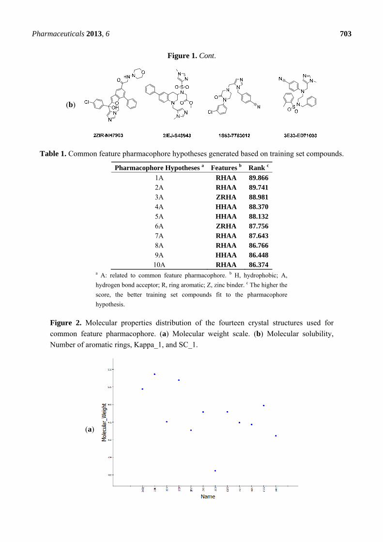
Figure 2. Molecular properties distribution of the fourteen crystal structures used for
common feature pharmacophore. (a) Molecular weight scale. (b) Molecular solubility,
Number of aromatic rings, Kappa_1, and SC_1.
(a)

Pharmaceuticals 2013, 6 704
Figure 2. Cont.
(b)
In the Common feature pharmacophore generation protocol no conformation generation was
performed, Principal and MaxOmitFeat values of 2 and 0 were set for all compounds in the training
set as the chosen compounds were the most active and they also were co-crystallized in the active site
of the enzyme. Since the active site of the FTase enzyme contains a zinc cation and of all the ten
chosen compounds interact with that zinc cation by performing a coordination bond, the features
selected to generate the hypotheses were hydrogen bond acceptor 05, hydrogen bond donor 05,
hydrophobic 05, ring aromatics, zinc binder 05, maximum pharmacophores 10, minimum interfeature
distance 2.0Å. All other control parameters were kept at their default values. The zinc binder feature
was modified to be able to identify the zinc binding groups in the active inhibitors that are not included
originally in DS 3.1. The best common feature of pharmacophore hypothesis (Pharm-3A) was selected
based on the inclusion of the zinc binder after customization in its features (Table 1).
2.2. Generation of Pharmacophore Hypotheses: Structure-Based Approach
Structure-based pharmacophore modeling has been extensively implemented by researchers
world-wide to provide successful novel drugs with potent activity. Mainly, it is used whenever there is
a shortage of information of ligands that bind to the receptor or to get more insight into the geometry
of the active site. In this study, a crystal structure of FTase enzyme with a bound ligand (PDB code:
3E33) crystallized at 1.9 Å resolution was utilized to generate the structure-based hypothesis. A sphere
of 10 Å radius which covers the most important residues that bind with the twenty three crystallized
ligands was generated using binding site tools available in DS 3.1. These important residues were
assigned by careful studying of the binding nature by which each one the 23 crystallized ligands
connect to the active site using the protocol Receptor-Ligand Pharmacophore Generation.

Pharmaceuticals 2013, 6 705
Pharmacophoric features from the active site were then generated by employing Interaction
Generation protocol. This protocol is capable of identifying hydrogen bond donors (HD), hydrogen
bond acceptors (HA) and hydrophobic pockets (HY) by referring to the active site residues. As a final
step to optimize the structure-based pharmacophore, Edit and Cluster tool was utilized to cluster and
remove any redundant features or features with no catalytic importance.
2.3. Validation of the Pharmacophore Hypotheses
Validation was conducted on three separate levels; the first level was performed using the ligand
pharmacophore mapping protocol, where the four test compounds mapped to the generated
pharmacophores. In the second level a set of 286 ligands extracted from literature and divided into
active and inactive molecules based using activity of 500 nM as threshold. Running validation through
the mapping method will test the ability of the generated pharmacophores to distinguish the active
molecules from inactive molecules. The last step in this level, Ligand pharmacophore mapping
Protocol available in DS 3.1 with Best flexible fitting method was used to measure the extent to which
the active molecules could match the pharmacophore and the results were represented by percentages.
Finally in the third level, within the common feature pharmacophore , an internal validation performed by
providing a set of active and inactive molecules where the results presented as roc curve files.
2.4. Database Screening
The generated pharmacophore hypotheses based on the aforementioned two approaches were used
as 3D queries to extract chemical compounds from commercially available databases. This process
will help in finding potential leads for this target that can be optimized further by investigating drug
likeness and synthetic feasibility. A selected compound from the screening method should map all the
pharmacophoric features in order to be considered a hit for the common feature pharmacophore
hypothesis. On the other hand, a one feature miss was tolerated for considering a compound to pass the
screening for structure based pharmacophore. The screening processes were performed using Ligand
Pharmacophore Mapping protocol with Best Flexible Conformation Search method. The candidate
molecules were further subjected to numerous filters in DS 3.1 (Lipiniski’s and Veber rules) to select
the drug-like compounds. The designated molecules were considered in molecular docking stage.
2.5. Molecular Docking
Molecular docking was conducted using CDOCKER docking protocol in DS 3.1 and GOLD
(Genetic Optimization for Ligand Docking) program version 5.1 from the Cambridge Crystallographic
Data Centre (CDCC). CDOCKER uses a CHARMm-based molecular dynamics (MD) scheme to dock
ligands within the active site of enzymes and receptors. First, random ligand conformations are
generated using high-temperature MD, and then the produced conformations are translated into the
binding site. Candidate poses are further created using random rigid-body rotations followed by
simulated annealing. A final minimization is used to refine the ligand poses [17,18]. GOLD docking is
based on using genetic algorithm to prospect the full flexibility of the ligand with partial flexibility of
the active site of the enzyme [19]. GOLD has been validated by over 300 crystal structure complexes

Pharmaceuticals 2013, 6 706
extracted from PDB and has showed success in more than 70% of the cases. The crystal structure PDB
(3E33, 1.9 Å) [20] of FTase enzyme was selected as it represents the best resolution among all the
available crystal structures for FTase in PDB and was prepared using Prepare Protein protocol that
removes water, corrects any defects in the protein structure and adds hydrogen. The Input site sphere
was 10 Å in radius using the ligand in the active site as a reference point for both CDOCKER and GOLD.
For CDOCKER, the candidate molecules from the screening process were docked in the active site
using CHARMm forcefield with top 10 poses to be presented and scored and keeping the other options
in their default values. On the other hand, for GOLD, early termination step was activated if the first
three poses have an rmsd value of less than 1.5 Å, other parameters were set as default. The final hit
molecules were selected based on a combination of docking score, mode of binding and molecular
interaction within the active site.
3. Results and Discussion
3.1. Common Feature Pharmacophore Hypothesis
The selection of training and test set compounds for pharmacophore hypotheses generation was
performed by subjecting twenty three ligands extracted from PDB of FTase enzyme to both Lipinisks’
rule of five; which limits the selection of molecules of appropriate characteristics with respect to size
and molecular weight, number of HBD and HBA, and logP values in addition to Vebers’ rules with
respect to the number of rotatable bonds and polar surface area, yielding fourteen candidate
compounds (Figure 1). In this way selecting the compounds will allow building a pharmacophore
hypothesis that possesses the essential properties and at the same time avoids chemical compounds that
could cause generated pharmacophore hypothesis to deviate from the optimum configuration. The
selected fourteen compounds were further divided into training set and test set using the Generate
training and test data protocol at DS 3.1. Splitting of these molecules is based on diversity and it is
assumed that 70% of them (10 compounds) are the training set. The crystallized biologically active
conformations were mapped without any conformation generation as they were considered to possess
the required and optimum conformation for pharmacophore hypothesis generation. In this study, the
selected features are HBA, HBD, hydrophobic, ring aromatic, and zinc binder. Ten pharmacophore
hypotheses based on common features were generated and they are displayed in (Table 1). According
to the table, all the training set compounds fit all the features of the pharmacophore hypotheses and the
rank scores ranged from 89.866 to 86.374. Only two of the 10 pharmacophore hypotheses contain Zn2+
feature and they are ranked in the third and the sixth positions.
The best pharmacophore hypothesis was chosen based on the fact that the presence of the zinc
binding feature is essential. Hypothesis three has showed high values compared to other hypotheses
and contains the zinc binding feature, named as Pharm-3A (Table 2). Pharm-3A comprises one zinc
binder, one ring aromatic, one hydrophobic and one HBA feature (Figure 3a). Each of the training set
molecules has mapped all the features in the pharmacophore hypothesis. For instance, compound
2ZIS-NH8903 has the imidazole ring mapped to the Zn+2 binding feature, whereas the nitro group
matched the HBA, in addition, one of the aromatic rings was mapped to the ring aromatic feature while
the other one was mapped to the hydrophobic feature (Figure 3b).

Pharmaceuticals 2013, 6 707
Table 2. Best-fit values of the training and test set compounds based on common feature
pharmacophore technique.
PDB ID Inhibitor NameFit Value
Pharm-3A
2ZIS NH8903 4.000 1N95 FTH1001 3.643 3E32 ED21003 3.191 1NI1 2C510 3.178 3KSQ Z96439 3.018 2F0Y 3MN963 2.564 1SA5 BMV440 2.112 1X81 JAN1 1.826 1MZC BME1003 1.164 1N9A FTI1 0.788 2ZIR NH7903 * 3.585 3E33 ED71003 * 3.096 2IEJ S48943 * 2.748 1S63 7783012 * 1.784
* Compounds used for generating the test set for common feature pharmacophore.
Figure 3. Pharm-3A and its overlay with a training set compound (a) Chemical features of
pharm-3A with its inter-feature distances. (b) 2ZIS-NH8903 overlaid on pharm-3A hypothesis.
HY; hydrophobic, RA; ring aromatic, HBA; hydrogen bond acceptor, ZB; zinc binder.
(a) (b)
3.2. Structure-Based Pharmacophore Hypothesis
This pharmacophore was generated in three stages; the first, Receptor-ligand pharmacophore
generation protocol was performed for each co-crystallized ligand inside the active site in order to
discover the potentially important amino acids that are of strategic contribution to ligand binding.
These amino acids will be later considered in the final pharmacophore of the active site. The results
collected from running Receptor-ligand pharmacophore generation protocol have showed a series of
amino acids that contribute to ligand bindings in the active site. Leu96, Tyr93 and Trp 106 performed

Pharmaceuticals 2013, 6 708
hydrophobic interaction with lipophilic groups found in the inhibitors’ structures. Tyr361 and Tyr166
have participated in ligand binding by forming ring aromatic interaction with aromatic rings in the
inhibitors’ structures. Moreover, Arg202 and Tyr93 were prominent amino acids by being HBDs to
their corresponding acceptors in the inhibitors. All the tested inhibitors have showed covalent
interaction between the zinc binding group of the inhibitors and the Zn2+ inside the active site.
Secondly, Interaction Generation protocol was generated based on the lipophilic and the
hydrophilic regions within the active site, which was later subjected to further cleaning yielding a
pharmacophore hypothesis (Pharm-B) containing six features that are complementary to Tyr361,
Tyr166, Phe360, Leu96, Tyr93 and the Zn2+ atom. The third stage involved customization of the
pharmacophore by selectively replacing HBA feature pointed to the Zn2+ atom by zinc feature
available on DS 3.1 (Figure 4). The generated pharmacophore from this step is then compared with
pharm-3A by utilizing Pharmacophore comparison pharmacophore. The results indicated that there
was a comparable similarity between the two pharmacophores with RMSD value of 2.52 (Figure 5).
The fourteen compounds used to generate the common feature pharmacophore Pharm-3A were used
to check whether Pharm-B can accommodate these compounds and map them. The fit values were
extracted for these compounds which showed good fitting values (Table 3) and all the fourteen
compounds mapped at least four features of the six pharmacophoric features of the structure-based
hypothesis and every time the zinc binding feature was occupied with zinc binding group. Compound
2ZIS-NH8903 mapping on Pharm-B is depicted in (Figure 6).
Figure 4. Structure-based pharmacophore hypothesis, Pharm-B. (a) Arrangement of
pharmacophoric features of Pharm-B. HY; hydrophobic, RA; ring aromatic, HBA;
hydrogen bond acceptor, ZB; zinc binder. (b) Pharmacophoric features are displayed with
inter-feature distances. Tolerance spheres were removed for simplification purposes.
(a) (b)

Pharmaceuticals 2013, 6 709
Figure 5. Superimposition of pharm-3A over Pharm-B showing the degree of similarity
between the two pharmacophores of RMSD value of 2.52. HY; hydrophobic, RA; ring
aromatic, HBA; hydrogen bond acceptor, ZB; zinc binder.
Table 3. Best-fit values of the training and test set compounds based on structure based
pharmacophore techniques.
PDB ID Inhibitor NameFit ValuePharm-B
2ZIS NH8903 5.088 1N95 FTH1001 4.979 3E32 ED21003 5.666 1NI1 2C510 4.713 3KSQ Z96439 4.324 2F0Y 3MN963 5.457 1SA5 BMV440 4.956 1X81 JAN1 4.627 1MZC BME1003 4.778 1N9A FTI1 4.962 2ZIR NH7903 * 4.933 3E33 ED71003 * 5.266 2IEJ S48943 * 5.579 1S63 7783012 * 4.167
* Compounds used for generating the test set for common feature pharmacophore.
Figure 6. 2ZIS-NH8903 overlaid on pharm-B hypothesis. HY; hydrophobic, RA; ring
aromatic, HBA; hydrogen bond acceptor, ZB zinc binder.

Pharmaceuticals 2013, 6 710
3.3. Validation of Pharmacophore Hypotheses
Ligand Pharmacophore Mapping Protocol was used to perform this step. The four test compounds
were used to score their fitting to the generated pharmacophore taking into consideration that there is
no conformation generation accompanied with rigid fitting technique. The four compounds were found
to completely match Pharm-3A with fitting values ranging from 3.58–1.78 (Table 2).
An internal validation step performed by generating a ROC curve was also used to confirm the
ability of Pharm-3A to be able to distinguish between active and inactive molecules. Within the
Common Feature Pharmacophore Generation protocol, a validation step was used by providing the
active compounds which were the four test set compounds (Figure 1b), and 85 inactive compounds
collected from literature. The AUC, accuracy and specificity of Pharm-3A were shown to be the best
among all the pharmacophore hypotheses predicted by the training set compounds by recording 93%,
95% and 55% respectively (Figure 7).
Figure 7. ROC curve of hypothesis Pharm-3A.
In another validation step, a database of 269 compounds of both active and inactive compounds was
collected from the literature and drawn using Chemsketch 12.0 and then converted to .sd format file.
Of the 269 compounds, 184 were considered active compounds based on their activity against FTase
enzyme of more than 500 nM. Using Ligand Pharmacophore Mapping Protocol, the 184 compounds
were used to investigate the ability of the generated pharmacophore hypothesis to map ligands that are
known inhibitors to this enzyme. Of 184 compounds, 153 compounds were mapped on all the features
of the common feature pharmacophore hypothesis with a hit rate of 83.15%. And the same compounds
were mapped on the structure-based pharmacophore and here, 151 of 184 compounds were mapped on
all the features of structure-based pharmacophore with a hit rate of 82.06%.
3.4. Database Screening
Both the common feature pharmacophore and the structure-based pharmacophore hypotheses that
were generated previously were subjected to database screening process utilizing Ligand Pharmacophore
Mapping Protocol using Best Flexible search option. These databases are ZINC-NCI_(92547),

Pharmaceuticals 2013, 6 711
ZINC-MayBridge_(75443), and ZINC-Key Organics_(54869) [21] which were prepared previously by
our group for the screening process. For Pharm-3A, Ligand Pharmacophore Mapping protocol was
used with MaxOmitFeat value of zero as the four features of the Pharm-3A should be fulfilled. A
compound will be determined as a hit if it maps all the four features that are included in the
pharmacophore hypothesis.On the other hand, a relaxed criteria was used for Pharm-B where the hit is
accepted if it can map five features out of the six and hence the Ligand Pharmacophore Mapping
protocol which allows some of the features to be missed was used. The extracted compounds were then
subjected to further screening processes relying on their fitness value, drug likeness according to
Lipinski's rule of five and Vebers’ rule. A compound is determined positive according to Lipiniski and
Veber if (i) less than five hydrogen bond donor groups; (ii) less than 10 hydrogen bond acceptor
groups; (iii) a molecular weight less than 500; (iv) A LogP value less than 5; (v) number of rotatable
bonds less than 10; polar surface area less than 140 Å2; and hydrogen bond donors and acceptors less
than 12. A subsequent level of filtration to achieve drug like compounds is to perform ADMET
screening techniques in DS 3.1 in which the databases are filtered according to their solubility,
intestinal absorption and Blood Brain Barrier penetration. A total of 1,296 compounds (885 from the
structure-based design and 411 from the common feature pharmacophore) were found to satisfy the
assigned filters for the molecular docking step (Figure 8).
Figure 8. Database screening of three databases employing pharm-3A and pharm-B hypotheses.
3.5. Molecular Docking
The 1,296 compounds emerged from both structure and ligand-based pharmacophores conjointly
with the training set were submitted to docking programs CDOCKER and GOLD. Consensus scores of
the candidate compounds from both CDOCKER and GOLD were performed, eventually compounds

Pharmaceuticals 2013, 6 712
enumerated the highest value from score summation of the two docking programs were chosen to be
candidate molecules. These scores of the candidate compounds were contrasted by scores obtained for
the training set compounds (the highest consensus score for the training set compound is 147.4) and
only compounds that scored equally or higher than the training set compounds were fully investigated
by studying their binding mode. Of the 1,296 compounds, twenty one compounds had shown scores
higher than training set scores. Based on molecular fitting mode inside the active site and their proper
interaction with zinc atom, two compounds were chosen as potential virtual leads namely ZINC39323901,
and ZINC01034774. Compound ZINC01034774 which was identified from structure-based
pharmacophore has a fit value, CDOCKER, and GOLD fitness scores of 3.28, 64.65, and 98.11
respectively. The imidazole ring is supposed to form a coordinate bond with zinc cation which has a
perfect pose when the pyridine-like nitrogen points directly at zinc cation of the active site with
optimum distance of 1.76 Å. In addition, internal π-π interaction is shown between the imidazole ring
and one of the benzene rings of the compound. The other benzene ring is positioned in a hydrophobic
pocket within the active site formed from the hydrophobic tail of farnesyl pyrophosphate (FPP),
Tyr166, and Lys164. The sulphonamide moiety interacts via hydrogen bond with Tyr361 phenol side
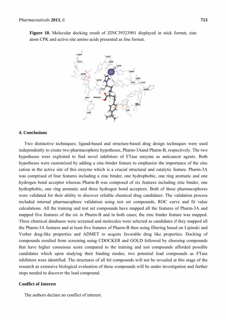
chain (Figure 9). Compound ZINC39323901 which was extracted from common feature pharmacophore
has a fit value, CDOCKER, and GOLD fitness scores of 3.64, 65.05, and 95.63 respectively. The
imidazole ring is in proximity to the zinc cation with a distance of 1.70 Å which is expected to form
coordination bond, and the benzene ring is expected to form π-π interactions with indole ring of
Trp102 side chain (Figure 10).
Figure 9. Molecular docking result of ZINC01034774 displayed in stick format, zinc atom
CPK and active site amino acids presented as line format.
Pharmaceuticals 2013, 6 713
Figure 10. Molecular docking result of ZINC39323901 displayed in stick format, zinc
atom CPK and active site amino acids presented as line format.
4. Conclusions
Two distinctive techniques; ligand-based and structure-based drug design techniques were used
independently to create two pharmacophore hypotheses, Pharm-3Aand Pharm-B, respectively. The two
hypotheses were exploited to find novel inhibitors of FTase enzyme as anticancer agents. Both
hypotheses were customized by adding a zinc-binder feature to emphasize the importance of the zinc
cation in the active site of this enzyme which is a crucial structural and catalytic feature. Pharm-3A
was comprised of four features including a zinc binder, one hydrophobic, one ring aromatic and one
hydrogen bond acceptor whereas Pharm-B was composed of six features including zinc binder, one
hydrophobic, one ring aromatic and three hydrogen bond acceptors. Both of these pharmacophores
were validated for their ability to discover reliable chemical drug candidates. The validation process
included internal pharmacophore validation using test set compounds, ROC curve and fit value
calculations. All the training and test set compounds have mapped all the features of Pharm-3A and
mapped five features of the six in Pharm-B and in both cases; the zinc binder feature was mapped.
Three chemical databases were screened and molecules were selected as candidates if they mapped all
the Pharm-3A features and at least five features of Pharm-B then using filtering based on Lipinski and
Verber drug-like properties and ADMET to acquire favorable drug like properties. Docking of
compounds resulted from screening using CDOCKER and GOLD followed by choosing compounds
that have higher consensus score compared to the training and test compounds afforded possible
candidates which upon studying their binding modes, two potential lead compounds as FTase
inhibitors were identified. The structures of all hit compounds will not be revealed at this stage of the
research as extensive biological evaluation of these compounds will be under investigation and further
steps needed to discover the lead compound.
Conflict of Interest
The authors declare no conflict of interest.

Pharmaceuticals 2013, 6 714
References
1. Sinensky, M.; Beck, L.A.; Leonard, S.; Evans, R. Differential inhibitory effects of lovastatin on
protein isoprenylation and sterol synthesis. J. Biol. Chem. 1990, 265, 19937–19941.
2. Habenicht, A.J.; Glomset, J.A.; Ross, R. Relation of cholesterol and mevalonic acid to the cell
cycle in smooth muscle and Swiss 3T3 cells stimulated to divide by platelet-derived growth
factor. J. Biol. Chem. 1980, 255, 5134–5140.
3. Reiss, Y.; Brown, M.S.; Goldstein, J.L. Divalent cation and prenyl pyrophosphate specificities of
the protein farnesyltransferase from rat brain, a zinc metalloenzyme. J. Biol. Chem. 1992, 267,
6403–6408.
4. Reiss, Y.; Goldstein, J.L.; Seabra, M.C.; Casey, P.J.; Brown, M.S. Inhibition of purified p21ras
farnesyl:protein transferase by Cys-AAX tetrapeptides. Cell 1990, 62, 81–88.
5. Maltese, W.A.; Erdman, R.A. Characterization of isoprenoid involved in the post-translational
modification of mammalian cell proteins. J. Biol. Chem. 1989, 264, 18168–18172.
6. Zhang, F.L.; Casey, P.J. Protein Prenylation: Molecular Mechanisms and Functional Consequences.
Annu. Rev. Biochem. 1996, 1996, 241–269.
7. Qian, Y.; Vogt, A.; Sebti, S.M.; Hamilton, A.D. Design and Synthesis of Non-Peptide Ras CAAX
Mimetics as Potent Farnesyltransferase Inhibitors. J. Med. Chem. 1996, 39, 217–223.
8. Strickland, C.L.; Windsor, W.T.; Syto, R.; Wang, L.; Bond, R.; Wu, Z.; Schwartz, J.; Le, H.V.;
Beese, L.S.; Weber, P.C. Crystal Structure of Farnesyl Protein Transferase Complexed with a
CaaX Peptide and Farnesyl Diphosphate Analogue. Biochemistry 1998, 37, 16601–16611.
9. Waldron, K.J.; Robinson, N.J. How do bacterial cells ensure that metalloproteins get the correct
metal? Nat. Rev. Micro. 2009, 7, 25–35.
10. Andreini, C.; Bertini, I.; Rosato, A. A hint to search for metalloproteins in gene banks.
Bioinformatics 2004, 2004, 1373–1380.
11. Roe, R.R.; Pang, Y.P. Zinc’s Exclusive Tetrahedral Coordination Governed by Its Electronic
Structure. J. Mol. Model. 1999, 134–140.
12. Huang, C.C.; Casey, P.J.; Fierke, C.A. Evidence for a catalytic role of zinc in protein
farnesyltransferase. Spectroscopy of Co2+-farnesyltransferase indicates metal coordination of the
substrate thiolate. J. Biol. Chem. 1997, 272, 20–23.
13. Sousa, S.F.; Fernandes, P.A.; Ramos, M.J. Farnesyltransferase New Insights into the
Zinc-Coordination Sphere Paradigm: Evidence for a Carboxylate-Shift Mechanism. Biophys. J.
2005, 88, 483–494.
14. Equbal, T.; Silakari, O.; Rambabu, G.; Ravikumar, M. Pharmacophore mapping of diverse classes
of farnesyltransferase inhibitors. Bioorg. Med. Chem. Lett. 2007, 17, 1594–1600.
15. Lu, A.; Zhang, J.; Yin, X.; Luo, X.; Jiang, H. Farnesyltransferase pharmacophore model derived
from diverse classes of inhibitors. Bioorg. Med. Chem. Lett. 2007, 17, 243–249.
16. Vaidya, M.; Weigt, M.; Wiese, M. 3D-QSAR with the aid of pharmacophore search and docking-
based alignments for farnesyltransferase inhibitors. Eur. J. Med. Chem. 2009, 44, 4070–4082.
17. Wu, G.; Robertson, D.H.; Brooks, C.L., 3rd; Vieth, M. Detailed analysis of grid-based molecular
docking: A case study of CDOCKER—A CHARMm-based MD docking algorithm. J. Comput.
Chem. 2003, 24, 1549–1562.

Pharmaceuticals 2013, 6 715
18. Nissink, J.W.; Murray, C.; Hartshorn, M.; Verdonk, M.L.; Cole, J.C.; Taylor, R. A new test set for
validating predictions of protein-ligand interaction. Proteins 2002, 49, 457–471.
19. Jones, G.; Willett, P.; Glen, R.C.; Leach, A.R.; Taylor, R. Development and validation of a
genetic algorithm for flexible docking. J. Mol. Biol. 1997, 267, 727–748.
20. Hast, M.A.; Fletcher, S.; Cummings, C.G.; Pusateri, E.E.; Blaskovich, M.A.; Rivas, K.;
Gelb, M.H.; van Voorhis, W.C.; Sebti, S.M.; Hamilton, A.D.; et al. Structural basis for binding
and selectivity of antimalarial and anticancer ethylenediamine inhibitors to protein farnesyltransferase.
Chem. Biol. 2009, 16, 181–192.
21. Irwin, J.; Shoichet, B. ZINC—A free database of commercially available compounds for virtual
screening. J. Chem. Inf. Model. 2005, 45, 177–182.
© 2013 by the authors; licensee MDPI, Basel, Switzerland. This article is an open access article
distributed under the terms and conditions of the Creative Commons Attribution license
(http://creativecommons.org/licenses/by/3.0/).