Development and NMR validation of minimal pharmacophore hypotheses for the generation of fragment...
15
Development and NMR validation of minimal pharmacophore hypotheses for the generation of fragment libraries enriched in heparanase inhibitors Rafael Gozalbes Silvia Mosule ´n Rodrigo J. Carbajo Antonio Pineda-Lucena Received: 15 January 2009 / Accepted: 9 April 2009 / Published online: 7 May 2009 Ó Springer Science+Business Media B.V. 2009 Abstract A combined strategy based on the development of pharmacophore hypotheses and NMR approaches is reported for the identification of novel inhibitors of hepa- ranase, a key enzyme involved in tumor metastasis through the remodeling of the subepithelial and subendothelial basement membranes, resulting in the dissemination of metastatic cancer cells. Several pharmacophore hypotheses were initially developed from the most active heparanase inhibitors known to date and, after their application to a pool of 27 known heparanase inhibitors and a database of 1,120 compounds approved by the FDA, a four-point pharmacophore model was selected as the most predictive. This model was subsequently applied to a database of 686 chemical fragments, and a subset of 100 fragments accomplishing completely or partially the four-point model was selected to perform nuclear magnetic resonance experiments to validate the hypothesis. The experimental studies confirmed the reliability of our pharmacophore model, its applicability to in silico databases in order to reduce the number of compounds to be experimentally screened, and the possibility of generating fragment libraries enriched in heparanase inhibitors. Keywords Pharmacophore Á Fragment-based screening Á NMR Á Heparanase Á Inhibitor Introduction Heparanase is an endoglycosidase that acts at the cell- surface and within the extracellular matrix to degrade polymeric heparan sulfate molecules (Fig. 1) into shorter oligosaccharides [1, 2]. Heparanase is involved in several physiological activities such as embryo development, hair growth and wound healing, and it is also implicated in cancer processes such as tumor angiogenesis and metas- tasis [3, 4]. Increased levels of heparanase have been found in numerous cancer processes at different organs and there is a direct correlation between the overexpression of hep- aranase and the invasiveness of tumor cells [5]. The implication of heparanase in cancer progression makes it a very attractive target for anti-angiogenic, anti-metastatic and/or anti-inflammatory therapies. The three-dimensional structure of the enzyme has not yet been elucidated, but the active-site residues of human heparanase have been described, and it is widely accepted that there are two essential acidic residues (Glu 225 and Glu 343 ) involved in the catalytic mechanism, acting as a putative proton donor and a nucleophile, respectively [6]. Despite the increasing interest in finding effective inhibitors against heparanase, and although a number of compounds with some inhibitory effect have been descri- bed, only one of them has reached so far the clinical trial phases [7]. Heparanase inhibitors reported until now differ in their chemical nature, origin (from natural products to synthetic compounds found through virtual or experimental screening of chemical libraries) and size (from big heparin- like polymers and oligosaccharide mimetics to small molecules with some ‘‘drug-like’’ features) [8]. Pharmacophore modeling is a useful technique for describing interactions of small molecules with macromo- lecular targets of therapeutic interest [9–11]. The IUPAC Rafael Gozalbes and Silvia Mosule ´n contributed equally to this work. R. Gozalbes Á S. Mosule ´n Á R. J. Carbajo Á A. Pineda-Lucena (&) Structural Biology Laboratory, Department of Medicinal Chemistry, Centro de Investigacio ´n Prı ´ncipe Felipe, Avenida Autopista del Saler 16, 46012 Valencia, Spain e-mail: [email protected] 123 J Comput Aided Mol Des (2009) 23:555–569 DOI 10.1007/s10822-009-9269-0
-
Upload
independent -
Category
Documents
-
view
0 -
download
0
Transcript of Development and NMR validation of minimal pharmacophore hypotheses for the generation of fragment...

Development and NMR validation of minimal pharmacophorehypotheses for the generation of fragment libraries enrichedin heparanase inhibitors
Rafael Gozalbes Æ Silvia Mosulen ÆRodrigo J. Carbajo ÆAntonio Pineda-Lucena
Received: 15 January 2009 / Accepted: 9 April 2009 / Published online: 7 May 2009
� Springer Science+Business Media B.V. 2009
Abstract A combined strategy based on the development
of pharmacophore hypotheses and NMR approaches is
reported for the identification of novel inhibitors of hepa-
ranase, a key enzyme involved in tumor metastasis through
the remodeling of the subepithelial and subendothelial
basement membranes, resulting in the dissemination of
metastatic cancer cells. Several pharmacophore hypotheses
were initially developed from the most active heparanase
inhibitors known to date and, after their application to a
pool of 27 known heparanase inhibitors and a database of
1,120 compounds approved by the FDA, a four-point
pharmacophore model was selected as the most predictive.
This model was subsequently applied to a database of 686
chemical fragments, and a subset of 100 fragments
accomplishing completely or partially the four-point model
was selected to perform nuclear magnetic resonance
experiments to validate the hypothesis. The experimental
studies confirmed the reliability of our pharmacophore
model, its applicability to in silico databases in order to
reduce the number of compounds to be experimentally
screened, and the possibility of generating fragment
libraries enriched in heparanase inhibitors.
Keywords Pharmacophore � Fragment-based screening �NMR � Heparanase � Inhibitor
Introduction
Heparanase is an endoglycosidase that acts at the cell-
surface and within the extracellular matrix to degrade
polymeric heparan sulfate molecules (Fig. 1) into shorter
oligosaccharides [1, 2]. Heparanase is involved in several
physiological activities such as embryo development, hair
growth and wound healing, and it is also implicated in
cancer processes such as tumor angiogenesis and metas-
tasis [3, 4]. Increased levels of heparanase have been found
in numerous cancer processes at different organs and there
is a direct correlation between the overexpression of hep-
aranase and the invasiveness of tumor cells [5]. The
implication of heparanase in cancer progression makes it a
very attractive target for anti-angiogenic, anti-metastatic
and/or anti-inflammatory therapies. The three-dimensional
structure of the enzyme has not yet been elucidated, but the
active-site residues of human heparanase have been
described, and it is widely accepted that there are two
essential acidic residues (Glu225 and Glu343) involved in
the catalytic mechanism, acting as a putative proton donor
and a nucleophile, respectively [6].
Despite the increasing interest in finding effective
inhibitors against heparanase, and although a number of
compounds with some inhibitory effect have been descri-
bed, only one of them has reached so far the clinical trial
phases [7]. Heparanase inhibitors reported until now differ
in their chemical nature, origin (from natural products to
synthetic compounds found through virtual or experimental
screening of chemical libraries) and size (from big heparin-
like polymers and oligosaccharide mimetics to small
molecules with some ‘‘drug-like’’ features) [8].
Pharmacophore modeling is a useful technique for
describing interactions of small molecules with macromo-
lecular targets of therapeutic interest [9–11]. The IUPAC
Rafael Gozalbes and Silvia Mosulen contributed equally to this work.
R. Gozalbes � S. Mosulen � R. J. Carbajo �A. Pineda-Lucena (&)
Structural Biology Laboratory, Department of Medicinal
Chemistry, Centro de Investigacion Prıncipe Felipe, Avenida
Autopista del Saler 16, 46012 Valencia, Spain
e-mail: [email protected]
123
J Comput Aided Mol Des (2009) 23:555–569
DOI 10.1007/s10822-009-9269-0

defines a pharmacophore as ‘‘the ensemble of steric and
electronic features that is necessary to ensure the optimal
supramolecular interactions with a specific biological tar-
get structure and to trigger (or to block) its biological
response’’ [12]. Pharmacophores describe the spatial
arrangement of the chemical features necessary for small
compounds to interact with the target, and they can be
determined either from the complementarities of a ligand
interacting with a binding site (structure-based pharmaco-
phores) or, most frequently, from an active compound or
the alignment of a set of known active molecules (ligand-
based pharmacophores). Pharmacophore representations of
ligand-protein interactions can be used to query, in virtual
databases, for compounds that have the same or similar
distribution of chemical features. Therefore, they provide a
way to identify compounds that could potentially interact
with the target in the same manner and produce a similar
biological activity [11].
The absence of the 3D structure of heparanase precludes
the development of structure-based pharmacophores, but
the existence of a number of small molecule heparanase
inhibitors indicates the potential of ligand-based pharma-
cophore approaches to identify novel inhibitors against this
target. Thus, in the context of the present study, several
pharmacophore hypotheses were developed based on the
alignment of the most active inhibitors of heparanase.
Taking into consideration that the pharmacophore
hypotheses were intended to be applied over a fragment
library, special attention was paid to the identification of
‘‘minimal’’ pharmacophores, that is, hypotheses where the
distances between the different features describing the
pharmacophore were relatively short. The different
hypotheses were evaluated by in silico applying them to a
database of structures comprising compounds known to
have some heparanase inhibitory activity and a set of
compounds approved by the FDA. The quality of the
pharmacophore hypotheses was determined by analyzing
their ability to retrieve and rank the known inhibitors from
the virtual database. Once the selected model was vali-
dated, we applied it to a database of chemical fragments,
composed by 686 compounds, and a subset of them,
accomplishing totally or partially the pharmacophore
model, was selected in order to perform an experimental
validation that included the acquisition of a set of NMR
and surface plasmon resonance experiments, thus provid-
ing a useful way to identify experimentally novel inhibitors
against heparanase.
Materials and methods
Protein preparation
A recombinant heparanase construct (details will be descri-
bed elsewhere) containing the catalytic site was expressed,
from a modified version of the pET-15b (Novagen) expres-
sion vector containing a TEV cleavage site, in Escherichia
coli BL21-DE3 codon plus (Stratagene) cells. Cells were
grown at 37 �C to an absorbance at 600 nm of 0.6 and
induced with 0.8 mM IPTG overnight (15 h) at 25 �C. The
expressed protein was purified using metal affinity chroma-
tography under native conditions. Unlabeled, uniformly
(15N) and selectively labeled (15N-Glu and 15 N-Gly) protein
preparations were produced in LB and M9, and NMR sam-
ples at different concentrations (5 lM for WaterLOGSY and
STD experiments, and 100 lM for HSQC experiments) were
prepared. Protein samples selectively labeled in Glu and Gly
were used to confirm the interaction of any of the fragments
with the catalytic glutamic amino acids (Glu225 and Glu343)
and/or with the preceding glycine residues (Gly223 and
Gly342) and the results were analyzed taking advantage of the
in-house backbone assignment obtained for this heparanase
construct.
Pharmacophore development
A database of 27 well characterized heparanase inhibitors
(Table 1) was constructed in silico, in order to generate and
validate pharmacophore hypotheses from it. The compounds
were extracted from the review of Hammond et al. [8], drawn
with MDL Isis Draw 2.5 (www.mdl.com) and exported to the
Molecular Operating Environment (MOE) software
(Chemical Computing Group, www.chemcomp.com) to
develop the pharmacophore hypotheses. The pharmaco-
phore development module in MOE allows the character-
ization of atoms by their chemical features (hydrogen bond
donor or acceptor ability, aromatic character, hydrophobic
nature, etc.), the generation of the subsequent pharmaco-
phore hypotheses, and the searching of conformation dat-
abases using molecule annotations related to ligand-receptor
binding. The plausibility of the theoretical models is mea-
sured by the good overlay of active compounds (since they
share a common binding mode) and the ability to separate
Fig. 1 Minimum heparan sulfate recognition sequence for heparan-
ase. The essential charged groups required for heparanase recognition
are highlighted in red, and other charged groups which may improve
substrate affinity are shown in blue. Adapted from [8]
556 J Comput Aided Mol Des (2009) 23:555–569
123
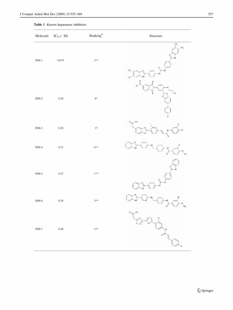
Table 1 Known heparanase inhibitors
Molecule
IC50 (µM)
Rankinga
Structure
INH-1 0.075 2**
INH-2 0.20 6*
INH-3 0.20 17
INH-4 0.23 4**
INH-5 0.27 1**
INH-6 0.29 3**
INH-7 0.40 11*
J Comput Aided Mol Des (2009) 23:555–569 557
123
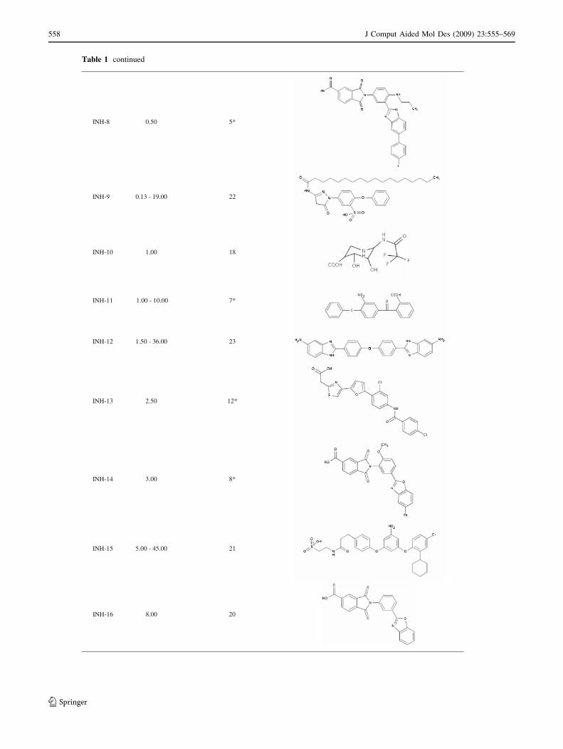
Table 1 continued
INH-8 0.50 5*
INH-9 0.13 - 19.00 22
INH-10 1.00 18
INH-11 1.00 - 10.00 7*
INH-12 1.50 - 36.00 23
INH-13 2.50 12*
INH-14 3.00 8*
INH-15 5.00 - 45.00 21
INH-16 8.00 20
558 J Comput Aided Mol Des (2009) 23:555–569
123
Table 1 continued
INH-17 8.00 - 25.00 16
INH-18 10.00 15
INH-19 10.00 14
INH-20 10.00 - 26.00 N.S.
INH-21 12.00 19
INH-22 17.00 N.S.
INH-23 24.00 - 32.00 10*
INH-24 25.00 13*
INH-25 26.00 - 36.00 9*
INH-26 > 100.00 N.S.
INH-27 > 100.00 N.S.
A detailed description of these inhibitors can be found in the review by Hammond et al. [8]. ‘‘N.S.’’ stands for
compounds ‘‘Not Satisfying’’ the pharmacophore modelaA database consisting of the 27 known heparanase inhibitors plus the 1,120 structures from the Prestwick Chemical
Library� was used to validate the pharmacophore model. The number in the column indicates the order in which the
inhibitor was retrieved with respect to the other inhibitors when using the 1,147 chemicals. * Compounds ranked
amongst the best 10% of the total database by the pharmacophore model, and ** those ranked amongst the best 2%
J Comput Aided Mol Des (2009) 23:555–569 559
123

actives from inactives. In the context of our work, we
developed a number of pharmacophore hypotheses based on
the alignment of several subsets derived from the most active
heparanase inhibitors (Table 1). Prior to the pharmacophore
model development, multiple conformations of each of the
27 known inhibitors were generated with MOE, using the
MMFF94x force-field and a 4 kcal/mol energy cutoff, with a
maximum of 250 conformations for each compound. Bear-
ing in mind the importance of validating models with unbi-
ased decoys [13], we generated a virtual database consisting
of the 27 known heparanase inhibitors plus a collection of
1,120 compounds (Prestwick Chemical Library�) that con-
tained 90% marketed drugs (www.prestwickchemical.com),
also subjected to the same conformational protocol described
previously, for the evaluation of the pharmacophore
hypotheses. The different pharmacophore models were
evaluated by their accuracy in ranking the known inhibitors
in the context of the total database.
Fragment library
A commercial database of 686 chemical fragments, most of
them accomplishing the so called ‘‘rule of three’’ (molec-
ular weight \ 300 Da, number of hydrogen bond acceptors
and hydrogen bond donors B 3, and cLogP B 3) [14]
(Fig. 2) and with a relative high chemical diversity as
assessed by a Tanimoto calculation using the MACCS keys
furnished by MOE (0.30), was purchased from Cerep
(www.cerep.com). The same conformational protocol
mentioned previously for the 27 known inhibitors was used
for the compounds of the fragment library. Based on the
best pharmacophore hypothesis developed, the fragment
collection was classified as a function of the number of
pharmacophore features satisfied.
Finally, a set of 100 fragments, accomplishing com-
pletely or partially the best pharmacophore model and with
a high chemical diversity (Tanimoto coefficient of this
subset was 0.27), was selected for the NMR experiments.
NMR experiments
All NMR spectra were recorded at 318 K with a Bruker
Ultrashield Plus 600 MHz NMR spectrometer equipped
with a 5 mm TCI cryogenically cooled probe. A typical
NMR sample for WaterLOGSY experiments contained a
concentration of 5 lM of protein, 300 lM of ligand (from
a DMSO-d6 stock at 50 mM), 50 mM NaCl, and 25 mM
phosphate buffer at pH 6.0. For each sample, a 1D 1H
reference spectrum and a WaterLOGSY spectrum were
recorded [15]. 8 K points were used for a sweep width of
9.6 kHz and a total of 1 K scans were accumulated for each
WaterLOGSY spectrum. A relaxation delay of 2.5 s was
applied with a mixing time of 1.5 s. The water selective
pulse in the preparation part was 5 ms, while in the exci-
tation sculpting [16] a 2 ms pulse was used. Gradients of
2 ms and 0.8 ms were applied in the preparation and
excitation sculpting parts of the pulse sequence, respec-
tively. An exponential function with a line broadening of
3 Hz was used previous to Fourier transform. Competition
STD [17] experiments with suramine were carried out for
Fig. 2 Percentage of compounds as a function of molecular weights
(MW), logP ranges, number of hydrogen bond acceptors (HBA) and
donors (HBD). Dotted, white and black bars represent the complete
fragment database, compounds satisfying the four-point pharmaco-
phore hypothesis and those not satisfying the pharmacophore model,
respectively
560 J Comput Aided Mol Des (2009) 23:555–569
123
each ligand, with a protein concentration of 5 lM and
500 lM for both ligand and suramine. For the STD
experiments a train of Gauss-shaped pulses was applied
with a final saturation time of 2.5 s with on/off resonance
frequencies of 0.3 and 33.0 ppm. Total transients for STD
spectra were 512. Heteronuclear 2D 15N HSQC were
acquired with spectral widths of 8 kHz for protons and
1.5 kHz for 15N, accumulating a total of 16 scans. NMR
data were processed using the program Topspin (Bruker
GmbH, Karlsruhe, Germany).
Surface plasmon resonance experiments
All the surface plasmon resonance experiments were per-
formed at room temperature using a Biacore T100 instru-
ment. Heparanase was diluted with 10 mM sodium acetate
buffer (pH 5) to a final concentration of 50 lg mL-1 and
immobilized on a carboxymethylated (CM5) sensor chip.
The running buffer was 10 mM HEPES (pH 7.4), 150 mM
NaCl, 50 lM EDTA, 0.005% surfactant P20. Activation of
the sensor chip surface was performed with a mixture of N-
hydroxysuccinimide (NHS, 115 mg mL-1) and 1-ethyl-
3(3-diaminopropyl) carbodiimide hydrochloride (EDC,
750 mg mL-1) for 7 min at 10 lL min-1. The amount of
heparanase immobilized on the activated surface was
2,400 RU. After immobilization of the protein, a 7 min
injection (at 5 lL min-1) of 1 M ethanolamine (pH 8.5)
was used to quench excess active succinimide ester groups.
Surface plasmon resonance binding experiments were
performed in the same running buffer used for heparanase
immobilization plus 2% dimethyl sulfoxide (DMSO) to
alleviate solubility problems of the fragments. The flow
rate and the contact time were 30 lL min-1 and 120 s,
respectively. Fragment concentrations ranging from 5 lM
to 1 mM were always used in these experiments. The
sensor surface was regenerated between experiments
removing any formed complex using 10 mM glycine
(pH 2.5) for 30 s at 30 lL min-1. Analysis of the binding
curves was performed with software provided by Biacore.
Results and discussion
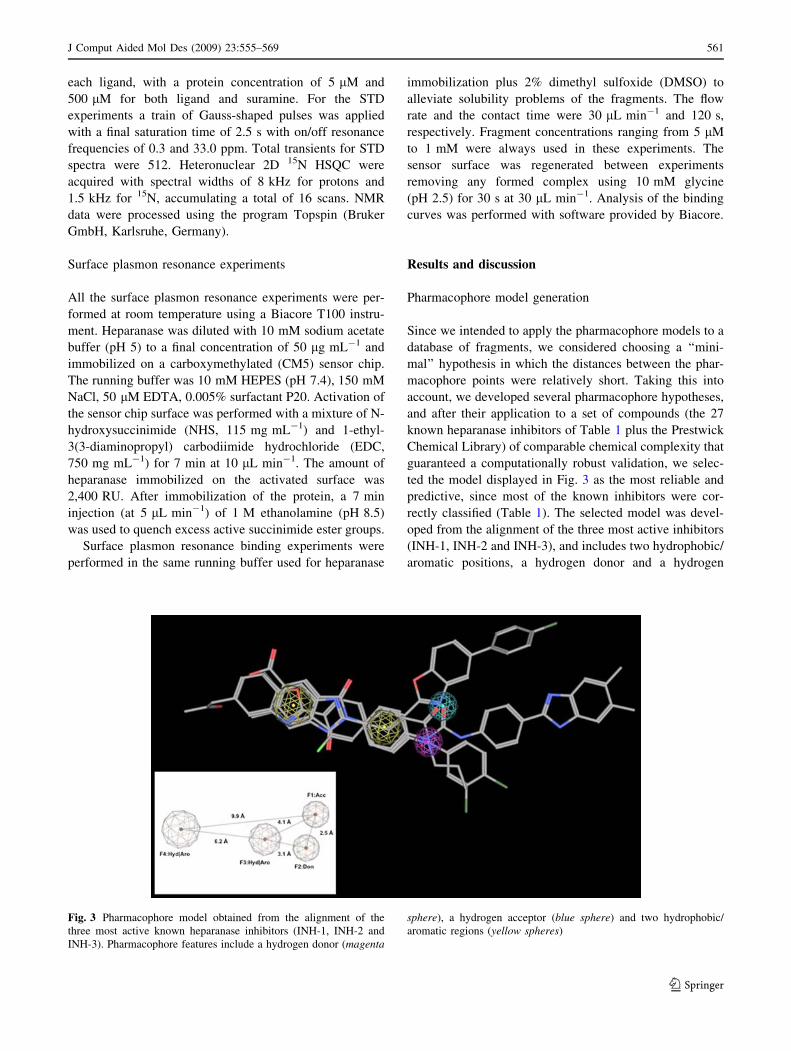
Pharmacophore model generation
Since we intended to apply the pharmacophore models to a
database of fragments, we considered choosing a ‘‘mini-
mal’’ hypothesis in which the distances between the phar-
macophore points were relatively short. Taking this into
account, we developed several pharmacophore hypotheses,
and after their application to a set of compounds (the 27
known heparanase inhibitors of Table 1 plus the Prestwick
Chemical Library) of comparable chemical complexity that
guaranteed a computationally robust validation, we selec-
ted the model displayed in Fig. 3 as the most reliable and
predictive, since most of the known inhibitors were cor-
rectly classified (Table 1). The selected model was devel-
oped from the alignment of the three most active inhibitors
(INH-1, INH-2 and INH-3), and includes two hydrophobic/
aromatic positions, a hydrogen donor and a hydrogen
Fig. 3 Pharmacophore model obtained from the alignment of the
three most active known heparanase inhibitors (INH-1, INH-2 and
INH-3). Pharmacophore features include a hydrogen donor (magenta
sphere), a hydrogen acceptor (blue sphere) and two hydrophobic/
aromatic regions (yellow spheres)
J Comput Aided Mol Des (2009) 23:555–569 561
123
acceptor. The scoring function used for evaluating the
ability of the different models in retrieving the known
inhibitors and ranking them as good candidates was the
root of the mean square deviation (rmsd) between the query
features and their matching ligand annotation points, as
defined by the MOE ‘‘Pharmacophore Search’’ tool.
The selected pharmacophore model was also applied to
a commercial library composed by 686 fragments, finding
that 114 of them (16.6% of the library) satisfied the four-
point pharmacophore query. After evaluating the ‘‘rule of
three’’ for these 114 compounds and comparing the results
with those obtained for the complete fragment library, it
was observed that compounds passing the pharmacophore
filter were globally more complex than the rest of the
library, thus having higher molecular weights as well as
number of hydrogen bond donors and acceptors (Fig. 2).
This is an expected result, specially considering that the
largest distance between any of the pharmacophore points
is nearly 10 A (Fig. 3), thus making unattainable for the
smallest fragments to satisfy this requirement.
Reliability of the pharmacophore model
based on the known inhibitors
As shown in Table 1, twenty-three out of 27 compounds
known to have heparanase inhibitory activity satisfied the
four-point pharmacophore hypothesis, with the model reject-
ing only four compounds, two of them being the ones showing
the lowest inhibitory activity (i.e., highest IC50 values, INH-26
and INH-27). A clear trend of activity was also found, with the
best predicted compounds being the most active ones. Fur-
thermore, we analyzed together the 27 reported inhibitors and
the 1,120 structures from the Prestwick Chemical Library to
evaluate the reliability of the model in retrieving the known
inhibitors and ranking them as the best candidates. The results
showed that 13 out of the 23 known inhibitors passing the
pharmacophore filter were ranked amongst the best 10%
compounds, and that for the eight inhibitors exhibiting sub-
micromolar IC50 values (INH-1 to INH-8), four of them were
amongst the 2% best scored compounds (including three
compounds not used to develop the pharmacophore model),
thus proving that, in the hypothetical context of an in silico
screening campaign, choosing the best scored compounds
would provide a higher percentage of hits.
Interestingly, we also applied the four-point pharmaco-
phore model to the heparan sulfate (Fig. 1), the natural
substrate of heparanase, and found out that this compound
fully satisfied the complete model.
Chemical diversity and chemotype analysis
An interesting issue when applying pharmacophore
approaches is the possibility of identifying structures with
the same pharmacophore features but very different
chemical structures [11] and this also holds true in our
case. As shown in Fig. 4, the pharmacophore model was
also correctly satisfied by other inhibitors (e.g., INH-9 and
INH-10, the best inhibitors from other chemotypes),
despite the fact that the pharmacophore hypothesis was
based on the alignment of three compounds from very
similar chemical series. Fig. 5 shows the structures of the
three fragments that scored best, as well as their superpo-
sition with the pharmacophore model, demonstrating that
the same observation (i.e., different chemical structures
compatible with the same pharmacophore model) could be
extrapolated to the fragment library.
A further analysis of the known inhibitors was also
carried out based on their particular chemical nature. Thus,
Table 1 includes several inhibitors that, at first instance,
could be considered to exhibit a similar behavior against
heparanase taking into account that all of them are char-
acterized by a long aliphatic chain. Nevertheless, the four-
point pharmacophore model was able to adequately predict
that INH-9, INH-18 and INH-19 were effective inhibitors of
heparanase, since they satisfied the four-point criterion, and
INH-26 and INH-27 were in fact ‘‘inactive’’ compounds (or
at least they are the less active from the series, their IC50
being higher than 100 lM). However, this analysis also
shows the limitations of the model since INH-22, the closest
structure to INH-26 and INH-27, did not satisfy the model,
despite the fact of showing an improvement of one log of
activity when compared to the other two compounds. It
seems evident that the slight chemical difference between
these three compounds is not appreciated by the model.
NMR validation of the pharmacophore model
To experimentally validate the pharmacophore model, we
carried out a number of NMR experiments over a collec-
tion of 100 fragments (Table 2) accomplishing totally (50
compounds out of the 114 fragments passing the 4-point
pharmacophore features, i.e., a 44% of that group) or
partially (25 compounds satisfying 3-points, and other 25
fragments covering only 2-points) the pharmacophore
model. The compounds of each subset were selected based
on their chemical diversity with the exception of the group
fulfilling completely the pharmacophore model that inclu-
ded the 18 best compounds scored by the pharmacophore
model, plus 32 chemically diverse compounds accom-
plishing totally the model. The compounds were identified
by their MACCS keys with MOE, and the Tanimoto
algorithm was used for clustering them.
Initially, the validation consisted in the acquisition of a
set of WaterLOGSY experiments over that subset of frag-
ments. This sensitive method allows an easy identification
of molecules binding to the target protein in the lM-range,
562 J Comput Aided Mol Des (2009) 23:555–569
123

and does not require neither labeled nor large amounts of
biomolecule. The results of these studies showed that the
higher the number of pharmacophore criteria fulfilled by the
fragments, the higher the percentage of hits obtained. Thus,
66% of the compounds that satisfied all the pharmacophore
features showed binding activity to heparanase in the
WaterLOGSY studies, representing a 6 and 30% enrich-
ment when compared to compounds accomplishing three or
only two pharmacophore features, respectively.
These results show the potential of this approach in
fragment-based hit generation where it could be useful to
start testing a reduced number of fragments and then to
proceed with a second phase of hit explosion by HTS.
Also, this strategy has additional advantages for those
researchers working in the academic environment where
the possibility of managing big collections of compounds is
somewhat limited.
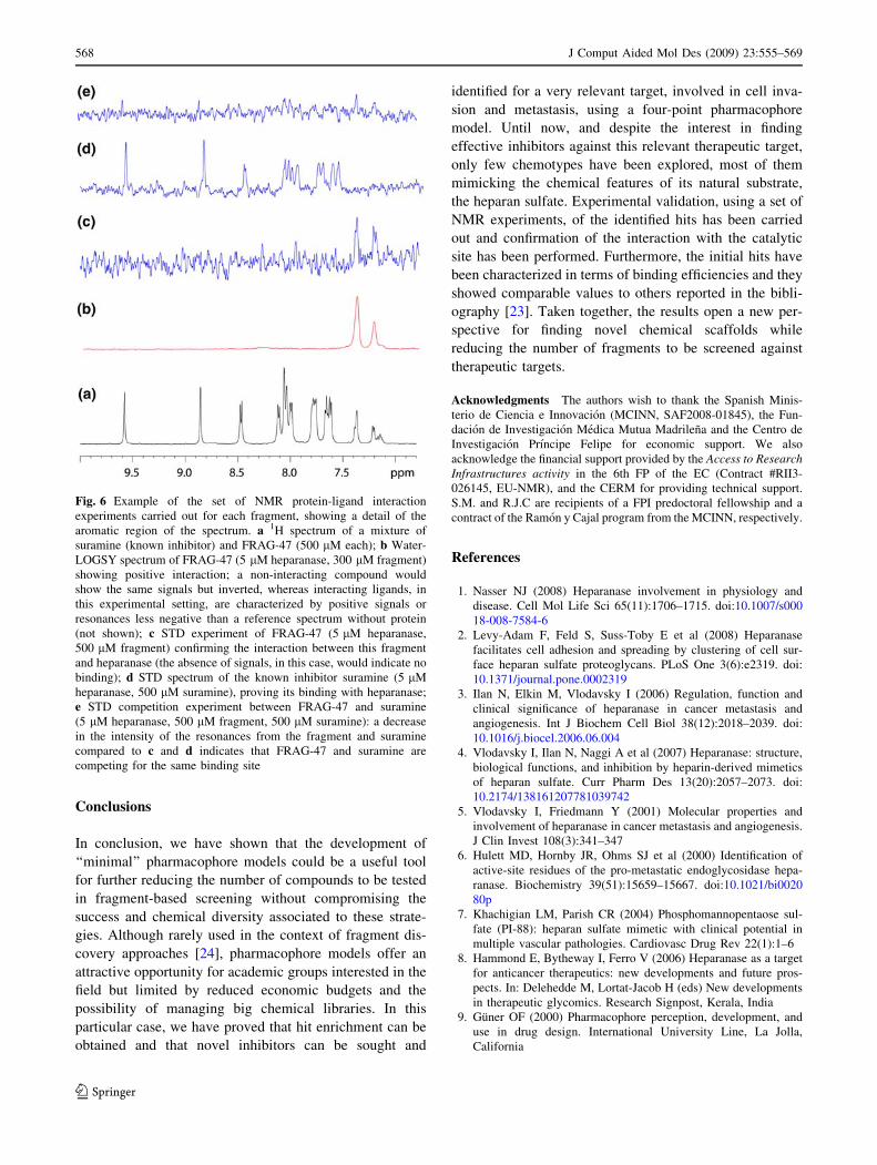
NMR-based hit generation
Although the WaterLOGSY experiment is extremely useful
in the identification of compounds interacting with a protein
target, it has the disadvantage of not easily providing infor-
mation about the binding site. Also, the signal intensity in the
WaterLOGSY experiment greatly depends on the orienta-
tion and solvent accessibility [18], so it is very difficult to
draw conclusions about affinity based on this information.
Thus, and in attempt to identify novel inhibitors against
heparanase, the subset of 100 fragments (Table 2) was
subjected to saturation transfer difference (STD) and com-
petition STD experiments (Fig. 6). The purpose of this
exercise was first to reduce the number of false positives
(WaterLOGSY and STD experiments on their own are error-
prone and, very often, produce false positives due to changes
in the pH, aggregation or non-specific binding [19]) by
Fig. 4 Structures (top) of the
three most active heparanase
inhibitors (from left to right,
INH-1, INH-9 and INH-10)
from different chemical
subfamilies and superposition
with the pharmacophore model
(bottom)
Fig. 5 Structures (top) of the three best scored compounds (from left
to right, FRAG-8, FRAG-28 and FRAG-3) from the fragment library
and superposition with the four-point pharmacophore model (bottom).
As described in the text, the pharmacophore model is compatible with
different chemical structures
J Comput Aided Mol Des (2009) 23:555–569 563
123

Table 2 Subset of 100 fragments selected for the NMR validation of the pharmacophore model
FRAG-1 FRAG-2 FRAG-3 FRAG-4
FRAG-5 FRAG-6 FRAG-7 FRAG-8
FRAG-9 FRAG-10 FRAG-11 FRAG-12
FRAG-13 FRAG-14 FRAG-15 FRAG-16
FRAG-17 FRAG-18 FRAG-19 FRAG-20
FRAG-21 FRAG-22 FRAG-23 FRAG-24
564 J Comput Aided Mol Des (2009) 23:555–569
123

Table 2 continued
FRAG-25 FRAG-26 FRAG-27 FRAG-28
FRAG-29 FRAG-30 FRAG-31 FRAG-32
FRAG-33 FRAG-34 FRAG-35 FRAG-36
FRAG-37 FRAG-38 FRAG-39 FRAG-40
FRAG-41 FRAG-42 FRAG-43 FRAG-44
FRAG-45 FRAG-46 FRAG-47 FRAG-48
J Comput Aided Mol Des (2009) 23:555–569 565
123
Table 2 continued
FRAG-49 FRAG-50 FRAG-51 FRAG-52
FRAG-53 FRAG-54 FRAG-55 FRAG-56
FRAG-57 FRAG-58 FRAG-59 FRAG-60
FRAG-61 FRAG-62 FRAG-63 FRAG-64
FRAG-65 FRAG-66 FRAG-67 FRAG-68
FRAG-69 FRAG-70 FRAG-71 FRAG-72
FRAG-73 FRAG-74 FRAG-75 FRAG-76
FRAG-77 FRAG-78 FRAG-79 FRAG-80
FRAG-81 FRAG-82 FRAG-83 FRAG-84
566 J Comput Aided Mol Des (2009) 23:555–569
123

assessing the interactions by two independent methods [20],
and second to obtain information about the binding site of the
interacting compounds via competition experiments [21].
The competition STD experiments were acquired in the
presence of suramine, a known ligand of heparanase
(KD = 0.5 lM). As it could be expected, when the interac-
tion between the heparanase and the fragments was assessed
by two independent methods (WaterLOGSY and STD
experiments), we found a decrease in the hit rate for the
whole subset. However, the comparison of fragments in the
same size range (MW and topological distance [22]) showed
that there is still a strong correlation between the fulfillment
of the pharmacophore model and the hit rate. Thus, it was
found that 30% of the fragments completely satisfying the
pharmacophore model showed binding activity to heparan-
ase, representing a 9% and 20% enrichment when comparing
to compounds accomplishing three or two pharmacophore
features, respectively (Table 2, bold and underlined). In
some cases, and just for fragments we found to be interacting
with heparanase as assessed both by the WaterLOGSY
and the competition STD experiments, HSQC experiments
were acquired. The HSQC-based chemical shift mapping
experiments proved that all the binders were indeed inter-
acting with the catalytic site of heparanase (data not shown).
Finally, surface plasmon resonance studies were per-
formed to measure the KD of the interactions between the
most promising compounds and heparanase. Fragments for
these experiments were selected based on their overall pro-
file in terms of the response in the WaterLOGSY and com-
petition STD experiments. Most of them show dissociation
constants in the mM range (1-5 mM) and binding efficien-
cies (BEI = pKD/MW (kDa)) equivalent to those reported
[23] for fragments of that size. With only one exception,
FRAG-86 (Table 2), the most promising compounds
belonged to the subset of fragments completely fulfilling the
4-point pharmacophore model. This compound, in particu-
lar, is the largest (MW: 263.7 Da, heavy atoms: 17 atoms,
diameter: 10) of the subset of fragments satisfying two fea-
tures of the pharmacophore model. In fact, when looking in
detail, this fragment is fulfilling almost three out of the four
pharmacophore features. Using this approach, we have
identified several fragments that, based on the biophysical
experiments carried out, could potentially be good starting
points for future hit-to-lead optimizations.
Table 2 continued
FRAG-85 FRAG-86 FRAG-87 FRAG-88
FRAG-89 FRAG-90 FRAG-91 FRAG-92
FRAG-93 FRAG-94 FRAG-95 FRAG-96
FRAG-97 FRAG-98 FRAG-99 FRAG-100
Fragments fulfilling 4 (FRAG-1 to FRAG-50), 3 (FRAG-51 to FRAG-75) and 2 (FRAG-76 to FRAG-100) features of the
pharmacophore model. Marked in bold and underlined are those fragments of comparable chemical complexity found to be
interacting with heparanase as judged by both WaterLOGSY and competition STD experiments
J Comput Aided Mol Des (2009) 23:555–569 567
123
Conclusions
In conclusion, we have shown that the development of
‘‘minimal’’ pharmacophore models could be a useful tool
for further reducing the number of compounds to be tested
in fragment-based screening without compromising the
success and chemical diversity associated to these strate-
gies. Although rarely used in the context of fragment dis-
covery approaches [24], pharmacophore models offer an
attractive opportunity for academic groups interested in the
field but limited by reduced economic budgets and the
possibility of managing big chemical libraries. In this
particular case, we have proved that hit enrichment can be
obtained and that novel inhibitors can be sought and
identified for a very relevant target, involved in cell inva-
sion and metastasis, using a four-point pharmacophore
model. Until now, and despite the interest in finding
effective inhibitors against this relevant therapeutic target,
only few chemotypes have been explored, most of them
mimicking the chemical features of its natural substrate,
the heparan sulfate. Experimental validation, using a set of
NMR experiments, of the identified hits has been carried
out and confirmation of the interaction with the catalytic
site has been performed. Furthermore, the initial hits have
been characterized in terms of binding efficiencies and they
showed comparable values to others reported in the bibli-
ography [23]. Taken together, the results open a new per-
spective for finding novel chemical scaffolds while
reducing the number of fragments to be screened against
therapeutic targets.
Acknowledgments The authors wish to thank the Spanish Minis-
terio de Ciencia e Innovacion (MCINN, SAF2008-01845), the Fun-
dacion de Investigacion Medica Mutua Madrilena and the Centro de
Investigacion Prıncipe Felipe for economic support. We also
acknowledge the financial support provided by the Access to ResearchInfrastructures activity in the 6th FP of the EC (Contract #RII3-
026145, EU-NMR), and the CERM for providing technical support.
S.M. and R.J.C are recipients of a FPI predoctoral fellowship and a
contract of the Ramon y Cajal program from the MCINN, respectively.
References
1. Nasser NJ (2008) Heparanase involvement in physiology and
disease. Cell Mol Life Sci 65(11):1706–1715. doi:10.1007/s000
18-008-7584-6
2. Levy-Adam F, Feld S, Suss-Toby E et al (2008) Heparanase
facilitates cell adhesion and spreading by clustering of cell sur-
face heparan sulfate proteoglycans. PLoS One 3(6):e2319. doi:
10.1371/journal.pone.0002319
3. Ilan N, Elkin M, Vlodavsky I (2006) Regulation, function and
clinical significance of heparanase in cancer metastasis and
angiogenesis. Int J Biochem Cell Biol 38(12):2018–2039. doi:
10.1016/j.biocel.2006.06.004
4. Vlodavsky I, Ilan N, Naggi A et al (2007) Heparanase: structure,
biological functions, and inhibition by heparin-derived mimetics
of heparan sulfate. Curr Pharm Des 13(20):2057–2073. doi:
10.2174/138161207781039742
5. Vlodavsky I, Friedmann Y (2001) Molecular properties and
involvement of heparanase in cancer metastasis and angiogenesis.
J Clin Invest 108(3):341–347
6. Hulett MD, Hornby JR, Ohms SJ et al (2000) Identification of
active-site residues of the pro-metastatic endoglycosidase hepa-
ranase. Biochemistry 39(51):15659–15667. doi:10.1021/bi0020
80p
7. Khachigian LM, Parish CR (2004) Phosphomannopentaose sul-
fate (PI-88): heparan sulfate mimetic with clinical potential in
multiple vascular pathologies. Cardiovasc Drug Rev 22(1):1–6
8. Hammond E, Bytheway I, Ferro V (2006) Heparanase as a target
for anticancer therapeutics: new developments and future pros-
pects. In: Delehedde M, Lortat-Jacob H (eds) New developments
in therapeutic glycomics. Research Signpost, Kerala, India
9. Guner OF (2000) Pharmacophore perception, development, and
use in drug design. International University Line, La Jolla,
California
Fig. 6 Example of the set of NMR protein-ligand interaction
experiments carried out for each fragment, showing a detail of the
aromatic region of the spectrum. a 1H spectrum of a mixture of
suramine (known inhibitor) and FRAG-47 (500 lM each); b Water-
LOGSY spectrum of FRAG-47 (5 lM heparanase, 300 lM fragment)
showing positive interaction; a non-interacting compound would
show the same signals but inverted, whereas interacting ligands, in
this experimental setting, are characterized by positive signals or
resonances less negative than a reference spectrum without protein
(not shown); c STD experiment of FRAG-47 (5 lM heparanase,
500 lM fragment) confirming the interaction between this fragment
and heparanase (the absence of signals, in this case, would indicate no
binding); d STD spectrum of the known inhibitor suramine (5 lM
heparanase, 500 lM suramine), proving its binding with heparanase;
e STD competition experiment between FRAG-47 and suramine
(5 lM heparanase, 500 lM fragment, 500 lM suramine): a decrease
in the intensity of the resonances from the fragment and suramine
compared to c and d indicates that FRAG-47 and suramine are
competing for the same binding site
568 J Comput Aided Mol Des (2009) 23:555–569
123

10. Guner OF (2002) History and evolution of the pharmacophore
concept in computer-aided drug design. Curr Top Med Chem
2(12):1321–1332. doi:10.2174/1568026023392940
11. Wolber G, Seidel T, Bendix F et al (2008) Molecule-pharmaco-
phore superpositioning and pattern matching in computational
drug design. Drug Discov Today 13(1–2):23–29. doi:10.1016/j.
drudis.2007.09.007
12. Wermuth CG, Ganellin CR, Lindberg P et al (1998) Glossary of
terms used in medicinal chemistry (IUPAC Recommendations
1997). Annu Rep Med Chem 33:385–395. doi:10.1016/S0065-77
43(08)61101-X
13. Huang N, Shoichet BK, Irwin JJ (2006) Benchmarking sets for
molecular docking. J Med Chem 49(23):6789–6801. doi:10.1021/
jm0608356
14. Congreve M, Carr R, Murray C et al (2003) A ‘rule of three’ for
fragment-based lead discovery? Drug Discov Today 8(19):876–
877. doi:10.1016/S1359-6446(03)02831-9
15. Dalvit C, Fogliatto G, Stewart A et al (2001) WaterLOGSY as a
method for primary NMR screening: practical aspects and range
of applicability. J Biomol NMR 21(4):349–359. doi:10.1023/A:
1013302231549
16. Hwang T-L, Shaka AJ (1995) Water suppression that works:
excitation sculpting using arbitrary waveforms and pulse field
gradients. J Magn Reson A 112:275–279. doi:10.1006/jmra.1995.
1047
17. Meyer B, Peters T (2003) NMR spectroscopy techniques for
screening and identifying ligand binding to protein receptors.
Angew Chem Int Ed Engl 42(8):864–890. doi:10.1002/anie.2003
90233
18. Ludwig C, Michiels PJA, Wu X et al (2008) SALMON: solvent
accessibility, ligand binding, and mapping of ligand orientation
by NMR spectroscopy. J Med Chem 51(1):1–3. doi:10.1021/jm
701020f
19. Stockman BJ, Kothe M, Kohls D et al (2009) Identification of
allosteric PIF-pocket ligands for PDK1 using NMR-based frag-
ment screening and 1H–15N TROSY experiments. Chem Biol
Drug Des 73(2):179–188. doi:10.1111/j.1747-0285.2008.00768.x
20. Klages J, Coles M, Kessler H (2007) NMR-based screening: a
powerful tool in fragment-based drug discovery. Analyst
132(7):693–705. doi:10.1039/b709658p
21. Siegal G, Ab E, Schultz J (2007) Integration of fragment screening
and library design. Drug Discov Today 12(23–24):1032–1039.
doi:10.1016/j.drudis.2007.08.005
22. Petitjean M (1992) Applications of the radius-diameter diagram
to the classification of topological and geometrical shapes of
chemical compounds. J Chem Inf Comput Sci 32(4):331–337.
doi:10.1021/ci00008a012
23. Dalvit C (2007) Ligand- and substrate-based 19F NMR screening:
principles and applications to drug discovery. Prog Nucl Magn
Reson Spectrosc 51:243–271. doi:10.1016/j.pnmrs.2007.07.002
24. Ji H, Stanton BZ, Igarashi J et al (2008) Minimal pharmacophoric
elements and fragment hopping, an approach directed at molec-
ular diversity and isozyme selectivity. Design of selective neu-
ronal nitric oxide synthase inhibitors. J Am Chem Soc
130(12):3900–3914. doi:10.1021/ja0772041
J Comput Aided Mol Des (2009) 23:555–569 569
123