Frontotemporal dementia and its subtypes: a genome-wide association study
14
686 www.thelancet.com/neurology Vol 13 July 2014 Articles Frontotemporal dementia and its subtypes: a genome-wide association study Raffaele Ferrari*, Dena G Hernandez*, Michael A Nalls*, Jonathan D Rohrer*, Adaikalavan Ramasamy, John B J Kwok, Carol Dobson-Stone, William S Brooks, Peter R Schofield, Glenda M Halliday, John R Hodges, Olivier Piguet, Lauren Bartley, Elizabeth Thompson, Eric Haan, Isabel Hernández, Agustín Ruiz, Mercè Boada, Barbara Borroni, Alessandro Padovani, Carlos Cruchaga, Nigel J Cairns, Luisa Benussi, Giuliano Binetti, Roberta Ghidoni, Gianluigi Forloni, Daniela Galimberti, Chiara Fenoglio, Maria Serpente, Elio Scarpini, Jordi Clarimón, Alberto Lleó, Rafael Blesa, Maria Landqvist Waldö, Karin Nilsson, Christer Nilsson, Ian R A Mackenzie, Ging-Yuek R Hsiung, David M A Mann, Jordan Grafman, Christopher M Morris, Johannes Attems, Timothy D Griffiths, Ian G McKeith, Alan J Thomas, P Pietrini, Edward D Huey, Eric M Wassermann, Atik Baborie, Evelyn Jaros, Michael C Tierney, Pau Pastor, Cristina Razquin, Sara Ortega-Cubero, Elena Alonso, Robert Perneczky, Janine Diehl-Schmid, Panagiotis Alexopoulos, Alexander Kurz, Innocenzo Rainero, Elisa Rubino, Lorenzo Pinessi, Ekaterina Rogaeva, Peter St George-Hyslop, Giacomina Rossi, Fabrizio Tagliavini, Giorgio Giaccone, James B Rowe, Johannes C M Schlachetzki, James Uphill, John Collinge, Simon Mead, Adrian Danek, Vivianna M Van Deerlin, Murray Grossman, John Q Trojanowski, Julie van der Zee, William Deschamps, Tim Van Langenhove, Marc Cruts, Christine Van Broeckhoven, Stefano F Cappa, Isabelle Le Ber, Didier Hannequin, Véronique Golfier, Martine Vercelletto, Alexis Brice, Benedetta Nacmias, Sandro Sorbi, Silvia Bagnoli, Irene Piaceri, Jørgen E Nielsen, Lena E Hjermind, Matthias Riemenschneider, Manuel Mayhaus, Bernd Ibach, Gilles Gasparoni, Sabrina Pichler, Wei Gu, Martin N Rossor, Nick C Fox, Jason D Warren, Maria Grazia Spillantini, Huw R Morris, Patrizia Rizzu, Peter Heutink, Julie S Snowden, Sara Rollinson, Anna Richardson, Alexander Gerhard, Amalia C Bruni, Raffaele Maletta, Francesca Frangipane, Chiara Cupidi, Livia Bernardi, Maria Anfossi, Maura Gallo, Maria Elena Conidi, Nicoletta Smirne, Rosa Rademakers, Matt Baker, Dennis W Dickson, Neill R Graff-Radford, Ronald C Petersen, David Knopman, Keith A Josephs, Bradley F Boeve, Joseph E Parisi , William W Seeley, Bruce L Miller, Anna M Karydas, Howard Rosen, John C van Swieten, Elise G P Dopper, Harro Seelaar, Yolande A L Pijnenburg, Philip Scheltens, Giancarlo Logroscino, Rosa Capozzo, Valeria Novelli, Annibale A Puca, Massimo Franceschi, Alfredo Postiglione, Graziella Milan, Paolo Sorrentino, Mark Kristiansen, Huei-Hsin Chiang, Caroline Graff, Florence Pasquier, Adeline Rollin, Vincent Deramecourt, Florence Lebert, Dimitrios Kapogiannis, Luigi Ferrucci, Stuart Pickering-Brown, Andrew B Singleton†, John Hardy†, Parastoo Momeni† Summary Background Frontotemporal dementia (FTD) is a complex disorder characterised by a broad range of clinical manifestations, differential pathological signatures, and genetic variability. Mutations in three genes—MAPT, GRN, and C9orf72—have been associated with FTD. We sought to identify novel genetic risk loci associated with the disorder. Methods We did a two-stage genome-wide association study on clinical FTD, analysing samples from 3526 patients with FTD and 9402 healthy controls. To reduce genetic heterogeneity, all participants were of European ancestry. In the discovery phase (samples from 2154 patients with FTD and 4308 controls), we did separate association analyses for each FTD subtype (behavioural variant FTD, semantic dementia, progressive non-fluent aphasia, and FTD overlapping with motor neuron disease [FTD-MND]), followed by a meta-analysis of the entire dataset. We carried forward replication of the novel suggestive loci in an independent sample series (samples from 1372 patients and 5094 controls) and then did joint phase and brain expression and methylation quantitative trait loci analyses for the associated (p<5 × 10 – ⁸) single-nucleotide polymorphisms. Findings We identified novel associations exceeding the genome-wide significance threshold (p<5 × 10 – ⁸). Combined (joint) analyses of discovery and replication phases showed genome-wide significant association at 6p21.3, HLA locus (immune system), for rs9268877 (p=1·05 × 10 – ⁸; odds ratio=1·204 [95% CI 1·11–1·30]), rs9268856 (p=5·51 × 10 – ⁹; 0·809 [0·76–0·86]) and rs1980493 (p value=1·57 × 10 – ⁸, 0·775 [0·69–0·86]) in the entire cohort. We also identified a potential novel locus at 11q14, encompassing RAB38/CTSC (the transcripts of which are related to lysosomal biology), for the behavioural FTD subtype for which joint analyses showed suggestive association for rs302668 (p=2·44 × 10 – ⁷; 0·814 [0·71–0·92]). Analysis of expression and methylation quantitative trait loci data suggested that these loci might affect expression and methylation in cis. Interpretation Our findings suggest that immune system processes (link to 6p21.3) and possibly lysosomal and autophagy pathways (link to 11q14) are potentially involved in FTD. Our findings need to be replicated to better define the association of the newly identified loci with disease and to shed light on the pathomechanisms contributing to FTD. Funding The National Institute of Neurological Disorders and Stroke and National Institute on Aging, the Wellcome/ MRC Centre on Parkinson’s disease, Alzheimer’s Research UK, and Texas Tech University Health Sciences Center. Lancet Neurol 2014; 13: 686–99 This online publication has been corrected. The corrected version first appeared at thelancet.com/ neurology on June 17, 2014 See Comment page 643 *Contributed equally †Joint last authors Laboratory of Neurogenetics, Department of Internal Medicine, Texas Tech University Health Science Center, Lubbock, Texas, USA (R Ferrari PhD, P Momeni PhD); Reta Lila Weston Research Laboratories, Department of Molecular Neuroscience, UCL Institute of Neurology, London, UK (R Ferrari, D G Hernandez MSc, J D Rohrer PhD, A Ramasamy PhD, Prof J Hardy PhD); Laboratory of Neurogenetics, National Institute on Aging, National Institutes of Health, Bethesda, MD, USA (D G Hernandez, M A Nalls PhD, A B Singleton PhD); Clinical Research Branch, National Institute on Aging, Baltimore, MD, USA (L Ferrucci MD); Institute of Brain, Behaviour and Mental Health, Faculty of Medical and Human Sciences, University of Manchester, Manchester, UK
-
Upload
independent -
Category
Documents
-
view
1 -
download
0
Transcript of Frontotemporal dementia and its subtypes: a genome-wide association study

686 www.thelancet.com/neurology Vol 13 July 2014
Articles
Frontotemporal dementia and its subtypes: a genome-wide association studyRaff aele Ferrari*, Dena G Hernandez*, Michael A Nalls*, Jonathan D Rohrer*, Adaikalavan Ramasamy, John B J Kwok, Carol Dobson-Stone, William S Brooks, Peter R Schofi eld, Glenda M Halliday, John R Hodges, Olivier Piguet, Lauren Bartley, Elizabeth Thompson, Eric Haan, Isabel Hernández, Agustín Ruiz, Mercè Boada, Barbara Borroni, Alessandro Padovani, Carlos Cruchaga, Nigel J Cairns, Luisa Benussi, Giuliano Binetti, Roberta Ghidoni, Gianluigi Forloni, Daniela Galimberti, Chiara Fenoglio, Maria Serpente, Elio Scarpini, Jordi Clarimón, Alberto Lleó, Rafael Blesa, Maria Landqvist Waldö, Karin Nilsson, Christer Nilsson, Ian R A Mackenzie, Ging-Yuek R Hsiung, David M A Mann, Jordan Grafman, Christopher M Morris, Johannes Attems, Timothy D Griffi ths, Ian G McKeith, Alan J Thomas, P Pietrini, Edward D Huey, Eric M Wassermann, Atik Baborie, Evelyn Jaros, Michael C Tierney, Pau Pastor, Cristina Razquin, Sara Ortega-Cubero, Elena Alonso, Robert Perneczky, Janine Diehl-Schmid, Panagiotis Alexopoulos, Alexander Kurz, Innocenzo Rainero, Elisa Rubino, Lorenzo Pinessi, Ekaterina Rogaeva, Peter St George-Hyslop, Giacomina Rossi, Fabrizio Tagliavini, Giorgio Giaccone, James B Rowe, Johannes C M Schlachetzki, James Uphill, John Collinge, Simon Mead, Adrian Danek, Vivianna M Van Deerlin, Murray Grossman, John Q Trojanowski, Julie van der Zee, William Deschamps, Tim Van Langenhove, Marc Cruts, Christine Van Broeckhoven, Stefano F Cappa, Isabelle Le Ber, Didier Hannequin, Véronique Golfi er, Martine Vercelletto, Alexis Brice, Benedetta Nacmias, Sandro Sorbi, Silvia Bagnoli, Irene Piaceri, Jørgen E Nielsen, Lena E Hjermind, Matthias Riemenschneider, Manuel Mayhaus, Bernd Ibach, Gilles Gasparoni, Sabrina Pichler, Wei Gu, Martin N Rossor, Nick C Fox, Jason D Warren, Maria Grazia Spillantini, Huw R Morris, Patrizia Rizzu, Peter Heutink, Julie S Snowden, Sara Rollinson, Anna Richardson, Alexander Gerhard, Amalia C Bruni, Raff aele Maletta, Francesca Frangipane, Chiara Cupidi, Livia Bernardi, Maria Anfossi, Maura Gallo, Maria Elena Conidi, Nicoletta Smirne, Rosa Rademakers, Matt Baker, Dennis W Dickson, Neill R Graff -Radford, Ronald C Petersen, David Knopman, Keith A Josephs, Bradley F Boeve, Joseph E Parisi , William W Seeley, Bruce L Miller, Anna M Karydas, Howard Rosen, John C van Swieten, Elise G P Dopper, Harro Seelaar, Yolande A L Pijnenburg, Philip Scheltens, Giancarlo Logroscino, Rosa Capozzo, Valeria Novelli, Annibale A Puca, Massimo Franceschi, Alfredo Postiglione, Graziella Milan, Paolo Sorrentino, Mark Kristiansen, Huei-Hsin Chiang, Caroline Graff , Florence Pasquier, Adeline Rollin, Vincent Deramecourt, Florence Lebert, Dimitrios Kapogiannis, Luigi Ferrucci, Stuart Pickering-Brown, Andrew B Singleton†, John Hardy†, Parastoo Momeni†
SummaryBackground Frontotemporal dementia (FTD) is a complex disorder characterised by a broad range of clinical manifestations, diff erential pathological signatures, and genetic variability. Mutations in three genes—MAPT, GRN, and C9orf72—have been associated with FTD. We sought to identify novel genetic risk loci associated with the disorder.
Methods We did a two-stage genome-wide association study on clinical FTD, analysing samples from 3526 patients with FTD and 9402 healthy controls. To reduce genetic heterogeneity, all participants were of European ancestry. In the discovery phase (samples from 2154 patients with FTD and 4308 controls), we did separate association analyses for each FTD subtype (behavioural variant FTD, semantic dementia, progressive non-fl uent aphasia, and FTD overlapping with motor neuron disease [FTD-MND]), followed by a meta-analysis of the entire dataset. We carried forward replication of the novel suggestive loci in an independent sample series (samples from 1372 patients and 5094 controls) and then did joint phase and brain expression and methylation quantitative trait loci analyses for the associated (p<5 × 10–⁸) single-nucleotide polymorphisms.
Findings We identifi ed novel associations exceeding the genome-wide signifi cance threshold (p<5 × 10–⁸). Combined (joint) analyses of discovery and replication phases showed genome-wide signifi cant association at 6p21.3, HLA locus (immune system), for rs9268877 (p=1·05 × 10–⁸; odds ratio=1·204 [95% CI 1·11–1·30]), rs9268856 (p=5·51 × 10–⁹; 0·809 [0·76–0·86]) and rs1980493 (p value=1·57 × 10–⁸, 0·775 [0·69–0·86]) in the entire cohort. We also identifi ed a potential novel locus at 11q14, encompassing RAB38/CTSC (the transcripts of which are related to lysosomal biology), for the behavioural FTD subtype for which joint analyses showed suggestive association for rs302668 (p=2·44 × 10–⁷; 0·814 [0·71–0·92]). Analysis of expression and methylation quantitative trait loci data suggested that these loci might aff ect expression and methylation in cis.
Interpretation Our fi ndings suggest that immune system processes (link to 6p21.3) and possibly lysosomal and autophagy pathways (link to 11q14) are potentially involved in FTD. Our fi ndings need to be replicated to better defi ne the association of the newly identifi ed loci with disease and to shed light on the pathomechanisms contributing to FTD.
Funding The National Institute of Neurological Disorders and Stroke and National Institute on Aging, the Wellcome/MRC Centre on Parkinson’s disease, Alzheimer’s Research UK, and Texas Tech University Health Sciences Center.
Lancet Neurol 2014; 13: 686–99
This online publication has been corrected.
The corrected version fi rst appeared at thelancet.com/neurology on June 17, 2014
See Comment page 643
*Contributed equally
†Joint last authors
Laboratory of Neurogenetics, Department of Internal
Medicine, Texas Tech University Health Science Center, Lubbock,
Texas, USA (R Ferrari PhD, P Momeni PhD); Reta Lila
Weston Research Laboratories, Department of Molecular
Neuroscience, UCL Institute of Neurology, London, UK
(R Ferrari, D G Hernandez MSc, J D Rohrer PhD, A Ramasamy PhD, Prof J Hardy PhD); Laboratory of
Neurogenetics, National Institute on Aging, National
Institutes of Health, Bethesda, MD, USA (D G Hernandez,
M A Nalls PhD, A B Singleton PhD); Clinical Research Branch, National
Institute on Aging, Baltimore, MD, USA (L Ferrucci MD);
Institute of Brain, Behaviour and Mental Health, Faculty of Medical and Human Sciences,
University of Manchester, Manchester, UK

Articles
www.thelancet.com/neurology Vol 13 July 2014 687
IntroductionFrontotemporal dementia (FTD) is the second most common form of young-onset dementia after Alzheimer’s disease and comprises about 10–20% of all dementias worldwide.1 FTD occurs in about three to 15 per 100 000 individuals aged between 55 years and 65 years.2 The disease has a slow and subtle onset: it is familial in 30–50% of patients and aff ects men and women almost equally.3 The main clinical syndromes are the behavioural variant1,4 and the language variants (semantic dementia and progressive nonfl uent aphasia).1,5 FTD can also co-occur with motor neuron disease (FTD-MND), and atypical parkinsonian disorders.3 The molecular pathology is heterogeneous and based on the type of neuronal lesions and protein inclusions: 40% or more of patients have frontotemporal lobar degeneration (FTLD) with tau pathology (FTLD-tau), about 50% have TDP-43 (TAR DNA-binding protein 43) pathology (FTLD-TDP),6 and the remaining 10% have inclusions positive for fused in sarcoma (FUS; FTLD-FUS) or ubiquitin/p62 (FTLD-UPS [ubiquitin proteasome system]).7 Mutations in three main genes are commonly associated with FTD: the microtubule-associated protein tau (MAPT),8 granulin (GRN),9,10 and C9orf72.11–15 Mutations in the charged multivesicular body protein 2B (CHMP2B), the valosin-containing protein (VCP), and ubiquilin 2 (UBQLN2) genes are rare causes of disease.13,16 Findings from a previous genome-wide association study (GWAS) of neuropathologically confi rmed FTLD-TDP (515 patients vs 2509 controls) showed TMEM106B to be a disease risk factor.17
We did a larger GWAS in samples from people with clinical FTD, and we report results for the discovery, replication, and joint-phase analyses, as well as for assessment of the eff ect on expression and methylation quantitative trait loci (QTL) exerted by associated or suggestive SNPs. We aimed to identify novel genetic risk loci associated with FTD and its subtypes.
MethodsStudy population 44 international research groups (appendix) contributed samples to this two-stage (discovery phase and replication phase) GWAS of clinical FTD. The patients included in the discovery phase were diagnosed according to the Neary criteria1 for FTD, whereas those included in the replication phase were diagnosed according to the Neary criteria,1 or the revised criteria for behavioural FTD4 and the language variants of FTD5 at every collaborative site. For each patient, the diagnosis was made by a neurologist with an interest in FTD or, in a minority (70 [3%] of 2621 patients), by pathological diagnosis. To cover the most relevant FTD clinical signatures, we included patients diagnosed with behavioural FTD, semantic dementia, progressive nonfl uent aphasia, or FTD-MND.18 We reviewed all patients with a diagnosis of language impairment to exclude cases of the logopenic variant of
primary progressive aphasia,5 most of which are associated with Alzheimer’s disease pathology. Samples were obtained from Australia, Belgium, Denmark, France, Germany, Italy, the Netherlands, North America (USA and Canada), Spain, Sweden, and the UK and all patients were of confi rmed European ancestry (to reduce genetic heterogeneity).
DNA was collected at the three institutions leading this project: the Department of Molecular Neuroscience at University College London (UCL), UK; the Laboratory of Neurogenetics of the National Institute on Aging at the National Institutes of Health (NIH), MD, USA; and the Laboratory of Neurogenetics at the Texas Tech University Health Sciences Center (TTUHSC), TX, USA. All samples were anonymous and stored with a patient-specifi c coded identifi cation number. Each DNA sample was assessed for quality with gel electrophoresis and DNA concentrations were assessed via spectrophotometer (Nanodrop; Wilmington, DE, USA) or fl uorometer (Qubit; Life Technologies, Grand Island, NY, USA). Samples from non-overlapping patients were genotyped at the Laboratory of Neurogenetics of the National Institute on Aging, NIH (40%), or at the core facility at the Institute of Child Health, UCL (60%). We obtained standardised clinical, pathological, and genetic data for each patient from all the collaborating groups (appendix). Sporadic cases along with probands from FTD families were included in the study. We excluded carriers of mutations in MAPT and GRN. We did not exclude individuals with C9orf72 expansions because this locus was identifi ed subsequent to sample collection. After quality control of genotyping data and detailed assessment of the clinical diagnosis, we used 2154 and 1372 samples in the discovery phase and replication phase, respectively, for association analysis (table 1). In total, after quality control, we analysed 3526 FTD samples (table 1). Further details about cases included in the study are provided in the appendix.
Control samples for the discovery phase were taken from studies previously done at the Laboratory of Neurogenetics of the National Institute on Aging at the NIH or at UCL. Control individuals were matched to patients on the basis of population ancestry and genotyping platform. Aggregate data for control samples were merged based on overlapping single-nucleotide polymorphisms (SNPs). The selected 7444 control samples were from France, Germany, Italy, the Netherlands, Sweden, the UK, and USA, and were used as controls in previous GWAS;19 all individuals had given consent for their samples to be used as controls. All were free of neurological illness at the time of sampling, but most had not been screened for the absence of a family history of FTD. For each patient, at least two controls were matched based on compatibility of genetic ancestry estimates by principal components analysis to accommodate the lack of precisely matched clinical controls. After quality control, we included 4308
(Prof J S Snowden PhD, S Rollinson PhD, Prof S Pickering-Brown PhD); Neuroscience Research Australia, Sydney, NSW, Australia (J B J Kwok PhD, C Dobson-Stone PhD, W S Brooks MBBS, Prof P R Schofi eld DSc, Prof G M Halliday PhD, Prof J R Hodges MD, O Piguet PhD, L Bartley MSc); University of New South Wales, Sydney, NSW, Australia (J B J Kwok, C Dobson-Stone, W S Brooks, Prof P R Schofi eld, Prof G M Halliday, Prof J R Hodges, O Piguet); South Australian Clinical Genetics Service, SA Pathology at Women’s and Children’s Hospital, North Adelaide, SA, Australia (E Thompson MD, Prof E Haan MBBS); Department of Paediatrics, University of Adelaide, Adelaide, SA, Australia (E Thompson, Prof E Haan); Memory Clinic of Fundació ACE, Institut Català de Neurociències Aplicades, Barcelona, Spain (I Hernández MD, A Ruiz MD, M Boada MD); Hospital Universitari Vall d’Hebron–Institut de Recerca, Universitat Autonoma de Barcelona (VHIR-UAB), Barcelona, Spain (M Boada); Neurology Clinic, University of Brescia, Brescia, Italy (B Borroni MD, Prof A Padovani MD); Department of Psychiatry (C Cruchaga PhD), Hope Center (C Cruchaga, Prof N J Cairns PhD), Washington University School of Medicine, St Louis, Missouri, USA; Department of Pathology and Immunology, Washington University, St Louis, Missouri, USA (Prof N J Cairns); NeuroBioGen Lab—Memory Clinic, IRCCS Istituto Centro San Giovanni di Dio Fatebenefratelli, Brescia, Italy (L Benussi PhD, G Binetti MD); Proteomics Unit, IRCCS Istituto Centro San Giovanni di Dio Fatebenefratelli, Brescia, Italy (R Ghidoni PhD); Biology of Neurodegenerative Disorders, IRCCS Istituto di Ricerche Farmacologiche Mario Negri, Milano, Italy (G Forloni PhD); University of Milan, Milan, Italy (D Galimberti PhD, C Fenoglio PhD, M Serpente PhD, E Scarpini MD); Fondazione Cà Granda, IRCCS Ospedale Maggiore Policlinico, Milan, Italy (D Galimberti, C Fenoglio, M Serpente, E Scarpini); Memory Unit, Neurology Department

Articles
688 www.thelancet.com/neurology Vol 13 July 2014
and Sant Pau Biomedical Research Institute, Hospital de
la Santa Creu i Sant Pau, Universitat Autònoma de
Barcelona, Barcelona, Spain (J Clarimón PhD, A Lleó MD,
R Blesa MD); Center for Networker Biomedical Research
in Neurodegenerative Diseases (CIBERNED), Madrid, Spain
(J Clarimón, A Lleó, R Blesa, P Pastor MD,
S Ortega-Cubero MD); Unit of Geriatric Psychiatry
(M L Waldö MD, K Nilsson PhD), Clinical Memory Research Unit (C Nilsson PhD), Department of
Clinical Sciences, Lund University, Sweden;
Department of Pathology and Laboratory Medicine, University of British Columbia, Vancouver,
Canada (Prof I R A Mackenzie MD);
Division of Neurology, University of British Columbia,
Vancouver, Canada (G-Y R Hsiung MD); Institute of
Brain, Behaviour and Mental Health, University of
Manchester, Salford Royal Hospital, Stott Lane, Salford,
UK (Prof D M A Mann PhD); Rehabilitation Institute of
Chicago, Departments of Physical Medicine and
Rehabilitation, Psychiatry, and Cognitive Neurology and
Alzheimer’s Disease Center; Feinberg School of Medicine, Northwestern University, IL,
USA (Prof J Grafman PhD, C M Morris PhD, Prof J Attems
MD, Prof T D Griffi ths FMedSci); Department of Psychology,
control samples in this study. The genotyping of control samples for the replication phase was done at the Laboratory of Neurogenetics of the National Institute on Aging, NIH (4594 [90%] of 5094) and at the core facility at the Institute of Child Health, UCL (500 [10%] of 5094). All control samples used in the replication phase were collected from the groups participating in the study (5094 samples passed quality control) and were of European ancestry from the following countries: France, Germany, Italy, the Netherlands, Spain, Sweden, the UK, and USA.
Investigators at every site obtained written informed consent from patients and control individuals. Every participating group provided consent for the use of these samples for the purposes of this study. Each study site obtained approval from a local ethics committee (UK ethics committee number 10/H0716/3) or institutional research ethics board.
ProceduresFor every sample, 2 μg of DNA extracted from either blood or the brain at each collaborative site was collected (whole genome amplifi ed DNA samples were excluded). Samples were securely stored at –20°C. Every sample was fi rst screened for integrity and purity by means of gel electrophoresis on 1% agarose gel and concentrations were analysed by spectrophotometric (Nanodrop) or fl uorometric (Qubit) quantifi cation. The same procedure was implemented at NIH, UCL, and TTUHSC.
Samples from patients and control individuals included in the discovery phase were genotyped using Illumina human 370K, 550K, and 660K Quad Beadchips and Omni Express chips (Illumina Inc, CA, USA). We used Illumina NeuroX custom chips for all samples included in replication phase genotyping. The NeuroX chip is a partially custom-designed chip that specifi cally
targets the main loci associated with several diff erent neurological disorders obtained from GWAS or whole-exome sequencing data. The NeuroX chip holds about 267K SNPs, of which 3759 were FTD-specifi c, being selected from SNPs that had p values of less than 1 × 10–⁴ during the discovery phase of the study. These SNPs were tag SNPs based on European ancestry linkage-disequilibrium patterns from the most up-to-date data for samples of European ancestry from the 1000 Genomes project.20 For all GWAS signifi cant hits and candidate SNPs, fi ve linkage-disequilibrium-based proxies or technical replicates were included on the array per locus, tagging associations within +/–250 kb and r² >0·5 from the most strongly associated proximal SNP. To replicate each locus, we picked the tag SNP most signifi cant in the discovery phase; if no linkage-disequilibrium-based proxies were available, technical replicates were included. All genotyping arrays (discovery phase and replication phase) were assayed on the Illumina Infi nium platform (Illumina, San Diego, CA, USA) at the Laboratory of Neurogenetics of the National Institute on Aging, NIH and at the core facility at the Institute of Child Health, UCL. All genotypes for this project were called centrally using Illumina Genome Studio and all 3759 SNPs of interest for FTD were manually examined to ensure high-quality genotype clusters before data export.
For the purpose of assessing possible biological relevance for any associated SNPs we used quantitative trait loci (QTL) data generated by the UK Brain Expression Consortium (UKBEC) and the North American Brain Expression Consortium (NABEC) for brain tissues assayed for genome-wide expression and methylation. Details about sample collection, RNA and DNA extraction, and genotyping are provided in the appendix.
Samples collected (n) Samples included in analysis (n) Samples from women (% [n/N]) Mean age at onset (years [range]; N)
Discovery phase
Replication phase
Total Discovery phase
Replication phase
Total Discovery phase Replication phase Discovery phase Replication phase
Australia 0 138 138 0 121 121 NA 36% (44/121) NA 59 (32–77); 112
Belgium 240 51 291 191 42 233 46% (88/191) 29% (12/42) 63 (29–90); 191 64 (43–84); 42
Canada 25 37 62 24 29 53 52% (12/23) 57% (8/14) 64 (43–85); 15 59 (43–75); 9
Denmark 35 0 35 7 0 7 71% (5/7) NA 57 (40–62); 7 NA
France 238 54 292 205 42 247 44% (91/205) 48% (20/42) 62 (39–79); 190 NA
Germany 349 34 383 320 33 353 NA 50% (15/30) 61 (36–83); 243 57 (29–72); 30
Italy 1035 563 1598 564 371 935 53% (297/561) 45% (168/371) 64 (31–83); 429 65 (31–87); 353
Netherlands 333 93 426 250 77 327 52 (129/250) 40% (31/77) 58 (29–76); 250 61 (51–69); 59
Spain 100 330 430 0 309 309 NA 43% (133/309) NA 65 (32–89); 308
Sweden 26 112 138 18 98 116 56% (10/18) 61% (60/98) 57 (38–75); 16 62 (28–78); 93
UK 494 372 866 401 284 685 43% (171/400) 40% (108/272) 60 (23–83); 372 61 (35–86); 167
USA 706 209 915 579 175 754 44% (257/579) 49% (85/174) 60 (23–85); 520 63 (24–93); 120
Total 3581 1993 5574 2559 (2154*) 1581 (1372*) 4140 (3526*) 47% (1186/2552) 44% (684/1550) 61 (23–90); 2233 62 (24–93); 1293
NA=not applicable. *The number of the samples that passed genotyping data quality control and were used for association analyses.
Table 1: Sample characteristics

Articles
www.thelancet.com/neurology Vol 13 July 2014 689
Weinberg College of Arts and Sciences, Northwestern University, IL, USA (Prof J Grafman); Newcastle Brain Tissue Resource, Institute for Ageing and Health, Newcastle University, Newcastle upon Tyne, UK (C M Morris, Prof J Attems MD, Prof T D Griffi ths FMedSci); Newcastle University, Institute for Ageing and Health, Campus for Ageing and Vitality, Newcastle upon Tyne, UK (C M Morris, Prof J Attems MD, Prof A J Thomas PhD, E Jaros PhD); Institute of Neuroscience, Newcastle University Medical School, Newcastle upon Tyne, UK (C M Morris, Prof T D Griffi ths); Biomedical Research Building, Campus for Ageing and Vitality, Newcastle University, Newcastle upon Tyne, UK (Prof I G McKeith MD); Clinical Psychology Branch, Pisa University Hospital, Pisa, Italy, Laboratory of Clinical Biochemistry and Molecular Biology, University of Pisa, Pisa, Italy (Prof P Pietrini MD); Taub Institute, Departments of Psychiatry and Neurology, Columbia University, New York, NY, USA 10032 (E D Huey MD); Behavioral Neurology Unit, National Institute of Neurological Disorders and Stroke, National Institutes of Health, Bethesda, MD, USA (E M Wassermann MD, M C Tierney MSc);
Statistical analysis We did standard quality control for GWAS data before association analyses. Briefl y, for the discovery phase, we extracted overlapping SNPs across all Illumina arrays used. This was done as a means of dealing with the low numbers of matched cases and controls per study site or chip type to facilitate the FTD subtype analyses. We maximised sample size for the subtype analyses by pooling as many possible samples while sacrifi cing some array content, leaving 228 189 autosomal SNPs as a basis for imputation after the quality control was completed. We excluded samples possibly mismatched for sex by assessing X chromosome heterozygosity. Samples with a call rate of greater than 95% and SNPs with a minor allele frequency greater than 1% were fi ltered and included in the analyses. We calculated Hardy-Weinberg equilibrium p values (exclusion at p values <1 × 10–⁵). We assessed non-random missingness per SNP by case-control status with exclusion at p values of less than 1×10–⁵ and non-random missingness per SNP by haplotype at p values for exclusion <1 × 10–⁵. We assessed the presence of relatedness by identifying and excluding fi rst-degree relatives (through identity by descent for any pairwise with an estimate of less than 0·125) and verifi ed European ancestry by principal components analysis compared with HapMap3 populations, with European ancestry ascertained at values for the fi rst two eigenvectors less than six SDs from the population mean for the combined Europeans from Utah (CEU) and Tuscans from Italy (TSI) reference samples.21 After preliminary quality assessment, principal components analysis as implemented in EIGENSTRAT22 was used to assess matching between cases and controls based on all available cases and
controls. Custom coding in R (version 2.7) was used to match cases to controls. We treated each subtype (behavioural FTD, semantic dementia, progressive nonfl uent aphasia, and FTD-MND) as a separate group in which the two most genetically similar unique controls per case were selected based on eigenvectors 1 and 2 to compensate for a lack of precisely matched controls at recruitment. In this respect, matched controls were unique per case and non-redundant across subtype datasets. Thus, cases and controls were matched for each subtype (behavioural FTD, semantic dementia, progressive nonfl uent aphasia, and FTD-MND) based on similarity of the fi rst two eigenvectors from principal components analysis and did not overlap across subtypes. We used logistic regression based on imputed dosages to assess the association between each SNP and any of the FTD subtypes, adjusting for eigenvectors 1 and 2 from principal components analysis as covariates. Eigenvectors were generated separately for each subtype and, as in the overall sample pool, parameter estimates for the fi rst two were associated with case status at p values of less than 0·05. We did fi xed-eff ects meta-analyses to combine results across subtypes and quantify heterogeneity across subtypes. Genomic infl ation was minimal across subtypes and in the meta analysis across subtypes (λ<1·05), therefore we did not use genomic control (see appendix for quantile-quantile plots and λ values per discovery phase analysis). SNPs were imputed to August, 2010 release of the 1000 Genomes haplotypes using default settings of minimac and were excluded if their minor allele frequency was less than 0·01 or imputation quality (Rsq) was less than 0·30 across all samples, leaving 6 026 385 SNPs for analyses. We used PLINK (version 1.07) for statistical analyses.
Behavioural variant frontotemporal dementia
Semantic dementia Progressive nonfl uent aphasia Frontotemporal dementia with motor neuron disease
Frontotemporal lobar degeneration (unspecifi ed)
Discovery phase
Replication phase
Total Discovery phase
Replication phase
Total Discovery phase
Replication phase
Total Discovery phase
Replication phase
Total Discovery phase
Replication phase
Total
Australia* NA 56 56 NA 26 26 NA 19 19 NA 20 20 NA 0 0
Belgium 135 27 162 13 1 14 22 2 24 21 2 23 0 10 10
Canada 22 5 27 1 1 2 0 5 5 1 7 8 0 11 11
Denmark 2 NA 2 0 NA 0 1 NA 1 4 NA 4 0 NA 0
France 135 30 165 3 0 3 8 3 11 59 8 67 0 1 1
Germany 209 18 227 45 8 53 55 6 61 11 1 12 0 0 0
Italy 443 186 629 28 22 50 69 86 155 24 16 40 0 61 61
Netherlands 159 37 196 47 31 78 24 6 30 20 3 23 0 0 0
Spain NA 194 194 NA 41 41 NA 51 51 NA 13 13 NA 10 10
Sweden 7 53 60 2 20 22 6 10 16 3 8 11 0 7 7
UK* 207 152 359 75 53 128 69 44 113 50 16 66 0 19 19
USA 315 25 340 147 12 159 81 15 96 36 21 57 0 102 102
Total 1634 (1377†)
783 (690†)
2417 (2061†)
361 (308†)
215 (190†)
576 (495†)
335 (269†)
247 (221†)
582 (486†)
229 (200†)
115 (94†)
344 (294†)
0 221 (177†)
221 (177†)
NA=not applicable. *Used the same control samples. †The number of the samples that passed genotyping data quality control and were used for association analyses.
Table 2: Sample characteristics, by subtype

Articles
690 www.thelancet.com/neurology Vol 13 July 2014
Neuropathology Department, Walton Centre FT, Liverpool, UK
(A Baborie MD); Neuropathology/Cellular Pathology, Royal Victoria
Infi rmary, Newcastle upon Tyne, UK (E Jaros);
Neurogenetics Laboratory, Division of Neurosciences, Center for Applied Medical
Research, Universidad de Navarra, Pamplona, Spain
(P Pastor, C Razquin PhD, S Ortega-Cubero, E Alonso BSc);
Department of Neurology, Clínica Universidad de Navarra, University of Navarra School of
Medicine, Pamplona, Spain (P Pastor); Neuroepidemiology
and Ageing Research Unit, School of Public Health, Faculty
of Medicine, The Imperial College of Science, Technology
and Medicine, London, UK (R Perneczky MD); West London Cognitive Disorders Treatment
and Research Unit, West London Mental Health Trust,
London TW8 8 DS, UK (R Perneczky); Department of
Psychiatry and Psychotherapy, Technische Universität
München, Munich, Germany (R Perneczky, J Diehl-Schmid MD,
P Alexopoulos MD, A Kurz MD); Neurology I, Department of Neuroscience, University of Torino, Italy, AO Città della
Salute e della Scienza di Torino, Italy (I Rainero MD, E Rubino MD,
Prof L Pinessi MD); Tanz Centre for Research in
Neurodegenerative Diseases and Department of Medicine,
University of Toronto, Toronto, Ontario, Canada (E Rogaeva PhD,
P St George-Hyslop MD); Cambridge Institute for Medical
Research and the Department of Clinical Neurosciences, University of Cambridge,
Cambridge, UK (P St George-Hyslop); Division of
Neurology V and Neuropathology, Fondazione
IRCCS Istituto Neurologico Carlo Besta, Milano, Italy (G Rossi PhD,
F Tagliavini MD, G Giaccone MD); Cambridge University
Department of Clinical Neurosciences, Cambridge CB2
0SZ, UK (J B Rowe PhD); MRC Cognition and Brain Sciences
Unit, Cambridge, UK (J B Rowe); Behavioural and Clinical Neuroscience Institute,
Cambridge, UK (J B Rowe); Department of Psychiatry and
Psychotherapy, University of Freiburg Medical School,
Germany
Figure 1: Manhattan plots identifying regions with genome-wide signifi cant associations(A) Single–nucleotide polymorphisms (SNPs) with genome-wide signifi cant p values (p<5×10–⁸) are depicted as red dots and locate to 6p21.3. The associated SNPs map to intron 5 of BTNL2 and to the intergenic region between HLA-DRA and HLA-DRB. (B) Manhattan plot for the behavioural variant frontotemporal dementia subtype in the discovery phase. SNPs with genome-wide signifi cant p values (p<5×10–⁸) are depicted as red dots and locate to 11q14. The associated SNPs map to intron 1 of RAB38 and to the intergenic region between RAB38 and CTSC.
1
p22.2 p21.2 p21.1 p12.3 p12.1 q12 q13 q14.1 q15 q16.1 q16.3 q21 q22.31 q23.2 q24.1 q25.3 q26 q27q23.3
Chromosome 6:32 360 000 32 380 000 32 400 000 32 420 000 32 440 000 32 460 000 32 480 000 32 500 000
2 3 4 5 6 7 8 9 10 11 13 15 17 19 220
56789
10
–log 10
(P)
Chromosome
Chromosome 6
A Subtype meta-analysis
BTNL2
rs1980493
BTNL2NM_019602·1
rs9268856 rs9268877
HLA-DRA HLA-DRB5
HLA-DRANM_061984·2
HLA-DRB9NM_019111·4
HLA-DRB5NP_002116·2
1
p15.4 p15.2 p15.1 p14.3 p14.1 p13 p12 p11.2 q11 q12.1 q13.4 q14.1 q14.3 q21 q22.3 q23.3 q24.3q24.2 q25q22.1
Chromosome 11:87 850 000 87 900 000 87 950 000 88 000 000 88 050 000 88 100 000
2 3 4 5 6 7 8 9 10 11 12 13 15 17 19 220
5678
–log 10
(P)
Chromosome
Chromosome 11
B Behavioural variant frontotemporal dementia
RAB38
RAB38NM_022337·2
rs302652 rs74977128
CTSC
CTSC

Articles
www.thelancet.com/neurology Vol 13 July 2014 691
(J C M Schlachetzki MD); Department of Molecular Neurology, University Hospital Erlangen, Erlangen, Germany (J C M Schlachetzki); MRC Prion Unit, Department of Neurodegenerative Disease, UCL Institute of Neurology, London, UK (J Uphill BSc, Prof J Collinge MD, Prof S Mead PhD); Neurologische Klinik und Poliklinik, Ludwig-Maximilians-Universität, Munich, Germany (A Danek MD); German Center for Neurodegenerative Diseases (DZNE), Munich, Germany (A Danek); University of Pennsylvania Perelman School of Medicine, Department of Neurology and Penn Frontotemporal Degeneration Center, Philadelphia, PA, USA (V M Van Deerlin PhD, Prof M Grossman MD, Prof J Q Trojanowski PhD); Neurodegenerative Brain Diseases group, Department of Molecular Genetics, VIB, Antwerp, Belgium (J van der Zee PhD, W Deschamps MSc, T Van Langenhove MD, M Cruts PhD, Prof C Van Broeckhoven PhD); Laboratory of Neurogenetics, Institute Born-Bunge, University of Antwerp, Antwerp, Belgium (J van der Zee, W Deschamps, T Van Langenhove, M Cruts, Prof C Van Broeckhoven); Neurorehabilitation Unit, Deptartment Of Clinical Neuroscience, Vita-Salute University and San Raff aele Scientifi c Institute, Milan, Italy (Prof S F Cappa MD); Inserm, UMR_S975, CRICM, F-75013; UPMC Univ Paris 06, UMR_S975, F-75013; and CNRS UMR 7225, F-75013, Paris, France (I Le Ber MD, A Brice MD); AP-HP, Hôpital de la Salpêtrière, Département de Neurologie-Centre de Références des Démences Rares, F-75013, Paris, France (I Le Ber, A Brice); Service de Neurologie, Inserm U1079, CNR-MAJ, Rouen University Hospital, France (Prof D Hannequin MD); Service de neurologie, CH Saint Brieuc, France (V Golfi er MD); Service de Neurologie, CHU Nantes, France (M Vercelletto MD); Department of Neurosciences, Psychology, Drug Research and Child Health (NEUROFARBA) University of Florence, Florence, Italy (B Nacmias PhD, Prof S Sorbi PhD, S Bagnoli PhD, I Piaceri PhD);
For the replication phase, we did standard quality control as for the discovery phase with slight adjustments to account for the bias in NeuroX array content (candidate neurological or neurodegenerative disease SNPs and exonic content). Standard content variants included on the NeuroX array that were used for sample quality control were called using a publicly available cluster fi le based on more than 60 000 samples.23 See the appendix for details about QTL statistical analysis.24–37 For quality control, variants with GenTrain scores greater than 0·70 (indicative of high-quality genotype clusters) were extracted fi rst to calculate call rates. Samples with call rates greater than 95% were excluded, as were samples whose genetically determined sex confl icted with that from the clinical data and samples exhibiting excess heterozygosity. Next, SNPs overlapping with HapMap phase 3 samples were extracted from the previous subset and pruned for linkage disequilibrium (SNPs excluded if r² >0·50 within a 50 SNP sliding window), and SNPs with minor allele frequency less than 5%, Hardy-Weinberg equilibrium p values less than 1 × 10–⁵, and per SNP missingness rates greater than 5%. At this stage, we used pairwise identity-by-descent fi ltering to remove samples that were cryptically related and principal components analysis to identify samples to be excluded when genetic ancestry was not consistent with European
descent based on comparisons with HapMap phase 3 reference populations. For replication analyses and due to an eff ort to maximise the restricted power of this phase compared to the discovery phase, analyses of each subtype included all control samples available, adjusting for the fi rst fi ve eigenvectors only from principal components analysis as covariates in the logistic regression model. No other adjustments were implemented. Additionally, we pooled the individual genotypes from diff erent subsets in the replication phase to help increase statistical power. For details about QTL statistical analysis, see appendix. This study used the high- performance computational capabilities of the Biowulf Linux cluster at the NIH.
Role of the funding sourceThe sponsors of the study had no role in study design, data collection, data analysis, data interpretation, or writing of the report. No pharmaceutical company or other agency paid to write this Article. The corresponding author had full access to all the data in the study and had fi nal responsibility for the decision to submit for publication.
ResultsIn the discovery phase, we analysed samples from 2154 patients (table 1) and 4308 controls. We fi rst did
0
567
–log 10
(P)
A Semantic dementia
0
5678
–log 10
(P)
B Progressive nonfluent aphasia
1 2 3 4 5 6 7 8 9 10 11 12 13 14 16 1815 17 19 20 21 220
567
–log 10
(P)
Chromosome
C Frontotemporal dementia with motor neuron disease
Figure 2: Manhattan plots identifying regions with suggestive associationsManhattan plots for semantic dementia (A), progressive nonfl uent aphasia (B), and frontotemporal dementia with motor neuron disease (C).

Articles
692 www.thelancet.com/neurology Vol 13 July 2014
Danish Dementia Research Centre, Neurogenetics Clinic,
Department of Neurology, Rigshospitalet, Copenhagen
University Hospital, Denmark (J E Nielsen MD,
L E Hjermind MD); Department of Cellular and Molecular
Medicine, Section of Neurogenetics, The Panum
Institute, University of Copenhagen, Denmark
(J E Nielsen, L E Hjermind); Saarland University Hospital,
Department for Psychiatry and Psychotherapy, Homburg/Saar,
Germany (Prof M Riemenschneider MD);
Saarland University, Laboratory for Neurogenetics, Kirrberger,
Homburg/Saar, Germany (Prof M Riemenschneider,
M Mayhaus PhD, G Gasparoni PhD, S Pichler MSc,
W Gu PhD); University Regensburg, Department of
Psychiatry, Psychotherapy and
separate association analyses for each subtype (behavioural FTD, semantic dementia, progressive nonfl uent aphasia, and FTD-MND; table 2) and then undertook a meta-analysis of the entire dataset. Findings from the meta-analysis showed 29 SNPs (appendix) exceeding genome-wide signifi cance (p value <5×10–⁸) at the HLA locus (6p21.3), encompassing the butyrophilin-like 2 (MHC class II associated) gene (BTNL2) and the major histocompatibility complex, class II, DR alpha (HLA-DRA), and DR beta 5 (HLA-DRB5; fi gure 1, table 3). To identify susceptibility loci for the behavioural FTD subtype we analysed 1377 patient samples (table 2) and 2754 control samples. Two non-coding SNPs at 11q14, locating to intron 1 of the gene RAB38, member RAS oncogene family (RAB38; rs302652) and encompassing RAB38 and cathepsin C (CTSC; rs74977128), passed the genome-wide signifi cance threshold (fi gure 1, table 3). Similarly, we did analyses on the other subtypes (table 1): 308 semantic dementia versus 616 controls, 269 progressive nonfl uent aphasia versus 538 controls, and 200 FTD-MND versus 400 controls. No SNP reached genome-wide signifi cance in either subtype, probably
due to the small sample size. However, several SNPs (appendix) showed suggestive associations (p values between 1 × 10–⁶ and 1 × 10–⁷; fi gure 2) and warrant further investigation in future screenings.
In the replication phase, we analysed samples from 1372 patients (table 1) and 5094 controls. We assessed the associated SNPs at 6p21.3 (rs9268877, rs9268856, and rs1980493) in the whole replication cohort (table 3). Table 3 shows fi ndings from the surrogate or proxy SNPs assessed for replication at 11q14 in 690 behavioural FTD cases: rs302668 and rs16913634. Combined analyses of discovery and replication phases showed genome-wide signifi cant association at 6p21.3 for all SNPs (table 3). Joint p values of the SNPs at 11q14 only revealed suggestive association for rs302668 (table 3) possibly because of decreased power due to proxy-based replication (r² of rs302652 to rs302668=0·65).
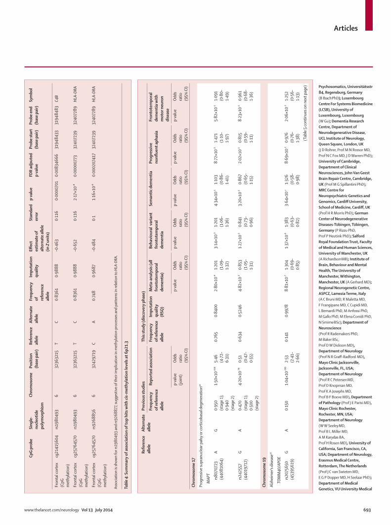
We then assessed biological relevance for the novel potential loci in human brain cortex tissues assayed for genome-wide expression and methylation. There was no eQTL in our dataset, but assessment of Zeller and colleagues’ dataset38 showed a cis-eQTL (p=5·05 × 10–³²;
Chromosome Base pair Candidate gene Minor allele
Major allele
Frequency of minor allele (r² when applicable)
Imputation quality
Odds ratio (95% CI) Standard error
p value
Discovery phase
Behavioural variant frontotemporal dementia
rs302652 11 87894831 RAB38 A T 0·259 0·9296 0·730 (0·65–0·82) 0·057 2·02×10–⁸
rs74977128 11 87936874 RAB38/CTSC C T 0·118 0·4182 1·815 (1·48–2·24) 0·107 3·06×10–⁸
All frontotemporal dementia*
rs9268877 6 32431147 HLA-DRA/HLA-DRB5 A G 0·440 0·7783 1·331 (1·22–1·45) 0·045 1·65×10–¹⁰
rs9268856 6 32429719 HLA-DRA/HLA-DRB5 A C 0·251 0·8563 0·752 (0·68–0·83) 0·050 1·30×10–⁸
rs1980493 6 32363215 BTNL2 C T 0·147 0·9642 0·720 (0·69–0·81) 0·060 4·94×10–⁸
Replication phase
Behavioural variant frontotemporal dementia
rs302668 (proxy) 11 87876911 RAB38 C T 0·325 (0·65) NA 0·877 (0.77–0.99) 0·064 0·041
rs16913634 (proxy) 11 87934068 RAB38/CTSC A G 0·104 (0·54) NA 0·964 (0.79–1.17) 0·098 0·710
All frontotemporal dementia*
rs9268877 6 32431147 HLA-DRA/HLA-DRB5 A G 0·449 NA 1·080 (0·98–1·18) 0·047 0·104
rs9268856 6 32429719 HLA-DRA/HLA-DRB5 A C 0·253 NA 0·878 (0·79–0·97) 0·053 0·014
rs1980493 6 32363215 BTNL2 C T 0·145 NA 0.85 (0·75–0·97) 0·068 0·020
Discovery and replication combined
Behavioural variant frontotemporal dementia
rs302668 (proxy) 11 87876911 RAB38 C T 0·292 (0·65) NA 0·814 (0·71–0·92) 0·064 2·44×10–⁷
rs16913634 (proxy)† 11 87934068 RAB38/CTSC A G 0·111 (0·54) NA 1·248 (1·14–1·37) 0·049 8·15×10–⁴
All frontotemporal dementia*
rs9268877† 6 32431147 HLA-DRA/HLA-DRB5 A G 0·4445 NA 1·204 (1·11–1·30) 0·039 1·05×10–⁸
rs9268856 6 32429719 HLA-DRA/HLA-DRB5 A C 0·252 NA 0·809 (0·76–0·86) 0·029 5·51×10–⁹
rs1980493 6 32363215 BTNL2 C T 0·146 NA 0·775 (0·69–0·86) 0·058 1·57×10–⁸
Replication and joint analyses were assessed for the same single-nucleotide polymorphisms (SNPs) at 6p21.3, whereas proxy SNPs were used to assess the association at 11q14 (for which r² values are included). The odds ratio is shown for the minor allele. NA=not applicable. *Denotes only minimal cross-subtype heterogeneity, with heterogeneity p values ranging from 0·793 to 0·944 based on Cochran’s Q test. †Heterogeneity p value <0·01 in the meta-analyses of the discovery and replication phases combined.
Table 3: Characteristics of single-nucleotide polymorphisms exceeding genome-wide signifi cance in the discovery phase
Articles
www.thelancet.com/neurology Vol 13 July 2014 693
Psychosomatics, Universitätsstr 84, Regensburg, Germany (B Ibach PhD); Luxembourg Centre For Systems Biomedicine (LCSB), University of Luxembourg, Luxembourg (W Gu); Dementia Research Centre, Department of Neurodegenerative Disease, UCL Institute of Neurology, Queen Square, London, UK (J D Rohrer, Prof M N Rossor MD, Prof N C Fox MD, J D Warren PhD); University of Cambridge, Department of Clinical Neurosciences, John Van Geest Brain Repair Centre, Cambridge, UK (Prof M G Spillantini PhD); MRC Centre for Neuropsychiatric Genetics and Genomics, Cardiff University, School of Medicine, Cardiff , UK (Prof H R Morris PhD); German Center of Neurodegenerative Diseases-Tübingen, Tübingen, Germany (P Rizzu PhD, Prof P Heutink PhD); Salford Royal Foundation Trust, Faculty of Medical and Human Sciences, University of Manchester, UK (A Richardson MB); Institute of Brain, Behaviour and Mental Health, The University of Manchester, Withington, Manchester, UK (A Gerhard MD); Regional Neurogenetic Centre, ASPCZ, Lamezia Terme, Italy (A C Bruni MD, R Maletta MD, F Frangipane MD, C Cupidi MD, L Bernardi PhD, M Anfossi PhD, M Gallo PhD, M Elena Conidi PhD, N Smirne BSc); Department of Neuroscience (Prof R Rademakers PhD, M Baker BSc, Prof D W Dickson MD), Department of Neurology (Prof N R Graff -Radford MD), Mayo Clinic Jacksonville, Jacksonville, FL, USA; Department of Neurology (Prof R C Petersen MD, Prof D Knopman MD, Prof K A Josephs MD, Prof B F Boeve MD), Department of Pathology (Prof J E Parisi MD), Mayo Clinic Rochester, Rochester, MN, USA; Department of Neurology (W W Seeley MD, Prof B L Miller MD, A M Karydas BA, Prof H Rosen MD), University of California, San Francisco, CA, USA; Department of Neurology, Erasmus Medical Centre, Rotterdam, The Netherlands (Prof J C van Swieten MD, E G P Dopper MD, H Seelaar PhD); Department of Medical Genetics, VU University Medical
CpG
prob
eSi
ngle
-nu
cleot
ide
poly
mor
phism
Chro
mos
ome
Posit
ion
(bas
e pa
ir)Re
fere
nce
alle
leAl
tern
ate
alle
leFr
eque
ncy
of
refe
renc
e al
lele
Impu
tatio
n qu
ality
Eff e
ct
estim
ate o
f al
tern
ate
alle
le
(in Z
uni
ts)
Stan
dard
er
ror
p va
lue
FDR
adjsu
ted
p va
lue
Prob
e st
art
(bas
e pa
ir)Pr
obe
end
(bas
e pa
ir)Sy
mbo
l
Fron
tal c
orte
x (C
pG
met
hyla
tion)
cg21
4156
04rs
1980
493
632
3632
15T
C0·
8361
0·98
88–0
·463
0·11
60·
0000
701
0·00
8346
6631
9484
3331
9484
83C4
B
Fron
tal c
orte
x (C
pG
met
hyla
tion)
cg25
7645
70rs
1980
493
632
3632
15T
C0·
8361
0·98
88–0
·652
0·11
62·
17×1
0– ⁸0·
0000
0773
3240
7239
3240
7289
HLA-
DRA
Fron
tal c
orte
x (C
pG
met
hyla
tion)
cg25
7645
70rs
9268
856
632
4297
19C
A0·
748
0·96
87–0
·484
0·1
1·16
×10– ⁶
0·00
0207
417
3240
7239
3240
7289
HLA-
DRA
Asso
ciatio
n is
show
n fo
r rs1
9804
93 a
nd rs
9268
877,
sugg
estiv
e of t
heir
impl
icatio
n in
met
hyla
tion
proc
esse
s and
pat
tern
s in
rela
tion
to H
LA-D
RA.
Tabl
e 4: S
umm
ary
of a
ssoc
iati
on o
f top
hit
s wit
h cis
-met
hyla
tion
leve
ls a
t 6p2
1.3
Re
fere
nce
alle
leAl
tern
ate
alle
lePr
evio
us st
udie
sTh
is st
udy
(disc
over
y ph
ase)
Freq
uenc
y of
refe
renc
e al
lele
Repo
rted
ass
ocia
tion
Freq
uenc
y of
refe
renc
e al
lele
Impu
tatio
n qu
ality
(R
SQ)
Met
a-an
alys
is (a
ll fr
onto
tem
pora
l de
men
tia)
Beha
viou
ral v
aria
nt
fron
tote
mpo
ral
dem
entia
Sem
antic
dem
entia
Prog
ress
ive
nonfl
uen
t aph
asia
Fron
tote
mpo
ral
dem
entia
with
m
otor
neu
ron
dise
ase
p va
lue
(join
t)O
dds
ratio
(9
5% C
I)
p
valu
e O
dds
ratio
(9
5% C
I)
p va
lue
Odd
s ra
tio
(95%
CI)
p va
lue
Odd
s ra
tio
(95%
CI)
p va
lue
Odd
s ra
tio
(95%
CI)
p va
lue
Odd
s ra
tio
(95%
CI)
Chro
mos
ome
17
Prog
ress
ive s
upra
nucle
ar p
alsy
or c
ortic
obas
al d
egen
erat
ion40
MAP
T
rs80
7072
3 (4
4081
064)
AG
0·95
0 (s
tage
1);
0·94
0 (s
tage
2)
1·50
×10– ¹¹⁶
5·46
(4
·72–
6·31
)
0·76
50·
8400
2·80
×10– ⁴
1·20
1 (1
·09–
1·32
)
3·14
×10– ³
1·20
1 (1
·06–
1·36
)
4·34
×10– ¹
1·10
3 (0
·86–
1·41
)
8·72
×10– ³
1·47
1 (1
·10–
1·97
)
5·82
×10– ¹
1·09
1 (0
·80–
1·49
)
rs24
2557
(4
4019
712)
GA
0·47
0 (s
tage
1);
0·50
0 (s
tage
2)
4·20
×10– ⁷⁰
0·51
(0
·47–
0·55
)
0·63
40·
5246
4·82
×10– ³
0·85
3 (1
·05–
1·31
)
1·27
×10– ²
0·84
1 (0
·73–
0·96
)
3·20
×10– ¹
0·86
7 (0
·65–
1·15
)
2·02
×10– ¹
0·81
5 (0
·59–
1·11
)
8·23
×10– ¹
0·96
1 (0
·68–
1·36
)
Chro
mos
ome
19
Alzh
eim
er’s
dise
ase41
TOM
M40
/APO
E
rs20
7565
0 (4
5395
619)
GA
0·15
01·
04×1
0– ²⁹⁵2·
53
(2·4
1–2·
66)
0·14
10·
9978
8·81
×10– ⁷
1·30
4 (0
·69–
0·85
)
1·37
×10– ⁶
1·38
3 (0
·63–
0·82
)
3·64
×10–2
1·32
6 (0
·58–
0·98
)
8·69
×10– ¹
0·97
6 (0
·76–
1·38
)
2·06
×10– ¹
1·25
2 (0
·56–
1·13
)
(Tab
le 5
cont
inue
s on
next
pag
e)

Articles
694 www.thelancet.com/neurology Vol 13 July 2014
Re
fere
nce
alle
leAl
tern
ate
alle
lePr
evio
us st
udie
sTh
is st
udy
(disc
over
y ph
ase)
Freq
uenc
y of
refe
renc
e al
lele
Repo
rted
ass
ocia
tion
Freq
uenc
y of
refe
renc
e al
lele
Impu
tatio
n qu
ality
(R
SQ)
Met
a-an
alys
is (a
ll fr
onto
tem
pora
l de
men
tia)
Beha
viou
ral v
aria
nt
fron
tote
mpo
ral
dem
entia
Sem
antic
dem
entia
Prog
ress
ive
nonfl
uen
t aph
asia
Fron
tote
mpo
ral
dem
entia
/ m
otor
ne
uron
dise
ase
p va
lue
(join
t)O
dds
ratio
(9
5% C
I)
p va
lue
Odd
s ra
tio
(95%
CI)
p va
lue
Odd
s ra
tio
(95%
CI)
p va
lue
Odd
s ra
tio
(95%
CI)
p va
lue
Odd
s ra
tio
(95%
CI)
p va
lue
Odd
s ra
tio
(95%
CI)
(Con
tinue
d fro
m p
revi
ous p
age)
Chro
mos
ome
9
Amyo
trop
hic l
ater
al sc
lero
sis39
C9or
f72/
MOB
3B
rs38
4994
2 (2
7543
281)
AG
0·26
01·
01×1
0–81·
23
(NA)
0·25
30·
9996
4·38
×10–4
1·16
6 (1
·07–
1·27
)
7·38
×10– ³
1·15
5 (0
·78–
0·96
)
9·89
×10– ¹
1·01
0 (0
·80–
1·25
)
9·03
×10– ¹
0·99
0 (0
·79–
1·31
)
2·12
×10– ⁶
1·95
7 (0
·39–
0·68
)
Chro
mos
ome
7
FTLD
-TDP
17
TMEM
106B
rs19
9062
2 (1
2283
787)
AG
0·67
91·
08×1
0– ¹¹1·
64
(1·4
1–1·
89)
0·60
00·
9588
7·88
×10– ²
1·08
0 (0
·99–
1·16
)
5·85
×10– ³
1·14
4 (1
·04–
1·26
)
8·36
×10– ¹
0·97
8 (0
·80–
1·20
)
8·98
×10– ¹
0·98
5 (0
·79–
1·23
)
3·11
×10– ¹
0·87
6 (0
·68–
1·13
)
rs69
6691
5 (1
2265
988)
CT
0·67
91·
63×1
0– ¹¹1·
64
(1·4
1–1·
89)
0·59
60·
9675
1·21
×10– ¹
1·07
0 (0
·87–
1·02
)
5·74
×10– ³
1·14
4 (1
·04–
1·26
)
5·27
×10– ¹
0·93
6 (0
·76–
1·15
)
7·26
×10– ¹
0·96
1 (0
·77–
1·20
)
3·62
×10– ¹
0·88
8 (0
·69–
1·14
)
rs10
2000
4 (1
2255
778)
TC
0·76
75·
00×1
0– ¹¹1·
66
(1·4
3–1.
96
0·69
30·
9538
4·59
×10– ¹
1·03
0 (0
·95–
1·12
)
5·71
×10– ²
1·10
4 (1
·00–
1·22
)
8·53
×10– ¹
0·98
0 (0
·79–
1·21
)
5·00
×10– ¹
0·92
1 (0
·72–
1·17
)
1·20
×10– ¹
0·80
5 (0
·61–
1·06
)
Chro
mos
ome
11
Mul
tiple
scle
rosis
44,4
5
RAB3
8
rs13
8633
0 (8
7819
427)
CT
0·13
02·
00×1
0– ⁶N
A0·
141
0·96
943·
35×1
0– ¹1·
050
(0·8
5–1·
06)
6·09
×10– ¹
1·04
0 (0
·84–
1·10
)
7·60
×10– ¹
1·04
0 (0
·72–
1·27
)
6·97
×10– ¹
1·06
0 (0
·68–
1·29
)
3·00
×10–1
0·82
9 (0
·58–
1·18
)
Chro
mos
ome
6
Mul
tiple
scle
rosis
43–4
5
HLA-
DRA
rs31
3538
8 (3
2413
051)
AG
0·23
08·
94×1
0– ⁸¹1·
99
(1·8
4–2·
15)
0·13
10·
9734
4·80
×10– ²
1·12
2 (1
·00–
1·26
)
2·10
×10– ¹
1·09
5 (0
·79–
1·05
)
1·25
×10– ¹
1·25
4 (0
·60–
1·06
)
5·02
×10– ¹
1·12
0 (0
·64–
1·24
)
6·10
×10– ¹
1·10
5 (0
·61–
1·33
)
rs31
2987
1 (3
2406
342)
AC
0·50
45·
70×1
0– ¹⁵1·
72
(1·5
9–1·
86)
0·33
70·
9379
3·43
×10– ¹
0·96
1 (0
·88–
1·04
)
3·15
×10– ¹
0·94
9 (0
·95–
1·16
)
4·94
×10– ¹
1·07
8 (0
·75–
1·15
)
8·24
×10– ¹
0·97
4 (0
·81–
1·30
)
2·72
×10– ¹
0·85
9 (0
·89–
1·53
)
Park
inso
n’s d
iseas
e46
HLA-
DRA
rs31
2988
2 (3
2409
530)
GA
0·45
01·
90×1
0– ¹⁰1·
26
(1·1
7–1·
35)
0·45
60·
9992
3·36
×10– ²
1·08
6 (0
·85–
0·99
)
3·27
×10– ²
1·10
6 (0
·82–
0·99
)
7·52
×10– ¹
1·03
3 (0
·79–
1·18
)
5·74
×10– ¹
1·06
5 (0
·75–
1·17
)
7·07
×10– ¹
1·04
9 (0
·74–
1·22
)
NA=
not a
pplic
able
. FLT
D-TD
P=fro
ntot
empo
ral l
obar
deg
ener
atio
n w
ith T
DP43
-pos
itive
inclu
sions
. RSQ
=r2 im
puta
tion
qual
ity co
effi c
ient
.
Tabl
e 5: B
idire
ctio
nal a
naly
sis o
f sin
gle-
nucl
eoti
de p
olym
orph
ism
s and
loci
ass
ocia
ted
wit
h ot
her n
euro
dege
nera
tive
dis
orde
rs a
nd o
ur st
udy
popu
lati
on
Articles
www.thelancet.com/neurology Vol 13 July 2014 695
Centre, Amsterdam, The Netherlands (Prof J C van Swieten); Alzheimer Centre and Department of Neurology, VU University Medical Centre, Amsterdam, The Netherlands (Y A L Pijnenburg MD, Prof P Scheltens MD); Department of Basic Medical Sciences, Neurosciences and Sense Organs of the Aldo Moro, University of Bari, Italy (G Logroscino MD, R Capozzo MD); Department of Medical and Molecular Genetics, King’s College London, Guy’s Hospital, London, UK (A Ramasamy); Department of Molecular Cardiology, IRCCS Fondazione S Maugeri, Pavia, Italy (V Novelli PhD); Cardiovascular Research Unit, IRCCS Multimedica, Milan, Italy (An A Puca MD); Department of Medicine and Surgery, University of Salerno, Baronissi (SA), Italy (A A Puca); Neurology Department, IRCCS Multimedica, Milan, Italy (M Franceschi MD); Department of Clinical Medicine and Surgery, University of Naples Federico II, Naples, Italy (Prof A Postiglione MD); Geriatric Center Frullone-ASL Napoli 1 Centro, Naples, Italy (G Milan MD, P Sorrentino MD); UCL Genomics, Institute of Child Health (ICH), UCL, London, UK (M Kristiansen PhD); Karolinska Institutet, Department NVS, KI-Alzheimer Disease Research Center, Stockholm, Sweden (H-H Chiang PhD, Prof C Graff MD); Department of Geriatric Medicine, Genetics Unit, Karolinska Universtiy Hospital, Stockholm (H-H Chiang, Prof C Graff ); Université Lille Nord de France, Lille, France (Prof F Pasquier MD, A Rollin MD, V Deramecourt MD, F Lebert MD); and Laboratory of Neurosciences, National Institute on Aging, National Institutes of Health, Bethesda, MD, USA (D Kapogiannis MD)
Correspondence to:Prof John Hardy, Reta Lila Weston Research Laboratories, Department of Molecular Neuroscience, University College London Institute of Neurology, London WC1N 3BG, [email protected]
See Online for appendix
appendix) at 11q14 for rs302652 (chr11:87894881, risk allele T) causing a decreased expression of RAB38 (Illumina ILMN_2134974 located on chr11:87846656-87846705) in monocytes. These data suggest a role in transcriptional processes in cis for this SNP. Furthermore, we identifi ed signifi cant cis-mQTL at 6p21.3 after multiple test correction for rs1980493 (risk allele T) that associated with changes in the methylation levels related to HLA-DRA in the frontal cortex (table 4).
To assess potential genetic overlap between FTD and closely related forms of neurodegenerative diseases we selected relevant SNPs for candidate loci and analysed them in our dataset. This analysis included published association studies for amyotrophic lateral sclerosis,39 progressive supranuclear palsy and corticobasal degeneration,40 Alzheimer’s disease,41 and FTLD-TDP.17 We also assessed whether the two loci identifi ed through this study had also been reported previously in other studies of neurological disorders.
For the C9orf72 locus (for amyotrophic lateral sclerosis), the SNP rs3849942 achieved a p value of 2·12 × 10–⁶ and an OR of 1·957 in the FTD-MND subtype consistent with our post-hoc analyses (about 23% of expansion carriers in this subtype; table 5, appendix). Association was modest in behavioural FTD (p=7·38 × 10–³; OR=1·155) as well as in the entire discovery cohort (p=4·38 × 10–⁴; OR=1·166), while there was no evidence for association in the semantic dementia or progressive nonfl uent aphasia subtypes (table 5). These results confi rm that the C9orf72 locus associates mainly with FTD- MND and to a lesser extent with behavioural FTD (appendix).
For the MAPT locus (PSP/CBD), the SNPs rs242557 and rs807072340 reached modest p values between 10–³ and 10–⁴
only in the entire cohort and in the behavioural FTD and progressive nonfl uent aphasia subtypes (rs8070723 only; table 5). The eff ect was small in our study although in the same direction as in the GWAS for progressive supranuclear palsy (5·4640 vs about 1·2–1·4 in our study; table 5). These results refl ect the fact that we excluded all known chromosome 17 mutation carriers and that tau pathology was a less common feature within our study population.
For the TOMM40/APOE locus (Alzheimer’s disease), the SNP rs2075650 reached a p value of 8·81 × 10–⁷ in the entire dataset and 1·37 × 10–⁶ in behavioural FTD, whereas the semantic dementia, progressive nonfl uent aphasia, and FTD-MND subtype p values were in the range of 10–¹ and 10–² (table 5). Several Alzheimer’s disease GWASs reported association with the minor allele of this SNP with ORs greater than 2·5,41 but in our study the OR was about 1·3 (table 5). This suggestive association might refl ect clinical overlap (about 15%) between patients with clinically diagnosed FTD and those with Alzheimer’s disease.42
For the TMEM106B locus (FTLD-TDP), we assessed the three associated SNPs reported by Van Deerlin and colleagues (rs1990622; rs6966915; rs1020004).17 All
achieved modest p values in the entire dataset with lowest p values in the range of 10–²–10–³ only in the behavioural FTD subtype (table 5). Van Deerlin and colleagues’ study17 was done on samples from patients with autopsy-confi rmed FTLD-TDP, whereas our cohort is mainly clinically defi ned. Additionally, the previous study included many GRN mutation carriers, who frequently present with behavioural FTD;17 in our study, GRN mutation carriers were excluded. Biochemical evidence has suggested that TMEM106B is directly related to GRN metabolism,13 thus we regard our data as a limited replication of the original fi nding (ie, they do not substantiate earlier fi ndings).
Finally, the RAB38 locus previously showed suggestive association in multiple sclerosis,43 whereas the HLA locus was reported to associate with multiple sclerosis,44,45 Parkinson’s disease,19,46 and Alzheimer’s disease.47 None of the SNPs reported in these studies, and which were assessed in our dataset (table 5),43–46 showed association with FTD, probably suggesting that diff erent risk haplotype sub-structures at the same loci associate with distinctive phenotypes.
DiscussionWe have identifi ed two novel potential loci for FTD: 6p21.3, encompassing the HLA locus (immune system), and 11q14, encompassing RAB38/CTSC (transcripts of which are related to lysosomal biology). Our data suggest that these loci might aff ect expression and methylation in cis and indicate that immune system processes, and possibly lysosomal and autophagy pathways, are potentially involved in the pathogenesis of FTD.
FTD is characterised by a broad range of clinical manifestations, diff erential pathological signatures, and substantial genetic variability, which imply complex disease mechanisms.15 In the search for novel disease risk loci associated with FTD we have done an extensive GWAS on a large cohort of mainly clinically diagnosed FTD samples from patients of European ancestry. Several limitations might apply to this study. In view of the phenotype heterogeneity of FTD, and considering that it is a rare neurodegenerative disorder,2 testing the hypothesis “common variant, common disease” for diseases of this kind is challenging and clearly benefi ts from large sample sizes. Additionally, our fi ndings might indicate association with specifi c loci without necessarily implying causality; low heritability due to common variability might also apply. However, the QQ plots and associated λ values (appendix) conformed to GWAS standards, lending support to our fi ndings.
We included samples from more than 3500 patients and, thus, we know of no larger GWAS for FTD. We have identifi ed two novel potential loci for FTD: 11q14, encompassing RAB38/CTSC, was suggestive for the behavioural FTD subtype, and 6p21.3, encompassing the HLA locus was statistically signifi cant for the entire cohort.

Articles
696 www.thelancet.com/neurology Vol 13 July 2014
For more on Biowulf see http://biowulf.nih.gov
For more on the expression of RAB38 see http://www.
genecards.org/cgi-bin/carddisp.pl?gene=RAB38
RAB3848 encodes the transmembrane protein RAB38, which is expressed in the thyroid, in elements of the immune system, and in the brain. From a functional perspective, RAB38 has been shown to mediate protein traffi cking to lysosomal-related organelles and maturation of phagosomes.49,50 CTSC is a lysosomal cysteine-proteinase that participates in the activation of serine proteinases in cells involved in immune and infl ammatory processes, including phagocytosis of pathogens and local activation and deactivation of infl ammatory factors (Online Mendelian Inheritance in Man [OMIM] number 602365). The SNP rs302652 at the RAB38/CTSC locus shows an eQTL in monocytes38 associated with decreased expression of RAB38, possibly indicating that a decreased function of RAB38 might be the mechanism by which the association at this locus is mediated. Both RAB38 and CTSC are implicated in lysosomal biology and an association with lysosomal and autophagic processes in FTD was previously suggested in two studies of GRN51 and TMEM106B.52 A possible role for autophagy has also been shown in Parkinson’s disease.53 Our fi ndings will need to be replicated in other FTD cohorts in follow-up studies (eg, fi ne-mapping studies) to lend support to the inference
that lysosomal biology and autophagy might be involved in the aetiology of FTD.54
The genetic association that we identifi ed with the HLA locus lends support to the notion of a link between FTD and the immune system. Our mQTL data showed that risk at this locus is associated with cis-changes in methylation levels of HLA-DRA in the frontal cortex. HLA associations have been previously reported in Alzheimer’s disease,47 Parkinson’s disease,19,46 and multiple sclerosis.44,45 Additionally, a general involvement of the innate and the adaptive immune responses has been suggested in the pathogenesis of neurodegenerative diseases,55,56 lending support to the idea that the immune system plays an important part within the spectrum of neurological disorders (panel).
Future studies should aim to replicate our fi ndings and elucidate the functional basis of FTD. Additionally, our data indicate that common pathways and processes might underlie diff erent forms of neurodegenerative disorders, including Alzheimer’s disease, Parkinson’s disease, multiple sclerosis, and FTD. Exploring the possibility of developing therapeutic measures targeting general damage responses could hold promise—after replication and validation of our fi ndings—for the development and implementation of treatment options for these neurological disorders, including FTD.ContributorsJH, PM, ABS, MAN, RF, and JDR designed the study. JDR, RF and JH did the clinical quality checks. RF coordinated sample collection, received samples at UCL and TTUHSC, and did material quality control for discovery and replication phases. DGH received samples at NIH and coordinated material quality control at NIH. JDR, JBJK, CDS, PRS, WSB, JRH, GMH, OP, LB, ET, EH, IH, AR, MB BB, AP, LB, GB, RG, GF, DG, ES, CF, MS, JC, AL, RB, MLW, KN, CN, IRAM, G-YRH, DMAM, JG, CMM, JA, TDG, IGM, AJT, PP, EDH, EMW, AB, EJ, MCT, PP, CR, SO-C, EA, RP, JDS, PA, AK, IR, ER, LP, ER, PStG-H, ER, GR, FT, GG, JBR, JCMS, JU, JC, SM, AD, VMVD, MG, JQT, JvdZ, TVL, CVB, WD, MC, SFC, ILB, AB, DH, VG, MV, BN, SS, SB, IP, JEN, LEH, MR, BI, MM, GG, SP, WG, MNR, NCF, JDW, MGS, HM, PR, PH, JSS, AG, AR, SR, ACB, RM, FF, CC, LB, MA, MG, MEC, NS, RR, MB, DWD, JEP, NRGR, RCP, DK, KAJ, BFB, WWS, BLM, AMK, HR, JCvS, EGPD, HS, YALP, PS, GL, RC, VN, AAP, MF, AP, GM, PS, H-HC, CG, FP, AR, VD, FL, DK, LF, and SPB collected and characterised samples. MK was responsible for genotyping at ICH. JH, PM, ABS, and SPB obtained funding for this study. JH, PM, and ABS supervised the study. MAN did statistical and association analyses. RF, MAN, and JH analysed and interpreted the data. AR helped in the interpretation of the e/mQTL data. RF, MAN, JH, and PM wrote the fi rst draft of the paper. All other co-authors participated in preparation of the paper by reading and commenting on drafts before submission.
Declaration of interestsCVB and MC are inventors on patent applications for GRN and C9orf72. PRS receives speaker fees from Janssen pharmaceutical. RR receives research support from the NIH (R01 NS080882, R01 NS065782, R01 AG026251, R01 NS076471, and P50 AG16574), the ALS Therapy Alliance, and the Consortium for Frontotemporal Degeneration Research, honoraria for lectures or educational activities not funded by industry. RR serves on the medical advisory board of the Association for Frontotemporal Degeneration and the board of directors of the International Society for Frontotemporal Dementia, and holds a patent on methods to screen for the hexanucleotide repeat expansion in the C9ORF72 gene. DWD is supported by NIH grants (P50 AG16574, P50 NS72187, P01 AG03949), the Mangurian Foundation, CurePSP, and the Robert E Jacoby Professorship for
Panel: Research in context
Systematic reviewWe searched PubMed for research and review articles on frontotemporal dementia using the following terms: “frontotemporal dementia AND genetics” and “frontotemporal dementia AND review”. 1,4,5,8–17,54 We compared our results to several previously published genome-wide association studies. We identifi ed only one directly relevant study that investigated a pathologically defi ned subtype of frontotemporal dementia (frontotemporal lobar degeneration with TDP43-positive inclusions; FTLD-TDP).17 The other studies were of related diseases such as amyotrophic lateral sclerosis,39 Alzheimer’s disease,41,47 progressive supranuclear palsy and corticobasal degeneration,40 multiple sclerosis,43–45 and Parkinson’s disease.19,46
InterpretationTo the best of our knowledge, ours is the fi rst genome-wide association study in samples from patients with clinical frontotemporal dementia. In view of the complexity and heterogeneity of the disease, mutations in only three main genes—MAPT, GRN, and C9orf72—have been associated with frontotemporal dementia, and these explain only a small proportion of cases. Most importantly, little is known about the mechanisms involved in the development of this disorder. Our fi ndings suggest that common variability in loci that point to immune processes and possibly to lysosomal biology and autophagy are involved in the pathobiology of the disease. These fi ndings provide a basis for future replication and functional studies.

Articles
www.thelancet.com/neurology Vol 13 July 2014 697
Alzheimer’s Research. NRGR is on the Scientifi c Advisory Board for Codman, TauRzx multicenter study, Consultation for CYTOX. RCP chairs a Data Monitoring Committee for Pfi zer and Janssen Alzheimer Immunotherapy, and is a consultant for GE Healthcare and Elan Pharmaceuticals. RCP receives royalties from Oxford University Press for Mild Cognitive Impairment. DK has served on a data safety monitoring board for Lilly Pharmaceuticals, as a consultant to TauRx, was an investigator in clinical trials sponsored by Baxter, Elan Pharmaceuticals, and Forest Pharmaceuticals in the past 2 years and receives research support from the NIH. BFB has served as an investigator for clinical trials sponsored by Cephalon Inc, Allon Pharmaceuticals, and GE Healthcare. BFB receives royalties from the publication of a book entitled Behavioral Neurology Of Dementia (Cambridge Medicine, 2009). BFB has received honoraria from the American Academy of Neurology. BFB serves on the Scientifi c Advisory Board of the Tau Consortium. BFB receives research support from the National Institute on Aging (P50 AG016574, U01 AG006786, RO1 AG032306, RO1 AG041797) and the Mangurian Foundation. BLM is on the Board Membership of The Larry L Hillblom Foundation, The John Douglas French Foundation, The Tau Consortium, Sagol School of Neuroscience Tel Aviv University. BLM holds consultancy for Tau Rx lts—Chair, Scientifi c Advisory Board bvFTD Trial Allon Therapeutics—Steering Committee AL-108-231 Study, Bristol-Myers Squibb-Advisory Board, Progressive Supranuclear Palsy (PSP), Neurology Scientifi c Advisory Board Meeting Siemens Molecular Imaging, and Eli Lilly US Alzheimer’s Disease Advisory Board, and receives royalties from Cambridge University Press Guilford Publications Inc, Neurocase. RF, DGH, MAN, JDR, AR, JBJK, CDS, WSB, GMH, JRH, OP, LB, ET, EH, IH, AR, MB, BB, AP, CC, NJC, LB, GB, RG, GF, DG, CF, MS, ES, JC, AL, RB, MLW, KN, CN, IRAM, G-YRH, DMAM, JG, CMM, JA, TDG, IGM, AJT, PP, EDH, EMW, AB, EJ, MCT, PP, CR, SO-C, EA, RP, JDS, PA, AK, IR, ER, LP, ER, PStG-H, GR, FT, GG, JBR, JCMS, JU, JC, SM, AD, VMVD, MG, JQT, JvdZ, WD, TVL, SFC, ILB, DH, VG, MV, AB, BN, SS, SB, IP, JEN, LEH, MR, MM, BI, GG, SP, WG, MNR, NCF, JDW, MGS, HRM, PR, PH, JSS, SR, AR, AG, ACB, RM, FF, CC, LB, MA, MG, MEC, NS, MB, KAJ, JEP, WWS, AMK, HR, JCvS, EGPD, HS, YALP, PS, GL, RC, VN, AAP, MF, AP, GM, PS, MK, H-HC, CG, FP, AR, VD, FL, DK, LF, SPB, JH, PM, and ABS declare no competing interests.
Acknowledgments We received intramural funding from the National Institute of Neurological Disorders and Stroke (NINDS) and National Institute on Aging (NIA), the Wellcome/MRC Centre on Parkinson’s disease, Alzheimer’s Research UK (ARUK, Grant ARUK-PG2012-18), and by the offi ce of the Dean of the School of Medicine, Department of Internal Medicine, at Texas Tech University Health Sciences Center. We thank Mike Hubank and Kerra Pearce at the Genomic core facility at the Institute of Child Health (ICH), UCL, for assisting RF in doing Illumina genotyping experiments (FTD-GWAS genotyping). The work done by the North American Brain Expression Consortium (NABEC) was supported in part by the Intramural Research Program of the National Institute on Aging, National Institutes of Health, part of the US Department of Health and Human Services (project number ZIA AG000932-04), and by a Research Grant from the Department of Defense (W81XWH-09-2-0128). Work done by the UK Brain Expression Consortium (UKBEC) was supported by the MRC through the MRC Sudden Death Brain Bank (CS), by a Project Grant (G0901254 to JH and MW), and by a Fellowship award (G0802462 to MR). DT was supported by the King Faisal Specialist Hospital and Research Centre, Saudi Arabia. Computing facilities used at King’s College London were supported by the National Institute for Health Research (NIHR) Biomedical Research Centre based at Guy’s and St Thomas’ NHS Foundation Trust and King’s College London. We thank AROS Applied Biotechnology AS company laboratories and Aff ymetrix for their valuable input. JBJK was supported by the National Health and Medical Resarch Council (NHMRC), Australia (project grants 510217 and 1005769). CDS was supported by NHMRC (project grants 630428 and 1005769). PRS was supported by NHMRC (project grants 510217 and 1005769) and acknowledges that DNA samples were prepared by Genetic Repositories Australia, supported by NHMRC Enabling Grant 401184. GMH was supported by NHMRC Research Fellowship 630434, Project Grant 1029538, and Program Grant 1037746. JRH was supported by the
Australian Research Council Federation Fellowship, NHMRC Project Grant 1029538 and NHMRC Program Grant 1037746. OP was supported by NHMRC Career Development Fellowship 1022684, Project Grant 1003139. IH, AR, and MB acknowledge the patients and controls who participated in this project and the Trinitat Port-Carbó (deceased) and her extended family who are supporting Fundació ACE research programmes. CC was supported by Grant P30-NS069329-01 and acknowledges that the recruitment and clinical characterisation of research participants at Washington University were supported by NIH P50 AG05681, P01 AG03991, and P01 AG026276. LB and GB were supported by the Ricerca Corrente, Italian Ministry of Health. RG was supported by Fondazione CARIPLO 2009-2633, Ricerca Corrente, Italian Ministry of Health. GF was supported by Fondazione CARIPLO 2009–2633. ES was supported by the Italian Ministry of Health. CF was supported by Fondazione Cariplo. MS was supported from the Italian Ministry of Health (Ricerca Corrente). MLW was supported by Government funding of clinical research within NHS Sweden (ALF). KN was supported by Thure Carlsson Foundation. CN was supported by Swedish Alzheimer Fund. IRAM and GYRH were supported by CIHR (grant 74580) PARF (grant C06-01). JG was supported by the NINDS intramural research funds for FTD research. CMM was supported by Medical Research Council UK, Brains for Dementia Research, Alzheimer’s Society, Alzheimer’s Research UK, National Institutes for Health Research, Department of Health, and Yvonne Mairy Bequest, and acknowledges that tissue samples made available for this study were provided by the Newcastle Brain Tissue Resource, which was funded in part by grants G0400074 and G1100540 from the UK MRC, the Alzheimer’s Research Trust and Alzheimer’s Society through the Brains for Dementia Research Initiative and an NIHR Biomedical Research Centre Grant in Ageing and Health, and NIHR Biomedical Research Unit in Lewy Body Disorders. CMM was supported by the UK Department of Health and Medical Research Council and the Research was supported by the National Institute for Health Research Newcastle Biomedical Research Centre based at Newcastle Hospitals Foundation Trust and Newcastle University and acknowledges that the views expressed are those of the authors and not necessarily those of the NHS, the NIHR or the Department of Health. JA was supported by MRC, Dunhill Medical Trust, and Alzheimer’s Research UK. TDG was supported by Wellcome Trust Senior Clinical Fellow. IGM was supported by NIHR Biomedical Research Centre and Unit on Ageing Grants and acknowledges the National Institute for Health Research Newcastle Biomedical Research Centre based at Newcastle Hospitals Foundation Trust and Newcastle University. The views expressed are those of the authors and not necessarily those of the NHS, the NIHR, or the Department of Health. AJT was supported by Medical Research Council, Alzheimer’s Society, Alzheimer’s Research UK, and the National Institutes for Health Research. EJ was supported by NIHR and Newcastle Biomedical Research Centre. PP, CR, SOC, and EA were supported partially by FIMA (Foundation for Applied Medical Research). PP acknowledges Manuel Seijo-Martínez (Department of Neurology, Hospital do Salnés, Pontevedra, Spain) and Ramon Rene, Jordi Gascon, and Jaume Campdelacreu (Department of Neurology, Hospital de Bellvitge, Barcelona, Spain) for providing FTD DNA samples. RP, JDS, PA, AK, and AD were supported by the German Federal Ministry of Education and Research (BMBF; grant number FKZ 01GI1007A—German FTLD consortium). IR was supported by Ministero dell’Istruzione, dell’Università e della Ricerca (MIUR) of Italy. PStG-H was supported by the Canadian Institutes of Health Research, Wellcome Trust, Ontario Research Fund. FT was supported by the Italian Ministry of Health (ricerca corrente) and MIUR grant RBAP11FRE9. GR and GG were supported by the Italian Ministry of Health (ricerca corrente). JBR was supported by Cambridge NIHR Biomedical Research Centre and Wellcome Trust (088324). JU, JC, and SM were supported by the MRC Prion Unit core funding and acknowledge MRC UK, UCLH Biomedical Research Centre, and Queen Square Dementia BRU. SM thanks John Beck, Tracy Campbell, Gary Adamson, Ron Druyeh, Jessica Lowe, and Mark Poulter. AD thanks Benedikt Bader, Manuela Neumann, Sigrun Roeber, Thomas Arzberger, and Hans Kretzschmar (deceased). VMVD and JQT were supported by grants AG032953, AG017586 and AG010124. MG was supported by Grants AG032953, AG017586, AG010124, and NS044266. VMVD thanks EunRan Suh for assistance with

Articles
698 www.thelancet.com/neurology Vol 13 July 2014
sample handling and Elisabeth McCarty-Wood for help in selection of patients. JQT thanks Terry Schuck, John Robinson, and Kevin Raible for assistance with neuropathological assessment of patients. CVB and the Antwerp site were in part funded by the MetLife Foundation Award for Medical Research (to CVB), the Belgian Science Policy Offi ce Interuniversity Attraction Poles programme; the Foundation for Alzheimer Research (SAO-FRA); the Medical Foundation Queen Elisabeth; the Flemish Government Methusalem Excellence award (to CVB); the Research Foundation Flanders (FWO); and the University of Antwerp Research Fund. JvdZ holds a postdoctoral fellowship of the FWO. CVB and the Antwerp site authors thanks neurologists S Engelborghs, P P De Deyn, A Sieben, and Rik Vandenberghe and neuropathologist J J Martin for the clinical and pathological diagnoses. Isabelle Leber and Alexis Brice were supported by the programme Investissements d’avenir ANR-10-IAIHU-06 and acknowledges the contribution of The French Research Network on FTLD/FTLD-ALS for the contribution in samples collection. BN, SS, SB, and IP were supported by Prin 2010-prot.2010PWNJXK; Cassa di Rispario di Firenze e Cassa di Risparmio di Pistoia e Pescia. JEN was supported by the Novo Nordisk Foundation, Denmark. MR was supported by the German National Genome Network (NGFN) and the German Ministry for Education and Research Grant Number 01GS0465. JDR, MNR, NCF, and JDW were supported by an MRC programme grant, the NIHR Queen Square Dementia Biomedical Research Unit and the Leonard Wolfson Experimental Neurology Centre. MGS was supported by MRC grant n G0301152, Cambridge Biomedical Research Centre, and thanks K Westmore for extracting DNA. HM was supported by the Motor Neuron Disease Association (Grant 6057). RR was supported by P50 AG016574, R01 NS080882, R01 NS065782, P50 NS72187, and the Consortium for Frontotemporal Dementia. DWD was supported by P50NS072187, P50AG016574, State of Florida Alzheimer Disease Initiative, and CurePSP Inc. NRGR, JEP, RCP, DK, and BFB were supported by P50 AG016574. KAJ was supported by R01 AG037491. WWS was supported by NIH AG023501, AG019724, Consortium for Frontotemporal Dementia Research. BLM was supported by P50AG023501, P01AG019724, Consortium for FTD Research. HR was supported by AG032306. JCvS was supported by Stichting Dioraphte Foundation (11 02 03 00), Nuts Ohra Foundation (0801-69), Hersenstichting Nederland (BG 2010-02), and Alzheimer Nederland. CG and HHC acknowledge families, patients, clinicians including Inger Nennesmo and Vesna Jelic, Laura Fratiglioni for control samples and Jenny Björkström, Håkan Thonberg, Charlotte Forsell, Anna-Karin Lindström, and Lena Lilius for sample handling. CG was supported by Swedish Brain Power (SBP), the Strategic Research Programme in Neuroscience at Karolinska Institutet (StratNeuro), the regional agreement on medical training and clinical research (ALF) between Stockholm County Council and Karolinska Institutet, Swedish Alzheimer Foundation, Swedish Research Council, Karolinska Institutet PhD-student funding, King Gustaf V, and Queen Victoria’s Free Mason Foundation. FP, AR, VD, and FL acknowledge Labex DISTALZ. RF acknowledges the help and support of June Howard at the Texas Tech University Health Sciences Center Offi ce of Sponsored Programs for tremendous help in managing Material Transfer Agreement at TTUHSC.
References1 Neary D, Snowden JS, Gustafson L, et al. Frontotemporal lobar
degeneration: a consensus on clinical diagnostic criteria. Neurology 1998; 51: 1546–54.
2 Rabinovici GD, Miller BL. Frontotemporal lobar degeneration: epidemiology, pathophysiology, diagnosis and management. CNS Drugs 2010; 24: 375–98.
3 Rohrer JD, Warren JD. Phenotypic signatures of genetic frontotemporal dementia. Curr Opin Neurol 2011; 24: 542–49.
4 Rascovsky K, Hodges JR, Knopman D, et al. Sensitivity of revised diagnostic criteria for the behavioural variant of frontotemporal dementia. Brain 2011; 134: 2456–77.
5 Gorno–Tempini ML, Hillis AE, Weintraub S, et al. Classifi cation of primary progressive aphasia and its variants. Neurology 2011; 76: 1006–14.
6 Mackenzie IR, Neumann M, Baborie A, et al. A harmonized classifi cation system for FTLD–TDP pathology. Acta Neuropathol 2011; 122: 111–13.
7 Halliday G, Bigio EH, Cairns NJ, Neumann M, Mackenzie IR, Mann DM. Mechanisms of disease in frontotemporal lobar degeneration: gain of function versus loss of function eff ects. Acta Neuropathol 2012; 124: 373–82.
8 Hutton M, Lendon CL, Rizzu P, et al. Association of missense and 5ʹ–splice–site mutations in tau with the inherited dementia FTDP–17. Nature 1998; 393: 702–05.
9 Baker M, Mackenzie IR, Pickering-Brown SM, et al. Mutations in progranulin cause tau–negative frontotemporal dementia linked to chromosome 17. Nature 2006; 442: 916–19.
10 Cruts M, Gijselinck I, van der Zee J, et al. Null mutations in progranulin cause ubiquitin-positive frontotemporal dementia linked to chromosome 17q21. Nature 2006; 442: 920–4.
11 DeJesus–Hernandez M, Mackenzie IR, Boeve BF, et al. Expanded GGGGCC hexanucleotide repeat in noncoding region of C9ORF72 causes chromosome 9p-linked FTD and ALS. Neuron 2011; 72: 245–56.
12 Renton AE, Majounie E, Waite A, et al. A hexanucleotide repeat expansion in C9ORF72 is the cause of chromosome 9p21-linked ALS-FTD. Neuron 2011; 72: 257–68
13 Ferrari R, Thumma A, Momeni P. Molecular Genetics of Frontotemporal Dementia. In: eLS. John Wiley & Sons, Ltd: Chichester. 2013. DOI: 10.1002/9780470015902.a0024457.
14 van der Zee J, Gijselinck I, Dillen L , et al. A Pan-European Study of the C9orf72 Repeat Associated with FTLD: Geographic Prevalence, Genomic Instability, and Intermediate Repeats. Hum Mutat 2013; 34: 363–73.
15 Rohrer JD, Rosen HJ. Neuroimaging in frontotemporal dementia. Int Rev Psychiatry 2013; 25: 221–29.
16 Ling SC, Polymenidou M, Cleveland DW. Converging Mechanisms in ALS and FTD: Disrupted RNA and Protein Homeostasis. Neuron 2013; 79: 416–38.
17 Van Deerlin VM, Sleiman PM, Martinez–Lage M, et al. Common variants at 7p21 are associated with frontotemporal lobar degeneration with TDP-43 inclusions. Nat Genet 2010; 42: 234–39.
18 Strong MJ, Grace GM, Freedman M, et al. Consensus criteria for the diagnosis of frontotemporal cognitive and behavioural syndromes in amyotrophic lateral sclerosis. Amyotroph Lateral Scler 2009; 10: 131–46.
19 International Parkinson Disease Genomics Consortium, Nalls MA, Plagnol V, et al. Imputation of sequence variants for identifi cation of genetic risks for Parkinson’s disease: a meta-analysis of genome-wide association studies. Lancet 2011; 377: 641–9.
20 1000 Genomes Project Consortium, Abecasis GR, Auton A, et al. An integrated map of genetic variation from 1092 human genomes. Nature 2012; 491: 56–65.
21 International HapMap 3 Consortium, Altshuler DM, Gibbs RA,, et al. Integrating common and rare genetic variation in diverse human populations. Nature 2010; 467: 52–58.
22 Price AL, Patterson NJ, Plenge RM, Weinblatt ME, Shadick NA, Reich D. Principal components analysis corrects for stratifi cation in genome-wide association studies. Nature Gen 2006; 38: 904–09.
23 Grove ML, Yu B, Cochran BJ, et al. Best Practices and Joint Calling of the HumanExome BeadChip: The CHARGE Consortium. PLoS One 2013; 8: e68095.
24 Millar T, Walker R, Arango JC, et al. Tissue and organ donation for research in forensic pathology: the MRC Sudden Death Brainand Tissue Bank. J Pathol 2007; 213: 369–75.
25 Beach TG, Sue LI, Walker DG, et al. The Sun Health Research Institute Brain Donation Program: description and experience, 1987–2007. Cell Tissue Bank 2008; 9: 229–45.
26 Hawrylycz MJ, Lein ES, Guillozet–Bongaarts AL, et al. An anatomically comprehensive atlas of the adult human brain transcriptome. Nature 2012; 489: 391–99.
27 Kang HJ, Kawasawa YI, Cheng F, et al. Spatio-temporal transcriptome of the human brain. Nature 2011; 478: 483–89.
28 Roth RB, Hevezi P, Lee J, et al. Gene expression analyses reveal molecular relationships among 20 regions of the human CNS. Neurogenetics 2006; 7: 67–80.
29 Trabzuni D, Ryten M, Walker R, et al. Quality control parameters on a large dataset of regionally dissected human control brains for whole genome expression studies. J Neurochem 2011; 119: 275–82.
30 Irizarry RA, Hobbs B, Collin F, et al. Exploration, normalization, and summaries of high density oligonucleotide array probe level data. Biostatistics 2003; 4: 249–64.

Articles
www.thelancet.com/neurology Vol 13 July 2014 699
31 Ramasamy A, Trabzuni D, Gibbs JR, et al. Resolving the polymorphism-in-probe problem is critical for correct interpretation of expression QTL studies. Nucleic Acids Res 2013; 41: e88.
32 International Parkinson’s Disease Genomics Consortium (IPDGC); Wellcome Trust Case Control Consortium 2 (WTCCC2). A two-stage meta-analysis identifi es several new loci for Parkinson’s disease. PLoS Genet 2011; 7: e1002142.
33 Li Y, Willer C, Sanna S, Abecasis G. Genotype imputation. Annu Rev Genomics Hum Genet 2009; 10: 387–406.
34 Li Y, Willer CJ, Ding J, Scheet P, Abecasis GR. MaCH: using sequence and genotype data to estimate haplotypes and unobserved genotypes. Genet Epidemiol 2010; 34: 816–34.
35 Gibbs JR, van der Brug MP, Hernandez DG, et al. Abundant quantitative trait loci exist for DNA methylation and gene expression in human brain. PLoS Genet 2010; 6: e1000952.
36 Barbosa-Morais NL, Dunning MJ, Samarajiwa SA, et al. A re-annotation pipeline for Illumina BeadArrays: improving the interpretation of gene expression data. Nucleic Acids Res 2010; 38: e1.7.
37 Shabalin AA. Matrix eQTL: Ultra fast eQTL analysis via large matrix operations. Bioinformatics 2012; 28: 1353–58.
38 Zeller T, Wild P, Szymczak S, et al. Genetics and beyond the transcriptome of human monocytes and disease susceptibility. PLoS One 2010; 5: e10693.
39 van Es MA, Veldink JH, Saris CG, et al. Genome-wide association study identifi es 19p13.3 (UNC13A) and 9p21.2 as susceptibility loci for sporadic amyotrophic lateral sclerosis. Nat Genet 2009; 41: 1083–87.
40 Höglinger GU, Melhem NM, Dickson DW, et al. Identifi cation of common variants infl uencing risk of the tauopathy progressive supranuclear palsy. Nat Genet 2011; 43: 699–705.
41 Seshadri S, Fitzpatrick AL, Ikram MA, et al. Genome-wide analysis of genetic loci associated with Alzheimer disease. JAMA 2010; 303: 1832–40.
42 van der Zee J, Sleegers K, Van Broeckhoven C. Invited article: the Alzheimer disease-frontotemporal lobar degeneration spectrum. Neurology 2008; 71: 1191–97.
43 Baranzini SE, Wang J, Gibson RA, et al. Genome wide association analysis of susceptibility and clinical phenotype in multiple sclerosis. Hum Mol Genet 2009; 18: 767–78.
44 International Multiple Sclerosis Genetics Consortium, Hafl er DA, Compston A, et al. Risk alleles for multiple sclerosis identifi ed by a genomewide study. N Engl J Med 2007; 357: 851–62.
45 Mero IL, Gustavsen MW, Sæther HS, et al. Oligoclonal band status in Scandinavian multiple sclerosis patients is associated with specifi c genetic risk alleles. PLoS One 2013; 8: e58352.
46 Hamza TH, Zabetian CP, Tenesa A, et al. Common genetic variation in the HLA region is associated with late-onset sporadic Parkinson’s disease. Nat Genet 2010; 42: 781–85.
47 Lambert JC, Ibrahim-Verbaas CA, Harold D, et al. Meta-analysis of 74 046 individuals identifi es 11 new susceptibility loci for Alzheimer’s disease. Nat Genet 2013; 45: 1452–458.
48 Jäger D, Stockert E, Jäger E, et al. Serological cloning of a melanocyte rab guanosine 5-prime-triphosphate-binding protein and a chromosome condensation protein from a melanoma complementary DNA library. Cancer Res 2000; 60: 3584–91.
49 Bultema JJ, Ambrosio AL, Burek CL, Di Pietro SM. BLOC-2, AP-3, and AP-1 proteins function in concert with Rab38 and Rab32 proteins to mediate protein traffi cking to lysosome–related organelles. J Biol Chem 2012; 287: 19550–63.
50 Seto S, Tsujimura K, Koide Y. Rab GTPAses regulating phagosome maturation are diff erentially recruited to mycobacterial phagosomes. Traffi c 2011; 12: 407–20.
51 Hu F, Padukkavidana T, Vægter CB, et al. Sortilin–mediated endocytosis determines levels of the frontotemporal dementia protein, progranulin. Neuron 2010; 68: 654–67.
52 Brady OA, Zheng Y, Murphy K, Huang M, Hu F. The frontotemporal lobar degeneration risk factor, TMEM106B, regulates lysosomal morphology and function. Hum Mol Genet 2013; 22: 685–95.
53 Westbroek W, Gustafson AM, Sidransky E. Exploring the link between glucocerebrosidase mutations and parkinsonism. Trends Mol Med 2011; 17: 485–93.
54 Hardy J, Rogaeva E. Motor neuron disease and frontotemporal dementia: sometimes related, sometimes not. Exp Neurol 2013; published online Nov 15. DOI:10.1016/j.expneurol.2013.11.006.
55 Amor S, Woodroofe MN. Innate and adaptive immune responses in neurodegeneration and repair. Immunology 2014; 141: 287–91.
56 Träger U, Tabrizi SJ. Peripheral infl ammation in neurodegeneration. J Mol Med 2013; 91: 673–81.