Advances in understanding the molecular basis of frontotemporal dementia
25
Recent advances in the molecular basis of frontotemporal dementia Rosa Rademakers, Department of Neuroscience, Mayo Clinic Florida, Jacksonville, FL, USA Manuela Neumann, and Institute of Neuropathology, University Hospital of Zurich, Zurich, Switzerland Ian R. A. Mackenzie Department of Pathology and Laboratory Medicine, University of British Columbia, Vancouver, Canada Abstract Frontotemporal dementia (FTD) is a clinical syndrome with heterogeneous molecular basis. Until recently, our knowledge was limited to a minority of cases associated with abnormalities of the tau protein or gene (MAPT). However, in 2006, mutations in progranulin (GRN) were discovered as another important cause of familial FTD. That same year, TAR DNA binding protein 43 (TDP-43) was identified as the pathological protein in the most common subtypes of FTD and ALS. Since then, significant efforts have been made to understand the normal functions and regulation of GRN and TDP-43 and their roles in neurodegeneration. More recently, other DNA/RNA binding proteins (FUS, EWS and TAF15) were identified as pathological proteins in most of the remaining cases of FTD. And just six months ago, abnormal expansion of a hexanucleotide repeat in C9ORF72 was found to be the most common genetic cause of both FTD and ALS. With this remarkable progress, it appears that all the common FTD-causing genes have now been discovered and the major pathological proteins identified. This review highlights recent advances in the molecular aspects of FTD, which will provide the basis for improved patient care through the future development of more targeted diagnostic tests and therapies. Introduction Frontotemporal dementia (FTD) accounts for 5–15% of all dementia and is the second commonest cause in the presenile age group. 1,2 FTD is a clinical syndrome, characterized by progressive deterioration in behavior, personality and/or language, with relative preservation of memory. 3–5 Clinical subtypes include the behavioral variant (bvFTD) and two forms of primary progressive aphasia; progressive non-fluent aphasia (PNFA) and semantic dementia (SD). In addition, FTD is often associated with an extrapyramidal movement disorder (parkinsonism or corticobasal syndrome) and/or motor neuron disease (MND). 6,7 Given the variability in phenotype, it is not surprising that the molecular basis of FTD is also heterogeneous (Table 1). A family history of FTD is present in 25–50% of cases, often with an autosomal dominant pattern of inheritance, indicating a strong genetic component. 8,9 In 1998, mutations in the microtubule associated protein tau gene (MAPT) on chromosome 17 were identified in a number of families with FTD and parkinsonism. 10–12 Since then, 44 different MAPT Correspondence to: Ian R. A. Mackenzie, Department of Pathology, Vancouver General Hospital, 855 West 12 th Avenue, Vancouver, British Columbia, V5Z 1M9 Canada, Tel: 1-604-87504480, [email protected]. NIH Public Access Author Manuscript Nat Rev Neurol. Author manuscript; available in PMC 2013 April 18. Published in final edited form as: Nat Rev Neurol. 2012 August ; 8(8): 423–434. doi:10.1038/nrneurol.2012.117. NIH-PA Author Manuscript NIH-PA Author Manuscript NIH-PA Author Manuscript
-
Upload
independent -
Category
Documents
-
view
4 -
download
0
Transcript of Advances in understanding the molecular basis of frontotemporal dementia

Recent advances in the molecular basis of frontotemporaldementia
Rosa Rademakers,Department of Neuroscience, Mayo Clinic Florida, Jacksonville, FL, USA
Manuela Neumann, andInstitute of Neuropathology, University Hospital of Zurich, Zurich, Switzerland
Ian R. A. MackenzieDepartment of Pathology and Laboratory Medicine, University of British Columbia, Vancouver,Canada
AbstractFrontotemporal dementia (FTD) is a clinical syndrome with heterogeneous molecular basis. Untilrecently, our knowledge was limited to a minority of cases associated with abnormalities of the tauprotein or gene (MAPT). However, in 2006, mutations in progranulin (GRN) were discovered asanother important cause of familial FTD. That same year, TAR DNA binding protein 43 (TDP-43)was identified as the pathological protein in the most common subtypes of FTD and ALS. Sincethen, significant efforts have been made to understand the normal functions and regulation ofGRN and TDP-43 and their roles in neurodegeneration. More recently, other DNA/RNA bindingproteins (FUS, EWS and TAF15) were identified as pathological proteins in most of the remainingcases of FTD. And just six months ago, abnormal expansion of a hexanucleotide repeat inC9ORF72 was found to be the most common genetic cause of both FTD and ALS. With thisremarkable progress, it appears that all the common FTD-causing genes have now beendiscovered and the major pathological proteins identified. This review highlights recent advancesin the molecular aspects of FTD, which will provide the basis for improved patient care throughthe future development of more targeted diagnostic tests and therapies.
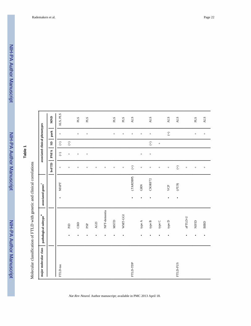
IntroductionFrontotemporal dementia (FTD) accounts for 5–15% of all dementia and is the secondcommonest cause in the presenile age group.1,2 FTD is a clinical syndrome, characterized byprogressive deterioration in behavior, personality and/or language, with relative preservationof memory.3–5 Clinical subtypes include the behavioral variant (bvFTD) and two forms ofprimary progressive aphasia; progressive non-fluent aphasia (PNFA) and semantic dementia(SD). In addition, FTD is often associated with an extrapyramidal movement disorder(parkinsonism or corticobasal syndrome) and/or motor neuron disease (MND).6,7 Given thevariability in phenotype, it is not surprising that the molecular basis of FTD is alsoheterogeneous (Table 1).
A family history of FTD is present in 25–50% of cases, often with an autosomal dominantpattern of inheritance, indicating a strong genetic component.8,9 In 1998, mutations in themicrotubule associated protein tau gene (MAPT) on chromosome 17 were identified in anumber of families with FTD and parkinsonism.10–12 Since then, 44 different MAPT
Correspondence to: Ian R. A. Mackenzie, Department of Pathology, Vancouver General Hospital, 855 West 12th Avenue, Vancouver,British Columbia, V5Z 1M9 Canada, Tel: 1-604-87504480, [email protected].
NIH Public AccessAuthor ManuscriptNat Rev Neurol. Author manuscript; available in PMC 2013 April 18.
Published in final edited form as:Nat Rev Neurol. 2012 August ; 8(8): 423–434. doi:10.1038/nrneurol.2012.117.
NIH
-PA Author Manuscript
NIH
-PA Author Manuscript
NIH
-PA Author Manuscript

mutations have been reported, accounting for 5–20% of familial FTD(www.molgen.ua.ac.be/ftdmutations).13 However, there remained a number of chromosome17 linked FTD families that were not explained by MAPT mutations. A major breakthroughoccurred in 2006 when progranulin (GRN) was identified as the second FTD-related gene onchromosome 17, with GRN mutations responsible for an even larger proportion of FTDfamilies.14,15 Much less common are mutations in the valosin containing protein gene(VCP) which cause the rare familial syndrome of inclusion body myopathy with Pagetdisease of bone and FTD16 and a mutation in the gene for charged multivesicular bodyprotein 2B (CHMP2B) found in a large Danish FTD pedigree.17 In addition, several familieswith a combination of FTD and amyotrophic lateral sclerosis (ALS) have been reported withgenetic linkage to a locus on chromosome 9p.18–26 Despite years of intense effort by manyresearch groups worldwide, the identity of the FTD/ALS gene on 9p remained elusive untiljust last year, when two independent studies identified the defect as being an expandedhexanucleotide repeat in a non-coding region of the chromosome 9 open reading frame 72gene (C9ORF72).27,28 Discovery of the C9ORF72 mutation has generated tremendousexcitement in the FTD and ALS research communities, as it appears to be the most commongenetic cause of both conditions (see below).
The neuropathology associated with clinical FTD is also heterogeneous.29 A commonfeature is the relatively selective degeneration of the frontal and temporal lobes, and the term“frontotemporal lobar degeneration” (FTLD) is often used for those pathological conditionsthat predominantly or commonly present with FTD. In addition, most cases of FTLD arefound to have abnormal intracellular accumulation of some disease-specific protein and ithas become popular to classify FTLD into broad categories, based on the molecular defectthought to be most characteristic.30,31 Until quite recently, the only FTLD subgroup weknew much about were those conditions characterized by the aggregation ofhyperphosphorylated tau protein in neurons and glia (FTLD-tau) (Table 1). However, mostFTD is not associated with tau pathology, but is characterized by neuronal inclusions thatwere originally identified with ubiquitin immunohistochemistry (FTLD-U).32,33 Just monthsafter publication of the GRN mutation discovery, another landmark paper reportedidentification of the transactive response DNA binding protein with molecular weight 43 kD(TDP-43) as the ubiquitinated pathological protein in most cases of FTLD-U (subsequentlyrenamed FTLD-TDP), as well as the vast majority of ALS.34,35 This provided strongevidence that FTD and ALS are closely related conditions with overlapping molecularpathogenesis. This concept was further strengthened in 2009 when, following the discoverythat mutations of the fused in sarcoma gene (FUS) cause autosomal dominant ALS,36,37 itwas shown that most of the ~10% of FTLD that do not have either tau or TDP-43 basedpathology are characterized by inclusions that are immunoreactive for FUS (FTLD-FUS).38–40 More recently, it has been shown that the FUS-positive inclusions in FTLD-FUSalso label for the other members of the FET protein family (including Ewing’s sarcomaprotein and TATA-binding protein-associated factor 15).41
The recent pace with which knowledge of the molecular genetics and neuropathology ofFTD has advanced has been truly remarkable (Box 1). In just over five years, we have gonefrom knowing virtually nothing of the molecular basis of most cases of FTD, to now beingable to determine the genetic cause in the majority of autosomal dominant families andbeing able to assign virtually all cases of FTLD to one of three major pathological subtypes(FTLD-tau, FTLD-TDP, FTLD-FUS).31 This insight is a crucial step towards improvedFTD patient care, as it provides the basis for more informed counseling and the potential formore specific diagnostic tests and targeted therapies. In this review, we will highlightseveral recent advances in our understanding of molecular aspects of FTD, focusing on therecent discovery of the C9ORF72 mutation and the roles of GRN, TDP-43, FUS and theother FET proteins in disease pathogenesis.
Rademakers et al. Page 2
Nat Rev Neurol. Author manuscript; available in PMC 2013 April 18.
NIH
-PA Author Manuscript
NIH
-PA Author Manuscript
NIH
-PA Author Manuscript

Advances in the molecular genetics of FTDGRN variants and its biological regulators in FTD
In less than six years, 69 different pathogenic GRN mutations have been reported in morethan 230 families worldwide, accounting for 5–20% of familial FTD and 1–5% of sporadiccases (www.molgen.ua.ac.be/ftdmutations).42 Progranulin (GRN) is a multifunctionalsecreted growth factor, expressed by many cell types including neurons.43 Pathogenicmutations are of various types and occur throughout the gene, but all cause disease viahaploinsufficiency.14,15 As a result, significantly reduced levels of GRN are consistentlyobserved in plasma, serum and CSF samples of symptomatic and asymptomatic GRNmutation carriers.44–46 Based on these findings, GRN enzyme-linked immunosorbent assays(ELISA) are now being developed as an inexpensive alternative to classical sequencinganalyses for diagnostic testing of FTD patients.
Genetic modifiers and regulators of GRN expression—The clinical phenotypeassociated with GRN mutations is quite variable47–52 and penetrance is incomplete.53
Understanding the factors that modify the expression of GRN mutations or regulate thenormal GRN gene is of potential therapeutic importance. One such genetic factor is theuncharacterized transmembrane protein 106B (TMEM106B), which was recently identifiedin a genome-wide association study (GWAS) of cases with known FTLD-TDP pathology.54
Genetic variants in and near TMEM106B appear to protect or delay the onset of FTD inindividuals with pathogenic GRN mutations, possibly by increasing GRN levels.54–57 Anumber of microRNAs, including miR-29b and miR-107 have also been implicated in GRNregulation.58,59 In addition, the minor T-allele of genetic variant rs5848, located in the 3′untranslated region of GRN, increases the binding of miR-659 to GRN, thereby reducingGRN levels.60 Genetic association studies have shown that carriers homozygous for the T-allele of rs5848 have a three-fold increased risk to develop FTLD-TDP compared withhomozygous C-allele carriers60, supporting a role for GRN in sporadic FTD and possiblyother neurodegenerative diseases such as Alzheimer’s disease.61–63 GRN expression mayalso be modified by certain exogenous factors. It was recently shown that GRN transcriptioncan be enhanced by small molecules including suberoylanilide hydroxamic acid (SAHA),64
while inhibitors of the vacuolar ATPase and some alkalizing drugs increase GRN productionand secretion through a translational mechanism.65
GRN cellular biology—Significant progress has also been made in recent years towardsour understanding of GRN biology and its neuroprotective function. The addition of GRN tostressed or GRN-depleted neuronal cells promotes neurite outgrowth.66–69 Theneuroprotective effects of GRN might be due, at least in part, to the activation of cellsignaling pathways involved in cell survival.66,70–73 A role for GRN in excitotoxicity andsynaptic transmission has also been suggested.68,74 Importantly, sortilin-1 (SORT1), aknown receptor for other neurotrophic factors in the brain, was identified in two independentstudies as the first known receptor for GRN.75,76 SORT1 has been shown to mediate GRNendocytosis and regulate the levels of GRN in vivo in mouse brain and in human plasma.More recently, tumor necrosis factor receptors were reported to directly interact withGRN.77 The identification of GRN receptors is exciting as it opens new avenues in GRNcell biology research and another potential route to FTD therapy.
C9ORF72 mutationSince 2006, increasing evidence has suggested the presence of a major locus for thecombined phenotype of FTD and ALS on chromosome 9p21, but the disease mutation hasremained elusive (Box 2).18–26 The key to the identification of the disease-causing mutationcame from the apparent non-Mendelian inheritance of a GGGGCC hexanucleotide repeat
Rademakers et al. Page 3
Nat Rev Neurol. Author manuscript; available in PMC 2013 April 18.
NIH
-PA Author Manuscript
NIH
-PA Author Manuscript
NIH
-PA Author Manuscript

located in a non-coding region of C9ORF72 (Figure 1a) in a large FTD-ALS familydesignated VSM-20 (Vancouver, San Francisco and Mayo family 20).27 Using primersflanking the repeat region, all affected individuals appeared homozygous by fluorescentPCR, while affected children seemed not to inherit an allele from their affected parent. Thisfinding suggested the presence of a repeat expansion that was too large to be amplified bythe PCR method, which was confirmed using a repeat-primed PCR assay (Figure 1b) andsouthern blot analysis. The polymorphic nature of this GC-rich hexanucleotide repeat wasindependently recognized in a Welsh FTD-ALS family using next-generation sequencing,also implicating this genomic region in disease pathogenesis.28
Mutation frequency—In the six months since the original discovery, numerous FTD andALS patients have been screened for the presence of GGGGCC repeat expansions inC9ORF72 using the repeat-primed PCR assay (Table 2).27,28,78–94 The mutation frequencyhas varied significantly among populations, with the highest being in genetically isolatedpopulations from Finland and Sardinia, and in cohorts where all patients had a pathologicaldiagnosis of FTLD-TDP (with or without ALS).27,28,81,85,88 The average mutationfrequencies in North American and European populations reported are 37% for familialALS, 6% for sporadic ALS, 21% for familial FTD and 6% for sporadic FTD patients. In allseries, the C9ORF72 mutation has been the most common genetic cause of familial ALS(more frequent than SOD1 mutations) and at least similar in frequency to GRN mutations inFTD families. To date, most of the patients included in the mutation screenings have beenCaucasian; however, C9ORF72 repeat expansions have also been identified in patients ofAfrican American, Middle Eastern and Asian race.86,88,94 Interestingly, independent of theclinical presentation or ethnic origin, all C9ORF72 mutation carriers inherit the expansionon the same genetic background, suggesting the presence of a common ancestor or,alternatively, the occurrence of multiple independent expansions on a fragile predisposingdisease haplotype.88,95,96
Clinical phenotypes—Several groups from North America and Europe have nowpublished descriptions of the demographic, clinical and neuropathological features of theircohorts of patients with the C9ORF72 mutation.27,28,78–94 The clinical presentation isheterogeneous and highly variable between and within families. Patients may present withFTD, ALS or features of both. The FTD subtype is most often bvFTD with PNFA lessfrequent. ALS usually has early involvement of both upper and lower motor neurons andbulbar presentation is particularly common.79,81,82,94 Several studies have found that ALSpatients with the mutation have a slightly earlier onset and shorter disease duration thanthose without the mutation.79,81,82,84,91,94 In addition to FTD and ALS, other features mayinclude memory disorder,78,85,87,90,92,93 psychosis,78,83,85,92,93 extrapyramidal movementdisorder (usually an akinetic-rigid syndrome)78,82,83,85,90,92 and cerebellar signs.85
Symptoms tend to accumulate and phenotypes converge with disease progression, so thatmost patients eventually develop at least some abnormalities of behavior, language andmotor function.78,80,82,85,94 There is wide variation in the age at onset (27 – 83 years, mean= 50s) and disease duration (1 – 22 years) and several studies have noted earlier diseaseonset in subsequent generations, consistent with genetic anticipation.78,81,82,84,85,94
Structural neuroimaging tends to show symmetric bilateral atrophy, primarily affectingfrontotemporal regions, but also involving other cerebral lobes and thecerebellum.78,85,87,92,97
Neuropathology—The neuropathology associated with the C9ORF72 mutation is acombination of FTLD-TDP and classical ALS.27,78,79,82,84,85,87,90,92–94,98,99 Regardless ofthe clinical phenotype, postmortem examination usually shows TDP-43 positive inclusionsin a wide range of neuroanatomical regions including the extramotor cerebral cortex,
Rademakers et al. Page 4
Nat Rev Neurol. Author manuscript; available in PMC 2013 April 18.
NIH
-PA Author Manuscript
NIH
-PA Author Manuscript
NIH
-PA Author Manuscript

hippocampus, basal ganglia, substantia nigra and lower motor neurons of the brainstem andspinal cord. In addition, a unique and highly characteristic feature of cases with the mutationis the presence of neuronal inclusions in the cerebellar granule cell layer, hippocampalpyramidal neurons and other neuroanatomical sites, that label for proteins of the ubiquitinproteasome system (ubiquitin, ubiquilins and p62) but that are negative for TDP-43 (Figure1c).27,78,79,82,84,85,87,90,92–94,98,99 This consistent finding supports the abnormal metabolismand accumulation of some, as yet unidentified molecule(s) that could include the mutantRNA, RNA-binding proteins or protein products of aberrant splicing. To date,immunohistochemical studies using commercial antibodies against C9ORF72 have failed todemonstrate any abnormal distribution or accumulation of the protein.27,79,82,85,87,93,94
Repeat size—All C9ORF72 mutation screenings performed to date have used the repeat-primed PCR method to detect the presence of a pathogenic GGGGCC repeat expansion.However, it is important to note that this method is only semi-quantitative and that thecharacteristic stutter pattern observed (Figure 1b) cannot be used to determine the exactnumber of repeats. In one family, southern blot analyses performed using DNA extractedfrom lymphoblast cell lines showed pathogenic repeat expansions of 700–1600 repeatunits;27 however, the minimal repeat size associated with disease may be considerablysmaller. Similar to other non-coding repeat expansion disorders, there is evidence forsomatic instability of the C9ORF72 repeat.27 This means that repeat lengths may varyamong different tissues within the same individual, making it difficult to accurately size therepeat and determine genetic/clinical/pathological correlations.100–102 Our current lack ofknowledge of the minimal pathogenic repeat-size combined with the technical challengesmentioned, raise important questions regarding genetic testing for this common mutation;particularly in the context of predictive genetic testing. Accurate sizing of the expandedrepeat in larger FTD and ALS patient series will be crucial to establish reliable cut-off sizesneeded to counsel individuals undergoing genetic testing. Future studies also need todetermine whether the repeat-length contributes to the variability in onset age and clinicalpresentation or whether other genetic and/or environmental modifiers are involved.
Disease mechanism—C9ORF72 is a completely uncharacterized protein whose functionis presently unknown. Two different C9ORF72 isoforms are predicted to be generated froma total of three different C9ORF72 transcripts;27 however, the relative expression of each ofthese transcripts in relevant brain regions has not yet been studied. Several groups haveshown ~50% loss of at least one C9ORF72 transcript in expanded repeat carriers,presumably resulting from the interference of the expanded GC-rich repeat with C9ORF72transcription regulation.27,28,84 Although these findings support a possible loss-of-functiondisease mechanism, the accumulation of GGGGCC repeat containing transcripts as nuclearRNA foci in frontal cortex and spinal cord of C9ORF72 mutation carriers has also beendemonstrated (Figure 1d), suggesting a possible toxic RNA gain-of function diseasemechanism.27 Based on commonalities with other non-coding repeat expansion disorders,these RNA foci may alter the function of one or more RNA-binding proteins resulting indownstream changes in gene expression and/or alternative splicing of a range oftranscripts.103 A number of cellular and animal models, either eliminating C9ORF72expression or overexpressing human C9ORF72 containing expanded GGGGCC repeats, arecurrently being generated to determine the relative contribution of each disease mechanismto neurodegeneration and TDP-43 aggregation.
Other FTD genes and genetic risk factorsWith the identification of the repeat expansion in C9ORF72, all previously published FTDfamilies with genome-wide linkage have now been accounted for. While it is unlikely thatany other common FTD-causing genes exist, rare mutations in other genes may each explain
Rademakers et al. Page 5
Nat Rev Neurol. Author manuscript; available in PMC 2013 April 18.
NIH
-PA Author Manuscript
NIH
-PA Author Manuscript
NIH
-PA Author Manuscript

a small number of the remaining families and combinations of genetic variants andenvironmental factors are likely to be responsible for disease in the majority of sporadicFTD patients. The use of exome and whole-genome sequencing will greatly facilitate thediscovery of rare genetic defects in the future. This was recently demonstrated with theidentification of the colony stimulating factor 1 receptor gene (CSF1R) as the cause ofhereditary diffuse leukoencephalopathy with spheroids (HDLS), 104 a disorder with variableclinical presentation that includes features of FTD. Additional GWAS, such as the largecollaborative study currently underway, which includes more than 2500 FTD patientsamples, may identify additional genetic risk factors.
ALS-related genes in FTD—Other rare genetic causes of FTD that have been identifiedin recent years include TARDBP and FUS, although mutations in each of these genesusually cause a pure ALS phenotype.36,37,105,106 This past year, UBQLN2, which encodes amember of the ubiquilin family that is involved in the degradation of ubiquitinated proteins,was also added to the list of ALS-FTD genes.107 In about 20% of UBQLN2 mutationcarriers, progressive dementia with abnormalities in both behavior and executive functionswere reported; however, none of these patients presented with FTD alone.
Advances in the molecular pathology of FTDTDP-43
TDP-43 is a highly conserved, predominantly nuclear protein, able to shuttle between thenucleus and cytoplasm. It has a number of well-described functions in RNA regulation suchas control of splicing, mRNA transport and stability; however, the complexity of TDP-43functions is just emerging.108–110
FTLD-TDP—Abnormal accumulation of TDP-43 in neuronal and glial inclusions is thecharacteristic neuropathological feature in approximately 50% of FTD patients (FTLD-TDP)and the vast majority of ALS cases.30,34,35 Pathological modifications of TDP-43 in thedisease state include a redistribution from the nucleus to the cytoplasm in cells withinclusions, hyperphosphorylation, ubiquitination and N-terminal truncation.35 FTLD-TDPincludes sporadic and genetic forms with mutations in GRN, VCP, TARDBP and therecently recognized C9ORF72 repeat expansion (see above).27,28,111–114 Based on themorphology and anatomic distribution of TDP-43 pathology, four distinct FTLD-TDPsubtypes are recognized.115–117 The relevance of this heterogeneity is supported by clinicaland genetic correlations (Table 1), as well as emerging evidence for distinct biochemicalproperties of TDP-43 in the different subtypes.35,117,118
Pathogenesis of TDP-43 proteinopathies—The neuropathological findings in FTLD-TDP implicate that both loss and gain-of-function mechanisms might be involved in TDP-43associated cell death. Addressing these fundamental questions is the focus of numerousresearch activities worldwide and detailed discussions of TDP-43 pathogenesis arepublished elsewhere.119–121 Briefly, current in vivo models provide evidence for bothscenarios by demonstrating that either reduced or increased expression levels of thephysiologically tightly autoregulated TDP-43 are not well tolerated.119–121 However, nomodel has fully recapitulated the neuropathological and biochemical features of humanTDP-43 related diseases. While the identification of TARDBP mutations is a clear indicatorthat dysfunction of TDP-43 is directly linked to neurodegeneration, the functionalconsequences of TARDBP mutations are still unresolved. There is no solid evidence thatTARDBP mutations act through a toxic-gain-of function mechanism and no functionalconsequences of TARDBP mutations on processing of the few RNA targets studied to datehave been reported. However, studies using crosslinking immunoprecipitation and high-
Rademakers et al. Page 6
Nat Rev Neurol. Author manuscript; available in PMC 2013 April 18.
NIH
-PA Author Manuscript
NIH
-PA Author Manuscript
NIH
-PA Author Manuscript

throughput sequencing have recently identified more than 6000 RNA targets of TDP-43, anda major challenge now is to dissect specific pathways regulated by TDP-43 and to identifypossible disease-relevant RNA targets.109,110
Another important but unresolved issue is the role of TDP-43 in the other genetic forms ofFTLD-TDP. The fact that mutations in GRN, VCP and C9ORF72 are all consistentlycharacterized by TDP-43 pathology suggests that dysregulation of TDP-43 might be acrucial common downstream mechanism leading to cell death in all of them. However, thesignificance of TDP-43 accumulation in C9ORF72 mutation carriers has recently beenchallenged by the identification of additional TDP-43 negative, ubiquitin-positive pathologythat is more abundant than TDP-43 pathology in distinct brain regions (Figure 1c), raisingthe possibility that another unidentified protein(s) might be more important in thepathogenesis in these cases (see above).27,78,79,82,84,85,87,90,92–94,98,99
FUS and other FET proteinsFUS belongs to the FET protein family that also includes Ewing’s sarcoma (EWS), TATA-binding protein-associated factor 15 (TAF15) and the drosophila orthologue cabeza. Theyare highly conserved, ubiquitously expressed, predominantly nuclear (Figure 2a),multifunctional DNA/RNA binding proteins,122 that can bind to a large number of partiallyoverlapping RNA targets.123
FTLD-FUS / FTLD-FET—In early 2009, FUS mutations were reported to be the cause of~3 % of familial ALS cases, in which the associated pathology is characterized by inclusionsthat are FUS positive but TDP-43 negative (ALS-FUS).36,37 Subsequently, FUS was foundto be the most characteristic marker for the pathology in most of the remaining tau/TDP-negative FTLD cases, which include three closely related but distinct clinicopathologicalentities; atypical FTLD-U (aFTLD-U), neuronal intermediate filament inclusion disease(NIFID) and basophilic inclusion body disease (BIBD).38–40,124 The identification ofFTLD-FUS as a new molecular subgroup31 provided further evidence that FTD and ALS areclosely related conditions and emphasized the pathogenic role of RNA binding proteins.However, despite there being some overlap in the phenotype and pathological features ofFTLD-FUS and ALS-FUS, significant differences were also observed.125,126 Moreover, thepublication of additional cases made it evident that ALS with FUS pathology is almostalways caused by a FUS mutation; whereas, cases of FTLD-FUS tend to be sporadic andnone has yet been associated with any genetic abnormality of FUS.38–40,124 Furtherevidence for different pathomechanisms has been provided by a recent study thatinvestigated the other FET protein members in a series of ALS-FUS and FTLD-FUScases.41 In cases of ALS-FUS with a range of different mutations, there was no co-accumulation of other FET proteins into FUS-positive inclusions and cells retained thephysiological nuclear staining of TAF15 and EWS (Figure 2b). In striking contrast, in allFTLD-FUS subtypes TAF15 and EWS were also found to co-accumulate in FUS-positiveinclusions and inclusion-bearing cells showed a reduction in the normal nuclear staining ofall three FET proteins, particularly TAF15 (Figure 2c). The addition of TAF15 and EWS tothe growing list of RNA binding proteins involved in neurodegeneration is further supportedby studies in which TAF15 was predicted as a potential candidate through an independentapproach using a yeast functional screen aimed to identify RNA binding proteins withsimilar function to TDP-43 and FUS127 and descriptions of genetic variants (ofundetermined pathogenic significance) in TAF15 and EWSR1 in a small number of ALScases.128,129 Although the respective roles of FUS, TAF15 and EWS in FTLD-FUS remainsto be elucidated, the term FTLD-FET now seems more appropriate for this molecular FTLDsubgroup.
Rademakers et al. Page 7
Nat Rev Neurol. Author manuscript; available in PMC 2013 April 18.
NIH
-PA Author Manuscript
NIH
-PA Author Manuscript
NIH
-PA Author Manuscript

Pathogenesis of FUS-proteinopathies—The above described differences in themolecular pathology of ALS-FUS and FTLD-FUS imply different pathological processesunderlying inclusion formation and cell death, with ALS-FUS being restricted todysfunction of FUS, while FTLD-FUS might involve dysfunction of all FET proteins(Figure 2).
In ALS-FUS, mutations in the C-terminus of the protein disrupt a region characterized asnon-classical nuclear localization sequence. This results in an impaired transportin-mediatednuclear import with redistribution of FUS to the cytoplasm (Figure 2b).130,131 Importantly,no alteration of other FET proteins is seen under these conditions.41 The degree of FUSnuclear transport impairment varies between different FUS mutations, but correlates withthe observed variability in disease course associated with different mutations and withdistinct pathological patterns of ALS-FUS pathology,125 thereby providing strong evidencethat impaired nuclear import of FUS is a key event in disease pathogenesis of ALS-FUS.
In FTLD-FUS, a more general defect of transportin-mediated nuclear import is postulatedthat affects the distribution of all FET proteins, with two broad scenarios plausible (Figure2c). First, a primary defect of transportin itself, either resulting from genetic variations inTNPO1, posttranslational modifications, or altered expression levels of transportin, couldresult in reduced efficiency of nuclear import of all FET proteins. However, in this scenarioone might also expect alterations in the subcellular distribution of other transportin cargos,such as hnRNPA1, which is not supported by preliminary data.41 Second, the proper nuclearimport of FET proteins might be affected by abnormal posttranslational modifications ofFET proteins, such as arginine methylation or phosphorylation, that have been shown tomodulate nucleocytoplasmic transport, protein-protein interaction and proteinstability.122,132–138 So far, biochemical analysis of protein extracted from FTLD-FUS brainshas revealed only increased insolubility of all FET proteins, without other obvious disease-associated changes, such as truncation or abnormal phosphorylation;39,41,139 however, moredetailed analysis is required.
The downstream effects of redistributed FUS or all FET proteins in the pathogenesis ofALS-FUS and FTLD-FUS, respectively, have not yet been determined. Similar to TDP-43,both a gain of toxic properties and a loss of functions via their sequestration in aggregatesare plausible. Results from initial in vivo models of ALS-FUS have been inconsistent andthe mechanisms remain unresolved.121,140
Molecular correlates of FTD phenotypesTable 1 lists the molecular subtypes of FTLD pathology with the associated genetic defectsand common clinical features. Each genetic cause is associated with a specificneuropathology. However, predicting the underlying molecular pathology or genetics, basedon the pattern of inheritance and clinical features, is often imprecise.141,142 SD is usuallysporadic and associated with FTLD-TDP type C with fewer cases having the pathology ofclassical PiD. Cases of sporadic PNFA are somewhat more likely to have FTLD-tau thanFTLD-TDP, but bvFTD may be associated with any of the major pathologies. Early-onsetbvFTD with severe psychobehavioral abnormality but minimal motor features or aphasia ischaracteristic of the aFTLD-U subtype of FTLD-FUS. When FTD is combined with ALS,the pathology is usually FTLD-TDP; whereas, FTD with prominent parkinsonism is moreoften FTLD-tau (PSP or CBD). In families with autosomal dominant inheritance of bvFTDor PNFA without significant motor dysfunction, the underlying gene defect may be amutation in C9ORF72, GRN or MAPT. When parkinsonism or primary lateral sclerosis(PLS) are also prominent features, a MAPT mutation is more likely; whereas, coexistence ofclassical ALS in a family strongly suggests a C9ORF72 mutation.
Rademakers et al. Page 8
Nat Rev Neurol. Author manuscript; available in PMC 2013 April 18.
NIH
-PA Author Manuscript
NIH
-PA Author Manuscript
NIH
-PA Author Manuscript

Conclusions and future directionsThe past six years have seen remarkable progress in our understanding of the molecularbasis of FTD. It appears that all the common FTD-causing genes have now been discoveredand the major pathological proteins identified. Although many aspects of the specificpathogenic mechanisms still need to be resolved, we are already in position to begintranslating this newly acquired knowledge into improved FTD patient care. The recentdiscoveries of GRN and C9ORF72 mutations allow for more informed genetic counseling.Knowledge of the signature pathological proteins is prompting attempts to develop moredisease-specific, molecular-based diagnostic tests, such as the quantification of total orpathological protein species in biofluids.143,144 Recognition of GRN insufficiency as animportant mechanism in familial and some sporadic forms of FTD, combined with improvedunderstanding of GRN regulation and cell biology, has already led to initial plans for GRN-based clinical trials (http://www.alzforum.org/new/pdf/FTLDSeries.pdf). The identificationof TDP-43, FET proteins and C9ORF72 has open up new avenues of research related toRNA regulation. Finally, a greater appreciation of the overlap between FTD and ALS is nowbringing these two areas of research and patient care closer together. Hopefully, patientswith FTD will soon experience real benefits from these and future advances.
Box 1
Important events in the molecular pathogenesis of FTD
• 1892: Arnold Pick describes lobar atrophy in a patient with presenile dementiaand aphasia.145
• 1911: Alois Alzheimer characterizes Pick bodies using silver stains.146
• 1960’s: descriptions of PSP and CBD clinicopathological syndromes.147,148
• 1974: different pathological subtypes of PiD disease described.149
• mid 1980’s - early 1990’s: identification of tau as major component ofpathological lesions in AD, PiD, PSP and CBD (reviewed in Lee et al.).150
• 1990: description of FTD cases without specific histopathology (DLDH).151
• mid 1990’s: identification of subset of FTD with FTLD-U pathology.152
• 1998: MAPT mutations identified in some families with FTD and parkinsonismgenetically linked to chromosome 17.10–12
• 2004–06: recognition that most cases of DLDH are really FTLD-U and thatFTLD-U is the most common FTD-associated pathology.33
• 2006: description of different patterns of FTLD-U that correlate with clinicalphenotypes, genetic abnormalities and biochemical properties ofinclusions.115,117
• 2006: discovery that GRN mutations cause autosomal dominant FTD andexplain all remaining chromosome 17 linked families.14,15
• 2006: TDP-43 identified as pathological protein in most cases of FTLD-U andALS.34,35
• 2008: identification of a subset of FTLD-U cases that lack TDP-43-immunoreactive pathology (aFTLD-U).153,154
• 2009: discovery that most cases of tau/TDP-43-negative FTLD have FUS-immunoreactive pathology (FTLD-FUS).38–40
Rademakers et al. Page 9
Nat Rev Neurol. Author manuscript; available in PMC 2013 April 18.
NIH
-PA Author Manuscript
NIH
-PA Author Manuscript
NIH
-PA Author Manuscript

• 2011: discovery that FTLD-FUS shows accumulation of other FET proteinmembers TAF15 and EWS.41
• 2011: FTD/ALS associated gene defect on chromosome 9p identified as repeatexpansion in C9ORF72.27,28
Abbreviations: AD, Alzheimer’s disease; aFTLD-U, atypical FTLD-U; C9ORF72,chromosome 9 open reading frame 72; CBD, corticobasal degeneration; DLDH,dementia lacking distinctive histopathology; EWS, Ewing’s sarcoma; FTD,frontotemporal dementia; FTLD, frontotemporal lobar degeneration; FTLD-U, FTLDwith ubiquitin immunoreactive inclusions; FUS, fused in sarcoma protein; PSP,progressive supranuclear palsy; PiD, Pick’s disease; TAF15, TATA-binding protein-associated factor 15; TDP-43, transactive response DNA binding protein with molecularweight 43 kD.
Box 2
History of the chromosome 9p FTD-ALS locus
Since 2006, at least 10 autosomal dominant families in which patients were affected withFTD, ALS, or both were published with conclusive or suggestive linkage to chromosome9p.18–26 The minimal candidate region shared by all families was a 3.7Mb regioncontaining only 10 known or predicted genes. In 2010, three genome-wide associationstudies (GWAS) in sporadic ALS populations identified a novel susceptibility locus onchromosome 9p which completely overlapped with the candidate region for familialFTD-ALS.155–157 Strongest association was identified in a ~80kb haplotype blockcontaining only three genes; MOBKL2B, IFNK and C9ORF72. An independent GWASstudy in patients with pathologically confirmed FTLD-TDP nominated the samechromosomal region, implicating the chromosome 9p gene defect in sporadic forms ofboth FTD and ALS.54 However, despite concentrated efforts by the FTD and ALSresearch communities, in-depth candidate-gene sequencing and targeted next-generationsequencing of the minimal candidate region failed to identify the causative mutation,suggesting that a complex mutational mechanism may be involved. In 2011, an expandedhexanucleotide repeat in the non-coding region of C9ORF72 was found to be the longsought-after cause of FTD and ALS on chromosome 9p.27,28
AcknowledgmentsRosa Rademakers is funded by NIH grants P50 AG016574, R01 NS065782 and R01 AG026251, the ALS TherapyAlliance and the Consortium for Frontotemporal dementia (CFR). Manuela Neumann is funded by the SwissNational Science Foundation grants 31003A-132864 and CRSII3 136222, the German Federal Ministry ofEducation and Research grant 01GI1005B, the Stavros-Niarchos Foundation, the Synapsis Foundation, and theHans and Ilse Breuer Foundation. Ian Mackenzie is funded by the Canadian Institutes of Health Research grants179009 and 74580 and the Pacific Alzheimer’s Research Foundation center grant C06-01.
References1. Bird T, et al. Epidemiology and genetics of frontotemporal dementia/Pick’s disease. Ann Neurol.
2003; 54 (Suppl 5):S29–31. [PubMed: 12833366]
2. Feldman H, et al. A Canadian cohort study of cognitive impairment and related dementias(ACCORD): study methods and baseline results. Neuroepidemiology. 2003; 22:265–274. [PubMed:12902621]
3. The Lund and Manchester Groups. Clinical and neuropathological criteria for frontotemporaldementia. J Neurol Neurosurg Psychiatr. 1994; 57:416–418. [PubMed: 8163988]
Rademakers et al. Page 10
Nat Rev Neurol. Author manuscript; available in PMC 2013 April 18.
NIH
-PA Author Manuscript
NIH
-PA Author Manuscript
NIH
-PA Author Manuscript

4. McKhann GM, et al. Clinical and pathological diagnosis of frontotemporal dementia: report of theWork Group on Frontotemporal Dementia and Pick’s Disease. Arch Neurol. 2001; 58:1803–1809.[PubMed: 11708987]
5. Neary D, et al. Frontotemporal lobar degeneration: a consensus on clinical diagnostic criteria.Neurology. 1998; 51:1546–1554. [PubMed: 9855500]
6. Burrell JR, Kiernan MC, Vucic S, Hodges JR. Motor neuron dysfunction in frontotemporaldementia. Brain. 2011; 134:2582–2594. [PubMed: 21840887]
7. Lomen-Hoerth C, Anderson T, Miller B. The overlap of amyotrophic lateral sclerosis andfrontotemporal dementia. Neurology. 2002; 59:1077–1079. [PubMed: 12370467]
8. Rohrer JD, et al. The heritability and genetics of frontotemporal lobar degeneration. Neurology.2009; 73:1451–1456. [PubMed: 19884572]
9. Seelaar H, et al. Distinct genetic forms of frontotemporal dementia. Neurology. 2008; 71:1220–1226. [PubMed: 18703462]
10. Hutton M, et al. Association of missense and 5′-splice-site mutations in tau with the inheriteddementia FTDP-17. Nature. 1998; 393:702–705. [PubMed: 9641683]
11. Poorkaj P, et al. Tau is a candidate gene for chromosome 17 frontotemporal dementia. Ann Neurol.1998; 43:815–825. [PubMed: 9629852]
12. Spillantini MG, et al. Mutation in the tau gene in familial multiple system tauopathy with preseniledementia. Proc Natl Acad Sci U S A. 1998; 95:7737–7741. [PubMed: 9636220]
13. Rademakers R, Cruts M, van Broeckhoven C. The role of tau (MAPT) in frontotemporal dementiaand related tauopathies. Hum Mut. 2004; 24:277–295. [PubMed: 15365985]
14. Baker M, et al. Mutations in progranulin cause tau-negative frontotemporal dementia linked tochromosome 17. Nature. 2006; 442:916–919. [PubMed: 16862116]
15. Cruts M, et al. Null mutations in progranulin cause ubiquitin-positive frontotemporal dementialinked to chromosome 17q21. Nature. 2006; 442:920–924. [PubMed: 16862115]
16. Watts GD, et al. Inclusion body myopathy associated with Paget disease of bone andfrontotemporal dementia is caused by mutant valosin-containing protein. Nat Genet. 2004;36:377–381. [PubMed: 15034582]
17. Skibinski G, et al. Mutations in the endosomal ESCRTIII-complex subunit CHMP2B infrontotemporal dementia. Nat Genet. 2005; 37:806–808. [PubMed: 16041373]
18. Boxer AL, et al. Clinical, neuroimaging and neuropathological features of a new chromosome 9p-linked FTD-ALS family. J Neurol Neurosurg Psychiatr. 2011; 82:196–203. [PubMed: 20562461]
19. Gijselinck I, et al. Identification of 2 Loci at chromosomes 9 and 14 in a multiplex family withfrontotemporal lobar degeneration and amyotrophic lateral sclerosis. Arch Neurol. 2010; 67:606–616. [PubMed: 20457961]
20. Le Ber I, et al. Chromosome 9p-linked families with frontotemporal dementia associated withmotor neuron disease. Neurology. 2009; 72:1669–1676. [PubMed: 19433740]
21. Luty AA, et al. Pedigree with frontotemporal lobar degeneration--motor neuron disease and TarDNA binding protein-43 positive neuropathology: genetic linkage to chromosome 9. BMC Neurol.2008; 8:32. [PubMed: 18755042]
22. Momeni P, et al. Analysis of IFT74 as a candidate gene for chromosome 9p-linked ALS-FTD.BMC Neurol. 2006; 6:44. [PubMed: 17166276]
23. Morita M, et al. A locus on chromosome 9p confers susceptibility to ALS and frontotemporaldementia. Neurology. 2006; 66:839–844. [PubMed: 16421333]
24. Pearson JP, et al. Familial frontotemporal dementia with amyotrophic lateral sclerosis and a sharedhaplotype on chromosome 9p. J Neurol. 2011; 258:647–655. [PubMed: 21072532]
25. Valdmanis PN, et al. Three families with amyotrophic lateral sclerosis and frontotemporaldementia with evidence of linkage to chromosome 9p. Arch Neurol. 2007; 64:240–245. [PubMed:17296840]
26. Vance C, et al. Familial amyotrophic lateral sclerosis with frontotemporal dementia is linked to alocus on chromosome 9p13.2–21.3. Brain. 2006; 129:868–876. [PubMed: 16495328]
Rademakers et al. Page 11
Nat Rev Neurol. Author manuscript; available in PMC 2013 April 18.
NIH
-PA Author Manuscript
NIH
-PA Author Manuscript
NIH
-PA Author Manuscript

27. DeJesus-Hernandez M, et al. Expanded GGGGCC hexanucleotide repeat in noncoding region ofC9ORF72 causes chromosome 9p-linked FTD and ALS. Neuron. 2011; 72:245–256. [PubMed:21944778]
28. Renton AE, et al. A hexanucleotide repeat expansion in C9ORF72 is the cause of chromosome9p21-linked ALS-FTD. Neuron. 2011; 72:257–268. [PubMed: 21944779]
29. Cairns NJ, et al. Neuropathologic diagnostic and nosologic criteria for frontotemporal lobardegeneration: consensus of the Consortium for Frontotemporal Lobar Degeneration. ActaNeuropathol. 2007; 114:5–22. [PubMed: 17579875]
30. Mackenzie IR, et al. Nomenclature for neuropathologic subtypes of frontotemporal lobardegeneration: consensus recommendations. Acta Neuropathol. 2009; 117:15–18. [PubMed:19015862]
31. Mackenzie IR, et al. Nomenclature and nosology for neuropathologic subtypes of frontotemporallobar degeneration: an update. Acta Neuropathol. 2010; 119:1–4. [PubMed: 19924424]
32. Lipton AM, White CL 3rd, Bigio EH. Frontotemporal lobar degeneration with motor neurondisease-type inclusions predominates in 76 cases of frontotemporal degeneration. ActaNeuropathol. 2004; 108:379–385. [PubMed: 15351890]
33. Mackenzie IR, et al. Dementia lacking distinctive histology (DLDH) revisited. Acta Neuropathol.2006; 112:551–559. [PubMed: 16900341]
34. Arai T, et al. TDP-43 is a component of ubiquitin-positive tau-negative inclusions infrontotemporal lobar degeneration and amyotrophic lateral sclerosis. Biochem Biophys ResCommun. 2006; 351:602–611. [PubMed: 17084815]
35. Neumann M, et al. Ubiquitinated TDP-43 in frontotemporal lobar degeneration and amyotrophiclateral sclerosis. Science. 2006; 314:130–133. [PubMed: 17023659]
36. Kwiatkowski TJ Jr, et al. Science. 2009; 323:1205–1208. [PubMed: 19251627]
37. Vance C, et al. Mutations in FUS, an RNA processing protein, cause familial amyotrophic lateralsclerosis type 6. Science. 2009; 323:1208–1211. [PubMed: 19251628]
38. Munoz DG, et al. FUS pathology in basophilic inclusion body disease. Acta Neuropathol. 2009;118:617–627. [PubMed: 19830439]
39. Neumann M, et al. A new subtype of frontotemporal lobar degeneration with FUS pathology.Brain. 2009; 132:2922–2931. [PubMed: 19674978]
40. Neumann M, et al. Abundant FUS-immunoreactive pathology in neuronal intermediate filamentinclusion disease. Acta Neuropathol. 2009; 118:605–616. [PubMed: 19669651]
41. Neumann M, et al. FET proteins TAF15 and EWS are selective markers that distinguish FTLDwith FUS pathology from amyotrophic lateral sclerosis with FUS mutations. Brain. 2011;134:2595–2609. [PubMed: 21856723]
42. Gijselinck I, Van Broeckhoven C, Cruts M. Granulin mutations associated with frontotemporallobar degeneration and related disorders: an update. Hum Mut. 2008; 29:1373–1386. [PubMed:18543312]
43. Bateman A, Bennett HP. The granulin gene family: from cancer to dementia. Bioessays. 2009;31:1245–1254. [PubMed: 19795409]
44. Finch N, et al. Plasma progranulin levels predict progranulin mutation status in frontotemporaldementia patients and asymptomatic family members. Brain. 2009; 132:583–591. [PubMed:19158106]
45. Ghidoni R, Benussi L, Glionna M, Franzoni M, Binetti G. Low plasma progranulin levels predictprogranulin mutations in frontotemporal lobar degeneration. Neurology. 2008; 71:1235–1239.[PubMed: 18768919]
46. Sleegers K, et al. Serum biomarker for progranulin-associated frontotemporal lobar degeneration.Ann Neurol. 2009; 65:603–609. [PubMed: 19288468]
47. Beck J, et al. A distinct clinical, neuropsychological and radiological phenotype is associated withprogranulin gene mutations in a large UK series. Brain. 2008; 131:706–720. [PubMed: 18234697]
48. Gass J, et al. Mutations in progranulin are a major cause of ubiquitin-positive frontotemporal lobardegeneration. Hum Mol Genet. 2006; 15:2988–3001. [PubMed: 16950801]
Rademakers et al. Page 12
Nat Rev Neurol. Author manuscript; available in PMC 2013 April 18.
NIH
-PA Author Manuscript
NIH
-PA Author Manuscript
NIH
-PA Author Manuscript

49. Le Ber I, et al. Progranulin null mutations in both sporadic and familial frontotemporal dementia.Human Mut. 2007; 28:846–855.
50. Moreno F, et al. “Frontotemporoparietal” dementia: clinical phenotype associated with the c.709-1G>A PGRN mutation. Neurology. 2009; 73:1367–1374. [PubMed: 19858458]
51. Snowden JS, et al. Progranulin gene mutations associated with frontotemporal dementia andprogressive non-fluent aphasia. Brain. 2006; 129:3091–3102. [PubMed: 17003069]
52. Masellis M, et al. Novel splicing mutation in the progranulin gene causing familial corticobasalsyndrome. Brain. 2006; 129:3115–3123. [PubMed: 17030534]
53. Cruts M, Van Broeckhoven C. Loss of progranulin function in frontotemporal lobar degeneration.Trends Genet. 2008; 24:186–194. [PubMed: 18328591]
54. Van Deerlin VM, et al. Common variants at 7p21 are associated with frontotemporal lobardegeneration with TDP-43 inclusions. Nat Genet. 2010; 42:234–239. [PubMed: 20154673]
55. Cruchaga C, et al. Association of TMEM106B gene polymorphism with age at onset in granulinmutation carriers and plasma granulin protein levels. Arch Neurol. 2011; 68:581–586. [PubMed:21220649]
56. Finch N, et al. TMEM106B regulates progranulin levels and the penetrance of FTLD in GRNmutation carriers. Neurology. 2011; 76:467–474. [PubMed: 21178100]
57. van der Zee J, et al. TMEM106B is associated with frontotemporal lobar degeneration in aclinically diagnosed patient cohort. Brain. 2011; 134:808–815. [PubMed: 21354975]
58. Jiao J, Herl LD, Farese RV, Gao FB. MicroRNA-29b regulates the expression level of humanprogranulin, a secreted glycoprotein implicated in frontotemporal dementia. PLoS One. 2010;5:e10551. [PubMed: 20479936]
59. Wang WX, et al. miR-107 regulates granulin/progranulin with implications for traumatic braininjury and neurodegenerative disease. Am J Pathol. 2010; 177:334–345. [PubMed: 20489155]
60. Rademakers R, et al. Common variation in the miR-659 binding-site of GRN is a major risk factorfor TDP43-positive frontotemporal dementia. Hum Mol Gene t. 2008; 17:3631–3642.
61. Brouwers N, et al. Genetic variability in progranulin contributes to risk for clinically diagnosedAlzheimer disease. Neurology. 2008; 71:656–664. [PubMed: 18565828]
62. Lee MJ, Chen TF, Cheng TW, Chiu MJ. rs5848 variant of progranulin gene is a risk ofAlzheimer’s disease in the Taiwanese population. Neurodegener Dis. 2011; 8:216–220. [PubMed:21212639]
63. Viswanathan J, et al. An association study between granulin gene polymorphisms and Alzheimer’sdisease in Finnish population. Am J Med Genet B Neuropsychiatr Genet. 2009; 150B:747–750.[PubMed: 19016491]
64. Cenik B, et al. Suberoylanilide hydroxamic acid (vorinostat) up-regulates progranulintranscription: rational therapeutic approach to frontotemporal dementia. J Biol Chem. 2011;286:16101–16108. [PubMed: 21454553]
65. Capell A, et al. Rescue of progranulin deficiency associated with frontotemporal lobardegeneration by alkalizing reagents and inhibition of vacuolar ATPase. J Neurosci. 2011;31:1885–1894. [PubMed: 21289198]
66. Gao X, et al. Progranulin promotes neurite outgrowth and neuronal differentiation by regulatingGSK-3beta. Protein Cell. 2010; 1:552–562. [PubMed: 21204008]
67. Ryan CL, et al. Progranulin is expressed within motor neurons and promotes neuronal cellsurvival. BMC Neurosci. 2009; 10:130. [PubMed: 19860916]
68. Tapia L, et al. Progranulin deficiency decreases gross neural connectivity but enhancestransmission at individual synapses. J Neurosci. 2011; 31:11126–11132. [PubMed: 21813674]
69. Van Damme P, et al. Progranulin functions as a neurotrophic factor to regulate neurite outgrowthand enhance neuronal survival. J Cell Biol. 2008; 181:37–41. [PubMed: 18378771]
70. Kleinberger G, et al. Increased caspase activation and decreased TDP-43 solubility in progranulinknockout cortical cultures. J Neurochem. 2010; 115:735–747. [PubMed: 20731760]
71. Nedachi T, Kawai T, Matsuwaki T, Yamanouchi K, Nishihara M. Progranulin enhances neuralprogenitor cell proliferation through glycogen synthase kinase 3beta phosphorylation.Neuroscience. 2011; 185:106–115. [PubMed: 21540081]
Rademakers et al. Page 13
Nat Rev Neurol. Author manuscript; available in PMC 2013 April 18.
NIH
-PA Author Manuscript
NIH
-PA Author Manuscript
NIH
-PA Author Manuscript

72. Rosen EY, et al. Functional genomic analyses identify pathways dysregulated by progranulindeficiency, implicating Wnt signaling. Neuron. 2011; 71:1030–1042. [PubMed: 21943601]
73. Xu J, et al. Extracellular progranulin protects cortical neurons from toxic insults by activatingsurvival signaling. Neurobiol Aging. 2011; 32:2326 e2325–2316. [PubMed: 21820214]
74. Guo A, Tapia L, Bamji SX, Cynader MS, Jia W. Progranulin deficiency leads to enhanced cellvulnerability and TDP-43 translocation in primary neuronal cultures. Brain Res. 2010; 1366:1–8.[PubMed: 20888804]
75. Carrasquillo MM, et al. Genome-wide screen identifies rs646776 near sortilin as a regulator ofprogranulin levels in human plasma. Am J Hum Genet. 2010; 87:890–897. [PubMed: 21087763]
76. Hu F, et al. Sortilin-mediated endocytosis determines levels of the frontotemporal dementiaprotein, progranulin. Neuron. 2010; 68:654–667. [PubMed: 21092856]
77. Tang W, et al. The growth factor progranulin binds to TNF receptors and is therapeutic againstinflammatory arthritis in mice. Science. 2011; 332:478–484. [PubMed: 21393509]
78. Boeve BF, et al. Characterization of frontotemporal dementia and/or amyotrophic lateral sclerosisassociated with the GGGGCC repeat expansion in C9ORF72. Brain. 2012; 135:765–783.[PubMed: 22366793]
79. Brettschneider J, et al. Pattern of ubiquilin pathology in ALS and FTLD indicates presence ofC9ORF72 hexanucleotide expansion. Acta Neuropathol. 2012 in press.
80. Byrne S, et al. Cognitive and clinical characteristics of patients with amyotrophic lateral sclerosiscarrying a C9orf72 repeat expansion: a population-based cohort study. Lancet Neurol. 2012;11:232–240. [PubMed: 22305801]
81. Chio A, et al. Clinical characteristics of patients with familial amyotrophic lateral sclerosiscarrying the pathogenic GGGGCC hexanucleotide repeat expansion of C9ORF72. Brain. 2012;135:784–793. [PubMed: 22366794]
82. Cooper-Knock J, et al. Clinico-pathological features in amyotrophic lateral sclerosis withexpansions in C9ORF72. Brain. 2012; 135:751–764. [PubMed: 22366792]
83. Floris G, et al. Frontotemporal dementia with psychosis, parkinsonism, visuo-spatial dysfunction,upper motor neuron involvement associated to expansion of C9ORF72: a peculiar phenotype? JNeurol. 2012 in press.
84. Gijselinck I, et al. A C9orf72 promoter repeat expansion in a Flanders-Belgian cohort withdisorders of the frontotemporal lobar degeneration-amyotrophic lateral sclerosis spectrum: a geneidentification study. Lancet Neurol. 2012; 11:54–65. [PubMed: 22154785]
85. Hsiung GY, et al. Clinical and pathological features of familial frontotemporal dementia caused byC9ORF72 mutation on chromosome 9p. Brain. 2012; 135:709–722. [PubMed: 22344582]
86. Kandiah N, et al. Case Report of an Asian Patient with FTD-ALS due to C9ORF72 mutation. CanJ Neurol Sci. 2012 in press.
87. Mahoney CJ, et al. Frontotemporal dementia with the C9ORF72 hexanucleotide repeat expansion:clinical, neuroanatomical and neuropathological features. Brain. 2012; 135:736–750. [PubMed:22366791]
88. Majounie E, et al. Frequency of the C9orf72 hexanucleotide repeat expansion in patients withamyotrophic lateral sclerosis and frontotemporal dementia: a cross-sectional study. Lancet Neurol.2012; 11:323–330. [PubMed: 22406228]
89. Mok KY, et al. High frequency of the expanded C9ORF72 hexanucleotide repeat in familial andsporadic Greek ALS patients. Neurobiol Aging. 2012 in press.
90. Murray ME, et al. Clinical and neuropathologic heterogeneity of c9FTD/ALS associated withhexanucleotide repeat expansion in C9ORF72. Acta Neuropathol. 2011; 122:673–690. [PubMed:22083254]
91. Sabatelli M, et al. C9ORF72 hexanucleotide repeat expansions in the Italian sporadic ALSpopulation. Neurobiol Aging. 2012
92. Simon-Sanchez J, et al. The clinical and pathological phenotype of C9ORF72 hexanucleotiderepeat expansions. Brain. 2012; 135:723–735. [PubMed: 22300876]
93. Snowden JS, et al. Distinct clinical and pathological characteristics of frontotemporal dementiaassociated with C9ORF72 mutations. Brain. 2012; 135:693–708. [PubMed: 22300873]
Rademakers et al. Page 14
Nat Rev Neurol. Author manuscript; available in PMC 2013 April 18.
NIH
-PA Author Manuscript
NIH
-PA Author Manuscript
NIH
-PA Author Manuscript

94. Stewart H, et al. Clinical and pathological features of amyotrophic lateral sclerosis caused bymutation in the C9ORF72 gene on chromosome 9p. Acta Neuropathol. 2012; 123:409–417.[PubMed: 22228244]
95. Mok K, et al. Chromosome 9 ALS and FTD locus is probably derived from a single founder.Neurobiol Aging. 2012; 33:209 e203–208. [PubMed: 21925771]
96. Rademakers R. C9orf72 repeat expansions in patients with ALS and FTD. Lancet Neurol. 2012;11:297–298. [PubMed: 22406229]
97. Whitwell JL, et al. Neuroimaging signatures of frontotemporal dementia genetics: C9ORF72, tau,progranulin and sporadics. Brain. 2012; 135:794–806. [PubMed: 22366795]
98. Al-Sarraj S, et al. p62 positive, TDP-43 negative, neuronal cytoplasmic and intranuclear inclusionsin the cerebellum and hippocampus define the pathology of C9orf72-linked FTLD and MND/ALS.Acta Neuropathol. 2011; 122:691–702. [PubMed: 22101323]
99. Troakes C, et al. An MND/ALS phenotype associated with C9orf72 repeat expansion: Abundantp62-positive, TDP-43-negative inclusions in cerebral cortex, hippocampus and cerebellum butwithout associated cognitive decline. Neuropathology. 2011
100. Lavedan C, et al. Myotonic dystrophy: size- and sex-dependent dynamics of CTG meioticinstability, and somatic mosaicism. Am J Hum Genet. 1993; 52:875–883. [PubMed: 8098180]
101. Matsuura T, et al. Somatic and germline instability of the ATTCT repeat in spinocerebellar ataxiatype 10. Am J Hum Genet. 2004; 74:1216–1224. [PubMed: 15127363]
102. Moseley ML, et al. SCA8 CTG repeat: en masse contractions in sperm and intergenerationalsequence changes may play a role in reduced penetrance. Hum Mol Genet. 2000; 9:2125–2130.[PubMed: 10958651]
103. Renoux AJ, Todd PK. Neurodegeneration the RNA way. Prog Neurobiol. 2011
104. Rademakers R, et al. Mutations in the colony stimulating factor 1 receptor (CSF1R) gene causehereditary diffuse leukoencephalopathy with spheroids. Nat Genet. 2012; 44:200–205. [PubMed:22197934]
105. Kabashi E, et al. Gain and loss of function of ALS-related mutations of TARDBP (TDP-43) causemotor deficits in vivo. Hum Mol Genet. 2010; 19:671–683. [PubMed: 19959528]
106. Sreedharan J, et al. TDP-43 mutations in familial and sporadic amyotrophic lateral sclerosis.Science. 2008; 319:1668–1672. [PubMed: 18309045]
107. Deng HX, et al. Mutations in UBQLN2 cause dominant X-linked juvenile and adult-onset ALSand ALS/dementia. Nature. 2011; 477:211–215. [PubMed: 21857683]
108. Buratti E, Baralle FE. The multiple roles of TDP-43 in pre-mRNA processing and geneexpression regulation. RNA Biol. 2010; 7:420–429. [PubMed: 20639693]
109. Polymenidou M, et al. Long pre-mRNA depletion and RNA missplicing contribute to neuronalvulnerability from loss of TDP-43. Nat Neurosci. 2011; 14:459–468. [PubMed: 21358643]
110. Tollervey JR, et al. Characterizing the RNA targets and position-dependent splicing regulation byTDP-43. Nat Neurosci. 2011; 14:452–458. [PubMed: 21358640]
111. Van Deerlin VM, et al. TARDBP mutations in amyotrophic lateral sclerosis with TDP-43neuropathology: a genetic and histopathological analysis. Lancet Neurol. 2008; 7:409–416.[PubMed: 18396105]
112. Kovacs GG, et al. TARDBP variation associated with frontotemporal dementia, supranucleargaze palsy, and chorea. Mov Disord. 2009; 24:1843–1847. [PubMed: 19609911]
113. Neumann M, et al. TDP-43 in the ubiquitin pathology of frontotemporal dementia with VCP genemutations. J Neuropathol Exp Neurol. 2007; 66:152–157. [PubMed: 17279000]
114. Cairns NJ, et al. TDP-43 in familial and sporadic frontotemporal lobar degeneration withubiquitin inclusions. Am J Pathol. 2007; 171:227–240. [PubMed: 17591968]
115. Mackenzie IR, et al. Heterogeneity of ubiquitin pathology in frontotemporal lobar degeneration:classification and relation to clinical phenotype. Acta Neuropathol. 2006; 112:539–549.[PubMed: 17021754]
116. Mackenzie IR, et al. A harmonized classification system for FTLD-TDP pathology. ActaNeuropathol. 2011; 122:111–113. [PubMed: 21644037]
Rademakers et al. Page 15
Nat Rev Neurol. Author manuscript; available in PMC 2013 April 18.
NIH
-PA Author Manuscript
NIH
-PA Author Manuscript
NIH
-PA Author Manuscript

117. Sampathu DM, et al. Pathological heterogeneity of frontotemporal lobar degeneration withubiquitin-positive inclusions delineated by ubiquitin immunohistochemistry and novelmonoclonal antibodies. Am J Pathol. 2006; 169:1343–1352. [PubMed: 17003490]
118. Hasegawa M, et al. Molecular Dissection of TDP-43 Proteinopathies. J Mol Neurosci. 2011;45:480–485. [PubMed: 21678031]
119. Lee EB, Lee VM, Trojanowski JQ. Gains or losses: molecular mechanisms of TDP43-mediatedneurodegeneration. Nat Rev Neurosci. 2012; 13:38–50. [PubMed: 22127299]
120. Mackenzie IR, Rademakers R, Neumann M. TDP-43 and FUS in amyotrophic lateral sclerosisand frontotemporal dementia. Lancet Neurol. 2010; 9:995–1007. [PubMed: 20864052]
121. Da Cruz S, Cleveland DW. Understanding the role of TDP-43 and FUS/TLS in ALS and beyond.Curr Opin Neurobiol. 2011; 21:904–919. [PubMed: 21813273]
122. Tan AY, Manley JL. The TET family of proteins: functions and roles in disease. J Mol Cell Biol.2009; 1:82–92. [PubMed: 19783543]
123. Hoell JI, et al. RNA targets of wild-type and mutant FET family proteins. Nat Struct Mol Biol.2011; 18:1428–1431. [PubMed: 22081015]
124. Urwin H, et al. FUS pathology defines the majority of tau- and TDP-43-negative frontotemporallobar degeneration. Acta Neuropathol. 2010; 120:33–41. [PubMed: 20490813]
125. Mackenzie IR, et al. Pathological heterogeneity in amyotrophic lateral sclerosis with FUSmutations: two distinct patterns correlating with disease severity and mutation. ActaNeuropathol. 2011; 122:87–98. [PubMed: 21604077]
126. Mackenzie IR, et al. Distinct pathological subtypes of FTLD-FUS. Acta Neuropathol. 2011;121:207–218. [PubMed: 21052700]
127. Couthouis J, et al. A yeast functional screen predicts new candidate ALS disease genes. Proc NatlAcad Sci U S A. 2011; 108:20881–20890. [PubMed: 22065782]
128. Couthouis J, et al. Evaluating the role of the FUS/TLS-related gene EWSR1 in amyotrophiclateral sclerosis. Hum Mol Genet. 2012
129. Ticozzi N, et al. Mutational analysis reveals the FUS homolog TAF15 as a candidate gene forfamilial amyotrophic lateral sclerosis. Am J Med Genet B Neuropsychiatr Genet. 2011; 156:285–290. [PubMed: 21438137]
130. Dormann D, et al. ALS-associated fused in sarcoma (FUS) mutations disrupt Transportin-mediated nuclear import. EMBO J. 2010; 29:2841–2857. [PubMed: 20606625]
131. Ito D, Seki M, Tsunoda Y, Uchiyama H, Suzuki N. Nuclear transport impairment of amyotrophiclateral sclerosis-linked mutations in FUS/TLS. Ann Neurol. 2010
132. Belyanskaya LL, Delattre O, Gehring H. Expression and subcellular localization of Ewingsarcoma (EWS) protein is affected by the methylation process. Exp Cell Res. 2003; 288:374–381. [PubMed: 12915128]
133. Jobert L, Argentini M, Tora L. PRMT1 mediated methylation of TAF15 is required for itspositive gene regulatory function. Exp Cell Res. 2009; 315:1273–1286. [PubMed: 19124016]
134. Rappsilber J, Friesen WJ, Paushkin S, Dreyfuss G, Mann M. Detection of arginine dimethylatedpeptides by parallel precursor ion scanning mass spectrometry in positive ion mode. Anal Chem.2003; 75:3107–3114. [PubMed: 12964758]
135. Fronz K, et al. Arginine methylation of the nuclear poly(a) binding protein weakens theinteraction with its nuclear import receptor, transportin. J Biol Chem. 2011; 286:32986–32994.[PubMed: 21808065]
136. Deloulme JC, Prichard L, Delattre O, Storm DR. The prooncoprotein EWS binds calmodulin andis phosphorylated by protein kinase C through an IQ domain. J Biol Chem. 1997; 272:27369–27377. [PubMed: 9341188]
137. Perrotti D, et al. TLS/FUS, a pro-oncogene involved in multiple chromosomal translocations, is anovel regulator of BCR/ABL-mediated leukemogenesis. EMBO J. 1998; 17:4442–4455.[PubMed: 9687511]
138. Leemann-Zakaryan RP, Pahlich S, Grossenbacher D, Gehring H. Tyrosine Phosphorylation in theC-Terminal Nuclear Localization and Retention Signal (C-NLS) of the EWS Protein. Sarcoma.2011; 2011:218483. [PubMed: 21647358]
Rademakers et al. Page 16
Nat Rev Neurol. Author manuscript; available in PMC 2013 April 18.
NIH
-PA Author Manuscript
NIH
-PA Author Manuscript
NIH
-PA Author Manuscript

139. Page T, et al. FUS immunogold labeling TEM analysis of the neuronal cytoplasmic inclusions ofneuronal intermediate filament inclusion disease: a frontotemporal lobar degeneration with FUSproteinopathy. J Mol Neurosci. 2011; 45:409–421. [PubMed: 21603978]
140. Lanson NA Jr, Pandey UB. FUS-related proteinopathies: Lessons from animal models. Brain Res.2012
141. Josephs KA, et al. Neuropathological background of phenotypical variability in frontotemporaldementia. Acta Neuropathol. 2011; 122:137–153. [PubMed: 21614463]
142. Rohrer JD, Warren JD. Phenotypic signatures of genetic frontotemporal dementia. Curr OpinNeurol. 2011; 24:542–549. [PubMed: 21986680]
143. Hu WT, Trojanowski JQ, Shaw LM. Biomarkers in frontotemporal lobar degenerations--progressand challenges. Prog Neurobiol. 2011; 95:636–648. [PubMed: 21554923]
144. Noto Y, et al. Elevated CSF TDP-43 levels in amyotrophic lateral sclerosis: specificity,sensitivity, and a possible prognostic value. Amyotroph Lateral Scler. 2011; 12:140–143.[PubMed: 21126161]
145. Pick A. Über die Beziehungen der senilen Hirnatrophie zur Aphasie. Prager medicinischeWochenschrift. 1892; 17:165–167.
146. Alzheimer A. Uber eigenartige Krankheitsfaelle des spaeteren Alters. Z Gesamte NeurolPsychiatrie. 1911; 4:356–385.
147. Rebeiz JJ, Kolodny EH, Richardson EP Jr. Corticodentatonigral degeneration with neuronalachromasia. Arch Neurol. 1968; 18:20–33. [PubMed: 5634369]
148. Steele JC, Richardson JC, Olszewski J. Progressive Supranuclear Palsy. A HeterogeneousDegeneration Involving the Brain Stem, Basal Ganglia and Cerebellum with Vertical Gaze andPseudobulbar Palsy, Nuchal Dystonia and Dementia. Arch Neurol. 1964; 10:333–359. [PubMed:14107684]
149. Constantinidis J, Richard J, Tissot R. Pick’s disease. Histological and clinical correlations. EurNeurol. 1974; 11:208–217. [PubMed: 4137107]
150. Lee VM, Goedert M, Trojanowski JQ. Neurodegenerative tauopathies. Annu Rev Neurosci. 2001;24:1121–1159. [PubMed: 11520930]
151. Knopman DS, Mastri AR, Frey WH 2nd, Sung JH, Rustan T. Dementia lacking distinctivehistologic features: a common non-Alzheimer degenerative dementia. Neurology. 1990; 40:251–256. [PubMed: 2300243]
152. Jackson M, Lennox G, Lowe J. Motor neurone disease-inclusion dementia. Neurodegeneration.1996; 5:339–350. [PubMed: 9117546]
153. Mackenzie IR, Foti D, Woulfe J, Hurwitz TA. Atypical frontotemporal lobar degeneration withubiquitin-positive, TDP-43-negative neuronal inclusions. Brain. 2008; 131:1282–1293.[PubMed: 18362096]
154. Roeber S, Mackenzie IR, Kretzschmar HA, Neumann M. TDP-43-negative FTLD-U is asignificant new clinico-pathological subtype of FTLD. Acta Neuropathol. 2008; 116:147–157.[PubMed: 18536926]
155. Laaksovirta H, et al. Chromosome 9p21 in amyotrophic lateral sclerosis in Finland: a genome-wide association study. Lancet Neurol. 2010; 9:978–985. [PubMed: 20801718]
156. Shatunov A, et al. Chromosome 9p21 in sporadic amyotrophic lateral sclerosis in the UK andseven other countries: a genome-wide association study. Lancet Neurol. 2010; 9:986–994.[PubMed: 20801717]
157. van Es MA, et al. Genome-wide association study identifies 19p13.3 (UNC13A) and 9p21.2 assusceptibility loci for sporadic amyotrophic lateral sclerosis. Nat Genet. 2009; 41:1083–1087.[PubMed: 19734901]
Rademakers et al. Page 17
Nat Rev Neurol. Author manuscript; available in PMC 2013 April 18.
NIH
-PA Author Manuscript
NIH
-PA Author Manuscript
NIH
-PA Author Manuscript

Key points
• All common FTD-causing genes and signature proteins have now beendiscovered
• Regulation of GRN is one potential therapeutic strategy for FTD
• Expansion of a GGGGCC hexanucleotide repeat in a non-coding region ofC9ORF72 is the most common genetic cause of FTD and ALS
• The pathomechanism of C9ORF72 mutation may include haploinsufficiencyand/or toxic RNA foci
• Most cases of tau/TDP-negative FTLD are characterized by inclusions that areimmunoreactive for FUS (FTLD-FUS) and the other FET proteins (EWS andTAF15)
• Differential involvement of the FET proteins implies different pathomechanismsin ALS with FUS mutations versus FTLD-FUS
Rademakers et al. Page 18
Nat Rev Neurol. Author manuscript; available in PMC 2013 April 18.
NIH
-PA Author Manuscript
NIH
-PA Author Manuscript
NIH
-PA Author Manuscript

Figure 1.Expanded GGGGCC hexanucleotide repeat in non-coding region of C9ORF72 causes FTDand ALS linked to chromosome 9p. a Overview of the genomic structure of C9ORF72.Numbered boxes represent coding (white) and non-coding (gray) exons and the position ofthe start codon (ATG) and stop codon (TAA) are indicated. The position of the (GGGGCC)nrepeat in the intronic region between exons 1a and 1b is indicated with a red star. b PCRproducts of repeat-primed PCR reactions separated on an ABI3730 DNA Analyzer andvisualized by GENEMAPPER software. Electropherograms are zoomed to 2,000 relativefluorescence units to show stutter amplification. One FTD patient with a pathogenicexpanded C9ORF72 repeat (top) and one FTD patient with a C9ORF72 normal repeat length(bottom) are shown. c In addition to FTLD-TDP and ALS pathology, all patients with theC9ORF72 mutation show a unique pattern of ubiquitin-positive (brown), TDP-43-negativeneuronal inclusions in the cerebellar granule layer and other specific neuroanatomicalregions. This disease-specific finding implies the mis-metabolism and accumulation of someyet unidentified protein(s). d RNA foci, visualized using a Cy3-labeled (GGCCCC)4oligonucleotide probe (red), in the nuclei of two lower motor neurons from an FTD-ALSpatient carrying the expanded GGGGCC repeat in C9ORF72.
Rademakers et al. Page 19
Nat Rev Neurol. Author manuscript; available in PMC 2013 April 18.
NIH
-PA Author Manuscript
NIH
-PA Author Manuscript
NIH
-PA Author Manuscript
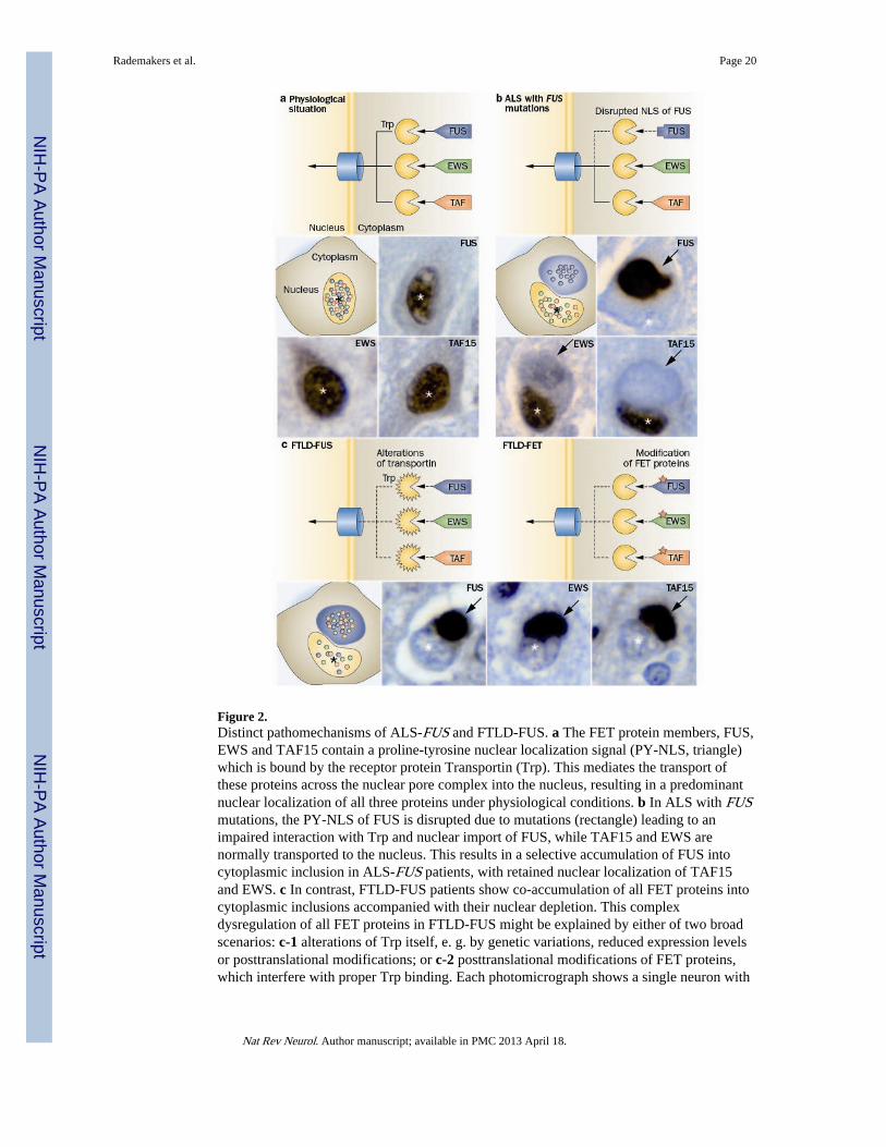
Figure 2.Distinct pathomechanisms of ALS-FUS and FTLD-FUS. a The FET protein members, FUS,EWS and TAF15 contain a proline-tyrosine nuclear localization signal (PY-NLS, triangle)which is bound by the receptor protein Transportin (Trp). This mediates the transport ofthese proteins across the nuclear pore complex into the nucleus, resulting in a predominantnuclear localization of all three proteins under physiological conditions. b In ALS with FUSmutations, the PY-NLS of FUS is disrupted due to mutations (rectangle) leading to animpaired interaction with Trp and nuclear import of FUS, while TAF15 and EWS arenormally transported to the nucleus. This results in a selective accumulation of FUS intocytoplasmic inclusion in ALS-FUS patients, with retained nuclear localization of TAF15and EWS. c In contrast, FTLD-FUS patients show co-accumulation of all FET proteins intocytoplasmic inclusions accompanied with their nuclear depletion. This complexdysregulation of all FET proteins in FTLD-FUS might be explained by either of two broadscenarios: c-1 alterations of Trp itself, e. g. by genetic variations, reduced expression levelsor posttranslational modifications; or c-2 posttranslational modifications of FET proteins,which interfere with proper Trp binding. Each photomicrograph shows a single neuron with
Rademakers et al. Page 20
Nat Rev Neurol. Author manuscript; available in PMC 2013 April 18.
NIH
-PA Author Manuscript
NIH
-PA Author Manuscript
NIH
-PA Author Manuscript

a cytoplasmic inclusion (arrow) and the nucleus indicated by the asterisk (*), immunostainedfor the FET protein indicated (brown stain).
Rademakers et al. Page 21
Nat Rev Neurol. Author manuscript; available in PMC 2013 April 18.
NIH
-PA Author Manuscript
NIH
-PA Author Manuscript
NIH
-PA Author Manuscript
NIH
-PA Author Manuscript
NIH
-PA Author Manuscript
NIH
-PA Author Manuscript
Rademakers et al. Page 22
Tabl
e 1
Mol
ecul
ar c
lass
ific
atio
n of
FT
LD
with
gen
etic
and
clin
ical
cor
rela
tions
maj
or m
olec
ular
cla
sspa
thol
ogic
al s
ubty
pe*
asso
ciat
ed g
enes
†as
soci
ated
clin
ical
phe
noty
pes
bvF
TD
PN
FA
SDpa
rkM
ND
FTL
D-t
au•
MA
PT+
(+)
(+)
+A
LS,
PL
S
•Pi
D+
+(+
)
•C
BD
++
+PL
S
•PS
P+
++
PLS
•A
GD
+
•N
FT-d
emen
tia+
•M
STD
++
PLS
•W
MT
-GG
I+
+PL
S
FTL
D-T
DP
•(T
AR
DB
P)(+
)+
AL
S
•ty
pe A
•G
RN
++
+
•ty
pe B
•C
9OR
F72
++
(+)
+A
LS
•ty
pe C
++
•ty
pe D
•V
CP
+(+
)A
LS
FTL
D-F
US
•(F
US)
(+)
AL
S
•aF
TL
D-U
+
•N
IFID
++
PLS
•B
IBD
++
AL
S
Nat Rev Neurol. Author manuscript; available in PMC 2013 April 18.

NIH
-PA Author Manuscript
NIH
-PA Author Manuscript
NIH
-PA Author Manuscript
Rademakers et al. Page 23
maj
or m
olec
ular
cla
sspa
thol
ogic
al s
ubty
pe*
asso
ciat
ed g
enes
†as
soci
ated
clin
ical
phe
noty
pes
bvF
TD
PN
FA
SDpa
rkM
ND
FTL
D-U
PS
•FT
D-3
•C
HM
P2B
+(+
)(A
LS)
FTL
D-n
i
aFT
LD
-U, a
typi
cal f
ront
otem
pora
l lob
ar d
egen
erat
ion
with
ubi
quiti
nate
d in
clus
ions
; AG
D, a
rgyr
ophi
lic g
rain
dis
ease
; AL
S, a
myo
trop
hic
late
ral s
cler
osis
; BIB
D, b
asop
hilic
incl
usio
n bo
dy d
isea
se; b
vFT
D,
beha
vior
al v
aria
nt F
TD
; C9O
RF7
2, c
hrom
osom
e 9
open
rea
ding
fra
me
72 g
ene;
CB
D, c
ortic
obas
al d
egen
erat
ion;
CH
MP2
B, c
harg
ed m
ultiv
esci
cula
r bo
dy p
rote
in 2
B g
ene;
FT
D, f
ront
otem
pora
l dem
entia
;FT
D-3
, FT
D li
nked
to c
hrom
osom
e 3;
FT
LD
, fro
ntot
empo
ral l
obar
deg
ener
atio
n; F
US,
fus
ed in
sar
com
a; G
RN
, pro
gran
ulin
gen
e; M
APT
, mic
rotu
bule
ass
ocia
ted
prot
ein
tau
gene
; MN
D, m
otor
neu
ron
dise
ase;
MST
D, m
ultip
le s
yste
m ta
uopa
thy
with
dem
entia
; NFT
-dem
entia
, neu
rofi
brill
ary
tang
le p
redo
min
ant d
emen
tia; n
i, no
incl
usio
ns; N
IFID
, neu
rona
l int
erm
edia
te f
ilam
ent i
nclu
sion
dis
ease
; par
k,pa
rkin
soni
sm; P
iD, P
ick’
s di
seas
e; P
LS,
pri
mar
y la
tera
l scl
eros
is; P
NFA
, pro
gres
sive
non
-flu
ent a
phas
ia; P
SP, p
rogr
essi
ve s
upra
nucl
ear
pals
y; S
D, s
eman
tic d
emen
tia; T
AR
DB
P, tr
ansa
ctiv
e re
spon
se D
NA
bind
ing
prot
ein
gene
; TD
P, T
DP-
43; U
PS, u
biqu
itin
prot
easo
me
syst
em; V
CP,
val
osin
con
tain
ing
prot
ein
gene
; WM
T-G
GI,
whi
te m
atte
r ta
uopa
thy
with
glo
bula
r gl
ial i
nclu
sion
s.
* indi
cate
s th
e ch
arac
teri
stic
pat
tern
of
path
olog
y, n
ot th
e cl
inic
al s
yndr
ome.
† gene
s in
whi
ch v
aria
tion
may
cau
se o
r in
crea
se th
e ri
sk o
f FT
D w
ith th
e co
rres
pond
ing
FTL
D p
atho
logi
cal s
ubty
pe.
(+)
rare
cau
se o
r un
usua
l phe
noty
pe.
Nat Rev Neurol. Author manuscript; available in PMC 2013 April 18.

NIH
-PA Author Manuscript
NIH
-PA Author Manuscript
NIH
-PA Author Manuscript
Rademakers et al. Page 24
Tabl
e 2
Freq
uenc
y of
the
C9O
RF7
2 re
peat
exp
ansi
on in
FT
D a
nd A
LS
patie
nt p
opul
atio
ns
Fam
ilial
FT
DSp
orad
ic F
TD
Fam
ilial
AL
SSp
orad
ic A
LS
Cas
es
Stud
y re
gion
aR
ef.
NC
9OR
F72
mut
atio
nN
C9O
RF
72 m
utat
ion
NC
9OR
F72
mut
atio
nN
C9O
RF
72 m
utat
ion
excl
uded
h
Eur
ope
Fi
nlan
d28
,88
2713
(48
.1%
)48
9 (1
8.8%
)11
252
(46
.4%
)28
961
(21
.1%
)Y
es
Ir
elan
d80
--
--
4718
(38
.3%
)38
619
(4.
9%)
NS
U
Kb
82-
--
-63
27 (
42.9
%)
500
35 (
7.0%
)N
o
U
Kb
8793
12 (
12.9
%)
163
6 (3
.7%
)-
--
-N
o
U
Kb
9316
120
(12
.4%
)20
916
(7.
7%)
--
--
No
N
ethe
rlan
ds88
,92
129
37 (
28.7
%)
224
5(2.
2%)
--
--
Yes
B
elgi
umc
8475
12 (
16.0
%)
230
9 (3
.9%
)15
7 (4
6.7%
)12
26
(4.9
%)
Yes
Fr
ance
8850
22 (
44.0
%)
150
14 (
9.3%
)-
--
-
G
erm
any
28,8
1,88
294(
13.8
%)
--
6915
(21
.7%
)42
122
(5.
2%)
Yes
It
aly
(mai
nlan
d)28
,81,
88,9
1-
--
-12
045
(37
.5%
)15
2355
(3.
6%)
Yes
It
aly
(Sar
dini
a)81
,88,
91-
--
-21
12 (
57.1
%)
133
9 (6
.8%
)Y
es
It
aly
(Sic
ily)
91-
--
--
-10
15
(4.9
%)
Yes
G
reec
e89
--
--
105
(50.
0%)
136
11 (
8.1%
)N
S
Nor
th A
mer
ica
U
S27
,78
171
20 (
11.7
%)
203
6 (3
.0%
)34
8 (2
3.5%
)19
58
(4.1
%)
No
U
Sd28
,88
--
--
163
59 (
36.2
%)
1014
56 (
5.5%
)N
S
C
anad
a94
--
--
6217
(27
.4%
)16
96
(3.6
%)
No
C
anad
ae27
,85
2616
(61
.5%
)3
0 (0
.0%
)-
--
-N
o
U
Se27
409
(22.
5%)
53f
8 (1
5.1%
)-
--
-N
o
U
Se79
186
(33.
3%)
60
(0.0
%)
146
(42.
9%)
43f
5 (1
1.6%
)N
S
Oth
er r
egio
ns
Is
rael
88-
--
-14
3 (2
1.4%
)-
-N
S
In
dia
88-
-31
0 (0
.0%
)-
-31
0 (0
.0%
)N
S
Nat Rev Neurol. Author manuscript; available in PMC 2013 April 18.

NIH
-PA Author Manuscript
NIH
-PA Author Manuscript
NIH
-PA Author Manuscript
Rademakers et al. Page 25
Fam
ilial
FT
DSp
orad
ic F
TD
Fam
ilial
AL
SSp
orad
ic A
LS
Cas
es
Stud
y re
gion
aR
ef.
NC
9OR
F72
mut
atio
nN
C9O
RF
72 m
utat
ion
NC
9OR
F72
mut
atio
nN
C9O
RF
72 m
utat
ion
excl
uded
h
A
siag
883
2 (6
6.7%
)10
0 (0
.0%
)20
1 (5
.0%
)23
80
(0.0
%)
NS
G
uam
88-
--
--
-90
0 (0
.0%
)N
S
A
ustr
alia
88-
--
--
-26
314
(5.
3%)
NS
AL
S, a
myo
trop
hic
late
ral s
cler
osis
; FT
D, f
ront
otem
pora
l dem
entia
; C9O
RF7
2, c
hrom
osom
e 9
open
rea
ding
fra
me
72; U
K, U
nite
d K
ingd
om; U
S, U
nite
s St
ates
.
a Onl
y ge
ogra
phic
al r
egio
ns w
ith a
t lea
st 1
0 FT
D o
r A
LS
patie
nts
are
liste
d.
b Coh
orts
are
par
t of
a la
rger
ser
ies
of U
K p
atie
nts
whi
ch a
re g
roup
ed in
Maj
ouni
e et
al.
with
hig
hly
com
para
ble
mut
atio
n fr
eque
ncie
s.56
c Num
bers
do
not i
nclu
de 2
3 pa
tient
s w
ith b
oth
FTD
and
AL
S of
whi
ch 8
5.7%
of
fam
ilial
and
6.3
% o
f sp
orad
ic p
atie
nts
wer
e C
9OR
F72
mut
atio
n ca
rrie
rs.
d In th
e sp
orad
ic A
LS
coho
rt, 5
.4%
(48
of
890)
of
whi
te a
nd 4
.1%
(2
of 4
9) o
f A
fric
an A
mer
ican
pat
ient
s ca
rrie
d a
C9O
RF7
2 re
peat
exp
ansi
on.
e Onl
y in
clud
ed p
atie
nts
with
a p
atho
logi
cal d
iagn
osis
of
FTL
D-T
DP.
f Incl
udes
indi
vidu
als
for
whi
ch n
o in
form
atio
n on
fam
ily h
isto
ry w
as a
vaila
ble.
g The
geo
grap
hica
l ori
gin
of th
e A
sian
pat
ient
s w
as n
ot r
epor
ted
in d
etai
l; ho
wev
er, t
he f
amili
al A
LS
patie
nt c
arry
ing
the
C9O
RF7
2 re
peat
exp
ansi
on w
as J
apan
ese.
h In s
tudi
es w
here
cas
es k
now
n to
car
ry m
utat
ions
in o
ther
gen
es w
ere
excl
uded
, the
fre
quen
cy o
f C
9OR
F72
repe
at e
xpan
sion
s ar
e ov
eres
timat
ed (
NS,
not
sta
ted)
.
Nat Rev Neurol. Author manuscript; available in PMC 2013 April 18.